Growth patterns of Candida albicans in relation to radicular dentin

Critical Role of Bcr1-DependentAdhesins in C. albicans BiofilmFormation In Vitro and In VivoClarissa J. Nobile
1,2, David R. Andes
3, Jeniel E. Nett
3, Frank J. Smith, Jr.
1, Fu Yue
4, Quynh-Trang Phan
4,
John E. Edwards, Jr.4,5
, Scott G. Filler4,5
, Aaron P. Mitchell1*
1 Department of Microbiology, Columbia University, New York, New York, United States of America, 2 Biological Sciences Program, Department of Biological Sciences,
Columbia University, New York, New York, United States of America, 3 Department of Medicine, Section of Infectious Diseases, University of Wisconsin, Madison, Wisconsin,
United States of America, 4 Los Angeles Biomedical Research Institute at Harbor-UCLA Medical Center, Torrance, California, United States of America, 5 The David Geffen
School of Medicine, University of California Los Angeles, Los Angeles, California, United States of America
The fungal pathogen Candida albicans is frequently associated with catheter-based infections because of its ability toform resilient biofilms. Prior studies have shown that the transcription factor Bcr1 governs biofilm formation in an invitro catheter model. However, the mechanistic role of the Bcr1 pathway and its relationship to biofilm formation invivo are unknown. Our studies of biofilm formation in vitro indicate that the surface protein Als3, a known adhesin, is akey target under Bcr1 control. We show that an als3/als3 mutant is biofilm-defective in vitro, and that ALS3overexpression rescues the biofilm defect of the bcr1/bcr1 mutant. We extend these findings with an in vivo venouscatheter model. The bcr1/bcr1 mutant is unable to populate the catheter surface, though its virulence suggests that ithas no growth defect in vivo. ALS3 overexpression rescues the bcr1/bcr1 biofilm defect in vivo, thus arguing that Als3 isa pivotal Bcr1 target in this setting. Surprisingly, the als3/als3 mutant forms a biofilm in vivo, and we suggest thatadditional Bcr1 targets compensate for the Als3 defect in vivo. Indeed, overexpression of Bcr1 targets ALS1, ECE1, andHWP1 partially restores biofilm formation in a bcr1/bcr1 mutant background in vitro, though these genes are notrequired for biofilm formation in vitro. Our findings demonstrate that the Bcr1 pathway functions in vivo to promotebiofilm formation, and that Als3-mediated adherence is a fundamental property under Bcr1 control. Known adhesinsAls1 and Hwp1 also contribute to biofilm formation, as does the novel protein Ece1.
Citation: Nobile CJ, Andes DR, Nett JE, Smith FJ Jr, Yue F, et al. (2006) Critical role of Bcr1-dependent adhesins in C. albicans biofilm formation in vitro and in vivo. PLoSPathog 2(7): e63. DOI: 10.1371/journal.ppat.0020063
Introduction
Biofilms are microbial communities that are associatedwith solid surfaces. Most bacteria and fungi exist predom-inantly in such communities in nature, and they form thebasis for numerous interactions that affect human health.Cells in a biofilm display phenotypes that are distinct fromtheir free-living counterparts, including extreme resistanceto many antimicrobial agents [1–4]. Their health impact isreflected in the fact that implanted medical devices, such asintravascular catheters, are major risk factors for blood-stream and deep tissue infection [5, 6]. These devices serve assubstrates for biofilm development; the mass and intrinsicdrug resistance of the biofilm limits efficacy of host defensesand antimicrobial therapy. These biofilm-based infectionsare estimated to cause about 50% of all nosocomialinfections [5, 7].
The fungal pathogen Candida albicans is a major cause ofdevice-associated infections [5, 8, 9]. It produces adherentbiofilms on a variety of different surfaces in vitro [3, 4, 10, 11].Biofilm formation begins with surface adherence of yeast-form cells, which grow to yield a basal layer. Basal layer cellsinclude some hyphae, or long tubular chains of cells, whichextend to yield an upper layer that is almost exclusivelyhyphae. As the biofilm matures, it produces an extracellularmatrix containing predominantly carbohydrate and protein[1, 12, 13].
C. albicans Bcr1, a C2H2 zinc finger protein, has a significantrole in biofilm formation: bcr1/bcr1 insertion and deletionmutants form only rudimentary biofilms on silicone cathetermaterial in vitro [14]. Bcr1 is required for expression ofseveral cell wall protein genes, and we have proposed thatBcr1 is a positive regulator of adherence. Many Bcr1 targetgenes had been identified initially as hyphal-specific genes,and BCR1 RNA accumulation depends upon the hyphaldevelopmental activator Tec1 [14]. Bcr1 is not required forhyphal morphogenesis, and we believe that it acts down-stream of Tec1 to activate the acquisition of hyphaladherence properties.Biofilms are considerably more complex in vivo than in
vitro. For example, in vivo, biofilms form on intravascularcatheters under conditions of vascular flow, and are exposedto and incorporate many plasma constituents. The complex-
Editor: Alexander Johnson, University of California San Francisco, United States ofAmerica
Received January 3, 2006; Accepted May 12, 2006; Published July 7, 2006
DOI: 10.1371/journal.ppat.0020063
Copyright: � 2006 Nobile et al. This is an open-access article distributed under theterms of the Creative Commons Attribution License, which permits unrestricteduse, distribution, and reproduction in any medium, provided the original authorand source are credited.
Abbreviations: CSLM, confocal scanning laser microscopy
* To whom correspondence should be addressed. E-mail: [email protected]
PLoS Pathogens | www.plospathogens.org July 2006 | Volume 2 | Issue 7 | e630636
ity involved in forming a biofilm in vivo underscores thequestion of whether the same mechanisms are required forbiofilm formation in vitro as in vivo. Indeed, several fungaland bacterial mutants have medium-dependent biofilmdefects in vitro [15, 16]. Thus, the functions of key regulatorsmust be appraised in vivo in order to connect questions indevelopmental biology to answers in antimicrobial therapy.
Recently developed animal models permit analysis of C.albicans biofilm formation in vivo. Central venous catheterinfection models have been described for both rabbits [17]and rats [18]. These catheter surfaces are substrates forextensive biofilm formation, and biofilm cells on thesesubstrates exhibit reduced antifungal susceptibility. Thesemodels further reflect the circumstances of human infection,in that the biofilm cells can lead to seeding and infection oforgans [18].
In this report, we test the roles of Bcr1 target genes inbiofilm formation in vitro. Our findings substantiate theproposal that Bcr1 is a regulator of adherence. We extend thisanalysis to an in vivo model, where our findings argue thatadherence is a fundamental property under Bcr1 control thatpromotes biofilm formation and that the adhesin Als3 is apivotal functional target of Bcr1 both in vitro and in vivo.Our findings highlight the complexity of in vivo biofilmformation, yet reveal a convergence of in vitro and in vivostudies to define a significant biofilm regulatory mechanism.
Results
Bcr1 Promotes Adherence In VitroWe proposed that Bcr1 acts in the hyphal development
pathway to promote adherence through stimulation ofexpression of several cell surface protein genes. Thishypothesis predicts that overexpression of BCR1 in yeastform cells may stimulate adherence. We tested this predictionby examining the effects of BCR1 expression in a tec1/tec1mutant (Figures 1 and 2), which is defective in producinghyphae in vitro [19]. The tec1/tec1 mutant is defective inbiofilm formation [14], and the mutant cells fail to adhere tosilicone catheter material (Figures 1A, 1C, and 2, Strain SetA). Introduction of a TEF1-BCR1 overexpression constructrestored biofilm formation ability partially to the tec1/tec1mutant (Figures 1F and 2, Strain Set A; p ¼ 0.018 for thecomparison of biomass determinations). Overexpression of
BCR1 in the tec1/tec1 mutant does not restore hyphalformation ability (Figure 1G–1I). We believe that the partialsuppression by TEF1-BCR1 reflects the failure to restorehyphal formation to the tec1/tec1 mutant. In any case, thesefindings support the idea that Bcr1 is a positive regulator ofadherence, but not of hyphal formation.To understand the mechanism of Bcr1-promoted adher-
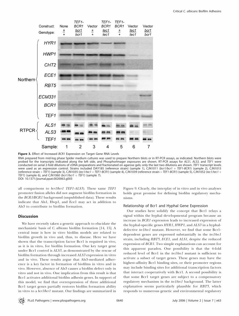
ence, we compared expression of Bcr1-dependent genes instrains with or without the TEF1-BCR1 construct (Figure 3).The genes HYR1, HWP1, CHT2, ECE1, RBT5, ALS1, and ALS3were expressed at much lower levels in a bcr1/bcr1mutant thanin the wild-type reference strain (Figure 3, samples 3 and 7).The presence of the TEF1-BCR1 construct restored expres-sion of these genes and biofilm formation in the bcr1/bcr1mutant, thus verifying the function of the construct (Figure 2;Figure 3, samples 2 and 7). The surface protein gene ECM331was expressed at higher levels in the bcr1/bcr1 mutant than inthe wild-type reference strain, and this elevated expressionwas also reversed by the TEF1-BCR1 construct (Figure 3,samples 2, 3, and 7). We note that TEF1-BCR1 did notsubstantially increase expression of Bcr1-dependent genes inthe otherwise wild-type reference strain background (Figure3, samples 5 and 3). Among the Bcr1-dependent genes, wefound that HYR1, HWP1, CHT2, ECE1, and ALS3 wereexpressed at reduced levels in the tec1/tec1 strain comparedto the reference strain (Figure 3, samples 3 and 6).Introduction of the TEF1-BCR1 construct increased expres-sion of these genes in the tec1/tec1 strain (Figure 3, samples 4and 6). These findings suggest that Bcr1 acts downstream ofTec1 to activate expression of target genes HYR1, HWP1,CHT2, ECE1, and ALS3. Furthermore, the augmented adher-ence during biofilm formation of the tec1/tec1 TEF1-BCR1strain highlights this particular group of Bcr1-dependentgenes as candidates for mediators of Bcr1-dependentadherence.
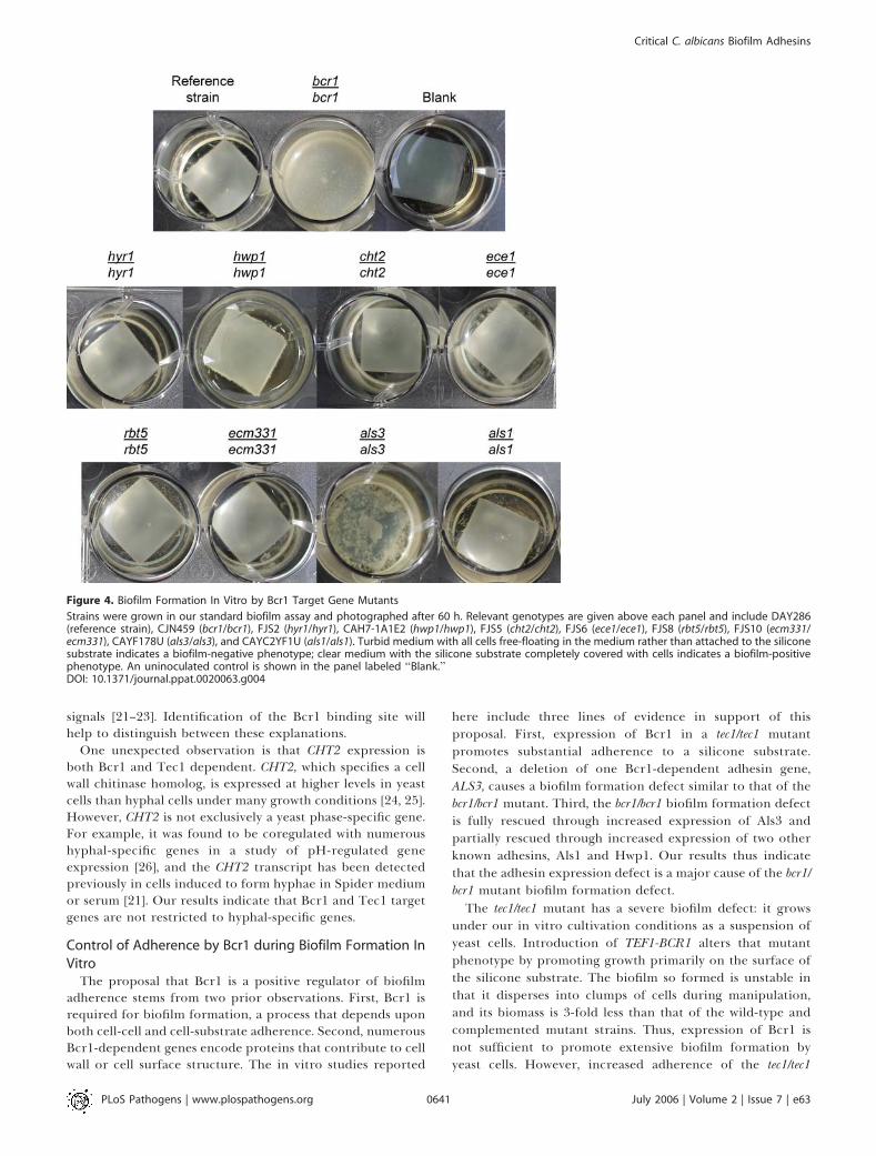
Key Role of Bcr1 Target Gene ALS3 in Biofilm Formation InVitroTo test the roles of Bcr1 target genes in biofilm formation,
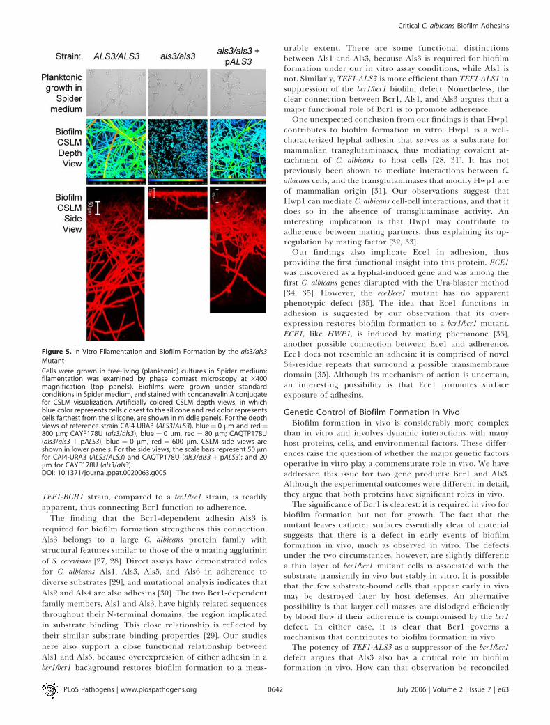
we carried out biofilm formation assays with mutantsdefective in each gene. We observed no significant biofilmdefect in hyr1/hyr1 (p¼ 0.463), ece1/ece1 (p¼ 0.850), cht2/cht2 (p¼0.909), or rbt5/rbt5 (p ¼ 0.323) mutant strains versus thereference strain (Figure 2, Strain Set B; Figure 4). The als1/als1and hwp1/hwp1 mutants also produced substantial biofilms(Figure 4), although the biofilms often sloughed off thesubstrate. Biofilm biomass determinations further indicatedthat the hwp1/hwp1 mutant has a partial biofilm defectcompared to the reference strain (Figure 2, Strain Set B, p¼ 0.022). In contrast to the als1/als1 and hwp1/hwp1 strains, theals3/als3 mutant displayed a severe defect in biofilm for-mation compared to the reference strain (Figure 2, Strain SetB; Figure 4; p¼ 0.005), and introduction of a single wild-typeALS3 allele rescued the defect substantially (Figure 2, StrainSet B; p¼ 0.005). Confocal scanning laser microscopy (CSLM)imaging revealed that the als3/als3 mutant formed a rudi-mentary biofilm of 20 lm in depth, while the wild-type andals3/als3þ pALS3 complemented strains produced biofilms ofover 200 lm in depth (Figure 5). CSLM depth images showedthat the rudimentary als3/als3 mutant biofilm was comprisedmainly of yeast cells, with few hyphae, whereas the biofilms ofthe wild-type and als3/als3 þ pALS3 complemented strains
PLoS Pathogens | www.plospathogens.org July 2006 | Volume 2 | Issue 7 | e630637
Critical C. albicans Biofilm Adhesins
Synopsis
The formation of biofilms (surface-attached microbial communities)on implanted medical devices such as catheters is a major cause offungal and bacterial infections. Prior studies of the fungal pathogenCandida albicans have shown that the regulator Bcr1 is required forbiofilm formation in vitro, but the mechanism through which itpromotes biofilm formation and its significance for biofilmformation in vivo was uncertain. The authors demonstrate thatBcr1 is required for biofilm formation in vivo in a rat model ofcatheter-based infection. Manipulation of Bcr1 target genes throughmutation and gene overexpression shows that the known surfaceadhesin Als3 has a pivotal role in biofilm formation and thatadhesins Als1 and Hwp1 also contribute to biofilm formation. Theresults thus indicate that adherence is the key property regulated byBcr1 and highlight a group of adhesins as potential therapeutictargets.
included abundant hyphae (Figure 5). It should be noted thatthe als3/als3 mutant is not defective in hyphal formation as itforms normal hyphae when assayed under hyphal inducingconditions (Figure 5). Hyphae are also apparent among thecells in the surrounding medium of an als3/als3 mutantbiofilm (unpublished data). These findings argue that Als3 hasa major role in biofilm formation and suggest that reducedexpression of ALS3 in the bcr1/bcr1mutant may account for itsbiofilm defect.
If reduced expression of ALS3 is the cause of the bcr1/bcr1mutant biofilm defect, then increased expression of ALS3 in abcr1/bcr1 mutant background should promote biofilm for-mation. To test this prediction, we introduced the TEF1promoter adjacent to the native ALS3 coding region to createa TEF1-ALS3 allele, permitting Bcr1-independent ALS3expression. RT-PCR measurement of ALS3 RNA levelsconfirmed that the TEF1-ALS3 allele permits expression ofALS3 in both BCR1/BCR1 and bcr1/bcr1 backgrounds (Figure6). In the wild-type reference strain background, TEF1-ALS3had no obvious effect on biofilm formation (Figure 6, toprow). In the bcr1/bcr1 mutant background, TEF1-ALS3 im-proved biofilm formation substantially (Figure 2; Figure 6,top row; p ¼ 0.002). These observations indicate that
increased ALS3 expression in the bcr1/bcr1 mutant promotessignificant biofilm formation ability.We used CSLM imaging to examine the structure of
biofilms that resulted from increased ALS3 expression. TheTEF1-ALS3 allele did not alter biofilm structure in theotherwise wild-type background (Figure 6, CSLM depth andside views): biofilm depth was about 400 lm; little stainingoccurred in the basal region; and hyphal staining wasprominent. The bcr1/bcr1 strain produced a thin rudimentarybiofilm comprised largely of yeast form cells, as expected [14].The bcr1/bcr1 TEF1-ALS3 strain produced a substantial biofilmthat included a basal poorly stained region (Figure 6), similarin appearance to those of the wild-type strain (Figure 6) andcomplemented bcr1/bcr1 mutant [14]. Thus, increased ALS3expression permits at least partial rescue of the bcr1/bcr1mutant defect in biofilm formation.
Bcr1 Function in Biofilm Formation In VivoIn order to determine whether Bcr1 may have a role in
biofilm formation in vivo, we turned to a rat venous cathetermodel [18]. Implanted catheters were allowed to stabilize for24 h and were then inoculated with wild-type, bcr1/bcr1mutant, or bcr1/bcr1 þ pBCR1 complemented strains. Biofilmformation was visualized after 12, 24, and 48 h by scanning
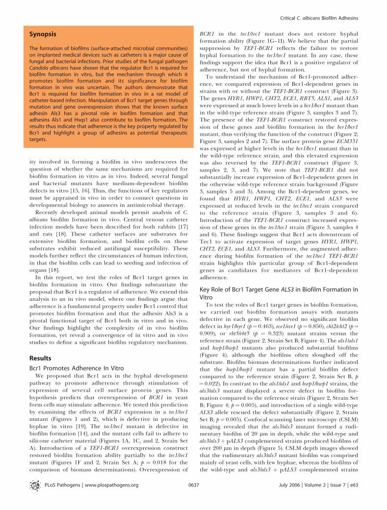
Figure 1. Effect of Increased BCR1 Expression on Adherence and Hyphal Morphogenesis
Strains were grown under in vitro biofilm assay conditions for 60 h and photographed (A–F) or grown in Spider suspension cultures and examined byphase contrast microscopy at 3400 magnification (G–I). For the biofilms assays, turbid medium with all cells free-floating in the medium rather thanattached to the silicone substrate indicates a biofilm-negative phenotype; clear medium with the silicone substrate completely covered with cellsindicates a biofilm-positive phenotype. Relevant genotypes are given above each panel for strains CJN1015 (reference strainþ TEF1) (A, G), CJN1060(bcr1/bcr1þ TEF1) (B), CJN1052 (tec1/tec1þ TEF1) (C, H), CJN1039 (reference strainþ TEF1-BCR1) (D), CJN1011 (bcr1/bcr1þ TEF1-BCR1) (E), and CJN1035(tec1/tec1þ TEF1-BCR1) (F, I).DOI: 10.1371/journal.ppat.0020063.g001
PLoS Pathogens | www.plospathogens.org July 2006 | Volume 2 | Issue 7 | e630638
Critical C. albicans Biofilm Adhesins
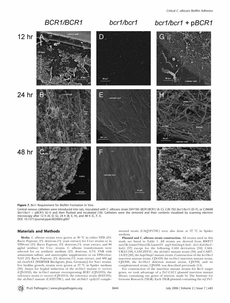
electron microscopy of the intraluminal catheter surface(Figure 7). The wild-type and bcr1/bcr1 þ pBCR1 comple-mented strains initiated biofilm formation by 12 h andyielded extensive adherent populations by 24 h (Figure 7A,7B, 7G, and 7H). Both strains produced mature biofilms by 48h that included abundant matrix material (Figure 7C and 7I),as previously reported for strain K1 [18]. In contrast, the bcr1/bcr1 mutant yielded few adherent cells at 12 and 24 h (Figure7D and 7E), and the catheter surface was devoid of biofilmmaterial after 48 h (Figure 7F). Despite the dramatic differ-ences in biofilm formation ability, the three strains grewcomparably in a mouse disseminated infection model;median mouse survival time was 13 d after inoculation withthe wild-type strain and 10 d after inoculation with either thebcr1/bcr1 mutant or bcr1/bcr1 þ pBCR1 complemented strains.Based on this evidence, Bcr1 is not required for growth invivo under non–biofilm-forming conditions but is requiredfor biofilm formation in vivo.
Als3 Function in Biofilm Formation In VivoOur observations above indicate that Als3 is a key mediator
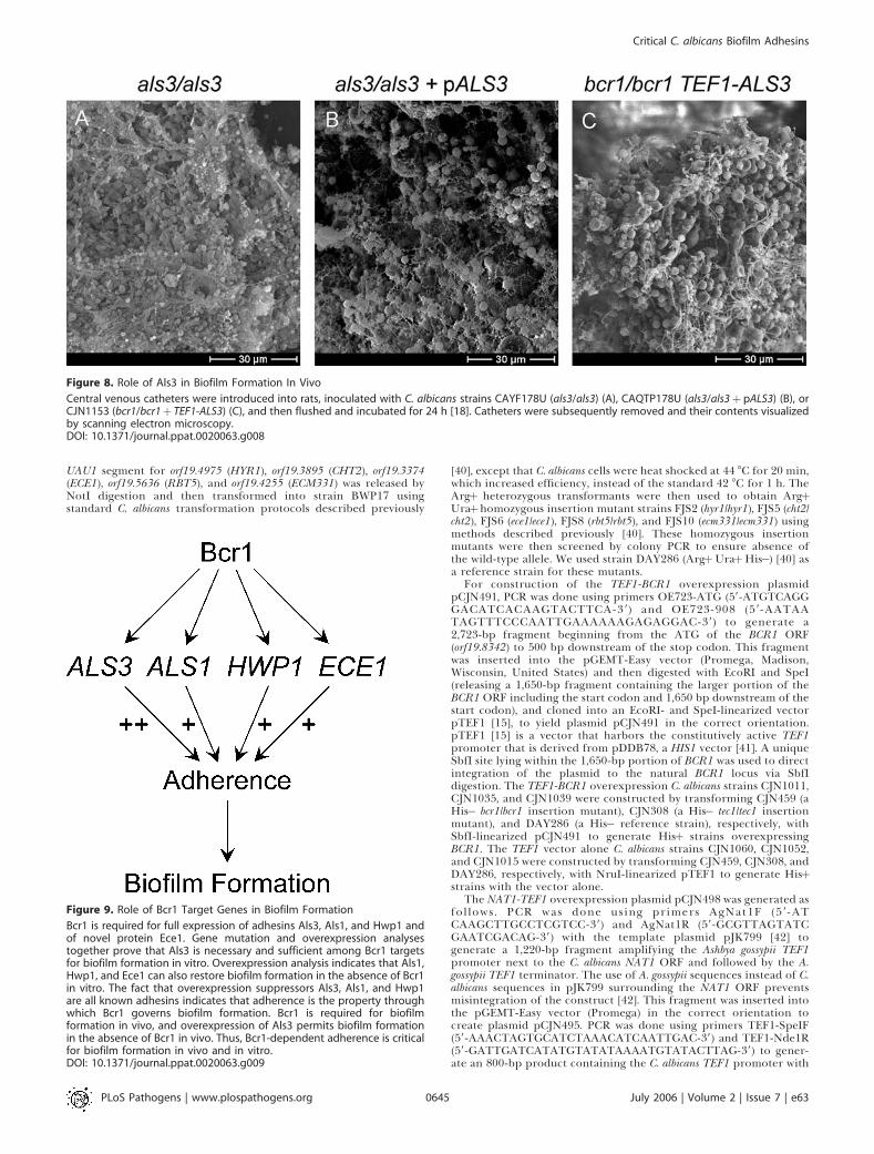
of Bcr1-dependent biofilm formation in vitro. To verify thatthese findings extend to in vivo biofilm formation, wecompared als3/als3 mutant and als3/als3 þ pALS3 comple-mented strains in the rat venous catheter model. Both strainsformed extensive biofilms within 24 h (Figure 8A and 8B).Therefore, Als3 is not absolutely required for biofilmformation in vivo.
To determine whether Als3 may contribute to biofilmformation in vivo, we tested the ability of the TEF1-ALS3expression construct to rescue the bcr1/bcr1 mutant biofilmdefect. The bcr1/bcr1 TEF1-ALS3 strain produced an extensive
biofilm containing both cells and matrix material (Figure 8C).This biofilm, formed after 24 h, was similar in overallappearance to that formed by the BCR1/BCR1 control strains(Figures 7B and 8B). TEF1-ALS3 expression thus rescuesbiofilm formation in a bcr1/bcr1 background (compare Figures7E and 8C). These findings support the model that ALS3 is acritical Bcr1 target gene that functions in biofilm formationin vivo.
Overexpression Assays of Bcr1 Target Gene Function InVitroOur in vivo assays suggest that Als3 may be one of several
Bcr1 targets that contribute to biofilm formation. Theanalysis of insertion and deletion mutant strains abovepointed toward Als1 and Hwp1 as additional candidatefunctional targets, although their biofilm defects were mild:biofilm biomass was reduced only slightly (Figure 2), andCSLM visualization revealed no qualitative defects (unpub-lished data). Thus, we turned to an alternative functionalanalysis strategy, gene overexpression, which has recentlybeen applied with considerable success on a genome-widescale in Saccharomyces cerevisiae [20]. Gene overexpression isparticularly useful in identifying functions among partiallyredundant genes, the situation that we postulate to exist here.To determine if increased expression of Bcr1-activated
target genes, other than ALS3, may rescue the biofilm defectof the bcr1/bcr1 mutant, we created genomic fusions of theTEF1 promoter to the CHT2, HYR1, RBT5, ALS1, HWP1, andECE1 coding regions. The TEF1-ALS1, TEF1-HWP1, and TEF1-ECE1 alleles improved biofilm formation ability considerably(p , 0.004 for all comparisons to bcr1/bcr1), although not tothe extent of TEF1-ALS3 (Figure 2, Strain Set C; p , 0.006 for
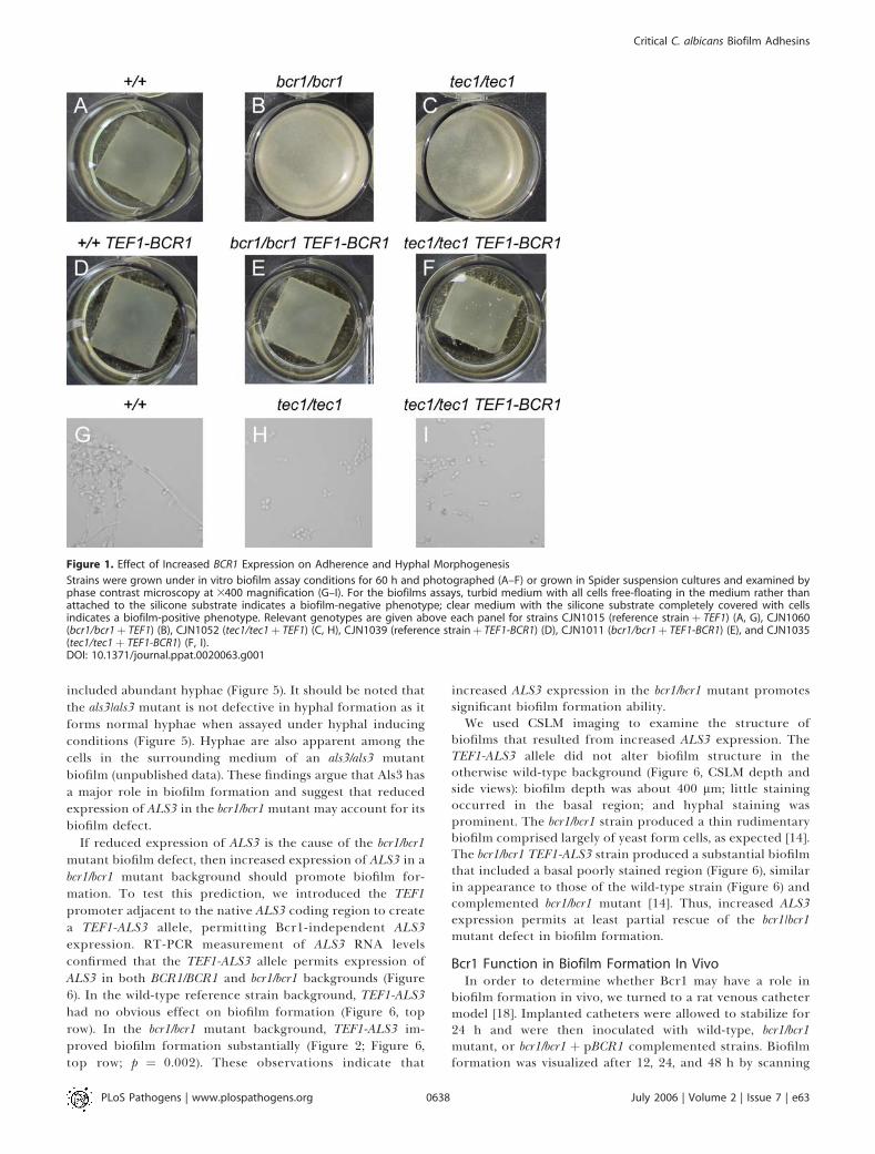
Figure 2. Biofilm Dry Mass Determinations
Biofilm dry mass determinations were made in quadruplicate after 60 h growth under standard biofilm conditions, as detailed in Materials and Methods.Reference strains DAY185 (shown) and CAI4-URA3 (not shown) gave similar results. Strains are grouped for convenience of comparison. Strain Set Acontains CJN896 (tec1/tec1), CJN1052 (tec1/tec1 þ TEF1), CJN1035 (tec1/tec1 þ TEF1-BCR1), CJN1023 (tec1/tec1 þ pTEC1), CJN702 (bcr1/bcr1), CJN1060(bcr1/bcr1þ TEF1), CJN1011 (bcr1/bcr1þ TEF1-BCR1), CJN698 (bcr1/bcr1þ pBCR1), respectively. Strain Set B contains FJS2 (hyr1/hyr1), FJS6 (ece1/ece1),FJS5 (cht2/cht2), FJS8 (rbt5/rbt5), CAYC2YF1U (als1/als1), CAH7-1A1E2 (hwp1/hwp1), CAYF178U (als3/als3), CAQTP178U (als3/als3þ pALS3), respectively.Strain Set C contains CJN1153 (bcr1/bcr1þ TEF1-ALS3), CJN1144 (bcr1/bcr1þ TEF1-ALS1), CJN1288 (bcr1/bcr1þ TEF1-ECE1), CJN1222 (bcr1/bcr1þ TEF1-HWP1), CJN1281 (bcr1/bcr1þ TEF1-CHT2), CJN1259 (bcr1/bcr1þ TEF1-HYR1), CJN1276 (bcr1/bcr1þ TEF1-RBT5), respectively.DOI: 10.1371/journal.ppat.0020063.g002
PLoS Pathogens | www.plospathogens.org July 2006 | Volume 2 | Issue 7 | e630639
Critical C. albicans Biofilm Adhesins
all comparisons to bcr1/bcr1 TEF1-ALS3). These same TEF1promoter fusion alleles did not augment biofilm formation inthe BCR1/BCR1 background (unpublished data). These resultsindicate that Als1, Hwp1, and Ece1 may act in addition toAls3 to contribute to biofilm formation.
Discussion
We have recently taken a genetic approach to elucidate themechanistic basis of C. albicans biofilm formation [14, 15]. Acentral issue is how in vitro biofilm models are related tobiofilm growth in vivo and, thus, to disease. Here we haveshown that the transcription factor Bcr1 is required in vivo,as it is in vitro, for biofilm formation. One key target geneunder Bcr1 control is ALS3, as demonstrated by the rescue ofbiofilm formation through increased ALS3 expression in vitroand in vivo. These results argue that Als3-mediated adher-ence is a key factor in formation of biofilms in vitro and invivo. However, absence of Als3 causes a biofilm defect only invitro and not in vivo. One implication from this result is thatBcr1 activates additional biofilm adhesin genes. In support ofthis model, we find that overexpression of three additionalBcr1 target genes partially restores biofilm formation abilityin vitro to a bcr1/bcr1 mutant. Our findings are summarized in
Figure 9. Clearly, the interplay of in vitro and in vivo analysesholds great promise for defining biofilm regulatory mecha-nisms.
Relationship of Bcr1 and Hyphal Gene ExpressionOur studies here solidify the concept that Bcr1 relays a
signal within the hyphal developmental program because anincrease in BCR1 expression leads to increased expression ofthe hyphal-specific genes HYR1, HWP1, and ALS3 in a hyphal-defective tec1/tec1 mutant. However, we find that some Bcr1-dependent genes are expressed substantially in the tec1/tec1strain, including RBT5, ECE1, and ALS1, despite the reducedexpression of BCR1. Two simple explanations can account forthis apparent paradox. One possibility is that the 4-foldreduced level of Bcr1 in the tec1/tec1 mutant is sufficient toactivate a subset of target genes. These genes may have thehighest-affinity Bcr1 binding sites, or their promoter regionsmay include binding sites for additional transcription factorsthat interact cooperatively with Bcr1. A second possibility isthat some Bcr1 target genes are subject to a compensatoryregulatory mechanism in the tec1/tec1 background. The latterexplanation seems particularly plausible for RBT5, whichresponds to numerous genetic and environmental regulatory
Figure 3. Effect of Increased BCR1 Expression on Target Gene RNA Levels
RNA prepared from mid-log phase Spider medium cultures was used to prepare Northern blots or in RT-PCR assays, as indicated. Northern blots wereprobed for the transcripts indicated along the left side, and PhosphorImager exposures are shown. RT-PCR assays for ALS1, ALS3, and TEF1 wereconducted on serial 2-fold dilutions of cDNA preparations and fractionated on agarose gels; only the last two dilutions are shown. TEF1 transcript levelswere used as an expression control. Strains included DAY185 (reference strain) (sample 1), CJN1011 (bcr1/bcr1 þ TEF1-BCR1) (sample 2), CJN1015(reference strainþTEF1) (sample 3), CJN1035 (tec1/tec1þTEF1-BCR1) (sample 4), CJN1039 (reference strainþTEF1-BCR1) (sample 5), CJN1052 (tec1/tec1þTEF1) (sample 6), and CJN1060 (bcr1/bcr1þ TEF1) (sample 7).DOI: 10.1371/journal.ppat.0020063.g003
PLoS Pathogens | www.plospathogens.org July 2006 | Volume 2 | Issue 7 | e630640
Critical C. albicans Biofilm Adhesins
signals [21–23]. Identification of the Bcr1 binding site willhelp to distinguish between these explanations.
One unexpected observation is that CHT2 expression isboth Bcr1 and Tec1 dependent. CHT2, which specifies a cellwall chitinase homolog, is expressed at higher levels in yeastcells than hyphal cells under many growth conditions [24, 25].However, CHT2 is not exclusively a yeast phase-specific gene.For example, it was found to be coregulated with numeroushyphal-specific genes in a study of pH-regulated geneexpression [26], and the CHT2 transcript has been detectedpreviously in cells induced to form hyphae in Spider mediumor serum [21]. Our results indicate that Bcr1 and Tec1 targetgenes are not restricted to hyphal-specific genes.
Control of Adherence by Bcr1 during Biofilm Formation InVitro
The proposal that Bcr1 is a positive regulator of biofilmadherence stems from two prior observations. First, Bcr1 isrequired for biofilm formation, a process that depends uponboth cell-cell and cell-substrate adherence. Second, numerousBcr1-dependent genes encode proteins that contribute to cellwall or cell surface structure. The in vitro studies reported
here include three lines of evidence in support of thisproposal. First, expression of Bcr1 in a tec1/tec1 mutantpromotes substantial adherence to a silicone substrate.Second, a deletion of one Bcr1-dependent adhesin gene,ALS3, causes a biofilm formation defect similar to that of thebcr1/bcr1 mutant. Third, the bcr1/bcr1 biofilm formation defectis fully rescued through increased expression of Als3 andpartially rescued through increased expression of two otherknown adhesins, Als1 and Hwp1. Our results thus indicatethat the adhesin expression defect is a major cause of the bcr1/bcr1 mutant biofilm formation defect.The tec1/tec1 mutant has a severe biofilm defect: it grows
under our in vitro cultivation conditions as a suspension ofyeast cells. Introduction of TEF1-BCR1 alters that mutantphenotype by promoting growth primarily on the surface ofthe silicone substrate. The biofilm so formed is unstable inthat it disperses into clumps of cells during manipulation,and its biomass is 3-fold less than that of the wild-type andcomplemented mutant strains. Thus, expression of Bcr1 isnot sufficient to promote extensive biofilm formation byyeast cells. However, increased adherence of the tec1/tec1
Figure 4. Biofilm Formation In Vitro by Bcr1 Target Gene Mutants
Strains were grown in our standard biofilm assay and photographed after 60 h. Relevant genotypes are given above each panel and include DAY286(reference strain), CJN459 (bcr1/bcr1), FJS2 (hyr1/hyr1), CAH7-1A1E2 (hwp1/hwp1), FJS5 (cht2/cht2), FJS6 (ece1/ece1), FJS8 (rbt5/rbt5), FJS10 (ecm331/ecm331), CAYF178U (als3/als3), and CAYC2YF1U (als1/als1). Turbid medium with all cells free-floating in the medium rather than attached to the siliconesubstrate indicates a biofilm-negative phenotype; clear medium with the silicone substrate completely covered with cells indicates a biofilm-positivephenotype. An uninoculated control is shown in the panel labeled ‘‘Blank.’’DOI: 10.1371/journal.ppat.0020063.g004
PLoS Pathogens | www.plospathogens.org July 2006 | Volume 2 | Issue 7 | e630641
Critical C. albicans Biofilm Adhesins
TEF1-BCR1 strain, compared to a tec1/tec1 strain, is readilyapparent, thus connecting Bcr1 function to adherence.
The finding that the Bcr1-dependent adhesin Als3 isrequired for biofilm formation strengthens this connection.Als3 belongs to a large C. albicans protein family withstructural features similar to those of the a mating agglutininof S. cerevisiae [27, 28]. Direct assays have demonstrated rolesfor C. albicans Als1, Als3, Als5, and Als6 in adherence todiverse substrates [29], and mutational analysis indicates thatAls2 and Als4 are also adhesins [30]. The two Bcr1-dependentfamily members, Als1 and Als3, have highly related sequencesthroughout their N-terminal domains, the region implicatedin substrate binding. This close relationship is reflected bytheir similar substrate binding properties [29]. Our studieshere also support a close functional relationship betweenAls1 and Als3, because overexpression of either adhesin in abcr1/bcr1 background restores biofilm formation to a meas-
urable extent. There are some functional distinctionsbetween Als1 and Als3, because Als3 is required for biofilmformation under our in vitro assay conditions, while Als1 isnot. Similarly, TEF1-ALS3 is more efficient than TEF1-ALS1 insuppression of the bcr1/bcr1 biofilm defect. Nonetheless, theclear connection between Bcr1, Als1, and Als3 argues that amajor functional role of Bcr1 is to promote adherence.One unexpected conclusion from our findings is that Hwp1
contributes to biofilm formation in vitro. Hwp1 is a well-characterized hyphal adhesin that serves as a substrate formammalian transglutaminases, thus mediating covalent at-tachment of C. albicans to host cells [28, 31]. It has notpreviously been shown to mediate interactions between C.albicans cells, and the transglutaminases that modify Hwp1 areof mammalian origin [31]. Our observations suggest thatHwp1 can mediate C. albicans cell-cell interactions, and that itdoes so in the absence of transglutaminase activity. Aninteresting implication is that Hwp1 may contribute toadherence between mating partners, thus explaining its up-regulation by mating factor [32, 33].Our findings also implicate Ece1 in adhesion, thus
providing the first functional insight into this protein. ECE1was discovered as a hyphal-induced gene and was among thefirst C. albicans genes disrupted with the Ura-blaster method[34, 35]. However, the ece1/ece1 mutant has no apparentphenotypic defect [35]. The idea that Ece1 functions inadhesion is suggested by our observation that its over-expression restores biofilm formation to a bcr1/bcr1 mutant.ECE1, like HWP1, is induced by mating pheromone [33],another possible connection between Ece1 and adherence.Ece1 does not resemble an adhesin: it is comprised of novel34-residue repeats that surround a possible transmembranedomain [35]. Although its mechanism of action is uncertain,an interesting possibility is that Ece1 promotes surfaceexposure of adhesins.
Genetic Control of Biofilm Formation In VivoBiofilm formation in vivo is considerably more complex
than in vitro and involves dynamic interactions with manyhost proteins, cells, and environmental factors. These differ-ences raise the question of whether the major genetic factorsoperative in vitro play a commensurate role in vivo. We haveaddressed this issue for two gene products: Bcr1 and Als3.Although the experimental outcomes were different in detail,they argue that both proteins have significant roles in vivo.The significance of Bcr1 is clearest: it is required in vivo for
biofilm formation but not for growth. The fact that themutant leaves catheter surfaces essentially clear of materialsuggests that there is a defect in early events of biofilmformation in vivo, much as observed in vitro. The defectsunder the two circumstances, however, are slightly different:a thin layer of bcr1/bcr1 mutant cells is associated with thesubstrate transiently in vivo but stably in vitro. It is possiblethat the few substrate-bound cells that appear early in vivomay be destroyed later by host defenses. An alternativepossibility is that larger cell masses are dislodged efficientlyby blood flow if their adherence is compromised by the bcr1defect. In either case, it is clear that Bcr1 governs amechanism that contributes to biofilm formation in vivo.The potency of TEF1-ALS3 as a suppressor of the bcr1/bcr1
defect argues that Als3 also has a critical role in biofilmformation in vivo. How can that observation be reconciled
Figure 5. In Vitro Filamentation and Biofilm Formation by the als3/als3
Mutant
Cells were grown in free-living (planktonic) cultures in Spider medium;filamentation was examined by phase contrast microscopy at 3400magnification (top panels). Biofilms were grown under standardconditions in Spider medium, and stained with concanavalin A conjugatefor CSLM visualization. Artificially colored CSLM depth views, in whichblue color represents cells closest to the silicone and red color representscells farthest from the silicone, are shown in middle panels. For the depthviews of reference strain CAI4-URA3 (ALS3/ALS3), blue¼ 0 lm and red¼800 lm; CAYF178U (als3/als3), blue ¼ 0 lm, red ¼ 80 lm; CAQTP178U(als3/als3 þ pALS3), blue ¼ 0 lm, red ¼ 600 lm. CSLM side views areshown in lower panels. For the side views, the scale bars represent 50 lmfor CAI4-URA3 (ALS3/ALS3) and CAQTP178U (als3/als3 þ pALS3); and 20lm for CAYF178U (als3/als3).DOI: 10.1371/journal.ppat.0020063.g005
PLoS Pathogens | www.plospathogens.org July 2006 | Volume 2 | Issue 7 | e630642
Critical C. albicans Biofilm Adhesins

with the fact that an als3/als3 null mutant has no biofilmdefect in vivo? One simple model is that additional adhesinscan partially compensate for the absence of Als3 in vivo butnot in vitro. Our overexpression studies implicate Als1 andHwp1 as candidate compensatory adhesins, in keeping withthis model. The distinction between the in vivo and in vitrosituations may reflect a higher level of expression of thecompensatory adhesins in vivo than in vitro. A secondpossibility is that host constituents, for example serumcomponents, may contribute to adherence. Thus, the samelow level of surface adhesin activity may support biofilmformation in vivo but not in vitro.
The restoration of biofilm formation through ALS3 over-expression in the bcr1/bcr1 mutant both in vitro and in vivoindicates that Bcr1 governs one main function relevant forbiofilm formation: adherence. Although transcription factormutants are useful for definition of functionally relatedgenes, the mechanistic basis for their phenotypic defects canbe complex because of the extent of their gene expressiondefects. Moreover, functional overlap among targets canobscure loss-of-function target gene mutant phenotypes. Ourresults here illustrate the utility of gene overexpression foridentification of critical target genes that govern a complexprocess.
Figure 6. Overexpression of ALS3 in the bcr1/bcr1 Mutant Restores Substantial Biofilm Formation In Vitro
Biofilms were grown under standard conditions and stained with concanavalin A conjugate for CSLM visualization. The top panels show the visualappearance. The next set of panels show depth views, in which blue color represents cells closest to the silicone and red color represents cells farthestfrom the silicone. The next set of panels show side views. For the depth views of reference strain DAY185 (BCR1/BCR1), blue¼ 0 lm and red¼ 600 lm;CJN1149 (BCR1/BCR1þ TEF1-ALS3), blue¼ 0 lm and red¼ 500 lm; CJN702 (bcr1/bcr1), blue¼ 0 lm and red¼ 80 lm; CJN1153 (bcr1/bcr1þ TEF1-ALS3),blue ¼ 0 lm and red ¼ 180 lm. For the side views, the scale bars represent 50 lm for DAY185 (BCR1/BCR1), CJN1149 (BCR1/BCR1þ TEF1-ALS3), andCJN1153 (bcr1/bcr1þ TEF1-ALS3); and 20 lm for CJN702 (bcr1/bcr1). The next set of panels show RT-PCR analysis of ALS3 expression of the indicatedstrains with successive 2-fold dilutions of cDNA from left to right. The bottom panels show RT-PCR of control TEF1 transcript levels.DOI: 10.1371/journal.ppat.0020063.g006
PLoS Pathogens | www.plospathogens.org July 2006 | Volume 2 | Issue 7 | e630643
Critical C. albicans Biofilm Adhesins
Materials and Methods
Media. C. albicans strains were grown at 30 8C in either YPD (2%Bacto Peptone, 2% dextrose,1% yeast extract) for Uraþ strains or inYPDþuri (2% Bacto Peptone, 2% dextrose,1% yeast extract, and 80lg/ml uridine) for Ura� strains. C. albicans transformants wereselected for on synthetic medium (2% dextrose, 6.7% YNB withammonium sulfate, and auxotrophic supplements) or on YPDþclon-NAT (2% Bacto Peptone, 2% dextrose,1% yeast extract, and 400 lg/ml clonNAT [WERNER BioAgents, Jena, Germany]) for Natþ strains.For biofilm growth, strains were grown at 37 8C in Spider medium[36]. Assays for hyphal induction of the tec1/tec1 mutant (þ vector)(CJN1052), the tec1/tec1 mutant overexpressing BCR1 (CJN1035), thereference strain (þ vector) (CJN1015), the reference strain (DAY185),the als3/als3 mutant (CAYF178U), and the als3/als3 þpALS3 comple-
mented strain (CAQTP178U) were also done at 37 8C in Spidermedium.
Plasmid and C. albicans strain construction. All strains used in thisstudy are listed in Table 1. All strains are derived from BWP17(ura3D::kimm434/ura3D::kimm434 arg4::hisG/arg4::hisG his1::hisG/his1::hisG) [37] except for the following CAI4 derivatives [34]: CAI4-URA3 [38], CAYC2YF1U, the als1/als1 mutant strain [39], and CAH7-1A1E2 [28], the hwp1/hwp1mutant strain. Construction of the bcr1/bcr1insertion mutant strain, CJN459; the tec1/tec1 insertion mutant strain,CJN308; the bcr1/bcr1 deletion mutant strain, CJN702, and itscomplemented strain, CJN698, was described previously [14].
For construction of the insertion mutant strains for Bcr1 targetgenes, we took advantage of a Tn7-UAU1 plasmid insertion mutantlibrary containing our genes of interest, made by The Institute forGenome Research (TIGR). Each TIGR plasmid containing the orf::Tn7-
Figure 7. Bcr1 Requirement for Biofilm Formation In Vivo
Central venous catheters were introduced into rats, inoculated with C. albicans strain DAY185 (BCR1/BCR1) (A–C), CJN 702 (bcr1/bcr1) (D–F), or CJN698(bcr1/bcr1 þ pBCR1) (G–I) and then flushed and incubated [18]. Catheters were the removed and their contents visualized by scanning electronmicroscopy after 12 h (A, D, G), 24 h (B, E, H), and 48 h (C, F, I).DOI: 10.1371/journal.ppat.0020063.g007
PLoS Pathogens | www.plospathogens.org July 2006 | Volume 2 | Issue 7 | e630644
Critical C. albicans Biofilm Adhesins
UAU1 segment for orf19.4975 (HYR1), orf19.3895 (CHT2), orf19.3374(ECE1), orf19.5636 (RBT5), and orf19.4255 (ECM331) was released byNotI digestion and then transformed into strain BWP17 usingstandard C. albicans transformation protocols described previously
[40], except that C. albicans cells were heat shocked at 44 8C for 20 min,which increased efficiency, instead of the standard 42 8C for 1 h. TheArgþ heterozygous transformants were then used to obtain ArgþUraþhomozygous insertion mutant strains FJS2 (hyr1/hyr1), FJS5 (cht2/cht2), FJS6 (ece1/ece1), FJS8 (rbt5/rbt5), and FJS10 (ecm331/ecm331) usingmethods described previously [40]. These homozygous insertionmutants were then screened by colony PCR to ensure absence ofthe wild-type allele. We used strain DAY286 (ArgþUraþHis�) [40] asa reference strain for these mutants.
For construction of the TEF1-BCR1 overexpression plasmidpCJN491, PCR was done using primers OE723-ATG (59-ATGTCAGGGACATCACAAGTACTTCA-39) and OE723-908 (59-AATAATAGTTTCCCAATTGAAAAAAGAGAGGAC-39) to generate a2,723-bp fragment beginning from the ATG of the BCR1 ORF(orf19.8342) to 500 bp downstream of the stop codon. This fragmentwas inserted into the pGEMT-Easy vector (Promega, Madison,Wisconsin, United States) and then digested with EcoRI and SpeI(releasing a 1,650-bp fragment containing the larger portion of theBCR1 ORF including the start codon and 1,650 bp downstream of thestart codon), and cloned into an EcoRI- and SpeI-linearized vectorpTEF1 [15], to yield plasmid pCJN491 in the correct orientation.pTEF1 [15] is a vector that harbors the constitutively active TEF1promoter that is derived from pDDB78, a HIS1 vector [41]. A uniqueSbfI site lying within the 1,650-bp portion of BCR1 was used to directintegration of the plasmid to the natural BCR1 locus via SbfIdigestion. The TEF1-BCR1 overexpression C. albicans strains CJN1011,CJN1035, and CJN1039 were constructed by transforming CJN459 (aHis� bcr1/bcr1 insertion mutant), CJN308 (a His� tec1/tec1 insertionmutant), and DAY286 (a His� reference strain), respectively, withSbfI-linearized pCJN491 to generate Hisþ strains overexpressingBCR1. The TEF1 vector alone C. albicans strains CJN1060, CJN1052,and CJN1015 were constructed by transforming CJN459, CJN308, andDAY286, respectively, with NruI-linearized pTEF1 to generate Hisþstrains with the vector alone.
The NAT1-TEF1 overexpression plasmid pCJN498 was generated asfollows. PCR was done using primers AgNat1F (59-ATCAAGCTTGCCTCGTCC-39) and AgNat1R (59-GCGTTAGTATCGAATCGACAG-39) with the template plasmid pJK799 [42] togenerate a 1,220-bp fragment amplifying the Ashbya gossypii TEF1promoter next to the C. albicans NAT1 ORF and followed by the A.gossypii TEF1 terminator. The use of A. gossypii sequences instead of C.albicans sequences in pJK799 surrounding the NAT1 ORF preventsmisintegration of the construct [42]. This fragment was inserted intothe pGEMT-Easy vector (Promega) in the correct orientation tocreate plasmid pCJN495. PCR was done using primers TEF1-SpeIF(59-AAACTAGTGCATCTAAACATCAATTGAC-39) and TEF1-Nde1R(59-GATTGATCATATGTATATAAAATGTATACTTAG-39) to gener-ate an 800-bp product containing the C. albicans TEF1 promoter with
Figure 8. Role of Als3 in Biofilm Formation In Vivo
Central venous catheters were introduced into rats, inoculated with C. albicans strains CAYF178U (als3/als3) (A), CAQTP178U (als3/als3þ pALS3) (B), orCJN1153 (bcr1/bcr1þ TEF1-ALS3) (C), and then flushed and incubated for 24 h [18]. Catheters were subsequently removed and their contents visualizedby scanning electron microscopy.DOI: 10.1371/journal.ppat.0020063.g008
Figure 9. Role of Bcr1 Target Genes in Biofilm Formation
Bcr1 is required for full expression of adhesins Als3, Als1, and Hwp1 andof novel protein Ece1. Gene mutation and overexpression analysestogether prove that Als3 is necessary and sufficient among Bcr1 targetsfor biofilm formation in vitro. Overexpression analysis indicates that Als1,Hwp1, and Ece1 can also restore biofilm formation in the absence of Bcr1in vitro. The fact that overexpression suppressors Als3, Als1, and Hwp1are all known adhesins indicates that adherence is the property throughwhich Bcr1 governs biofilm formation. Bcr1 is required for biofilmformation in vivo, and overexpression of Als3 permits biofilm formationin the absence of Bcr1 in vivo. Thus, Bcr1-dependent adherence is criticalfor biofilm formation in vivo and in vitro.DOI: 10.1371/journal.ppat.0020063.g009
PLoS Pathogens | www.plospathogens.org July 2006 | Volume 2 | Issue 7 | e630645
Critical C. albicans Biofilm Adhesins
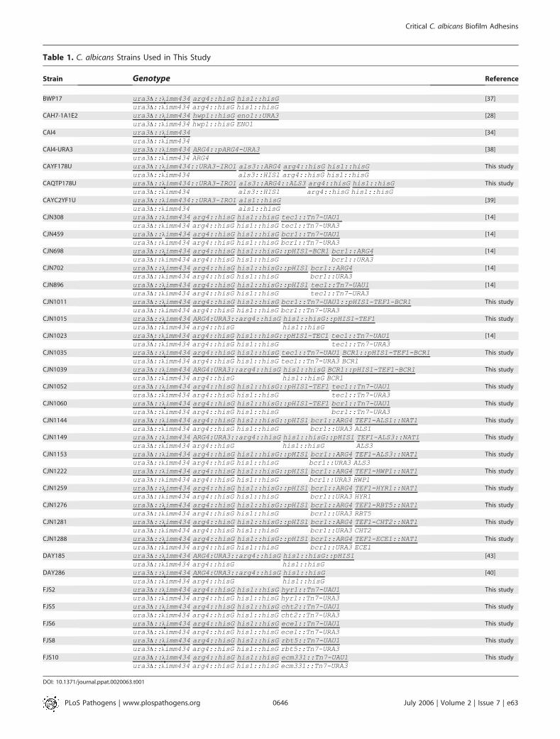
Table 1. C. albicans Strains Used in This Study
Strain Genotype Reference
BWP17 ura3D::kimm434 arg4::hisG his1::hisG [37]
ura3D::kimm434 arg4::hisG his1::hisG
CAH7-1A1E2 ura3D::kimm434 hwp1::hisG eno1::URA3 [28]
ura3D::kimm434 hwp1::hisG ENO1
CAI4 ura3D::kimm434 [34]
ura3D::kimm434
CAI4-URA3 ura3D::kimm434 ARG4::pARG4-URA3 [38]
ura3D::kimm434 ARG4
CAYF178U ura3D::kimm434::URA3-IRO1 als3::ARG4 arg4::hisG his1::hisG This study
ura3D::kimm434 als3::HIS1 arg4::hisG his1::hisG
CAQTP178U ura3D::kimm434::URA3-IRO1 als3::ARG4::ALS3 arg4::hisG his1::hisG This study
ura3D::kimm434 als3::HIS1 arg4::hisG his1::hisG
CAYC2YF1U ura3D::kimm434::URA3-IRO1 als1::hisG [39]
ura3D::kimm434 als1::hisG
CJN308 ura3D::kimm434 arg4::hisG his1::hisG tec1::Tn7-UAU1 [14]
ura3D::kimm434 arg4::hisG his1::hisG tec1::Tn7-URA3
CJN459 ura3D::kimm434 arg4::hisG his1::hisG bcr1::Tn7-UAU1 [14]
ura3D::kimm434 arg4::hisG his1::hisG bcr1::Tn7-URA3
CJN698 ura3D::kimm434 arg4::hisG his1::hisG::pHIS1-BCR1 bcr1::ARG4 [14]
ura3D::kimm434 arg4::hisG his1::hisG bcr1::URA3
CJN702 ura3D::kimm434 arg4::hisG his1::hisG::pHIS1 bcr1::ARG4 [14]
ura3D::kimm434 arg4::hisG his1::hisG bcr1::URA3
CJN896 ura3D::kimm434 arg4::hisG his1::hisG::pHIS1 tec1::Tn7-UAU1 [14]
ura3D::kimm434 arg4::hisG his1::hisG tec1::Tn7-URA3
CJN1011 ura3D::kimm434 arg4::hisG his1::hisG bcr1::Tn7-UAU1::pHIS1-TEF1-BCR1 This study
ura3D::kimm434 arg4::hisG his1::hisG bcr1::Tn7-URA3
CJN1015 ura3D::kimm434 ARG4:URA3::arg4::hisG his1::hisG::pHIS1-TEF1 This study
ura3D::kimm434 arg4::hisG his1::hisG
CJN1023 ura3D::kimm434 arg4::hisG his1::hisG::pHIS1-TEC1 tec1::Tn7-UAU1 [14]
ura3D::kimm434 arg4::hisG his1::hisG tec1::Tn7-URA3
CJN1035 ura3D::kimm434 arg4::hisG his1::hisG tec1::Tn7-UAU1 BCR1::pHIS1-TEF1-BCR1 This study
ura3D::kimm434 arg4::hisG his1::hisG tec1::Tn7-URA3 BCR1
CJN1039 ura3D::kimm434 ARG4:URA3::arg4::hisG his1::hisG BCR1::pHIS1-TEF1-BCR1 This study
ura3D::kimm434 arg4::hisG his1::hisG BCR1
CJN1052 ura3D::kimm434 arg4::hisG his1::hisG::pHIS1-TEF1 tec1::Tn7-UAU1 This study
ura3D::kimm434 arg4::hisG his1::hisG tec1::Tn7-URA3
CJN1060 ura3D::kimm434 arg4::hisG his1::hisG::pHIS1-TEF1 bcr1::Tn7-UAU1 This study
ura3D::kimm434 arg4::hisG his1::hisG bcr1::Tn7-URA3
CJN1144 ura3D::kimm434 arg4::hisG his1::hisG::pHIS1 bcr1::ARG4 TEF1-ALS1::NAT1 This study
ura3D::kimm434 arg4::hisG his1::hisG bcr1::URA3 ALS1
CJN1149 ura3D::kimm434 ARG4:URA3::arg4::hisG his1::hisG::pHIS1 TEF1-ALS3::NAT1 This study
ura3D::kimm434 arg4::hisG his1::hisG ALS3
CJN1153 ura3D::kimm434 arg4::hisG his1::hisG::pHIS1 bcr1::ARG4 TEF1-ALS3::NAT1 This study
ura3D::kimm434 arg4::hisG his1::hisG bcr1::URA3 ALS3
CJN1222 ura3D::kimm434 arg4::hisG his1::hisG::pHIS1 bcr1::ARG4 TEF1-HWP1::NAT1 This study
ura3D::kimm434 arg4::hisG his1::hisG bcr1::URA3 HWP1
CJN1259 ura3D::kimm434 arg4::hisG his1::hisG::pHIS1 bcr1::ARG4 TEF1-HYR1::NAT1 This study
ura3D::kimm434 arg4::hisG his1::hisG bcr1::URA3 HYR1
CJN1276 ura3D::kimm434 arg4::hisG his1::hisG::pHIS1 bcr1::ARG4 TEF1-RBT5::NAT1 This study
ura3D::kimm434 arg4::hisG his1::hisG bcr1::URA3 RBT5
CJN1281 ura3D::kimm434 arg4::hisG his1::hisG::pHIS1 bcr1::ARG4 TEF1-CHT2::NAT1 This study
ura3D::kimm434 arg4::hisG his1::hisG bcr1::URA3 CHT2
CJN1288 ura3D::kimm434 arg4::hisG his1::hisG::pHIS1 bcr1::ARG4 TEF1-ECE1::NAT1 This study
ura3D::kimm434 arg4::hisG his1::hisG bcr1::URA3 ECE1
DAY185 ura3D::kimm434 ARG4:URA3::arg4::hisG his1::hisG::pHIS1 [43]
ura3D::kimm434 arg4::hisG his1::hisG
DAY286 ura3D::kimm434 ARG4:URA3::arg4::hisG his1::hisG [40]
ura3D::kimm434 arg4::hisG his1::hisG
FJS2 ura3D::kimm434 arg4::hisG his1::hisG hyr1::Tn7-UAU1 This study
ura3D::kimm434 arg4::hisG his1::hisG hyr1::Tn7-URA3
FJS5 ura3D::kimm434 arg4::hisG his1::hisG cht2::Tn7-UAU1 This study
ura3D::kimm434 arg4::hisG his1::hisG cht2::Tn7-URA3
FJS6 ura3D::kimm434 arg4::hisG his1::hisG ece1::Tn7-UAU1 This study
ura3D::kimm434 arg4::hisG his1::hisG ece1::Tn7-URA3
FJS8 ura3D::kimm434 arg4::hisG his1::hisG rbt5::Tn7-UAU1 This study
ura3D::kimm434 arg4::hisG his1::hisG rbt5::Tn7-URA3
FJS10 ura3D::kimm434 arg4::hisG his1::hisG ecm331::Tn7-UAU1 This study
ura3D::kimm434 arg4::hisG his1::hisG ecm331::Tn7-URA3
DOI: 10.1371/journal.ppat.0020063.t001
PLoS Pathogens | www.plospathogens.org July 2006 | Volume 2 | Issue 7 | e630646
Critical C. albicans Biofilm Adhesins
added NdeI and SpeI restriction sites upstream and downstream ofthe promoter, respectively. This PCR fragment was digested withNdeI and SpeI and ligated into NdeI- and SpeI-digested plasmidpCJN495 to create pCJN498 containing the A. gossypii TEF1 promoternext to the C. albicans NAT1 ORF, followed by the A. gossypii TEF1terminator, followed by the C. albicans TEF1 promoter in the correctorientation.
The TEF1-ALS3 overexpression C. albicans strains CJN1149 andCJN1153 were constructed by transforming DAY185 (a Hisþreference strain) [43] and CJN702 (a Hisþ bcr1/bcr1 deletion mutant),respectively, using PCR products from template plasmid pCJN498and primers ALS3-F-OE-Ag-NAT-Ag-TEF1p (59-AGCCAAACAATCCGAAGCAACGTAAAGTACGATATCAAAGAATCATAACTTTGCTTTCTATTTGATAACCCGCCTCAAATCAAGATTGGGAGGTTAACAATCAAGCTTGCCTCGTCCCC-39) and ALS3-R-OE-Ag-NAT-Ag-TEF1p (59-TAGACCAAGTCAATGAATTAAAACTGTTGAAAACACCAGTGATTGTCTTTGCAGTCGCAACCGACAAATATATGAGTAACAATGTATATTGTTGTAGCATTATAAAATGTATACTTAGAA-39). These primers amplify the entire A. gossypii TEF1promoter, the C. albicans NAT1 ORF, the A. gossypii TEF1 terminator,and the C. albicans TEF1 promoter with 100 bp of hanging homologyto 500 bp upstream into the promoter of ALS3 for the forwardprimer and 100 bp of hanging homology from exactly the start codonof the ALS3 ORF. The homology in these primers allows forhomologous recombination of the entire cassette directly upstreamof the ALS3 natural locus so that ALS3 can be overexpressed with theTEF1 promoter instead of its natural promoter. The transformationinto C. albicans strains was done as described above except anadditional 5-h recovery step in YPD at 30 8C was done after the cellswere heat shocked at 44 8C for 20 min in order to allow for NAT1expression. The cells were then plated onto YPDþ 400 lg/ml clonNatplates for 2 d at 30 8C to select for Natþ transformants, andtransformants were checked by colony PCR. We used strain DAY185(Argþ UraþHisþ) [43] as a reference strain for these strains.
The als3D/als3D mutant, CAYF178U, was constructed from strainBWP17. The two alleles of ALS3 were serially disrupted using themarkers HIS1 and ARG4. The disruption cassettes were amplifiedwith the following primers: ALS3-5DR (59-CCTCATTACACCAACCATACAACTTTGTGGTCTACAACTTGGGTTATTGAAACAAAAACAGTTTTCCCAGTCACGACGTT-3 9) and ALS3-3DR (59-GGTTGATTCAGCAGTAGTAGTAACAGTAGTAGTTTCATCAGCACTAGAAGAAATGATAGGTGTGGAATTGTGAGCGGATA-39).The disruption of ALS3 was verified by PCR using the followingprimers: 3Confirm-1 (59-ATGACACCATGTCAAGTTCAGA-39)and 3Confirm-2 (59-GTTGGTTGTTCAATGACACTGG-39). Tocomplement the als3D/als3D mutant with a wild-type copy ofALS3, a full–length version of ALS3 was digested from pGEMTwith PvuI and SphI [29], and then subcloned into pDS10 at theSphI site [44]. The construct was linearized with Bsp1407I andintegrated into the ALS3 locus of the als3D/als3D Ura� strain,selecting Uraþ. Excision of the URA3-dpl200 marker was thenselected by plating on 5-FOA medium. ALS3 complementationwas confirmed by PCR using primers 59-TGAAGCAGCCTTTAGTGGCCT-39 and 59-AGAAGTGGAAGCAGCTGTGGA-39.
URA3 and the adjacent IRO1 locus was restored in the als1D/als1Dstrain [39], als3D/als3D, and als3D/als3D::ALS3 strains as follows. Ura�derivatives of these mutants were selected by plating on syntheticmedia containing 5-FOA and uridine. A 3.9-kb URA3-IRO1 fragmentwas released from pBSK-URA3 by NotI/PstI digestion and used totransform the Ura� strains [44]. The restoration of URA3 to its nativelocus was confirmed by PCR us ing the pr imers 5 9-TGCTGGTTGGAAT GCTTATTTG-39 and 59-TGCAAATTCTGCTACTGGAGTT-39.
In vitro biofilm growth conditions. For in vitro biofilm growthassays, strains were grown in YPD overnight at 30 8C, diluted to anOD600 ¼ 0.5 in 2 ml of Spider medium (with auxotrophicsupplements), and added to a sterile 12-well plate with a preparedsilicone square (1.5 3 1.5 cm cut from Cardiovascular Instrumentsilicone sheets [Wakefiled, Massachusetts, United States]). Thesilicone square was previously treated with bovine serum (B-9433;Sigma, St. Louis, Missouri, United States) overnight and washed withPBS in order to prepare it for the biofilm assay. The inoculatedplate was incubated at 37 8C for 90 min at 150 rpm agitation forinitial adhesion of cells. To remove unadhered cells, the squareswere washed with 2 ml of PBS, and the squares were moved to afresh 12-well plate containing 2 ml of fresh Spider medium. Thisplate was incubated at 37 8C for an additional 60 h at 150-rpmagitation to allow for biofilm formation.
Microscopic visualization of in vitro biofilms. For the in vitroexperiments, biofilms were observed visually and by CSLM. For in
vitro CSLM imaging, biofilms were stained with 25 lg/ml con-canavalin A Alexa Fluor 594 conjugate (C-11253; Molecular Probes,Eugene, Oregon, United States) for 1 h in the dark at 37 8C with 150rpm agitation. CSLM was performed with an upright ZeissAxioskop2 FS MOT LSM 510 multiphoton microscope using a ZeissAchroplan 340/0.8W objective. In order to visualize concanavalin Aconjugate staining, a HeNe1 laser with 543-nm excitation wave-length was used. All in vitro CSLM images were assembled into sideand depth views using the Zeiss LSM Image Browser (version3.2.0.115) software. For all side views, the silicone is located at thetop of the image. Depth views are artificially colored imagesindicating cell depth using a color gradient, where blue representscells closest to and red represents cells farthest from the siliconesubstrate.
RNA isolation and expression analysis. Overnight cultures wereinoculated in 5 ml of YPD at 30 8C. The next day, 100 ml of Spidermedium was inoculated with the YPD overnight culture to obtain anOD600 ¼ 0.05, and was grown at 37 8C for 12 h (OD600 ¼ ’8). Cellswere immediately harvested by vacuum filtration. RNA extractionand Northern analysis were performed as previously described [40].For RT-PCR analysis for detection of ALS1 and ALS3, 10 lg of totalRNA was DNase treated at 37 8C for 1 h, ethanol precipitated, andresuspended in 100 ll of DEPC water. cDNA was synthesized andRT-PCR was done as previously described for ALS1 and ALS3 [45]with reverse transcriptase and without reverse transcriptase (as acontrol).
In vivo biofilm model. A rat central venous catheter infectionmodel [18] was selected for in vivo biofilm studies. The catheterdiameter was chosen in an attempt to permit blood flow around theextraluminal catheter surface. To mimic material used in patients,polyethylene tubing (inner diameter �0.76 mm, outer diameter 1.52mm) was chosen. Specific-pathogen-free Sprague-Dawley ratsweighing 400 g were used (Harlan Sprague-Dawley, Indianapolis,Indiana, United States). A heparinized (100 U/ml) catheter wassurgically inserted into the external jugular vein and advanced to asite above the right atrium (2 cm length). The catheter was securedto the vein and the proximal end tunneled subcutaneously to themidscapular space and externalized through the skin. The catheterswere placed 24 h prior to infection to allow a conditioning periodfor deposition of host protein on the catheter surface. Infection wasachieved by intraluminal instillation of 500 ll of C. albicans cells (106
cells/ml). After a dwelling period of 4 h, the catheter volume waswithdrawn and the catheter was flushed with heparinized 0.15 MNaCl.
Catheters from two animals were removed at three time points (12,24, and 48 h) after C. albicans infection to determine biofilmdevelopment on the internal surface of the intravascular devices.The distal 2 cm of the catheter was cut from the entire catheterlength, and biofilms were imaged using both CSLM and scanningelectron microscopy. Scanning electron microscopy was used forarchitectural investigation of the biofilm process. Catheter segmentswere washed with 0.1 M phosphate buffer (pH 7.2) and placed infixative (1% glutaraldehyde and 4% formaldehyde). The samples werethen washed with buffer for 5 min and placed in 1% osmiumtetroxide for 30 min. The samples were then dehydrated in a series of10-min ethanol washes (30%, 50%, 70%, 85%, 95%, and 100%). Finaldesiccation was accomplished by critical point drying (Tousimis,Rockville, Maryland, United States). Specimens were mounted onaluminum stubs and sputter-coated with gold. Samples were imagedin a scanning electron microscope (Hitachi S-5700 or JEOL JSM-6100) in the high-vacuum mode at 10 kV. The images were processedfor display using Adobe Photoshop 7.0.1.
Disseminated murine candidiasis models. Groups of ten 20-g maleBalb/C mice were inoculated via the lateral tail vein with 5 3 105
blastospores with each strain of C. albicans. The mice were monitoredthree times daily for survival.
Biofilm dry mass measurements. For dry mass measurements, eachsilicone square was weighed prior to inoculation with the strain ofinterest. Biofilms were grown for 60 h on the silicone square (asdescribed above). The silicone squares containing their respectivebiofilms were then removed from the wells, dried overnight in a fumehood, and weighed the following day. Total biomass of each biofilmwas calculated by subtracting the weight of the silicone prior tobiofilm growth from the weight of the silicone after biofilm growth.The average total biomass for each strain was calculated from fourindependent samples after subtracting the mass of a blank siliconesquare with no cells added. Statistical significance (p-values) wascalculated with the Student’s two-tailed t-test function in MicrosoftExcel.
PLoS Pathogens | www.plospathogens.org July 2006 | Volume 2 | Issue 7 | e630647
Critical C. albicans Biofilm Adhesins

Supporting Information
Figure S1. Mouse Survival Data
Disseminated murine candidiasis assays. Groups of ten 20-g maleBalb/C mice were inoculated via the lateral tail vein with 5 3 105
blastospores with each strain of C. albicans. The mice were monitoredthree times daily for survival.
Found at DOI: 10.1371/journal.ppat.0020063.sg001 (44 KB PDF).
Accession Numbers
Information for the following C. albicans genes can be found at theCandida Genome Database (CGD) Web site (http:/ /www.candidagenome.org): BCR1 (orf19.723), TEC1 (orf19.5908), HWP1(orf19.1321), ALS3 (orf19.1816), ALS1 (orf19.5741),HYR1 (orf19.4975),CHT2 (orf19.3895), ECE1 (orf19.3374), RBT5 (orf19.5636), and ECM331(orf19.4255).
Acknowledgments
We are grateful for the online availability of the Candida genomedatabase (CGD) and the CandidaDB Web Server. We thank all
members of the Mitchell laboratory for insightful discussions andcomments on this manuscript. We also acknowledge our manuscriptreviewers, who suggested that we broaden our gene overexpressionanalysis. We are grateful to Allison Fay for her help in constructingplasmid pCJN498, Paula Sundstrom for generously providing us withher hwp1/hwp1 mutant strain, Julia Kohler for sharing her plasmidpJK799, and Bill Nierman and his colleagues at The Institute forGenome Research (TIGR) for constructing a plasmid insertionmutant library from which several C. albicans insertion mutants weremade. We are indebted to Theresa Swayne, Sudhindra Swamy, andSharmilee Ramcharan for CSLM advice.
Author contributions. CJN, DRA, JEN, SGF, and APM conceivedand designed the experiments. CJN, DRA, JEN, and QTP performedthe experiments. CJN, DRA, JEN, JEE, SGF, and APM analyzed thedata. FJS, FY, QTP, JEE, and SGF contributed reagents/materials/analysis tools. CJN, DRA, JEE, SGF, and APM wrote the paper.
Funding. This study was supported by National Institutes of Health(NIH) grants R01 AI057804 and R01 AI067703 (APM), NIH K08AI01767 (DRA), NIH T32 HL07899 (JEN), RO1 AI19990 (JEE), and R01AI054928 (SGF).
Competing interests. The authors have declared that no competinginterests exist.
References1. Douglas LJ (2003) Candida biofilms and their role in infection. Trends
Microbiol 11: 30–36.2. Fux CA, Costerton JW, Stewart PS, Stoodley P (2005) Survival strategies of
infectious biofilms. Trends Microbiol 13: 34–40.3. Kumamoto CA (2002) Candida biofilms. Curr Opin Microbiol 5: 608–611.4. Ramage G, Saville SP, Thomas DP, Lopez-Ribot JL (2005) Candida biofilms:
An update. Eukaryot Cell 4: 633–638.5. Kojic EM, Darouiche RO (2004) Candida infections of medical devices. Clin
Microbiol Rev 17: 255–267.6. von Eiff C, Jansen B, Kohnen W, Becker K (2005) Infections associated with
medical devices: Pathogenesis, management and prophylaxis. Drugs 65:179–214.
7. Bertagnolio S, de Gaetano Donati K, Tacconelli E, Scoppettuolo G,Posteraro B, et al. (2004) Hospital-acquired candidemia in HIV-infectedpatients. Incidence, risk factors and predictors of outcome. J Chemother16: 172–178.
8. Beck-Sague C, Jarvis WR (1993) Secular trends in the epidemiology ofnosocomial fungal infections in the United States, 1980–1990. NationalNosocomial Infections Surveillance System. J Infect Dis 167: 1247–1251.
9. Maki DG, Tambyah PA (2001) Engineering out the risk for infection withurinary catheters. Emerg Infect Dis 7: 342–347.
10. Chandra J, Patel JD, Li J, Zhou G, Mukherjee PK, et al. (2005) Modificationof surface properties of biomaterials influences the ability of Candidaalbicans to form biofilms. Appl Environ Microbiol 71: 8795–8801.
11. Kuhn DM, Chandra J, Mukherjee PK, Ghannoum MA (2002) Comparison ofbiofilms formed by Candida albicans and Candida parapsilosis on bioprostheticsurfaces. Infect Immun 70: 878–888.
12. Baillie GS, Douglas LJ (2000) Matrix polymers of Candida biofilms and theirpossible role in biofilm resistance to antifungal agents. J AntimicrobChemother 46: 397–403.
13. Chandra J, Kuhn DM, Mukherjee PK, Hoyer LL, McCormick T, et al. (2001)Biofilm formation by the fungal pathogen Candida albicans: Development,architecture, and drug resistance. J Bacteriol 183: 5385–5394.
14. Nobile CJ, Mitchell AP (2005) Regulation of cell-surface genes and biofilmformation by the C. albicans transcription factor Bcr1p. Curr Biol 15: 1150–1155.
15. Richard ML, Nobile CJ, Bruno VM, Mitchell AP (2005) Candida albicansbiofilm-defective mutants. Eukaryot Cell 4: 1493–1502.
16. Chagneau C, Saier MH Jr (2004) Biofilm-defective mutants of Bacillussubtilis. J Mol Microbiol Biotechnol 8: 177–188.
17. Schinabeck MK, Long LA, Hossain MA, Chandra J, Mukherjee PK, et al.(2004) Rabbit model of Candida albicans biofilm infection: Liposomalamphotericin B antifungal lock therapy. Antimicrob Agents Chemother48: 1727–1732.
18. Andes D, Nett J, Oschel P, Albrecht R, Marchillo K, et al. (2004)Development and characterization of an in vivo central venous catheterCandida albicans biofilm model. Infect Immun 72: 6023–6031.
19. Schweizer A, Rupp S, Taylor BN, Rollinghoff M, Schroppel K (2000) TheTEA/ATTS transcription factor CaTec1p regulates hyphal developmentand virulence in Candida albicans. Mol Microbiol 38: 435–445.
20. Sopko R, Huang D, Preston N, Chua G, Papp B, et al. (2006) Mappingpathways and phenotypes by systematic gene overexpression. Mol Cell 21:319–330.
21. Braun BR, Johnson AD (2000) TUP1, CPH1 and EFG1 make independentcontributions to filamentation in Candida albicans. Genetics 155: 57–67.
22. Liu TT, Lee RE, Barker KS, Lee RE, Wei L, et al. (2005) Genome-wide
expression profiling of the response to azole, polyene, echinocandin, andpyrimidine antifungal agents in Candida albicans. Antimicrob AgentsChemother 49: 2226–2236.
23. Weissman Z, Kornitzer D (2004) A family of Candida cell surface haem-binding proteins involved in haemin and haemoglobin-iron utilization. MolMicrobiol 53: 1209–1220.
24. McCreath KJ, Specht CA, Robbins PW (1995) Molecular cloning andcharacterization of chitinase genes from Candida albicans. Proc Natl AcadSci U S A 92: 2544–2548.
25. Cao YY, Cao YB, Xu Z, Ying K, Li Y, et al. (2005) cDNA microarray analysisof differential gene expression in Candida albicans biofilm exposed tofarnesol. Antimicrob Agents Chemother 49: 584–589.
26. Bensen ES, Martin SJ, Li M, Berman J, Davis DA (2004) Transcriptionalprofiling in Candida albicans reveals new adaptive responses to extracellularpH and functions for Rim101p. Mol Microbiol 54: 1335–1351.
27. Hoyer LL (2001) The ALS gene family of Candida albicans. Trends Microbiol9: 176–180.
28. Sundstrom P (2002) Adhesion in Candida spp. Cell Microbiol 4: 461–469.29. Sheppard DC, Yeaman MR, Welch WH, Phan QT, Fu Y, et al. (2004)
Functional and structural diversity in the Als protein family of Candidaalbicans. J Biol Chem 279: 30480–30489.
30. Zhao X, Oh SH, Yeater KM, Hoyer LL (2005) Analysis of the Candida albicansAls2p and Als4p adhesins suggests the potential for compensatory functionwithin the Als family. Microbiology 151: 1619–1630.
31. Staab JF, Bradway SD, Fidel PL, Sundstrom P (1999) Adhesive andmammalian transglutaminase substrate properties of Candida albicansHwp1. Science 283: 1535–1538.
32. Zhao R, Daniels KJ, Lockhart SR, Yeater KM, Hoyer LL, et al. (2005) Uniqueaspects of gene expression during Candida albicans mating and possible G(1)dependency. Eukaryot Cell 4: 1175–1190.
33. Bennett RJ, Uhl MA, Miller MG, Johnson AD (2003) Identification andcharacterization of a Candida albicans mating pheromone. Mol Cell Biol 23:8189–8201.
34. Fonzi WA, Irwin MY (1993) Isogenic strain construction and gene mappingin Candida albicans. Genetics 134: 717–728.
35. Birse CE, Irwin MY, Fonzi WA, Sypherd PS (1993) Cloning and character-ization of ECE1, a gene expressed in association with cell elongation of thedimorphic pathogen Candida albicans. Infect Immun 61: 3648–3655.
36. Liu H, Kohler J, Fink GR (1994) Suppression of hyphal formation in Candidaalbicans by mutation of a STE12 homolog. Science 266: 1723–1726.
37. Wilson RB, Davis D, Mitchell AP (1999) Rapid hypothesis testing withCandida albicans through gene disruption with short homology regions. JBacteriol 181: 1868–1874.
38. Sanchez AA, Johnston DA, Myers C, Edwards JE Jr, Mitchell AP, et al. (2004)Relationship between Candida albicans virulence during experimentalhematogenously disseminated infection and endothelial cell damage invitro. Infect Immun 72: 598–601.
39. Fu Y, Ibrahim AS, Sheppard DC, Chen YC, French SW, et al. (2002) Candidaalbicans Als1p: An adhesin that is a downstream effector of the EFG1filamentation pathway. Mol Microbiol 44: 61–72.
40. Davis DA, Bruno VM, Loza L, Filler SG, Mitchell AP (2002) Candida albicansMds3p, a conserved regulator of pH responses and virulence identifiedthrough insertional mutagenesis. Genetics 162: 1573–1581.
41. Spreghini E, Davis DA, Subaran R, Kim M, Mitchell AP (2003) Roles ofCandida albicans Dfg5p and Dcw1p cell surface proteins in growth andhypha formation. Eukaryot Cell 2: 746–755.
42. Shen J, Guo W, Kohler JR (2005) CaNAT1, a heterologous dominant
PLoS Pathogens | www.plospathogens.org July 2006 | Volume 2 | Issue 7 | e630648
Critical C. albicans Biofilm Adhesins

selectable marker for transformation of Candida albicans and otherpathogenic Candida species. Infect Immun 73: 1239–1242.
43. Davis D, Wilson RB, Mitchell AP (2000) RIM101-dependent and-independ-ent pathways govern pH responses in Candida albicans. Mol Cell Biol 20:971–978.
44. Park H, Myers CL, Sheppard DC, Phan QT, Sanchez AA, et al. (2005) Role of
the fungal Ras-protein kinase A pathway in governing epithelial cellinteractions during oropharyngeal candidiasis. Cell Microbiol 7: 499–510.
45. Green CB, Cheng G, Chandra J, Mukherjee P, Ghannoum MA, et al. (2004)RT-PCR detection of Candida albicans ALS gene expression in thereconstituted human epithelium (RHE) model of oral candidiasis and inmodel biofilms. Microbiology 150: 267–275.
PLoS Pathogens | www.plospathogens.org July 2006 | Volume 2 | Issue 7 | e630649
Critical C. albicans Biofilm Adhesins
Copyright © 2022 FDOKUMEN