Conductive electrospun PANi-PEO/TiO 2 fibrous membrane for photo catalysis
Upload
independentCategory
view
1download
0
DOI: 10.1002/cphc.201100865
Supercapacitors Based on c-Type Cytochromes UsingConductive Nanostructured Networks of Living BacteriaNikhil S. Malvankar,*[a, b] T�nde Mester,[b] Mark T. Tuominen,[a] and Derek R. Lovley[b]
1. Introduction
Supercapacitors have emerged as novel energy storage devi-ces to complement or replace batteries in high-power applica-tions such as electric devices and vehicles,[1, 2] but improve-ments in the amount of capacitance or energy stored are nec-essary for most practical applications.[3] There is the possibilitythat capacitance of redox-active materials can be combinedwith double-layer capacitors to maximize supercapacitance forthe development of future-generation, high-energy supercapa-citors.[3] Biological materials are relatively inexpensive and envi-ronmentally sustainable and might be used to significantly en-hance device performance. For example, genetically engi-neered viruses with peptides have been used to improve bat-tery capacity.[4] The viruses coat themselves with iron phos-phate and then bind specifically to carbon nanotubes, forminga battery cathode in which electrons can travel from carbonnanotube to the iron phosphate in a short time.[4]
Supercapacitors are generally classified as electrical double-layer capacitors, where the capacitance arises due to the elec-trostatic charge accumulation at the electrode/electrolyte inter-face, or as pseudocapacitors, where electro-active speciescause fast surface redox reactions.[2] The heme groups of c-type cytochromes readily interconvert between oxidationstates over a very wide redox potential window, which makesc-type cytochromes highly efficient for rapid electron ex-change. The microorganism Geobacter sulfurreducens, which re-spires by reducing metals,[5] contains the highest number ofgenes coding for c-type cytochromes[6] and most of them havemultiple hemes.[6] It has been proposed that these abundantmulti-heme c-type cytochromes in the periplasm and the outersurface of G. sulfurreducens play an important ecological role,permitting temporary storage of electrons in cytochromes andthus continued, short-term electron transfer across the innermembrane when natural electron acceptors, such as FeIII
oxides, are temporarily unavailable.[7] This is a fundamental
mechanism for a broad range of biochemical and geologicalprocesses in natural environments and for the development ofnovel methods of energy generation.[8] For example, it hasbeen suggested that the capacitor behavior plays an importantrole for bioremediation of uranium.[9]
Although it has been suggested that the c-type cytochromesin G. sulfurreducens biofilms act as electron sinks and can alsofunction as capacitors,[10, 11] capacitance was not measured andit had not been demonstrated that c-type cytochromes werestoring electrons.
Here, we report on direct measurements of the in vivo ca-pacitance of G. sulfurreducens biofilms and demonstrate thatfaradic reactions of the cytochromes confer large pseudocapa-citance to biofilms with a specific capacitance that is compara-ble to synthetic supercapacitors.[1] These findings suggest newprospects for future low-cost and environmentally sustainableenergy storage devices.
Supercapacitors have attracted interest in energy storage be-cause they have the potential to complement or replace bat-teries. Here, we report that c-type cytochromes, naturally im-mersed in a living, electrically conductive microbial biofilm,greatly enhance the device capacitance by over two orders ofmagnitude. We employ genetic engineering, protein unfoldingand Nernstian modeling for in vivo demonstration of chargestorage capacity of c-type cytochromes and perform electro-chemical impedance spectroscopy, cyclic voltammetry and
charge–discharge cycling to confirm the pseudocapacitive,redox nature of biofilm capacitance. The biofilms also showlow self-discharge and good charge/discharge reversibility. Thesuperior electrochemical performance of the biofilm is relatedto its high abundance of cytochromes, providing large electronstorage capacity, its nanostructured network with metallic-likeconductivity, and its porous architecture with hydrous nature,offering prospects for future low cost and environmentally sus-tainable energy storage devices.
[a] Dr. N. S. Malvankar, Prof. M. T. TuominenDepartment of PhysicsUniversity of Massachusetts, Amherst203, Morrill Science Center IVN639 North Pleasant Street, Amherst, MA 01003 (USA)Fax: (+ 1) 413-577-4660E-mail : [email protected]
[b] Dr. N. S. Malvankar, Dr. T. Mester,+ Prof. D. R. LovleyDepartment of MicrobiologyUniversity of Massachusetts, Amherst (USA)
[+] Current address :University of Michigan Medical School and Veterans Affairs Medical Re-search Center, Ann Arbor, Michigan 48105, USA.
Supporting information for this article is available on the WWW underhttp://dx.doi.org/10.1002/cphc.201100865.
ChemPhysChem 2012, 13, 463 – 468 � 2012 Wiley-VCH Verlag GmbH & Co. KGaA, Weinheim 463

2. Results and Discussion
2.1. Direct Measurements of Biofilm Capacitance
In microbial fuel cells in which two gold anodes separated bya non-conducting gap were provided as electron acceptors(Figure 1), biofilms of G. sulfurrreducens grew across the non-
conducting gap, as previously described.[12] When biofilm ca-pacitance was measured by applying ac voltage across thesplit electrodes (Figure 1 b), the impedance data could be ana-lyzed with the previously described ([12] and references there-in) equivalent circuit model (Figure 2). As the biofilms grewover the electrodes, the capacitance increased by two ordersof magnitude over the capacitance of the double-layer capaci-tor that formed when bare split electrodes were immersed inthe medium (Figure 3).
When G. sulfurreducens was grown with the alternate elec-tron acceptor fumarate, there was no increase in capacitanceover time (Figure 4). Fumarate-grown biofilms exhibit low con-ductivity even though they bridge the non-conducting gap.[12]
In a similar manner, biofilms of Escherichia coli and Pseudomo-nas aeruginosa, which can also bridge the gap, but are non-conductive,[12] did not have significant capacitance. These re-sults demonstrated that the ability for electron conductionthrough the biofilm, which in G. sulfurreducens can be attribut-ed to a network of conductive pili,[12] is necessary for the bio-film to function as a capacitor.
Furthermore, the high porosity of the biofilms, which arecomprised of >90–95 % electrolyte,[13] provides high electrolyt-
ic accessibility for the movement of ions that is necessary tomaintain electroneutrality associated with the capacitor forma-tion.
As previously reported,[12] removing the electron donor ace-tate from the media completely suppressed the fuel cell cur-rent (Figure 5). However, there was no impact on capacitance(Figure 5). When acetate additions were resumed, current pro-duction returned to its original rate. The finding that a biofilmof metabolically inactive cells had the same capacitance as a
Figure 1. a) Split-electrode geometry and capacitance formation. Gold elec-trodes separated by a 50 mm non-conductive gap are shown in yellow. Elec-trostatic double layer ions are shown in white and c-type cytochromes ena-bling pseudocapacitance are shown in red. The conductive biofilm matrix isshown in green. b) Setup for two-electrode ac impedance spectroscopymeasurements. The anode and cathode of the microbial fuel cell were tem-porarily disconnected and an ac voltage was applied across the split-anode(configuration shown). PEM: Proton exchange membrane. Biofilm immersedin an electrolyte (blue) is shown in green.
Figure 2. Representative impedance spectrum of Geobacter sulfurreducensbiofilm. Red open circles show the impedance data and red line shows thefit to the impedance data using equivalent circuit model presented in theinset. Impedance spectrum for control electrodes is presented in the Sup-porting Information. Inset : Equivalent circuit used to extract capacitancevalues from the impedance spectra. Rsolution represents ionic conductivity ofthe electrolyte, Cbiofilm is the capacitance at the biofilm–electrode interface,Celectrode is the geometric capacitance due to electrodes and Rbiofilm is due toelectronic conductivity of the biofilm.[12]
Figure 3. Capacitance increase due to the biofilm. Capacitance over days insplit electrode bridged by a G. sulfurreducens biofilm (blue solid circles) andcorresponding bare split-electrodes (control) without biofilm (blue open cir-cles). Error bars : Standard deviation. Capacitance was measured with two-electrode ac impedance spectroscopy.
464 www.chemphyschem.org � 2012 Wiley-VCH Verlag GmbH & Co. KGaA, Weinheim ChemPhysChem 2012, 13, 463 – 468
N. S. Malvankar et al.

current-producing biofilm suggested that the capacitance wasassociated with a stable component of the biofilm, rather thana transient molecule that required continuous replenishmentthrough metabolic activity. The fact that G. sulfurreducens bio-films exhibit metallic-like conductivity,[12] providing efficientpathways between the electrode and electro-active species inthe biofilm, suggests that the charge transfer causing the ca-pacitance is due to the electrons present within the biofilm.
2.2. Contribution of Cytochromes to Biofilm Capacitance
It was previously proposed that c-type cytochromes confer ca-pacitance to individual cells of G. sulfurreducens.[7] To evaluatewhether the capacitance of biofilms could be attributed to c-type cytochromes, the capacitance of the biofilms of differentstrains of G. sulfurreducens with different capabilities for cyto-
chrome production was evaluated. For example, the c-type cy-tochromes OmcB, OmcE, OmcS, and OmcT are among themost abundant outer surface cytochromes of G. sulfurredu-cens.[14] In strain ST, the genes for OmcS and OmcT have beendeleted,[15] whereas in strain BEST, the genes for OmcB andOmcE have also been deleted.[14] The biofilms of both of thesestrains had a lower content of cytochrome hemes and less ca-pacitance than the wild-type DL-1 strain (Figure 6). StrainKN400, which produces higher current densities than strainDL-1 but has a lower cytochrome content,[16] had a biofilmheme content intermediate between that of strains BEST, STand wild-type DL-1 and also had an intermediate capacitance(Figure 6). There was a strong correlation (r2 = 0.998) betweenthe capacitance and the cytochrome content of the differentstrains, suggesting that c-type cytochromes contribute to thebiofilm capacitance.
If c-type cytochromes were responsible for the capacitanceof the G. sulfurrreducens biofilms then it would be expectedthat this capacitance would be in the form of “pseudocapaci-tance” which is characteristic of fast and reversible redox reac-tions.[17] Cyclic voltammetry of G. sulfurreducens biofilms dis-played a set of anodic and cathodic peaks (Figure 7), consis-tent with pseuduocapacitance,[3, 18] whereas the voltammo-grams obtained for control electrodes in the absence of bio-film were rectangular (Figure 7), which is characteristic of pureelectrostatic capacitance.[3, 18]
The presence of pseudocapacitance in G. sulfurreducens bio-films was also evident from galvanostatic charge and dischargecycling (Figure 8). In this analysis, the biofilms clearly deviatedfrom the ideal triangular shape expected for pure electrostaticdouble-layer capacitance.[3, 18] Such a deviation is typical ofpseudocapacitive contributions.[3, 17, 18] This result is consistentwith the expected pseudocapacitive behavior of c-type cyto-chromes and further suggests that the large capacitance of thebiofilm is due to the redox chemistry of c-type cytochromes.
Figure 4. Capacitance over days in split electrode bridged by non-conduc-tive biofilms. Green: Escherichia coli biofilm, Violet: Pseudomonas aeruginosabiofilm, Red: G. sulfurreducens biofilm grown with fumarate as electron ac-ceptor. Error bars: Standard deviation. Capacitance was measured with two-electrode ac impedance spectroscopy.
Figure 5. Effect of acetate removal on Geobacter sulfurreducens fuel cell cur-rent and biofilm capacitance. Error bars: Standard deviation. Capacitancewas measured with two-electrode ac impedance spectroscopy.
Figure 6. Comparison of capacitance of biofilms of various Geobacter sulfur-reducens strains and their total cytochrome heme content. Blue: calculatedcapacitance; Green: cytochrome heme content; Red: Measured capacitance;Error bars: Standard deviation. Capacitance was evaluated with three-elec-trode electrochemical impedance spectroscopy.
ChemPhysChem 2012, 13, 463 – 468 � 2012 Wiley-VCH Verlag GmbH & Co. KGaA, Weinheim www.chemphyschem.org 465
Supercapacitors Based on c-Type Cytochromes

2.3. Computed CapacitanceDue to c-Type Cytochromes isComparable with MeasuredCapacitance
Pseudocapacitance due toredox-active species is definedas the derivative of charge ac-ceptance and a change in poten-tial,[2] and for c-type cyto-chromes, it can be calculated byEquation (1):[17]
Cf ¼ QðF=RTÞ expðDE � F=RTÞ½1þ expðDE � F=RTÞ�2
ð1Þ
where Q is the total charge asso-ciated with the cytochromehemes (each heme stores one
electron), F is the Faraday constant, R is the molar gas constantand T is the temperature. DE is the difference between theelectrode potential (E) and the formal potential (E0’) of the bio-film. (See the Supporting Information for details.) The formalpotential, measured using cyclic voltammetry, for each of thestrains was: strain DL-1, �350 mV vs Ag/AgCl; strain KN400,�370 mV vs Ag/AgCl; strain ST, �330 mV vs Ag/AgCl; strainBEST, �390 mV vs Ag/AgCl, which was comparable to thevalues previously reported in the literature.[19, 20] E is the elec-trode potential measured at open circuit condition which forthe various strains was: strain DL-1, �450 mV vs Ag/AgCl;strain KN400, �483 mV vs Ag/AgCl; strain ST, �441 mV vs Ag/AgCl; strain BEST, �530 mV vs Ag/AgCl. Double-layer capaci-tance was neglected in the calculation of total capacitance, be-cause its value is small for gold electrodes.[21] There was goodagreement between the pseudocapacitance calculated fromheme abundance and the measured capacitance (Figure 6).These results further suggested that capacitance of the G. sul-furreducens biofilms could be attributed to the cytochromes.
2.4. Unfolding the c-Type Cytochromes Diminishes theBiofilm Capacitance
Addition of acetylmethionine, which denatures c-type cyto-chromes,[22] diminished the capacitance of the biofilm as wellas current production (Figure 9). In contrast, denaturing the cy-tochromes did not affect the conductivity of microbial nano-wire networks.[12] These experiments confirmed that the ob-served large enhancement of capacitance in biofilms is due tothe charge storage ability of c-type cytochromes. The rate ofinhibition of current production was faster than the loss of ca-pacitance, which was attributed to the fact that flux of acetyl-methionine into the biofilm which diminished the capacitanceis rate limited by the diffusion,[23] whereas the suppression incurrent is due to an instantaneous change in the redox poten-tial of the c-type cytochromes.[22]
Figure 7. Representative cyclic voltammetry of Geobacter sulfurreducens bio-film (red curve) and the corresponding control electrodes (black curve) inthe voltage range �0.7 V to 0.1 V versus Ag/AgCl. Scan rate 100 mV s�1.
Figure 8. Typical galvanostatic charge and discharge profile of Geobacter sul-furreducens biofilm, at 1 mA applied current.
Figure 9. Capacitance of the biofilm (blue solid circles) and control electrodes (blue open circles) over hours afteradding acetylmethionine (AcMet, 120 mM) at pH 7. Microbial current at the time of capacitance measurement isshown by red trace. Inset: Capacitance of biofilm and control before (dark blue bars) and after (light blue bars)adding AcMet for 100 h. Error bars: Standard deviation. Capacitance was measured with two-electrode ac impe-dance spectroscopy.
466 www.chemphyschem.org � 2012 Wiley-VCH Verlag GmbH & Co. KGaA, Weinheim ChemPhysChem 2012, 13, 463 – 468
N. S. Malvankar et al.
2.5. G. sulfurreducens Biofilms Show Low Self-DischargeBehavior
Self-discharge is a major limiting factor affecting the perfor-mance of supercapacitors.[17] Self-discharge causes the open-circuit voltage of a charged capacitor to decrease over time.During self-discharge, the leakage current discharges the ca-pacitor even in the absence of any external electrical current.The self-discharge is typically due to overcharging of the ca-pacitor beyond the decomposition limit of the electrolyte ordue to the result of internal redox reactions involving redox-active molecules or impurities present in the electrolyte.[17]
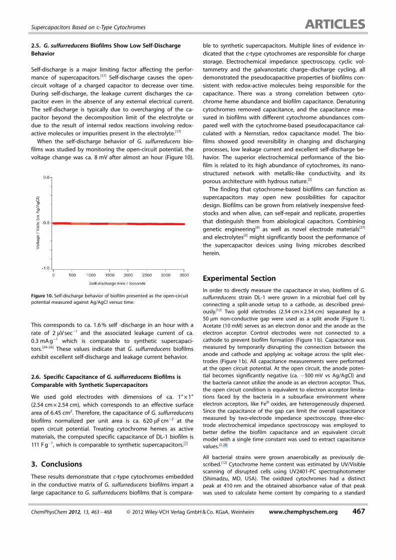
When the self-discharge behavior of G. sulfurreducens bio-films was studied by monitoring the open-circuit potential, thevoltage change was ca. 8 mV after almost an hour (Figure 10).
This corresponds to ca. 1.6 % self -discharge in an hour with arate of 2 mV sec�1 and the associated leakage current of ca.0.3 mA g�1 which is comparable to synthetic supercapaci-tors.[24–26] These values indicate that G. sulfurreducens biofilmsexhibit excellent self-discharge and leakage current behavior.
2.6. Specific Capacitance of G. sulfurreducens Biofilms isComparable with Synthetic Supercapacitors
We used gold electrodes with dimensions of ca. 1“ � 1”(2.54 cm � 2.54 cm), which corresponds to an effective surfacearea of 6.45 cm2. Therefore, the capacitance of G. sulfurreducensbiofilms normalized per unit area is ca. 620 mF cm�2 at theopen circuit potential. Treating cytochrome hemes as activematerials, the computed specific capacitance of DL-1 biofilm is111 F g�1, which is comparable to synthetic supercapacitors.[1]
3. Conclusions
These results demonstrate that c-type cytochromes embeddedin the conductive matrix of G. sulfurreducens biofilms impart alarge capacitance to G. sulfurreducens biofilms that is compara-
ble to synthetic supercapacitors. Multiple lines of evidence in-dicated that the c-type cytochromes are responsible for chargestorage. Electrochemical impedance spectroscopy, cyclic vol-tammetry and the galvanostatic charge–discharge cycling, alldemonstrated the pseudocapacitive properties of biofilms con-sistent with redox-active molecules being responsible for thecapacitance. There was a strong correlation between cyto-chrome heme abundance and biofilm capacitance. Denaturingcytochromes removed capacitance, and the capacitance mea-sured in biofilms with different cytochrome abundances com-pared well with the cytochrome-based pseudocapacitance cal-culated with a Nernstian, redox capacitance model. The bio-films showed good reversibility in charging and dischargingprocesses, low leakage current and excellent self-discharge be-havior. The superior electrochemical performance of the bio-film is related to its high abundance of cytochromes, its nano-structured network with metallic-like conductivity, and itsporous architecture with hydrous nature.[2]
The finding that cytochrome-based biofilms can function assupercapacitors may open new possibilities for capacitordesign. Biofilms can be grown from relatively inexpensive feed-stocks and when alive, can self-repair and replicate, propertiesthat distinguish them from abiological capacitors. Combininggenetic engineering[4] as well as novel electrode materials[27]
and electrolytes[3] might significantly boost the performance ofthe supercapacitor devices using living microbes describedherein.
Experimental Section
In order to directly measure the capacitance in vivo, biofilms of G.sulfurreducens strain DL-1 were grown in a microbial fuel cell byconnecting a split-anode setup to a cathode, as described previ-ously.[12] Two gold electrodes (2.54 cm � 2.54 cm) separated by a50 mm non-conductive gap were used as a split anode (Figure 1).Acetate (10 mM) serves as an electron donor and the anode as theelectron acceptor. Control electrodes were not connected to acathode to prevent biofilm formation (Figure 1 b). Capacitance wasmeasured by temporarily disrupting the connection between theanode and cathode and applying ac voltage across the split elec-trodes (Figure 1 b). All capacitance measurements were performedat the open circuit potential. At the open circuit, the anode poten-tial becomes significantly negative (ca. �500 mV vs Ag/AgCl) andthe bacteria cannot utilize the anode as an electron acceptor. Thus,the open circuit condition is equivalent to electron acceptor limita-tions faced by the bacteria in a subsurface environment whereelectron acceptors, like FeIII oxides, are heterogeneously dispersed.Since the capacitance of the gap can limit the overall capacitancemeasured by two-electrode impedance spectroscopy, three-elec-trode electrochemical impedance spectroscopy was employed tobetter define the biofilm capacitance and an equivalent circuitmodel with a single time constant was used to extract capacitancevalues.[2, 28]
All bacterial strains were grown anaerobically as previously de-scribed.[12] Cytochrome heme content was estimated by UV/Visiblescanning of disrupted cells using UV2401-PC spectrophotometer(Shimadzu, MD, USA). The oxidized cytochromes had a distinctpeak at 410 nm and the obtained absorbance value of that peakwas used to calculate heme content by comparing to a standard
Figure 10. Self-discharge behavior of biofilm presented as the open-circuitpotential measured against Ag/AgCl versus time.
ChemPhysChem 2012, 13, 463 – 468 � 2012 Wiley-VCH Verlag GmbH & Co. KGaA, Weinheim www.chemphyschem.org 467
Supercapacitors Based on c-Type Cytochromes

curve made with oxidized bovine heart cytochrome c (Sigma, MO,USA). For the specific capacitance calculation, the molecularweight of the heme (616.5 g mole�1) was used (NIH’s Molecular Li-braries Roadmap Initiative–Pubchem Compound).
All electrochemical experiments were performed using Solar-tron 1287 potentiostat/galvanostat at room temperature. Impe-dance spectroscopy was performed using a Solartron 1252/1287impedance analyzer. Impedance was measured by sweeping fre-quency between 100 mHz and 300 KHz with 0.1 V amplitude acvoltage excitation. Data fitting was performed with ZView software(Scribner Inc.) which uses LEVM algorithm developed by J. RossMacdonald.[28] For three-electrode electrochemical impedancespectroscopy, a gold split electrode was used as a working elec-trode, the reference electrode was Ag/AgCl, 3 M KCl (BAS, IN, USA)and the counter electrode was carbon cloth.[12] For all impedancemeasurements, open circuit potential was monitored until itreached a constant value before over imposing ac signal. Cyclicvoltammetry was performed in the voltage range �0.7 V to 0.1 Vvs Ag/AgCl reference electrode at a scan rate of 100 mV s�1. Thegalvanostatic charge and discharge profiles were measured at theapplied current of 1 mA.[3] The self-discharge behavior was ob-served by measuring the open-circuit potential of the electrode inthe presence of biofilm versus Ag/AgCl reference electrode. Thedata was collected and analyzed using CoreView software (ScribnerInc).
Acknowledgements
This work was supported by the Office of Naval Research (grantno. AN00014-10-1-0084), the office of Science (BER), US Depart-ment of Energy (award no. DE-SC0004114 and CooperativeAgreement no. DE-FC02-02ER63446) as well as the NSF Center forHierarchical Manufacturing (grant no. CMMI-1025020). We thankLaura Francis for helpful discussions.
Keywords: bacteria · cytochromes · electrochemistry · redoxchemistry · supercapacitors
[1] T. A. Skotheim, J. R. Reynolds, Handbook of Conducting Polymers,2nd ed. , CRC Press, Boca Raton, 2007.
[2] L. L. Zhang, X. S. Zhao, Chem. Soc. Rev. 2009, 38, 2520 – 2531.[3] S. Rold�n, C. Blanco, M. Granda, R. Men�ndez, R. Santamar�a, Angew.
Chem. 2011, 123, 1737 – 1739; Angew. Chem. Int. Ed. 2011, 50, 1699 –1701.
[4] Y. J. Lee, H. Yi, W. J. Kim, K. Kang, D. S. Yun, M. S. Strano, G. Ceder, A. M.Belcher, Science 2009, 324, 1051 – 1055.
[5] D. R. Lovley, T. Ueki, T. Zhang, N. S. Malvankar, P. M. Shrestha, K. Flana-gan, M. Aklujkar, J. E. Butler, L. Giloteaux, A. E. Rotaru, D. E. Holmes, A. E.
Franks, R. Orellana, C. Risso, K. P. Nevin, Adv. Microb. Physiol. 2011, 59,1 – 100.
[6] B. A. Methe, K. E. Nelson, J. A. Eisen, I. T. Paulsen, W. Nelson, J. F. Heidel-berg, D. Wu, M. Wu, N. Ward, M. J. Beanan, R. J. Dodson, R. Madupu,L. M. Brinkac, S. C. Daugherty, R. T. DeBoy, A. S. Durkin, M. Gwinn, J. F.Kolonay, S. A. Sullivan, D. H. Haft, J. Selengut, T. M. Davidsen, N. Zafar, O.White, B. Tran, C. Romero, H. A. Forberger, J. Weidman, H. Khouri, T. V.Feldblyum, T. R. Utterback, S. E. Van Aken, D. R. Lovley, C. M. Fraser, Sci-ence 2003, 302, 1967 – 1969.
[7] A. Esteve-NfflÇnez, J. Sosnik, P. Visconti, D. R. Lovley, Environ. Microbiol.2008, 10, 497 – 505.
[8] D. R. Lovley, Geobiology 2008, 6, 225 – 231.[9] J. Zhao, Y. Fang, T. D. Scheibe, D. R. Lovley, R. Mahadevan, J. Contam.
Hydrol. 2010, 112, 30 – 44.[10] Y. Liu, H. Kim, R. R. Franklin, D. R. Bond, ChemPhysChem 2011, 12, 2235 –
2241.[11] D. Schrott Germ�n, B. P. Sebastian, R. Luciana, E.-N. Abraham, J. Pablo
Busalmen, Electrochim. Acta 2011, 56, 10791 – 10795.[12] N. S. Malvankar, M. Vargas, K. P. Nevin, A. E. Franks, C. Leang, B. C. Kim,
K. Inoue, T. Mester, S. F. Covalla, J. P. Johnson, V. M. Rotello, M. T. Tuomi-nen, D. R. Lovley, Nat. Nanotechnol. 2011, 6, 573 – 579.
[13] D. Herbert-Guillou, B. Tribollet, D. Festy, L. Ki�n�, Electrochim. Acta1999, 45, 1067 – 1075.
[14] J. W. Voordeckers, B. C. Kim, M. Izallalen, D. R. Lovley, Appl. Environ. Mi-crobiol. 2010, 76, 2371 – 2375.
[15] T. Mehta, M. V. Coppi, S. E. Childers, D. R. Lovley, Appl. Environ. Microbiol.2005, 71, 8634 – 8641.
[16] H. Yi, K. P. Nevin, B. C. Kim, A. E. Franks, A. Klimes, L. M. Tender, D. R.Lovley, Biosens. Bioelectron. 2009, 24, 3498 – 3503.
[17] B. E. Conway, Electrochemical Supercapacitors : Scientific Fundamentalsand Technological Applications, Kluwer/Plenum, New York, 1999.
[18] S. Rold�n, Z. Gonz�lez, C. Blanco, M. Granda, R. Men�ndez, R. Santama-r�a, Electrochim. Acta 2011, 56, 3401 – 3405.
[19] H. Richter, K. P. Nevin, H. Jia, D. A. Lowy, D. R. Lovley, L. M. Tender,Energy Environ. Sci. 2009, 2, 506 – 516.
[20] S. M. Strycharz, A. P. Malanoski, R. M. Snider, H. Yi, D. R. Lovley, L. M.Tender, Energy Environ. Sci. 2011, 4, 896 – 913.
[21] E. Katz, I. Willner, Electroanalysis 2003, 15, 913 – 947.[22] R. Nakamura, F. Kai, A. Okamoto, G. J. Newton, K. Hashimoto, Angew.
Chem. 2009, 121, 516 – 519; Angew. Chem. Int. Ed. 2009, 48, 508 – 511.[23] S. J. Hagen, J. Hofrichter, A. Szabo, W. A. Eaton, Proc. Natl. Acad. Sci. USA
1996, 93, 11615 – 11617.[24] E. Frackowiak, S. Delpeux, K. Jurewicz, K. Szostak, D. Cazorla-Amoros, F.
Beguin, Chem. Phys. Lett. 2002, 361, 35 – 41.[25] K. Fic, G. Lota, E. Frackowiak, Electrochim. Acta 2010, 55, 7484 – 7488.[26] A. B. Fuertes, G. Lota, T. A. Centeno, E. Frackowiak, Electrochim. Acta
2005, 50, 2799 – 2805.[27] Y. Zhu, S. Murali, M. D. Stoller, K. J. Ganesh, W. Cai, P. J. Ferreira, A. Pirkle,
R. M. Wallace, K. A. Cychosz, M. Thommes, D. Su, E. A. Stach, R. S. Ruoff,Science 2011, 332, 1537 – 1541.
[28] E. Barsoukov, J. R. Macdonald, Impedance Spectroscopy : Theory, Experi-ment and Applications, 2nd ed. , Wiley, Hoboken, 2005.
Received: October 28, 2011Published online on January 17, 2012
468 www.chemphyschem.org � 2012 Wiley-VCH Verlag GmbH & Co. KGaA, Weinheim ChemPhysChem 2012, 13, 463 – 468
N. S. Malvankar et al.
Copyright © 2022 FDOKUMEN