Copia de Página 348 - Food and Agriculture Organization of ...
350
I 9 - 13 October, 1989 at the Chinese Academy of Agricultural Sciences, Beijing, China I I r Food and Agriculture Organization of the United Nations, Rome. United Nations Development Programme, New York. " Chinese Academy of Agricultural Sciences, Beijing, China. Institute for Advanced Studies, University of Malaya, Kuala Lumpur, Malaysia. · 1
-
Upload
khangminh22 -
Category
Documents
-
view
0 -
download
0
Transcript of Copia de Página 348 - Food and Agriculture Organization of ...

I
9 - 13 October, 1989 at the Chinese Academy of Agricultural Sciences,
Beijing, China
I I
r
Food and Agriculture Organization of the United Nations, Rome. United Nations Development Programme, New York.
1" Chinese Academy of Agricultural Sciences, Beijing, China.
Institute for Advanced Studies, University of Malaya, Kuala Lumpur, Malaysia. · 11

FAO /UNDP WORKSHOP ON BIOTECHNOLOGY IN ANIMAL
PRODUCTION AND HEALTH IN ASIA AND LATIN AMERICA
9 - 13 October, 1989 at the Chinese Academy of Agricultural
Sciences, Beijing, China
PROC
Editor: T.K. Mukherjee Department of Genetics and Cellular Biology University of Malaya Kuala Lumpur
E I
Food and Agricultural Organization of the United Nations, Rome. United Nations Development Programme, New York.
Chinese Academy of Agricultural Sciences, Beijing, China.
s
Institute for Advanced Studies, University of Malaya, Kuala Lumpur, Malaysia.
FAO LIBRARY AN: 301999-2043

All correspondence regarding this publication may be made to Director, Animal Production and Heal th Division, Food and Agriculture Organization (FAO) of the United Nations, Via delle Terme di Caracalla, 00100 Rome, Italy.
Typeset and Layout by:
Printed by: ·
City Reprographic Services N o.2, Jalan Vivekananda, Brickfields, 50470 Kuala Lumpur.Malaysia. Tel: 2742276
City Reprographic Services No. 2, Jalan Vivekananda, Brickfields, 50470 Kuala Lumpur.Malaysia. Tel: 2742276

TABLE OF CONTENTS
Editor's Note
I FOREWORD
II INTRODUCTION TO THE REGIONAL WORKSHOP
III RECOMMENDATIONS
A. General
B. Specific MOET r-DNA technology Immunogens and vaccines Animal feed biotechnology
C. Regional training courses
D. UNDP Project (RAS 89 /001)
E. Publication of bulletins and manuals
F. Acknowledgements
IV ANNEXES
ANNEX A
ANNEXB
ANNEXC
ANNEXD
ANNEXE
Organising committee
List of participants
Agenda of workshop
Summary of the Asian National Position Papers
Summary of the scientific sessions
Page
1
2
3
4
4
5 5 6 6 7
7
8
8
9
10
10
11
23
26
27

Page
REVIEW
1. Introductory address: Biotechnology for animal production and health in developing countries 29
H.A. ]asiorawski
2. Biotechnological developments in animal production and health in Asia. 32
T.K. Mukherjee
3. Growth stimulators for farm animals: Mode of acction, effects on meat quality and potential risks originating from residues. 49
H.H.D. Meyer & H. Karg
4. Hormonal control of lactation and possibilties for its manipulation. 59
D. Schams and H. Karg
5. Efficiency, limits and use of embryo transfer in domestic animals. 68
D. Chupin
6. Embryo transfer in cattle - new aspects of embryo production. 83
J. Fulka
7. Developments in biotechnology and their application to the diagnosis and control of virus diseases. 91
T. Barret
8. A recombinant vaccine for rinderpest. 104 T. Yilma
9. Development of recombinant rinderpest vaccine and its evaluation in rabbit system. 111
K. Yamanouchi
10. The molecular biology of the paramyoxoviruses. 113 T. Barret
11. Contribution to E.T. technology and present state of research. 125
J. Fulka
Page
ASIAN NATIONAL POSITION PAPERS
12. China Wang Ruixiang 128
13. India P.N. Bhat 150
14. Indonesia B. Gunawan 167
15. Malaysia A. Latif Ibrahim 175
16. Pakistan Muhammad Anwar 193
17. Philippines I.F. Dalmacio, A.S. Arganosa 204
18. South Korea K.S. Im 212
19. Thailand Vanda K. Sujarit 227
LATIN AMERICAN NATIONAL POSITION PAPERS
20. Argentina A.A. Schudel 240
21. Brazil D.S. Santos 246
22. Chile J.E. Correa and R. Gatica 253
23. Colombia O.C.Marina 258
24. Cuba CM.Mella 263
25. Mexico E.G. Padilla 266
26. Uruguay ]. Saizar 278
TECHNICAL PAPERS
27. Cloning and expression of porcine GH cDNA gene. 284
S.Q.Qi
28. Genetically engineered hormone of production in relation to improving animal performance. 287
Y.X. Zheng, N.X. Du
Page
29. In vitro fertilization and embryo transfer in cattle. 292
S.E. Jiang, L. Zhang J.M. Liu, S.H. Huang Z.Q. Jin, C.H. Gao W.Z. Meng, Y.D. Zhu
30. Successful ultra-rapid freezing of day-3 mouse embryos: II. Effect of trehalose as non-permeating cryoprotectant. 295
S.H. Kim, K.M. Chung C.K. Lee, K.S. Im
31. Application of a simple and easy freezer for embryos and study on freezing method. 299
Y.R. Luo, Y.H. Liu Z.Q. Zhu, C.K. Zhu
32. Application of biotechnology in exploitation of animal genetic resources in china. 302
Y.C. Chen
33. Embryo transfer technology for genetic improvement in Brazil. 304
A.R. de Bem
34. Improvement of copra meal quality for use in animal feeds. 312
A.F. Zamora M.R. Calapardo K.P. Rosario E.S. Luis I.F. Dalmacio
35. Differential tests on serum antibody EIA horses from that of attenuated EIA vaccine immunized horses. 321
].L. Lu, X.G. Kong X.D. Ning, R.K. Zhang G.F. Chu, W.H. Xiang
36. Production of monoclonal antibodies for diagnosis of bacterial disease in Korea. 328
S.H.An

37. Biotechnology on animal health in Indonesia and production of malignant catarrhal fever (MCF) conjugate.
T.A. Peranginangin
38. Development of monoclonal antibody aginst Newcastle disease virus and for other veterinary application in Thailand.
U. Tantaswadi
Page
331
340


1
Editor's note:
This publication embodies the invited reviews, national position papers and contributed technical papers on various aspects of livestock biotechnology devleopment that have been presented in the workshop. It is hoped that the present collection will serve as an updated record and source of information pertaining to biotechnology in animal production and health in Asian and Latin American countries. The review papers portray the serendipitous development of topics covered.
Editing the proceedings was not an easy task. The national position papers were too long and therefore had to be condensed before being sent for printing. Many of the technical papers did not conform to the norms of average publications. Preservation of these technical papers in their original form in this publication has been attempted after making minor changes. Since the editor was given the task of publishing the proceedings within three months, there was not enough time to interact with the authors to get their consent on the revised version of the papers. Hopefully the fellow scientists of the developing countries will forgive the editor for this.
The Food and Agriculture Organization (F.A.0) of the United Nations deserve our thanks for consistently organizing workshops of this nature, which is of tremendous benefit to third world scientists. The recent establishment of the Asian Network on Biotechnology for Animal Production and Health (ANBAPH) with funds from UNDP is another milestone among FAQ's endeavours to assist some countries in Asia developing their potential for livestock biotechnology research.

2
I.FOREWORD
There appear to be good prospects for the beneficial application of biotechnology to livestock in Asia. A number of Asian countries already have a national capability including trained scientists and research facilities. In 1988 F AO initiated a regional review of these national biotechnology resources for livestock which indicated eight counries with active or developing programmes.
As a result F AO then arranged for the creation of a regional network to link these national groups and thus increase the total impact of biotechnology in the region. The aim of the network is to strengthen national research and development, to enable common goals to be identified and work plans to be shared, to facilitate training, to develop common communication systems for updating of staff in this rapidly developing field and to enable the results of research and development to be made available to all countries in the region for use in practice.
A three year project "Biotechnology Development Network for Animal Production and Health in Asia" (RAS/89/001) funded by UNDP for US$1.2 million and operated by FAQ is starting in 1990. The participating countries are: India, Indonesia, Malaysia, Pakistan, People's Republic of China, Philippines, Republic of Korea, Thailand. The regional coordination is located in China.
A Workshop was held for participantsfrom all the Asian countries in Beijing in October 1989 at which a workplan for the project was finalized. The Workshop also provided scientific updates on frontier topics. Representative scientists from a similar FAQ Network in the Latin America and Caribbean Region also attended the Workshop from the following countries: Argentina, Brazil, Columbia, Cuba, Chile, Mexico, Uruguay. Participants presented national position papers on the state of biotechnology for livestock.
These proceedings of the Workshop are being published by F AO to provide interested scientists in all the countries involved in Asia and Latin America with a record of this valuable meeting and also to share the scientific information with all who are concerned with this emerging field of activity in developing countries.
Dr. John Hodges Senior Officer (Animal Breeding & Genetic Resources) FAO,Rome

3
II. INTRODUCTION TO THE REGIONAL WORKSHOP
The workshop on biotechnology in animal production and healthin Asia and Latin America was held from 9th to 13th October, 1989 at the Chinese Academy of Agricultural Sciences, Beijing, China. It was organized by UNDP /F AO in cooperation with Institute of Animal Sciences and International Division of Chinese Academy of Agricultural Sciences (CAAS). This is the first time that scientists from Latin America and Asia have joined together to consider problems pertaining to animal production and health, which may be solved by modern biotechnological tools.
Mr. Yan Jici, Vice Chairman of National People's Congress, China, in welcoming the participants of the Workshop, stressed the need for the development of livestock biotechnology in the regions and hoped that China could play an important role in the development of the Asian Network.
Thereafter, Mr. Wang Lian Zheng, Vice Minister, Ministry of Agriculture, officially opened the Workshop. He emphasized the role of scientists in the region for the improvement of livestock in general. He pointed out the disparity between the progress of plant biotechnology research and animal biotechnology research in China. While significant progress has been made in plant biotechology research, animal biotechnology is just emerging as a strong research component. He offered to host the regional coordinating centre (RCC) at Beijing, China.
Dr. H.A. Jasiorowski, Director of FAQ Animal Production and Health Division, in his introductory speech, summarised FAQ's
work to promote livestock biotechnology research and development in the Asian and Latin American regions. While describing the achievements of biotechnology in U.S.A., Europe and Japan, he pointed out that only a few countries in the world hadhigh investment levels in biotechnology and that advances in commercialisation and privatisation of research only served to increase the pressure to patent new technological achievements. Dr. Jasiorowski advocated that the development of biotechnology in developing countries, especially Asia and Latin America is essential. He pointed out that F AO policy is to provide all possible support towards such development. F AO had started its livestock biotechnology programme in 1985. Three expert committee meetings have already been held and proceedings of the meetings published. The livestock biotechnology bibliography is published regularly and the networks in Asia and La tin America were formed with modest financial support by FAQ.
There were 26 invited experts from 8 countries of Asia and 7 countries of Latin America who presented country reports and technical reports on the progress of biotechnology in animal production and health. Six experts working in different fields of biotechnology outside the networks presented two papers each, one of which was a general paper reviewing the modern developments in their area of research, and another paper on their own research. In addition to these participants, there were observers and F AO staff attending the Workshop. The participants together with members of the organizing committee are listed in Appendix 1.

4
Objective
The main purpose of the Workshop was to enable selected experts to discuss progress and current issues in their respective countries and to approve the project formulation framework (PFF) and project document(PD)oftheAsianNetworkforBiotechnology in Animal Production and Health (ANBAPH) which is to be funded by UNDP and executed by FAO.
Other important objectives of the Workshop included:
1. A review of national capabilities for biotechnology applied to animal production and health in the regions.
2. The identification of special strengths of individual institutes engaged in biotechnology research and assign them to run specific training courses for scientists and technicians within the Asian and Latin American networks.
3. A discussion on the possibility of establishing links between the Asian and La tin American networks.
Papers presented:
The Workshop discussed 23 technical papers on the major areas of biotechnology including:
1. Hormonal control of growth and lactation.
2. Genetically engineered hormone production.
3. Molecular characterization of virus, bacteria and fungi.
4. Cloning and expression of GH gene in mice; gene probes.
5. Embryo transfer, culture, bisection and sexing.
6. Superovulation of embryos. 7. In-vitro fertilization of gametes. 8. Virus diseases and vaccine biotech-
nology. 9. Monoclonal antibody production
against diseases. 10. Improvement of feed quality using
biotechnology methods.
The eight country reports from Asia and seven similar reports from Latin America reviewed the progress made and future potential of biotechnology in these two regions.
III. RECOMMENDATIONS
A. General Recommendations 1. That in order to promote further devel
opment of biotechnology research and its application at farmers' level, UNDP should approve the proposed ANBAPH in January 1990. The Workshop recommended that a similar F AO network of collaborating centres in Animal Production and Health Biotechnology for Latin America be proposed again for UNDP fundings as per recommendations made by the Expert Consultation meeting held at Havana in 1988. This Latin American Network for Biotechnology in Animal Production and Health be named as LANBAPH.
2. That the participants of the Workshop noted the UNDP reviewer's comment but whole-heartedly agreed with and supported FAO's reply. The meeting fully supported the project formulation framework (PFF) and project document (PD).
3. That the institutes listed on page 29 of the project document of proposed ANBAPH should continue as FAO Biotechnology Network in Asia. In time, new member countries of the Asian region may join the network. In such situations, some addi-

tional funds to support training and research programmes for those countries should be provided.
4. That the ANBAPH establishes link with the LANBAPH, especially in the exchange of lectures in the training programmes, information and products.
5. That there is willingness of member countries to contribute to the development of the two networks, which include financial, physical and personnel support.
6. That efforts be made by FAO to contact other donor agencies to help in different programmes of the two networks.
7. That FAO should continue and possibly increase the research contract agreements with the LANBAPH, according to the priorities established in the Expert Consultation meeting at Havana in 1988, until suitable funding for the project from other sources is obtained. In the eventof UNDP funds not being granted in 1990, similar support for ANBAPH be given.
8. That FAO should sponsor a meeting of LANBAPH in 1990 to discuss the current status of projects and future programme for 1991. It was anticipated that UNDP project funds, if approved, would support a meeting of ANBAPH in 1990. If this was not so, F AO should support such a meeting.
9. That FAO should appoint an international advisory group made up of leading scientists in specific areas of biotechnology. Members of this group may be asked to advise PAO and UNDP, as and when necessary.
5
B. Specific Comments and Recommendations
MOET
1. The Workshop noted that A.I. programmes in the countries of the region are not backed up by performance recording systems. The introduction of performance recording systems, besides being expensive, may well be difficult to operate due to large variations in the environmental circumstances in which the livestock has to perform. In this context, multiple ovulation and embryo transfer (MOET) in conjunction with an open nucleus breeding system may well emerge as the most efficient technique for livestock selection and improvement under the prevailing conditions.
2. In order to expedite genetic improvement oflivestock species, it is desirable that the technique such as sex determination and in-vitro fertilization (IVF) be perfected in developing countries and employed in conjunction withE.T. to support the above programme.
3. It was generally agreed that the augmentation of fertility through E.T. is vital for genetic improvement of buffaloes. Current results of E.T. in buffaloes indicate that superovulation in this species has not achieved sufficient success to make the E.T. technique economically viable. The problem of the small number of oocytes in buffalo ovaries and the large
· numbers of atretic follicles indicates that probably an entirely different hormone regime is required and that further research is essential.
4. It was noted that in the region IVF in buffaloes has shown considerable sue-

6
cess up to 4-cell stage. Transfer of these embryos into rabbits/ sheep I goats for the development of these embryos to a transferrable stage into the buffalo uteri is a distinct possibility. The problems connected with this need to be resolved in order to make this an economically viable technique.
The meeting also noted that:
5. IVF in cattle is rapidly progressing in some of the laboratories. This technique should be transferred to other laboratories in order to improve efficiency of embryo production.
6. It was suggested that a data base on the best available buffalo and cattle embryos in the ANBAPH should be developed, and arrangements for inter-country transfer of these elite embryos be made possible in future.
7. It was proposed that the information on the production of hormones related to growth, reproduction and superovulation should be disseminated through ANBAPH and LANBAPH to those countries not in possession of it.
r-DNA technology. 1. It was clearly recognised that health pro
tection in the Asian and Latin American regions needs to be improved and that this should be achieved using products which are safe, and where possible, of low cost- such products were likely to be developed using biotechology.
2. Growth enhancement and increase in milk yield can be achieved through the use of somatotropins (as has been shown in experiments in Western countries as well
as in the regions). It was considered that while this technique may have some potential in the regions further studies are required. In China, there is considerable progress in fish and this should be tested in large scale in other countries.
3. Itwasnoted that-a number of genes which characterize disease resistance and fertility in domesticlivestock and birds (e.g. Javanese thin tail sheep in Indonesia) have been identified in Asia. In order to take ad vantage of these, it is necessary to establish their existence at DNA/RNA level. Restriction fragment length polymorphism (RFLP) is a very powerful tool which should be used to try to identify such genes.
It is therefore essential that a data base be builtupwithregard toRFLP and their resistance to various diseases and with fertility traits.
4. Itwasreported that a few gene probes for detection of diseases have been prepared in Asian and Latin American regions, and a few more are in the process of preparation. It is recommended that information on the preparation of these probes be disseminated to other countries in the two networks.
5. It was noted that once the restriction mapping techniques are fairly standardized, RFLP data can be used to characterize various breeds of livestock in the two regions.
Immunogens and Vaccines The meeting recognized that:
1. Currently in both regions the competence exists to manufacture new diagnostic reagents (e.g. preparation of monoclonal antibodies and diagnostic probes). These

need to be further strengthened and encouraged to produce cheap diagnostic reagents for use in the two regions.
2. Recombinant vaccines are under development in some of the laboratories of the regions. Vaccinia based rabies vaccine is undergoing field testing in India. Similarly E.Coli K88 pilli vaccines, developed in South Korea and China are ready for field testing. A number of other recombinant vaccines, developed in USA, Japan and European countries, are now available for field testing.
It is recommended that strict measures to protect the potential spread of the diseases through the vaccines be undertaken. However, once the field tests are successful, it is recommended that cooperative efforts for production of these recombinant vaccines be made.
Animal Feed Biotechnology In most of the countries in the Asian
region, significant developments have been made for the utilization of fibrous crop residues and agro-industrial by-products, using biotechnological tools. In most cases, different types of microbes (e.g. fungi, bacteria) are used for breaking the ligno-cellulosic bonds, which in turn, increases the digestibility of the ligno-cellulosic materials. Most of these researches have reached a stage whereby an economic analysis of the systems should be made and where there is a favourable cost benefit analysis. This information should be transmitted to farmers.
C. Regional training courses in various countries under ANBAPH programme
The following training programmes are
7
recommended within the proposed UNDP project. The Asian participants of the Workshop suggested that US$120,000 from the proposed research allocation in the UNDP project be transferred to training in order to ensure the training aspect was comprehensive.
1990:CHINA Embryo bisection and in-vitro fertilization in cattle and pigs.
KOREA Monoclonal antibody production against infectious bovine rhinotracheitis (JSR) and bovine viral diarrhoea (BVD) in cattle.
INDIA Genome mapping in viruses
1991: PHILIPPINES Improvement of fibrous crop residues using biotechnological methods (for extension workers and educated farmers).
MALAYSIA Manipulation of rumen microbes for higher digestibility of non-conventional feed stuff in animals.
THAILAND Embryo transfer and culture in buffaloes and cattle.
1992: PAKISTAN In-vitro fertilization and embryo transfer in buffaloes.
INDONESIA Fungal biotechnology in the improvement of by-product for animal feed (for extension workers and educated farmers).

8
D. UNDP Project: (RAS/89/001) Selection of regional coordinating centre
1. The meeting unanimously selected the Institute of Animal Sciences, CAAS, China as the regional coordinating centre for the proposed UNDP project.
2. The meeting unanimously selected Professor Chen Youchun of China as regional coordinator and Professor T.K. Mukherjee of Malaysia as international coordinator for the proposed UNDP project.
E. Publication of Animal Biotechnology Bulletins and Manuals
1990: Publication of Bulletin: Vol.1 No.1 June Vol.1 No.2 December
Publication of Manual: 1. Embryo transfer technology for live
stock development 2. Biotechnology and animal feed
improvement
1991: Publication of Bulletin: Vol.2 No.1 March Vol.2 No.2 June Vol.2 N o.3 September Vol.2 No.4 December
Publication of Manual: 1. Biotechnology and production re
lated hormones 2. Biotechnology and modification of
rumen microbial ecosystems
1992: Publication of Bulletin: Vol.3 No.L March Vol.3 No.2 June Vol.3 No.3 September
Vol.3 N o.4 December
Publication of Manual 1. Biotechnology and animal disease
diagnosis 2. Biotechnology and development of
vaccines 3. Useofrecombinant DNA techniques
in animal improvement
The Animal Biotechnology Bulletin should contain network activities, 2-3 very short research papers, abstracts of latest papers published in a language other than English and a list of references of latest animal biotech papers published in international journals by regional workers. A list of references on livestock biotechnology is published by FAO, Rome. Therefore, the bulletin does not need to contain those references.
The regional coordinator in consultation with the international coordinator shall invite suitable scientists, within and outside the region, to contribute papers to the bulletin at least 6 months before the publication of a particular volume.
The regional coordinator in consultation with the FAQ Senior Animal Production Officer (Animal Breeding and Genetic Resources) and the international coordinator shall invite contributors to each of the proposed manuals, well in advance of the proposed date of publication. Most of the contributors should be from the Asian and Latin American regions since there are many international au th ors' reviews on the proposed subjects already available. The contributors should stress the work already done by Asian and Latin American scientists, although a review based on global achievements is desirable.

F. Acknowledgements
The participants of the Workshop and the invited consultants acknowledge the commendable effort of F AO in promoting biotechnological activities in animal production and health in developing countries. The success of this Workshop is attributed to the excellent work of F AO on the establishmentof the two biotechnology networks, the selection of highly qualified scientists to participate in the Workshop and the con-
9
structive background preparation done for this meeting.
The participants considered the bringing together of the two networks to be valuable to have stimulated beneficial interaction between the Scientists of the two regions.
The participants particularly acknowledge the excellent working conditions and hospitality provided by the Government of the People's Republic of China.

10
IV. ANNEXES
ANNEX A
ORGANISING COMMITTEE OF WORKSHOP
Chairman:
Secretary:
Members:
Professor Zhicheng Liu Vice President Chinese Academy of Agricultural Sciences (CAAS) Beijing
Associate Professor Yueying Jing Institute of Animal Sciences, CAAS
Dr. Wanjing Chen, CAAS Dr. Xiaosong Gan, CAAS Prof. Youchun Chen, CAAS Dr. Lin Han, CAAS P.rof. Ruixiang Wang, CAAS Mr. Qian Xu, CAAS Ms. Zhihong Pang, CAAS Mr. Pei Ma, CAAS Mr. Zhi Yin, CAAS Ms. Minzao Iiang, CAAS Ms. Fengrong Zhang, CAAS Ms. Ke Chen, CAAS
ANNEXB
LIST OF PARTICIPANTS
EXPERTS FROM DEVELOPED COUNTRIES
Country: Name: Official Address:
Tel.:
Country: Name: Official Address:
Tel.: Telex: Fax:
Country:
Tel.: Fax:
Country: Name: Official Address:
Tel.: Fax:
Country: Name: Official Address:
CZECHOSLOVAKIA DR. JOSEF FULKA Czechoslovakia Academy of Sciences Institute of Animal Physiology and Genetics · 27721 Libechov, Czechoslovakia
02540385
FRANCE DR. DANIEL CHUPIN Station de Physiologie de la Reproduction 37380 Nouzilly France
47427805 750954 F INRATOU 47427743
JAPAN PROF.KAZUYA YAMANOUCHI Lab.of Animal Science, Inst.of Medical Science University of Tokyo, 4-6-1 Shirokanedai Minato-Ku, Tokyo 108, Japan
03-441-8111 03-446-3669
WEST GERMANY DR. HEINRICH H.D. MEYER Institute of Physiology Technical University of Munich D-8050 Freising-Weihenstephan, FRG
FRG 8161-713511 FRG 8161-12047
WEST GERMANY PROF. DR. D.SCHAMS Department of Physiology Technical University of Munich
11
12
Tel.: Fax:
Country: Name: Official Address:
Tel.: Telex: Fax:
Country: Name: Official Address:
Tel.:
Country: Name: Official Address:
Tel.: Telex: Cable: Fax:
D-8050 Freising-Weihenstephan, FRG
08161-713509 816112047
UNITED KINGDOM DR. T. BARRETT Institute for Animal Health Pirbright Laboratory Ash Rd., Pirbright, Woking Surrey, 6V24 ONF, United Kingdom
0483-232441 859137 A VRIG 0483-232448
USA PROF. TILAHUN YILMA, DVM University of California VM Microbiology & Immunology Davis, California 95616 USA
Area code: 916 office: 752-8306 Lab: 752-4559
ASIA
CHINA PROF. CHEN YOUCHUN Prof. of Animal Science Institute of Animal Science Chinese Academy of Agricultural Sciences Beijing 100094 P.R. China
2581177-401 222720 CAAS CN 3668 8316545

Country: Name: Official Address:
Tel.:
Country: Name: Official Address:
Tel.: Cable:
Country: Name: Official Address:
Tel.: Telex: Cable: Fax:
Country: Name: Ofgficial Address:
Tel.:
CHINA DR. ZENG YIXIANG Department of Veterinary Science Nanjing Agricultural University Nanjing, 210014 P.R. China
648150-2328
CHINA PROF. LU JINGLIANG National Veterinary Biotechnological Laboratory Harbin Veterinary Research Institute Chinese Academy of Agricultural Science No.11: Maduan Street Herbin 150001 P.R,China
223901 3833
CHINA PROF. WANG RUIXIANG Institute of Animal Science, Chinese Academy of Agricultural Sciences Malianwa, Haldian Beijing 100094 China
2581177-452 222720 CAAS CN 3668 8316545
CHINA ZHUYUDING Institute of Animal Science, CAAS Mallanwa, Haldian Beijing 100094 China
2581177-452
13

14
Telex: Cable: Fax:
Country: Name: Official Address:
Tel.: Telex:
Country: Name: Official Address:
Tel.: Telex: Cable: Fax:
Country: Name: Official Address:
Tel.:
Country: Name: Official Address:
Tel.:
222720 CAAS CN 3668 8316545
CHINA QI SHUNZHANG Beijing Agricultural University Beijing China
2582244-386 (office), 2582224-0339 (home) 222487 BAU CN
CHINA LUO YINGRONG Institute of Animal Science, CAAS Beijing 100094 P.R. China
2581177-453 222720 CAAS CN 3668 8316545
CHINA CHENJINGBO Xinjiang Research Institute of Animal Sciences Est Friendship Road Urumgi, Xinjiang, 830000 China
41144
INDIA DR. PUSHKAR NATH BHAT Director Indian Veterinary Research Institute Izatnagar 243122 UP India
Office 78435 (BR)

Res. 74113 Telex: Cable:
Country: Name: Official Address:
Tel.: Telex: Cable:
Country: Name: Official Address:
Tel.: Telex~
Cable:
Country: Name: Official Address:
Country: Name: Official Address:
73308
577-205 IVRI IN VETEXIZATNAGAR
INDIA DR. C.R. BALAKRISHNAN Senior Scientist National Dairy Research Institute Karnal (Haryana) India 132001
4293 0396-204 NORI IN DAIRYSEARCH
INDONESIA DR.B.GUNAWAN Director Research Institute for Animal Production P. 0. Box 123 Bog or Indonesia
(0251)25151,27157,27150 48307BPTIA BALITANKBOGOR
INDONESIA DR. THOMAS ADAT PERANGINANGIN Directorate General of Livestock Services Jalan Salemba Raya No.16 Jakarta Indonesia
MALAYSIA PROF. T.K. MUKHERJEE Deparment of Genetics and Cell Biology University of Malaya Kuala Lumpur Malaysia
15
16
Tel.: Telex: Cable: Fax:
Country: Name: Official Address:
Tel.: Res. Telex: Cable: Fax:
Country: Name: Official Address:
Tel.: Telex: Cable: Fax:
Country: Name: Official Address:
Tel.: Telex:
03-7565318 UNIMAL MA39845 UNIVSEL 3-7573661
MALAYSIA PROF. MOHAMED MAHYUDDIN DAHAN Dean, Faculty of Food Science and Biotechnology University Pertanian Malaysia Serdang, Selangor Malaysia
Office 03-9486314 03-7747819 UNIPER MA 37454 UNIPERIAMA, SERDANG MALAYSIA 03-94825
MALAYSIA PROF. ABDUL LATIF IBRAHIM Faculty of Vet. Med. and Animal Sc. University Pertanian Malaysia 43,000, UPM Serdang Selangor Darul Ehsn Malaysia
(03)9488317 MA37454 UNIPERTAMA, SERDANG, MALAYSIA 03 9483247 03 9482507
PAKISTAN DR. MUHAMMAD ANWAR Director, Research Animal Regredution Pakistan Agricultural Reserch Council P. 0. Box 1031 Islamabad Pakistan
812758 5604-PARC-PK

Cable: Fax:
Country: Name: Official Address:
Tel.: Cable:
Country: Name: Official Address:
Tel.: Telex: Cable:
Country: Name: Official Address:
Tel.:
Country: Name: OFFcial Addres:
Tel.:
AGRESCOUNCIL 812
PAKISTAN DR. MUHAMMAD TAHIR Dep. of Animal Breeding and Genetics Faculty of Animal Husbandry University of Agriculture, Faisalabad Pakistan
0411-25911 AGRIV ARSITY
PHILIPPINES DR. ARTURO S. ARGANOSA Livestock Research Division PCARRD, Los Banos, Laguna 4030 Pakistan
50015TO19 40860 P ARRS PM AGRESPAIL
PHILIPPINES DR. IDA F. DALMACIO National Institutes of Biotechnology and Applied Microbiology University of the Philippines at Los Banos College, Laguna, 4031 Philippines
2721, 2722, 3368
SOUTH KOREA PROF. KYUNG SOON IM Department of Animal Science College of Agriculture, Seoul National University Suwon 440-744 South Korea
331-292-5520
17

18
Fax:
Country: Name: Official Address:
Tel.:
Fax:
Country: Name: Official Address:
Tel.:
Country: Name: Official Address:
Tel.:
Country: Name: Official Address:
331-291-7722
SOUTH KOREA DR. SOOHWAN AN. Virology Division Veterinary Research Institute R.D.A. Anyang 480 Korea
0343-(49)-2151 (Office) 02-(577)-7312 (Home) 82-343-46-8511
THAILAND DR. VANDAK. SUJARIT Faculty of Veterinary Medicine Kasetsart University Bangkok 10903 Thailand
66-02-579-7539
THAILAND DR. URASRITANTASWASDI National Animal Health and Production Institute Dept. of Livestock Development Central kaset, Bangkhen, Bangkok 10900 Thailand
66-02-5798906-14
LATIN AMERICA
ARGENTINA DR.ALEJANDRO A. SCHUDEL Institute de Virologio CICV-INTA-CASTELAR CC 77-1708 Buenos Aires Argentina

Tel.: Telex: Fax:
Country: Name: Official Address:
Tel.: Telex:
Country: Name: Official Address:
Tel.:
Telex: Fax:
Country: Name: Official Address:
Tel.: Telex: Fax:
621-1676/1447/1672 18517 INTAAR 17518INTAAR 54-111-1917
BRAZIL DR. A. ROBERTO DEBEM EMBRAPA/CENARGEN Centro Nacional de Recursos Geneticos e Biotecnologia Rep rod ucao Animal SAI-Norte-Parque Rural Caixa Postal 10.2372 70.770-Brasilia-DF-Brazil
273-0100 RI 693 (061) 1622
BRAZIL DR. DIOGENES SANTIAGO SANTOS Centro de Biotecnologia do Estado do Rio Grande do Sul Universidade Federal do Rio Grande do Sul Govemo do Estado do Rio Grande do Sui Av. Bento Goncolves, 9500 Caixa Postal 15,005 91,500 Porto Alegre RS-Brasil
(0512) 36.5056-36.3261 36-2779 520145UFRS BR (0512) 36.2779 BIOT /RS
CHILE DR. JORGE E. CORREA Instituto de Reproducci_n Animal Universidad Austral de Chile Casilla 567, Valdivia Chile
56 (63) 212681 ext. 418 271035 UN AUS CL 56(63)212589
19
20
Country: Name: Official Address:
Tel.:
Country: Name: Official Address:
Tel.: Telex: Fax:
Country: Name: Official Address:
Resident Address:
Tel.:
Country: Name: Official Address:
Tel.:
COLUMBIA DR. 6LGA C. MARINO Laboratorio de Immunologia ICA-LIMV Apartado Aereo 29743 Bogota, Columbia
2688754 2686174
CUBA DR. CARLOS M. MELLA, M.D., PH.D. Centro de Ingenieria Genetica Y Biotecnologia Apartado 6162 LaHabana Cuba
20-1402, 20-1089 CUBACIB 511072 21-8070
MEXICO DR. EVERARDO GONZALEZ PADILLA Instituto Nacional de Investigaciones Forestales, Agicolas y Pecuaias INIF AP-SARH Insurgentes Sur 694 Col. Del Valle Mexico D.F. Mexico
6877418
URUGUAY DR. JULIA SAIZAR Departamento de Virologia Centro de Investiaciones Veterinarias 'Miguel C. Rubino' A.P.6577 Montevideo Uruguay
(0392) 2101

Fax:
Name: Official Address:
Tel.: Telex: Cable: Fax:
Name: · Official Address:
Tel.: Telex: Cable: Fax:
Name: Official Address:
Tel.: Telex: Cable:
(0392) 5202
F.A.O.
DR. H.A. JASIOROWSKI Director, Animal Production & Health Division FAO Via della Terme di Caracalla OOlOORome
(6) 57973371 625852 or 625853 FOODAGRI ROME (6) 5782610 I 57973152
DR. JOHN HODGES Senior Officer (Animal Breeding and Genetic Resources) AGAP FAO Via della Terme di Caracalla 00100Rome
(6) 57973364 625852 or 625853 FOODAGRI ROME (6) 5782610 I 57973152
21
Note: Duetoothercommitments Dr. Hodges was unable to attend the Workshop.
DR. DAVID EDWARD STEANE Animal Production Officer, AGAP FAO Via della Terme di Caracalla OOlOORome
(6) 57974103 625852 or 625853 FOODAGRI ROME

22
Fax:
Country: Name: Official Address:
Tel.: Telex: Cable: Fax:
(6) 5782610 I 57973152
FAO/UNDP CONSULTANT
MALAYSIA PROF. T.K. MUKHERJEE Deparment of Genetics and Cell Biology University of Malaya Kuala Lumpur Malaysia
7565318 UNIMAL MA39845 UNIVSEL 3-7573661

Monday, 9 October 08.00 Registration
ANNEXC
AGENDA OF WORKSHOP
09.00 Opening ceremony (Chairman: Mr. Huang Yongning, Director, International Cooperation Department, Ministry of Agriculture)
09.00 Opening address and official opening by Dr. Wang Lianzheng, Vice Minister, Ministry of Agriculture
09.15 Welcome by Vice President, Chinese Academy of Agricutural Sciences
23
09.30 Introduction to Workshop by Dr. H.A. Jasiorowski, Director, Animal Production & Health Division, F AO
10.00 Break
Session 1: Animal Genetics and Hormonal Manipulation (Chairman: Dr. ChenYouchun, Director, Institute of Animal Science)
10.30 Mukherjee: Biotechnological developments in animal production and health in Asia (Review of UNDP Project) .
11.45 Meyer: Growth stimulators for farm animals: Mode of action, effects on meat quality and potential risks originating from residues
12.30 Lunch
Animal Genetics and Hormonal Manipulation (Chairman: Dr. Balakrishnan)
13.15 Schams: Hormonal control of lactation and possibilities for its manipulation 14.00 Qi Shunrnchang: Cloning and expression of porcine GH CCDNA 14.45 Du Nianxing: genetically engineering hormone production in relation to improv
ing animal performance 15.15 Break
Session 2: Animal Reproduction (Chairman: Dr. Anwar) 15.35 Chu pin: Embryo transfer review 16.10 Fulka: In-vitro fertilization review
Tuesday 10 October (Chairman: Dr. Peranginangin) 08.00 Luo Yingrong, Zhu Yuding: ET and IVFin cattle 08.45 Im: Culture and transfer of mouse embryo
Session 3: Asian Country Reports (Chairman: Prof. T.K. Mukherjee) 09.30 China (Dr. Wang) 10.00 Break 10.15 India (Dr. Bhat)
24
10.45 Indonesia (Dr. Gunawan) 11.15 Malaysia (Prof. Latif) 11.45 Pakistan (Dr. Anwar) 12.15 Lunch 13.00 Philippines (Dr. Dalmacio) 13.30 S. Korea (Prof. Im) 14.00 Thailand (Dr. Vanda Sujarit) 14.30 Break
Session 4: Animal Diseases (Chairman: Prof Latif) 14.45 Barrett: Review of virus diseases 15.30 Yilma: Review of vaccine biotechnology 16.15 Yamanouchi: Virus vaccine 16.45 Lu Jingliang: Monoclonal antibody production for diagnosis of equine infectious
anaemia 17 .15 An: Production of monoclonal antibodies for diagnosis of bacterial disease in Korea
Wednesday 11 October 07.45 Depart for Institute of Animal Science 08.00 Visit Institute 09.00 Depart for visit to: Great Wall, Ming Tombs,Tiananmen Square
Thursday 12 October Session 5: Latin American Country Reports (Chairman: Dr. A. Schudel) 08.30 Chile 09 .00 Mexico 09 .30 Brazil 10.00 Break
Chairman: Dr. Padilla 10.15 Argentina 10.45 Cuba 11.15 Columbia 11.45 Uruguay 12.15 Lunch
Session 6: Research and Technical Papers (Chairman: Dr. Sujarit) 13.00 Chupin: Embryo Transfer 13.30 Fulka: I.V. Fertilization 14.00 Barrett: Paramyxoviruses 14.30 Yilma: New Rinderpest Vaccine 15.00 Break 15.15 Im: Culture and Transfer of Mouse Embryo 15.45 Dalmcio: Improvement of copra meal quality for use in animal feeds 16.15 Balakrishnan: Cytogenetic implications of crossbreeding

25
swamp and river buffaloes 16.30 Im: Country report for S. Korea 17.30 Chen: Application of Biotechnology in China
Friday 13 October Session 7: Asian Biotechnology Network (Chairman: Dr. Bhat) 09 .00 Mukherjee: Presentation of project formulation framework and project document 10.00 Discussion 11.00 Break 11.30 Continuation of discussion 12.30 Lunch 13.30 Continuation of work on project document and project formulation framework 16.00 Closing ceremony
Address by Prof. Liu Zhicheng, Standing Vice-President, CAAS Closing Address by Dr. H. Dall, FAO Representative, China

26
ANNEXD
SUMM_ARY OF THE ASIAN NATIONAL POSITION PAPERS
The national position papers highlighted the research and development activities in livestock in generaC and more specifically current achievements in livestock biotechnology research and related future development plans.
Embryo transfer and its allied techniques (culture and storage, in-vitro fertilization, embryo bisection) had been the subject of intensive study during the past 3 - 10 years in almost all the eight countries in the region. Some developments have occurred in relation to in-vitro fertilisation of cattle and pig embryos in China. This coupled with successful embryo bisection technique has raised the number of transferable embryos. Similar developments have also been noted in Korea. Progress in the culture and storage techniques of embryos has been reported in most of the national papers.
In the 8 countries approximately 1700 embryos have been transferred in bovine, caprine and porcine species of which 60% embryos were frozen. Success rates obtained in different countries were not clearly indicated in the papers, but a general trend of higher success rates than the previous reports is noted.
Itwas clearthatproblems withsuperovula ti on techniques, especially in buffaloes still remain. Therefore some countries, e.g. India, Malaysia, Thailand and Pakistan, have stressed the need for research with different types of hormone.regimes in buffaloes and
cattle. The birth of E.T. calves in buffaloes have been simultaneously reported in Pakistan, India and Thailand.
On a small scale, some institutes are practising MOET in Open Nucleus Breeding Schemes but with a very small number of animals in the nucleus herd. It is expected that these numbers will increase in future.
Since the last FAO biotechnology workshop at Bangkok from 17 -21October,1988, significant progress has been made by China, Korea, India and Malaysia in relation to research on r-DNA technology. A significant amount of antigen gene of equine infectious anemia has been successfully cloned in China. Molecular characterization of pox virus especially caprine virus and of different FMD virus types have been made in India. Korean researchers as well as those in China have produced genetically engineered bovine and porcine growth hormones, microinjection of which in recipient animals resulted in relevant genetic expression. Molecular characterization of New Castle Disease (NCD) virus is in progress.
Several papers indicated that much research appertaining to restriction fragment length polymorphism of bovine and chicken DNA, preparation of gene constructs and DNA hybridization and sequencing techniques is progressing well. Plans have been made for similar type of research in Pakistan, Thailand, Philippines and Indonesia.

27
ANNEXE
SUMMARY OF THE SCIENTIFIC SESSIONS
In addition to the national position papers a total of 23 scientific papers were presented in 5 sessions during the workshop. 11 papers were from contributors outside the region and 12 papers from within the regions.
At the first session, Professor T.K. Mukherjee of Malaysia reviewed the progress of biotechnology in Asia. Subsequently Professor Meyer from West Germany discussed the mode of action of growth stimulation for farm animals, and their effects on meat quality and potential risks originating from residues. While expressing his views on the use of two groups of growth promoters - somatotropins and B-agonists, Prof. Meyer pointed out that limitations of use of growth promoters on myo-fibre size and type, intra-muscular fat and glycogen, and contents of vitamins and minerals.
Professor Schams from West Germany critically reviewed the effect of hormonal coi;ttrol and manipulation oflacta ti on. While appreciating the fact that there is enormous potential of the use of hormones for increased lactating activity, he concluded that milk production still will be mainly determined by feed supply and good management.
Dr. Qi Shun Zhang of China presented his experimental results in a pa per on which porcine-CH cDN A was reverse transcripted from m-RNA and then expressed in E coli vector using TAC promoters. Rats injected withp-GHcDNAshowedhigherfibia width compared to control rats. In another paper
from China, Drs. Zheng and Nianxing showed various possibiliies of genetically engineered hormone production in relation to improving animal performance and vaccine production.
The second session was mainly concerned with embryo transfer (ET) technology. Dr. Chupin from France in his general review on E.T. in domestic animals suggested that the main limitation of the use of E.T. in developing countries was in the number of ova shed and its variability. Therefore he suggested that more research on superovulation techniques and improved methods of culture are needed to obtain larger number of transferable embryos in order to maximise the number of E.T. calves born per donor.
Similarly, in a review, Dr. Fulka of Czechoslovakia stressed the importance of in-vitro fertilisation (l.V.F) in order to increase the number of transferable embryos. While the success rate of I.V.F. is increasing in his laboratory, still there is concern about the low proportion of in-vitro developed blastocysts. Cellular and molecularinvestigtions to elucidate such problems should help in further standardization of bovine embryo transfer technology. Drs. Luo Yingrong and Zhu Yuding of China in their papers supported this view. Dr. Luo in a second paper showed the application of a simple and easy freezing technique of embryos in which he used a self-manufactured, inexpensive but effective freezer operated by a battery cell. Dr. Im from Korea in his paper discussed a successful ultra-rapid freeezing of 3~day old
28
mouse embryos, particularly the effect of Trehalose as non permeating cryoprotectant. (This paper was presented in Session 5).
In the third scientific session on animal diseases diagnostic and vaccine biotechnology, three reviews by Dr. Barrett from U.K., Dr. Yilma from U.S.A. and Dr. Yamanouchi from Japan, dealt with virus diseases, virus vaccines and virus vaccine biotechnbology. They discussed in detail their own work on therecombinantrinderpestvaccine biotechnology. Dr. An from Korea and Dr. Lu Jingliang from China presented reviews of monoclonal antibody production in their own countries.
Some of the papers presented in the second and third sessions were followed in Session 4 by more specific information based
on the results of their own particular work presentations were given by Drs. Chupin, Fulka, Barrett and Yilma. In the fifth and final scientific session, Dr. Dalmacio from Philippines and Dr. Balakrishnan from India presented two unrelated papers. Dr. Dalmacio'spaperdescribed current results of trials involving biotechnologically improved copra meal quality. Experiments in chickens showed that this improved copra meal could produce better growth rate in broiler chicks up to 5 weeks of age. Dr. Balakrishnan presented slides to show chromosomal polymorphism in swamp and river buffaloes and raised some pertinent questions based on this work. Prof. Chen provided a brief overall summary of the biotechnical developments taking place in China at present.

Proc. FAO/CAAS Workshop on Biotechnology in Animal Production and Health in Asia and Latin America, Beijing, Oct 9-13,1989, pp 29 - 31
Introductory Address
Biotechnology for Animal Production and Health in Developing Countries
Dr. H.A. J asiorowski Director
Animal Production and Health Division FAO,Rome
On behalf of the Director-General of FAO I am pleased to welcome you here today as we start an intensive group activity for 5 days. We are committed to ensure that the fruits of biotechnology now appearing in the laboratories of the developed world, are made available to the livestock farmers in developing countries as expeditiously as possible and applied to the topics most needed. It is a unique occasion, for we here represent the top expertise in biotechnology for animals from Asia, Latin America and the developed regions of Europe, North America and Japan. It is a feature of biotechnology that it can bring together scientists working in diverse environments and tackling different problems at home, to focus their united efforts upon the unsolved and often intractable problems of livestock production and health in developing countries. Science has been applied steadily for decades to raise the traditional low productivity of animals in the tropics. A few percentage point increments in output have been achieved. Now biotechnology appears to offer in some cases, quantum increases in productivity. Some feel it may even be able to solve certain problems which have to date yielded nothing to modern science.
When future generations look back to this period of initiating biotechnology, which
is really characterised by the advent of molecular biology, they will, no doubt, see clearly that it was the beginning of a revolution with far-reaching consequences for animal production and health. We, on the other hand, are involved in the process of evaluating the new techniques and of predicting their likely effects, benefits and possible applications. It is a difficult task. It is however, a task to which we in PAO have committed ourselves during the last 4 years. We first carried out a suryey of the likely applications of biotechnology to animals in developing countries. Then we held a Global ExpertConsultationin Rome in 1986 to which outstanding scientists in the frontiers of molecular biology were invited. They identified the broad areas of likely applications and urged us to begin more detailed studies of specific animal topics in developing countries which might be susceptible tD biotechnology solutions. We star•ed this by sending consultants to survey the existing work in progress, national research and developmentfacilitiesandhumanresourcesinLatin America and Asia. It is a pleasure for me to acknowledge the presence of both these colleagues here today; Dr. A. Schudal from Argel}tina and Professor T.K. Mukherjee from Malaysia. Their reports stand as valuable contributions analysing the current

30
situation and potentials.Next we arranged two Workshops to bring together scientists in each region from key institutions. These were held, as many of you know having been present, in Havana, Cuba in September and in Bangkok, Thailand in October already beingwidelyrecognised and quoted.
We are grateful that following our presentations to UNDP, they responded positively to the proposal that a Development Network should be established in Asia. They have approved a regional project for $1.2 million for three years, and the 8 countries which we originally identified are included in this projet. It is gratifying to have had such positive receptions to this project from the participating governments who wHI be responsible for maintaining the Network after the project is complete. It is of course, an Institution Building project in the true sense. We in F AO are pleased and proud to be associated with the national scientists here in Asia at this time as you draw up work plans for this regional project. We also look forward to three years of fruitful cooperation together in establishing the Network. Professor Mukherjee has played a special role in visiting all the countries again and in completing detailed discussions with scientists and government officers in each country which have resulted in the Draft Project Document and Project Formulation Framework which will be finalized during this Workshop.
We havemadesimilarproposals to UNDP for a comparable Regional project in Latin America. To date there has not been a positive response, but we are hopeful that the success of the Asian project will engender further interest on the part of UNDP for Latin America. In anticipation of this we have asked our Latin American colleagues from the 8 countries to join us here in this
Workshop. The benefits will hopefully extend both to scientific exchanges and also to the methods of designing and operating a regional network project for biotechnology. I encourage them to participate in the discussions with their insights as we deal with the agenda items which are specific to Asia at this stage. As I said earlier, biotechnology is bringing us all closer together and we shall value their views. In anticipation of a UNDP Regional Project for Latin America, we have asked the participants from that region each to bring a national position pa per with them. Whereas it will not be presented publicly here, like the national position papers from the Asian countries, which are an essential contribution to the Regional project, nevertheless we hope that they will be held in readiness and we intend to present them in consolidated form to UNDP with a further request for positive action for Latin America.
We also have here a number of world class experts from developed countries who will share with us their perceptions of biotechnology and also up-date us on the frontiers of their own special research and development area. They come from Europe, America and Japan. We welcome you and trust that your interest in this Asian Project will extend beyond the Workshop and that when the occasion affords, you may be able to make further inputs and perhaps receive trainees from the project in your home laboratories.
I have not given details of the technical areas which are to be covered in the Workshop and in the Regional Project, for in my opening addresses in Havana and Bangkok last year, I covered these topics in detail. Nevertheless, it is appropriate for me to recognise that among us here are experts in both animal production and health, cover-
ing the different topics we earlier identified as important for developing countries. These are animal breeding and genetics, reproduction, including embryo transfer ruminant digestion and use of animal feed, manipulation of the life processes of growth and lactation, disease diagnosis and vaccine production.
We trust that our discussions, decisions and the subsequent work of this project will result in the flow of benefits sooner or later to the livestock producers of the region. We must guard against the idea that this project is for scientists. It is not. It is for livestock producers. I urge that in drawing up workplans we be realistic about the problems to be tackled. That does not that we should only work on those which may yield short term results, though one hopes there will be some of these. We should also realistically assess the topics which may take a longer time to resolve. Remember that it is
31
not the main aim of the project to produce technical results for application in the field within the lifetime of the project, though we all hope this may happen in some areas. The main aim of the project is Institution Building, which means developing the capability of being self sustaining after the project is over.
Before I declare the Workshop open, I would like to express the regrets of my colleague Dr. John Hodges who has been the responsible officer in F AO for organizing this Workshop, and whom most of you know as he participated in the earlier Workshops in Rome, Havana and Cuba. Unfortunately he is not able to be with us this week as he is required to undergo some minor surgery, which though not serious, is urgent. He sends his greetings and regrets. I have pleasure in declaringthis Workshop open.
Proc. FAO/CAAS Workshop on Biotechnology in Animal Production and Health in Asia and Latin America, Beijing, Oct 9-13, 1989, pp 32 -48
Biotechnology Developments in Animal Production and Health in Asia
T.K. Mukherjee Department of Genetics and Cell Biology
University of Malaya Kuala Lumpur
Malaysia
Summary
Biotechnology developments affecting livestock improvement during the last few years in North America and Europe have been marked by biological developments that are absolutely amazing. Against this background, development of this new technology in Asia (excluding] a pan) is remarkably slow although recently Governments of Asian countries are building infra-structure for research and development activities pertaining to Biotechnology in Animal Production and Health.
The present paper summarizes some of the above aspects and presents current status of biotechnological developments in 8 countries of Asia (China, India, Indonesia, Malaysia, Pakistan, Philippines, South Korea and Thailand). Detail country reports have been presented in this workshop by respective country representatives.
Embryo transfer and its allied techniques had been the subject of intensive study in these countries during the past 3-10 years. In the most important area of biotechnology research i.e. molecular biology, sporadic attempts have been made by different institutions within each country but these are not concerted efforts. Similarly research on
production of immunogens and vaccines, using modern techniques and on biotechnology for animal feed production in several ins ti tu tions has been successful, but cooperative efforts for large scale use of the products derived are yet to be seen. Therefore, F AO' s efforts to create a livestock biotechnology network require un-equivocal support.
Introduction
The development of biotechnology has opened up exciting possibilities for increasing animal productivity in developed countries. Realising its tremendous potential in developing countries. Food and Agriculture Organization (PAO) of the United Nations convened a global meeting of international experts in 1986 at Rome to discuss the role of biotechnology in livestock production and health in developing countries. Papers presented in that meeting have been documented in a monograph entitled "Biotechnology for Livestock Production" (F AO, 1989). In one of the papers of this monograph, new developments of biotechnological tools in several Asian countries were traced (Mukherjee and Bhubanendran, 1989). Subsequently further documentation of the serendipitous development of bio-
FAO LIBRARY AN: 302001

technology in various areas of livestock and Veterinary Sciences, in selected Asian countries have beenmade through: (i) a270-page technical report prepared by an F AO consultant, who visited several institutions in 8 different countries of Asia in October-November, 1987 and (ii) a Livestock biotechnology workshop, held at Bangkok in October, 1988 under the auspices of PAO and Kasetsart University, Bangkok. National papers presented in this workshop have been printed.
The present paper on the same theme is based on the national position papers prepared by selected livestock biotechnologists from each of the eight following countries: China, India, Indonesia, Malaysia, Pakistan, Philippines, South Korea and Thailand. While requesting these writers, a format was presented to them with the hope that some meaningful comparisons could be made between the countries in regard to the• status of development in each subareas of livestock biotechnology, although not all the writers of the national position papers followed the format. While the readers could obtain details from the above national technical reports, this paper presents in summarized form the development of Asian biotechnologyin animal health and production under the following headings:
A. Nucleus herd improvement using multiple ovulation and embryo transfer (MOET) in open nucleus breeding systems (ONBS).
B. Application of r-DNA technology for genetic improvement and genetic resistance to diseases in livestock/poultry.
C. Immunodiagnostics and vaccines through biotechnological methods.
33
D. Biotechnology for enhancing animal feed production.
A. Nucleus herd improvement using MOETinONBS
Conventionally, most of the genetic improvement programmes in the above countries in Asia involve crossbreeding programmes in institutional herds and distribution ofcrossbred progeny to smallholders for genetic improvement of their cattle.
Both natural matings and artificial insemination are practised. Only in a few countries (China, Korea, India, and Pakistan), large scale national or provincial development programmes are in progress in which progeny testing of sires and subsequent selection of sires with higher genetic merit are the most important activities. Within breed selection e.g. in Korea for Korean Native Cattle, in China for specialized pure lines of pigs for lean meat prod uction, and in Pakistan for Nili-Ravi buffaloes has been practised for many years. Success of carabao breeding programme in Philippines, production of Thai-Danish cattle in Thailand, development of a new breed of cattle (Mafriwal) and a new breed of goat (Jermasia) in Malaysia, and genetic improvement programme for thin - tail sheep in Indonesia have been recorded.
In spite of these developmental activities, genetic progress in each cla_ss of livestock in the region is considerably slow when compared to genetic gain recorded in livestock of N. America, Europe and Oceania. Therefore planners argue implementation of MOETin ONBS. Chengdu Fengherangshan dairy farm in China, Anand Dairy Cooperatives in Gujrat, India and Livestock Station, Suweon,Korea are already practising MOET

34
in their nucleus herds. Similar operations in private dairy farms in Philippines, Thailand, Pakistan and Indonesia were established but the progress of work is still slow. Malaysian Government's Central Animal Husbandary Institute at Kluang have recently undertaken large scale embryo transfer with a view to establish MOET in ONBS when the newly developed biotechnology centre at Jerantut, Pahang, is in operation.
Embryo transfer (E.T.) technology and allied techniques
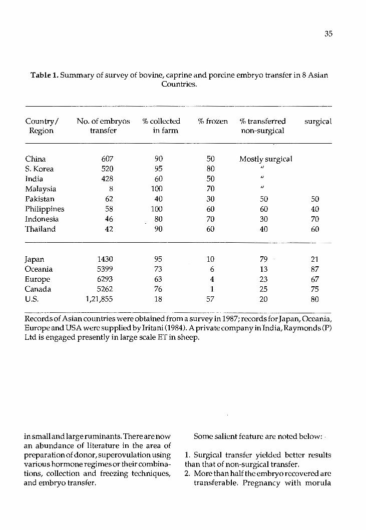
Embryo transfer technology seemed to be the most investigated area in the abovementioned Asian countries. As evidenced from Table 1,numberof embryo transferred in Asia is very low compared to Western nations. These figures may not be exact but an approximation has been made from the papers collected by the author in different countries and from the national position papers, some of which present meaningful statistics.
From the survey of eight Asian countries, China, South Korea and India were found to have undertaking large srnle integrated plans for genetic improvement. Research in different institutions of these countries are centrally coordinated through the efforts of Central Academy of Agricultural Sciences, Beijing, Livestock Experiment Station, Suweon, and Department of Biotechnology, New Delhi respectively. Many institutions within each country are involved.
Since early 1980's, private E.T. companies are advocating the propagation of this technology. Amongstthemare ANSA farms, Magnolia Dairy Farm of San Miguel corporation and Biogenetics (Phil) in Philippines. Similar private efforts for cattle E.T. have been seen in Indonesia, Thailand and Paki-
stan, but initial success in E.T. has not been vigorously pursued in later years. Embryo transfer research in swamp buffalo, cattle and goats are being conducted at Universiti Pertanian Malaysia and in Nili - Ravi buffaloes and cattle at National Agriculture Research Council (NARC), Islamabad, Pakistan. Successful transfer to a Nili - Ravi recipeint in Pakistan has resulted in the production of E.T. twins in buffaloes recently. Almost at the same time, production of buffalo calves through E.T. has been recorded in India and Thailand.
Embryo transfer in buffaloes is still a problem due to very poor recovery of embryo. While this point will be discussed at length in the Indian, Thai and Pakistani paper, a few observations on this problem are recorded here:
i) Prasit (1987) suggested E.T. to be acceptable in buffaloes, more research concerning the recovery of embryo in proper developmental stages, and a greater understanding of the reproductive tract of swamp buffaloes are necessary.
ii) Sharifuddin and Jainudin (1984) found neither the uterus, nor the uterine horn could be exteriorized for non-surgical collection of buffaloes. This view is also supported by Dr. Madan of Karnal, India.
Therefore coordinated efforts in these areas are to be seen in order to create conditions for more success in E.T. of buffaloes. Basic research on various aspects of embryo transfer in laboratory animals (in-vitro fertilization, freezing, cryopreservation etc) have been attempted earlier by Im and his associates at Korea, Bhattacharya and Agrawal in India, and Chen Xiu and Luo in China. Some of these concepts have now been extended in the form of basic research
35
Table 1. Summary of survey of bovine, caprine and porcine embryo transfer in 8 Asian Countries.
Country/ No. of embryos % collected % frozen % transferred surgical Region transfer in farm non-surgical
China 607 90 50 Mostly surgical S. Korea 520 95 80 II
India 428 60 50 II
Malaysia 8 100 70 II
Pakistan 62 40 30 50 50 Philippines 58 100 60 60 40 Indonesia 46 80 70 30 70 Thailand 42 90 60 40 60
Japan 1430 95 10 79 21 Oceania 5399 73 6 13 87 Europe 6293 63 4 23 67 Canada 5262 76 1 25 75 U.S. 1,21,855 18 57 20 80
Records of Asian countries were obtained from a survey in 1987; records for Japan, Oceania, Europe and USA were supplied by Iritani (1984). A private company in India, Raymonds (P) Ltd is engaged presently in large scale ET in sheep.
in small and large ruminants. There are now an abundance of literature in the area of preparation of donor, su perovulation using various hormone regimes or their combinations, collection and freezing techniques, and embryo transfer.
Some salient feature are noted below:
1. Surgical transfer yielded better results than that of non-surgical transfer. 2. More than half the embryo recovered are
transferable. Pregnancy with morula

36
stage embryos, is better than early blastocyst stage.
3. There are no significant difference between frozen and fresh embryos in terms of pregnancy.
4. South Korean workers found no significant differences in pregnancy when transfer with 1 . e1Tibryo compared with 2 embryos at Livestock Experiment Station, Suweon;Xorea (Table 2).
5. Small ruminants' embryo transfer seem to be more successful compared to large ruminants, perhaps because of higher rate of superovulation.
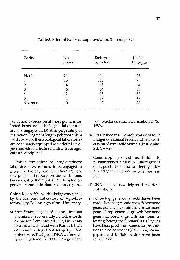
6. 1st to 3rd parity gives more usable embryos (Luo rong, 1989) (Table 3). Some authors suggested superovulation after 90-150 days post partum oestrus gives more usable embryos.
7. There are contradictory evidences regard-
ingthe superiority of PMSG or FSH as superovulating hormones.
8. Except for China, where indigenous production of hormones are increaisng, other countries still depend on foreign supply of necessary hormones.
9. Considerable progress have been made in China, Korea and India in research pertaining to in-vitro fertilisation, chimera production, cloning of embryos etc.
B. r-DNATechnologyandltsApplications
Recombinant DNA technology research in Asia is mainly confined to basic research in which molecular biologists are engaged in research involving prokaryotes. This involves discovery of suitable vectors (plasmids and non plasmids) for propagation of
Table 2. Pregnancy of embryo transfer by different methods at livestock experiment station, Suweon, Kore.
Method of Embryo No. No. No. Transfer Recipient Pregnancy Abortion
1 embryo Fresh 8 4 (50.0) Frozen-thawed 73 27 (40.0) Subtotal 81 31 (38.2)
AI+ 1 embryo Fresh 12 8 (66.7) Frozen-thawed 10 5 (50.0) 2 twins Subtotal 22 13 (59.1)
2embryos Fresh 5 2 (40.0)
Total 108 46 (42.6)
37
Table 3. Effect of Parity on superovulation (Luo rong, 89)
Parity No. Donors
Heifer 21 1 15 2 14 3 6 4 12 5 4 6&more 10
genes and expression of these genes in selected hosts. Some biological laboratories are also engaged in DNA fingerprinting or restriction fragment length polymorphism work. Most of these biological laboratories are adequately equipped to undertake major research and train scientists from agricultural disciplines.
Only a few animal science/veterinary laboratories were found to be engaged in molecular biology research. There are very few publsihed reports on the work. done, hence most of the reports here is based on personal communication or country reports.
China: Most of the work is being conducted by the National Laboratory of Agro-biotechnology, Beijing Agriculture University.
a) Specific antigen gene of equine infectious anemia was successfully cloned. After its extraction from infected cells, DNA was cleaved and isolated with Barn Hl, then combined with gt DNA using T4 - DNA polymerase. The ligated DNA were transferred intoE-coli Y 1090. Five significant
Embryos J]sable collected Embryos
114 75 113 70 138 84 64 33 95 57 39 17 47 36
positive cloned strains were selected (Xu, 1989).
b) RFLP is used in recharacteriza ti on of some indigenous animal breeds and in classifica tsion of some wild animals (Inst. Anim. Sci, CAAS).
c) Gene mapping method is used to identify resistant genes in MHC B-L subregion of 0 - type chicken, and to identify other related gene in the vicinity of GH gene in pig.
d) DNA sequence is widely used at various institutions.
e) Following gene constructs have been made: bovine genomic growth hormone gene, porcine genomic growth hormone gene, sheep genomic growth hormone gene and porcine growth hormone releasingfactor gene. Similar C-DNA genes have been produced. Genes for production related hormones (Calf renin, bovine pepsin and buffalo renin) have been constructed.

38
f) Routine DNA hybridization and some microinjections in laboratory animals are in progress.
India: Major work has been undertkaen by
Ind~an Veterinary Research Institute (IVRI), Izatnagar and Bangalore campus and National Institute of Immunology (NII), New delhi, in the following areas:
a) Molecular characterization of pox virus especially caprine virus (both virulent and non virulent types) is in progress using gene sequencing techniques. Identification of promoter region in the genome is being made in collaboration with virus research institute, Pirbright, U .K. (Mrs. P. Bhat- Personalcommunication).
b) Studies on moelcular cloning in clostridial species for vaccine production have beeninitiated.Earlyworkinvolved DNA isolation of nonpathogenic Clostridial species (C. perfringenes, type C and type D), its characterization and isolation of plasmid DNA from host vector (Bhat et al, 1986).
Another group of workers has been engaged in the characterization of gene sequences in clostridium welchi type D and location of gene blocks responsible for production of "alfa toxin" (Srivastava, Singh and Ashok Kumar, Annual Report of IVRI, 1986).
c) Molecular characterization of FMD virus type 0, A and C has been attempted. (Suryanarayanaetal., 1986). Complimentary DNA's for viral RNA type A and 0 were prepared. Ac-DNA probe has been successfully used for the identification of virus specific sequences (Rao et al., 1986).
d) Coned-DNAforthemajorantigenofFMD disease has been shown to be expressed in E. coli (Surayanarayana -et al., 1986). The c-DNA has been cloned in the Barn Hl site and at the pst 1 site of expression vector, PBR 222.
e) A partial library of Y. chromosome derived DNA sequence of bovine origin in E. coli has been constructed at N.I.I. (Khandekar et al., 1986).
f) Characterization of extra chromosomal plasmids responsible for desired characters of starter culture (Batish-personal communication) and identification of plasmids for lactose utilization in strains of streptococci (Dutta and Sinha -NDRI Annual Report).
Malaysia:
In Malaysia, genetic engineering research for animal production and health has been undertaken by the faculty of Biotechnology and Food Sciences and Veferinary Faculty of University Pertanian Malaysia (UPM) and two Departments of University Malaya (UM) - Department of Genetics and Cellular Biology and Institute for Advanced Studies. Research in progress includes:
i) Molecular characterization of New Castle Disease (NCD) virus at U.P.M.
ii) Collection of baseline data on diversity of rumen bacteria and fungsi has been completed (UPMmainly; bacterial work at U.M. also). At U.P.M., studies on genomic and plasmid DNA in selected microbes and fungi have begun.
iii) Facilities for gene mapping and finger printing and recombinant DNA techniques for xylanase and other enzymes

are available at U.P.M. One of the researchers at U.P .M.' s biotechnology group, Dr. Abdullah Sipat has cloned xylanase gene for Bacteroides succinogens, using E.coli HB 101 as the host.
iv) Mitochondrial and nuclear RFLP work as markers for the identification of different breeds of buffaloes (at UPM) and chickens (at UM) is in progress. cloning of pituitary growth hormone gene using retroviruses as a vector has been planned.
v) A study has been completed at U.M. to relate the transformable antibiotic resistance traits in E. coli strains of bovine, avian and procine sources from slaughter houses with the frequency of R plasmids, which are extrachromosomal genetic markers, and are suspected to be responsible for drug resistance (Koh et al., 1988).
vi) Integrated, well equipped laboratories had been set up for r-DNA technology work for research at UPM and UM. UPM's, B.Sc (Biotechnology) students and UM's, M.Sc (Biotechnology) students will use their respective laboratories for their practical work as well.
Philippines: Basic work on several aspects of biotech
nology is being conducted at the National Institute of Applied Microbiology and Biotechnology, Los Banos, Philippines. The Institute uses protoplast fusion and recombinant DNA technology in the genetic improvement of antibiotic producing micr<;>bial strains. Research on local prqductiqn of tylosin and antibiotics for animal feed is also in progress.
Thailand, Pakistan and Indonesia:
39
The Veterinary Faculty of Kasetsart University, Thailand, Central Research Institute for Animal Sciences, Bogor, Indonesia, and Department of Animal Sciences, Agriculture University at Faisalabad have plans to start work on identification of genetic markers for production and disea.se traits in livestock.
C. Biotechnology for enhancement of feed production
Almost all the countries visited are involved in some kind of workpn biodegradation of organic wastes for production of animal feed or biogas. Majority of these studies can not be categoricallycalled as biotechnological work siri¢e these involve known and old methods\->_(degrading lignocellulosic materials for improvement of nu tri ti ve value offeed. Therefore, only those projects/laboratory researches, which have either microbiological treatment for biodegradation or deal with huge machineries/ ferrnenters/ power plants are recorded here:
China:
1. In most of the provinces and autonomous regions of China, microbial freatment of lignocellulosic low quality roughages is to some extent practised.
2. Yeast factories located at Shanghai, Jilin • and Guang Dan, Nanjing Fermentation
factory and Shunde Sugar Factory produces single cell proteins containing 40-85 protein. Production of SCP using petroleum hydrocarbon, natural gases al).d
I
methanol are also commonly practised.
3. Enzyme ptoduct factories in Wuxi and Tianjing produce various types of enzymes. Shanghai Biochemistry Institute
40
and Beijing Institute of Biochemistry produce enzyme through genetic engineering work.
4. At least 18 amino acids are produced using fermentation technology.
Philippines:
1. National Biotechnology Institute, Los Banos:
a) Production of tylosin and feed antibiotics.
b) Low cost biogas systems using crop residues.
c) Production of genetically improved lignolytic microorganisms.
d) Enzyme engineering techniques for synthesis of ligninases.
e) A low technology tumbler process of producing microbial proteins using selected strains of fungi and yeast. This can be used as a feed ingredients in poultry and pigs.
2. Maya Farms (near Manila): a) Production ofbiogas from farm wastes
- this farm produces 94 tonnes of manure and produces 7400 cubic meters ofbiogas thatgenerate 7,400 kw- hours a day, all of it are used to run electric generators. This farm is considered to be most efficient in biogas production in Asia.
Malaysia:
1. Universiti Pertanian Malaysia: a) Utilization of agro-industrial wastes
as animal feed using biotechnological methodologies (Jalaludin, 1986).
b) Role of rumen in the digestion of fibres in ruminants (Ho et al, 1987) with a view to develop in-vitro system of digestion of fibrous materials.
2. Dunlop (Malaysia) Sdn. Bhd. a) Fermentation of POME using filamen
tous fungi and subsequent use of dehydrated fermented slurry (45.6 per cent crude protein) as an ingredient of poultry /pigfeed. This industrial process was developed by Dunlop (Malaysia) but subsequently stopped for not being cost effective.
3. University of Malaya: a) Characterization of rumen bacteria
with a view to genetic manipulation of bacteira in future for effective digestion of fibre materials (Shaiful, 1988).
b) Production of protein biomass utilizing palm oil mill effleunt as substrate for thermophilic and mesophilic fungi (Kuthubuteen, 1988).
India:
1. Indian Institute of Technology, New Delhi:
a) Biochemical engineering research for development of single cell proteins from waste biomass (Drs. S.K. Mukhopadhaya and Dr. T.K. Ghosh at Indian Institute of Technology, New Delhi).
2. N.D.R.I. Karnal: a) Enrichment of wheat straw using fun
gal (coprinus) and urea treatment (Dr. B.N. Gupta).
Pakistan:
1. N.A.R.C., Islamabad a) Fungal treatment of agricultural wastes
(cereal straws and sugar cane bagasse) (Dr. Amana t Ali).
2. Department of Animal Nutrition, University of Faisalabad:
a) Mycelial biomass protein production

from rice husk, rice polish and rice straw using Trichoderma harzanum (Dr. Abu Saeed Hashmi, 1987).
Indonesia:
1. Central Research Institute for Animal Science, Bogor.
a) Protein enrichment of cassava tuber using yeast inoculum (Candida ingenuosa and Canandida utalis) (Dr. Jinadasa Darma).
b) Protein enrichment of rice straw and wood chips using Pleurotus sp. (Dr. Jinadase Darma).
2. National Centre for Research in Biotechnology:
a) Protein enrichment of rice straw using Coprinus sp. (Dr. Triadi Basuki).
South Korea There are some Universities /Institutions
engaged in work on single cell protein production, according to Director of Livestock Research Station, Suweon. Due to lack of time, visits were not possible to these institutions.
D. Production of monoclonal antibodies and vaccines
Most of the Asian laboratories dealing with the diagnosis of viral and bacterial diseases, and production of vaccines have improved their techniques during the past few years. Use of immunoflorescence microscope, radioimmunoassay, enzyme linked immunosorbent (Elisa) techniques are quite common in most of the laboratories visited.
Work on production of monoclonal antibodies had been quite intensive in South
41
Korea and China. Hybridoma techniques are also being used in some laboratories of India, Malaysia and Philippines. Similar work are also in progress in Indonesia, Thailand and Pakistan. Table 4 shows a list of diseases forthe diagnosis of which monoclonal antibody is used in different countries.
General Discussion
Most of the scientists in animal production and heal th research in the region felt the need for a vigorous approach to undertake biotechnology based research because they felt keeping up with the modern techniques is vital for development. However, there are some scientists who argue for the necessity of consolidating the present work for animal improvement through conventional means before undertaking very highly sophisticated techniques.
Of the areas of biotechnology discussed in this paper, embryo transfer seemed to be the most actively planned area in all the countries. Large scale Government funds are available in China, India, and Pakistan for development of E.T. China has developed considerable indigenous technology e.g. production of hormones, embryo freezers and nitrogen tanks which could be suitable for relatively poorer countries in Asia.
South Korea's efforts were very practical which included a cooperative approach of Government, Research Institutions, Universities and Veterinary clinics. Initial private initiatives to develop E.T. in Philippines, Thailand and Indonesia had been successful but not sustained. More recently, respective Governments are showing greater interest in E.T. Malaysia's programme is still in the research level.

42
Table 4. Monoclonal antibody (MAb) tests available for diagnosis of diseases in Asian Countries
Country
China
S. Korea
Thailand
HVT, ILT
Test/ genetically engineered Vaccine ( GEV)
M Ab (Monoclonal antibody) Harbin Inst. Vet. Med., CAAS Lanchou Inst. Vet. Medicine Xinjiang Inst. Vet. Medicine Fujian Agriculture College
M Ab (Monoclonal antibody) Veterinary Research Institute, Anyang
Elisa (same institute)
GEV (genetically engineered vaccine) (same institute)
M Ab (Work in progress) at National Inst. of Animal health and production, Bangkok
M Ab (Work in progress at the same institute)
Viral or bacterial diseases
Equine Infectious anemia Hog cholera NCD Ovine brucella 0-typeFMD Pig E coli K88 Pig treponema X23
a) Infectious Bovine Rhinotracheitis virus (IBRV)
b) Bovine Rota virus (BR) c) Transmissible Gastroenteritis
Virus (TGEV) d) Japanese Encephalitis Virus (JEV) e) Porcine Parvovirus (PPV) f) Enterotmcigenic E. coli (ETEC) g) Pseudorabies Virus (PRV) h) New Castle Disease Virus
(NCDV) i) Herpes Virus of Turkeys (HVT)
BR, IBRV (Cattle) IB, NCD, HVT, ILT, IBD, (Chickens) Rabbies (Dogs). Hog cholera, Pseudorabies, japanese en-cephalities, Porcine parvoirus (Pigs)
Piglet Diarrhoea
New Castle Disease
pig pneumonia

43
M Ab (Work inprogress at Assay of progesterone Chulalongkorn U.)
Philippines MAb & Elisa Pasturella haemolytica (Work in progress at Inst. biotech, Los Banos)
Elisa New Castle Disease Virus (Work in progress at Bureau of Animal Industries, Manila)
Indonesia Elisa and M Ab. IB, NC (Chickens), Brucella, Anthray, Veterinary Research, Babesiasia (Cattle), Anaplasmosis Institute, Bogor (Cattle & Buffalo)
Malaysia Pelleted field vaccine (UPM - testing in different countries in progress)
Elisa IB, NC (Chickens) HS, Brucella (Cattle). Detection of progesterone level under field conditions in bovines.
M Ab (Work in progress) NCD (Chickens)
India Elisa (IVRI) Sheep pox, Buffalo pox, FMD, NCD, Marek's disease
M Ab (Work in progress at FMC (Type 0, A, Asia 1) IVRI)
Elisa (N.I.I.) Leptospirosis (dogs) Detection of pregnancy
M Ab (N.I.I. - Work in progress) Rabbi es
Pakistan Elisa (NARC, Islamabad) Pasturella multicode
Elisa (Univ. Agriculture, Pasturella multicode Faisalabad -workin,progress)
M Ah(NARS, Islamabad Pasturella multicode work in progress)

44
Genetic engineering research in relation to domestic animals is just starting to develop in China, South Korea, India and Malaysia where substantial commitment has been made by the R & D funds of respective Governments. There are capable scientists in all 8 countries who could undertake molecular biology work but because of limited research funds, such work has not gained momentum yet bymanyinstitutions.
It is strongly believed in Asian countries that biotechnology can utilize raw materials from various wastes to convert it to renewable resources. Microbial digestion of such raw materials using fungi and bacteria had been the basis of research in many of the processes discovered as viable commercial processes will take time. Characterization of rumen microbes in relation to its digesting properties of various fibrous material in the feed will continue to be an important research in the animal biotechnology.
The discovery of monoclonal antibodies for diagnosis of different disease in South Korea, China and other countries will lead to many industrial activities. Similar plans and continuous vigorous research is necessary in other countries to improve their own potential for various aspects of biotechnology, for example, to improve their own potentials for diagnostic facilities, and perhaps later, vaccine production against spe- · cific antigens.
In general, one could conclude by saying that the interest for development of research potential for various aspects of biotechnology exist in all the countries but due to lack of funds and facilities, concrete cooperative efforts have not been made. In light of these facts, the creation of an Asian Biotechnology Network will stimulate biotechnology research and development in the region.
References
(A list of references relevant to Asian Biotechnology research is given below)
1. Amanat Ali., A. Azim, P.A. Mir and M.A. Bhatti. (1987). Treatment of cereal straws with white rot-fungi. Pakistan]. Agric. Res. 8(4-): 1-5.
2. Annual Research report (1986). Livestock Experiment Station, Rural Development Administration, Suweon, Korea. pp 205.
3. Annual Report (1986). Indian Veterinary Research Institute, Izatnagar (U.P.) India. pp. 182.
4. Annua1Report(1986-1987). Department of Biotechnology, Ministry of Science and Technology, Govt. of India, New Delhi. pp 59.
5. Annual Report on Research projects (1987). National Dairy Research Institute (IDRC), Kamal,Haryana,India. pp. 231.
6. Annua1Report(1985-1986).Nationallnstitute of Immunology, New Delhi, India, pp. 86.
7. Azim, A., A. Ali, P.A. Mir and M.A. Bhatti (1987). Improving nutritional value of sugarcane bagasse through biological treatment. Pak. J. Agri. Res. 8(4): 10-14.
8. An, S.H., Y.H. Kim and D.S. Sul (1986). Application of monoclonal antibodies in Veterinary field in Korea. Proc. Seminar on production and utilization of monoclonal antibody for control of anima1 disease. Seoul, Korea. pp. 111-122.

9. An, S.H. (1986). Monoclonal antibody against porcine parvovirus. Annual Research Report of Vet. Res. Institute, Rural Development Administration, Suweon, Korea. pp 11.
10. An,S.H.(1986).RapiddiagnosisofJapanese Encephalitis using monoclonal antibody. Annual Res. Report of Vet. Res. Institute, Rural Development Administration, Suweon.
11. Bhat, P.N., P.P. Bhat, V.P. Singh and Asok Kumar (1986). Establishment of Recombinant DNA technology centre for research in vaccine production and improvement of animal production: Studies on molecular cloning in clostridial species for vaccine production. Annual Report of Indian Veterinary Research Institute, Izatnagar, Indian. p. 32-33.
12. Bhat,P.N.,M.R. Ansariandl.V. Mogha (1986). Studies on superovula ti on, nonsurgical collection and cryopreserva tion. Annual Report oflndian Veterinary Research Institute, Izatnagar, India. p. 34.
13. Bhattacharyya,N.K. (1984). Progress in embryo technology and its application in animal science. First National Animal Physiology Research Workers Conference, Kamal, India. pp. 41 (Published monograph).
14. Choi, C.O., S.H. An, C.G. Lee and S.M. Cho. (1986). Production of mono<::lonal antibody to infectious laryngotrachei tis virus by cell fusion. Proc. Seminar on antibodyf or control of animal diseases, Seoul, Korea. pp. 23.
15. Collier, H.M. and W.H. Chick. (1979). Research priorities in the plantation in-
45
dustry. Inst. of Chemistry (Malaysia) Conf. on Chemical Research in Malaysia, pp. 12. Kuala Lumpur, Malaysia.
16. Darminto, P.Y. and P. Ronohardjo. (1986). Penerapan uji Enzyme-linked immunosorbent assay (Elisa) untuk deteksi antibodi virus infectious bronchitis. Penyakit Hewan, 18(32): 118-123.
17. Geol, V.K andP.N. Bhat.(1986). Studies on the embryo technology and engineering with p articular reference to cattle and buffalo; studies on su perovula tion, non-srugical collection and cryopreservation of embryos in cattle. Annual Report of Indian Vet. Res. Ins. Izatnagar, U.P. p. 33-34.
18. Ho, Y.W.,N.AbdullahandS.Jalaludin. (1987). Contribution of rumen anaerobic fungi in the utilization of cellulosic materials for the ruminants. Proc. Colloq. An. Prod. & Health, Universiti Pertanian Malaysia, Serdang. p. 75-77.
19. Im, KS. and Y.B. Lee. (1983). Studies on the mocrosurgical dichotomy of embryos in mouse and rat. Korean}. amin. Sci. 25(4): 362-368.
20. Im, KS., Y.B. Lee and KM. Chung. (1984). Studies on embryo transfer in cattle. Seoul Natl. Univ. College of Ag. Res, 9(1-1): 83-93.
21. Im, KS., Y.B. Lee, KM. Chung and J.C. Kim. (1989). Studies on embryo transfer in catlte. II. Studies on superovulation and conception of heifers in puberty. Seoul Natl. Univ. College of Ag. Research. 11(1): 23-31.
22. Keon, C.H., S.H. An, Y.H. Kim, J.B. Lee, B.H. Kim and Y.S. Lee (1986). Production and characterization of monoclo-
46
nal antibody against Japanese encephalitis virus. Seminar Prod. & utilization MAB (RDA & FFTC/ ASP AC, Seoul, Korea), pp. 8-1to8-19.
23. Latif, A. Ibrahim. (1986). Vaccination of free range chickens against New Castle Disease. 35th. Annual Meeting, Proc. West Poultry Diseases. Peu tro Vallartta, Mexico. pp. 85-86.
24. Lee,J.H.,H.J.ParkandM.H.Cho.(1986). Interactions between recipients and embryos affecting proegnancy rates in bovine embryo transfer. Korean J. embryo transfer. 1(1): 83-89.
25. Manan, A. (1987). Anaerobic cellulose digestion of palm oil mill effluents by rumen microorganism. M.Sc. thesis, University of Malaya, Kuala Lumpur, Malaysia. pp. 214.
26. Martin,0.C.,A.V. Nocom, E.C. Sanchez and Juan M. Malabanan (1985). Bovine embryo transfer at ANSA farms- A case report, Philippine J. Vet. & Amin. Sci. 11: 324.
27. Mukherjee, T.K. and S. Bhubanendran. (1986). Application of biotechnology for improvement of livestock production and health in Asia (Paper presented at FAO Expert consultation on Biotechnology for Livestock Production and Health, Rome, 6-10 October, 1986). pp. 25.
28. Inderjeet and R.C. Gupta. (1987). Variations in recovery rates and morphological normalcy of caprine embryos collected surgically from superovula ted goats. Workshop embryo Biotech., National inst. Immunology, New Delhi. p.10.
29. Iritani, A. (1984). Present status of bovine embryo transfer in Japan. Embryo Transfer Newsletter. 3(2): 21-23.
30. Jim, D.I.,K.S. Im, B.K. Ohhand Y.B. Lee. (1986). Effect of cryoprotectants, ice induction, cooling rate and storage length of the survival of mouse embryo. Korean. J. Anim. Sci. 28(7): 474-479.
31. Jo, C.H., C.K. Cheong and W.S. Hwang. (1987). Studies on effects of ethylene glycol on the survival of frozen-thawed mouse embryos. KoreanJ. Vet. Res. 27(2): 331-334.
32. Jun, M.H. (1986). Production of monoclonal antibodies against infectious bovine rhinotracheitis virus. Seminar Prod. MAB (RDA & FFTC (ASP AC) pp. 44-1 to 4-11.
33. Khandekar,P. andR. Chaudhury. (1987). DNA probe for sexing of male cattle. Workshop Proc. on Natl. Embryo Biotech.NationalinstitUteofimmunology, New Delhi. p. 30.
34. Kim, U.H., S.H. An and Y.D. Yoon. (1987). Monoclonal antibody production against piglet diarrhoea agent (Enterotoxigenic E.coli) by cell fusion-hybridoma cell technique. Korean J. Vet. Res. 27(2): 259-267.
35. Kim, U.H. (1986). Rapid diagnosis of transmissible gastroenteritis in pig using monoclonal antibody. Seminar on Prod. & utilisation of MAB (RDA & FFTU I ASP AC, Seoul, Korea), p. 7-1 to 7-14.
36. Kim, CK., K.S. Lim Im, X. Zheng and R.H. Foote. (1986). Studies on in-vitro capacitation by lysolecithin and in vitro

fertilizing ability of ejaculated rabbit sperm. Korean J. Anim. Reprod. 10(1): 109-120.
37. Koh, C.L. and C.H. Kok. (1984). Antimicrobial resistance and conjugative R. plasmids in Escherichia coli strains isolated from animals in Peninsular Malaysia. South East AsianJour. Trop. Med. Pub. Health. 15(1): 37-43.
38. Luo Rong. (1989). China Dairy Cattle 2: 25-29 (cited in National Position paper of china).
39. Nonya Razak, Yusof Samah, Rahmah Mohamad, Noor Embi, Othman Omar and Ghazali Ismail. (1987). Pili of Pseudomonas pseudomalleic and its haemagglu tinating and adherence properties; Proc. 10th Malaysian Micro Conf. (Kuala Lumpur, Malaysia), pp. 10.
40. Padolina, W .G. (1987). Development and utilization of biotechnology in Agriculture. (Paper presented during the seminar on quality through appropriate technology sponsored by the Philippine Productivity Movement through PCARRD, PCIERD and NEC; held on 1st. october, 1987 at Bicutan, Taguig,MetroManila).
41. Rao, B.U., V.S. Suryanaryana, C. Butchaiah and N. Banumathi. (1986). Research on moelcular characterization of foot and mouth disease cirus and biotechnological aspects of immunogens and diagnostics. Annual Report Indian Vet. Res. Inst. Izatnagar, U.P. p. 73.
42. Rao, K.V. and 0. Ramakrishna. (1987). Oestrogen and prosgesterone change sin superovulated prepuberal buffalo calves. Workshop Embryo Biotech. National Inst. Immunology,New Delhi,
47
p.12.
43. Rohani, A.K., 0. Othman and A.A. Saiful. (1988). Degradation of cellulosic agriculture wastes by Aspergillus terreus SUK-1. Proc. 11th. Ann. Conf. MSAP. pp. 84-90.
44. Sharifuddin, W. and M.R. Jainudeen. (1984). Superovulation and non-surgical collection of ova in the water buffalo (Bubalus bubalis) Proc. 10th. Int. Cong. anim. Reprod. & A.I., Univ. Illionis, Urbana - Champaign, pp. 240-242.
45. Shaiful, A.A. A. and T.K. Mukherjee. (1987). Microbial growth, protein and energy contents of stored goat feed pellets. Biological Wastes. 7: 42-47.
46. Singh, B., A.K. Sinha and B.K. Singh. (1987). Superovulatory response in circumpuberal and adult Black Bengal goats. Proc. Workshop Embryo Biotech., National Inst. Immunology, New Delhi, p. 8.
47. Supar. (1986). Penggunaan metode enzyme-linked immunosorbent assay (ELISA) untuk deteksi antigen Pili K 99 dan K 88 pada Escherichia coli dari anak babi diare. Penyakit Hewan 18(32): 159-168.
48. Suryanaryana, V.V.S.,B.U.RaoandJ.D. Padayatty. (1986). Expression in E.coli of the clonal cDNA for the major antigen of foot and mouth disease virus Asia 1,63/72.J. Genet,655(1&2):19-30.
49. Wani, G.H., B.A. Buchoo and N.A. Sudan. (1987). Superovulation in sheep. Proc. Workshop Embryo Biotechn, National Inst. Immunology, New Delhi. p.6.

48
50. Xu Dianxih. (1987). Proc. 4th. Congr. Anim. Reproduction & A.I. p. 67 (Cited in National Paper of China).
51. Yang, B.S., i<:.S. Im and Y.B. Lee. (1985). Studies on-the solubility of zona Pellucida and dev_elopmental potency of
isolated blastomere in mice. Korean J. Amin. Sci. 2700: 706-710.
52. Yeong, S.W. (1981). Biological utilization of palm oil byproducts by chickens. Ph.D thesis, University of Malaya, Kuala Lumpur, Malaysia. pp. 168.
Proc. FAO/CAAS Workshop on Biotechnology in Animal Production and Health in Asia and Latin America, Beijing, Oct 9-13,1989, pp 49 - 58
Growth Stimulators for Farm Animals: Mode of action, effects on meat quality
and potential risks originating from residues
Heinrich H.D. Meyer and Heinrich Karg Institute of Physiology
Technical University Munich D-8050 Freising-Weihenstephan, FRG
Introduction
Several hormones are involved in endocrine or paracrine regulation of growth which has been reviewed previously (Karg 1989) and growth might be manipulated by all these hormones. However, only three groups have gained scientific or practical importance in the past and they will be described here. Porcine and bovine somatotrokpin (pST and bST) were tested more intensively on a scientific base since larger quantities of these products are available; effects on feed efficiency and carcass composition as well as the mode of action were of predominantinterest. Also B-agonist have been investigated quite intensively during the past decade for similar aspects. These compounds, however, have oral bioavaibility and residues might present a risk for the consumer. Much more information in this respect is still needed. B-Agonists can be given to the animal very easily with constant dosage via food; some compounds have been used illegally and got practical importance by this way. Most investigations were done with sex hormones (estrogens and androgens), their effects on growth promotion and feed efficiency, p.re studied since almost 40 years and they have gain'ed tremendous practical importance. Their
correct use seems to represent no risk to the consumer (WHO 1982; Lamminget al. 1987), but unfortunately there is illegal use in Central Europe and under latter conditions biologically active residues in meat can not be excluded.
Somatotropin
Biochemistry and physiology: Porcine and bovine somatotropin are 191 or 190 amino acid proteins, they share a high degree of homology (90%) and they are produced in the anterior pituitary; today both proteins can be synthesized by transformed bacteria (Seeburg et al. 1983). Liver, skeleton, adipose tissue and muscle are the major target tissues. The receptor for bST has been cloned and the structures of the receptor are well known (Leung et al. 1987), but the mode of signal transduction into the cell is still not understood. However, there are clear metabolic effects of somatotropin (Figure 1) and the reduced glucose utilization by adipose tissue seems to be most important. After treatment with somatotropin fatty acid synthesis is d~creased due to a reduced activity of the involved enzyme systems of the "Pentosephosphate Cycle" and the "fatty acid synthase" (Ether-
FAO LIBRARY AN: 302003

50
Figure 1. Synopsis of CH-physiology
Synopsis of GH-physiology
lipogenesis inhibition
reduced insullnsensltlvlty chondrocyte stimulation
j i l I fat l I .--1-ive-r-,-,tl 1.~~'_ 1~~~1 i j muscle t I skeleton t J
lactate +
blood glucose- t blood insulin t
ton 1989). As a result glucose and insulin in blood are elevated, but somatotropin prevents glucose uptake by liver and adikpose tissue due to impaired insulin sensitivity. More glucose is available for the muscles and the skeleton; growth of these tissues is additionally stimulated directly by somatotropin and by IGFI (insulin like growth factor I) originating from the liver after stimulation by somatotropin. Further data support the idea that amino acid flux to the muscle is facilitated and hepatic amino acid degradationisreduced (Etherton 1989). The recent detection of somatotropin receptor mRNA in skeletal muscle indicates that somatotropinmay stimulate muscle growth directly (Baumbach et al. 1989), but also hepatic IGFI may be of major importance for musclegrowth(Guleretal.1989). Also bone growth seems to be stimulated directly by somatotropin (Hochberg et al. 1989) as well as indirectly via IGN I (Guler et al. 1989).
Implications to animal production: Improved growth rates have been shown for veal calves given bST by daily injection (Kirchgessner et al. 1987) and many studies have been done with pigs given frequent injections of pST (van der Wal 1989). Also feed efficiency and N-retention is improved; carcasses become leaner and fat content is reduced. The efficiency of pST is depending on several factors like sex, age, breed and environment. It has little effect in fast growing, lean animals (Huster et al. 1988), but the effect is more pronounced in chinese breeds with a higher fat content (McLaughlin et al. 1989). The metabolic effects are also enlarged in females and in castrated males if compared to intact males with a faster growth rate, a lower daily fat deposition and a higher endogenous production of anabolic sex hormones (Campbell et al. 1989). In consequence to better N-retention the excreted N-amount and the load to our envi-

ronment is decreased (van Weerden and Verstegen 1989). Due to lower fat content the thermal isolation of the animals is reduced, which restricts an useful application of pST to warmer ambient temperatures <Curtis 1989). However, pST has no practical importance up to now, due to limited availability of slow release devices, and it is an open question whether pST or bST can be used economically with acceptance by farmers and consumers.
Meat quality and residues: Due to the multiple metabolic effects of somatotropin meat quality is altered in several aspects. First, the overall fat content is strongly reduced which is of predominant importance for the consumer. It has been reported that further aspects of meat quality are affected (Wassmuth and Reuter 1973, Demeyer et al.
51
1989): shear force of the muscle is elevated, flavour and colour may be reduced. In addition content of thiamin (vitamin B1) in muscle is lower after pST treatment (Prusa, 1989), which points to an increased glycolyticATP-productionin the animal, because thhiamin is an essential cofactor for pyruvate metabolism and oxidative ATP-production from carbohydrates.Residues of somatotropin are of no importance to the consumer due to the protein nature of somatotropin, its complete degradation during digestion and its species limitations.
B-Agonists
Biochemistry and physiology: The metabolic effects of B-agonists to the liver and fat cell is summarized in Figure 2 and is already
Figure 2. Biochemistry of ~-Agonists in Fat and Liver Cell
Biochemistry of '3-Agonists in Fat and Liver Cell
Epinephrine
Receptor'-...... J G-Proteln "-...... t Adenylatecyclase
Cell membrane Otr~~~~~~~~~~~~
~ ATP cAMP
10-6M
Kinase~ Klnast([) Liver
/ ,-__ LipaseGi)...'. Lipase(]) :Phosph~Phosphorylase
i ! Lipids -NFA Glucose --- Glycogen
i i NFA Glucose
52
well documented in text books (Lehninger 1976). Glycogen and triacylglycerol degrading enzymes are activated; as a result non esterified fatty acids and glucose are secreted into the blood and their levels in blood increase initially. In addition more lactate and insulin is found in blood (Zimmerli and Blum 1989), but lipogenesis is impaired. After adaptation within oen week blood levels of the mentioned compounds return to normal and feed intake may be reduced, especially in ad libitum fed animals. Hence more energy is available for th emuscle which facilitates muscle growth and protein accretion. Recent results indicate that B-agonists may stimulate the muscle also directly. Skeletal muscle of treated animals contain es more mRNA for actin as well as myosin-light-chain (Smith et al. 1987) and in addition the activity of the proteolytic enzymes Cathepsin B, Cathepsin L plus Ca++ -dependant protease is reduced (Forsberg et al. 1987, Wang and Beermann 1988, Morgan et al. 1989).
B-Agonists may have numerous side effects: vasodilatation, tokolysis, anthiasthmatic, muscle tremor, increased heart rate, increased respiratory rate and restiveness. However, these effects are not common for all B-agonists and the extend of side effects depends not only on the dose but also on the interaction of the used compound with different B-receptors.
Implications for animal production: Some B-agonists have good oral bioavailability (clenbuterol, Salbutamol) and many compounds are sold on the pharmaceutical markets. Hence, the continuous application tot he animal is very easy and the two mentioned compounds have been applied illegally on the European continent within the recent two years. However, B-agonists have not yet been licensed as growth pro-
motors anywhere in the world.
Improved growth rates, strongly reduced fat content of the carcass, better N-retention and better gain/ feed-ratio have been documented for many different animals and breeds like sheep, pig, steer, bull, veal calf, chicken, rat and mouse (Hanrahan 1987, quirke and Schmidt 1988). There is no doubt that such compounds could be used very economically in animal production.
Meat quality and residues: B-agonists are more or less bioavailable after oral consumption and therefore it is essential to do a careful toxicological evaluation of any compound to be registered. Adverse effects to the consumer will be generally related to those effects mentioned as side effects in the animal (see above). Many B-agonists have been tested in man and a lot of dosage depending effects are known, but there is very poor knowledge about residues of Bagonists in edible tissues after its application for growth promotion. Own results show (Rinke and Meyer 1989), that after feeding 10 ug Clenbuterol/kg bodyweight daily highest residue levels were found in lung and liver (40-100 ng/ g). The consumption of 200 g liver therefore provides and effective dose (about 10 ug) to the consumer.
However, application of B-agonists in animal production could be of greatestinterest also for the consumer, because overall fat content of the carcasses is very much reduced. Alterations of further meat quality are studied intensively at present: glycolytic fibres seem to be stimulated, intramuscular glycogen plus fat is lower and protein composition seems to be modified (Hanrahan 1987). Prior to a final udnerstanding of all effects further studies are essential on localisation of different B-receptors in different organs, their selective stimulation by single

B-agonists and the resulting specific effects.
Anabolic Sex Hormones
Biochemistry and physiology: Natural androgen sand estrogens - the anabolic sex hormones - are lipophilic steroids, but also some aromatic lactones like zeranol or stillbens like diethylstilbestrol express good estrogenic activity. Their biochemical mode of action is different from growth hormone and B-agonists. They can pass the cell membrane and there is a permanent equilibrium between extracellular and intra-cellular steroid concentration. In the cell steroid hormones bind to specific receptors, the conformation of the receptor is modified and this activated receptor binds to a "regulatory element" on the DNA (Beato et al. 1987). By this way selective genes are activated and transcription rate is increased. The produced mRNA gives the matrix for a higher translation rate and more protein synthesis (Figure 3). Steroid receptors are present in most sexual organs, and these organs represent the predominant targetfor sex hormones. But also skeletal muscle contains androgen (Krieg 1976) and estrogen receptors (Meyer and Rapp 1985) being essential for a direct stimulation of muscle growth. Receptor levels in different muscles are proportioal to the stimlation of the specific muscle and its allometric growth (Sauerwein and Meyer 1989). But anabolic sex hormones are also able to stimulate growth hormone (Davis 1984) and IGF-I production (Breier et al. 1988), which will contribute to the increased growth rate. The higher energy requirement for taster growth of the skeletal muscle may induce indirectly, that fat deposition is impaired (Campbell et al. 1989).
53
Figure 3. Schematic representation of the action of steroids in a skeletal muscle cell
Steroids
{Estrogens, Androgens)
S~I~ recept~r !
Membrane
Q-sterold
Nucleus DNA In chromatin
A- Amlnoaclds Ribosome \__)
specific proteins
Implication to animal production: Anabolic sex hormones have been used in animal production since almost 40 years. Their practical application is almost limited to ruminants and they are most effective in animals with no or low endogenous sex hormone production (steers, veal calves, heifers). About 5-20% in growth rates and feed efficiency were obtained in many studies. Optimal combinations of an estrogen plus trenbolone may also improve growth ratesofbulls(about+10%)withendogenous sex hormone production (Bouff ault and Willemart 1983). In addition feed efficiency and N-retention are improved (van der Wal and Berende 1983). Gradingofcarcass com-

54
position is better, whereas meat quality seems to be rarely affected. Such effects have been observed inmost breedsd ofcattle and their seems to be no or little difference within differnt breeds (Meissonnier 1983, Roche an O'Callaghan 1984). This is in line with similar receptor levels in skeletal muscle of different breeds as observed in our laboratory.
Preparations of anabolic sex hormones have been registered in about 50 countries and these anabolics represent a big value that allow a more economic animals production. In the EC the application of sex hormones for growth promotion is forbidden since January 1988, but the application of unregistered hormone preparations has been observed very frequently (Jansen et al. 1984, Moretti et al. 1985, Rapp and Meyer 1987).
Residues: After correct treatment with registered preparations of physiological steroids the hormone levels remain in the normal range of sex hormones of untreated animals. The endogenous human produc-
tion of sex hormones is always more than 1000 times higher, if compared to the residues ingested iwth meat (Table 1). In addition a low oral bioavailability of physiological steroids has to be considered. Correct treatment assumes that the depot containing among mg-amounts is removed from the carcass. Under these prerequisites physiological steroids were regarded as safe by several scientific committes (WHO 1982, EEC 1982). The xenobiotic compounds zeranol and trenbolone have been investigated very itnensively and a careful risk assessment has been done by the Lamming-Committee (Lamming et al. 1987).
The illegal praxis is different from correct use of hormones in the following aspects: cocktails with unknown ingredients without any declaration or with wrong declarations are used; injection sites are often intramuscular in edible tissue; mostly dosage is much to high. Only in the injection site hormone residues in mg-quantities can be found, that cause hormonal efects after consumption. After single oral intake of even higher hormone quantities only a depres-
Table 1: Amounts of physiological sex hormones in food compared to endogenous production in man (microgram)
Maximal amount in 250 g meat veal calf or steer, untreated or correct treatment Bull untreated
Endogenous daily production child woman, cyclic man
Estrogen
0.01 0.01
10 200 100
Testosterone
0.01 0.1
30 200
6000

55
Table 2. Residues after illegal hormone application and potential effects
residues found in injections sites: 0.2 - 20 mg of applied hormone
effects of ethinylestradiol: child: 2 - 5 µg single dose child: 5 µg daily women: 50 µg daily
relative oral bioavailability in man:
gonadotropin depression breast stimulation withdrawal bleeding
estradiol diethystilbestrol ethinylestradiol 33 1 4.5
sion of gonadotropin secretion is observed (Table 2). The risk that injection sites are consumed on several consecutive days is very low. However, also hormones with high oral bioavailability have been misused (ethinylestradiol, diethylstilbestrol, methyltestosteron), which have to be considered as somewhat more problematic (Karg et al. 1989).
Conclusions and Outlook
Further investigations are essential especially for the first two groups of growth promotors - somatotropin and B-agonists. The use of somatotropins is limited especially due to rare possibilities ofconstant parenteral application. Concerning B-agonists the whole residue problem needs much more investigations.
Risks will originate only from illegal use of growth promotors because the amount, the site and the type ofresidues is unknown. Analytical methods for its control have been developed and need permanent improve-
ment for the detection of new actual substances.
In addition more research on improvement of meat quality is needed: how do growth promotors influence myofiber type and size, intramuscular fat and glycogen, content of vitamines and minerals? Also cumulative effects of different growth promotors are rarely understood. Studies with combination might open new perspectives for minimal dosages, insignificant residues as well as optimal feed conversion, energy and N-retention.
References
For review tables and references see Karg, H. Manipulation of Growth. From Biotechnology for Livestock production - AF AO I Plenum Publication (Plenum Publishing Corporation, 1989).
Additional references:
Baumbach W.R., D.L. Homer andJ.S. Logan: Structure and regulation of the rat growth

56
hormone receptor. In: Biotechnology in growth regulation Edits. R.B. Heap, G.G. Prosser, G.E. Lamming Butterwroth London, 221 (1989).
Beato, M.J., J. Arnemann, G. Chalepakis, E. Slater and T. Willman: Gene Regulation by steroid hormones. J. Steroid Biochem 27, 9-14 (1987).
Boufaault,J.C. and J.P. Willemart: Anabolic activity of trenbolone or in association with estrogens. In: Anabolics in Animal Production Public health aspects, analytical methods and regulatiuon. Edt. E. Meissonnier OIE, Paris, 155-179 (1983).
Breier, B.H., P.D. Gluckmann and J.J. Bass: The somatotrophic axis in young steers: Influence of nutritional status and oestradiol-17B on hepatic high- and low-affinity somatotrophic binding sites. J. Endocrinol. 118, 243 (1988).
Campbell, R.G., R.J. Johnson and R.H. King: Implications of biotechnological techniques for manipulation animal growth and development on tissue and dietary nutrient requirements of pigs. In: Bi6technology for control of grwoth and product quality in swine, implications and acceptability. Ed ts. P. van der Wal., G.J. Nieuwhof, R.D. Politiek, Pudoc Wageningen, 137-144 (1989).
Curtis, S.E.: Potential side-effects of exogenous somatotropin in pigs. In: Biotechnology for control of growth and product quality in swine, implications and acceptability. Ed ts. P. van der Wal., G.J. Nieuwhof, R.D. Politiek, Pudoc Wageningen, 155-158 (1989).
Demeyer, D.,R. Verbeke,G. van de Voorde, J. Fabry, E. Deroover and R.H. Dalrypmle: Effect of recombinant porcine somatotropin
on carcass and meat quality of belgian and dutch pigs. In: Biotechnology for control of growthand productqualityinswine,implications and acceptability. Edts. P. van der Wal., G.J. Nieuwhof, R.D. Politiek, Pudoc Wageningen, 191-199 (1989).
Davis, S.L., K.L. Hossner and D.L. Ohlson: Endocrine regulation of growth in ruminants. In Manipulation of growth in farm animals. Edts. J.P. Roche, D. O'Callaghan, Martinus Nijhoff Publishers, Boston, 151-178 (1984).
Etherton, T.D: Mechanisms by which porcine growth hormone (pGH) and insulinlike growth factors (IGFs) regulate pig growth performance: approaches from the pGH and I GP receptors to the whole animal. In: Biotechnology for control of growth and product quality in swine, implications and acceptability. Edts. P. van der Wal., G.J. Nieuwhof, R.D. Politiek, Pudoc Wageningen, 111-125 (1989).
EEC Publication EUR 8913 (1982).
Forsberg,N.E.,A.R. Nassar,R.H. Dalrymple and C.A. Ricks: Cimaterol reduces cathepsin B activity in sheep skeletal muscle. Fed. Proc. 46, 1176A (1987).
Guler, H.P., J. Zapf, K. Binz and E.R. Froesch: Growth Promotion using recombinant insulin-like growth factor-I. In: Biotechnology in growth regulation. Ed ts. R.B. Heap, C.G. Prosser, G.E. Lamming. Butterworth London, 18-20 Sept. (1988).
Hanrahan, J.P.: Beta-agonists and their effects on animal growth and carcass quality. Elsevier applied Science London, New York (1987).
Hochberg, Z., G. Maor, D. Lewinson and M.

Silbennann: The direct effects of growth hormone on chondrogenesis and osteogenesis. In: Biotechnology in growth regulation. Edts. R.B. Heap, C.G. Prosser, G.E. Lamming. Butterworth London, 123-128 (1989).
Buster, E., D. Ekkel, E. Kallweit, R.E. Ivy and F. Ellendorff: The use of recombinant porcine growth hormone to influence growth and body composition in pigs. Acta Endocrinol. 117 (?uppl. 287), 74 Abstr (1988).
Jansen, E.H.J.M., H. van Blitterswijk and R.W. Stephany: Monitoring and identification of residues of anabolic preparations in slaughtered cattle by HPLC with diode array detection. The Vet. Quart. 6, (2) 60-65 (1984).
Karg, H., B. Hoffmann and H.H.D. Meyer: Honnonale Leistungsforderer bei lebensmittelliefemden Tieren. Endokrinologie Informationen 13, 60-66 (1989).
Kirchgessner, M., F.X. Roth, D. Schams and H. Karg: Influence of exogenous growth hormone (GH) on performance and plasma GH concentrations of female veal calves. J. Anim.Physiol.Anim.Nutr.58,50-59(1987).
Krieg, M.: Characterization of the androgen receptor in the skeletal muscle of the rat. Steroids 28, 261 (1976).
Lamming, G.E., G. Ballarini, E.E. Baulieu, P. Brookes, P.S. Elias, R. Ferrando, C.L. Galli, R.J. Heitzman, B. Hoffmann,H. Karg,H.H.D. Meyer, G. Michel, E. Poulsen, A. Rico, F.X.R. van Leeuwen and D.S. White: Scientific report on anabolic agents in animal production. The Veteimary Record,389-392 (1987).
Lehninger, A.L.: Biochemistry. The molecular basis of ceil structure an dfunction, Worth Publishers, Inc., New York (1975).
57
Leung, D.W., S.A. Spencer, G. Cachianes, R.G.Hammonds,C.Collins, W.J. Henzel,R. Bamard,M.J. Waters and W.I. Wood: Growth hormone receptor and serum binding protein: purification, cloning and expression. Nature 330, 537-543 (1987).
McLaughlin, C.L., C.A. Baile, S.Z. Qi, L.C. Wang, and J.P. Xie: Responss of Beijing black hogs to porcine somatotropin. J. Anim. Sci 67, 116-127 (1989).
Meissonnier, E.: Anabolics in Animal Production. Public health aspects, analytical methods and regulation. OIE, Paris (1983).
Meyer, H.H.D. and M. Rapp: Estrogen receptor in bovine skeletal muscle. J. Anim. Sci. 60, 294 (1985).
Moretti, G., G. Cavina, R. alimenti, P. Cammarata and L. Valvo: HPLC analysis of steroid formulation, acting as anabolics, unlawfully used in veterinary. Anal. Chem. Symp. Ser. 23, 471-477 (1985).
Morgan, J.B., S.J. Jones and C.R. CALKINS: Muscle protein turnover and tenderness in broiler chickens fed crimaterol. J. anim. Sci. 67, 2646-2654 (1989).
Prusa, K.J.: Nutritional and sendory characteristics of pork from pigs administered somatotropin. In: Biotechnology for control of growth and productqualityinswine,implications and acceptability. Edts. P. van der Wal, G.J. Nieuwhof, R.D. Politiek, Pudoc Wageningen, 183-189 (1989).
Quirke, J.P. and H. Schmid: Control and regulation of animal growth. Pudoc Wageningen (1988).
Rapp, M. and H.H.D. Meyer: Untersuchungen von illegalen nieht deklarierten

58
Injektionspraparaten auf sexualhormonwirksame steroidale Anabolika und Entwicklung eines Radioimmuntests fur 19-Nortestosteron. Arch. F. Lebensmittelhyg. 38, 35-41 (1987).
Rinke, L. and H.H.D. Meyer 1989 (manuscript in preparation).
Sauerwein, H. and H.H.D. Meyer: Androgen and Estrogen receptors in bovine skeletal muscle: Relation to steorid-induced allometric muscle growth. J. Anim. Sci 67, 206-212.
Seeburg, P.H., S. Sias, J. Adelman, H.A. de Boer, J. Hayflick, P. Jhurani, D.V. Goeddel and J.L. Heyneker: Efficient bacterial expression of bovine and porcine growth hormones. DNA 2, 37-45 (1983).
Smith, S.B., D.K. Garcia, S.K. Davis, M.A. Patton and D.B. Anderson: Specific gene expression in longissimus muscle of steers fed Ractopamine. J. Anim. Sci. 65, (Supp. 1), 278 (1987).
van der Wal, P., G.J. Nieuwhof and R.D. Politiek: Biotechnology for control of growth
and product quality in swine, implication and acceptability. Pu doc Wageningen (1989).
van Weerden, E.J. and M.W.A. Verstegen: Effect of pST on environmental N Pollution. In: Biotechnology for control of grwoth and product quality in swine, implications and acceptability. Proc. Int. Symp. Agricultural University, Wageningen, Netherlands 12-14 bee. 237-243 (1989).
Wang, S.Y. and D.H. Beermann: Reduced calcium-dependant proteinase activity in Cimaterol-induced muscle hypertrophy in lambs. J. Anim. Sci. 66, 2545-2550 (1988).
Wassmuth, R. and H. Reuter: Untersuchungen uber den EinfluB des exogenen Somatotropins (STH) auf einige Leistungsmerkmale und Blutbestandteile bei Schweinen in der Endmast. IL Der EinfluB des exogenen Somna totropins a uf Mastleistung und Fleischbeschaffenheit. Z. Tierzuchtg. Zuchtgsbiol. 90, 56-64 (1973).
Zimmerli, U.V. und Blum, J.W.: Akute und chronische Effekte peroral verabreichter Badrenerger Agonisten beim Kalb. J. Anim. Physiol. Anim. Nu tr. (in press).

Proc. FAO/CAAS Workshop on Biotechnology in Animal Production and Health in Asia and Latin America, Beijing, Oct 9 -13, 1989, pp 59 - 67
Hormonal Control and Manipulation of Lactation
D. Schams and H. Karg Lehrstuhl fur Physiologie der Fortpflanzung und Laktation, Techn. Universitat Munchen, 8050 Freising-Weihenstephan·
Federal Republic of Germany
Introduction
Lactation is based on development and proliferation of glandular capacity for synthesis of milk (mammogenesis), resulting in onset (lactogenesis) and maintenance (galactopoesis) of active secretion. The physiological involvement of hormones comes from the sexual as well as from the metabolic regulatory site. Components of these complexes were targets of numerous investigations, also to elaborate potentials for manipulations.
Mammogenesis
As any type of growth mammogenesis also depends on insulin, glucocorticoids and thyreoidea hormones. The rise of the sexual steroids traditionally has been connected with the stimulations occurring during puberty.
The dominant development of the bovine mammary gland occurs during pregnancy. Experimental success to simulate this natural process by treatments (i.e. artificial induction of glandular development and lactogenesis without pregnancy) involves mainly steroid hormones (in some recipes additionally agents with prolactin releasing properties), but this is rather of
theoretical interest. Under practical conditions the milk yield achieved is beyond acceptable reproducibility compared to the level obtained by the natural way of induction; perhaps pregnancy specific hormones (placentallactogen etc.) and peripartal shifts may play a rule for completion.
LACTOGENESIS AND GALACTOPOESIS
Hormonal ituation around parturition
The key function for lactogenesis at term of pregnancy is the depletion of sexual steroids, especially progesterone, that diminishes during luteolysis. The onset of lactation is connected with parturition, a phase characterized in the bovine by accentuated elevations of prolactin and glucocorticoids during a period of decreasing insulin and increasing somatotropin levels. The essentiality of prolactin for lactogenesis is obvious (Schams et al. 1972) since treatments with dopaminergic prolactin inhibitors around parturition results in almost complete suppression of milk production. This is not the case during established lactation (Karg et al. 1972), when in the bovine only a partial r.eduction (15%) can be achieved; hence prolactin must be considered as a minor component of the galactopoetic
FAO LIBRARY AN: 302005

60
hormone complex in this species.
The lactation curve in regard to hormonal and metabolic changes
The genetic progress for milk yield has focussed on the first period post partum. The requirement of nutrients in high yielding cows then usually exceeds the possibility of feed intake, causing the negative energy balance after the anabolic situatioon during pregnancy. The shift at parturition from the pregnant uterus to the mammary gland is concomitant for blood supply and the availability of the nutrients. The metabolic changes are reflected by the high level of growth hormone and the low levels of insulin and T3, respectively (Hart et al. 1978; Blum et al. 1983; Giesecke 1987). Low insulin concentrations on the one side express an antianabolic effect on muscle and fat tissue lowering the extra mammary glucose uptake (Brockman and Laarveld 1986) on the other side the conditioning effect of insulin required for growth hormone induced IGF-1 synthesis in the liver is suppressed during this period. The direct growth hormone effect on body fat reserves with the increase of the free fatty acids becomes dominant.
In Fig. 1 (Kart et al. 1988) the reciprocal course of the mean values of bST vs IGF-1 is evident, showing approach to 'normal' levels during recovery from the negative energy balance. Also the difference between dairy and dual purpose catle with significant higher mobilization of growth hormone and lower levels of I GF-1 in the dairy breed is striking (Schams et al. 1989).
We conclude that the main action of bST -also extra po lated to exogenous application -is the mobilization of energy from - prefer-entially fat - reserves, that has been defined
Figure 1. Lactation period of brown swiss cows and blood parameters (mean; n=9)
:c E ..._ -(./') gi
~ u.. u..
QJ V')
--0 E E
0 ""CJ u -:J CTI
E
c "E :J --V') ~
c ~
........ ~ I E
u.. l!J 0'1
c
0,7
0,5
0,3
0,1
80 so 40 30 20 18 12 8 4
300
200
100
+-~~-,.---,~~~,....----,...~
0 8 16 24 32 40 Weeks post par tum
earlier as homeorhesis (partitioning effect) by Bauman et al. 1984 and has been confirmed recently with respiration ch amber experiments (KirchgeBner et al. 1989). Additionally we understand the exogenous bST effect as a stimulation ofIGF-1 secretion (Fig. 2; Karg et al. 1988), which may also have a direct local effect at the mammary gland where evidences for the presence of growth hormone receptors are still missing.
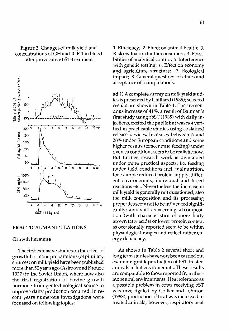
Figure 2. Changes of milk yield and concentrations of GH and IGF-1 in blood
after provocative bST-treatment
e - 1 C'I c:
1.::1 6C
2
_ 16C e ~1200 c:
u.. SC{; !:::?
LCO
_,
.. . . . . . , •1l\ix;ldq : .. ,: !! ------------------ ::-·---:~ ... ---
a I . . . . .
-1.. o 1.. a 12 16 zo 21.. 2a n dqs t
r::Si ! 1.ng s.i:.l
PRACTICAL MANIPULATIONS
Growth hormone
The first extensive studies on the effect of growth hormone preparations (of pituitary sources) on milk yield have been published more than50years ago (Asimov and Krouze 1937) in the Soviet Union, where now also the first registration of bovine growth hormone from gentechnological source to improve dairy production occurred. In recent years numerous investigations were focussed on following topics:
61
1. Efficiency; 2. Effect on animal health; 3. Risk evaluation for the consumers; 4. Possibilities of analytical control; 5. Interference with genetic testing; 6. Effect on economy and agriculture structure; 7. Ecological impact; 8. General questions of ethics and acceptance of manipulations.
ad 1) Acompletesurveyonmilkyieldstudies is presented by Chilliard (1989); selected results are shown in Table 1. The tremendous increase of 41 %, a result of Bauman's first study using rbST (1985) with daily injections, excited the public butwasnotverified in practicable studies using sustained release devices. Increases between 6 and 20% under European conditions and some higher results (concentrate feeding) under oversea conditions seem to be realistic now. But further research work is demanded under more practical aspects, i.e. feeding under field conditions incl. malnutrition, for example reduced protein supply, different environemnts, individual and breed reactions etc .. Nevertheless the increase in milk yield is generally not questioned; also the milk composition and its processing properties seem not to be influenced significantly; some shifts concerning fat composition (with characteristics of more body grown fatty acids) or lower protein content as occasionally reported seem to be within physiological ranges and reflect rather energy deficiency.
As shown in Table 2 several short and long term studies have now been carried out examinin gmilk production of bST treated animals in hot environemnts. These results are comparable to those reported from thermoneutral environments. Heat tolerance as a possible problem in cows receiving bST was investigated by Collier and Johnson (1988); production of heat was increased in treated animals, however, respiratory heat

62
Table 1.BST: Effects of various treatments on milk production of dairy cows
start durat. prep. applic. effect source
wk.11 10d hbST daily + 15 Peel et al.; 1982 wk.17 1Ld hbST daily + 22 Pocius & Herbein; 1986 wk.35 10d hbST daily +34 Fronk et al.; 1982
wk.12 27wk rbST daily +41 Bauman et al.; 1985
wk.11 24wk rbST depot + 6 Profittlich; 1989 wk.10 24wk rbST depot + 12 Rohr et al.; 1986 wk.14 24wk rbST depot + 20 Oldenbroek et al.; 1987 wk. 9 34wk rbST depot + 19 Rijpkema et al.; 1987 wk. 8 36wk rbST depot + 11 Bauman et al.; 1989
Table 2. Responses of Lactating Dairy Cows to bST in Warm Environments
Treatment Dose Response Reference Length(Wk) mg/d kg/d Percent
Elvinger et al., 1987 39 6.25 4.4 21 12.5 5.4 26 25 8.2 39
Hutchinson et al., 1986 27 13.5 8 30 27 7 27 40 5 19
Mollet et al., 1986 27 13.5 12.6 27 2.2 40 4.4
Mboe et al., 1986 4 25 5.5
Staples and Head, 1987 1.4 50 (IU) 8.5
Mattos et al., 1989 12 500mg 2 11.5

loss and skin vaporization heat loss was also .increased resulting in a similar rectal temperature as in controls. Assuming adequate feed, water and environmental management, there is no evidence that bST cannot be used in the tropical and subtropical regions.
ad 2) The predicted negative effect on animal health has not been confirmed in general. There are no evidence from evaluations of now up to 4 lactations concerning increased metabolic disorders or mastitis. Certainly further research must investigate long term effects on individuals and the conditions for management.
The increase of somatic cell counts in milk as shown in few studies (Heeschen 1988) should be followed up even there is evidence that there is no connection to pathological conditions. We assume that the (mitogenetic) effect of IGF-1 in general will open new fields of immunological and clinical research and may perhaps signalize certain indications of bST to supporttherapeutic treatments as shown by Burvenich et al. 1988 in connection with beneficial effects to overcome Escherichia coli mastitis.
The real problem of the negative interference of bST treatmentwi th fertility is known also from the comparable genetic progress in milk yield. In practice this could be avoided by commencement of bST treatment after conception only. This would be in accordance with the 'late lactation effect' providing also the advantage of more 'persistency' of the lactation curve.
ad 3 and 4) Generally bST is considered from any scientific points of view as orally not active in human due to the species specificity and the protein structure of the hormone which does not allow an absorption in the active form. Residue studies have
63
given further evidence that bST under normal treatment conditions enhance only insignificantly within the very low natural cpncentrations in milk (Fig. 3) (Schams 1988; Schams 1989a and 1989b). Also tbe bST depending IGF-1 level may be enhanced within physiological ranges, this peptide (70 aminoacids) can not be considered as orally active; it is present naturally in much higher levels for instance in colostrum, in human milk or in toher animal products (Schams 1989a). Analytical significance -except from provocative studies with overdosage (again without biological relevance) - can not be expected from residue studies using milk or milk products. The only chance at the moment to get some monitoring effects (up to 80% efficiency) are extensive investigation susing blood samples from the animals in vivo and perhaps simultaneous measurement of bST and IGF-1 (Schams 1989b). In summary, there is neither an obvious risk for the consumer, nor a realizable ,control method from products in case of treatment.
ad 5) In regard to the interference of bST treatment with genetic evaluations actual estimates predict the loss of information as small (not exceeding 20%). Hence strategies to 'live with bSt' have been already worked out by geneticists (Simianer and Wollny 1989).
ad 6, 7 and 8) Economy and ecology could be considered to be influenced positively under the aspect that more milk yield may be obtained with less animals, less feed and less waste. These positive aspects are apart from controversial prospects concerning results on farm structure and consumer acceptance. Since these preconditions are different in various areas general prognosis can not be given. Certainly countries with surplus production may have developed
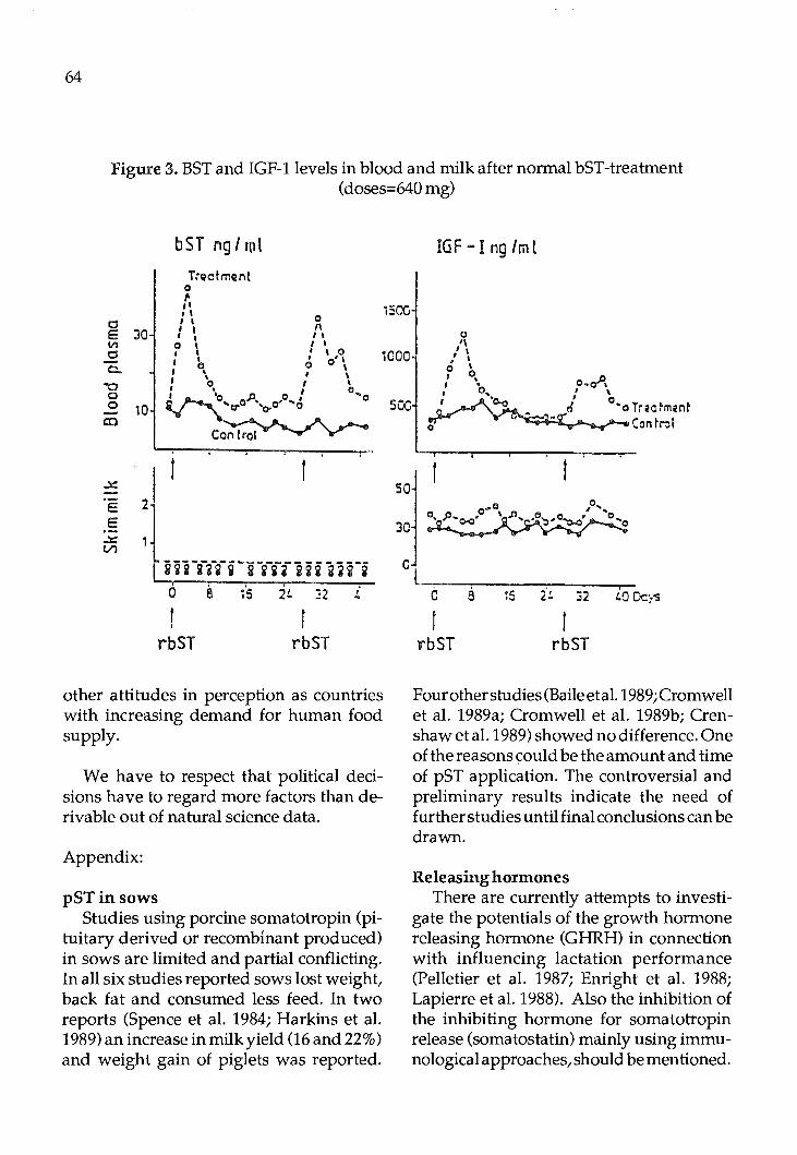
64
Figure 3. BST and IGF-1 levels in blood and milk after normal bST-treatment (doses=640 mg)
bST ng I ml IGF - Ing/ml Ti"Qctmcrnt
0 ,. ,, 1'001
I I 0
c:::J I I I I " E 30 I I I I 1~00 0
Vl 0 I I I I\ c:::J I I I \ 0 , \ - I I
, o'' I I
0 ' ' 0 c. \ I ' I I ' "O 0 \ I . o .. o 0 I 'o oJJ• 0 10 'o- •
co
E E
...x: Vl
I r
:l _ ---------------------1 iii iii i iiiiiiiiliii
i
0 8 ;'5 2!. :::2 t.
I I
rbST rbST
other attitudes in perception as countries with increasing demand for human food supply.
We have to respect that political decisions have to regard more factors than derivable out of natural science data.
Appendix:
pSTinsows Studies using porcine somatotropin (pi
tuitary derived or recombinant produced) in sows are limited and partial conflicting. In all six studies reported sows lost weight, back fat and consumed less feed. In two reports (Spence et al. 1984; Harkins et al. 1989) an increase in milk yield (16 and 22%) and weight gain of piglets was reported.
soc
0 0 I • o.o-", I . o, I \
·c.o I 0 o "'o Tr;ia~mi!nt Con ~r.il
0 t.0 Dcys
rbST rbST
Four other studies (Baile et al. 1989; Cromwell et al. 1989a; Cromwell et al. 1989b; Crenshaw et al. 1989) showed no difference. One of the reasons ~ould be the amount and time of pST application. The controversial and preliminary results indicate the need of further studies until final conclusions can be drawn.
Releasing hormones There are currently attempts to investi
gate the potentials of the growth hormone releasing hormone (GHRH) in connection with influencing lactation performance (Pelletier et al. 1987; Enright et al. 1988; Lapierre et al. 1988). Also the inhibition of the inhibiting hormone for somatotropin release (somatostatin) mainly using immunological approaches, should be mentioned.

No practical use has been obtained yet out of these studies. An interesting experiment (Wollny et al. 1987) showing a synergistic effect of TRH (the Thyreotropin Releasing Hormone) and GHRH treatment concerning the growth hormone release in cattle recalls an earlier pilot study of our group (Schams et al. 1974). The peripartal treatment of heifers with TRH resulted in increased milk yield for the total lactation period. The potential of the tripeptide TRH to release not only Thyreotropin but also prolactin and growth hormone may stimulate again approaches to manipulate components responsible for lactation.
Thyreoidea hormones Selection of high yielding dairy cattle
was always concomitant with optimizing thyreoidea hormone output; the level of T3
reflects rather the actual energy balance and may even be in negative correlation with the individual milk output (Blum 1983). The thyreoidea dependent turnover rate, exogenously stimulated, may enhance the milk yield but - opposite to the mode of action of bST - also involves other parts of the body to an extent that it may interfere seriously with the body conditions of animals. Former experiments with thyroxin injections or feeding of iodinated proteins are consequently only of theoretical interest anymore.
In general - even appreciating potentials to apply hormonal preparations-we should not overlook that milk production still will remain in first order a matter of sufficient feed supply and good management. Improvements in the range of the procedures discussed here are also approachable for example with 3 times (or more, as expected from the milking machine roboters) milking or just by simply optimizing prestimula ti on of the udder.
65
References
References are listed in the review article:
Karg, H. and Mayer, H. (1989). Manipulation of lactation. 'Biotechnology for Livestock Production'. AF AO Plenum Publication (Plenum Publishing Corporation): 181-205.
Additionally references:
Baile, C.A., Azain, M.J., Buonomo, F.C., Kasser, T.R. (1989). Effect of somatotropin treatment in sows during late gestation on birth weight and performance of pigs. J. anim. Sci. 67 (Suppl. 1), 528 (abstract).
Bauman, D., Hard, D.L., Crooker, B.A., Partridge, M.S., Garrik, K., Sandles, L.D., Erb;H.N.,Franson,S.E.,Hartnell,G.F.,Hintz, R.L. (1989). Longterm evaluation of a prolonged-release formulation of n-methionyl bovine soma totropin in lactating dairy cows. J. Dair. Sci. 72, 642-651.
Burvenich, C.H., Vandeputte-van Messom, G., Roets, E., Massart-Leen, A., Akers, R.M., Reynaert-VanSichem,R., Devrieze, L. (1988). Bovine somatotropin and acute Escherichia coli mastitis: Ann. Med. Vet., 132, 601-606.
Chilliard, Y. (1989). Long-term effects of recombinant bovine somatotropin (rBST) on daify cow performances: a review in: Use of somatotropin in livestock production. Sejrsen, K. Vestagaard, M., NeimannSorensen, A. Ed ts. Elsevier Applied Science, London/New York, pp. 61-87.
Crenshaw, T.D., Grieshop, CM., McMurtry, J.P., Schricker, B.R. (1989). Exogenous porcine prolactin and somatotropin injections did not alter sow lactation··performance. J. Dairy Sci. 67 (Suppl. 1), 632 (Abstract).

66
Cromwell, G.L. and Stahly, T.S., Edgerton, L.A., Moneque, H.L Burnell, T.W., Schenck, B.C., Elkins, T.T. (1989a). Recombinant porcine somatotropion for lactating sows. J. Dairy Sci. 72 (Suppl. 1), 630 (Abstract).
Cromwell, G.L., Stahly, T.S., Edgerton, L.A. (1989b). Weekly administration ofrecombinantporcine somatotropin to lactating sows. J. Dairy Sci. 67 (Suppl. 1), 631 (Abstract).
Elvinger, F., Head, H., Wilcox, C.J., Natzke, R.P., Eggert, R.G. (1987). Effects of administration of bovine somatotropin on lactation milk yield and composition. J. Dairy Sci. 70 (Suppl. 1), 121 (Abstract).
Enright, W.J., Chaplin, L.T.,Moseley, W.M., Tucker, H.A. (1988). Effects of infusions of various doses of bovine growth hormonereleasing factor on growth hormone and lactation i nHolstein cows: J. Dairy Sci., 71, 99-108.
Giesecke, D. (1987). Lipid mobilization and insulin function in cows with high milk yieldi Adv. Anim. Physiol. Anim. Nutr. Suppl. J. Anim. Phys. Anim. Nutr. Ed. Giesecke D.P. Parey (18), Hamburg/Berlin.
Harkins, M., Boyd, R.D., Bauman, D.E. (1989). Effect ofrecombinant porcine somatotropin on lactational performance and metabolite patterns in sows and growth of nursing pigs. J. Anim. Sci., in press.
Heeschen, W. (1988). BST in der Milch. In: Proceed. BST-Symp. Landbauforschung Braunschweig-Volkenrode, Bu:ndesforschungsanstalt Nov. 1987. Ed. ellendorff et al., 220-226.
Hutchinson, C.F., Tomlinson, J.E., Mc Gee, W.H. (1986). The effect of exogenous recombinant or pituitary extracted bovine growth
hormone on performance of dairy cows. J. Dairy Sci. 69 (Suppl. 1), 152 (Abstract).
Karg, H., Schams, D., Mayer, H. (1988). Endokrine Grundlagen der Laktation and Nahrstoffverteilung. In: Proceed. BST-Symp. Landbauforschung Braunschweig-Volkenrode, Bundesforschungsanstalt Nov. 1987. Ed. Ellendorff et al., 79-99.
KirchgeBner, M., Schwab, W., Muller, H.L. (1989). Effect of bovine growth hormone on energy metabolism of lactating cows in longterm administration. In: Energy metabolism off arm anomals. Proceed. of the 11th Symp., Lunteren, Sept. 1988/Netherlands. EAAP publicationNo.43.PudocWageningen, 143-146.
Lapierre,H.,Pelletier,G.,Petitclerc, D. (1988). Effect of human growth hormone-releasing factor (1-29)NH
2 on growth hormone re
lease and milk production in dairy cows. J. Dairy Sci. 71, 92-98.
Mattos, W.,Pires,A.V.,deFaria, V.P., Duque, J.A.,Madsen,K.S. (1989). The effect of sometribove (recombinant methionyl bovine somatotropin) on milk yields and milk composition in lactating dairy cows i nBrasil. J. Dairy Sci. 72 (Suppl. 1), 1093 (Abstract).
Mboe, A.Z., Head, H.H., Bachman, KC., Wilcox, C.J. (1986). Effect of growth hormone on milk yield, feed intake and some physiological functions during environmental stress. J. Dairy Sci. 69 (Suppl. 1), 119 (Abstract).
Mollet, T.A., DeGeeter, M.J., Belyea, R.L., Youngquist, R.A., Lanza, G.M. (1986). Biosynthetic or pituitary extracted bovine growth hormone induced galactopoesis in dairy cows. J. Dairy Sci. 69 (Suppl. 1), 118 (Abstract).

Oldenbroek, J .K., Garssen, G.J ., Forbes, A.B., Jonker, L.J. (1987): The effect of treatment of dairy cows of different breeds with recombinantly derived bovine somatotropin in a sustained delivery vehicle. 38th Annual Meeting EAAP, Lisbon, N 5.
Pelletier, G., Petitclerc, D., Lapierre, H., Bemier-Cardou, M. (1987). Injection of synthetic human growth hormone-releasing factors in dairy cows. 1. Effect on feed intake and milk yield composition. J. Dairy Sci. 70, 2511-2517.
Profittlich, C.H. (1989). Untersuchungen zum EinfluB von rekombinantem bovinem SomatotropinaufLeistungund Stoffwechsel bei Milchkuhen uber 2 Laktationen. Diss. agr. Universitat Gottingen.
rijkpkema, Y.S., Van Reeuwijk, L., Mol, E.P. (1987). Responses of dairy cows to longterm treatment with somatrotropin in a prolonged release formulation. 38th Annual Meting EAAP, Lisbon.
Rohr, K., Daenicke, R., Lebzien, P., Schlunsen, D. (1986): In: Rohr K. (1988): Milchleistung und Milchzusammensetzung. BST Symp. Nov. 1987 Braunschweig. Sonderheftder Landbauforschung Volkenrode. Ed. Ellendorff et al., 104-115.
Schams, D. (1988). Analytik des endogenen und exogenen Wachstumshormons beim Rind. In: Proceed. BST-Symp. Landbauforschung Braunschweig-volkenrode. Bundesforschungsanstalt, Nov. 1987. Ed. Ellendorff et al., 211-219.
Schams, D. (1989a). Somatotropin and related peptides in milk: In: Proceed. CEC-
67
seminar on 'Use of somatotropin in livestock production', Brussels, Sept. 1988, in press.
Schams, D. (1989b). 1st der Einsatz von Somatotropin kontrollierbar ? . Tierarztl. Umschau in press.
Schams, D., Graule, B., Theyerl-Abele, M., Graf, F., Wollny, C. (1989). Insulin-like growth factor I and somatotropin during lactation and after treatment with Sometribove (recombinant methionyl BST) in GermanFleckvieh and German Black and White cows. J. Dairy Sci. 67 (Suppl. 1), 843 (Abstract).
Simianer, H., Wollny, C. (1989). Impact of the potential use of bovine somatotropin on the efficiency of a conventional dairy cattle breeding scheme. Livestock Prod. Sci. 22, 31-47.
Spence, C.A., Boyd, R.D., Bauman, D.E., Butler, W.R., Wray, C.I. (1984). Effect of exogenous growth hormone on fetal storage and lactation performance in sows. J. Anim. Sci. 59 (Suppl. 1), 146 (Abstract).
Staples, C.R., Head, H.H. (1987). Short-term administration of bovine somatotropin to lactating dairy cows in a subtropical environment. J. Dairy Sci. 71, 3274-3282.
Wollny, C., Fleet, LR., Davis, A.J., Goode, J.A.,Mason, W.T., Heap,R.B. (1987). Growth hormone release after arterial injection of bovine growth hormone-releasing factor (GRF), GRF 1-29 and thyrotrophin-releasing factor (TRH) in lactating and pregnant cows. J. Endocrin. 112 (Suppl.) 146 (Abstract).
Proc. FAO/CAAS Workshop on Biotechnology in Animal Production and Health in Asia and Latin America, Beijing, Oct 9-13,1989, pp 68 - 82
Efficiency, Limits and Use of Embryo Transfer In Domestic Mammals
D. Chupin 1.N.R.A. Physiologie de la Reproduction
37380 NOUZILL Y - FRANCE
Introduction
Thanks to considerable research efforts the world over the embryo transfer technique and its use in practice have developed over the last 10 years. Because of its complexity and the technical and physiological limitations, embryo transfer cannot claim to replace artificial insemination. However, if properly used with selected animals, it can afford much progress in genetics and animal breeding.
Itis thought that little progress may come from improved collection and transfer procedures as long as men and materials are adapted to each other. The main limitation here is clearly the mean number of ova shed and its variability. As international trade, deep freezing is the second important step that must be improved. More sophisticated techniques may bring improvements that will make ET more efficient; splitting to increase the number of usable embryos and sexing to get only the desirable gender.
However ET is used in almost every countries and a survey of its use and development in Europe is presented.
I -PRODUCTION OF EMBRYOS
Leaving out the technique of embryo recovery (cervical in cow, surgical or laparoscopic in sheep and goat), two steps influence the number of transferable embryos per donor: ovarian stimulation and ovulation (superovulation) and fertilization.
A)SUPEROVULA TION The increase of number of follicles grow
ing to ovulation is obtained by gonadotrophin injection during luteal phase of oestrous cycle (return in oestrus induced by prostaglandin) in the cow or at the end of progestagen treatment in the ewe and goat.
1) Type of treatment: Until 1979, the only hormone giving rise
to superovulation available was PMSG. The average number of usable embryos per treated donor was 2 .5. As from 1980, further to comparative tests (ELSDEN et al. 1978) a preparation of pituitary extracts (FSH-P Bums-Biotec) replaced PMSG in most cases. Optimum conditions for use of this preparation were defined: Total dose, number of injections per day, distribution of doses per injection (decreasing) (cow: CHUPIN &
FAO LIBRARY AN: 302007

PROCUREUR, 1983a; ewe: COGNIE et al. 1985; goat: BARIL et al. 1988) giving on average 5 usable embryos per treated donor. However, the variability of ovarian response did not decrease: 30% of donors produced no usable embryo and only 45% produced at least 4 (extremes were 0-47).
The causes of this variability were sought not only in the animal (differnces in follicular stock, differences in sensitivity) but also in the variation in efficiency in the batches of hormone administered.
Analysis of the FSH and LH content in different batches of FSH-p Burns-Biotec showed, alongside constant quantities of FSH, important and variable quantities of LH (from 1 to 3 times the quantity of FSH).
The effect of the LH contamination on superovulation induced by a given dose of FSH (32 mg. Armour equivalent to 450 micrograms pure FSH) was studied in the cow using pituitary extracts from pigs prepared at Nouzilly (CHUPIN et al. 1984-1985). Both excessive and insufficient LH reduce superovulation. It is also possible to define an optimum level which differs for the breeds studied. A FSH/LH ratio of 3 gives a maxim um response in Holsteins and Browns (7,5 usable embryos) whilst Charolais give a maximum response to a preparation much richer in LH (FSH/LHratio0,5 to 1 giving 6.5 usable embryos). The Montbeliard breed shows no differences for FSH/ LH ratios ranging from 0.5 to 6. ( CHUPIN et al. 1987b).
These studies have led to the emergence of a French hormone preparation in two separate vials to cater for the differing LH requirements, with dilution instructions adapted to the main breeds of donors.
69
This separate packing of the two hormones has become even more necessary since studies on ewes and goats have demonstrated that it is possible to improve treatment efficiency by giving successive injections of decreasing dosages of FSH followed by increasing dosages of LH ( COGNIE et al. 1986, BARIL et al. 1988). The development of similar treatment for cattle has yet to be worked on.
The lower efficiency rate of treatment using PMSG is explained not by limited ovarian stimulation but by lower embryo quality. Since 1978 (SCHAMS et al.) it has been known that this is due to the long halflife of PMSG which, since it is present in the blood at high levels even after oestrus, leads to retarded growth of follicles which do not ovulate but which lead to hormonal imbalances hindering gamete transit. Countermeasures have been available since 1980 (SAUMANDE et CHUPIN 1981, SAUMANDE etal.1984). Theyincludetheinjection during heat of an antibody against PMSG which suppresses any parasite postoestrous follicle stimulation.
The method is truly efficient since, without changing the number of ovulations, the number of usable embryos per donor cow is increased from 2.5 to 5. (a percentage increase of 45% to 70%). Difficulties in developing a reliable product by vaccination (in goats or bulls) delayed the emergence of a commercially-viableanti-PMSG. Since 1987, INTERVET has been elaborating a monoclonal anti-PMSG (AGUERetGIELEN 1987, GIELEN et AGUER, 1988).
The efficiency of this PMSG-anti-PMSG treatment in ewe and goat has not been studied.
2) Limits and prospects

70
Whatever the gonadotrophin used two features limit the efficiency of treatments: the relatively low mean and the high variability.
To increase the mean, it is tempting to use higher doses of hormones. With both FSH or PMSG this results rapidly in a limit in number of ovulations and a decrease in the quality of the embryos recovered (CHUPIN 1988). With extreme doses (100 mg FSH or 7500 iu PMSG) the high number of follicles stimulated cannot ovulate (SAUMANDE et CHUPIN, 1986) a phenomenon which can be explained by a lack of maturity of the oocytes (CHUPIN unpublished).
The high variability originates both in the variability of the batches of hormones and in the variability of the ovarian status (SAUMANDE et al. 1978, MONNIAUX et al. 1983, SAUMANDE 1987). Studies on respective role of FSH and LH in pituitary extracts come to the appearances of several new preparations characterized by known and constant amounts stimufull of both hormones [OV ASET (Sanofi) ,FOLL TROPIN (Vetrapharm), STIMUFOLL (Merieux)]. This source of variability is now under control.
As it is proved that the ovarian response relies on the number and respective sizes of follicles at the time of stimulation (SAUMANDE et al. 1978), numerous attempts have tried to modify this parameter (see SAUMANDE 1987forreview). Theeffectof administration of limited amount of PMSG or FSH on day 2 or 3 of oestrous cycle is controversial: most reports concluded to a negative effect on ovulation rates ( CHUPIN et SAUMANDE 1979, LUSSIER et CARRUTHERS 1987) but some significantly positive effect has been observed in particular situations where the response of the control group was abnormaly low (WARE
etal.1987,RAJAMAHENDRAN etal.1987). This kind of pretreatment may prove useful in difficult situations: low nutritional level and postpartum period.
In normal situations the decreased ovarian stimulation is explained by the presence on the ovaries at the time of superovulation of one or some large follicles (>10 mn). Similar alteration during non pretreated cycles leads to low stimulation (SAUMANDE et al. 1978). Through long term (2-3 weeks) administration of GnRH agonist it is possible to clean the ovaries of any large follicles. Atthe time of gonadotrophin treatment all the small follicles (3-5 mm) will be stimulated leading to an increased ovulation rate and a decreased variability. This has been verified in the ewe with BUSERELIN (BREBION et COGNIE 1989).
The efficiency of embryo production would be increased if bad responders could be detected before treatment. Till now this has never been obtained by following of steroids or gonadotrophins, nor with echographic examination of the ovaries. The only possibility is to select treated animals before AI according to the evolution of steroid levels in milk or blood during treatment (SAUMANDE et al. 1985, TAMBOURA et al. 1985) or according to echographic diagnosis at onset of oestrus (CHUPIN et PROCUREUR 1983b): the bad responders are not inseminated.
Superovulation treatment can be repeated every 6 weeks (SAUMANDE et CHUPIN 1977,CHUPIN etSAUMANDE 1979, BARIL et al 1988, TORRES et SEVELLEC 1987). Although some animals maintain their level ofresponse for up to 2 years (17 treatments), the mean ovulation rate decerases and reaches a minimum after 3 to 5 repetitions. In cow it has not been possible to detect

antibody formation against either gonadotrophins, but changing from PMSG to FSH in animals no responding after several treatments restores an ovarian response equal to the first one (CHUPIN et PROCUREUR 1983c). In goats, antibodies can be detected after repeated treatments with a good correlation between titers of antibodies and decrease in ovarian response (BARIL et BECKERS personal communication).
Remark: Embryo recovery through the cervix in the cow can be repeated many
· times. On the opposite surgical recovery in small ruminants produces adhesions which limit the possibility of repeated recoveries (TORRES and SEVELLEC 1987). With laparoscopic recovery the appearance of adhesions is limited and recovery can be repeated more than 7 times (V ALLET et al. 1987, BRAIL et al. 1988).
B) FERTILIZATION
After hormonal stimulation the ovulation are spread on a longer period than normal: 6 hours in the ewe, 12 hours in the goat and 24 hours in the cow. To obtain the highest fertilization rates it is necessary to know precisely this timing of ovulation for the whole population of donors or, more useful, for each donor individually.
Onset of oestrus is not strictly related to timing of ovulation and oestrus detection cannot be used as the only criterium to choose AI schedule.
Ovulation by itself can be detected by repeated observations through laparoscopy or echography. Difficult to use in practice, these methods have been used in research works to precise ovulation ranges in groups of ewes, goats and cows (BARIL et VAL LET
71
1989, THAYER et al. 1985).
The interval between LH peak and ovulation being the less variable, knowledge of this peak gives a good estimate of timing of ovulations. With the next appearance of ELISA kits this assay will become feasible on farm and the optimum time for AI chosen for each donor.
The highest fertilization rates are produced by hand mating. With cervical deposit of the semen (cervical AI) fertilization rates are lower and the selection of high fertility males must be severe. In cow, two cervical AI (deposit in the uterine body) 12 and 24 hours after onset of oestrus with normal semen dosage (25 x 106 in France) is the common practice. One intervention at heat + 24 hours gives acceptable results (SCHIEWEetal.1987).Forsmallruminants, semen deposit through laparoscopy (uterine AI) allows, if timed properly, fertilization rates close to those got with hand mating even with five times less spermatozoa than for cervical AI (ewe: BREBION et al. 1988; goat: BARIL et al. 1988, BARIL et VALLET 1989 personal communication).
Attempts to control by a injection of GnRH the timing of ovulation have failed since animals tend to have two LH peaks: one induced by GnRH, followed by the endogenous one which produces disturbances in final follicular growth and oocyte maturation (COGNIE et al. 1987, WALKER et al. 1987).
II-TRANSFER
The choice and preparation of the recipient are totally empirical today. No serious research on the preparation of the female animal for her role as a recipient has been
72
carried out: quantity and quality of feedstuff, environment, importance of changes in surroundings or feed programme, adult cows or heifers ... At time of transfer, there is also the problem of synchrony (embryo/ recipient timing is crucial with a tolerance of +I -12 hours) and the presence and appearance of the corpus luteum. The latter is in itself an eminently subjective criterion since it is well known that quantities of progesterone secreted have no connection with the morphological appearance of the corpus luteum.
In the cow cervical transfer became more widespread with the rise in success rates. From 20-30% at the end of the Seventies, today's rates hav risen to 50% on average and some teams regularly obtain over 60%. Variations are considerable from technician to technician.
In these circumstances, the surgical transfer, which gives a 10-15 % fillip to the success rate, is only used for very high value transfers (genetically or financially speaking). The exact reasons for the difference between the two techniques are ill-known.
In sheep and goat the cervical route cannot be used and the transfer must be done by surgery or laparoscopy. With improvements of skillness and of equipments, la paroscopic transfer becomes as efficient (if not more) as surgical transfer (VALLET et al., 1989).
In any cases embryo(s) has to be deposited on the side where at least one functional corpus luteum is observed (or palpated).
As regard the age of embryo the main limit is the technique of transfer. In the cow, using the cervical route, the deposit must be done in the uterus: the embryo must be 5 day old or more.
In small ruminant during surgery or laparoscopy both uterus and oviducts can be reached easily. Embryos from pronuclear to blastocyststages(l to7 day old) are transferable with high success rates (V ALLET et al., 1989a, 1989b).
A future subject for research mainly in cows should be to explain why, when 100 carefully-chosen embryos have been meticulously implanted in 100 strictly-selected recipients, 21 days later (i.e. 14 days after transfer) only 75 gestations are obtained (which means 55 calving, that is, neither more nor less than after normal AI despite the fact that all main causes of failure have been eliminated during this initial phase: non-fertilization, halted development at an early stage, non-ovulation, abnormal ovarian structure). Between 21 days and-calving, the comparable level of losses after ET or Alleads us to think that no negative result can be attributed to the transfer, even at the moment of implantation.
Till now nothing has been found to improve this efficiency (hormones or drugs) except the addition of trophoblastic vesicles at the time of transfer (HEYMAN et al., 1987).
III - STORAGE
Between the uteri of donors and recipients, embryos have to be handled and stored in vitro for various periods going from some minutes to several months or years. Optimal parameters must be defined to allow highest survival.
Under routine optimized conditions embryos spend the shorter feasible time in vitro (i.e.1 to2hours). Thisisdoneinsimple solutions (P .B.S) enriched with BSA (2 to 4
g/l) at room temperature ( + 20°C).
For longer intervals (6to12 hours) conditions have to be improved = true culture Media (Menezo B2 or HAM FIO for instance) supplemented with 10% fetal calf serum, at+ 37-38°C under controlled atmosphere (5-10% 02,5% C0
2). Even then success
rates after transfer are lowered (HEYMAN et al., 1984).
When some more delays are needed between recovery and transfer (24-48h) to permit transport, waiting for results of tests (i.e. sexing) or to make up for a lack of synchrony between donors and recipients, the storage at 0°C to + 4 °C can be used. In normal PBS+ BSA or 10%-20% featal calf serum, (but without any cryoprotectant) survival for 24h allows normal pregnancy rate after transfer, the stage of the recipient on the day of transfer being the same as the stage of the donor on the day of recovery (no evolution of the embryos during 24h) (ewe: Driancourt et al., 1988, cow: Nibard et al., 1988-1989). If 48 hours are needed this 0°C storage can be used but with some lowering of success rates after transfer (BONDURANT et al., 1982, LINDNER et al., 1983).
To store embryos for longer periods, suppression of enzymatic reactions is absolutely required and is obtained only at low sub-zero temperatures. For easiness, storage temperature is that of liquid nitrogen (-1960C). Efficient techniques have emerged to bring embryos to this temperature with limited harmful effects, and they have become practicable, thanks to programmable equipment which minimizes the consequences of the variability of embryo production and also lays the foundations for embryo banks giving the maximum usage of a permanent donor and the opportunity to develop trade. (RENARD 1984).
73
Three stages are required in order to freeze embryos successfully:
- the cells must be enriched in a nontoxic product replacing water, thus reducing the risk of ice crystal formation duringcoolingand thawing. Glycerol is usually used for cow embryo (BOUYSSOU et CHUPIN 1982). For sheep embryo ethylene glycol is more efficient (COCERO et al., 1988). For goat embryo glycerol (CHEMINEAU et al., 1986) or ethylene glycol (BARIL et al., 1988) have been used.
- careful control of the phase change of water (from liquid to solid)= seeding 1or2 degrees below the crystallization temperature to avoid excessive overcooling.
- dehydration of cells, obtained during the slow cooling stage (0.3°C per minute) between the change of phase (-70C) and the final temperature (between -30 and -35°C depending on the author). Progressive crystallization of the medium brings up the osmotic pressure of the remaining liquid phase and the embryo reads to this by contracting and expelling water. Then the embryos are plundged in liquid nitrogen.
This freezing technique is used by more or less all the embryo transfer teams throughout the world with minor variations: it enables 85% to 95% of embryos to survive depending how strict prefeezing selection was.
After thawing, preferably rapid, in water at +20° or +35°C, the cryoprotectant agent should be removed from the cells. This is usually performed by 4 to 6 successive stages of decerasing concentration. During these
74
stages, the embryos are examined and only those which have survived are transferred. The transfer success rates are no different from those obtained by the same teams with fresh embryos (50 to 60%).
This complex chain is efficient but costly in terms of time (2H30 to freeze) and equipment and complex to implement since it calls for a laboratory for the washing and examination of embryos. Part of the research effort over the last 10 years has concerned simplication of these various stages.
The latter end of the chain (thawingwashing-transfer) must aim at an operation as simple and fast as that of a semen straw.
The intiial development was a sucrose solution designed to draw the glycerol out of the embryo: this molecule does not penetrate the cells, the embryo reacts to the rise in osmotic pressure by contracting which leads to the expelling of water and glycerol. When replaced in a medium where the osmotic pressure is the same as the culture medium or the uterime medium, the embryo swells slowly without any excessive mechanical stress on the membranes. (RENARD and HEYMAN 1983, CHUPIN 1984).
J.P. RENARD et al (1982) in France and S. LEIBO (1982) in Texas had the idea of putting a volume of sucrose solution separated by an air bubble from the embryo in its glycerol solution in the straw itself before freezing. After thawing, the straw is simply shaken to mix the various environments and after about ten minutes, the straw is placed in the transfer gun and the transfer carried ou twithou tthe embryo ever leaving its straw or being examined. This means embryos which have not survived are not eliminated, which lowers the success rate by 10%, but this figure can be kept down by
having a stricter selection before embryos are frozen. (CHUPIN et al., 1984).
By using the fact that one properties of sucrose is that it does notpenetrate cells, it is possible to simplify the freezing process itself. In fact, by using a glycerol+ sucrose solution, sufficient enrichment in cryoprotectant medium and sufficient dehydration of cells is achieved to eliminate the need for a slow cooling phase. Careful control of the phase change suffices before dropping the embryos very quickly into liquid nitrogen. RENARD and HEYMAN (1984) suggest a pause of 30 to 60 minutes at-30"C which can be carried out in a domestic freezer. Or even more simply, the phase change can be obtained in the neck of the nitrogen flask. After 5 minutes, the straws are dropped into liquid nitrogen. (CHUPIN 1986). True survival is confirmed by the birth of calves (DUMONT et al., 1988).
Going still faster, preservation of embryos by vitrification has seen suggested (MASSIP 1986). Due to the need of high concentrations of cryoprotectants (glycerol and propylene glycol) the vitrification medium is far from innoxiousness and if birth of mouse pups is confirmed, very few calves have been produced by this way, and no goat embryos survived (CHUPIN unpublished result).
Extensive use of these techniques mean that survival rates are near to routine techniques (70% living embryos 24 hours after thawing in a culture medium at + 37°C). There is no doubt that after a number of improvements, they will replace current techniques or at least be used as a standby when sophisciated equipment fails.
Whatever the technique (routine or fast) research is needed to improve media= buffer

efficiency during freezing and effect of protein source (BSA of serum) are not at their best.
IV-SPLITTING
The maximum number of transferable embryos per treated donor being limited, the possibility of dividing each embryo in several pieces can be a solution to increase the yield. The technique as well as the number of pieces depend on the stage of the embryos.
Till now the classical technique is to cut mechanically at blastocyst stage. Starting with a lot of tools (until 6) monitored by pneumatic or mechanic micromanipulators the whole procedure has been simplified to two (a holding pipette and a knife) or even one, the knife, the embryos being hold by grooves at surface of the plastic petri dish. Results have been published with some tens of embryos for cow (OZIL 1983), ewe (CHESNEetal., 1987)andgoat(TSUNODA et al., 1985).
Apart from being time consuming the first limit of this technique is that not all the embryos are "cu table"= only the excellent grade (top 70% of transferable embryos) can give pieces able to survive and produce living animals with pregnancy rates similar to whole embryos. The second limit is that convincing results have been presented only for two pieces. This allows for the birth of one calf per embryo.
Splitting is not used widely on farm. The main uses are research (Monozygotic twins for less variable experimental groups), breeding (increase of the number of calves tested per selected animals or testing both animal on two different criteria) sexing one half or
75
increase of number of calves in poorly superovulated donors.
Researches are still needed on the optimum use of this demi-embryos= transfer of one or two halves per recipient, with or without the zone pellucida, optimum stage of the embryonic development and, less successful, freezing.
In small ruminants oviductal stages can be used as easily as uterine ones = this allows to manipulate embryos before compaction. The separation ofblastomeres could be less harmfull than cutting. Birth of kids from 4 cell goat embryos separated in two sets of 2 cells have been presented (TSUNODA et al., 1984). The possibility to use the technique with 8or16 cells embryo to produce more than 2 identical embryos has not been extensively studied. This opens the way to cloning.
V-SEXING
An important part of the cost of ET made calves (or lambs or kids) is the production in half cases of the undesired gender. It is a general belief that control of sex either through selection of spermatozoa or by diagnosis of sex of the embryo will induce an increase in the use of ET.
1) Treatment of sperm to predetermine sex. Almostinumerable attempts to select sex
by acting on sperm have been published (see AMANN 1989 for review). Two different ways have been explored.
- Separate semen in two populations according to differences in physical characteristics of X and Y bearing spermatozoa. weight, size, electric charges and motility.

76
- Inactivate the undesired sperm cells through modifications of the medium (extender or female genital tract): hormones, pH and antibodies.
None of these methods is convincing nor repeatable. The main difficuty is that to measure efficiency of such traits the only criteria was sex ratio after birth, i.e. these experiments extend for a long time. Recently some other techniques have been developed that will allow more rapid and systematic check of the efficiency of selection: chromosomal examination after in vitro fertilization of Hamster oocytes or DNA probes.
The second difficulty is that most of the published data refers to limited numbers of offsprings born and experimental bias are difficult to avoid.
2) Diagnosis of sex of preimplantation embryos (see HARE 1986 for review).
Chromosomal analysis. This is the basic technique. Some tens of cells (biopsy) are cultivated with a mitostatic agent, spread on a slide and stained before examination for meta phases and chromosomes count. When one good metaphase is seen the accuracy of this diagnosis is 100% but for one third of the biopsies nothing can be analyzed, at leastwitholdembryos(day13incow), which are not routinely used due to freezing difficulties and sanitary regulations (post hatching). When applied on younger embryos (day 7 or 8 in cow) this test has to be performed on fewer cells and as much as 2/3 of the embryos cannot be sexed (TORRES et POPESCU 1980)
- HY antibody. This male specific antigene can be detected on the cell surface of the embryos. By immunofluores-
cence, positively fluorescing (male) embryos can be separated from negatively fluorescing (female) embryos. The survival of the embryos is not greatly modified and an accuracy of 79 to 89% has been claimed by WHITE et al. (1987a, 1987b) for cow and sheep embryos. Other authors have not been able to repeat these experiments, and, although announced in 1983, no commercial kit is available.
The HY antibody can also be associated with cytolytic reaction that will kill all (or most of) the male embryos.
- DNA probe. A DNA probe specific of bovine Y chromosome has been elaborated in an INRA-Pasteur InstituteCEA joint program (V AIMAN et al. 1988). This probe hybridizes with the denaturedDNAofkilledcells,andhas to be used with half embryos or biopsies of 10-15 cells. To reach the same accuracy as with more than 1000 cells (close to 100%), the amount of DNA of the 10-15 cells is amplified before hybridization. The reading (gold-silver precipitation or radioactive) occurs 24h later. During this delay the biopsied embryo is stored at 0°C without any decrease of viability (NIBARD et al. 1988-1989).
- Metabolic test. During the first stages of embryonic development, females have two times more X-linked enzymes than males (for instance glucose 6-phosphate dehydrogenase). A colorimetric assay of the level of such enzyme in the cells or better in the medium could give a very simple diagnosis. This seems feasible in the mouse (WILLIAMS 1986).

Whatever the technique the feasibility of embryo sexing will reduce the need for recipient (half) but more than double the cost of each eligible embryos since all the cost (plus the cost of sexing) will be supported by half the number of embryos.
VI - ET ACTIVITY FOR CATTLE IN EUROPE
Not withstanding the limits enumerated in this report ET is used on farm for 10 years and this activity is increasing (AETE statistics). In Europe in 1988 (western Europe without England and some countries of eastemEuropeie. Poland, DDR and Czechoslovakia) 19956 donors have been treated yielding 92884 transferable embryos-56288 have been transferred fresh, 19396afterfreezing-thawing and the remaining are stored frozen for future use on farm or for trade.
In France ET activity increases regularly year after year= 1986: 2840 donors - 1987: 4852 donors - 1988: 5974 donors. 11722 embryos have been transferred fresh in 1988 and 8190 embryos after freezing. 1653 have been imported (living animals cannot be imported from outside EEC). 70% of transfers are made in dairy breeds (mainly holstein but also Normande and Montbeliard). In these breeds 80% is for on farms retransfer of embryos and 20% for collective breeding schemes (Bull mothers or breeding nuclei).
In beef breed (mainly Charolais) the figures are 50% for on farm re transfer and 50% for trade.
Splitting has only limited use and is quoted in the AETE survey for 1988 in Poland (18 embryos) and Czechoslovakia (545 embryos).
77
Sanitary regulation have been legaly established in France to prevent any spreading of viruses or bacteria through embryo transfer: buildings, equipments, men training, handling of embryos and media, storage. Legal agreement has to be obtained and reexamined each year.
CONCLUSIONS
At top efficiency levels, embryo transfer can result in the birth of 2 to 3.5 offsprings per collected donor. This can be repeated 6 times a year to obtain on average 12or15 offspring per yearlong donor. This may be the startingpointforeconomically-viablebreeding from the genetic, husbandry or speculative point of view. However, the high level of variability at each stage and therefore in the final result should not be underestimated: for example, an annual production of 12 offsprings can mask variations between 0 and 60 or more. Another point is that programmes, if they are to be optimized, must be structured around donors sufficiently different from the rest of the animal population. Lastly, success depends on the human protagonistshavingthemeans to acquire and maintain a high-level of technical efficiency.
REFERENCES
Aguer, D. and Gielen, J. 1987. Use of PMSG and neutra-PMSG, Monoclonal anti-PMSG, in donor cow for embryo transfer 3rd scientific meeting AETE, 4.5 Sept. 1987. LYON. p 175.
Amann, R.P. 1989. Treatment of sperm to predetermine sex. Theriogenology, 31, 49-60.
78
Baril, G., Casamitjana, P ., Perrin, J. and Vallet, J.C. 1988. Embryo production, freezing and transfer in Angora, Alpine and saanen goats. 4thscientificmeetingAETE, 9-lOSept.1988. LYON. p 67-93.
Baril,G.and Vallet,J.C.1989. Time of ovulation in dairy goats induced to superovulate with pFSH during and out of the breeding season. (Submitted).
Bondurant, R.H., Anderson, G.B., Boland, M.P., Cupps, P.T. and Hughes, M.A. 1982. Preliminary studies on bovine embryo survival following short term storage at +4°C. Theriogenology, 17, 223-229.
Bouyssou, B. and Chupin, D.1982. Two step freezing of cattle blastocysts with Diemthyl sulfoxide (DMSO) or glycerol. Theriogenology, 17, 159-164.
Brebion, P ., Lajous, D ., Poulin, N ., Procureur, R. and Vallet,J.C.1988.Intrauterineinsemination increases fertilization rate and embryoqualityinsuperovulated Lacauneewes. 4th Scientific Meeting AETE, 9-10 Sept. 1988. LYON., 95.
Brebion, P. and Cognie, Y. 1989. Increased superovulationintheewefollowing14days of GnRH Agonist pre-treatment. 5th Scientific Meeting AETE, 8-9. Sept. 1989. LYON. 106.
Chemineau, P., Procureur, R., Cognie, Y., Lefevre, P.C., Locatelli, A. and Chupin, D. 1986. Production, freezing and transfer of embryos from a bluetongue-infected goat herd without bluetongue transmission. Theriogenology, 26, 279-290.
Chesne, P., Colas, G., Cognie, Y., Guerin, Y. and Sevellec, C.1987. Lamb production using superovulation, embryo bisection and trans-
fer. Theriogenology, 27, 751-757.
Chupin, D.1984. Thepresentstateoftheart of emrbyo transfer in France. In: "The reproductive potential of cattle and sheep". ORTA VANT, R. and SCHINDLER, H., Eds. INRA. Paris. Les colloques de l'INRA, n° 27, Rehovot (Israel) 21-23Fevrier1984, p. 235-253.
Chupin, D. 1986. Quick freezing of rat and cow embryos after dehydration at room temperature. In: "seminaire sur la congelation des embryons et des ovocytes" 28 juin 1986 Annecy (France) p. 165-175.
Chu pin, D.1988. Superovulation parPMSG ou FSH pour le transfert embryonnaire. Coll. Soc. Fr. Etudes Fertil. 26, 213-232.
Chupin, D. and Saumande, J. 1979. New attempts to decrease the variability of ovarian response to PMSG in cattle. Ann. Biol. Anim. Bioch. Biophys. 19 (hors serie) 1489-1498.
Chupin, D. and Procureur, R. 1983a. Efficiency of pituitary extracts (FSH) for ind uction of superovulation in cattle. Anim. Reprod. Sci. 6, 11-23.
Chupin, D. andProcureur,R.1983b.Prediction of bovine ovarian response to PMSG by ultrasonic echography theriogenology, 19, 119.
Chupin, D. and Procureur, R. 1983c. La stimulation de l'ovaire pour produire des embryons chez les bovins. In: "La rep rod uction des bovins: Anoestrus postpartum, transplantation embryonnaire". CR. joumees d'information ITEB-UNCEIA, Paris 16-17. mars. 1982, p 75-83.
Chu pin, D., Combamous, Y. and Procureur,
R. 1984. Antagonistic effect of LH on FSH induced superovulation in cattle. Theriogenology, 21, p 229.
Chupin, D., Florin, B. and Procureur, R. 1984. Comparison of two methods for onestep in straw thawing and direct transfer of cattle blastocysts. Theriogenology, 21, 455-459.
Chupin, D., Cognie, Y., Combarnous, Y., Procureur, R. and Saumande, J. 1985. Effect of purified LH and FSH on ovulation in the cow and ewe. In "Follicular growth and ovulation rate in farm animals". ROCHE, J.F.,and O'CALLAGHAN, D. Eds.,Martinus Nijhoff., Seminaire CEE, Dublin (Ireland) 30-31Oct.1985, p 63-71.
Chupin, D., Procureur, R. and Combarnous, Y. 1987. Effect for LH induced superovulation in our from breeds of cows. 3rd Scientific meeting AETE, 4-5 Sept. 1987, Lyon, p. 151.
Cocero, M.J., Procureur, R., De La Fuente, J. and Chupin, D. 1988. Glycerol or Ethylene glycol for cryoprotection of deep frozen ewe embryos. Theriogenology, 29, 238.
Cognie, Y., Chupin, D. and Saumande, J. 1985. Comparison of two FSH treatment schedules to induce superovulation in ewes. Theriogenology 23, 185.
Cognie, Y., Chupin, D. and Saumande, J. 1986. The effect of modifying the FSH/LH ratio during the superovulatory treatment in ewes. Theriogenology, 25, 148.
Cognie, Y., Theron, M.C. and Crozet, N. 1987. Effect of gonadotrophin relasing hormone (GnRH) tn the production of ovulated oocytes for in vitro fertilization in sheep. 3rd Scientific Meeting AETE, 4-5 Sept. 1987
79
Lyon, p 163.
Draincourt, M.A., Lorentz, R., Chupin, D., Webb, R. and Wilmut, I. 1988. Survival of ovine embryos stored at 4°C for 24 hours. Theriogenology 30, 441-446.
Dumont, P., Escouflaire, P., Chupin, D., Mercier, A., Tainturier, D., Bruyas, J.F. and Fieni, F. 1988. Congelation rapide d'embryons dans les vapeurs d'azote. 4th Scientific Meeting AETE, 9-10 Sept. 1988. Lyon. p. 109.
Elsden, R.P., Nelson, L.D. and Seidel, G.E. 1978. Superovulating cow with follicle stimulating hormone and pregnant mare's serum gonadotrophin. Theriogenology, 9, 17-26.
Gielen, J. and Aguer, D. 1988. Optimum time of Neutra PMSG administration in superovulated dairy cows for embryo transfer. 4th Scientific meeting AETE, 9-10 Sept. 1988. Lyon, p. 115.
Hare, W.C.D. 1986. Embryo sexing of from animals. In: "Development Biology vol 4" GWATKINR., Ed. p. 195-216.
Heyman, Y. 1984. Embryomic loss in cattle after transfer of fresh, frozen or cultured embryos. 10th Int. Cong. Anim. Reprod. and A.I., June 10-141984 Urbana(USA)p .....
Heyman, Y., Chesne, P., Chupin, D. and Menezo, Y. 1987. Improvement of survival rate of frozen cattle blastocysts after transfer with trophoblastic vesicles. Therionology, 27, 477-484.
Leibo, S.P. 1982. A one step method for direct nonsurgical transfer of frozen thawed bovine embryos. In: "Int. Cong.: Embryo transfer in Mammals" Annecy, France Sept. 1982. p. 97.

80
Lindner, G.M., G.B., Bondurant, R.M. and Cupps, P.T. 1983. Survival of bovine embryos stored at 4 °C. Theriogenology 20, 311-319.
Lussier, J.G. and Carruthers, T.O. 1987. Endocrine and superovulatoryresponses in hifers pretreated withFSH or follicular fluid. Theriogenology, 27, 253.
Massip, A. 1986. La vitrification, methode d' avenir pour la cryoprotection des embryons. In: "Seminaire sur la congelation des embryons et des ovocytes". 28 juin 1986 Annecy (France) p. 177-193.
Monniaux, D., Chu pin, D. and Saumande,J. 1983. Superovulatory responses of cattle. Theriogenology, 19: 55-81.
Nibard, M., Mechekour, F., Schwartz, J.L., Procureur, R., Chesne, P., Chupin, D. and Thibier, M. 1988. Taux de gestation apres transfert d' embryons bovins biopses maintenus 24 heures en survie a G°C. 4th Scientific Meeting AETE, 9-lOSept.1988, Lyon. p.105.
Nibard, M., Mechekour, F., Procureur, R., Chesne,P.,Chupin, D.and Thibier,M.1989. Pregnancy rates after cervical transfer of biopsed bovine embryos. Theriogenology 31, 234.
Ozil,J.P.1983. Productionofidentical twins by bisection of blastocyts in cow. J. Rep rod. Pert. 69, 463-468.
Rajamahendran, R., Canseco, R.S., Denbow, C.J., Gwazdavskas, F.C. and Vinbon, W.E. 1987. Effect of low dose of FSH given at the beginning of the estrous cycle on subsequent superovulatory response in holstein cows. Teriogenology 28, 59-65.
Renard, J.P. 1984. Methods of conserving
gametes and embryos of farm animals. Livestock Production Science. II, 49-59.
Renard,J.P.,Heyman, Y.andOzil,J.P.1982. Congelation de l'embryon bovin: une nouvelle methode de decongelation pour le transfert cervical d' embryons conditionnes en paillette. Ann. Med. Vet., 126, 23-32.
Renard,J.P.andHeyman, Y.1983.Effetdela methode d'addition et de dilution du cryoprotecteur sur la viabilite des blastocystes de vache apres decongelation. i6e Cong. Int. du froid, Paris, Commission C 1, pp 69-75.
Renard, C., Buixuan Nguyen, N. and garnier, V. 1984. Two step freezing of two cell rabbit embryos after partial dehydration at room temperature. J. Reprod. Fert., 71, 573-580.
Saumande, J. 1987. Superovulation chez les bovins: actualites et perspectives. 3rd Scientific Meeting AETE,4-5 Sept. 1987, Lyon, pp 97-142.
Saumande, J. and Chupin, D. 1977. Superovula ti on: a limit to egg transfer. Theriogenology 7, 141-149.
Saumande,J. and Chupin, D. 1981. Production of PMSG antiserum in cattle: assay of inhibitory activity and use in uperovulated heifers. Theriogenology 15, 108.
Saumande, J. and Chupin, D. 1986. Induction of superovulation in heifer: the inhibitory effect of large doses of PMSG. Theriogenology. 25, 233-247.
Saumande, J., Chupin, D., Mariana, J.C., Ortavant, R. and Mauleon, P. 1978. Factors affecting the variability of ovulation rates after PMSG stimulation. In: "control of reproduction in the cow". SREENAN,MJ.,Ed.

Martirtus Nijhoff, La Haye, pp 195-224.
Saumande, J., Procureur, R. and Chu pin, D. 1984. Effect of injection time of anti PMSG antiserum on ovulation rate and quality of embryos in superovulated cows. Theriogenology, 21, 727-731.
Saumande,J., Tamboura,D. and Chu pin, D. 1985. Changes in milk and plasma concentrations of progesterone in cows after treatment to induce superovulation and their relationships with number of ovulations and of embryos collected. Theriogenology, 23, 719-731.
Schams, D., Menzxer, C., Schallenberger, E., Hoffman, B., Hahn, J. and Hahn, R. 1978. Some studies on pregnant mare serum gonadotrophin (PMSG) and on endocrine responses after application for superovulation in cattle. In: "control of reproduction in the cow". SREENAN, J., Ed. Martinus Nijhoff, La Haye, pp 122-143.
Schiewe, M.C., Looney, C.R., Johnson, C.A., Hill, KG. and Godke, R.A. 1987. Transferable embryo recovery rates foll owing different insemination oched ule in su perovula ted beef cattle. Theriogenology 28, 395-406.
Tamboura, D., Chupin, D. and Saumande,J. 1985. Superovulationincows: a relationship between progesterone secretion before ovulation and the quality of embryos. Anim. Reprod. Sci., 8, 327-334.
Thayer, KM., Forres, D.W. and Welsh, T.H. 1985. Real time ultrasound evaluation of follicular development in superovulated cows. Theriogenology, 23, 233.
Torres, S. and Popescu, P. 1980. Transfer of cow blastocysts after sexing. Theriogenology, 14, 309-318.
81
Torres, S. and Sevellec, C. 1987. Repeated superovulation and surgical recovery of embryos in the ewe. Reprod. Nu tr. Develop. 27, 859-863.
Tsunoda, Y., Yasui, T. and SUGIE, T. 1984. Production of Monozygotic twins following transfer of separated half embryos in the goat. Jpn. J. Zootech. Sci., 55, 643-647.
Tsunoda, Y., Tokunaga, T., Sugie, T. and Katsumata, M. 1985. ProductionofMonozygotic twins following transfer of bisected embryos in the goat. Theriogenology, 24, 337-343.
Vaiman, M., Cotinot, C., Kirszembaum, M., Leonard, M., Chesne, P., Heyman, Y., Stinnakre, M., Bishop, C. and Fellous, M. 1988. Sexing of bovine embryos using male specific nucleic acide probes. Proc. 3rd world cong. sheep and Beef cattle Breeding 19-23 June 1988, Paris, vol. 1, pp 93-105.
Vallet,J.C., Baril, G.,Rougier, F., Chupin, D., Procureur, R. and Corteel,J.M., 1987. Feasibility and repeatability of embryos recoveries from dairy goats under laparoscopy. 3r Scientific Meeting AETE, 4-5 Sept. 1987, Lyon, p 159.
Vallet, J.C., Baril, G. and Loysel, C. 1989. Efficiency of laparoscopic embryo transfer in goats. 5th Scientific Meeting AETE, 8-9 Sept. 1989, Lyon, p 186.
Vallet, J.C., Folch, J., Poulin, N. and Cognie, Y. 1989. Surgical or laparoscopic embryo transfer in sheep before or after the first cleavages. 5th Scientific Meeting AETE, 8-9 Sept. 1989, Lyon, p 188.
Walker, S.K, Smith, D.H., Seamark, R.F. and Godfrey, B. 1987. Variations in the tim-
82
ing of multiple ovulations following gonadotrophin releasing hormones treattnentand its relevance to collecting pronuclear embryos of sheep. Theriogenology, 28, 129-137.
Ware, C.B., Northey, D.L. and First, N.L. 1987. Effect of administration of FSH at the beginning of the cycle on the subsequent response to superovulation treatment in heifers. Theriogenology, 27, pp 292.
Williams, T.J. 1986. A technique for sexing mouse embryos by a visual colorimetic assay of the X-linked enzyme, glucose G-
phosphatedehydrogenase. Theriogenology, 25; 733-739.
White,K.L.,Anderson,G.B.andBondurant, R.H. 1987a. Expression of a Male-specific factor on various stages of preimplantation Bovine Embryos. Biol. Reprod., 37, 867-873.
White, K.L., Anderson, G.B., Paschen, R.L. and Bondurant,R.M.1987. Detectionofhistocompatibility-Y antigen: identification of sex of preimplantation ovine embryos. J. Reprod. Immunol. 10, 27-32.

Proc. FAO/CAAS Workshop on Biotechnology in Animal Production and Health in Asia and Latin America, Beijing, Oct 9-13,1989, pp 83 - 90
Embryo Transfer In Cattle - New Aspects of Embryo Production
J. Fulka The Czechoslovak Academy of Sciences,
Institute of Animal Physiology and Genetics, 277 21 Libechov, Czechoslovakia.
Introduction
The production of mammalian embryos in vitro has been a dream of several scientific groups workingindifferntcountriesfor last few decades. At the beginning laboratory animals were used as a model and mainly scientific aspects were followed in these studies. After slow accumulation of basic knowledge and many negative results it was finally revealed that this idea can be under certain realistic conditions. Among many scientists working in this field two pioneers, Dr. Chang and Dr. Austin, should be mentioned. Both described independently in the year 1951 a final process of sperm maturation, called capacitation, as a prerequisite for its penetration throughzona pellucida and vitelline membrane. On the basis of their observation successful fertilization was realized in the rabbit and later in the mouse, hamster and some other laboratory mammals.
The application of these procedures to farm animals was for long time accompanied with disappointing observations. The penetration of sperms into the cytoplasm of bovine, ovine and porcine oocytes occurred onlyveryexceptionallywithanypossibility to control this process. According to the
present knowledge these random findings can be considered rather as artefacts.
Gradually, however, with the introduction of embryo transfer technology in cattle breeding, the production of embryos started to be crucial problem. The conventional method based on the stimulation of follicular growth by exogenous gonadotropins does not offer satisfactory answer. In spite of many experiments leading to variouis modifications of the gonadotropin application the crop of embryos in caftle remains unpredictable. In average it does not exceed the level 6-8. Only exceptionally in well managed herds under strick selection of donors with more than 10-12 transferable embryos can be calculated. Therefore, it is not surprising that the price of bovine embryos remains relatively high and from economical aspects acts as serious barrier. Moreover, the regulation of foijicular growth is at present far from clear and the radical improvement can't be expected in near future.
For this reason the attention was paid again to original idea - to the production of bovine embryos under controlled environmentin vitro. The realization was,however, accompanied by several basic problems. First
FAO LIBRARY AN: 302009

84
of all, how to secure sufficient amount of bovine oocytes, to find effective treatment of spermatozoa to be able to fertilize the oocytes and to create the conditions for further development after fertilization.
OOCYTES
Maturation in vivo
As a source of female gametes two categories of them can be used. Into first category oocytes matured in the ovary should be included. Such oocytes are aspirated from the prevolatoryfollicles by means oflaparoscopy or flushed from the oviducts shortly after ovulation. In both cases the amount of oocytes correlates with response of selected donors on previous hormonal stimulation. It means that the animal factor plays again important role. Moreover, laparoscopy requires the experience and the procedure is time consuming. The same animal can be used repeatedly.
To obtain oocytes after ovulation is even more difficult. The oocytes are situated in the oviducts and their flushing can be carried out only after surgical access to the genital tract. In some cases such oocytes were flushed from slaughtered animals. For practical aims both, aspiration and flushing of oviducts, have strong limitations and served mainly for experimental purposes. It should be noticed that first calf born as a result ofin vitro fertilization originated from the oocytes obtained by this way (Brackett et al., 1982).
Maturation in vitro
Since 1935 (Pincus and Enzman) it is known that mammalian oocytes liberated from their follicles mature in suitable cul-
ture conditions spontaneously. The sequence of events as well as conditions required for resumption and completion of nuclear changes are species specific. In detail studies it was also found that such oocytes are morphologically indistinguishable from oocytes matured in the ovary after LH peak. This basic observations led to formulate the conception about the exploi ta ti on of oocytes present in the ovary. The amount of the oocytes is high and under natural conditions only very small proportion of them can be fertilized during female reproductive life. The other are lost due to follicular atresia.
These simple facts were strong motivation for extensive studies focussed to the exploitation of the supplies of oocytes and production of germ cells which would be able to secure after fertilization normal development to the term. The results carried out during last few years have been evaluated from various aspects and summarized in several comprehensive reviews (Thibault et al., 1987; First et al., 1988; Leibfried-rutledge et al., 1989). The basic knowledge which is for disposal was obtained with maturation of mouse oocytes. It is true, however, that not all of them can be totally generalized and species specificity must be taken into consideration. For this reason we want to briefly mention here some points which are typical for bovine oocytes.
On the bovine ovaries at any stage of estrous cycle different classes of follicles are present. According to the literature and own experience as a best material for cultivation serve the oocytes aspirated from follicles of 3-5 mm diameter. In this developmental stage the oocytes attained their final size and are fully equipped for the resumption of nuclear maturation in culture. Oocytes from smaller follicular classes (0.5-1.6 mm diameter) are still very active in their RNA

synthesis and after isolation not all of them start nuclear maturation called germinal vesicle breakdown (GVBD). Some proportion remains in germinal vesicle (GV) stage or progresses to metaphase I (MI) without continuation to metaphase II (MII). In contrast, the oocytes aspirated from follicles close to 3 mm or bigger mature in culture in very high proportion and more than 80% complete nuclear maturation to MIL
The time sequence of nuclear changes leading to the formation of chromosomes arranged in MII is strictly species specific. The cow belongs to the group with the maturation interval about 20-22 hrs. This interval corresponds closely with that observed in oocytes matured in preovulatory follicles after endogenous LH surge. Only for interest in the rabbit maturation is completed already after 10 hrs in culture, whereas in the pig it takes 36-38 hrs.
At the beginning of cultivation, immediately after isolation of oocyte-cumulus complex, nucleus GV has typical configuration. Finely granulted nucleoplasm with few chromocentres is separated with well visible nuclear membrane. The presence of nucleolus typical for some other species is rarely detectable in bovine oocytes. In culture nuclear changes proceed very rapidly.
In details the time sequence was described by Motlik et al. (1978). From this study it is obvious that already after 4 hrs chromatin forms fine network localized in whole nuclear volume. Within further 2 hrs chromosomes are arranged as late diakinesis and at 10-12 hrs after beginning of culture MI can be seen. Completely mature bovine oocyte can be found in culture after 20-22 hrs. At this time chromosomes form MII and first polar body is extruded. The speed of maturation does not proceed quite uniformly due
85
to the heterogeneity of oocytes used for culture.
The culture conditions differ in details from laboratory to laboratory. In most of them for this purpose serves modified Parker's medium (TC 199) which is supplemented with 2.92 mmol/l Ca-lactate, 2 mmol/l Na-pyruvate, 33.9 mmol Na-bicarbonate,4.43 mmol/Hepes buffer,50i.u./ml penicillin, 50 ug/ml streptomycin sulphate and 20% of bovine inactivated serum. This medium in amount of 150 ul is placed on watch glass under the paraffin oil and equilibrated in a temperature of 38.5°C in gas mixture composed of 4% C02, 10% 0
2 and 86% N
2 for 2 hrs. In each droplet 20-30
oocytes are placed and after 24 hrs the ma turation should becompletedinmostofthem. According to our experience the oocytes mature without any hormonal support in high proportion (80-90%). To improve conditions various modifications of culture medium were suggested including addition of follicle - stimulating hormone (FSH), Luteinizing hormone (LH) and estradiol-17B. Also cow sera obtained from animals of selected days of estrous cycle were tested (Younis et al., 1989). The effect of hormones seems to be difficult to evaluate, because some other authors obtained promising results without any hormonal support.
However, the succes of cultivation depends not only on culture conditions, but also on the treatment of ovaries and oocytes before and during isolation. The ovaries should be removed from the abdomen cavity as soon as possible, not later than 30 min after the death of donor. Also during transport and isolation the temperature should be about 20-30°C.
The attention should be paid als9 to the selection of oocytes after their aspiration

86
from follicles. In spite that the oocytes are aspirated from healthy looking follicles the population of oocytes differs profoundly. According to the experimental results the best source for cultivation are oocytes with compact cumulus oophorus. Such oocytes are surrounded by 3-4 layers of well organized cumulus cells with the rest of parietal granulosa cells. The oocytes with expanded cumulus or devoid of follicular cells completely should be excluded from further procedures. The expansion of cumulus characterized only oocytes after proper maturation. Cumulus cells are in this case more or less dispersed, but corona radiata remains well organized.
The presence of follicular cells and their organizationseemtobeimportantformaturation process. These cells provide the oocytes with instructions responsible for the synthesis of substances playing their roles at fertilization and further developmentof embryos. Premature interruption of these cell to cell communication influences negatively the quality of cytoplasm leading to the fertilization or postfertilization abnormalities. More frequent among them are arrested development of male pronucleus (Thibault and Gerard, 1970- rabbit, Fulka et al., 1982 - cattle) and polyspermy. While nuclear maturation can be effectively controlled under light microscope after fixation and staining of the oocytes, cytoplasmic maturation can be evaluated only on the ground of developmental ability after fertilization. It was found that the quality of cytoplasm can be substantially improved by the supplementation of additional granulosa cells to culture medium. By this way the reduction of mentioned abnormalities was very significant. This system suggested originally for rabbit oocytes (Fulka and Motlik, 1980; Motlik and Fulka, 1981) and sheep oocytes (Staigmiller and Moor, 1984) has
been gradually introduced also for cultivation of bovine oocytes.
It can be concluded that the experience accumulated during last few years has helped to create in vitro conditions convenient to certain extent for production of physiologically competent oocytes. This possibility looks optimistic particularly in connection with the unlimited source of immature oocytes which can be easily obtained from slaughtered animals.
Fertilization in vitro
For successful fertilization not only oocytes, but also spermatozoa require special treatment necessary for the induction of terminal events. These events, capacitation and acrosome reaction, occur in natural situation in female reproductive tract and form spermatozoa competent forfertiliza ti on. The definable changes take place on surface sperm membrane and lead to the fusion between outer acrosome membrane and plasma membrane. In the past induction of processes responsible for these events in farm animals was simulated according to the procedures effective in laboratory animals. These techniques rely upon long incubation with albumin to modify cholesterol content of sperm membranes, upon action of high salt concentration to remove decapacitation factors or to incubate spermatozoa in alkaline pH to change intracellular pH. All these treatments were strongly artificial without effective control and results of fertilization enormously fluctuated.
To find more physiological approach First and Parish (1988) and their collaborators collected oviductal fluid during estrous cycles and observed the capacitation activity which appears atestrual but not at luteal phase of cycle. The experiments aimed for

the identification of effective substance revealed its heat stability. The isolation of different fractions after various treatments led to the conclusion that the activity of oviductal fluid resides in heparin or heparansulphate. Later, studies with both these compounds specified heparin as an active capacitating agent in bovine.
This discovery can be considered as a milestone in the field of cattle oocytes f ertilization in vitro. In very short time the procedures were stabilized and reproducible results are at present recorded in most laboratories. In detailed studies in was found that heparin inducescapacitation of bovine spermatozoa in dose and time dependent manner. The dose of 7.5 ug/ ml in Tyrode buffer exerts maximal effect after 4 hrs incubation. However, satisfactory capacitation can be reached withinmuchshorterinterval. Pav lok et al. (1988) incubated bovine spermatozoa with 200 i.u./ml heparin only 15 min and described more than 70% of penetrated oocytes. Other laboratories use for the treatment of spermatozoa various modifications with similar effectivity, but heparin plays substantial role in all of them.
The molecular aspects leading to membrane changes remain unknown. Itis known, however, that 3H heparin binds to the surfaceduringincubationand its removal does not prevent capacitation. Treatment with heparin has similar effect on epididymal, ejaculated and deep frozen spermatozoa. Only slight differences between these three classes exist and all can be used for fertilization. Polyspermy occurs more frequently after application of epididymal spermatozoa. From practical aspects deep - frozen spermatozoa are most suitable, they are for disposal at any time in sufficient quantity and known quality. Also ejaculates from most fertile bulls can be chosen.
87
Cultivation of bovine embryos
The technology of bovine embryos production in vitro can be effective only, if such embryos reach the stages of late morulae or blastocysts. At these stages embryos can be transferred unsurgically or be frozen in liquid nitrogen. Unfortunately, bovine embryos develop in culture maximally to 8 cell stage and then stop further division. This crucial problem was studied extensively in recent period. To overcome the arrest, two systems were suggested.
1. The transfer of early cleavage stages to the oviducts of alien species. For this purpose mainly sheep as a transient recipient was recommended. The ligated oviducts of cycle - synchronized animals with the stage of embryos development provide adequate environment for further growth of bovine embryos to morula or blastocyst within 6-7 days after deposition. The speed of cell division in this environment closely correlates with that occurring in natural conditions. Moreover, very high number of bovine embryos (up to 200) can be introduced to sheep oviduct. However, in many cases not all of them are obtained back after flushing. Their losses can be as high as 30%. From flushed ones about 30% develop to morula or blastocyst.
Also ligated rabbit oviduct supports embryonic development. According to our experience very high pregnancy rate can be achieved after transfer of embryos cultivated for 3 days in the rabbit. During this interval the differentiation of embryos was remarkable and less viable, and remained more or less arrested in their development. As in the sheep also in the rabbit recovery rate reaches about 70 M. It can be admitted that the use of alien oviducts means very effective approach in the cultivation of
88
bovine embryos. On the other hand this approach requires extra time and additional animals. also the losses of embryos are relevant point in this part of technology.
2. Another approach also frequently studied involve the supplementation of culture media with oviductal or follicular cells and by this way it creates better environment. This idea arises from the hypothesis that embryonic development does not exist as autonomous process and that somatic elements interact with cleaving embryos. Positive effect of such media was proved by gandolfi and Moor (1988) in the experiments with ovine embryos. Also in bovine embryos in media enriched with somatic elements a 8 cell block can be more or less suppressed. The composition of such media differs according to the laboratories, the beneficial effect was observed everywhere. However, it should be stressed that the proportion of embryos cleaving to morulae or blastocysts is lower than in alien oviducts. In spite of partial improvement the conditions suboptimal for in vitro cultivation remain.
As exception can be the results of Pavlok et al. (1989). These authors produce bovine embryos in culture without any additional somatic cells. Basic culture medium TC 199 used for cultivation of oocytes supplemented with 20% of bovine inactivated serum supports the development to rhorula (32 cells) or blastocyst (64 cells). During the 6 or 7 days cultivation, medium is changed only once, approximately after 90 hrs. The proportion of embryos which attain progressive stage reaches 30-40%, i.e. very similar value as the other authors describe after cultivation in the oviducts. It should be added that some other laboratories in Czechoslovakia under apparently identical conditions were not able to offer compa-
rable results. From some aspects the production of bovine embryos in vitro can be considered rather as art than science.
All these experiments should be regarded only as a means to the production of normal calves. In this respect the figures are only fragmentary. From the literature source it seems evidentthatthe group working atthe University of Dublin under the leadership of Prof. Gordon accumulates most practical results. The continuous embryo production allows to use them in field condition in a large scale. According to the reports published in journals also the pregnancy rate is satisfactory. Much fundamental knowledge was obtained during recent years at the University of Madison. Also there some calves were born. In Denmark at the Veteirnary and Agricultural University this project was developed, too. Old and successful tradition continues also in France (INRA-Jouy-en-Josas) and AFR (in Cambridge) supplies the projects with theoretical ideas.Inauthor'slaboratoryalsoverypromising results were obtained, including establishment of pregnancies after transfer of embryos produced completely in vitro and transferred as morulae or blastocysts. Similar results were obtained in Japan (National Inst. of Anim. Industry - Tsukuba, Norindanchi), where for transfers fresh and also deep-frozen embryos were used. The pregnancy rate recorded after transfer of both classes of embryos seems to be very hopeful. Furthermore in Germany (Munich) some pregnancies were established.
It must be admitted that the progress in the technology of embryo production was surprisingly high. The experimental activity, however, continues further, because some questions remain to be answered. One of them concerns low proportion of in vitro developed blastocysts. It would be useful to

know if this development insufficiently was either predetermined already before the time of oocytes maturation or affected later by thein vitro cultivation techniques of oocytes and early embryos, respectively. The elucidation of such problems could help in further stabilization of bovine embryo production technology.
REFERENCES
First N.L., Leibfried-Rutledge M.H. and Sirard M.N. (1988). Cytoplasmic control of oocyte maturation and species differences in the development of maturational competence. In: "Meiotic Inhibition, Molecular Control of Meiosis", 1-46, Liss, New York.
First N.L. and Parrish J.J. (1988). Sperm maturation and in vitro fertilization. 11th Int. Congr. Anim. Reprod. Artif. Insem., dublin, 160-167.
Fukuda Y.,IchikavaM.,NaitoK. and Toyoda Y. (1988). Normal development of bovine oocytesmatured, fertilized and cultured with cumulus cells in vitro.11th Int. Congr.Anim. Rep rod. Artif. Insem., Dublin, p. 327.
Fukui Y. and Ohno H. (1988). In vitro development to blastocyst of in vitro matured and fertilized bovine oocytes. Vet. Rec. 122, 282.
FulkaJ. and MotlikJ. (1980). In vitro maturation. Proc. 9th Int. Congr. Anim. Reprod. and A.I. Madrid, Vol. II, 55-62.
FulkaJ. Jr., Pavlok A. and FulkaJ. (1982). In vitro fertilization of zona-free bovine oocytes matured in culture, J. Reprod. Pert. 64, 495-499.
Gandolfi F. and Moor R.M. (1988). Interac-
89
tions between somatic and germinal cells during early development. 11th Int. Congr. Anim. Reprod. Artif. Insem., Dublin, 169-177.
Leibfried-RutledgeM.L.,FlormanH.N.and First N.L. (1989). The molecular biology of mammalian oocyte maturation. Mol. Biol. Pert. 12, 259-301.
LuK.H.,GordonI.,ChenH.B.,GallagherM. and McGovern H. (1988). Birth of the twins after transfer of cattle embryo, produced by in vitro technique. Vet. Rec. 122, 559.
Lu K.H., Gordon I., Gallagher M. and McGovern H. (1987). Pregnancy established in cattle by transfer of embryos derived from in vitro fertilization of oocytes matured in vitro. Vet. Rec. 121, 259-260.
MotlikJ. and FulkaJ. (1981). Fertilization of rabbit oocytes co-cultured with granulosa cells. J. Reprod. Pert. 63, 425-429.
Pavlok A., Motlik J., Kanka J. and Fulka J. (1989). In vitro techniques of bovine oocyte maturation, fertilization and embryo cultivation resulting in birth of calf. Reprod. Nutr. Develop., 29(4), 27.
Pavlok A., Torner H., Motlik J., Fulka J., Kauffold P. and Duschinski U. (1988). Fertilization of bovine oocytes in vitro: effect of different sources of gametes on fertilization rate and frequency of fertilization anomalies. Anim. Reprod. Sci., 16, 207-215.
SirardM.A.,ParrishJ.J., Ware CB., LeibfriedRutledge M.L. and First N.L. (1988). The culture of bovine oocytes to obtain developmentally competent embryos. Biol. Reprod. 39, 546-552.
Thibault C. and Gerard M. (1970). Facteur

90
cytoplasmatique necessaire a la formation du pronucleus male dans L' ovocyte de Lapine. C.R. Hebd. Seanc. Acad. Sci., Paris D 270, 2025-2026.
Thibault C., Marquant-Le Guienne B. and Gerard M. (1988). In vitro developmentofin vivo and in vitro fertilized bovine eggs on
tubal and uterine cell monolayers. Reprod. Pert. (Abstr. Ser., No. 2), 24.
Younis A.I., Brackett B.G. and Fayrer-Hosken R.A. (1989). Influence of serum and hormones on bovine oocyte maturation and fertilization in vitro, Gamete Res. 23, 189-201.

Proc. FA O/CAAS Workshop on Biotechnology in Animal Production and Health in Asia and Latin America, Beijing, Oct 9-13,1989, pp 91 -103
Developments in Biotechnology and their Application to the Diagnosis and Control
of Virus Disease
T.Barrett ARFC Institute for Animal Health, Pirbright Laboratory, Ash Road,
Pirbright, Woking, Surrey GU24 ONF, UK.
1. Introduction
Recent advances in the field of immunology and molecular biology have revolutionised our approach to the detection and prevention of virus diseases of human and veterinary importance. These range from the simplified ELISA detection techniques using purified virus proteins or genetically engineered virus proteins to highly sophisticated recombinant DNA techniques used to generate disease resistant plants and animals. In addition new ways to produce cheaper and safer vaccines are being vigorously investigated. These techniques may in the near future help to eradicate many other virus diseases as was smallpox in the 1970' s.
This paper does not aim at a comrpehensive review of all that has been achieved in the last 10 years or so in the field of molecular biology and virus disease but at a general overview of the techniques and how they have been applied to the solution of some specific problems in virology. Reference will mainly be to virus diseases being studied at the Institute for Animal Health, Pirbright and to the techniques being used to study these viruses. The talk will focus on two areas; virus diagnosis and recombinant vaccines.
2. New Techniques for the Diagnosis of Virus Disease
In the last twenty years techniques for the diagnosis of virus disease have improved beyond all expectations. What was impossible or extremely difficult and time consuming twenty years ago is now a simple laboratory procedure and as a result laboratory confirmation and characterisation of the causative virus is now routine in many cases. This has been largely due to advances in immunology particularly the development of monoclonal antibodies and the enzyme-linked immunsorbent assay (ELISA).
2.1 Enzyme-linked immunsorbent assay (ELISA)
The development of enzyme-linked immunosorbent assays in the early 1970srevolu tionised virus disease diagnosis (Engvall and Perlmann, 1971). One of the standard methods for detecting virus antibodies is the virus neutralisation test which can be carried out relatively simply on a micro scale using mictotitre plates. There are several disadvantages associated with this technique, however, themostimportantofwhich is the necessity to have good tissue culture facilities. Even if these are available bacte-
FAO LIBRARY AN: 302011

92
rial or fungal contamination may make diagnosis impossible. In addition, non-specific cytotoxicity factors present in some serum samples may give false results. The use of ELISA eliminates the need for tissue culture. It also allows the supply of standard reagents which can be distributed to central laboratories and stored ready for use in field stations where only minimal laboratory facilities are available (Ferris et al., 1988). Such an ELISA "kit" for the detection of rinderpest antibodies is produced at Pirbright and is in routine use in Africa. This is an indirect ELISA where sonicated, clarified rinderpest infected tissue culture cells are used as the ELISA antigen to bind virus specific antibodies which are then detected by rabbit antibovine IgG conjugated to horseradish peroxidase. The use of such relatively simple technology has been an essential element in the seromonitoring for the recent Pan Afircan rinderpest campaign (PARC). It is used to monitor the immune response to vaccination and indirectly as a management tool for the evaluation of vaccination team performance and cold-chain integrit)r(Andersonetal., 1989). Thetesthas also been adapted to detect other morbillivirus antibodies e.g. antibodies to the recently discovered seal morbillivirus have been detected using the rinderpest antigen plates and the seal antibody detected by peroxidase conjugated protein A (Bostock et al., 1989).
The ELISA has been frther modified by the development of "blocking" or "competitive" ELISA. The advantage of this system is that sera from different types of animal can be analysed without the need for speciesspecific antibody conjugates. The virusspecific antibody,eithermonoclonal orpolyclonal, can be produced in any suitable animal species (eg mouse, rabbit, guinea pig). A pre-titrated standard amount of these anti-
bodies is mixed with the test serum which competes or blocks the reaction of antibodies in the test serum to the antigen coated plate. The amount of the standard antigen bound is inversely proportional to the concentration of competing antibody in the test serum.
Variations of ELISA using competitive methods employing either virus specific polyclonal or monoclonal antibodies are available for FMDV, swine vesicular disease, African horse sickness and bluetongue viruses (Anderson, 1984; Armstrong and Barnett, 1989; Afshar et al., 1989; Hamblin et al., 9186; Hamblin et al., 1989, in preparation).
Another development has been the double antibody sandwich ELISA for the detection of virus antigen (Crowther & Abu Elzein, 1979). In this testthe microtitre plate is coated with a high titre virus specific antibody, usually rabbit antibody, which is used to "trap" the virus antigen. The trapped antigen is then detected using a second antibody, usually derived from guinea pig, and an enzyme conjugated an ti guinea pig an tibod y. This improves the sensitivity and specificity of the test Using an antigen trapping sandwich ELISA and high titre, typespecific guinea pig antisera, the task of typing FMDV has been greatly simplified again with increased sensitivity over complement fixation tests (Roeder and Le Blanc Smith, 1988).
ELISA systems have also been used to detect virus from clinical samples; for example a double antibody sandwich ELISA system to detect coronaviruses and rotaviruses in faecal samples is available (Reynolds et al., 1984).
New biotechnology techniques involv-
ing recombinant DNA technology can be used to produce pure, highly specific antigensfor such ELISA systems and so increase the specificity of the technique and eliminate the need to grow large amounts of a potentially dangerous virus in special containment laboratories (see section 2.3).
2.2 Use of monoclonal antibodies for characterisation and diagnosis of virus disease
One of the most improtant advances in the field of immunology in the last two decades was the development of monoclonal antibodies (Kohler and Millstein, 1975). These are poweful tools which can be used in the diagnosis of virus disease and for analysis of immunologically important regions (epitopes) of virus proteins. They can also be used to aid epidemiologists in their task of differentiating virus strains and tracing the source of virus outbreaks with greater certainty. This is illustrated by the use of monoclonal antibodies to differentiate strains of bovine viral diarrhoea (BVD) from other closely related pestiviruses (McHugh et al., 1988) and rinderpest from peste des petits ruminants virus (Libeau Lefevre, 1989). By studying the reactivity of a new virus can be obtained. Depending on the specific reactions with monoclonal an ti bodies serologically closely related viruses can be rapidly classified, for example, as either BVD or classical swine fever. The test can also determine which previous virus isolate it most closely resembles.
At the Institute for Animal Health, Pirbright monoclonal antibodies are used to characteirse field strains of FMDV. A panel of about 100 monoclonal antibodies are available against reference strains of four of the seven serotypes of FMDV. Some of these were produced at Pirbright and others were
93
obtained from collaborating European laboratories. These are in routine use for profiling new isolates prior to selection of representative strains for more detailed analysis, e.g. sequence analysis (section 2.3.2). In addition monoclonal antibodies are being used to map the positions and examine the antigenic importance of epitopes on the virus using a combination of monoclonal antibody escape mutants (i.e. viruses selected for resistance to a particular monoclonal antibody) and direct RNA sequencing (Bolwell et al., 1989; Xie et al., 1987 and see section 2.3.2).
The use of monoclonal antibodies as tools for differentiating viruses can also be exploited in a more general way to diagnose the presence of a virus in a particular group which may have many serotypes using a monoclonal antibody made to a common determinant. For example, a specific monoclonal antibody which reacts with all serotypes ofbluetongue virus, but not other orbiviruses, is being used in sensitive blocking ELIS As for the detection of antibodies against all serotypes of bluetongue virus antibodies (Afshar et al., 1989).
2.3. Recombinant DNA technology By far the greatest advance in our under
stsanding of virus disease has come from the ability to clone specific virus genes to determine their exact sequence and to study their structure and function. In this way a whole range of virus isolates and their relationships can be examined in great detail, jmmunologically important proteins can be expressed in large amounts for structural/ functional studies and recombinant viruses can be developed which are safe and effective for vaccination against the disease (see section 3). In addition specific nucleic acid probes are then available which can be used for the detection of minute quantities of
94
virus which may remain latent in some tissues and be responsible for chronic diseases in the affected host (see section 2.3.3).
A recent example of the use of recombinant DNA technology for virus diagnosis is the production of the hepatitis B surface antigen from the cloned cDNA in mouse L cells. The cells secrete the antigen which is indistinguishable from conventionally produced antigen and has the advantage of being non-infectious thus eliminating the need for elaborate biosafety containment facilities for its production. (Mimms et al., 1989) The most successful application to date has been the use of recombinant baculoviruses containing the gene for the required virus protein under the control of the -highly efficient polyhedrin gene promoter to produce large amounts of pure virus protein. The baculovirus system has been used to produce pure virus antigens both for ELISA detection systems and for use as vaccines.
2.3.lBaculovirus expression of foreign genes
The insect baculovirus autographica californica nuclear polyhedrosis virus has been widely used to express large amounts of foreign proteins, including many virus proteins, in recombinant baculoviruses under the control of the powerful polyhedrin gene promoter (Matsura et al., 1987). The polyhedrin gene codes for the very abundant polyhedrin protein which surrounds and protects the large crystalline arrays of virus that are produced by infection with these agents. Recombinant viruses lacking the polyhedrin protein gene which has been replaced by the foreign gene can be easily identified and these instead produce large amounts of the foreign protein. One advantage of this system for the production of large amounts of foreign an ti gens is the ease
of handling the Spodoptera frugiperda cells (Sf9) used to grow the virus. Their optimum growth temperature is 28"C and they do not require a C02 atmosphere. Milligram quantities of the expressed protein can be produced and the viruses are stable for long periods at 4"C (Luckow and Summers, 1988).
Since the production of the protein in large amounts only involves the use of the safe recombinant baculovirus and insect tissue culture cells, the hazards associated with the growth of large amounts of dangerous virus are avoided. The system has been used to produce a variety of antigens from many viruses e.g. Hantaan virus (Schmaljohn et al., 1988), rabies virus (ReidSanden et al., 1988) and bluetongue virus (Inumaru et al., 1987). More recently the complete polyprotein of polio virus has been expressed in this system, resulting in the formation of structurally recognisable empty poliovirus particles (Urawaka et al., 1989). This technique has great potential for the production of safe vaccines for other dangerous viruses such as FMDV for which safe live virus vaccines have been impossible to develop and whose large scale production for inactivated vaccines is hazardous and requires expensive isolation facilities.
2.3.2 Direct RNA sequencing In the case of several RNA virus diseases,
which vary more than DNA viruses in their antigenic makeup, direct sequencing of the genome RNA has been employed to study the antigenically variable parts of the virus. This enables the most detailed comparisons to be made between different virus isolates. Specific oligonucleotide primers are first made to the more consrved regions of the genes being studied and these are used with revrse transcriptase to sequence the RNA from new isolates using the dideoxy chain termination method (Sanger et al., 1977;

Zimmem and Kaesberg, 1978). This technique is now used routinely to characterise new FMDV isolates at Pirbright. Direct RN A sequencing has proved to be a very powerful epidemiological tool which has been used to show that many outbreaks of FMDV serotypes 0 and A in Western Europe were probably due to incomplete inactivation of vaccine virus or laboratory escapes (Becke & Strohmaier, 1987; Knowles et al., 1988). Using biochemical and sequencing analyses of FMDV strains recently isolated in the Middle East, Samuel et al., 1988 were able to show that two distinct type A variants were co-circulating in that region, both of which differed from the classical A22 subtype. Direct RNA sequencing can also be used to study the genetics of picoma viruses (King, 1988) and other RNA viruses such as infectious bronchitis virus (Cavanagh & Davies, 1988) and the variability of the infleunza haemagglutinin molecule as it passes through human populations.
2.3.3 Nucleic acid hybridisation Nucleic acid hybridisation is the process
whereby a labelled DNA or RNA from a known source is used as a probe to search for its complementary sequence. !twill then hydrogen bond to its complement to form a stable double-stranded structure which remains bound even under stringent washing conditions. If the nculeic acid species are related, but not identical, hybridisation can also occur but the stringency of the washing conditions (salt concentration and temperature) must be reduced to allow the hybrids to remain double-stranded. The relatedness of nculeic acids can be determined by altering the stimgency of the washing procedure. Nucleic acid hybridisation has been in use for years but the recent advances in DNA cloning technology has greatly increased the usefulenss of this technique in the detection of virus diseases. DNA and
95
RNA probes of high specific activity can now be made to individual genes or parts of these genes. There are many examples where virus infections have been diagnosed using thistechnique.AtPirbrightwehavedR@veloped specific cDNA probes derived from the nucleocapsid (N) protein genes of different morbillivirus infections without the need for virus isolation. In this way using tissue from post-mortem samples it is possibel to identify the virus (Diallo et al., 1989). The technique is most useful for differentiating peste des petits ruminants virus (PPRV), a virus disease of sheep and other small ruminants, from the closely related rinderpest virus which causes disease in large ruminants. These two disease co-circulate in many parts of Africa and the importance of small ruminants as asymptomatic carriers of rinderpest is being investigated. In addition we have been able to show using cDNA probes specific for all four morbilliviruses that the recent seal morbilivirus isolated from diseased European seals was not id~ntical to CDV or any of the other known morbilliviruses but is a genetically distinct virus (Mahy et al., 1988; Bostock et al., 1989). The use of non-radioactive labelling methods for the cDNA probes means that the techniques can be adapted for use in field situations (Barrett et al., 1989b). In the case 6f bluetongue virus a cDNA probe derived from segment 3 RNA can cross-hybridise with all serotypes and is a useful probe for the detection of bluetongue virus in the blood of infected animals (Roy et al., 1985). DNA probes have also been used to detect FMDV nucleic acid sequences persisting in bovine oesophageal-pharyngeal fluids after infectious virus could no longer be isolated (Rossi et al., 1988). This method could be used to screen animals for persistent virus infections undetectable by other means.

96
2.3.4 Restriction enzyme analysis The discovery of restriction endonu
cleases in theearly1970'srevolutionised the study of large DNA molecules and was particularlyusefulin thestudyoflarge DNA viurses. Restirction endonucleases cut DNA at specific sequences to produce a rep rod ucible number of fragments for each DNA genome. The pattern of these fragments can be then resolved by electrophoresis on agarose gels. This allows variants of the virus to be distinguished and is a very useful tool both for the study of the epidemiology of large DNA viruses and for differentiating related but distinct viruses such as herpes virus types 1 and 2. At Pirbright this technique is being used to study the epidemiology of African swine fever and capripox viruses (Dixion and Wilkinson, 1988; Kitchingetal., 1987;Gershon& Black, 1988a).
2.3.5 Polymerase chain reaction This technique has proved to be the most
sensitive yet developed for the detction of minute amounts of nucleic acids, either of viral or cellular origin. As with direct sequence analysis of RN A this technique requires a knowledge of the sequence of the nucleic acid species which is to be detected e.g. a paritcular virus nucleic acid. Once this is known a set of two synthetic oligonucleotide primers are produced complementary to both the plus and minus sense of the nucleic acid and separated on the nucleic acid sequence a convenient distance, usually 200-500 bases. These primers are then used to produce copies of the nucleic acid being detected by a series of cyclical or chain reactions to amplify the particular region of the nucleic acid between the two primers. The cycles include a denaturing step carried out at high temperature (about 96°) whereby the newly synthesised double strand DNA is dissociated~ an annealing step at lower temperature (about 45°) whereby the prim-
ers are allowed to annal with the new DNA again. This is repeated 25 to 30 times doubling the amount of DNA produced each time. The cycles can be carried out automatically and theuseoftheheat-stableTaqpolymerase, derived from Therm us aquaticus, eliminates the need for the addition of new enzyme after each denaturing step. This results in the amplification of the nucleic acid more than 10 7-fold. The resulting DNA fragment can be identified by its size (corresponding to the distance apart of the primers on the nucleic acid) ort agarose gels. If the nucleic acid species to be detected is RNA it must first be copied into DNA by an initial reverse transcription step. It was first shown to be a useful way of diagnosing sickle cell anaemia by Saiki et al. in 1985 and the technique is now being used to diagnose many improtant human virus infections which otherwise would be undetectable until the disease had become evident e.g. CMV in the blood of carrier patients and the AIDS virus before any immune response can be detected (Clewley, 1989; Boerman et al., 1989; Ouetal., 1988). With chemotherapy for some virus diseases, in particlar for he.rpes and AIDS viruses, now available early diagnosis is of great medical importance. The technique can also be used to sequence relevant parts of any nculeic acid without the need to first clone the DNA in bacteria since the amplified DNA is produced in sufficient quantities to sequence directly. In this way functionally and immunologically improtant parts of a range of viurs isolates can be sequenced once the sequence of the type virus has been determined.
3. Recombinant Virus Vaccines
Many DNA viruses have been used as vectors to express a protein gene from another virus to produce recombinant viruses which can be used to vaccinate animals

against the disease from which the foreign gene was obtained. In this way multivalent recombinant vaccines could be produced. The most commonly used vector virus is vaccinia but other viruses including fow lpox, adenovirus, retrovirus and herpes virus have also been used successfully to obtain recombinant viruses (see Bostock, 1989 for review). At Pirbright the capripox virus is being investigated as a possible delivery system for foreign antigens (Gerhson & Black, 1988b).Itishoped to produce a multivalent vaccine which could protect cattle, sheep and goats from capripoxvirus infections and also from some other relevant diseases such as rinderpest or peste des petits ruminants virus. The advantages of recombiannt poxvirus vaccines are their relative heat stability and the safety of their non-poxvirus component since only the antigenic components of the other virus are present.
However, most recombinant vaccines to date have been based on the vaccinia vector system or the avipox vector system (Mackett and Smith, 1986; Taylor et al., 1988). Recombinant vaccinia viruses have been produced whichcontian antigenic determinants from many human and veterinary pathogens and many diseases of veterinary importance have been shown to be prevented by vaccination with such recombinants. Recombinant RN A viruses, usually referred to as chimeric viruses, are also being investigated as potential vaccines (see section 3.2).
3.1 Recombinant vaccinia virus vaccines
Recombination of transfected DNA with vaccinia virus was first shown by Sam and Dumbell, 1981 and the potential of this technique for the production of multivalent vaccines based on vaccinia as a vector to
97
introduce protein genes from other viruses was quickly realised. A multitude of vaccinia recombinants have been produced carrying foreign genes derived from organisms ranging from E. coli to man (see Mackett and smith, 1986 for review). Many immunologically relevant genes from veterinary pa tho gens have been incorporated in to vaccinia recombinants. In general the genes for the surface gl ycoproteins of viruses have been introduced but in some cases internal virus protein genes have been introduced and these have also been shown to be effective in producing a protective cell-mediated immune response.
In the case of rabies vaccinia recombinants the work has progressed to the stage of limited field trials in Belgium (Pastoret et al., 9189). The vaccinia-rabies recombinant, which is relatively heat-stable, is concealed in a capsule in a suitable bait which is preferentially eaten by the target vaccinee. In this case, the target is the fox which is the principal carrier of rabies in Europe. At Pirbright one of the most important virus diseases of cattle and other ruminants in the third world, rinderpest, is being studied in great detail at the molecular level and vaccinia recombinants carrying the fusion protein gene of rinderpst have been developed.
Antibodies to this protein are important both in neutralisation and prevention of the spread of the virus in the infected animal. We have shown that the recombinant vaccines produce authentic rinderpest fusion protein and protect cattle from lethal challenge with the virus using only a single-shot vaccination (Barrett et al., 1989a; Belsham et al., 1989). Similar results have been reported by Yilma et al., 1988. Research on other poxvirus vectors is not so advanced but shows greast promise and these may prove to be acceptable as vaccines since vaccinia

98
virus is a potential human pathogen.
3.2 Recombinant RNA viruses Small RNA viruses (picoma viruses) such
as polio, which has a positive sense RNA genome of approximately 7,000 bases, can be copied in to DNA (cDNA) which can then be used to transfect susceptible host cells and produce new infectious RNA containing virus. As the cDNA can be manipulated to produce insertions or deletions at any point, changes can be made in the DNA which can then be used to produce new viruses. This technique can be used to study the function of any region of the virus genome and, in addition, can be used to produce hybrid (chimeric) viruses. Chimeric viruses are those in which part of a natural virusproteinhasbeenreplaced with a small part of a protein derived from another viurs which is known to be highly antigenic for that virus. The resulting virus should then have characteristics of both the parent virus and the donor virus and induce antibodies to both. This technique is being used to create chimeric viruses containing immunologically relevant epitopes from dangerous human pathogens such as the AIDS virus using the extremely attenuated polio Sabin type 1 vaccine (Evans et al., 1989). This technology does not then require the handling of the dangerous pathogen once the genome sequence is known since the desired DNA sequence can be chemically synthesised. The effectiveness of such chimeric viruses using both polio and other non-virulententeroviruses as hosts and parts of foot and mouth disease virus as epitope donors is being evaluated at the Pirbright laboratory.
3.3 Deletion mutant vaccines It is desirable to be abfe to distinguish a
vaccination from a natural infection with a virus. This is usually not possible using a
live-attenuated vaccine but in the case of Aujesky' s disease, caused by a herpes virus, certain genes coding for non-essential glycoproteins have been deleted from the virus vaccine and so infection with the deleted virus vaccine can be readily distinguished from a naturally acquired infection (Kit, 1987).
4. Synthetic Peptide Vaccines
Although not used commercially as yet the potential of peptide vaccines toFMDV is being investigated at Pirbright. Peptides against the 140-160 region of VP of FMDV produce a high humoral response and show protection both in guinea pig model systems and in cattle (Dimarchi et al., 1986; Doel et al., 1988).
Conclusions
Advances in the techniques for detection of virus diseases are rapidly being applied to field situations and combined with efficient zoosanitary measures can help in the control and elimination of disease. At Pirbright there is continous contact with field veterinarians and training is provided in many of these techniques. As researchers continue to develop and improve virus detection methods and to develop safer and more effective vaccines there will be a need for more training to develop the skilsl needed to use these techniques to their full advantage in developing countries. There is also a need to evaluate the effects on the environment of the release of any new vaccines. Small scale, controlled field trials need to be carried out to determine the safety for man and animals of any new genetically engineered vaccines. Itis important for developing countries to acquire the skills needed for

dealing with such vaccines so that their scientists can critically evaluate the risks and the benefits associated with them. The most productive way this can be achieved is by setting up close collaborations with scientists in the countries where these vaccines will be of most benefit so that a proper collaborative progrmamecan be established which will be mutually beneficial to the laboratories concerned.
Acknowledgements
I thank my colleagues at the Instiutte for Animal Health, Pirbright, especially Dr C. Hamblin, Dr J. Anderson, Dr J. Crowther, Dr C.J. Bostock and Dr M.S. Shaila, for helpful discussions when preparing this paper.
References
Afshar, A., Thomas, F.C., Wright, P.F., Shapiro, J.L. and Anderson, J. (1989). Comparison of competitive ELISA indirect ELISA and standard ACID tests for detecting bluetongue virus antibodies in cattle and sheep. Vet. Rec. 124, 136-141.
Anderson, J. (1984). Use of a monoclonal antibodyina blocking ELISA to detect group specific antibodies to bluetongue virus. J. Imm.Mth. 74, 139-149.
Anderson, J., Anderson, E.C., chidno, F., Joshua, G.E., Mpelumbe, I.E. and Kavhishe, T.E. (1989). Further evaluation of the indirect ELISA for the detection of antibodies to rinderpest virus and its suitability for seromonitoring and management of rinderpest vaccination campaigns. Trop. Anim. Health Prod. Submitted.
99
Armstrong, R.M. and Barnett, l.T.R. (1989). An enzyme-linked immunosorbent assay (ELISA) for the detection and quantification of antibodies against swine vesicular disease virus (SVDV). J. Virol. Meth. 25, 71-80.
Barrett, T., Belsham, G.J., Subbarao, S.M. and Evans, S.A. (1989a). Immunisation with a vaccinia recombinant expressing the F protein protects rabbits from challenge with a lethal dose of rinderpest viurs. Virology. 170, 11-18.
Barrett, T., Subbarao, S.M., Belsham, G.J. and Mahy, B.W.J. (1989b). The molecular biology of the morbilliviruses in "The paramyxoviruses". Ed. D.W. Kingsbury, Plenum Press.
Beck, E. and Strohmaier, K. (1987). Subtyping of European foot-and-mouth disease virus trains by nucleotide ~quence determination.]. Virol. 61, 1621-1629.
Belsham, G.J., Anderson, E.c., Murray, P .K., Anderson, J. and Barrett, T. (1989). The immune response and protection of cattle and pigs generated by a vaccinia virus recombinant expressing the F protein of rinderpest virus. Vet. Rec. 124, 655-568.
Boerman,R.H.; Arnoldus, E.P.J.,Raap, A.K., Bloem, B.R., Verhey, M., Van Gernert, G., Peters,A.C.B. and VanDerPioeg,M. (1989). Polymerase chain reaction and viral culture techniques to detect HSV in small volumes of cerebrospinal fluid; an experimental mouseencephalitisstudy.J.Virol. Meth. 25, 189-197.
Bolwell, C., Clarke, B.E., Parry, N.R., Ouldridge, E.L Brown, F. and Rowlands, D.J. (1989).Epitopemappingoffoot-and-mouth disease viru sneutralising monoclonal antibodies. J. Gen. Virol. 70, 59-68.
100
Bostock, C.J. (1989). Viruses as vectors. Proceedings of the 1st Congress of the European Society of Veterinary Virology, Liege. Vet. Microbiology Supplement, in press.
Bostock, C.J., Barrett, T. and Crowther, J.R. (1989). Characterisation of the European seal morbillivirus. Proceedings of the 1st Congress of the E un>pean Society for Veterinary Virology, Liege. Vet. Micro biol. supplement in press.
Cavanagh, D.and Davies,P.J. (1988). Evolution of avian coronavirus IBV: sequence of the matrix glycoprotein gene and in tergenic region of several serotypes. J. Gen. Virol. 62, 621-629.
Clewley, J.P. (1989). The polymerase chain reaction, a review of the practical limitations for human immunodeficiency virus diagnosis. J. Virol. Meth. 25, 179-187.
Crowther,J.R.andAbuElzein,E.M.E.(1979). Application of the enzyme linked immunosorbent assay to the detection and identification of foot-and-mouth disease viruses. J. Hyg. Camb. 83, 513-519.
Diallo, A., Barrett, T., Barbron, M./, Shaila, M.S. and Taylor, W.P. (1989). Differentiation of rinderpest and peste de petits ruminants viurses using specific cDNA clones.J. Virol. Methods. 23, 127-136.
Dimarchi, R., Brooke, G., Gale, C., Cracknell, V., Doel, T. and Mowat, N. (1986). Protection of cattle agaisnt foot-and-mouth disease by a synthetic peptide. Science 232, 639-641.
Dixon, L.K. and Wilkinson, P.J. (1988). Genetic diversity of African swine fever virus isolates from soft ticks ( Ornithodorus moubata) inhabiting warthog burrows in Zambia. J.
Gen. Virol. 69, 2981-2993.
Doel, T.R.,Gale,C., Brooke,G. and Dimarchi, R. (1988). Immunization agaisnt foot-andmouth disease with synthetic peptide repreesntingtheC-terminalregionofVP1.J. Gen. Virol. 69, 2403-2406.
Engvall, E. and Perlmann, P. (1971). Enzyme-linked immunosorbent assay (ELISA). Quantitative assay of immunoglobulin G. Immunochemistry 8, 871-874.
Evans, D.J., Mckeating, J., Meredith, J.M., Burke, KL., Katrak, K., John, A., Ferguson, M.,Minor, D.M., Weiss,R.A. andAlmond,J. (1989). An engineered poliovirus chimera elicits broadly reactive HIV-1 neutralising antibodies. Nature 339, 385-388.
Ferris, N .P ., Powell, H. and Donaldson, A.I. (1988). Use of precoated immunoplate and freeze-dried reagents for the diagnosis of foot-and-mouth disease and swine vesicular disease by enzyme-linked immunsorben t assay (ELISA). J. Virol. Meth. 19, 197-206.
Gershon, P.D. and Black, D.N. (1988a). A comparison of the genomes of capripox virus isolates of sheep, goats and cattle. Virology. 164, 341-349.
Gershon, P .D. and Black, D .N. (1988b). Vaccines based on genetically engineered capripoxviruses. Poxvirus/Iridovirus VII International meeting. August 22-26, 1988, Heidelbrg, Germany.
Hamblin, C., Barnett, I.T.R. and Hedger, R.S. (1986). A new enzyme-linked immunosorbent assay (ELISA) for the detection of antibodies against foot-and-mouth disease viurs. I.development and method of ELISA. ]. Immunol. Methods. 93, 115-121.
Inurnaru, S., Ghiasi, H. and Roy, P. (1987). Expression of BTV group-specific antigen VP3 in insect cells by baculovirus vector: its use for the detection of BTV antibodies. J. Gen. Virology, 68, 1627-1635.
King, A.M.Q. (1988). Preferred sites of recombination in poliovirus RN A: an analysis of 40 intertypic cross-over sequences. Nuc. Acid Res, 16, 11705-11723.
Kit, S. (1987). Second-generation pseudorabies virus vaccine with deletions in thyrnidine kinase and glycoprotein genes. Arn. J. Vet. Res. 48, 780-793.
Kitching,R.P.,Mcgrane,J.J.,Harnrnond,J.M., Miah,A.H.,Mustafa,A.H.M. and Majurnder, J.R. (1987). Capripox in Bangladesh. Trop. Anirn. Health. Prod. 19, 203-208.
Knowles, N.J., Marquardt, O. and Samuel, A.R. (1988). Antigenic and molecular characterisation of virus isolates from recent outbreaks of foot-and-mouth disease in the Federal Republic of Germany. Session of the Research Group of the Standing Technical Committee of the European Commission for the Control of Foot-and-Mouth Disease, Prague, Czechoslovakia, 20-23 September 1988.
Kohler, G. and Milstein, C. (1975). Continuous cultures of fused cells secrting antibody of predefined specificity. Nature 256, 495-497.
Libeau, G. and Lefevre, P.C. (1989). Comparison of rinderpest and peste des petits ruminants viruses using antinucleoprotein monoclonal antibodies. Vet. Microbiol. in press.
Luckow, V.A. and Summers, M.D. (1988). Trends in the development of baculovirus
101
expression vectors. Biotechnology 6, 47-55.
McHugh, P.H., Mackie, D.P., Mchulty, M.S. and Mcferran, J.B. (1988). Production of monoclonal antibodies to BVDV and their reactivity with selected pestivirusisolates.J. Vet. Med. 35, 207-213.
Mackett, M. and Smith, G .L. (1986). Vaccinia virus expression vectors. J. Gen. Virol. 67, 2067-2982.
Mahy, B.W.J., Barrett, T., Evans, S.A., Anderson, E.C. and Bostock, C.J. (1988). Characterisation of a seal morbillivirus. Nature 336,115.
Matsura, Y., Possee, R.D., Overton, H.A. and Bishop, D.H.L. (1987). Baculovirus expression vectors: the requirements for high level expression of proteins, including glycoproteins.J. Gen. Virol. 68, 1233-1250.
Mimms, L., Goetze, A., Swanson, S., Floreani, M., Edwrads, B., Macioszek, S., Okasinski, G. and Kiang, W. (1989). Second generation assays for the detection of antibody to HBsAg using recombinant DNA-derived HBsAg. J. Virol. Meth. 25, 211-231.
Ou, C.Y., Kwok, S., Mitchell, S.W., Mack, D.H., Sninski, J.J. Krebs, J.W., Feorino, P., Warfield, D. and Schochetrnan, G. (9188). DNA amplification for direct detection of HIV-1 in DNA of peripheral blood mononuclear cells. Science, 239, 295-297.
Pastoret, P.P., Brochier, B., Languet, B., Thomas, I., Paquot, A., Baudin, B., Kieney, M.P., Lecocq, J.P., Debruyn~ J., Costy, F., Antoine, H. and Desmetre, P. (1988). Field trial of fox vaccination agaisnt rabies using a vaccinia-rabies recombinant virus. Vet. Rec. 123, 481-483.

102
Reid-Sanden,F.'L., Sumner,J.W., Smith,J.S., Fekadu, M., Shaddock, J.S. and Bellini, W.J. (1988). Expression of the rabies N gene in a baculovirus vector: diagnostic impact.}. C1in. Micro biol.
Reynolds, D.J., Chasey, D., Scott, A.C. and Bridger, J.C. (1984). Evaluation of ELISA and electron microscopy for the detection of coronviruses and rotaviruses in bovine faeces. Vet. Rec. 114, 397-401.
Roeder,P.L.and Le BlancSmith,P.M. (1988). Detection and typing of foot-and-mouth disease virus by enzyme-linked immunosrbent assay: a sensitive rapid and reliable technique for primary diagnosis. Res. Vet. Sci. 43, 225-232.
Rossi, M.S., Sadir, A.M., Schudel, A.A., and Palma, E.L. (1988). Detection of foot-andmouth disease virus with DNA probes in bovine esophageal-pharyngeal fluids. Arch. Virol. 99, 67-74.
Roy, P., Ritter, G.D., Akashi, H., Collisson, E. and Inaba, Y. (1985). A genetic probe for identifying bluetonguevirus infetions in viro and in vitro. J. Gen. Viro1. 66, 1613-1619.
Saiki, R.K., Scharf, S., Faloona, F., Mullis, K.B., Hom, G.T., Erlich, H.A. and Arnheim, N. (1985). Enzymatic amplification of Bglobin genomic sequence sand restriction site analysis for diagnosis of sickle cell anaemia. Science 230, 1350-1354.
Sam,C.K. and Dumbell,K.R. (1981). Expression of poxvirus DNA in coinfected cells .and marker rescue of thermosensitive mutants by subgenomic fragments of DNA. Annals de Virologie, 132E, 135-150.
Samuel, A.R., Knowles, N.J. and Kitching, R.P. (1988). Serological analysis of some recent type A foot-and-mouth disease virus iso1ate from the Midd1e East. Epid. Inf. 101, 577-590.
Sanger, F., Nicklen, S. and Coulson, A.R. (9177). DNA sequencing with chain-terminating inhibitors. Proc. Nat. Acad. Sci. USA. 74, 5463-5467.
Schmal john, C.S., Sugiyama, K., Schmal john, A.L. and Bishop, D.H.L. (1988). Baculovirus expression of the sma11 genome segment of Hanttan virus and potential use of the expressed nucleocapsid protein as a diagnositc. J. Gen. Virol. 69, 777-786.
Taylor, J., Weinberg, R., Languet, B., Desmettre, P. and Paoletti, E. (1988). Recombinant fowlpox virus inducing protective immunity in non-avian species. Vaccine, 6, 497-503.
Urawaka, T., Ferguson, M., Minor, P.D., Cooper, J., Sullivan, M., Almond, J.W. and Bishop, D.H.L. (1989). Synthesisisimmunogenic, but non-infectious poliovirus particles in insect ceHs by a baculovirus expression vector. J. Gen. Virol. 70, 1453-1463.
Xie, Q.C., Mccahon, D., Crowther, J.R., Belsham, G.J. and Mccullough, KC. (1987). Neutralisation of foot-and-mouth disease virus can be mediated through any of at least three separate antigenic sites. J. Gen. virol. 68, 1637-1647.
Yilma, T.,Hsu, D.,Jones, L.,Owen,S.,Grubman, M., Mebus, G., Yamanaka, M., and Dale, B. (1988). Protection of cattle against rinderpestwith vaccinia virusrecombinants exprssing the HA and F gene. Science. 242, 1058-1061.

Zirnmem, D. and Kaesberg, P. (1978). 3'terminal nucleotide sequence of encephalomyocarditis virus RNA determined by re-
103
verse transcriptase and chain-terminating inhibitors. Proc. Natl. Acad. Sci., USA, 75, 4261-4267.
Proc. FAO/CAAS Workshop on Biotechnology in Animal Production and Health in Asia and Latin America, Beijing, Oct 9-13, 1989, pp 104 -110
A Recombinant Vaccine for Rinderpest
Tilahun Yilma Laboratory of Molecular Biology for Tropical Diseases
Department of Veterinary Microbiology and Immunology University of California, Davis
Davis, California 95616
Introduction
The invention of vaccine is one of the most significant developments in the effort to control human and animal diseases. Recently, a remarkable new generation of vaccines has been developed, namely, subunit, synthetic peptide, and virus vector vaccine. One of these recombinant vaccines uses vaccinia virus as a vector. When the vaccinia vector is inoculated into an animal, the virus replicates in the host's cells, and both vaccinia virus and the foreign genes are expressed. If the foreign gene is for an immunogenic protein of an infectious agent, the animal becomes immune both to vaccinia virus and to the agent from which the foreign gene was taken.
Vaccination has been practised by traditional peoples of China, Turkey, and Africa for hundreds, if not thousands, of years. Modern vaccination began in 1796 when Edward Jenner noticed that milkmaids who had been infected with cowpox did not subsequently develop smallpox. He began to vaccinate people against smallpox by scratching them with scabs from cowpox lesions. The exact origin of vaccinia virus is unknown; the virus used for small pox vaccination does not exist in nature and is distinct from cowpox virus and the variola virus of smallpox. Vaccinia virus may have originated from horsepox. What is clear,
however, is that the vaccinia virus vaccination procedure has protected hundreds of millions of people worldwide from smallpox, with minimal side effects and with no adverse environmental impacts, despite release of the vaccinia virus on thousands of occasions under poor hygienic conditions.
Because animals vaccinated with living or killed whole organisms can develop a full range of immune responses, serologic tests may not differentiate vaccinates from carrier animals that have recovered from natural infection. This inability to differentiate vaccinates from naturally infected animals causes problems in disease diagnosis and control, as well as causing significant economic losses due to bans on importation of infected livestock and their products. Finally, many live vaccine are unstable and must be refrigerated; this requirement complicates their use in developing countries.
The nature of the host's immune response to an injected antigen is critical in vaccination. Inactivated vaccines commonly employ an adjuvant of some type to boost the immune response. Live vaccines cannot use adjuvants because they inactivate the immunizing agent; on the other hand, live agents generally do not need additional help to induce immunity.
FAO LIBRARY AN: 302013

Live (attenuated) vaccines have advantage over inactivated, subunit, and synthetic peptide vaccines, because they replicate in the host to simplify the antigen. But they also have the following drawbacks: (1) often one cannot distinguish vaccinates from those recovered from infection; (2) attenuated strains might revert to virulence in the vertebrate host or arthropod vector; (3) new strains of the agent may arise from the vaccine strain; and (4) the vaccine may induce disease in pregnant or debilitated animals. Many of these disadvantages could be overcome by the use of subunit or synthetic peptide vaccines, which also are advantageous because the immune system of the animal is stimulated with only the relevant antigens. The major problem with subunit and synthetic peptide vaccines, including inactivated vaccines, is that the antigenic mass cannot be greater than the amount of protein injected; there is no amplification of the antigen, for example, as in an attenuated live virus vaccine.
Recombinant DNA Technology
Recombinant DNA technology has made it possible to insert and express heterologous genes in a variety of different viruses. Thus far, simian virus 40, bovine papillomavirus, adenovirus, and members of the retrovirus family have been used most extensively as expression vectors. Although the relatively small size of these viruses facilitates gnetic engineering, this property also restricts the amount of DNA that can be inserted into the virus particle. In addition, virus vectors usually have a limited host range and are defective, requirin,,g either helper-virus or special cell lines for replication. Furthermore, many of these vectors are oncogenic and therefore may be unsuitable for use as vaccines in humans and domestic
105
animals. Disadvantages of these viral vector systems can be overcome by inserting and expressing foreign DNA in vaccinia virus.
Vaccinia virus is a large, double-stranded DNA virus that packages a complete transcription system and replicators in the cytoplasm of infected cells1• The virus has a wide host range including humans, cattle, horses, swine, sheep, goats, mice, and monkeys, and a large genome which can accept heterologous genes. Recombinant DNA technology has developed convenientrnrker rescue techniques and plasmid sequences homologous to the virus DNA, allowing recombination within infected cells2•
There are a number of advantages to the vaccinia virus vector system. Like subunit vaccines, this system permits expression of single genes coding for immunogenic proteins. There is also amplification of the antigen, as for live virus vaccines, through DNA replication and transcription. It is possible to make a polyvalent vaccine expressing several genes representing various serotypes of the same agent or a number of unrelated agents. Rapid diagnostic kits can be developed to distinguish vaccinated animals from those exposed to the whole agent by infection or immunization as for example, kits we have developed for vesicular stomatitis and foot and mouth disease'. Finally, unlike many live vaccines, the lyophilized form of vaccina virus is heat stable. This is a major advantage in carrying out a successful vaccination program in developing countries.
The strategy for using vaccinia virus as a vector first involves the creation that consists of a v~ccinia virus promoter fused to a foreign protein binding sequence, flanked by DNA from a nonessential region of the vaccinia virus genome. The chimeric gene then is incorporated into the vaccinia virus

106
genome by homologous recombination in tissue culture cells that have transfected with the plasmid and infected with wild type vaccinia virus.
Although any nonessential region of the vaccinia virus genome can be used as the site of gene insertion, the thyamidine kinase (TK) gene locus provides some advantages, recombinants are then TK, which distinguishes them from wild type TK +virus. The TK phenotype provides a simple method of selection and also serves to attenute viral pathogenecity4. Alternatively, recombination may be observed by the expression of the inserted foreign gene.
Another selection marker that we use in our laboratory for rapid screening of recombinant sof interest is the co-expression of the E.coli B-galactosidase (Lac Z) gene with the foreign gene of interest. Expression of the Lac Z gene leads to the development of blue plaques in the presence of XCol (5-brom-4-chloro-3-indolyl-1-D galactopyrenoside). One is thereby able to distinguish TK recombinants from spontaneous TK mutants5
•
Development of Rinderpest Vaccine
To illustrate the potential for infectious vaccinia virus recombinant vaccines, we describe here one that we have just developed for rinderpest. This disease is not only the single most important disease of livestock in developing countries, bu tit also has played a crucial role in the development of the veterinary profession. Rinderpest is an acute, febrile, highly contagious viral disease of ruminants, particularly cattle and buffalo, manifested by a rapid course and high mortality. The disease is characterized by inflammation, hemorrhaging, necrosis,
and erosion of the gastrointestinal tract accompanied by bloody diarrhea, wasting and death6
•
The Plowright tissue culture vaccine (PTVC) is widely used for vaccination against rinderpest, but there have been difficulties in sustaining the manufacture of the vaccine, delivery to the field, the need for skilled personnel, a lack of refrigeration, and vaccine instability. In contrast, the lyophilized form of vaccinia virus is heat stable, easily produced and transported, and administered by scarification.
The rinderpest virus (RPV), is enveloped and has a single-stranded RNA genome with a minus polarity. The virus is in the family Paramyxoviridae and is a member of the morbillivirus group, along with measles virus of humans, distemper virus of dogs, and peste-des-petits-ruminants virus (PPRV) of goats and sheep. In paramyxoviruses, the hemagglutinin (HA) and fusion (F) surface protein have been shown toprovideprotectiveimmunity. We propagated the highly virulent Kabeta "0" strain of RPV in primary bovine kidney cells and characterized eight viral proteins7• We made cDNA copies of the HA and F mRNAs, and determined the complete nucleotide sequence of both the HA and F genes8
,9• Standard procedures were used to construct vaccinia virus recombinants expressing the HA gene (vRVH) and the F gene (vRVF) of RPV10•
Protective immune response studies in cattle were conducted in the high containment facility at the Plum Island Animal Disease Laboratory according to proper institutional guidelines. Humoral responses of cattle were assessed by serum neutralisation (SN) assay. Two separate studies were conducted in animals that were shown to be

seronegative to RPV prior to vaccination. In the first study, two animals each were vaccinated with either recombinant (vRVH or vRVF), a cocktail of both recombinants (vRVH+vRVF), or PTCV as a positive control (Table 1). An additional two were left unvaccinated for a negative control. Vaccinia virus recombinantswere administered by intradermal inoculation and scarification with 4 x 10⁸ PFU or virus, and one ml of PTCV (106 TCID
50) was administered sub
cutaneously. Four weeks after the primary vaccination, a second dose of recombinant vaccine was administered.Positive.controls were not revaccinated with PTCV.
In the second study, only a single vaccination was administered (Table 2). Five animals were used per group for either recombinant (vRVH or vRVF)-0r a cocktail of
107
both recombinant s(vRVH=vRVF). Two animals were vaccinated with PTCV for a positive control, and two were left unvaccinated for a negative control. For a vaccinia virus control, two were vaccinated with vc50, a vaccinia virusrecombinantexpressingthe G gene of vesicular stomatitis virus2
•
Pock lesions developed as early as four days in all animals vaccinated with the recombinants, but were limited to the site of inoculation and were healed completely by two weeks. All animals vaccinated with the recombinants or PTCV produced SN antibodies to RPV. As expected, control animals had no detectable antibody titer (Tables 1 and 2).
To evaluate protection, all animals were challenged with a heavy dose (103 TCID
50) of
Table 1. SN titers of cattle and response to challenge with RPV.
Average Post-Pre-challenge Challenge
Titres Titres
Day Day Day Day Day Day Day Day Vaccine 0 7 14 21 28 35 42 63
none 0 0 0 0 0 0 0 Dead PTCV 0 130 374 760 640 640 384 768 vRVH 0 48 443 704 389 1280 1786 1792 vRVH+vRVF 0 16 256 261 384 1152 1408 896 vRVF 0 2 8 12 12 56 96 160
Footnote: Cows were vaccinated intradermally with 108 PFU of vaccinia virus recombinants on day 0 and day 28. As a positive control, two cows were vaccinated once with PTCV. They were challenged 42 days after the primary vaccination with 1x1000 lethal dose of RPV. SN titers are expressed as the reciprocal of the dilution of serum that gave complete protection against cytopathic effect of 550 TCID
50 of RPV.
108
Table 2. SN titers of cattle after a single vaccination and challenge with RPV.
Average Post-Pre-challenge Challenge
Titres Titres
Day Day Day Day Day Day Day Day Vaccine 0 7 14 21 28 35 42 63
none Dead Dead v50 0 0 0 0 0 0 Dead Dead PTCV D 11 896 576 1152 384 448 640 vRVH 0 22 154 269 378 106 198 538 vRVH+vRVF 0 10 157 158 333 147 243 301 vRVF 0 2 6 8 6 10 72 1347
Footnote: Cows were vaccinated intradermally with 108 PFU of vaccina virus recombinants on day 0. As a positive control, two cows were vaccinated with PTCV; for vaccinia virus control two cows were vaccinated with v50; two unvaccinated controls were also included. They were all challenged 35 days after vaccination with 1x1000 lethal dose of RPV. Titers are expressed as the reciprocal of the dilution of serum that gave complete protection against cytopathic effects of 125 TCID50 of RPV.
RPV subcutaneously in the prescapaular lymph node region. As low as one TCID50
induced clinical rinderpest with 100% mortality (data not shown). In the first study animals were challenged on day 42, and in the second study on day 35, following primary immunization.
The animals vaccinated with the recombinants or PTCV were completely protected from rinderpest, exhibiting no detectable illness and a normal temperature of 101°F. All controls developed high fever (l08°F) by day two after challenge and died by day 6.
They also developed lesions typical of severe rinderpest, characterized by sloughing and erosion of the epithelial lining of the
gastrointestinal tract and bloody diarrhoea. After daily monitoring for two weeks and a lack of clinical disease in vaccinated animals, the experiment was terminated.
Rinderpest is. a potential candidate for eradication using the vaccinia virus recombinant vaccine. There is only one serotype of RPV, although there are different strains manifesting different degrees of pathogenicityin the field. A vaccine against one strain will immunize against all, including PPRV of sheep and goats. Because of the close an tigenic relationships of the morbilliviruses, the recombinant vaccine for rinderpestmay well have protective activity against distemper in dogs and measles in humans (Plowright, 1968). The use of vaccinia virus
as a vector which can express heterologous viral antigens allows its use as a live virus vaccine for rinderpest, combining the safety advantages of a subunit vaccine with the antigen amplification and native presentation given by a live attenuated virus vaccine.
Thus we have clearly demonstrated, at least for rinderpest, the potential of vaccinia virus recombinant vaccines to protect against a massive challenge dose of virus. It is important, however, that safety considerations be addressed before introducing Ii ve recombinant viruses into the-environment. With this in mind, we constructed the recombinant vaccines with the attenuated strain (Wyeth) used worldwide in successful smallpox eradication. It has been demonstrated that insertional inactivation of the TK gene further attenuates this virus4• We are also investigating the use oflymphokine genes such as interferon-gamma or interleukin-2 to enhance the immune response and to further attenuate vaccinia virus recombinant vaccines12
'13
• It was recently demonstrated that expression of interleukin-2 prevented disseminated vaccinia virus infection in immunodeficient mice14
•
Delivery Procedure
In the World Health Organization smallpox eradication campaign, vaccine was produced by extensively scarifying the skin of a calf and seeding the wounds with vaccinia virus. The scabs from the calf were collected after 7 days, ground in saline, filtred, and the filtrate used for vaccination of humans after testing for pyrogenicity. One calf would yield over 250,000 doses or vaccine. Once lyophilized, smallpox vaccine is very stable for several years. Smallpox vacdne was administered by scarifying the skin using a bifurcated needle or an air-
109
gun injector. Similar production and delivery procedures could be used for a vaccinia virus recombiannt vaccine, or it could be propagated in tissue culture where faciliteis are available. Nothing needs to be invented or re-invented for developing countries to be able to cheaply produce large quantities of vaccine. Once the recombinant is available, nomadic herdsmen and other livestock owners could independently conduct both the production and vaccination phases of the program. Alternatively, the governments of developing countries could centrally produce the vaccine for distribution, as in the campaign to eradicate smallpox. In either case, i tis not necessary to import manufactured vaccine nor syringes and needles.
Conclusion
We have developed an effective vaccinia virus recombinant vaccine for one of the most devastating livestock diseases in Africa and Asia, which eliminates many of the economic and logistical problems of vaccine production, distribution, and administration in these areas. This gives us encouragement to pursue similar strategies in developing vaccines for other livestock diseases.
Acknowledgements
This work was supported by Cooperative Agreement DAN-4178-A-00-6040-00 from the United States Agency for International Development of the State Department.
References
1. Moss, B.: Reproduction of Poxviruses. In Comprehensive Virology, vol. 3 (ed.

110
Franekl-Conrat, E.),New York: Plenum Press. (1974) pp. 405-474.
2. Mackett, M., Yilma, T., Ross, J.K. and Moss, B.: Vaccinia virus Recombinants: Expression of VSV Genes and Protective Immunization of Mice and Cattle. Science 227, (1985): 433-435.
3. Workman, T., Shen, D., Woodard, L. and Yilman, T.: An Enzyme-linked Immunoassay for the Detection of Bovine Antibodies to Vesicular Stoma ti tis. Am. J. Vet. Res. 47, (1986): 1507-1512.
4. Buller, R.M.L., Smith, G.L., Cremer, K., Notkins, A.L. and Moss, B.: Decreased Virulence of Recombinant Vaccinia Virus Expression Vectors is Associated with a Thymidine Kinase-negative Phenotype. Nature 317, (1985): 813.
5. Chakrabarti, S., Brechling, K. and Moss, B.: Vaccinia virus Expression Vector: Coexpression of B-galactosidase Provides Visual Screening of Recombinant Virus Plaques. Molecular and Cellular Biology 5, (1985): 3403-3409.
6. Plowright, W: Virology Monographs 3: Rinderpestvirus. Springer-Verlag, Wein, New York. (1968) pp. 25-110.
7. Grubman, M., Mebus, C., Dale, B., Yamanka, M. and Yilma, T.: Analysis of the Polypeptides Synthesized in Rinderpest Virus-infected Cells. Virology 163, (1988): 261-267.
8. Yamanaka, M., Hsu, D., Dale, B., Crisp, T., Grubman, M. and Yilman, T.: Cloning and Sequence Analysis of the Hemagglutinin Gene of the Virul~nt Strain of Rinderpest Virus. Virology
1656, (1988): 251-253.
9. Hsu, D., Yamanka, M., Miller, J., Dale, B., Grubman, M. and Yilman, T.: Cloning of the Fusion gene of Rinderpest Virus: Comparative Sequence Analysis with Other Morbillivurses. virology 166, (1988): 149-153.
10. Yilma, T., Hsu, D., Jones, L., Owens, S., Grubman, M., Mebus, C., Yamanka, M. and Dale, B.: Protective of Cattle Against Rinderpest with Infectous Vaccinia Virus Recombinants Expressing the HA or F Gene. Science (in press).
11. Yilma, T., Mackett, M., Rose, J.K. and Moss, B.: Vaccinia virus Recombinants Expressing Vesicular Stomatitis Genes Immunize Mice and Cattle. In: Quinnan G.V. ed. Vaccinia Viruses as Vectors for Vaccine Antigens. New York: ElsevierNorth Holland, pp. 187-200, 1985.
12. Yilma, T., Anderson, K., Brechling, K. and Moss, B.: 1987. In Chanock, R.M., Glensburg, H., Lerner, R. and Brown, F. eds. Vaccines 87, Modern Approaches to New Vaccines including Prevention of AIDS. Cold Spring Harbor Laboratory. Cold Spring Harbor, N. Y ., pp. 393-396.
13. Anderson, P .K., Pennie, H. and Yilman, T.: Enhancement of a Secondary Antibody Response by Interferon-gamma Treatment at Primary Immunization. J. Immunol. 40, 3599-3604, 1988.
14. Flexner, C., Hugin, A. and Moss, B.: Prevention of Vaccinia Virus Infection in Immunodeficient Mice by Vector-directed IL-2 Expression. Nature 330, (1987): 259-262.
Proc. FAO/CAAS Workshop on Biotechrwlogy in Animal Production and Health in Asia and Latin America, Beijing, Oct 9-13,1989, pp 111 -112
Development of Recombinant Rinderpest Vaccine and Its Evaluation in Rabbit System
Kazuya Yamanouchi Institute of Medical Science,
University of Tokyo, Japan.
Introduction
Outbreak of rinderpestis still continuing in Africa. Near East and South Asia, and is causing enormous damages on cattle ind us-try.
Since current live vaccine is an excellent vaccine with high immunogenicity, eradication of rinderpest by this vaccine appears to be theoretically possible. However, the current vaccine has a drawback, e.g. heat fragility, in the field use in the developing countries above-mentioned. The establishment of "cold chain" system to deliver the vaccine safety seems to be remote goal.
In this aspect, the strategy used for the global eradication of smallpox: is considered to be an ideal approach for the eradication of rinderpest. The smallpox eradication was successfully accomplished by the development of highly heat-stable dried smallpox vaccine which enable to deliver the vaccine without cold chain system. By considering this, we attempted to develop recombinant rinderpest vaccine using vaccinia virus which consists of smallpox vaccine, as a vector.
Rinderpest Virus
Rinderpest virus (RV) is a member of morbillivirus subgroup of family para-
myxoviridae, and contains 6 structural proteins, i.e. hemagglutinin (H), fusion (F) protein, matrix (M) protein, nucleoprotein (NP), phospho (P) protein and large (L) protein. Among them, H protein serves as the receptor-binding protein and is responsible for virus neutralization, so that it is considered to be the most important protective antigen. Therefore, we first isolated the H gene of RV. As the vaccinia virus, we selected the LC16m0 strain. This particular strain was derived from the Lister vaccine, whichwasmostwidelyusedinglobaleradication of smallpox. The LC16m0 strain had been shown to be remarkably attenuated for humans compared with its parent Lister vaccine.Oneofthederivativeofthe LC16m0, the LC16m8, was licensed in Japan in 1975. Since the LC16m8 shows lower replication capacityinrabbits than the LC16m8 although both strains are of attenuated nature, we selected the LC16m0 as a vector for our vaccinia rinderpestrecombinant(VRR). The H gene of RV was inserted within the HA region of the LC16m0 strain.
Recombinant Vaccine
In constructing VRR, we had compared two promoters, A type inclusion body of cowpox virus (ATI) promoter and P7.5k promoter of vaccinia virus. We had also attempted to modify the H gene of RV by deleting the extra sequences containing
FAO LIBRARY AN: 302015

112
additional ATG sequence in the outframe of coding region. By comparing the in vitro expression of H protein and immunogenicity inrabbits, we chose the VRRcontaining the modified H gene withP7.5k promoter as a candidate vaccine.
Thus constructed recombinant vaccine was examined for heat stability as well as for immunogenicity in rabbits.
The lyophylized recombinant vaccine was shown to be as stable as the smallpox vaccine to heat; the decrease in virus titer being less than 1/10 of the untreated one after heating at 37°C for one month or at 45°C for one month. Thus the insertional inactivation of the HA region of vaccinia virus did not affect the heat stability.
Immunogenicity
Immunogenicity of the recombinant vaccine was evaluated in rabbit system in detail. Previously we had shown that the intravenous inoculation of the lapinized (L) strain of RV into rabbits produces acute clinical signs consisting of high fever and diarrhea. Marked immunosuppression as demonstrated by decreased lymphoproliferative response to mitogenic stimulus and delayed type skin reaction to tuberculin, is also induced. Moreover, the production of antinuclear antibody is also observed. These findings indicate that RV infection causes
disturbance in immune regulation mechanisms. Based on these findings, we employed the clinical signs, immunosuppression and autoimmunity as indicators of virus infection in challenge test.
Rabbits were immunized with 108 PFU of the recombinant vaccine intradermally. All the rabbits produced anti H antibody as demonstratedbyimmunoprecipitationand virus neutralizing antibody from one week after immunization. Four weeks later, the rabbits were challenged with 1()3 ID
50 of the
L strain of RV. None of the vaccinated rabbits developed clinical signs. Immune disturbance in terms of immunosuppression and autoimmunity induction was not observed. Recovery of challenge virus from the lymphoid tissues or the histological lesion of the lymphoid necrosis characteristic to the L strain of R\i' was not found. In contrast, all the control rabbits, either inoculated with the parental vaccinia virus or uninoculated, showed the above-mentioned indicators of RV infection. Thus, the recombinant vaccine was found to be highly immunogenic in rabbits.
Duration of immunity in rabbits is being pursued. Thus far, high antibody level has been maintained for more than 13 months.
Confirmation of the immunogenicity of the recombinant vaccine in cattle is in progress.

Proc. FAO/CAAS Workshop on Biotechnology in Animal Production and Health in Asia and Latin America, Beijing, Oct 9 -13, 1989, pp 113 -124
The Molecular Biology of the Paramyxoviruses
T. Barrett AFRC Institute for Animal Health,
Pirbright Laboratory, Ash Road, Pirbright, Woking, Surrey GU24 ONF, UK.
Introduction
The paramyxoviruses are a group oflarge enveloped viruses of about 200nm diameter which contain an unsegmented RNA genome of negative polarity. They generally have a wide geographic distribution and include many important human and animal respiratory viruses, some of which are extremely pathogenic and cause high morbidity and mortality in the infected host species. In recent years a great deal of effort has been put into studying these viruses at the molecular level in an attempt to understand the virus genome structure, replication strategy and the molecular basis of pathogenicity. The complete genome sequence of several of these viruses has not been established and we are beginning to understand something of about the determinants of pathogenicity at the molecular level. The family Paramyxoviridae contains three genera: the paramyxoviruses, which include human and animal parainfluenza viruses, mumps virus, simian virus 5 (SV5) and Newcastle disease virus (NDV); the morbilliviruses, which include measles (MV), canine distemper (CDV) and rinderpest (RPV); the pneumoviruses, which include hguman and bovine respiratory syncytial viruses pneumonia-virus of mice and turkey rhinotrachaeitis virus. The latter group is sufficiently different from the first two groups to justify separate classification, but
for the present they remain in the family Paramyxoviridae. The classification is given in Figure 1.
Genome Structure
The genome of all paramyxoviruses consists of a single strand of negative sense RNA of approximately 15 kb. The paramyxoviruses and morbilliviruses have a similar genome organisation with a 3' leader RNA followed by the structural protein genes in the following order 3'-N-P-M-F-GK-5.' Itis assumed thatthe3'leaderRNAon the virus genome contains the promoter sequences for RNA transcription, replication and encapsidation. The L gene is followed by the 3' antigenome RNA leader sequence which presumably contains the promoter sequences for the syn thesis of new genome RNA from the full-length antigenome RNA template. The intergenic regions (i.e. sequences net t\anscribed into mRN As) are generally conserved within and among virus groups in theparamyxoviruses and morbilliviruses but the intergenic regions are neither conserved in sequence nor in legnth in the pneumoviruses (Crowley et al., 1989; Collins et al., 1987). In some paramyxoviruses (SV5 and mumps) there is, in addition, a small hydrophobic membrane bound protein (SH) gene located between the F and HN genes (Hiebert et al., 1985;
FAO LIBRARY AN: 302017

114
FIGUREl: Classification of viruses in the family Paramyxoviridae.
PARAMYXOVIRIDAE
Genus Paramyxovirus
Morbillivirus
Pneumovirus
Elliott et al., 1989).
Human Pathogens parainfluenza 1-4 mumps
measles
respiratory syncytial virus
The nucleocapsid (N) protein is closely associated with the genome RNA which it encloses and protects. It is the most abundant protein in the infected cell. The P protein is a phosphorylated polymerase-associated protein which functions to mediate the binding of the polymerase, or L protein, to the nucleocapsid RNA. It may also function in transcription and replication. The haemagglutinin protein is the virus cell receptor binding protein and is the protein against which most neutralising antibodies are directed. It also has neuraminidase activity (HN) in the paramyxoviruses, but not in the morbilliviruses (H), and this activity suggests that the paramyxoviruses bind to a sialic acid containing cell receptor protein while the morbilliviruses may not. How-
Animal Pathogens parainfluenza of cattle and birds Newcastle disease Sendai virus
canine distemper rinderpest peste des petits ruminants phocine distemper
bovine RSV pneumonia virus of mice turkey rhinotracheitis
ever, the nature of the cell receptor protein is unknown for all paramyxoviruses. The fusion or F protein is responsible for the fusion of the virus membrane with the host cell membrane and also allows the viruses to spread from cell to cell without first having to be released from the cell.
There are also two non-structural proteins encoded in the P gene region of the genome. The first to be discovered was the C protein which is encoded in the same mRNA as the P gene but in an alternative reading frame. The second non-structural protein is called protein V and is encoded in a different mRNA transcribed from the P gene region of the genome but is edited during transcription in a novel way by the addition of non-temp lated G residues. This results in a mRNA which produces a prot~in with the

same N-terrninal sequence as the P protein but which has a new cysteine-rich C terminus derived from a different reading frame. The phenomenon was first described for SV5 virus where the genome sequence encodes the C and V protein mRNA and the P gene is produced from a mRNA which has two extra G residues added during transcription (Thomas et al., 1988). Cattaneo et al., 1989 then discovered the same editing in MV and the authors pointed outthat a possible consensus sequence for the addition of extra G residues was present in the P gene sequence of all paramyxoviruses for which sequence data were available, with the exception of the pneumoviruses. We have seen this variation in P gene transcripts in cDNA clones derived from the RPV P gene mRNA and others have observed this variation in mumps P gene cDNA clones, so the phenomenon is likely to be a general one among the paramyxoviruses and morbilliviruses
The pneumoviruses have a completely different genome organisation with the genes arranged in the following order in respiratory syncytial virus (Collins et al., 1987): 3'-1B-1C-N-P-M-1A-G-F-22K-L-5'. In contrast the non-structural proteins are encoded in separate region ofthe genome and not overlapping the P gene, indicating a different origin for these viruses. In addition the G protein which is the virus cell attachment protein is located 3' to the F protein and it is heavily glycosylated with both N- and 0-linked sugar residues. The pneumoviruses, unlike the other paramyxoviruses which are genetically stable, vary antigenicallyeven w ithin strains of one serotype (GarciaBarrenoetal., 1989). TheHN orHproteinsof the other viruses in the paramyxovirus group only contain N-linked sugar residues. The lA protein is a small, membrane bound protein equivalent to the SH protein of other paramyxoviruses (Olmsted and Collins, 1989).
115
As in all other paramyxoviruses and rhabdoviruses, the L gene is located at the 5' end of the genome and its position probably reflects the catalytic amounts of the polymerase protein required for efficient virus replication (Meier et al., 1987). The L protein gene of RSV overlaps the 22K gene; the termination/ polyadenylation signal for the 22K gene is located at nucleotides 56-68 within the L gene. In consequence, L gene transcription yields two mRNAs, and abundant truncated mRNA and has less abundant L protein mRNA (Collins et al., 1987). The genome organisation of the Paramyxoviridae is shown in Figure 2.
Replication Strategy
Since the genome RNA is of negative polarity it is not itself infectious, unlike the RNA of positive stranded RNA viruses such as polio of FMDV. Once the virus particle has bound to the cell receptor and fused with the cell memrbane the released ribonucleoprotein complex must first be transcribed using its own transcriptase enzymes to produce mRN A's for each of the virus-specified proteins. There is a transcription promoter located atthe 3' end of the genome RN A and all transcription starts at this site; there is no evidence that initiation of.transcription can occur internally and UV mapping data support the proposal that all transcription must begin at the 3' end of the genome. Between each gene there are semi-conserved stop and start signals in the RNA sequence which signal the addition of polyadenylated tails to the end of the completed message and the start of the next messenger RNA. At each intergenic junction the transcriptase has a possibility of detaching from the template or continuing to synthesise the next mRN A. In this way, since transcription must begin again at the 3' end if the tran-

116
FIGURE2: Organisation of genes on the genome RNAs in the difference genera of the
Paramyxoviridae.
R h ab do virus - V sy
Para in flu enza-S endai rnd· -SV5
Morb 111 ivlrus-M easies [N"fJ
P neu movlru s-RS V rsl f1Cl IN I
scriptase detaches, a gradient of mRNA concentration is produced with progressively less mRNA produced from the genes more distant from the 3' promoter (Cattaneo et al., 1987).In thiswaythecorrectamountof · each protein required for replication is produced. Alterations in the amounts of these proteins can disrupt transcription since low levels of L protein expressed in cell transfected with the L gene of the rhabdovirus VSV can complement is mutants of the L gene whereas cells expressing large amount sof L protein do not complement is mutants and, in addition, also prevent replication of wild-type virus in these cells (Meier et al., 1987). In some cases the transcriptase may not recognise the polyadenylation signal at the end of one gene and so continue to transcribe the next gene. This leads to the production of bicistronic and tricistronic mRNAs. These have been found in all paramyxoviruses but their function, if any, is unknown.
CLI
[ill] P/V ffil rn ~ Flfili]l"iN ~ [P7Cl [Ml [Fl M [iJ
I p I fMl f1Al f<il fFl j22KjL I
For genome replication to occur the stopstart signals at the intergenic regions which control mRNA transcription must first be overriden. It is though that this is accomplished by the binding of the nucleocapsid protein to the neacent RNA and that this occurs late in infection when sufficient amounts of N protein have accummulated. Unlike transcription in their other group of single-stranded negative RNA viruses, the Rhabdoviridae, a free leader RNA is not synthesised during transcription. This RN A is thought to contain the RNA encapsidation signals and is produced in large amounts during transcription of these viruses. Very little is known about .the replication mechanism of the paramyxoviruses but in the case of MV no free leader RNA has been detected in infected cells. Instead leader containing and leader-free transcripts of the N gene as well as leader containing antigenome RNA have been shown to be produced late in infection and that synthesis of the leader-

containing RNA is sensitive to inhibition by cyclohexirnide (Castenada and Wong, 1989). It thus appears that there are two possible RNA transcription initiation sites at the 3' end of the MV genome RNA, one at the beginning of the N gene to produce N mRNA and one at the extreme 3' end to produce antigenome template RNA for the production of new virion RNA. Initiation at the 3' end to produce antigenome RNA depends on a sufficient buildup of N protein and the leader-containing mRNAs may represent abortive attempts to produce antigenome RNA in the presence of insufficient N protein.
Function of Non-Coding Regions
There is no known function of the 5' and 3' untranslated regions of virus mRNAs, other than to act as signals for the attachment of ribosomes and to provide stop signals at the end of each message. They are generally short, usually less than 100 nucleotides at either end. However, in the morbilli viruses there are two exceptions to this rule. The 3' untranslated region of the M gene mRNA has an untranslated region of some 430 nucleotide residues and the 5' untranslated region of the mRNA of the F gene, which is the next gene along the genome, is 574 residues in measles virus (Bellini et al., 1986; Richardson et al., 1986). This leaves a region of nearly 1000 nucleotides between the end of the M gene coding sequence and the start of the F gene coding sequence which has no known function in MV.
The organisation of the 5' untranslated region varies in different members of the morbillivirus group with several possible initiation codons in frame in the analogous region of CDV (Barrett et al., 1987) and a short upstream open reading frame in this
117
region of RPV (Tsukiyama-et al., 1988; Hsu et al., 1988). The structure of the 5' region of the morbilli virus F mRN As is shown in Figure 3.
Between different members of the group sequence homology is strong in the coding region of the gene but no homology can be seen in the 5' untranslated"region. By making vaccinia virus constructs of the RPV F gene either with or without this 5' region we were able to show that this region is important forthe efficient translation of the F gene (Barrett et al., 1989). In another series of experiments, by making sequential deletion of the upsteram AUG condons in the 5' region of the CDV F gene, we were also able to demonstrate that the fourth AUG, in an analogous position to the start codons of the other morbillivirus F genes, was the only one necessary for production of the F protein but that deletion of the other 5' nucleotide sequences progressively reduced the efficiency of translation of the F protein (Evans et al., manuscript in preparation). These experiments were carried out using the T7 promoter on the "Bluescript" plasmid to drive the synthesis of F gene mRNA in cells expressing the T7 polymerase from a vaccinia-T7 polymerase recombinant virus (Fuerst ef al., 1986). The mRNA produced then directed the translation of CDV F protein which was analysed by immuneprecipitation and polyacrylamide gel electrophoresis. The long intervening region between the coding sequences of the Mand F genes in the morbilliviruses, which is not found in other paramyxoviruses, may have some important structural role in determining the folding of the genome RNA and/ or it may have a role in directing more efficient translation of the F protein mRNA, as is evidenced by the experiments just described. InotherRNA viruses,e.g. thepicomaviroses

118
FIGURE3: Diagrammatic representation of the mRNAs for the F proteins of the morbilliviruses.
The positions of the possible AUG initiation codons in the 5' region of the mRNAs are shown. The hatched aeras denote open reading frames. Stop codons are indicated by*.
FUSION PROTEIN TRANSLATION
ATCATQQ
0 2373'
MV F CS' 3'
t------'11>- FUSION'PAOTEIN TRANSLATION
AQCATQA
0 23CS6
RPV F cs' 3'
1------• FUSION PROTEIN TRANSLATION
CDV F
and retroviruses, long untranslated regions which often contain unused AUG start codons are found. Again these have been shown to increase the efficiency of ribosome binding and are referred to as "ribosome
21116
3'
landing pads" or more precisely internal ribosome attachment sites (IRAS) (Pelletier and Sonnenberg 1988;Jang et al., 1989; Katz et al., 1986).

Persistent Infection
Paramyxoviruses readily form persistent infections in tissue culture cells, often through the generation of defective interfering particles on ts mutants. In the case of MV and CDV persistant infections with the viruses can be established in the infected host and are associated with chronic demyelinating neural diseases. In the case of MV the fatal chronic disease is subacute sclerosing panencephalitis (SSPE) and in the case of CDV old dog encephalitis (ODE) or chronic distemper encephalitis (CDE). An intensive study of the moelcular biology of SSPE has been carried out over the past fifteen years, ever since chronic MV infection was established as the cause of the disease. There appears to be a block in the production of one or other of the virus envelope proteins in the brain and nervous tissue allowing mutations to accumulate in these genes. In addition, since no virus proteins are expressed at the cell surface, they remain invisible to the host's immune surveillance system. In this way a persistent non-producti ve infection is established and the virus nucleocapsids slowly accumulate in the brain tissue and spread from cell to cell. A chronic inflammatory disease of the CNS develops and is characterised by a strong humoural immune response to MV in the absence of infectious virus, although virus nucleocapsids can be demonstrated in the affected brain tissues (see ter Meulen et al., 1982 and 1983 for reviews). In a rat model system it was shown that host tissue factors in brain tissue can restrict the expression of MV envelope proteins specifically. The gradient n mRNA concentra\:ion from the N to the L gene mRNA noted in section 3 is more exagerrated in brain tissue extracted from experimentally infected rats and it is mainly specifically the mRNAs for surface glycoproteins that are reduced. In contrast,
119
in persisently infected tissue culture cells the gradiant in mRNA is similar to that found in lyticallyinfected cells bu tthe cncentra tions are much lower in the case of tissue culture persistence (Schneider-Schaulies et al., 1988 and 1989). This phenomenon of greatly reduced transcription of the virus envelope protein genes had already been noted in RNA extracted from the brains of patients with SSPE (Cattaneo et al., 1987 a and b). It is possible that brain tissue contains some inhibitory factors or lacks specific stimulatory factors required for virus transcription.
Once persistence is established in the brain tissue only functional N, P and L proteins are required for nucleocapsid replication. The genes required for virus assembly, the M, H and F genes are not required for nucleocapsid synthesis and so mutatiosn can accumulate and further inactivate these genes which explains why it is very rare that infectious virus can be isolated from the brains of SSPE patients and sequence data suggests that at least one SSPE isolate (Halle) is a laboratory contaminant since the sequence of several envelope genes of this isolate are almost identical to the laboratory Edmonston strain (Buckland et al., 1987). This conservation in sequence would not be possible in the persistent state. In one unusual case of measles inclusion body encephalitis (MIBE) recently studied, an unusually high rate of mutation of C residues to U residues was noted in the Mand F genes of the causative virus (Cattan~o et al., 1988). In addjtion the strong cell-mediated immune response which is engendered may contribute significantly to the disease process (Billeter et al., 1989). In the rat model system T4 lymphocytes which recognise basic myelin protein (MBP) have been isolated and adoptive transfer of these cells to naive syngeneic rats has been shown to
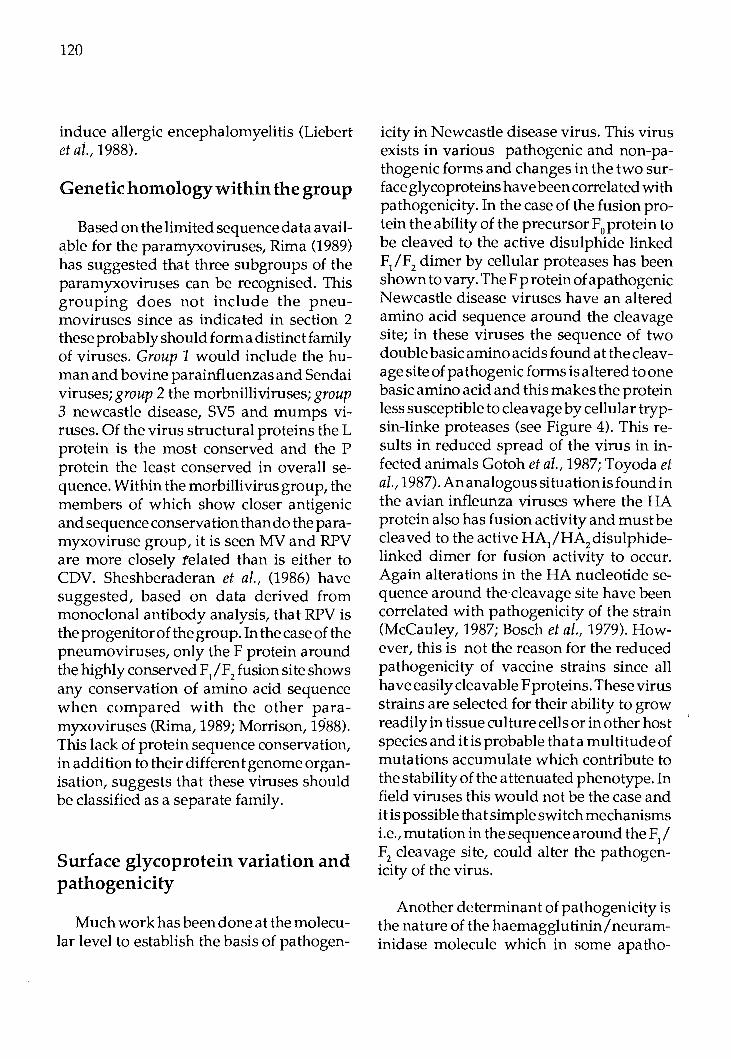
120
induce allergic encephalomyelitis (Liebert et al., 1988).
Genetic homology within the group
Based on the limited sequence data available for the paramyxoviruses, Rima (1989) has suggested that three subgroups of the paramyxoviruses can be recognised. This grouping does not include the pneumoviruses since as indicated in section 2 these probably should form a distinct family of viruses. Group 1 would include the human and bovine parainfluenzas and Sendai viruses; group 2 the morbnilliviruses; group 3 newcastle disease, SYS and mumps viruses. Of the virus structural proteins the L protein is the most conserved and the P protein the least conserved in overall sequence. Within the morbillivirus group, the members of which show closer antigenic and sequence conservation than do the paramyxoviruse group, it is seen MV and RPV are more closely related than is either to CDV. Sheshberaderan et al., (1986) have suggested, based on data derived from monoclonal antibody analysis, that RPV is the progenitor of the group. In the case of the pneumoviruses, only the F protein around the highly conserved F/F2 fusion site shows any conservation of amino acid sequence when compared with the other paramyxoviruses (Rima, 1989; Morrison, 1988). This lack of protein sequence conservation, in addition to their different genome organisation, suggests that these viruses should be classified as a separate family.
Surface glycoprotein variation and pathogenicity
Muchworkhasbeendoneatthemolecular level to establish the basis of pathogen-
icity in Newcastle disease virus. This virus exists in various pathogenic and non-pathogenic forms and changes in the two surface glycoproteins have been correlated with pathogenicity. In the case of the fusion protein the ability of the precursor F
0protein to
be cleaved to the active disulphide linked F/F2 dimer by cellular proteases has been shown to vary. The F protein of a pathogenic Newcastle disease viruses have an altered amino acid sequence around the cleavage site; in these viruses the sequence of two double basic amino acids found at the cleavage site of pathogenic forms is altered to one basic amino acid and this makes the protein less susceptible to cleavage by cellular trypsin-linke proteases (see Figure 4). This results in reduced spread of the virus in infected animals Gotoh et al., 1987; Toyoda et al., 198 7). An analogous situation is found in the avian infleunza viruses where the HA protein also has fusion activity and must be cleaved to the active HA/HA
2 disulphide
linked dimer for fusion activity to occur. Again alterations in the HA nucleotide sequence around the cleavage site have been correlated with pathogenicity of the strain (McCauley, 1987; Bosch et al., 1979). However, this is not the reason for the reduced pathogenicity of vaccine strains since all have easily cleavable Fproteins. These virus strains are selected for their ability to grow readily in tissue culture cells or in other host species and it is probable that a multitude of mutations accumulate which contribute to the stability of the attenuated phenotype. In field viruses this would not be the case and it is possible that simple switch mechanisms i.e., mutation in the sequence around the F
1 I
F2 cleavage site, could alter the pathogenicity of the virus.
Another determinant of pathogenicity is the nature of the haemagglutinin/neuraminidase molecule which in some apatho-

121
FIGURE4: Amino acid sequence (single letter code) around the F/F2 cleavage site of pathogenic
and apathogenic NDV s. Basic amino acids (R =arginine; K =lysine) are indicated by a(+) (from Toyoda et al., 1987).
c terminus F2 N terminus F1
Miyadera V TT S ...... c Herts .. .... .. <l.l :J Italian '-
> Australia-Victoria .. La Sota .. .. .. ..
...... c 026 <l.l
... .. ... .. :J Queensland "- .. ..... T > <t: Ulster .. N .. ..
genie forms of NDV have a C-terminal amino acid extension due to alteration of the normal stop codon. The HN protein of paramyxoviruses, like the neuraminidase protein of infleunza virus (Fields et al., 1981), is anchored atthe N terminus. The C terminus is, therefore, exposed at the surface of the virus and contains the virus receptor binding site and so the HN is unable to bind to the receptor until the C-terminal extension is cleaved from the molceule (Sato et al., 1987). At present we are investigating this possibility by sequence analysis of strains of RPV with different pathogenicities.
Summary
The paramyxoviruses include many important human and animal pathogens. The structure of their genomes and the types
G
.
.. .. .. -..
+ + + + GRRQRR F I GA 11 GS
+ + + t-. .. .. . .. .. m n .. .. .. .. .. .. + + + t - .. .. .. . .. .. - .. .. .. -- .. + -t + .... .. .. ... .. K .. .. .. .. • ••• .. G + s + .. .. ... .. .. ... .. ... .. .... ..
+ + ... G K .. G .. L .. ... .. .. ... • G + + E GK .. G .. L ., ... ,,, ... .... .. +
G! ... G K .. L .. .. 1111 ...... ..
of proteins they encode have been extensively studies in recent years but their mechanism of replication is, as yet, poorly understood. The study of these viruses has contributed greatly to our understanding of novel virus transcription mechanisms, i.e., RNA editing during transcription; the mechanisms underlying persistence of viruses in the host in the face of a strong antiviral humoural immune response; and to our understanding of the determinants of virus pathogenicity.
Acknowledgements
I wish to acknowledge my colleagues at the Institute for Animal Health, Pirbright who are involved in the work on morbilliviruses described here: Dr. S. Subbarao, Dr. G. Belsham, Miss S.A. Evans, Mr'. R.C.

122
Chamberlain and Miss L. Goatley. I also thanks Dr. S. Subbarao for helpful discussions in preparing this lecutre and Miss F. Berrie for typing the manuscript. The molecular biology studies on rinderpest virus are supported by a grant from the Wellcome Trust.
References
Barrett, T., Clarke, D.K., Evans, S.A. & Rima, B.K. (1987). The nucleotide sequence of the gene encoding the F protein of canine distemper virus: a comparison of the deduced amino acid sequence with other paramyxoviruses. Virus Res. 8, 373-386.
Barrett, T., Belsham, G.J., Subbarao, S.M. & Evans, S.A. (1989). Immunization with a vaccinia recombinant expressing the F protein protects rabbits from ch allenge with a lethal dose of rinderpest virus. Virology 170, 11-18.
Bellini, W.J.,Englund, G.,Richardson, C.D., Rozenblatt, S. & Lazzarini, R.A. (1986). The matrix genes of measles virus and canine distemper virus: cloning nucleotide sequence, and deduced amino acid sequences. J. Virol. 58, 408-416.
Billeter, M.A., Cattaneo, R., Schmid, A., Eschle, D., Kalin, K., Rehmann, G., Udem, S.A., Sheppared, R.D., Baczko, K., Liebert, U.G., Schneider-Schaulies, S., Brinckmann, U. & Ter Meulen, V. (1989). Host and viral features in persisent measles virus infections of the brain. In "Genetics and Pthogenicity of Negative Strand Viruses" Eds. D. Kolakofsky and B. Mahy. Elsevier, pp 356-366.
Bosch, F.X., Orlich, M., Klenk, H.D. & Rott, R. (1979). The structure of the haemag-
glutinin, a determinant for the pathogenicity of influenza viruses. Virology 95, 197-207.
Buckland, R., Gerald, C., Baker, R. & Wild, T.F. (1987). Fusion glycoprotein of measles virus: nucleotide sequence of the gene and comprison with other paramyxoviruses. J. Gen. Virol. 68, 1695-1703.
Castaneda,S.J.&Wong, T.C.(1989).Measles virus synthesizes both leaderless and leadercontaining polyadenylated RNAs in vivo. J. Virol. 63, 2977-2986.
Cattaneo, R., Rebman, G., Baczko, K., ter Meulen, V. & Billeter, M.A. (1987a). Altered ratios of measles virus transcripts in diseased human brains. Virology 160, 523-526.
Cattaneo, R., Rebman, G., Schmid, A., Baczko, K., ter Meulen, V. & Billeter, M.A. (1987b). Altered transcription of a defective measles virus genomes derived from a diseased human brain. EMBO J. 6, 681-688.
Cattaneo,R.,Kaelin,K., Baczko,K. & Billeter, M.A. (1989). Measles virus editingp rovides an additional cysteine-rich protein. Cell 56, 759-764.
Collins, P.L., Olmsted, R.A., Spriggs, M.K., Johnson, P.R. & Buckler-White, A.J. (1987). Gene overlap and site-specific attenuation of transcription of the viral polymerase L gene of human respiratory syncytial virus. Proc. Natl. Acad. Sci. USA 84, 5134-5138.
Crowley, J.C., Dowling, P.C., Menonna, J., Silverman, J.I., Schuberm, D., Cook, S.D. & Blumberg, B.M. (1989). Sequence variability and function of measles virus 3' and 5' ends and intercistronic regions. Virology 164, 498-506.
Elango, N., Kovamees, J., Varsanyi, T.M. & Norrby,E.(1989).Mumpsvirus:cDNAcloning and characterisatio nof six genes, determination of gene order and intergenic sequences. In "Genetics and Pathogenicity of Negative Strand Viruses" Eds. D. Kolakofsky and B. Mahy. Elsevier, p 249-253.
Elliott,.G.D.,Afzal,M.A.,Martin,S.J.&Rima, B.K. (1989). Nucleotide sequence of the matrix, fusion and putative SH protein genes of mumps virus and their deduced amino and sequences. Virus. Res. 12, 61-75.
Evans,S.A.,Belsham,G.J. &Barrett, T. (1989). The role of the 5' non-translated regions of the fusion protein mRNAs of canine distemper virus and rinderpest virus, manuscript in preparation.
Fields, S., Winter, G. & Brownlee, G.G. (1981). Structure of the neuraminidase gene in human influenza virusA/PR/8/34.Nature 290, 213-217.
Fuerst, T.R., Niles, E.G., Studier, F.W. & Moss, B. (1986). Eukaryotic transient expression system based on a recombinant vaccinia virus that synthesizes bacteriophage T7 RNA polymerase. Proc. Natl. Acad. Sci., U.S.A. 83, 8122-8126.
Garcia-Barreno, B., Palomo, C., Penas, C., Delgado, T., Perez-Brena, P. & Melero, J.A. (1989). Marked differences in the antigenic structure of human respiratory syncytial virus F and G glycoproteins. J. Virol. 63, 925-932.
Gotoh, B.,Hamaguchi,M. &Nagai, Y. (1987). Structural comparison of the cleavage activation site of the fusion glycoprotein between virulent and avirulent strains of Newcastle disease virus. Virology 158, 242-247.
123
Hiebert, S.W., Paterson, R.G. & Lamb, R.A. (1985). Identification and predicted sequence of a previously unrecognised small hydrophobic proteins, SH, of the paramyxovirus simian virus 5, J. Virol. 55, 744-751.
Hsu, D., Yamanaka, M., Miller, J., Dale, B., Grubman, M. & Yilma, T. (1988). Cloning of the fusion gene of rinderpest virus: comparative sequence analysis with other morbiliviruses. Virology 166, 149-153.
Jang, S.K., Davies, M.V., Kaufman, R.J. & Wimmer, E. (1989). Initiation of protein synthesis by internal entry of ribosomes into the 5' non-translated region of encephalomyocarditis virus RNA in vitro. J. Virol. 63, 1651-1660.
Katz, R.A., Cullen, B.R., Malavarca, R. & Shalka, A.M. (1986). Role of the avian retrovfrus mRNA leader in expression: evidence for novel translational control. Mol. Cell Biol. 6, 372-379.
Liebert, U.G.,Linington, C. & Ter Meulen, V: (1988). Induction of autoimmune reactions to myelin basic protein in measles virus encephalitis in Lewis rats. J. Neuroimmunol. 17, 103-110.
McCauley, J.W. (1987). Influenza virus glycoprotein variation and its role in disease. In "The molecular basis of virus disease", SGM Symposium.
Meier, E., Harmison, E.G., & Schubert, M. (1987). Homotypic and heterotypic exclusion of VSV replication by high levels of recombinant protein L. J. Virol. 61, 3133-3142.
Morrison, T.G. (1988). Structure, function and intracellular processing of paramyxovirus membrane proteins. Virus Res. 10, 113-136.

124
Olmsted, RA. &Collins,P.L. (1989). ThelA protein of respiratory syncytial virus is an integral membrane protein present as multiple, structurally distinct species. J. Virol. 63, 2019-2029.
Pelletier, J. & Sonenberg, N. (1988). Internal . binding of ribbsome to the 5' non-coding · region fo a eukaryoticmRN A: translation of poliovirus. Nature 334, 320-325.
Richardson, C., Hall, D ., Greer, P ., Hasel, K., Berkovich, A., Englund, G., Bellini, W.J., Rima, B. & Lazzarini, R. (1986). The nucleotide sequence of the mRNA encoding the fusion protein of measles virus (Edmonston strain): A comparison offusion proteins from several different paramyxoviruses. Virology 155, 508-523.
Rima, B.K. (1989). Comparison of amino acid sequences of the major structural proteins of the paramyxo- and morbilliviruses. In "Genetics and Pathogenicity of Negative Strand Viruses" Eds. D. Kolakofsky and B. Mahy. Elsevier, pp 254-263.
Sato, H., Hattori, S., Ishida, N., Imamura, Y. &Kawakita,M.(1987).Nucleotidesequence of the haemagglutinin neuramfnidase gene of Newcastle disease virus a virulent strain D26: evidence for a longer coding region with a carboxyl terminal extnsion as compared to virulent strains. Virus Res. 8, 217-232.
Schneider-Schaulies, A., Liebert, U.G., B aczko, K., Cattaneo, R., Billeter, M. & Ter Meulen, V. (1989). Restriction of measles virus gene expression in acute and su bacu te encephalitis of Lewis rats. Virology 171,525-534.
Schneider-Schaulies, S., Liebert, U.G., Baczko, K. & Ter Meulen V. (1988). Molecu-
lar biological analysis of measles virus gene expression in the CNS of acutely and persistently infected rat brain cells. Virus Res. (Suppl.) 2, 80.
Sheshberadaran, H., Norrby, E., McCullough, K.C., Carpenter, W.C. & Orvell, C. (1986). The antigenic relationship between measles, canine distemper and rinderpest viruses studied with monoclonal antibodies. J. Gen. Virol., 67, 1381-1392.
TerMeulen, V. &Carter,M.J. (1982).Morbillivirus persistent infections in animals and man. In "Virus Persistence", SGM Symposium 33. Eds. B.W.J. Mahy, A.C. Minson & G.K. Darby, Cambridge University Press, pp 97-132.
Ter Meulen, V., Stephenson, J.R. & Kreth, H.W. (1983 ). Subacute sclerosingpanencephalitis. In "Comprehensive Virology" 18, H. Fraenkal-CowratandR.R. Wagner, Eds. pp 105-15, Plenum Press, New York.
Thomas, S.M., Lamb, R.A. & Patterson, R. (1988). Two mRNAs that differ by two nontemplated nucleotides encode the amino coterminal proteins P and V of the paramyxovirus SV5. Cell 54, 891-902.
Toyoda, T., Sakaguchi, T., Imai, K., Inocencio, N.M., Gotoh, B., Hamaguchi, M. & Nagai, Y. (1987). Structural comparison of the cleavage activation site of the fusion glycoprotein between virulent and a virulent strains of Newcastle disease virus. Virology 158, 242-247.
Tsukiyama, K., Yoshikawa, Y. & Yamana uchi, K. (1988). Fusion glycoprotein (F) of rinderpest virus: Entire nucleotide sequence of the mRNA, and several features of the F protein. Virology 164, 523-530.

Proc. FAO/CAAS Workshop on Biotechnology in Animal Production and Health in Asia and Latin America, Beijing, Oct 9-13,1989, pp 125 -127
Contribution to ET Technology and Present Stage of Research
Josef Fulka The Czechoslovak Academy of Sciences,
Institute of Animal Physiology and Genetics, 277 21 Libechov, Czechoslovakia.
Introduction
In recent years, namely from 1973 the research in the Institute of Physiology and Genetics of Farm Animals was focussed on embryo transferin cattle. During this period all procedures, as superovulation, estrus synchronization, embryo flushing and transcervical transfer were studied. Some methodologies were adopted from literature source, while the other were partofresearch program. During the last 4 years all experimental results were summarized and offered to the Breeding organization for the application in field condition.Within a short period of time several special groups were established and embryo transfer introduced in praxis. All steps are carried out on the farms without transprot of donors or recipients to other place. The number of transfers increases from year~ to year and in 1988 about 20,000 transfers were performed. The pregnancy rate gradually increases, too. At the beginning about 40% of recipients became pregnant and now this figure reaches more than 60%.
At present two aims are followed:
1. To produce male progeny from the best cows for A.I. service. 2. To establish the pregnancies with
twins to increase the population of calves for meat production. To reach this aim, additional embryos are transferred to previously inseminated recipients to contralateral uterine horns.
Frozen Embryos
In most cases fresh embryos are used, however, in particular instances deep frozen embryos are transferred. Unfortunately the pregnancy rate decreases to less than 50%. The research at present is focussed on the simplification of freezing procedures without equipment which is necessary for traditional methods. The r~sults obtained until now fluctuate and do not exceed those gained by classical appraoc;h.
Embryo Bisection
The splitting of blastocysts to produce identical twins was also introduced into praxis. For the di vision differen ttools can be used and experienced worker is able to perform splitting under stereomicroscope with home-made instruments. By this way 100 pregnancies from 100 embryos can be established.
However, it should be added that ET has strong limitations in high economical
FAO LIBRARY AN: 302019
126
demands due to price of embryos. The reason can be seen in low average superovtilation response after application of exogenous gonadotropins. Only 5-6 healthy looking embryos are in our conditions obtained from ilushed donor. Exceptionally the crop can be higher, after strict selection of donors reaches 10-12 good embryos. High price is acceptable in embryos from genetically valuable animals used for breeding purposes. For production of calves, single or twins, this price can be considered as a strong di sad vantage.
lJ.t-vitro Fertilization
For this reason we were forced to loo~ for a cheaper source of bovine embryos. Such source can be found in exploitation of ovarian oocytes, their fertilization in vitro and cultivation to the stage convenient for unsurgical transfer for freezing. This project has been followed for several years, at the beginning in laboratory animals and later, from the year 1985 also in farm animals, particularly in cattle. Due to the experience gained during the experimentation with laboratory mammals the positive results were obtained relatively early and first calf was born 2 years ago. From this time the attention was paid to the stabilization of penetration rate and to the depression of fertilization anomalies, such as polyspetmy and arrested development of male pronuclei. The frequency of both of them was substantially depressed to reasonable level, not exceeding 5 % . Present projects are aimed to elucidate detials connected with the development of bovine embryos originated in vitro and to compare them with parameters of naturally developed embryos. As methods, electron microscopic evaluation of selected structures and autoradiographic determination of metabolic activity, particularlyofRNAand proteinsynthesis,areused.
Preliminary observation revealed in healthy looking blastocysts small proportion of cells containing additional secretion vacuoles with lipid material. In blastocysts with retarded development such cells are most frequent and signalize degenerative changes, which lead probably to the death of embryo.
Nuclear Transfer
Very recently efforts are concentrated to the nuclear transfer in mammals with the aim to produce multiple identicalindividuals. For this purpos~ single blastomere was fused with enucleated oocyte and the division evaluated in vitro and in vivo. It was revealed that in vitro most embryos constructed by this way divide to eight cell stage and in vivo, after transfer to rabbit oviduct, the division of embryonic cells proceeds very intensively, however, the organization typical for normal embryo does not occur. After cytological evaluation, it was found that all cellular organelles are well conserved in eight cell embryos without any degenerative changes. Also embryonic RNA synthesis, which starts in cattle at eight cell stage, after fusion with cytoplasm is totally supressed and begins again when the embryo reaches eight cell stage. Further experiments are in progress at present.
Oocyte Maturation
Very extensive studies were carried out in the area of ovarian oocytes maturation. The results were published in many papers and help significantly in the program of embryoproductionin vitro. The description of sequence of nuclear changes, called germinal vesicle breakdown (GVBD), has helped to compare maturation in vivo with the maturation in vitro in pig and cattle. In appropriate conditions the sequence of these events is indistinguishable. However, as it

was observed later, in the cytoplasm of most oocytes the development of male pronucleus is deeply influenced. The sperm head does not transform to the male pronucleus. Important contribution increasing quality of cytoplasm was the supplementation of culture media with additional granulosa cells. Original results obtained in the rabbit, were valid also for pig and bovine oocytes.
At present basic processes responsible for chromatin condensation and nuclear membrane dissolution are studied. The cytoplasm activity called maturation promoting factor /MPF/ plays key role in nuclear changes. The synthesis of this activity starts in fully grown oocytes immediately after isolation of fully grown oocytes from antral follicles and reaches its maximal level at metaphase II. For the study of MPF, the method of cell fusion was applied and by
127
this way species nonspecificity of MPF was demonstrated. After fusion of rabbit MI oocyte to pig oocyte with intact GV1 chromatin condensation occurs immediately instead during 16-20 hrs in naturally maturing pig oocytes.
On the contrary, growing oocytes isolated from preantral follicles are not able to synthesize active MPF and to mature in vitro. Moreover, such oocytes fused with fully grown oocytes in GV, prevent the maturation and in giant cell two intact GVs are present. Many detials connected with this activity were described in special journals and the experience from these studies is valuable in the experiments concerning nuclear transplantation. The relation between cytoplasm and nucleus seems to be very important for further fate of reconstructed embryo.

Proc. FAO/CAAS Workshop on Biotechnology in Animal Production and Health in Asia and Latin America, Beijing, Oct 9-13, 1989, pp 128 -149
Biotechnology in Animal Production and Health in China
Wang Ruixiang Institute of Animal Science, CAAS
Beijing, China.
Introduction
New technology revolution in the World is changing social mode of production and industrial structure. !tis expected to lead us to another great leap of social productive forces. Biotechnology in its modern sense is one of the signs of this new technology revolution. Its abruptrising is not an accidental phenomenon, but an inevitable outcome of tremendous development of the science of biology. Because biotechnology shows broad prospect for overcoming many problems (e.g. because it has limitless economical potentials), finding new areas for ind us trial structure and reformation in agriculture, medicine and industry, the biotechnology a tracts great attention in China, as well as in manyocuntriesin the World. With improvement ofliving standard in China more milk, meat and eggs etc. and animal products are needed.Biotechnology applied to animal production and health is one of the very important ways of increasing animal productivity.
The high technology research and development programme has been formulated in China in 1986. Biotechnology is one of the 7 key areas identified in this strategic programme. According to the national policylimited in targets and focus on key projects, there are 3 major subejcts identified in biotechnology area, namely:
- Research and devleopmentof new plant/ animal varieties with high yield/ performance, high quality and resistance to adversity. Here genetic engineering breeding of lean meat pigs and chicken resisted to Marek' s disease and MOET with associated techniques are included.
- Research and development of new medicaments, vaccines, monoclonal antibody production, immunodiagnostic and basic research on biotechnology as new expression vector preparation and DNA recombination both in human and veterinary medicine are included.
- Protein engineering.
To obtain themain target products (e.g. new animal varieties, genetic improvement in livestock, vaccines, ELISA kits etc) and to catch with the world trend in biotechnological advancements are the general objectives of Chinese biotechnology programme. However there are speific objectives in biotechnology applied to animal production and health. The final aim is to resolve the problems in agriculture, medicine and industry. Meanwhile, it is hoped that with development of biotechnology, the levels of science and technology as a whole would be promoted.
In order to fulfill above mentioned objec-

tives, the National Committee of Science and Technology has appointed an Expert Committee for various biotechnological areas and Expert Groups for each subject, which are responsible for putting the programme into effect. The Expert Committee is responsible for development of strategy and overall arrangement. The Expert Groups are responsible for resolution of subjects into projects and topics and recommendation of personnel to be in-charge of the projects and topics. Under National committee of Science and Technology the National Bioengineering Development Centre, agency for administrating and coordinating work in biotechnology, has been established. There are also a Biotechnology Leadership Group, an Expert Consultative Group and Office in the Ministry of Agriculture. Besides National and local institutions traditionally committed to livestock research have started specialist departments of biotechnology.New institutes and laboratories have been established, for example Biotechnology Research and Development Centre, CAAS; Biotechnology Research Centre and Embryo Engineering DepartmentofJiangsu Academy of Agricultural Sciences, etc.
Present Status or Future Plans
A. NucleusherdimprovementusingMOET inONBS.
1. Present genetic improvement programmes in livestock. During 1950s and 1960s in order to put an
end to the backwardness of animal husbandry, genetic improvement in cattle, sheep, horses and pigs was carried on as before in USSR. For selection of breeding stock the comprehensive test including pedigree test, individual selection according to its constitution, judging, growth and
129
development and performance, and progeny test, was used.
The selected breeding stock then mated like to like to produce better offsprings. Within a breed several lines were developed. In new breed development crossbreeding for formation of a new breed was mainly used. As a result, several new breeds have been developed in cattle, sheep, horses and pigs. Since 1970s, some principles of population and quantitative genetics have been applied to livestock genetic improvement. For example, systematic breeding method is being used to develop specialized pig lines of lean meat type. BLUP method is being used for evaluation of breeding bulls and rams.
In poultry, the situation is different. Because poultry industry in China has been developed quite later, at the beginning, modern breeding methods were practised. Family selection, family selection combined with individual selection and reciprocal recurrent selection (RRS) are used in egg chicken, meat duck and Shaoxin Duck breeding respectively. The Shaoxin Ducks are noted for their high egg production.
To increase effects of animal breeding and hybrid vigor in a whole region, the breeding and multiplication systems are being established for each class of livestock. For example, in Heilongjiang Province1
, it is planned that at provincial level there will be 3 breeding farms for Harbin White Pigs, 1 farm for Sanjiang White Pigs and 1 farm for Min Pigs (these breeds are used as female lines), and 2 breeding farms for Landrace, Duroc and Large White as male lines. In these farms, pure breeding is carried out to provide breeding stocks of high quality for pig multiplication farms, which are built at city and country level. In commercial farms
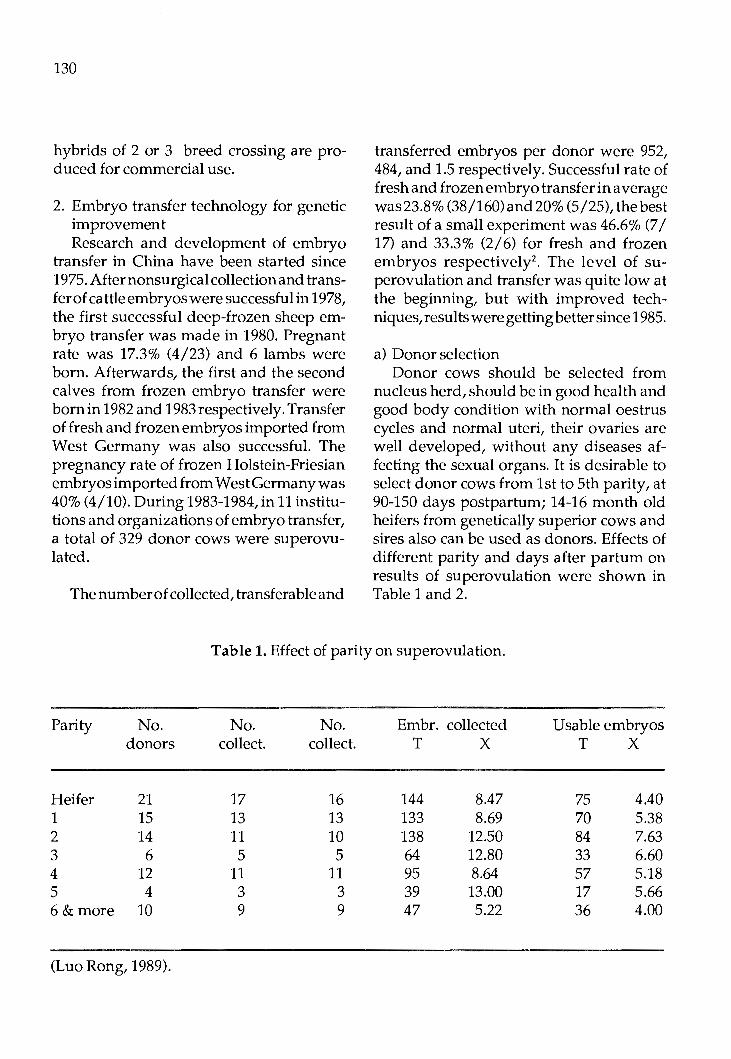
130
hybrids of 2 or 3 breed crossing are produced for commercial use.
2. Embryo transfer technology for genetic improvement Research and development of embryo
transfer in China have been started since 1975. After nonsurgical collection and transfer of cattle embryos were successful in 1978, the first successful deep-frozen sheep embryo transfer was made in 1980. Pregnant rate was 17.3% (4/23) and 6 lambs were born. Afterwards, the first and the second calves from frozen embryo transfer were born in 1982and1983respectively. Transfer of fresh and frozen embryos imported from West Germany was also successful. The pregnancy rate of frozen Holstein-Friesian embryos imported from West Germany was 40% (4/10). During 1983-1984, in 11 institutions and organizations of embryo transfer, a total of 329 donor cows were superovulated.
The number of collected, transferable and
transferred embryos per donor were 952, 484, and 1.5 respectively. Successful rate of fresh and frozen embryo transfer in average was23.8% (38/160)and20% (5/25), the best result of a small experiment was 46.6% (7 I 17) and 33.3% (2/6) for fresh and frozen embryos respectively2
• The level of superovulation and transfer was quite low at the beginning, but with improved techniques, results were getting better since 1985.
a) Donor selection Donor cows should be selected from
nucleus herd, should be in good health and good body condition with normal oestrus cycles and normal uteri, their ovaries are well developed, without any diseases affecting the sexual organs. It is desirable to select donor cows from 1st to 5th parity, at 90-150 days postpartum; 14-16 month old heifers from genetically superior cows and sires also can be used as donors. Effects of different parity and days after partum on results of superovulation were shown in Table 1 and 2.
Table 1. Effect of parity on superovulation.
Parity
Heifer 1 2 3 4 5 6&more
No. donors
21 15 14 6
12 4
10
(Luo Rong, 1989).
No. collect.
17 13 11 5
11 3 9
No. collect.
16 13 10 5
11 3 9
Embr. collected T X
144 8.47 133 8.69 138 12.50 64 12.80 95 8.64 39 13.00 47 5.22
Usable embryos T X
75 4.40 70 5.38 84 7.63 33 6.60 57 5.18 17 5.66 36 4.00

131
Table 2. Effect of days postpartum when superovulation was carried out on result of superovulation.
Days No. No. No. sue. Embr. collected Usable embryos postp. donors coll.
within 90 days 8 8 90-150 9 8 151-300 17 15 Over 300 days 4 4
(Luo Rong, 1989).
b) Superovulation Before 1983, hormones used for superovu
lation were mainly FSH and PMSG bought from overseas. Since 1984, hormones for superovulation were mainly made in China. FSH is made inNingpuo Hormone Factory, Jiangsu Province. PMSG is produced in Changchun Bioproduct Factory, Jiling Province. 15-methyl-PGF is produced in Shanghai Wuzhou Pharmaceutical factory. LH is made in Wuhang Biochemistry Pharmaceutical Factory. Some time FSH with higher potency prepared in the Institute of Zoology, Chinese Academy of Science, is used for superovulation.
Several superovulation methods exist in China.
Cattle:
Superovulation treatment is carried out at day 9-12 of oestrus cycle with FSH. FSH is injected for 4 days in decreasing doses, twice a day. Total dose of FSH is 440IU. At the lastday3 of superovulation treatment 4mg of 15-methyl-PGF is injected. (All these hormones used aremade in China).
coll. x T x T
8 8
15 4
77 9.62 46 5.73 85 10.63 52 6.63
133 8.87 65 4.33 45 11.25 18 4.50
A total of 98 donor cows were superovulated with this method. 82 (83.7%) of them were successful. Total of 710 embryos were collected from 78 donor cows. The number of·embryos collected per donor and usable embryos/ donor was 9 .1and5.4 respectively. Superovulation treatment with FSH inejcted in decreasing doses was compared with that ofincreasingorder. Usable embryos/ donor was 5.3 for decreasing doses and 1.0 for increasing doses. The cost of superovulation treatment with hormones in China was RMB 30-40/ donor, only 1/10th the cost of imported hormones.
- At 9-12 days of oestrus cycle (0 day is the 1st day of standard oestrous) FSH plus LH (4:1) is injected for 4 days in decreasing doses at the ratio: 8, 6, 6 and 5 or 5, 4, 3and2. ThetotaldoseofFSHir 500RU.At the 3rd day of treatment 4-6mg of 15-methyl-PGF is given into uteri of donor.
During 1986-1988, a total of 21 donor cow were superovulated and 169 embryos were collected from them (8.1 embryos/ donor). Usable embryos per donor was 6.7. In 1987, from 2 donors, 21 and 53 were colelcted resp. and all of them were usable.

132
- 12 and 6 donor cows from Black & White dairy cattle were superovulated with PMSG + PGF + HCG and FSH + GHF + LH respectively. Results obtained were compared. Th~re were 106 matured follicles (8.8/ donor),85 (7.1/ donor) corpora lutiin lstgroupand 107maturedfollicles and 103 (17.2/ donor) in 2nd group5•
- In 1985 and 1987, 13 donor cows were superovulated with PMSG and FSH. Twelve of them were successful (92.3%) and 23 (3.3/donor) embryos were collected. 2.8 usable embryos per donor was obtained6
•
- 49, 44 and 6 Japanese Brown Cattle were superovulated with FSH in decreasing
doses, FSH + LH and PMSG respectively. The number of embryos/donor and usable embryos/ donor in these 3 groups were 11.58 and 7.73, 11.79 and 7.24 and 6.6 and 2.8 respectiv~ly.
The results showed that treatment with FSH was better than that with FSH + LH and the later was better than treatment with PMSG. During superovulation 3 injections was better than 1 injection of PGF. The usable embryos could be increased by 3.8%.7
In sheep and goats:
6 goats and 8 sheep were superovulated with FSH as shown in Table 3. 8
Table 3. Hormone treatment.
Day Time Hormones 300U 340U
1 am FSH 50 60 70 70 pm FSH 50 60 70 70
2 am FSH 50 50 50 60 PGF 0.9mg
pm ffiH 50 50 50 60 PGF 0.9mg 0.9mg 0.6mg 0.6mg
3 am FSH 50 40 30 40 PGF 0.6mg 0.6mg 0.6mg
pm FSH 50 40 30 40

At the same time of insemination 120-lSOD of LH was injected. The results of treatment are given in Tables 4 and S.
Better results were obtained in Group 2 and 4, but the factors affecting the results could not be defined.
Atday12or13donorewesaresuperovulated with FSH for 3 days, lOOD per day. At the same day when ewes come to oestrus lSOD of FSH in one dose is injected intravenously .9
From 44 superovulated donor ewes a total of 230 (S.2/ donor) were collected. 129 of
133
them were fertilized embryos. The fertility rate of ova was S6.1 %.
- Donor gilts 8-9 month old, body weight 80-lOS kg are superovulated with 1) PMSG 2000D, 2) PMSG 400D - HCG 200D, 6) PMSG 300D - HCG 160D. At the time of lstinseminationHCG SOOD is injected, in groups 4 and S HCG SOOD, in group 6 300D is additionally injected after 1st HCG injection S7h later.
The results of superovl!lation revealed that percentage of gilts not reacted to superovulation treatment in groups 1-6 was 0. 14.3, 60.0, 33.3, 22.2 and 0. Over 1/2 of gilts
Table 4. Results of superovulation.
Groups No.donor No.in Time of LH No. corp oestrus oestrus (days) luti
1 2 2 2.7S 120 6.0 2 7 7 3.07 120 10.1 3 1 1 3.00 120 12.0 4 4 4 3.2S lSO 13.5
Total 14 14 3.02 10.6
Table 5. Results of superovulation (continued).
Groups Eggs Eggs oll. Normal embryos shed T % T %
1 12 8 66.7 7 87.S 2 S2 39 7S.O 36 '92.3 3 12 s 41.7 1 20.0 4 S4 47 87.0 24 Sl.l
Total 130 99 76.1 68 68.7
134
in group 3 only shed 5 eggs. The average number of eggs shed and collected were 36.0 and27.3ingroup 1,27.5and 15.7ingroup2, 16.0 and 12.0 in group 3, 25.2 and 13.0 in group 4, 25.0 and 14.4 in group 5, and 29.0 and 23.0 in group 6. Fertility rate of eggs shed was 27.4%, 56.4%, 100%, 78.2%, 4.7.4% and 41.3% in groups 1-6 respectively. With PMSG doses decreased, the fertility rate was increased (P<0.01).10
- Superovulationinrabbitsisinduced with FSH and HCG (made in China). FSH is injected subcutaneously twice a day for 3 days. To induce ovulation lOOU of HCG is injected intravenously at the 4th day of treatment. The optimum dose of FSH if 30-60U. Rabbits are inseminated with 0 .8-1. 0ml of semen containing 1.0-2.5 x 10⁸ sperms.
Results showed that more than 60 embryos could be collected from one donor. 1, 2 and 4 cell embryos were collected from oviducts 24-30h after insemination, 8-16 cell embryos - from oviducts and uteri 48-54h after insemination and morula and early blastocysts - from uteri 68-80h insemiantion.11
- Superovulationinrabbitsisinduced with PMSGindifferentdoses:50, 100, 150,200 and 250U. lOOU of HCG is injected when rabbits come to oestrus. Donros treated with 100 and 150U gave better results. 78% and 88% of superovulated donors came to oestrus, 24.4 and 28.1 eggs in average were shed and 17.0 and 17.3 embryos were collected.
- Donor rabbits are treated with FSH and HCG to induce superovulation. Rabbits are injected 5-6, 7-8 and 9-lOU of FSH respectively. At the time as oestrus occurred lOOU of HCG is injected.
Better results were obtained from donor rabbits treted with 5-6 and 7-8U of FSH. Number of eggs shed in average was 20.0 and 22.5; embryos collected was 16.2 and 16.0 respectively.12
Effect of repeated superovulation on oestrous cycle was studied in Shanghai No. 7 Farm using Black & White dairy cows as donors. Donor cows were 3-14 year old, 2nd-10th parity, more than 50 days postpartum, 1-2 normal oestrous cycles was observed. Superovulation was induced mainly with FSH (mad(( in Wuhan Biochemistry Pharmaceutical Factory).
For some of donors (in 1st superovulation there were 2 and in the second ovulation, 4 cows) PMSG (produced in Inner Mongolia Biochemical Pharmaceutical Factory) was used. Atday12-16of cycle LH was injected twice a day for 3-4 days. Total dose was 300-400U. PMSG 2240-3360 was injected at day 16 of cycle. After 48h of hormonal treatment 2-4mg of 15-methyl-PGF or 250-500U of ICI 80996 was injected intramuscularly. At the time when oestrus occurred LH or LHR was given. 22 donor cows of 65 superovulated cows were repeatedly superovulated 126.9 days (62-289) after 1st treatment, 5 cows were treated 3rd time 155.4 (110-221) days after 2nd superovulation. The results revealed that percentage of successful superovulation and usable embryos per donor of these 3 treaments repeated were89.28%,81.80% and 100.0% and 3.5, 3.6 and 4.8 embryos respectively. Duration of oestrous cycle after each repeated su perovula ti on was 28 .5 (14-48), 29 .2 (21-38) and 27.2 (20-41) days respectively. The normal oestrous cycle in this farm was 21 days. It was concluded that repeated superovulation at interval of 4-5 months did not effect reproductive performance.13
In sheep study repeated superovulation was carried out in 1979. 14 donor ewes from 23 superovula ted were repeatedly treated, 6 and 1 of them were superovulated 3 and 4 times respectively. Ovulation and fertility rate in 1st and 2nd to 4th repeated su perovulation were 11.8 and 40.3%, 8.8 and 87.3%, 7.0 and 79.2%, and 4.0 and 100.0%. No significant difference was observed between treatments. The oestrous cycle after superovulation was prolonged, averaging 19 .6 days.9
c) Mating 12, 24 and 36h after stable standing oe
strus donor cows were inseminated with 2X dose of semen with motility more than 30%.3
48-56h after oestrus occured donor cows were inseminated 3 times with interval of 12h.4 Zhong et. al. (1987)14 reported that with increased dose (20-30 millions of sperms), times of insemination (2-3 times) and shortened interval between insemination (10-12h), fertility rate of eggs could be 91.4 % (32/35) and normal embryos could be 75% (24/32). In case of pigs, sheep and rabbits both natural mating and artificial mating are used.
d) Collection of ova: procedures and comparative efficiency In cattle, embryos are recovered at 7.5
days after oestrus (lstdayis day 0) or at6.5-7 days after insemination nonsurgically. Donor is deprived of water and feed for 24h. The donoris led into stock, tail head clipped, washed with disinfectant solution and a epidural injection of 4ml 2% procaine is given. The vulva area is washed and disinfected, and the technician inserts one arm into rectum to estimate number of corpora lutea and follicles and zie of ovaries.
The vulva lips are parted and cerival expander is introduced in to vagina and then
135
placed in lumen of the cervix, which facilitates passage of the catheter. A sterile two way catheter with a stiffening stainless steel rod is inserted gently into the vagina on the expanded cervix lumen. The catheter then is manipulated into the selected horn so that the inflatable ballon is situated at the palpable bifurcation of the two horns. The balloon is inflated with 10-15ml of air, depending on the type of catheter, additional 2-6ml of air is added as required. The horns are flushed with approximately 450-500ml of medium (a modified Dulbecco' s PBS containing 1-2% calf serum) warmed to 37"C. The medium with ova is flowed out through drainage tube and embryo filter appratus. After the donor has been flushed, antiseptic solution containing 2-3 million U of penicillin and streptomycin is given into uterine horns.
During 1979-1981 in Beijing Western Suburb Farm 60 donor cows were superovula ted. Ovulationrateinaveragewas 12.5,5.4 ova Id on or was collected. The recovery rate was 43.2%. In Shanghai No. 7 Dairy Farm, the recovery rate of ova shed from 65 donor cows was 62.2%. In Shanghai Institute of Dairy Cattle, the recovery rate was 59.7% (884/1481). The better recovery rate from 5 superovulated cows was 83.3% (75/90).13
In the sheep 230 ova were collected from 440 ova shed, the recovery rate was 52.3% (1979, Institute of Animal Science, CAAS).9
e) Examination and evaluation of ova and embryos Isolated embryos are kept viable in
modified Dulbecco's PBS, PH 7.2-7.6, complemented with 20% calf serum in covered petri dish at room temperature 15-25°C. As soon as possible the embryos are located under a dissecting microscope, embryos are washed 3 times or more with sterile buffer and then evaluated. Embryos are evaluated

136
mainly morphologically according to: - comapctness of the cells in embryo
regularity of the shape of embryos variation in cell size colour and texture of the cytoplasm presence of large vesicles presence of extruded cells diameter regularity of the zona pellucida presence of cellular debris
Embryos are classified into A, B and C classes. Ideal embryos are compact and spherical. The blastomeres are of similar size with even colour and texture, neither very light nor very dark. The cytoplasm is not granular or unevenly distributed and contains some moderate-sized vesicles. The perivitellien space is empty and of regular diameter, and the zona pellucida is even and neither wrinkled nor collapsed. No cellular debris should exist.
f) Transfer and/ or freezing of embryos - In 1980(Yaoetal.)156-7 day old embryos
were frozen according to method of Willadson (1978). DMSO was used as cryoprotectant. Before freezing, embryos were passed through 0.25M, O.SM, 0.75M, l.OM, l.25M and l.5M DMSO-PBS containing0.4% BSAatroom temperature, in each solution for 5 min. In 1.SM D AMSOPBS embryos were equilibrated for 10-20 min. Embryos were sealed in ampules with buffer, 1 embryo in 1 ampule. In a freezing device ampules were cooled from room temperature to -7°C at rate of lC/ min. ampules were seeded at-7°C. ampules were cooled to -6D°C at 0.3 C/min. When ampules reached -60°C, they were plunged into liquid nitrogen.
For thawing, ampules were transferred to a -50°C alcohol bath, they were warmed to -l0°C at 4 C/min, then transferred to 20°C
water bath.
There were 40 embryos frozen with this method. After 2-375 days storage in liquid nitrogen, they were thawed. 17 of them was normal. 20 embryos were transferred to 13 recipients, 2 of them became pregnant. 3 calves were born.
- Rapidfreezing-thawingmethod (Bilton, 1980; Lehn - Jenson, 1980). At room temperature embryos were transferred through 0.17M, 0.33M, O.SOM, 0.67M, 0.83M and l .OOM glycerol - PBS containing 0.4% calf serum albumin, in each solution for 5 min.
Embryo was sealed in ampule with 0.25M buffer. Ampules were cooled to -7C from temperature at a rate 1 C/min, seeded and then they were cooled to-36Cat0.3°C/min. Embryos were plunged into liquid nitrogen. To thaw ampules, they were transferred to a 35°C water bath for 1-2 min. Cryoprotectant was removed by passing embryos through glycerol - PBS in contrary order of concentration, in each solution for 10 min.
63 embryos were frozen and stored in liquid nitrogen for 1-297 days. 30 (52.6%) were found to be normal in 57 recovered embryos. 29 embryos were transferred to 22 recipients, but none of them was pregnant.
- Recently, in Institute of Animal Science, CAAS, cattle, sheep and mouse embryos were deep-frozen (Luo et al, 1989). Freezing buffer was PBS + 20% calf serum + 1.4M glycerol. Glycerol frozen was added in 3 steps, each step for 7-10 min. Embryos were frozen in straws. Straws were cooled to-7C at a rate of 1°C/min, equilibrated for 5 min, seeded, cooled to-8kC at 0.1, deep into liquid nitrogen. To thaw straws, transfer them to a 30°C water
bath. Glycerol was removed in 3 steps, each step for 5-7 min. Place embryos into 1.4M glycerol+ 1.0M saccharose in ratio of 2:1 (first step), then place them into 1.4M glycerol + 1.0M saccharose + PBS with 20% CS (1:1:1), finally into 1.0M saccharose + PBS with 20% CS (1:2). Embryos were washed 2-3 times with 'PBS containing 20% CS.
A total of 899 mouse embryos were frozen-thawed and cultured in co2 incubator at 37C for 72h. 81.9% (736/899) of them developed further. In sheep, there were 103 embryos of Hu sheep and 40 embryos of Han sheep frozen and thawed. According to morphological evaluation, 62.2% (90/143) of them were of A and B classes and 11.8 % (19 /143) in C class. In 1988, 40 recipient ewes were transferred and 20 became pregnant. 27 lambs were born. In cattle 108 embryos frozen using simple freezing devise devloped in Institute of Animal Science. 62 embryos were thawed, 43 of them were usable. Success rate of frozen embryos was 24.2% (8/33).16
In the rabbits, 84 embryos were frozen using rapid freezing and thawing method. 35 of them were morphologically normal (41.7%).112embryos were frozen using slow freezing and thawing method. After thawing 72 were normal (64.3%). 84 (not all normal) frozen-thawed embryos were transferred to 15 recipients and 2 became pregnant. 5 baby rabbits were born in 1982.Chen Xiu (1987) reported that survival rate of frozen-thawed rabbit morula was higher than that of 8-16 cell emrbyos. They were 66.2% and 58.2% respecitvely. Pregnancy rate of transfer was 66.7%.12
Embryo Transfer:
The recipients should be large, healthy,
137
mature heifers or young cows. At least one normal estrous cycle should be established before use, whether recipients are going to be syncrhonized or are selected because they were in estrus on the same day as the donor. Transferring embryos in cattle is mainly nonsurgical.
Stimulating results have been achieved in MOET of dairy cattle during last 5 years. In 1984-1986 a total of 97 donor cows were superovulated in Chengdu Fenghuangshan Dairy Farm.18 470 (5.7 I donor) embryos were collected from 82 cows. 278 (3.4/ donor) were usable. 159 embryos were transferred to 109 recipients and 44 of them became pregnant. Pregnancy rate was 40.4%. 44 calves were born (4 cows gave twin). Number of embryos collected per donor cows increased from 2.9 in 1983 to 8.9 in 1986, usable embryos/ donor - from 0.8 to 5.1 and the pregnancy rate-from 17.7% (3/17) to55.1 % (27 I 49) in Hebei Provincial Centre for Embryo Transfer.19 in the last 5 years 65 calves were born.
In Jilin Provincial Academy of Agricultural Science 485 embryos were recovered from 70 superovulated cows in 1986. Usable embryos/ donor was 3.8. 197 recipients of yellow cattle were transferred. Success rate was 50.3%, 93 calves were born. As Jin (1989)20 reported that from 12 superovulated cows 137embryoswerecollected, 11.4 embryos per donor and 118 (9.8/ donor) of them were usable. 16 calves were born from 32 recipients transferred using frozen embryos in the Institute of Animal Science, CAAS in 1986 (Zhu et al, 1986).
In Institute of Animal Science, JAAS 352 embryos were collected from 50 donor cows, 5 usable embryos/ donor and 200 embryos were transferred to recipients (one embryos toeach),and pregnancyratewas40% (1987).

138
In the pigs, in the Institute of Animal Science, CAAS, 5 recipient sows were transferred surgically (10615 embryos to each), 2 of them were pregnant and 16 piglets were born in 1988. In lutai Farm, Hebei Province, 35 donor gilts and sows were superovulated with PMSG _HCG. 30 (85.7%) of them were successfullysuperovulated.212(63.9%,212/ 332) embryos were collected surgically from 14 donors, 15.1/ donor. 201 of them were usable(14.4/ donor). lOrecipientswereused.
In the sheep, Australian Merino ewes were used as donors, 49 recipient ewes of Kazakh Sheep were transferred in Xinjang Tacheng Farm. 83 lambs were born and 46 of them survived (Farm Daily, 11th July, 1988).
g) In-vitro capacitation and in-vitro fertilization In-vitro fertilization system for the mice
was established and 11 test tube mice were obtained.21
In the rabbits, 72 oocytes were recovered from oviducts of superovulated rabbits. Matured oocytes were inseminated in-vitro with sperm capacitated in rabbit genital tracts. After incubation, 56 eggs developed into 2-8 cell stage. Fertilityrate was 77.8%.22
In 1986, the first in China, 5 test tube rabbits were born from 2 fost mother rabbits. This work has been done by Embryo Engineering Lab, Jiangsu Pronvincial Academy of Agricultural Science, cooperating with Scientists from overseas. Oocytes collected from rabbit oviducts were in-vitro fertilizated with sperm capacitated in-vivo.
The culture development rate of eggs fertilized in-vitro and in-vivo was 76.8 (43 I 56%) and 82.3 (14/17%) respectively. 5 (5/ 27) and 2 (2/14) baby rabbits were obtained from in-vitro and in-vivo fertilization respectively in 1987.23
In pigs, preliminary experiment indicated that oocytes collected from follicles and matured in-vitro were insemianted with spermcapacitated in-vitro. Fertility rate was 20% (3/15).FengZiyun(1989)reported that the extension rate of the cumulus of oocytes collected from porcine ovaries in slaughter house and cultured in-vitro for 18 h was 95.6% (216/226). After 44h culture the first polar body appeared in 46.5% (105 I 226) of oocytes. Fertility rate ofoocytes inseminated with sperm capacitated in-vitro was 41.7% (5/12).24
The first Chinese test tube lamb was born in Inner Mongolian University on 10th March, 1989 and there are still 9 pregnant ewes with in-vitro fertilized embryos. Dr. Xu Rigan is in charge of this research project. The first World test tube goat born on 9th March, 1984 in Japan was obtained by Japanese scientists in cooperation with Dr. Xu. (People's Daily, 3 November, 1989).25
In cattle, Lu Kehuan (1988) reported that oocytes collected from cattle ovaries in slaughter house were matured and inseminated in-vitro and transferred into sheep oviducts for 6 days. Embryos were recovered from oviducts. 17 recipient cows were transferred with 2 embryos to each and 2 cows - 1 embryo/recipeint. 13/19 (68.4%) became pregnant (7 of them with twin). From 13 pregnant cows (1-for autopsy, 1-aborted) 16 calves were born (10 of them were twins). In 1987, 2 oocytes, were matured and inseminated in-vitro and then were transferred to red pient cow nonsurgically. Twin calves were born. In 1988,81.7% of 536 oocytes cultured in-vitro were matured. The matured oocytes were inseminated with sperm capacitated in-vitro. 270 fertilized ova were transferred into sheep oviducts for future development. After six days, 116 embryos were collected and 2 of
them being in late morula stage could be used for transfer (Zhu et al., 1989) .26 Experimental Animal Research Centre (Inner Mongolia) reported that in-vitro fertilization of ovarian oocytescollected from sla ughtered cows was successful. 70-80%. 30-40% of fertilized eggs were able to develop further (Science & Technology Daily, 1st July, 1988).
Recently, Laboratory of Animal Reproduction, Nanjing Agricultural University in cooperation with Experimental Farm achieved considerable progres in in-vitro fertilization. Oocytes collected from ovaries of Holstein-Friesian cows in slaughter house were cultured in culture medium in co2 incubator for maturation. The mature oocytes were inseminated with sperm capacitated in-vitro, then they were developed in-vitro. The well developed embryos were transferred to synchronized cows.27
55.5% of oocytes collected from cattle ovaries in slaughter house, matured and inseminated in-vitro were able to cleavage. 64 % of them developed to morula and blastocyte stage and they (fresh or frozenthawed) were transferred to recipient cows.28
Fan Biqin (1985)29 reported that cattle ovarian oocytes could be stored frozen for long time. 1407 oocytes collected from ovaries of slaughtered cattle were frozen in 4 different freezing buffers and stored. After thawing morphologically normal rate of thawed oocytes was 88.1%and86.5%.
The survival rate of oocytes thawed slowly was insignificantly higher than that of rapidly thawed oocytes. In-vitro cultured oocytes, in which the first polar body was appeared, were 15.1 %.
h) Embryo bisecting/ cloning Studies on production of monozygotic twins, triplets and multiplets in China
139
started only a couple of years ago.
In the cattle, 8 late morula and blastocytes previously softened in PBS + 0.5% pronase were bisected with micrograss needles with the aid of micromanipulator under stereomicroscope. 14 (6 monozygotic pairs) demiembryos without zona pellucida were transferred to 7 dairy heifers (one pair into each recipient), 2 transferred to 7 dairy heifers (one pair into each recipient), 2 of them became pregnant. Male monozygotic twins were born at full term; another female twins aborted at 5 months of pregnancy.30 39 embryos of dairy cattle were bisected under stereomicroscope with microglass needles by hand. 74 demi-embryos were obtained (successful rate was 94.9%, 74/78). After 2h culture in PBS complemented with 20% CS at 37c (Table 6) they were transferred to recipients of Yellow Cattle. Pregnancy rate was 17.1 % (6/35).31
Simpleandrapidmethodofembryosplittingwas developed in the Instiute of Animal Science, CAAS. Using this method in 1988, 201 fresh cattle embryos were bisected and 402 demi-embryos were obtained. 206 recipients were transferred. 38 frozen-thawed embryos were bisected and transferred to 38 cows. Each of 15 embryos was splitted into 4 parts and 25 recipients were transferred (Zhu et al., 1988).
In sheep and goats, 17 previously softened embryos were bisected with microglass needles under stereomicroscope by hand. 10 pairs of demi-embryos without zona pellucid a were transferred to 10 recipient ewes, 4 of them became pregnant and 5 lambs were born, including 1 pair of female monozygotic twins (Jiang et al, 1987). 19 goat embryos were biseced in Northestern Agricultural University. 19 pairs of demiembryos were transferred to 18 recipients.
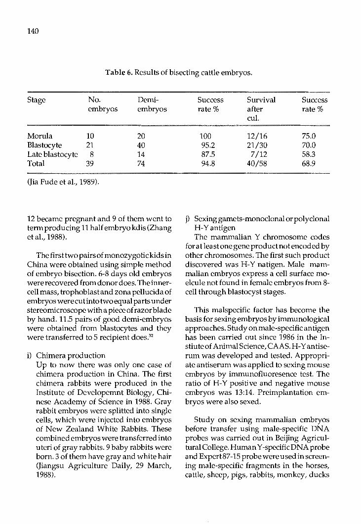
140
Table 6. Results of bisecting cattle embryos.
Stage No. Demi-embryos embryos
Morula 10 20 Blastocyte 21 40 Late blastocyte 8 14 Total 39 74
(Jia Fude et al., 1989).
12 became pregnant and 9 of them went to term producing 11 half embryo kdis (Zhang et al., 1988).
The first two pairs of monozygotic kids in China were obtained using simple method of embryo bisection. 6-8 days old embryos were recovered from donor does. The innercell mass, trophoblast and zona pellucida of embryos werecutinto two equal parts under stereomicroscope with a piece of razor blade by hand. 11.5 pairs of good demi-embryos were obtained from blastocytes and they were transferred to 5 recipient does.32
i) Chimera production Up to now there was only one case of chimera production in China. The first chimera rabbits were produced in the Institute of Developemnt Biology, Chinese Academy of Science in 1988. Gray rabbit embryos were splitted into single cells, which were injected into embryos of New Zealand White Rabbits. These combined embryos were transferred into uteri of gray rabbits. 9 baby rabbits were born. 3 of them have gray and white hair (Jiangsu Agriculture Daily, 29 March, 1988).
Success rate%
100 95.2 87.5 94.8
Survival after cul.
12/16 21/30 7/12
40/58
Success rate%
75.0 70.0 58.3 68.9
j) Sexing garnets-monoclonal or polyclonal H-Yantigen The mammalian Y chromosome codes
for at least one gene product not encoded by other chromosomes. The first such product discovered was H-Y natigen. Male mammalian embryos express a cell surface moelcule not found in female embryos from 8-cell through blastocyst stages.
This malspecific factor has become the basis for sexing embryos by immunological approaches. Study on male-specific antigen has been carried out since 1986 in the Instiute of Animal Science, CAAS. H-Y antiserum was developed and tested. Appropriate antiserum was applied to sexing mouse embryos by immunofluoresence test. The ratio of H-Y positive and negative mouse embryos was 13:14. Preimplantation embryos were also sexed.
Study on sexing mammalian embryos before transfer using male-specific DNA probes was carried out in Beijing Agricultural College. Human Y-specific DNA probe and Expert 87-15 probe were used in screening male-specific fragments in the horses, cattle, sheep, pigs, rabbits, monkey, ducks

artd chicken as in human Y chromosomes.
B.Application of r-DNA technology for genetic improvement and genetic resistance to diseases in livestock/poultry.
1. Gene mapping/DNA polymorphisms: a) Restriction fragment length polymor
phism
RFLP are used in recharacterization of some indigenous animal breeds and in classification of some wild animals. b) Hyperbolic region probes: No work done
yet. c) Oligonucleotide probes: No work done
yet. d) Gene mapping.
Gene mapping method is used to identify resistant genes in MHC B-L subregion of 0 type chicken, and to identify other related genes in the vicinity of GH gene in pig.
e) DNA sequencing.
DNA sequencing techniques are widely used to characterise cloned genes, to search for control (regulation) sequences and to identify new open reading frame DNA sequences.
2. Gene cloning (Progress in the following areas has been made)
a) Vector used Plasmids: p8R322, pucl8/19 Lambda vector: Charon 28, 1059, EMBL 3/4, gtl0/11 Cosmids: p 81110, pLAB-i Others: Vaccinia, SV40, adenovirus and pliovirus derived vectors.
141
b) Gene constructs made: Bovine genomic growth hormone gene Procine genomic growth hormone gene Sheep genomic grwoth hormone gene Porcine growth hormone releasing factor gene
c) Genes for production related hormone Bovine growth hormone cDNA gene Porcine growth hormone cDNA gene Sheep growth hormone cDNA gene.
d) Genes for production related enzymes Calf rennin gene Bovine pepsin geRe Buffalo rennin gene
e) Genes for immune response E.coli antigenic factor K88 and K99 Chicken MD virus coating protein gene Swine cholera virus antigen factor gene
f) Genes for resistance to diseases 0-type chcken MHC B21 gene.
3. DNA hybridization techniques
a) Southern blotting/ western blotting/Dot blotting etc. DNA hybrid techniques are used in many institutes and laboratories for detecting gene integration and exprssion.
b) Radio active probes Radio-active probes can be synthesised and labelled locally.
c) Non isotopic DNA detection. Biotin labelled probes are used for DNA detection.
4. Gene transfer
a) Methods used

142
Microinjections are used to transfer genes into mice, rabbits and pigs. CaP04 mediated transfer and Electroporation are used to transfer genes into cultured cells.
b) Efficacy of the methods used: Gene integration and expression have been detected, indicating that the methods used for gene transfer are effective. Effect of genes used on production/health traits has not been observed.
C.IMMUNODIAGNOSTICS AND VACCINES
1. ELISA tests for diagnostic purpose A monoclonal ELISA reagent was devel
oped. Mcab released by ovine brucella resistant M
28• 1°
2• c-1 hybridoma and purified
with Sephadex G-200, DEAE-DE52cellulose and Protein A - Sepharose CL-4B was labelled with horseradish peroxidase. A total of 603 cattle serum samples were tested for brucellosis by dot blot (DB) and compared with agglutination test (AT) and complement fixation test ( CFT). The results revealed that DB was more sensitive and specific than ATandCFT.
Recently, a monoclonal antibody specific to equine infectious anemia (EIA) was developed in Harbin Institute of Veterinary Medicine, CAAS. This Mcab was labelled with HRP and was used in DB test. Vaccinated horses appeared positive in DB test, while EIA horses were positive in ID test and healthy ones without vaccination were negative in both DB and ID test. Total of 33,900 horses in 5 provinces were tested (Zhou, 1989).33
Diagnostic kits for rapid diagnosis of newcastle disease were developed in Fujian Agricultural College (Cheng, 1988). Subse-
quently, specific Mcab against newcastle disease was obtained. Two highly sensitive and specific diagnostic reagents: fluorescent and eymze conjugated antibodies were also developed.
2. Monoclonal antibody production Since the end of 1983, the cell fusion tech
nique has been mastered in succession in National Supervisory Institute of Veterinary medicaments, Harbin Instiute of Vet. Medicine, Lanchou Instiute of Vet. Medicine, Central China Agricultural College, Xinjiang Institute of Vet. Medicine and other institutes. Monoclonal antibodies against hog cholera virus, equine infectous anemia weak cytotoxin, ovine brucella, 0-type foot and mouth disease, new castle disease virus, pgi E. Coli K88 antigen and pig treponema ~3 were developed. Monoclonal antibodies lA, 4D
4, 2A
5, 2D
4, 1A
9, 2A
2 and lD
16against
anthrax, and Mcab AG24-1, AG90-5, BGl-8, BG17-12, BG66-10, BG78-15 and EG19-11 were developed. BG78-15 was specific to 0
12
factor and other 6 were specific 0 4 factor. These monoclonal antibodies could be used for diagnostic purpose and vaccine prodution.
3. Genetically engineered vaccines Specific antigen gene of equine infectous
anemia was successfully cloned. Extracted from infected cells, DNA was cleaved and isolated with BamHI then combined with gtDNA using T4-DNA polymerase. The ligated DNA were transferred into E. Coli Yl 090. Five significantposi tive cloned strians were selected (Xu, 1989).
D. Biotechnology for enhancing animal feed production
1. Genetically enginered froage crops production.

2. Bioconversi9n of idnustrial, agricultural and animal wastes as animal feed.
a) Microbial treatment of ligno-cellulosic low quality roughages, crop residues, industrial wastes (oil mill effluents, rubber factory wastes, brewery wastes etc.)
In 1970s, research on resolution of agrocellulose by microorganisms was carried out in some of the Provinces and Autonomous Regions. Because of many difficulties, it has not resulted in any break-through yet.
b) Development of single cell protein. Single cell protein being dry cells of yeast,
algae, mould and bacteria contains 40-85% protein. To develop SCP resources is one of the importanttasks of agrobiotechnology. It is considered that development of SCP is a important way for resolving protein deficiency. Development of SCP in Chianmainly focusses on yeast production. There are 32 yeast factories located in Shanghai, Jilin and Guang dun etc. Yield is about 10000 m.t. a year. In Jilin Province paper pulp wastes were used to produce candida tropicalis berkhout Ck 4. In Nanjing Fermentation Factory waste water of bean products is used to produce geotrichus Candid um Link. Yield is more than 240 t a year. In Shunde Suger Factory, Guangdu Province, brewery waste is used for producing forage yeast and yield 100 ta year.
In the Institute of Environment Chemistry, CAAS paper pulp is used to culture Actinomyces. Recovery rate of products is 43-63%. Protein content is 42-55%.
In the end ofl 950s, production ofchlorella and geotrichus candidum link was very popular, because chlorella was difficult to harvest and due to high cost, its production has been stopped. Now only Yanzhou Mill and Nanjing Fermentation Factory etc con-
143
.tinue to produce Geotrichus Candid um Link. Maximum yield per year is 700t.
Later many institutions have been engaged in research on developemnt of SCP usingpetroleumhydrocarbon, natural gases and methanol. Production tehcnology with conversion rate 90% and 2.5gyeast/litter /h was established usingparaffin wax as a raw material in Shanghai Organic Institute. In Sichuan Institute of Biology pseudomonospore bacteria were cultured in natural gas. Production rate was lg/L/h. Saccharomycete and bacteria with high assimilation ability of methanol have been selected in Institute of Microbiology, CAAS and Beijing Forage Institute.
Pig blood was treated with enzyme to convert difficult digested red blood cell protein into small soluble protein. Its digestibility was greatly increased (China Food Daily, 19 May, 1989).
e) Enzyme and protein engineering to enhance feed and dairy products quality.
Enzyme engineeringin biotechnology has an important place. Now here are about 20 enzyme preparations commercially available.
Enzyme engineering in China has been developed quite later. There are only a few enzyme product factories in Wuxi and 'Fianjing. Furthermore, the enzyme products do not meet the need of industry and people. Some enzymes for genetic engineering work have been produced in Shanghai Biochemistry Ins ti tu te and Beijing Institute.
f) Fermentation technology After glutamic acid fermentation went
into production, lysine and aspartic acid were put into production one after another. Among 18 amino acids produced the tech-

144
nology of production of 8-9 amino acids (lysine, glycine, glutamic acid, isolecucine, cystine, arginine, aspartic acid, alanine and cysteine) have been well developed. Production of phenylalanine, .threonine, histidine, tryptophan, methionine, tyrosine is still in small scale. Production technique ofleucine, proline, valine and serine is needed to develop.
In amino acid production by enzymatic method, it is important to improve the culture condition of bacteria. Recently, in Xinjiang Fancaohu Pharmaceutical Factory the 1-glutamic acid production rate and conversion rate have been incrased to 7.65% and 63.4% respectively by changing additive qu_sintity of biotic.
References
1. Chen Runsheng. Heilongjiang J. Anim. Husb. and Vet. Med. 1989(3): 11.
2. TianShupeng. BeijingJ. Agr. Sci.1985 (8): 33-36.
3. Luo Rong. China Dairy Cattle. 1989 (1): 36-39.
4. Qi Yizin. China Dairy Cattle. 1989 (2): 25-29.
5. Ma Yubin.J. North-west University. 1985 (3): 111-113.
6. Luan Chengxiang. Innermon. J. Anim. Sci. 1988 (1): 4-5.
7. Xu Dianxin. Proc. 4th Cong. Anim. Reprod. & A.I. 1987: 67.
8. Yang Bingli. J. China Sheep. 1989 (1): 22.
9. Zhu Chengkuan et al. Proc. 2d Cong. Anim. Reprod. & A.I. 1985: 257-260.
10. Niu Shuli et al. Proc. 4th Anim. Reprod. & A.I. 1987: 61-62.
11. Fan Biqin. J. China Rabbits. 1986 (1): 25-30.
12. Chen Xiu. Proc. 4th Cong. Anim. Reprod. & A.I. 1987: 68.
13. Shanghai Institue of Dairy Cattle. Proc. 2nd Cong. Anim. Reprod. & A.I. 1983: 236-239.
14. Zhong Shi fan et al. Proc. 4th Cong. Anim. Reprod. & a.I. 1987: 59.
15. Yeo Zhenyuan et al. Proc. 2d Chong. Anim. Reprod. & A.I. 1983: 232-235.
17. WangXiugeetal.Proc.2nd Cong.Anim. Reprod. & A.I. 1983: 244-246.
18. Chen xiu et al. Proc. 4th Cong. Anim Reprod. *A.I. 1987: 62.
19. Transfer Centre. Sci. & Technique Daily. 11August1988.
20. Jin Zhengtai. Agrobiotechnology Information. 25/4/1989.
21. Yin Bailin et al. Proc. 4th Cong. Anim. Reprod. & A,.I. 1987: 60.
22. Zhu Yuding et al. Annual reports of Sci. Res. Ins. Anim. Sci. CAAS 1986-1987: 246.
23. Su Mu. JiangsuJ. Agri. Sci. 1986 (12): 32.
24. FengZhiyun. Agrobiotech. Information. 25/01/1989.

25. Lu Kehuan. GuangxiJ. Anim. Sci. & Vet. Med. 1988 (1): 4-8.
26. Zhu Yudingetal. Agrobiotech. information. 10/12/1988.
27. Fen Ziyun. Agrobiotech. Information. 25/01/1989.
29. Fan Biqin. Jiangsu. J. Agriculture. 1985 (2): 27-31.
30. Tan Lilingetal. Acta Vet.Zootech. Sinka. 1989. 20: 96.
31. Jia Fude et al. Heilongjiang J. anim. Sci. & Vet. Med. 1989 (4): 1-3.
145
32. Fan Biqin. Agrobiotech. Information 10 I 10/1988.
33. Zhou,C.J. Agrobiotech. lnformatlon. 25 / 02/1989.
Acknowledgements
The author gratefully acknowledge help of Dr. Chen Yongfu of National Laboratory of Agro-biotechnology, Beijing Agricultural University. He has written Part B of this paper.

146
Institution: Head of Institution: Address of Institution:
Researcher Dr. Chen Yongfu
Qi Shun Chang Professor
Sun Yong Ming Lecturer
Zhou Min Lecturer
Zhang Wei Hong Lecturer
Zhang Xie Xian Lecturer
Institution: Head of Institution: Address of Institution:
Researcher Wang Ruixiang Prof. Assoc.
Luo Yingrong Prof. Assoc.
Feng Shu tang Lecturer
APPENDIX A
HUMAN RESOURCES
National Laboratory of Agro-Biotechnology Dr. Chen Yong Fu Beijing Agricultural University, Beijing
Research Title 1. Production of transgenic pigs for high performance and for
disease resistance; 2. Cloning & expression of calf rennin gene in E. Coli & in B.
subtilis; 3. Developemnt of quicker methods for construction of gene
library for conservation of animal genetic resources.
Cloning and expression of porcine GH cDNA gene
Cloning and characterisation fo pig genes expressed In-vivo in pituitary
Cloning and characterization of cattle genes expressed In-vivo in pituitary
Molecular Biology of swine cholera virus
Sex Control "in Farm Animals
Institute of Animal Science, CAAS Prof. Chen Youchun Malianwa,Haidian, Beijing 100094
Research Title 1. Embryo transfer and manipulation in the pigs 2. In-vitro fertilization in the pigs
Embryo transfer in cattle and sheep and embryo cryopreserva ti on
Embryo transfer and in-vitro fertilization in the pigs
Zhu Yuding Prof.
Zhou Dingnian Prof. Assoc.
Chen Yuanmin Prof. Assoc.
JiangShie Prof. Assoc.
Institution: Head of Institution: Address of Institution:
Researcher. ZhengYihui Prof. Assoc.
Niu Shuli Lecturer
Du Chengping Prof. Assoc.
ChenFuyian Prof. Assoc.
DuNianxing Prof. Assoc.
Zheng Yixiang
Han Zhengkang Prof.
Embryo transfer and embryo bisection/ cloning in cattle
Sexing embryos using H-Y antigen
Sexing sperm using Percoll gradient centrifugation
In-vitro fertilization in cattle
Nanjing Agricultural University
Weigang, Nanjing
Research Title Embryo bisecting
Embryo transfer and in-vitro fertilization in the pigs
Monoclonal antibodies production and application
The same_as_above
Genetically engineered hormone vaccine production applied to improving animal performance
The same as above
The same as above
147
148
Institution: Head of Institute: Address of Institute:
Researcher Lu Jingliang Prof.
NingXide Lecturer
LiuXinghan Lecturer
Institution: Head of Institution: Address of Institution:
Researcher GuoZhiqin Prof.
Institution: Head of Institution: Address of Institution:
Researcher Fan Biqin
Institution: Head of Institution: Address of Institution:
Researcher Wang Jianchen Prof.
Dou Zhongying Prof. Assoc.
Harbin Institute of Veterinary Medicine, CAAS
Harbin
Research Title Monoclonal antibody for diagnosis of equine infectious anemia
The same as above
Polyvalent vaccine for piglet E. Coli diarrheal disease
Xinjiang Academy of Animal Science Prof. Guo Zhiqin Wulumuqi, Xinjiang autonomous Region
Research Title Embryo transfer and bisecting
Embryo Engineering Laboratory, J AAS Prof. Fan Biqin Jiangsu Academy of Agricultural Science, Nanjing.
Research Title Embryo transfer and in-vitro fertilization in rabbits
North-west Agricultural University
Yanglin Zhen, Shanxi Province
Research Title Embryo transfer and bisecting in sheep and goats
In-vitro fertilization and embryo bisecting in cattle

149
Institution: ·Department of Biotechnology Head of Institution: Address of Institution: North-east Agricultural college, Harbin, Heilongjiang Province
Researcher ·Research Title Qin Pengchun In-vitro fertilization in cattle Prof.
Institution: Heilongjiang Institute of Animal Science Head of institution: Address of institution: Qiqihar, Heilongjiang Province
Researcher Zhong Shifan Prof. Assoc.
Research Title Embryo transfer in cattle
Some other institutions and researchers are also shown in text and reference list.

Proc. FAO/CAAS Workshop on Biotechnology in Animal Production and Health in Asia and Latin America, Beijing, Oct 9-13, 1989, pp 150 -166
Biotechnology Applied to Animal Production and Health in India
P.N. Bhat Indian Veterinary Research Institute (IVRI)
Izatnagar (UP) 243 122 India
Introduction
1. India is the seventh largest country in the world with an area of 32,87,263 sq km. The mainlandlyingentirelyinthenorthernhemisphere extends, between latitude 8° 4' and 37°6' north and longitude 68° 7' east, and has been categorised into four well defined regions as, the great mountain zone, plains of the Ganges and the Indus, the desert region and the southern Peninsula. The climate is broadly tropical monsoon type. The rainfall is ill-distributed and varies from place to place and year to year. The total cropped area is 175 million hectares out of which only 38 million hectares is under irrigation. Around 70% oflndian population is dependent on income from agriculture. Thirty percent of total land holdings are held by 70% of small and marginal farmers who hold 80% of the total livestock in the country. Most of the land is used for cereal production and a small part is under permanent pastures and grazing land. Crop agriculture in India depends on bullocks and male buffalo to meet its requirement of motive power. Bullocks supply motive power for ploughing, water lifting, transport and other agriculture operations and contribute approximately 40,000 MgW. of power to agriculture operations.
2. There are 182 million cattle, 64.5 million buffalo,81.5 million goat,41.3 million sheep, 8.8 million chicken in India (PAO, 1985).
These produce a total of 41 million tonnes of milk, 1.04 million tones of meat, 40,000 megawatts of power, 38.0 and 25.4 million tonnes of greasy and scoured wool and 812 million metric tonnes of eggs to meet the requirement of human population of 759 million. Per capita availability of milk (148g), meat (3.7g) and eggs is far below the recommended levels.
3. The reasons for low productivity in our Livestock are:
1) Large losses from diseases and para-sites
2) Heat stress 3) Shortage of feeds and fodders 4) Poor management 5) Lower reproduction 6) Lower genetic potential for rapid
growth and large milk yield
4. India would need around 125 million bullocks to provide animal power to obtain a food production target of 250 MMT at the turn of century. The available feed and fodder situation, however, demands that we reduce the numbers. This paradox needs to be resolved. With the diminishing land resources for agriculture due to urbanisation and fast increasing human population, the total cropped area is likely to be further reduced. The National objectives for livestock production, therefore, are (1) to improve prod ucti vi typer animal, (ii) to reduce livestock numbers to match the existing feed

resources, (iii) to increase animal power to support higher food production, (iv) to provide sufficient and cheaper nutrients and (v) to provide cheap health care to sustain higher levels of productivity.
5. Health care is vital for the success of any animal improvement programme. Making available suitable vaccines and drugs at economical prices within the reach of the small and marginal farmer is one of the biggest challenges to veterinary scientists.
Three major problems, as far as animal production is concerned are:
(a) Howtomakehealthprotectioncheap, efficient and long lasting.
(b) Augumentation of fertility of farm livestock so as to improve production of meat, milk, wool and pelt.
(c) Augumentation of feed resources by converting available straws to protein and energy rich feeds.
All these problems can be solved only through use of biotechnological tools now becoming available.
6. Biotechnology has emerged as a frontier area of science as it has opened up possibilities of development of a variety of useful products to combat the above mentioned problems. The current developments in biotechnology, particularly in genetic engineering, are being directed towards producing safe and cheap health products for humans and animals.
7. TheGovernmentoflndiaiscommitted to the development of better livestock wealth to provide high quality animal products for human consumption. This involves the production of livestock with better productivity and their maintenance through effective disease control.
151
8. Programmes in biotechnology applications in agriculture and allied sciences are funded by Department of Biotechnology (DBT) and Indian Council of Agricultural Research (ICAR) within its own Institute and agricultural universities.
Present Status and Further Plans
Present Genetic Improvement Programmes:
Cattle & Buffaloes: The current breeding plan for cattle involves cross breeding of non-descript indigenous Zebu cows with Holstein and Jersey breeds of cattle or their frozen semen to produce cross-breds. The programme is spread over all the districts of the country. Currently 8 milion cross-bred breedable females are available and another 8 million are in the pipe line. It is proposed to raise this number to about 25 million breedable cows, which will produce 70 million tonnes of milk annually. In the breeding tracts of well-defined breeds, breeding is restricted to recognised native draft and milk breeds in order to produce animal power which supports agriculture. With the growth of mechanised agriculture it is planned to reduce the numbe of animals contributing to power progressively. At the present rate of reductionitwill take about 50 years. In the case of buffaloes improvemen is being accelerated through use of progeny tested bull semen for milk, meat and draft power. The buffalo cows are likely to add 30 million tonnes to the total milk production of the country a year, raising the total production of milk to 100 million tonnes a year.
Sheep & Goats: There is progressive emphasis on introduction of better quality rams for cross-breeding in major breeds of sheep for improvement of carpet wool and meat.

152
Similarly, in goats, cross-breeding for milk and meat is the major emphasis. There, however, is a feeling that the number of goats should progressively be reduced, since they cause ecological imbalance. The breeding of native breeds of sheep and goats is, however, to continue.
Poul try: The present emphasis in poultry is to develop superior genotypes of broiler and layers as an industrial proposition with eventual aim of totally replacing the present indigenous stock by beter developed breeds. This has been obtained to a large extent. Some well adopted local breeds and new syntheses are however, being encouraged as an interim measure.
Embryo Transfer Technology for Genetic Improvement
Cattle Herd Improvement for Increased Productivity Using Embryo Transfer Technology-Mission Mode.
The conventional methods of cattle and buffalo herd improvement through corss breeding and artificial insemination are slow in yielding desired results because of low female reproductive rates and the consequent low seleetion intensities. This problem can be obviated by the use of Multiple Ovulation and Embryo Transfer (MOET). By the judicious application fo this technique, it is possible to enhance cattle and buffalo producti vitywithoutund ulyincreasing the existing population. With this in view the S&T project on Embryo Transfer has been launched in 1986-87.
Project Structure: The project has focussed on establishing a multi-agency mission with clear targets, time frame and specifically indentified and measurable roles for each agency coordinated by the Department of Biotechnology with the National Dairy
Development Board as the lead implementing agency. The collaborating agencies are the Indian Veterinary Research Institute, the National Dairy Development Institute, the National Institute of Immunology and the Central Frozen Semen Production and Training Institute closely linked with the existing animal breeding infrastructure of the Ministry of Agriculture and NDDB.
Project Objects: The major objectives of the project are to create (i) a skilled pool of scientists, technicians and practitioners in embryo transfer (ii) institutional infrastructure for research, development and practice of embryo transfer and (iii) a 'nucleus' herd of superior bulls and bull mothers for producing elite stock.
Role of different agencies: The lead implementing agency aims to produce a seed stock of 2500 bulls and bull mothers and an embryo bank of 10 ,000 embryos by 1992 through a network of one main laboratory, four Regional Centres and twenty five State Centres.
The collaborating agencies support the lead implementing agency through R&D in the areas of su perovula ti on, oocyte maturation, in vitro fertilization, embryo splitting, cloning and sexing of embryos and related areas. They also marginally assist in adding to the embryo bank and the seed stock.
Project Progress
Lead Implementing Agency The project has so far achieved establish
ment of the main laboratory at Bidaj and three Regional Centres located at Nasik in Maharashtra, Rae Bareli in Uttar Pradesh and Nekarikallu in Andhra Pradesh. The
Centre at Nasik is fully operational with nearly 100 donors and 300 recipients.
Twelve of the proposed 25 State Centres to be located all over the country have been identified and training of scientists for six of them has been completed. The first centre at Erode has already become operational and six more centres will become operational by March, 1989.
The major accomplishments of the project include successful birth of 10 superior buffalo calves using both surgical and nonsurgical embryo transfer techniques.Nearly 6000 buffaloes with yield above 3000 kg/ lactation have been identified for selection of donor animals. Out of 783 embryos of cows and buffaloes harvested, 354 embryos have been transferred and 78 pregnancies established.
Indo-US Project: The Indo-US Project No. 386-0470, entitled "Studies on Embryo Transfer Technology and Bioengineering in Livestock Species and their Patho-biological Implications" focuses on the development of technology of embryo transfer as a mechanism for enhancing the genetic manipulation of specific animal production traits. It involves studies on embryo transfer technology, genetic engineering and and transmission of infectious diseases with particular reference to the buffaloes and cattle, however, small ruminants and possibly laboratory animals can be used for either their practical application or their usefulness in elucidating basic mechanisms associated with reproductive function. Emphasis is given on mutual collaborative research between Indian and United States (US) scientists and, where applicable, Indian scientists are receiving short-term trainingin identified US institutions.
153
Six Indian institutions, are responsible for specific research areas within the project. The Indian Council of Agricultural Research (ICAR) is responsible for the coordination and management of the project. USAID I New Delhi upon ICAR request is sreving as agent to arrange training of Indian scientists, acquisition of US consultants and purchase of specified scientific equipment. All are concentrating on the following main objectives:
a) Superovulationandsynchronization of donor and recipient animals for ova/ embryos transfer.
b) Collection of ova (surgical and nonsurgical) for in vitro fertilization, embryo culture, cryopreservation, sexing, cloning, engineering, embryo environmental interactions and embryo uterine secretory proteininteractions.
c) Field application of embryo transfer technology.
Research achievements in India and elsewhere have been described in the paper presented at the Beijing Workshop but due to space shortage, the detaildescriptions are omitted here.
Application of r-DNA Technology for genetic improvement
One major focus is the study ofresistance to diseases in livestock and poultry. The approach taken up by the Indian Veterinary Research Institute is primarily to follow restriction fragment length polymorphism for gene mapping and DNA polymorphism. So far in the livestock sector this work has been initiated only at the Indian Veterinary Research Instiutte and concentration is primarily on the native breeds, their crossbreds and buffaloes to begin with. The techniques of extraction of genomic and mitochondrial
154
DNA from lymphocytes have been standardised, in respect to cattle and buffaloes. A large number of probes primarily related to histocompatibility locus have been procured from abroad. The present work is analysing polymorphism both in mitochondrial DNA as well as in genomic DNA and will be progressing in the direction of identifying genes for disease resistance and to map them if possible.
The work in respect to gene cloning utilizing some of the constructs already developed is also being planned at a number of centers. Presently the work is being taken up on cloning of FSH and the LH genes in the case of cattle and buffaloes at National Institute of Immunology. The FSH gene has been cloned and alpha & beta sub-units of ovine leutinizing hormones and folicle stimulating hormone have been cloned. Restriction analysis of ovine FSH and ovin LH, c-DNAs clones has also been done.
A male specific probe has also been developed by NII. Most of the radio active probes are now locally prepared. India has the capability to produce most of the radio chemicals.
Immuno diagnostics
ELISA tests for diagnostics purposes
Research work carried at central foot and mouth typing laboratory at Mukteswar and foot and mouth laboratory at Bangalore campus of IVRI has successfully launched a new sand witch Elisa for sub-typing of FMD virus. They are in a process of launching a strain comparison test within the sub-types based one-DNA fingerprinting and sequencing techniques.
Monoclonal antibody production A number of laboratories are concentrat
ing their efforts on monoclonal antibody production. These are primarily used for diagnostic purposes. So far monoclonal antibodies are locally produced against Rinderpest, Foot and Mouth Disease, Brucellosis and Tuberclosis. A battery of four monoconals for Rinderpest and eight for Foot and Mouth Disease have been developed.
Vaccines
A genetically engineered vaccines So far one genetically engineered vaccine
for rabies engineered into vaccinia virus has been prepared by National Instiutte of Immunology. It has proved to be very effective on mice tests. Attempts are under way at IVRI for developing an engineered vaccine for sheep pox and Rinderpest.
Biotechnology for enhancing animal feed production
So far not much work hs been done in India. IVRI is launching a project on biconversion of Agricultural Wastes primarily straws using genetic engineered bacteria or fungus. The microbial treatment of lignin in low quality roughages has been going on for several years with very indifferent results. Work on manipulation of rumen environment has yielded exciting results.
Resources Available
Institutions involved 1. IVRI - National Biotechnology Centre
(NBC) (IVRI) 2. National Dairy Research Instiutte

(NDRI) 3. National Institute of Immunology (NII) 4. Tamil Nadu Agricultural University -
Madras Veterinary College (TNA U) 5. Haryana Agricultural University- Col
lege of Vety. Medicine (HAU) 6. Central Institute for Research On Buf
falo(eIRB) 7. Central Institute for Research on Goats
(CIRG) 8. GB Pant University of Agriculture &
Technology (GBPUAT) 9. Punjab Agricultural University (PAU)
10. National Dairy Development Board (NDDB)
11. Andhra Pradesh Agricultural University (APAU)
Research Areas
National Biotechnology Centre (IVRI) 1. Recombinant DNA Unit (Genetic Engi
neering of Bacteria, Viruses, Eukaryotes and parasites).
2. Hybridoma technology and immuno-
155
diagnostics. 3. Embryo Transfer Technology and Engi-
neering. 4. Immunobiology of reproduction. 5. Oncology of Birds and animals. 6. Post-graduate programme leading to
Masters and Doctorate Degree in Biotechnology.
National Dairy Research Institute (NDRI) 1. RecombinantDNAworkleadingtofer
mentation systems and biodegradation of feeds.
2. Embryo transfer and associated technologies.
3. Reproductive biology.
These topics are also covered by other Universities and Institutions.
Budget- 5 Years (1985-1990) IVRI US$ 11 Million NDRI US$ 8 Million NDDB US$ 15 Million Others US$ 5 Million
Personnel with numbers of each category of staff and training:
No. Researchers 20 10 5
20 5 5
15 2 5 5 2 4 2
Name of Institution IVRI NDRI NII NDDB GBP University APAU IVRI NDRI NII IVRI IVRI IVRI NDRI
Subject ETT and associated technologies -do--do--do--do--do-
.· Genetic Engineering -do--do-Vaccinology Cancer Biology (Biodegradation of lignin in straws).
156
International cooperation A number of International agencies are
helping in development of biotechnology in the country. These are UNDP, FAO, Overseas Agency of Great Britain, Indo-US Subcommissionon Science, Indo-Soviet Integrated long term protocol. These collaborations are between Institutions and Governments and expatriate staff through TOKTEN Programme of UNDP.
National Contribution to Network -Funds
All funds available for National Research effort will also become available to the Network for purposes of continuing cooperative programmes of research and training. The present staff and equipment can be utilized for the Network. The National training courses can be hooked to the Network, so that training slots for each training course are made available to the Network. The senior staff can be drafted as consultants and experts within the Network. Equipment available within the Institutes will be used for cooperative research programmes.
National Expectations from Network 1. The expectations from the Network is primarily in relation to trainings available at various institutions within the Network which could be availed off by the national staff.
2. It could also take advantage of research planning meetings in which scientists from developed countries will interact with national research staff and help in developing effective programmes of experiments which will be required for development of livestock resources within the country.
3. Equipment: It is expected that cost of specialised equipment and their maintenance will be provided by the Network. Disposables will be the first charge on the contingent grant of the Network for the purpose of training and cooperative research. It would be of interest to have a Network publication system which could be taken advantage by the participating Institutes.
Topics on which cooperative research and development 1s required:
1. Genetic engineering of bacteria, viruses, yeasts and parasites of animal health importance in order to develop vaccines and diagnostic reagents for animal diseases.
2. Immunodiagnostic kits using hybridoma technology.
3. Embryo transfer and associated technologies of in vitro fertilization and embryo culture, embryo splitting, cloning and sexing.
4. Multipleovulationandembryotransfer for ONBS.
5. Cancer Research using animal models.
6. Use of Restriction fragment length polymorphism (RFLP) as gene markers to developb effective selection scheme for improvement of animals for disease resistance and high productivity.
7. Biodegradationofindustrial,agricultural and other wastes using rDNA technology to convert them into high
value Protein and energy rich feed for livestock and poultry.
Activities in the Private Sector in the Country
The private and public sector organizations are progressively taking interest in products developed through Biotechnology. Currently a small beginning has been made in the production of bio-chemicals andenzymes. Scale is not of high order. Some vaccine production units are interested in developing diagnostic kits. At the present time, they have not shown much interest in contributing or participating in the pro-
157
gramme of Network. But they may take up this work at a later date.
Special considerations In the general area of Biotechnology, the
joining of India in the Network is subject to consideration ,that all products which are developed as a result of national effort will remain a property of the Government of India/ICAR/DBT the funding agencies. Any cooperation within the network will not automatically give any right to either the cooperating network or to any other agency of the United Nations for s:ommercial exploitationoftheproductsdevelopedinindia.
ANNEXUREl TRAINING COURSES BEING OFFERED
No. Title of the Course Frequency No. of seats
1. Master Course in Veterinary Annual 15 Biotechnology
2. Ph.D. Course in Biotechnology Annual 05 3. Animal Cell Culture Quarterly 05 3. Animal Cell Culture Quarterly 10 4. B-Cell Biology Annual 05 5. Development & Handling of Molecular Annual 05
Probes in diagnosis of Animal diseases 6. Application of Monoclonal antibodies Quarterly 05
in early diagnosis 7. Cloning, Sequencing and chromosomal Annual 10
localization of animal genes 8. ELISA in Medicines Annual 10 9. Genome analysis of viruses Annual 10
10. Genetic Engineering of RP viruses Annual 10 11. Embryo Transfer technique in Quarterly 05
Cattle and Buffaloes 12. Genetic Engineering of bacteria Annual 10 13. Genetic Engineering of Eucaryotes Annual 05 14. M 13 cloning and DNA sequencing Annual 05
158
APPENDIX A AN UPDATE ON THE CURRENT RE
SEARCH WORK GOING ON IN NATIONAL BIOTECHNOLOGY
CENTRE AT INDIAN VETERINARY RESEARCH INSTITUTE,
IZATNAGAR (UP)
1. Genetic Engineering of Virus 1.1 During the period under report capripoxvirus DNAs from virulent field isolates were analysed by restriction enzyme (Hindlll, Haelll, EcoRI, BamHI, Pstl, Sall and A val). Based on the Hind III profiles all the virus isolates could be identified on the basis of their HindIII 'C', 'D', 'E' and 'F' pattern only. The EcoRI and Sall restriction enzyme profiles for Jaipur isolate was diffemt from Roumanian and Ranipet. The enzyme pro,. file of Jaipur isolate had an additional Eco RI site. The estimates of molecular weights (143 Kbp to 147 Kbp) generated by differnt enzymes are similar to the other capripoxvirus DNA. There is positive homology of HindIII-'S' fragment (across the capripox genomes) with KSG, HindIII-'S' fragment. Based on the results, one research article has been sent for publication.
1.2 Molecular characterization of field isolates of rinderpest viurs
A highly virulent (field isolate used as challenge virus for vaccine testing) rinderpestvirus strain was attempted to be adapted in verocells by following the 3 methodologies i.e. Adsorption, fusion and co-culture. The virus was adapted by co-culture method only. Following radio-immunopreci pi ta ti on assay the virus specific proteins bands were precipitated by using hyper immune serum as well as monoclonal antibody (N specific antibody} against rinderpest virus.No variation in protein profile was observed between RBOK strain and virulen tfield isolate
at this stage.
2. Genetic Engineering of Eukaryo-tes
2.1 In continuation to our earlier work concerning the genes coding for gonadotrophins; isolation of total RNA and fractionation in to Poly A+ and Poly A-RNA was carried out from buffalo pituitary. Total cellular RNA was isolated usingguanidium thiocyanate procedure under colder conditions from freshly collected buffalo adenohyprophysis. RN A was precipitated with isopropanol left overnight at -20"C and then RNA pellet was resuspended in 70% ethanol, centrifuged, dried· under vaccum and dissolved in sterile water for further fractionation into m-RNA using oligo-dt cellulose column. Fractions of poly A.,. and poly A-RNA were precipitated with ethanol and stored at-70°C under 70% ethanol. In order to confirm the integrity, the RNA was characterized by denaturing agarose gel electrophoresis using formaldehyde formamide followed by staining with ethidium bromide. Visualization of gels under U.V. transilluminator showed streaks of distinct bands of RNA at 289, 185 and 45 regions. Suggesting a good yield of RNA fractionation of poly+ and poly A- was confirmed by agarose gel electrophoresis under denaturing conditions.
3. Studies on molecular cloning in Clostridial species
3.1 Eight different strains (four C.perfringens type A and four C.perfringens type D) collected were tested for mouse pathogenecity test. Two strains were found non-pathogenic.
3.2 Two protocols were attempted to screen the eight different strains of C.perfringens isolated from animal :sources. Different plasmid profiles were observed.

3.3 Attempts are being mde to cure bacterial cells plasmid (s) for tetracyclin resistant in one isolate of C.perfringens.
3.4 To minimize the DNaseactivity, DNase inhibitors such as DEP and higher concentration of EDTA were used but no significant improvement was observed.
3.5 GenomicDNAfromC.perfringenstype D was isolated. The size was resolved in gel electrophoresis. It was found the most of the DNA population was above lambda DNA. It was also digested with restriction enzymes such as EcoRI, Hind III, Sal I, BamHI, BgI II.
3.6 The UV Spectra, of the epsilon toxin purified to homogenity from Clostridium perfringens type D, in near ~ regio~ ~t different time intervals of toxm storage 1mtially showed a garssion shaped band which gradually turned to a shoulder as a function of time. The maxima initially centered around 280nm showed a gradual blue shift to270nm.
3.7 The CNBr treated toxin and pure toxin were subjected to high performance gel permeation liquid chromat?graphy. The pure toxin, was selected as a smgle homogenous peak and CNBr treated toxin resulted in three fragments of comparatively low molecular weight.
3 .8 The epsilon toxin has been purified by HPLC using Semi-preparative gel permeation column for raising the antibodies.
3.9 Isolation of total genomic DNA from Clostridium perfringens type D has been attempted.
159
4. Molecular Biology of Clostrid-ium chauvoei
4.1 For large scale isolation of plasmid DNA from Cl.chauvoei, bacterial strain No. 49, (IVRI, Izatnagar) was cultured in modified anaerobic medium after revival in RCM broth. During study, this anaerobe was found to be resistant for another aminoglycosidic antibiotic - Kanamycin also in addition to four antibiotics - gentamycin, neo~ mycin, amikacin and streptomycin. , Gentamycin (5 ug/ ml)- subinhibitory dose, was invariably added to the culture media to induce the replication of plasmids so as to scale up the final DNA yield. Plasmid DNA was isolated by modified procedure of SDS lysis and alkaline lysis methods and s~owed very little chromosomal DNA smeanng on agarose gel under UV-light. Plasmid DNA preparations were further purified of RNA and proteins contaminations by RNase and proteinase K treatments. Purity of plasmid DNA preparations was checked by absorbance at 260 and 280 nanometer. Two plasmids of ca 5Kb and 7Kb were isolated from gentamycin R Cl.chauvoei.
5. Research on Haemoprotista 5.1 During the period under review, cell lines of macroschizont-infected lymphoblasts for Theileria annulata were maintained by serial passage in cell culture and by.clJ'.'opreservation. The vaccine doses (Theilena vaccine) were prepared and the cattle were vaccinated in the Jersey Breeding Farm, Bhopal (Pure bred Bos taurus) and Military Farm, Agra (Cross-breds).Noadvers~ fe~~back information was received. The M11I:. tary Farm Agra were advised to continue vaccination of all new-borns added to the herd. This recommendation was accepted and we implemented this by supply and vaccination of another 100 new-born ~C!l ves.
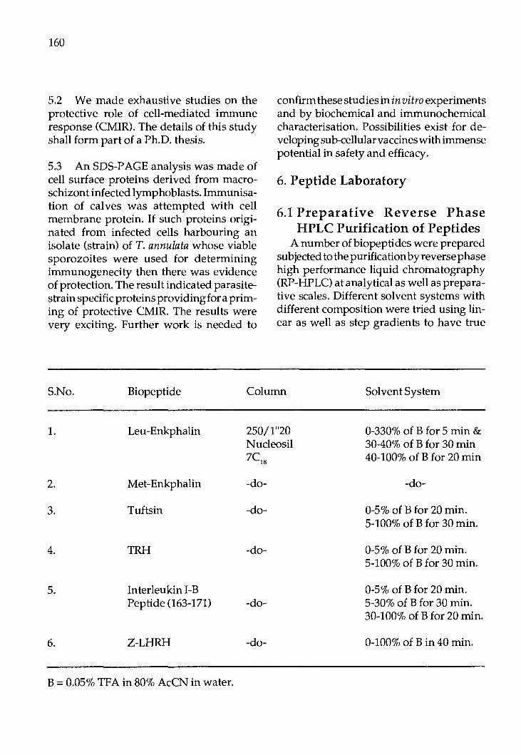
160
5.2 We made exhaustive studies on the protective role of cell-mediated immune response (CMIR). The details of this study shall form part of a Ph.D. thesis.
5.3 An SDS-PAGE analysis was made of cell surface proteins derived from macroschizont infected lymphoblasts. Immunisation of calves was attempted with cell membrane protein. If such proteins originated from infected cells harbouring an isolate (strain) of T. annulata whose viable sporozoites were used for determining immunogenecity then there was evidence of protection. The result indicated parasitestrain specific proteins providing for a priming of protective CMIR. The results were very exciting. Further work is needed to
confirm these studies in in vitro experiments and by biochemical and immunochemical characterisation. Possibilities exist for developing sub-cellular vaccines with immense potential in safety and efficacy.
6. Peptide Laboratory
6.1 Preparative Reverse Phase HPLC Purification of Peptides
A number of biopeptides were prepared subjected to the purification by reverse phase high performance liquid chromatography (RP-HPLC) at analytical as well as preparative scales. Different solvent systems with different composition were tried using linear as well as step gradients to have true
S.No. Biopeptide Column Solvent System
1. Leu-Enkphalin 250/1"20 0-330% of B for 5 min & Nucleosil 30-40% of B for 30 min 7C1s 40-100% of B for 20 min
2. Met-Enkphalin -do- -do-
3. Tuftsin -do- 0-5% of B for 20 min. 5-100% of B for 30 min.
4. TRH -do- 0-5% of B for 20 min. 5-100% of B for 30 min.
5. Interleukin I-B 0-5% of B for 20 min. Peptide (163-171) -do- 5-30% of B for 30 min.
30-100% of B for 20 min.
6. Z-LHRH -do- 0-100% of Bin 40 min.
B = 0.05% TFA in 80% AcCN in water.

separation of peptides. For different biopeptides different solvent gradients of water, acetonitrile contamining0.05% TFA were adopted and the systems which suited best for separation are listed in Table (1).
6.2 In all, the separation at preparative scale were carried out at a flow rate of 3 ml/ min and detection was performed at 220 mm using variable wave length UV detector. The peptide containing aromatic amino acids were also monitored at wavelength 280nm.
6.3 Chemical Characterization TheHPLC purified peptide were chekced
for their amino acids by two methods: a) On two dimensional thin layer chro
matography (2D-TLC). b) AsPITCAminoacidderivativeanaly
sis using reverse phase HPLC. The method (b) was also used to deter
mine the end amino acids of these known peptides. Peptide was first derivatized with PITC and then hydrolysed. The end amino acid PITC derivative was identified from its retention time comparison with standard PITC derivative. This methodology will be extended to unknown protein sample as well as sequence determination of small peptide fragments.
SI.No. 1.
Test L+Mab+AMGC
161
6.4 Biophysical Characterization Biophysical characterization of biopep
tides as well as some model peptides were performed using circular dichroism (CD) spectroscopy. CD spectra were recorded in water at different pH and also in trifluoroethanaol (TFE). Trifluoroethanol concentration was also varied to study the effect of polarity on the ordered conformation acquired by these peptides in TFE. Further characterization in detail using other biophysical methods like IR, NMR, fluorescence and difference UV will be undertaken in the next phase.
7. Immunology of Avian Tumors
7.1 Background: Marek' s disease, a herpes virus infection
of chickens is widespread in India. This is the only cancer against which a potent vaccine is available. Inspi te of vaccination, some birds develop cancer as a result of emergence of virulent MD viruses. In the recent years newer approaches on immunization against cancer using tu:mor antigen have been attempted. Recovery of a tumor antigen in pure form and its large scale production for immunization are difficult. The possibility of using surrogate tumor antigen in the form of anti-idiotype antibody to the
% MATSACell 17
2. L + Ab2 (purified) + ARGC 0.0 3. 4. 5. 6. 7.
Note: L Mab
L + Ab2 (Fl) purified + AGGC [Mab+ Ab21gG purified]+ L + AMGC [Ab
1 (Fl) + Ab
2 (1) purified] + L + ARGC
[Mab+ Ab2 (Fl) purified] + L + AMGC [Ab
1 (Fl) + Ab
2 purified] + L + ARGC.
0.0 1.0 0.0 1.0 13
= peripheral blood lymphocytes from MDV infected bird. = Monoclonal antibody to MA TSA.

162
Ab2 = Anti-idiotype antibody to MATSA raised by injection of Mab to MATSA in rabbits.
Ab2 (Fl) AMGC
= =
Anti-ideiotype antibody to Fl fraction to MATSA. Anti-mouse globulin conjugate.
ARGC = Anti-rabbit globulin conjugate. AGGC = Anti-goat globulin conjugate. [ ] = Incubated at 37°C for 45 minutes.
tumor antigen (MATSA) has been explored as an alternate to the viral vaccine.
7.2 During the period under report, emphasis was given for characterisation of antiideiotype antibody to Marek's disease tumor associated surface antigen (MATS A) by competitive binding assay. The tests carried out were as under:-
8. F&M Disease 8.1 Immunization of the mice is in progress for the production of monoclonals against FMD virus.
8.2 Procurement of consumbales etc., are in progress for the monoclonal programme.
8.3 A course of ELISA co-sponsored by the Government of India and APHCA was organised from 24th April to 4th May, 1989 where six participants participated (India-2, Nepal-1, Bangladesh-2, Sri Lanka-1). The APHCA Secretariat representative Dr. C.H. Giam delivered the vale@ictory address. The course covered the following areas through lab lecture and hands in training.
a) Processing and purification of FMD virus particle.
b) Preparation of regents for ELISA test. c) ELISA test for detection of the antigen
and typing strain differentiation and antibody assay.
8.4 The evaluation of the course was done
by the participants and was graded as excellent excepting that the duration of the course could have been 15 days.
8.5 Moelcular characterization of Asia-1 and A-22 was done and a sub-unit vaccine for Asia-1 has been prepared which is currently under test in cattle.
9. Oocyte culture, in vitro fertiliza-tion and ova culture in livestock
9.1 During the period under report, pilot study was conducted on oocyte maturation by bringing the ovaries in cold PBS instead of 35oC after the slaughter. Most of the oocyte cultured showed abnormal nuclear maturation when incubated with ca paci ta ted spermatozoa some of them fertilized but clear cleavage did not occur. Further work will be done on this experiment during next winter.
9.2 Experiment to capacitate ejaculated buffalo semen modifying the procedure adopted in cattle was repeated. Live spermatozoa were separated by swim up method using seprm TL. They were then centrifuged at 5000 rpm and resuspended after washing in the same media were incubated at 37°C in a C02 incubator at 5% C0
2 level
after addition of heparin. The morpholocial changes observed in cattle spermatozoa during capacitation were noted during 3 hr of incubation. More trials will be done during the next few months.
9.3 Studies were repeated on maturation of oocytes in TC-199 media supplemented with oestrus buffalo semen. The nuclear maturation and release of polar body was better than that of FCS. The polar body release was found to occur mainly 24-36 hr. A group of oocytes matured for 24 hr were then taken in a new fertilization media and incubated with heparinized epididymal spermatozoa. Some oocytes showed release of second polar body, showing fertilization but no proper cleavage was noted. Studies are being conducted to get better fertilization and cleavage rate in buffalo slaughter house ovarian oocytes.
10. Studies on hormanal profile of desi, crossbred cows and buffa: loes with special reference to ovarian steroids and gonadotrophins.
10.1 The blood samples of six buffaloes superovulated as per the standard techniques of superovulation collected during and following the treatment were analyzed for oestradiol 17Band progesterone concentration. Progesterone concentration in two buffaloes did not rise significantly after induced oestrus. On the day of flushing, the mean concentration· of progesterone was 420-440 pg/ml in comparison to 2200-5800 pg/ml in the remaining four buffaloes.
10.2 Results indicated thatFSH-P treatment had successfully induced formation of functional corpus luteum in four out of the six treated animals. This was confirmed by the progesterone profile and presence of average number of 3 CL. The progesterone profile of the rest of the two treated animals, however, reflected absence of any functional CL. The palpation examination revealed further absence of any structural CL in these animals.
163
10.3 Oestradiol 17B levels during the treatment period in all the buffaloes did not reveal any superovulatory response of the treatment.
10.4 Further during this period arrangements were made for the procurement of chemicals and radio-isotopes. Efforts are being made for the standardization of RIA of LH assay.
11. Isolation, purification and evaluation of follicle stimulating hormone from the pituitaries of buffaloes
11.1 Pituitaries collected from the local slaughter house were subjected to processing and bu FSH and bu LH from the buffalo pituitaries were prepared from the common lot of pituitaries. Ten doses of bu FSH were actually administered for trial to one group of buffaloes. FSH-P was concurrently administered to yet another group of buffaloes for comparison and evaluation of the biological effect of the bu FSH prepared in this laboratory.
Further, procedure for the bioassay of the LH was also standardized during the period under report, to evaluate the bu LH prepared as stated.
12. Preparation of pregnant mare's serum gonadotrophin
The third batch of serum brought over from IVRI, Mukteswar during December and the fourth batch of serum brought over on 6.1.88 were processed with various modifications in the purification protocol. Partially purified PMSG was dispensed as 100 IU I vial.
164
13. Training course on Embryo Transfer Technology
The 6th training course on embryo transfer technology was organized at the Institute during 10th May to 29th May, 1989. A total of 6 veterinarians/ scientists were trained during this course including 2 from China and one from France. So far 6 training courses have been conducted and a total of 63 veterinarians/ scientists have been trained in the techniques of embryo transfer.
APPENDIXB BIOTECHNOLOGY ACTIVITY
REPORT OF NATIONAL DAIRY RESEARCH INSTI
TUTE, KARN AL, HARYANA (Prepared by Dr. C.R. Balakrishnan)
I. The Institute
National Dairy Research Institute (NDRI) is a premier Centre in Dairying dedicated to the cause of dairy development in India through research, teaching, training and extension. Close interaction between scientists, students, farmers and dairy industry constitute the basic tenet. The Institute has two regional stations one in the South at Bangalore and the other in the East at Kalyani near Calcutta. Besides B.Tech. (Dairying) (a four year Graduate course) the Institute also offers M.Sc. and Ph.D. programmes through Disciplines of Dairy Microbiology, Dairy Chemistry, Dairy Technology, Dairy Engineering, Animal Biochemistry, Dairy Cattle Breeding, Dairy Cattle Nutrition, Dairy Cattle Physiology, Dairy Extension,a nd Dairy Economics, Statistics & Management. Thus the Institute has all the infrastructure and trained manpower to undertake research/ teaching in the unique multidisciplinary area such as Biotechnology.
IL Centre of Excellence in Animal Biotechnology
Keeping in view the following thee factors viz. (1) the Mandate (to increase milk producing capacity of dairy animals and to provide innovative dairy products to consumers), (2) the Capability (highly trained scientists and availability of animals, milk and equipments for research and teaching) and (3) the Need (to make a quantum leap in making available required amount of milk and milk products to the leap-frogging population), the United Nations Development Programme in Collaboration with the Indian Council of Agricultural Research decided to establish a Centre of Excellence in Animal Biotechnology at NDRI in 1986. The Centre will be provided with most modem equipments required for biotechnology research and training. Selected scientists will be trained in specialised areas in advanced centres of the world.
The centre will also have the advantage ofreceiving eminent scientists from around the world as consultants. In return, the centre is to initiate research in adopting biotechnological tools for improving the quality of dairy animals, and to multiply them in large numbers in a shorter time; to improve nutrient utilisation capacity of ex is ting feeds and fodders; to make innovative dairy processes and products etc. The centre will also generate a breed of specially trained animal biotechnologists through Post Graduate programmes.
III. Progress todate in the Establishment of the Centre
The centre initiated its activities in 1986. Out of total 99 manmonths provided for training Indian scientists in foreign coun-

tries 51.0 manmonths have been utilised. Equipments worth $0.4 million have been imported and laboratories are being set up. A separate Molecular biology laboratory complex is being developed for providing centralised comprehensive facility for students and researchers. An M.Sc. in Animal Biotechnology programme was started in 1989with 11 students. Several major areas of biotechnological research have been identified and proejcts initiated. Special funds are made available to the coordinating unit which channelises it for biotechnology activities in the various Divisions to supplement their regular budget provisions. Most equipments are installed in functionally centralised laboratories and made available to any student/researcher in the Institute.
IV. Highlights of Research Activities
a) Embryo technology: The need to adopt embryo technology
(ET) in developing countries is still under debate.However,IndiaingeneralandNDRI in particular have come to the conclusion that if adopted and executed scientifically and rationally the ET is the only means of making available a very large number of animals with superior milk producing ability within a short span of time. Traditional genetic improvement programmes will take too long. Thus NDRI has joined the "Science and Technology Project on Cattle Herd Improvement for increased prod ucti vi ty using Embryo Transfer Technology". This is a National Technology Mission Project.
A total of 154 animals have been inducted as nucleus stock for this programme. Over 315 embryos were flushed from 65 donor cows, out of which 8 were frozen and 107
165
transferred. This resulte din 34 prgnancies leading to 20 calves born. The first buffalo calf thorughETwas born on 13th May, 1989. Bisection of buffalo embryo was attempted and pregnancies resulted from transfer of bisected embryos.
b)Improvement of lactic cultures through genetic manipulation The quality of fermented milk products
depend upon the ability of starter cultures to produce desirable changes such as acidity, flavour, aroma etc. Plasmid DNA plays significant role in these, thus making genetic manipulation possible. Several strains of lactic cultures were isolated and their plasmid profile worked out. The number of plasmids ranged between 0 and 12. Conjugation studies were carried out which showed that lactase and proteinase activity were plasmid mediated.
Seven wild Lactobacilli isolates, exhibiting strong antibacterial activity against potential pathogens harboured 1-6 plasmids. Lac plasmid was largest and a small plasmid encoded Prt+ phenotype. antibacterial activity from L. casei could be co-transferred with Lac+ phenotype to S. lactis through conjugation. Some of the transconjugants exhibited greater inhibition activity against Salmonella typhimurium as compared to the donors. Attempts to produce actobacilli protoplasts and their regeneration were successful.
c) Plasmid linked nisin production Nisin, an antibiotic, is produced by cer
tain strains of lactic streptococci. Nisin is useful in manufacture of milk products such as cheese. Eight strains have already been isolated which produce large quantities of nisin. Further work is in progress.

166
d) Plasmid linked lactose utilisation Attempts are being made to determine
whether lactose utilisation in lactic streptococci in plasmid linked. e) Purification of enzymes by using
monoclonal antibodies Monoclonal antibodies are being devel
oped for large scale purification of enzymes through affinity chromatography.
f) Biotechnology of cellulases Cellulases are essential for obtaining
useful biomass from wood and other poor quality roughages. Attempts are beingmade to isolate, purify and immobilise cellulases.
g) Inhibition of spermatogenesis Attempts are being made to create teaser
bulls through non-surgical methods. A sterility inducing drug prepared by National Instituteoflmmunology,New Delhi is under trial for this purpose.
h) Genetic improvement of lignocellylolytic organisms In a country like India major source of
feed for majority of dairy animals ar roughages like paddy or wheat straw which contain large proportion oflignocellulose which is not available to the animals. If this can be bioconverted to cellulose, the nutrient quality of roughages can be improved tremendously. Attempts are being made to create fungi which will break down ligno-cellulose and utilise lignin for energy, leaving the cellulose for the rumen microflora.
i) Bioconversion of ligno-cel· lylolytic waste Attempts are being made (through Indo
Du tch Collaboration) for treating poor quality roughages with fungi to increase their nutrient availability.

Proc. FAO/CAAS Workshop on Biotechnology in Animal Production and Health in Asia and Latin America, Beijing, Oct 9-13, 1989, pp 167 -174
Biotechnology in Animal Production and Health in Indonesia
B. Gunawan Research Institute for Animal Production
Balai Penelitian Ternak P.O. Box 123
Bogor, Indonesia
Summary
In Indonesia's 5th five years Development Plan, the national policy has reached a stage where advanced biotechnology is required to support and maintain its national grwoth. The Agency for Agricultural Re-. search and Development (AARD) in the Department of Agriculture will play the major role in developing a biotechnological competence in Indoensia and in applying these new research tools to agricultural sciences. The main objectives are to increase the food production for the increasing demand of the expanding human population and to expand exportation of agricultural products. To optimise accelerated effectiveness in using biotechnology, it is very important to have the network of biotechnology, relating various national agencies and scientists, nationally as well as internationally such as with FAO, CSIRO - Australia, ACIAR-Australia, CRSP - USA and others. The national contributions to the .network could possibly be in terms of offering training, consultants and experts on certain fields of biotechnology. On the other hand our expectation from the network is to be able to send staff for further training in the areas of advanced embryo transfer technology, gene maping, fingerprint probes, genetic engi..:
neering etc; to attend research planning meetings, to receive publications or certain equipment and to be able to discuss topics on which cooperative research and development is required. Some of our current research program in agricultural biotechnology and the experimental results are presented in the paper.
Introduction
In Indonesia's 5th five years Development Plan, the national policy has reached a stage where advanced technology is required to support and maintain its national grwoth. Biotechnology as a breakthrough of advanced technology, offers many possibilities in speeding up the development in industry, agriculture, animal production and human health. Efforts to implement the use of the technology is underway. The agency for Agricultural Research and Development (AARD) in the Department of Agriculture will play the major role in developing a biotechnological competence in Indoensia and in applying these new research tools to agricultural science. The main objectives are to increase the food production for the increasing demand of the expanding human population and to expand the exportation of
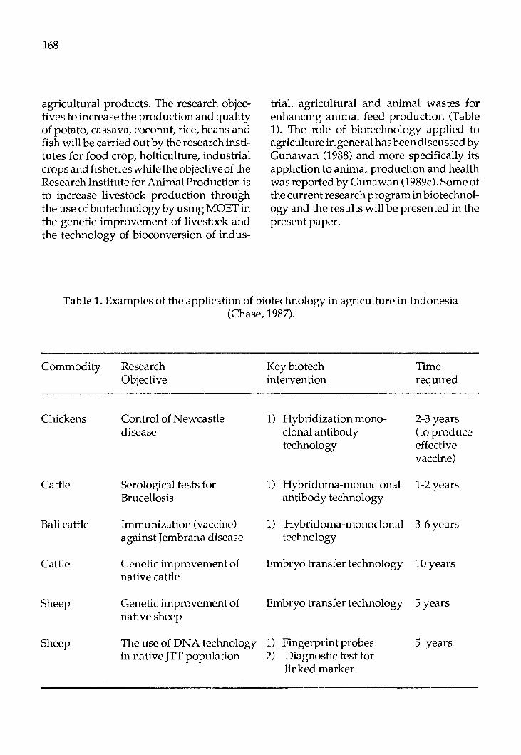
168
agricultural products. The research objectives to increase the production and quality of potato, cassava, coconut, rice, beans and fish will be carried out by the research institutes for food crop, holticulture, industrial crops and fisheries while the objective of the Research Institute for Animal Production is to increase livestock production through the use of biotechnology by using MOET in the genetic improvement of livestock and the technology of bioconversion of indus-
trial, agricultural and animal wastes for enhancing animal feed production (Table 1). The role of biotechnology applied to agriculture in general has been discussed by Gunawan (1988) and more specifically its appliction to animal production and health was reported by Gunawan (1989c). Some of the current research program in biotechnology and the results will be presented in the present paper.
Table 1. Examples of the application of biotechnology in agriculture in Indonesia (Chase, 1987).
Commodity Research Keybiotech Time Objective intervention required
Chickens Control of Newcastle 1) Hybridization mono- 2-3 years disease clonal antibody (to produce
technology effective vaccine)
Cattle Serological tests for 1) Hybridoma-monoclonal 1-2 years Brucellosis antibody technology
Bali cattle Immunization (vaccine) 1) Hybridoma-monoclonal 3-6years against J embrana disease technology
Cattle Genetic improvement of Embryo transfer technology 10years native cattle
Sheep Genetic improvement of Embryo transfer technology 5 years native sheep
Sheep The use of DNA technology 1) Fingerprint probes 5 years in native JTT population 2) Diagnostic test for
linked marker

Present Status and Future Plans
A. Nucleus herd improvement
1. Present genetic improvementprogrames in each class of livestock using conventional quantitative genetic method. In dairy cattle and buffalo, efforts have
been stressed mainly to improve reproductive efficiency. The results indicate that in buffaloes the precentage of heat has been increased from 50% to about 70% with the treatment of hormone PG2a 2 cc. However the percentage of pregnancy using the AI technique is still very low, less than 30%. Further research efforts are being pursued to overcome the problem of low fertility in buffaloes. The technique of frozen semen for cattle and buffaloes has been developed at RIAP and has been used by Artificial Insemination Centers and farmers. A crossbreeding program between Swamp and Murrah buffaloes has been carried out since the last three years to improve growth rate and reproductive traits. The crossbred buffaloes have about 30% higher body weight at 2 years of age over the purebreeds. The data on the reproduction traits are being collected.
A long term sheep breeding program was started in 1980 in collaboration with "Small Ruminant Research Support Program (SR-CRSP)" from the USA. The breeding objective is to increase the reproductive performance(littersize)oftheJavaneseThin Tail sheep (JTT). Ovulation rate has been used as the selection criteria by countirg the "Corpora Lutea" in the ovary of the female sheep by using "laporoscopy". The experiment was outlined by Gunawan (1988a). Research is still in progress and the current results show that the multiple births is suspected to be controlled by a few genes of large effect ("major genes"). The segrega-
169
tion of the litter size from dams of high and low litter size is presented in Table 2. Research is still in progress to progeny testing of the carrier rams.
A long term breeding program to produce a new breed of Indonesian egg laying ducks is also investigated at the Research Institute for Animal Production. The experiment was outlined by Gunawan (1988b).
2. The use of embryo transfer technology for genetic improvement. In Indonesia, the use of embryo transfer
(ET) technique to increase livestock prod uction has just reached the embryonic stage. The technique just introduced in 1984 when a governmental company imported both frozen embryos and expertise from the USA. Some of the results of the use of embryo transfer technique carried out at various research Institutes are presented in Table 3. We have not yet started to use ET technology in the genetic improvementoflocal livestock, but a research proposal and the budget plan has been submitted to the USDA and government of Indoneisa to get approval to apply the technique in the genetic improvement of local sheep and cattle. Meanwhile, our current activities at RIAP is trying to improve the egg recovery rate and success rate of embryo transfer in cattle, to use the technique in buff aloes and to study the technique of in vitro maturation and fertilizc.tion.
B. Application of r-DNA technology for genetic improvement and genetic resistance to diseases in livestock/poultry
We are now in the stage of preparation to use the DNA technology in the improvement of livestock production. The project will be carried out in collaboration with the Australian Centre for International Agricul-
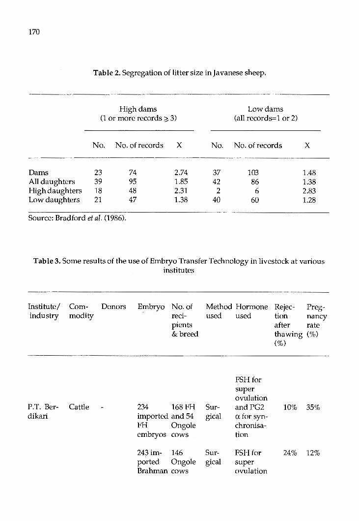
170
Table 2. Segregation of litter size in Javanese sheep.
High dams Low dams (1 or more records ~ 3) (all records= 1 or 2)
No. No. ofrecords X No. No. of records x
Dams 23 74 2.74 37 103 1.48 All daughters 39 95 1.85 42 86 1.38 High daughters 18 48 2.31 2 6 2.83 Low daughters 21 47 1.38 40 60 1.28
Source: Bradford et al. (1986).
Table 3. Some results of the use of Embryo Transfer Technology in livestock at various institutes
Institute/ industry
P.T. Ber-dikari
Commodity
Cattle
Donors Embryo No. of recipients & breed
234 168FH imported and 54 FH Ongole embryos cows
243 im- 146 ported Ongole Brahman cows
Method Hormone used used
FSHfor super ovulation
Sur- andPG2 gical a for syn-
chronisa-ti on
Sur- FSHfor gical super
ovulation
Rejec- Preg-tion nancy after rate thawing (%) (%)
10% 35%
24% 12%
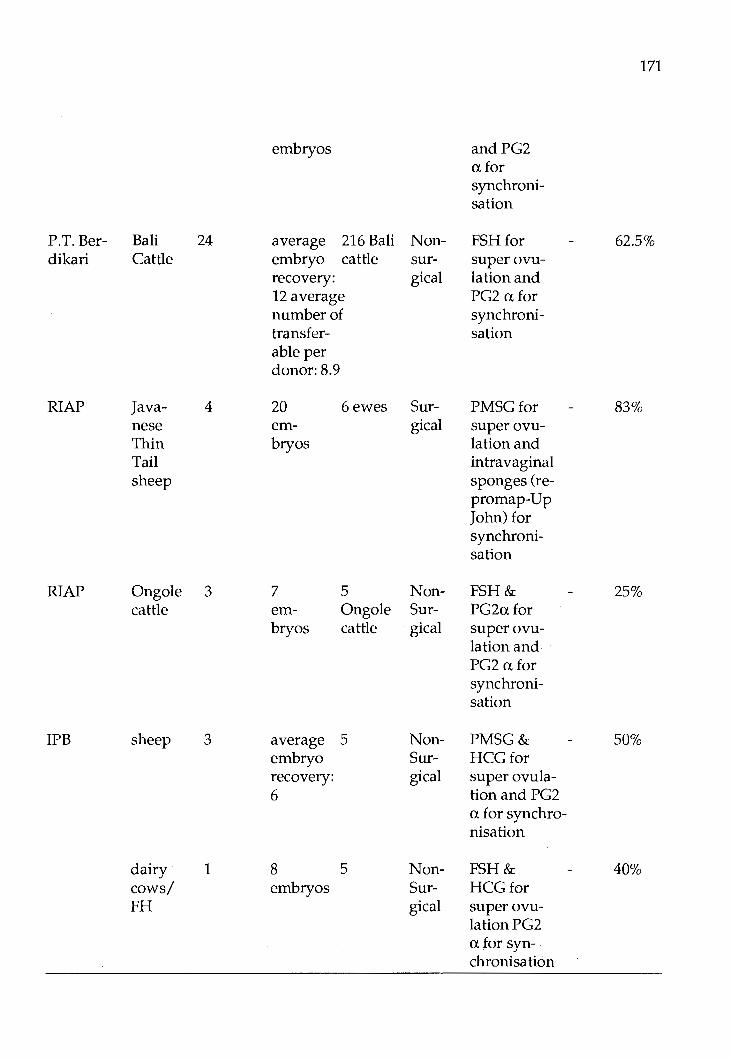
171
embryos andPG2 afor synchroni-sation
P.T. Ber- Bali 24 average 216 Bali Non- FSHfor 62.5% dikari Cattle embryo cattle sur- super ovu-
recovery: gical lationand 12 average PG2 a for number of synchroni-transfer- sation able per donor: 8.9
RIAP Java- 4 20 6ewes Sur- PMSGfor 83% nese em- gical super ovu-Thin bryos lation and Tail intra vaginal sheep sponges (re-
promap-Up John) for synchroni-sation
RIAP Ongole 3 7 5 Non- FSH& 25% cattle em- Ongole Sur- PG2afor
bryos cattle gical superovu-lation and PG2 afor synchroni-sation
IPB sheep 3 average 5 Non- PMSG& 50% embryo Sur- HCGfor recovery: gical super ovula-6 tionand PG2
a for synchro-nisation
dairy 1 8 5 Non- FSH& 40% cows/ embryos Sur- HCGfor PH gical super ovu-
lationPG2 afor syn-chronisation

172
ture Research. For a start, we are going to use our genetic materials from a selection line of JTT sheep. It is mentioned in the previous paragraph that there is a single gene responsible for ovulation rate/ lambing rate segregating in the Priangan QTT) sheep. Pure bred or crossbred family materials (2 or 3 generations) will be collected as long as normal, carrier or doble homozygotes can be distinguished. Some large half sib families e.g. 25-50 (female) genotyped progeny would be sufficient. A test will be conducted for linkage between RFLP (Restriction Fragment Length Polymorphism), defined by a number of candidate genes e.g. FSH, inhibin, gonadotrophins as well as use fingerprint probes to detect closedly linked VNTR markers. This approach was used in Booroola study in Australia (Hetzel, personal communication). A program would be as follows:
Year 1 - Post-doc from RIAPB to work at CSIRO Rockhampton.
- Equip lab at RIAP. - Start collecting family materials
from JTT sheep population (blood to be frozen).
Year 2 - Post-doc returns and starts a lab (with assistance from CSIRO staff).
- Continue to collect family materials.
- Test candidate genes ( 6) on family materials.
- Set up fingerprinting methods. Year 3 - Test fingerprint (VNTR) mark
ers on family materials plus any new candidate genes.
- Design simple diagnostic test for linked marker.
In a poultry breeding farm, they have begun a program of blood typing from the B-complextobeabletoselectbreedingstocks
with known disease resistant, characteristics. They can therefore design matings to results in the healthiest progeny possible. Theyarealsocurrentlytestingabloodanalysis procedure that will permit to monitor for elevated stress levels in the breeders. By detecting and controlling unacceptable levels of stress in chickens, it is possible to protect the immunity to disease that they possess. They have also started to develop and to use biotechnology for improved diagnosis and immune monitoring to be able to evaluate and monitor the chickens with a greater sensitivity than was previously possible.
C. Immunodiagnostics and vaccines
Using conventional biotechnology, the Research Institute for Veterinary Science has already produced a range of experimental vaccines and diagnostic reagents. The products currently available are: (i) diagnostic reagents such as Ros Bengal antigen, Milk Ring test antigen, Salmonella pullorum antigen, Mycoplasma gallisepticum antigen, Haemophilus paragallinarum antigen, Salmonella typing antisera (group and type), Clostridial an tisera (FITC conjugated) and Brucella abortus ELISA diagnostic reagents. (ii) Vaccines such as Neonatal colibacillosis, Swine brucellosis (Brucella swins), Fowl cholera (Pasteurella multocide) and New Castle Diseases vaccines. They have also started to use modern biotechnology to employ methods in immuno chemistry and molecular biology to produce a range of products such as monoclonal antibodies to provide highly specific diagnostic reagents and conjugates, DNA probes, protein gene products and recombinant vaccines. They are also trying to introduce genes into the germ cells of fish or animals to promote growth rates or enhance disease resistance.
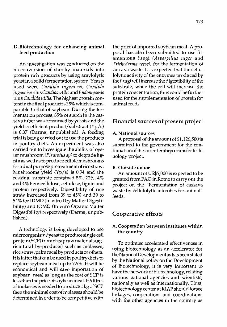
D. Biotechnology for enhancing animal feed production
An investigation was conducted on the bioconversion of starchy materials into protein rich products by using amylolytic yeast in a solid fermentation system. Yeasts used were Candida ingeniosa, Candida ingenoisa plus Candida utilis and E ndomycosis plus Candida utilis. The highest protein contentin the final product is 35 % which is comparable to that of soybean. During the fermentation process, 85% of starch in the cassava tuber was consumed by yeasts and the yield coefficient product/substract (Yp/s) is 0.37 (Darma, unpublished). A feeding trial is being carried out to use the products in poultry diets. An experiment was also carried out to investigate the ability of oyster mushroom (Pleurotus sp) to degrade lignin~ as well as to produce edible mushrooms for a dual purpose pretreatment of rice straw. Mushrooms yield (Yp/s) is 0.94 and the residual substrate contained 5%, 22%, 4% and 4% hemicellulose, cellulose, lignin and protein respectively. Digestibility of rice straw increased from 39 to 45% and 39 to 54% for IDMD (In vitro Dry Matter Digestibility) and IOMD (In vitro Organic Matter Digestibility) respectively (Darma, unpublished).
A technology is being developed to use microorganism/ yeast to produce single cell protein (SCP) from cheap raw materials (agricultural by-products) such as molasses, rice straw, palm meal by products or others. It is latter that can be used in poultry diets to replace soybean meal up to 7.5%. It will be economical and will save importation of soybean meal as long as the cost of SCP is less than the price of soybean meal. If 6 litres of molasses is needed to produce 1 kg of SCP then the minimal cost of molasses should be determined in order to be competitive with
173
the price of imported soybean meal. A proposal has also been submitted to use filamentous fungi (Aspergillus niger and Trichoderma reesei) for the fermentation of cassava waste. It is expected that the cellulolytic activity of the enzymes produced by the fungi will increase the digestibility of the substrate, while the cell will increase the protein concentration, thus could be further used for the supplementation of protein for animal feeds.
Financial sources of present project
A. National sources A proposal of the amount of $1, 126 ,500 is
submitted to the government for the continuation of the curentembryo transfer technology project.
B. Outside donor An amount of US$5 ,000 is expected to be
granted from FAO in Rome to carry out the project on the "Fermentation of cassava waste by cellulolytic microbes for animal" feeds.
Cooperative effrots
A. Cooperation between institutes within the country
To optimise accelerated effectiveness in using biotechnology as an accelerator for the National Development as has been stated by the National policy on the Development of Biotechnology, it is very important to have the network of biotechnology, relating various national agencies and scientists, nationally as well as internationally. Thus, biotechnology center at RIAP should forsee linkages, cooperations and coordinations with the other agencies in the country as

174
well as abroad. The essential cooperation should be effected with: • The Indonesian Institute for Sciences
(LIP). CD Inter University Center of Biotechnology
of Institute Technology Bandung (ITB). CD Inter University Center of Biotechnology
of Institute for Agriculture Bogor (IPB). • Inter University Center of Biotechnology
of The University Gadjahmada Yogyakarta (UGM).
CD Biotrop of SEAMEO, Bogor. • etc.
B. International cooperation CD Australian Centre for International
Agricultural Research (ACIAR), Australia.
•PAO. 111 CRSP(CollaborativeResearchSupport
Program), USA.
Acknowledgements
lwish to thank Dr. Jasiorowski, the Director of the Division of Animal Production and Health, F AO in Rome for inviting me to attend this Network meeting of Biotechnology. I also thank to Mr. Ibrahim Abu Zahab, PAO Representative in Indonesia for helping the arrangements of tickets and visas. I thank to Prof. Mukherjee for advice and providing the format of the present national position paper.
References
Bradford, G.E., Subandriyo and Iniguez, L.C. 1986. Breeding stategies for small ruminants in integrated crop-livestock production systems. Proceedings of a Workshop on Small Ruminant Production Systems in
South and Southeast Asia. Bogor, Indonesia, 6-10 October 1986, pp. 318-331. Chase, S.S. 1987. Strengthening biotechnology research in agriculture in Indonesia. A consultancy report. Submitted to the Agency for Agricultural Research and Development (AARD) Government of Indonesia.
Gunawan, B. 1987. Prospect of biotechnology in the development of agriculture, animal production and human welfare in Indonesia. Proceedings Seminar Bioteknologi Pertanian, Pusat Antar Universitas (PAU) Bioteknologi, Institut Pertanian Bogor, 21 December 1987.
Gunawan, B. 1988a. Teknologi Pemuliabiakan Domba Lokal Indonesia. Proceedings Seminar Expor Temak Potong, Hotel Horizon, Jakarta, 2-3September1988.
Gunawan, B. 1988b. Teknologi Pemuliabiakan Itik Petelur Indoneisa. Seminar Nasional Peternakan & Forum Peternak "U ggas dan Aneka Temak" II, Balai Penelitian Temak, Ciawi-Bogor, 18-20 July 1988.
Gunawan, B. 1988c. Biotechnology Applied to Animal Production and Helath in Indonesia. Regional Workshop on Biotechnology in Animal Production and Health in Asia. Bangkok, Thailand, 17-21October1988.

'Proc. FAO/CAAS Workshop on Biotechnology in Animal Production and Health in Asia and Latin America, Beijing, Oct 9-13, 1989, pp 175 -192
Biotechnology in Animal Production and Health in Malaysia
A. Latif Ibrahim University Pertanian Malaysia Serdang, Selangor, Malaysia
Introduction
In Malaysia there are at least 3 major areas of research for future scientific exploration as to the role of biotechnology in animal health and production. These are genetics and reproduction, nutrition and animal health and disease control.
Reproduction and Genetics. One of the most serious problems in ruminant prod uction is low reproductive efficiency or loss which occur between conception and weaning. Detailed studies on infertility and sterility in ruminants are being conducted. Increased fertility has been achieved through the application of embryo transfer (ET) technique. This technique when combined with recombinant DNA, would enhance the genetic ability and adaptation of the embryo. For example with adaptation, the introduction of desirable genes would permit animals to resist certain environemntal challenges such as specific diseases or to be tolerant of harsher climatic factors. Additions of recombinant DNA to ET technique would yield many dairy bulls of higher breeding value for milk. Adequate mapping of the genome of domestic animals remains a necessary first step before certain inherent problem of animal production can be tackled by recombinant DNA. Recombinant DNA is only fruitful if complete informa-
tion on the manipulated genome is available.
Monoclonal antibody technology also provides a much needed mechanism for improving methods of selective breeding. Monoclonal antibody technology has shown that the potential is much larger and that it can serve as a mechanism for identifying not only gene system that influence susceptibility to infectious disease but also those that control the whereabouts of desirable performance traits.
Nutrition. Most feeding systems in Malaysia incorporate a larger proportion of fibrous material and by products from the agricultural industry. Unfortunately the feeding value of agrobased byproducts are relatively low and as such large amount of thebyproductarelefttowaste. Theinability of the ruminant to utilize the agro byproq ucts is primarily a problem of communication and solubilization by the rumen organism of the cell wall components of padi sfraw, palm pressed fibre etc. If rumen organism can be modified to increase cellulase production and/ or increase ligno-cellulose breakdown, then both the extent and rate of digestion in the rumen would be stimulated. This would allow higher feed intake and therefore higher animal production. Recombinant DNA has been ~pplied to
176
produce bacteria with altered abilities to carry a number of functions. It is possible to use such a technique to modify the rumen organism to increase digestibility and the rate of digestion of agro by-products. A small increase ofdigestibility of agro byproducts through the introduction of organism altered by these technique will have enormous benefit for the Ii vestock industry.
Health. Biotechnology has already made some contribution in the field of animal health and this is in the areas of vaccine production and diagnostic methods. The introduction of genetic engineering will open the possibilities of producing effective vaccine against livestock diseases. Attempts are now being made to develop new vaccines for diseases such as Newcastle disease, fowlpox, 'infectious bronchitis and haemorrhagic septicemia by the applicationi of genetic engineering techniques. The development of hybridoma technique has also increased the possibility of defining the antigenic determination on the surface of the virus which are of primary importance for the induction of immunity against disease. Monoclonal antibodies will be used for the purification of viral vaccine by the immune adsorption technique. The application of monoclonal antibodies has provided means to detect antigenic difference masked or undetectable with polyclonal antisera. Other biotechnological techniques that can be applied to differentiate virus isolates include comparison of viral structural protein by polyacryalmide gel electrophoresis and comparing viral nucleic acid by electrophoresis after endonucelase digestion of genome. We can also expect ~ethods s~ch as sequencing the base of nucleic and ammo acids that will be applied in the future for diagnostic works.
Major Research Areas for the present and future 1. Animal Reproduction
i. Embryo transfer for increased female reproductive rates.
ii. Embryo manipulation including preservation, sexing and splitting.
iii. Genetic modification of embryo through embryo fusions, nuclear transfer and direct gene transfer.
iv. Gemate manipulation including gemate preservation, sexing and in vitro fertilization.
2. Animal genetics i. Establishment of genomic library of
local farm animals through chromosomes and gene mapping.
ii. Identification and transfer of germ plasma associated with resistance to disease and health problem.
iii. Identification of genetics factors related to reproduction.
iv. Determination of genetic markers for identification of different breeds of farm animals.
3. Nutrition i. Screening and characterization of
rumen microbes for cellulolytic activities.
ii. Genetic modification of rumen microbes for enhanced lignocellulose utilization.
4. Animal Health and Disease Control i. Vaccinedevelopmentthroughmodi
fied pathogen or clones immunizing antigen.
ii. Hybridoma derived monoclonal antibodies for application in diagnostics, vaccination and separation technology.
iii. Nucleic acid probes for use in diagnosis of infectous diseases.

iv. Genomic analysis of animal viruses.
A. Nucleus Herd Improvement Using ET Technology
1. Present genetic improvement programmes . In the large ruminant sector, few genetic improvement programmes are in existence. Some effort has been made during the past 30years through a national artificial insemination scheme to improve the productivity of the local dairy cows. Progeny. testing programmes of selected bulls have been undertaken by Central Animal Husbandry Research Station, Kluang. Large scale small ruminant breeding and selection programmes have been undertaken by Rubber Research Institute of Malaysia (sheep) and University of Malaya (goats). In the sheep programme, crossbred progeny of various grades (Local x Dorset Horn) are distributed as multipliers to small sheep programmes; crossbreeding of local does with German Fawn males have produced Fl offspring. Subsequent production of F2, F3 and F4 offspring with selection for body weight in males and litter size in females has resulted in the formation of a new breed - Jermasia. Crossbred progeny have been distributed to small holders during last 8 years (Mukherjee-personal communication).
~· Embryo transfer technology for genetic improvement
There are at leasttwo major constraints to the. application of embryo transfer for ge~ehc improvement in Malaysia. Firstly there 1s no scheme to identify superior genetic females (cattle, goats, buffalo and sheep). Secondly, Malaysia has yet to utilise artificial insemination technology effectively for genetic improvement of livestock. Thus the prospects for embryo transfer for genetic
177
improvement of livestock in Malaysia is far from encouraging.
Presently, scientists in the Division of Veterinary Services (D VS), Malaysian Agriculture Research and Development Institute and Universiti Pertanian Malaysia are ad~pting the embryo transfer technology to smt the temperate x indigenous crosses.
a. Donor Selection Since the emphasis on embryo transfer is
experimental, the necessity for genetic selection of the donor does not exist. At the DVS, Sahiwal-Friesian cows are superovulated in order to increase the number of these crsosbreds whereas at UPM, the emphasis is on conservation of indigenous cattle (Kedah-Kelantan and Local Indian Dairy) through the development of an embryo bank.
i. Superovulation Both Pregnant Mare Serum Gonadotropin
(PMSG) and FollicularStimulating Hormone (FSH-Porcine) have been employed for superovulatingcattle, buffalo (Sharifuddin, Wand Jainuden, M.R. [1988]. Embryo Collection in the Swamp Buffalo. Proc. 11th Int. Congr. Anim. Reprod. of AI, Dublin, Ireland) goats and sheep (Rosnina Yusoff [1989]. Superovulation and egg recovery in goats. M.S. Thesis, UPM, Serdang.) Besides being expensive, response in terms of ova shed with either preparation has been highly variable.
ii. Methods used: In all species gonadotropins are admini
stered between Day 10to12 of the oestrous cycle and followed by a luteal dose of Prostaglandin F2 alpha 48 hours later.
iii. Local Production of Hormones Production of hormones for superovula-
178
tion at the present state of development would be too costly.
iv. Purification of hormones No effort has been made to produce gonadotropins in Malaysia.
v. Effect of Repeated Superovulation The effects of repeated gonadotropin therapy on the ovulatory response has not been investigated.
b. Mating Both artificial insemination and natural mating are employed in cattle and buffalo whereas only natural mating has been practised in goat and sheep.
c. Collection of Ova The nonsurgical technique of collecting ova in cattle and buffalo involves flushing of the uterine horns on day 5-7 of the cycle with a 2- or 3-way Foley catheter inserted through the cervix. In goat and sheep, ova are collected by surgical techniques.
The efficiency of ova collection with the nonsurgical technique has ranged from 0-75% for cattle and 0-5% in the buffalo. With surgical technique, collection efficiency in goat and sheep has ranged from 25 to 95%.
d. Examination and evaluation of ova and embryos
Immediately following collection, the flushings are examined under a stereomicroscope. Ova are examined for cleavage and the embryos are evaluated for stage of development in relation to the day of the cycle and morphology of the blastomeres.
e. Transfer and/ or freezing of embryos A few calves and goats have been born
following transfer of fresh as well as frozen embryos to synchronized recipients. The transfer of embryos in cattle has been similar to the artificial insemination technique whereas in goats it has been by surgery or by laparoscopy.
In all institutions, facilities are available for freezing embryos. At UPM, embryos of indigenous cattle and goat are being frozen as part of a long-term plan to conserve the gene pool which is rapidly being depleted through crossbreeding.
Future Research Programme The major thrust in Malaysia would be to
utilise embryo transfer technology in selected herds in the ruminant sector. Because the technology should not only be economical but also should be realistic, there is a growing interest in the technology associated with manipulation of oocytes for in vitro maturation and fertilization of oocytes. Current techniques using hormone stimulation and embryo collection are more expensive. The in vitro fertilization (IVF) technique could provide a cheap source of embryos for commercial transfer in the future particularly because the vast number of oocytes present in the ovaries could be tapped from slaughtered animals or by laparoscopy of the live animal. Further refinement of this technique will provide a more acceptable method for mass production of embryos in the future. The IVF technique would be studied in cattle and buffalo as the current methods of embryo collection are not only expensive but also impractical.
While embryo transfer technology is being exploited in genetic improvement of cattle, its value in tropical countries would be to conserve valuable gene pools of tropically adapted cattle for future genetic programs. This is possible through long term preserva-
tion of embryos in liquid nitrogen. Such embryo banks could serve for the exchange of genetic resources in Asia. Cryopreservation of embryos of indigenous and exotic breeds of livestock would be undertaken to develop a national gene bank of frozen embryos. These embryos will be utilised to study disease resistance of offspring born from the development of exotic embryos in indigenous breeds of cattle, sheep and goats.
Studies will also be undertaken to bisect embryos for the induction of twins and chimeras for research in genetics.
B. Application of RDNA Technology for Genetic Improvement And Genetic Resistance to Disease in Livestock/Poultry
i) Gene mapping/DNA polymorphism
a) RFLP of water buffaloes It is our aim to utilise the DNA restriction
fragment length polymorphism (RFLP) and the biochemical isoenzyme polymorphisms to study the genetic similarities and differences in buffaloes. Motochondrial DNA was chosen instead of the nuclear genome because of the following reasons: i) Itis a much smaller moelcule (16 kile bp) than nuclear genome. ii) The rate of mutation was estimated to be 7-10 times faster than nuclear genome, iii) It is maternally inhrited and hence it is not reshuffled during fertilisation, making it an ideal tool for the study of phylogeny and evolution.
Biochemical polymorphism of buffaloes was studied using polyacrylamide gel electrophoresis. Serum proteins were analysed using gradient gel andalso SDS-P AGE. Serum leucine aminopeptidase (LAP) and
179
amylase will be analysed. Attempts have been made to find the optimal electrophoretic sy'stems for the LAP and amylase. The LAP system has been successfully obtained but the amylase did not stain up satisfactorily. Other Red Blood cell enzymes screened including the following: GLO I, PGP, IMP, pyrophosphatase,redcellacid phosphatase, catalase ceruloplasmin, peptidase D, ITP and peroxidase. Prelimary results showed that the transferring in the serum protein has shown different patterns between the river and the swamp buffaloes. The genetic basis and the number of alleles /loci involved in each enzymes system will be analysed and mapped.
Samples were also planned to be collected from a controlled family used for breeding experiments.
There were altogether 109 blood samples collected from various places in South East Asia. All blood samples were processed through a tedious procedure carried out at 4"C. Blood was separated into different fractions. The red cell and the serum were used forisoenzyme analysis. The white cells were
. stored at -50°C for the analysis of nuclear DNA in the future. Mitochondrial DNA was e~tracted from the mitochondrial fraction separated from the serum.
For the DNA restriction fragment length polymorphism analysis, 22 blood samples from the river buffaloes, 30 blood samples from the swamp buffaloes, 20 blood samples from the Fl hybrids, 25 blood samples from the F2 progenies, 28 blood samples from the backcross3 I 4 swamp buffaloes and 13 blood samples collected from the swamp buffaloes in Trengganu were extracted according to the Hirt Extraction method. Proteinase K, SDS, RNAase, salmon sperm DNA were used during the DNA extraction. Agarose

180
electrophoresis were performed using Lamda DNA Hindlll digest as standard markers. After electrophoresis, the gels were stained in ethidium bromide solution. It was then Southern transferred to a S&S nylon membrane, baked at 80°C for three hours and kept at room temperature. At this stage it can be kept for long period. Plans have been made to collect the fresh liver from the swamp buffaloes slaughtered in the Department of Animal Sciences, Universiti Pertanian Malaysia, to be used as the DNA probes.
Most empahsis will be placed on gene mapping. It is now accepted that the most efficient way to obtain the information on the number and distribution of polymorphic marker genes is to make use of DNA restricted fragment length polymorphisms (RFLP). These have been detected at high frequencies in man. A linkage map of the human genome, based on the pattern of inheritance of about 400 marker loci, mostly RFLPs has recently been established (DonisKaller et al., 1987).
A special type of RFLP, which is termed variable number of tandem repat polymorphisms or VNTR can be detected when repeated motifs of hypervariable minisatellite sequences (Jeffrey et al., 1985)areused to probe genome DNA. Because of the great value of VNTR for genetic analysis and manipulation of polygemic traits, it is desired that a major effect be made to produce a buffalo chromosome map based on VNTR markers. Since these restriction patterns are specific for an individual they are called 'DNA fingerprinting or DNA bar codes'.
. , ?pp;i;a.tic cell genetic approach (In situ hybfjqi,~a,pon) by use of inactivated sendai v!rt;t~, ~r. polychleneglycol will also apply. · Tµis technique is. a straightforward rnap:c.
ping technique involving the hybridization of radiolabelled DNA probes to fix metaphase chromosomes and the subsequent visualization of the signal as silver grain after auto radiography.
Thus, using RFLP or VNTR and In situ cell hybridization, we will achieve the goal to make up a detailed buffalo gene map consisting of a few highly informative marker loci per chromosome.
Once this goal is achieved, cloning of a particular gene will be made for various DNA fragments which suppose to be responsible to disease resistance or composition of milk. Before insertion of these probes, the function of isolated genes will be studied. MHC (major histocompatibility complex) and resistance to some disease and parasites will be given priority. Performance of those transgenic buffaloes will also be investigated.
RFLP work in chickens is also in progress at the University of Malaya in order togenetically distinguish various lines of egg type chickens in which major genes have been incorporated (sex-linked dwarf, autosomal naked neck, frizzle and curly). IGF probes are being used for DNA hybridization with the southern blots of restricted fragments of DNA. Similar work has been planned in future for goats and fish (M ukherjee-personal communication).
C. Immunodiagnostics and Vaccines
1. Eli~a Test for Diagnostic Purpose
1~)', Laboratory test The enzyme linked irnmunosorbent assay (ELISA) has been developed to detect

both antibodies and antigen for a number of diseases. An ELISA is also being developed for rapidly screening for the binding of a bank of monoclonal antibodies to a range of Newcastle disease virus isolates.
i). Newcastle Disease In 1986, a joint research project on the
establishment of improved methods for the diagnosis and control of Ii vestock disease in South East Asia using Enzyme Linked Immunosorbent Assay (ELISA) was undertaken by the Regional Veterinary Laboratory Benalla, Australia, Research Institute for Animal Diseases, Bogor, Indonesia and Universiti Pertanian Malaysia, Serdang, Malaysia. The project is supported by a grant from the Australian Centre for International Agriculture Research (ACIAR). The objectives of the projects are:
i. to initiate and carry out research for the development of ELISA system application to a number of production limiting livestock diseases in Malaysia and Indonesia. The disease for which ELISA are being developed are:
a) brucellosis in cattle, sheep, goat and pigs
b) anthrax in cattle, buffalo, sheep and goats
c) leptospirosis in cattle, buffalo and pigs d) haemorrhagic septicemia in cattle and
buffalo e) Newcastle disease in poultry
ii. to provide a collaborative framework for research and development of ELISA for the control of diseases affecting animal prod uction in Malaysia, Indonesia and other countries in South East Asia.
For Malaysia, indirect ELISA technique for detecting IgG class specific antibodies to
181
Newcastle disease virus (NDV) in chickens was developed using purified antigen. The optimal conditions of antigen concentration, conjugate dilution, substrate concentration, reaction times and sera dilutions were established using the signal/ noise (SI N) ratio as the determining criterion. Using regression analysis, a standard curve was constructed from the transformed values of absorbances and the corresponding titres. The titres of all test sera at a single working dilution were predicted using the standard curve. The immune response of chickens following vaccination with NDV were monitored using ELISA and the haemagglutination inhibition (HI) test. There was close correlation between the HI and the ELISA titres, but ELISA titres were considerably higher.
ii. Trypanosomiasis in Malaysia Trypanosoma evansi, causative organism
of the disease known as "surra" was first reported in Malaysia in 1903. The disease is of major importance in Indonesia; Thailand, Vietnam and China but there is little recent information on its incidence and economic importanceinMalaysia.Althoughoutbreaks of disease have been reported in horses apd buffalo, detection of the parasite is difficult and it is possible that its presence often goes undetected. Trypanosomiasis cause infertility and abortion and sometimes death. L0sses in productivity due to trypanosomiasis could therefore affect the income of small holder farmers. It is therefore essential that more sensitive diagnostic techniques are employed to determine the extent of effect of this parasite in livestock in Malaysia.
Currently, a survey of trypanosomiasis is being conducted to determine the prevalence of trypanosomal antibodies and circulating antigen in serum from buffalo and cattle from different areas in Malaysia. The

182
test employed is the ELISA (enzyme-linked immunosorbent assay) and information obtained from the survey should identify a) the prevalence of infection b) areas in which trypanosomiasis occurred and c) the species of animal most affected.
iii) Canine Dirofilariasis Dirofilaria immitis is a filarial parasite
which has been reported to infect a wide range of mammalian hosts. The dog acts as a main reservoir of infection as well as being the main host. The canine host can develop polysystemic disorders with cardiopulmonary signs occurring most frequently. Congestive heart failure eventually develops. The diagnostic tests for canine dirofilariasis rely upon the detection of circulating microfilariae. Thus, infected dogs without circulating microfilariae are a problem for diagnosis. In a survey of 200 urban stray dogs, the period prevalence was 42% based on necropsy.
· . An ELISA using antigen prepared from live adult worms was attempted to detect antibodies against D. immitis in sera of these dogs. The accuracy of the assay was only 53%. However in a commercial kit for the detection of antigens of D. immitis, using the same sera samples the accuracy was also quite low, 56%.
lb) Field Test
(l)The use of ELISA technique for diagnosis and seroepidemiological studies on bovine brucellosis
Since brucellosis was first recognised in Malaysia in 1950, numerous serological surveys have been conducted to determine the prevalence rate of the disease. An average Qf 32% reactor are for government cattle ~rrmsrand 16% reactor rate:for smc,lllholqer herq§ Ji;t, ·the state of Perak has1 been re-
corded. The prevalence rates in other states of Peninsular Malaysia are yet to be determined.
(2)Monoclonal Antibody Production Realising the importance of monoclonal
antibodies, the National Biotechnology Programme has provided funds to research institutes to acquire the capability to prepare and use monoclonal antibodies as diagnostic reagents.
i) Applications of monoclonals for differentiation of Newcastle Disease virus strains.
In this study a panel of 39 mabs to the NDV-V 4 strain were tested using ELISA to investigate the reactivity of these mabs with seven reference strains and a local field isolate.
ii) Production of monoclonal antibodies agaisnt virulent markers of Pseudomonas Pseudomallei
Objective: Prevention and control of melioidosis in domestic animals through the development of a rapid sensitive and accurate diagnostic technique.
Aim: To produce monoclonal antibodies agaisnt known yirulent factors of P. pseudomallei particularly the lethal exotoxin, hemolysin and pili and to develope an enzyme .linked immunoabsorbent assay (ELISA) for diagnosis.
Pseudomonas pseudomallei is the causative organism of m~lioidosis an usually lethal disease of man and animals in S.KA and tropical Australia. In Malaysia, meliodosis is endemic amongst pigs, goats and sheep. This disease if unchecked will hinder the current effort to expand sheep and goat husbandry in this country. P. pseudomallei is known toinyade various organs in the host
mimicking a wide spectrum of disease, thus hindering accurate diagnosis of melioidosis. Currently diagnosis for P. pseudomallei infection is dependent on bacteriological methods. The serum agglutination test(SAT) is frequently used to detect melioidosis but its reliability has been questioned. In view of this there is an urgent need to introduce an accurate and sensitive test for diagnosis of melioidosis.
2. Ongoing Research
Studies carried out in our laboratories have identified a number of factors, produced by this pathogen which contribute to its virulence. The lethal exotoxin produced by this organism is believed to be important in the pathogenesis of melioidosis. The exotoxin has been shown to lethal to animals and toxic to cells in culture. Animals immunized against the exotoxin were protection in experimented animals when challenged either with Ii ve P. pseudomallei or the exotoxin itself. This has prompted several studies in our laboratories to develop an efficacious toxoid vaccine for melioidosis. The exotoxin has been successfully purified in our laboratory and shown to consist of two subunits MW 37000 and 16000 dalton. We have also been able to prepare homogenous preparations of the heat-stable hemolysin of P. pseudomallei. Studies have been extended to produce monoclonal antibodies against the purified lethal exotoxin and the hemolysin of P. pseudomallei. Positive hybridomas to the exotoxin did not show reactivity with hemolysin or the cell wall fraction of the pathogen.
The monoclonals showed specificity towards the minor subunit of the exotoxin. Our results also demonstrate that exotoxins purified from isolates of P. pseudomallei from
183
human, goats and deer are antigenically similar. The monoclonal antibody against the exotoxin thus obtained will be useful in the detection of the presence of exotoxin and provides a means towards diagnosis of melioidosis. Current efforts involving large scale production of the monoclonals in ascites fluid is in progress. In addition to the above the availability of purified exotoxin has enable us to screen serum samples from sheep for the presence of antitoxin antibody using the ELISA. Results obtained so far has shown that there is positive correlation with the serumagglutination test for melioidosis. In fact the ELISA appears to be more sensitive than the SAT since antitoxin antibodies could be detected in many samples found to be negative by the SAT. We are currently actively calloborating with the Veterinary Research Institute of Malaysia to screen more serum samples from melioidosis infected stock to ascertain the validity of the ELISA.
3. Vaccine Newcastle Disease Poultry have a unique role in the live
stock sector of the countries of the AsiaPacific region. Itis the only livestock species that is widely accepted by people from a wide variety of cul tu res and background. It is estimated that 35% of the world poultry population comes from the Asia Pacific region. Three systems of poultry production are usually being practised, the intensified commercial system, the smallholder 'rural' system which normally require small capital inputs and the village or backyard system.
In many developing countries the village chicken is sometimes the dominant form of poultry raising. The rural households keep a few chickens of local breeds that survive by scavangingforfood and water. They are sometimes supplied with simple housing
184
and provided with supplemental food. The poultry are maintained virtually without cost, and they return some meat and eggs and some income because of the good prices paid for village chickens and eggs in the market place. The village poultry have attracted the attention of many who are concerned with rural welfare in developing countries. The chickens are poorly productive mainly because diseases ca use enormous losses. One of the most important disease is Newcastle disease. The disease in commercial poultry is usually adequately controlled by vaccination. However, the use of continental Newcastle disease vaccination in village poultry has been an unsatisfactory process.
The resources of the village chicken will be fully utilized until Newcastle disease is controlled. Vaccination is the only safeguard agaisnt endemic Newcastle but the present system of using commercial vaccine to vaccinate the chickens is not practicable. The chicken which are multi age is scattered over the villages. The birds are difficult to catch for formal vaccination. Moreover, the vaccine is heat-stable and complex cold chains are required to link the vaccine producers and users. A new approach is required to control Newcastle disease in the village poultry.
A research project was then undertaken to develop a new simple, cheap and effective Newcastle disease vaccine for the village chicken. The main objective of the project was to produce an oral vaccine which seemed the only feasible way of delivering the vaccine virus onto the village chicken. Our hypothesis was to isolate an immunogenic and heat tolerant Newcastle disease virus. The virus would then be incorporated into chicken feed which would be given to the village chicken. It was assumed that
chicken fed with the vaccine would be protected against Newcastle disease.
The virus selected for the project was the a virulent Australian V 4 strain of Newcastle disease virus. Early work with the V 4-Newcastle disease virus have demonstrated that the V4-NDV induces an adequate antibody response in chickens and spreads readily by contact and that naturally infected chicken and V4 vaccinated chicken resist challenge with the virulent Newcastle disease.
A heat tolerant variant of the V4-Newcastle disease virus was isolated and used as vaccine. The vaccine was prepared by coating the V4 vaccine virus onto food pellets in a laboratory model Uni-Glatt Fluidised Bed Granulator. A nominal dose of vaccine was 10gm of pellets containing 106
fifty percent egg infectious dose (EIDSO) of vaccine virus. Experiments were then undertaken totes t the efficacy of the food pellet vaccine. Studies on broiler chickens raised under laboratory condition showed that two doses of the vaccine were sufficient to protect against the virulent challenge virus which killed all controlled chicken even by contact spread. The experiments were extended to village chicken kept under simulated village conditions at the University. These chickens had housing at night, access of enclosed grassed area during the day and fed scraps with grain supplements. After two exposures to the oral vaccine, they are substantially resistant to challenge. Field trials were then undertaken in 15 villages which were supplied with the vaccine once each month. Some 60% of the village birds resisted artificial challenge and outbreaks of Newcastle disease were not recorded in the villages.
A notable feature of the project in Malay-

sia was the effort dev0ted to transfer of technology before and during the field trials. We realise that the mostimportantphase on the application of any new technology is the successful introduction of result in practise. Results of research conducted at universities and in research institutes generally found their way to the user group through some type of extension education. The transfer of technology was undertaken with the University Centre for Extension and Continuing Education which developed an excellent working relation with the farmers. The programmes were explained carefully in the villages and field days were arranged on campus where farmers oculd vaccinate the chicken. There was an early appreciation of the simplicity of the technology. Villages were then asking to be included in the scheme and some individual owners indicated willingness to pay for the vaccine.
A large scale control programme involving studies on the economic impact of the disease and the benefit of the new technology is in progress in Malaysia.
Vaccination has played a major role in the control of poultry diseases in Malaysia. It is important that the development of veterinary vaccine keeps pace with changing circumstances. Improvement of vaccine can be achieved through the application of our knowledge of immunological functions and molecular biological methods. Recent work on the molecular cloning and nucleotide sequencing of ND virus strains has provided a great understanding of the molecular pathogenecity of NDV strains.
Since 1985 a Malaysian research scientist (Yu so ff et al) has been working on a research project on the molecular biology of NDV. The research involved cloning and sequencing of the L gene of NDV which comprises
185
approximately half of the viral genome and mapping of the antigenic sites of the HN and F glycoproteins which are immunogenic and antibodies to the two proteins have been shown to provide protection against the disease.
The following techniques have been used in the studies:
- cDNA cloning. DNA and RNA sequencing using the Sanger method using 32P and 35S isotopes. colony hybridisations including nick translation of nucleic acid probes using 32P; and dot blots. extraction of DNA and RNA from bacteria and virus. use of computer programs eg. Staden ad Microgenie to analyse nucleic acid and protein sequences. nuclease mapping. end-labelling. restriction enzyme mapping. tissue culture technique involving virus.
- construction of hybridomas for production of monoclonal antibodies.
- purification of monoclonal antibodies and peroxidase labelling.
- competitive binding assays - Western blotting. - ELISA
We are now constructing a genomic library of a local Malaysian velogenic isolate strain VRI with the aim of cloning and sequencing its entire genome. Initially, only the HN gene will be sequenced and compared to the HN gene sequence of the published Texas strain, theonlyvelogenicstrain to be sequenced so far. The vaccine strains S and F will also be studied in this manner, the ultimate goal would be to construct a subunit vaccine against NDV as well as to improve
186
the current diagnostic methods.
4. Technology Development in Animal Health
i) Restriction endonuclease technique of DNA of infectious
bovine rhinotracheitis virus Bovine Herpes Virus I (BHV-1), some
times commonly known as Infectious Bovine Rhinotrachietis virus (IBRv), is a doublestranded (DS) DNA-containing virus belonging to the herpesvirus group. It is associated with a variety of clinical syndromes in cattle but the two major syndromes associated with it are respiratory disease (IBR) and genital disease (infectious pustular vulvo-vaginitis) (IPV). Its occurrence in various clinical forms suggests that strains with differing tissue affinities may exist in the field.
In 1983, IBRv was isolated for the first time from water buffaloes and cattle at the Universiti Pertanian Malaysia (UPM) farm. Following this, comparative studies on the serological and cytopathological properties of buffalo and cattle strains of BHV-1 and respiratory and genital strains of BHV-11 within the two species, were carried out. The initial objective of this project is to compare the DNA fingerprints of Malaysian cattle isolates of IBRv with that of a reference IBRv strain.
ii) DNA extraction and gene cloning-Haemophilus paragallinarum Haemophilus paragallinarum is the causa
tive agent of infectious coryza in chicken. In Malaysia, the disease occurs widely and it has a substantial economic impact on the poultry industry. It causes air sac lesions, morbidity, mortality and drop in egg production. Past research on infectious coryza
has been concentrated on defining the dynamics of the disease and characterizing the clinical and pathological features of the disease. At present, there is very little information on antigenic make-up, biological, biochemical and immunological properties of Haemophilus paragallinarum. Thus, studies concerning the antigenic make-up of the bacteria at molecular level will facilitate the selection of strains for vaccines as well as for diagnostic reagent. In our study, we propose to extract the DNA from Haemophilus paragallinarum and clone the Haemophilus paragallinarum DNA into Escherichia coli in order to construct a genomic library for detailed molecular analysis.
At present, we have been able to extract the DN As from all the three reference strains and endonuclease digestion using Hind III. The restriction fragments were run on agarose gel electrophoresis. We hope to ligate the sized selected restricted fragments into cloning vector (puC 18) digested with Hind III and use the ligated mixtures to transformed competent JM 101 E.coli.
5. Future Research Programme
1. Monoclonal antibody: a) To establish storage protocal for my
eloma NSl, SP0/2 and 3T3. b) To study physical parameters for cul
turing myelomas and hybridomas in fermenter.
2. Cloning of poultry viruses for vaccines.
3. Diagnostics: a) Preparation of nucleic acid probes for
use in diagnosis of infectious disease.

D. Biotechnology for Enhancing Animal Feed Production Bioconversion of Indutrial, Agricultural and Animal Wastes as Animal Feed
Manipulation of rumen microbes
Introduction
Livestock production in Malaysia is characterized by low input levels and low productivity where the major constraint is the shortage of suitable feed materials. Pasture production is not economically competitive compared to rubber, oil palm and cocoa cultivation. As land owners are not keen to convert their land from crops to pastures, other feed sources have to be utilized to support and improve livestock production. The utilization of locally-available agricultural byproducts (such as those from the palm oil and rice milling industries) represents a vast potential resource which offers a good opportunity for increasing livestock production in the country.
The various feed materials ingested by the ruminant animal are fermented by the rumen microbes prior to digestion by the host animal itself. This fermentation results in the formation of volatile fatty acids and microbial cells that constitute the major energy and nitrogen sources respectively made available for the animal's metabolism. The rumen houses a diverse population of microorganisms including bacteria, fungi and protozoa whose roles are intimately related to the nutrition of the ruminant animal, as they all contribute to the overall fermentation process. Therefore, there is a need to acquire baseline data on the diversity and perforamnce of the rumen microbes and to recognize and establish the dynamic
187
interactions by the ruminant livestock between the microbes, feedstuffs and the host animal.
Byproducts from the palm oil industry such as the palm press fibre (PPP) has great potential as a fibre source but contains very high levels of lignin which restrict cellulose availability, and palm kernel cake contains high levels of fat which may be toxic to the rumen microbes. These factors will limit the extent and rate of fermentation in the rumen. There is a need to study the density, characterization and dynamics of the rumen microbes so that the knowledge acquired can be used in the manipulation of the rumen microorganisms in Malaysian livestock to improve the utilization of the agricultural byproducts and other feed sources.
Current Progress
Ruminant Ecology and Metabolism
Bacteria
A total of 90 bacterial colonies were isolated from the rumen of .buffalo fed ad lib. with PPP and supplements. Of these, 36 colonies were found to be cellulolytic. Twenty-nine were identified as Bacteriodes succinogenes, 6 were Ruminococcus fiavefaciens and one was a mixed culture of Butyrivibrio sp. and B. succinogenes. No R. albus was isolated. Pure cultures of B. succinogenes fermented acetate and succinate from cellulose. R. fiavefaciens fermented acetate, lactate and succinate but in slightly lower amounts compared to B. succinogenes.
The Butyrivibrio sp. was found to be not cellulolytic but produced large amounts of butyrate and some lactate from cellobiose.
188
Direct examination of electron microgra phs of partially digested filter paper and PPP revealed that they were heavily colonised primarily by a polymorphic gram-negative rod-shaped bacteria resembling B. succinogenes and a slightly elongated gramvariable coccoid resembling R. fiavdfaciens. In lesser numbrs as a secondary colonizer were rod-shaped bacteria resembling Treponema. bryantii. They probably siphon fermentation products from the cellulolytic bacteria. Ratio and types of cellulolytic bacteria isolated agree well with the data generated by direct morphological examination. It also attests to a general similarity between the cellulolytic bacterial populations of the buffalo and the cattle. Fibrous, crystalline forms of feed select for B. succinogenes as an adaptation, which was found to be the case for buffalo fed PPP. Buffalo B. succinogenes may well be more cellylolytic than those of cattle as they are more primitive on the evolutionary scale.
Fungi
Studies on rumen fungi colonizing various cellulosic feed materials as PPP, rice straw and guinea grass in local K-K cattle and swamp buffalo showed that the rumen fungi were well-equipped both morphologically and enzymatically (Ho, Abdullah & Jalaludin, unpublished data) to contribute to fibre digestion and rumen fermentation. All rumen fungi were found to be cellulolytic. They grew well on filter paper and could degrade cellulose in agar roll-tubes forming clearing zones around the colonies. Most fungi produced extensive rhizoidal systems which ramified and penetrated deeply into the tissues of the fibrous materials, degrading the plant cell walls and loosening the plant tissues. Some fungi developed specialized appressorium-like struc-
tures for penetration of the plant Gell wall. The development of" a ppressoria" for penetration of cell walls probably enhance the spread of the rhizoids from cell to cell enabling maximum colonization of plant tissues within a short time. Warty spores of the rumen fungi resembling the resting spores of aerobic chytrids were observed. If these warty spores represent a form of resting spore, they may play a part in the transmission of the fungi between animals, as they are better equipped to survive the unfavourable environment outside the rumen than the thin-walled zoospors.
Rumen Metabolism
Studies in rumen metabolism in all cattle and swamp buffaloes fed straw-based diets include rumen liquor characteristics (pH, NH3 and UFA concentrations). Bacterial urease activity was only slightly higher in cattle. Urea transferred into the rumen was not influenced by molosses supplementation, but buffaloes transferred twice as much urea into the rumen than cattle.
Future Research Programme
1. The effects of various feed materials on the density, characterization and dynamic/interactions of the rumen microbes.
2. Screening and characterization of rumen microbes for cellulolytic utilization.
3. Manipulation of rumen microbes and their application to biologically treat feedstuffs to improve their utilization.

189
References molecular mechanism of the 0. pseudo-mallei exotoxin. (Submitted to Toxin
1. Mahmood, Z., Ibrahim, A.L., Della- Speecialist Conference, Queensland, Porta, A.J. and Spencer, T.L. Proceeding Australia).
of the First Congress Vet. Association Malaysia, March 10-12, 1989, Kuala 7. A. Latif Ibrahim, I. Aini and S. Turiman Lumpur. Preliminary Studies on the (1987). TechnologyTransferofFoodPel-ApplicationsofMonoclonalsandELISA lets Newcastle Disease Vaccine. Proc. of for Differentiation of Newcastle Dis- Newcastle Disease Workshop, UPM-ease Virus Strains. Serdang,Malaysia 16-20thMarch, 1987.
2. Trypanosoma evansi in Buffalo. Paper pre- 8. A. Latif Ibrahim, I. Aini, P. Spradbrow sented at the Final Research Coordina- and A. Mustaffa Babjee (1987). Vaccina-tion Meeting on "The Use of Nuclear ti on of Village Chicken with Food Pellet Techniques to Improve Domestic Buf- Newcastle Disease Vaccine. Proc. of falo Production in Asia-Phase II", 20-24 Newcastle Disease Workshop, UPM-February 1989, Rockhampton, Austra- Serdang,Malaysia, 16-20thMarch, 1987. lia.
9. Latif Ibrahim A., and A. Mustaffa Bab-3. Rahmah Mohamed, Siti Rohana Idris, jee (1984). Experience with Newcastle
Ghazally Ismail, Nyonya Razak and Disease Vaccine of Australian Origin in Noor Embi (1988). Monoclonal Anti- Malaysia.Jn Della Porta, A.J. Veterinary body to the Subunit of Pseudomonas Viral Diseases. Their Significance in pseudomallei exotoxin. Adv. Biochem. South-East Asia and the Western Pa-Asia & Oceania, Proc. 7th F AOB Sym- cific, Geelong, Australia, 27-30 August posium. 111-112. 1984.
4. Rahmah Mohamed, Maheswaran Peri- 10. Ibrahim, A.L., I. Aini, P. Spradbrow and asamy, Goh Kim Hong, Ghazally Ismail M.C. Lai (1986). Vaccination of Free and Noor Embi (1988). Inhibition of Range Chickens Against Newcastle DNA synthesis in cultured cell sby the Disease. Proc. of the 35th Western Poul-exotoxin of P. pseudomallei exotoxin. try Disease Conference, Mexico. p. 85-Submitted to Microbiology and Immu- 86. nology.
11. Aini, I., A.L. Ibrahim, 0. Fauziah and A. 5. Rahmah Mohamed, Noor Embi and Aziz Hussein (1986). Field Trials With
Ghazally Ismail (1989 ). Monoclonal anti- an Oral Newcastle Disease Vaccine. body to the lethal exotoxin. Paper Proc. of the 5th International Confer-preesnted at First Seminar on National ence on Livestock Production and Dis-Biotechnology Programmes 1-3 June eases in the Tropics, Kuala Lumpur p.
1989. 171-173.
6. Rahmah Mohamed, Maheswawaran 12. Aini,I., A. L. Ibrahim, P. Spradbrow and Periasamy, Ghazally Ismail and Noor Ch'ng Hung Seng (1987). Development Embi (1989). An investigation on the of Food Pellet Newcastle Disease Vac-

190
cine. Proceedings of Newcastle Disease Workshop. UPM-Serdang,Malaysia, 16-20th March, 1987.
13. Aini,l.,A.L. lbrahirnand P.B. Spradbrow (1987). Efficacy of Food Pellet Newcastle Disease Vaccine: Laboratory and Simulated Village Experiments. Proceedings of Newcastle Disease Workshop UPMSerdang,Malaysia 16-20thMarch, 1987.
14. Spradbrow, P.B., Latif Ibrahim, A., Chulan, U., Milliken, G., Shapcott, R. and Kingston, D. (1981). The Response of Australian Chickens Naturally Infected With A virulent Newcastle Disease Virus to Challenge with Velogenic Newcastle Disease Virus. A. Vet. J. 56: 12-16.
15. Spradbrow,'P.B., Latif Ibrahim, A., AhrnadMustaffa,B.andKim,S.J.(1978). Use of Virulent Australian Strain of Newcastle Disease Virus as a Vaccine. Avian Dis. 22(2): 329-335.
16. Yusoff, K., Millar, N.S., Chambers, P. and Emmerson, P.T. (1987). Nucleotide sequence analysis of the L gene of Newcastle disease virus: homologies with Sendai and vesicular stomatitis viruses.NucleiAcidsRes.15,3961-3976.
17. Yusoff, K., Nesbit, M., McCartney, H., Emmerson, P.T. and Samson, A.CR. (1988a).Mappingofthreeantigenicsites on the haemagglutinin-neuraminidase protein of Newcastle disease virus. Virus Res. 11, 319-333.
18. Yusoff, K., Nesbit, M., Samson, A.CR. and Emmerson, P.T. (1988b). Location of epitopes within the haemagglutininneuraminidase (HN) protein of Newcastle disease virus by sequencing
the HN genes of monoclonal antibodyresistant mutants. Virus Research (Suppl.) 2, 12.
19. Yusoff, K., Nesbit, M., McCartney, H., Samson, A.CR. and Emmerson, P.T. (1989a). Effect of amino acid substitutions of glycosylation of the haemagglutinin-neuraminidae glycoprotein of NewcastlediseasevirusstrainBeaudette CJoumalof General Virology70, 1549-1552.
20. Yusoff, K., Nesbit, M., McCartney, H., Meulemens, G., Alexander, D., Collins, M., Emmerson, P .T. and Samson, A.CR. (1989b). Location of neutralizing epitopes on the fusion protein of Newcastle disease viurs. Journal of General Virology (in press).
21. Abdullah, N. (1986). Jn vitro fermentation of mixed rumen microorganisms on malasses-based substrates. Pertanika, 9: 331-337.
22. Abdullah, N., Mahyuddin, M. and Jalaludin, S. (1986). Fermentation and degradation of guinea grass in cattle and buffaloes. Proc. 15th Int. Conf. Trop. Vet. Med., 18-22 August, 1986, Kuala Lumpur, p. 1-3.
23. Abdullah, N., Ho, Y.W., Mahyuddin, M. and Jalaludin, S. (1987). Microbial colonization and digestion of guinea grass in the rumen of cattle and buffalo. Proc. lOthMalaysianMicrobiologySymposium, August 1987, Kuala Lumpur (in press).
24. Abdullah,N.,Mahyuddin,M.,Jalaludin, S&Ho, Y.W. (1988):Degradationofrice straw by rumen microbes. Proc. 11th Malaysian Microbiology Symposium

Aug. 1988, Kuala Lumpur.
25. Ho, Y.W. and Jalaludin, S. (1986). Rumen anaerobic fungi and their role in fibre digestion. Proc. 9th Malaysian Microbiology Symposium, UPM, 22-23 August, 1986, p. 23-25.
26. Ho, Y.W.,Abdullah,N.andJalaludin,S. (1988). Penetrating structures of anaerobic rumen in cattle and swamp buffalo. J. Gen. Microbiol. 134: 177-181.
191
27. Ho, Y.W.,Abdullah,N.andJalaludin,S. (1988): Colonization of guinea grass by anaerobic rumen fungi in swamp buffalo and cattle. Animal Feed Science and Technology, 22: 161-171.
28. Ho, Y.W., Abdullah, N. & Jalaludin, S. (1988): Application of biotechnology in improvement of fibrous crop residues. Regional Workshop on Biotechnology in Animal Production and Health in Asia, Bangkok, Thailand Oct. 1988.

192
APPENDIX
RESOURCES AVAILABLE
Human resources No detailed figure available
Institutions Head Address Malaysian Agriculture Director General P.O. Box 12301, Pejabat Besar Pos, Research and 50774 Kuala Lumpur. Development Institute
Division of Director General Block A, 8 & 9th Floor, Exchange Square, Veterinary Services Bukit Damansara, 50630 Kuala Lumpur.
Universiti Pertanian Vice Chancello:r 43400 Serdang, Selangor Darul Ehsan. Malaysia
Universiti Kebangsaan Vice Chancellor 43600 Bangi, Selangor Darul Ehsan. Malaysia
Universiti Malaya Vice Chancellor 59100 Kuala Lumpur.
Guthries (M' sia) Sdn Bhd Chief Executive Jalan Sungei Ujong, 70200 Seremban.

Proc. FAO/CAAS Workshop on Biotechnology in Animal Production and Health in Asia and Latin America, Beijing, Oct 9-13,1989, pp 193 -203
Biotechnology in Animal Production and Health in Pakistan
Muhammad Anwar Pakistan Agricultural Research Council
P.O. Box 1031, Islamabad Pakistan
Introduction
Livestock population in Pakistan was estimated in 1986 to include 17.65 million cattle, 15.76 million buffaloes, 30.3 million goats, 23.52 million sheep and 137.00 million poultry birds. Livestock holders tend to emphasize numbers rather than higher productivity which can be obtained only when animals get optimum rations over and above their maintenance needs. The aggregate availability of animal feeds from fodders, crop residues, range, and concentrates relative to total livestock units is about 70 percent of the optimum requirements.
Meat supply has to expand at the rate of about 6.5 percent per annum to meet domestic demand. The target for meat (other than poultry) production has been fixed as 2.19 million tons in 1999/00 as against 1.1 million tons produced in 1987 /88.
Pakistan is currently producing about 13 million tons of fresh milk and improting 380,000 tons of fresh milk equivalent in the form of dried powdered milk. The projected requirement in 1999 I 00 is 23 million tons and it is hoped that this can be met entirely fromdomesticproductioniflivestocksector is given due priority. The target growth rate in milk production would be5.2 percent per annum.
To achieve these ambitions but entirely achievable targets of production of animal products, following strategy is proposed: - Promote productivity per animal rather
than increase numbers, - encourage livestock holders to adopt
improved methods of animal husbandry, - increase the quantity and nutritional
quality of animal feeds, - develop biotechnology techniques e.g.
embryo transfer etc., - strengthing of research and teaching in
stitutes in the country.
National Plans
In Pakistan, development of biotechnology in the country, especially in relation to animal production has been given due consideration. Very recently, National Commission on Agriculture (NCA), has submitted its final report. It has been recommended by the NCA, that:
"The pace of breed improvement using the progeny tested bulls can be better accelerated through artificial insemination (AI) ascompared tonaturalmating. This process can be further accelerated by commissioning the Embryo Transfer Technology (ETT) programme where it can maximally exploit the elite breeding female for the production of multiple future candidate breeding bulls.

194
Therefore, establishment of an ET unit along with the programme of improvement of livestock breeds through artificial insemination".
E.T units in public as well as in private have been established and are working successfully. Work on buffalo is in progress.
In animal nutrition sector, following activities has been included: - bioconversion systems for animal wastes, - a circularly integrated agro-industrial
complex utilizing unconventional feed resources,
- cattle feed manufacturing- a solution to transfer of advanced technology.
Objectives
Main objectives of biotechnology devel-opment would be,
- more milk, meat & eggs, - more income for farmer, - quality animal rather than quantity, - less import of animal products and
more export, - self sufficiency in milk, meat and eggs.
Some salient secondary objectives would be:
- to increase livestock growth rate higher than human population growth rate,
- to reduce the budget spent on import of dairy products,
- to make sure that farmer may get reasonable price of his product,
- to eliminate the role of middleman in livestock marketing,
- to find reasonable solution/ substitute to be used in animal feeding in dry areas,
- to increase employment chances, - to bring the animal protein upto pre-
scribed level,
- to preserve local indigenous breeds and to improve its productivity.
- to increase per acre production of fodder.
For the objectives, following research projects are in progress:
- Pakistan Livestock Sector Development Project Agricultural Research roject (ARP-II) National Commission on Agriculture Seventh Five Year Plan (1988-93) USAID - TIP AN Project National Agricultural Research Centre's Master Research Plan (1988-2000).
Present Status or Future Plans
A. Nucleus Herd Improvement Using MOETinONBS
1. Present genetic improvement programmes in each class of livestock a) Buffalo: For the improvement of this black
gold, our premier dairy animal, followingresearch projects has been completed/ are in progress: (under Pakistan Agricultural Research· Council umbrella, 1988-89). - Studies required for development of
progeny tested bulls to improve buffalo milk production in Punjab
- Studies on pubertal development in buffalo bulls
- Coordinated research project on buffalo reproduction
- Research on introduction of enzyme immunoassaytechniqueforearlypregnancy diagnosis and disease surveillance
b) Cattle (under PARC umbrella, 1988-89) - Studies on the production perform
ance and adaptability of crossbred
cows under the sub-tropical environmental conditions of the Punjab.
- Studies on the performance and adaptability of crossbred cows having different proportions of Sahiwal, Jersey and Holstein Friesian inheritance.
c) Sheep (under PARC umbrella, 1988-89) - Coordinated research project on im
provement of wool and sheep in Pakistan Fattening potential of different indigenous breeds of sheep/ goats for mutton production Incidence, taxonomy and seasonal variation of gastro-intestinal parasites of economic importance in sheep and goats of N.W.F.P (North-West Frontier Province).
d) Goats (under PARC umbrella, 1988-89) - Coordinated research project on im
provement of goats in Pakistan - Identification and documentation of
goat breeds in Sindh province.
e) Camel (under PARC umbrella, 1988-89) - Identification and documentation of
camel breeds in Pakistan
f) Mule (under PARC umbrella, 1988-89) - Draught efficiency of mules
g) Poultry (under PARC umbrella, 1988-89) - Performance testing pf poultry - Comparative study on the perform-
ance of various genetic groups of Aseel and its crosses with exotic breeds of poultry under controlled and field conditions
- Nutritional evaluation and utilization of Agro-industrial by-products for poultry feeding.
- Study of mycoplasmosis and its serological identification in Avian myco-
195
plasma species. - Study of isolation, identification and
pathogenicity of Avian mycoplasma.
h) Other topics (under PARC umbrella, 1988-89)
- Establishment of Animal Reproduction Department at Tandojam, Sindh
- Anthelmintic evaluation of indigenous medicinal plants for veterinary usage
- Epidemiology of major livestock disease in Pakistan.
2. Embryo transfer technology for genetic improvement.
In cattle, ET labs have been established in private and public sector. At PARC, work on buffalo has been initiated. Further work pertaining to micro-surgery, sexing etc. is in progress. In private sector, commercial use of ET has been initiated.
A project has been initiated to purify hormones required for superovulation.
a) Donor selection: Following criteria will be followed to select a donor cow or buffalo:
- the age of the donor would be between 3-8 years,
- she had calves regularly each year of her reproductive life,
- she had exhibited regular estrus cycles, - the donor would be genetically su-
periorregarding milking ability. Breeding values of cows would be computed from their own individual performance and the records of their calves and other relatives.
b) Superovulation i) Hormones used and their efficacy.
Following hormones would be used for superovulation:
- Follicle stimulating hormone (FSH).

196
This hormone in cattle produces 7-10 corpora lutea on an average, and 5-7 transferable embryos are recovered subsequently.
- Pregnant Mare Serum gonadotropin (PMSG). This hormone would widely be used toproducesuperovulationdue to being less expensive and widely available in the local market. However, PMSG is less efficient in producing transferable embryos as compared toFSH.
- The same two hormones would be used for superovulation in buffaloes. But due to weak leuteinizing hormone surge at the time of estrus in the buffaloes, two other hormones namely Goandotropin releasing hormone (GnRH), and human chorionic gnadotropin (hcG) would also be used to induce ovulation.
ii) Method used - Following dose rate of these hormones would be used:
Day
9
10
11
12
FSH - In cattle 30-40 mg per cow A typical regimen is as follows:
post- estrus FSH
Morning-6 mg intramuscular Evening-6 mg intramuscular Morning-5 mg intramuscular Evening-5 mg intramuscular Morning-4 mg intramuscular Evening-4 mg intramuscular PGF
2 X (Lutalyse 5 ml i/m)
Morning~3 mg intramuscular Evening-3 mg intramuscular
In buffaloes, a different regimen would be used. Forty mg of FSH would be injected over four days. Five mg of FSH would be injected each morning and evening and PGF
2
X would be injected with 6th injection of FSH.
PMSG: 200 I.U. would be injected on 9th day post estrus. PGF2 X would be given 48 hours after the gonadotropin treatment. In buffaloes 3000 I.U. of PMSG would be used. Gonadotropinreleasing hormone or human chorionic gonadotropin would be used at the time of heat.
iii) Local production of hormones: At present the superovulatory hormones (FSH & PMSG) or estrus synchronization hormone (PGF2 X) are not produced locally. However ,except FSHand GnRH, other hormones of foreign origin are available in the local market.
iv) Purification of the hormones: A project has been submitted to PARC for financial assistance. Under this project, FSH of bovine origin will be purified and will be used as a potential source of superovulation in cattle and buffaloes.
A scheme to make PMSG in the country would be submitted very soon through this laboratory.
v) Effect of repeated superovulation on ostrus cycle. No untoward effect of superovulation on subsequent oestrus cycle has been observed either in cattlle or in buffaloes. Corpora lutea of superovulatory cycle are regressed by giving PGF2 X after flushing the animal with Phosphate buffer. Next cycle is the normal estrus cycle. A second su perovula tory attempt is made after giving the animal a rest of 60 days post flushing.
c) Mating-natural or artificial or both. Natural mating and artificial insemination would be used at the time of estrus both in cattle and buffaloes.
d) Collection of ova-procedures and comparative efficiency procedure would be:

At present two methods of embryo collection are in vogue namely, surgical and non-surgical collection.
Non-surgical collection and transfer has quite comparable results with surgical transfer in cattle; surgical transfer or collection has not been tried so far in buffaloes.
Since superovulatory response in buffaloes is very poor, the non-surgical embryo recovery rate is also very poor. Only one embryo could be recovered in 12 superovulatory attempts with PMSG and FSH in a recent trial in buffaloes.
e) Examination and evaluation of ova and embryos - Embryoes would be examined under sterioscopic microscope and they would be evaluated according to following criteria:-
- Shape and symmetry, - colour and texture, - number of cells and vesicles, - condition of zona pellucida.
Following grading system would be utilized:
- good embryo - round, intact zona pellucida no vesicle, standard colour,
- fair embryo - some protrusion of cells, - poor embryo - many vesicles, no
symmetry, very dark colour, - unfertilized - cytoplasm scattered, no
cells.
f) Transfer and/ or freezing of embryoNon-surgical embryo transfer would be carried out both in cattle and buffaloes. If no recipient is available, the embryo would be cryoprotected according to following schedule:
- exposure of embryos to glycerol concentration of 0.25m,0.5m,1mand1.5 m for five minutes each,
- embryos would be placed in 1/4 cc French straw and following cooling regimen would be applied in a pro-
197
grammable embryo freezer, - thawing of embryos would be done at 37°C for 20 seconds, the cryopreservatant would be removed by placing the embryos at 1.5 m, 0.74 m, 0.5 m 0.25 m and finally in Phsophate buffer saline for five to ten minutes in each step. - two freezing techniques are in use. Fast and slow freezing. In fast freezing, the ter-
minal temperature is -38°C, at which the straw is plunged into liquid nitrogen. Thawing is done at 37°C in water bath for 20 seconds. Results of freezing with this method are better than slow freezing where terminal temperature is -60°C and thawing is done by keeping the straw first at ice for 10 minutes and then at 37°C for few seconds.
g) In-vitro capacitation and in-vitro fertilization: No work in this aspect is being done in cattle or buffalo. However, in future, following method would be adopted: "In-vitro fertilization procedure would
be standardized by harvesting ova from grafian follicles on ovaries of genital organs which are brought from slaughter houses. Six million sperms would be put around the ovum in a bicarbonate culture medium. Five percent carbon dioxide and 95 percent air would be provided to the ovum placed in a petri dish. Microscopic examination for fertilization and cleavage would be done after 24 hours. The fertilized eggs would be cultured upto morula or blastocyst stage and subsequently they would be deep frozen or freshly transferred in surrogate InDthers.
h) Embryo bisecting/ cloning: Embryos would be bisected into two, four or eight parts under a micromanipulator or with simple razor blade. The result of embryos bisected with micromanipulator and razor blade would be compared. The bisected

198
embryos have great research application where we can produce genetically identical off-springs for various experiments in animalnutrition, physiology and animal breeding and genetics.
i) Chimera production: Chimera production would be mainly focussed in cattle and bufflao, sheep and goats. Cells of embryos recovered from buffalo and cows would be mixed to made a chimera. It would be produced to study function of various cells in the embryo.
j) Sexing garnets by using monoclonal or polyclonal H-Y antigen: First trial on sexing of cattle and buffalo embryos would be done using polyclonal antibodies against H-Y antigen present on male embryos. AntigenAntibody complex could be observed under flourescent microscope. The male embryo would emit a green light whereas female embryo would not give any light due to lack of H-Y antigen on the surface of its cells.
k) Nuclear transplantation in embryo: Growth hormone gene and milk production gene are planned to be transferred in the embryo at one cell stage. Such genes would be transferred in the male pronucleus atone cell stage during in-vitro fertilization of cattle and buffalo ova.
1) Cooperating units: These will be - Animal Sciences Institute,
National Agricultural Research Centre, Islamabad
- Department of Animal Reproduction, Faculty of Veterinary Science, University of Agriculture, Faisalabad
B. Application of r-DNA Technology for Genetic Improvement and Genetic Resistance to Diseases in Livestock/Poul-try.
The buffalo provides about 71 percent of milk and 23 percent of meat in the country. There are two distinct breeds, namely, NiliRavi and Kundi in the country. The two breeds have some morphological and production divergences. Chromosomal investigations on these two breeds are limited. Studies on different levels of polymorphism in these two breeds would help in establishing a partial genetic map of the two breeds. This research backed by current cytological investigations underway in the Department of Animal Breeding and Genetics (DABG), Pakistan Agriculture University, will help in obtaining karyotype of these two breeds.
The D ABG is, at present, engaged in cytogenetics and bio-chemical polymorphism research in cattle, buffalo and goat. There are basic facilities to undertake research in DNA polymorphism in the genetic laboratory of the department. Radio isotopes facilities are available in the neighbouring institutes which can be utilized for research purposes. DNA hybridization procedures can be adopted in the laboratory of this department with a little input from FAO.
In this project, the genetic map of NiliRavi and Kundi buffalo by Restriction Fragment Length Polymorphism (RFLP) technique will be investigated. It will include agarose get electrophorosis of DNA of several buffaloes belonging to two breeds. Two hundred (200) blood samples from each of the two breeds will be randomly collected. DNA of these samples will be isolated, purified and electrophorosed with several restriction enzymes. DNA will be hybridized with known DNA probes using southern

blotting techniques and then investigated for the presence of polymorphism in both the breeds.
Cooperating units: Following are two units:
- Department of Animal Breeding and Geen tics, Faculty of Animal Husbandy, University of Agriculture, Faisalabad.
- Department of Animal Breeding, Faculty of Animal Husbandry & Veterinary Science, Sindh Agriculture University, Tandojam
C. Immunodiagnostics and vaccines
Research and development under Biotechnology Network in Asia for Animal Health will focus on immunodiagnosis and vaccine development. Under this, three main areas will be covered.
a) ELISA test for diagnostic purpose. Two tests can be used:
- Laboratory test: Since the first development of enzymelinked immunosorbent assay in 1971 by Engrall and Perlmann, this technique has been exploited to the maximum for the diagnosis of infectious diseases. Speed, sensitivity, specificity, safety, longshelf life of reagents and potential for automation and field use have made ELISA the diagnostic test of choice. ELISA has been applied to viral, bacterial, mycotic and parasitic infections of animals.
Work on ELISA was started at Animal Sciences Institute (ASI), National Agricultural Research Centre (NARC), Islamabad
199
in 1986. We have already established ELISA methods for antibody titration against Pasteurella multocida Robert's type I and Foot and Mouth Disease at ASL
- Field tests: Under the Biotechnology Network, ELISA protocols for differnt diseases will be established and the techniques will be shared with provincial institutes viz. Veterinary Research Institutes,Peshawar,Lahhore,Quetta, Poultry Research Institutes, Rawalpindi and Karachi, Divisional and District Headquarters diagnostic laboratories in the country. Technology will be developed for the preparation of ELISA kit for diagnosis of different diseases for field application.
During the initial years efforts in particular will be focussed on development of ELISA for serotyping Foot and Mouth Disease virus, infectious Bovine rhinotracheitis, Infectious bursal disease, rinderpest, brucellosis and babesiosis. Later the technique will be applied to other animal diseases.
b) Monoclonal antibody production: Types produced. Monoclonal antibodies have opened up a new era in serology since these are able to identify specific epitopes on each serotype/ subserotype/ strain. An additional advantage is that once produced, these can serve as the source of reagent for an indefinite period. Research on monoclonal antibody production in Pakistan is lacking and still there is no laboratory in the country which is involved in work on monoclonal antibody production.
- Diagnostic purpose: The work under this biotechnology network envisages that monoclonal antibodies will be produced against animal pathogens

200
and other related antigens. Work initially will be focussed on livestock diseases prevalent in Pakistan. These antibodies will be developed with three main objectives:
- Monoclonal antibodies for diagnostic purposes. Antibodies developed will be used for the diagnosis of animal diseases. Most important among these will be monoclonal antibodies produced for serotyping and subserotyping of Foot and Mouth Disease virus, antibodies for use in the diagnosis of rinderpest, IBR, HS etc.
- Vaccines: Monoclonal antibodies for vaccines. The monoclonal antibodies will be used to identify specific epitopes which are protective. Later, these monoclones will be used for isolating these antigens also by affinity chromatography. Diseases of particular importance under this category will be HS: mycoplasma infection in sheep and goats, Newcastle disease in poultry etc.
- Other uses: Monoclonal antibodies will also be raised against T and B lymphocytes and other components of immune system of buffaloes for identifying different marks and for their use in understanding buffalo immunology.
c) Geentically engineered vaccines: - Monovalent vaccines: Work on this
aspect will be carried out during the later part of this Network when sufficient manpower will be available to carry out this work. Research will mainly be focussed on the development of vaccines of particular importance to South East Asia in particlar Foot and Mouth Disease.
Research units: Initial research work will be carried out as ASI, NARC, Islamabad. Active collaboration will be with Foot and Mouth Disease Research Centre, Veterinary Research Institute, Lahore.
D.Manipulation and Control of Rumen Permentation for Maximizing the utilization of Fibrous Materials.
Pakistan posesses a fairly sizable number of livestock, the productivity of which is however low, mainly because of poor genetic potential and inadequate nutrition. Almost 69 percent of the feed resources to support the various ruminant livestock population are provided from crop residues, agro-industrial by-products and grazing of rangelands and waste lands.
It is widely recognized that major limitations to the utilization of these fibrous materials as feedstuffs for ruminants are associated with their poor digestibility, low voluntary intake and low content of essential nutrients such as nitrogen, sulpher and minerals. Methods to improve use of these materials include.
i) those which chemically or physically alter the fibrous material itself,
ii) those which provide additional nutrients to ensure an appropriate substrate environment in which fibre fermentation rate is increased, and
iii) those which provide an alternative source of nutrients for the animal which does not adversely interest with the processes of fermentation to the roughage.
When the choice of supplements is made, the intention is usually to achieve both (ii) and (iii). The level of intake of the fibrous material, the yield of nutrients from that

roughage and theyieldofnutrientsfrom the supplement are interacting variables. To optimise total nutrient supply, the conditions for ruminal fermentation must be held within an optimal range of pH, with continuing adequacy of microbial mutrients such as nitrogen (as ammonia and any essential peptides), sulphur and minerals.
The very slowly fermented residues of fibrous particulate material which will be left in the rumenoreticulum after, say, 24 and 36 hours of exposure to microbial activity, needs to be removed as rapidly as possible.
Studies have shown that the microbial population present in the rumen, the significance of various groups of microorganisms and their interactions, the rate of removal of feed residues from the rumen and factors affecting the microbial protein suppl yto the animal are closely related to the rate of digestion. However, the complexity of the physiological processes which control the amount of particulate material removed through the reticulomasal orifice per unit time is not fully understood. Manipulation of the rumen environment to maintain a maximal rate of digestion of cell wall constitutents and removal of the residual particulate material from the rumen are likely to result in increased digestion and intake and have utilization of diets based on low quality fibrous crop residues. In considering the nutritional limits to the utilization of fibrous materials, it is of great significance to consider the factors which potentially influence the rate of microbial fermentation and digestion of such feedstuffs in the rumen. The main objective of this study is to maximize the utilization of fibrous mateirals through various supplementations.
Work plan: In a series of experiments on
201
fistulated male buffalo and cattle calves we will study the interactions between supplements and poorly digestible roughages, which might be used to achieve higher intakes of roughage, minimize sustitution effects, and sustain good if not maximum rates of fibre fermentation and digestion. Optimum requirements of rumen microorganisms for N,S and other minerals as well as the optimum conditions for the synchronization of nutrients available to rumen microbes will be established. The parameters studied will be rumen pH, rumen N, microbial protein synthesis, production of VF A, NDF, lignin, microbial and bacterial counts, degradation of variuous feedstuffs. Results will lead to identify the best alternative patterns of feeding of supplements.
Summary
Basic Statistics National Policy: These are summarized as:
- Promote productivity per animal instead of increasing livestock numbers by appropriate price policies for both the outputs of meat and milk as well as the inputs;
- encourage livestock holders to adopt improved methods of animal husbandry through increased returns from these practices as compared to the traditional ways;
- devote increasing attention and effort toenhancethequantityandnutritional quality of feed and fodder supplies and their processing;
- stimulate the modernization and efficiency of marketing channels. Private sectors involved inlivestockand poultry industry should extend veterinary

202
and livestock production service to the farmers;
- devotion of higher acreage of crop production to fodder by using high yielding fodder crop varieties;
- increasing public investments to at least three times the present levels over the Seventh Plan period;
sustaining a growth rate of 6 percent for the livestock sector;
Objectives: - to increase the livestock and poultry
products not only to the level of selfsufficiency but also to produce surpluses for export.
- to bring down the difference of supply and demand of animal protein.
- to increase the per-capita availability of livestock and poultry products to the level of desired standard.
- to incrase the farmer's income.
to ensure the stability of the resource base for sustained use in future.
- to secure the widest possible participation in this activity and improve the welfare of people dependent on livestock for their living and income.
Institutions Involved in Work
1. Embryo Transfer Technology: i. Animal Sciences Institute,
National Agricultural Research Centre, Islamabad.
Scientist: Dr. Nematullah and three other staff members.
ii. Department of Animal Reproduction, Faculty of Veterinary Sciences, University of Agriculture, Faisalabad Scientist: Prof. Dr. Alaudin and
his staff members.
2. Application for r-DNA technology: i. Deprtment of Animal Breeding and
Genetics, Faculty of Animal Husbandry, University of Agriculture, Faisalabad Scientist: Dr. Zaheer Ahmad and
his staff members.
ii. Department of Animal Husbandry, Faculty of A'nimal Husbandry and Veterinary Sciences, Sind Agricultural University, Tandojam Scientist: Dr. G.B. Isani and his staff
members.
3. Immunodiagnostics and vaccines: i. Animal Sciences Institute,
National Agricultural Research Centre, Islamabad Scientist: Dr. M. Afzal and his staff.
ii. Veterinary Research Institute, Ghazi Road, Lahore Cantt. Scientist: Dr. Shabbir Ahmad and
his staff.
4. Animal feed production: i. Deprtment of Animal Nutrition,
Faculty of Animal Husbandry, University of Agriculture,

Faisalabad Scientist: Dr. S. Abrar Hussain and
his staff.
ii. Animal Nutrition section, Livestock Department, Quetta Scientist: Mr. Taimur Shah and his
staff.
Activities in the Private Sector in the Coun-try Fields of activity:
- Embryo Transfer Technology.
203
- Animal feed ingredients. - Compound feed for livestock/poul-
try.
Organizations: - Uqab Breeding Ltd. - Sona Vanda.
Scale-on national level
Proc. FAO/CAAS Workshop on Biotechnology in Animal Production and Health in Asia and Latin America, Beijing, Oct 9-13, 1989, pp 204-211
Biotechnology in Animal Production and Health in Philippines
I.F. Dalmacio+ and A.S. Arganosa++ +National Institute of Biotechnology and
Applied Microbiology, UPLB, College, Laguna. ++Philippines Centre for Agriculture Research
and Development, Los Banos.
Introduction
Based on the National Economic and Development Authority (NEDA) July 1988 publication entitled "Update on the Medium-Term Philippine Development Plan 1988-1992", the following national development goals shall still be pursued:
a) Alleviation of poverty. b) Generation of more productive em
ployment. c) Promotion of equity and social justice. d) Attainment of sustainable economic
growth.
The 1988 population of 58.7 million is expected to rise to 64.3 million by 1992. In order to meet the nutritional requirement of the population, the livestock industry would have to be further improved. To meet the demand of the livestock and poultry industries, livestock production is targetted to grow annually by 2.0% and poultry production by 1.6%; hence the application of biotechnology in animal production, nutrition and health is timely, relevant and necessary to achieve this goal.
The proper ''blending" of new and traditional technologies shall be the basic strategy in the application of advanced agricultural technologies so that gains in efficiency
and competitiveness can be achieved without the needless sacrifice of human and physical resources. Existing village level organizations particularly the productionoriented farmers' associations shall be strengthened through training to enable them to eventually manage the technologies transferred. While governemnt shall continue to undertake and support the bulk of R & D activities, private agricultural corporations shall be encouraged to conduct their own research especially in technological areas like biotechnology. Available incentives for this purpose shall be strengthened.
So far, conventional breeding method is being used. This necessitates the importation of good breeding stock (poultry and cattle) now and then to upgrade the native breeds and to replace partially the parental stock to avoid inbreeding.
National plans for development of biotechnology
Listed below are the research and development thrusts in agriculture prepared by the Bureau of Agricultural Research, Dept. of Agriculture in coordination with the Philippne Council for Agricultural Resources Research and Development
(PCARRD) and the Philippine Council for Aquatic and Marine Resources Development (PCAMRD) as part of the National Agricultural Research and Extension Agenda 1988-1992. It is worthwhile to note that biotechnology is listed as one of the thrusts of the governemnt.
a. Profitable production and post-production environment-sensitive technologies.
b. Development, rehabilitation, conservation, utilization and management of resources and ecosystem.
c. Production, processing, storage and distribution systems for seed stocks and planting materials.
d. Breeding and management systems for better production.
e. Biotechnology in production and postproduction systems.
f. Integrated and commodity-based approaches towards better food production and nutrition and balanced ecosystems.
g. Efficient and effective technology transfer systems for producers.
h. Efficient marketing, distribution and support systems.
i. Assessment of plans, programs and policies and formulation of appropriate policies.
j. Support for emergency projects.
In the order of priority, the group of animals that will be given attention from 1988-1992 are cattle, carabao, swine, goat, sheep, chicken and duck.
With regard to science and technology, research and development shall stress the use of indigenous resources and shall progressively shifttoward innovative processes and quality products of increasing technological content. It shall also aim at the accumulation of state-of-the-art knowledge and skills needed for the long term, self, relient
205
mastery of selected high technologies that could boost the national economy. The scientific and technological service systems shall be built up to increase productivity and enhance the technological receptivity of local production units in industry and agriculture, as well as to facilitate the immediate application and commercialization of R & D results.
The development of advanced technological capabilities especially with respect to the absorption, adaptation and mastery of technologies in strategic areas shall be pursued to provide the momentum towards technological self-reliance. To widen the national technological base, scientific and technological education and training shall be expanded through increased assistance to educational institutions. The active participation of nongovememntal organizations in bringing technology particularly to the rural areas shall be encouraged.
Objective of biotechnology-development: a. To increase animal production beyond
the rate of which conventional breeding methods permit.
b. To improve and preserve good genetic traits in the animal population.
c. To improve the nutritional status of animals through the development of safe and potent vaccines, accurate diagnostic procedures and treatments.
d. To improve the health status of animals through the development of safe and potent vaccines, accurate diagnostic procedures and treatments.
e. To further develop research capability in animal biotechnology through improved infrastructure, facilities, equipment and manpower.
206
Present Status
A. Nucleus herd improvement using MOET inONSS
1. Present geneticimprovementin each class of livestock. As part of the dairy development strat
egy, improted purebred dairy breeds of cattle, e.g., Holstein Friesian, Red Sindhi and Brown Swiss, wich have inherently high milk yield have been and are still being introduced into the country. Crossbreeding of the imported breeds with indigenous cattle are being undertaken through artificial insemination (Al) and through natural breeding in order to obtain animals with improved milk production and better resistance to diseases. Through the years, country-born purebreds and crossbreds aer produced to form part of the local dairy herd (Robles, 1986).
Upgrading the native cattle using exotic breeds like Brahman, Sta. Gertrudis and Indu-Brazil has been the practice in the past aimed at improving the local beef cattle industry. At present, the Bureau of Animal Industry (BAI) through its various artificial breeding centers and sub-centers is actively conducting a massive upgrading program of the indigenous stock using frozen semen of the Brahman breed.
At the Philippine Genetics Inc., (PGI), nucleusherdimprovemkentusingmultiple ovulation and embryo transfer are being conducted at an experimental scale. Genetic improvement of cattle is usually undertaken through the introduction of superior genetic materials initially acquired from foreign countries. Repeated superovulation is also being applied through hormones procured from other countries.
At the San Miguel Corporation, embryo transfer is being done. However, no additional information can be released regarding their work.
After a long period of neglect, efforts are now directed towards the improvement of the genetic make-up of the carabao. A breeding scheme of mating female carabos with Murrah and Nili-Ravi buffaloes to produce the Fl crossbreds was developed. Due to lack of buffalo bulls, locally produced and imported semen of Murrah from India and Nili-Ra vi from Pakistan are being utilized in the breeding scheme using Al. Since the carabos are dispersed in a wide area at the villge level, the application of AI is not readily feasible. Thus, estrus synchronization is utilized as a tool for the application of AI in carabaos (Momongan, 1988). This program is currently being undertaken by the Philippine Carabao Research and Development Centre (PCRDC) based at the Institute of Animal Science, UPLB.
Genetic improvement programmes for sheep and goats are being caried out by the research centers of different state universities and by the Philippine Asean Goat and Sheep Centre (PARGSC). The activities on goat and sheep production are directed to crossbreeidng trials with consideration to meat and milk production, reproduction capacities, adaptation to local pasture and feeding conditions, disease resistance, and other factors necessary to develop a goat and sheep industry. The production program also aims to establish a pool of breeding goats and sheep for both upgrading and conservation of indigenous germplasm (Parawan and Ovalo, 1984).
Breeds of swine like Yorkshire, Landrace and Duroc are being imported from time to time by different swine breeding farms to

improve the genetic make-up of the nucleus herd.
Genetic improvement of the native pigs is being done under a limited scale through AI activities done by the technicians of the BAI. National swine performance testing center is now operational and testing pigs to determine the genetically superior breeders.
Poultry breeding work in the Philippines has been done in a very limited scale only. Funding allocated for this purpose is very meager and not meant to be continuing. Upgrading of native chickens and ducks is being done but without any organized effort of performance testing and selection. Research activities designed to genetically develop egg-type and broiler strains of chickens have been done. However, since these projects were done when there was already an extensive importation of the commercial hybrids, their continuation did not get further support and the developed stocks were not adopted by the commercial firms. Present efforts are focused on the conservation and improvement of the existing indigenous poultry particularly chicken and ducks (Lambio, 1989).
2. Embryo transfer (ET) technology for genetic improvement
a. Selection of donors/recipients Since most of the ET work in the country were done by foreign companies that brought along with them the frozen embryos, it is assumed that the donors were selected based on the objectives of the company.
A case report of ET experiments at ANSA farms, South Cotabato mentioned selection of donors aimed at multiplying the number
207
ofoffsprings with the superior genetic makeup of the farms' three lines of Brahman cattle; Manso, Albacrata and lmperator. The selected cows are reported to be local-born. American Brahman Breeders Association (ABBA) registered Brahman cattle with high unassisted live calving record, high percentage of calves weaned and above-herd average on growth rate. The recipient cows were selected based on age, disease-free condition, with proven fertility and mothering ability, history of ease of calving and good body size (Martin et al., 1985).
b. Superovulation (i) hormones used and their efficacy (ii) methods used
The work of Llaguno (1982) mentioned the use of prostaglandin for synchronization of recipient and donor carabaos. The donor animals were administered 2500 IU PMSG given i.m. at day 10 after first prostaglandin injection. At day 11 the second dose of prostaglandin was administered and at the onset of estrus, 1500 IU of HCG was given i.m. to effect ovulation.
Martin et al. (1985) reproted the use of standard superovulation protocol using FSH and natural prostaglandin (PGF2 alpha). The study of Apelo et al. (9187) on the superovulation of goats used FSH, PMSG and PGF2 alpha. Following Betteridge (1977) protocol in superovulation, FSH was given in 10 equally divided dosages i.m. starting day 10-11 of estrous cycle for 5 days. On the 4th day of administration, PGF2 alpha was given i.m. at the rate of 10 mg. PMSG was administered i.m. as single dose, 1000 IU on day 15-17 of the cycle as suggested by Moore (1977). PGF2 alpha was injected 48-72 hours afterPMSG.
(iii) local production of hormones; none,
208
all are imported or distributed by pharmaceutical companies. (iv) purification of hormones; none (v) effect of repeated superovulation on
estrous cycle; Not yet investigated. However, there are
some indirect hints on irregular estrous cycles in goats after treating with PGF2s. Will repeated synchronizaztion of estrus always give the same response in terms of number of follicles superovulated?
c. Mating: Natural or artificial or both Most of the ET work done in cattle involved the use of AI. The donor cows were inseminated with frozen or fresh semen. However, some donor cows were reported to have been serviced by natural breeding. In the work of apelo et al (1987) all the does were bred naturally while in that of Llaguno (1982), all the caracows were scheduled for AI.
d. Collection of ova All the available information point to the use of simple nonsurgical technique in the collection or recovery of embryos.
e. Examination and evaluation of ova and embryo The only information available is from
the work of Apeloetal. (1987). To determine the ovarian activity of the animal after treatmen t, laparotomy was conducted to expose the uterus and ovaries. A midline premammary incision was performed on the animal. Presence of corpus hemorrhagicum, corpus luteum, preovulatory and growing follicles were noted.
No report is available on the evaluation of embryos prior to transfer.
f. Transfer and/ or freezing of embryo (i) various transfer and breeding tech-
niques A nonsurgical transfer procedure was
always used in all the reports gathered. No work has been done in freezing of embryos.
(ii) comparative results: None.
g. In-vitro capacitation and in vitro fertilization: No report.
h. Embryo bisecting/ cloning: No report.
i. Chimaera production: No report.
j. Sexing gametes using monoclonal or polyclonal H-Y antigen: No report.
k. Nuclear transplantation in embryo: No report.
B. Application of r-DNA technology for genetic improvement and genetic resistance to disease in livestock/poultry ..
1. Gene mapping/DNA polymorphism: None
2. Gene cloning: None 3. DNA hybridization techniques: None 4. Gene transfer: None
C. Immunodiagnostics and vaccines 1. ELISA tests for diagnostic purpose:
None 2. Monoclonal antibody production:
a) Types produced (i) Diagnostic purpose (ii) Vaccines - BIOTECH works on
the production, standardization and evaluation of Pasteurella vaccines for cattle, goat, swine and fowl bu tnot through monoclonal antibody.
(iii) Other uses.
3. Genetically engineered vaccine: None.

D. Biotechnology for enhancing animal feed production
1. Genetically engineered forage crops production: No report.
2. Bioconversion of industrial, agricultural and animal wastes as animal feed.
a) Microbial treatment of low quality roughages, crop residues, industrial wastes
Initial work on cellulase and ligninase production by fungi using agricultural wastes (ground corn cobs, coconutcoir dust, corn stover, rice straw and sugarcane bagasse) with and without pretreatment have been done at the National Institutes of Biotechnology and Applied Microbiology (BIOTECH). Two of the 18 test cultures (Phanaerichaete chrysosporum and PICOPWRA isolate), determined to be ligninolytic were used to produce the enzyme in nutrient-supplemented corn cobs. Ligninolytic activities of the enzyme from both isolates were almost the same.
Cellulasewas produced usingTrichoderma reesei NRRL 11460 Rut C30 and mixed culture of T. reesei Rut C30 and Penicinium funiculosum Mg 171 with 2% lactose and 2% corn cobs as substrate, respectively. Results showed that the kind of substrate and microorganisms used affected cellulase production.
Current work on the genetic analysis of a local cellulolytic P. funiculosum by protoplast fusion also aims to isolate high cellulase-yielding strains which can be used for economical production of the enzyme (Santos, 1988).
Hydrolysis of copra meal using cellulase and mannanase from T. viride and Streptomyces sp. # 17, respectively, resulted to 32.02% crude fiber reduction of the meal.
209
When cellulase from T. reesei rut C 30 with total enzyme activity of 50 units was used to hydrolyze copra meal, a decrease of 26.42 % crude fiber content was achieved. Mannanase (from modified yeast meat broth) -treated copra meal had high relative nutritive value and lysine content. Higher body weight gain of broilers were achieved when fed diets containing treated meal than when fed with diets containing unhydrolyzed meal.
Efforts to improve the quality of sugarcane bagasse have also been attempted at PGI through enzyme treatment; the enzyme being purchased from BIOTECH.
b) Identification and genetic manipulation of rumen microbes The effect of diet (rice straw, na pier grass,
ipilipil and sugarcane tops) on the bacterial flora of cattle and buffalo rumen has been studied (Roxas, 1974; Aranas et al., 1972; Eala et al., 1973; So, 1982). No bacterialidentification were done; however, one study (So, 1982) dealt with the identification of the ciliates.
No reports on genetic manipulation of rume microbes were encountered.
c) Development of single cell protein (SCP) SCP (yeast) production using whey,
molasses and other agricultural products or wastes as substrates have been conducted. However, their utilization in feeds were not reported. Pilotproductionand utilization of Spirulina has been undertaken (Zafaralla, 1988). As component of broiler feed in a 26-day feeding trial, Spirulina exhibited higher coefficient of digestion than soybean oil meal (SBOM). However, the alga cannot substitute entirely for SBOM, the optimum replacement level being only 50% (gerpacio, 1988).
210
d) Fermentation technology i) Aerobic fermentation The following BIOTECHresearchesapply
'aerobic' fermentation: feed-grade lysine production, cellulase, ligninase and mannanase production, Spirulina production and tylosin production.
ii) Anaerobic fermentation
iii) Utilization of methanogenic bacteria Maya Farms has benefited from bi
ogas technology. The farms' organic wastes undergo anaerobic fermentation which in turn evolves a combustible gas which provides the fuel needs of the farm as well as industrial activities such as meat processing and ice-making. Theresidualsludgeispelletized and used as fertilizer.
Future Plans
In line with the objectives of the network, BIOTECH in the next five years plans to optimize:
1. the conditions for maximum production of ligninase and cellulase by selected strains under large scale;
2. the pilot-scale process for biogas production from distillery slops; and
3. mass produce Spirulina in outdoor production ponds.
BIOTECH would also undertake bigger scale production of mannanase and lysine, whole cell and crude capsular antigens (CCA) of Pasteurella multocida. In addition, it will embark on alternative, locally available and inexpensive media for vaccine production and on the isolation and characterization of Hemophilus sp. from swine, cattle and poultry. Other activities to be conducted along this line include: 1) capsular and somatic serotyping of P. multocida and P. haemolytica
from cattle, carabao and fowls in the Philippines; 2) development of monoclonal antibody technique for isolation, characterization and production of protective antigen of Philippine pasteurella strains for vaccine production;3)developmentofELISAkitfor detection of hemorrhagic septicemia in the field and monitoring vaccination response.
Genetic improvement of cellulolytic isolates will still be carried out while genetic improvement of lignolytic microorganisms will be started in the next five years. Protein enrichment of agricultural lignocellulosic wastes for use as animal feed will also be tried. SCP from sewage sludge, if it gets funding from USAID, will also be worked on within the next five years.
To support the above activities, downstream processing equipment will be developed.
The Philippine Genetics, Inc., if successful in its work on embryo transfer I freezing and superovulation, will also venture into in vitro capacitation and in vitro fertilization, embryo bisecting/ cloning, chimera production, sexing gametes using monoclonal or polyclonal H-Y antigen and nuclear transplantation in embryo.
The Bureau of Animal Industry is trying to develop vaccines agaisnt new castle disease, hog cholera, fow 1 pox, pasteurellosis swine plague and anthrax.
References
Apelo, CL., N.G. Natural and J.P. Mangalindan. 1987. Reproductive response of goats (Capra hircus) following induction of superovulation with gonadotrophina. Phil. J. Vet. Anim. Sci. 13(4) 17-23.
Aranas, T.J., L.C. Eala, A.Y. Robles and P.L. Lopez. 1972. Preliminary observation of the rumen contents of carabao and cattle fed napiergrass.Phil.J.Anim. Sci.8(2): 139-142.
Betteridge, K.J. 1977. Techniques and results of embryo transfer in cattle. In "Embryo Transfer in Farm Animals". Monograph 16. Canada Dept. of Agric. Ottawa: 1-9.
Dalmacio, I.F. 1984. Status of basic research in food microbiology in the Philippines. Proc. Syrup. on Current Status of Basic Research in Microbiology. Mycology and Plant Pathology and Biology in the year 2000. 27-28 October, 1983. UPLB.
Department of Agriculture Bureau of Agricultural Research. 1988. The National Agricultural Research and Extension Agenda 1988-1992.
Eala, L.C., T.J. Aranas and P.L. Lopez. 1973. Dietary effects in the ruminal microbial count of carabao. Anim. Husb. Agr. J. 8(3): 18-22.
Lambio, A. L. 1989. Poul try breeding studies on domestic fowls both for egg and meat production in the Philippines. Unpublished report. UP at Los Banos, College, Laguna. 20
PP·
Llaguno, G.V. 1982. Studies on superovulation and embryo transfer in caracows. Unpublished report for on-goning researches. NSDB. 6 pp.
Martin, O.C., A.V.A. Nocom, E.O. Sanchez and J.M. Malabanan. 1985. Bovine embryo transfer at ANSA farms- a case report. paper presented at the 22nd annual convention of
211
the PSAS. Nov. 21-22, 1985. PICC, Metro Manila. 13 pp. Momongan, V.G. 1988. Smallholder carabao production in the Philippines. Phil. Agric. Special Issue; 77-90.
Moore, N.U. 1977. Procedure and results obtainable in sheep and goats. In K.J. Betteridge "Embryo Transfer in Farm Animals" Ontario, Canada: 89-94.
National Economic and Development Authority (NEDA) 1988. Update on the Medium-Term Philippine Development Plan 1988-1992.
TheNationalinstitutesofBiotechnologyand Applied Microbiology 1988. Annual Report.
Obias, E.D. 1988. Practical application of biogas technology. Maya Farms Int. Syrup. on Appl. of Bio tech. for Small Industries Development in Developing Countries. Bangkok Thailand. 21-24. Sept. 1988.
Parawan, 0.0. and H.B. Ovalo. 1984. Goat production in the Philippines. Goat prod uction and research in the tropics J.W. Copland,ed.,Proc. Workshop. Univ. of Queensland. Brisbane, Australia. 6-8 Feb. 1984.
Robles, A.Y. 1986. Performance of purebred and crossbred dairy animals in Southeast Asia. Anim. Husb. Agric. J. 2096): 6.
Roxas, D.B. 1974. In vitro and in vitro utilization by cattle and carabao of rice straw based ration, M.S. Thesis, UPLB.
Santos, M.E. 1988. Genetic analysis of a local cellulolytic penicillium funiculos um by pro top last fusion. UPLB Basic Research.

Proc. FAO/CAAS Workshop on Biotechnology in Animal Production and Health in Asia and Latin America, Beijing, Oct 9-13,1989, pp 212 - 226
Biotechnology for Livestock Production and Health in The Republic of Korea
Kyung Soon Im Department of Animal Science
College of Agriculture Seoul National University
Suweon, South Korea
Summary
During the past 25 years of economic development in Korea, per capita consumption of animal products (meat, milk, eggs) have increased tremendously, in spite of the decrease of farm populations. In order to meet this increasing trend of demand of animal products, efficient animal production systems using biotechnological tools are strategically very important for Korea.
In this paper, firstly, the present status of biotechnology in South Korea has been described. Considerable progress was made in the area of embryo transfer and its allied techniquesinvariousSouthKoreanlnstiutes. Transfer, culture, bisection and in vitro fertilization techniques carried out in different institutions and results obtained are presented in tabular forms in the main text. Sexing garnets using monoclonal or polyclonal H-Y antibody has been attempted.
Most of the work on recombinant DNA hybridization, and gene transfer has been conducted by the Genetic Engineering
Center,KAIST,butotherinstitutesarein the process of building up facilities for Genetic Engineering work. Development of a recombinant E.coli vaccine is in progress at the Veterinary Research Institute, Anyang. Production of different types of monoclonal antibodies against virus diseases has been made at the above institute and in other institutions.
A list of researchers and institutes involved in biotechnology research is enclosed in the main text.
Introduction
During the past 25 years, economic situation in Korea has greatly improved as 5-year economic development plan was successfully carried out for five times. Supported by imporved economic situation, pattern of food intake among Koreans has changed from rice to animal products: meat, egg and milk. Table 1, 2 and 3 show rice, meat and milk consumption respectively.

Table 1. Per capita and total rice consumption.
Year
Per capita (kg)
Total (1,000 M/T)
1971
139.5
4,597
Korean Rural Economic Institute (1986).
1981
131.9
5,100
1991
115.0
5,139
Table 2. Per capita and total meat consumption.
Year
Per capita (kg)
Total (1,000 M/T)
1971
6.4
211
Korean Rural Economicinstitute (1986).
1981
10.2
394
1991
17.6
787
Table 3. Per capita and total milk consumption.
Year
Per capita (kg)
Total (1,000 M/T)
1971
2.2
73
Korean Rural Economic Institute (1986).
1981
14.4
558
1991
32.0
1,430
213
2001
97.0
4,830
2001
24.7
1,230
2001
54.4
2,709

214
Rice consumption per person per year was 139.Skgin 1971, thereafter decreased to 131.9kgin 1981and was expected to be 115.0 kgin 1991and97.0 kgin2001. Total demand of rice will be increased to 5,139,000 MIT until 1991 with increase of population, however subsequently a decrease is expected i.e. 4,830,000 MIT in 2001.
In case of beef consumption, as shown Table 4, percentage of self supply decreases annually from 1988 and its supply by import increases annually and expected to be 117,226 MIT in 2001. In order to increase productivity and international competitive power of beef and milk production, high technologies such as embryo transfer, use of growth hormone, embryo sexing and rapid diagnosis of disease might be applied.
In Korea farm population also decreases annually so that labour efficiency in animal production might be improved by use of biotechnology. All of animal products including egg, meat and milk might be supplied in favourable and constant condition. Objectives of animal productivity at present and future is illustrated in Table 5. Present perforamnce on economical characters of Korean cattle, pig, Holstein and hen, and calving rate of Holstein and Korean cattle are illustrated in Table 5. In order to attain these objectives in 2001, application of biotechnology such as embryo transfer, growth hormone production, embryo bisecting, nuclear transplantation in embryo and genetically engineered vaccine production are urgently required.
Tab le 4. Prospect of beef consumption and its self supply.
Year
1987 1988 1989 1990 1991 1996 2001
Total consumption (MIT)
139.445 151.134 164.133 177.921 192.724 285.535 417.726
Korean Rural Economic Institute (1987).
Self supply ( % ) (MIT)
152.600 145.600 136.400 136.400 150.900 239.900 300.500
Import supply (MIT)
5.534 27.733 41.521 41.824 45.635
117.226

215
Table 5. Objectives of animal productivity at present and in future.
Content 1988
' Korean cattle body wt 500kg/18m Pig body wt 90kg/150d Milk production 5000kg/year Egg production 270kg/year Calving rate (Holstein) 80% Calving rate (Korean cattle) 50%
A. Present Status on Biotechnology Research
As shown in Table 6, research work on biotechnology in recent years are limited to utilization of bovine follicular oocytes, production of poly- or monoclonal H-Y antibody for embryo sexing, gene recombination and transfer, bisecting embryo, freezing and storage of embryos, embryo transfer and use of porcine somatotropin.
On study of utilization of bovine follicular oocytes, about 45 oocytes per cow were recovered, in vitro maturation rate was 75-80%, in vitro fertilization rate was 68.8% and 11.2% was developed to morula or blastocysts. On study of production of poly- or monoclonal H-Y antibody for embryo sexing,separationrateofXXembryowas81.3% in mouse and almost 100% in cow. However, more study might be continued on methods to get easily and effectively monoclonal H-Y antibody.
In a study on expression of human growth hormone gene in transgenic mice, 6 giant mice were produced, which had high levles of hGH in their serum and 1.5-2.0 times
1992 2001
500kg/16m 500kg/12m 90kg/130d 90kg/100d
7500kg/year 1 OOOOkg/ year 290kg/year 320kg/year
90% 120% 70% 100%
heavier body weight than control.
Study on bisecting embryo has been carried out in order to produce identical twin. Pregnancy rate with cloning embryo was 6.2 % in cow, and blastomere development rate was 63.6-70% in cow.
As conception rate obtained with freezing cow embryo was 37.5-50.0%, imported or internal frozen embryo would be transferred for production of breeding stock in Holstein cow. Many research works related to embryo transfer such as estrus synchronization, superovulation and hormone were carried out.
Research work on use of growth hormone for meat production was initially carried.
Research work on use of growth hormone for meat production was initially carried out with porcin somatotropin.
1. Embryo transfer technology.
a) Historical development Historical development of E.T. is summarized in Table 7.
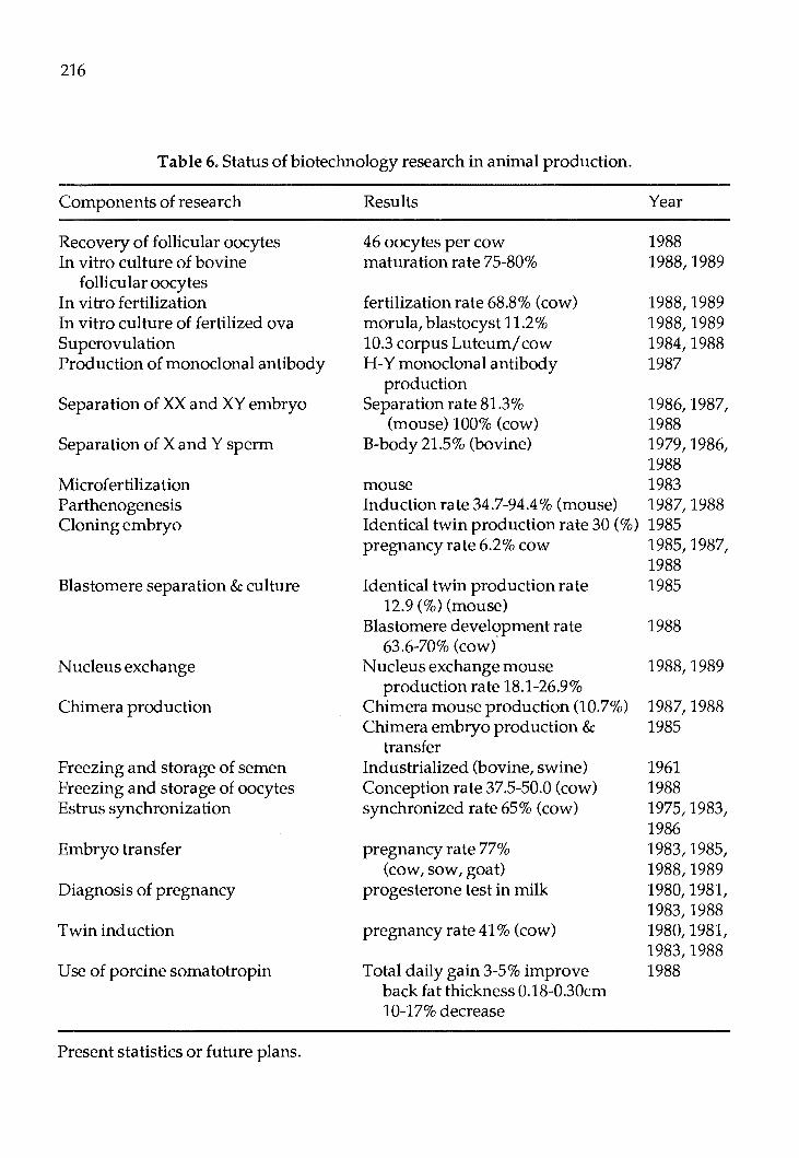
216
Table 6. Status of biotechnology research in animal production.
Components of research
Recovery of follicular oocytes In vitro culture of bovine
follicular oocytes In vitro fertilization In vitro culture of fertilized ova Superovulation Production of monoclonal antibody
Separation of XX and XY embryo
Separation of X and Y sperm
Microfertilization Parthenogenesis Cloning embryo
Blastomere separation & culture
Nucleus exchange
Chimera production
Freezing and storage of semen Freezing and storage of oocytes Estrus synchronization
Embryo transfer
Diagnosis of pregnancy
Twin induction
Use of porcine somatotropin
Present statistics or future plans.
Results
46 oocytes per cow maturation rate 75-80%
fertilization rate 68.8% (cow) morula, blastocyst 11.2 % 10.3 corpus Luteum/ cow H-Y monoclonal antibody
production Separation rate 81.3%
(mouse) 100% (cow) B-body 21.5% (bovine)
mouse Induction rate 34.7-94.4% (mouse) Identical twin production rate 30 (%) pregnancy rate 6.2% cow
Identical twin production rate 12.9 (%)(mouse)
Blastomere develc::>pment rate 63.6-70% (cow)
Nucleus exchange mouse production rate 18.1-26.9%
Chimera mouse production (10.7%) Chimera embryo production &
transfer Industrialized (bovine, swine) Conception rate 37.5-50.0 (cow) synchronized rate 65% (cow)
pregnancy rate 77% (cow, sow, goat)
progesterone test in milk
pregnancy rate 41 % (cow)
Total daily gain 3-5% improve back fat thickness 0.18-0.30cm 10-17% decrease
Year
1988 1988,1989
1988, 1989 1988, 1989 1984,1988 1987
1986, 1987, 1988 1979, 1986, 1988 1983 1987, 1988 1985 1985, 1987, 1988 1985
1988
1988,1989
1987, 1988 1985
1961 1988 1975, 1983, 1986 1983, 1985, 1988, 1989 1980, 1981, 1983,1988 1980, 1981, 1983,1988 1988

217
Table 7. Historical development of E.T.
Author Year Results
Chan Sea
1971 1975
Rabbit: 7 farrowed among 13 transferred. Goat: 5 farrowed among 10 transferred.
Go Gu Chung Suk Kim
1981 1982 1983 1983 1985
Korean cattle: one pregnant among 5 transferred. Holstein: one pregnant among 6 transferred. Holstein: 8 farrowed among 35 transferred. Holstein: 26 farrowed among 79 transferred (33%). Twin production with E.T. 22-35%.
Oh Im
1986 1986
Bovine frozen embryo: 27 pregnant (45.0%) among 60 transfer. Korean cattle calf born from Holstein cow.
In 1971, 7 kids were farrowed from 13 does by embryo transfer in rabbit. In 1975, 5 kids were farrowed from 10 does by embryo transfer in goat. Embryo transfer work in cow were intensively carried out between 1981to1986andeightcalves were farrowed by 35 recipients in 1983. Thereafter embryo transfer technology was used for production of twin. Frozen embryos imported from U.S. were transferred to produce superior offspring.
Institutes and dairy farms which are applying embryo transfer technology to produce superior offspring are as follows: Livestock Experiment Station, National
Breeding Station, Dusan Dairy Farm, Mail Dairy Farm, Samyang Dairy Farm, Sunjin Dairy Farm and Lee Animal Hospital.
b) Superovulation
1) Hormones used and their efficacy At the beginning of sperovulation treat
ment, PMSG was commonly used because this hormone is easy to get in the market. Number of corpus luteum following PMSG andFSHadministrationisillustratedin Table 8 and 9. It is difficult to point out which hormone is effective in ovulation in Table 8, however in Table 9 FSH showed higher ovulation rate than PMSG.
Table 8. Formation of corpus luteum following PMSG and FSH administration.
No. of corpus luteum
Hormon No. of cow Amount Total No/cow Reference
6 2,750IU 23 3.8 Gu &Chung (1982)
43 3,030IU 289 6.7 Chung et. al. (1983)
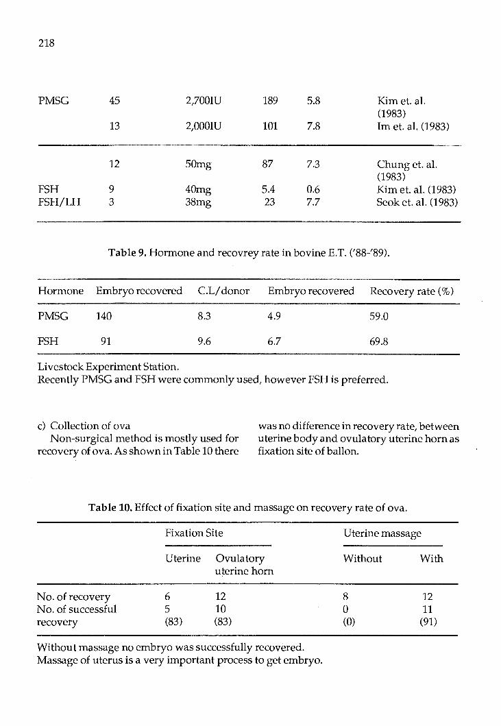
218
PMSG
FSH FSH/LH
45
13
12
9 3
2,700IU
2,000IU
50mg
40mg 38mg
189
101
87
5.4 23
5.8
7.8
7.3
0.6 7.7
Kim et. al. (1983) Im et. al. (1983)
Chung et. al. (1983) Kim et. al. (1983) Seok et. al. (1983)
Table 9. Hormone and recovrey rate in bovine E.T. ('88-'89).
Hormone Embryo recovered C.L/ donor Embryo recovered Recovery rate(%)
PMSG 140 8.3 4.9 59.0
FSH 91 9.6 6.7 69.8
Livestock Experiment Station. Recently PMSG and FSH were commonly used, however FSH is preferred.
c) Collection of ova Non-surgical method is mostly used for
recovery of ova. As shown in Table 10 there
was no difference in recovery rate, between uterine body and ovulatory uterine horn as fixation site of ballon.
Table 10. Effect of fixation site and massage on recovery rate of ova.
No. of recovery No. of successful recovery
Fixation Site
Uterine Ovulatory
6 5 (83)
uterine horn
12 10 (83)
Without massage no embryo was successfully recovered. Massage of uterus is a very important process to get embryo.
Uterine massage
Without
8 0 (0)
With
12 11 (91)
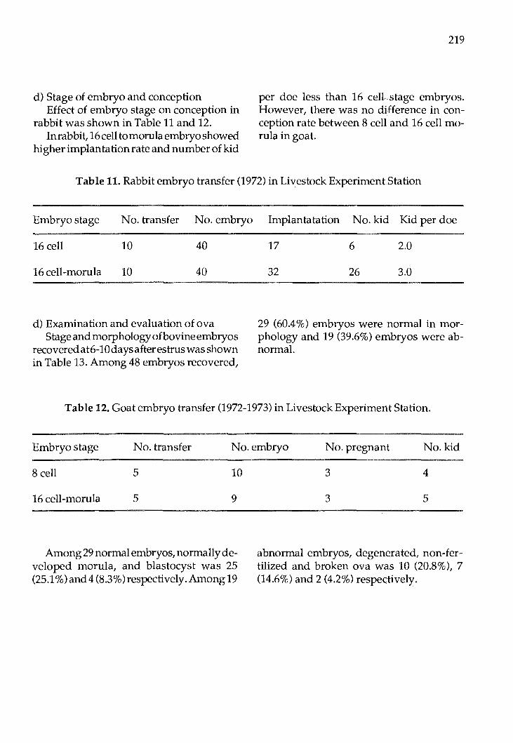
d) Stage of embryo and conception Effect of embryo stage on conception in
rabbit was shown in Table 11 and 12. In rabbit, 16 cell tomorula embryo showed
higher implantation rate and number of kid
219
per doe less than 16 cell.stage embryos. However, there was no difference in conception rate between 8 cell and 16 cell morula in goat.
Table 11. Rabbit embryo transfer (1972) in Livestock ExperimentStation
Embryo stage No. transfer No. embryo Implantatation No. kid Kid per doe
16 cell 10 40
16 cell-morula 10 40
d) Examination and evaluation of ova Stage and morphologyofbovineembryos
recovered at6-10 days after estrus was shown in Table 13. Among 48 embryos recovered,
17 6 2.0
32 26 3.0
29 (60.4%) embryos were normal in morphology and 19 (39.6%) embryos were abnormal.
Table 12. Goat embryo transfer (1972-1973) in Livestock Experiment Station.
Embryo stage No. transfer No.embryo No. pregnant No. kid
8 cell 5 10
16 cell-morula 5 9
Among 29 normal embryos, normally developed morula, and blastocyst was 25 (25.1 %) and4 (8.3%)respectively. Among 19
3 4
3 5
abnormal embryos, degenerated, non-fertilized and broken ova was 10 (20.8%), 7 (14.6%) and 2 (4.2%) respectively.
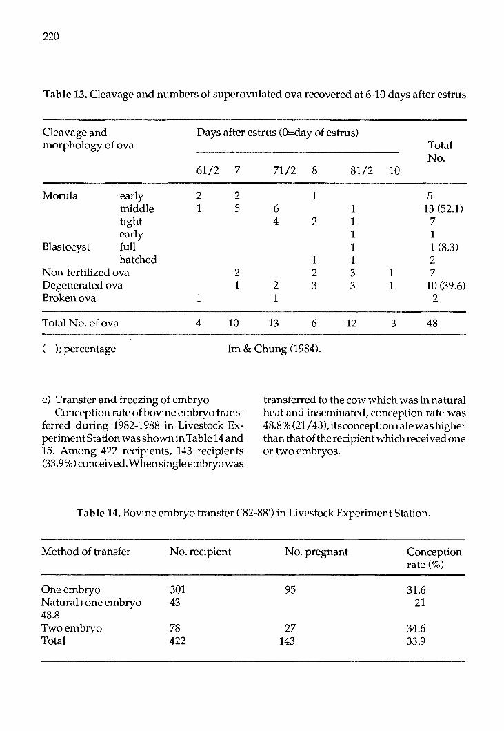
220
Table 13. Cleavage and numbers of superovulated ova recovered at 6-10 days after estrus
Cleavage and Days after estrus (O=day of estrus) morphology of ova Total
No. 61/2 7 71/2 8 81/2 10
Morula 'early 2 2 1 5 middle 1 5 6 1 13 (52.1) tight 4 2 1 7 early 1 1
Blastocyst full 1 1 (8.3) hatched 1 1 2
Non-fertilized ova 2 2 3 1 7 Degenerated ova 1 2 3 3 1 10 (39.6) Broken ova 1 1 2
Total No. of ova 4 10 13 6 12 3 48
( ) ; percentage Im & Chung (1984).
e) Transfer and freezing of embryo Conception rafe of bovine embryo trans
ferred during 1982-1988 in Livestock ExperimentStationwasshownin Table 14 and 15. Among 422 recipients, 143 recipients (33.9%) conceived. When single embryo was
transferred to the cow which was in natural heat and inseminated, conception rate was 48.8% (21/ 43),itsconceptionratewashigher thanthatoftherecipientwhichreceivedone or two embryos.
Table 14. Bovine embryo transfer ('82-88') in Livestock Experiment Station.
Method of transfer
One embryo Natural+one embryo 48.8 Two embryo Total
No. recipient
301 43
78 422
No. pregnant
95
27 143
Conception rate(%)
31.6 21
34.6 33.9
221
Table 15. Conception rate of bovine E.T. according to year ('82-88') in Livestock Experiment Station.
Year Embryo No. transfer
'82 fresh 24 '83-'84 fresh 85 '85-'88 fresh 74
frozen 239
Pregnancy rate of bovine frozen embryo was shown in Table 16. Among80recipeints which were transferred with frozen embryo, 30 recipients (37.5%) were pregnant. Pregnancy rate of fresh and frozen embryos by surgical and non-surgical transfer carried out by National Breeding Station in coop-
No. pregnant Conception rate(%)
3 12.5 30 35.5 35 47.3 75 31.4
eration with American Holstein service in 1980-1983 was summarized in Table 17. Fresh embryo showed higher pregnancy rate (55.6%) than frozen embryo (26.2%). Surgical transfer showed higher pregnancy rate (35.0%) than non-surgic;aJ (30.8%) in both fresh and frozen embryo.
Table 16. Pregnancy rate following transfer of frozen bovine embryos.
Experiment No. of recipients No. of recipients Rate of pregnant pregnancy
I 5 2 40 II 34 7 21 III 2 1 50 IV 34 17 50 v 2 1 50 VI 3 2 67
Chung et al. (1987).

222
Table 17. Pregnancy rate of fresh and frozen embryos by surgical and non-surgical transfer.
Surgical Embryo
No. Preg. %
Frozen 37 12 32.4
Fresh 3 2 66.7
Total 40 4 35.0
f) In vitro capacitation and in vitro fertilization An experiment was conducted to investi
gate the effects of breeds, medium supplements and ovarian phases on the developmental pattern of bovine oocytes.
The cleavage rate of oocytes was higher in Korean native cattle (KNC: 22.0%, 18/82) thaninHoslteins(4.4%4/93).Elevenamong 82 (13.2%) of KNC oocytes developed to morula, bu tnone of Holstein oocytes cleaved up to 8 cells. Cleavage rate of KNC oocytes was higher in PBS (fetal bovine serum) supplemented medium (22.0% 18/82) than in BSA (bovine serum albumin) supplemented medium (14.8% 4/27). The cleavage rate of oocytes was not different between luteal phase and follicular phase, but the rate of
Non-surgical Total
No. Preg. % No. Preg. %
24
15
19
4 16.7 61 16 26.4
8 53.3 18 10 55.6
12 30.8 79 26 32.9
embryo with regular blastomeres was higher in luteal phase than ~n follicular phase.
An experiment was carried out to investigate the effects of incubatin time, pH of medium and the sampling time from slaughter to incubation, on in vitro maturation of sow foolicular oocytes. After 24 and 48 hours incubation, the proportion of the oocytes matured to the second meta phase stage was 21 % and 54.7% respectively. g) Embryo bisecting/ cloning
As shown in Table 18 when the mouse and cow embryos were bisected and cultured, bisected ratio was 81.1 and 78.4% in mouse and cow respectively and developed rate was 72.1 and 100%. Fifteen demi cow embryos were transferred to eleven recipients and the two were pregnant.
223
Table 18. Embryo cloning, culture and transfer (84-88) in Livestock Experiment Station.
Species Embryo Separated embryo
Cultured (developed)
No. transfer
No. pregnant
Mouse 477 387 (81.1) 282 (72.1)
Cow 28 22 (78.4) 15 (100.0) 11 2
h) Sexing gametes using monoclonal or polyclonal H-Y antibody Shim et al. (1986) carried out experiment
to control the sex of offspring in mice by sxing embryos. H-Y antisera were prepared in inbred SD female rat by repeated immunization of testis supernatant and spleen cells from same strain of 270 embryos treated with H-Y antibody and compliment, 126 embryos(46.7%)weredeveloped tonorrnal blastocysts. Following transfer of 126 blastocysts, 16 embryos (12.6%) were survived to term and 13 female (81.3%) were produced.
Goh et al. (unpublished) cultured 82 frozen-thawed bovine morula with H-Y antiserum and compliment.
Among82rnorula treated, 15 (18.3%)were developed to blastocysts of which five were sexed by chromosome analysis and all were XX embryo. Testis supernatant, a source of H-Y, obtained from BALBI C mice was used to immunize females of same strain. Lymphocytes of mouse producing antibodies to H-Ywerefused withSp210-Ag14rnyelorna cells and distributed to 384 wells of 96 well rnicrotiter plates. Eighty hybridorna colonies were formed, resulting in 20.8 percent of fusion efficiency.
Three strong positive wells from hybridorna colonies were selected for cloning by ELISA and two of them were also found to be positive by indirect irnrnunofluorescence test. Twelve wells of ELISA-positive were selected after cloning and 2D 45D4 clones from them were confirmed to produce monoclonal ntibodies to H-Ybyindirectirnmunofluorescence test (Shim et al., 1988).
B. Application of r-DNA Technology for Genetic Improvement and Genetic Resistance to Diseases in Livestock and Poultry
1. DNA hybridization techniques
a) southern blotting Lee et al. (1989) carried out experiment on
expression of human growth hormone gene in transgenic mice. The structural gene of growth hormone was fused with mouse MT-1 prornotor and the fusion plasmid was designated as pMThGH.
The 2.6KB Barn HI/Bst Ell DNA fragment of pMThGH was used as a source of human growth hormone fusion in these studies. for rnicroinjection, DNA fragments were diluted with TE buffer in the range of
224
2-8 ng/rnl. The microinjected one-cell eggs were surgically transferred to the oviducts of synchronized recipients. The litters developed from microinjected eggs were weaned about 4-6 weeks after birth and a piece of tail from each mouse was cut off about 1-1.5 cm to analyze foreign DNA.
The genomic DNAs isolated from each tail were analyzed by dot hybridization and Southern blot to determine which mouse carried MThGH gene.
Of 106 microinjected eggs cultured for 96 hours invitro, 53 eggs (50.0%) were developed to normal blastocysts. Of 382, microinjected egg transferred to the oviducts of 23 recipients, 140 mice produced. Of 77 mice analyzed by dot hybridization and Southern blot, 6 mice were positive for MThHG gene and each carried at least one copy of hGH gene. Transgenic mice had very high levels of hGH in their serum, 450-6, 100 ng/ ml,andbodyweightwasl.5-2.0timeshigher than control.
2. Gene transfer
a) Method used (microinjection) The experiments were carried out by
Chung et al. (1988) to develop new techniques necesary for elevating utilization efficiency of high qua Ii ty embryos and production of cloned animals by nuclear transplantation.
Pronuclei removed from the mouse embryos on pronuclear stage by micromanipulation procedure were transplanted to enucleated mouse embryos on same development stage by similar micromanipulation procedures. Fusion of transplanted pronuclei to the cytoplasm of recipient embryos was mediated by Sendai virus.
Of 194 pairs pronuclei transplanted, 126 ( 64.9%) pairs were fused to the cytoplasm of enucleated recipients embryos. The number of embryos developed to 2 cell embryo, morula and blastocysts after the fusion was 108 (85.7%), 94 (74.6%) and 88 (69.8%) respectively.
Lee et al. (1989) produced nuclear transplanted mice between ICR and F
1 hybrid
(57BL x CBA).
A method that combines microsurgical method of the zygote pronuclei with introduction of donor pronuclei by a virus mediated cell fusion technique was used. Technical efficiency of this procedure com prised of zona cutting, enucleation, karyoplast injection and fusion step was more than 90% at each step.
Development rate of these nuclear transplanted embryos to blastocysts was about 60% and when these embryos were transferred to foster mother mice, four were born.
C. lmmunodiagnostics and Vaccines
1. Monoclonal antibody production
a) Type produced Veterinary Research Institute produced
monoclonal antibodies against several viruses.
These are antibodies specific to infectious bovine rhinotracheitis virus (IBRV), bovine rota virus (BRV), transmissible gastroenteritis virus (TGEV), Japanese encephalitis virus (JEV), porcine parvovirus (PPV), pseudorabis virus (PRV), Newcastle disease virus (NDV), infectious larynotracheitis virus

(UTV), Marek's disease virus (MDV) and infectious laryngotracheitis virus (ILTV). Using these monoclonal antibodies, rapid sensitive diagnostic tests are being undertaken.
Resources Available
Human Resources
Chung, C.S. Professor. Effects of porcine growth hormone on lipogenesis in adipose tissue. Dept. of Animal Science, Chungbuk National University.
Chung, H.M. Researcher. Studies on nuclear transplantation in mouse embryos. College of Animal Husbandry, Konkuk University.
Chung, KM. Research technician. Effects of breeds, medium additives and ovarian phases on in vitro maturation, fertilization and cleavage of bovine follicular oocytes. College of Agriculture, Seoul National University.
Chung, KS. Professor. Studies on in vitro maturation of bovine follicular oocytes. College of Animal Husbandry, Konkuk University.
Chung, Y.C. Professor. Studies on maturation in vitro of rat follicular oocytes and fertilization in vitro of cumulus removed and intect oocytes after maturation. Dept. of Animal Science, Chungang University.
Goh, G.D. Professor. Studies on the sexing of bovine embryo by the chromosomal analysis and H-Y antibody. College of Animal Agriculture, Kangweon National University.
225
Hwang, W .S. Professor. Studies on the twin birth of cattle by several methods. Veterinary College seoul National University.
Im, KS. Profesor. In vitro capacitation and fertilising ability of ejaculated rabbit sperm treated withlysophosphatidylcholine. Dept. Animal Science, Seoul National University.
Jung, J.K Researcher. Swine embryo transfer. Livestock Experiment Station.
Kang, M.S. Professor. Effects of the glycerol cryoprotectants containing sucrose on the mouse embryo survival rate deerrnined by FDA test. College of Agriculture, Cheju National University.
Kirn, D.I. Professor. Study on the sexing of preirnplantation mouse embryos exposed to H-Y antisera. College of Animal Agriculture, Kanweon National University.
Kim, D.K Professor. Studies on maturation in vitro and fertilization ability of bovine folicular oocytes. Dept. of Animal Science, Chun gang University.
Kim, H.S. Researcher. Studies on the factors affecting superovulationinduction in cattle. Livestock Experiment Station.
Kirn, J.B. Professor. Separation of X and bearing superrnatozoa. College of Animal Husbandry, Konkuk University.
Kim, J .K Professor. Effects of seeding procedures in a liquid nitrogen container on the survival rate of mouse embryos. College of Agriculture, Cheju National University.
Kirn, S.K Professor. Studies on the in vitro maturation and fertilization rate of bovine folliocular oocytes. College of Agriculture, Chungnarn University.
226
Koh, T.H. Researcher. In vitro maturation of porcine follicular oocytes. College of Agriculture, Korea University.
Lee, C.S. Researcher. Production of nuclear transplanted mice. Genetic Engineering Center, KAIST.
Lee, K.S. Senior researcher. Expression of human growth hormone gene in transgenic mice. Genetic Engineering Center, KAIST.
Lee, K.S. Professor. Effects of cooling rates and plunging temperatures on survival of hamster embryos. College of Agriculture, Chungnam National University.
Lee, K.W. Section Chief. Influence of frozen embryos conception in cattle. National Animal Breeding Institute.
Oh, S.J. Researcher. A study on the synchronization and frozen embryo transfer in cattle. Livestock Experiment Station.
Park, C.S. Professor. Effects of cooling rates and plunging temperatures on survival of hamster embryos. College of Agriculture, ChungnamNational University.
Park, H.K. Professor. Studies on the in vitro maturation and fertilization rate of bovine follicular oocyts. College of Agriculture, Kyungbuk University.
Pho, M.C. Researcher. Studies on the industrial utilization of frozen bovine embryos. Dusan Research Laboratory.
Shim, H.S. Researcher. Sex ratio of mouse off springs following in vitro treatment of Hy antibody to mouse embryos prior to transfer. College of Animal Husbandry, Konkuk University.
Shin, W.J. Professor. Effects of genotypes of embryo and recipient on the survival of embryo transfer. Dept. of Animal Science, Chunbuk University.
Seo, K.D. Professor. Density of immunoglobulins in reproductive tract fluids and serum. Dept. of Animal Science, Yonam Junior College of Livestock & Horticulture.
Son, J.K. Researcher. Cytogenic studies of polyploidy manipulation. Korea Institute of Science and Technology.
Song, H.B. Profesor. In vitro fertilization of bovine follicular oocytes matured in culture with bull spermatozoa preincubatedin uteri isolated from hamsters. College of Agriculture, Taegu University.
Suk, H.B. Professor. One-step straw method for the handling of frozen-thawed mouse and bovin embryos. Dept. of Animal Science, Dankuk University.
Yeo, J.S. Professor. Development of new breed by the introduction of polyploid in chicken I, production of polyploid. Department of Animal Science, Yeungnam University.
Yang, B.S. Researcher. Study on the solubility of zona pellucida and developmental petancy of isolated blastomere in mouse.
Yun, C.H. Professor. Study on the freezing of rat embryos. College of Agriculture, Gyeongsang National University.
Yun, Y.T. Research technician. Studies on improvement of devleopmental potency of in vitro fertilized bovine follicular oocytes. Dept. of Animal Science, Chun gang Uni versity.
Proc. F AO/CAAS Workshop on Biotechnology in Animal Production and Health in Asia and Latin America, Beijing, Oct 9-13,1989, pp 227 - 239
Biotechnology Applied To Animal Production and Health in Thailand
Vanda Sujarit Faculty of Veterinary Medicine
Kasetsart University Bangkok 10903, Thailand
Introduction
Biotechnology development programs in Thailand started in the late seventies, first with the formation of centers in important universities. Livestock biotechnology started from the various attempts of individuals to investigate certain areas of animal reproduction, animal nutrition, molecular biology, vaccine production and disease diagnosis. In 1983, National Center of Genetic Engineering and Biotechnology (NCGEB) was created under the initiative of the Ministry of Science, Technology and Energy. Its primary objectives are to be the focal point for strengthening Thailand's capabilities in genetic engineering and biotechnology and related areas and to apply them to national economic and social development. One area of the emphasis on technology application is the agriculture applications. NCGEB funded RSD projects in wide ranging areas in development of farm animals from embryo manipulation, embryo transfer, steroid immunization to recover fertilization through production of growth hormone by genetic engineering for use in promoting growth of economic animals. It promotes extensive collaboration among institutions and also forms industry-university linkage in relevant projects with the aim of transferring the technology.
The Department of Livestock Development (DLD), Ministry of Agriculture and Cooperatives is concerned about the situation of small scale farmers. Collaborating with the university scientists to develop biotechnology for livestock improvement results in transferring technology from the laboratory to the on-farm application.
This paper discusses the present status, approach and future trends of biotechnology development for animal production and health in Thailand, including the policy and cooperatives of the Royal Thai Government.
Current Status of Livestock Biotechnology Development
The development of biotechnology for livestock improvement and health has been directed mainly to dairy cattle production. The approximate numbers of livestock population in Thailand are shown in Table 1. The dairy cattle expansion is one of the top priorities in the strategies of the DLD. The number of dairy cattle in Thailand is indicated in Table 2.
Previously, the consumption of milk had not been a tradition for the Thai people.

228
Table 1. Approximate livestock population in Thailand
Horse Beef Cattle Dairy Cattle Buffalo Pig Goat Sheep Chicken Duck Goose
1985
21,000 4.3 Million
53,000 5.2 Million 5.9Million
80,000 57,000
7.0Million 1.9 Million 406,000
1986
19,000 4.3Million
65,000 4.9Million 5.8Million
80,000 72,000
8.7Million 1.9 Million 412,000
Source: Office of Agricultural Economics.
Table 2. The number of dairy cattle in Thailand
1987
18,000 4.4Million 150,000
4.6Million 5.8Million
79,000 95,000
9.2Million 1.9Million 499,000
Year Female Dairy Catt;e Calves and Heifers Cows (More than 3 years) (Head) (Head) (Head)
1978 18,935 10.363 8,572 1979 20,658 10,891 9,767 1980 23,319 12,086 11,233 1981 25,661 12,841 12,820 1982 30,046 16,280 13,766 1983 39,426 20,233 19,193 1984 48,489 24,639 23,850 1985 57,094 1986 69,907
Average 16.88 15.91 17.96 Increase ( % )
Sources: - Office of Agricultural Economics. - ASIAN LIVESTOCK OCTOBER 1986.

However, this attitude has been changed gradually and the demand for milk production increases with the national campaign for milk consumption. Thailand can only produce about 10% of its local demand for dairy products. The rest has to be imported resulting in an enormous loss of foreign currency. Therefore, the development of biotechnology to accelerate the dairy production was established. Subsequently, other areas of biotechnology and genetic engineering to improve technology, vaccine production, disease diagnostics, and semen bank. The artificial insemination in dairy cattle was established in Thailand thirty years ago. The AI Division of the DLD takes full responsibilities for bull and boar semen production, deep-frozen semen production, semen quality control and other relevant tasks. With the strong background and experiences in AI, coupled with the developmentof embryo transfer technology, dairy cattle improvement programs are led.
Current developments of biotechnology for livestock production and health are as follows:
A. Animal breeding and reproduction, MO ET
Crossbreeding programs are presently utilized in various classes of livestock for genetic improvement. In cattle, the best crossbreeds have 62.5-75% exotic blood; for milk production 75% Holstein Friesian are used. The pure western breeds do not show a satisfactory performances due to diseases, pests, climate, improper care, etc. Crossbreeding is achieved by means of artificial insemination.
Embryo transfer and associated technolo-
229
gies have been investigated. The successes were reported in dairy cattle (3 ,4,7). Studies were conducted by Kasetsart University, DLD and private sector, with the funding from NCGEB. Two approaches to meet the objectives of increasing the milk production rate and improving dairy cattle breed, are a) importing the frozen embryos of superior pure breed and transfer to native cows, and b) collecting and transferring fresh embryos. Basic steps of embryo transfer technology were investigated under local conditions including donor selection, superovulation, AI, embryo collection and evaluation and transferring. Freezing the embryos are undertaken and special embryo manipulations i.e. sexing, splitting and cloning are expected after the basic steps are completed.
High performances crossbreds and pure breed cows were selected to be the donors. It was found that the animal managements were an important factor for donor selection. Follicle Stimulating Hormone (FSH) 40-50 mg was reported to be the suitable doses in tropical environment to enhance multiple ovulation. The administrations were done twice daily with the reducing doses for 5 days. (4). However, Pregnant Mare Serum Gonadotrophin (PMSG) has also been used at the dosage of 2,000 or 2,500 i. u. followed by 25 mg prostaglandin F22 48 hours later (7). Non-surgical collection of embryo was conducted at 7-8 days after AI. The recovery rate of the embryo was 8.5 embryos per one flush. However, the quantity of transferable embryos was considerably low. The pregnancy rate was 30-35%. From 1986 to present, fourteen calves were born from the transferring of imported frozen embryos. They were observed for their performances and adaptabilities in the tropical condition. From the local flushing, four calves were born and four remained pregnant.

230
The success of embryo transfer in pig was reported by scientists at Chulalongkorn University(S).Morerecently,embryotransfer in buffalo was reported (6), and 2 calves were born from 1 recipient early in 1989. Embryo transfer in pig may be used for achieving and increasing rate of reproduction of superior genetic animals and also for the possibility of export or import of embryos between countries. The basic techniques were underlined. Three trials of embryo transfer in gilts which previously experienced estrus were carried out (5). Synchronization of estrus was done by using oral Altrenogest 20 mg daily for 18 consecutive days. The animals were in heat between day 5 and 7 after cessation of the hormone treatment. All donor animals were hand mated twice at 12 hours interval. Surgical embryo recovery and transfer were performed on day 3 or 4 mating. Seven piglets were reported born from one recipient. The report indicated the possibility of using repeated donor for pig embryo transfer although moderate adhesion was observed on the horn, fallopian tube and fimbria.
In buffalo, non-surgical embryo transfer were carried out in 3 donors and recipients. Single egg and superovulation collection of embryos were performed on an average at 7 days after mating. Two recipients were reported pregnant. Further development of the technique would help facilitate genetic improvement and distribution of this species.
Considering the nucleus herd improvement using MOET, open nucleus breeding system with MOET has been established by a dairy private company in collaboration with Kasetsart University. The work is undertaken and no report is available at the present time. By embryo transfer techniques,
works are extended from dairy cattle to beef cattle. Establishing the embryo bank by cryopreservation is under investigation.
The in vitro oocytes maturation, sperm capacitation and sexing, in vitro fertilization and related techniques are investigated at Mahidol University with the funding from STDB. No report is available now.
Germ plasm technology on semen and gene pool for semen bank are under the investigation of DLD.
B.r-DNA technology
r-DNA technology for genetic improvement and genetic resistance to diseases especially in human is undertkaen at Mahidol University. Work on identification of genetic markers for production and disease traits in livestock are planned.
Growth hormone (GH) plays a very important role in promoting growth of animals, and in increasing milk yield of cow. The growth hormone is usually obtained from extracts of pituitary gland of animal, and as a consequence of a very minute amount of the GH is available. A project supported by NCGEB (7) aims at studying GH gene of cow and of a giant catfish (Pangasiandos gigas Chevy) and to use the gene for the GH production and for future construction of transgenic animal containing GH. To obtain GH gene from a cow, mRNA was extracted from the pituitary gland using guanidi um thiocyanate and Cscl high speed centrifugation. Poly A-mRNA was obtained from Oligo-dT column and used for a library construction. The cDNA library is being screened for E. coli clone containing GH by colony hybridization using oligonucleotide as the probe.

Hormone Immuno Neutralization to increase productivity in animals was investigated (8). Pregnenolone - hemisuccinate -HSA or link of ovidin in dextran solution was produced as an immunogen to vaccinate cattle and buffalo. Titer of antibody produced against pregnenolone was measured by determining testosterone and LH levels. In buffaloes having body weight over 300 kg, testostrone levels were suppressed due to increasing titer after immunization but contrary effect was seen in young buffalo bulls having body weight around 160 kg. Factors influencing the growth rate of buffaloes and cattle have been assessed in parameters of rearing pattern and breed. Assessment of suitable dose and a combination of the formulate vaccine in a particular breed and management is necessary to the future ensurable input of utilization of this vaccine. Potential uses of pregnenolone immunogen were illustrated in terms of immunocastration enhancing of growth rate of buffaloes in small farms and using as an au to - self genera ting growth promo tor for fattening cattle. Further characterization of the mechanism of action in metabolism of cell levels may be hypothesized as an accelerator to the growth gene levels, thus stimulating the mRNA for protein biosynthesis of the said growth promotor.
Concerning DNA - hybridization technique, scientist at Chulalongkorn Univeristy (personal communication) has developed DNA probe technique for the detection of Aujeszky's disease. The disease carries latent infection in the gene which cannot be detected by the regular method. The specific Aujeszky's disease genes were cloned and combined with the plasmid of E. coli. After culturing and extraction, the genes wre labelled with 32P-dCTP. This labelled probe would then be hybridized with the gene of viral sample. If the animal was in-
231
fected, the signal on the film of autoradiograph would be shown by this sensitive probe.
Molecular cloning of Herpes simplex virus type 2 DNA in transcription vecto rhas been prepared for rapid diagnosis. When the DNA of HSV was extracted and purified from infected cells using triton NaCl, it could be used for cloning without a need for further purification step. DNA of HSV-2 was cleaved using Bgl, II, ligate to a transcription vector, pSP72 and cloned into E.coli JM 109. Positive colonies were selected using Biotin-labelled probes. The technics of labelling probes using Photobiotic and bionick DNA labelling system were now being assessed (9).
C. Immunodiagnostics and vaccines
The National Institute of Animal Health and Production (NIAHP), DLD provjdes the ELISA test for swine fever diagnosis. In the past, the diseases were diagnosed from the symptoms, etiology and history. The development of biotechnology and genetic engineeringin monoclonal antibody creates better disease diagnosis, better and safe vaccines for resistance. ELISA tests were utilized for the diagnosis of Aujeszky disease, IBR, BEF (Bovine Ephemoral Fever), IBD in poultry and etc.
NIAHP under the cooperation with The Japanese International Cooperation Agency (JICA) has developed the monoclonal antibody production for the identification of New castle disease viral strains. The virus is purified and immunized the BALB/ c mice. Subsequently, spleen is obtained by splenectomy and hybridized withmyeloma. The

232
hybridromal cells are cloned and biological properties are measured by ELISA method. These cells producing monoclonal antibodies are obtained for diagnostic purpose. The work is in progress (personal communication).
The monoclonal antibody production for diagnostic purpose of Foot and Mouth Disease are planned at the Veterinary Biologics Division, DLD.
The employment of genetic engineering technique in preparing antigens for the diagnosis of Opisthorchis viverrini infection has been attempted at Mahidol University (10). StartingwithmRNA preparation from adult fluke, it then reverse transcribe to complementary DNA which will be cloned into expression vector, i.e., bacteriophage and plasmid in E.coli. Results from the early stage of the work showed thatmRNA of the parasite has characteristic which is diferent from ordinary mRNA. The mRNA of 0. viverrini consists of a very small and undetectable proportion of poly( A) tailed mRNA. This characteristic makes it impossible to synthesize cDNA from poly (A) mRNA
prepared by ordinary technique. The overcoming efforts for immunodiagnostic purpose are in progress.
For vaccine production, various kinds of vaccine can be produced at the Veterinary Biologics Division, DLD. (Table 3). Compared to the animal population, the number of vaccine that can be produced is still far from the demand, resulting in the importation and economic loss. Work at the DLD is in progress to develop biotechnology and genetic engineringutilizingmonoclonal antibody to produce FMD vaccine.
Atthe present time, monovalent vaccines for FMD are produced by tissue culture technique. It is expected that the trivalent vaccine (type 0, A, Asia I of FMD) will be on trial in the near future.
D. Biotechnology of enhacing animal feed production
The improvement in utilization of agroind us trial by-product in animal feeding and
Table 3. Target for vaccine production in animal in 1989.
Animals
Cattle - buffalo
Pig
Poultry
Source: DLD.
Kind of vaccine
8
3
7
Vaccine production (doses/year)
21 million
6 million
280million
ruminant nutrition through biotechnology was conducted (10, 11, 12). Studiesonbiodegradation of organic wastes for production of animal feed using microbiological treatment are undertaken at Kasetsart University. The research is entitled-Enahncement of lignocellulosic bioconversion through coupling actions of commercial-organism inmixed culture fermentation. The obejctives of the project are: 1) to select the breed of white rot fungi that can digest lignin in order to improve the agricultural by-product. 2) to investigate the enzyme and its property to digest lignocellulose of selected fungiand the correlation withdigestability. 3) to develop the solid state cultivation utilizing the selected white rot fungi. Works are in progress and the results have not been concluded.
N a.tional Policy and Inferastructure Development
Biotechnology is one of the three priority areas for development (the others are Material Sciences/Metallurgy and Electronics/ Computer). As such, institutional building, financial support and restructing of R&D and industry are given special attention and consideration (1).
In addition to the establishment of NCGEB, a new funding body, Science and Technology Develop[ment Board (STDB) was formed to support R&D in both public and private sectors as well in promoting commercialization of biotechnology.
Thailand adopted the NETWORK approach to accelerate biotechnology R&D through mobilizing existing resources from various organizations and institutions. As a result several biotechnology networks have been established or being formulated with each membr of the network acting as a cen-
233
ter of excellence (2).
For animal biotechnology, the following organizations and their areas of experties have been mobilized:
1. Kaseasart University - Cattle embryo transfer, feed technol
ogy and antibiotic production for use in animal feed and disease control.
2. Chulalongkorn University - Pig and buffalo embryo transfer.
3. Mahidol University - IVF, Monoclonal antibody and growth
hormone production. 4. Developmentoflivestock Development
- Semen bank and vaccine production including pilot facilities.
In addition, theNCGEBprovidesgenetic engineering materials such as splitting enzymes and vectors, in support of biotechnology R&D including feed biotechnology, cellulose fermentation and vitamin production. NCGEB also serves as coordinating body for all the biotechnology networks.
Future Trends for Research and Biotechnology Development
Future direction of research and development especially with respect to animal production and health should be toward more breadth and depth. Systematic application of biological knowledge should be applied and transfer to the end-user. Conventional animal breeding is still a useful tool for development of animal breed and varieties, accompanied by new technology being developed. The more advances in molecular genetics, embryo amnipulation and gene transfer will have wide impact in improving production efficiency. The end result will be increase in the quality and production, and decreased cost. Health will be
234
improved as result of new methods of disease diagnosis.
The future programme of biotechnology development for animal production will be in connection with the current status and can be summarized as follows:
1. Embryo transfer and associated technology - Imrpovement of higher genetic effi
ciency in dairy cattle, beef cattle and buffalo by embryo transfer technology. R&D to develop IVF in farm animals and the applications on-farm. Establishing the embryo bank utilizing cryopreservation methodology, and extend to genetic engineering i.e. sexing, spolitting and cloning. Setting up the open nucleus breeding system with MOET.
2. Improvement in utilization of agro-industrial by-product in animal feeding and ruminant nutrition through biotechnology.
3. Research programme in animal health - Setting up of a bioengineering vaccine
and diagnostic test for FMD by using monoclonal antibody and DNA probe diagnosis.
- Anthelmintic resistance in nematode parasite of cattle and sheep.
- Molecular and ecological genetics and control strategies.
4. Research programme in molecular technology. - Isolation, modification and gene trans
fer. - Embryology and genetic regulation of
development. - Genetic and environmental manipu-
lation of rumen microorganism. - Expression of genes when introduced
to animal cell.
5. Research programme in animal breeding and genetics - Consequences of selection of adapted
animals tropically for better production and adaptive characters. Manipulation of embryo and the transfer of nuclear materials in different genotypes. Evaluation of local and exotic genetic materials in different environments.
The need for development of animal biotechnology in Thailand is not for keeping up with the modem techniques but more for enhancing role of agriculture. There are capable scientists in various universities and govenment agencies who could undertake the work on biotechnology, molecular biology and genetic engineering. The biotechnology network promoted by the NCGEB exist among the in-country institute and such work has been done to a certain extent in various aspects of biotechnology.Nevertheless, the rate of the activities is unappreciated due to lack of funds, facilities and expertises. Concrete cooperative efforts among institutes are actively sought.
Thailand proposes to cooperate with regional organizations by offering existing training and R&D facilities and services of the animal biotechnology network with Kasetsart University acting as a coordinator. Appendix A indicates available personnel to coordinate the development of animal biotechnology for livestock improvement and health. Priority areas of expertises to be shared are: ·
- Embryo transfer technology in cattle buffalo and pig.
- In vitro treatment of feeds by microor-

ganism. - Production of monoclonal antibodies. - Manipulating and control of rumen
fermentation.
In addition, the genetic engineering material services for R&D can also be extended.
Conclusion
The need for development of animal biotechnology in Thailand and other ASEAN countries exists in the wide range application of the results of biotechnology researches. While the biotechnology program still needs to be developed further, it provides ample basis for international cooperation in various forms.
Biotechnology and genetic engineering applied to animal production and health have strong potential on economical benefit. Although the application of technology in North America and Europe are at the advanced level and producing high performance Ii vestock, the development of these new technology in developing world is remarkably slow. The primary difficulties involve financial support. External funding provides the opportunities for scientists to have advanced training and facilitates procurement of equipments and supplies.
To obtain greater benefits from the applications of biotechnology to animal production and health in the developing world, Thailand offers to cooperate with regional organizations with the similar aims of maintaining the highest levels of international coperations in the use of biotechnology products. Similarly, other countries in the network are expected to derive benefits through mutual cooperations.
235
References
1. Yuthavong, Y. (1987). Theimpactofbio-technology and genetic engineering on development in Thailand. J. Sci. Soc. Thailand 13: 1-8.
2. NCGEB Activities Report 198301988 (1988). National Center of Genetic Engi-neering and Biotechnology Bangkok.
3. Sujarit, V.K., et al (1986). "Sai-fon": An Embryo Transfer Calf. 13th Annual Veteirnary Science Conference. TVMA. 2-4December,1986. Bangkok, Thailand.
4. Singhajan, S. et al (1986). Superovula-tionwithFSHinEmbryoTransferTech-nology.13thAnnualVeterinaryScience Conference, TVMA. 2-4December,1986. Bangkok, Thailand.
5. Chantaraprateep, P., et al (1988). Em-bryo Transfer in Thai Swamp Buffalo (Bubalus bubalis). 15th Veterinary Sci-ence Conference, TVMA. 2-4 Novem-ber, 1988. Bangkok, Thailand.
6. Chataraprateep, P., et al (1988). Embryo Transfer in Thai Swamp Buffalo (Bub-alus bubalis). 15th Veterinary Science Conference, TVMA. 2-4 November, 1988. Bangkok, Thailand.
7. Parnpai, R., et al (1986). The Succes of Embryo Transfer in Dairy Crossbreed. 13th Annual Vetemary Science Confer-ence, TVMA. 2-4 December, 1986. Bang-kok, Thailand.
8. Panyim, S. et al (1989). Production of Growth Hormone (Gh) by Genetic Engienering for Use in Promoting Growth of Economically Important
236
Animals. (Abstract) NCGEB Annual Conference, Bangkok.
9. Kamonpatana, M., et al (1989). Hormone Immuno Neutralization to Increase Productivity in Cattle and Buffalo. (Abstract) NCGEB Annual Coference, Bangkok.
10. Yoosook, C., et al (1989). Molecular Cloning of Herpes ?Simplex Virus Type 2 DNA in Transcription Vector for Use in Rapid Daignosis. (Abstract) NCGEB Annual Conference, Bangkok.
11. Dharmkrong-at,AandS.Sirisinha(1989) Preparation of Opisthorchis viverrini Antigens by Genetic Engienering Techniques for Use in Immunodiagnosis.
(Abstract) NCGEB Annual Conference, Bangkok.
12. Chairatanayuth, P. (1984). The Composition of Soybean Refuse Treated with Alakli or Hemicellulase. Proceedings of 3rd Annual Workshop on AAFARR Network, Victoria.
13. Chairatanayuth, P. (1986). Inclusion of Amaranth Crop Residue in Diet for Cattle. 5th Annual Workshop of AAFARRNetwork, Bogor.
14. Chairatanayuth, P. and V. Wannamolu (1986). The Composition and Digestibility Threshed Sorghum Head residue with UreaandAlkali.6thAnnualWorkshop of AAFARRNetwork, Los Banos.
APPENDIX A
HUMAN RESOURCES
Institution: Faculty of Veterinary Medicine, Kasetsart University
Head of Institution: Dr. Pirom Srivoranart
Address: 50 Paholyothin Road, Bangkok 10900
Researcher
Dr. Vanda Sujarit
Dr. Samutra Sirivejapundu
Dr. Suvichai Rojanasathien
Dr. M.L. Suntaranu Tingyai
Dr. Yangyong Intaraksa
Dr. Kiti Srisuphat
Researcher Project
- Embryo Transfer in Cattle
- Cytogenetics
- Embryo Transfer in Cattle
- Infertility
- Embryo Transfer in Cattle - Hormonal Immunodetection
- Ruminant nutrition and mineral analysis
- Immunoassay - Reproduction in buffalo
- Monoclonal antibody
237
Present & Projected Application
- ET applied to farm level in tropical environemnt
- Infertiligy problem solving
- Reproductive and infertility problem solving
- Farm and feed management

238
Institution: Faculty of Veterinary Science, Chulalongkorn
Head of Institution: Dr. Songkram Luangtongkum
Address: Henri Dunung Road, Bangkok 10500
Research
Dr. P. Chantaraprateep) Dr. C. Lohachit) Dr. P. Virakul) Dr. M. Techakhumpu) Dr. A. Kunawongkrit)
Dr. M. Kamonpatana) Mr. R. Pampai)
Research Project
Embryo Transfer in farm animals
Application of nuclear reproduction techniques in buffalo and cattle
Present & Projected Application
Genetic improvement and fertility investigation
Dr. Ratree Wongwatcharadumrong
- Diagnosis of Aujeszky Immunodiagnosis Disease
- Comparative studies of DNA probes among antigen detection methods for diagnostic purpose
Institution: National Instiutte of Animal health and Production, DLD
Head of Institution: M.R. Amnuapom Kasemsun
Address: In Kasetsart University, Bangikok 10900
Research
Dr. Urasri Tantaswasti
Dr. Wasana Pinyochon
Research Project
Development of monoclonal antibody for the differentia-of New Castle Disease Vaccine
Development of ELISA technic for the detection of swine fever.
Present & Projected Application
To identify the strains of New Castle Disease Vaccine

Institution: Department of Animal Science, Kasetsart University
Head of Institution: Dr. Charnwit Wacharakubta
Address: 50 Paholyothin Road, Bangkok 10900
Researcher
Dr. Charnnean Satayapunt
Dr. Pornsri Chairatanayuth
Dr. Durnrong Leenanuruksa
Research Project
- Dairy Health - Reproduction - Embryo transfer
- Ruminant nutrition
- Dairy production and management
Institution: The Veterinary Biologics Division, DLD
Head of Institution:
Address: Arnphur Pakchong Nakorn-ratcheasima
Research
Dr. Ab. Kongtun Dr. Suneechit Kongtun Dr. Pichit Makarasen
Research Project
Development of ELISA for FMD diagnosis and vaccine production
239
Present & Projected Application
Present & Projected Application
Diagnostic purpose

Proc. F AO/CAAS Workshop on Biotechnology in Animal Production and Health in Asia and Latin America, Beijing, Oct 9 -13, 1989, pp 240 - 245
Biotechnology for Animal Production and Health in Argentina
A.A. Schudel Instituto de Vriologia CiCV-INTA-Castelar
77 - 1708 - Moron Buenos Aires, Argentina
Argentina has valuable agriculture resources traditonally exporting red meat, wool and, recently, fine furs. The productive characteristics are greatly determined by ecological conditions favourable for extensive and semi-intensive cattle grazing which permit to obtain good quality products.
The incorporation of new technologies, particularly biotechnology in the existing production and health management techniques undoubtedly gives new opportunity and ways for further improvement. Firstly, it increases productivity and further controls outbreak of diseases and secondly, these new technologies must compete in price with the traditional systems and maintain or improve the quality of the product that is being commercialized.
In the past six years, various institutions of the country have begun to develop activities on Biotechnology in the training of their human resources as well as in the development of research particularly in Animal Health and Production (Annex 1).
The National Secretariat of Science and Technology has begun a National Program that supports the activities which are carried out in different institutions. Instituto
Nacional de Technologia Agropecuaria (INT A) has developed a national program of advanced Biotechnology applied to the Plant and Animal production which include, in addition to various lines of research, the training of human resources in other countries.
The application of Biotechnology in Animal Health and Production are considered at present with immediate priority for the country. In animal production, embryo manipulations will allow to increase, in a short time, the genetic quality of the animal herd, in particlar in bovines. There is now special interest in the genetic characterization of breeds and production of transpenies. In Animal Health, the development of vaccine against Foot and Mouth Disease, Brucellosis, Ticks and Tick transmitted diseases, as well as the development of Diagnosis Methods are the areas to be emphasized.
Centro de lnvestigaciones En Ciencias V eterinarias Inta - Argentina
The National Institute of Agricultureal Technology belongs to the Agriculture Secretariat of the Nation, created in 1956 with the purpose of "impelling and strengthen-

ing the development of farming research and extension, and of accelerating - the benefits of these fundamental functions -the use of new technology and improvementof the farming industry and rural life". It has as an autonomous financing source, a 1.5% tax over the value of farming exports. National research activities are performed in different operative units under a national program with short, medium and long range objectives including the corresponding system of control and evaluation. It has a permanent staff of 1700 professionals, 32% of them with postgraduate degrees in foreign Universities.
The Research Center in Veterinary Sciences (CICV) belongs to INTA and its main objective is to create and adapt knowledge, improvement of the intrinsic productive capacity of the species being exploited, and their chances of marketing, transformation and use, to expand markets and find new uses. It is unique in the country and in Sou th America, and has acquired a sound national reputation with the establishment of solid mechanisms of international cooperation.
In the past six years the CICV has given priority to research, development and transference activities performed in Animal Health and Production through the use of modem biotechnologies.
A. Animal Health
1. Foot and Mouth Disease
1.1.Development of technology for the production of immunogens through Genetic Engineering Technology
Its main objective is the development of a
241
vaccine against FMD virus from immunogenic fractions of structural proteins obtained from bacteria.
The genomic sequences belonging to the main moitopes of 0 1 Campos and C
0 virus
strains - active in this country - were determined using the direct sequencing techniques for VP2 protein.
A p03VP2 plasmid expressing E. coli
antranilate synthetase (TrpE) together with aminoacids 137 to 213 of VP2 of FMDV 0
2
Campos was constructed. In addition, a plasmid (p0
2-VP
2NT) coding for TrpE and
137-172 of VP2
of FMDV 02
Campos was obtained.
Using a similar expression plasmidi and synthetic oligonucleotides, clone p02VPC was constructed, which codes for a fusion protein located between TrpE and the 200-213 region of FMDV 0
2 Campos. A fusion
protein only containing region 200-213 of VP 2 of FMDV 0 2 Campos was also constructed by using synthetic oligonucleotides which after being purified were hybridized by sequence complementarity. As expression vector P ATh3 was used selecting the recombinants by colony hybridization.
Kaplan, G.; Giavedoni, L.; Piccone, M.E.; Rosemblit, N.; Zuloaga, G.; Marcoveccnio, F; Zabal, O.; Palma, E.L.
1.2 Expression of FMDV antigens in an eucariotic vector
Its main objective is the development of a subunit vaccine against FMD through the introduction of the gene coding for VP2
protein of FMDV 02
Campos and C. Resende, in to the late promoter of type 2 adenovirus together with polyadenilation sequences for the processing of transcribers.
242
, Plasmids will be used in transient transfections, identifying expressed proteins through anti VP1 antibodies. A cell line expressing VP 2 will be obtained, the protein will be purified and its immunogenic capacity will be evaluated in vivo and in vitro. Polacino, P.; Giavedoni, L.; Sadir, A.M.; Maccovecchio, F.; Palma, E.L.
1.3.FMDV Variability Its objective is the determination of vari
able and constant structural regions of FMDV and the factors defining the variability. Through the sequencing and analysis of VP 2
protein of FMDV C0
- present in Argentina -a close relationship was shown between VP2
of C0
Resende and C0
Argentina B4, but different from C
0 Argentina 85, closely re
lated to C Indaial. 0
A variant selection method for FMDV variants (0
2 - ~4 - C) resistant to neutraliza
tion (N-) was established based on polyclonal antibody selection pressure, showing in the strains obtained, marked variations with regard to electrophoretic mobility of VP1,
resistance to neutralization against a homologous virus and lower pathogenicity for suckling mice, indicating mutant selection in different regions of the viral genome.
Piccone, E.; Carrillo, E.; Rieder Rojust, E.; Campos,R.; Kaplan, G.;Zabal, O.;Palma,E.
Work in collaboration with the Center of Molecular Biology - U .A. Madrid - Spain.
2. Brucellosis
2.1.Production of Brucella antigens by Genetic Engineering
The project proposes to provide the technology that will allow the differentiation between vaccinated and Brucella abortus infected animals and to develop a new or
modified vaccinial strain with improved protective properties. For that purpose, lambda gtll genomic libraries of S-19 and 544 strains were constructed. Clones reacting with rabbit by porimmune anti B. abortus S-19 sera were isolated and characterized. A soluble 50 kD protein, recloned in a pP06 plasmid reactive with bovine sera. Thhis gene will be modified as to void its antigenic capacity and reinserted in B. abortus by conjugation with the E. coli strain carrying the plasmid, as to obtain a recombinant B. abortus strain.
Rosseti, D.; Cravero, S.; Sanmartino, L., Gonzalez Tome, J.; Rossi, S.
Work performed in cooperation with the United Nations University.
3. Diagnosis
3.1. Development of diagnosis methods for animal diseases by nucleic acid hybridization
The aim of this work is the development of probes for the diagnosis of Leptospirosis, FMDV and BHV-1, so as to increase the sensitivity of methods of detection of carrier and persistent animals or for the detection of contaminants in semen. In FMD a radioactive and biotinilated probe was developed by cloning the conserved sequences, which reacts with equal sensitivity with the three serotypes (A-C-D) prevalent in the country; it has the same specificity and higher sensitivity than the. methods for the detection of infectivity.
In BHV-1, a probe obtained from the cloning of fragments of the left hand of the viral genome using the BamHl restriction enzyme was developed. The radioactive or biotinilated probe has specificity and sensitivity (10 po) similar to methods based on
detection of infection.
For Leptospira interrogans serovar pomone a highly purity and high molecular weight DNA ahs been obtained. Cloning is presently being performed.
Giavadoni, L.; Barriola, J.L.; Ruiz, M.; Saravi, M.A.; Rodrigues, M.; Palma, E.
Work in collaboration with the Center of Molecular Biology - U.A. de Madrid-Spain and the Dept. of Vet. Medicine, V. de Nebraska, USA.
3.2.Production of monoclonal antibodies
This newly initiated project has as main objective the production of monoclonal antibodies specific for the diagnosis of rotavirus-BHV-1-FMDV, and their provision to the participating members of the Biotechnology Network of PAO.
As participants of the Latin-American Network of Biotechnology- F AO.
4. Training Babesiosis. Characterization of surface epitopes of 16kD protein.
Dr. Carlos Suarez Dept. of Veterinary Microbiology & Pathology Washington State University- USA.
Mu ta genesis and expression in Vaculovirus. Dr. G. Kaplan College of Physician and Surgeons Columbia University- USA
Antiidotipic vaccines. Dr. M. Borca College of Biotechnology Nebraska State University- USA
BHV-1 obtention of recombinants. Dra. A. Bratanich Dept. of Vet. Sciences University of Nebraska - USA
243
Haemophilus: genomic characterization Dr. M. Gottschal Dept. of Microbiology University of Montreal - Canada
Regula ti on of expression mechanisms Dr. Luis Giavedoni University of California - Davis - USA
B. Animal Production
1. Transfer of Embryos
1.1. Obtaining, conservation and micromanipulation of eggs and gametes
This project is well advanced in sheep and bovines, it has achieved the conservation, in vitro culture and division of bovine embryos. In vitro fertilization and nuclear transplant is being proposed. This project is complementary to recently implemented activities regarding the establishment of open nucleus to improve bovines.
Alberto, R.; Palma, E.; Cabodevilla, P. In collaboration with INRA. Nouzilly
Jouy an Josas, France.
1.2 Elimination of contaminants in embryos
The recommended techniques for the elimination of contaminants from embryos -in particular for FMDV through in vitro and in vivo infections - have been implemented. The first stage on bovine embryos has been successfully implemented, and the first births are expected in November.
In the recently initiated second stage, sheep and goats have been included and in a third stage alpacas and vicuhas will be considered. Caamano, C.; Salaamone, A.; Villar, J.; Carrillo, B.J. In collaboration with

244
APHIS-USDA-USA.
2. Growth Hormone
2.1.Effect of growth hormone over the metabolism of Ca
This is a project to be initiated and will aim to determine the effects of the administration of GH over the metabolism of Ca and P in grazing, suckling and pregnant ruminants in particular with regard to incorporation, mobilization and elination of bone Ca.
Corbellini, C. In collabboration with Cornell Univer
sity, USA.
3. Training Nutrition: Modification of nutritional
bioavailability of forage by microbial digestion.
Dr. W. Arakaki. Tokyo University, Japan.
ANNEXl
INSTITUTIONS AND LINES OF RESEARCH
AREA OF ANIMAL HEALTH Facultad de Farmacia y Bioquimica-UBA Junin 954 CP 1113 Capital Federal Tel. 961-7370 Dr. 0. Margni, Director Diagnosis methods for bovine Brucellosis. Dr. C.A. Fossatti.
Facultad de Bioquimica y Cs. Bioquimi~an Universidad del Litoral. Santa Fe Detection of brucella antigens in serum and bovine milk. Dr. A.J. Marcipar.
CEVAN Serrano 661 Capital Federal Tel. 854-6490/8209 /5602 Dr. J.L. Torre, Director Molecular Biology of Baculovirus. Dr. E. Scodeller Development of monoclonal antibodies against Foot and Mou th disease virus. Dr. C. Periolo.
CEFARRIN Serrano 665 CP 1414 Capital Federal Tel. 854-6490/8209 /5602 Dr. Henri Borda, Director Obtaining of thermosensitive mutants for their use in animal vaccines. Dr. L. Cherquetti.
1
INTECH Km125 Chasconus Pcia, de Bs. An. Dr. C. Ugalde, Director Diagnosis reagents and animal vaccines Production of biomolecules for animal use.
CICV-INTA CC 77 - 1708 Moron Tel. 681 -1477 /1676 Dr. B.J. Carrillo, Director Vaccine development by Genetic Engineering against Foot and Mouth disease. Dr. E. Palma- Dr. P. Polacino. Vaccine development against brucellosis. Dr. 0. Rossetti. Variability of Foot and Mouth disease virus. Dr. E. Carrillo - R. Campos, M.E. Piccone. Development of diagnosis methods for animal virus. Dr. L. Giavedoni. Production of monoclonal antibodies for the characterization of animal viruses. Dr. E. Smitsaart.
AREA OF ANIMAL PRODUCTION Instituto de Biologia de la reproduccion y desarrollo embrionario Univer Nae. Lomas de Zaamora

Ruta 4 km 2 CC 95 C P 1882 Lomas de zaamora Dr. J. Herkovitz, Director Embrionaryreproduction and transference. Dr. J.L. Garden.
IMBICE Calle 526e10y11 190 La Plata Tel. 081-43795/53795/211182 Dr. N. Bianchi, Director Molecular genetic studies in bovines. Dr. L. Vidal Rioja.
Facultad de Farmacia y Bioquimaca - UBA Junin 984 CP 1113 Capital Federal Tel. 961-7370 Dr. J. del Acha, Director Production of Growth Hormone for cattle improvement.
CICV-INTA CC 77 - 1708 Moron Tel. 62 - 1477 /1676 Dr. B.J. Carrilo, Director
245
Embryo transference (obtention, conservation and micromanipulation of eggs and gametes). Dr. R. Alberio. Elimination of contaminants in embryos. Dr. J. Villar. Effect of growth hormone in bovines. Dr. I. Cortellini.

Proc. FAO/CAAS Workshop on Biotechnology in Animal Production and Health in Asia and Latin America, Beijing, Oct 9-13, 1989, pp 246 -252
Modern Biotechnology Applied To Animal Production and Health in Brazil
D.S. Santos Centro de Biotecnologia do Estado do Rio Grande do Sul
Universidade Federal do Rio Grande do Sul Av. Bento Goncolves, 9500
Caixa Postal 15,005 91,500 Porto Alegre RS-Brasil
Introduction
Major efforts have been made to apply Modern Biotechnology to animal production and health in Brazil in order to increase the supply of animal protein. For the last fifty years in Brazil the Ii ves tock population has steadily increased, and it appears that genetic components have contributed to a great extent to about half of this increase. Before the Second World War the most significant livestock enhancement technology was the exploitation of heterosis (hybrid vigor) as in the case of Indian cattle, zebu, coupled with improvement of sanitation conditions, rural infrastructure and massive vaccination against microbially mediated diseases such as foot and mouth disease, anthrax, brucellosis, classical swine pest which became responsible for the other fifty per cent of the increase of the popula,.. tion of goats, sheep, pigs and oxen which amount to 8, 10, 12 and 150 millions respectively.
As supported by the evidence of increased animal population, cattle breeding has been highly successful. This success is based partly on an increased understanding of parameters involved, but to a great extent on scientifically improved and more efficient methods of artificial insemination and
embryo transfer which helped to improve the productivity and profitability of livestock in Brazil.
Al though there has been some progress in the development of conventional biotechnology applied to livestock and animal health, the level of research and development using modern techniques (gene cloning and monoclonal antibodies) still lags far behind that of developed countries.
Unfortunately, the introduction of the latest achievements is complicated by the fact that Veterinary Medicine and Zootechnics in Brazil is lagging behind the modem requirements in its methods and material basis. Most of Brazilian industries and Government laboratories are still producing many outdated and sometimes ineffective vaccines as well as pharmaceuticals for animal health, considerably lagging behind the leading veterinary pharmaceutical industries of developed countries. To implement this situation, the most important task is to strengthen the basic biological research to perfect the systems of new products such as vaccines, monoclonal antibodies for diagnostic purposes and new technologies applied to transfer, manipulation and cloning, oocyte maturation and in vitro fertilization.

Role of Government in Biotechnology
The Brazilian Government was one of the first to recognise the importance of Biotechnology as a strategic technology and as such, by 1983, CNPq (Brazil's leading agency for the funding of basic research) had already begun the promotion of Biotechnology by dissemination of information. By 1985, the Federal Government founded a Secretary of Biotechnology to integrate biotechnology activities in both industry and academic sectors and to act as national co-ordinator of Brazilian biotechnology activities.
In order to contribute to the development of the field in the country the Secretary adopted a five years program that w.ould involve the public as well as the private sector.
Brazilian Research Centres Which Apply Modern Biotechnology to Livestock and Animal Health
In recent years, relatively modest but significant investments have been made in Brazil toward the establishment of a scientific ground to support research in Modem Biotechnology applied to livestock and animal health.
Federal funding agencies such as FINEP (Studies and Projects Funding Agency) linked to the Special Secretary of Science and Technology as well as some State Governments have financed the opening of few but very well equipped laboratories which are engaged in research projects of economical importance for the country.
These new Centers have emphasized in fundamental research, human resources and
247
industrial development, with a view to advocating argumenting regional competition. Among these new founded Centers there are two which are putting a great deal of effort to apply modern molecular biology techniques in livestock and animal health. These Institutions are:
TheN a tional Center of Genetic Resources and Biotechnology (CENARGEN /EMBRAP A), has always had a strong commitment to applied research in agriculture and livestock and in recent years it has made a determined move to be at the forefront of Biotechnology. It has close relations with industry, which commercialize results of research. It has many research laboratories across the country and employs a large number of Brazil's agriculture and livestock research workers some of them involved in Mammalian Reproductive Technology, which includes:
a. Donor Selection and Superovulation. b. Control of Reproduction. c. Embryo Sex Determination. d. Oestrus Control. e. In vitro Fertilization
State of Rio Grande do Sul Center of Biotechnology
In 1988 the Center of Biotechnology in the State of Rio Grande do Sul in the southern part of Brazil moved into a new building located on Campus at the Federal University of Rio Grande do Sul in thecityof Porto Alegre, the capital of the State. The Center was conceived for carrying out both scientific research and training activities. The amount of US$16,000 ,000 have been spent to build up the Center within a period fo six years. The investments started to pay off dividends as the first generation of graduate students who received formal training at

248
this Center are now pursuing advanced degrees in Brazil and in foreign countries (U.S.A., U.K.,Germany,Sweden,Franceand Holland). The eventual success of the Center raises the possibility that high standard research will soon become a reality in this region of South America.
The Center of Biotechnology is endowed by a team of eleven established Ph.D. scientists trained abroad with advanced degrees in different areas of biological research. They have been appointed to lead both research and training activities. The research projects fall into one of the following areas: parasitic animal disease, viral animal disease, bacterial animal diseases, plant biotechnology, microorganisms involved in nitrogen fixation, molecular biology of fungi. A more detailed description of the projects applied to animal health is presented further on.
The Center is placed at the Campus do Vale, UFRGS, located at about 20 km from downtown Porto Alegre/RS. Its facilities include laboratories for: basic research, industrial development, production of biological reagents used in biological research. The installations occupy an area of 2.400 m2
•
They house labor a tori es for cell and virus culture, handling radioactive material including scintillation counter, photography, centrifuges, bacteriological incubators, animal house facility, power supplies, HPLC, freeze dryer, backup power plant, telex, facsimile, computers, auditorium with audiovisual equipment, gas, and compressed air and miscellaneous equipment.
In an effort to bring closer ties with the developing industry, the Center invite private entrepreneurs as well as federal or state institutions to develop research projects which are considered to be important to the region. About 20% of the total area in the
Center is being reserved for such projects.
At the international level the Center is developing ana interactive network which includes the following associations:
- the FAO Latin American Biotechnology Netowrk to study animal parasitic disease; agreement with Argentina and Uruguay to carry out research on Molecular Biology of E. granulosus; agreement with University of Granada, Granada, to carry on research also on E. granulosus; member of CYTED-D, (Science and Technology for Development) recently created in Europe to commemorate the discovery of America; the British Council with the Brazilian Secretary of Science and Technology and the Rio Grande do Sul State Science Funding Agency (FAPERGS) for the enrolment of graduate students at various British Universities.
The Training Program
As mentioned, a formal Training Program in Experimental and Applied Molecular Biology started in 1988, through an expanded Fellowship Program, supported by the Brazilian Federal Government. The students enrolled in this Program were recruited in Brazil and neighboring countries. At present, undergraduate and graduate students are trained at the Center either by joining one of the established research groups from the Center for a period of 1-4 years, or by taking an intensive 5-month course in Modern Biotechnology.
Following the successful training course in Modern Biotechnology held in 1988, twenty students have joined Graduate Programs in Brazilian and foreign universities. A list of these graduate students are available.
Research at the Center focuses mainly but not exclusively on molecular biology of viral, bacterial and parasitic diseases relevant to the Southern Cone of Latin America.
Graduate Program in Biotechnology Applied to Agriculture and Livestock
At the Federal University of Rio Grande do Sul in Porto Alegre is being under organization a multidisciplinary Graduate Program involving both the Biotechnology Center and CENARGEN /EMBRAP A. The first groups of students should be enroled by March 1990. The Graduate Progr~n: ain::s to substantially increase research trammg m Biotechnology applied to plants and livestock, the scientific manpower available to support the Program has a broad basis of competence ranging from physicists to veterinary medical doctors.
The Program will take advantage of the complementary vocational nature of the two Institutions involved.
Basic operating funds for the Program will come from the Federal Government through a Special Training Program in Strategic Areas (RHAE PROGRAM) which includes Biotechnology and is under the aegis of the Special Secretary of Science and Technology.
To fully accomplish the outlined goals, the Center requires to establish close international cooperation with developed countries to assimialte modern technologies currently used in world leading laboratories. It also requires an active academic exchange (seminars, symposia, scholar exchange) between collaborating laboratoeis, strengthening the local groups participat-
249
ing in this joint effort. In this regard, it must be emphasized that the Center has the capability to accomodate scholars from visiting laboratories, providing suuitable working conditions to carry out research in a competitive way. Based on the unique features offered by the Center of Biotechnology, together with its geographical location at the southern cone of Latin America: it is proposed here to make otu of this Institution an international coverging laboratory for collaborative research amongst neighbouringcountries, and local institutions such as the Veterinary and Agriculture Schools with the active support of a well established academic institution from a developed country. The overall aims pursued in this proposal are summarized as follows:
a. International short term training courses.
b. Middle to long term visits of foreign scholars to develop specific projects at the Center and the sponsoring foreign laboratory.
c. Training abroad of graduate students.
d. Training abroad of postdoctorals in areas related to this proposal.
e. Short to middle term stay abroad of staffs from the Center to acquire expertise or to carry on part of the proposal in a qualified foreign laboratory.
f. Periodical scientific meetings in areas related to the main proposal.
250
The Scientific Projects Developed At The Center of Biotechnology Which Apply to Animal Health
Section One: Tick Borne-Diseases
Aims:
1. To study molecular vaccines against the tick Boophilus microplus;
2. To study molecular vaccines against Babesiaspp using recombinant DNA techniques; to characterize Babesia spp proteases for inclusion in a vaccine;
3. To clone specific repetitive sequences of Babesia spp DNA for use in diagnostic and identification of the parasite.
Subsection 1: Tick Control
a) Vaccine against the Tick Boophilus microplus having its Midgut as the Target Besides vaccine and chemotherapy, con
trol of babesiosis and anaplasmosis can be also accomplished with the control of their vec~or. ·Furthermore, tick control is highly desuable because of economical losses caused by damage to the animal skin. All present methods of tick control have drawbacks such as: use of highly toxic agents to bathe infested animals or, need of intensive a~d qualifie? la?or work when using special grasses m biological controls. An antitick vaccine might be a better alternative to establish in the system of cattle and dairy production. Besides, it is theoretically safer than the chemical bathings.
In a first step, crude extracts from tick might will be used as antigens. The sera of immunized animals will then be used to characterize the antigens involved. The
antigens will be produced through either recombinant DNA techniques or from cultured midgut cells or from both.
b) Monoclonal Antibodies to Tick Midgut Tissue to
Isolate the Antigens Involved in Protective Immunity Extracts of midgut tissue obtained from
parthenogenetic ticks will be injected into BALBI C mice. The spleen cells will be fused with SP2/ 0 myeloma cells using polyethylene-glycol. The hybrid cells will be screened by immunoenzymatic assay using sonicated ~xn:a.ct of ~i~gut tissue and cloned by the limiting dilution technique. This monoclonal antibodies will be used to identify the corresponding antigen in Western blots of ticks midgutextract. Alternatively, they will be used to screen cultures of midgut cells.
Subsection 2: Babesiosis
a) Mole~ular Vaccine Against Babesia spp Parasites, Using Recombinant DNA Technology Expression libraries in Escherichia coli
will be made with genomic DNAs of Babesia spp ~ragmented with DNase I, using the express10n vector lambda gtll. The libraries will be screened with sera from hyperimmu.ne animals or from animals naturally resistant to the parasite. The DNA of the putative clones will be sequenced and the amino acid sequence derived from the DNA sequence will be analyzed for possible epitopes. A vaccine will be tested either with the recombinant proteins themselves or with synthetic peptides based on the analysis of the derived amino acid sequences. Alternatively, parasite proteins will be tentatively expressed in eukaryotic vectors such as vaccinia virus or in transformed cells.
b) Characterization of Babesia spp Proteases

Virulence of Babesia bovis seems to be related to the activity of a proteas~ of babesial origin. The enzyme is able to split kinins from bovine kininogen. Animal death with hypotensive shock to be related with the liberationofbradykinin. These facts suggestthat antibodies against this protease might be good for protecting animals against the hypotensive shock. The proteinase from B. bovis will be biochemical and physical-chemically characterized. It will also be analysed by recombinant DNA techniques. Clones with the proteinase coding sequence will be useful for searching the homologous protein in B. bigemina. These proteases will be tested as protective agents against babesiosis in cattle.
c) Cloning of Repetitive DNA Sequences of Babesia spp Genome Repetitive DNAsequencesofBabesia spp
genome will be cloned for their use as probes for diagnostics, detection of parasites in the vector for biolbgical studies, differentiation between the two specis of Babesia, etc. Libraries in Escherichia coli of genomic DNA of Babesia spp will be screened with the homologous total genomic DNA labelled withradioactivephosphorous. The hybridization conditions will be for repetitive DNA sequences.
DNA of the putative clones will be tested for specificity to each Babesia species and checked for non-cross-hybridization to bovine and other parasites DNA. The DNA of the chosen clones will be sequenced and relevant regions of the sequences will be analysed for PCR (polymerase chain reaction).
251
Section Two: Hydatidosis
Aims:
1. Construction of a protoscoleces cDNA expression library in gtll;
2. Construction of a subgenomic expression library using protoscoleces DNA fragments generated by mung bean nuclease treatment;
3. Screening of the gene libraries with antisera to antigen 5 and partially purified antigens of hydatid fluid;
4. Analysis of the recombinants and subcloning in expression vectors.
The following investigations are in progress:
A. Cloning and Expression of Genes Encoding Specific Antigens from the Hydatid Disease Agent Echinococcus gramilosus.
B. Recombinant DNA in the Study of E. granulosus.
Section Three: Foot-and-Mouth Disease Virus
Aims:
1. Molecular cloning of cDNA sequences encoding VPl and VP3 of FMDV strains A24cruzeiro, A Venceslau and C3Indaial.
2. Expression of VPl and VP3-VP3 in Escherichia coli. Test of the immunogenicity of the fusion proteins in guinea pigs.
3. Production of monoclonal antibodies (MAb) against FMDV strains
252
A24Cruzeiro, AVenceslaau, C3Indaial and 01 Campos.
4. Characterization of the MAb by dot blot, western blot and mouse protection assay.
The following researchers are being carried out: A. Molecular Biology of Foot-and-Mouth
Disease Virus (FMDV). B. Expression of the VPl Antigenic Deter
minant in E. coli. C. Monoclonal Antibodies against FMDV. D. Development of the FMDV Project at the
Center of Biotechnology.
Concluding Remarks
To summarize the present situation, the Center of Biotechnology has most of the basic infrastructure to provide opportunities to scientists· to develop research and training people in the area of Modern Bio-
technology. However, to consolidate the development of this Center it has requested the acquisition of additional laboratory equipment to be used in training courses and research, support for visiting scientists coming to the Center to conduct research and training, funding to upgrade the number of periodical publications in the library of this Center, funding for attendance to scientific meetings, as well as stipends and scholarships for graduate student and/ or postdoctorals willing to specialize abroad. Furthermore, it is of the uppermost importance establishing close interactive collaboration with international institutions for the Center of Biotechnology of Rio Grande do Sul to reach the desired level of excellence. Accordingly, FAQ Latin American Biotechnology Network would help to accelerate progress in Modern Biotechnology for the development and spread of economically and socially sustainable livestock production system in Brazil.
Proc. FAO/CAAS Workshop on Biotechnology in Animal Production and Health in Asia and Latin America, Beijing, Oct 9-13,1989, pp 253 -257
Biotechnology Applied To The Animal Production In Chile
J.E. Correa and R. Gatica Animal Reproduction Institute, Universidad Austral de Chile,
Valdivia, Chile.
Introduction
Embryo transfer work was introduced in 1971 at the Austral University of Chile, Valdivia, in the South of Chile. Presently, there are 2 university groups working in embryo transfer (Valdivia and Chillan). The first lamb obtained by embryo transfer was born in 1971 (Correa, 1972). Then, autodidacticly, surgical ova recovery techniques and a simple method for transfer embryos in sheep were developed (Correa, 1976). Later, non-surgical technques for embryo recovery and transfer, as well as autodidacticly were developed in cattle (Correa et al., 1979; 1980). In 1981, commercial activities began on embryo transfer in cattle (Del Campo, 1982).
The first calf obtained by frozen embryo was born in 1981, with frozen embryos imparted from New Zealand (Correa et al., 1982; 1984). Later, calves and lambs from embryos frozen in Chile were obtained (Del Campo, 1983; Correa, 1984).
Other techniques such as in vitro fertilization in cattle (Risopatron, 1989), micromani pula ti on of rabbit embryos (Britton, 1988), ovine and caprine embryos are currently in progress (Gatica and Correa, unpublished data).
The Animal Reproduction Unit, Department of Veterinary Medicine, Universidad de Concepcion, at Chillan, started to work in embryo transfer just a few years ago, when J.F. Cox, a Veterinarian, graduated at the Universidad Austral de Chile, began to work on superovulation, synchronization of oestrus and transfer techniques in goats.
The biotechnology in animal health has been poorly developed until now. However, last year, Chile started to work in a regional programme supported by the UNDP, on massive production of monoclonal antibodies against human diseases, but in the near future it is planned to incorporate animal diseases such as brucellosis, hidatiosis and trichinosis in this UNDP programme. This programme received financial support of US$430,000 for 10 Latin American countries with US$43 ,000 for each country. Most of the money for Chile will be dedicated for training young scientific researchers in this area.
National Programmes
At present there is no planned national programme on biotechnology in animal health and production. However since 1983 a National Committee on Biotechnology

254
(CNB) started to work in order to develop this area, including its use in animal production.
The CNB was officially established in 1987 under the sponsorship of the Naational Commission of Scientific Research and Technology {CONICYT) with the following objectives:-- To promote the development of biotech
nology in the country; - To stimulate the development of science
and technologies related to biotechnology;
- To develop the international and regional cooperation in biotechnology research;
- To stimulate the trainingofyoungpeople in sciences related to biotechnology.
The National Programme on Technology, conceptualised by CNB has divided the activities into 9 areas: 1. Bacterial mineral leaching. 2. Biological nitrogen fixation. 3. Plant tissue culture. 4. Anaerobic degradation of biological resi
dues. 5. Diagnostic reagents for human, animal
and plant diseases 6. Lignocellulosic agricultural and forestry
residues. 7. Massive culture of microalgae. 8. Biotechnology of industrial enzymes. 9. Biotechnology on mammalian embryos.
Hence, the activities on health and animal production are coordinated in areas 5 and 9.
Present Status
A. At present, there is not a single nucleus breeding scheme using MOET. 1. MOET:
In Chile there are one million cows, about 12% of them are inseminated with semen produced in Chile or imported mainly from the USA and Canada. The Austral University of Child has the only A.I. Center in Chile with about 50 bulls and 2 places for collection of semen (Valdivia and Los Angeles). The production capacity of the A.I. Center is around 500,000 doses, but the demand of semen is around 100,000 doses per year. There is good cooperation between the A.I. Center and the breeders. The Center has access to the male calves of the best cows of the country and progeny tests on milk and meat are run every year.
Semen from other species are frozen but the number of females inseminated is low.
2. An embryo transfer service is given to breeders by the Animal Reproduction Institute at very low costs for the users (about US$100chargeper superovulationand flushing the donor and US$100 per each recipient pregnant). Over 500 pregnancies have been obtained during the last years (Del Campo and Del Campo, 1988). Recently, a private enterprise started to offer the service of embryo transfer but the number of flushing and transfer have been small.
a) Selection of donors Usually, the selection of the donoris done by the owner of the cow because he wishes to get some off springs from a cow with higher genetic merit. Because she is old, she has perhaps some problem to get pregnant or she is a high milk producer.
b) Superovulation i) The following hormones have been sued to induce superovulation: pregnant mare serum gonadotrophin (PMSG, Lab. Intervet, U.K.), follicle stimulating hormone (FSH, Lab. Vetrepharrn, Can-

ada) and horse anterior pituitary extract (HAP, prepared in our laboratory).
ii) The most common methods used to induce superovulation are: a single dose of PMSG, 2,000 to 3 ,000 iu, given on day 8 to 14 of the oestrous cycle followed by a prostaglandin F2 alfa injection 48 h later. Other treatment is based on the several injections of FSH every 12 h during 5 days, 20 to 35 mg, starting on days 8or14 of the oestrous cycle with one injection of PGF2 alfa on the third day of FSH treatment. Finally, we have used HAP injected every 24 h given during 5 days (60 mg per day) or 3 days (100 mg per day). The best results are obtained with the treatment of 5 days.
iii) HAP have been prepared in our laboratory according ot the method of Moore and Shelton (1964) used in Dublin, at the Department of Professor Gordon.
A graduate student of our Master programme studied the effect of season and sex of the donor horses on the activity of the HAP. He did not find any effect of the subjects studied.
iv)There is no experience on purification of hormones, but it is planned to work on purification on HAP.
v) No data are available on repeated superovulation treatment in our cases.
c) Mating Only A.I. We have been using double insemination at 60 and 72 h after PG injection but now a single insemination at 60 h after the PG injection is performed.
d) Collection of Ova Non-surgical collection of ova were car-
255
ried out initially with the British catheter, Frankling, as it was described by Newcomb et al., 1978. However, there were two problems: usually it produces some hemorrhagic contamination of the collected medium and secondly it was really a disposable type, expensive and difficult to get in Chile. We have used a common urethral Foley catheter, extended by ourselves with glue. Finally, we have used Japanese and German catheters. The latter (Rush or Minitub) was found to be the best.
e) Ova and embryos are usually observed at 30X with a stereomicroscope. Ova are classified as fertilized or unfertilized: then embryos are recorded as excellent, good, fair, poor, and dead or degenerating according to the IETS score.
f) Initially, transfers were done surgically by the flank according to the technique described by alexander and Marcus, 1977. In the same way a few imported frozen embryos were transferred surgically (Correa et al., 1984). At present, all the transfers are done non-surgically with a Cassou or Minitub gun.
In relation to the freezing techniques, we have used a method described by Willadsen, 1977. Initially, DMSO as cryoprotector and later glycerol at 1.SM in PBS with20% of bovine serum have been used. The temperature is decreased 1°C per minute until -7°C, kept 10 minutes and seeding, then 0.3°C per minute until -30°C and finally O.l°C until -36°C and plunged directly to liquid nitrogen at-196°C. Preliminary studies have carried out with a new curve as follows: directly to -7°C, decreasing 0.4°C per minute -35°C, kept for 10 minutes and plunged in liquid nitrogen; a 53% of success have been obtained with 15 frozen embryos (Becerra and Del Campo, 1988).

256
g) Workson invitrocapacitationandinvitro fertilization started just a few years ago studying the e.ffect of temperature on the storage of ovary. According to the results obtained, there is no difference in the fertilization rates of the oocytes obtained from ovaries kept at 4°C or 38°C (Risopatron, 1989).
h) Preliminary studies on micromanipulation started with bisection of fresh and frozen rabbit embryos .(Britton, 1988).
i) A ovine-caprine chimera project is currently in progress (Gatica and Correa, CONICYT 0998 I 88). Embryos of2-8 cells are surgically collected on days 1 to 3 after natural mating of sheep and goat, divided and the blastomeres mixed; then are cultured and transferred. Chimeric embryos have been obtained but no pregnancy recorded until now.
B. r-DNA Technology As far as we know, there is no informa
tfonofChileangroupsworkingwithr-DNA technology for genetic improvement and genetic resistance to diseases in livestock/ poultry. However there are at least 3 or 4 groups working with these techniques at the Biochemical and Molecular Biology Departments of the Universities of Chile, Catholic and Austral.
C. Immunogens and Immunodiagnostics: 1. ELISA tests have been used for: a) Brucellosis in cattle, (Institute of Microbi
ology, Austral University of Chile). Equipment and techniques for virus diseases in poultry are nearly to be stablished a~the Pathology Institute, Faculty of Vet
·erinary Sciences, Austral University. 2. Monoclonal antibody production a) A programmeforproductionofantibodies
against malaria is starting, also it is planned to start the production of antibodies against brucellosis, hidatidosis and trichinosis atthe laboratories of Cell Biology and Genetics, faculty of Medicine, Universidad de Chile and Instiute of Experimental Medicine, Faculty of Medicine, Universidad Austral de Chile.
D. Biotechnology for Animal Feed Production:
At present, there are no groups working in biotechnology for enhancing animal feed production. However there are some people working in biogas production, and reduction of ambiental contamination in several agricultural sciences faculties of · Chilean Universities.
Financial Sources of Present Project
A. Currently, there are just a few small research projects funded by Chilean Institutions.
a) Production of ovine-caprine chimeras FONDECYT (about US$22,000), 1988-1989.
b) Embryo transfer in goats, Concepcion University Research Council.
National and International Cooperation
A. There are 3 veterinary faculties in Chile, the cooperation between them is occasional, specially through the participation of lecturers to help in short postgraduate courses.
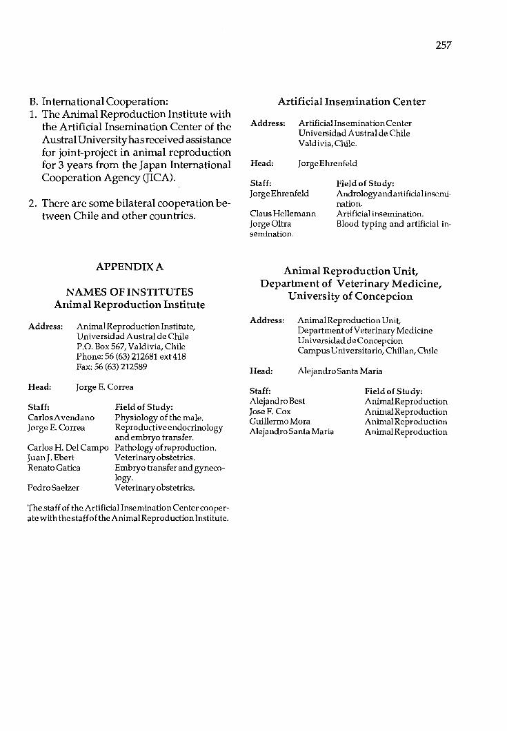
B. International Cooperation: 1. The Animal Reproduction Institute with
the Artificial Insemination Center of the Austral University has received a5sistance for joint-project in animal reproduction for 3 years from the Japan International Cooperation Agency (JICA) ..
2. There are some bilateral cooperation between Chile and other countries.
APPENDIX A
NAMES OF INSTITUTES Animal Reproduction Institute
Address: Animal Reproduction Institute, UniversidadAustral de Chile P.O. Box 567, Valdivia, Chile Phone: 56 (63) 212681 ext418 Fax:56(63)212589
Head: Jorge E. Correa
Staff: CarlosAvendano Jorge E. Correa
Carlos H. Del Campo JuanJ. Ebert Renato Gatica
Pedro Saelzer
Field of Study: Physiology of the male. Reproductive endocrinology and embryo transfer. Pathology of reproduction. Veterinary obstetrics. Embryo transfer and gynecology. Veterinary obstetrics.
The staff of the Artificial Insemination Center cooperate with the staff of the Animal Reproduction Institute.
Artificial Insemination Center
Address: Artificiallnsemination Center UniversidadAustral de Chile Valdivia, Chile.
Head: Jorge Ehrenfeld
Staff: Field of Study:
257
Jorge Ehrenfeld Andrologyandartificialinsemination.
Claus Hellemann JorgeOltra semination
Artificial insemination. Blood typing and artificial in-
Animal Reproduction Unit, Department of Veterinary Medicine,
University of Concepcion
Address: AnimalReproduction Unit, Department of Veterinary Medicine Universidad de Concepcion Campus Universitario, Chillan, Chile
Head: Alejandro Santa Maria
Staff: Alejandro Best JoseF. Cox Guillermo Mora Alejandro Santa Maria
Field of Study: Animal Reproduction Animal Reproduction Animal Reproduction Animal Reproduction
Proc. F ,AO/CAAS Workshop on Biotechnology in Animal Production and Health in Asia and Latin America, Beijing, Oct 9-13, 1989, pp 258 -262
Biotechnology for Animal Production and Health in Colombia
Olga C. Marino Laboratorio de Immunologia
Instituto Colombiano Agropecuario ICA-LIMV
Apartado Aereo 29743 Bogota, Colombia
Introduction
Since Biotechnology provides the technological tools for basic studies oriented toward the knowledge of animals and its environment, Instituto Colombiano Agropecuario (ICA) has encharged Animal Especial Projects Division, to execute research and develop technology addressing major animal health and production problems of the economically important species of the country. There exist limitations like deficient knowledge on the disease behaviour of susceptible species and its effect on production and productivity, deficient diagnostic methods for infectious and parasitic agents, lac}} of knowledge on its structural characteristics and antigenic variations and limited development and evaluation of immunogenic products -all these leading to difficulties on control and erradication campaigns and high risks on production.
On the other hand, the available technology has not contributed significantly towards the marketing ability of our animal products.
Objectives
For the above mentioned reasons the main
applications of Biotechnology in the country will be oriented to animal heal th, reproduction and feeding based on the following general objectives:
1) Identify the etiological agents responsible for the viral, bacterial and parasitic diseases that affect the animal species of economic importance.
2) Study the structural, biological and immunological aspects of these agents to optimize the diagnosis and contribute to its control and erradication.
3) Study and improve the genetic material of the native and productive animal species and develop methodologies to obtain better food products for animals, based on traditional and non-traditional raw materials.
Work Areas
Based on the on-going research and the priorities defined by the needs of the country, the Institute has defined work areas for the next five years as follows:
A)lnfectious agents studies. B) Immunological studies.
C) Genetic engineering. D) Animal reproduction. E) Animal nutrition.
In order to cover these areas it is considered important to apply the Biotechnology tools in more specific topics.
1. Animal infections diseases diagnosis by DNA and monoclonal antibodies probes and/ or restriction enzymes studies applied to viral agents that cause important problems like - Infectious Bovine Rinotracheitis (IBR),
gp-90, go-74, gp-77. - Food and Mouth Disease (FMDV). - Bovine Leucosis (BLV), gp-51, p24,
TAA. - Vesicular Stomatitis (VSV) and - Bovine Viral Diarrhea (BVD),noncyto-
pathogenic strains.
2. Diagnostic and research application of monoclonal antibody technology.
- Isolate protective antigens of IBR. - Serological differentiation of virus strains IBR, DVB. - Improvementandcomplementationof present diagnosis, FMDV serotyping. - Analysis and studies on acquired immunity, bypeptideanalysisof known antigenstostudytheircapabilitytostimulate T-cells protective response (Brucella OMP-peptides).
Soluble and cellular antigens of therapeutic importance in immune response (Species antibody class and cell receptors).
3. Immunogens Recombinant DNA vaccines, chemically synthesized or prepared by gene suppression. - VSV recombinant in Vaccinia virus. - Liposomes or other adjuvants appli-
cable for non-conventional vaccines. - IBR immunogenic protein gene.
259
4. Obtainable biologically important proteins by DNA recombinant technology. - Bovine Interleukines and Interferon.
5. Genetic improvement of productive and native species. - Detection of susceptibility genes for BLV (Locus W6ofBoLAMHC)andresistance to ectoparasites (Ticks). - Preservation of native germplasm and sex determination of embrions. - In vitro fertilization. - Embryonic micromanipulation.
6. Nutritional improvement of animal food. - Genetic manipulation of native forage to provide more nutrients for the animals. - Ruminal microbiology modifications for more efficient digestion and/ or utilization of native forage by the animal.
Immediate Needs
The immediate needs are the implementation of molecular biology and monoclonal antibodies work groups, based on the actual infrastructure as described in the former document "Advances in the Animal Biotechnology". Once the groups get the complete training and the basic technology running the activities will follow the priorities determined by the projects.
Subjects of Studies
In order to generically organize the work, two main subjects of interest have been defined:
I - Genetic, antigenic and immunogenic characterization of RNA and DNA viruses.

260
The primary objectives are to obtain a better knowledge on the functional and antigenical variability of the viruses, to apply this knowledge for study and comparison of field, vaccine and challenge virus strains and to the eventual development of immunogens of practical applications.
II - Physicochemical,antigenicandimmunogenic characterization of structural proteins of protective interest in bacteria and of infection associated proteins (soluble antigen) in hemoparasites.
In order to improve the knowledge of antigens that play a role in the virulence and immunogenicity mechanisms, as well as in the differential diagnosis.
Also to improve the knowledge of hemoparasites biology to look for better protection mechanisms.
Methodology
The basic procedures of moelcular biology and monoclonal antibodies applicable to implement both areas will be:
1: Polynucleotide autoradiography by 32P labelling of the purified viral RNA and mapping of the oligonucleotides by electrophoresis for comparison of the strains to define variability patterns.
Steps: a. Isolation and characterization of the viral
agent from the field case (epidemic) and classical serological classification.
b. Virus replication both in tissue culture and experimental animals.
c. 32P radioactive viral labelling. d. Isolation and purification of labelled vi
ral RNA. e. Viral RNA digestion with specific ribo
nucleases.
f. Oligonucleotide uni- and bi-dimensional electrophoresis.
g. Oligonucleotide mapping by autoradiography.
h. Oligonucleotide mapping comparison between selected viruses.
i. Definition of the variability patterns.
2. RNA, DNA nucleic acid and specific oligonucleotide sequencing.
Steps: a. alkaline partial RNA digestion of the
purified nucleic acid. b. 5' extreme 32P labelling. c. Separation and selection of the labelled
oligonucleotides in polyacrylamide gel. d. Digestion of selected fragments with base
specific enzymes. e. Separation in polyacrylamide gels of the
digested fragments. f. Autoradiography fragmentation pat
terns. g. Residue identification and sequence.
For DNA genomes: a. Restriction enzymes specific fragipenta
tion. b. Fragmentation selection by RNA hybridi
zation from gel to nitro-cellulose paper (blotting).
c. Amplification and cloning of selected fragments.
g. Sequence and autoradiography detection.
Specific oligonucleotide sequence.
Steps: a. RNAse specific digestion of the purified
genomic RNA. b. Enzymatic digestion (alkaline
phosphatase) of terminal oligonucleotide groups.
c. 5' terminal 32P labelling (polynucleotide kinase and 32P ATP).

d. Fragment separation by bidementional gel electrophoresis.
e. Autoradiography map and fragment selection for sequence (as above).
3. Cloning of viral genomic fragments that codifies for immunogenic peptides.
Steps: a. Genomic RNA fragment selection. b. cDNA syntehsis. c. Plasmid or virus cDNA insertion. d. detection of peptide productive clones. e. Isolation and purification of expressed
peptides. f. Evaluation of immunogenic capacity of
the obtained peptides. g. Scale-up based on results. h. Cloning and amplification of DNA se
lected fragments. i. Characterization of expressed peptides.
4. Characterization of viral structural polypeptides and ofinfectioninduced polypeptides by iso-electrofocusing.
Steps: a. Radioactive labelling of viral polypep
tides (35S-Metionine). b. Separation of polypeptides by iso-elec
trophocusing. c. Bidimensional electrophoresis of struc
tural polypeptides. d. Variability pattern of the polypeptides. e. Fragmentation fragments by proteolytic
enzymes from the virus immunogenic polypeptides.
f. Fragmentation and variability patterns comparison of the virus under study.
5. Immunogenic viral polypeptide sequence.
Steps: a. Peptide purification and antigenic char-
261
acterization by immunoblotting. b. Specific chemical and enzymatic fragmen
tation. c. Separation of generated peptides. d. Selection by liquid chromatography
(HPLC) of immunogenic selected peptides.
e. Peptide sequence (total or partial). f. Evaluation of the immunogenic capacity
of the characterized peptides based on amino acid sequence and field strain protection capacity against infection.
g. Chemical synthesis of selected peptides. h. Evaluation of immunogenic capacity of
the synthetized peptides and scale-up the promising peptides.
7. Monoclonal antibodies for antigenic characterization and identification of immunogenic polypeptides.
Steps: a. BALB-C mice immunization with the
peptide. b. Availability of lymphoid cells. c. Fusion oflymphocytes and myeloma cells. d. Progressive selection of productive clones
of specific antibodies using ELISA or RIA. e. Affinity and specificity determination
ELISA or RIA. f. Subculture and expansion.
Applications: I. Peptide characterization, both genetic
engineering or chemically synthesized. II. Etiological agents diagnosis. III. Identification of soluble factors of inter
est in immunoregulation and characterization of animal lymphoid cell population needed for the complete study of a specific agent.
Institutional Cooperation
Since 1985 the Institute has participated

262
with other research Institutions of similar characteristics in the country, universities as well as veterinary biological products company, VECOL, in order to integrate a work group with biotechnological goals. Presently, the groups are coordinated by COLCIENCIAS, the national institution
responsible for science and technology development, and through this channel the Institute has participated in several biotechnology meetings, of national, regional and international nature oriented to cooperate in common interest.

Proc. FAO/CAAS Workshop on Biotechnology in Animal Production and Health in Asia and Latin America, Beijing, Oct 9-13, 1989, pp 263 -265
Biotechnology In Animal Health and Production In Cuba
Carlos M. Mella Centro de Ingenieria genetica y Biotechnologia
apartado 6162 La Habana, Cuba
InJuly1986, thefirstGeneticEngineering and Biotechnology Center (CIGB) was opened in Cuba. The second Cuban center is going to be established and located outside of Havana atthe Camaguey province in 1989-90. The country has a numebr of other research centers and institutions which are also working in the same field. These are the Center for Immunoassay, the Center for Agricultural Health (CENSA), the Center for Animal Breeding (CIMA) and the Agricultural Faculties and Technological Units at the Universities.
The N ationalAcademy of Science of Cuba (ACC) is developing several biotechnological programs and have some additional proposals of new programs directed to improve biotechnological research. This is an indication of the priorities given by the government to this field. As a result, the country has already developed several bioactive substances like human, natural and recombinant Alfa and Gamma Interferons, human recombinant Epidermal Growth Factor, monoclonal antibodies for use in purification and immunoassays, restriction endonucleases as well as enzymes with industrial use like sucrose, invertase and amylase.
On the other hand, various measuring instruments and machines are now available to support the very intense developing
program in this sense. Two examples are the Ultramicroanalytical System for use in the field of immunoassays and the appropriate studies on fermentation techniques with the development of automatic instrumentation which have made possible the existence of the first Cuban fermentor.
A continuosu increment on animal health and production may be the result of applying the advances obtained by the National Research Program in this field.
Applied Research That Will Be In Practical Use Before 1991
Artificial Insemination (A.I.) The fully developing and application of
A.I. technology to the dairy cattle has made it possible to get new cattle breeds adapted to our tropical climate with a high production level. The Mambi, Siboney and Tropical Holstein breeds are practical evidences in this sense. The attainment of a dairy mother as Hubre Blanca, a Mundial Champion record level cattle with 110. 9 kg of milk per day is an indication of how far an under developed country can go in terms of scientific results and their practical use.
There are 8 research and production centers in the country to assure quality and
264
preservation sperms. These centers support our breeding program. Recent development on sperm quality control and insemination techniques are also being studied.
Embryo Transfer
The introduction of embryo transfer in Cuba was as early as in 1975. The first two new-born cattle were attained after one year.
In 1984 eight newborn were obtained from Hubre Blanca. In 1985 the first group of newly born cattle from frozen embryos and the use of embryo microsurgery were reported.
At present there are ten facilities to support the use of embryo transfer technique in production, theirworkismainlytoproduce high quality animals, with a level of 200 existing embryo transfers per facility per year. Cuba is supp lying Charolais and Cebu frozen embryos to the Internatioal Embryo Bank in Praha.
A complete research directed to apply these results in small ruminants is being developed now. The first calf born with this technique in buffalo was reported this year.
Recently, our research is focusing on the embryo sex determination, methods for in vitro fertilization and the very new field of transgenic animals. The gene library for bovines was recently established and the first group of transgenic laboratory animals were obtained.
Animal Nutrition
During the last few years, a system for swine production based on feeding of mo-
lasses based ration enriched by protein from Candida utilis has been proved to be positive. This feeding system provides a 15% increment on protein content in the mixture and a daily gain of 580 g per animal per day.
Another product derived from sugar cane and enriched by the biotechnological method is Sacharina. Its very high protein content comes from a dry phase fermentation process. It has been possibletosubstitutefrom20 to 70% of the grain content on conventional concentrate feed. The use of this feed has been very well evaluated in cattle, sheep and poultry.
Computer Science and Animal Husbandry
Alternative computer systems for help on animal alimentary, reproductive and health planning were created. These systems enable us to know reproductive performance in a dairy operation and to check every animal either during productive or pregnancy cycle to improve the management and predict the reproductive behaviour of dairy cows.
In 1963 the National Veterinary Laboratory system was created; by now 56 laboratories are available, 17 are located in the province or central level and they cover the veterinary services to the 14 provinces and one special municipality. There are 102,000 veterinary units in the country which receive diagnostic aids from these laboratories. The research program in this field is continuously growing.
Production of diagnostics. Several diagnostic reagent sets and the
technology for their application on animal health have been developed. These involved

production of monoclonal and polyclonal antibodies, antigens and hormones either in their natural form or labelled with enzymes or radioactive isotopes.
the U1 trarnicroanalytical System (SUMA) is a photometric/ fluorometric plate reader system widely used in the country. Using this system 21,987 can be tested for diagnosis of Bovine Leukemia with a minimal spent of 0.6 ml of the reactive antigen. The SUMA is also used for diagnosis of pigs' dysentery, infectious bovine rinotracheitis (IBR) or the determination ofaflatoxin b 1 in concentrates and grains.
Immunoassay methods have been also validated on evaluation of reproductive performance and health in dairy cattle. A program for their general use at veterinary services will be developed soon.
The very safe diagnosis of Bovine Leukemia by Hybridization Technique has been recently applied. Two more diagnostic probes are under research by now, Mycoplasma bovia and Babesia bovis.
Therapy and Vaccines The Pneumonia caused by Pasteurella
multocida type a, can be prevented by the use of a new vaccine developed in the country. The vaccine agaisnt IBR has had very positive results on pregnant cows and male cattle used for semen preservation.
The fight against Bovine Bacilar Ichterohemoglobinuria (BBIH) involved a conventional vaccine as well as the research for a
265
recombinant vaccine which may be evaluated before 1991.
The Glucane attained from S. cerevisiae is being used as an immunomod ulator and as an adjuvant in various vaccines.
Probably one of the more interesting results has been the very high antiviral activity of human interferon produced on yeast against the viral encephalo-mycocarditis of pigs.
Practical Value of The Scientific Results
Applied research techniques in combination with basic tools available are actually able to improve animal health and production in Cuba. The present summary only comprises few lines of those more demonstrative applied research. In the background there is an enormous amount of results that will rpoduce in their respective time of maturation the new steps in our developing programs. As a general criteria we can say that the power of biotechnological research will improve the rate of our development to high levels of effieicny. If this is so, then Biotechnology will prove to be an alternative method for achieving development in countries with limited resources such as Cuba. In this sense our country will be happy to see hwo the world in which we live can attain self sufficiency in food and sound health for its human inhabitants.

Proc. FAO/CAAS Workshop on Biotechnology in Animal Production and Health in Asia and Latin America, Beijing, Oct 9-13, 1989, pp 266 -277
Biotechnology Applied to Animal Production and Health in Mexico
E.G. Padilla Instituto Nacional de Investigaciones Forestales
Agricolas y Pecuarias INIFAP - SARH, MEXICO
HISTORICAL DEVELOPMENT OF LIVESTOCK PRODUCTION TECHNOLOGY AND BIOTECHNOLOGY
1. AnimalHealth:
In relation to animal health, Mexico has historically had a well recognized capacity for diagnosis of animal diseases caused by microbes and parasites and to produce biologicals to prevent them. The first Veterinary School in the continent was founded in Mexico in 1853, and none of its alumni produced the first rabies vaccine for humans. In this cehtury the Veterinary School became part of the National University and established laboratories in microbiology, parasitology and pathology.
On the other hand, the Ministry of Agriculture, due to the foot and mouth disease (FMD) outbreak, created a diagnostic laboratory in 1947 and a vaccine production unit. These facilities, after eradication of FMD, became the Central Veterinary Diagnostic Center and started to produce several other vaccines.
In 1962 through an international project, a National Research Center for Animal Production and Health was created with a strong program on development of human
resources, research facilities and experimental stations.
This research center was in charge of production of diagnosis reagents and several vaccines which complemented those distributed by private laboratories. In 1975 an official laboratory for Veterinary Biologicals (PRONABIVE) was established to produce vaccines and diagnostic materials which are important for official campaigns those associated with public health and some which are not attractive as a business for the private sector, but are required in the country, such as Venezuelan Equine Encephalitis, that was developed jointly by the USDA and the Mexican Research Institute.
Under the control of the Animal Health Department, in the early 1970's, a national network of diagnostic labortories was setup in cooperation with producers, backed up by a central reference laboratory. The network grew up to have 108 laboratories located all over the country.
As far as production of conventional vaccines, the rabies vaccine for human was developed and used at the end of last century. In this century it was produced in chicken embryos and later in tissue culture. INIFAP developed a strain isolated of bat

vampires' salivary glands which is widely used in bovines and dogs but its effectivity has been proved also in cats, sheep and goats. This vaccine is being produced by official as well as private laboratories.
Brucella abortus vaccine has been produced and used in Mexico since 1940 against B. mellitensis in 1968. Against Clostridia and Pastuereella since 1940, and Anthrax vaccine since 1907.
For other major viral diseases, vaccines produced in the country have been available for Hog Cholera since 194G, Newcastle Disease since 1947, and Pseudorabies since 1977. The outbreak of FMD of 1947 was controlled and finally eradicated with a vaccine developed and produced in Mexico.
The INIFAP researches have been involved in the development of vaccines against Hog Cholera (PAV-250), Venezuelan Equine Encephaalitis (TC-83), Porcine Erysipelas, and Marek's disease, as well as Rabies, previously mentioned.
Nowadays, the production of veterinary vaccines in Mexico is around 2000 million doses per year, including avian vaccines; human vaccines produced and utilized in the country are nearly 90 million doses.
As part of technological developments, in diagnosis and biological production, tissue culture has been available in INIF AP since 1966; more sophisticated cultures for blood parasites such as Babesia sp. were established in 1979. Monoclonal antibodies techniques were established in the early 1980's as well as immunoenzymatic techniques for diagnostic purposes, even though they are mainly used for research purposes.
267
Establishment of two research centers
In the present decade, two research centers for Genetical Engineering, Biotechnology, and in general, Molecular Biology have been established; CINVESTA V and CIINGEBI. The first one mostly oriented to plants and, the second, covers biomedical sciences and related industries. These centers are involved in basic as well as applied research using DNA techniques such as gene cloning, DNA sequencing, gene mapping, gene constructs, DNA hybridization, and gene transfer. They have developed a few pa tents for ind us trial purposes in an ti biotic prod uction as well as some hormones and bioacti ve molecules.
These centers offer M.Sc. and Ph.D. degrees and there is also an excellent possibility for the animal research groups to interact with them in joint projects.
2. Animal Production
Aspects of animal production and reproduction e.g. animal breeding, and genetic improvement, technologies as A.I. control of ovarian function, performance testing, progenytestingin dairy cattle, embryo transfer and radioimmunoassays for different hormones have been implemented and used in development programs, research and in some cases as routine production practices.
The first bovine A.I. programs date back to 1936, but only in 1946 the first A.I. center was set up. In the mid SO's, a series of A.I. stations were operating along the country using semen diluted and refrigerated. The widespread use of frozen semen started in the mid 60' sand in 1978 the straws replaced the traditional ampules. Pellets have been used on a limited basis mostly by custom freezing services.
268
To date about 1.6 million doses of bull frozen semen are sold in Mexico. Of these, 870,000 are imported and the rest are from Mexican bulls. The official A.I. services of the Ministry of Agriculture has distributed from 400,000 to 500,000 doses through a network of more than 60 frozen semen banks located all over the country, where liquid nitrogen is also available.
In other species, A.I. is very limited, however there are two A.I. centers for swine and ram, and stallion semen are available.
The control of ovarian function has been mostly operated by veterinarians; induction of estrus and estrus synchronization are the most frequent practices. As everywhere else steroids, pros ta gland ins and GnRHare more widely used, while use of gonadotrophins is rare.
Genetic improvement through selection and crossbreeding has been common. Crossbreeding evaluation projects are under way at INIFAP for beef and dual purpose systems in bovine in tropical and semitropical areas with Bos taurus/Bos indicus.
Performance testing exists in beef cattle, swine and sheep, either in official stai tons or in the farm or ranch.
In pigs, five farms sell crossbreed or synthetics seeds tock, male and female lines, utilizing basically an open nucleus breeding system approach. There are approximately another 200 farms that sell seedstock, either purebred or crossbred but that still depend partialy of imported females.
In poultry the schemes are basically the same as anywhere in the world for intensive systems; there are 9 companies with grandparent stock for broilers, 3 for laying hens,
18 with parent stock for laying hens and 61 for broilers. In all cases, the basic lines (grandgrand parent stock) belongs to international companies.
For dairy cattle, the Holstein Friesian Breeders Association has been keeping a production control program for about 40 years, but it was reorganized in the mid 60' s to progeny test A.I. bulls. The list of the first tested bulls was published 16 years ago. Last year they had about 55 ,000 cows under control and approximately 20 young bulls located in Mexico enter the test every year. The information they dev~lop is from intensive production systems, with an average production of 6,500 kg of milk/ cow /lactation.
Embryo transfer
The application of embryo transfer has been increasing since 1976, when the first experiences started. To our knowledge, the first calves from frozen embryos were born in 1979 in one of INIFAP stations.
Currently there are more than 40 private or official groups doing embryo transfer, and more than 3 ,000 embryos were transferred in 1988, most of them frozen.
A large program has been set up from a government agency, LICONSA, which has a genetic improvement program based in an elite nucleus of imported Holstein Friesian and Brown Swiss cows and top quality semen. Their goal for 1989 is to freeze 18 ,000 embryos and transplant more than 16,000. More than 30 pregnancies have been achieved with split embryos. The program is ready to start a MOET-ONBS to produce A.I. bulls and some outstanding bulls for natural mating.
Animal feeding
In relation to animal feeding, the basic techniques are the preservation of forage as silage or hay, mechanical and chemical treatments to stover, corn cobs and different straws and the utilization of different sources of non protein nitrogen, including poultry litter,mostofwhichisusednowadaysasruminant feed.
These technologies have been screened or validated by national research groups, which have devoted most of their time to the nutritional eva.iuation of numerous local products and by-products for animal feeding, and to develop recommendations for their practical use in the feed industry or by the producers themselves. The group of animal nutritionists in the country is one of the largest as far as specialities in animal production and, has gained a good reputation among the producers and the feed industry.
Nutritionists, and Range and Forage scientists have developed an important bulk of information to better utilize the natural grazing lands and introduce improved pastures. More than 7 million hectares of improved pastures are being utilized by producers.
NATIONALPOLICIESFOR THEDEVELOPMENT OF BIOTECHNOLOGY
In Mexico so far, there i-s no specific plan for biotechnology development and utilization.
The National Plan for the administration that started in December 1st, 1988, has been presented and published; however, the science and technology program and the rural
269
development program will be presented by the end of October, 1989.
The National Plan defines that basic research will be granted continuously and increasing financial support by the government be available. However, technological development and developmental research will have to rely more and more in the financing of the productive sector that may require the potential developments.
The program emphasises development of new technologies in animal production and health, and in the alternative use of biomass for animal feeding, underlining the efforts to reduce cereal consumption by animals through the development of alternative feed stuffs. Most programs give empahsis to the development and training of human resources at the highest level.
The objectives of a development in modern biotechnology and genetical engineering for animal production and health, and theparticipationofMexicoinaLatinAmerican Network would be to found mechanisms to apply basic knowledge to solve local problems, develop and exchange experience and useful materials within the region and provide a more efficient support to the national programs and the local industry in animal production and health matters.
As secondary objectives, the program should train researchers in modern biotechnology, transfer technology to the productive sector, allow the country a self determination in biotechnological aspects and enlarge the benefits of the basic research groups financed by the country.
270
PRESENTSTATUSORFUTUREPLANS
A. Embryo Transfer and Genetic Improvement
1. As previously stated in this paper, genetic improvement in general is made by routine procedures used elsewhere; performance and progeny testing, crossbreeding, selection and use of synthetics in swine and poultry.
There are projects to implement in dairy and dual purpose cattle, scheme of open nucleus breeding systems (ONBS) and multiple ovulation embryo transfer (MOET). The government enterprise LICONSA will probably start next year with Holstein Friesian and Brown Swiss cattle; INIFAP, associated with producers is promoting crossbred bull testing (Bos taurus and Bos indicus) for tropical dual purpose systems and a proejct will be presented to the Milk National Commission to use MOET-ONBS, for Holstein Friesian, as a screening and complement for the progeny testing program, and for testing of Jersey bulls. ONBS is used in at least five seedstock swine companies.
2. Multiple ovulation embryo transfer techniques in use: a) Donor selection for MOET is made by
looking at the cow index scores in dairy, performance record in the herd, outs tan ding conformation within a breed or simply to accelerate multiplication of a given breed. They are usually disease free.
In some cases MOET is used as a last resource to obtain progeny from a valuable cow which is hard to settle.
In companies which sell embryos a frequent additional criteria is individual yield
of embryos after M.0. and A.I.
Research is being conducted in INIF AP to define repeatability of ovarian response in MO unselected zebu cows.
b) For MO, FSH-P and purified FSH with very little or no contamination with LH are the most commonly used hormones. so far there is no difference between them. PMSG and anti-PMSG have been tested in very limited studies, but so far practitioners prefer FSH-P.
The results of number of ova embryos recovered and transferable or freezable embryos show great variation; an estimated average would be 6 and 3, respectively. However, when cows are selected for MO response, these averages increase; LICONSA group has collected 8, 123 freezable embryos out of 1,725 flushings.
FSH-P doses range from 20-50 mg. Limited research by INIF AP in non selected zebu cows indicate that doses as low as 18 mg can give satisfactory results (Cordoba et al., 1988).
Most S.O. protocols include injections of FSH-P at 12 h intervals in decreasing doses for 4 to 5 days starting between days 8 to 12 of the estrous cycle. Presensibilization with FSH-P on days 2 or 3 has been studied with good results.
To manipulate the estrous cycle in the donor, prostaglandins and norgestomet implants are routinely used. INIFAP-LICONSA are conducting some trials to improve the donor's response to FSH treatments and testing alternatives to PGF and Norgestomet.
c) There is no local production of hormones,
and to our knowledge there are no serious efforts to produce FSH, prostaglandins or progestagens for synchronization of estrus locally, despite the fact that some groups have the knowledge and expertise to do so. There are some efforts being made to purify LH (Gamboa et al., 1987).
d) On the efect of repeated M.O. on estrous cycles, only few groups keep accurate records and that information is being analyzed. Repeated M.O. occurs mostly in companies that sell or distribute embryos. The E.T. private practitioners are more concerned with the deleterious effects (physical injuries) of repeated flushing on the reproductive tract than on the effects of frequent FSH injections and manipulation of the estrous cycle. Those groups that repeatedly M.0. the same cow do so at 30 to 40 days intervals. In 10 cows closely followed there have not been deleterious effects on their further reproductive performance.
In a private group a Simbrah cow has produced around 180 embryos in 6 months, and 52 freezable embryos out of 57 in a single 5.0. Those embryos are still frozen. A Hosltein cow in LICONSA has produced 148 freezable embryos in nine consecutive flushings.
3. Mating: Virtually all embryo donors are artificially inseminated with frozen semen. Most commonly two doses of semen are utilized either at one time or with a 12 h interval.
4. Almost all uterine flushings are conducted with Foley catheters. Single flushing is conducted in most cases. However if there are difficulties to correctly place the tip of the catheter, each uterine horn is flushed indi-
271
vidually.
Generally speaking the number of corpora lutea is estimated by rectal palpation before ova or embryos collection. This is used by practitioners to have an idea of their relative efficiency. Flushing fluid from the uteri is passed through a filter (70 um) to facilitate a faster searching process. Dulbecca's phosphate buffered saline (DPBS) solution is used for collection and manipulation of embryos. Antibiotics plus 1-5N fetal calf semen (PCS) are added to the flushing media, while 10-15N of PCS in the other steps of embryo manipulation.
Nearly all DPBS is imported and most ET practitioners do notuse the information that shows that bicarbonated media modified with HEPES is a cheaper alternative for short embryo culture periods when freezing is not necessary. In general, practitioners are fond of their routines and the lack of deeper knowledge of the theory behind the techniques prevents them of introducing modifications, to simplify or reduce the cost of the technique.
5. As far as examination and grading of embryos is concerned, world wide accepted procedures are followed. It is a subjective method of observation of embryos in sterile Petri dishes under a stern-microscope.
6. Transfer and/ or freezing of embryos: Transfer of embryos to recipient cows is performed nonsurgically using the "Universal" ET Cassou gun or the one designed by French workers which has a blunt tip and lateral orifices. Before transfer, recipients are palpated rectally to assess the presence of the corpus luteum. Few groups are using ELISA kits to determine progesterone in recipient cows before the transfer. INIF AP is carrying out a research project in coopera-
272
tion with one ET center to determine the efficiency of ELISA kits for progesterone.
Nearly all ET practitioners or groups have embryo freezers. Embryos are frozen with glycerol in A.I. plastic straws. Mostly excellent or good quality embryos are exposed to glycerol in three or more steps. Then they are cooled to -7°C (1°C I min), "seeded" and cooling is continued (0.3 C/min) to-30°C or -40°C. Once they reach this temperature, embryos are plunged in liquid nitrogen. Thawing is carried out in water at 30-37°C. Glycerol is then diluted in 3-6 steps. After that embryos are graded and transferred.
Doctoral students from INIFAP have conducted research towards developing simpler systems to dilute cryoprotectants from frozen-thawed embryos. A promising technique has been developed using a mixture of cryoprotectants of low molecular weight. However this technique has been tested with murine embryos only under in vitro conditions (Hernandez-Ledezma et al., 1987).
Comparative results between transfer of fresh or frozen-thawed embryos do not exist under planned well conducted experiments. Practical results show that pregnancy percent with fresh embryos ranges from 0 to 60% with an average of around 45%, and it is generally expected to use three frozen embryos to obtain one pregnancy.
7. In vitro capacitation and in vitro fertilization: Apparently there has not been any work done in this area in farm animals. However there is expertise in the country in humans, and one INIF AP researcher, did his Ph.D. dissertation in freezing-thawing of oocytes and· IVF in mice. (HernandezLedezma, Ph.D. dissertation, W.S.U., 1988).
A government group is implementing the basic techniques and INIF AP will be part of their project to set up a routine production line of embryos with these procedures.
8. Embryo bisecting/ cloning: Bisection of embryos is being done in at least two laboratories. There is evidence of at least 30 pregnancies so far. INFAP has submitted a research proposal to F AO to pursue embryo splitting techniques.
A private group in the state of Tamaulipas has been transferring consistently two embryos ipsilateral to the CL; results on twin pregnancies is not available yet, however the conception rate increased from 20 percentage points by the use of two em-bryos instead of one. ·
9. Chimera production: To our knowledge, no successful research has been conducted in Mexico with mammals, however one researchers at CIIGB in his Ph.D. program overseas, worked successfully in GH chimeric rodents.
B Application of r-DNA Technology
At least three research institutions in Mexico are using routinely r-DNA technology: CINVESTAV (lrapuato, Gto.) with emphasis in plants and crops; CIIGB from the National University in the state of Morelos in biomedicine and associated industries and the Biochemistry Department of the School of Meidicne in the state of Nuevo Ledn which is working in gene mapping and growth hormone gene expression in prokaryotic and eukaryotic cells.
These groups have the technology for gene mapping, DNA sequencing and all
associated techniques including probes development; DNA hybridization and several methods of gene transfer.
According to the 1988 report of CIIGB, their main research lines are:
- Molecular Biology and Biochemistry of Bacteria. Molecular Biology and Biochemistry of Parasites. Molecular Biology and Biochemistry of Viruses. Cellular Biochemistry of Peptidergic Neurons. Structure, Function and Manipulation of Peptides and Proteins. Methodological Development in Molecular Biology. Industrial Microbiology. Basic Studies in Biotechnology. Optimization and Consolidation of Processes and Prototypes.
INIF AP has held three meeting with the director and researchers of this center to establish more permanent cooperation for training of researchers and joint projects. The current work of CIIGB is in problems of humans, which can be useful in animals, such as GH, RH-LH, some viruses and bacterias, and there is interestto start within the next three years some projects on transgenic animals.
Some projects of INIF AP include: - Isolation of antigens from a genomic
library of Haemophilus pleuropneumonia (INIFAP-FESC).
- Studies on structure, genomic and protein function of porcine rotavirus (INIF AP-CIIGB).
- Immunogenic analysis of Trichinella spiralisin pigs (INIFAP-CINVESTA V).
- Hybridization of a DNA probe for Aujezky's disease diagnosis in pigs.
273
(FES-C-INIF AP-CIIGB). - Contruction of DNA probes for diag
nosis of hemoparasites Anaplasma marginale and Babesia spp. (INIFAPUniversity of Missouri G.).
- Differentiation of strainsofBHV-1 and BHV-4 through DNA hybridization (INIFAB-IMSS-Universite de Liege).
C. Immunodiagnostics and Vaccines
1. ELISA: Severallaboratorieshaveroutine ELISA for different etiological agents such as Rabies virus (INIFAP-PRONABIVE), Brucella abortus, Clamydia psittaci, Coxiella bumetti (Vet-School-UNAM), Anaplasma maraginale (INIFAP), Vesicular stomatitisand MalignantCatharral Fever (CPA) and Cisticercosis (UNAM). A new method for diagnosis of cisticercosis is being developed by INIFAP-Universityof Tamaulipas.
lnstitutionsareworkingtodevelopELISA for diagnosis of Hog Cholera, BVD, I BR, Pl3, and Haemonchus contortus.
2. Monoclonal Antibodies (MoAbs.): They're being used on a limited scale for disease diagnosis and to identify different disease agents. There are only a few laboratories that produce MoAbs for diagnosis of Rabies, Hog Cholera, Bordetella bronchiseptica and Trichinella spiralis. Other laboratories use Mo Abs for research to monitor the expression of epi topes that might be important as antigens for disease prevention as is the case of Babesiosis and Anaplasmosis.
3. Genetically Engineered Vaccines: There is interest for the development of monovalent vaccines for Hog Cholera, aujezky and Rabies to be able to carry out safer control/ eradication programs.

274
INIF AP is collaborating with the University of Missouri in a collaborative research effort for the development of a polyvalent vaccine for Anaplasmosis and Babesiosis. We believe that once the Mexican groups develop the required confidence and expertise in the handling of bioengineering, there will be multiple applications of the knowledge acquired in the process.
D. Biotechnology for Enhancing l/\.nimal Feed Production
1. Genetically engineered forage crop production: Forage crops in Mexico belong to two main categories: those for the dry and temperate areas and the areas of the tropic. The farmer is represented by alfalfa, maize, rye grass, oats, barley, among others. The latter is tropical forage and legumes like African Star, Guinea Grass, Napier, Centrosema, Kudzu, Sugarcane, King Grass, Taiwan Grass, Clitorea, among others.
2. Bioconversion of industrial, agricultural and animal wastes as animal feed: Agricultural, industrial and animal wastes are causing in some areas unacceptable environmental effects, so their bioconversion to aniamal feed is an important and interesting altema tive. There are also research programs to produce usable energy from them.
In these lines, the following projects can be mentioned:
- Lactic fermentation of the solid portion of swine excreta, on a continuous apparatus, and the production of swine feed (CINVESTAV-INIFAP-CONACYT). Production of edible mushrooms using agricultural wastes as substrate (INN-INIREB).
- Swine excreta-agricultural wastes si-
lage as ruminant feed (INIFAP /PG. Swine farmer).
- Biodegradation of the solid wastes from a screw worm-fly factory and its potential use as animal feed (INIF APGMAEGB).
- Protein enrichment of sugarcane wastes (UNAM-1).
- Production of fermentation-enriched cassava meal (UNAM-1).
- Fermentation of high-protein content agricultural wastes (UNAM-1).
- Dairy industry waste treatment and the production of ensiled ruminant feed (INIFAP).
- Fruit juice plant residues-silage for ruminant feeding (INIFAP /Jogos del Valle).
- Sugarcane mill effluents for ruminant feeding (INIFAP-Productos de Maiz).
In relation to identification and genetic manipulation of rumen microbes, we are not aware of any groups doing research in Mexico. It would seem that it is simpler to manipulate the environment of the rumen and thus obtain better condition for beneficial microbiota that will make the rumen fermentation more efficient. Some research in this line is being carried out:
- Synthetsis and application of AAH (acetohydroxamic acid) to reduce ureolysis in the rumen (INIFAP-Univ. of Tabasco).
- A combination and later processing of urea and cassavas meal to reduce ureolysis in the rumen (INIFAP /Int. Found for Science).
- Slow-release device for the administration of ionophore-antibiotics in grazing ruminants (INIFAPELANCO).
- The use of sugar-mill ashes as buffers to control the lactic fermentation in the
rumen (INIFAP). - Alkaline pretreatment of sugarcane
and pineapple-waste silages to reduce the ethanolic fermentation and increase theproductionoflacticacid (INIFAP I Int. Found for Science).
Single cell proteins For ehe development of single cell protein
(SCP) for animal feed in Mexico, researchers have followed diverse approaches using locally available substrates.
Sugarcane, molasses and fibrous byproducts are used to produce yeast ( terula) SPC biomass using mixed cultures.
Petroleum byproducts, methanol, cassava roots and other starchy staples were used as substrates to grow Torula torulapsis (CINVESTAV-IPN).
Other projects are: - Solid fermentation of cassava
(UNAM). Nutritional enrichment of conventional and non-conventional starchy food by fermentation employing mixed lactic bacteria (UAM-1).
- Fermentation of methanol to obtain biomass (IMP).
A pilot plant has been set up by CINVESTA V (DP) for their different research lines.
Even though no enzyme or protein engineering is being done, alternative feeds development of milk whey with Kluyveromyces moroxiamis, lactase-inulinase and lactase-pectinase are under study. Papin production from papaya and studies on the kinetics of proteolytic enzymes from native plants are being carried out (UNAM-I and Tech. Inst. of Tijuana).
275
In fermentation technology there are few research groups involved, either to preserve or improve feed stuffs or to produce energy.
In anaerobic fermentation, silage technology is widely used. The process was known since ancient times, and found in Egyptian paintings and Greek writings; the modernizing of ensilage was started in 1877 and progressed in such a way that today it is generally accepted that provided certain basic principles of silage making are adhered to, the production of a high quality feed for ruminants is assured.
In the case of most tropical grasses, these basic principles are not commonly met, so ensiling is a risky venture for the rancher. Methods to aid the fermentation of tropical grasses are under study: nutritional and non-nutritional additives, wilting, types of silos, etc., to try to manipulate and control the biochemical process.
Examples of some current projects are: - Manipulation of silage fermentation
(INIFAP). - Sugarcane silages kinetics of fermen
tation (INN). - Silage of sugarcane bagasse with
byproducts of ethanol fermentation industry.
For aerobic fermentation, one project is conducted at UAM-I (Univ. Autonomo Metropolitana-Intapalapa) under the leadership of Dr. Gustavo Viniegra. They are fermenting cassava roots with fungi to improve the quality and quantity of protein for animal feed.
In the use of methanogenic bacteria, the Institute of Electrical Research (IIE) uses methane produced by fermentation with animal wastes as substrates, to move water
276
p~mps, generate electricity and produce combustible gas for home purposes.
Industrial use of fermentation technology for animal feed is done by the Mexican Government enterprise FERMEX, a co-investment with Japan which is producing more than 3000 ton/year of lysine HCl, leucine and glut(lmic acid through biotechnology.
COOPERATIVE EFFORTS
In the ear lier part of this pa per a series of cooperative efforts have been mentioned. In general there is a good attitude for cooperation among Mexican groups, directly working in animal production and health or related areas, whose results can be applied to animals.
Cooperation with the industry is mostly associated with the evaluation of some of their products.
The cooperation between institutions, besides some joint projects, is in mutual training, sharing of equipment and in some cases materials.
International exchange within Latin American countries involves training, information and materials exchange. Some of the F AO networks in animal health have been helping successfully. A short course in cultivation of Babesia spp. in vitro was recently offered by INIF AP with the sponsorship of the Animal Production and Health Division of F AO.
Acknowledgements
This paper was prepared with the cooperation of: Armando Shimada, Moises Montato, J. Hernandez L., Irma Tajada, EverardoGonzalez,Eliseo Hernandez, Pablo Correa, Diodoro Batalla, Francisco Trigo, Francisco Suarez, Carlos Vega, Alvaro Aguilar, German Colmenares, Pernin Jinenez and Roberto Saldala. Coordinator and preparation of final draft: Everrardo Gonzalez.
REFERENCES
Centro de Investigation sobre Ingenier a Genetica y Biotecnologie- UNAM. Informe 1988, Mexico.
CXIA-SARH. 1987. Biotecnolog a Alimentaria. Directoria do Instituciones Nacionales. Mexico.
Cozdoba S.M.L.A., Gonzalez P.E., Arriola B.J., Y. VazquezP.C.1988.Respuestaovirica a la superovulacion con hermona faliculo estimulante en ganade cobu. Tee. Pee. en Mez. 20(1): 24, 35.
Gamboa, J.J., Flores R., Carranza M., Reynoso W.A.,Peblano V.M.,Pedroza,J. y Salas A. 1987. La importancia de los nistemac analiticos homologis en el estudio de las especios animales: obtencion de la hormona lu teinizante caprina. rounien de Inv. Pee. en Mez. p. 389.
Hernandez L.J.J., G.T. Gaskins and R.W. Right. 1988. Freezing of mouse embryos with a cryoprotectant mixture of glycerol and 1, 2-propanediol-thering. 29(1): 258.
Henzandez L.J.J.1988. Improvement of feezing techniques for mammalian embryos and

oocytes using the mouse as a model. PhD. disseration, Washington St. Univ.
Plan Nacionalde Desarrollo 1989-1994. Mexico. Talleres Graficos de la Nacion.
Quintero R. 1988. Situacion de la Biotecnologia y Proteccion de la Innovaciin en Biotecnologia en Mexico. Reporte de Grupe a CONACYT.
SARH. 1988. Estadistica Basica 1900-1984 para la Fl ........ del Desarrello Rural Integral. Mexico.
SARH. 1989. Analisis de Costos, Precios y Utilidados para: leche de bovino, carne de bovino, huevo para plato, camo de polla y came de cordo. Mexico.
APPENDIXl
Institutions and Head
INIFAP: Ing. Sergio Reyes Osorio, Insurgentes Sur 894, Col. del Valle, Mexico, D.F.
Facultad de Medicine Veterinaria y Zootecnia: Dr. Jose M .. Berruecos Villalobos,
277
Circuito, Universitario, Cd. Universitarla, UNAM, Mexico, D.F.
Facultad de Estudios Superiores Cuatitlan UNAM: Dr. Jose Luis Galvin M., Apartado Postal222,Cuatitlanizcalli,Edo.deMexico, 59450Mex.
Centro de Investigacion sobre Ingenieria Genetica y Biotecnologia-UNAM: Dr. Francisco Bolivar, Av. Universidad s/n, Chimalpa, Morelos, apartado Postal 510-3, 62330 Guemavaca Mor. Mex. (Mostly Biomedical Research).
CINVESTA V-Irapuato: Dr. Alejandro Blanco Labra. Km 9.6, Libramiento Norte, Carretera Irapuato-Leon, Apartado Postal 629; Irapuato, Gto., Mexico. (Oriented to biotechnology for crops) 28 Researchers. ·
Laboratorio de Ingenieria y Expresion de Genes. Dept. de Bioquimica. Escuela de Medicina. Univ. Autonoma de Nuevo Leon colzada Madoro y. Dr. Eduardo Aguirre Pequeto, Monterrey, N.L., Mexico. Senior Researcher Dr. Hugo Barrera Saida ....
There are also other institutions engaged in livestock biotechnology.

Proc. FAO/CAAS Workshop on Biotechnology in Animal Production and Health in Asia and Latin America, Beijing, Oct 9 -13, 1989, pp 278 - 283
Biotechnology in Animal Production and Health in Uruguay
Julia Saizar Departmento de Virologia
Centro de Investiaciones veterinarias 'Miguel C. Rubino'
A.P.6577 Montevideo, Uruguay
Introduction
1. Historical development of livestock technology in Uruguay
Scientific advance in general, suffered in Uruguay a setback of twelve years, during the time of the military government which took over in 1973, and biotechnology was not an exception.
Upon the return to demoncracy in 1985, only four years ago, highly qualified scientists who were forced to abandon the country returned, and at the same time the country was open for international and bilateral cooperation. With the assistance of the United Nations Development Program (UNDP) and UNESCO, the Government established the Basic Sciences Development Program, which aims for the development of Biology, Physics, Informatics, Mathematics and Chemistry. Under this program, returned those scientists who were working abroad, and brought with them important direct contacts with the laboratories they had been working at.
Very rapidly, inter-institutional programs were established, to take full advantage of the few human resources available, spread
out in national institutions as well as in the private sector.
Why is development needed? The development of biotechnology in the
field of animal production is of the utmost importance for Uruguay, because its economy depends largely on agriculture and livestock production. It is essential for further genetic improvement of livestock at national level, and potentially for exports of frozen embryos to other countries in the region, of highly qualified genetic material. Today, Uruguay exports live animals. It enables breeding tests in a much faster way, as it is possible to test bulls through their sisters' performance, instead of their offsprings, th us gaining one genera ti on' s time. Much interest has been shown in this field.
Regarding diagnosis, it opens a new field which has recently been developed and requires investment in training of human resources and equipment.
2. Are there any national plans for development of biotechnology in the country, especially in relation to animal production and health?

No national plans in biotechnology exist yet in Uruguay. However, the Government has created the National Committee for Biotechnology, which has representatives of the public and private sectors. It was created as a basic need to enable the country to enter into UNDP' s Regional Programme of Biotechnology for Latin America and the Caribbean. Its objectives are:
a. Assess the country's possibilities in biotechnology.
b. Assist those institutions (private or public) interested in doing research in this field through contacts with regional and international institutions and labortories.
c. Assist scientists with scarce funding through this Regional Project for specific training or assistance to meetings within the region. One research project is being funded through this program. Even when its objective is the production of DNA probes for the diagnosis of viral diseases in plants, the staff developing the rDNA technology will be able to assist the veterinary sector in the production of probes for the diagnosis of animal diseases.
d. Establish research links between the University, national research laboratories and the private industry.
e. To aware national authorities of the importance of biotechnology development in the country. In this sense, a plan for the development of biotechnology has been prepared and is under consideration at national level.
TheslowbutsteadyworkoftheNational Committee has been successful and the Government has established a sector of biotechnology in a Project funded by the Inter-
279
american Development Bank (IDB), through which research projects in this field will be financed.
This is generally speaking of biotechnology, a much broader subject of which animal production is an important input in Uruguay's economy.
Concerning regulations, it has been established by decree, that artificial insemination (AI) and embryo transfer (ET) must be performed by veterinarians. It has also been approved by a recent sanitary decree which regulates the importation of embryos.
One specialized society has been created, (CRIA), Commission of Reproduction and AI within the Veterinary Medicine Association with a specific section on ET.
Present Status or Future Plans
A. Nucleus Herd Improvement Using MOETinONBS
1. Present genetic improvement programs Genetic improvement in Uruguay has
been traditionally performed through AI, mainly in dairy and beef cattle and sheep. While it is true that a faster genetic improvement is achieved through AI, when used at national level, it should be considered that at herd level, where high quality females are superovulated and ET performed, the intensity of selection will be increased and the inter-generation intervals shortened. If ET is used with selected females, their genetics can be spread to big herds through AI.
In 1980 was born the first calf through ET, at the Veterinary Research Centre "Miguel C. Rubino" (CI.Vet.). Since then, several groups of private veterinarians are working

280
in this field, at a rate of 150 /250 ET per year. Herd improvement is achieved at private level in each farmer's herds, with registered cattle. There is not yet diffusion at national level.
Much interest exists at the Cl.Vet., the Faculty of Veterinary Sciences from the UniversityofUruguay(Fac. Vet.), groups of private veterinarians who perform ET and a cooperative group of dairy farmers who own 10,000 controlled cows, to initiate a MOET program. Females of high genetic quality going to slaughter houses will be superovulated and inseminated (in vitro fertilization could be used later). An embryo bank will be established, to assist farmers of low resources to improve the quality of their herds.
The Faculty of Veterinary Sciences has a project with JICA in this field, and this year in vitro fertilization was achieved, al though only at an experimental stage.
ET is being performed chiefly in cattle (dairy or beef), but much interest has been shown in sheep. AI will be also used in swine, to obtain cross-breeds adapted to Uruguay's conditions, to increase the hog population of the country.
In poultry, it is essential to investigate towards the production of Uruguay's own grand-parent lines.
a. Donor selection ET is performed chiefly in dairy and beef
cattle. Donors are selected according to their production performance, and belong to the national herd. No importation of donor has been done, chiefly because Uruguay has a herd of high genetic quality. Presently, live animals are being exported to other countries of the region, and it is expected that in
the future it will be changed to frozen embryos. The objective of farmers with ET is to win prizes at cattle fairs, to back up the sale of their breeders.
b. Superovulation Superovulation is accomplished with the
administration of Porcine Foliculo-Stimulating Hormone (FSH-P) (Folotropin) in decreasing doses (2 daily injections during 4 days).
Synchronization is achieved with prostaglandine F2 (PGF
2), which is admini
stered with the fifth injection of FSH-P.
Hormones are neither produced nor purified in Uruguay.
c. Mating For ET purposes, ma ting is done through
AI only.
d. Collection of ova It is done non surgically, either through
gravity in a closed circulation circuit or through interrupted aspiration. Each expert or team has its own methodology, which can be the ones mentioned, or a combination of both. Pregnancy results are comparable to those obtained by other countries in scientific literature (50/60%).
e. Examination and evaluation of ova and embryos Embryos and ova are examined carefully
and classified before performing ET.
f. Tranter and/or freezing of embryos ET is performed mainly surgically. Re
ceptor females are generally virgin heifers.
The embryo freezing technique is Willadsen's (from -70°C, decreasing 0.3°C upto -36°C with glycerol). Thawing is in

three steps with glycerol. No data is available on pregnancy results with frozen embryos.
g. In vitro capacitation and fertilization Only on an experimental basis, the
Fact.Vet. was able to perform successfully in vitro fertilization, with the assistance of a JICA expert. Under JICA's assistance, important pieces of equipment were received, a micro-manipulator was one of the most important pieces of equipment.
Good results have been obtained at CI.Vet. with in vitro capacitationof oocytes. With sperms capaci tation, certain problems have been encountered.
Nothing has been done so far in embryo bisecting/ cloning; chimera production; sexing garnets or nuclear transplantation of embryos.
B. Application of r-DNA Technology for Genetic Improvement and Genetic Resistance to Diseases in Livestock/Poul-try
Before dealing with each particular item under this subject, it should be mentioned that several research groups are working with these techniques in basic research, but so far very few have entered the field of animal production, either directly or through inter-institutional joint research. The important thing to be highlighted is that the technology exists and can be available for prospective consumers working in applied research.
The fact that no diffusion of the results achieved has been made, is perhaps the main obstacle in introducing these techniques in animal production.
281
1. Gene mapping/DNA polymorphisms The five techniques mentioned under this
item are being handled in basic research: (a) restriction fragment length polymorphisms; (b) hypervariable region probes; (c) oligonucleotide probes; (d) gene mapping; (e) DNA sequencing.
However, the Biological Research Institute "Clemente Estable" (IIBCE), one of the laboratories where these techniques are being handled, is being consulted by veterinarians who are interested on the following:
- identification of parents - verification of the purity of breeds - selection tests in cattle and sheep - sex determination in embryos with
DNA probes - correlation of cell markers with pro
ductive characteristics - pedigree certification - species determination in meat prod-
ucts
The above-mentioned illustrates how quickly this technology will be applied at full, once its benefits are known.
2. Gene cloning Under this item, a few techniques are
being handled: use of vectors; genes for immune response; search for sequences in local genomic or c-DNA library.
No research work is being done in: gene constructs; genes for production of related hormones and for enzymes; genes for resisance to diseases; other genes; mutant cell lines.
3. DNA hybridization techniques A DNA probe for the diagnosis of Infec
tious Bovine Rhinotracheitis (IBR) is being produced in a joint venture between the CJ.Vet. The Dept. of Molecular Biology of IIBCE and the Fae.Vet., through a project

282
funded by the University of the Republic.
If the IDB project mentioned before is approved, a DNA probe for the diagnosis of BVD will also be produced. Much interest exists in producing a probe for the diagnosis of Babesia Bigemina and Bovis. These protozoa have common antigens, and it is expected thattheprobewillsolvethe problem of identifying each one with 100% certainty. However, for the time being probes in Uruguay are beingmade frm viral DNA/RNA, as the technique for parasites is more cumbersome.
The CI.Vet. will start working soon in red blood cells cul tu re to obtain clones from babesia's metabolic antigens from the supernatant. The CJ.Vet. is the reference center in F AO' s Haemoparasi tes Regional Network. The diagnosis ofbabesia in large scale is very complicated through immuno fluorscence (IF). The production of a purified antigen for ELISA, is also difficult, for which reason a r-DNA probe is believed to differentiate between Bovis and Bigemina.
4. Gene transfer No work has been carried out in this field.
There is a proejct on transgenic laboratory animals, which has not yet been initiated due to lack of funding.
C Immunodiagnostics and Vaccines
1. ELISA This testis being carried out at CI.Vet. for
the diagnosis of IBR, BVD (identification of circulating antibodies or antigens), Tuberculosis, Brucella Ovis, Rotavirus and Babesia.
Regarding anaplasma, preimmunitation with A. centrale is being done in Rubino
since 1940. It is important to know what is happening with A. marginale and the card test being used preesntly is not specific enough to differentiate between them. Through a project between the University of Illinois/Venezuela/Uruguay, a staff member from the Parasitology Dept. of CI.Vet. will receive in service training in Venezuela in anaplasma antigen purification, to set up the ELISA test.
2. Monoclonal antibody production Mab production is not used for diagnos
tic purposes in animal production presently. However, one research group at the Immunology Dept. of the Faculty of Chemistry, has developed this technology to produce an anti-idiotypic vaccine against Equinococcus granulosus. Another group also working on E. granulosus is constructing a c-DNA library from scolex m-RNA in an expession ".'ector (lambda gt 11), which will provide a complete antigen library, useful for the search of the anti-hydatidic vaccine mentioned. Once again, it should be stressed the importance of the inter-institutional and inter-disciplinary activities to enhance research results.
3. Genetically engineered vaccines A monovalent genetic engineering vac
cine against Foot Rot (F. nodosus) is under research, in a joint venture between a private laboratory, IIBCE and CI.Vet.
D. Biotechnology for Enhancing Animal Feed Production
Very little is being done in this field, except for a project on treatment of lignocellulose with bacteria to produce animal feed, which is at preliminary stages.
Cooperative Efforts
A. National Resources 1. Cooperative program between IIBCE/
Fact. Vet./ CI.Vet. CI. Vet. provides purified antigen, IIBCE develops probe technology and CI.Vet./Fac.Vet. perform probe testing in field samples, infected cells and histological cuttings. Benefits: IIBCE handles new technique; CI.Vet./ Fae.Vet. obtains probe for diagnostic purposes. Their staff is trained in new methodology.
2. Cooperative program between private laboratory, IIBCE and CI.Vet. CI.Vet. provides strains, IIBCE develops technology and the private laboratory produces the vaccine. Benefits: Biotechnologymethodology used in industrial scale.
B. Outside Donors 1. Cooperative project between CI. Vet. and
Dept. of Immunology of Faculty of Chemistry. Benefits: CI.Vet. uses reagents produced. Faculty of Chemistry employs young professionals who produce these reagents. Local training of staff in CI.Vet.
283
2. Cooperative project between the Dept. of Immunology from the Faculty of Chemistry and the Imperial College in London.
3. Cooperative project between the Dept. of Biophysics from the Faculty of Sciences and Institute Pasteur & Mme. Curie from France.
4. Joint project with CI.Vet., the Agricultural National Institute (INTA) from Agentinaand the UniversityofMontreal. Beneifts: CI.Vet. and INT A will train their staff in Canada to develop the vaccine with Canadian expertise.
5. Cooperative research with CI.Vet./Venezuela/Universityoflllinois. Benefits: Uruguay and Venezuela will train their staff.
6. Cooperative project to be funded by EEC to work jointly between CI.Vet. and Weybridge Laboratory, where a bacteriologistfromCI.Vet.spentoneyearworking in this field. Bendfits: Uruguay will have the technology to control clostridial vaccines.

Proc. FAO/CAAS Workshop on Biotechnology in Animal Production and Health in Asia and Latin America, Beijing, Oct 9-13, 1989, pp 284 -286
Cloning and Expression of Porcine GHcDNAGene
Qi Shun Zhang Beijing Agricultural University,
Beijing, China.
It is proved in many laboratories that the treatment with porcine growth hormone can increase the feed efficiency and lean tissue in pig significantly. The main results of our experiments carried out in 1987 and 1988 are as follows:
about 0.5 g pure pGH can be obtained from 4000 pituitary glands by our experiment and this amount can only be used for injection of 8 pigs. It is clear that such method of production of pGH is not practical. Seeburg et al. reported in 1983 that they expressed
Table 1. Response of Pigs on the Treatment with pGH
Protein content in feed % Increament of daily gain % Increament of feed efficiency% Increament of lean tissue kg/head
Tr. l
14 15.1 15.0 3.34
Tr. 2
16 18.7 19.0 4.96
Tr. 3
18 24.4 22.3 5.89
Injection started with body weight about 60 kg, 2 mg/pig/ day, continued for 28 days.
There are about 3 hundred million pigs in China. However, it still cannot meet the demand for pork, especially for the lean pork since our population is so large and pork is the chief meat consumed in China. Furthermore, the feed resources is limited in our country, so the treatment with pGH on pigs is one of the good methods of increasing pork supply since more lean pork can be obtained with less feed. The problem is how to produce enough porcine growth hormone. If we isolate it from pituitary gland only
pGH in E.coli in high efficiency. We also try to produce the stuff by genetic engineering.
Firstly, we isolated total mRNA from pituitary glands of pig, by In vitro translation. We can see a dense band with MW about 24kd, which is corresponding to the MW of pre-procine growth hormone. It suggest that there would be a large portion of pGH mRN A of whole length in our preparation. This sample was then used to synthesize cDNA by reverse transcriptation. From
FAO LIBRARY AN: 302021

the results of electrophoresis of total cDN A, we can see thatthe length distribution of the dscDNA is in the range of 300-1000 bp. The longer cDNA portion was then collected and ligated to plasmidpUC19.E.ColiJM107 was transformed with the recombinant plasmid. About 4000 recombinant clones were obtained and from which clones containing pGH cDNA were screened out by probe hybridization.
At first, the clones were screened by hybridization in situ with a probe complementary to the middle region of pGH cDN A and more than 40 positive clones were resulted. From these positive clones about 18 positive clones were obtained by dot-blotting with a probe complementary to the 5'end region of pGH cDNA. Since these 18 clones were hybridized to both the probes, it suggests that they might contain the whole length sequence of pGH. Two of the 18 clones were taken out randomly for further analysis. After enzymatic digestion with a variety of endonucleases the cloned cDNA was proved to contain the whole length of pGH gene coding sequence. The cloned sequence was then sequenced by Sanger method.
Firstly, the first nucleotide of our sequences in 5' terminal end is very close to the cap site of procine growth hormone chromosomal gene and its3' end contains poly A sequence, which shows that our sequences are quite complete and longer than that isolated by Seeburg et. al.; secondly, there are certain differences among our two sequences and Seeburg' s. Among the differences Jia' s sequence and See burg's sequence do not cause amino acid substitution, whereas Wang's sequence contains two different amino acid codons in the leading sequences. In addition, pGH gene contains two initiate codons, which is in good agree-
285
mentwith the published data of bovine and ovine growth hormone gene sequences. The significance of biological function is unknown.
In order to achieve high expression of pGH in E. coli, the pGH cDNA was modified as follows: firstly, leading sequence was removed and a SD sequence plus a initiate codon was added to the first codon of the mature hormone sequence; secondly, the first twenty codons were modified so that the commonly used codons in E. coli were used in places where codon usages are different between E. coli and pigs. The modified structure was then put under the control of a strong promoter for efficient transcription. High expression was first achieved by use of a strong TAC promoter, in coperation withprofessor McConnell's laboratory, Trinity College, Dublin. The expression level is more than 30% of the total bacteria protein. Afterwards an expression vector was constructed in our laboratory by use of the temperature inducible lambda promoter. The expression level of the later system is comparable with that of the previous one. The expressed pGH reacts with pGH specific antibody as proved be western blotting. Its N-terminal analysis shows that the first twenty amino acids are the same as that of natural pGH (Fig. 1) except a methionine in the extremeN-terminal, which was caused by the addition of the intiiate codon.
Fig.1: The First Twenty Amino Acids
met phe pro ala met pro leu ser ser leu phe ala asn
ala val leu arg ala gln his
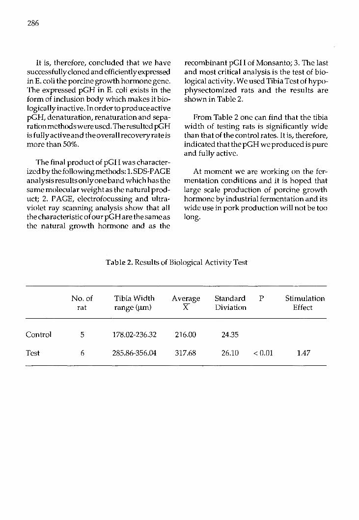
286
It is, therefore, concluded that we have successfully cloned and efficiently expressed in E.coli the porcine growth hormone gene. The expressed pGH in E. coli exists in the form of inclusion body which makes it biologically inactive. In order to produce active pGH, denaturation, renaturation and separation methods were used. The resulted pGH is fully active and the overall recovery rate is more than 50%.
The final product of pGH was characterized by the following methods: 1. SDS-PAGE analysis results only one band which has the same molecular weight as the natural product; 2. PAGE, electrofocussing and ultraviolet ray scanning analysis show that all the characteristic of our pGH are the same as the natural growth hormone and as the
recombinant pGH of Monsanto; 3. The last and most critical analysis is the test of biological activity. We used Tibia Test of hypophysectomized rats and the results are shown in Table 2.
From Table 2 one can find that the tibia width of testing rats is significantly wide than that of the control rates. It is, therefore, indicated that the pGH we produced is pure and fully active.
At moment we are working on the fer'"' mentation conditions and it is hoped that large scale production of porcine growth hormone by industrial fermentation and its wide use in pork production will not be too long.
Table 2. Results of Biological Activity Test
Control
Test
No.of rat
5
6
Tibia Width range (µm)
178.02-236.32
285.86-356.04
Average x
216.00
317.68
Standard P Diviation
24.35
26.10 < 0.01
Stimulation Effect
1.47

Proc. FAO/CAAS Workshop on Biotechnology in Animal Production and Health in Asia and Latin America, Beijing, Oct 9-13,1989, pp 287 - 291
Genetically Engineered Hormone Production in Relation to Improving Animal Performance
Zeng Yixiang and Du Nianxing Division of Immunology and Microbiology,
Department of Veterinary Science, Nanjing Agriculture University,
Nanjing, P.R. China.
Promoting animal's growth has always been the popular sentiment of all the workers involved in animal husbandry and veterinary medicine. Biotechnology development had led to a series of trials in this field and as a result many successful reports. The utilization of growth hormone and the transgenic animals which were produced by the technology of gene microinjection in practice had brought out great value for all developing countries. Now we onlywantto report some research projects in Nanjing Agricultural University in the following three fields: genetical vaccine of somatostatin, synthetic peptide immunological castration and the multi-pregnancy vaccine.
I. Genetical vaccine of somatostatin
Antiserum against somatostatin can enhance the animal's growth and decrease the animal's feed consumption by its neutralizing the somatostatin and increasing the secretion of the growth hormone, TSH and other corresponding hormones in vivo. In 1970s, many researchers reported that passive immunization of domestic or experimental animals with an antiserum against synthetic smatostatin caused the increase secretion of the growth hormone. Maccecchini reported that passive immu-
nizing rats with monoclonal or polyclonal antibodies against somatostatin can enhance the animal growth by 10-15% compared with the control group. At a series reports, Spencer reported the similar results in sheep and lamb. In 1973, Itakura chemically synthesized somatostatin gene and made it express in E. Coli. From these results, we think, with the genetical engineering method we can produce a fusion protein including the somatostatin and this fusion protein can be used as a new kind of hormone vaccine and this vccine will probably be applied greatly in the production of animal husbandry.
This research project is divided into three parts. The first is on the chemical synthesis and cloning of somatostatin gene.
Chemical synthesis and gene cloning
As well known, somatostatin is a cyclic tetradecapeptide, the amino acid sequence of which in most veterbrates are the same. Many experimental results show that only the chemically synthesized somatostatin conjugated to the carry protein can induce the high titre of the antibodies, because the somastatin is a small molecule which is immunologically indistinguishable among different animal species, and is subject to
FAO LIBRARY AN: 302023

288
rapid enzymatic destruction both in blood and in tissues. So the designed sequence is almost the same to the natural somatostatin gene.
For the convenience of the gene manipulation, several other features were built into the nucleotide sequence in addition to the 14 codons for the structure information of somatostatin. First, to facilitate insertion into plasmid DNA, the 5' ends have single stranded cohesive termini for the BamHI and HindIII restruction endonucleases, second a methionine codon proceeds the normal NH2-terminal amino acid of somatostatin, and the COOH-terminal codon is followed by two nonsense codons.
Twooligonucleotide fragments, A52 and A23, were chemically synthesized with AB 380 DNA Synthesizer by the solid-phase phosphitriester method and then assayed by PAGE. The bands position compared with the marker bromophenol blue and xylene cyanol show that the base number of the chemically synthesized oligonucleotide is equal to the number of design.
Two fragments were annealed first, then extended it into blunt end with the polymerase Kienow. The extended mixture was first assayed also by PAGE, then extracted by phenol, precipitated with ehtanol and then ligated with thepUC12 DAN digested with Smal and transformed to E. Coli strain JM83.
Screening of the colonies: pUC12 plasmid carry the lac promotor system, the ampicillin resistance marker and the B-galactosidase structure gene flanked by a multiple restriction site. Bacteria which carry the pUC12 plasmids wereidentifiedby their blue color in the IPTG and Xgal medium for the synthesis of B-galactosidase. If the for-
eign somatostatin gene is inserted at the Smal site, the B-galactosidase gene will be in out-frame and the bacteria cannot produce any more B-galactosidase and then were identified by their white color in the IPTG and Xgal medium. 74 white colonies which were ampicilin resistant were chosen.
Identification by restriction endonuclease digestion: pUC12 plasmid DNA has only single Smal site at the polylinker region, when foreign gene was inserted, the sequence of the base composition will be changed, and the Smal site will be disappeared, however if the recircularization of vector DNA take place, the Smar site will be recovered. By this technique, we had chosen 10 transformants which lack Smal recognition sequence.
Southern hybridization assay: The labelled fragment A23 was used as probe, the transfer of DNA from agarose gel to nitrocellulose paper and the hybridization of the filters were performed by the method of Southern. Nine colonies were identified positive.
DNA sequence assay: pUC12 plasmid DNA having the same sequence flanked the polylinkerregion as M13 phage DNA, so we chose the oligonucleotide (5'GTAAAACGACGGCCAGR-3') as the sequence primer for the p UC12 DNA system. The result shows that the sequence is the same as designed, so it is certain that ihe somatostatin gene hasbeen successfully cloned into the E. Coli strain JM83.
Gene expression
The second part of this project is on the expression of somatostatin gene in E. Coli. The plasmid PBD
2 carry the E. Coli. UV5
promotor and a part of coding sequence of

the E. Coli lacZ gene, the truncated lacZ gene is flanked by a polylinker region at their 3' end, which has several unique restriction sites, coding sequence of any gene can be ligated in frame to thetruncated lacZ genes.
Somatostatingene fragment extracted and purified from plasmid pSom bacterium was ligaged with the plasmid pBD2 DNA. Transformation of E,. Coli D29A1 with the chimeric plasmid DNA were identified by the ampicillin resistance, in situ hybridization, restriction enzyme characterization and southern hybridization, this transformation led to the synthesis of a polypeptide including the sequence of amino acids corresponding to somatostatin. The Chimeric protein was purified and characterized by the Bgalaclosidase affinity chromatography, assayed by PAGE and the expression of somatostatin gene in E. Coli. D29A1 is determined after the radioimmunoassay to the chimeric protein and its mixture by treatment with cyanogen bromide.
The results of radioimmunoassay had shown that the somatostatin and B-galactosidase (fusion protein) is stable and dissolvable. The fusion protein can directy react with the antibody, so it is obvious that the somatostatin molecular is on the surface of the fusion form. Somatostatin expressed in E. Coli. strain D29A1 is 0.71 % of the total bacterial protein.
Production of somatostatin vaccine
The third one is on the production of somatostatin vaccine and its utilization in experimental animals.
The strain producing hybrid protein were grown in LB-rich medium at 37°C in the presence of 25 ug/ ml ampilicillin and then
289
was harvested by centrifugation and disrupted by sonication. Cell debris was removed by centrifugation and then the fusion protein extract was obtained with the Sephadex G-200 chromatography.
At one month of age the rats were given the immunizations against the appropriate antigen. The fusion protein was dissolved in the elution buffer and mixed to a firm emulsion with 2 parts Freund' s Complete Adjuvant. The emulsion was injected subcutaneously. The amount of antigen administered to each rat was 250 ug.
At the start of treatment there was no difference between the mean weights of the control and treated groups.
Within the first two weeks of the initial immunization a clear difference in the weight of the treated and control rats were apparent, the difference in the mean weight of the two groups became significant (by student's t-test) after 2 weeks of treatment, but only significantly so early in the experiment. Later this experiment stopped because of the high temperature in July in Nanjing.
Somatostatin vaccine will play a great role in the animal production. Now this research is under way.
II.lmmuno-castrating effect of chemically synthesized peptide vaccine.
In the practice of animal husbandry and veterinary medicine, surgical ca~tration is always the tradition method. Immunological castration will be the trend in this field. So our laboratory started this research project in 1987.

290
Leuteinizing hormone releasing hormone (LHRH) was covalently conjugated to Bovine Serum Albumin (BSA) and each BSA molecule bears 18 molecularsof LHRH. This complex was used as an immuno-castra ting peptide vaccine (ICPV). Three male New Zealand white rabbits, aged two months, were inoculated with the emulsion of ICPV with Freund's Complete Adjuvant. Each rabbit received 0.5 mg of ICPV at the first injection and 0.15-0.38 mg of ICPV at 2nd-5th injection at intervals of three weeks.
Another three male rabbi ts, of same strain, same age and weight used as control, were kept in the same condition. When all the rabbits were5monthsold the sexual behaviour and reproductive organs were examined and all the rabbits were slaughtered for anatomical and histological observations. The experimental results demonstrated that the immune response induced by the injection of ICPV in young male rabbits neutralize the biological activity of LHRH and inhibit the secretion of pituitary LH, FSH and testicular tesuerone resulting in blocking the development of reproductive organs and elimilating the reproductive ability, therefore, immunization with ICPV is expected as an alternation to surgical castration.
Since many difficulties will be overcome in obtaining enough LHRH by the traditional method, we also had decided to construct a recombinant bacteria which can produce the LHRH fusion protein. We are sure this new kind of hormone vaccine will develop a good market in the world in the near future.
III.Steroids-carrier complex's effect on the animal's reproduction
Increasing ovulation rate and induction of multi-pregnancy by active immunization against steroids are significant both in practice and theory.
In 198 7, we also started this project. Firstly, testosterone and oestrone were covalently conjugated with BSA 20. Merino ewes were inoculated twice with the T-BSA/E-BSA and DEAE-dextran adjuvant 6 weeks and 3-weeks before the insemination respectively, other 13 ewes were used as control ( C-group). The antibodies titre against testosterone and oesteone is 1:120, 1:3 for group T and 1:2, 1 :50 for group Eat the time of insermination. The lambing rate is 167.71 % for group T (P<0.01), 156.25% for group E (P<0.05) and 115.38% for group C. The level of progersterone on Day 12 of the cycle was 9 .94 ± 4.21 ng/nl for group T and 9.31 ±3.53 ng/ml for group E, significantly higher than that of group C (3.71 ± 0.92 ng/ml) (P<0.05 and P<0.01 respectively). The concentration of oestradiol was similar in all groups. The key problem in this experiment is to use of DEAD-dextran adjuvant instead of Reund' s Complete Adjuvant and then control the antibodies titre within the range of 1:50 for group T and 1:80 for group E.
Audrostenedione was covalently conjugated to BSA and each BSA bear 13 steroid molecules. This complex, mixes with Freund's Complete Adjuvant, can be used as the fecundin. 2md of fecundin were injected twice 4 weeks and 1 week before the insemination. The lambing rate was 285% for treated group and 228% for control one. The 1,2,3 and 4 pregnancy rate in these two groups were 3.7, 25.93, 51.85, 18.32% and

19.22,36.53,40.38,38.5% respectively. The2, 3, 4 pregnancy rate for the treated group is higher than that of control one, and the initial weight had no difference between these two groups. In the same condition, when theAustralianfecundin was used, the dead pregency number is high because of the hypoimmunological recation.
References
1. Zeng Xiyiang, Du Nianxing and Tang Jinyan. J. Biotechnology. 4. 4(1988) 292-297.
291
2. Lin Gongping, liu Shenjiang, Du Nianxing and Xu Weiyan, unpublished material.
3. Cui Li ping, et al., unpublished material.
4. Zeng Yixing, Du Nianxing and Tang Jinyang, unpublished material.

Proc. F AO/CAAS Workshop on Biotechnology in Animal Production and Health in Asia and Latin America, Beijing, Oct 9-13, 1989, pp 292 -294
In vitro Fertilization and Embryo Transfer in Cattle
·Jiang Shi-e, Zhang Li, Liu Jian-ming, Huang Shao-hua, Jin Zhan-qun, Gao Chang-heng, Meng Wen-zhu, Zhu Yu-ding
Laboratory of Animal Bioengineering Institute of Animal Science, CAAS, Beijing, China.
Summary
Bovine follicular oocytes surrounded by cumulus cells as a cumulus-oocyte complexes were matured, fertilized and developed in vitro utilizing co-culture system with bovine cumulus cells. The oocytes were cultured int he maturation culture systems A and B. The culture of oocytes in system A has good effects on the maturation, fertilization and development of early embryos. Five recipient cows received fresh embryos and one of them was pregnant; other five received embryos after freezing and thawing and two of them were pregnant. The results of this study proved that the embryos obtained from oocytes had undergone in-vitro maturation, fertilization and further development to the morulae or blastocysts, even after freezing, have a satisfying viability.
Introduction
The first bull calf was born after in-vitro fertilization (IVF) in 1981 (Brackett et al. 1982). Since then, fertilization has been accomplished in vitro-for three domestic ruminants - cattle, sheep and goats. Recently, a great deal of attention is being paid worldwide to the bovine in-vitro fertilization (BIVF) and the tremendous progress in BIVF
has been achieved. In-vitro fertilization of oocytes would have a major impact on bovine embryo transfer. It has been demonstrated that BIVF is a practical technique which can be used to produce large amount of cheap embryos in larboratory and is very important for further development of genetic engineering, nucleotransfer and embryo sexing. All of them will have a tremendous influence on scientific research and animal production.
The authors have studied in-vitro maturation of oocytes and developed a co-culture system for culture of early embryos with cumulus cells. The embryos fertilized in-vitro and undergone development to morulae or blastocysts were transferred either fresh or after freezing-thawing.
Materials and Methods
Collection of Oocytes: The ovaries were collected from the
slaughter house and brought to laboratory in Dulbecco's phosphate buffered saline (PBS) at 35-38°C within 3 hrs. Follicular oocytes were collected by cutting off follicles of 2-Smm in diameter with scissors and then cut out the oocytes from follicles.
FAO LIBRARY AN: 302025

Maturation of Oocytes: The oocytes were washed 2-3 times with
modified Dulbecco's phosphate buffered saline (mPBS) and once with maturation medium, and then were cultured in the following co-culture systems. System A: 20-50 oocyteswereintroducedinto2.5mlmaturation medium (TCM-199+,20% oestrum cow serum - OCS+bovine cumulus cells) in a polystyrene culture dish and covered with paraffin oil; System B: 20-40 oocytes were introduced into a microdrop (0.2-0.3ml) of maturation medium (TCM-199+20% fetal calf serum-FCS) in a polystyrene culture dish and covered paraffin oil. The oocytes in both systems were cultured in a C02incubator (5% co2 in air) at 39°C for 22-24 hrs.
Sperm Capacitation and In-vitro Insemination:
The frozen semen (2-3 peleltes) was thawed in water bath (38oC) and then washed 3 times with BO medium without bovine serum albumin (BSA) but supplemented with 10mM caffeine by centrifugation at 700g for 5 min. After washing the semen was extended with BO medium (2ml) containing 5mM caffeine and BSA (5mg/ ml) and, for capacitation, 10ul of calcium Ionophore A 23187 was added into extended semen just 1 min before used.
Before insemination the cumulus-oocyte complexes were introduced into 100ul microdrop of BO medium containing BSA (5mg/ml) and caffeine (2.SmM) and then the 20ul capacited spermatozoa was added into the microdrops which were covered with paraffin oil and incubated in a co2 incubator (5% co2 in air) at 39°C for 6 hrs.
In-vitro Culture of Early Embryos: Six hours after insemination, the ova
(about10-15ova/microdrop)withcumulus cells from system A were transferred into
293
new microdrops of development medium (TCM199+ 10-20% FCS), which were covered with paraffin oil. The ova from system B were transferred into the same microdrops used for maturation and the 2/3 old medium was replaced by new one, and cultured in C0
2incubator (5% C02 in air) at
39°C for further development.
For the further development of embryos the incubation medium was replaced with new medium every 36-48 hrs.
Estimation of Maturation and FertilizationRates: Maturation and fertilizationrates of ova were examined 22-24 hrs after maturation and 20 hrs after insemination, respectively. After fixing in methnol: acetic acid (3:1 v /v), the ova were stained with 1 %acetoorcein for microscopic evaluation. The ova exhibiting a meta phase figure was regarded as normally matured. The ova which had one set of male and female pronuclei and one sperm tail were regarded as normally fertilized.
Embryo Transfer and Freezing. After 7-8 days developmental culture the embryos developed into morulae or blastocysts, part of which were non-surgically transferred to the uteri of cows at Day 6, 7 or 8 (Day O=Oestrus) of the cycle and the others were transferred after freezing and thawing. Pregnancy diagnosis was done by rectal palpation after 90 days.
Results
The maturation rates of oocytes cultured in systems A and B examined after 22-24 hrs were 83.7% (165-197) and 80% (100/125) respectively. The fertilization rate examined after 20 hrs of insemination was 62.6% (72-115). The cleavage rate of oocytes in systems
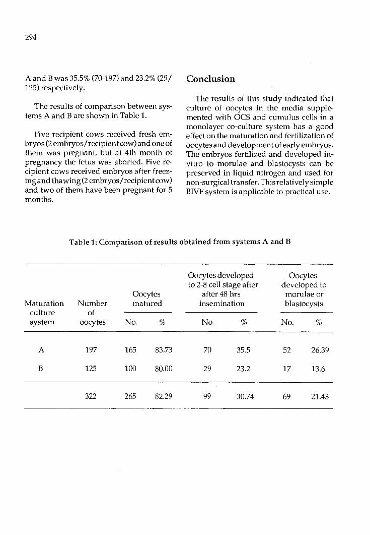
294
A and B was 35.5% (70-197) and 23.2% (29 I 125) respectively.
The results of comparison between systems A and B are shown in Table 1.
Five recipient cows received fresh embryos (2 embryos/ recipient cow) and one of them was pregnant, but at 4th month of pregnancy the fetus was aborted. Five recipient cows received embryos after freezing and thawing (2 embryos I recipient cow) and two of them have been pregnant for 5 months.
Conclusion
The results of this study indicated that culture of oocytes in the media supplemented with OCS and cumulus cells in a monolayer co-culture system has a good effect on the maturation and fertilization of oocytes and development of early embryos. The embryos fertilized and developed invitro to morulae and blastocysts can be preserved in liquid nitrogen and used for non-surgical transfer. This relatively simple BIVF system is applicable to practical use.
Table 1: Comparison of results obtained from systems A and B
Oocytes developed Oocytes to 2-8 cell stage after developed to
Oocytes after 48 hrs morulae or Maturation Number matured insemination blastocysts
culture of system oocytes No. % No. % No. %
A 197 165 83.73 70 35.5 52 26.39
B 125 100 80.00 29 23.2 17 13.6
322 265 82.29 99 30.74 69 21.43
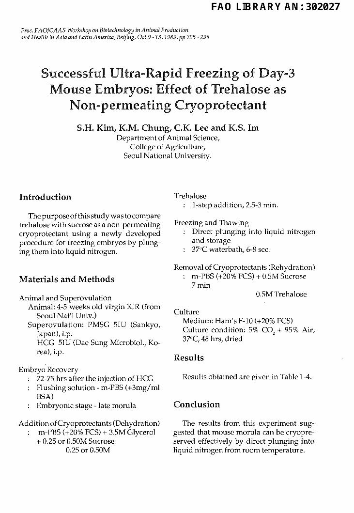
Proc. FAO/CAAS Workshop on Biotechnology in Animal Production and Health in Asia and Latin America, Beijing, Oct 9-13, 1989, pp 295 -298
Successful Ultra-Rapid Freezing ofDay-3 Mouse Embryos: Effect of Trehalose as
Non-permeating Cryoprotectant
S.H. Kim, K.M. Chung, C.K. Lee and K.S. Im Department of Animal Science,
College of Agriculture, Seoul National University.
Introduction
The purpose of this study was to compare trehalose with sucrose as a non-permeating cryoprotectant using a newly developed procedure for freezing embryos by plunging them into liquid nitrogen.
Materials and Methods
Animal and Superovulation Animal: 4-S weeks old virgin ICR (from
Seoul Nat'l Univ.) Superovulation: PMSG SIU (Sankyo,
Japan), i.p. HCG SIU (Dae Sung Microbiol., Korea), i.p.
Embryo Recovery 72-75 hrs after the injection of HCG Flushing solution- m-PBS (+3mg/ml BSA) Embryonic stage -late morula
Addition of Cryoprotectants (Dehydration) m-PBS (+20% FCS) + 3.5M Glycerol + 0.2S or 0.50M Sucrose
0.2S or 0.50M
Trehalose : 1-step addition, 2.5-3 min.
Freezing and Thawing Direct plunging into liquid nitrogen and storage 37°C waterbath, 6-8 sec.
Removal of Cryoprotectants (Rehydration) m-PBS (+20% FCS) + 0.5M Sucrose 7min
0.5M Trehalose
Culture Medium: Ham's F-10 (+20% FCS) Culture condition: 5% C0
2 + 9S% Air,
37°C, 48 hrs, dried
Results
Results obtained are given in Table 1-4.
Conclusion
The results from this experiment suggested that mouse morula can be cryopreserved effectively by direct plunging into liquid nitrogen from room temperature.
FAO LIBRARY AN: 302027
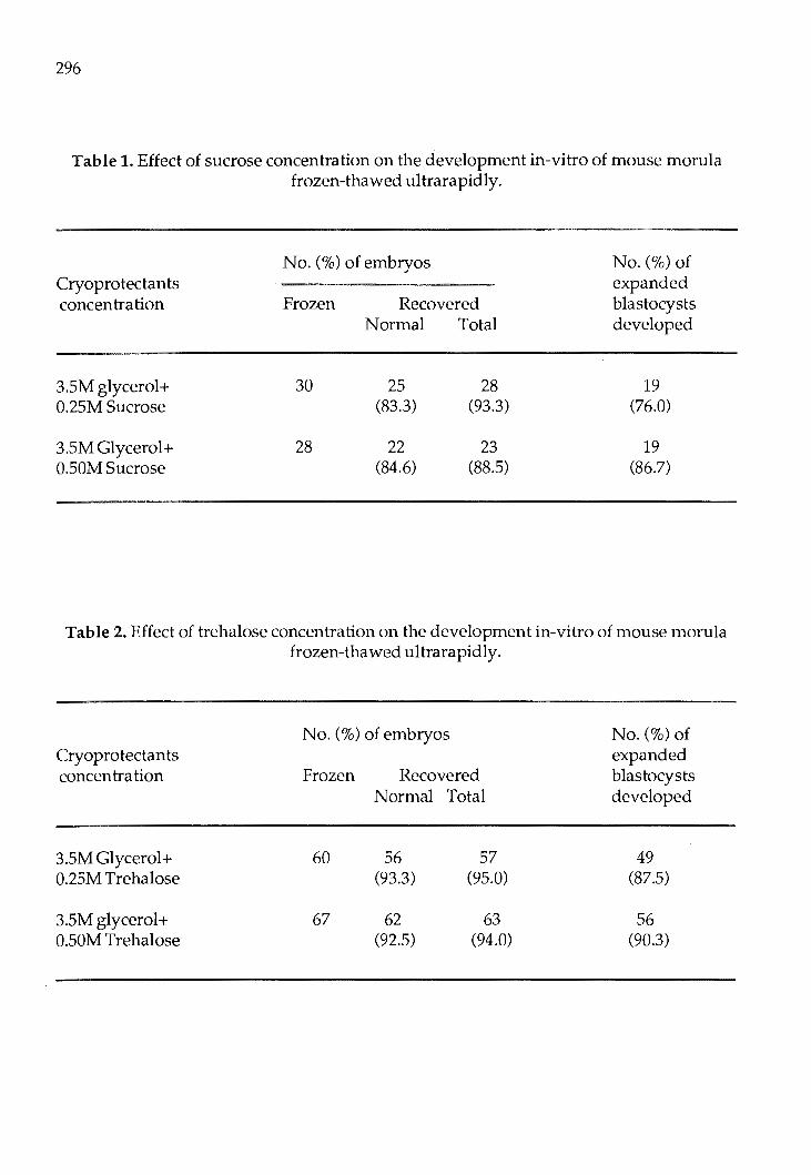
296
Table 1. Effect of sucrose concentration on the development in-vitro of mouse morula frozen-thawed ultrarapidly.
No. (%)of embryos No.(%) of Cryoprotectants expanded concentration Frozen Recovered blastocysts
Normal Total developed
3.SM glycerol+ 30 25 28 19 0.25M Sucrose (83.3) (93.3) (76.0)
3.SM Glycerol+ 28 22 23 19 O.SOM Sucrose (84.6) (88.5) (86.7)
Table 2. Effect of trehalose concentration on the development in-vitro of mouse morula frozen-thawed ultrarapidly.
No. (%) of embryos No.(%) of Cryoprotectants expanded concen tra ti on Frozen Recovered blastocysts
Normal Total developed
3.SM Glycerol+ 60 56 57 49 0.25M Trehalose (93.3) (95.0) (87.5)
3.SM glycerol+ 67 62 63 56 O.SOM Trehalose (92.5) (94.0) (90.3)

297
Table 3. Effect of Non-permeating cryoprotectants on the development in vitro of mouse morula frozen-thawed ultrarapidly.
No. ( % ) of embryos No.(%) of Cryoprotectants expanded concentration Frozen Recovered blastocysts
Normal Total developed
3.SM Glycerol+ 20 16 18 15 O.SOM Sucrose (80.0) (90.0) (93.8)
3.SM Glycerol+ 20 17 20 17 O.SOM Trehalose (85.0) (100.0) (100.0)
Table 4. Effect of equilibration time in 3.5M glycerol and O.SOM trehalose on the development in vitro of mouse morula frozen-thawed ultrarapidly.
No.(%) of embryos No.(%) of Eqilibration expanded time (min) Frozen Recovered blastocysts
Normal Total developed
<2 30 25 28 18 (83.3) (93.3) (72.0)
2.5-3 30 27 30 23 (90.0) (100.0) (85.2)
5 30 26 29 18 (86.6) (96.6) (69.2)
10 30 24 27 16 (80.0) (90.0) (66.6)

298
The effect of sucrose or trehalose concentrations in freezing solution is not significantly different. However, the effect of equilibrationtimein3.5Mglyceroland0.50M trehalose were-higher at 2.5 to 3 minutes than at shorter (<2 minutes) or longer (5, 10 minutes) times.
Therefore, trehalose appeared to be as effective cryoprotectant as sucrose in supporting the development of direct plunging into liquid nitrogen.

Proc. FAO/CAAS Workshop on Biotechnology in Animal Production and Health in Asia and Latin America, Beijing, Oct 9-13,1989, pp 299 -301
Application of a Simple and Easy Freezer for Embryos and Study on Freezing Method
Luo Ying-rong, Liu Yun-hai, Zhu Zhi-qing and Zhu Cheng-kuan Institute of Animal Science,
Chinese Academy of Agricultural Sciences, Beijing, China.
Abstract
Simple and Easy freezer for embryos has been designed on the principle of operation of liquid nitrogen tanks. There is a temperature gradient in the gas phase above the surface of liquid nitrogen. The temperature of chamber with embryo can be changed whenmoved up and down. The freezer has been extensively used to freeze embryos of mouse, sheep, cow and chinese yellow cattle in China. The developing rate of frozenthawed mouse embryos could reach 90.7% (577 /636). 77.2% (491/636)offrozen-thawed embryos cultured in vitro could access to expaned blastocyst. Morphologically normal embryos of sheep and cow after freezing-thawing was 76.2% (109 /143) and 69.4% ( 43 I 62), respectively. Successful rate of transfer was 50% (20/40) and 39.1% (9/23), respectively.
Introduction
Successful frozen cattle embryo transfer made in 1973, marked a significant achievement of cryobiological technique in the field of animal reproduction. In China, frozen embryos of cow was successfully transfered in 1982. Because efficient freezer was not available, the study on embryo freezing
technique and application of frozen embryo transfer in China was impeded. So development of a simple and easy freezer (SEF) which can be conveniently carried and operated in the worksite, and study on freezing method, using this sort of feezer, has become an urgent need for successful frozen embryo transfer.
On the principle based on liquid nitrogen tank, there is a temperature gradient in the gas phase above the surface of liquid nitrogen. The temperature of chamber with embryos can be changed by moving it up and down. Referred to the character and Problems of related freezer abroad, a simple and easy freezer for embryos with a digital thermometer was designed. The SEF has been extensively used in scientific institutes, universities and farms. Embryos of cattle, sheep and mouse have been frozen by the SEF and good results obtained.
Materials and Methods
1. Simple and easy embryo freezer l) Structure: The SEF is mainly made up
of 4 parts as follows: a) Stainless steel chamber for straws with embryos: 12 plastic straws (0.25ml., F. German) can be put in it simultaneously. There is a
FAO LIBRARY AN: 302029

300
thermocouple in the center. b) Updown regulation equipment. c) Sy-2 digital thermometer (Beijing normal university). d) seeding pincers.
2) Performance: a) Temperature range: lOO'C to-50°C. b) Temperature indicator: 0.1°C. c) cooling rate range: O, l to 0.2°C/min. d) Power supply: 9Vintegreted battery. e) Liquid nitrogen consumption: it is equal to the amount of natural evaporation of liquid nitrogen tank itself.
2. Freezing method 1) 1.4M glycerol PBS containing 20% FCS
was used as a freezing buff er. Glycerol was added by3 stepsatroom temperature, each step for 7 to 10 min.
2) The chamber with embryos was cooled at a rate of 1°C/ min. from room temperature to -7°C and equilibrated for 5 to 10 min at this temperature. Then seeding was induced and after 10 min of equilibrium, samples were cooled at the rate of 1°C/min. to -36°C, then the straws with embryos were immersed into liquid nitrogen.
3) Frozen embryos were thawed rapidly in 3G°C water bath. When embryos with glycerol was removed immediately by 3 steps, each step for 5 to 7 min using PBS buffer contained 0.3M sucrose.
Results and Conclusion
Day 3.5 or day 4 mouse embryos were morphologically accessed and good embryos were frozen by the method mentioned above. Frozen-thawed embryos were cultured for 72 hr. in C0
2incubator af37°C. The results
are shown in Table 1.
The results indicated that 90.7% (577 I 636) of mouse embryos frozen by the SEF could be developed and after thawing 77 .2% (491/636) of embryos could be hatched or expanded.
Day-6 and day-6.5 embryos of Hu sheep and Han Sheep were morphologically accessed and frozen by the SEP. Morphologically normal rate after thawing was shown in Table 2.
Table 1. In vitro development of D-3.4 \5 and D-4 mouse embryos frozen by the SEF.
Number of
embryos hatched
636 174 (27.4%)
expanded
317 (49.8%)
In vitro development
partially developed undeveloped totally developed
86 (13.5%)
58 (9.1 %)
577 (90.7%)

301
Table 2. Morphologically recovery rate of sheep embryos frozen by the SEF.
No. No. of normal embryos after thawing Breed of
embryos A+B grade(%) 0 C grade(%) Total
Hu sheep 103 62 (60)
Han sheep 40 28 (70)
Total 143 90 (62.2)
As shown in Table 2, among frozenthawed embryos, embryos of A and B grade was62.2% (90 /143), and embryos of C grade-11.8% (19 /143).A totalof76.2% (109 /143)of embryos was morphologically normal.
40 recipient ewes were transfered, 20 of them became pregnant and 27 la_mbs were born.
The SEF was also used to freeze embryos of cow and chinese yellow cattle. Live offsprings were obtained when frozen cattle embryos were transfered. In Hebei, Embryo Transfer Center, 108 embryos of cow were frozen, from 62 frozen-thawed embryos 43 usable embryos was obtained, usable rate was69.4%. Successful transferratewas39.1 % (9 /23).
References·
1. Luo, Y.R. 1980 Simple System to freezing embryos. Jap. J. Vet. Res. 28: 12-18.
19 (18.4) 81 (78.6)
0 28 (70)
19 (11.8) 109 (76.2)
2. Frank, G.C. 1986. Effect of freezing type, cryoprotant and processing methool on viability of frozen methods. 26: 135-146.
3 . .Zhu Yu-ding et al., 1983. Effectofcryoprotants and procossing methods on frozen efficiency old sheep embryos. Proceedings of Animal science institute of Chi-
. ··nese Academy Agriculture, 272-278.
4. Sang Yen Ozi et al., 1988. Cryoprotection and transfer of cow embryos. Proceedings of Chinese cattle embryo transfer conference 160-165.
5.Rodriguse,J.C.1986.Alternativemechanical system to freezing embryos, Therio-
0genology. 25: 190.
6: Leibo, S.P. 1988. Cryoprotection of embryos. IX international. Congress on animal reproduction and artificial insemination proceedings. 121-126.

Proc. FAO/CAAS Workshop on Biotechnology in Animal Production and Health in Asia and La.tin America, Beijing, Oct 9 -13, 1989, pp 302 - 303
Application Biotechnology in Exploitation of Animal Genetic Resources in China
Chen Youchun Institute of Animal Science,
Ct\AS Beijing, 100094 P.R. China.
Linkage between research on biotechniques and its application practise is a matter of our consideration. We have a few things to report here in this regard.
In recent years a few books under the title "Breeds of domestic animal and poultry in China" were published. Until now 240 animals breeds were recognized and recorded in these publications. There were 60 pig breeds, 28 cattle breeds, 30 sheep breeds, 23 goat, 26 horse, 10 ass and a few poultry breeds.
1. Storage of semen and embryo for endangered breeds.
Small tailed Han sheep is a breed known for its early maturity and prolificacy, the lambing percentage of which was261 %. The embryos from such a breed were now conserved in gene bank. Liaoning Cashmere goat has its hair as thin as 17urn, an adult buck produces 570 g of hair, best of them gives 2000 g. Experimental work on storing semen and embryos was started in 1986.
2. ET applied in cattle and sheep.
Some large scale transfer of imported Holstein embryos has been practiced in province Jilin, Heilongjiang and Autonomous region of inner Mongolia. Synthetic line of Simmental with Red Holstein was
produced experimentally to accelerate production of dual purpose nuclear stud in China.
The usability of sheep embryo after thawing were 76.2% (109/143) with pregnancy rate 55%. This figure indicates the possibility of application in setting the gene bank.
3. Half embryo transfer.
In Shanxi province, 36 goat embryos were divided into two, then 36 half embryos were transfered into 18 recepients. Pregnancy diagnosis 50 days alter found 6 with normal lambs.
6 frozen goat embryos after thawing were halved, then transfered in 6 recepients. At 50 days after operation 3 were pregnant.
Cattle's half embryos were experimentally used, and transfered into 11 cows with 6 of them in pregnancy.
4. DNA mapping
DNA mapping was also tried to use in cluster analysis in combination with blood polymorphism techniques. The aim of these works is to find genetic distance and probable hybrid vigor between certain breeds. Its a kind of work on the verge of application.
FAO LIBRARY AN: 302031

Finally to say, the biotechniques in China is still very young, butthe applicationofitin animal genetics has now started its way. Some advanced laboratories were success-
303
ful in cloning porcine growth hormone and vaccine-cloning. A full practice of using biotechniques still needs greater effort in future.

Proc. FAO/CAAS Workshop on Biotechnology in Animal Production and Health in Asia and Latin America, Beijing, Oct 9-13, 1989, pp 304 -311
Embryo Transfer Technology for Genetic Improvement in Brazil
A. Roberto de Bern Researcher-EMBRAP A/ Cenargen
Cx. Postal 10.2372 CEP 70.849 Brasilia/DP Brasil
Summary
The Animal genetic Resource Area (ARGA) of the National Center for Genetic Resource and Biotechnology ( CEN AR GEN), basically develops two activities:
a) Animal Genetic Conservation - this project consists in the preservation of races and species of domestic animals in danger of extinction and, for that, natural habitats are utilized as local site for in situ preservation. The freezing of semen and embryos of these animals constitute an ex-situ preservation of the Animal Germoplasm Bank.
b) Embryo Biotechnology - there are two Embryo Biotechnology laboratories at CENARGEN - one for experimental embryology, that utilizes the biological model Mus musculus and the other for applied embryo logy reserved for domestic animals of economic interest.
For the development of the activities of the above mentioned research, the Animal Genetic Resource Area has a 500 ha farm and 19 staff members, including researchers, laboratory technicians, administrative personnel and field workers.
The objective of the present report is to describe in detail the results obtained in the field of animal embryology.
The mouse, when used as a biological model, has proved extreme! y useful in training programs, and in the development of support techniques to research activities such as in vitro embryo culture. Mouse embryos have low costs and are easy to obtain.
As far as mouse embryo production is concerned, in the last4 years, 1131 females were super-ovulated and 12.705 embryos were rescued, of which 8.509 (67%) were viable. These embryos were utilized in the following research activities:
FAO LIBRARY AN: 302033
Research activities Methodology
Cooling 0°C/24 hours Freezing Isocriogen Vitrification Classic Bissection E.T. surgery Sex deter- Cytogenetics mination & Cytoxicity
As far as the bovines are concerned, in 1989, 302 embryos were obtained from 52 donors with 2.8 viable embryos per donor and 1.4 product per embryo, utilizing nonsurgical transfer methodology. In 43% of the cases, the thawed embryos were sucessful in establishing pregnancy. From the bissection of bovine embryos and non-surgical transfer of the hemi-embryos, 0.65 products were born per original embryo.
The research project with equines was carried out based on the knowledge obtained with the bovine system. In the last two years, 120 embryos giving 40% pregnancy were harvested from mares and transferred fresh via non-surgery. 51 embryos were frozen and 15% pregnant animals were obtained. One product from a mare hemiembryo was obtained.
We are beginning a project with frozen caprine embryos and in the next few years 3 new projects will be initiated:
- in vitro fertilization - Animal cloning - Transgenic animals
Our laboratory has been the reference point for animal Embryo Biotechnology at national level. We are starting a program on cryopreservation of semen and embryo of domestic species in danger of extinction in South America, in collaboration with FAO.
305
Results Summary
85.3% in vitro growth 77% in vitro growth 58% in vitro growth 30% no. of fetuses
current project
Introduction
In 1973, Brazil started her embryo transfer research with bovine species in collaboration with France, the INRA, Institut National de Recherche Agronomique, UNCEIA - Union National des Cooperatives de InseminationArtificiel, and the Maison Alfort School of Veterinary Medicine.
At that time, embryo collection was carried out by means of midline surgery, general anaesthesia with fl uotane and by a ca theter introduced into the oviduct. Apart from the death of many cows, results from experiments allowed for direct observation of hypertrophied ovaries and thrilling visualization of the first embryo. Embryos were first collected in Brazil, at the Lages School of Veterinary Medicine in 1975, in Santa Catarina. On that occasion, an embryo transfer project for bovine species was vehemently rejected as being something for the third millenium.
In 1976, some freelance veterinary surgeons from the State of Sao Paulo started the first successful non-surgical embryo transfer and collections at farm level. Since then, in various parts of the country isolated groups of vets and cattle breeders have attempted to employ this new technology with relative sucess.
306
In 1985, on the occasion of the founding of the SBTE - Brazilian Society for Embryo Transfer - in Brasilia, embryo transfer became a scientific activity and, at the same time, a political organ in charge of the systematic standardizing of activities in this area together with the Federal Government, universities, research institutes and cattle breeders.
However, notwithstanding the significant potential of genetic improvement programs, cattle embryo transfer growth was disorganized - and what is even more serious - the choice of donors has been made more on an emotional basis than on their genetic merit.
Another serious problem is the exaggerated numbers of petitions made to the Ministry of Agriculture for the setting up of new embryo transfer centers, gigantic projects and poorly-trained personnel.
In our opinion, the Federal Government together with the SBTE and Cattle Breeders Associations should elaborate an embryo transfer program, at national level, applied to those species of animal husbandry interest. There is an enormous amount of space for national herd genetic breeding using this biotechnology.
The situation of the 1989 unofficial SBTE data for embryo transfer in Brazil is as follows:
Embryo transfer in Brazil (1989) = 20.000
in Sao Paulo (only) = 15.000
births (prognostic) = 5.000
One important aspect, especially eco-
nomically, has been the international commercialization of embryos. On the one hand, there has beenanimportationof dairy-cattle embryos from the USA, Canada and Israel and of beef-cattle embryos from France, Germany and England. On the other hand, there has been illegal exportation of zebu embryo, mainly Nelore to Central America.
With special reference to the future of embryo transfer in Brazil, we could enumerate the following important topics:
- Awareness that Artificial Insemination (AI) is an important genetic improvement tool.
- Systematic training of personnel. - Effective selection of male and female
donors and receivers. - Engagementofbreedersandnutrition
ists in embryo transfer programs.
BOVINE SPECIES EMBRYO TRANSFER PRACTICE IN BRAZIL
1. CHOICE OF DONORS Chocie of donors is the starting point of
any embryo transfer program. Evidently, the genetic value evaluated by means of progeny tests would be desirable. However, in developing countries this practice has not been developed for technical and economical reasons.
In Brazil, the choice of donors should follow the dicates of common sense, with a view to selecting the several types within a lot to preserve the minimum of genetic variability of each breed in question.
Equal consideration should be given to the choice of receivers. Mainly healthy animals with regular sexual cycles. In our case, Girolandas cows have presented the best results in embryo transfer programs.

2. SUPEROVULATION
2.1. Superovulation Methodology In 1973, superovulation was done in the
oestrogenic phase. It was important that the heat be detected in order to begin treatment in or around the sixteenth day. This was due to the impossibility of inducing or synchroninzing the heat and the mediocre results in embryo quality. With the advent of prostaglandins (F group) tested by ourselves for the first time in Brazil in 1975, the superovula ti on occurs between the eighth and the twelfth day after the heat. At present in Brazil the hormone used is the FSH from pigs are made in the USA. The dose varies between 18 mg and 50 mg. These extremes are used, respectively, inNelore and Holandes cows.
In Brazil, LABORCLIN, Curitiba, Parana is producing FSH (Follicle Stimulating Hormone) from the hypophyses of pigs. While still in the initial phase, it deserves all our confidence.
With regard to the sperovulation of the same donor it is routine that embryo transfer teams repeat successfully the procedure at every 60 and 90 days. In Central Brazil results with zebu cows Bos indicus with 243 collections resulting in 1994 structures (X == 8,2), with an average 4.1 viable embryos per cow receiving several consecutive superovulations.
3. Embryo Recovery The embryo collection itself consists of
the flushing of the uterus with PBS at an approximate temperature of 25°C pH 7.2 and osmolarity of 290 miliosmol.
The collection catheter passes the uterine colon, held fixed by an air balloon. Gravity helps to introduce the liquid of up 500 to
307
1000 ml depending on the size of the u terns. Liquid recuperation is also by gravity.
A filter helps and quickens the process of looking for embryos. Our staff developed one, using a veterinary syringe of 50 or 100 ml. A plastic sieve with a screen of 83 micrometers was placed on the upper part of the filter thus avoiding embryo exit. The liquid coming from the uterus enters the filter from one side and the embryos are recovered below directly onto a Petri plaque. Use of this filter a voids direct pressure of the liquid on the embryos.
The collection rate, number of embryos in relation to the Corpus Luteus, is on average 70%. This collection rate can be increased and the following details are important.
- Adequate immobilization of the donor
- Light epidural anaesthesia - Use cervical dilator if difficulty in
passing catheter appears - Balloon well-placed and regular touch
pressure - Hold uterine junction with fingertips
on starting collection - Massage of uterus - Tranquility and safety - Good helpers - Plenty of good luck
4. Examination and Evaluation of Embryos Techniques for the production, collec
tion, transfer and preservation of embryos have evolved in recent years. However, the morphological evaluation under the magnifying glass of between ten to one hundred times is still somewhat subjective.
This embryo morphological evaluation orembrionicclassificationsetsmorphological standards which help the form but not the life of the embryo. Only practice and

308
daily handling of embryo give the necessary discernment nearest to the real embrionary quality.
The classification proposed by the SBTE for the evaluation of embryos can be divided into seven categories:
1) Mo - Morula 2) Mc - Compact Morula 3) Bi- Initial Blastocyst 4) Bl - Blastocysst 5) Bx - Expanded blastocyst 6) Bn - Hatching blastocyst 7) Be-Hatched blastocyst
In the individual judgement of embryos, several characteristics should be observed, such as size, form, embryo colour, cytoplasm homogeneity, form and integrity of membrane, size and presence of the cells in theEPVand the presence of vesicles. Within the described parameters, embryos received identification varying according to the different authors. The SBTE classifies under six headings:
1) Embryo I (excellent) 2) Embryo II (good) 3) Embryo III (regular) 4) Embryo IV (poor) 5) Dg - Degenerated Embryo 6) Nf - Not Fecundated
Embryos I, II and III are transferable. The resutls expressed in gestations (45 to 60%) are equal both for the excellent and good embryos. When indicated for freezing and micromanipulation, only embryos I and II are usable.
In the case of a non-superovulated donor, 80% of the embryos are morphologically normal. In the superovulated cows the average is 60%. When there is a strong response to superovulation the quality of the embryos diminishes.
At the moment, there is no good method for the vital adjudgement of embryos except for gestation. Under such conditions, morphological criteria and good touch has been the choice of techniques of those working with embryo transfer.
5. Freezing of Embryos - Use oflsocriogen The embryo industry of developed coun
tries, notably in cattle, has generated a supply of sophisticated equipment, expensive and many times not suitable to the conditions of developing countries.
For current freezing programs, the rescue of species in imminent danger of extinction deserves speical attention.
This rescue operation was developed with a view to making feasible with the use of simple equipment the freezing of embryos at farm level.
The equipment tested - the Isocriogen, was projected by the EMBRAP-CENARGEN staff. It consists of a piece of styrofoam type p-3 which is used to condition liquid nitrogen, a steel cylinder number 304, American model and a digital thermometer.
The pre-selected embryos to be frozen are dehydrated in three stages.
Stage 1- Solution PBS-4 The embryos are well washed in PBS solution (Phosphate Buffer Saline) with 0.4% of BSA (Bovine Serum Album).
Stage 2 - Solution G-5 The embryos are placed in PBS solution with 5% Glycerol for five minutes.
Stage 3 - Solution G-10 The embryos are transferred to PBS solution with 10% Glycerol for ten minutes.

While dehydrating in the G-10 solution, the embryos are acclimatized individually in paillettes (0.25 ml) with this solution.
The preparation of the Isocriogen is easy. However, it should be synchronized with the paillettizatio of the embryos.
6. Embryo Transfer (Innovulation) In 1976, we performed variousinnovula
tions in France by means of midline surgery similar to surgical collecting. Apart from difficulties such general anaesthesia and surgery, results were not significant. In the same year, we experimented with equipment proposed by TEST ART, named "patacult" transvaginally. Once again, the results were mediocre.
Many researchers have worked to diminish the time and simplify the process of innovulation. At present, there are two instruments indicated for transcertical innovulation, both with French paillettes of 0.25 ml. The German model has a 5 mm diameter and the French one 3 mm. The latter is best indicated because it has the ideal length, descartable flangles, less traumatic and ideal for young receivers.
7. Bissection of Embryos to Produce Monozytoic Twins The embryo bissection technique used is
simple and was adapted in the laboratory in CENARGEN. It consists of the use of two mandmade micro-instruments:
- A suction micropipette with a Bunsen point, serving to keep in position and to immobilize the embryo.
- Fragment of a shaving blade, especially adapted, serving to divide the embryo in two equal halves on a vertical plane.
These instruments are fitted to the arms
309
of the micromanipulator with either independent or simultaneous command.
The embryos are placed in PBS (tamponned solution) together with 0.8% of bovine albumen and bissected under a stereoscope microscope increasing from sixty to eighty times more.
The embryos thus obtained are transferred in pairs to the same receiver or separately to two.
In June, 1986 the first pair of hemi-embryos were transferred toa Chianina-Nelore mestizo cow. The pair of embryos had been obtained by the CENARGEN staff through the bisection of the original embryo of the Red Indubrasil breed. In April 1987, two male calves, genetically identical (monozygotic twins) were born as a result of this transfer.
Research in this field has turned now to the freezingofhemi-embryos with a view to sexing of one the halves while the other is transferrd or stored.
Our results today with the bisection of embryos vary approximately in 60% gestation, taking into account the inn ovulation of the hemi-embryo. These results can be improved with greater rigour in the choice of embryos and receivers. This methodology is being carried out at farm level.
8. In Vitro Fecundity of Bovine Embryo In this particular case, using a small part
of the ovary and in the near future, we will be able to obtain a large number of embryos from the laboratory alone. The objectives of these are:
- increase in the economical and genetical potential of embryo transfer.
- extrapolation of the techniques and

310
applied to animals in danger of extinction.
9. Cloning Preliminary investigations are being
made.
10. Transfer of Genes Attempts for gene transfer have not been
made yet.
APPENDIX A
PUBLICATION OF ANIMAL GENETIC RESOURCES UNIT - CENARGEN
(1987-89)
Micrornanipulation and Transfer of Embryos - BEM, A.R. de ENCONTRO SOBRE RECURSOS GNETICOS, Jaboticabal, 12 a 14 de April e 1988, Annual.
Use of Mouse (Mus rnusculus) as Models Biological - BEM, A.R. de; RIBEIRO, S. T. dos S.J. & SOUSA, R.A. - 1° CONGRESSO BRASILEIRO DE ANIMALS DE LABORATORIO, Bahia, 29/08 a 02/09/88.
Embryo Technology Applied to "Ex Situ" Equine Conservation - FARINASSO, A.; FARIAS, C. de J. & BEM, A.R. de-III REUNIAO ANUAL E I RENIAO INTERNACIONAL DA SOCIEDADE BRASILEIRA DE TRANSFERENCIA DEEMBRIOES -1988, Rev. Centro de Ciencias Rurais, Santa Maria, Vol. 18, 47; Suplernento.
Bisection and Transfer of Berni-embryos in Bovines - RIBEIRO, S.T. dos S.J.; BEM, A.R. de; SILVA, A. da & ZANENGA, CA. -III RENIAO ANUAL EI REUNIAAO IN-
TERNACIONALDASOCIEDADEBRASILEIRA DE TRANSFERENCIA DE EMBRIOES -1988,Rev. CentrodeCienciasRurais, Santa Maria, Vol. 18, 50; Suplernento.
Bisection and Transfer of Berni-embryos in Mouse (Mus rnusculus) - RIBEIRO, S.T. dosS.J.;ZANOTTO,F.P. & BEM,A.R. de-III REUNIAO ANUAL EI REUNIAO INTERNACIONAL DA SOCIEDADE BRASILEIRA DE TRANSFERENCIA DE EMBRIOES -1988,Rev. CentrodeCienciasRurais, Santa Maria, Vol. 18, 51; Suplernento.
Crioconservation of Brazilian Naturalized Bovine Breeds - MARIANTE, A. da S.; BEM, A.R. de; VASKE, T.R.; TROVO, J.B. de F. & RIBEIRO, S.T. dos S.J. - III REUNIAO ANUAL EI REUNIAO INTERNACIONAL DASOCIEDADE BRASILEIRA DE TRANSFERENCIA DE EMBRIOES - 1988, Rev. Centro de Ciencias Rurais, Santa Maria, Vol. 18, 57; Suplernento.
Bisection of Bovine Embryos- BEM, A.R. de & SOUSA, R.V. de - IV REUNIAAO ANUALDASOCIEDADEBRASILEIRADE TRANSFERECIA DE EMBRIOES, Porto Alegre, 04 a 05 de seternbro de 1989.
Feasibility Study of Mouse (Mus rnusculus) Embryos after Freezing at D°C for 24 hours - MONCAO, M.; BEM, A.R. de & ROSAS, C. de A. - IV REUNIAO ANUAL DA SOCIEDADE BRASILEIRA DE TRANSFERENCIA DE EMBRIOES, Porto Alegre, 04 a 05 de seternbro de 1989.
Mouse (Mus rnusculus) Embryo Freezing for Vitrification - EGITO, A.A. do; RIBEIRO, S.T. dos S.J. & BEM, A.R. de - IV REUNIAO ANUAL DA SOCIEDADE BRASILEIRA DE TRANSFERENCIA DE EMBRIOES, Porto Alegre 04 a 05 de seternbro de 1989.
Evaluation of the Mitotic Index for Mouse (Musrnusculus)-SOUSA,R.V.&BEM,A.R. de-NREUNIAOANUALDASOCIEDADE BRASILEIRA DE TRANSFERENCIA DE EMBRIOES, Porto Alegre, 04 a 05 de sternbro de 1989.
Equine Ernbriology: Collecting, Cooling and Freezing - ROSAS, C. de A.; FARIAS, C.J.; FALEIROS, R.R.; FARINASSO, A. & BEM, A.R. de - IV REUNIAO ANUAL DA SOCIEDADE BRASILEIRA DE TRANSFERENCIA DE EMBRIOES, Porto Alegre, 04 a 05 de seternbro de 1989.
Identification of the Berni-embryo Sex in Mouse Using the Cytogenetic Method: The colchininetreatrnent- LIMA, V.F.B.;JORGE, W .; BEM, A.R. da & MOREIRA-FILBO, C.A. - IV REUNIAO ANUAL DA SOCIEDADE BRASILEIRA DE TRANSFERENCIA DE EMBRIOES, Porto Alegre, 04 a 05 de seternbro de 1989.
Preliminary Results of the Cellular Cycle of Mouse Embryos Syncronization - LIMA, V.F.B.; JORGE, W.; BEM, A.R. de & MOR-
311
EIRA-FILBO, C.A. - IV REUNIAO ANAUALDA SOCIEDADE BRASILEIRA DE TRANSFERENCIA DE EMBRIOES, Porto Alegre, 04 a 05 de setembrode 1989.
Identification of Berni-embryo sex in Mouse through the Cytogenetic Method Synchronization of the Cellular Cycle -LIMA, V.F.B.; JORGE, W.; BEM, A.R. de & MOREIRA-FILBO, C.A. - IV REUNEIAO ANUAL DASOCIEDADE BRASILEIRA DE TRANSFERENCIA DE EMBRIOES, Porto Alegre, 04 a 05 de setembro de 1989.
The Brazilian Genetic Resources Conservation Program - MARIANTE, A. da S. & TROVO, J.B. de F. Rev. Brasil. Genet. 12, 3 Supplement, 241-256 (1989).
New Perspectives for Embryo Growing-1988, BEM,A.R. de, Rev. Bras. Tecnol. Vol. 9 (3) 41-45.

Proc. FAO/CAAS Workshop on Biotechnology in Animal Production and Health in Asia and Latin America, Beijing, Oct 9-13, 1989, pp 312 -320
Improvement of Copra Meal Quality For Use in Animal Feeds
Agnes F. Zamora, Marilou R. Calapardo, Katharine P. Rosario, Edwin S. Luis and Ida F. Dalmacio
University of the Philippines at Los Banos (UPLB) College, laguna, Philippines
Introduction
Copra meal is an agro-industrial fibrous residue which is abundant in many Asian countries, and by far, the most abundant feed stuff ingredient that the Philippines can offer. It contains 20% protein and 60% carbohydrates in the form of galactomannan (61 %), mannan (26%) and cellulose (13%) (Takahashi et al., 1984), and is used as a component of animal feed. However, its optimum utilization cannot be fully realized due to low digestibility attributed to its high fiber content. Besause of this, the approach then taken atthe Nationallnstitutes of Biotechnology and Applied Microbiology (BIOTECH) at UPLB is to improve the feeding quality of copra meal through microbial action. The theoretical effects include (1) marked reduction in fiber content thereby incrasing the digestibility of copra meal: (2) increased energy value by making simpler sugar molecules more available to the animal and (3) improved nutritional quality by making proteins that are bound by the polysaccharide molecules also more available (Teves, 1987).
From previous studies, mannanase was produced using Streptomyces sp. no. 17 (isolated from Philippine soils), from a
complex medium, modified malt yeast broth, thereby making enzyme production expensive. Hence, succeeding experiments utilized coconut water as substrate in order to bring down the cost of mannanase production. Another approach, solid substrate fermentation, was also tried. With this method, enzyme production and hydrolysis of copra meal occur simultaneously. Moreover, since a lesser volume of water is used in solid substrate fermentation as compared to the amount used during enzyme treatment, drying of the treated meal prior to storage would require less time.
To further improve the nutritional quality of copra meal, cellulase, produced from Trichoderma reesei rut C30, was also used to hydrolyze copra meal.
Production of Crude Mannanase
The mean enzyme activity, specific ac ti vity and protein content of the resulting enzyme solution using coconut water as substrate are shown in Table 1. Enzyme activities and protein content ranged from 2.79-97.34 units and 1.14-2.80 mg/ml. The highest enzyme activity of 97.34 units with specific activity of 65.62 was obtained from
FAO LIBRARY AN: 302035

coconut water medium (CWM) containing 3 % copra meal.
Table 2 lists the activities of the mannanase produced from coconut water with 3% copra meal and from modified yeast malt broth (MYMB). The wide range of val-
313
ues could be attributed to the coconut water composition which may have varied from time to time during the duration of the experiment. Neverthelss, the results indicate that coconut water is a practical substrate for mannanase production.
Table 1. Mean enzyme and specific activities and protein content of mannanase produced using coconut water with varying amounts of copra meal as enzyme production
medium.
Weight of copra meala (gms)
0.5 1.0 1.5 2.0 2.5 3.0 3.5
a weight (g) in 100 ml medium.
Enzyme Activityb
5.55 28.64 39.65 39.59 22.44 97.34 2.79
Protein (mg/ml)c
1.14 1.69 1.77 2.80 2.18 1.15 1.78
Specific Activityd
5.14 17.28 24.40 14.36 10.10 65.12 1.99
b 10 units of enzyme is equivalent to the activity that will produce 5.5 mg mannose/ml enzyme/ 30 minutes. c Lowry method. d enzyme unit/mg protein.

314
Table 2. Mannanase activity of Streptomyces sp. no. 17 produced from Modified Yeast Malt Broth (MYMB) and Coconut Water Medium (CWM).
Medium Batch No. Enzyme Unit
Modified Yeast Malt 1 13.20 Broth (MYMB) 2 9.50
4.00
4 4.60
5 11.80
6 13.60
7 11.00
8 8.34
Coconut Water Medium 1 24.45 (CWM) 2 10.46
3 22.27
4 17.17
5 32.32
6 16.83
7 18.60
8 46.10
9 29.09
10 16.08
10 units of enzyme is equivalent to the activity that will produce 5.5 mg mannose in 1 ml enzyme in 30 minutes of reaction. From Zamora et al., 1989.
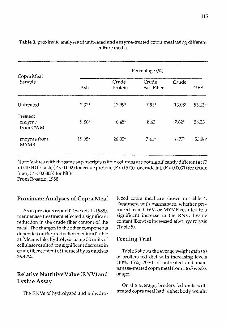
315
Table 3. proximate analyses of untreated and enzyme-treated copra meal using different culture media.
Copra Meal Sample
Untreated
Treated: enzyme fromCWM
enzyme from MYMB
Ash
7.32b
9.86b
19.95a
Crude Protein
17.99b
6.45b
26.05a
Percentage(%)
Crude Fat Fiber
7.95a
8.63
7.4P
Crude NFE
13.08a 53.63a
7.62b 58.25b
6.77b 53.96a
Note: Values with the same superscripts within columns are not significantly different at (P < 0.0004) for ash; (P < 0.002) for crude protein; (P < 0.575) for crude fat; (P < 0.0001) for crude fiber; (P < 0.0003) for NFE. From Rosario, 1988.
Proximate Analyses of Copra Meal
As in previous report (Teves et al., 1988), mannanase treatment effected a significant reduction in the crude fiber content of the meal. The changes in the other components depended on the production medium (Table 3). Meanwhile, hydrolysis using 50 units of cellulase resulted to a significant decrease in crude fiber content of the meal by as much as 26.42%.
Relative Nutritive Value (RNV) and Lysine Assay
The RNVs of hydrolyzed and unhydro-
lyzed copra meal are shown in Table 4. Treatment with mannanase, whether produced from CWM or MYMBresulted to a significant increase in the RNV. Lysine content likewise increased after hydrolysis (Table 5).
Feeding Trial
Table 6 shows the aveage weight gain (g) of broilers fed diet with increasing levels (10%, 15%, 20%) of untreated and mannanase-treated copra meal from 1to5 weeks of age.
On the average, broilers fed diets with treated copra meal had higher body weight

316
Table 4. Relative Nutritive Values (RNVs) of untreated and Mannanase-treated copra meals.
Kind of copra meal %RNV % increase
Untreated
Enzyme-treated enzyme produced from CWM 14.44
enzyme produced from MYMB 24.21
All treatments whose means have been assigned the same letter are not significantly different (P < 0.01). From Zamora et al., 1989.
Table 5. Available lysine of untreated and mannanase-treated copra meal using Modified Yeat Malt Broth (MYMB) and coconut water (CWM) as culture media by
Streptomyces sp. no. 17.
Sample
Untreated copra meal
Enzyme-treated copra meal usingCWM
Enzyme-treated copra meal usingMYMB
available lysine (mg lysine/mg N)
% increase
19.08
61.92
Note: All treatments whose means have been assigned the same letter are not significantly different (P < 0.01). From Zamora et al., 1989.

gain (781.18 g) compared to those fed diets with untreated meal (726.65 g). As the level of copra meal increased in the diet, body weight gain consistently decreased, supporting the previous findings of Teves et al. (1988). From the first up to the fourth week of feeding, diets containing 10% copra meal (treated and untreated) effected the highest body weight gains (Figure 1).
Solid Substrate Fermentation
Results of the preliminary study on solid substrate fermentation are shown in Tables 7 and 8. From these data, trea tment2D seems to be the most suitable formulation for solid
317
substrate fermentation using Streptomyces sp. no. 17.
Other experiments concerning solid substrate fermentation which will be undertaken include the determination of the effect of:
(1)nitrogensourceandothernutrientson the growth and enzyme activity of Streptomyces sp. no. 17; and
(2)inocula ti on using a combination of microorganisms on the quality of copra meal. In this connection, screening of -galactosidase-producingmicroorganisms is being pursued.
Table 6. Weight gain (g) of broilers fed diets with increasing levels of ntreated and mannanase-treated copra meal from 1to5 weeks of age.
Types of copra Meal in diet
Untreated
Mannanase-treated
Mean
From Zamora et al., 1989.
10%
782.52
834.27
808.40
LEVEL
15% 20% Mean
695.93 701.52 726.65
820.72 689.14 781.15
758.32 695.33
318
Table 7. Effect of inoculum concentration on enzyme activity, total sugar, crude protein, crude fiber and lysine content of copra meal inoculated with Streptomyces sp. no. 17 after
one week incubation at 34°C.
Treatment* Inoculum Enzyme Total Crude Crude Lysine (ml) activity sugar protein fiber ug/mg
(U) mg/ml (%) (%) N
IA 50 3.74 5.5 21.18 6.12 43.17 IB 100 10.31 7.34 21.08 6.14 84.77 IC 150 0.43 10.23 21.24 7.06 121.83 ID 200 8.43 9.40 20.96 6.18 45.33 IE 250 4.23 4.20 23.11 7.10 49.62
Table 8. Effect of moisture on enzyme activity, total sugar, crude protein, crude fiber, and lysine content of copra meal inoculated with Streptomyces sp. no. 17 after one week
incubation at 34°C.
Treatment* Vol. Enzyme Total Crude Crude Lysine water activity sugar protein fiber ug/mg (ml) (U) (mg/ml) (%) (%) N
2A 50 5.36 4.67 20.42 5.89 94.88 2B 100 5.29 9.82 20.51 6.26 95.33 2C 150 4.51 5.09 20.94 7.04 73.16 2D 200 6.93 10.53 20.68 5.66 119.67 2E 250 8.80 3.90 20.11 5.72 52.16
*Legend:
Copra meal Vol. of seed culture Vol. of water lA 50g 50ml 100ml lB 50g lOOml 100ml 1C 50g 150 100 lD 50g 200 100 lE 50g 250 100 2A 50g lOOml 50ml

2B 2C 2D 2E
50g 50g 50g 50g
100 100 100 100
100 150 200 250
319
Figure 1. Average weekly cumulative body weight gain of broilers up to 4 weeks of feeding.
600
400
20T
LEGEND: flfi Tl 1 (10% UCM)
IIIIIl T12 (15% UCM)
IBlJ T13 (20% UCM)
8 T21 (10% MTCM)
~ T22 (15% MTCM)
0 T23 (20% MTCM)
;,
~ r~
~. I' t! l\. t .
:\ ,
' oL ~.w.w.~..u..-.u-1--ll.:.::LJ.w.u::~...u......u_1.--.1..MJ~c::lU..-U-..l.l.-L...-~·'~~LL.....1.J......1.1-'-~
4 1 2 3
FEEDING TIME (weeks)

320
References
Rosario, KL. P. 1988. A comparative analysisof Streptomyces no. 17 mannanase-treated copra meal using modified yeast malt broth and coconut water as culture media. B.S. Biology thesis, Univ. of the Phil. at Los Banos. 53 p.
Takahashi, R., I. Kusakabe, H. Kobayashi, K. Murakami,A.MaekawaandT.Suzuki.1984. Purification and some properties of mannanase from Streptomyces sp. Agric. Biol. Chem. 48(9): 2189-2195.
Teves,F.G.1987. Thenutritivevalueofcopra meal treated with bacteial mannanase in broiler diets. M.S. thesis. Univ. of the Phil. at Los Banos. 93 p.
Teves, F.G., A.F. Zamora, M.R. Calapardo and E.S. Luis. 1988. Nutritional value of copra meal treated with bacterialmannanase in broiler diets. In: Recent Advances in Biotechnology and Applied Biology. Proc. GIAM VIII: 497-507.
Zamora, A.F., M.R. Calapardo, KL.P. Rosario,E.S. Luis and L. DelaMar.1989. Bacterial mannanase production using coconut water and nutritive value of mannanasetreated copra meal in broiler diet. Paper presented at the 18th Phil. Soc. for Microbiol. Annual Convention. 11-12 May 1989. Baguio City, Philippines.

Proc. FAO/CAAS Workshop on Biotechnology in Animal Production and Health in Asia and Latin America, Beijing, Oct 9-13, 1989, pp 321 -327
Differential Tests on Serum Antibody EIA Horses From That of Attenuated EIA Vaccine
Immunized Horses
Lu Jingliang, Kong Xiangang, Ning Xide, Zhang Rikuo, Chu Guifang, Xiang Wenghua
Harbin Veterinary Research Institute of the Chinese Academy of Agricultural Sciences,
No. 101 Maduan Street, Harbin, The People's Republic of China
Abstract
In present research, a diagnostic method, combining dot blot (DB) test with immunodiffusion (ID) test, was developed to differentiate serum antibody of equine infectious anemia virus infected horses from that of EIA attenuated vaccine immunized ones. A monoclonal antibody (McAb) specific to EIA donkey leucocyte attenuated virus labelled with horseradish peroxidase (HRP) was used in DB test. vaccinated horses appear positive in DB test while EIA horses are positive in ID test and healthy ones without vaccination are negative in blot DB and ID test. A total of 10159 horses from various provinces and area were examinated byacombinationofDBandID test for serum antibodies to vaccine antigen or EIA virus. Of them, 184 horses were infected with EIA virus, 9437 were immunized with the vaccine and 538 were healthy. The results above were confirmed by pathological examination in 106 EIA horses and 21 vaccinated ones. The EIA epidemic was under control in infected areas after positive horses were eliminated and new EIA cases have not been detected in vaccinated horss even
though repeated quarantines. It has been proved that the method established in the studyisvaluablefordifferentiationbetween EIA horses and vaccinated ones.
Vaccine inoculation has been adopted extensively in various countries (or prevention and control of animal and poultry infectious diseases. As a general rule, quarantine and epidemic source removal are not undertaken before inoculation and infected animals (or poultry) along with vaccinated ones can show the same serum reaction by common serodiagnositical tests use in every country, therefore, differentiation ofinfected from immunized animals (or poultry) is a big problem to be solved worldwide. In order to overcome the difficult problem in prevention and control of equine infectious anemia (EIA), one of the contemporary high techniques - lymphocyte hybridoma technique was used in our laboratory to produce a monoclonal antibody (McAb) A4 with specificity to EIA donkey leucocyte attenuated virus. The method, a combination of immunodiffusion (ID) and dot blot (DB) test with McAb A4-enzyme conjugate was employed to differentiate the serum anti-
FAO LIBRARY AN: 302037

322
body to EIA wild virus from that to EIA attenuated virus. Theresultsarereportedas following.
Materials and Methods
McAb conjugate McAb A4 and others labelled with horse
radish peroxidase and the preparation of the McAbs-EIA attenuated virus antigen complexes have been reported previously (2, 3).
Tested sera 202 serum samples were collected from
healthy horses in a farm which was EIA free and without EIA vaccine inoculation. 88 sera were from experimentally immunized horses in the institute and 286 were from vaccinated ones in the field. 16 samples were from the horses immunized with the vaccine and then challenged with EIA virulent virus in a veterinary biological products and medical appratus factory. 14 samples were from experimentally infected horses (or donkey) in the institute and 127 from naturally infected horses in EIA epidemic areas. In addition, 10159 sera from unidentified horses were tested for the antibody to EIA wild virus or vaccine strain.
Procedure of DB test Nitrocellulose membrance was used as solid phase carried in DB test.
1. 2.5ulofpositive,negativeand testedsera were spotted on NC membrane respectively and dried for 15 mins.
2. Blotted NC membrance was washed for 3 times (3-5 mins. every time) with 0.02M phosphate buffered saline (PBS) containing 0.05% Tween-20 (PBS-T, pH7.2).
3. The membrane was shifted into HRPMcAb A4 solution (preparate with PBS-
T) to incubate for 1 hour at room-temperature.
4. Washed as step 2. 5. The NC membrane was soaked in sub
strate soltine for 30 mins. The substrate was composed of 3 mg 3, 3'diaminobenzidine(D AB) and 60ul of 30% H20 2 in 20 ml 0.5M Tris-HCl (pH7.6).
6. The colour change was stopped by washing it in distilled water. Sera appearing red-brown were read as positive.
In the meanwhile, DB tests in blocking test and strain-specific identification were carried out by blotting EIA virulent and attenuated an ti gens onto NC membranes as well as using unlabelled McAb A4 and HRPsheep anti-mouse IgG.
ID test and pathological examination ID test and pathological examining were undertaken according to ·the serological and synthetical diagnoses in the prevention and control measures on EIA promulgated by the Agricultural Ministration.
Results
Identification of Virus Strain-specificity ofMcAbA4
1. Cross-reactive DB test The reaction between McAb A4-EIA attenuated antigen complex and various sera (respectively from healthy horses without EIA vaccination, EIA vaccinated horses, EIA horses, gland ed horses, J a panese B encephalitis horses, an cattles with leukaemia) was determined by crossreactive DB test. The results showed that there was a positive reaction only in sera from EIA vaccinated horses but negative in the rest sera.

2. Blocking test Blocking test was done by dotting EIA
attenuated virusantigenonNCpaper, then blocking with either the sera from vaccinated horses or EIA ones or healthy ones, afterwardsreactingwithMcAb A4 and HRPanti-mouse IgG sequencially. The paper blocked with vaccinated sera showed negative reaction while that healthy or EIA sera appeared positive. The results demonstrate that McAb A4 agaisnt EIA attenuated virus antigen is highly specific.
3. Identification of strain-specificity EIA virulent and attenuated virus anti
gens were added on NC membrane respectively, and then the NC membrane was
323
treated with McAb A4 followed by HRPanti-mouse IgG. The experiment showed that McAb A4 was specific to EIA donkey leucocyte attenuated virus, not a virulent one.
Examination of the sera from various horses by McAb A4 and other McAbs
In DB test, with HRP-McAb A4, the sera from healthy and experimently or naturally infected horse with EIA virulent virus strains gave negative reaction whereas those from experimentally immunized horses, vaccinated horses and the EIA virus challenged ones after vaccination were positive. However, McAb A4 and other McAbs (2C4, 2D3Al and A12H12) could not distinguish
Table 1. The DB test results of various horse sera with five McAbs.
sera from
McAbs A B c D E F
A4 O* 0 86 203 16 0
B4 14** 35 88 217 16 168 6 10 83 204 16 0
2C4 6 10 88 217 16 168 6 10 86 201 16 0
2D3Al 6 10 88 217 16 168 6 10 81 204 16 0
A12H12 6 10 88 217 16 168 6 10 83 202 16 0
6 10 88 217 16 168
A: experimently infected ones. B: naturally infected ones. C. Immunized ones in laboratory. D: vaccinated ones in spot. E: challenged ones after vaccination. F: healthy ones. *:positive **:tested.

324
the sera of vaccinated horse from those of infected ones. the results (Table 1) indicate that McAb A4 can be used to differntiate vaccinated horses from EIA ones in the survey.
Successive Examinations for vaccinated and infected horse by DB and ID test
Five horses (No. 12, 25, 28, 29 and 42) suffered from prolong examinations for two
years after vaccination (data of No. 42 horse as Table 2). The results showed that the sera were negative within 2 weeks postinoculation (p. l) then began to turn to positive from 3 weeks and maintained over 2 years (p.1).
25 infected horses from epidemic area which were trailed for examination were positive in ID test and negative in DB test and the situation lasted for over tw:o years
Table 2. The results of successive examination for no. 42 horse by DB test with McAb A4
Year Month Day Result
1981 2 18 (vaccinated) 2 25 3 4 3 11 + 3 25 + 4 15 + 4 25 + 6 7 + 6 15 + 8 13 + 9 23 +
10 1 + 11 23 + 12 11 +
1982 1 12 + 2 21 + 3 21 + 4 26 + 9 15 + 9 23 +
1983 2 21 +

when examined again. In the study, it has not been observed that
negative DB test was transfered to positive in the sera of infected horses or positive DB test to negative in that of vaccinated ones.
Result comparison between DB and ID tests The detective results of 28 vaccinated horses, 127 infected ones and 202 healthy ones by DB and ID tests are listed in Table 3. Immunized horses were positive by both DB and ID tests (although the incidence in the later was lower), infected ones were only ID positive and healthy horses were negative by both.
A pool of 3 sera from immunized horses with same volume was diluted and tested by ID and DB tests. The serum became negative by ID in 1:20 dilution and positive reaction has been observed in DB test even up to 1:3200 dilution. The results show that DB test is more sensitive than ID test.
Differential diagnosis of the sera from field horses by a combination of DB and ID tests
As McAb A4 was specific to EIA donkey leucocyte attenuated vaccine strain, i twas employed in DB test to seek out vaccinated horses, subsequently the rest of the horses was tested by ordinary ID test to
325
reveal infected ones. The healthy horses showed negative in both DB and ID tests. In this way, serological examination was carried out in 10159 horses from several areas and the results are showed in Table 4. It was confirmed that 9437 horses were vaccinated, 184 were infected and 538 were neither vaccinated nor infected.
Pathological examination on the horses which was pisitive in DB or ID test
In order to confirm the diagnosis, which DB positive horses were judged as vaccinated and ID positive ones as infected, 106 horses of ID positive and 21 of DB positive were further examined patologically (Table 5). Characteristic pathological changes were observed in all of ID positive horse, including meaningful proliferation ofinterstitial tissue and rare necrosis in parenchymatous organs, positive iron reaction in liver, proliferation and necrosis of T celland B cell areas in immune organs. The lesions are consistent with the pathological diagnositic standard of EIA in China. But they were not found in DB positive horses. The diagnostic result of a combination of DB and ID tests is identical with that of pathological examination on EIA vaccinated or infected horses.
Table 3. The examination results of known horses by DB and ID test.
Tests
DB
ID
Vaccinated horses
280/280
60/280
Infected horses
0/127
127/127
Healthy horses
0/202
0/202
326
Table 4. The results of seroexamination on field horses by a combination of DB test and ID test.
Sera DB ID source No. positive negative positive negative
A 8974 8538 436 90 346 B 551 427 124 14 110 c 26 1 25 25 0 D 62 44 18 18 0 E 8 0 3 3 0 F 476 389 87 11 76 G 62 38 24 18 6
Total 10159 9437 722 184 538
*: Representing various provinces or areas.
Table 5. The results of pathological examination
Horses EIA lesions acute subacute
ID positive 20 17
DB positive 0 0
Discussion and Conclusion
McAb A4 specific to EIA donkey leucocyte attenuated vaccine strain was prepared by lymphocyte hybridoma technique. With the enzyme-conjugate DB test could identify the horses immunized with attenu-
FreeEIA chronic lesions
69 0
0 21
Total
106
21
ated vaccine but not infected ones. ID test was adopted to select infected horse from the rest horses. A combination of DB and ID tests is valuable and practical for differentiation of vaccinated horses from infected ones.

LuJingliang et al (4) and Coggins et al (5) have already reported that infected horses can be diagnosed in the light of ID test, which has been verified over and over again in experimently or naturally infected cases. On the other hand, the capacity of DB test with McAb A4 to detect the sera from vaccinated horses has been demonstrated by cross-reactive DB test, blocking test, strainspecificity identification, examination for various known horses along with pathological examination etc. in the paper. Therefore, the result is believed to be extremely trustworthy.
McAb A4 has been used in DB test by which 10159 sera from field horses were tested for distinguishing between vaccinated and infected animals with the aid of ID in this study. As reward for efforts mentioned above, EIA epidemic was decreased and controlled obviously in epidemic area by segregation or slaughter of infected cases. In the meantime, no EIA horse was found by quarantine several times in the vaccinated ones diagnosed by DB rest. The study on
327
structural proteins or antigen compositions of EIA attenuated and virulent strains is being carried on.
References
1. Shen Rongxian et al., Scientia Agricultura Sinka, 4, 1, 1979.
2. Lu Jingliang et al., Scientia Agricultura Sinica, 5, 76, 1985.
3. Lu Jingliang et al., Chin. J. Anim. Poul. Infe. Dise. (Proceedings of the National Symposium on Animal Husbandry and Veterinary Biotechnology), pp72, 1988.
4. Lu Jingliang et al., Proceedings of the International Symposium on Immunity to Equine Infectious Anemia, ppl 18, 1983.
5. Coggins, L. Proceedings of the International Symposium on Immunigy to Equine Infectious Anemia, ppl18, 1983.

Proc. F AO/CAAS Workshop on Biotechnology in Animal Production and Health in Asia and Latin America, Beijing, Oct 9-13, 1989, pp 328 -330
Current Research Projects On Biotechnology In Animal Health In Korea
Soo-HwanAn Veterinary Research Institute
480 Anyang, Korea
Diagnosis of Animal Diseases
Monoclonal antibodies
Conventional sero-diagnostic tests usually have many undesired nonspecific or cross-reactions. Therefore the need for homogeneous diagnostic an ti bodies with defined specificity is essential in many assay systems.
Monoclonal antibody (MAb) research in the field of animal health was initiated from 1984 in Korea. Since then several kinds of MAb against etiologic agents of veterinary importance have been characterized for the development of rapid and sensitive diagnostic tests.
The methods used for the production of MAb were similar with the methods that have already been reported by others, but some minor modifications have been applied. In conventional fusion methods, immunization of Balb/c mice usually takes about 3 months period, and efficiency of fusion is somewhat variable when the polyethylene glycol (PEG) is used as a fusing agent. Therefore researches for improving the cell fusion method are now being under study.
To improve immunization protocols, in-
trasplenic and in vitro immunization methods were tested for standardization. With the intrasplenic route of injection, a localized high concentration of antigen in the target organ is presumably to maximize the number of specific B cell blasts in the spleen by direct local inoculation. Uptake and elimination of antigen in other parts of the body, as is the case with other routes of injection, is minimized by this procedure.
With in vitro immunization, an effective immunization could be achieved with a relatively low dose of antigen in 3-4 days. Since muramyl di peptides have been shown to increase the in vitro immune responses without greatly enhancing the mitogenic potential of cultured lympocytes, antigen specific B lymphocyte will preferentially be activated. An additional advantage is that a significant immune response could also be induced against certain antigens which are normally regarded as poorly immunogenic or even non-immunogenic.
The yield of hybridoma cells produced by PEG induced fusion technique is low, which is a considerable disadvantage. Thus electro fusion of cells is under test for a high yield of hybridized cells that are capable of producing antibodies with a given specificity.
Recently, studies were conducted to de-
FAO LIBRARY AN: 302039
termine the feasibility of culturing mouse hybridoma cells inside semipermeable microcapsules for use in the production of MAbs. For the polyanionic polysaccharide gel molds, k-carrageenan and sodium alginate were used, and for the polycationic gel surface former, chitosan (a chitin polymer) was used. This micro-encapsulation technique for hybridoma cells is under evaluation in Kangweon National University. At present MAbs against several bacteria and viruses have been produced and characterized, and rapid and sensitive diagnostic assays were tested or are being under test using these MAbs (Table 1).
Gene characterization
The electrophoretic patterns (electropherotypes) of double stranded genomic viral RN A were determined by gel electrophoresis for the rapid and sensitive diagnosis of rotavirus infections in animals. Viral RNA was extracted from fecal samples by a procedure similar to that described by Herring et al. Briefly, 0.2g of fecal samples were diluted with 0.8 ml of sodium acetate buffer, sonicated, and clarified. The supernatants were harvested, extracted with 0.5 ml of phenolchloroform solution, and RNA were precipitated with ethanol. Electrophoresis of RNA segments was performed in 10% polyacrylamide gel or 1 % agarose gel, and the gels were stained with silver. Rotaviruses have a characteristic RNA electrophoretic pattern in gels in which 11 segments of double stranded RNA are arranged in four regions. This method is particularly useful to identifying the rota viruses which are not detected by ELISA and cell cultures.
329
The electropherotyping method is also useful to detect and identify the RN As of bovine and mouse reoviruses and Ibaraki virus.
Restriction endonuclease analysis have been used successfully in the identification and characterization of different isolates of herpesviruses such as Aujeersky's disease virus (ADV) and infectious bovine rhinotracheitis virus (IBRV). The cleavage patterns of genomes indicated that the field isolates of ADV similar to Tiwan NT strain, and IBRV to Colorado strain, were prevalent in Korea.
C. Probes
Nucleic acid hybridization, the formation of a duplex between two complementary nucleotide sequences, is the technique widely used in modern molecular biology. Recently, detection of specific viral sequences within cells or tissues for elucidation of porcine parvovirus and ADV is under test using non-isotopic in situ hybridization method.
Development of Vaccines
Cloning and expression of genes encoding protective immunogens
Studies on cloning and expression of specific genes are recently initiated for the development of subunit vaccines, such as hog cholera, bovine viral diarrhea and E. coli.

330
Table 1. Production of monoclonal antibodies and its diagnostic use in veterinary field.
Pathogens
Salmonella anteritidis (group D) typhi
Brucella abortus melitensis ovis
Leptospira pomona loterohemorrhage canicola hardjo
Bovine viral diarrhea virus Infectious bovine rhinotracheitis virus
Hog cholera virus Aujeersky' s disease virus Porcine parvovirus Transmissible gastroenteritis virus Japanese encephalitis virus
Rabies virus
Newcastle disease virus Infectious laryngotracheitis virus Infectious broncheitis virus Avian reovirus Marek' s disease virus Herpes virus of turkey
ELISA IFA MAT IPA IDA MRIDEA
Enzyme linked immunosorbent assay. Indirect immunofluorescence assay. Micro-agglutination test. Indirect peroxidase assay. Immunodot assay. Modified radial immunodiffusion enzyme assay.
Diagnostic methods
ELISA,IFA
ELISA,IFA
MAT
IFA,IPA IFA,IPA
IFA, IPA IF A, IP A, MRIDEA IFA, IPA IF A, IP A, IDA IFA, IPA
IFA, IPA
IFA, IPA IFA, IPA IFA IFA IFA IFA

Proc. FAO/CAAS Workshop on Biotechnology in Animal Production and Health in Asia and Latin America, Beijing, Oct 9-13, 1989, pp 331 -339
Biotechnology on Animal Health in Indonesia and Production of Malignant Catarrhal
Fever (MCF) Conjugate
Th. Adat Peranginangin Directorate General of Livestock Services
Jalan Salemba Raya No. 16 Jakarta, Indonesia.
1. Economic Aspect
Animal health is important for two reasons. First, there is a direct relationship between animal health and product yields. Losses from disease are costly but preventable, latest estimates are approximately US $150 million per annum. Screening of imported livestock is a first step towards prevention, but many of the major diseases are endemic to Indonesia and South East Asia and therefore coordinated vaccination and surveillance programmes are important steps to be taken in the control and possible elimination of animal diseases.
Second, certain diseases like Anthrax, Brucellosis, Tuberculosis, Rabies, Cysticercus and Trychinella occuringin animals may be transferred to the human population with serious consequences. Clinically these zoonotic diseases are found mostly in people who have close contact with animals and their waste product e.g. agricultural workers and animal productmanufactures, health workers and children through pets. These diseases give rise to significant indirect cost by putting pressure on the primary health care system.
Currently Indonesia has limited member
of animal vaccine programmes in place within the public sector, while the private market for vaccines is growing rapidly (Table 1). Demand from the public sector has fallen rapidly as national budgets have been cut back with the result that the major domestic producer Pusat Veterinaria Parma (Vetma) has almost ceased production.
The country is importing over 700 million doses of vaccine per annum costing in excess of US$ 20 million and these imports are increasing 37% per annum with every prospect of this growth rate continuing as commercial poultry and cattle farming expands. Vetma, a Govt. Institute has sufficient installed capacity to produce 115 million dose of animal vaccine per annm and has been actively lobbying with the Government to permit sufficient investment to raise this capacity to 500 million doses over the next 10 years.
2. Institution structure
2.1. Production and development
2.1. a Public sector Production is concentrated at the wholly
owned Government institute Vetma. The
FAO LIBRARY AN: 302041

332
FMD Fowl Pox Coryza Gumboro Inf. Bronchitis Mareks ND ND+IB Others
Total
Table 1. Major animal vaccine import 1982-1985
1985 No.doses
(OOO's)
5,751 10,687 21,160 11,400 14,060 42,885
543,733 47,750 21,630
719,026
%of Total
0.8 1.5 2.9 1.6 2.0 6.0
75.6 6.6 3.0
100.0
% Volume increase per annum 1982-85
12 23 53 15 19 23 39 34 68
37
Source: Directorate General of Livestock Services (Indonesia).
Table 2. Vaccine production at Vetma (million doses).
1984/1985 1986/1987
Anthrax 2.2 0.4 Brucellosis 0.3 0.54 FMD 1.5 1.56 ND 93.4 16.0 Fowl Cholera 1.8 0.40 Rabies 0.6 0.55 HS 5.3 1.8
Total 105.1 21.25
Source: Pusat Veterianria Parma.

company has developed from a research institute on FMD to a production facility for a wide range of vaccines including ND, Fowl Cholera, FMD, Rabies and Brucellosis (Table 2). The company employes over 250 people, consisting of 25 veterinarians, one medical doctor, two biologists and one pharmacists.
The company is restricted by Government decree to producing solely for the Ministry of Agriculture and is administered through the Directorate General of Livestock Services (DGL). Current budgetary constraints have dramatically reduced production to near critical levels (Table 2).
2.1.bPrivate sector Currently the private market is princi
pally being supplied with imported vaccine through a small member of approved distributors, and the small private production is concentrated in P.T. Vaksindo, a subsidiary of Daria Varya. This company's main production is vaccine against various avian disease and canine Rabies vaccine. P.T. Eurindo also produces ND vaccine locally.
2.1.c Research Research efforts within the animal area
under the umbrella of the agency for Agricultural Research and Development are split between the Research Institute for Animal Production (Ciawi) and the Research Institute for Animal Diseases (Balitvet). Work is supervised by the Research Coordinating Centre for Animal Sciences.
Balitvet is based in Bogor and conducts research in six departmental areas; virology, bacteriology, parasitology, toxicology, pathology and epidemiology with the objective of control and eradication of infecticms animal diseases. Balitvet employed 60 qualified scientists over half of whom are
333
trained or currently being trained for higher degrees. Research results with application in vaccine production are transferred to Vetma Surabaya.
There are seven regional Disease Investigation Centres (DIC) which conduct research investigation and active service in the region concerned. Each centre employs about 10 veterinarians plus around 25 veterinarian assistants and consisting of four laboratories; pathology, parasitology, bacteriology and virology. The centres are under the umbrella of DGLS.
A national reference coordinating committee is in existance to help improve the linkages between Balitvet-DGLS. The committee is chaired by the Director of the Research Coordination Centre for Animal Science and includes among its membership the DGLS, the Directors of DIC, the Director of Balitvet, and foreign representatives of animal health projects. This committee is hoping to improve communications between the national and regional laboratories and thus aid and improve research and information transfer.
2.2. Technology The relevant vaccines and production
methods in Indonesia are:
2.2.aViral
*ND vaccine (various strains, activated and inactivated)
- Simple specific pathogen free (SPF) egg embryo incubated and freeze drying.
*Mareks disease (MD) vaccine - Quil chick embryo fibroblast put into
roller bottle cell culture, sonicated, filtered and freeze dried.

334
*Rabies vaccine - Roller bottle cell culture; inactivation
and separation by either filtration (indicates manufactured at Vetma) or centrifugation (indicates manufactured at Vaksindo).
*FMD vaccine - Animal cell culture using baby ham
ster kidney (BHK) cells. - BHK cells in fermenters up to 1500
litres. Technology obtained from Welcome L~boratories.
2.2.b Bacterial *Haemorrhagic Septicaemia (HS) vaccine - simple 251 batch fermenters with no
control.
*Anthrax - solid medium surface fermentation in
Roux flasks.
*Brucella vaccine (strain 19) - as Anthrax.
*Various antigens ( eg. Brucella Rose Bengal)
- simple 1 litre laboratory flask incubation.
3. Area specific selection
Table 3 illustrates the most significant diseases in Indonesian Livestock. ND is by far the most important being endemic to the region and capable within days of destroying whole flocks in villages and rapidly growing commercial poultry sector. Other economically important viral disease indicated are FMD, although it had been eradicated since 1983, and Mareks disease. Rabies does not appear in the list as its economic significance is very small when only
animal production losses are considered. Rabies, however is of considerable importance to human health and is considered a barrier to the development of the tourist industry in many areas. Malignant catrrhal fever (MCF) and Jembrana disease (JD), two diseases are recently identified also as economically important. M CF infects Bali cattle and buffalo. JD infects Bali cattle, the only Indonesian purebred cattle, and buffalo. The buffalo, beside .for meat are also used for draught.,
Paasitic diseases are also economically important to Indonesia but their treatment, both prophylactic and therapeutic, involves the use of chemically synthesised pesticides, anthelmintics and antiprotozoal drugs. Work into drugs and vaccine required to eliminate parasite diseases is not recommended for immediate implementation of biotechnology.
Bacterial diseases as shown in Table 3 are least economically significant of the three but the should be still considered in future biotechnology implementation, as many of the developments will be directly transferable at a small extra cost.
3.a. Technical The technical skills required for animal
vaccine production are shown in Table 4, which indicates the relationships between the technologies and the short-listed animal vaccine areas with respect to potential future developments. This table also indicates thatthe major biotechnology developments are occurring in Rabies and FMD vaccine productions whilst hybridoma technology is important in the development of diagnostics. Production of other viral and bacterial vaccine would help the general skills associated with fermentation (e.g. animal cell culture and biochemical engifteering) but

Table 3. Economic significance of diseases
Type
ND Viral Fasciolosis Parasite Trypanoso- Parasite miasis FMD Viral
HS Bacterial
Ascariasis Parasite
Brucellosis Bacterial
Cascado Parasite Anthrax Bacterial Mare ks Viral MCF Viral
JD Viral (?)
Source: DGLS Indonesia.
Species effected
Poultry Ruminant Cattle, Buffalo Cattle,
Cattle,
Cattle/
Cattle/
Cattle All Poultry Bali cattle, Buffalo Bali cattle
Significance
Very High Very High Med/High
Med/High Buffalo Med/High Buffalo Low/Med Pig Low/Med Pig Low/Med Low/Med Med/High Med/High
Med/High
Estimated doses (US$m)
40
15
9
7
7
4 4 3 ?
?
Vetma Vacs
x 32 22
x
x
x
x
x x
335
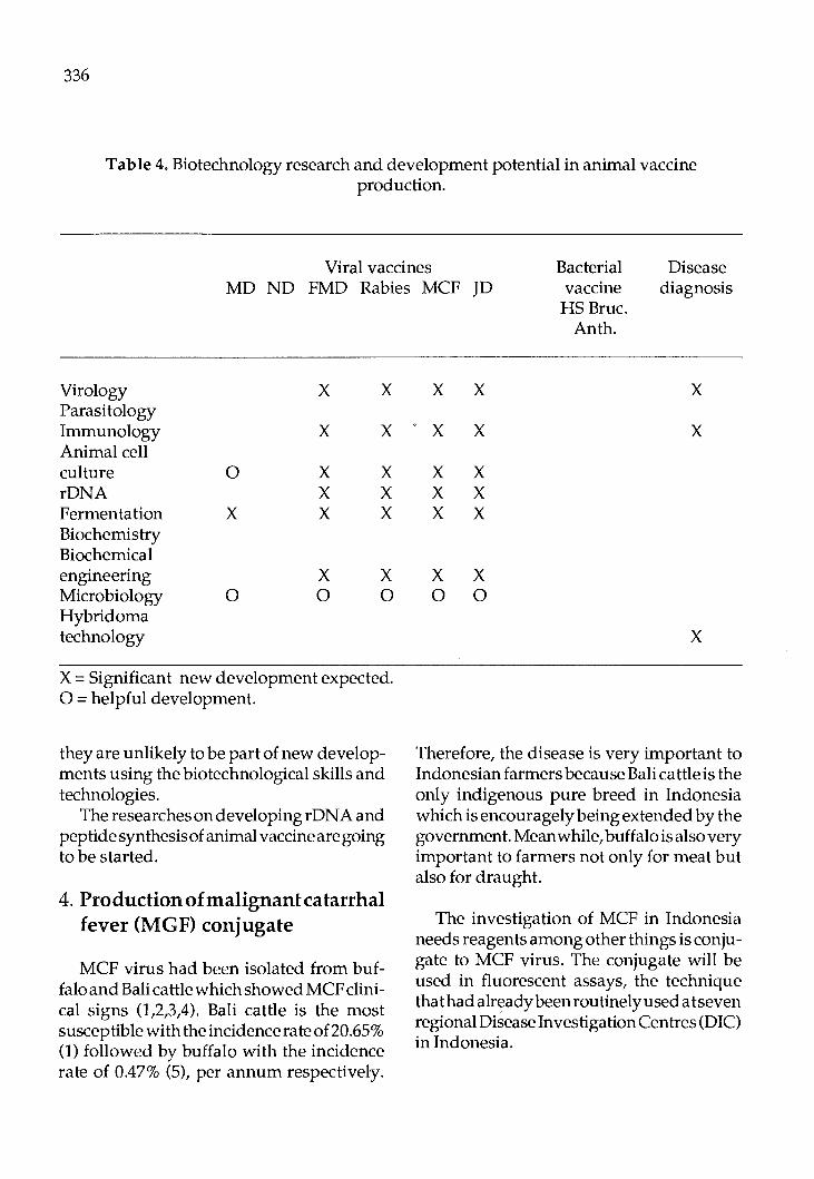
336
Table 4. Biotechnology research and development potential in animal vaccine production.
Viral vaccines Bacterial vaccine HS Brue.
Disease diagnosis MD ND FMD Rabies MCF JD
Virology Parasitology Immunology Animal cell culture rDNA Fermentation Biochemistry Biochemical engineering Microbiology Hybridoma technology
0
x
0
x
x
x x x
x 0
x
x
x x x
x 0
X = Significant new development expected. 0 =helpful development.
they are unlikely to be part of new developments using the biotechnological skills and technologies.
The researches on developing r DNA and peptide synthesis of animal vaccine are going to be started.
4. Production of malignant catarrhal fever (MGF) conjugate
MCF virus had been isolated from buffaloandBalicattlewhichshowedMCFclinical signs (1,2,3,4). Bali cattle is the most susceptiblewiththeincidencerateof20.65% (1) followed by buffalo with the incidence rate of 0.47% (5), per annum respectively.
x x
x x
x x x x x x
x x 0 0
An th.
x
x
x
Therefore, the disease is very important to Indonesian farmers because Bali cattle is the only indigenous pure breed in Indonesia which is encouragely being extended by the government. Meanwhile, buffalo is also very important to farmers not only for meat but also for draught.
The investigation of MCF in Indonesia needs reagents among other things is con jugate to MCF virus. The conjugate will be used in fluorescent assays, the technique that had already been routinely used a tseven regional Disease Investigation Centres (DIC) in Indonesia.
4a. Materials and Methods
Materials K6/ AP virus in bovine fetal kidney pri
mary cell culture (BFK cell) that was named due to the code number of a buffalo from which the virus was isolated, was employed as the antigen. A sheep was injected with the virus to produce antibody to MCF virus. Fluoresce in iscthiocyanate (FITC) was implemented to label the antibody.
For fluorescence standard, it had been employed bovine antiserum to well virus and anti bovine IgG conjugated with FITC in indirect fluorescent antibody technique (IF AT) against infected BFK cell. The antiserum was obtained from Plum Island Animal Disease Centre, USA under the courtesy of Dr. J.A. House and the antibovine IgG conjugated with FITC was manufactured by Capple Laboratory.
4b. Methods
BFK cell was infected with K6 I AP virus identified by the formation of cytopathic effects (CPE) in cyncytia form. The cell was trypsinized and centrifuged at 5000 rpm for five minutes. The packed cell was overflowed in PBS at the concentration of 3x10⁵ per ml.
The suspension was injected into jugular vein five times, 2 ml each, at interval of one week. The sera of preinoculation, one week after the first up to the fifth inoculations were taken for the judgement of antibody existance through neutralisation test (NT).
The serum of one week after the fifth inoculation was prepared for producing the conjugate after being mixed with normal BFK cell, 10 ml with 1 ml packed cell, over
337
night at -20"C.
Immunoglobulin G (IgG) was separated through precipitation using saturated ammonium sulfate to the amount of the serum volume. The mixture was steered for 45-60 minutes at 4 °C. The occurring precipitate was dissolved in the original serum volume of water and was precipitated again. This step was repeated twice.
The precipitate, then was dissolved in PBS pH 7.2 up to the original serum volume and loaded into dialysis tubing and dialysed against 20 times serum volume of water, overnight at 4°C. The dialysis was repeated until the IgG solution free from sulfate ion, proved by adding barium chloride (0.5%) into dialysate sample. The absence of white precipitate indicated the IgG solution free from sulfate ion. The dialysis was then continued against PBS.
The concentration of IgG solution was measured using Coleman spectrometer and the solution was diluted with PBS until the concentration 10-30 mg per ml. The solution was put into dialysis tubing for FITC conjugation.
Conjugation of IgG was conducted .due to Clarke and Shephard method (8).
The excess of FITC was omitted by dialyisngthe conjugated IgG against: aquqdest, PBS pH 7-7.5, O.OlM with 2-X4, 20-50 mesh Dowex (10 gram per 25 ml of conjugate), PBS pH 7-7.5, 0.01 M without Dowex, with the volume of each was 10-15 times the conjugate volume. The occuring precipitate was omitted through centrifugation or filtration. Supernatant was taken and used as conjugate after being added thiomarsal or merthyolate 1:10 000.
338
The specifity of staining of the conjugate could be raised through column chromatography with DEAE cellulose (anion exchaner) due to Riggs et al. method (9).
The titer of conjugate, then was confirmed. The solution was diluted in two folds. Each dilutionleveloftheconjugatewasemployed to test K6/ AP virus infected BFK cell. The fluorescence was compared with the result of IFAT using bovine antiserum to WCll in 1 :20 dilution and anti bovine IgG conjugated with FITCin 1:20dilution,againstthe same cell. The highest dilution level showing bright fluorescence with minimal nonspecific back ground was ascertained as dilution level of the conjugate.
4c. Result
Preinoculation serum had no antibody activity of inhibiting the growth of K6/ AP virus in BFK cell. Moreover, the serum of first, second and third injections also had no antibody activity. The antibody activity developed after fourth and raised after fifth injections of sera. The antiserum to K6/ AP as much as 20 ml after being absorbed by 2 ml of normal BFK cell was used for conjugate.
It produced 20 ml of IgG solution (10 mg per ml PBS) conjugated with FITC. The conjugate dilution manifested bright fluorescence up to 1:16 dilution.
The conjugate had already been used for FAT on MCF research at DIC, Region I Medan, North Sumatra, Indonesia at 1:10 dilution.
By the same technique, the sheep conjugate had already been produced repeatedly.
4d. Discussion
A virus had been isolated from buffalo and Bali cattle that showed MCF clinical signs. The virus was reacted against antibody to well virus (1,2,3,4). well virus is MCF virus isolated from wildebeest in Africa (7).
The antibody developed in the sheep owned the specific activity against K6/ AP virus that could be identified from NT. Where the sera of preinoculation and at least two weeks postinoculation revealed no antibody activity against K6/ AP virus. Meanwhile, the serum of four weeks postinoculation possessed antibody activity to K6/ AP virus which ascended titer after five weeks.
The possibility of the existence of antibody to cell membrane, was omitted through the absorbsion by the normal BFK cell, the cell culture that was implemented to grow K6/ AP virus.
The successful production of the conjugate and the discovery of the way to isolate MCF virus, open the new atmosphere to MCF diagnosis in Indonesia. thus, the diagnosis can be carried out earlier, so that the spreading of the disease can be prevented. Beside, the disease status of the population in the field can be monitored.
However, it is reliable to produce the antibody using purified virus. Purification can be done against free virus. Whereas MCF virus is known as a cell associated virus (6,7).
Recently, the virus is successfully freed through high subculture and already purified. Beside, the production of antibody through monoclonal antibody technique is
being studied.
References
1. Peranginangin, Th. A. 1988. Epidemiological feature and virus isolation of malignant catarrhal fever (MCF) in Bali cattle. The paper presented in 6th Congress of Federation on Asian Veterinary Association, October 16-19, 1988. Denpasar, Bali, Indonesia.
2. Peranginangin, Th. A. 1988. Isolation of virus from buffalo infected with suspected rnalignantcatarrhalfever, pp.110-112.In P .W. Daniels, Sundarisrnan and Purnorno Ronohardjo ed. Malignant Catarrhal Fever in Asian Livestock. Australian Centre for International Agricultural Research, Canberra.
3. Peranginangin, Th. A., S.I. Sitepu, A. Suryadi and E. Susanto. 1986a. The isolation of a syncytiogenic virus from buffalo showing clinical signs of MCF. Annual Report on Animal Disease Investigation in Indonesia During the Period 1984-1985 Jakarta, Direktora t Kesiha tan Hai wan. 45-51. (Indonesian, English abstract).
4. Peranginangin, Th. A., E. Susanto, S.I. Sitepu and A. Suryadi. 1986b. The identification of a syncytio-genic virus as the
339
cause of MCF in Deli Serdang with indirect FAT. Annual Report on Animal Disease Investigation in Indonesia During the Period 1984-1985. Jakarta, Direktorat Kesihatan Haiwan. 52-56. (Indonesian, English abstract).
5. Peranginangin, Th. A. 1986. Epidemiological aspects of malignant ca tarrhal fever in Deli Serdang district, North Sumatra. Annual Report on Animal Disease Investigation in Indonesia During the Period 1984-1985. Jakarta, Direktorat Kesihatan Haiwan. 199-206. (Indonesian, English abstract).
6. Castro, H.E. and B. Daley. 1982. Electron microscopic study of African strain of malignant catarrhal fever in bovine cell cultures. Arn. J. Res. 43(4): 576-582.
7. Plowright, W. 1965. Growth and characterisation of the virus of bovine rnaligantn catarrhal fever in East Africa. J. Gen. Microbiol. 39: 253-266.
8. Clarke, H.F. and C.C. Shephard. 1963. A dialysis technique for preparing fluorescent antibody. Virology 20: 642-644.
9. Riggs, J.L., P.C. Loh and W.C. Ereland. 1960. A simple fractionation method for preparation of fluorescein-labelled gamma globulin. Proc. Soc. Expth. Biol. Med. 105: 655-658.

Proc. FAO/CAAS Workshop on Biotechnology in Animal Production and Health in Asia and La.tin America, Beijing, Oct 9 -13, 1989, pp 340
Development of Monoclonal Antibody Against Newcastle Disease Virus and For Other
Veterinary Applications in Thailand
Urasri Tantaswasdi National Animal Health and Production Institute
Department of Livestock Development Bangkok 10900, Thailand.
Monoclonal antibodies (mah) have emerged as important diagnostic and research tools in various scientific disciplines because they are able to generate superior serological reagents in large amounts which are monospecific and homegenous. These are very useful in diagnosis, treatment and epidemiology.
In Thailand, the Department of Livestock Development (DLD), the government agency directly concerned with animal health and production, is involved in mah research, particularly concerning infectious diseases of veterinary importance. The National Animal Health and production Institute, DLD, with the assistance of the Japan International Cooperation Agency (JICA) is studying the production of mah against Newcastle disease virus (NDV) in order to differentiate NDV strains, for conventional procedures, which do not yield rapid answers and are unable to distinguish serotypes among avian paramyxoviruses. Hybridoma technology can produce highly specific antibodies which permit both the grouping and differentiation of avian paramyxoviruses in general and NDV isolates in particular. Experimental production of mah against NDV has just begun. Chicken embryos have been inoculated with a local
strain of NDV. The chorioallantoic fluid is harvested and purified by rate-zonal centrifugation. The purified antigen is used to immunize Balb I c mice to produce mah. At present, immunization is going on. Following this, immunized spleen cells will be mixed and fused with mouse myeloma cells and hybridoma culture fluids will be screened by enzyme linked immunosorbent assay and hemagglutination inhibition test. Antibody-producing hybridomas will be cloned and analysed to identify the epitope ofNDV.
In addition to this work, the Foot and Mouth Disease Center, DLD, again with the assistance ofJI CA is developing mah against foot and mouth disease virus for strain differentiation and estimation of virus infection associated antigen. Most of this work is conducted by Japanese experts together with Thai counterparts. Moreover, the Faculty of Science, Mahidol University, has proposed to the DLD a project for the development of an assay for circulating Fasciola gigantica antigens using mah to be carried out with funding from STDB. So at present in Thailand, advanced hybridoma technology is beginning to be exploited. The outcome of this work is sure to contribute to better disease control in the future.
FAO LIBRARY AN: 302043