Kollektive Wohnformen - neu interpretiert, am Beispiel Havanna/Kuba
Upload
independentCategory
view
2download
0
2558
Published OnlineFirst March 9, 2010; DOI: 10.1158/0008-5472.CAN-09-2840 Published OnlineFirst March 9, 2010; DOI: 10.1158/0008-5472.CAN-09-2840 Published OnlineFirst March 9, 2010; DOI: 10.1158/0008-5472.CAN-09-2840 Published OnlineFirst March 9, 2010; DOI: 10.1158/0008-5472.CAN-09-2840
Tumor and Stem Cell Biology
CancerResearch
Constitutively Active Stat3 Enhances Neu-MediatedMigration and Metastasis in Mammary Tumors viaUpregulation of Cten
Isaia Barbieri1,2, Sara Pensa1,2, Tania Pannellini4, Elena Quaglino1,3, Diego Maritano1,2, Marco Demaria1,2,Alessandra Voster1,2, James Turkson6, Federica Cavallo1,3, Christine J. Watson5,Paolo Provero1,2, Piero Musiani4, and Valeria Poli1,2
Abstract
Authors' AGenetics, BBiologicalCenter, “5DepartmeKingdom;University o
Note: SupResearch
I. Barbieri a
Present adCarle, Via C
Present adBiomedicheGiacosa, T
Correspon10126 TurE-mail: vale
doi: 10.115
©2010 Am
Cancer R
DownlDownlDownlDownl
The transcription factor signal transducer and activator of transcription 3 (STAT3) is constitutively activat-ed in tumors of different origin, but the molecular bases for STAT3 requirement are only partly understood.To evaluate the contribution of enhanced Stat3 activation in a controlled model system, we generated knock-in mice wherein a mutant constitutively active Stat3C allele replaces the endogenous wild-type allele. Stat3Ccould enhance the tumorigenic power of the rat Neu oncogene in mouse mammary tumor virus (MMTV)–Neutransgenic mice, triggering the production of earlier onset, more invasive mammary tumors. Tumor-derived cell lines displayed higher migration, invasion, and metastatic ability and showed disrupteddistribution of cell-cell junction markers mediated by Stat3-dependent overexpression of the COOH terminaltensin-like (Cten) focal adhesion protein, which was also significantly upregulated in Stat3C mammary tumors.Importantly, the proinflammatory cytokine interleukin-6 could mediate Cten induction in MCF10 cells in anexquisitely Stat3-dependent way, showing that Cten upregulation is a feature of inflammation-activated Stat3.In light of the emerging pivotal role of Stat3 in connecting inflammation and cancer, our identification of Ctenas a Stat3-dependent mediator of migration provides important new insights into the oncogenic role of Stat3,particularly in the breast. Cancer Res; 70(6); 2558–67. ©2010 AACR.
Introduction
The latent signal transducer and activator of transcription3 (STAT3) transcription factor can be activated by a wide va-riety of cytokines and by a number of growth factors and on-cogenes, including c-Src, epidermal growth factor receptor(EGFR), and ErbB-2, defined as HER2 in humans and Neu
ffiliations: 1Molecular Biotechnology Center, 2Department ofiology and Biochemistry, and 3Department of Clinical and
Sciences, University of Turin, Turin, Italy; 4Aging ResearchG. d'Annunzio” Universi ty Foundation, Chiet i , I taly;nt of Pathology, University of Cambridge, Cambridge, Unitedand 6Department of Molecular Biology and Microbiology,f Central Florida, Orlando, Florida
plementary data for this article are available at CancerOnline (http://cancerres.aacrjournals.org/).
nd S. Pensa contributed equally to this work.
dress for D. Maritano: Azienda Ospedaliera Santa Croce eoppino 26, 12100 Cuneo, Italy.
dress for A. Voster: Merck Serono S.A.-Istituto di Ricerche“A. Marxer,” RBM SpA, Via Ribes 1, 10010 Colleretto
urin, Italy.
ding Author: Valeria Poli, University of Turin, Via Nizza 52,in, Italy. Phone: 39-011-6706428; Fax: 39-011-6706432;[email protected].
8/0008-5472.CAN-09-2840
erican Association for Cancer Research.
es; 70(6) March 15, 2010
on June 2, 201cancerres.aacrjournals.org oaded from on June 2, 201cancerres.aacrjournals.org oaded from on June 2, 201cancerres.aacrjournals.org oaded from on June 2, 201cancerres.aacrjournals.org oaded from
in the rat (1, 2). Accordingly, STAT3 is found to be constitu-tively active in a high percentage of human and mouse tu-mors and tumor-derived cell lines of both hematologic andsolid origin, which often become addicted to its activity forcontinuous survival and growth (3, 4). Known STAT3 activi-ties that can contribute to tumorigenesis include enhancingtumor cell proliferation and survival, downmodulating anti-tumor immune responses, promoting angiogenesis, and in-ducing cell movement and epithelial-to-mesenchymaltransition (EMT; refs. 5, 6). Moreover, an important role forthis factor in linking inflammation and cancer downstreamof autocrine or paracrine production of interleukin-6 (IL-6)has recently emerged (7).The prooncogenic role of STAT3 was shown by the find-
ing that overexpression of the constitutively active mutantform Stat3C can transform cultured cells (8–10). Recently,in vivo Stat3C overexpression was shown to enhance malig-nant progression of skin tumors (11) or to trigger the onsetof lung adenocarcinomas (12). Despite the efforts of manylaboratories, the mechanisms underlying in vivo STAT3prooncogenic activities are however still incompletely un-derstood. Indeed, many of its known proproliferative orantiapoptotic target genes have been identified upon acutestimulation and are not consistently induced in tumorsdisplaying lower but continuous Stat3 phosphorylation, sug-gesting that distinct repertoires of target genes may be in-duced under these two conditions. Thus, overexpressionand/or acute stimulation does not seem to be the best
6. © 2010 American Association for Cancer Research. 6. © 2010 American Association for Cancer Research. 6. © 2010 American Association for Cancer Research. 6. © 2010 American Association for Cancer Research.
Stat3 Cooperates with Neu in Breast Tumors
Published OnlineFirst March 9, 2010; DOI: 10.1158/0008-5472.CAN-09-2840
experimental system to assess the Stat3 prooncogenic func-tions in vivo.Mammary tumors often display STAT3 constitutive activ-
ity, which is required for continuous proliferation and resis-tance to apoptosis of tumor-derived cells (13–15) andcorrelates with high expression of the EGFR family membersEGFR and/or HER2 (13, 16, 17). The HER2 oncogene is oftenamplified in human mammary tumors, particularly those dis-playing loss of estrogen sensitivity and poor prognosis (18).Accordingly, overexpression of the rat oncogenic form Neu inthe mammary gland of mouse mammary tumor virus(MMTV)–Neu transgenic mice (NeuT) triggers the onset ofinvasive multifocal breast carcinomas (19) and is widelyused as a model for human breast cancer (20). Recently,both we and others have shown that Stat3 is not requiredfor Neu-mediated tumorigenesis but is essential for meta-stasis formation, both in vivo and in vitro (21, 22). Defectivemetastatic ability could be ascribed both to altered expres-sion of proangiogenic and inflammatory genes, leading toimpaired angiogenic and inflammatory responses (22),and to cell autonomous defects in anchorage-independentgrowth (21).Here, we address some of the outstanding questions about
which role constitutively active Stat3 plays in vivo by gener-ating knock-in mice wherein a mutant Stat3C allele replacesthe wild-type endogenous gene and assessing its pro-oncogenic activity in the context of Neu-mediated mammarytumorigenesis, providing important insights into the molec-ular mechanisms involving Stat3 in breast cancer.
Materials and Methods
Animals, treatments, and analysis. Mice were main-tained in the transgenic unit of the Molecular BiotechnologyCenter (University of Turin) under a 12-h light-dark cycle andprovided food and water ad libitum. Procedures were con-ducted in conformity with national and international lawsand policies as approved by the Faculty Ethical Committee.Stat3C/WT female mice were bred to NeuT males to generatethe NeuT;Stat3C/WT or NeuT;Stat3WT/WT littermates used,thereby called N-C or N-WT. Palpable tumor onset was as-sessed in blind by weekly palpation. Mice were sacrificedby cervical dislocation.Cell lines derivation and culture. Tumor cell lines were
isolated from three different N-C and N-WT mice as de-scribed (23), subjected to differential trypsinization to elimi-nate fibroblasts, cultured in complete medium (RPMI 1360,20% FCS, 2 mmol/L Glutamax, 100 units/mL penicillin, and100 μg/mL streptomycin) and passaged with 0.05% trypsin-EDTA. MCF10A cells, cultured as previously described (24),were obtained from American Type Culture Collection androutinarily tested for Mycoplasma infection, morphology un-der both subconfluent and confluent conditions by phasecontrast microscopy, and the ability to undergo EMT. ForIL-6 or EGF treatments, cells were starved from serum andEGF overnight and then treated with recombinant IL-6(500 ng/mL) plus soluble IL-6 receptor (250 ng/mL) as previ-
www.aacrjournals.org
on June 2, 201cancerres.aacrjournals.org Downloaded from
ously described (25) or with 10 ng/mL human recombinantEGF (Sigma-Aldrich) with or without pretreatment overnightwith 100 μmol/L Stat3 inhibitor S3I-201 (26). C1 cells re-ceived 200 μmol/L S3I-201.Transwell invasion and migration assays. Cells (1 × 10⁵)
were placed in serum-free medium on Transwell inserts coat-ed or not with 2 μg of Matrigel (BD Biosciences). After incu-bation in wells containing RPMI ± 20% FCS, inserts werestained with Diff Quick Staining Set (Medion Diagnostic)and cells on the lower surface counted in blind.S.c. tumors and lung metastasis. Cells (1 × 10⁵) were
injected into the right fat pad or into the tail vein of nudeCD1 female mice. S.c. tumors were measured with a caliperweekly, and mice were sacrificed whenever the tumor ex-ceeded 10 mm of diameter. I.v. injected mice were sacrificedafter 3 or 5 wk; lungs were fixed with 2% paraformaldehyde,0.1 mol/L L-lysine, and 10 mmol/L sodium metaperiodate.Semiserial sections at 100-μm intervals were stained withH&E, and neoplastic lesions were counted in blind.Western blots. Total and nuclear protein extracts were ob-
tained as previously described (25, 27). Samples were frac-tionated on SDS-PAGE and transferred to a polyvinylidenedifluoride membrane (Millipore).Whole-mount image analyses. Images of whole-mount
(WM) preparations were taken with a Nikon Coolpix 950 dig-ital camera (Nital Spa) mounted on a stereoscopic Leica MZ6 microscope (Leica Microsystems). Specific instrument set-tings and quantification protocol are reported in Supplemen-tary Data.RNA extraction and analysis. Total RNA was extracted
with Trizol reagent (Invitrogen) and purified using InvitrogenMicro-to-Midi Total RNA Purification System (Invitrogen).Reverse transcription and real-time PCR were performed asdescribed (24, 28) using the ABI Prism 7300 Real-Time PCRSystem (Applied Biosystems) and the Universal Probe Library(Roche Italia) probes.COOH terminal tensin-like silencing. Cells were plated
at a density of 60% in 24-well plates or on coverslips forimmunofluorescence analysis and incubated for 72 h with1 μmol/L Accell SMARTpool siRNA (E-054907-00) or withthe Accell nontargeting siRNA (D-001910-01-05, Dharmacon,Thermo Fisher Scientific) in SMARTpool delivery mediumaccording to manufacturer's protocol. As a control for siR-NA entry, cells were incubated in parallel with an AccellGreen nontargeting siRNA (D-001950-01), and a glyceralde-hyde-3-phosphate dehydrogenase–targeting siRNA was usedas positive control. Fluorescence-positive cells were ∼90%for all experiments.Antibodies. Rabbit polyclonal against p-Stat3 or p-Stat5
(Cell Signaling Technology), Stat3 or Stat5 (Santa Cruz Bio-technology), and zonula occludens-1 (Zo-1; Zymed). Mousemonoclonal against β-catenin (BD Biosciences), vinculin(Sigma-Aldrich), E-cadherin 5H9 (Millipore), and prolifer-ating cell nuclear antigen (PCNA; Ylem). Antihuman COOHterminal tensin-like (CTEN; ref. 29) was a kind gift from Prof.S.H. Lo (University of California-Davis); antimouse Cten wasgenerated in house by immunization with a MBP-Cten fusionprotein and purified against the recombinant protein.
Cancer Res; 70(6) March 15, 2010 2559
6. © 2010 American Association for Cancer Research.

Barbieri et al.
Cancer Res; 70(6) March 15, 20102560
on June 2, 201cancerres.aacrjournals.org Downloaded from
Published OnlineFirst March 9, 2010; DOI: 10.1158/0008-5472.CAN-09-2840
Histology and immunohistochemistry. Mammary tissuewas processed as previously described (30). Formalin-fixedtissue sections were stained with H&E or overlaid with bio-tinylated goat anti-mouse and anti-rabbit immunoglobulin(Vector Laboratories) followed by avidin-biotin complex/alkaline phosphatase (DAKO). Terminal deoxynucleotidyltransferase-mediated dUTP nick-end labeling (TUNEL) wasperformed using ApopTag Plus Peroxidase In situ Apoptosiskit (Millipore).Immunofluorescence. Cells plated on glass coverslips were
washed in PBS, fixed in 4% paraformaldehyde, quenchedwith 50 mmol/L ammonium chloride, permeabilized with0.3% Triton X-100 in PBS, saturated with 3% bovine serumalbumin, and incubated with primary antibodies at roomtemperature for 1 h, followed by fluorescein- or rhodamine-labeled secondary antibodies (Sigma) or phalloidin (Sigma)and then by Hoechst-dye. An Axiovert 200M Zeiss microscopeor the Axio-Observer-Z1 Zeiss microscope with the ApoTomesystem for optical sectioning were used. Images were acquiredwith MetaMorph software (Molecular Devices) or the AxioVi-sion release 4.6.3 software (Carl Zeiss, Inc.), respectively.Statistical analysis. Kaplan-Meier survival curves were
analyzed by Prism4 (GraphPad software); P values werecalculated using the log-rank test. All other P values were cal-culated using Student's t test (unpaired, two tailed).
Results
Stat3C knock-in mice display constitutive Stat3 nuclearlocalization, phosphorylation, and transcriptional activi-ty. Mice carrying a mutant Stat3C allele have been generatedby a knock-in strategy similar to the one previously described(ref. 25; Supplementary Fig. S1). Stat3 was expressed at phys-iologic levels in all tissues analyzed in both the homozygousand heterozygous mutant mice (Supplementary Fig. S2A).Stat3C/C mice died at young age due to immune defects,7
whereas Stat3C/WT mice were viable and fertile.
7 Unpublished observation.
Figure 1. Stat3C enhances Neu-mediated mammary glandtumorigenesis. A, Kaplan-Meier curve showing the percentage of N-WT(n = 20) or of N-C (n = 24) female mice free of palpable tumors as afunction of age (P = 0.0014). B, ferric hematoxylin staining of WMmammary glands from N-C and N-WT mice at the indicated ages. Notethe amplification of neoplastic lesions ranging from atypical hyperplasiato in situ and invasive carcinomas (dark dots and nodules) in the N-Cmice. The central black oval areas are mammary lymph nodes (scale bar,5 mm). Digital quantification of the neoplastic lesions (mean width in μm)in samples from three different animals per genotype is shown with therelative P values. C, histology (H&E) performed on mammary samplesfrom 12-wk-old mice showing pseudoalveolar aggregates of the N-Ctumor cells invading the surrounding fibroadipose tissue. Proliferationand apoptosis are evaluated by means of anti-PCNA (PCNA) and TUNELstaining (scale bar, 150 μm). B and C, representative images of sectionsfrom three independent animals per genotype.
Cancer Research
6. © 2010 American Association for Cancer Research.

Stat3 Cooperates with Neu in Breast Tumors
Published OnlineFirst March 9, 2010; DOI: 10.1158/0008-5472.CAN-09-2840
Stat3C expressed by the knocked-in allele was indeed con-stitutively active. Stat3C/C mice displayed slightly enhancedStat3 nuclear localization and tyrosine phosphorylation inthe liver (Supplementary Fig. S2B), leading to significantly in-creased levels of several acute phase mRNAs, well-knownStat3 transcriptional targets (Supplementary Fig. S2D). Uponlipopolysaccharide (LPS)–mediated activation, nuclear local-ization of active, phosphorylated Stat3 was prolonged in theliver of the Stat3C/C mice (Supplementary Fig. S2C and notshown). Both basal and induced nuclear localization andphosphorylation were also increased in Stat3C/C primarymouse embryonic fibroblasts (MEF; Supplementary Fig.S2E), correlating with increased basal expression of a subsetof known Stat3 target genes (Supplementary Fig. S2F). Bothtranscriptional activity and DNA binding affinity of theStat3C protein were comparable with those of the wild-typeform, as judged by normal, and sometimes prolonged, LPS-or IL-6–mediated induction of tested target genes in the liveror in MEF cells, respectively (not shown), and by EMSA com-petitions (Supplementary Fig. S2C).Stat3C expression enhances Neu-mediated mammary
gland tumorigenesis. Spontaneous tumor onset could not
www.aacrjournals.org
on June 2, 201cancerres.aacrjournals.org Downloaded from
be assessed in aging Stat3C/C mice due to their early mor-tality. No preneoplastic lesions or spontaneously arisingtumors were detected in heterozygous Stat3C/WT mice up to24 months of age, and breast gland morphology was normalin virgin, pregnant, and lactating Stat3C/WT female mice.7 Wedecided therefore to assess potential cooperation of one con-stitutively active Stat3C allele with the Neu oncogene inmammary tumorigenesis. NeuT mice, which develop multi-ple foci of atypical hyperplasia progressing to invasive metas-tasizing carcinoma by 22 to 27 weeks (30), were intercrossedwith Stat3C/WT mice to obtain N-C and N-WT mice. N-C micedeveloped palpable tumors significantly earlier than their N-WT control littermates (Fig. 1A; P = 0.0014). This was alsoconfirmed by the presence of more diffused and advancedneoplastic lesions in the N-C mice in WM mammary glandpreparations performed at 7, 12, and 17 weeks of age (Fig.1B). Analysis of sectioned WM preparations also revealedlarger and more advanced tumor lesions (Fig. 1C), with nor-mal proliferation rates as assessed by PCNA staining, butsensibly reduced TUNEL-positive apoptotic nuclei. In partic-ular at 12 weeks, N-C mice already showed invasive carcino-mas, with tumor cells grouped in solid masses and small
Figure 2. Disrupted distribution ofcell-cell contacts in the C cell linesassessed by immunofluorescencestaining. The C1 and WT1tumor-derived cell lines wereplated on coated glass slides andincubated with phalloidin-TRITC orwith antibodies against E-cadherin,β-catenin (mouse monoclonal),or Zo-1 (rabbit polyclonal) followedby Hoechst staining and incubationwith FITC-labeled anti-mouseor TRITC-labeled antirabbitantibodies. Note the less regulardistribution of the epithelialmarkers and the absence ofcortical actin/presence of actinstress fibers in the C1 cells. Scalebar, 20 μm.
Cancer Res; 70(6) March 15, 2010 2561
6. © 2010 American Association for Cancer Research.
Barbieri et al.
2562
Published OnlineFirst March 9, 2010; DOI: 10.1158/0008-5472.CAN-09-2840
aggregates invading the surrounding fibroadipose tissue,whereas only atypical hyperplastic foci and few in situ carci-nomas were detected in the N-WT mice (Fig. 1C; H&E).Generation of tumor-derived cell lines. Neu protein levels
and phosphorylation were similar in tumors of both geno-types (Supplementary Fig. S3C and D). Likewise, phosphory-lation of both Stat3 and Stat5, another STAT factor oftenactivated in breast tumors, could be detected in both N-Cand N-WT tumors (Supplementary Fig. S3A, B), and no con-sistent differences were detected in the main biochemicalpathways known to be involved in Neu-mediated mammarytumorigenesis, including the expression or activity of Pten,Akt, Gsk3-β, Src, and Erk (Supplementary Fig. S3E).To generate a tool to identify the specific contribution of
enhanced Stat3 activity to the malignancy of Neu-trans-formed breast tumor cells, three independent cell lines werederived from tumors of each genotype and named C or WT 1,2, or 3. All cell lines maintained Neu expression and activitythrough at least 10 passages in culture (Supplementary Fig.S3H and I). All three C cell lines displayed increased nuclear,phosphorylated Stat3, suggesting enhanced activity of theStat3C allele, whereas levels of phosphorylated Stat5 seemed
Cancer Res; 70(6) March 15, 2010
on June 2, 201cancerres.aacrjournals.org Downloaded from
unchanged (Supplementary Fig. S3F, G and S5A). According-ly, the mRNA levels for SOCS3, a typical Stat3 target, wereincreased (Supplementary Fig. S4). C and WT cell lines didnot display any significant difference in Pten, Akt, Gsk3-β,Src, and Erk expression or activation (Supplementary Fig.S3J), except that both WT-1 and -2 cells underwent loss ofPten expression, a feature never displayed by the C cell lineseven after >30 passages (Supplementary Fig. S5B). Pten lossoccurred reproducibly between the first and second passagein culture in the WT-2 cell line, which as a consequence ac-quired faster growth rates (not shown). Cells were thereforeroutinely checked for Pten expression before all experimentsdescribed.C and WT cell lines did not differ significantly in terms of
proliferation or resistance to starvation-mediated apoptosis(data not shown). This is not surprising though in apparentcontradiction with the observed reduced apoptotic index ofthe N-C tumors, because in vitro stabilization of tumor cellsinvolves the selection of cells able to survive and can thusmask differences evident in vivo. However, all WT cell linesexhibited a more differentiated phenotype, growing in tightislets with well-organized and continuous adherent and tight
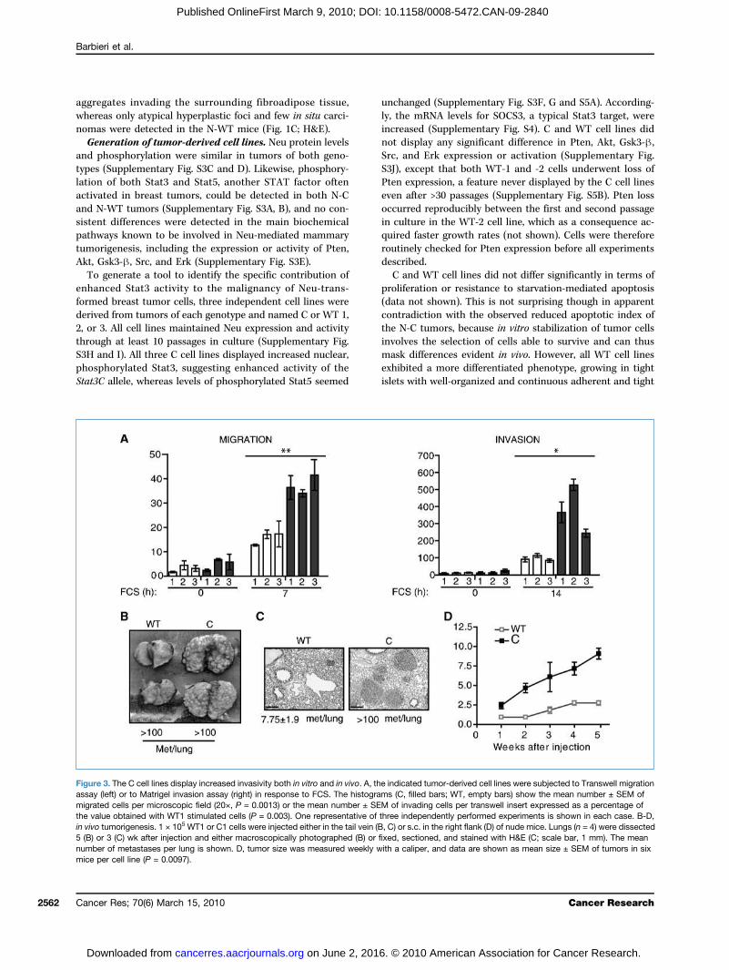
Figure 3. The C cell lines display increased invasivity both in vitro and in vivo. A, the indicated tumor-derived cell lines were subjected to Transwell migrationassay (left) or to Matrigel invasion assay (right) in response to FCS. The histograms (C, filled bars; WT, empty bars) show the mean number ± SEM ofmigrated cells per microscopic field (20×, P = 0.0013) or the mean number ± SEM of invading cells per transwell insert expressed as a percentage ofthe value obtained with WT1 stimulated cells (P = 0.003). One representative of three independently performed experiments is shown in each case. B-D,in vivo tumorigenesis. 1 × 10⁵ WT1 or C1 cells were injected either in the tail vein (B, C) or s.c. in the right flank (D) of nude mice. Lungs (n = 4) were dissected5 (B) or 3 (C) wk after injection and either macroscopically photographed (B) or fixed, sectioned, and stained with H&E (C; scale bar, 1 mm). The meannumber of metastases per lung is shown. D, tumor size was measured weekly with a caliper, and data are shown as mean size ± SEM of tumors in sixmice per cell line (P = 0.0097).
Cancer Research
6. © 2010 American Association for Cancer Research.
Stat3 Cooperates with Neu in Breast Tumors
Published OnlineFirst March 9, 2010; DOI: 10.1158/0008-5472.CAN-09-2840
junctions, as shown by staining of E-cadherin, β-catenin, andZo-1 (Fig. 2; Supplementary Fig. S6). Accordingly, actin fiberswere mainly located in the cortical region. Despite similarexpression levels of the same epithelial markers detectedby Western blot (not shown), all C cell lines displayed discon-tinuous E-cadherin, β-catenin, and Zo-1 distribution, sug-gesting less organized cell-cell contacts. In keeping withtheir less differentiated, spindle-like phenotype, all C cellsshowed reduced cortical actin and evident actin stress-fibersthat were never detected in the WT cells.Stat3C enhances migration, invasivity, and metastatic
potential of NeuT tumor cells. Migration and extracellularmatrix invasion of the C and WT cell lines were assessedby Transwell assays with or without a Matrigel coating(Fig. 3A). Migration was at least doubled in all C cell lines(P = 0.0013), which also showed strongly enhanced Matrigelinvasion potential (P = 0.03), suggesting higher invasive andmetastatic activity. To evaluate these parameters in vivo, C1or WT1 cells were injected i.v. into nude mice and the forma-tion of lung metastases was assessed after 3 or 5 weeks ofinjection. C1 cells displayed a strongly enhanced metastaticpotential, producing more and faster growing lung metasta-ses both at 5 (Fig. 3B) and 3 weeks (Fig. 3C). Similar resultswere obtained with the other cell lines (data not shown). Ofnote, WT2 cells were excluded from this analysis to avoidnonspecific interference of their Pten loss. C1 cells were alsoable to produce s.c. tumors displaying fast and regulargrowth and reaching a diameter of 10 mm 5 weeks after in-jection, whereas tumors produced by the WT1 cells grewmuch slower and never reached the 10-mm size (Fig. 3Dand not shown).Gene expression profiling of the C and WT cell lines.
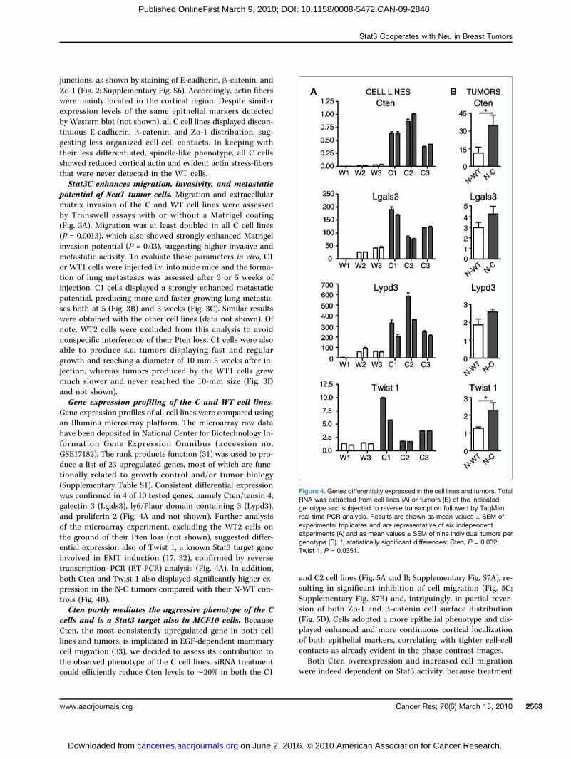
Gene expression profiles of all cell lines were compared usingan Illumina microarray platform. The microarray raw datahave been deposited in National Center for Biotechnology In-formation Gene Expression Omnibus (accession no.GSE17182). The rank products function (31) was used to pro-duce a list of 23 upregulated genes, most of which are func-tionally related to growth control and/or tumor biology(Supplementary Table S1). Consistent differential expressionwas confirmed in 4 of 10 tested genes, namely Cten/tensin 4,galectin 3 (Lgals3), ly6/Plaur domain containing 3 (Lypd3),and proliferin 2 (Fig. 4A and not shown). Further analysisof the microarray experiment, excluding the WT2 cells onthe ground of their Pten loss (not shown), suggested differ-ential expression also of Twist 1, a known Stat3 target geneinvolved in EMT induction (17, 32), confirmed by reversetranscription–PCR (RT-PCR) analysis (Fig. 4A). In addition,both Cten and Twist 1 also displayed significantly higher ex-pression in the N-C tumors compared with their N-WT con-trols (Fig. 4B).Cten partly mediates the aggressive phenotype of the C
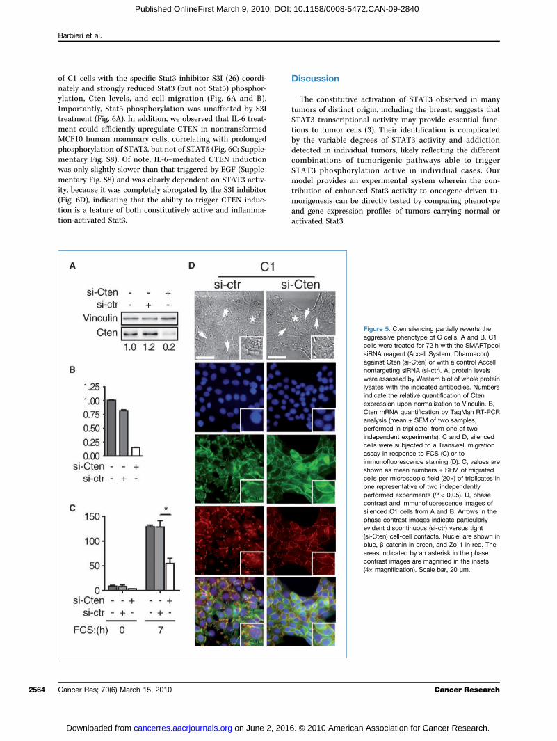
cells and is a Stat3 target also in MCF10 cells. BecauseCten, the most consistently upregulated gene in both celllines and tumors, is implicated in EGF-dependent mammarycell migration (33), we decided to assess its contribution tothe observed phenotype of the C cell lines. siRNA treatmentcould efficiently reduce Cten levels to ∼20% in both the C1
www.aacrjournals.org
on June 2, 201cancerres.aacrjournals.org Downloaded from
and C2 cell lines (Fig. 5A and B; Supplementary Fig. S7A), re-sulting in significant inhibition of cell migration (Fig. 5C;Supplementary Fig. S7B) and, intriguingly, in partial rever-sion of both Zo-1 and β-catenin cell surface distribution(Fig. 5D). Cells adopted a more epithelial phenotype and dis-played enhanced and more continuous cortical localizationof both epithelial markers, correlating with tighter cell-cellcontacts as already evident in the phase-contrast images.Both Cten overexpression and increased cell migration
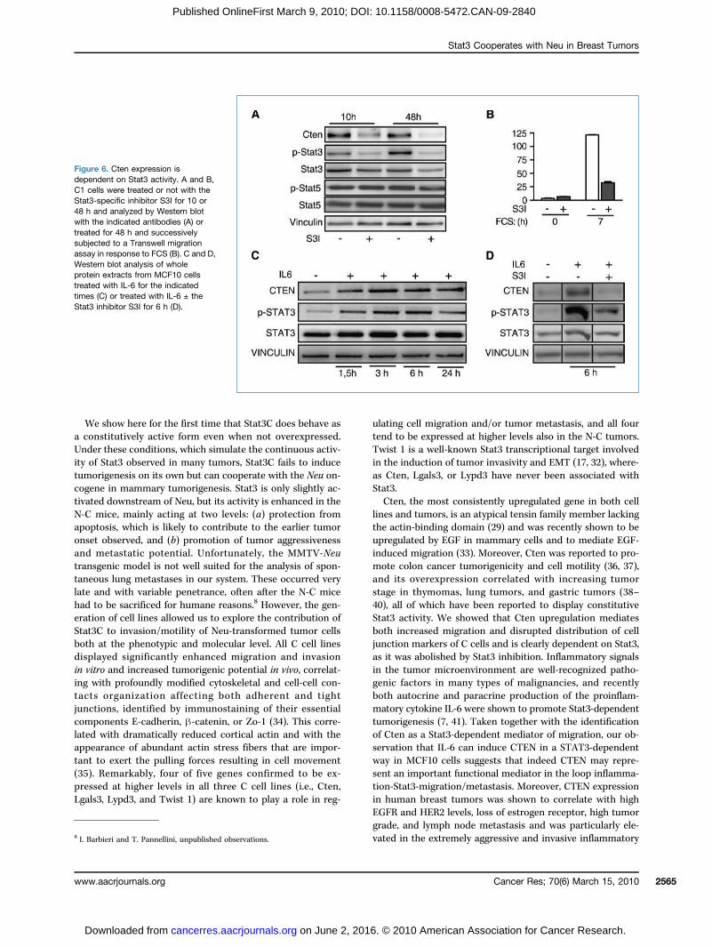
were indeed dependent on Stat3 activity, because treatment
Figure 4. Genes differentially expressed in the cell lines and tumors. TotalRNA was extracted from cell lines (A) or tumors (B) of the indicatedgenotype and subjected to reverse transcription followed by TaqManreal-time PCR analysis. Results are shown as mean values ± SEM ofexperimental triplicates and are representative of six independentexperiments (A) and as mean values ± SEM of nine individual tumors pergenotype (B). *, statistically significant differences: Cten, P = 0.032;Twist 1, P = 0.0351.
Cancer Res; 70(6) March 15, 2010 2563
6. © 2010 American Association for Cancer Research.
Barbieri et al.
2564
Published OnlineFirst March 9, 2010; DOI: 10.1158/0008-5472.CAN-09-2840
of C1 cells with the specific Stat3 inhibitor S3I (26) coordi-nately and strongly reduced Stat3 (but not Stat5) phosphor-ylation, Cten levels, and cell migration (Fig. 6A and B).Importantly, Stat5 phosphorylation was unaffected by S3Itreatment (Fig. 6A). In addition, we observed that IL-6 treat-ment could efficiently upregulate CTEN in nontransformedMCF10 human mammary cells, correlating with prolongedphosphorylation of STAT3, but not of STAT5 (Fig. 6C; Supple-mentary Fig. S8). Of note, IL-6–mediated CTEN inductionwas only slightly slower than that triggered by EGF (Supple-mentary Fig. S8) and was clearly dependent on STAT3 activ-ity, because it was completely abrogated by the S3I inhibitor(Fig. 6D), indicating that the ability to trigger CTEN induc-tion is a feature of both constitutively active and inflamma-tion-activated Stat3.
Cancer Res; 70(6) March 15, 2010
on June 2, 201cancerres.aacrjournals.org Downloaded from
Discussion
The constitutive activation of STAT3 observed in manytumors of distinct origin, including the breast, suggests thatSTAT3 transcriptional activity may provide essential func-tions to tumor cells (3). Their identification is complicatedby the variable degrees of STAT3 activity and addictiondetected in individual tumors, likely reflecting the differentcombinations of tumorigenic pathways able to triggerSTAT3 phosphorylation active in individual cases. Ourmodel provides an experimental system wherein the con-tribution of enhanced Stat3 activity to oncogene-driven tu-morigenesis can be directly tested by comparing phenotypeand gene expression profiles of tumors carrying normal oractivated Stat3.
6. © 2010 American
Figure 5. Cten silencing partially reverts theaggressive phenotype of C cells. A and B, C1cells were treated for 72 h with the SMARTpoolsiRNA reagent (Accell System, Dharmacon)against Cten (si-Cten) or with a control Accellnontargeting siRNA (si-ctr). A, protein levelswere assessed by Western blot of whole proteinlysates with the indicated antibodies. Numbersindicate the relative quantification of Ctenexpression upon normalization to Vinculin. B,Cten mRNA quantification by TaqMan RT-PCRanalysis (mean ± SEM of two samples,performed in triplicate, from one of twoindependent experiments). C and D, silencedcells were subjected to a Transwell migrationassay in response to FCS (C) or toimmunofluorescence staining (D). C, values areshown as mean numbers ± SEM of migratedcells per microscopic field (20×) of triplicates inone representative of two independentlyperformed experiments (P < 0,05). D, phasecontrast and immunofluorescence images ofsilenced C1 cells from A and B. Arrows in thephase contrast images indicate particularlyevident discontinuous (si-ctr) versus tight(si-Cten) cell-cell contacts. Nuclei are shown inblue, β-catenin in green, and Zo-1 in red. Theareas indicated by an asterisk in the phasecontrast images are magnified in the insets(4× magnification). Scale bar, 20 μm.
Cancer Research
Association for Cancer Research.
Stat3 Cooperates with Neu in Breast Tumors
Published OnlineFirst March 9, 2010; DOI: 10.1158/0008-5472.CAN-09-2840
We show here for the first time that Stat3C does behave asa constitutively active form even when not overexpressed.Under these conditions, which simulate the continuous activ-ity of Stat3 observed in many tumors, Stat3C fails to inducetumorigenesis on its own but can cooperate with the Neu on-cogene in mammary tumorigenesis. Stat3 is only slightly ac-tivated downstream of Neu, but its activity is enhanced in theN-C mice, mainly acting at two levels: (a) protection fromapoptosis, which is likely to contribute to the earlier tumoronset observed, and (b) promotion of tumor aggressivenessand metastatic potential. Unfortunately, the MMTV-Neutransgenic model is not well suited for the analysis of spon-taneous lung metastases in our system. These occurred verylate and with variable penetrance, often after the N-C micehad to be sacrificed for humane reasons.8 However, the gen-eration of cell lines allowed us to explore the contribution ofStat3C to invasion/motility of Neu-transformed tumor cellsboth at the phenotypic and molecular level. All C cell linesdisplayed significantly enhanced migration and invasionin vitro and increased tumorigenic potential in vivo, correlat-ing with profoundly modified cytoskeletal and cell-cell con-tacts organization affecting both adherent and tightjunctions, identified by immunostaining of their essentialcomponents E-cadherin, β-catenin, or Zo-1 (34). This corre-lated with dramatically reduced cortical actin and with theappearance of abundant actin stress fibers that are impor-tant to exert the pulling forces resulting in cell movement(35). Remarkably, four of five genes confirmed to be ex-pressed at higher levels in all three C cell lines (i.e., Cten,Lgals3, Lypd3, and Twist 1) are known to play a role in reg-
8 I. Barbieri and T. Pannellini, unpublished observations.
www.aacrjournals.org
on June 2, 201cancerres.aacrjournals.org Downloaded from
ulating cell migration and/or tumor metastasis, and all fourtend to be expressed at higher levels also in the N-C tumors.Twist 1 is a well-known Stat3 transcriptional target involvedin the induction of tumor invasivity and EMT (17, 32), where-as Cten, Lgals3, or Lypd3 have never been associated withStat3.Cten, the most consistently upregulated gene in both cell
lines and tumors, is an atypical tensin family member lackingthe actin-binding domain (29) and was recently shown to beupregulated by EGF in mammary cells and to mediate EGF-induced migration (33). Moreover, Cten was reported to pro-mote colon cancer tumorigenicity and cell motility (36, 37),and its overexpression correlated with increasing tumorstage in thymomas, lung tumors, and gastric tumors (38–40), all of which have been reported to display constitutiveStat3 activity. We showed that Cten upregulation mediatesboth increased migration and disrupted distribution of celljunction markers of C cells and is clearly dependent on Stat3,as it was abolished by Stat3 inhibition. Inflammatory signalsin the tumor microenvironment are well-recognized patho-genic factors in many types of malignancies, and recentlyboth autocrine and paracrine production of the proinflam-matory cytokine IL-6 were shown to promote Stat3-dependenttumorigenesis (7, 41). Taken together with the identificationof Cten as a Stat3-dependent mediator of migration, our ob-servation that IL-6 can induce CTEN in a STAT3-dependentway in MCF10 cells suggests that indeed CTEN may repre-sent an important functional mediator in the loop inflamma-tion-Stat3-migration/metastasis. Moreover, CTEN expressionin human breast tumors was shown to correlate with highEGFR and HER2 levels, loss of estrogen receptor, high tumorgrade, and lymph node metastasis and was particularly ele-vated in the extremely aggressive and invasive inflammatory
Figure 6. Cten expression isdependent on Stat3 activity. A and B,C1 cells were treated or not with theStat3-specific inhibitor S3I for 10 or48 h and analyzed by Western blotwith the indicated antibodies (A) ortreated for 48 h and successivelysubjected to a Transwell migrationassay in response to FCS (B). C and D,Western blot analysis of wholeprotein extracts from MCF10 cellstreated with IL-6 for the indicatedtimes (C) or treated with IL-6 ± theStat3 inhibitor S3I for 6 h (D).
Cancer Res; 70(6) March 15, 2010 2565
6. © 2010 American Association for Cancer Research.

Barbieri et al.
2566
Published OnlineFirst March 9, 2010; DOI: 10.1158/0008-5472.CAN-09-2840
breast cancers (33). In this context, CTEN may provide a cru-cial point of functional convergence between inflammation-induced constitutively enhanced Stat3 activity, altered EGFR,and/or HER2-mediated signaling and invasion of the sur-rounding tissues and lymph nodes. Our data support the ideaof Stat3 playing an essential role in metastasis formationdownstream of Neu (21, 22, 42) and provide further insightsinto the molecular mechanisms linking Stat3 activation, in-flammatory response, and migration/metastasis.
Disclosure of Potential Conflicts of Interest
No potential conflicts of interest were disclosed.
Cancer Res; 70(6) March 15, 2010
on June 2, 201cancerres.aacrjournals.org Downloaded from
Acknowledgments
We thank E. Hirsch, G. Forni, P.P. Pandolfi, and S. Cabodi for criticallyreading the manuscript, S.H. Lo for the kind gift of the anti-CTEN antibody,and L. Silengo for continuous support.
Grant Support
Italian Cancer Research Association (AIRC), Italian Ministry for Universityand Research PRIN and FIRB, Ricerca Finalizzata Piemonte, Progetto Alfierifrom CRT Foundation, and Association for International Cancer Research.
The costs of publication of this article were defrayed in part by the paymentof page charges. This article must therefore be hereby marked advertisement inaccordance with 18 U.S.C. Section 1734 solely to indicate this fact.
Received 07/31/2009; revised 11/30/2009; accepted 01/11/2010; publishedOnlineFirst 03/09/2010.
References
1. Fernandes A, Hamburger AW, Gerwin BI. ErbB-2 kinase is requiredfor constitutive stat 3 activation in malignant human lung epithelialcells. Int J Cancer 1999;83:564–70.
2. Turkson J, Jove R. STAT proteins: novel molecular targets for cancerdrug discovery. Oncogene 2000;19:6613–26.
3. Kortylewski M, Jove R, Yu H. Targeting STAT3 affects melanoma onmultiple fronts. Cancer Metastasis Rev 2005;24:315–27.
4. Al Zaid Siddiquee K, Turkson J. STAT3 as a target for inducingapoptosis in solid and hematological tumors. Cell Res 2008;18:254–67.
5. Pensa S, Regis G, Boselli D, Novelli F, Poli V. STAT1 and STAT3 intumorigenesis: two sides of the same coin? In: Stephanou A, editor.JAK-STAT Pathway in Disease. Austin: Landes Bioscience; 2009, p.100–21.
6. Yu H, Kortylewski M, Pardoll D. Crosstalk between cancer and im-mune cells: role of STAT3 in the tumour microenvironment. Nat RevImmunol 2007;7:41–51.
7. Bromberg J, Wang TC. Inflammation and cancer: IL-6 and STAT3complete the link. Cancer Cell 2009;15:79–80.
8. Bromberg JF, Wrzeszczynska MH, Devgan G, et al. Stat3 as anoncogene. Cell 1999;98:295–303.
9. Dechow TN, Pedranzini L, Leitch A, et al. Requirement of matrixmetalloproteinase-9 for the transformation of human mammary ep-ithelial cells by Stat3-C. Proc Natl Acad Sci U S A 2004;101:10602–7.
10. Azare J, Leslie K, Al-Ahmadie H, et al. Constitutively activated Stat3induces tumorigenesis and enhances cell motility of prostate epithe-lial cells through integrin β6. Mol Cell Biol 2007;27:4444–53.
11. Chan KS, Sano S, Kataoka K, et al. Forced expression of a consti-tutively active form of Stat3 in mouse epidermis enhances malignantprogression of skin tumors induced by two-stage carcinogenesis.Oncogene 2008;27:1087–94.
12. Li Y, Du H, Qin Y, Roberts J, Cummings OW, Yan C. Activation of thesignal transducers and activators of the transcription 3 pathway inalveolar epithelial cells induces inflammation and adenocarcinomasin mouse lung. Cancer Res 2007;67:8494–503.
13. Diaz N, Minton S, Cox C, et al. Activation of stat3 in primary tumorsfrom high-risk breast cancer patients is associated with elevated le-vels of activated SRC and survivin expression. Clin Cancer Res 2006;12:20–8.
14. Gritsko T, Williams A, Turkson J, et al. Persistent activation of stat3signaling induces survivin gene expression and confers resistance toapoptosis in human breast cancer cells. Clin Cancer Res 2006;12:11–9.
15. Leslie K, Lang C, Devgan G, et al. Cyclin D1 is transcriptionally reg-ulated by and required for transformation by activated signal trans-ducer and activator of transcription 3. Cancer Res 2006;66:2544–52.
16. Berclaz G, Altermatt HJ, Rohrbach V, Siragusa A, Dreher E, SmithPD. EGFR dependent expression of STAT3 (but not STAT1) in breastcancer. Int J Oncol 2001;19:1155–60.
17. Lo HW, Hsu SC, Xia W, et al. Epidermal growth factor receptorcooperates with signal transducer and activator of transcription 3to induce epithelial-mesenchymal transition in cancer cells via up-regulation of TWIST gene expression. Cancer Res 2007;67:9066–76.
18. Menard S, Balsari A, Tagliabue E, et al. Biology, prognosis and re-sponse to therapy of breast carcinomas according to HER2 score.Ann Oncol 2008;19:1706–12.
19. Muller WJ, Sinn E, Pattengale PK, Wallace R, Leder P. Single-stepinduction of mammary adenocarcinoma in transgenic mice bearingthe activated c-neu oncogene. Cell 1988;54:105–15.
20. Marcotte R, Muller WJ. Signal transduction in transgenic mousemodels of human breast cancer-implications for human breast can-cer. J Mammary Gland Biol Neoplasia 2008;13:323–35.
21. Barbieri I, Quaglino E, Maritano D, et al. Stat3 is required for anchor-age-independent growth and metastasis but not for mammary tumordevelopment downstream of the ErbB-2 oncogene. Mol Carcinog2010;49:114–20.
22. Ranger JJ, Levy DE, Shahalizadeh S, Hallett M, Muller WJ. Iden-tification of a Stat3-dependent transcription regulatory network in-volved in metastatic progression. Cancer Res 2009;69:6823–30.
23. Nanni P, Pupa SM, Nicoletti G, et al. p185(neu) protein is requiredfor tumor and anchorage-independent growth, not for cell prolifer-ation of transgenic mammary carcinoma. Int J Cancer 2000;87:186–94.
24. Schiavone D, Dewilde S, Vallania F, Turkson J, Di Cunto F, Poli V.The RhoU/Wrch1 Rho GTPase gene is a common transcriptional tar-get of both the gp130/STAT3 and Wnt-1 pathways. Biochem J 2009;421:283–92.
25. Maritano D, Sugrue ML, Tininini S, et al. The STAT3 isoforms α and βhave unique and specific functions. Nat Immunol 2004;5:401–9.
26. Siddiquee K, Zhang S, Guida WC, et al. Selective chemical probeinhibitor of Stat3, identified through structure-based virtual screen-ing, induces antitumor activity. Proc Natl Acad Sci U S A 2007;104:7391–6.
27. Alonzi T, Maritano D, Gorgoni B, Rizzuto G, Libert C, Poli V. Essentialrole of STAT3 in the control of the acute-phase response as revealedby inducible gene inactivation in the liver. Mol Cell Biol 2001;21:1621–32.
28. Vallania F, Schiavone D, Dewilde S, et al. Genome-wide discoveryof functional transcription factor binding sites by comparativegenomics: the case of Stat3. Proc Natl Acad Sci U S A 2009;106:5117–22.
29. Lo SH, Lo TB. Cten, a COOH-terminal tensin-like protein with pros-tate restricted expression, is down-regulated in prostate cancer.Cancer Res 2002;62:4217–21.
30. Di Carlo E, Diodoro MG, Boggio K, et al. Analysis of mammary car-cinoma onset and progression in HER-2/neu oncogene transgenicmice reveals a lobular origin. Lab Invest 1999;79:1261–9.
31. Breitling R, Armengaud P, Amtmann A, Herzyk P. Rank products: asimple, yet powerful, new method to detect differentially regulated
Cancer Research
6. © 2010 American Association for Cancer Research.

Stat3 Cooperates with Neu in Breast Tumors
Published OnlineFirst March 9, 2010; DOI: 10.1158/0008-5472.CAN-09-2840
genes in replicated microarray experiments. FEBS Lett 2004;573:83–92.
32. Cheng GZ, Zhang WZ, Sun M, et al. Twist is transcriptionally inducedby activation of STAT3 and mediates STAT3 oncogenic function.J Biol Chem 2008;283:14665–73.
33. Katz M, Amit I, Citri A, et al. A reciprocal tensin-3-cten switch med-iates EGF-driven mammary cell migration. Nat Cell Biol 2007;9:961–9.
34. Niessen CM. Tight junctions/adherens junctions: basic structure andfunction. J Investig Dermatol 2007;127:2525–32.
35. Pellegrin S, Mellor H. Actin stress fibres. J Cell Sci 2007;120:3491–9.36. Albasri A, Seth R, Jackson D, et al. C-terminal Tensin-like (CTEN) is
an oncogene which alters cell motility possibly through repression ofE-cadherin in colorectal cancer. J Pathol 2009;218:57–65.
37. Liao YC, Chen NT, Shih YP, Dong Y, Lo SH. Up-regulation of C-
www.aacrjournals.org
on June 2, 201cancerres.aacrjournals.org Downloaded from
terminal tensin-like molecule promotes the tumorigenicity of coloncancer through β-catenin. Cancer Res 2009;69:4563–6.
38. Sasaki H, Yukiue H, Kobayashi Y, Fukai I, Fujii Y. Cten mRNA ex-pression is correlated with tumor progression in thymoma. TumourBiol 2003;24:271–4.
39. Sakashita K, Mimori K, Tanaka F, et al. Prognostic relevance ofTensin4 expression in human gastric cancer. Ann Surg Oncol2008;15:2606–13.
40. Sasaki H, Moriyama S, Mizuno K, et al. Cten mRNA expression wascorrelated with tumor progression in lung cancers. Lung Cancer (Am-sterdam, the Netherlands) 2003;40:151–5.
41. Grivennikov S, Karin M. Autocrine IL-6 signaling: a key event intumorigenesis? Cancer Cell 2008;13:7–9.
42. GuoW, Pylayeva Y, Pepe A, et al. β4 integrin amplifies ErbB2 signalingto promote mammary tumorigenesis. Cell 2006;126:489–502.
Cancer Res; 70(6) March 15, 2010 2567
6. © 2010 American Association for Cancer Research.
Correction
Correction: Online Publication Dates forCancer Research April 15, 2010 Articles
The following articles in the April 15, 2010 issue of Cancer Research were publishedwith an online publication date of April 6, 2010 listed, but were actually publishedonline on April 13, 2010:
CancerResearch
Garmy-Susini B, Avraamides CJ, Schmid MC, Foubert P, Ellies LG, Barnes L, Feral C,Papayannopoulou T, Lowy A, Blair SL, Cheresh D, Ginsberg M, Varner JA. Integrinα4β1 signaling is required for lymphangiogenesis and tumor metastasis. Cancer Res2010;70:3042–51. Published OnlineFirst April 13, 2010. doi:10.1158/0008-5472.CAN-09-3761.
Vincent J, Mignot G, Chalmin F, Ladoire S, Bruchard M, Chevriaux A, Martin F,Apetoh L, Rébé C, Ghiringhelli F. 5-Fluorouracil selectively kills tumor-associatedmyeloid-derived suppressor cells resulting in enhanced T cell-dependent antitumorimmunity. Cancer Res 2010;70:3052–61. Published OnlineFirst April 13, 2010.doi:10.1158/0008-5472.CAN-09-3690.
Nagasaka T, Rhees J, Kloor M, Gebert J, Naomoto Y, Boland CR, Goel A. Somatichypermethylation of MSH2 is a frequent event in Lynch syndrome colorectalcancers. Cancer Res 2010;70:3098–108. Published OnlineFirst April 13, 2010.doi:10.1158/0008-5472.CAN-09-3290.
He X, Ota T, Liu P, Su C, Chien J, Shridhar V. Downregulation of HtrA1 promotesresistance to anoikis and peritoneal dissemination of ovarian cancer cells. CancerRes 2010;70:3109–18. Published OnlineFirst April 13, 2010. doi:10.1158/0008-5472.CAN-09-3557.
Fiorentino M, Judson G, Penney K, Flavin R, Stark J, Fiore C, Fall K, Martin N, Ma J,Sinnott J, Giovannucci E, Stampfer M, Sesso HD, Kantoff PW, Finn S, Loda M, Mucci L.Immunohistochemical expression of BRCA1 and lethal prostate cancer. Cancer Res2010;70:3136–9. Published OnlineFirst April 13, 2010. doi:10.1158/0008-5472.CAN-09-4100.
Veronese A, Lupini L, Consiglio J, Visone R, Ferracin M, Fornari F, Zanesi N, Alder H,D'Elia G, Gramantieri L, Bolondi L, Lanza G, Querzoli P, Angioni A, Croce CM,Negrini M. Oncogenic role of miR-483-3p at the IGF2/483 locus. Cancer Res2010;70:3140–9. Published OnlineFirst April 13, 2010. doi:10.1158/0008-5472.CAN-09-4456.
Lu W, Zhang G, Zhang R, Flores LG II, Huang Q, Gelovani JG, Li C. Tumor site–specific silencing of NF-κB p65 by targeted hollow gold nanosphere–mediatedphotothermal transfection. Cancer Res 2010;70:3177–88. Published OnlineFirst April13, 2010. doi:10.1158/0008-5472.CAN-09-3379.
Geng H, Rademacher BL, Pittsenbarger J, Huang C-Y, Harvey CT, Lafortune MC,Myrthue A, Garzotto M, Nelson PS, Beer TM, Qian DZ. ID1 enhances docetaxel cyto-toxicity in prostate cancer cells through inhibition of p21. Cancer Res 2010;70:3239–48.Published OnlineFirst April 13, 2010. doi:10.1158/0008-5472.CAN-09-3186.
Yoo BK, Chen D, Su Z-z, Gredler R, Yoo J, Shah K, Fisher PB, Sarkar D. Molecular mech-anism of chemoresistance by astrocyte elevated gene-1. Cancer Res 2010;70:3249–58.Published OnlineFirst April 13, 2010. doi:10.1158/0008-5472.CAN-09-4009.
Lu ZH, Shvartsman MB, Lee AY, Shao JM, Murray MM, Kladney RD, Fan D, Krajewski S,Chiang GG, Mills GB, Arbeit JM. Mammalian target of rapamycin activator RHEB isfrequently overexpressed in human carcinomas and is critical and sufficient for skinepithelial carcinogenesis. Cancer Res 2010;70:3287–98. Published OnlineFirst April13, 2010. doi:10.1158/0008-5472.CAN-09-3467.
www.aacrjournals.org 4785

Correction
Hattermann K, Held-Feindt J, Lucius R, Müerköster SS, Penfold MET, Schall TJ,Mentlein R. The chemokine receptor CXCR7 is highly expressed in human gliomacells and mediates antiapoptotic effects. Cancer Res 2010;70:3299–308. PublishedOnlineFirst April 13, 2010. doi:10.1158/0008-5472.CAN-09-3642.
Nadiminty N, Lou W, Sun M, Chen J, Yue J, Kung H-J, Evans CP, Zhou Q, Gao AC.Aberrant activation of the androgen receptor by NF-κB2/p52 in prostate cancer cells.Cancer Res 2010;70:3309–19. Published OnlineFirst April 13, 2010. doi:10.1158/0008-5472.CAN-09-3703.
Acu ID, Liu T, Suino-Powell K, Mooney SM, D'Assoro AB, Rowland N, Muotri AR,Correa RG, Niu Y, Kumar R, Salisbury JL. Coordination of centrosome homeostasisand DNA repair is intact in MCF-7 and disrupted in MDA-MB 231 breast cancercells. Cancer Res 2010;70:3320–8. Published OnlineFirst April 13, 2010. doi:10.1158/0008-5472.CAN-09-3800.
McFarlane C, Kelvin AA, de la Vega M, Govender U, Scott CJ, Burrows JF, JohnstonJA. The deubiquitinating enzyme USP17 is highly expressed in tumor biopsies, is cellcycle regulated, and is required for G1-S progression. Cancer Res 2010;70:3329–39.Published OnlineFirst April 13, 2010. doi:10.1158/0008-5472.CAN-09-4152.
Dudka AA, Sweet SMM, Heath JK. Signal transducers and activators of transcription-3binding to the fibroblast growth factor receptor is activated by receptor amplifica-tion. Cancer Res 2010;70:3391–401. Published OnlineFirst April 13, 2010. doi:10.1158/0008-5472.CAN-09-3033.
Cho SY, Xu M, Roboz J, Lu M, Mascarenhas J, Hoffman R. The effect of CXCL12 pro-cessing on CD34+ cell migration in myeloproliferative neoplasms. Cancer Res2010;70:3402–10. Published OnlineFirst April 13, 2010. doi:10.1158/0008-5472.CAN-09-3977.
Published OnlineFirst 05/11/2010.©2010 American Association for Cancer Research.doi: 10.1158/0008-5472.CAN-10-1347
Cancer Res; 70(11) June 1, 2010 Cancer Research4786

2010;70:2558-2567. Published OnlineFirst March 9, 2010.Cancer Res Isaia Barbieri, Sara Pensa, Tania Pannellini, et al. and Metastasis in Mammary Tumors via Upregulation of CtenConstitutively Active Stat3 Enhances Neu-Mediated Migration
Updated version
10.1158/0008-5472.CAN-09-2840doi:
Access the most recent version of this article at:
Material
Supplementary
http://cancerres.aacrjournals.org/content/suppl/2010/03/08/0008-5472.CAN-09-2840.DC1.html
Access the most recent supplemental material at:
Cited articles
http://cancerres.aacrjournals.org/content/70/6/2558.full.html#ref-list-1
This article cites 41 articles, 17 of which you can access for free at:
Citing articles
http://cancerres.aacrjournals.org/content/70/6/2558.full.html#related-urls
This article has been cited by 12 HighWire-hosted articles. Access the articles at:
E-mail alerts related to this article or journal.Sign up to receive free email-alerts
Subscriptions
Reprints and
To order reprints of this article or to subscribe to the journal, contact the AACR Publications
Permissions
To request permission to re-use all or part of this article, contact the AACR Publications
on June 2, 2016. © 2010 American Association for Cancer Research. cancerres.aacrjournals.org Downloaded from
Published OnlineFirst March 9, 2010; DOI: 10.1158/0008-5472.CAN-09-2840
Copyright © 2022 FDOKUMEN