Synthesis and characterization of xanthan–hydroxyapatite nanocomposites for cellular uptake

Brie et al. Journal of Biological Engineering 2014, 8:14http://www.jbioleng.org/content/8/1/14
RESEARCH Open Access
Comparative in vitro study regarding thebiocompatibility of titanium-base compositesinfiltrated with hydroxyapatite or silicatitanateIoana-Carmen Brie1,2, Olga Soritau1*, Noemi Dirzu3, Cristian Berce2, Adriana Vulpoi4, Catalin Popa3, Milica Todea4,Simion Simon4, Maria Perde-Schrepler1, Piroska Virag1, Otilia Barbos1, Gabriela Chereches1, Petru Berce3
and Valentin Cernea1,2
Abstract
Background: The development of novel biomaterials able to control cell activities and direct their fate is warrantedfor engineering functional bone tissues. Adding bioactive materials can improve new bone formation and betterosseointegration. Three types of titanium (Ti) implants were tested for in vitro biocompatibility in this comparativestudy: Ti6Al7Nb implants with 25% total porosity used as controls, implants infiltrated using a sol–gel method withhydroxyapatite (Ti HA) and silicatitanate (Ti SiO2). The behavior of human osteoblasts was observed in terms ofadhesion, cell growth and differentiation.
Results: The two coating methods have provided different morphological and chemical properties (SEM and EDXanalysis). Cell attachment in the first hour was slower on the Ti HA scaffolds when compared to Ti SiO2 and porousuncoated Ti implants. The Alamar blue test and the assessment of total protein content uncovered a peak ofmetabolic activity at day 8–9 with an advantage for Ti SiO2 implants. Osteoblast differentiation and de novomineralization, evaluated by osteopontin (OP) expression (ELISA and immnocytochemistry), alkaline phosphatase(ALP) activity, calcium deposition (alizarin red), collagen synthesis (SIRCOL test and immnocytochemical staining)and osteocalcin (OC) expression, highlighted the higher osteoconductive ability of Ti HA implants. Higher solublecollagen levels were found for cells cultured in simple osteogenic differentiation medium on control Ti and Ti SiO2
implants. Osteocalcin (OC), a marker of terminal osteoblastic differentiation, was most strongly expressed inosteoblasts cultivated on Ti SiO2 implants.
Conclusions: The behavior of osteoblasts depends on the type of implant and culture conditions. Ti SiO2 scaffoldssustain osteoblast adhesion and promote differentiation with increased collagen and non-collagenic proteins (OPand OC) production. Ti HA implants have a lower ability to induce cell adhesion and proliferation but an increasedcapacity to induce early mineralization. Addition of growth factors BMP-2 and TGFβ1 in differentiation medium didnot improve the mineralization process. Both types of infiltrates have their advantages and limitations, which canbe exploited depending on local conditions of bone lesions that have to be repaired. These limitations can also beoffset through methods of functionalization with biomolecules involved in osteogenesis.
Keywords: Implants, Porous titanium, Hydroxyapatite, Silicatitanate, Osteoblasts, Cell adhesion,Differentiation, Mineralization
* Correspondence: [email protected] Institute of Oncology “Prof. Dr. I. Chiricuta” Cluj-Napoca, Cluj-Napoca,RomaniaFull list of author information is available at the end of the article
© 2014 Brie et al.; licensee BioMed Central Ltd. This is an Open Access article distributed under the terms of the CreativeCommons Attribution License (http://creativecommons.org/licenses/by/2.0), which permits unrestricted use, distribution, andreproduction in any medium, provided the original work is properly credited. The Creative Commons Public DomainDedication waiver (http://creativecommons.org/publicdomain/zero/1.0/) applies to the data made available in this article,unless otherwise stated.

Brie et al. Journal of Biological Engineering 2014, 8:14 Page 2 of 19http://www.jbioleng.org/content/8/1/14
BackgroundCurrent procedures to repair bone defects include theuse of grafts or implants, yet these approaches face import-ant limitations. Bone tissue engineering has seen tremen-dous development over the years, as much collaborativeeffort of scientists, surgeons and engineers has been spentto create bone implants and/or grafts which enhance bonerepair and regeneration.The very first requirements of any implantable material
are related to the toxicological aspects: bone implants/grafts need to be sterile, free of pyrogens, made of puremolecules and biocompatible with tissues and body fluids[1]. The classic paradigm of bone tissue engineering high-lights several key players: a biocompatible scaffold, osteo-genic cells, morphogenic signals and vascularization [2].The biocompatibility requirement stresses the selectionof bone graft/implant composition so that it could beaccepted and integrated by the host tissue.The biomaterial’s surface topography, chemistry and
mechanical characteristics influence cell functions bytriggering specific molecular events at the cell-materialinterface: cell adhesion, spreading, migration, prolifera-tion and differentiation [3-5].One of the best biomaterials is titanium (Ti), which
stands out with its remarkable properties: good biocom-patibility, low density, good mechanical properties andbetter resistance to corrosion. But, even if well toleratedby the human body, Ti and its alloys are bioinert andcannot promote tissue bonding to the implant. Manyclinical problems regarding Ti implants are reported inthe literature, such as weakness of osseointegration inthe case of long-term implants and tissue inflammatoryresponse at the implantation sites due by release of titan-ium particles in surrounding tissues [6-8]. Other problemsare caused by loss of ability of bone remodeling caused byimplant stiffness and biomechanical mismatch [9,10].In order to solve those medical problems, surface coat-
ing of titanium implants with specifically engineered bio-active materials is beneficial in that it stimulates newbone formation and promotes better osseointegration[11]. Bioactivity is a surface property of the implant whichallows the chemical integration of synthetic materials withthe host’s tissue, inducing the formation of extracellularmatrix with biomineralization of calcium phosphate nano-crystallites at the bioactive substrate/tissue interface [12].New methods that combine the bioactivity of HA or bio-active glass and the mechanical properties of Ti or Ti al-loys have been intensively investigated in the past decades,and implants coated with plasma-sprayed HA havealready entered the clinical practice [13,14]. The chemicaland crystallographic structure of HA is similar to boneminerals and consequently is biocompatible and osseo-conductive, yet its poor mechanical properties are obsta-cles in the designing of bone implants [15]. The release of
toxic elements by the metal implants coated with bioactiveceramics and the differences in thermal expansion be-tween the ceramic substrate and metal are other disadvan-tages [16]. In order to avoid brittleness and to increasethe bond strength between HA and Ti alloys, differentmethods of HA coatings were tested: plasma spray,pulse laser-deposition [17,18], combined laser and in-duction plasma spraying [19], mechanical alloying [20],sol–gel process [21], HA growth in simulated body fluid[22] or electrophoretic deposition [23]. A method forobtaining biocomposites from Ti powder, HA and bio-active glass, with the aim of improving the mechanicaland biological properties of HA, was described by Ninget al. [15].Bioactive glasses coatings of metal implants are also
used to improve bone-binding ability by promoting pro-tein adsorbtion and forming biologically active apatitelayers upon implantation [24,14]. Saino et al. reportedenhancement of human osteoblasts SAOS-2 calcium de-position after culturing on Ti-6Al-4 V scaffolds coatedwith bioglass [25].In the present study, the in vitro biocompatibility was
determined using Ti6Al7Nb implants with 25% totalporosity, processed with Selective Laser Melting (SLM)technology, infiltrated with hydroxyapatite and silicatita-nate using a sol–gel method, in an attempt to improve thebioactivity of the material. Human osteoblast behaviorwas observed in terms of adhesion, cell growth and differ-entiation. The ability of Ti implants to induce osseoinduc-tion was studied by scanning electron microscopy (SEM)and fluorescence microscopy with cytochemical stains forcell adhesion. Osteoblast proliferation was assessed throughviability tests and assessment of total protein synthesis,whilst the expression of molecules involved in osteoblastdifferentiation (osteopontin, osteocalcin, alkaline phosphaseand collagen) was investigated through immunocytochem-ical staining and quantitative assays. The mineralizationprocess, as an important component of implant integra-tion in bone tissue, was evaluated through measurementsof the calcium deposits on the Ti implants. The experi-ments were conducted under different environmentalconditions: standard medium with fetal calf serum(FCS), serum-free medium, specific osteogenic differen-tiation mediums: simple and complex (supplementedwith growth factors).
Materials and methodsImplantsThe atomized Ti6Al7Nb powder (MCP HEK GmbH),medical grade, as the control material, was processed witha Selective Laser Melting machine Realizer II SLM 250with a Nd: YAG (fibre laser). Test specimens of 10 mm ×5 mm × 3 mm were manufactured with a laser power of50 W, the laser spot size 150 μm, layer thickness 50 μm,

Brie et al. Journal of Biological Engineering 2014, 8:14 Page 3 of 19http://www.jbioleng.org/content/8/1/14
hatch spacing 100 μm and scan speed 400 mm/s. The suc-cessive layers were deposited in the “z” direction. Thesamples were built on a titanium plate whose temperaturewas kept at approximately 473°K (200°C), in argon atmos-phere. The manufactured specimens were cleaned ultra-sonically in distilled water for 10 min. and dried at 353°K(80°C) for 30 min [26].In order to improve the bioactivity of the resulted ti-
tanium base specimens, their pores were infiltrated withcalcium phosphate precipitates and silicatitanate gel, re-spectively. Two types of calcium phosphate precipitatesA (pH = 4.5) and B (pH = 10) were synthetized throughwet chemical precipitation from calcium nitrate tetrahydrate-Ca (NO3)2•4H2O and diammonium hydrogen phosphate-(NH4)2HPO4 (Sigma Aldrich). The silicatitanate gel wasobtained by the sol–gel method from titanium isopropox-ide (TIP) with the formula Ti{OCH(CH3)2}4 and tetraethy-lorthosilicate (TEOS) with the formula Si(OC2H5)4. Thepreparation of TiO2/SiO2 gel included 2 steps. The firststep involved the hydrolysis of Ti{OCH(CH3)2}4 to formthe uniform sol. TIP was diluted with ethanol and a smallamount of nitric acid HNO3 to form a transparent colloid.Then, distilled water with a small amount of nitric acidand the rest of ethanol were mixed together and droppedinto the above colloid solution. The molar ratio of Ti{OCH(CH3)2}4:C2H5OH:H2O:HCl was 1:15:10:0.89. Thecolloid was then left to homogenize at room temperaturefor one hour. In the second step, TEOS was hydrolysedwith a molar ratio of Si(OC2H5)4:C2H5OH:H2O:HCl =1:7.6:25:0.28. The TiO2 solution was then added dropwiseto the above SiO2 sol to form the transparent TiO2/SiO2
sol mixture. The transparent mixture was left to homogenizeat room temperature for 30 min before carrying out theinfiltration into the pores of the titanium base specimens.Concerning the infiltration of porous titanium implants,
the obtained disks were immersed in the hydroxyapatite(HA) solution and silicatitanate gel, kept in vacuum(100 mbar) for 15 min. Subsequently, the samples weredried at 60°C , then at 110°C in an oven. All the specimenswere sintered for 1 h in air atmosphere, using a furnace, at600°C for samples with HA and 400°C for those infiltratedwith silicatitanate gel [20]. The infiltrated samples weresterilized by exposing them to dry heat, at 180°C for2 hours.
Cell cultureHuman osteoblasts were isolated from patella bonepieces harvested during arthroplasty as described byTomuleasa et al. [27]. Briefly, the bone fragments werefirst mechanically processed and then digested for30 min with an enzymatic cocktail: 0.1% colagenase IV(Gibco) + 0.25% trypsin EDTA-4 (Sigma). The resultedbone explants were cultured in Dulbecco’s modified Eagle’smedium (DMEM)/F-12HAM (Sigma) containing 20% fetal
calf serum (FCS), 2 mM L-Glutamine, 1% antibiotics, 1%non-essential aminoacids (NEA) (all reagents from Sigma),in 25-cm2 culture flasks (Nunc) in a humidified 7% CO2atmosphere. The first cells appeared near the explantsafter 14 days and the culture reached confluence after 6–8weeks, when the explants were removed and cells weretrypsinized and replated. The isolated cells were character-ized at the second passage by immunocytochemical stain-ing, showing positivity for osteoblastic lineage markerssuch as osteonectin (ON) and osteopontin (OP). The ob-tained cells showed a spindle-like shape in the first pas-sages and a polygonal shape with multiple dendriticextensions in more advanced passages. The medium waschanged twice each week and the cells were used afterfour to six passages.In the differentiation experiments we used serum-free
simple osteogenic medium (OS) consisting on DMEM/F-12HAM without phenol red, 2 mM L-Glutamine, 1%antibiotics, 1% NEA, 10 nM dexamethasone, 50 μg/mlascorbic acid and 10 mM β-glycero-phosphate or com-plex osteogenic medium (OC) consisting of the abovementioned simple osteogenic medium but supplementedwith growth factors: 3 ng/ml transforming growth factorβ1 (TGFβ1) and 4 ng/ml bone morphogenetic protein 2(BMP2). All the reagents were purchased from SigmaAldrich. In some experiments cells were grown in theabsence of fetal serum (FCS) in DMEM/F12 HAMmedium without phenol red (serum-free medium, SF)after day 18, considering that the cell environment is animportant component of variability in experiments andthat the proteins present in fetal serum (FCS) may inter-fere with certain tests (e.g. determination of collagen orother proteins).
Scanning electron microscopy (SEM)The scaffolds seeded with osteoblasts were fixed with 4%paraformaldehyde in phosphate buffered saline (PBS)after 1 hour and 28 days of cultivation on Ti implants. Im-plants were washed three times with PBS and immersedin PBS before analysis. Specimens were characterized witha Quanta 3D FEG Scanning Electron Microscope equippedwith an energy-dispersive X-ray microanalyzer (EDX).Control implants without cells were also used for SEMand EDX analysis.
Cell adhesion and proliferationCell adhesion at 2 hours and cell proliferation wereassessed with the Alamar blue test, which is used mainlyto measure cell viability. Resazurine, a non-fluorescentdye is converted to resorufin (red fluorescence) in meta-bolically active cells, through a reduction mechanism.Fluorescence intensity depends on the number of viablecells. 8 × 10⁵ cells/well were suspended in 1.5 ml DMEM/F12HAM complete medium and seeded on the implants

Brie et al. Journal of Biological Engineering 2014, 8:14 Page 4 of 19http://www.jbioleng.org/content/8/1/14
placed in 12-well plates. Each assay was performed intriplicate. After 2 hours of cultivation, 150 μl of Alamarblue (Invitrogen) were added to each well. The plates wereincubated for 1 hour at 37°C, in the dark. The mediumwas subsequently transferred to another 12-well plate andfluorescence intensity was measured using a BioTekSynergy 2 plate reader (excitation 540 nm, emission620 nm). The cells were quantified at different time inter-vals: 2 hours, 24 hours, 9 days and 18 days.Cell adhesion was also assessed through fluorescence
microscopy with cytochemical stains: DAPI (4,6-diamidino-2-phenylindole) for nuclei and TRITC (tetramethylrhoda-mine isothiocyanate) phalloidin (Sigma) for actin filaments.After 1 hour cultivation in complete medium on the Tiimplants, cells were fixed with 4% paraformaldehyde solu-tion and permeabilized with 0.1% Triton X-100 in PBS for20 min at room temperature. TRITC phalloidine (1:20 inPBS) was used for the staining of filamentous actin. Thesamples were counterstained with an antifade mediumcontaining DAPI (UltraCruz™ Mounting medium-SantaCruz Biotechnologies) in order to highlight the nuclei.Slides were examined in reversed phase fluorescence witha Zeiss Axiovert D1 microscope, using filters at 340/360 nm for DAPI and 546 nm for TRITC. Cultures ofosteoblasts on plastic dishes were used as negative con-trols. Manufactured discs of synthetic resins composedby 70% wt Bis-GMA(2,2-bis[4-(2-hydroxy-3-methacrylox-ypropoxy)phenyl]-propane), 30% wt TEGDMA (triethy-lene glycol dimethylacrylate), 1%DHEPT(Dihydroxyethyl-P-Toluidine), 0.035%BHT (Butyl hydroxytoluene), 1.08%BPO (Benzoyl peroxide) and 25.93% βTCP (Tricalciumphosphate) were used as positive control in adhesionstudies.Protein synthesis, as an indicator of cell proliferation,
was evaluated by determining the total protein contentin culture medium and cell lysates, using the microplateBioRad Protein assay - a colorimetric assay based on theBradford dye-binding method. The test consists in obtain-ing a calibration curve of standard proteins (bovine serumalbumine, BSA), which will serve for extrapolation of thevalues obtained from samples in spectroscopy absorbance.The osteoblasts were cultured on titanium implants, in24-well plates, either in complete medium (with FCS) orin serum-free medium. Ten microliters of medium werecollected from the cultures at different time points. Celllysis was performed with CelLyticTM MTMammalianTissue Lysis/Extraction ReagentCell (Sigma). Ten microli-ters of samples as well as decreasing known concentra-tions of standard BSA were added in each well of a 96-well microplate. After adding 200 μl Dye Reagent (diluted1:4 with deionizated water), the microplate was incubated15 min at room temperature, and absorbance was mea-sured with a BioTek Synergy 2 microplate reader at595 nm.
Cell differentiation and secretion of the extracellularmatrixOsteopontin (OP) was evaluated in cell culture medium(complete medium and serum-free medium) and cell ly-sates, using a R&D Quantikine ELISA kit according to themanufacturer’s instructions. Briefly, complete mediumwas harvested at days 8, 14, 21 and 28 and serum-freemedium at day 21 and 28. Cell lysates were harvested after8, 14 and 24 days of cultivation. In each well of a micro-plate (coated with mouse monoclonal antibody againstOP), 50 μl of standards and undiluted samples wereadded. After 2 hours of incubation at room temperatureand washing, 200 μl of OP conjugate were added, followedby 2 hours incubation and further washing. After 30 minincubation with the substrate solution, the stop solutionwas added and optical density was determined with a mi-croplate Biotek Synergy2 reader set to 450 nm.Alkaline phosphatase (ALP) activity was assessed in
cell medium and cell lysates, using a fluorimetric AlkalinePhosphatase detection kit (Sigma Aldrich). The cultureconditions were: medium with FCS, serum-free medium,simple medium and complex differentiation medium.Samples were harvested at different intervals, from 8 to35 days. 20 μl from each sample, negative control(DMEM/F12 HAM medium without phenol red) andpositive control were added in each well of a black 96-wellmicroplate. The samples were incubated for 20 min at 65°C, cooled for 2 min on ice to stabilize at roomtemperature and 180 μl mixture of Dilution Buffer andFluorescent Assay Buffer at 1:8 ratio were added. Activityof alkaline phosphatase was measured at different pointsin time, with a Biotek Synergy 2 fluorometer at 360 nmexcitation and 440 nm emission.Calcium deposits that are formed after the mineralization
process can be highlighted with Alizarin red - an anthra-quinone derivate and calcium chelator. Osteoblasts seededand cultivated on titanium implants, either in mediumwith FCS or serum-free medium, were fixed with 4% para-formaldehyde after 14, 21 and 28 days. After washing withdeionized water, Ti implants were incubated for 10 minat room temperature with 2% Alizarin red solution(pH 4.1). After intensive washing of implants a destain-ing method was used, with 10% (w/v) cetylpyridiniumchloride (CPC) (Sigma) in 10 mM sodium phosphate(pH 7.0). After 15 min at room temperature, aliquotsfrom the extracted stain were transferred to a 96-wellplate and diluted 10 fold with CPC solution. The violet-colored supernatant was read with a microplate readerat 562 nm.
Collagen detectionThe most abundant protein of the extracellular matrix,collagen, was determined with the SIRCOL collagen assay,a colorimetric method based on staining of collagen with
Brie et al. Journal of Biological Engineering 2014, 8:14 Page 5 of 19http://www.jbioleng.org/content/8/1/14
Sirius red. Samples were cultured in serum-free medium,simple medium and complex osteogenic differentiationmedium. Medium was harvested at day 21 and 28 forserum-free medium and at day 35 for differentiationmedium. 100 μl of collagen standards, samples, negativecontrols (medium) and 1 ml Sircol Dye Reagent wereadded to low protein binding microcentrifuge tubes andincubated 30 min on an orbital shaker. After centrifugingand draining the tubes, 750 μl of ice-cold Acid-Salt WashReagent were added followed by another centrifugationfor removal of the unbound dye. 250 μl of Alkali Reagentwere used to release the Sircol dye from the collagen-dyecomplex. Samples were transferred on a 96-well plate andabsorbance was measured with a microplate reader at555 nm.
Immunocytochemical stainingOsteoblasts cultivated on titanium implants for 3, 14, 21and 28 days in complete medium with FCS, were fixedwith 4% paraformaldehyde solution and permeabilizedwith 0.1% Triton X-100 for 20 min at room temperature.The samples were kept for 20 minutes at room temperaturewith 10% BSA to avoid non-specific antibody binding.Osteopontin, osteocalcine, and collagen 1A1 (all mouseanti-human primary antibodies from Santa Cruz Biotech-nologies) were diluted at a ratio of 1:50 in 1% BSA and in-cubated overnight at 4°C with the samples. Secondarygoat anti-mouse antibodies IgG1 marked with FITC(fluorescein isothiocyanate) and Texas Red (Santa CruzBiotechnologies) were added and incubated for 45–60 mi-nutes at 37°C. Samples were counterstained with an anti-fade medium containing DAPI in order to highlight thenuclei and were subsequently examined with a reversedphase epifluorescence Zeiss Axiovert D1 microscope at488 nm for FITC, 546 nm for Texas red and 340/360 nmfor DAPI.
Statistical analysisStatistical analysis was performed with the GraphPadPrism 5 software, using the Bonferroni Multiple Com-parison test. Statistical significance was set at p < 0.05.
ResultsThe success of bone grafting with Ti implants dependson the properties of the implant (mechanical features,surface structure, chemical and physical features, poros-ity) and on the host’s characteristics (bone bed, associ-ated diseases, etc.) [28]. The biochemical and biophysicalsignals induced at the cell-material interface, which relaycomplex information resulting from molecular, topo-graphic and mechanical properties of the substrate, trig-ger a cross-talk between the material and the cells withconsequences on cellfunctions such as: early attachment(in the first seconds), cell adhesion and spreading,
proliferation and differentiation [21]. The ability of thebiomaterial to promote cell adhesion and proliferation isa critical factor for osseointegration. Adhesion allows thehost’s osteoblastics to attach to the substrate and activatesa complex machinery of reorganization of intra and extra-cellular molecules. It promotes a specific cellular responseconsisting of the induction of a differentiation processestowards more specialized cell types, migration, proliferationand gene expression of specific proteins [29]. To enhancebone ingrowth, solutions to enhance surface modificationare currently being studied. One of the most promising isbioactive coating which seems to accelerate new bone for-mation at the bone–implant interface [30].In the present study we used an original method to coat
the porous titanium implants, through infiltration withcalcium phosphate precipitates and silicatitanate gel, invacuum conditions followed by sintering at 600°C (forsamples with HA) and at 400°C (for silicatitanate sam-ples). The total porosity of Ti control implants beforeinfiltration was evaluated by the Archimedes method(ISO 2738–99) and reached values of nearly 25%. Afterinfiltration of implants total porosity decreased to 22%for titanium infiltrated with hydroxiapatite and to 23%for titanium infiltrated with silicatitanate. The micro-structure was analyzed with a metallographic micro-scope that revealed mainly irregular interconnectedpores, with a minimum diameter dmin = 70–100 μm anda maximum diameter dmax = 200–400 μm [26]. Threedifferent porous titanium implants were obtained andstudied: porous titanium without infiltration (Ti Ctrl),titanium infiltrated with hydroxiapatite (Ti HA) and ti-tanium infiltrated with silicatitanate (Ti SiO2).
Structural analysisThe surfaces of the resulted implants were characterizedusing scanning electon microscopy (SEM) and elementalanalysis by energy dispersive X-ray spectroscopy (EDX).The SEM analysis revealed differences in the surface
structure for the three implants studied (Figure 1). Infil-tration with HA yielded a rough surface consisting ofHA crystals covering almost entirely the titanium surface(Figure 1B). In case of the silicatitanate-infiltrated titan-ium (Ti SiO2), the pores were covered with silicatitanategel and the film coating was transparent and almost con-tinuous (Figure 1C).The EDX analysis showed a pronounced decrease in
titanium elemental content on the surface of HA infil-trated samples (30.25%), and a much lower decrease inTi SiO2 implants (43.65%). The Ti implants infiltratedwith HA had a Calcium (Ca) content of 17.04% and aPhosphorus (P) content of 7.37%, with a Ca/P ratio of2.31 (Table 1).Most studies in the literature reported Ca/P ratios ran-
ging between 0.5 and 1.9 [31]. Kitsugi et al. [32] used

Figure 1 SEM images of the surfaces of the studied substrate. Legend A. Non-infiltrated titanium, Ti Ctrl; B. Titanium infiltrated withhydroxyapatite, Ti HA; C. Titanium infiltrated with silicatitanate, Ti SiO2 (magnification 240×).
Brie et al. Journal of Biological Engineering 2014, 8:14 Page 6 of 19http://www.jbioleng.org/content/8/1/14
four kinds of calcium phosphate ceramics with Ca/P ratiosof 1.0, 1.5, 1.66 and 2. Transmission electron microscopyanalysis showed that the bone-bonding behavior of cal-cium phosphate ceramics did not vary with the calcium/phosphate molar ratio [32]. Silica content was 6.99%.
Osseoinduction: cell adhesion and proliferationThe adherence of cells to the materials was evaluatedthrough SEM imaging and cytochemical staining of nucleiat 1 hour after seeding the cells on titanium implants.Cell proliferation on substrates was assessed by a viabil-ity assay (Alamar blue test) and by quantification of thetotal protein contents in the cell culture medium or cel-lular lysates.SEM images were captured one hour after seeding the
implants with osteoblasts (Figure 2). We noticed changesof cell shape, with development of dendritic extensionsand their attachment at several points, for the Ti Ctrl(Figure 2A) and Ti SiO2 implants (Figure 2C). Cellsgrown on Ti HA implants maintained a rounded shape(Figure 2B). Our observations are in agreement withcertain data from the literature. A similar behavior ofthe biomimetic apatite that restricted spreading andpromoted extension of cellular projections of MC3T3-E1 preosteoblasts, along the textured surfaces, was
Table 1 The chemical elemental content of the studiedsamples (EDX analysis)
Chemicalelement
Ti control(A%)
Ti infiltrated withHA (A%)
Ti infiltrated withSiO2 (A%)
Aluminum 9.65 1.42 4.46
Niobium 3.37 0.78 1.43
Titanium 86.98 30.25 43.65
Phosphorus 7.37
Calcium 17.04
Silicon 6.99
Legend: HA- hydroxyapatite, SiO2- silicatitanate.
observed through confocal microscopy by Chou et al.[33]. In a comparative study of the attachment of osteo-blastic cells on titanium and hydroxyapatite, Goto et al.[34] found more osteoblasts attached on HA surfacewhich did not form well-defined and polarized stress fi-bers or vinculin-positive focal adhesions (as did thosecultivated on Ti surface).A quantitative morphometric analysis of cell adhesion
was also performed on cells stained with DAPI (for nuclei)and TRITC phalloidin (for actin F). The images were cap-tured with a CCD camera (Axiocam MRM) adapted to aZeiss Axio Observer D1 inverted microscope and analysedusing Axiovision Release 4.6.3. software. The software wascalibrated to measure actual dimensions for the 10× ob-jective lens using a stage micrometer. Adherent cells wereobserved on the surface of plastic dishes (negative control)and on all titanium implants in a similar number. Positivecontrol consisting by resins compound discs did not allowcells attachment (Figure 3).Cells were counted in 3 different microscopic fields,
through random selection. Using the unpaired t test withWelch’s correction we found statistically significantdifferences between the Ti Ctrl and the other implants(Ti HA, Ti SiO2 and positive control) with the firstmore intensely promoting cell attachment in the firsthour (Figure 4).Osteoblast adhesion at two hours and their prolifera-
tion at 24 hours, 9 days and 18 days were evaluatedusing the Alamar blue assay. The results are illustratedin Figure 5 and they show a similar ability of all implantsto induce cell adhesion early after seeding.The differences between the Alamar blue test results
and cell count results can be explained by the fact thatAlamar blue test incorporates a fluorometric/colorimetricREDOX indicator based on detection of innate cellularmetabolic activity. Another possible explanation is thatthe cells were counted at 1 hour after seeding and the via-bility test was performed at 2 hours, and this period of

Figure 2 SEM images of the of studied substrates 1 hour after seeding with osteoblasts, showing cells attached to the surface of theimplants. Legend: A. non-infiltrated titanium, Ctrl Ti (magnification 8000×); B. titanium infiltrated with hydroxyapatite, Ti HA (magnification8000×); C. titanium infiltrated with silicatitanate, Ti SiO2 (magnification 6000×).
Brie et al. Journal of Biological Engineering 2014, 8:14 Page 7 of 19http://www.jbioleng.org/content/8/1/14
time was probably decisive for the completion of cell ad-hesion on Ti HA implants. In some experimental modelsit was shown that certain reductases involved in the re-duction of Alamar blue are present not only in mitochon-dria, but in subcellular components and Alamar bluereduction may signify an impairment of cellular metab-olism [35]. Some interactions and assay compatibilityproblems have been specifically demonstrated in screen-ing bioengineered nanomaterials [36]. Another possibleexplanation is the integrin-triggered production of re-active oxygen species (ROS) derived from mitochondriaby signals generated during the early phase of cell attach-ment with associated changes in the cytoskeleton and inthe phosphorylation levels of several proteins [37].In our experiments, the statistical analysis with Two-
way ANOVA Bonferroni posttests showed a significant in-crease of sample fluorescence intensity for all Ti implants
Figure 3 Fluorescence images (magnification 100×) of osteoblasts attcultivated on plastic dihes-negative Ctrl, non-infiltrated titanium Ti Ctrl, twith silicatitanate Ti SiO2, composite resins as positive Ctrl. Staining was(for actin F, red staining).
and controls cells without substrate after 9 days of cultiva-tion, with a significant advantage for Ti SiO2 implants.Cell viability decreased dramatically after 18 days, as apossible consequence of the progression of the cells to amore advanced stage of differentiation.The effect of various substrates and media formulation
on cell growth and proliferation was determined by theassessment of the rate of total protein synthesis usingthe microplate BioRad Protein test. The results are dis-played in Figure 6.In the presence of fetal serum, no significant differences
between samples were observed (Figure 6A). Differencesemerged when cells were cultivated in serum-free conditions,especially for cells grown on Ti SiO2 implants (Figure 6B).Statistical analysis with two-way ANOVA Bonferroni posttestshowed significant differences at 28 days, favoring the im-plants of Ti infiltrated with SiO2 (***p < 0.001).
ached to the surface of the studied implants. Legend: cellsitanium infiltrated with hydroxyapatite Ti HA and titanium infiltratedmade with DAPI (for nuclei, blue staining) and TRITC-phalloidin

Figure 4 Cell count results one hour after seeding of theosteoblasts on the implants. Legend: cells cultivated on plasticdishes-Negative Ctrl, noninfiltrated, control Ti (Ti control), titaniuminfiltrated with hydroxyapatite (Ti HA), Ti infiltrated with silicatitanate(Ti SiO2) and composite resins as positive Ctrl.
Brie et al. Journal of Biological Engineering 2014, 8:14 Page 8 of 19http://www.jbioleng.org/content/8/1/14
It is known that cellular lysates should reflect the amountof total intracellular proteins. Protein levels were deter-mined in the cellular lysates using the same microplateBioRad Protein assay. We found that the protein levels inthe cellular lysates decreased progressively in time in allTi samples (Figure 7).
Figure 5 The viability of the osteoblasts on the titaniumimplants, evaluated with Alamar blue assay and showing theiradhesion to the substrates (at 2 h) and their proliferativecapacity (on day 1, 9 and 18 day). Legend: noninfiltrated, controlTi (Ti control), titanium infiltrated with hydroxyapatite (Ti HA), Tiinfiltrated with silicatitanate (Ti SiO2), cells cultivated on plasticdishes (ctrl ) ***p < 0.0001.
This finding suggests a decreased synthesis of proteinswhen cells progress in more advanced stage of differenti-ation, a decrease in cell number (as observed with Alamartest), or both.
Osseoconductivity and osseointegration: osteoblastdifferentiation and de novo mineralizationIn the present study, OP was determined through anELISA method both in cell culture medium (completeand serum-free mediums) and in cellular lysate. Theevaluations were made in samples harvested at day 8, 14,21 and 28 in the case of medium with FCS and at day 21and 28 for serum-free medium. Cell lysates were har-vested after 8, 14 and 24 days of cultivation (Figure 8).Medium collected from osteoblasts cultured on plasticdishes was used as for comparison.One interesting finding is that of consistently higher
values of the OP in samples grown on plastic in thepresence of FCS (Figure 8A). One explanation might in-volve the immobilization of the OP protein in the newlysynthesized matrix on Ti implants. We also found differ-ences in OP secretion depending on culture conditions:in simple osteogenic medium, OS (containing 10 nMdexamethasone, 50 μg/ml ascorbic acid and 10 mM β-glycero-phosphate) or in complex osteogenic medium,OC (supplemented with growth factors: 3 ng/ml TGFβ1and 4 ng/ml BMP2) (all reagents acquired from SigmaAldrich). These differentiation-favorable environmentsinduced an increase in the OP level for the Ti Ctrl im-plants (Figure 8B).The assessment of OP levels in cell lysates, at 8 and
14 days of culture, showed no differences between sub-strates (Figure 9).The expression of OP was also studied in immuno-
stained samples fixed after 3, 14 and 21 days. Each typesof implant (Ti Ctrl, Ti HA and Ti SiO2) showed positivityfor OP until day 14, after which the protein expression de-creased (Figure 10). This phenomenon can be explainedby an earlier initiation of the mineralization process (espe-cially for implants with HA), which could be correlatedwith the disappearance of OP, as some authors considerOP as an inhibitor of mineralization [38,39].For a better visualization of the expression of OP and
of cell adhesion to the implants’ surface, we used a triplestaining with fluorescein isothiocyanate (FITC) for osteo-pontin expression, phalliodin TRITC for F-actin fibers andDAPI for nuclei (Figure 11).This triple coloration allowed a more accurate observa-
tion of osteoblasts cultured for 14 days on titanium im-plants. DAPI staining of nuclei showed the number permicroscopic field of cells adhered to the implants’ surface,with no notable differences between implants. OP proteinstained with FITC-conjugated antibodies, showed charac-teristic intracellular aggregates with increased fluorescence

Figure 6 Levels of total proteins secreted in culture mediums by osteoblasts cultivated on the implants’ surface, measured with theBio-Rad protein microplate assay. A. in complete medium with fetal calf serum, FCS (measured on day 8, 14, 21 and 28); B. in serum-freemedium (measured on day 21 and 28) Legend: noninfiltrated, control Ti (Ti control), titanium infiltrated with hydroxyapatite (Ti HA), Ti infiltratedwith silicatitanate (Ti SiO2), cells cultivated on plastic dishes (ctrl).
Brie et al. Journal of Biological Engineering 2014, 8:14 Page 9 of 19http://www.jbioleng.org/content/8/1/14
intensity in cells cultivated on Ti Ctrl and Ti SiO2 im-plants. Cells grown on the HA implants were probablyembedded and masked by HA crystals. Actin filaments(stained with TRITC phalloidin) had a more intensive andobvious expression in the case of control Ti and SiO2 Tiimplants, as these substrates promoted the adherence ofosteoblasts on a larger area while also inducing a flattenedshape and the development of dendritic spines. Cellsgrown on Ti HA had a more compressed cell body withextensions that could not be seen due to the HA layer. Inour experiments only Ti SiO2 implants showed expressionof stress fibers.Osseointegration is a later stage in the evolution of
an implant and is characterized by the establishment of
Figure 7 Levels of total proteins secreted in cellular lysates ofosteoblasts cultivated on the implants and collected on day 8,14 and 24, as measured with the Bio-Rad protein microplateassay (***p < 0.001; **p < 0.01). Legend: noninfiltrated, control Ti(Ti control), titanium infiltrated with hydroxyapatite (Ti HA), Tiinfiltrated with silicatitanate (Ti SiO2), cells cultivated on plasticdishes (ctrl).
bone-implant contact and the development of peri-implantbone as a consequence of osteogenic mineralization [40].In the present study, mineralization was assessed by quan-tifying calcium deposition on Ti implants with a modifiedAlizarin red method. Evaluations were made in Ti im-plants seeded with osteoblasts, after 14, 21 and 28 days ofcultivation in medium with FCS or serum-free medium.Because Ti HA implants contain large amounts of cal-cium, we assessed the amount of calcium in Ti HA im-plants without cells growing on their surface and theresults were subtracted from those obtained for Ti HAimplants cultivated with cells.In the first 14 days an equal amount of calcium was
observed in all titanium implants, when cultivated incomplete medium. After 21 and 28 days of cultivation incomplete medium, a gradual increase in calcium depositsoccurred mostly in Ti HA implants and, in a lower degree,in Ti SiO2 implants. Serum-free medium significantly in-creased the deposition of calcium on the surface of Ti HAimplants and on control samples after 28 days of cultiva-tion (Figure 12).Evaluation of alkaline phosphatase (ALP) activity in
the environment of osteoblasts cultured on Ti implantsrevealed different cell behaviors depending on the typeof cultivation medium (medium with FCS, serum-freemedium, simple osteogenic and complex osteogenic dif-ferentiation medium), culture duration and the type oftitanium implants (Figure 13). The highest levels of ALPactivity were obtained in Ti Ctrl implants and controlswithout substrate after 14 days of cultivation incomplete medium and for Ti SiO2 implants after 21 days(Figure 13A). (Two-way ANOVA Bonferroni posttest;**p < 0.01). A common trend of progressive decline inALP activity was observed after 28 days. In serum-free

Figure 8 Levels of osteopontin (OP) secreted in the culture medium by osteoblasts cultured on the implants’ surface. A. in medium wihfetal calf serum (FCS), collected on day 8, 14, 21 and 28 (analysis with two-way ANOVA Bonferroni posttest *p < 0.05; **p < 0.01; ***p < 0.001); B. inserum-free medium (collected on day 21 and 28), in simple (OS) and in complex (OC) differentiation mediums (collected on day 35). Legend: cellscultivated in plastic dishes (ctrl), non-infiltrated, control Ti (Ti control), titanium infiltrated with hydroxyapatite (Ti HA), Ti infiltrated withsilicatitanate (Ti SiO2).
Brie et al. Journal of Biological Engineering 2014, 8:14 Page 10 of 19http://www.jbioleng.org/content/8/1/14
conditions ALP activity increased significantly after21 days in Ti SiO2 implants, and after 35 days in Ti Ctrland Ti HA implants as well control samples in simpleosteogenic medium (Figure 13B). (Two-way ANOVABonferroni posttest; *p < 0.05; **p < 0.01).ALP activity was also evaluated in cellular lysates ob-
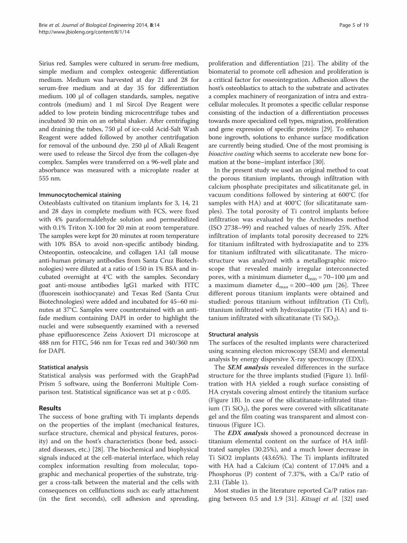
tained after 8 and 14 days of osteoblast cultivation. Theresults showed higher levels of the enzyme in cellular ly-sates when compared with the complete medium-derivedsamples. At 14 days, the highest ALP activity was found inthe lysates obtained from the cells cultivated on Ti SiO2
implants (Figure 14).
Figure 9 Levels of osteopontin (OP) in cellular lysates ofosteoblasts cultured on the titanium implants and measuredon day 8 and 14 of cultivation. Legend: non-infiltrated, controlTi (Ti control), titanium infiltrated with hydroxyapatite (Ti HA), Tiinfiltrated with silicatitanate (Ti SiO2), cells cultivated on plasticdishes (ctrl).
Collagen detectionLevels of the most abundant protein of the extracellularmatrix, collagen, were determined with the SIRCOL colla-gen assay, in samples cultured in serum-free medium (SF),simple osteogenic medium (OS) and complex osteogenicdifferentiation medium (OC). Medium was harvested onday 21 and 28 (for the serum-free medium) and on day 35(for the two differentiation mediums). In the serum freemedium, an important increase in soluble collagen con-centration was seen on day 28 (compared with day 21) forthe Ti SiO2 implants (Figure 15). The results also showedsignificant differences between culture mediums with re-spect to their content in soluble collagen, as one-wayANOVA tests showed. Notable differences were observedbetween all samples of Ti implants and control withoutsubstrate cultivated in medium with FCS and samples cul-tivated 28 and 35 days in simple osteogenic medium (OS)and complex osteogenic medium (OC). Analysis withtwo-way ANOVA Bonferroni posttest; *p < 0.05 showed asignificant increase of soluble collagen in control samplesafter 35 days of cultivation in OC medium (Figure 15).Collagen secretion was also investigated by immuno-
cytochemical staining of cells grown on titanium sam-ples. Type I collagen was expressed by all samples grownon titanium, with a higher observed intensity in controlTi and Ti infiltrated with SiO2 after 14 days and muchmore intensely after 28 days of cultivation (Figure 16).The osteoblast-osteocyte transition was observed after
28 days of cultivation especially on titanium samples withSiO2 and in a lower degree on control titanium-culturedsamples. This was suggested by the strong expression ofosteocalcin, as revealed through immunocytochemicalstaining. In silicatitanate- and HA-infiltrated implants,

Figure 10 Immunocytochemical staining of the osteoblasts cultivated on the surface of the implants showing the expression ofosteopontin (OP). Osteoblasts were stained with an anti OP-FITC monoclonal antibody on day 3, 14 and 21 days (magnification 100×). Legend:non-infiltrated control Ti (Ti control), titanium infiltrated with hydroxyapatite (Ti HA), Ti infiltrated with silicatitanate (Ti SiO2).
Brie et al. Journal of Biological Engineering 2014, 8:14 Page 11 of 19http://www.jbioleng.org/content/8/1/14
deposition of osteocalcin seems to occur in the extra-cellular matrix, with a more uniform distribution of theprotein observed in the case of SiO2 implants. In com-parison, in control titanium implants osteocalcin waslocated in the intracellular space of the osteoblasts(Figure 17).SEM images taken after 28 days show various obvious
morphological changes of cells depending on the type ofimplant. A typical osteocytic morphology - with longdendrite-like processes interacting with neighboring cells -was observed mainly on the SiO2 surface implant and to alesser extent in control titanium (Figure 18A and C). Ti
Figure 11 Immunocytochemical staining of osteoblasts on the surfacinfiltrated with SiO2) showing the expression of actin F and osteoponantibody, phalloidin TRITC (for actin F) and DAPI (for nuclei). The images wimage analysis software (magnification 200×). Legend: non-infiltrated, continfiltrated with silicatitanate (Ti SiO2).
HA implants had a more particular morphology: the cellsappeared to be partially included in the HA and in thenewly formed matrix substrate. The cell extensions werealso likely to be masked (Figure 18B).
DiscussionsOsseoinduction, osteoconduction and osseointegrationare the three interdependent steps involved in bonehealing, and the specific response of the bone to implantinsertion is similar to its response to fractures [41].Osseoinduction – defined by Friedenstein in 1968 as therecruitment of undifferentiated osteoprogenitor cells and
e of the implants (noninfiltrated Ti, Ti infiltrated with HA and Titin (OP). Osteoblasts were stained with anti OP-FITC monoclonalere taken on day 14 and composed with Axiovision Release 4.6.3.rol Ti (Ti control), titanium infiltrated with hydroxyapatite (Ti HA), Ti

Figure 12 Levels of calcium deposition on the implants byosteoblasts cultured in medium with fetal calf serum, FCS(measured on day 14, 21 and 28) and in serum-free medium,SF (measured on day 21 and 28). Quantification of the Calciumcontent was done using a modified Alizarin red method. Legend:noninfiltrated, control Ti (Ti control), titanium infiltrated withhydroxyapatite (Ti HA), Ti infiltrated with silicatitanate (Ti SiO2), cellscultivated on plastic dishes (ctrl).
Brie et al. Journal of Biological Engineering 2014, 8:14 Page 12 of 19http://www.jbioleng.org/content/8/1/14
their induction into osteogenic competent cells - dependsmainly on cell adhesion phenomena [42,36]. The qualityof the interface between cells and implants is essential forthe induction of events involved in implant integration[43]. The adhesiveness of bone grafts/implants is relatedto their ability to support selective attachment, prolifera-tion, differentiation and migration of anchorage-dependentcells [44]. Some examples of osseoinductive materials usedas implants are: the polymer polyhydroxyethylmethacrylate(poly-HEMA), metals (porous titanium), natural mate-rials such as hydroxyapatite (HA), synthetic ceramics
Figure 13 Levels of alkaline phosphatase activity in the mediums conwih fetal calf serum (FCS), evaluated on day 8, 14, 21 and 28 (*p < 0.05); B.and complex (OC) differentiation mediums (collected on day 35) Legend: nhydroxyapatite (Ti HA), Ti infiltrated with silicatitanate (Ti SiO2), cells cultivat
and composites such as HA/poly(lactic-coglycolic acid)(PLGA) or HA/collagen [2].The aim of this study was to compare the osseoinduc-
tive, osteoconductive and osseointegrative properties oftwo kinds of titanium implant coatings, with the purposeof selecting materials with increased bioactivity. Ti6Al7Nbimplants with 25% total porosity, processed with SLMtechnology, were infiltrated with hydroxyapatite and silica-titanate through a sol–gel method. Gravimetric methodrevealed a slow decrease of porosity to 22% for Ti HA im-plants and to 23% for Ti SiO2. Uncoated Ti implants wereused as controls. SEM and EDX analysis revealed that thetwo methods of coating conferred, as expected, differentphysical and chemical properties. For the Ti Ha implants,the EDAX analysis resulted in 30.25% Ti content (A%)with a Ca/P ratio of 2.3. The attachment of cells on the TiHA scaffolds was delayed in the first hour when comparedto Ti SiO2 and porous uncoated Ti implants, as revealedby SEM images and cell counting. However, this initialdisadvantage was subsequently surmounted, as evidencedby the assessment of proliferation with Alamar blue andtotal protein content. The metabolic activity of osteoblastswas highest after 8–9 days of cultivation in all Ti implants,with an observed advantage in SiO2 infiltrated Ti im-plants. This advantage was maintained until day 28, asproved by the increasing protein content in serum-freemedium. Similar results were obtained by Waselau et al.[45] in a comparative study analyzing the effects of bio-active glass S53P4 and beta-tricalcium phosphate onosteogenic differentiation of human adipose stem cells inthe presence of BMP-2 and BMP-7. qDNA measurementsrevealed significantly greater cell populations in the case
taining osteoblasts cultured on implant surfaces. A. in mediumin serum-free medium, SF (collected on day 21 and 28), simple (OS)oninfiltrated, control Ti (Ti control), titanium infiltrated withed on plastic dishes (ctrl).
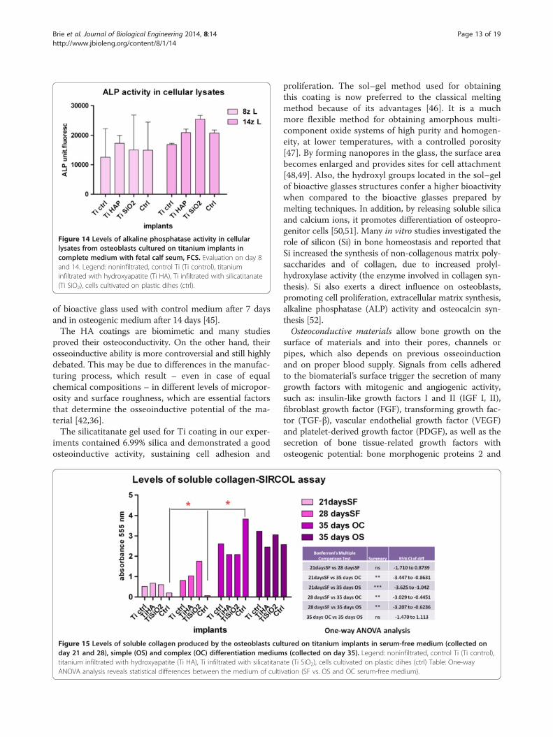
Figure 14 Levels of alkaline phosphatase activity in cellularlysates from osteoblasts cultured on titanium implants incomplete medium with fetal calf seum, FCS. Evaluation on day 8and 14. Legend: noninfiltrated, control Ti (Ti control), titaniuminfiltrated with hydroxyapatite (Ti HA), Ti infiltrated with silicatitanate(Ti SiO2), cells cultivated on plastic dihes (ctrl).
Brie et al. Journal of Biological Engineering 2014, 8:14 Page 13 of 19http://www.jbioleng.org/content/8/1/14
of bioactive glass used with control medium after 7 daysand in osteogenic medium after 14 days [45].The HA coatings are biomimetic and many studies
proved their osteoconductivity. On the other hand, theirosseoinductive ability is more controversial and still highlydebated. This may be due to differences in the manufac-turing process, which result – even in case of equalchemical compositions – in different levels of micropor-osity and surface roughness, which are essential factorsthat determine the osseoinductive potential of the ma-terial [42,36].The silicatitanate gel used for Ti coating in our exper-
iments contained 6.99% silica and demonstrated a goodosteoinductive activity, sustaining cell adhesion and
Figure 15 Levels of soluble collagen produced by the osteoblasts culday 21 and 28), simple (OS) and complex (OC) differentiation mediumtitanium infiltrated with hydroxyapatite (Ti HA), Ti infiltrated with silicatitanaANOVA analysis reveals statistical differences between the medium of culti
proliferation. The sol–gel method used for obtainingthis coating is now preferred to the classical meltingmethod because of its advantages [46]. It is a muchmore flexible method for obtaining amorphous multi-component oxide systems of high purity and homogen-eity, at lower temperatures, with a controlled porosity[47]. By forming nanopores in the glass, the surface areabecomes enlarged and provides sites for cell attachment[48,49]. Also, the hydroxyl groups located in the sol–gelof bioactive glasses structures confer a higher bioactivitywhen compared to the bioactive glasses prepared bymelting techniques. In addition, by releasing soluble silicaand calcium ions, it promotes differentiation of osteopro-genitor cells [50,51]. Many in vitro studies investigated therole of silicon (Si) in bone homeostasis and reported thatSi increased the synthesis of non-collagenous matrix poly-saccharides and of collagen, due to increased prolyl-hydroxylase activity (the enzyme involved in collagen syn-thesis). Si also exerts a direct influence on osteoblasts,promoting cell proliferation, extracellular matrix synthesis,alkaline phosphatase (ALP) activity and osteocalcin syn-thesis [52].Osteoconductive materials allow bone growth on the
surface of materials and into their pores, channels orpipes, which also depends on previous osseoinductionand on proper blood supply. Signals from cells adheredto the biomaterial’s surface trigger the secretion of manygrowth factors with mitogenic and angiogenic activity,such as: insulin-like growth factors I and II (IGF I, II),fibroblast growth factor (FGF), transforming growth fac-tor (TGF-β), vascular endothelial growth factor (VEGF)and platelet-derived growth factor (PDGF), as well as thesecretion of bone tissue-related growth factors withosteogenic potential: bone morphogenic proteins 2 and
tured on titanium implants in serum-free medium (collected ons (collected on day 35). Legend: noninfiltrated, control Ti (Ti control),te (Ti SiO2), cells cultivated on plastic dihes (ctrl) Table: One-wayvation (SF vs. OS and OC serum-free medium).

Figure 16 Immunocytochemical staining (FITC) for collagen 1A of the osteoblasts cultured in complete medium on titanium implantsand evaluated on day 14 and 28. Legend: noninfiltrated control Ti (Ti control), titanium infiltrated with hydroxyapatite (Ti HA), Ti infiltrated withsilicatitanate (Ti SiO2) (magnification 100×).
Brie et al. Journal of Biological Engineering 2014, 8:14 Page 14 of 19http://www.jbioleng.org/content/8/1/14
7 (BMP-2 and BMP-7) [3,45]. Osseointegration allowsbone anchorage and consolidation between the newlyformed bone and the implant. Because initial osseointe-gration is dependent on bone induction and conduction,materials that are too toxic to allow cellular adhesionand development into bone forming cells will not beosseointegrated [3]. Formation of hydroxyapatite andsurrounding bone tissue by binding to the extracellularmatrix is essential for osseointegration. The most im-portant questions to answer when investigating a systemof osteoblasts cultured on implants are whether the sub-strate supports de novo matrix mineralization, if it issimilar to its naturally occurring counterpart, and how itcould be distinguished from the presence of mineralscontained in the substrate.Osteoconductivity and osseointegration of Ti HA, Ti
SiO2 and porous Ti implants used in the present studywere evaluated by assessing the differentiation andmineralization processes. Osteopontin (OP) is one of themost important non-collagenous phosphoproteins andan indicator of the differentiation process of osteoblasts.
Figure 17 Immunocytochemical staining for osteocalcin Texas red, comedium on titanium implants evaluated after 28 days of cultivation.hydroxyapatite (Ti HA), Ti infiltrated with silicatitanate (Ti SiO2) (magnificatio
OP expression was evaluated in different culture media(complete medium, serum-free medium and osteogenicmedium) at different points in time. Using the ELISAmethod, no significant differences were found amongthe implants. Interestingly, the highest OP values weredetected on day 14, for the cultures grown in completemedium on plastic dishes. This may be explained by theimmobilization of OP on the implant surface in thenewly synthesized matrix, as OP was reported to bepresent both in an immobilized form in the extracellularmatrix of mineralized tissues and in a soluble form intissue fluids [53,54]. The osteogenic differentiation mediuminduced a discrete increase in OP levels after 35 days ofosteoblast cultivation on control Ti implants. A strong OPexpression, evaluated by immunocytochemical staining,was detected after day 3 for Ti Ctrl and Ti SiO2 implants.It continued to increase until day 14 and decreased after21 days. With a combined immunostaining for osteopon-tin and filamentous actin, different behaviors of implantswere observed with regard to the rearrangement of actinfibres. The Ti SiO2 and Ti Ctrl implants induced a more
unterstained with DAPI in osteoblasts cultured in completeLegend: non-infiltrated, control Ti (Ti control), titanium infiltrated withn 100×).

Figure 18 Scanning electron microscopy images of osteocytes attached to the surface of titanium implants after 28 days ofcultivation. Legend: Osteocytes are indicated by arrows. A. noninfiltrated control Ti (magnification ×300); B. Ti infiltrated with hydroxyapatite(Ti HA) (magnification ×500); B1- image of an osteoblast with 2000× magnification; C. Ti infiltrated with silicatitanate (TiSiO2) (magnification ×1200);C1- image of an osteocyte with ×2500 magnification.
Brie et al. Journal of Biological Engineering 2014, 8:14 Page 15 of 19http://www.jbioleng.org/content/8/1/14
flattened morphology of cells, and were associated withthe expression of stress fibers.The actin cytoskeleton is involved in cell adhesion and
cell motility, and its fiber arrangement modulates cellshape differently depending on the type of nanoscaleand microscale surface roughness as well as the pattern-ing of the surface of the material on which cells aregrown. Mechanical stimulation of osteoblasts or stemcells induced by the properties of the implant surfacetriggers the reorganization of the focal adhesion plaquesfollowed by the rearrangement of the cytoskeleton andthe activation of signaling pathways involved in osteo-genic cell differentiation such as transcription factorsCbfa1 (Core Binding Factor A1) and Osterix. As a con-sequence osteoblasts synthesize higher amounts of colla-gen I, osteopontin, osteocalcin and bone sialoprotein,and induce higher levels of alkaline phosphatase activity[55-57]. The canonical Wnt (Wingless/Integrated) signal-ing pathway is also activated with consequences on β-catenin, alkaline phosphatase and osteocalcin expression,as Galli et al. showed in a study using mesenchymal andosteoblastic cells growing on polished titanium discs ver-sus acid-etched and sand-blasted (SLA) surfaces. Differ-ences were also observed among various cell types [58].For instance, OP is also involved in cell adhesion phenom-ena of cell-cell or cell-extracellular matrix interactionsthat occur during cell proliferation and migration. Ele-vated concentrations of osteopontin were found in sites ofbone resorption and its expression was strongly increasedby mechanical stimuli. The effects of OP on HA formationseem to be related to its phosphorylation state. Exogenousphosphorylated OP is known to inhibit mineralization, butdephosphorylation by tissue-nonspecific alkaline phos-phatase (ALP) reverses this effect [38,39]. The phospho-rylation state of OP can be influenced by the physicaland chemical properties of the substrate and, as a
consequence, modulates its signaling ability [59]. This factmay explain the different cell responses observed in ourexperiments, most notably the increased expression of OPin complete medium after 8–14 days especially in SiO2 Tiand control Ti implants, which correlates with concomi-tant increases of other specific proteins such as ALP.ALP is a key enzyme that can both promote and inhibit
mineralization. It is a cell-membrane-associated enzyme,expressed also in matrix vesicles, and in association withother proteins enhances deposition of hydroxyapatitealong the collagen fibrils. ALP hydrolyzes its substrate, in-organic pyrophosphate, to inorganic phosphate, the latterbeing a substrate for the HA mineral [60]. In our experi-ments, the highest activity of ALP was found in the sam-ples obtained from the lysate of cells cultivated on Ti SiO2
implants. However, the activity of ALP was much higher(2–2.5 fold) in serum-free conditions, especially in Ti HAand SiO2 implants. ALP activity decreased rapidly after28 days, indicating that the mineralization process isnearly complete. In osteogenic differentiation mediums,the expression profile of ALP was different. A strong ALPactivity was detected on day 35 in simple osteogenicmedium from the Ti Ctrl and Ti HA, accompanied by ele-vated levels of collagen. SiO2 implants did not respond tothe differentiation medium in terms of ALP activity, butexhibited similar levels of collagen. The presence of BMP-2 and TGF β1 in the complex osteogenic medium in-creased ALP activity only in Ti HA implant cultures(Figure 19).Collagen synthesis, as an indicator of matrix forma-
tion, was significantly increased in Ti SiO2 samples after28 days. In addition, higher soluble collagen levels wereobserved in control Ti and Ti SiO2 implants cultivatedin simple osteogenic differentiation medium. These find-ings do not exclude possibly similar collagen synthesison Ti HA implants, yet we could not demonstrate this

Figure 19 A schematic synthesis of results regarding the most important elements implicated in the mineralization process. Ca2+
deposition, collagen synthesis and ALP activity, in samples cultivated in serum-free medium (SF) and in differentiation medium (complex osteogenicmedium OC and simple osteogenic medium OS) Legend: noninfiltrated, control Ti (Ti control), titanium infiltrated with hydroxyapatite (Ti HA), Tiinfiltrated with silicatitanate (Ti SiO2).
Brie et al. Journal of Biological Engineering 2014, 8:14 Page 16 of 19http://www.jbioleng.org/content/8/1/14
because in such implants the collagen fibers could notbe visualized (as they may have been masked by the ac-celerated deposition of newly formed HA crystals).Calcium deposition was investigated as a marker of
mineralization. Differences between implants emergedafter 21 and 28 days of cultivation in complete medium,with a gradual increase in calcium deposition observedmostly in Ti HA implants and in a lower degree in TiSiO2 implants. Serum-free medium significantly in-creased the deposition of calcium on the surface of TiHA implants after 28 days of cultivation. A similar out-come was reported by Yamada et al. [61], as they ob-served accelerated bone-implant integration after theaddition of pure nanopolymorphic crystalline HA tomicro-roughened titanium, as well as a significantly in-creased osteoconductivity [61].The complex osteogenic differentiation medium used
in this study also contained TGF-β1 (in a low dose-3 ng/ml), a growth factor with dual biological activity, as itcan regulate osteoblast differentiation not only positivelybut also negatively. Ochiai et al. [62] observed that onlyrepeated high doses of TGF-β1 suppressed osteoblastdifferentiation with decreased ALP activity, although aunique low dose of TGF-β1 strongly induced osteoblastdifferentiation [62]. On the other hand, TGF-β1 protectspre-osteocytes from apoptosis [63]. Similar results regard-ing the osteogenic medium were obtained by Tirkkonenet al. [64] in an adipose stem cell osteogenic differentiationstudy that compared the efficiency of BMP-2 and BMP-7,VEGF and osteogenic medium. Stem cells were grown oncommercially available bioactive glass scaffolds and bi-phasic granules. The bioactive glass induced an increasedcell proliferation whereas calcium phosphate led to a moresignificant collagen production. They did not find a bene-fit after the addition ofgrowth factors in comparison withplain osteogenic medium. Moreover, BMP-7 inhibited theproliferation and osteogenic differentiation of adiposestem cells [64]. The lack of improvement of the osteogenic
differentiation of stem cells through the addition of BMP-2 or BMP-6 was also reported by other authors [65-67].The master of controlling osteoblast differentiation intoosteocytes is RUNX2 (runt-related transcription factor)[68] and in this context BMP2 induces osteoblast differ-entiation through Runx2-dependent ATF6 (bZIP tran-scription factor) expression, which directly regulatesosteocalcin transcription [69]. Prior to the mineralizationprocess, as the cells pass through progressive differenti-ation stages, some specific markers are revealed, the earli-est being RUNX2, followed by ALP (an early-mid marker),osteopontin (OP), osteocalcin (OC) and osteonectin (ON)[70]. Roach (1996) studied the roles of matrix non-collagenous proteins such as OP, OC, bone sialo-protein(BSP) and ON and their tissue localization using doubleimmuno-histochemistry. The appearance of OP and BSPahead of the mineralization front, prior to mineralization,suggested that both proteins are necessary for the initi-ation of bone mineralization. OC and ON were present infully mineralized matrix, possibly having a function incontrolling the amount and rate of crystals formation [71].In our study, after the immunocytochemical analysis,
osteocalcin had the highest expression in Ti SiO2 andCtrl Ti implants after 28 days of cultivation, and it wasassociated with increased expression of collagen I. Sucheffects of bioactive glass were explained by Silver et al.[72], through a marked alkalinization of the intracellularand extracellular environment that influence the activ-ities of intracellular enzymes and signaling pathways,which in turn increases collagen synthesis [72]. Similarobservations were reported regarding orthosilicic acid ordifferent types of bioglasses [73,74]. Osteocalcin, a ter-minal marker of osteoblastic differentiation, is present inhigh amounts in the bone matrix. It binds calcium andinhibits bone growth by inhibiting the activity of trans-glutaminase [75]. After posttranslational modificationwith the addition of carboxylated glutamic acid residues(gla residues), OC gains a high affinity for hydroxyapatite

Brie et al. Journal of Biological Engineering 2014, 8:14 Page 17 of 19http://www.jbioleng.org/content/8/1/14
crystals. The inhibitory role of OC possibly involves aconservation of cells in a late-osteoblast stage of differ-entiation, with associated prevention of osteocytic differ-entiation [76] and initiation of the remodeling process ofnew bone.Through interpretation of the results derived from our
study, two kinds of osseointegrations were found. Thefirst type consists of a predominant collagen proteinmatrix construct and a self-limiting mineralization processthrough increased expression of osteocalcin, which wasobserved in the case of the porous titanium and infiltratedsilicatitanate implants. The second type belongs to HA-infiltrated titanium, characterized by an early increase incalcium deposits, with a lesser degree of collagen andnon-collagenous protein synthesis. Both types of osseoin-tegration have potential applications, however in vivostudies are required to clarify the most suitable type of im-plant depending on the site of implantation (consideringthe mechanical load and the type of bone in need of re-pair) as well as the associated pathological conditions suchas osteoporosis. Despite some limitations regarding themechanical properties of porous apatite ceramics, the ex-cellent osteoconductivity of HA coatings and the possibil-ity of associating bioactive molecules (such as proteins,aminoacids, antibiotics, growth factors, anticancer andanti-osteoporosis drugs) to calcium phosphate indicatethe potential advantages of the use of this type of coatingespecially in osteopenic bone repair [77,78]. On the otherhand, the osseoinductive properties of SiO2-coated Ti im-plants (as described in our study) and their ability to pro-mote angiogenesis and enhance neocartilage formation[73], can provide other important applications in implan-tology, especially for repairing bones at sites where mech-anical strength is of lesser importance.
ConclusionsTwo types of titanium coatings were comparatively stu-died with regard to bone regeneration, and they induceddifferent behaviors of osteoblastic cells. Ti implants infil-trated with hydroxyapatite (HA) demonstrated an in-creased capacity to induce early mineralization with alower ability to induce cell adhesion and proliferation.Conversely, the implants infiltrated with SiO2, as well asporous titanium preserved the attachment and adhesionof osteoblasts, promoted celldifferentiation and inducedthe production of the protein component of the extracel-lular matrix (collagen and the non-collagenic proteinsosteopontin and osteocalcin). The addition of growth fac-tors BMP-2 and TGFβ1 in the differentiation medium didnot improve the mineralization process. Both types ofcoatings have their advantages and limitations, which canbe exploited depending on the local conditions and on thebone lesions in need of repair. Their properties can be
improved through methods of functionalization with bio-molecules involved in osteogenesis.
Competing interestsThe authors declare that they have no competing interests.
Authors’ contributionsStudy design: PB, SS, CP, IB, OS, VC Scaffolds manufacturing: CP, MT, SS, CBScanning electron microscopy and EDX analysis: AV Cell cultures: MPS, ND,OB Biocompatibility assays: OS, GC, PV, CB Data analysis: IB, OS, TM, AV, CP,MT Data interpretation. IB, OS, Manuscript preparation: IB, OS, MPS, MT, AV.Approving final version of manuscript: IB and OS takes responsibility for theintegrity of the data analysis. All authors read and approved the finalmanuscript.
AcknowledgementsThis study was supported within the PCCE contract 5/2010, BIOMAPIMproject and PN-II-PT-PCCA-2011-3, contract no. 78/2012, financed by theRomanian National Council for the Higher Education Scientific Research. NDwould like to thank to Technical University, Cluj-Napoca PhD program(contract 19585/2012) for financial support.
Author details1The Institute of Oncology “Prof. Dr. I. Chiricuta” Cluj-Napoca, Cluj-Napoca,Romania. 2University of Medicine and Pharmacy “Iuliu Hatieganu”Cluj-Napoca, Cluj-Napoca, Romania. 3Technical University, Cluj-Napoca,Romania. 4Faculty of Physics & Institute of Interdisciplinary Research inBio-Nano-Sciences, Babes Bolyai University, 400084 Cluj-Napoca, Romania.
Received: 6 December 2013 Accepted: 13 June 2014Published: 19 June 2014
References1. Baroli B: From natural bone grafts to tissue engineering therapeutics:
brainstorming on pharmaceutical formulative requirements andchallenges. J Pharm Sci 2009, 98(4):1317–1375.
2. Amini AR, Laurencin CT, Nukavarapu SP: Bone Tissue Engineering: RecentAdvances and Challenges. Crit Rev Biomed Eng 2012, 40(5):363–408.
3. Sul YT, Johansson CB, Petronis S, Krozer A, Jeong Y, Wennerberg A,Albrektsson T: Characteristics of the surface oxides on turned andelectrochemically oxidized pure titanium implants up to dielectricbreakdown: the oxide thickness, micropore configurations, surfaceroughness, crystal structure and chemical composition. Biomaterials 2002,23:491–501.
4. Park H, Temenoff JS, Mikos AG: Biodegradable orthopedic implants. InEngineering of functional skeletal tissues. Edited by Bronner F, Farch-Carson MC,Mikos AG. London: Spriinger-Verlagm; 2007:60.
5. Ventre M, Causa F, Netti PA: Determinants of cell-material crosstalk at theinterface: towards engineering of cell instructive materials.J R Soc Interface 2012, 9:2017–2032.
6. Nautiyal VP, Mittal A, Agarwal A, Pandey A: Tissue response to titaniumimplant using scanning electron microscope. Natl J Maxillofac Surg 2013,4(1):7–12.
7. Ungersboeck A, Geret V, Pohler O, Schuetz M, Wuest W: Tissue reaction tobone plates made of pure titanium: a prospective, quantitative clinicalstudy. J Mater Sci Mater Med 1995, 6:223–229.
8. Vijayaraghavan V, Sabane AV, Tejas K: Hypersensitivity to titanium: a lessexplored area of research. J Indian Prosthodont Soc 2012, 12(4):201–207.
9. Donesz-Sikorska A, Krzak-Roś J, Kochanowska IE, Będziński R, Kaleta J: Newspecific metal-silica biocomposites for medical implants. Bioinspired,Biomimetic and Nanobiomaterials 08/2012; doi:10.1680/bbn.12.00018.
10. Nouri A, Hodgson PD, Wen C: Chapter 21- Biomimetic Porous TitaniumScaffolds for Orthopaedic and Dental Applications. In Biomimetics,Learning from nature. Edited by Amitava M, In–Tech. 2009:415–450.
11. Marcu T, Todea M, Maines L, Leordean D, Berce P, Popa C: Metallurgicaland mechanical characterisation of titanium based materials forendosseous applications obtained by selective laser melting.Powder Metall 2012, 55(4):309–314.
12. Kim HM: Ceramic bioactivity and related biomimetic strategy.Curr Opinion Solid State Mater Sci 2003, 7:289–299.

Brie et al. Journal of Biological Engineering 2014, 8:14 Page 18 of 19http://www.jbioleng.org/content/8/1/14
13. Jones JR: Review of bioactive glass: from Hench to hybrids.Acta Biomater 2013, 9(1):4457–4486.
14. Polo-Corrales L, Latorre-Esteves M, Ramirez-Vick JE: Scaffold design forbone regeneration. J Nanosci Nanotechnol 2014, 14(1):15–56.
15. Ning CQ, Zhou Y: On the microstructure of biocomposites sintered fromTi, HA and bioactive glass. Biomaterials 2004, 25:3379–3387.
16. Wu Z, He L, Chen Z: Fabrication and characterization of hydroxyapatite/Al2O3 biocomposite coating on titanium. Trans Nonferrous Met SocChina 2006, 16:259–266.
17. Wang CK, Lin JH, Ju CP, Ong HC, Chang RP: Structural characterization ofpulsed laser-deposited hydroxyapatite film on titanium substrate.Biomaterials 1997, 18(20):1331–1338.
18. Prosecká E, Buzgo M, Rampichová M, Kocourek T, Kochová P, Vysloužilová L,Tvrdík D, Jelínek M, Lukáš D, Amler E: Thin-layer hydroxyapatite depositionon a nanofiber surface stimulates mesenchymal stem cell proliferationand their differentiation into osteoblasts. J Biomed Biotechnol 2012,428503:10. doi:10.1155/2012/428503.
19. Roy M, Balla VK, Bandyopadhyay A, Bose S: Compositionally gradedhydroxyapatite/ tricalcium phosphate coating on Ti by laser and inductionplasma. Acta Biomater 2011, 7(2):866–873. doi:10.1016/j.actbio.2010.09.016.
20. Hannora AE, Mukasyan AS, Mansurov ZA: Nanocrystalline hydroxyapatite/sicoating by mechanical alloying technique. Bioinorg Chem Appl 2012,390104:14. doi:10.1155/2012/390104.
21. Kim HW, Kim HE, Knowles JC: Fluor-hydroxyapatite sol–gel coating ontitanium substrate for hard tissue implants. Biomaterials 2004,25(17):3351–3358.
22. Lin FH, Hsu YS, Lin SH, Sun JS: The effect of Ca/P concentration andtemperature of simulated body fluid on the growth of hydroxyapatitecoating on alkali-treated 316 L stainless steel. Biomaterials 2002,23(19):4029–4038.
23. Eraković S, Janković A, Veljović D, Palcevskis E, Mitrić M, Stevanović T,Janaćković D, Mišković-Stanković V: Corrosion stability and bioactivity insimulated body fluid of silver/ hydroxyapatite and silver/ hydroxyapatite/lignin coatings on titanium obtained by electrophoretic deposition. J PhysChem B 2013, 117(6):1633–1643. doi:10.1021/jp305252a.
24. Mozafari M, Salahinejad E, Shabafrooz V, Yazdimamaghani M, Vashaee D,Tayebi L: Multilayer bioactive glass/zirconium titanate thin films in bonetissue engineering and regenerative dentistry. Int J Nanomedicine 2013,8:1665–1672.
25. Saino E, Maliardi V, Quartarone E, Fassina L, Benedetti L, De Angelis MG,Mustarelli P, Facchini A, Visai L: In vitro enhancement of SAOS-2 cellcalcified matrix deposition onto radio frequency magnetron sputteredbioglass-coated titanium scaffolds. Tissue Eng Part A 2010, 16(3):995–1008.
26. Leordean D, Dudescu C, Marcu T, Berce P, Bâlc N: Aspects related to theapplication of the selective laser melting technology to customizedTi-6Al-7Nb implants. J Rapid Prototyping, in press.
27. Tomuleasa CI, Foris V, Soritau O, Pall E, Fisher-Fodor E, Lung-Illes V, Brie I,Virag P, Perde-Schrepler M, Postescu ID, Chereches G, Barbos O, Tatomir C:Effects of 60Co γ-rays on human osteoprogenitor cells. Rom J MorpholEmbryol 2009, 50(3):349–355.
28. Søballe K: Hydroxyapatite ceramic coating for bone implant fixation. Mechanicaland histological studies in dogs. Acta Orthop Scand Suppl 1993, 255:1–58.
29. Yim EK, Leong KW: Significance of synthetic nanostructures in dictatingcellular response. Nanomedicine 2005, 1(1):10–21.
30. Wang G, Lu Z, Liu X, Zhou X, Ding C, Zreiqat H: Nanostructured glass-ceramiccoatings for orthopaedic applications. J R Soc Interface 2011, 8(61):1192–1203.doi:10.1098/rsif.2010.0680.
31. Ponche A, Bigerelle M, Anselme K: Relative influence of surfacetopography and surface chemistry on cell response to bone implantmaterials. Part 1: Physico-chemical effects. Proc Inst Mech Eng H JEng Med 2010, 224:1471.
32. Kitsugi T, Yamamuro T, Nakamura T, Oka M: Transmission electronmicroscopy observations at the interface of bone and four types ofcalcium phosphate ceramics with different calcium/phosphorus molarratios. Biomaterials 1995, 16(14):1101–1107.
33. Chou YF, Huang W, Dunn JC, Miller TA, Wu BM: The effect of biomimeticapatite structure on osteoblast viability, proliferation, and geneexpression. Biomaterials 2005, 26(3):285–295.
34. Goto T, Yoshinari M, Kobayashi S, Tanaka T: The initial attachment andsubsequent behavior of osteoblastic cells and oral epithelial cells ontitanium. Biomed Mater Eng 2004, 14(4):537–544.
35. Rampersad SN: Multiple applications of Alamar Blue as an indicator ofmetabolic function and cellular health in cell viability bioassays.Sensors (Basel) 2012, 12(9):12347–12360. doi:10.3390/s120912347.
36. Geys J, Nemery B, Hoet PH: Assay conditions can influence the outcomeof cytotoxicity tests of nanomaterials: better assay characterization isneeded to compare studies. Toxicol In Vitro 2010, 24(2):620–629.doi:10.1016/j.tiv.2009.10.007.
37. Zeller KS, Riaz A, Sarve H, Li J, Tengholm A, Johansson S: The role of mechanicalforce and ROS in integrin-dependent signals. PLoS ONE, 8(5):e64897.doi:10.1371/journal.pone.0064897.
38. Harmey D, Hessle L, Narisawa S, Johnson KA, Terkeltaub R, Millán JL:Concerted regulation of inorganic pyrophosphate and osteopontin byakp2, enpp1, and ank: an integrated model of the pathogenesis ofmineralization disorders. Am J Pathol 2004, 164(4):1199–1209.
39. Addison WN, Azari F, Sørensen ES, Kaartinen MT, McKee MD:Pyrophosphate inhibits mineralization of osteoblast cultures by bindingto mineral, up-regulating osteopontin, and inhibiting alkaline phosphataseactivity. J Biol Chem 2007, 282(21):15872–15883.
40. Khan MR, Donos N, Salih V, Brett PM: The enhanced modulation of keybone matrix components by modified Titanium implant surfaces.Bone 2012, 50(1):1–8. doi:10.1016/j.bone.2011.07.040.
41. Albrektsson T, Johansson C: Osteoinduction, osteoconduction andosseointegration. Eur Spine J 2001, 10(Suppl 2):S96–S101.
42. Barradas AM, Yuan H, Van Blitterswijk CA, Habibovic P: Osteoinductivebiomaterials: current knowledge of properties, experimental models andbiological mechanisms. Eur Cell Mater 2011, 21:407–429.
43. Anselme K, Ponche A, Bigerelle M: Relative influence of surfacetopography and surface chemistry on cell response to bone implantmaterials. Part 2: biological aspects. Proc Inst Mech Eng H 2010,224(12):1487–1507.
44. Lutolf MP, Hubbell JA: Synthetic biomaterials as instructive extracellularmicroenvironments for morphogenesis in tissue engineering.Nat Biotechnol 2005, 23:47–55.
45. Waselau M, Patrikoski M, Juntunen M, Kujala K, Kääriäinen M, Kuokkanen H,Sándor GK, Vapaavuori O, Suuronen R, Mannerström B, Von Rechenberg B,Miettinen S: Effects of bioactive glass S53P4 or beta-tricalcium phosphateand bone morphogenetic protein-2 and bone morphogenetic protein-7on osteogenic differentiation of human adipose stem cells. J Tissue Eng 2012,3(1):2041731412467789. doi:10.1177/2041731412467789.
46. Drnovšek N, Novak S, Dragin U, Čeh M, Gorenšek M, Gradišar M: Bioactiveglass enhances bone ingrowth into the porous titanium coating onorthopaedic implants. Int Orthop 2012, 36(8):1739–1745. doi:10.1007/s00264-012-1520-y.
47. Ponta O, Vanea E, Cheniti A, Berce P, Simon S: Structural and surfacecharacterization of nanostructured spray dried titanosilicatemicrospheres. Mater Chem Phys 2012, 135:863–869.
48. Rahaman MN, Day DE, Bal BS, Fu Q, Jung SB, Bonewald LF, Tomsia AP:Bioactive glass in tissue engineering. Acta Biomater 2011, 7(6):2355–2373.doi:10.1016/j.actbio.2011.03.016.
49. Mahony O, Jones JR: Porous bioactive nanostructured scaffolds for boneregeneration: a sol–gel solution. Nanomedicine (Lond) 2008, 3(2):233–245.doi:10.2217/17435889.3.2.233.
50. Pamula E, Kokoszka J, Cholewa-Kowalska K, Laczka M, Kantor L, Niedzwiedzki L,Reilly GC, Filipowska J, Madej W, Kolodziejczyk M, Tylko G, Osyczka AM:Degradation, bioactivity, and osteogenic potential of composites madeof PLGA and two different sol–gel bioactive glasses. Ann Biomed Eng 2011,39(8):2114–2129. doi:10.1007/s10439-011-0307-4.
51. Jones JR, Lin S, Yue S, Lee PD, Hanna JV, Smith ME, Newport RJ: Bioactiveglass scaffolds for bone regeneration and their hierarchicalcharacterisation. Proc Inst Mech Eng H 2010, 224(12):1373–1387.
52. Jugdaohsingh R: Silicon and bone health. J Nutr Health Aging 2007, 11(2):99–110.53. McKee MD, Nanci A: Osteopontin at mineralized tissue interfaces in bone,
teeth, and osseointegrated implants: ultrastructural distribution andimplications for mineralized tissue formation, turnover, and repair.Microsc Res Tech 1996, 33(2):141–164.
54. Gericke A, Qin C, Spevak L, Fujimoto Y, Butler WT, Sørensen ES, Boskey AL:Importance of phosphorylation for osteopontin regulation ofbiomineralization. Calcif Tissue Int 2005, 77(1):45–54.
55. Vandrovcová M, Bačáková L: Adhesion, growth and differentiation ofosteoblasts on surface-modified materials developed for bone implants.Physiol Res 2011, 60(3):403–417.

Brie et al. Journal of Biological Engineering 2014, 8:14 Page 19 of 19http://www.jbioleng.org/content/8/1/14
56. Roffers-Agarwal J, Xanthos JB, Miller JR: Regulation of actin cytoskeletonarchitecture by Eps8 and Abi1. BMC Cell Biol 2005, 6:36.
57. Orza A, Soritau O, Olenic L, Diudea M, Florea A, Rus Ciuca D, Mihu C,Casciano D, Biris AS: Electrically conductive gold-coated collagen nanofibersfor placental-derived mesenchymal stem cells enhanced differentiationand proliferation. ACS Nano 2011, 5(6):4490–4503. doi:10.1021/nn1035312.
58. Galli C, Piemontese M, Lumetti S, Ravanetti F, Macaluso GM, Passeri G: Actincytoskeleton controls activation of Wnt/β-catenin signaling inmesenchymal cells on implant surfaces with different topographies.Acta Biomater 2012, 8(8):2963–2968. doi:10.1016/j.actbio.2012.04.043.
59. Kazanecki CC, Uzwiak DJ, Denhardt DT: Control of osteopontin signalingand function by post-translational phosphorylation and protein folding.J Cell Biochem 2007, 102(4):912–924.
60. Hunter LW, Shiekh FA, Pisimisis GT, Kim SH, Edeh SN, Miller VM, Lieske JC:Key role of alkaline phosphatase in the development of human-derivednanoparticles in vitro. Acta Biomater 2011, 7(3):1339–1345. doi:10.1016/j.actbio.2010.10.027.
61. Yamada M, Ueno T, Tsukimura N, Ikeda T, Nakagawa K, Hori N, Suzuki T,Ogawa T: Bone integration capability of nanopolymorphic crystallinehydroxyapatite coated on titanium implants. Int J Nanomedicine 2012,7:859–873. doi:10.2147/IJN.S28082.
62. Ochiai H, Okada S, Saito A, Hoshi K, Yamashita H, Takato T, Azuma T:Inhibition of insulin-like growth factor-1 (IGF-1) expression by prolongedtransforming growth factor-β1 (TGF-β1) administration suppressesosteoblast differentiation. J Biol Chem 2012, 287(27):22654–22661.doi:10.1074/jbc.M111.279091.
63. Kanaan RA, Kanaan LA: Transforming growth factor beta1, bone connection.Med Sci Monit 2006, 12(8):RA164–RA169.
64. Tirkkonen L, Haimi S, Huttunen S, Wolff J, Pirhonen E, Sándor GK, Miettinen S:Osteogenic medium is superior to growth factors in differentiation ofhuman adipose stem cells towards bone-forming cells in 3D culture.Eur Cell Mater 2013, 25:144–158.
65. Cruz AC, Silva ML, Caon T, Simões CM: Addition of bone morphogeneticprotein type 2 to ascorbate and β-glycerophosphate supplementationdid not enhance osteogenic differentiation of human adipose-derivedstem cells. J Appl Oral Sci 2012, 20(6):628–635.
66. Zuk P, Chou YF, Mussano F, Benhaim P, Wu BM: Adipose-derived stemcells and BMP2: part 2. BMP2 may not influence the osteogenic fate ofhuman adipose-derived stem cells. Connect Tissue Res 2011, 52(2):119–132.doi:10.3109/03008207.2010.484515.
67. Khanna-Jain R, Agata H, Vuorinen A, Sándor GK, Suuronen R, Miettinen S:Addition of BMP-2 or BMP-6 to dexamethasone, ascorbic acid, andβ-glycerophosphate may not enhance osteogenic differentiation ofhuman periodontal ligament cells. Growth Factors 2010, 28(6):437–446.doi:10.3109/08977194.2010.495719.
68. Raggatt LJ, Partridge NC: Cellular and Molecular Mechanisms of BoneRemodeling. J Biol Chem 2010, 285(33):25103–25108. doi:10.1074/jbc.R109.041087.
69. Jang WG, Kim EJ, Kim DK, Ryoo HM, Lee KB, Kim SH, Choi HS, Koh JT: BMP2protein regulates osteocalcin expression via Runx2-mediated Atf6 genetranscription. J Biol Chem 2012, 287(2):905–915. doi:10.1074/jbc.M111.253187.
70. Pilia M, Guda T, Shiels SM, Appleford MR: Influence of substrate curvatureon osteoblast orientation and extracellular matrix deposition. J Biol Eng 2013,7(1):23.
71. Roach HI: Why does bone matrix contain non-collagenous proteins? Thepossible roles of osteocalcin, osteonectin, osteopontin and bonesialoprotein in bone mineralisation and resorption. Cell Biol Int 1994,18(6):617–628.
72. Silver IA, Deas J, Erecińska M: Interactions of bioactive glasses withosteoblasts in vitro: effects of 45S5 Bioglass, and 58S and 77S bioactiveglasses on metabolism, intracellular ion concentrations and cell viability.Biomaterials 2001, 22(2):175–185.
73. Arumugam MQ, Ireland DC, Brooks RA, Rushton N, Bonfield W: The EffectOrthosilicic Acid on Collagen Type I, Alkaline Phosphatase andOsteocalcin mRNA Expression in Human Bone-Derived Osteoblasts.In Vitro Key Engineering Materials 2006, 309–311:125. doi:10.4028/www.scientific.net/KEM.254-256.869.
74. Bosetti M, Zanardi L, Hench L, Cannas M: Type I collagen production byosteoblast-like cells cultured in contact with different bioactive glasses.J Biomed Mater Res A 2003, 64(1):189–195.
75. Meury T, Akhouayri O, Jafarov T, Mandic V, St-Arnaud R: Nuclear alpha NACinfluences bone matrix mineralization and osteoblast maturation in vivo.Mol Cell Biol 2010, 30(1):43–53. doi:10.1128/MCB.00378-09.
76. Boskey AL, Gadaleta S, Gundberg C, Doty SB, Ducy P, Karsenty G: Fouriertransform infrared microspectroscopic analysis of bones ofosteocalcin-deficient mice provides insight into the function ofosteocalcin. Bone 1998, 23:187–196.
77. Gómez-Morales J, Iafisco M, Delgado-López JM, Sarda S, Drouet C: Progresson the preparation of nanocrystalline apatites and surface characterization:Overview of fundamental and applied aspects. Prog Cryst Growth CharactMater 2013, 59:1–46.
78. Hayashi K, Fotovati A, Ali SA, Oda K, Oida H, Naito M: Prostaglandin EP4receptor agonist augments fixation of hydroxyapatite-coated implants ina rat model of osteoporosis. J Bone Joint Surg (Br) 2005, 87(8):1150–1156.
doi:10.1186/1754-1611-8-14Cite this article as: Brie et al.: Comparative in vitro study regarding thebiocompatibility of titanium-base composites infiltrated withhydroxyapatite or silicatitanate. Journal of Biological Engineering 2014 8:14.
Submit your next manuscript to BioMed Centraland take full advantage of:
• Convenient online submission
• Thorough peer review
• No space constraints or color figure charges
• Immediate publication on acceptance
• Inclusion in PubMed, CAS, Scopus and Google Scholar
• Research which is freely available for redistribution
Submit your manuscript at www.biomedcentral.com/submit
Copyright © 2022 FDOKUMEN