Coccidioidomycosis in Brazil - MDPI
14
Fungi Journal of Review Coccidioidomycosis in Brazil: Historical Challenges of a Neglected Disease Rossana Cordeiro 1, * ,† , Santiago Moura 1,† ,Débora Castelo-Branco 1, * , Marcos Fábio Rocha 1,2 , Reginaldo Lima-Neto 3 and José Júlio Sidrim 1 Citation: Cordeiro, R.; Moura, S.; Castelo-Branco, D.; Rocha, M.F.; Lima-Neto, R.; Sidrim, J.J. Coccidioidomycosis in Brazil: Historical Challenges of a Neglected Disease. J. Fungi 2021, 7, 85. https:// doi.org/10.3390/jof7020085 Academic Editor: Theo N. Kirkland Received: 5 December 2020 Accepted: 19 January 2021 Published: 27 January 2021 Publisher’s Note: MDPI stays neutral with regard to jurisdictional claims in published maps and institutional affil- iations. Copyright: © 2021 by the authors. Licensee MDPI, Basel, Switzerland. This article is an open access article distributed under the terms and conditions of the Creative Commons Attribution (CC BY) license (https:// creativecommons.org/licenses/by/ 4.0/). 1 Department of Pathology, Faculty of Medicine, Federal University of Ceará, Fortaleza 60430-270, Brazil; [email protected] (S.M.); [email protected] (M.F.R.); [email protected] (J.J.S.) 2 Postgraduate Program in Veterinary Sciences, School of Veterinary Medicine, Ceará State University, Fortaleza 60740-000, Brazil 3 Center of Medical Sciences, Department of Tropical Medicine, Federal University of Pernambuco (UFPE), Recife-PE 50740-600, Brazil; [email protected] * Correspondence: [email protected] (R.C.); [email protected] (D.C.-B.) † These authors contributed equally to the work. Abstract: Coccidioidomycosis is a deep-seated fungal infection that occurs exclusively in semiarid areas in the Americas. In Brazil, coccidioidomycosis occurs exclusively in rural areas in the northeast region and affects counties that are hit by recurrent droughts, poverty and economic stagnation. Since 1978, approximately 136 cases of the disease have been reported in Brazil, according to scientific publications. However, a lack of governmental epidemiological data as well as a similarity to tuberculosis have led scientists and experts to assume that a greater number of cases occur in the country, which are not diagnosed and/or reported. In this review, general characteristics of coccidioidomycosis are presented, followed by a description of the main clinical and epidemiological data of cases in Brazil. The purpose of this article is to discuss the inclusion of coccidioidomycosis in the list of neglected tropical diseases. We believe that the adoption of coccidioidomycosis as a neglected tropical disease will enable the creation of an effective epidemiological surveillance system and the development of feasible public health solutions for its control in vulnerable populations. Keywords: Coccidioides; coccidioidomycosis; neglected tropical diseases; fungal infection; northeastern Brazil 1. Coccidioidomycosis: General Characteristics 1.1. Occurrence Coccidioidomycosis (CM) is a fungal infection endemic within arid and semiarid areas of the Western Hemisphere located between 40 ◦ N and 40 ◦ S latitudes. CM is caused by the two sibling species Coccidioides immitis and C. posadasii, which present subtle phenotypic differences, such as conidium size [1] and thermotolerance [2], but cause disease with similar symptomatology. A distinctive parameter between the two pathogens is their geographical distribution: while C. immitis occurs in southern California and, recently, in eastern Washington [3], C. posadasii occurs mainly in Nevada, Arizona, Texas, Utah, and Latin America [3,4]. According to the epidemiological data available, the US has the highest incidence rates of the disease among endemic countries [5], and the states of California and Arizona have the highest numbers of cases [6]. The Center for Disease Control and Prevention (CDC) Morbidity and Mortality Weekly Report [7] data showed a considerable increase in the number of CM cases in the US. Overall, the report estimated that the incidence of CM increased from 5.3 per 100,000 inhabitants in 1998 to 42.6 in 2011. However, the current incidence of the disease remains unknown in the US [5]. CM cases outside the US have also been reported. The second country in number of CM cases is Mexico, where disease prevalence is comparable to that of endemic regions in J. Fungi 2021, 7, 85. https://doi.org/10.3390/jof7020085 https://www.mdpi.com/journal/jof
-
Upload
khangminh22 -
Category
Documents
-
view
1 -
download
0
Transcript of Coccidioidomycosis in Brazil - MDPI
FungiJournal of
Review
Coccidioidomycosis in Brazil: Historical Challenges of aNeglected Disease
Rossana Cordeiro 1,*,†, Santiago Moura 1,† , Débora Castelo-Branco 1,* , Marcos Fábio Rocha 1,2,Reginaldo Lima-Neto 3 and José Júlio Sidrim 1
Citation: Cordeiro, R.; Moura, S.;
Castelo-Branco, D.; Rocha, M.F.;
Lima-Neto, R.; Sidrim, J.J.
Coccidioidomycosis in Brazil:
Historical Challenges of a Neglected
Disease. J. Fungi 2021, 7, 85. https://
doi.org/10.3390/jof7020085
Academic Editor: Theo N. Kirkland
Received: 5 December 2020
Accepted: 19 January 2021
Published: 27 January 2021
Publisher’s Note: MDPI stays neutral
with regard to jurisdictional claims in
published maps and institutional affil-
iations.
Copyright: © 2021 by the authors.
Licensee MDPI, Basel, Switzerland.
This article is an open access article
distributed under the terms and
conditions of the Creative Commons
Attribution (CC BY) license (https://
creativecommons.org/licenses/by/
4.0/).
1 Department of Pathology, Faculty of Medicine, Federal University of Ceará, Fortaleza 60430-270, Brazil;[email protected] (S.M.); [email protected] (M.F.R.); [email protected] (J.J.S.)
2 Postgraduate Program in Veterinary Sciences, School of Veterinary Medicine, Ceará State University,Fortaleza 60740-000, Brazil
3 Center of Medical Sciences, Department of Tropical Medicine, Federal University of Pernambuco (UFPE),Recife-PE 50740-600, Brazil; [email protected]
* Correspondence: [email protected] (R.C.); [email protected] (D.C.-B.)† These authors contributed equally to the work.
Abstract: Coccidioidomycosis is a deep-seated fungal infection that occurs exclusively in semiaridareas in the Americas. In Brazil, coccidioidomycosis occurs exclusively in rural areas in the northeastregion and affects counties that are hit by recurrent droughts, poverty and economic stagnation.Since 1978, approximately 136 cases of the disease have been reported in Brazil, according to scientificpublications. However, a lack of governmental epidemiological data as well as a similarity totuberculosis have led scientists and experts to assume that a greater number of cases occur inthe country, which are not diagnosed and/or reported. In this review, general characteristics ofcoccidioidomycosis are presented, followed by a description of the main clinical and epidemiologicaldata of cases in Brazil. The purpose of this article is to discuss the inclusion of coccidioidomycosisin the list of neglected tropical diseases. We believe that the adoption of coccidioidomycosis as aneglected tropical disease will enable the creation of an effective epidemiological surveillance systemand the development of feasible public health solutions for its control in vulnerable populations.
Keywords: Coccidioides; coccidioidomycosis; neglected tropical diseases; fungal infection; northeastern Brazil
1. Coccidioidomycosis: General Characteristics1.1. Occurrence
Coccidioidomycosis (CM) is a fungal infection endemic within arid and semiarid areasof the Western Hemisphere located between 40 N and 40 S latitudes. CM is caused by thetwo sibling species Coccidioides immitis and C. posadasii, which present subtle phenotypicdifferences, such as conidium size [1] and thermotolerance [2], but cause disease withsimilar symptomatology. A distinctive parameter between the two pathogens is theirgeographical distribution: while C. immitis occurs in southern California and, recently, ineastern Washington [3], C. posadasii occurs mainly in Nevada, Arizona, Texas, Utah, andLatin America [3,4].
According to the epidemiological data available, the US has the highest incidencerates of the disease among endemic countries [5], and the states of California and Arizonahave the highest numbers of cases [6]. The Center for Disease Control and Prevention(CDC) Morbidity and Mortality Weekly Report [7] data showed a considerable increase inthe number of CM cases in the US. Overall, the report estimated that the incidence of CMincreased from 5.3 per 100,000 inhabitants in 1998 to 42.6 in 2011. However, the currentincidence of the disease remains unknown in the US [5].
CM cases outside the US have also been reported. The second country in number ofCM cases is Mexico, where disease prevalence is comparable to that of endemic regions in
J. Fungi 2021, 7, 85. https://doi.org/10.3390/jof7020085 https://www.mdpi.com/journal/jof

J. Fungi 2021, 7, 85 2 of 14
the US [8]. Mexican states located close to the US border show the highest rates of clinicalcases as well as the highest indexes of cutaneous reactivity to Coccidioides antigens [9].Recent studies showed that skin test surveys with Coccidioides antigens revealed up to64% positive reactions in the Mexican population [10]. In addition to Mexico, CM hasbeen reported in several countries in Latin America: Guatemala, Honduras, Colombia,Venezuela, Brazil, Paraguay, Bolivia, and Argentina. As CM is a non-notifiable diseasein these countries, its real incidence in Latin America is also unknown; this hampers anunderstanding of the broader epidemiological picture of the disease in the Americas [5,8].
In addition to humans, CM has also been reported in nonhuman primates as well asin dogs, equine, camelids, and even in bats [11–14].
1.2. Mycological Features
Infection in both humans and nonhumans begins with the inhalation of aerosolizedarthroconidia released by the mycelial phase (infective form) after soil disturbance. Inhaledarthroconidia may escape from phagocytosis and form a spherule (parasitic form), as a re-sult of intricate genetic and morphological shifts. Spherules internally develop endospores,as a consequence of mitotic nuclear division, and then endospores are released after cellwall rupture. Each endospore is likely to form another spherule, resulting in an exponentialincrease in Coccidioides spp. parasitic structures. The cycle continues unless endosporesreturn to the soil, where they undergo morphological shifts and convert to their mycelialsaprobic stage [4,15].
Although Coccidioides species are well-recognized soil inhabitants, their recovery fromnatural sources is a difficult task. Coccidioides spp. have been isolated from alkaline soilin endemic areas [16–20]. Moreover, molecular detection in soil samples has also beenreported [21–23]. A recent hypothesis presents Coccidioides spp. as endozoans of smallmammals, instead of primary soil fungal species [24]. Coccidioides may live as spherulesin granulomas without causing apparent disease in these mammals. When the host dies,spherules are then released from granulomas and convert into hyphae. Using dead animalsas growth substrate, hyphae produce arthroconidia that may be inhaled by other smallmammals, allowing the fungal life cycle to be sustained [24].
1.3. Clinical Aspects
CM clinical manifestations are highly variable. It is estimated that 60% of patientspresent asymptomatic infection that can be detected only by serological or skin tests.
Primary respiratory signs and symptoms of CM infection may be indistinguishablefrom common bacterial pneumonia and commonly involve cough, dyspnea, thoracic pain,fever, arthralgia, myalgia, and fatigue, occurring up to three weeks after arthoconidiainhalation [25–27]. Radiographic images may show lobar or segmental consolidation,multifocal consolidation, and nodules. Moreover, adenopathy and pleural effusion arealso seen [28]. In endemic regions, coccidioidal pneumonia may represent up to 29% ofcommunity-acquired pneumonia [29,30]. Some patients may present erythema nodosumor multiform erythema, which are considered favorable prognostic markers [25]. Sponta-neous regression of primary respiratory infection is also reported, even without antifungaltherapy [26,31]. Chronic disease is assumed when clinical symptoms last beyond sixweeks [32]. Alterations in imaging include pulmonary infiltrates, particularly in the apicalor subapical regions [32], residual nodule, chronic cavity, persistent pneumonia with orwithout adenopathy, and pleural effusion [28].
Extrapulmonary disease develops in fewer than 5% of immunocompetent patientsand may involve different organs and tissues [33]. Skin is the most frequently related focusof dissemination, while the central nervous system is the most severe one [33]. Meningitisis the most common clinical presentation of central nervous system disease and, withoutappropriate therapy, has high mortality rates. In addition, the musculoskeletal system,lymph nodes, and pericardium have also been reported as sites of dissemination [34,35].Disseminated CM rates are higher in individuals with immunosuppression induced by
J. Fungi 2021, 7, 85 3 of 14
previous disease or drugs as well as during pregnancy [36]. Ethnicity seems to play a rolein the risk of disease dissemination; African-American descendants are about twofold morelikely to progress from primary pulmonary to extrapulmonary disease [36]. When notcorrectly diagnosed and treated, disseminated CM may be fatal [31,36]. Therefore, accurateand early diagnosis is essential for proper patient management.
1.4. Diagnosis
As the clinical signs and symptoms described above are not pathognomonic, a well-performed anamnesis, consisting of a history of living or travelling to endemic regions,is extremely important for early clinical suspicion of this infection [26,31]. Althoughradiographic features may help to diagnose pneumonia, laboratory tests are indispensablefor CM diagnosis. This is particularly true for Latin America, where tuberculosis is highlyendemic and may mimic chronic CM [37–39].
The gold standard for CM diagnosis is direct visualization of spherules or cultureisolation of the pathogen from clinical specimens [26]. The most common clinical specimensfor research of Coccidioides spp. are those from respiratory secretions, such as sputum,bronchoalveolar lavage, and endobronchial and transbronchial biopsies. However, thefungus may also be isolated from other sites, such as urine, blood, bone marrow, skinbiopsies, and cerebrospinal fluid [40,41]. Microscopic visualization of the specimensshows round spherules (10–100 µm), with a thick birefringent wall, at different stages ofdevelopment. Internal or released endospores (2–5 µm) may also be observed [41]. Cultureisolation of Coccidioides species requires biological safety level three facilities, which are notavailable in most clinical laboratories in Latin American countries [40].
Serological tests based on detection of immunoglobulin M (IgM) and immunoglobulinG (IgG) antibodies are also performed for CM diagnosis [6,25]. Tests can be performedin many clinical specimens, such as serum, pleural fluid, peritoneal fluid, cerebrospinalfluid, and synovial fluid [42]. Currently, detection of humoral response in CM is achievedby gel immunodiffusion, enzyme immunoassay, and lateral flow. Complement fixationand latex agglutination tests are performed less frequently [41,43]. Commercial kits areavailable for antibody (Immy Immuno Mycologics, Norman, OK, USA; Miravista Diag-nostics, Indianapolis, IN, USA; Meridian Bioscience, Cincinnati, OH, USA) and antigen(Miravista Diagnostics, USA; Associates of Cape Cod, Inc., East Falmouth, MA, USA) de-tection. Coccidioides antigens can be detected in urine, EDTA-treated serum, cerebrospinalfluid, bronchoalveolar lavage, and other biological fluids [40].
Molecular techniques based on polymerase chain reaction (PCR) and DNA hybridiza-tion may also be useful for the diagnosis of infections caused by Coccidioides spp., once itenables the detection of fungal DNA in contaminated samples and clinical samples thatyielded negative cultures. In addition, molecular diagnosis eliminates the risk of handlinginfecting filamentous cultures of the pathogen [40,44,45]. The following molecular targetsshowed promising results for the identification of Coccidioides: ITS (internal transcribedspacer) region of ribosomal DNA [46,47]; antigen-2/proline-rich antigen (Ag2/PRA) [45];Coccidioides specific-antigen (CSA) [48]; and sequence of a genus-specific transposon [49].Furthermore, matrix-assisted laser desorption/ionization time-of-flight mass spectrome-try (MALDI-TOF MS) is a technology that has been used to identify yeasts, molds, andthermally dimorphic fungi, including Coccidioides spp. [50,51].
1.5. Treatment
Most cases of CM are self-limited, not requiring therapeutic intervention. In cases ofmild forms of the disease, patients can receive clinical and radiological follow-up in anoutpatient regimen. In severe clinical conditions, chronic and disseminated diseases requirespecific drug approach and/or surgery. Amphotericin B is often used in patients withrespiratory failure and other severe manifestations. Since the 1980s, azole derivatives haveemerged as an important therapeutic arsenal. Fluconazole and itraconazole are used insequential or combined treatment, especially in chronic forms of the disease. The duration

J. Fungi 2021, 7, 85 4 of 14
of therapy with azoles often varies from months to years, and for some patients, life-longsuppressive therapy may be necessary in order to avoid relapses [31]. Few clinical data areavailable for modern azole drugs. Voriconazole and posaconazole have been claimed assuccessful drugs in patients who failed to respond to other azoles [52]. The cost of illness ofCM in the US is high; for instance, in 2017, 7466 new CM cases were reported in California,which represented a total cost of about US $700,000 to health service providers [53]. Thereis no information available on the cost of CM treatment in other endemic countries.
2. Coccidioidomycosis in Brazil2.1. Background: A Historical Overview
The first case of CM reported in Brazil occurred in 1978 [54], in a 28-year-old malepatient from Pirapiranga (1041′02′′ S–3751′54′′ W), a city located in the semi-arid areaof Bahia State. The patient presented respiratory symptoms, including persistent drycough and hemoptysis; chest images showed heterogeneous opacities and cavitation inthe apical segment of the left lower pulmonary lobe. The patient was improperly treatedfor tuberculosis, but after lobectomy, histopathological study revealed round spherulessuggestive of Coccidioides spp. [55]. The second case of CM in Brazil was described oneyear later by Vianna et al. [54], in a 35-year-old male patient from Floriano, Piauí State(646′01′ ′ S–4301′22′ ′ W), who complained of epigastric pain, without any other symp-toms. X-ray imaging showed multiple pulmonary nodules, and lung biopsies revealedspherules suggestive of Coccidioides spp. In addition, the patient showed a positive reactionto a coccidioidin skin test. Ten years later, CM was detected again in semi-arid Brazil. A74-year-old male patient from Jaguaribara, in Ceará State (539′29′ ′ S–3837′12′ ′ W), whohad been suffering from progressive dysphonia in the last six months without pulmonarysymptoms, had a presumptive diagnosis of carcinoma, but histology showed Coccidioidesspherules [56].
The international scientific community turned its attention to CM in Brazil after anoutbreak occurred in Oeiras, Piauí State (0701′30′′ S–4207′51′′ W). In August 1991, threeindividuals and eight of their dogs who went on an armadillo hunt developed severepneumonia; three dogs died a few days later. Mycological diagnosis was achieved afterdirect examination and culture of sputum samples. Inoculation of the same sputum samplesin mice assured in vivo positive results for Coccidioides spp.; patients also showed positivityto spherulin skin tests [18]. Three years later, another outbreak took place in Aiuaba, CearáState (634′26′ ′ S–4007′26′ ′ W), when four men and two dogs presented acute pulmonarydisease after armadillo hunting [57].
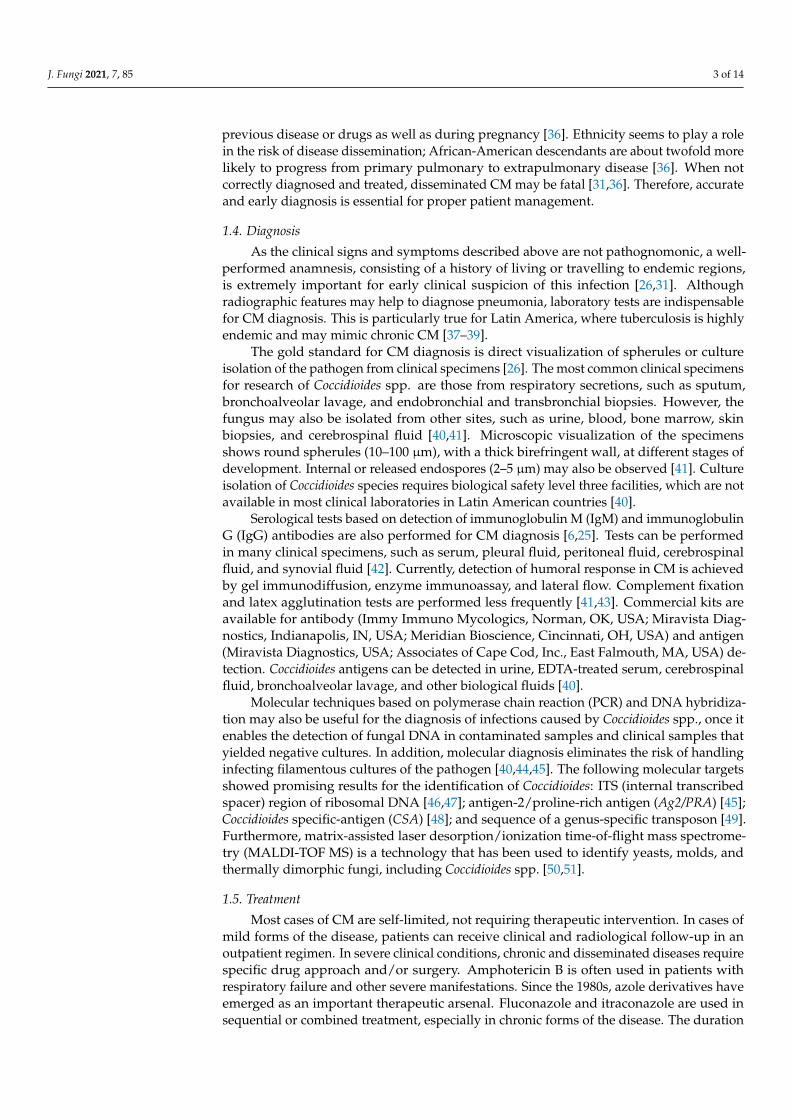
In 1998, Brazil was included in the international list of countries with endemic areas ofthe disease [58], with zones limited to the semi-arid areas of the States of Maranhão, Piauí,and Bahia. The most recent area of occurrence of CM in Brazil is the city of Serra Talhada,in Pernambuco State (759′09′′ S–3817′45′′ W), where four individuals presented acutepneumonia after armadillo hunting [58–60] (Figure 1).
2.2. Occurrence and Epidemiological Aspects: The Disease of Armadillo Hunters
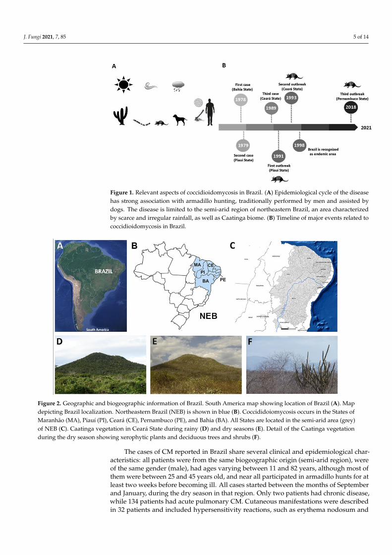
In Brazil, CM occurs exclusively in the northeast region, where approximately 136 casesof the disease have been reported since 1978: 94 cases in the state of Piauí [61]; six casesin the state of Maranhão [18,61,62]; 30 cases in the state of Ceará [63–68]; two cases in thestate of Bahia [55,68]; four cases in the state of Pernambuco [59,60] (Figure 2).
J. Fungi 2021, 7, 85 5 of 14
Figure 1. Relevant aspects of coccidioidomycosis in Brazil. (A) Epidemiological cycle of the diseasehas strong association with armadillo hunting, traditionally performed by men and assisted bydogs. The disease is limited to the semi-arid region of northeastern Brazil, an area characterizedby scarce and irregular rainfall, as well as Caatinga biome. (B) Timeline of major events related tococcidioidomycosis in Brazil.
Figure 2. Geographic and biogeographic information of Brazil. South America map showing location of Brazil (A). Mapdepicting Brazil localization. Northeastern Brazil (NEB) is shown in blue (B). Coccididoiomycosis occurs in the States ofMaranhão (MA), Piauí (PI), Ceará (CE), Pernambuco (PE), and Bahia (BA). All States are located in the semi-arid area (grey)of NEB (C). Caatinga vegetation in Ceará State during rainy (D) and dry seasons (E). Detail of the Caatinga vegetationduring the dry season showing xerophytic plants and deciduous trees and shrubs (F).
The cases of CM reported in Brazil share several clinical and epidemiological char-acteristics: all patients were from the same biogeographic origin (semi-arid region), wereof the same gender (male), had ages varying between 11 and 82 years, although most ofthem were between 25 and 45 years old, and near all participated in armadillo hunts for atleast two weeks before becoming ill. All cases started between the months of Septemberand January, during the dry season in that region. Only two patients had chronic disease,while 134 patients had acute pulmonary CM. Cutaneous manifestations were describedin 32 patients and included hypersensitivity reactions, such as erythema nodosum and

J. Fungi 2021, 7, 85 6 of 14
erythema multiforme [62]. One patient developed pericarditis without pulmonary in-volvement [35]. Although there are no official clinical-epidemiological data, the scientificliterature indicates that only two cases of acute pulmonary disease evolved to death, despiteantifungal treatment [63,69]. Most patients were treated with deoxycolate amphotericin Band oral fluconazole; a minority of patients were treated with only one of these drugs alone.Liposomal amphotericin B is not available in Brazilian public hospitals. All patients weretreated at the governmental hospital network of the Brazilian Unified Healthcare System,abbreviated SUS.
The role of Dasypus novemcinctus armadillos (Cingulata order, Dasypodidae family)in the ecology of C. posadasii in Brazil was initially studied by Eulalio et al. [70], whodescribed naturally infected animals in Piauí State. Since then, armadillos have beendetermined to figure as sentinels for CM in Brazil. As mentioned above, the majority of CMcases in Brazil have been linked to armadillo hunting [18,57,59,64–67], a cultural practiceperformed exclusively by men. Armadillos are mammals with terrestrial habits and candig deep burrows, which makes them vulnerable to arthroconidia exposure and infection.C. posadasii strains were obtained after direct cultivation and/or animal inoculation of soilsamples collected in or nearby armadillo burrows in Ceará State [17] and Piauí State [18].Additionally, de Macedo et al. [22] detected C. posadasii-related DNA in soil samples fromarmadillo burrows in Piauí State.
The physical and chemical characteristics of the soils where C. posadasii can be foundin Brazil are not known in detail. Only a single study details the granulometric andmineralogic properties of positive soil samples for C. posadasii [17]. Authors referred to thenaturally infected soils as Luvisol type, with pH 6.5 and a high sandy texture content (52%);technical research has revealed that the salinity of semi-arid area soils from Ceará Stateranges from <1% to up to 40% and the soil pH ranges from 4.1–8.6. Further studies on theenvironmental characteristics of CM endemic areas in Brazil may allow better correlationwith the data already described for the US.
2.3. Epidemiological Surveys Based on Unofficial Data: How Reliable Are These Studies?
In Brazil, CM is considered a non-notifiable disease, which compromises understand-ing its impact in the country and hinders the establishment of surveillance and controlmeasures. Failures in recording epidemiological data are common, and consequently,information on the disease is incomplete. As a consequence, data provided by the Braziliangovernment show divergences from scientific publications, which may indicate that theformer probably includes other diseases, such as paracoccidioidomycosis.
For instance, the search for deaths caused by CM between 1996 and 2017 in theNotifiable Diseases Information System from SUS revealed 48 events, distributed as follows:five in the north region, 22 in the northeast region, nine in the southeast region, 11 in thesouth region, and one in the midwest region. However, these governmental data areincoherent and unreliable, since the disease occurs exclusively in the northeast region. Inturn, data obtained by the Department of Information Technology of the Brazilian UnifiedHealth System showed that 829 cases of CM occurred in the country in 2011, a remarkablyhigh number of cases, considering the scientific notification in that period. Data providedby the Hospital Admissions Information System from SUS show 400 cases of CM per yearbetween 2000 and 2007, most of which were reported in the south and southeast regions(MS, 2011). Once again, government data seem to mistakenly report other infections asCM, since the south and southeast regions of Brazil do not present the adequate biologicalconditions for the occurrence of the disease.
Among the most important strategies for epidemiological investigation in popula-tions exposed to fungi is intradermal testing. This approach, which uses spherulin orcoccidioidin, structural antigens obtained from the parasitic and saprophytic phases ofCoccidioides spp., respectively, has been used in the US since the 1940s. So far, only twotests of this nature have been performed in Brazil. In 1991, Wanke et al. [18] performedthe intradermal spherulin test on 20 individuals associated with the first CM outbreak, in
J. Fungi 2021, 7, 85 7 of 14
Oeiras, Piauí, and they found reactivity in only one of them. In 1993, Diógenes et al. [71]carried out an epidemiological survey with spherulin in 87 residents of Jaguaribara, CearáState, where the third case of CM was registered. The study demonstrated reactivity in23 individuals, thus suggesting the occurrence of CM in that region.
This scenario suggests that, although the disease has been occurring for over threedecades in northeastern Brazil, and despite the efforts made by local research groups,government authorities responsible for epidemiological surveillance continue to ignore theexistence of the disease in the region.
2.4. Northeastern Brazil at a Glance: Biogeographical Aspects, Social Characteristics, and HealthIssues in a Singular Region
Northeastern Brazil (NEB) is a region located between 2.5 to 16.1 S and 34.8 to 46 Wand encompasses the States of Maranhão, Piauí, Ceará, Rio Grande do Norte, Paraíba,Pernambuco, Sergipe, Alagoas, and Bahia. It corresponds to an area of approximately1,540,000 km2, or about 18% of the Brazilian territory, with more than 53 million inhabitants,or almost 25% of the Brazilian population. Inside NEB there is a vast area classified assemi-arid, located between 3 to 18 S and 35 to 46 W. This semi-arid region occupies982,563.3 km2, with nearly 22,600,000 inhabitants, or almost 12% of Brazilian population,distributed among 1262 municipalities. The area of NEB comprises the most populoussemiarid region of the world [72].
The predominant biome in NEB is the Caatinga Phytogeographic Domain (nearly52.5% of NEB with 844.453 km2), followed by the Savanna/Cerrado (29.4%), AtlanticForest (10.7%), and Amazon (7.4%). The Caatinga occurs only in NEB, and it is formed byxerophytic plants, mostly deciduous trees and shrubs, and annual therophytic herbs [73],with more than 3300 plant species, of which over 500 species are endemic [74].
In this region, the mean rainfall is typically below 1000 mm per year, which is con-centrated within a few months per year. During most of the year, rainfall is absent ornegligible. Droughts are recurrent and have been reported since the 16th century. Since thattime, at least 54 long-lasting droughts have been reported, accounting for 31 drought yearsduring the 20th century and 11 drought years in the 21st century [75]. In the semi-aridregion, the typical mean annual rainfall is below 800 mm, and climate studies reveal thatthis area presents a risk of drought above 60%. The inappropriate use of soil, based mainlyon slash-and-burn practices, has contributed to intense land degradation. Many ruralareas in NEB are in the process of desertification and are no longer cultivated. In addition,extensive livestock farming is practiced in many areas, intensifying soil degradation [76].
NEB also presents important social and demographic characteristics. The officialpopulation census [77] reveals that its population is formed by brown (62.5% vs. 46.8%in Brazil), black (11.9% vs. 9.4% in Brazil), white (24.7% vs. 42.7% in Brazil) and nativeBrazilian (0.8% vs. 1.1% in Brazil) people. The NEB population has the lowest average ofschooling years (6.7 years vs. 8.0 in Brazil). In this region, 52.6% of the population has notfinished primary education. Nearly 20% of the population age 25 or over living in NEB isuneducated (vs. 11.2% in Brazil). In 2016, the estimated illiteracy rate among people age 15and over in NEB was 14.8% (vs. 7.2% in Brazil). Overall, the smallest proportion of peoplein Brazil with complete higher education (9.9%) is found in NEB [78].
It is believed that the rainfall irregularity and scarcity, the fragile soils, the intenseland degradation and desertification, as well as the socioeconomic characteristics of itsinhabitants are important factors that make NEB one of the world’s most vulnerableregions to climate change [76]. This condition, in turn, may exacerbate regional povertyand inequalities in a region historically marked by social and economic imbalances.
Official economic data show that nearly 6.4 million people living in NEB (almost12% of its total population) have daily earnings below US $1.90 and are regarded aspeople living in extreme poverty by the World Bank [79]. Considering the prevalenceof CM in NEB, it is clear that the disease is present in areas with economic and socialvulnerabilities; in accordance with the Atlas of Human Development in Brazil, issued bythe United Nations Development Programme, of the 35 municipalities where cases of CM

J. Fungi 2021, 7, 85 8 of 14
have been reported, 16 have low a Human Development Index (HDI, 0.500–0.599), whilethe remaining 19 municipalities present a medium HDI (0.600–0.699) [80].
Such economic and social inequalities have made NEB’s population historically vul-nerable to many infectious diseases. Local authorities face bureaucratic problems inhiring human resources, and a shortage of physicians is a recurrent problem in rural ar-eas [81]. Moreover, structural problems in the Brazilian health system, including flaws ingovernance, lack of funding, and suboptimal resource allocation [81], have led to the emer-gence/reemergence of infectious diseases such as whooping cough, measles, congenitalsyphilis, and leprosy in the region [82]. In addition, many severe diseases are also endemicin NEB and cause significant health issues, including vector-borne diseases (Chagas disease,dengue fever, Zika, Chikungunya), vaccine-preventable diseases (tuberculosis, meningitis,pneumonia), water-transmissible diseases (schistosomiasis, diarrhoea), and HIV/AIDS. Ofthe 20 diseases recognized by the World Health Organization as neglected tropical diseases,17 of them occur in Brazil, and 14 are endemic in NEB [83].
3. Neglected Tropical Diseases
Neglected tropical diseases (NTDs) are a diverse group of infectious diseases primarilyassociated with poverty and limited access to healthcare systems and education that affectmore than one billion people in rural or peri-urban areas in low- and middle-incomecountries in Africa, Asia, and Latin America [84]. As they affect mainly poor people,NTDs result in loss of productivity and cost billions of dollars every year to developingeconomies, further aggravating poverty [84]. Therapy for such diseases includes highlytoxic drugs that may potentially cause severe adverse side effects. In some cases, thereis no provision of curative therapy, and treatment relies on symptom alleviation [85]. Inaddition, local governments have mostly failed in controlling NTDs, and some diseases areunderreported in several countries [86,87].
According to the WHO [88], NTDs are those that (1) affect disproportionately pop-ulations living in poverty, resulting in high morbidity and mortality; (2) affect primarilypopulations living in tropical and subtropical areas; (3) may be controlled or eliminatedby preventive chemotherapy, intensified case management, vector control, veterinary pub-lic health, safe water supply, sanitation and hygiene; and (4) are neglected by research.Therefore, according to these criteria, the NTD portfolio is composed of the followingdiseases: buruli ulcer; Chagas disease; dengue fever and chikungunya; chromoblasto-mycosis; dracunculiasis; echinococcosis; foodborne trematode infection; human Africantrypanosomiasis; leishmaniasis; leprosy; lymphatic filariasis; mycetoma; onchocerciasis;rabies; scabies; schistosomiasis; soil-transmitted helminthiases; snakebite envenoming;trachoma; and yaws [89]. Among these diseases, only dracunculasis, human African try-panosomiasis, and yaws are not reported in Brazil. Except for buruli ulcer, echinococcosis,and onchocerciasis, the remaining NTDs are endemic in NEB.
Brazil accounts for the largest number of cases of dengue fever, leprosy, schisto-somiasis, Chagas disease, and leishmaniasis in the Americas, most of which occur inareas with low socioeconomic indexes, such as NEB. Approximately 10,000 NTD-relateddeaths are recorded in Brazil annually, and it was estimated that, in 2016, NTDs caused475,410 disability-adjusted life-years (DALYs) in the country [90]. In Brazil, NTDs signif-icantly contribute to the loss of health in individuals at all ages, with a higher burdenamong males, children under 1 year, and people age 70 and above [90].
Fungal infections are also prevalent in Brazil and are estimated to affect more than3.8 million people [91]; in some cases, the financial impact of these infections on publichealth may exceed US $100,000 per patient [92]. However, among such infections, onlymycetoma and chromoblastomycosis are included in the WHO NTD list [89]. Therefore,specialists have pleaded for the inclusion of other mycoses in the WHO NTD list, such ascryptococcosis, entomophthoramycosis, histoplasmosis, lacaziosis, paracoccidioidomyco-sis, and sporotrichosis [93–96]. According to these authors, all aforementioned diseasesfulfill the WHO criteria for NTD, once they are important causes of morbidity and mortality
J. Fungi 2021, 7, 85 9 of 14
affect primarily the poorest people living in rural areas or in areas lacking basic services,such as clean water, sewer systems, and paved roads; and are neglected by research andthe pharmaceutical industry.
At the present time, the coronavirus disease 2019 (COVID-19) pandemic is a demand-ing health problem faced by more than 180 counties [97]. The Severe Acute RespiratorySyndrome Coronavirus 2 (SARS-CoV-2) has been a concern in Latin American countriessince the first disease case was reported in Brazil in February 2020 [98]. Since then, Brazilhas been recognized as one of the leading countries in number of cases of and deaths dueto SARS-COV-2 infection [97]. According to the Economic Commission for Latin Americaand the Caribbean (ECLAC) report [99], the COVID-19 pandemic will drastically impactLatin America and the Caribbean [100], and it is expected that people living in rural areaswill be the most affected. It is estimated that about 30 million people living in these areaswill reach extreme poverty, which means an increase of 4.9% (from 20.3% in 2019 to 25.2%in 2020) in this population [99]. The COVID-19 pandemic will have financial and structuralimpacts on the health systems of affected countries, which may have repercussions onNTD control and management in the poorest regions of Latin America.
4. Could Coccidiodomycosis Be Considered a Neglected Disease?
A review of CM cases in Brazil suggests the disease fulfills the WHO criteria for NTD:(1) The disease occurs in rural areas of NEB, affecting mainly socially and economically vul-nerable people. Our experience shows that patients often do not find adequate treatmentin their local cities, being forced to travel long distances to more advanced centers in searchof hospitalization. (2) Logistical difficulties and economic limitations occasionally haveled to discontinuation of treatment by the patient, himself. The impact of early treatmentinterruption on the health and quality of life of these patients is unknown. (3) Governmentauthorities have little control over the disease, which leads to systematic failures in preven-tion, treatment, detection, and reporting of cases. Misdiagnosis of CM as tuberculosis mayoccur, since the latter is endemic and has a high incidence in Brazil [65,101], which canworsen the clinical condition of some patients, due to lack of suitable treatment. (4) Overthe decades, few financial resources have been allocated to research focused on improvingdiagnosis as well as the development of more effective drugs. National health authoritieshave failed to establish health education measures to prevent the disease, especially inremote rural areas.
On the other hand, CM has been considered a neglected disease in the US [102]. Inthat country, the incidence of CM has increased over the years [7], and research on thissevere fungal infection is scarce [5]. Although a prophylactic vaccine for CM seems to befeasible [15,103], the pharmaceutical industry has shown little interest. However, as theproduction of a safe vaccine has high cost due to patents as well as animal and human tests,pharmaceutical companies are necessary for its development [102].
The recognition of CM as a neglected disease in Latin America will allow for theestablishment of measures to define its prevalence in Brazil. With this information, arisk assessment and epidemiological surveillance system can be created, allowing for theestablishment of specific prophylactic measures in high-risk populations as well as thedevelopment of health education strategies. Taken together, these strategies may reducethe environmental risks of acquiring CM in areas already impacted by poverty and thehigh incidence of other infectious diseases.
5. Conclusions
CM is an important underreported disease that has been occurring in Brazil for overthree decades, but has been overlooked by public health authorities. The disease occurs inareas extremely vulnerable to climate change and in poor populations facing economic andsocial challenges. The recent economic crisis afflicting the country as well as the presentCOVID-19 pandemic has worsened access to health services by local populations, especiallythose living in rural areas. The data presented here suggest that the CM fits the definition of
J. Fungi 2021, 7, 85 10 of 14
an NTD. The inclusion of CM in the WHO NTD list could support efforts for the preventionand control of the disease, especially in economically vulnerable populations. The Braziliangovernment needs to establish public policies for the prevention and control of this diseaseas well as to allocate a greater amount of financial resources in scientific research to expandthe knowledge on the impact of CM in the country.
Author Contributions: Conceptualization and methodology, R.C. and S.M.; formal analysis, R.C.,S.M. and D.C.-B.; writing—original draft preparation, R.C. and S.M.; writing—review and editing,D.C.-B. and R.L.-N.; supervision, M.F.R. and J.J.S.; funding acquisition, R.C. All authors have readand agreed to the published version of the manuscript.
Funding: This research was funded by Conselho Nacional de Desenvolvimento Científico e Tec-nológico, Brazil (Process 309464/2019-6).
Informed Consent Statement: Not applicable.
Conflicts of Interest: The authors declare no conflict of interest.
References1. Fisher, M.C.; Koenig, G.L.; White, T.J.; Taylor, J.W. Molecular and Phenotypic Description of Coccidioides posadasii sp. nov.,
Previously Recognized as the Non-California Population of Coccidioides immitis. Mycologia 2002, 94, 73–84. [CrossRef] [PubMed]2. Mead, H.L.; Hamm, P.S.; Shaffer, I.N.; Teixeira, M.M.; Wendel, C.; Wiederhold, N.P.; Thompson, G.R.; Muñiz-Salazar, R.;
Castañón-Olivares, L.R.; Keim, P.; et al. Differential thermotolerance adaptation between species of Coccidioides. bioRxiv 2020,12, 247635.
3. McCotter, O.Z.; Benedict, K.; Engelthaler, D.M.; Komatsu, K.; Lucas, K.D.; Mohle-Boetani, J.C.; Oltean, H.; Vugia, D.; Chiller, T.M.;Cooksey, G.L.S.; et al. Update on the Epidemiology of coccidioidomycosis in the United States. Med. Mycol. 2019, 57, S30–S40.[CrossRef] [PubMed]
4. Kirkland, T.N.; Fierer, J. Coccidioides immitis and posadasii, A review of their biology, genomics, pathogenesis, and host immunity.Virulence 2018, 9, 1426–1435. [CrossRef]
5. Kollath, D.R.; Miller, K.J.; Barker, B.M. The mysterious desert dwellers: Coccidioides immitis and Coccidioides posadasii, causativefungal agents of coccidioidomycosis. Virulence 2019, 10, 222–233. [CrossRef]
6. Gabe, L.M.; Malo, J.; Knox, K.S. Diagnosis and Management of Coccidioidomycosis. Clin. Chest Med. 2017, 38, 417–433. [CrossRef]7. CDC. Increase in Reported Coccidioidomycosis—United States 1998–2011. Morb. Mortal. Wkly. Rep. 2013, 62, 217–221.8. Laniado-Laborin, R.; Arathoon, E.G.; Canteros, C.; Muñiz-Salazar, R.; Rendon, A. Coccidioidomycosis in Latin America.
Med. Mycol. 2019, 57, S46–S55. [CrossRef]9. Baptista-Rosas, R.C.; Riquelme, M. Epidemiología de la coccidioidomicosis en México. Rev. Iberoam. Micol. 2007, 24, 100–105.
[CrossRef]10. Davila, A.; Candolfi-Arballo, O.; Narvaez-Hernandez, E.; García-Arellano, A.; Lopez-Larios, A.; Cano-Rangel, A.; Castañon-Olivares, L.
Coccidioidin skin test in two Mexican populations from endemic areas of coccidioidomycosis in Mexico. Int. J. Infect. Dis. 2018, 73, 167.[CrossRef]
11. Davidson, A.P.; Shubitz, L.F.; Alcott, C.J.; E Sykes, J. Selected Clinical Features of Coccidioidomycosis in Dogs. Med. Mycol. 2019,57, S67–S75. [CrossRef] [PubMed]
12. Fernandez, J.A.; Hidalgo, M.N.; Hodzic, E.; Diab, S.S.; Uzal, F.A. Pathology of coccidioidomycosis in llamas and alpacas. J. Vet.Diagn. Investig. 2018, 30, 560–564. [CrossRef] [PubMed]
13. Koistinen, K.; Mullaney, L.; Bell, T.; Zaki, S.; Nalca, A.; Frick, O.; Livingston, V.; Robinson, C.G.; Estep, J.S.; Batey, K.L.; et al.Coccidioidomycosis in Nonhuman Primates: Pathologic and Clinical Findings. Vet. Pathol. 2018, 55, 905–915. [CrossRef][PubMed]
14. Cordeiro, R.D.A.; Silva, K.R.D.C.E.; Brilhante, R.S.N.; Moura, F.B.P.; Duarte, N.F.H.; Marques, F.J.D.F.; Filho, R.E.M.;de Araújo, R.W.B.; Bandeira, T.D.J.P.G. Coccidioides posadasiiInfection in Bats, Brazil. Emerg. Infect. Dis. 2012, 18, 668–670.[CrossRef] [PubMed]
15. Van Dyke, M.C.C.; Thompson, G.R.; Galgiani, J.N.; Barker, B.M. The Rise of Coccidioides: Forces Against the Dust Devil Unleashed.Front. Immunol. 2019, 10, 2188. [CrossRef]
16. Litvintseva, A.P.; Marsden-Haug, N.; Hurst, S.; Hill, H.; Gade, L.; Driebe, E.M.; Ralston, C.; Roe, C.; Barker, B.M.; Goldoft, M.;et al. Valley fever: Finding new places for an old disease: Coccidioides immitis found in Washington State soil associated withrecent human infection. Clin. Infect. Dis. 2015, 60, e1–e3. [CrossRef]
17. Cordeiro, R.A.; Brilhante, R.S.; Rocha, M.F.; Fechine, M.A.; Camara, L.M.; Camargo, Z.P.; Sidrim, J.J. Phenotypic characterizationand ecological features of Coccidioides spp. from Northeast Brazil. Med. Mycol. 2006, 44, 631–639. [CrossRef]
18. Wanke, B.; Lazera, M.; Monteiro, P.C.; Lima, F.C.; Leal, M.J.; Ferreira Filho, P.L.; Kaufman, L.; Pinner, R.W.; Ajello, L. Investigationof an outbreak of endemic coccidioidomycosis in Brazil’s northeastern state of Piauí with a review of the occurrence anddistribution of Coccidioides immitis in three other Brazilian states. Mycopathologia 1999, 148, 57–67. [CrossRef]
J. Fungi 2021, 7, 85 11 of 14
19. Elconin, A.F.; Egeberg, R.O.; Egeberg, M.C. Significance of soil salinity on the ecology of Coccidioides immitis. J. Bacteriol. 1964,87, 500–503. [CrossRef]
20. Stewart, R.A.; Meyer, K.F. Isolation of Coccidioides immitis (Stiles) from the Soil. Exp. Biol. Med. 1932, 29, 937–938. [CrossRef]21. Alvarado, P.; Teixeira, M.M.; Andrews, L.; Fernandez, A.; Santander, G.; Doyle, A.; Perez, M.; Yegres, F.; Barker, B.M. Detection of
Coccidioides posadasii from xerophytic environments in Venezuela reveals risk of naturally acquired coccidioidomycosis infections.Emerg. Microbes Infect. 2018, 7, 1–13. [CrossRef] [PubMed]
22. De Macedo, R.L.; Rosado, A.S.; da Mota, F.F.; Cavalcante, M.A.; Eulálio, K.D.; Filho, A.D.D.; Martins, L.M.; Lazéra, M.D.S.;Wanke, B. Molecular identification of Coccidioides spp. in soil samples from Brazil. BMC Microbiol. 2011, 11, 108–109. [CrossRef][PubMed]
23. Greene, D.R.; Koenig, G.; Fisher, M.C.; Taylor, J.W. Soil isolation and molecular identification of Coccidioides immitis. Mycologia2010, 92, 406–410. [CrossRef]
24. Taylor, J.W.; Barker, B.M. The endozoan, small-mammal reservoir hypothesis and the life cycle of Coccidioides species. Med. Mycol.2019, 57, S16–S20. [CrossRef] [PubMed]
25. Stockamp, N.W.; Thompson, G.R., 3rd. Coccidioidomycosis. Infect. Dis. Clin. N. Am. 2016, 30, 229–246. [CrossRef]26. Malo, J.; Luraschi-Monjagatta, C.; Wolk, D.M.; Thompson, R.; Hage, C.A.; Knox, K.S. Update on the Diagnosis of Pulmonary
Coccidioidomycosis. Ann. Am. Thorac. Soc. 2014, 11, 243–253. [CrossRef]27. Thompson, G.R., 3rd. Pulmonary coccidioidomycosis. Semin. Respir. Crit. Care Med. 2011, 32, 754–756. [CrossRef]28. Jude, C.M.; Nayak, N.B.; Patel, M.K.; Deshmukh, M.; Batra, P. Pulmonary Coccidioidomycosis: Pictorial Review of Chest
Radiographic and CT Findings. Radiographics 2014, 34, 912–925. [CrossRef]29. Valdivia, L.; Nix, D.; Wright, M.; Lindberg, E.; Fagan, T.; Lieberman, D.; Stoffer, T.; Ampel, N.M.; Galgiani, J.N. Coccidioidomycosis
as a Common Cause of Community-acquired Pneumonia. Emerg. Infect. Dis. 2006, 12, 958–962. [CrossRef]30. Twarog, M.; Thompson, G.R., 3rd. Coccidioidomycosis: Recent updates. Semin. Respir. Crit. Care Med. 2015, 36, 746–755.
[CrossRef]31. Galgiani, J.N.; Ampel, N.M.; Blair, J.E.; Catanzaro, A.; Geertsma, F.; Hoover, S.E.; Johnson, R.H.; Kusne, S.; Lisse, J.; MacDonald, J.D.;
et al. 2016 Infectious Diseases Society of America (IDSA) Clinical practice guideline for the treatment of coccidioidomycosis.Clin. Infect. Dis. 2016, 63, e112–e146. [CrossRef] [PubMed]
32. Ampel, N.M. The treatment of coccidioidomycosis. Rev. Inst. Med. Trop. São Paulo 2015, 57, 51–56. [CrossRef] [PubMed]33. Parish, J.M.; Blair, J.E. Coccidioidomycosis. Mayo Clin. Proc. 2008, 83, 343–348. [CrossRef] [PubMed]34. Arsura, E.L.; Bobba, R.K.; Reddy, C.M. Coccidioidal pericarditis: A case presentation and review of the literature. Int. J. Infect. Dis.
2005, 9, 104–109. [CrossRef] [PubMed]35. Brilhante, R.S.N.; Cordeiro, R.A.; Rocha, M.F.G.; Fechine, M.A.B.; Furtado, F.M.; Nagao-Dias, A.T.; Camargo, Z.P.; Sidrim, J.J.C.
Coccidioidal pericarditis: A rapid presumptive diagnosis by an in-house antigen confirmed by mycological and molecularmethods. J. Med. Microbiol. 2008, 57, 1288–1292. [CrossRef]
36. Odio, C.D.; Marciano, B.E.; Galgiani, J.N.; Holland, S.M. Risk Factors for Disseminated Coccidioidomycosis, United States.Emerg. Infect. Dis. 2017, 23, 308–311. [CrossRef] [PubMed]
37. Chong, S.; Lee, K.S.; Yi, C.A.; Chung, M.J.; Kim, T.S.; Han, J. Pulmonary fungal infection: Imaging findings in immunocompetentand immunocompromised patients. Eur. J. Radiol. 2006, 59, 371–383. [CrossRef]
38. Winn, R.E.; Johnson, R.; Galgiani, J.N.; Butler, C.; Pluss, J. Cavitary coccidioidomycosis with fungus ball formation. Diagnosis byfiberoptic bronchoscopy with coexistence of hyphae and spherules. Chest 1994, 105, 412–416. [CrossRef]
39. Muñoz, B.; Castañón, L.R.; Calderón, I.; Vázquez, M.E.; Manjarrez, M.E. Parasitic mycelial forms of Coccidioides species in Mexicanpatients. J. Clin. Microbiol. 2004, 42, 1247–1249. [CrossRef]
40. Cordeiro, R.; Brilhante, R.; Rocha, M.; Sidrim, J. Coccidioides. In Molecular Detection of Human Fungal Pathogens; CRC Press: BocaRaton, FL, USA, 2011; pp. 135–144.
41. Saubolle, M.A. Laboratory Aspects in the Diagnosis of Coccidioidomycosis. Ann. N. Y. Acad. Sci. 2007, 1111, 301–314. [CrossRef]42. Pappagianis, D. Serologic studies in coccidioidomycosis. Semin. Respir. Infect. 2001, 16, 242–250. [CrossRef] [PubMed]43. Ampel, N.M. The diagnosis of coccidioidomycosis. F1000 Med. Rep. 2010, 2, 2. [CrossRef] [PubMed]44. Duarte-Escalante, E.; Frías-De-León, M.G.; Zúñiga, G.; Martinez-Herrera, E.; Acosta-Altamirano, G.; Reyes-Montes, M.D.R.
Molecular markers in the epidemiology and diagnosis of coccidioidomycosis. Rev. Iberoam. Micol. 2014, 31, 49–53. [CrossRef][PubMed]
45. Bialek, R.; Kern, J.; Herrmann, T.; Tijerina, R.; Ceceñas, L.; Reischl, U.; González, G.M. PCR Assays for Identification ofCoccidioides posadasii Based on the Nucleotide Sequence of the Antigen 2/Proline-Rich Antigen. J. Clin. Microbiol. 2004, 42, 778–783.[CrossRef] [PubMed]
46. Binnicker, M.J.; Buckwalter, S.P.; Eisberner, J.J.; Stewart, R.A.; McCullough, A.E.; Wohlfiel, S.L.; Wengenack, N.L. Detection ofCoccidioides Species in Clinical Specimens by Real-Time PCR. J. Clin. Microbiol. 2006, 45, 173–178. [CrossRef]
47. Tintelnot, K.; de Hoog, G.S.; Antweiler, E.; Losert, H.; Seibold, M.; Brandt, M.A.; van den Ende, A.H.; Fisher, M.C. Taxonomic anddiagnostic markers for identification of Coccidioides immitis and Coccidioides posadasii. Med. Mycol. 2007, 45, 385–393. [CrossRef]
48. Pan, S.; Cole, G.T. Molecular and biochemical characterization of a Coccidioides immitis-specific antigen. Infect. Immun. 1995,63, 3994–4002. [CrossRef]
J. Fungi 2021, 7, 85 12 of 14
49. Saubolle, M.A.; Wojack, B.R.; Wertheimer, A.M.; Fuayagem, A.Z.; Young, S.; Koeneman, B.A. Multicenter Clinical Validation of aCartridge-Based Real-Time PCR System for Detection of Coccidioides spp. in Lower Respiratory Specimens. J. Clin. Microbiol.2017, 56, e01277-17. [CrossRef]
50. Patel, R.A. Moldy application of MALDI: MALDI-ToF Mass Spectrometry for fungal identification. J. Fungi 2019, 5, 4. [CrossRef]51. Walsh, T.J.; McCarthy, M.W. The expanding use of matrix-assisted laser desorption/ionization-time of flight mass spectroscopy in
the diagnosis of patients with mycotic diseases. Expert Rev. Mol. Diagn. 2019, 19, 241–248. [CrossRef]52. Kim, M.M.; Vikram, H.R.; Kusne, S.; Seville, M.T.; Blair, J.E. Treatment of Refractory Coccidioidomycosis with Voriconazole or
Posaconazole. Clin. Infect. Dis. 2011, 53, 1060–1066. [CrossRef] [PubMed]53. Wilson, L.; Ting, J.; Lin, H.; Shah, R.; MacLean, M.; Peterson, M.W.; Stockamp, N.; Libke, R.; Brown, P. The Rise of Valley Fever:
Prevalence and Cost Burden of Coccidioidomycosis Infection in California. Int. J. Environ. Res. Public Health 2019, 16, 1113.[CrossRef] [PubMed]
54. Vianna, H.; Passos, H.V.; Santana, A.V. Coccicioidomicose. Relato do primeiro caso ocorrido em nativo do Brasil. Rev. Inst. Med.Trop. S. Paulo 1979, 21, 51–55. [PubMed]
55. Gomes, O.M.; Serrano, R.R.P.; Pradel, H.O.V.; Moraes, N.L.T.B.; Varella, A.L.B.; Fiorelli, A.I.; Espósito, I.; Saad, F.A.; Furlaneto, J.A.;Rothman, A.; et al. Coccidioidomicose pulmonar. Primeiro caso nacional. Rev. Assoc. Méd. Bras 1978, 24, 167–168.
56. Kuhl, I.A.; Kuhl, G.; Londero, A.; Diógenes, M.J.N.; Ferreira, M.F. Coccidioidomycosis laríngea: Relato de caso. Rev. Bras Otorrinol.1996, 62, 48–51.
57. Sidrim, J.; Silva, L.C.D.; Nunes, J.; Rocha, M.; Paixão, G.C. Le Nord-Est brésilien, région d’endémie de coccidioïdomycose:A propos d’une micro-épidémie. J. Mycol. Méd. 1997, 7, 37–39.
58. Pappagianis, D. Coccidioides Immitis, 9th ed.; Ajello, L., Hay, R., Eds.; Topley & Wilson’s Microbiology and Microbial Infections:London, UK, 1998; pp. 357–371.
59. Araújo, P.S.R.; Souza-Junior, V.R.; Padilha, C.E.; Oliveira, M.I.; Arraes, L.C.; Vieira, R.; Antunes, A.; Lima-Neto, R.G.; Marsden, A.Coccidioidomycosis: First cases reported in Pernambuco, Brazil. Rev. Inst. Med. Trop. S Paulo 2018, 60, e75. [CrossRef]
60. de Lima-Neto, R.G.; Inácio, C.P.; Costa, R.B.; Cordeiro, R.D.A.; Neves, W.W.; Neves, R.P.; Magalhães, O.M.C.; Filho, A.M.L.;de Araújo, P.S.R. Coccidioidomycosis in a Pediatric Patient. Mycopathologia 2021. [CrossRef]
61. Eulálio, K.D. Echo-Epidemiological and Clinical Manifestations of Coccidioidomycosis in the States of the Piauí and the Maranhão.Ph.D. Thesis, Instituto Oswaldo Cruz, Rio de Janeiro, Brazil, 2008.
62. Deus-Filho, A.D.; Deus, A.C.B.; Meneses, A.O.; Soares, A.S.; Lira, A.L.A. Manifestações cutâneo-mucosas da coccidioidomicose:Estudo de trinta casos procedentes dos estados do Piauí e Maranhão. Bras. Dermatol. 2010, 85, 45–51. [CrossRef]
63. Morais, J.L.D.S.; Borges, M.C.M.; Cavalcante, L.M.M.B.; Motoyama, P.V.P.; Libório, M.P.; Távora, L.G.F. Coccidioidomycosis in areference center in Northeast Brazil: Clinical/epidemiological profile and most common radiological findings. Rev. Soc. Bras.Med. Trop. 2020, 53, 20200249. [CrossRef]
64. Brilhante, R.S.; Moreira Filho, R.E.; Rocha, M.F.; Castelo-Branco, D.S.; Fechine, M.A.; Lima, R.A.; Picanço, Y.V.; Cordeiro, R.A.;Camargo, Z.P.; Queiroz, J.A.; et al. Coccidioidomycosis in armadillo hunters from the state of Ceará, Brazil. Mem. Inst.Oswaldo Cruz 2012, 107, 813–815. [CrossRef] [PubMed]
65. Cordeiro, R.D.A.; Brilhante, R.S.N.; Rocha, M.F.G.; Bandeira, S.P.; Fechine, M.A.B.; de Camargo, Z.; Sidrim, J.J.C.; de Camargo, Z.Twelve years of coccidioidomycosis in Ceará State, Northeast Brazil: Epidemiologic and diagnostic aspects. Diagn. Microbiol.Infect. Dis. 2010, 66, 65–72. [CrossRef] [PubMed]
66. Togashi, R.H.; Aguiar, F.M.B.; Ferreira, D.B.; Moura, C.M.; Sales, M.T.M.; Rios, N.X. Coccidioidomicose pulmonar e extrapulmonar:Três casos em zona endêmica no interior do Ceará. J. Bras. Pneumol. 2009, 35, 275–279. [CrossRef] [PubMed]
67. Da Costa, F.A.M.; Reis, R.C.; Benevides, F.; Tomé, G.D.S.; Holanda, M.A. Coccidioidomicose pulmonar em caçador de tatus.J. Pneumol. 2001, 27, 275–278. [CrossRef]
68. Martins, M.A.; Araújo, E.M.P.A.; Kuwakino, M.H.; Heins-Vaccari, E.M.; del Negro, G.M.B.; Vozza, J.A.; Lacaz, C.S. Coccid-ioidomycosis in Brazil. A case report. Rev. Inst. Med. Trop. S Paulo 1997, 30, 299–304. [CrossRef]
69. Veras, K.N.; Figueirêdo, B.C.S.; Martins, L.M.S.; Vasconcelos, J.T.P.; Wanke, B. Coccidioidomycosis: An unusual cause of acuterespiratory distress syndrome. J. Bras Pneumol. 2003, 29, 45–48. [CrossRef]
70. Eulalio, K.D.; de Macedo, R.L.; Cavalcanti, M.D.A.S.; Martins, L.M.S.; Lazéra, M.D.S.; Wanke, B. Coccidioides immitis isolated fromarmadillos (Dasypus novemcinctus) in the state of Piauí, northeast Brazil. Mycopathologia 2001, 149, 57–61. [CrossRef]
71. Diógenes, M.J.N.; Jamacuru, W.F.; Silva, M.A.B.; Carvalho, F.F. Inquérito epidemiológico com esferulina em Jaguaribara, CE,Brasil, 1993. Bras Dermatol. 1995, 70, 525–529.
72. Marengo, J.A.; Torres, R.R.; Alves, L.M. Drought in Northeast Brazil—Past, present, and future. Theor. Appl. Clim. 2017,129, 1189–1200. [CrossRef]
73. Moro, M.F.; Macedo, M.B.; Moura-Fé, M.M.; Castro, A.S.F.; CostaA, R.C. Vegetação, unidades fitoecológicas e diversidadepaisagística do estado do Ceará. Rodriguésia 2015, 66, 717–743. [CrossRef]
74. Fernandes, M.F.; Cardoso, D.; de Queiroz, L.P. An updated plant checklist of the Brazilian Caatinga seasonally dry forests andwoodlands reveals high species richness and endemism. J. Arid. Environ. 2020, 174, 104079. [CrossRef]
75. Marengo, J.A.; Bernasconi, M. Regional differences in aridity/drought conditions over Northeast Brazil: Present state and futureprojections. Clim. Chang. 2015, 129, 103–115. [CrossRef]
J. Fungi 2021, 7, 85 13 of 14
76. Marengo, J.A.; Alves, L.M.; Alvala, R.C.; Cunha, A.P.; Brito, S.; Moraes, O.L. Climatic characteristics of the 2010-2016 drought inthe semiarid Northeast Brazil region. An. Acad. Bras. Ciênc. 2018, 90, 1973–1985. [CrossRef] [PubMed]
77. Brazilian Institute of Geography and Statistics-IBGE. Censo Demográfico. 2010. Available online: https://www.ibge.gov.br/en/statistics/social/labor/18391-2010-population-census.html (accessed on 30 October 2020).
78. Brazilian Institute of Geography and Statistics-IBGE. Continuous PNAD 2016: 51% of the Brazilian Population Aged 25 orOver Had Only Complete Primary Education. 2017. Available online: https://agenciadenoticias.ibge.gov.br/en/agencia-press-room/2185-news-agency/releases-en/19005-continuous-pnad-2016-51-of-the-brazilian-population-aged-25-or-over-had-only-complete-primary-education (accessed on 30 October 2020).
79. Brazilian Institute of Geography and Statistics-IBGE. 2019. Extreme Poverty Affects 13.5 Million Persons and Hits Highest Levelin Seven Years, Brazilian Institute of Geography and Statistics. 2019. Available online: https://agenciadenoticias.ibge.gov.br/en/agencia-news/2184-news-agency/news/25895-extreme-poverty-affects-13-5-million-persons-and-hits-highest-level-in-seven-years (accessed on 30 October 2020).
80. Atlas of Human Development in Brazil. 2013. Available online: http://www.atlasbrasil.org.br (accessed on 30 October 2020).81. Massuda, A.; Hone, T.; Leles, F.A.G.; de Castro, M.C.; Atun, R. The Brazilian health system at crossroads: Progress, crisis and
resilience. BMJ Glob. Health 2018, 3, e000829. [CrossRef] [PubMed]82. França, E.B.; Lansky, S.; Rego, M.A.S.; Malta, D.C.; França, J.S.; Teixeira, R.; Porto, D.; Almeida, M.F.; Souza, M.F.M.; Szwarcwald, C.L.;
et al. Leading causes of child mortality in Brazil, in 1990 and 2015: Estimates from the Global Burden of Disease study. Rev. BrasEpidemiol. 2017, 20 (Suppl. S1), 46–60. [CrossRef] [PubMed]
83. Martins-Melo, F.R.; Carneiro, M.; Heukelbach, J.; Ribeiro, A.L.P.; Werneck, G.L. The burden of Neglected Tropical Diseases inBrazil, 1990–2016: A subnational analysis from the Global Burden of Disease Study 2016. PLoS Negl. Trop. Dis. 2018, 12, e0006559.[CrossRef] [PubMed]
84. WHO (World Health Organization). Investing to Overcome the Global Impact of Neglected Tropical Diseases: Third WHO Report onNeglected Tropical Diseases; World Health Organization: Geneva, Switzerland, 2015; Available online: https://www.who.int/neglected_diseases/9789241564861/en/\T1\textgreater (accessed on 30 October 2020).
85. Nii-Trebi, N.I. Emerging and Neglected Infectious Diseases: Insights, Advances, and Challenges. BioMed Res. Int. 2017, 2017, 1–15.[CrossRef] [PubMed]
86. Hotez, P.J. Global urbanization and the neglected tropical diseases. PLoS Negl. Trop. Dis. 2017, 11, e0005308. [CrossRef]87. Hotez, P.J.; Aksoy, S. Ten years of progress in neglected tropical disease control and Elimination. PLoS Negl. Trop. Dis. 2017,
11, e0005355. [CrossRef]88. WHO (World Health Organization). Recommendations for the Adoption of Additional Diseases as Neglected Tropical Diseases; World
Health Organization: Geneva, Switzerland, 2016.89. WHO (World Health Organization). Report of the Tenth Meeting of the WHO Strategic and Technical Advisory Group for Neglected
Tropical Diseases; World Health Organization: Geneva, Switzerland, 2017.90. Martins-Melo, F.R.; Ramos, A.N.; Alencar, C.H.; Heukelbach, J. Trends and spatial patterns of mortality related to neglected
tropical diseases in Brazil. Parasite Epidemiol. Control. 2016, 1, 56–65. [CrossRef]91. Giacomazzi, J.; Baethgen, L.; Carneiro, L.C.; Millington, M.A.; Denning, D.W.; Colombo, A.L.; Pasqualotto, A.C. In association
with the LIFE program the burden of serious human fungal infections in Brazil. Mycoses 2016, 59, 145–150. [CrossRef] [PubMed]92. Borba, H.H.L.; Steimbach, L.M.; Riveros, B.S.; Tonin, F.S.; Ferreira, V.L.; Bagatim, B.A.Q.; Balan, G.; Pontarolo, R.; Wiens, A.
Cost-effectiveness of amphotericin B formulations in the treatment of systemic fungal infections. Mycoses 2018, 61, 754–763.[CrossRef] [PubMed]
93. Molloy, S.F.; Chiller, T.; Greene, G.S.; Burry, J.; Govender, N.P.; Kanyama, C.; Mfinanga, S.; Lesikari, S.; Mapoure, Y.N.;Kouanfack, C.; et al. Cryptococcal meningitis: A neglected NTD? PLoS Negl. Trop. Dis. 2017, 11, e0005575. [CrossRef] [PubMed]
94. Queiroz-Telles, F.; Fahal, A.H.; Falci, D.R.; Caceres, D.H.; Chiller, T.; Pasqualotto, A.C. Neglected endemic mycoses. Lancet Infect.Dis. 2017, 17, e367–e377. [CrossRef]
95. Rodrigues, M.L. Neglected disease, neglected populations: The fight against Cryptococcus and cryptococcosis. Mem. Inst. OswaldoCruz 2018, 113, e180111. [CrossRef]
96. Griffiths, J.; Colombo, A.L.; Denning, D.W. The case for paracoccidioidomycosis to be accepted as a neglected tropical (fungal)disease. PLoS Negl. Trop. Dis. 2019, 13, e0007195. [CrossRef] [PubMed]
97. Dong, E.; Du, H.; Gardner, L. An interactive web-based dashboard to track COVID-19 in real time. Lancet Infect. Dis. 2020,20, 533–534. [CrossRef]
98. Rodriguez-Morales, A.J.; Gallego, V.; Escalera-Antezana, J.P.; Méndez, C.A.; Zambrano, L.I.; Franco-Paredes, C.; Suárez, J.A.;Rodriguez-Enciso, H.D.; Balbin-Ramon, G.J.; Savio-Larriera, E.; et al. COVID-19 in Latin America: The implications of the firstconfirmed case in Brazil. Travel Med. Infect. Dis. 2020, 35, 101613. [CrossRef]
99. ECLAC. Cómo evitar que la crisis del COVID-19 se transforme en una crisis alimentaria: Acciones urgentes contra el hambre enAmérica Latina y el Caribe. Econ. Comm. Latin Am. Caribb. 2020.
100. Hotez, P.J. Ten global “hotspots” for the neglected tropical diseases. PLoS Negl. Trop. Dis. 2014, 8, e2496. [CrossRef]101. Brazil. Boletim Epidemiológico. Secretaria de Vigilância em Saúde. Ministério da Saúde. 2020. Available online: http:
//www.aids.gov.br/pt-br/pub/2020/boletim-epidemiologico-de-turbeculose-2020 (accessed on 10 October 2020).
J. Fungi 2021, 7, 85 14 of 14
102. Kirkland, T.N. The Quest for a Vaccine Against Coccidioidomycosis: A Neglected Disease of the Americas. J. Fungi 2016, 2, 34.[CrossRef] [PubMed]
103. Barnato, A.E.; Sanders, G.D.; Owens, D.K. Cost-effectiveness of a potential vaccine for Coccidioides immitis. Emerg. Infect. Dis.2001, 7, 797–806. [CrossRef] [PubMed]