Clinical significance of Maspin promoter methylation and loss of its protein expression in invasive...
10
RESEARCH ARTICLE Clinical significance of Maspin promoter methylation and loss of its protein expression in invasive ductal breast carcinoma: correlation with VEGF-A and MTA1 expression Gayatri Sharma & Sameer Mirza & Rajinder Parshad & Anurag Srivastava & Siddartha Datta Gupta & Pranav Pandya & Ranju Ralhan # International Society of Oncology and BioMarkers (ISOBM) 2010 Abstract Maspin is a serine protease inhibitor with tumor- suppressor activity. Maspin can suppress tumor growth and metastasis in vivo and tumor cell motility and invasion in vitro. Previous studies indicate that the loss of Maspin expression is closely linked to aberrant methylation of the Maspin promoter. We examined the promoter methylation status of Maspin in tumor and corresponding serum of breast cancer patients. In addi- tion, protein expression of this gene was also assessed to determine possible correlation between promoter hyper- methylation and gene silencing. Further, we investigated the correlation of Maspin expression with vascular endothelial growth factor (VEGF-A) and MTA1 expres- sion. Maspin methylation was analyzed by methylation- specific PCR in 100 invasive ductal breast carcinoma patients’ tumors and circulating DNA in a prospective study. Promoter hypermethylation was correlated with expression of the encoded protein in tumors by immu- nohistochemistry. Significant correlation was observed between promoter hypermethylation of Maspin ( r = +0.88; p ≤ 0.0001) in tumors and paired sera. Significant association was found between Maspin promoter hyper- methylation and loss of its protein expression (p =0.01, OR=3.1, 95% CI=1.3–7.4). The expression of VEGF-A and MTA1 was lower in tumors with high Maspin expression compared to tumors with loss of Maspin expression. Our results indicate that aberrant promoter methylation is associated with loss of Maspin immuno- reactivity in breast cancer tissues. Further, loss of Maspin expression is significantly correlated with increased expression of VEGF-A and MTA1. Keywords Breast cancer . DNA methylation . Maspin . MTA1 . Gene silencing G. Sharma : S. Mirza : R. Ralhan Department of Biochemistry, All India Institute of Medical Sciences, Ansari Nagar, New Delhi 110029, India R. Parshad : A. Srivastava Department of Surgery, All India Institute of Medical Sciences, Ansari Nagar, New Delhi 110029, India S. D. Gupta Department of Pathology, All India Institute of Medical Sciences, Ansari Nagar, New Delhi 110029, India G. Sharma : P. Pandya Dev Sanskriti Vishwa Vidhyalaya, Hardwar, India R. Ralhan Department of Otolaryngology—Head and Neck Surgery, University of Toronto, Toronto, ON, Canada R. Ralhan Department of Otolaryngology—Head and Neck Surgery, Mount Sinai Hospital, Joseph and Wolf Lebovic Health Complex, 600 University Avenue, Room 6-416, Toronto, ON, Canada M5G 1X5 R. Ralhan Department of Pathology and Laboratory Medicine, Mount Sinai Hospital, Joseph and Wolf Lebovic Health Complex, 600 University Avenue, Room 6-416, Toronto, ON, Canada M5G 1X5 Present Address: R. Ralhan (*) Joseph and Mildred Sonshine Family Centre for Head and Neck Disease, Mount Sinai Hospital, Joseph and Wolf Lebovic Health Complex, 600 University Avenue, Room 6-416, Toronto, ON, Canada M5G 1X5 e-mail: [email protected] DOI 10.1007/s13277-010-0087-8 Tumor Biol. (2011) 32:23–32 Received: 8 May 2010 / Accepted: 26 July 2010 / Published online: 10 August 2010
-
Upload
independent -
Category
Documents
-
view
5 -
download
0
Transcript of Clinical significance of Maspin promoter methylation and loss of its protein expression in invasive...
RESEARCH ARTICLE
Clinical significance of Maspin promoter methylationand loss of its protein expression in invasive ductal breastcarcinoma: correlation with VEGF-A and MTA1 expression
Gayatri Sharma & Sameer Mirza & Rajinder Parshad &
Anurag Srivastava & Siddartha Datta Gupta &
Pranav Pandya & Ranju Ralhan
# International Society of Oncology and BioMarkers (ISOBM) 2010
Abstract Maspin is a serine protease inhibitor with tumor-suppressor activity. Maspin can suppress tumor growthand metastasis in vivo and tumor cell motility andinvasion in vitro. Previous studies indicate that the lossof Maspin expression is closely linked to aberrantmethylation of the Maspin promoter. We examined thepromoter methylation status of Maspin in tumor andcorresponding serum of breast cancer patients. In addi-tion, protein expression of this gene was also assessed todetermine possible correlation between promoter hyper-methylation and gene silencing. Further, we investigatedthe correlation of Maspin expression with vascularendothelial growth factor (VEGF-A) and MTA1 expres-sion. Maspin methylation was analyzed by methylation-specific PCR in 100 invasive ductal breast carcinomapatients’ tumors and circulating DNA in a prospectivestudy. Promoter hypermethylation was correlated with
expression of the encoded protein in tumors by immu-nohistochemistry. Significant correlation was observedbetween promoter hypermethylation of Maspin (r=+0.88; p≤0.0001) in tumors and paired sera. Significantassociation was found between Maspin promoter hyper-methylation and loss of its protein expression (p=0.01,OR=3.1, 95% CI=1.3–7.4). The expression of VEGF-Aand MTA1 was lower in tumors with high Maspinexpression compared to tumors with loss of Maspinexpression. Our results indicate that aberrant promotermethylation is associated with loss of Maspin immuno-reactivity in breast cancer tissues. Further, loss of Maspinexpression is significantly correlated with increasedexpression of VEGF-A and MTA1.
Keywords Breast cancer . DNA methylation .Maspin .
MTA1 . Gene silencing
G. Sharma : S. Mirza :R. RalhanDepartment of Biochemistry,All India Institute of Medical Sciences, Ansari Nagar, New Delhi110029, India
R. Parshad :A. SrivastavaDepartment of Surgery, All India Institute of Medical Sciences,Ansari Nagar, New Delhi 110029, India
S. D. GuptaDepartment of Pathology, All India Institute of Medical Sciences,Ansari Nagar, New Delhi 110029, India
G. Sharma : P. PandyaDev Sanskriti Vishwa Vidhyalaya, Hardwar, India
R. RalhanDepartment of Otolaryngology—Head and Neck Surgery,University of Toronto, Toronto, ON, Canada
R. RalhanDepartment of Otolaryngology—Head and Neck Surgery,Mount Sinai Hospital, Joseph and Wolf Lebovic Health Complex,600 University Avenue, Room 6-416,Toronto, ON, Canada M5G 1X5
R. RalhanDepartment of Pathology and Laboratory Medicine,Mount Sinai Hospital, Joseph and Wolf Lebovic Health Complex,600 University Avenue, Room 6-416,Toronto, ON, Canada M5G 1X5
Present Address:R. Ralhan (*)Joseph and Mildred Sonshine Family Centre for Head and NeckDisease, Mount Sinai Hospital, Joseph and Wolf Lebovic HealthComplex, 600 University Avenue, Room 6-416,Toronto, ON, Canada M5G 1X5e-mail: [email protected]
DOI 10.1007/s13277-010-0087-8Tumor Biol. (2011) 32:23–32
Received: 8 May 2010 /Accepted: 26 July 2010 /Published online: 10 August 2010
Introduction
Maspin, a serine proteinase inhibitor (serpin) family, plays arole in development of the mammary gland [1] and isexpressed in myoepithelial cells and normal secretoryepithelial cells [2]. In vitro studies and animal models haveshown that Maspin inhibits tumorigenesis by modulatingtumor cell growth, invasion, and metastasis [2, 3]. Maspinalso inhibits endothelial cell motility and angiogenesis [4].Earlier studies have demonstrated a tight link between theloss of Maspin expression in breast cancer cells and theaberrant cytosine methylation and histone deacetylation ofits promoter [5–7]. Maspin interacts with diverse group ofintercellular and extracellular proteins, regulating celladhesion, motility, apoptosis, and angiogenesis and iscritically involved in mammary gland development [8].The tissue-specific expression of Maspin is epigeneticallycontrolled, and aberrant methylation of Maspin promoter isclosely associated with Maspin gene silencing [5, 9–15].The promoter methylation of the Maspin gene leads to genesilencing in cancers, such as breast, thyroid, skin, and colon[5, 11, 13]. In contrast, overexpression of Maspin in gastric,pancreatic, and ovarian cancers results from promoter CpGdemethylation [10, 12, 14]. This clearly indicates that bothmethylation and demethylation of Maspin promoter couldregulate Maspin gene expression.
Recent studies suggest that Maspin overexpressioncorrelates with increased expression of vascular endothelialgrowth factors A, C, and D in human ovarian carcinomaand melanoma [16, 17]. The most potent of these cytokinesis vascular endothelial growth factor (VEGF-A), a heparinbinding glycoprotein with potent angiogenic, mitogenic,and vascular permeability-enhancing activities specific forendothelial cells.
The metastasis tumor antigen (MTA) family of proteins,a group of 70 to 80 kD polypeptides, have been shown tobe functional components of the Mi-2/NuDR complex, amajor macromolecular form of histone deacetylases [18],which has been linked to transcriptional repression, cellularproliferation, and cancer [19–21]. In vertebrates, the MTAfamily is comprised of three members: MTA1, MTA2, andMTA3. MTA1 is overexpressed in a variety of humanmalignancies, and its overexpression is also associated withtumor progression and metastasis in multiple cancer types[19, 22, 23]. The expression level of MTA1s, a naturalvariant of MTA1, is increased in estrogen receptor (ER)-negative human breast cancers [21].
In a parallel study in an independent cohort of Invasiveductal carcinomas of breast (IDCs), we recently demonstrat-ed loss or reduced cytoplasmic expression of Maspin in 36of 59 (61%) tumors by immunohistochemical analysis andupregulation of Maspin expression by curcumin in breastcancer cells (MCF7 and MDA-MB-231) that correlated with
the upregulation of p53 protein and downregulation of Bcl-2,suggesting Maspin-mediated apoptosis in MCF7 cells. Theupregulation of Maspin expression by curcumin in breastcancer cells taken together with the clinical data suggested apotential therapeutic role for curcumin in inducing Maspin-mediated inhibition of invasion of breast carcinoma cells.
In the present report, we hypothesized that methylation-induced gene silencing of Maspin may account for the lossof its protein expression in IDCs of breast. In an independentcohort of 100 IDCs, we determined the promoter methyla-tion status of Maspin, its relationship with its proteinexpression in these tumors. Further, we also examined therelationship of Maspin with expression of the other keycomponents involved in angiogenesis and metastasis such asVEGF-A and MTA1. The association between promoterhypermethylation of Maspin in primary breast tumors andpaired serum DNA was also investigated to explore thepossibility of using serum DNA methylation analysis of thisgene as surrogate non-invasive marker for their tumor status.
Materials and methods
Tissue specimens
Surgically resected specimens or trucut biopsies fromuntreated primary breast carcinoma patients, matched normalbreast tissues (5–10 cm away from the site of the tumor), andperipheral blood samples were collected from 100 breastcancer patients enrolled in the Out Patients Department ofSurgical Disciplines, All India Institute of Medical Sciences,New Delhi, India, after approval of the study by InstitutionalHuman Ethics Committee. Written consent was taken fromall the patients enrolled in the study. The age of the patientsranged from 30 to 81 years (median age is 50 years). Allpatients were diagnosed with Invasive Ductal Carcinoma.
Ten milliliters of blood was collected from breast cancerpatients at the time of surgery. Bloodwas also collected from 30healthy females (age range 22–70 years); ten of these womenwere premenopausal and 20 were postmenopausal; four werenulliparous. All these 30 healthy females did not have history ofany chronic disease and had no evidence of disease at the timeof enrollment in the study. Serum was isolated from clottedblood by centrifugation at 1,000×g for 10 min. A part of eachtumor and representative adjacent normal tissue was kept informalin for histopathological characterization to confirm thediagnosis and immunohistochemistry and the other part wassnap frozen and stored at −70°C.
DNA extraction
DNA was extracted from breast tumor and normal tissues,breast cancer cell lines MDA-MB-231, and normal lym-
24 Tumor Biol. (2011) 32:23–32
phocytes using standard technique of digestion withproteinase K in the presence of sodium dodecyl sulfate(SDS) at 37°C overnight, followed by phenol/chloroformextraction. Serum DNA was extracted using Qiamp DNABlood Mini Kit (Qiagen, Hilden, Germany) according tomanufacturers’ instructions. The quality and integrity ofDNA from tissues was checked by electrophoresis on 0.8%agarose gel, quantitated spectrophotometrically, and storedat −20°C till further use.
Methylation-specific PCR
The sensitive methylation-specific PCR (MSP) was used todetect promoter methylation, as it can detect 0.1% cancercell DNA from a heterogeneous cell population [24].Bisulfite modification of the DNA (up to 2 μg) fromvarious sources was carried out as described [25]. Primersand PCR conditions used for MSP of Maspin are frompublished study [26] and have been standardized in ourlaboratory. For positive and negative controls of the MSP,breast cancer cell line (MCF7) was used as a positivecontrol, maternal blood DNA collected after 3rd trimesterwas used as positive control for unmethylated DNA, andwater with no DNA template as a control for contaminationwere included in each experiment. After amplification, eachPCR product was electrophoresed using a 2% agarose gel,stained with ethidium bromide and visualized under UVillumination.
Immunohistochemistry
Immunohistochemical analysis was carried out usingparaffin-embedded breast carcinoma tissue sections asdescribed by us previously [27]. Briefly, after antigenretrieval by microwave heating in Tris–Cl. Buffer(pH 9.0) for 15 min, tissue sections were rinsed with0.01 M phosphate buffer saline, pH 7.4 (PBS). Sectionswere incubated with primary antibody (1:100 dilutions) at4°C overnight in a humidified chamber. Monoclonal anti-bodies were purchased against Maspin (550839) from BDPharmingen (San Diego, CA), VEGF-A (sc-7269), andMTA1 (sc-9446) from Santa Cruz Biotechnology Inc.(Santa Cruz, CA). After extensive washing with PBS,sections were incubated with biotin-linked universal anti-bodies and subsequently with horse radish peroxidase-streptavidin conjugate (LSABTM Kit, Dako Cytomation,Glostrup, Denmark) and color was developed using 3,3’-diaminobenzidine hydrochloride as chromogen, counter-stained with Mayer’s hematoxylin, and mounted forevaluation using microscope (OLYMPUS BX-51, Japan).In the negative control, primary antibody was replaced byisotype-specific IgG.
The results of immunohistochemistry were assessed bythe pathologist (S.D.G), who independently examined theimmunostaining scoring of the tissue sections. The slideswere scored for the percentage of cells showing immuno-reactivity: −, no detectable staining and +, ≥10% positivetumor cells showed reactivity. The nuclear/cytoplasmicimmunoreactivity of the Maspin [28] and MTA1 [29] wasconsidered as positive. Tumors were regarded as ERα, PR,and p53 positive, if the staining was localized in nuclei[30]. While for VEGF-A tumors were regarded as positive,if the staining was localized in cytoplasm [31].
For HER2/neu, the scoring system was as follows: nostaining or membrane staining in fewer than 10% of tumorcells, 0; faint, barely perceptible membrane staining in morethan 10% of tumor cells, the cells are stained only in part ofthe membrane, 1+; weak to moderate complete membranestaining observed in more than 10% of tumor cells, 2+; andstrong, complete membrane staining in more than 10% oftumor cells, 3+ [32].
Cell lysis and Western blot
Cells and tumor tissues were lysed on ice using radio-immunoprecipitation buffer (0.05 mol/l Tris–HCl,pH 7.4, 0.15 mol/l NaCl, 0.25% deoxycholic acid, 1%NP-40, 1 mmol/l ethylenediaminetetraacetic acid,0.5 mmol/l dithiothreitol, 1 mmol/l phenylmethylsulfonylfluoride, 5 mg/ml leupeptin, and 10 mg/ml aprotinin).Lysates were then centrifuged at 13,000 rpm. at 4°C for10 min. Protein extracts were solubilized in SDS gelloading buffer (60 mmol/l Tris base, 2% SDS, 10%glycerol, and 5% β-mercaptoethanol). Samples contain-ing equal amounts of protein (50 μg) were separated ona 10% SDS–polyacrylamide gel electrophoresis andelectroblotted onto Immobilon-P membranes (Millipore,Bedford, MA, USA) in a transfer buffer. Immunoblottingwas performed using antibodies against Maspin (1:1,000), VEGF-A (1: 1,000), MTA1 (1: 1,000), and anti-β actin antibodies (1: 1,500), as an internal control. Thesignal was developed with enhanced chemiluminescence(Santa Cruz, CA) after incubation with appropriatesecondary antibodies.
Statistical analysis
Statistical analysis of correlation of methylation of genes andprotein expression with known clinicopathological character-istics was performed with Fisher exact test (two-sided). Two-sided p values were calculated, and p≤0.05 was consideredto be significant. Sensitivity and specificity was calculatedusing receiver-operating characteristic analyses. All of thestatistical analyses were performed using SPSS software(version 10.0; SAS Institute, Cary, NC).
25Tumor Biol. (2011) 32:23–32
Results
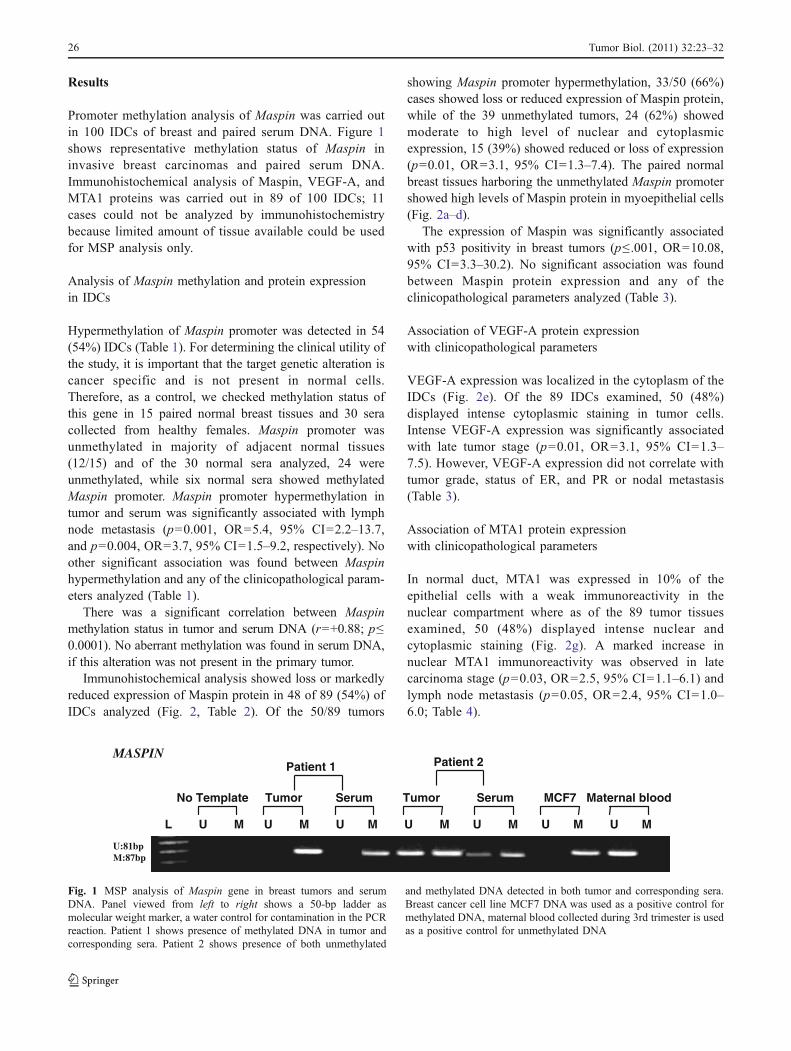
Promoter methylation analysis of Maspin was carried outin 100 IDCs of breast and paired serum DNA. Figure 1shows representative methylation status of Maspin ininvasive breast carcinomas and paired serum DNA.Immunohistochemical analysis of Maspin, VEGF-A, andMTA1 proteins was carried out in 89 of 100 IDCs; 11cases could not be analyzed by immunohistochemistrybecause limited amount of tissue available could be usedfor MSP analysis only.
Analysis of Maspin methylation and protein expressionin IDCs
Hypermethylation of Maspin promoter was detected in 54(54%) IDCs (Table 1). For determining the clinical utility ofthe study, it is important that the target genetic alteration iscancer specific and is not present in normal cells.Therefore, as a control, we checked methylation status ofthis gene in 15 paired normal breast tissues and 30 seracollected from healthy females. Maspin promoter wasunmethylated in majority of adjacent normal tissues(12/15) and of the 30 normal sera analyzed, 24 wereunmethylated, while six normal sera showed methylatedMaspin promoter. Maspin promoter hypermethylation intumor and serum was significantly associated with lymphnode metastasis (p=0.001, OR=5.4, 95% CI=2.2–13.7,and p=0.004, OR=3.7, 95% CI=1.5–9.2, respectively). Noother significant association was found between Maspinhypermethylation and any of the clinicopathological param-eters analyzed (Table 1).
There was a significant correlation between Maspinmethylation status in tumor and serum DNA (r=+0.88; p≤0.0001). No aberrant methylation was found in serum DNA,if this alteration was not present in the primary tumor.
Immunohistochemical analysis showed loss or markedlyreduced expression of Maspin protein in 48 of 89 (54%) ofIDCs analyzed (Fig. 2, Table 2). Of the 50/89 tumors
showing Maspin promoter hypermethylation, 33/50 (66%)cases showed loss or reduced expression of Maspin protein,while of the 39 unmethylated tumors, 24 (62%) showedmoderate to high level of nuclear and cytoplasmicexpression, 15 (39%) showed reduced or loss of expression(p=0.01, OR=3.1, 95% CI=1.3–7.4). The paired normalbreast tissues harboring the unmethylated Maspin promotershowed high levels of Maspin protein in myoepithelial cells(Fig. 2a–d).
The expression of Maspin was significantly associatedwith p53 positivity in breast tumors (p≤ .001, OR=10.08,95% CI=3.3–30.2). No significant association was foundbetween Maspin protein expression and any of theclinicopathological parameters analyzed (Table 3).
Association of VEGF-A protein expressionwith clinicopathological parameters
VEGF-A expression was localized in the cytoplasm of theIDCs (Fig. 2e). Of the 89 IDCs examined, 50 (48%)displayed intense cytoplasmic staining in tumor cells.Intense VEGF-A expression was significantly associatedwith late tumor stage (p=0.01, OR=3.1, 95% CI=1.3–7.5). However, VEGF-A expression did not correlate withtumor grade, status of ER, and PR or nodal metastasis(Table 3).
Association of MTA1 protein expressionwith clinicopathological parameters
In normal duct, MTA1 was expressed in 10% of theepithelial cells with a weak immunoreactivity in thenuclear compartment where as of the 89 tumor tissuesexamined, 50 (48%) displayed intense nuclear andcytoplasmic staining (Fig. 2g). A marked increase innuclear MTA1 immunoreactivity was observed in latecarcinoma stage (p=0.03, OR=2.5, 95% CI=1.1–6.1) andlymph node metastasis (p=0.05, OR=2.4, 95% CI=1.0–6.0; Table 4).
U:81bpM:87bp
MASPIN
L U M U M U M U M U M U M U M
No Template Tumor Serum Tumor Serum MCF7 Maternal blood
Patient 1 Patient 2
Fig. 1 MSP analysis of Maspin gene in breast tumors and serumDNA. Panel viewed from left to right shows a 50-bp ladder asmolecular weight marker, a water control for contamination in the PCRreaction. Patient 1 shows presence of methylated DNA in tumor andcorresponding sera. Patient 2 shows presence of both unmethylated
and methylated DNA detected in both tumor and corresponding sera.Breast cancer cell line MCF7 DNA was used as a positive control formethylated DNA, maternal blood collected during 3rd trimester is usedas a positive control for unmethylated DNA
26 Tumor Biol. (2011) 32:23–32
Relationship of Maspin expression with VEGF-Aand MTA1
The expression of VEGF-A and MTA1 was significantlylower in tumors with high Maspin expression compared totumors with loss of Maspin expression. Forty-four percentof the cases with intense Maspin expression had highVEGF-A expression compared to 67% of cases withnegative Maspin expression (p=0.03, OR=2.6, 95% CI=1.1–6.1, Table 4).
Similarly, only 11% of the cases with intense Maspinexpression had high MTA1 expression as compared to 82%of those with negative Maspin (p=0.001, OR=12.0, 95%CI=4.3–32.2, Table 4).
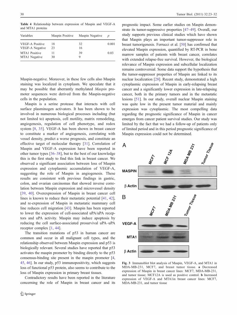
The immunohistochemistry results for Maspin, VEGF-A, and MTA1 were further confirmed by Western blotanalysis using MCF7, MDA-MB-231, and breast tumortissue. Immunoblot analysis showed clear increased expres-sion of VEGF-A and MTA1 and decreased expression ofMaspin in both the breast cancer cell lines and tumorTissue. (Fig. 3).
Discussion
The salient findings of our study are: (a) correlation ofMaspin promoter methylation with loss of its proteinexpression in IDCs; (b) association of loss of Maspinexpression and cytoplasmic accumulation of VEGF-A andnuclear accumulation of MTA1 suggesting role of Maspinin angiogenesis and metastasis; (c) association of loss ofMaspin expression with nuclear accumulation of p53; (d)correlation between Maspin hypermethylation in tumor andpaired serum DNA, suggesting the utility of serum DNA asa surrogate non-invasive tool for methylation analysis.
In this study, we analyzed 100 IDC breast cancer tissuesas well as 15 adjacent normal breast tissues for Maspinpromoter methylation by MSP. The data revealed thatMaspin promoter was methylated in over 54% of the breasttissues, suggesting that promoter methylation of Maspincan be a frequent event in human breast carcinogenesis,which is in general agreement with earlier studies [9, 33,34]. With respect to the epigenetic status of the Maspinpromoter in normal breast tissues, we found that majority ofadjacent normal breast tissues as well as normal serumtaken from healthy females had unmethylated Maspinpromoters.
In general, aberrant methylation of the Maspin promoterwas associated with loss of Maspin immunoreactivity;however, some cases (17 breast tumor tissues) that werescored as Maspin-positive also showed an aberrant meth-ylation of the Maspin promoter. In most of these cases, onlysome cells were Maspin-positive and other cells were
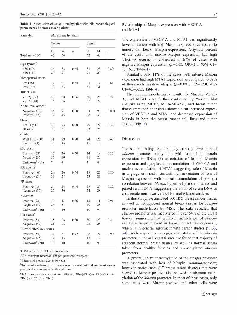
Table 1 Association of Maspin methylation with clinicopathologicalparameters of breast cancer patients
Variables Maspin methylation
Tumor Serum
U M p U M pTotal no.=100 46 54 52 48
Age (years)a
>50 (59) 26 33 0.64 31 28 0.89≤50 (41) 20 21 21 20
Menopausal status
Pre (38) 17 21 0.84 21 17 0.61Post (62) 29 33 31 31
Tumor size
T1+T2 (56) 28 28 0.36 30 26 0.72T3+T4 (44) 18 26 22 22
Node involvement
Negative (33) 24 9 0.001 24 9 0.004Positive (67) 22 45 28 39
Stage
I & II (51) 28 23 0.68 29 22 0.32III (49) 18 31 23 26
Grade
Well Diff. (50) 21 29 0.70 24 26 0.63Undiff. (28) 13 15 15 13
p53 Status
Positive (33) 13 20 0.50 14 19 0.23Negative (56) 26 30 31 25
Unknownb (11) 7 4 7 4
ERα status
Positive (46) 20 26 0.64 18 22 0.80Negative (54) 26 28 23 26
PR status
Positive (48) 24 24 0.44 28 20 0.22Negative (52) 22 30 24 28
Her2/neu
Positive (23) 10 13 0.86 12 11 0.91Negative (57) 26 31 29 28
Unknownb (20) 10 10 10 9
HR statusc
Positive (53) 25 28 0.80 30 23 0.4Negative (47) 21 26 22 25
ERα/PR/Her2/neu status
Positive (55) 24 31 0.72 28 27 0.90Negative (25) 12 13 13 12
Unknownb (20) 10 10 10 9
TNM refers to UICC classification
ERα estrogen receptor, PR progesterone receptoraMean and median age is 50 yearsb Immunohistochemical analysis was not carried out in these breast cancerpatients due to non-availability of tissuec HR (hormone receptor) status: ERα(−), PR(+)/ERα(+), PR(−)/ERα(+),PR(+) vs. ERα(−), PR(−)
27Tumor Biol. (2011) 32:23–32
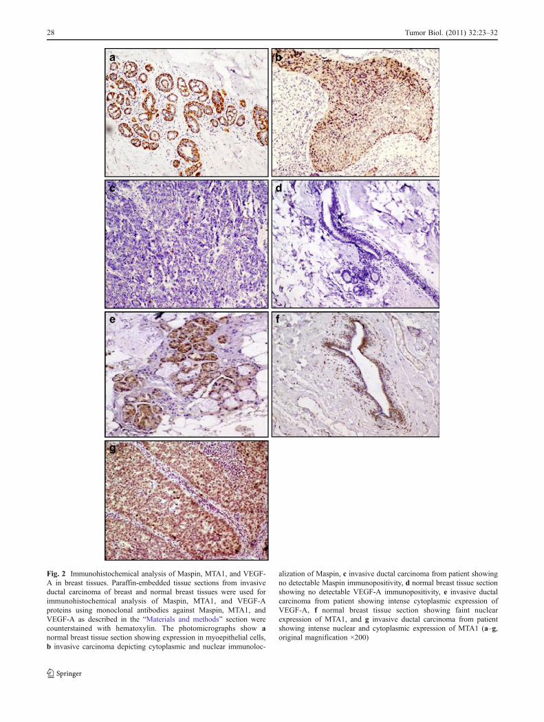
Fig. 2 Immunohistochemical analysis of Maspin, MTA1, and VEGF-A in breast tissues. Paraffin-embedded tissue sections from invasiveductal carcinoma of breast and normal breast tissues were used forimmunohistochemical analysis of Maspin, MTA1, and VEGF-Aproteins using monoclonal antibodies against Maspin, MTA1, andVEGF-A as described in the “Materials and methods” section werecounterstained with hematoxylin. The photomicrographs show anormal breast tissue section showing expression in myoepithelial cells,b invasive carcinoma depicting cytoplasmic and nuclear immunoloc-
alization of Maspin, c invasive ductal carcinoma from patient showingno detectable Maspin immunopositivity, d normal breast tissue sectionshowing no detectable VEGF-A immunopositivity, e invasive ductalcarcinoma from patient showing intense cytoplasmic expression ofVEGF-A, f normal breast tissue section showing faint nuclearexpression of MTA1, and g invasive ductal carcinoma from patientshowing intense nuclear and cytoplasmic expression of MTA1 (a–g,original magnification ×200)
28 Tumor Biol. (2011) 32:23–32
Variables Maspin VEGF-A MTA1
+ − p + − p + − p(Total no.=89) 41 48 50 39 50 39
Age (years)a
>50 (52) 25 27 0.65 34 18 0.04 29 23 0.92≤50 (37) 16 21 16 21 21 16
Menopausal status
Pre (34) 14 20 0.46 25 9 0.009 22 12 0.20Post (55) 27 28 25 30 28 27
Tumor size
T1+T2 (54) 24 30 0.70 27 27 0.14 27 27 0.14T3+T4 (35) 17 18 23 12 23 12
Node involvement
Negative (29) 14 15 0.70 14 15 0.29 12 17 0.05Positive (60) 27 33 36 24 38 22
Stage
I & II (53) 23 25 0.70 21 27 0.01 22 26 0.03III & IV (47) 18 23 29 12 28 13
Grade
Well Diff (48) 19 31 0.10 32 18 0.30 30 20 0.71Undifferentiated (28) 15 12 14 13 15 12
p53 status
Positive (33) 5 28 0.001 19 14 0.84 19 14 0.84Negative (56) 36 20 31 25 31 25
ERα status
Positive (38) 16 22 0.50 22 16 0.77 24 14 0.25Negative (51) 25 26 28 23 26 25
PR status
Positive (40) 18 22 0.85 23 17 0.82 25 15 0.27Negative (49) 23 26 27 22 25 24
Her2/neu
Positive (22) 10 12 0.88 13 9 0.70 12 10 0.88Negative (57) 27 30 31 26 30 27
Unknownb (20)
HR statusc
Positive (44) 20 24 0.90 25 19 0.90 27 17 0.33
Negative (45) 21 24 25 20 23 22
ERα/PR/Her2/neu
Positive (54) 0.53 0.90 0.53Negative (25) 24 30 30 24 30 24
Unknownb (20) 13 12 14 11 12 13
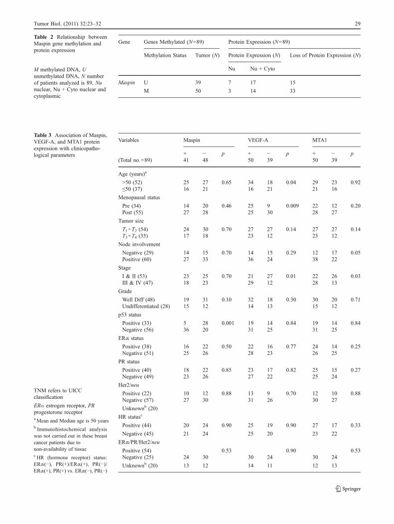
Table 3 Association of Maspin,VEGF-A, and MTA1 proteinexpression with clinicopatho-logical parameters
TNM refers to UICCclassification
ERα estrogen receptor, PRprogesterone receptoraMean and Median age is 50 yearsb Immunohistochemical analysiswas not carried out in these breastcancer patients due tonon-availability of tissuec HR (hormone receptor) status:ERα(−), PR(+)/ERα(+), PR(−)/ERα(+), PR(+) vs. ERα(−), PR(−)
Gene Genes Methylated (N=89) Protein Expression (N=89)
Methylation Status Tumor (N) Protein Expression (N) Loss of Protein Expression (N)
Nu Nu + Cyto
Maspin U 39 7 17 15
M 50 3 14 33
Table 2 Relationship betweenMaspin gene methylation andprotein expression
M methylated DNA, Uunmethylated DNA, N numberof patients analyzed is 89, Nunuclear, Nu + Cyto nuclear andcytoplasmic
29Tumor Biol. (2011) 32:23–32
Maspin-negative. Moreover, in these few cells also Maspinstaining was localized in cytoplasm. We speculate that itmay be possible that aberrantly methylated Maspin pro-moter sequences were derived from the Maspin-negativecells in the population.
Maspin is a serine protease that interacts with cellsurface plasminogen activators. It has been shown to beinvolved in numerous biological processes including (butnot limited to) apoptosis, cell motility, matrix remodeling,angiogenesis, regulation of cell phenotype, and redoxsystem [8, 35]. VEGF-A has been shown in breast cancerto constitute a marker of angiogenesis, correlating withvessel density, predict a worse prognosis, and constitute aneffective target of molecular therapy [31]. Correlation ofMaspin and VEGF-A expression have been reported inother tumor types [36–38], but to the best of our knowledgethis is the first study to find this link in breast cancer. Weobserved a significant association between loss of Maspinexpression and cytoplasmic accumulation of VEGF-A,suggesting the role of Maspin in angiogenesis. Theseresults are consistent with previous findings in gastric,colon, and ovarian carcinomas that showed inverse corre-lation between Maspin expression and microvessel density[39, 40]. Overexpression of Maspin in breast cancer celllines is known to reduce their metastatic potential [41, 42],and re-expression of Maspin in metastatic mammary cellline reduces cell migration [43]. Maspin has been reportedto lower the expression of cell-associated uPA/uPA recep-tors and uPA activity. Maspin may induce apoptosis byreducing the cell surface-associated prosurvival uPA–uPAreceptor complex [1, 44].
The transition mutations of p53 in human cancer arecommon and occur in all malignant cell types, and therelationship observed between Maspin expression and p53 isbiologically relevant. Several studies have reported that p53activates the maspin promoter by binding directly to the p53consensus-binding site present in the maspin promoter [4,45, 46]. In our study, p53 immunopositivity, which suggestsloss of functional p53 protein, also seems to contribute to theloss of Maspin expression in primary breast tissues.
Contradictory results have been reported in the literatureconcerning the role of Maspin in breast cancer and its
prognostic impact. Some earlier studies on Maspin demon-strate its tumor-suppressive properties [47–49]. Overall, ourstudy supports previous clinical studies which have shownthat Maspin plays an important tumor-suppressor role inbreast tumorigenesis. Ferrucci et al. [50] has confirmed thatelevated Maspin expression, quantified by RT-PCR in bonemarrow samples of patients with breast cancer, correlateswith extended relapse-free survival. However, the biologicalrelevance of Maspin expression and subcellular localizationremains controversial. Some data support the hypothesis thatthe tumor-suppressor properties of Maspin are linked to itsnuclear localization [28]. Recent study, demonstrated a highcytoplasmic expression of Maspin in early-relapsing breastcancer and a significantly lower expression in late-relapsingcancer, both in the primary tumors and in the metastaticlesions [51]. In our study, overall nuclear Maspin stainingwas quite low in the present tumor material and mainlyexpression was cytoplasmic. The most compelling dataregarding the prognostic significance of Maspin in canceremerges from cancer patient survival studies. Our study waslimited by the fact that we had a follow-up of patients onlyof limited period and in this period prognostic significance ofMaspin expression could not be determined.
MASPIN
β Actin
β Actin
MTA1
A
VEGF-A
B
Fig. 3 Immunoblot blot analysis of Maspin, VEGF-A, and MTA1 inMDA-MB-231, MCF7, and breast tumor tissue. a Decreasedexpression of Maspin in breast cancer lines: MCF7, MDA-MB-231,and tumor tissue; MCF12A is used as positive control. b Increasedexpression of VEGF-A and MTA1in breast cancer lines: MCF7,MDA-MB-231, and tumor tissue
Table 4 Relationship between expression of Maspin and VEGF-Aand MTA1 proteins
Variables Maspin Positive Maspin Negative p
VEGF-A Positive 18 32 0.001VEGF-A Negative 23 16
MTA1 Positive 11 39 0.03MTA1 Negative 30 9
30 Tumor Biol. (2011) 32:23–32
In conclusion, to our knowledge, this is the first reportdemonstrating the relationships between Maspin promotermethylation, and loss of expression of Maspin. Our data alsosuggest that expression of Maspin is significantly correlatedwith MTA1 and VEGF-A expression. Furthermore, thecorrelation between Maspin methylation in tumor and pairedserum DNA underscores the utility of serum DNA as asurrogate non-invasive tool for methylation analysis.
References
1. Sheng S, Carey J, Seftor EA, Dias L, Hendrix MJ, Sager R.Maspin acts at the cell membrane to inhibit invasion and motilityof mammary and prostatic cancer cells. Proc Natl Acad Sci USA.1996;93:11669–74.
2. Liu J, Yin S, Reddy N, Spencer C, Sheng S. Bax mediates theapoptosis-sensitizing effect of maspin. Cancer Res. 2004;64:1703–11.
3. Jiang N, Meng Y, Zhang S, Mensah-Osman E, Sheng S. Maspinsensitizes breast carcinoma cells to induced apoptosis. Oncogene.2002;21:4089–98.
4. Zhang M, Volpert O, Shi YH, Bouck N. Maspin is anangiogenesis inhibitor. Nat Med. 2000;6:196–9.
5. Domann FE, Rice JC, Hendrix MJ, Futscher BW. Epigeneticsilencing of maspin gene expression in human breast cancers. Int JCancer. 2000;85:805–10.
6. Maass N, Biallek M, Rosel F, Schem C, Ohike N, Zhang M, et al.Hypermethylation and histone deacetylation lead to silencing ofthe maspin gene in human breast cancer. Biochem Biophys ResCommun. 2002;297:125–8.
7. Primeau M, Gagnon J, Momparler RL. Synergistic antineoplasticaction of DNA methylation inhibitor 5-aza-2'-deoxycytidine andhistone deacetylase inhibitor depsipeptide on human breastcarcinoma cells. Int J Cancer. 2003;103:177–84.
8. Khalkhali-Ellis Z. Maspin: the new frontier. Clin Cancer Res.2006;12:7279–83.
9. Akiyama Y, Maesawa C, Ogasawara S, Terashima M, Masuda T.Cell-type-specific repression of the maspin gene is disruptedfrequently by demethylation at the promoter region in gastricintestinal metaplasia and cancer cells. Am J Pathol. 2003;163:1911–9.
10. Bettstetter M, Woenckhaus M, Wild PJ, Rummele P, Blaszyk H,Hartmann A, et al. Elevated nuclear maspin expression isassociated with microsatellite instability and high tumour gradein colorectal cancer. J Pathol. 2005;205:606–14.
11. Boltze C, Schneider-Stock R, Quednow C, Hinze R, Mawrin C,Hribaschek A, et al. Silencing of the maspin gene by promoterhypermethylation in thyroid cancer. Int J Mol Med. 2003;12:479–84.
12. Goelz SE, Vogelstein B, Hamilton SR, Feinberg AP. Hypomethy-lation of DNA from benign and malignant human colon neo-plasms. Science. 1985;228:187–90.
13. Reis-Filho JS, Torio B, Albergaria A, Schmitt FC. Maspinexpression in normal skin and usual cutaneous carcinomas.Virchows Arch. 2002;441:551–8.
14. Rose SL, Fitzgerald MP, White NO, Hitchler MJ, Futscher BW, DeGeest K, et al. Epigenetic regulation of maspin expression in humanovarian carcinoma cells. Gynecol Oncol. 2006;102:319–24.
15. Sato N, Maitra A, Fukushima N, van Heek NT, Matsubayashi H,Iacobuzio-Donahue CA, et al. Frequent hypomethylation ofmultiple genes overexpressed in pancreatic ductal adenocarcino-ma. Cancer Res. 2003;63:4158–66.
16. Bolat F, Gumurdulu D, Erkanli S, Kayaselcuk F, Zeren H, AliVardar M, et al. Maspin overexpression correlates withincreased expression of vascular endothelial growth factors a,c, and d in human ovarian carcinoma. Pathol Res Pract.2008;204:379–87.
17. Chua R, Setzer S, Govindarajan B, Sexton D, Cohen C, ArbiserJL. Maspin expression, angiogenesis, prognostic parameters, andoutcome in malignant melanoma. J Am Acad Dermatol.2009;60:758–66.
18. Zhang H, Stephens LC, Kumar R. Metastasis tumor antigenfamily proteins during breast cancer progression and metastasis ina reliable mouse model for human breast cancer. Clin Cancer Res.2006;12:1479–86.
19. Balasenthil S, Broaddus RR, Kumar R. Expression of metastasis-associated protein 1 (mta1) in benign endometrium and endome-trial adenocarcinomas. Hum Pathol. 2006;37:656–61.
20. Hofer MD, Kuefer R, Varambally S, Li H, Ma J, Shapiro GI, et al.The role of metastasis-associated protein 1 in prostate cancerprogression. Cancer Res. 2004;64:825–9.
21. Kumar R, Wang RA, Mazumdar A, Talukder AH, Mandal M, YangZ, et al. A naturally occurring mta1 variant sequesters oestrogenreceptor-alpha in the cytoplasm. Nature. 2002;418:654–7.
22. Mazumdar A, Wang RA, Mishra SK, Adam L, Bagheri-YarmandR, Mandal M, et al. Transcriptional repression of oestrogenreceptor by metastasis-associated protein 1 corepressor. Nat CellBiol. 2001;3:30–7.
23. Nicolson GL, Nawa A, Toh Y, Taniguchi S, Nishimori K,Moustafa A. Tumor metastasis-associated human mta1 gene andits mta1 protein product: role in epithelial cancer cell invasion,proliferation and nuclear regulation. Clin Exp Metastasis.2003;20:19–24.
24. Lewis CM, Cler LR, Bu DW, Zochbauer-Muller S, Milchgrub S,Naftalis EZ, et al. Promoter hypermethylation in benign breastepithelium in relation to predicted breast cancer risk. Clin CancerRes. 2005;11:166–72.
25. Frommer M, McDonald LE, Millar DS, Collis CM, Watt F, GriggGW, et al. A genomic sequencing protocol that yields a positivedisplay of 5-methylcytosine residues in individual DNA strands.Proc Natl Acad Sci USA. 1992;89:1827–31.
26. Murai S, Maesawa C, Masuda T, Sugiyama T. Aberrant maspinexpression in human endometrial cancer. Cancer Sci. 2006;97(9):883–8.
27. Sharma G, Mirza S, Prasad CP, Srivastava A, Gupta SD, RalhanR. Promoter hypermethylation of p16ink4a, p14arf, cyclind2 andslit2 in serum and tumor DNA from breast cancer patients. LifeSci. 2007;80:1873–81.
28. Mohsin SK, Zhang M, Clark GM, Craig Allred D. Maspinexpression in invasive breast cancer: association with otherprognostic factors. J Pathol. 2003;199:432–5.
29. Jang KS, Paik SS, Chung H, Oh YH, Kong G. Mta1 over-expression correlates significantly with tumor grade and angio-genesis in human breast cancers. Cancer Sci. 2006;97:374–9.
30. Goldhirsch A, Wood WC, Gelber RD, Coates AS, Thurlimann B,Senn HJ. Meeting highlights: updated international expertconsensus on the primary therapy of early breast cancer. J ClinOncol. 2003;21:3357–65.
31. Ryden L, Stendahl M, Jonsson H, Emdin S, Bengtsson NO,Landberg G. Tumor-specific vegf-a and vegfr2 in postmenopausalbreast cancer patients with long-term follow-up. Implication of alink between vegf pathway and tamoxifen response. Breast CancerRes Treat. 2005;89:135–43.
32. Rhodes A, Jasani B, Anderson E, Dodson AR, Balaton AJ.Evaluation of her-2/neu immunohistochemical assay sensitivityand scoring on formalin-fixed and paraffin-processed cell linesand breast tumors: a comparative study involving results fromlaboratories in 21 countries. Am J Clin Pathol. 2002;118:408–17.
31Tumor Biol. (2011) 32:23–32
33. Fitzgerald M, Oshiro M, Holtan N, Krager K, Cullen JJ, FutscherBW, et al. Human pancreatic carcinoma cells activate maspinexpression through loss of epigenetic control. Neoplasia.2003;5:427–36.
34. Futscher BW, O'Meara MM, Kim CJ, Rennels MA, Lu D,Gruman LM, et al. Aberrant methylation of the maspin promoteris an early event in human breast cancer. Neoplasia.2004;6:380–9.
35. Khalkhali-Ellis Z, Hendrix MJ. Elucidating the function ofsecreted maspin: inhibiting cathepsin d-mediated matrix degrada-tion. Cancer Res. 2007;67:3535–9.
36. Cho JH, Kim HS, Park CS, Kim JK, Jung KY, Shin BK, et al.Maspin expression in early oral tongue cancer and its relation toexpression of mutant-type p53 and vascular endothelial growthfactor (vegf). Oral Oncol. 2007;43:272–7.
37. Frey A, Soubani AO, Adam AK, Sheng S, Pass HI, Lonardo F.Nuclear, compared with combined nuclear and cytoplasmicexpression of maspin, is linked in lung adenocarcinoma toreduced vegf-a levels and in stage i, improved survival.Histopathology. 2009;54:590–7.
38. Solomon LA, Munkarah AR, Schimp VL, Arabi MH, Morris RT,Nassar H, et al. Maspin expression and localization impact onangiogenesis and prognosis in ovarian cancer. Gynecol Oncol.2006;101:385–9.
39. Song SY, Lee SK, Kim DH, Son HJ, Kim HJ, Lim YJ, et al.Expression of maspin in colon cancers: its relationship withp53 expression and microvessel density. Dig Dis Sci.2002;47:1831–5.
40. Terashima M, Maesawa C, Oyama K, Ohtani S, Akiyama Y,Ogasawara S, et al. Gene expression profiles in human gastriccancer: expression of maspin correlates with lymph nodemetastasis. Br J Cancer. 2005;92:1130–6.
41. Reddy KB, McGowen R, Schuger L, Visscher D, Sheng S.Maspin expression inversely correlates with breast tumor progres-
sion in mmtv/tgf-alpha transgenic mouse model. Oncogene.2001;20:6538–43.
42. Shi HY, Zhang W, Liang R, Abraham S, Kittrell FS, Medina D, etal. Blocking tumor growth, invasion, and metastasis by maspin ina syngeneic breast cancer model. Cancer Res. 2001;61:6945–51.
43. Vecchi M, Confalonieri S, Nuciforo P, Vigano MA, Capra M,Bianchi M, et al. Breast cancer metastases are molecularly distinctfrom their primary tumors. Oncogene. 2008;27:2148–58.
44. Sheng S. A role of novel serpin maspin in tumor progression: thedivergence revealed through efforts to converge. J Cell Physiol.2006;209:631–5.
45. Zhang M. Toward re-expressing tumor suppressor gene maspin inbreast cancer. Clin Breast Cancer. 2002;3:353–4.
46. Zou Z, Gao C, Nagaich AK, Connell T, Saito S, Moul JW, et al.P53 regulates the expression of the tumor suppressor gene maspin.J Biol Chem. 2000;275:6051–4.
47. Maass N, Hojo T, Ueding M, Luttges J, Kloppel G, Jonat W, et al.Expression of the tumor suppressor gene maspin in humanpancreatic cancers. Clin Cancer Res. 2001;7:812–7.
48. Maass N, Teffner M, Rosel F, Pawaresch R, Jonat W, Nagasaki K,et al. Decline in the expression of the serine proteinase inhibitormaspin is associated with tumour progression in ductal carcino-mas of the breast. J Pathol. 2001;195:321–6.
49. Xia W, Lau YK, Hu MC, Li L, Johnston DA, Sheng S, et al. Hightumoral maspin expression is associated with improved survival ofpatients with oral squamous cell carcinoma. Oncogene.2000;19:2398–403.
50. Ferrucci PF, Rabascio C, Gigli F, Corsini C, Giordano G, BertoliniF, et al. A new comprehensive gene expression panel to studytumor micrometastasis in patients with high-risk breast cancer. IntJ Oncol. 2007;30:955–62.
51. Joensuu KM, Leidenius MH, Andersson LC, Heikkila PS. Highexpression of maspin is associated with early tumor relapse inbreast cancer. Hum Pathol. 2009;40:1143–51.
32 Tumor Biol. (2011) 32:23–32