Maspin is physically associated with 1 integrin regulating cell adhesion in mammary epithelial...
14
The FASEB Journal • FJ Express Full-Length Article Maspin is physically associated with 1 integrin regulating cell adhesion in mammary epithelial cells Nathalie Cella, Alejandro Contreras, Khatri Latha, Jeffrey M. Rosen, and Ming Zhang 1 Baylor College of Medicine, Department of Molecular and Cellular Biology, One Baylor Plaza, Houston, Texas, USA ABSTRACT Maspin is a tumor-suppressor serpin (serine protease inhibitor), which inhibits cell invasion and migration. Here, we analyzed maspin function in cell adhesion in nontransformed mammary epithelial cells and investigated the underlying mechanism in- volved in this process. We report that maspin acts in the early steps in the cell adhesion process. Addition of recombinant maspin rapidly increased MCF-10A cell adhesion to the endogenously deposited matrix, and conversely both an antimaspin antibody (Ab) and maspin knockdown by RNA interference resulted in decreased cell adhesion. Mutation analyses revealed that a region of 86 amino acids located between aa 139 and aa 225 was responsible for maspin effect on adhesion. In addition, we show that maspin is associ- ated with detergent-insoluble cortical cytoskeleton ele- ments. Collectively, these results suggest that maspin is part of the supramolecular structure of the adhesion plaque and it modulates cell adhesion via a 1 integrin- dependent mechanism.—Cella, N., Contreras, A., Latha, K., Rosen, J. M., and Zhang, M. Maspin is physically and functionally associated with 1 integrin regulating cell adhesion in mammary epithelial cells. FASEB J. 20, E721–E731 (2006) Key Words: maspin cell adhesion 1 integrin RSL ECM Tumor cells need to recognize and invade the sur- rounding extracellular matrix (ECM) in order to un- dergo metastatic growth in distant sites, a process intrinsically dependent on cell adhesion to the ECM. Maspin (SERPINB5) is a tumor-suppressor gene be- longing to the family of serpins (serine protease inhib- itors). Maspin is expressed in many tissues such as the mammary gland, prostate, skin, stomach, and uterus (1) and is down-regulated in carcinoma cells (2). Maspin has distinct biological effects, including inhibi- tion of tumor growth, cell migration, and invasion (3) and inhibition of angiogenesis (4, 5). Maspin’s tumor- suppressing effect appears to depend in part on its ability to increase cell adhesion to ECM (6, 7). The molecular mechanisms underlying maspin’s diverse activities are under intense investigation. While the inhibition of the pericellular urokinase-type plasmino- gen activator (uPA) system by maspin has been re- ported in prostate tumor cells (8, 9), biochemical analyses under several different biological contexts have not detected any inhibitory activity on several different proteases (10, 11). These differences may reflect different cell models and/or experimental ap- proaches. Alternatively, it could indicate that maspin has a different mechanism of action other than pro- tease inhibition. We have shown that transgenic ani- mals expressing maspin in the mammary gland have smaller and fewer alveolar structures, lack functional differentiation, and could not nurse their young (12). No increase in protease inhibitory activity was detected in the mammary gland extracts of transgenic mice, indicating that another mechanism underlies maspin’s function in this system. Since the majority of the studies on maspin have been performed on tumor models that do not express maspin, the mechanism of action and cellular distribution of endogenously expressed maspin have not been investigated. In this study, we take advantage of the MCF-10A human mammary epithelial cell model, which naturally expresses high levels of maspin, to investigate its role in cell adhesion and the mechanism underlying this process. MATERIALS AND METHODS Antibodies and reagents Monoclonal antibody (mAb) anti-human maspin (Pharmin- gen) was used for Western blot and immunofluorescence analyses on MCF-10A cells. An affinity-purified rabbit poly- clonal antibody (pAb) raised against maspin reactive site loop (RSL) peptide (AbS4A) (2) and a mouse polyclonal anti- maspin (whole molecule) Ab were both used in cell adhesion assays. For immunoprecipitation assay, we used a purified rabbit polyclonal anti-maspin Ab. Both mouse and rabbit antisera have been produced in our laboratory. For the studies of 1 integrin, the following antibodies were used: A rabbit pAb (Chemicon) was used for immunofluorescence analyses and immunoprecipitation. Purified function-block- ing anti-1 integrin (AIIB2) (a gift from Dr. Karl S. Matlin) was used in cell adhesion assays. Anti-tubulin Ab was pur- chased from Sigma, Inc., and anti-human MHC I Ab (W6/32, 1 Correspondence: Baylor College of Medicine, Depart- ment of Molecular and Cellular Biology, ALKEK Bldg., Rm. N630, One Baylor Plaza, Houston, TX 77030. E-mail: [email protected] doi: 10.1096/fj.05-5500fje E721 0892-6638/06/0020-0721 © FASEB
-
Upload
independent -
Category
Documents
-
view
0 -
download
0
Transcript of Maspin is physically associated with 1 integrin regulating cell adhesion in mammary epithelial...
The FASEB Journal • FJ Express Full-Length Article
Maspin is physically associated with �1 integrinregulating cell adhesion in mammary epithelial cells
Nathalie Cella, Alejandro Contreras, Khatri Latha, Jeffrey M. Rosen, and Ming Zhang1
Baylor College of Medicine, Department of Molecular and Cellular Biology, One Baylor Plaza,Houston, Texas, USA
ABSTRACT Maspin is a tumor-suppressor serpin(serine protease inhibitor), which inhibits cell invasionand migration. Here, we analyzed maspin function incell adhesion in nontransformed mammary epithelialcells and investigated the underlying mechanism in-volved in this process. We report that maspin acts in theearly steps in the cell adhesion process. Addition ofrecombinant maspin rapidly increased MCF-10A celladhesion to the endogenously deposited matrix, andconversely both an antimaspin antibody (Ab) andmaspin knockdown by RNA interference resulted indecreased cell adhesion. Mutation analyses revealedthat a region of 86 amino acids located between aa 139and aa 225 was responsible for maspin effect onadhesion. In addition, we show that maspin is associ-ated with detergent-insoluble cortical cytoskeleton ele-ments. Collectively, these results suggest that maspin ispart of the supramolecular structure of the adhesionplaque and it modulates cell adhesion via a �1 integrin-dependent mechanism.—Cella, N., Contreras, A.,Latha, K., Rosen, J. M., and Zhang, M. Maspin isphysically and functionally associated with �1 integrinregulating cell adhesion in mammary epithelial cells.FASEB J. 20, E721–E731 (2006)
Key Words: maspin � cell adhesion � �1 integrin � RSL � ECM
Tumor cells need to recognize and invade the sur-rounding extracellular matrix (ECM) in order to un-dergo metastatic growth in distant sites, a processintrinsically dependent on cell adhesion to the ECM.Maspin (SERPINB5) is a tumor-suppressor gene be-longing to the family of serpins (serine protease inhib-itors). Maspin is expressed in many tissues such as themammary gland, prostate, skin, stomach, and uterus(1) and is down-regulated in carcinoma cells (2).Maspin has distinct biological effects, including inhibi-tion of tumor growth, cell migration, and invasion (3)and inhibition of angiogenesis (4, 5). Maspin’s tumor-suppressing effect appears to depend in part on itsability to increase cell adhesion to ECM (6, 7). Themolecular mechanisms underlying maspin’s diverseactivities are under intense investigation. While theinhibition of the pericellular urokinase-type plasmino-gen activator (uPA) system by maspin has been re-ported in prostate tumor cells (8, 9), biochemical
analyses under several different biological contextshave not detected any inhibitory activity on severaldifferent proteases (10, 11). These differences mayreflect different cell models and/or experimental ap-proaches. Alternatively, it could indicate that maspinhas a different mechanism of action other than pro-tease inhibition. We have shown that transgenic ani-mals expressing maspin in the mammary gland havesmaller and fewer alveolar structures, lack functionaldifferentiation, and could not nurse their young (12).No increase in protease inhibitory activity was detectedin the mammary gland extracts of transgenic mice,indicating that another mechanism underlies maspin’sfunction in this system. Since the majority of the studieson maspin have been performed on tumor models thatdo not express maspin, the mechanism of action andcellular distribution of endogenously expressed maspinhave not been investigated. In this study, we takeadvantage of the MCF-10A human mammary epithelialcell model, which naturally expresses high levels ofmaspin, to investigate its role in cell adhesion and themechanism underlying this process.
MATERIALS AND METHODS
Antibodies and reagents
Monoclonal antibody (mAb) anti-human maspin (Pharmin-gen) was used for Western blot and immunofluorescenceanalyses on MCF-10A cells. An affinity-purified rabbit poly-clonal antibody (pAb) raised against maspin reactive site loop(RSL) peptide (AbS4A) (2) and a mouse polyclonal anti-maspin (whole molecule) Ab were both used in cell adhesionassays. For immunoprecipitation assay, we used a purifiedrabbit polyclonal anti-maspin Ab. Both mouse and rabbitantisera have been produced in our laboratory. For thestudies of �1 integrin, the following antibodies were used: Arabbit pAb (Chemicon) was used for immunofluorescenceanalyses and immunoprecipitation. Purified function-block-ing anti-�1 integrin (AIIB2) (a gift from Dr. Karl S. Matlin)was used in cell adhesion assays. Anti-tubulin Ab was pur-chased from Sigma, Inc., and anti-human MHC I Ab (W6/32,
1 Correspondence: Baylor College of Medicine, Depart-ment of Molecular and Cellular Biology, ALKEK Bldg., Rm.N630, One Baylor Plaza, Houston, TX 77030. E-mail:[email protected]
doi: 10.1096/fj.05-5500fje
E7210892-6638/06/0020-0721 © FASEB
Sigma) was a gift from Dr. Michael Z. Gilcrease. A polyclonalanti-bax was from Upstate Cell Signaling (Charlottesville,VA). The following secondary antibodies were used: AlexaFluor® 594-conjugated goat antimouse IgG (MolecularProbes, Eugene, OR), and FITC-conjugated goat anti-rabbit(Zymed) were used for MCF-10A staining. For Western blotanalyses, horseradish peroxidase (HRP)-conjugated anti-mouse and anti-rabbit IgGs were employed (Sigma, St. Louis,MO). Reconstituted basement membrane (Matrigel) wasfrom BD Biosciences, and soybean trypsin inhibitor was fromRoche Biochemicals.
Cell culture
MCF-10A human mammary cells (CRL-10317; American TypeCulture Collection) were cultivated as monolayers in Dulbec-co’s modified Eagle medium (DMEM)/F12 (Invitrogen) con-taining 5% donor horse serum, 20 �g/ml epidermal growthfactor (EGF), 100 �g/ml cholera toxin, 10 �g/ml insulin, 500�g/ml hydrocortisone, 50 U/ml penicillin, and 50 �g/mlstreptomycin at 37°C and 5% CO2. Growth factors andhormones were purchased from Sigma. For three-dimen-sional (3D) cultures, cells were trypsinized, transferred totrypsin inhibitor solution, washed with serum-free medium,and embedded in Matrigel (5�105 cells/ml). After Matrigelsolidification cells were overlaid with the medium describedabove and allowed to grow for at least 15 d, a time requiredfor cells to form spheroids with hollow lumens. Medium waschanged every two days.
RNA interference
Sequences of small interfering RNA (siRNA) for the maspinmRNA have been previously described (13). Selected siRNAs(229 and 455) were cloned into pSUPER.retro.puro vector(Oligo Engine) and transfected into PT67 packing cell line(Clonetech, Palo Alto, CA). Virus-containing supernatantswere added to MCF-10A cells for 2 d in the presence of 2�g/ml of polybrene. A commercially available siRNA nega-tive-control (sequence 1; Ambion) and an empty pSUPER.retro were used as controls. Two days after infection cells wereselected with 1 �g/ml of puromycin. Maspin down-regulationby RNAi was confirmed by Western blot analysis, quantified byImage J analysis, and cell pools stably expressing maspinsiRNAs were used in adhesion assays.
Immunoprecipitation
MCF-10A cells were cross-linked with 2 mM DTSSP (3,3�-Dithiobis [sulfosuccinimidylpropionate]) plus 2 mM DSS(Disuccinimidyl suberate) in PBS for 30 min at room temper-ature. Reaction was stopped with 50 mM Tris, pH 7.4. Celllysates were prepared in modified radio-immuno-precipita-tion assay (RIPA) buffer: 50 mM Tris pH 7.4, 1% TritonX-100, 0.1% SDS, 0.5% sodium deoxycolate, 150 mM NaCl, 1mM Na3VO4, 10 mM NaF, 10 mM �-glycerophosphate, 1 mMEDTA pH 8.0, 1 mM EGTA, 100 mM lactose, 1 mM PMSF, 5�g/ml aprotinin, and 5 �g/ml leupeptin. Extracts werecleared by centrifugation and protein concentration wasdetermined by the Bradford method. Whole cell extract (500�g) was incubated with 2 �l of specific antisera or controlrabbit irrelevant antiserum at 4°C for 2 h. Protein A-Sepha-rose-coupled beads (Amersham Pharmacia Biotech) wereadded and incubated for 30 min to 1 h at 4°C under constantagitation. Beads were centrifuged, washed three times withice-cold extraction buffer containing 25 mM lactose, andboiled for 5 min in sample buffer containing 5% �-mercap-toethanol. Samples were split into two and loaded separately
into 9 and 12% SDS-PAGE gels, respectively. Both gels weretransferred to PVDF membrane (Bio-Rad) and analyzed byWestern blot with anti-�1 integrin (9% gel) and antimaspin(12% gel). Appropriate secondary antibodies were added andproteins were visualized with enhanced chemiluminescence(ECL) chemiluminescent substrate (Pierce).
Preparation of Triton X-100 soluble and insoluble fractions
Fractionation was as described (14). Briefly, MCF-10A cellswere washed twice with microtubule stabilization buffer (0.1M pipes, pH 6.9; 2 M glycerol; 1 mM EGTA; 1 mM magnesiumacetate) and extracted on ice for 5 min in buffer containing0.2% Triton X-100, 1 mM sodium orthovanadate, 1 mMPMSF, 5 �g/ml aprotinin, and 5 �g/ml leupeptin. Theinsoluble fraction was solubilized at 4°C for 20 min in 1�RIPA buffer (50 mM Tris pH 7.4, 150 mM NaCl, 1% TritonX-100, 0.1% SDS, 0.5% sodium deoxycolate supplementedwith protease inhibitors). Both fractions were cleared bycentrifugation, and supernatants were analyzed by Westernblot as described in previous section.
Construction, expression and purification ofhis/glutathione S-transferase (GST)-recombinant maspin
Glutathione S-transferase (GST) and his fusion proteins(Amersham Pharmacia Biotech and BD Bioscience Clontech,respectively) were transformed in E. coli BL21 cells, ex-pressed, and purified as instructed by the manufacturers.GST-maspin containing a Arg3Gln point mutation (Mp*),C-terminal deletion (Mp-�RSL) and N-terminal deletion(Mp-�N) have been previously described (5), as well asfragment containing aa 1–139 (Mp-N) (13). To generateMp(1–225) and Mp(225–346) mutants, we introduced a BglIIsite in maspin cDNA by site directed mutagenesis (Stratagenekit) using the following primers: sense: 5�-CCTTTCCA-GAATACAGATCTGAGTATGCTCATTG-3�; antisense: 5�- CAA-TGAGCATACTCAGATCTGTATTCTGGAAAGG-3�, compris-ing nucleotides 847 to 880. This vector was digested with BglIIand EcoRI, generating a fragment encoding a peptide fromamino acids 225 to 346, which was subcloned to the BamH1and EcoR1 sites of pGEX-4T1 bacteria expression vectorMp(225–346). The generation of Mp(1–225) was performedby the digestion of mutant pGEX-Mp construct with BglII andEcoRI (an additional EcoRI site is located in the vectorsequence flanking the stop codon of maspin cDNA), blunt-ending the restriction sites and self-ligation of the blunt-ends.
Adhesion assay
For the generation of the endogenous MCF-10A-depositedmatrix, we followed the method described previously (15).MCF-10A cells were plated in 96-well dishes and allowed toreach confluence. Cells were washed with PBS and treated for5 min with fresh sterile 20 mM NH4OH, and wells wereextensively washed with water. Wells were blocked with 10mg/ml of heat-denatured BSA in PBS (80°C 15 min) for 1 hat room temperature. Subconfluent cultures were trypsinized,washed with warm serum-free DMEM/F12 medium, andincubated with antibodies or recombinant proteins (0.5 �M)for 30 min at 37°C (when assaying endogenous maspinenzyme-free cell dissociation solution (Cell and Mol. Tech-nologies, Inc.) was used instead of trypsin). In all the assays,2 � 104 cells/well were plated in triplicates and allowed toadhere for 30 min at 37°C. When his-maspin was used, anirrelevant his-protein was used as a control. Wells were washedwith warm serum free DMEM/F-12 and adhered cells were
E722 Vol. 20 July 2006 CELLA ET AL.The FASEB Journal
fixed with 5% gluteraldehyde and stained with crystal violet. Ablank value corresponding to BSA-coated wells (�5% ofmaximal cell adhesion) was automatically subtracted. Dye wassolubilized in 10% acetic acid, and absorbance was deter-mined at 590 nM. For the function blocking study, cells werefirst incubated with the anti-�1 function blocking Ab for 10min and recombinant maspin was added for another 25 min.Alternatively, cells were incubated first with maspin for 10min, followed by the Ab for 25 min. Adhesion was plotted aspercentage of the control value. Statistical analysis was doneby the Student’s t test with P � 0.05 considered statisticallysignificant or by the ANOVA analysis followed by post hoctest.
Fluorescence microscopy
For all the assays cells were plated on glass coverslips at50–70% density. To determine the extracellular accessibilityof maspin, incubation with the first Ab was done withoutfixation or permeabilization on ice, to avoid its internaliza-tion (16). To block nonspecific sites, cells were first incubatedin D-MEM/F12 containing 2% BSA for 1 h at 4°C. MaspinAbS4A Ab (anti-RSL) was then added in the same medium for2 h at 4°C. Preimmune serum was used as a negative control.Cells were then washed with ice-cold PBS, fixed with 4%paraformaldehyde, and treated with appropriate secondaryantibodies for 30 min at room temperature. Cells werewashed again and incubated with 10–20 mg/ml of Hoechst33342 for nuclei staining (Molecular Probes). To investigatemaspin association with cytoskeleton, cells were first treatedwith cytoskeleton (CSK) buffer for 3 min (10 mM pipes, pH6.8; 300 mM sucrose; 100 mM sucrose; 100 mM NaCl; 3 mMMgCl2; 1 mM EGTA; 0.5% Triton X-100; protease inhibitors),washed twice with PBS, and fixed with 4% paraformaldehydefor 15 min at room temperature. For the other stainingreactions, cells were fixed in 4% paraformaldehyde andreaction proceeded as described above. Single optical sectionwas analyzed by a laser-scanning confocal microscope (model510; Carl Zeiss MicroImaging, Inc.) using a Plan-Apochromat63X/1.4 numerical aperture (NA) objective lens for Fig. 1, aPlan-Apochromat 100X/1.4 NA for Figs. 6B and 7 or aPlan-Neofluar 40�/1.30 NA for Fig. 6C. The acquisitionsoftware used was LSM image browser (Carl Zeiss MicroIm-aging, Inc.).
Images analysis
Colocalization was done using the public domain ImageJprogram, developed at the U.S. National Institutes of Health(http://rsb.info.nih.gov/ij/) using the Colocalization Testplugin developed by Wayne Rasband (Research ServicesBranch, National Institute of Mental Health, Bethesda, MD)and Tony Collins (Wright Cell Imaging Facility, UniversityHealth Network, Toronto, Canada), available at http://www.uhnresearch.ca/facilities/wcif. The Colocalization Test calcu-lates the Pearson’s correlation coefficient (Robs) between thetwo selected channels (red and green) (17) and compares itwith coefficients that are calculated if there was only randomoverlap (Rrand) (18). The observed correlation (Robs) be-tween the red and the green channel was considered signifi-cant if it was greater than 95% of the Rrand values. Pearson’scorrelation ranges from –1 to 1, and is equal to 1 for 100%colocalization and 0 for random overlap of proteins. The testhas been applied to background-subtracted images.
RESULTS
Maspin rapidly modulates cell adhesion
MCF-10A is a nontransformed immortalized humanmammary epithelial cell line, which has been exten-sively used as a model to explore different aspects ofcell-extracellular matrix (ECM) interactions. Thesecells deposit endogenous ECMs and can nucleate ad-hesive complexes typical of epithelia (19, 20). In addi-tion, MCF-10A cells can recapitulate other epithelialfeatures when grown in a 3D environment, undergoinggrowth arrest, forming uniform spherical colonies that
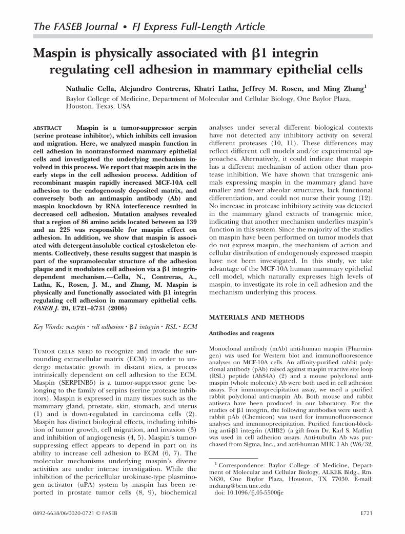
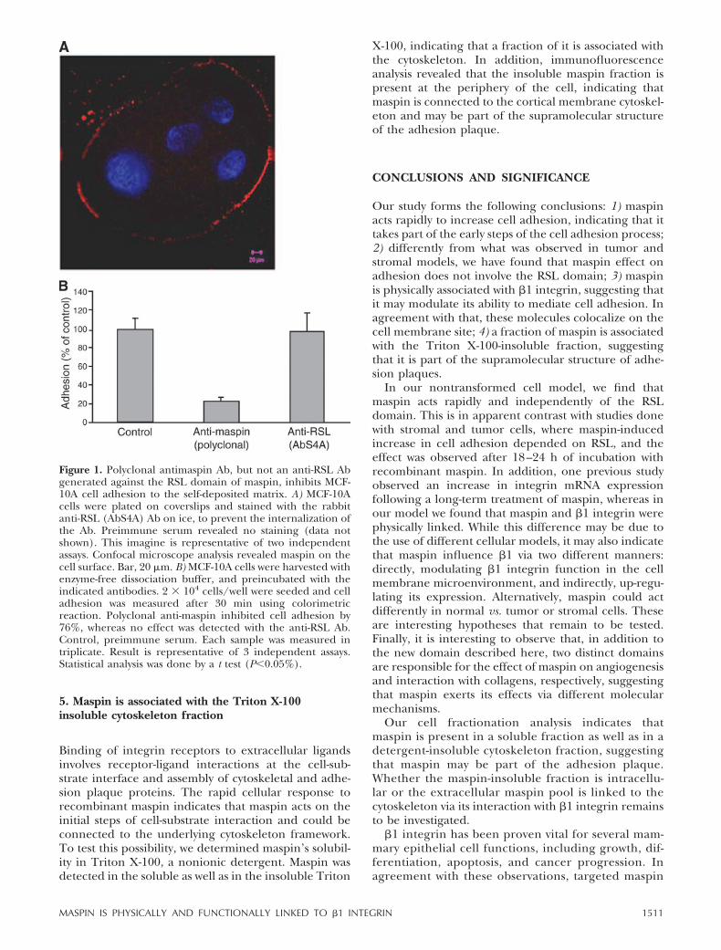
Figure 1. Polyclonal anti-maspin Ab, but not an anti-RSL Ab,generated against the RSL domain of maspin, inhibits MCF-10A cell adhesion to the self-deposited matrix. A) MCF-10Acells were plated on coverslips and stained with the rabbitanti-RSL (AbS4A) Ab on ice, to prevent the internalization ofthe Ab. Preimmune serum revealed no staining (data notshown). This imagine is representative of two independentassays. Confocal microscope analysis revealed maspin on thecell surface. Bar, 20 �m. B) MCF-10A cells were harvested withenzyme-free dissociation buffer, and preincubated with theindicated antibodies. 2 � 104 cells/well were seeded and celladhesion was measured after 30 min using colorimetricreaction as described in Materials and Methods. Polyclonalanti-maspin inhibited cell adhesion by 76%, whereas no effectwas detected with the anti-RSL Ab. Control, preimmuneserum. Each sample was measured in triplicate. Result isrepresentative of three independent assays. Statistical analysiswas done by a t test (P�0.05%).
E723MASPIN IS PHYSICALLY AND FUNCTIONALLY LINKED TO �1 INTEGRIN

present polarized cells with a hollow lumen and basalsecretion of basement membrane components (21).These cells express high endogenous levels ofmaspin, providing an excellent model for investigat-ing its function in cell adhesion. Since adhesionprimary involves the interaction between the cellmembrane and the substrate, we first investigated ifmaspin is present on the cell surface by immunofluo-rescence. To address this issue, cells grown on cover-slips were incubated with anti-maspin without any per-meabilization or fixation on ice, in order to prevent Abinternalization (see details in Material and Methods).Figure 1A showed maspin labeling along the perimeterof the cell, confirming that it is present on the externalside of the plasma membrane. Given that maspin isexpressed on the cell surface, we asked if incubation ofcells with anti-maspin could prevent cell adhesion to itsendogenously assembled matrix. To test this possibility,subconfluent MCF-10A cells were harvested with en-zyme-free dissociation buffer, preincubated with poly-clonal anti-maspin Ab or preimmune Ab (control) inserum-free medium, and allowed to adhere to theself-deposited matrix for 30 min. We observed a 76%decrease in cell adhesion in the presence of a poly-clonal anti-maspin antiserum compared to the controlserum (Fig. 1B).
To gain further support for this finding, we em-ployed RNA interference to specifically down-regulatemaspin expression in these cells. Maspin siRNA 229 and455 have been previously described (13). MCF-10A cellswere transfected with these siRNA-expressing vectorsand selected, and maspin protein down-regulation wasconfirmed by Western blot analysis (Fig. 2A, upperpanel). Protein loading was controlled by reprobing themembrane with an anti-tubulin Ab (Fig. 2A, lowerpanel). Normalization of maspin bands with that oftubulin indicated that maspin down-regulation by RNAiwas �82% in both 229 and 455 siRNAs cells (Fig. 2B).siRNA-expressing cells exhibited significantly less adhe-sion compared with the control cells (Fig. 2C), furthersupporting maspin’s role in adhesion. Previously,Seftor et al. observed an increase in integrin geneexpression after 18–24 h of incubation with recombi-nant maspin (22), suggesting that maspin acts indi-rectly in the adhesion process. In our experiments,however, maspin effect was observed within 30 min. Tore-ensure that, in our model, maspin acts in a differentkinetics, we applied a similar approach, incubatingMCF-10A cells with recombinant GST-maspin for ashort period of time. For this purpose cells weretrypsinized, which renders the endogenous maspininactive (10, 23, 24). After harvesting, cells were washedand preincubated either with GST or increasing con-centrations of GST-maspin and adhesion assay wascarried out as described in Material and Methods. Weobserved a dose-dependent increase in cell adhesion,which was up to 3-fold higher than control cells incu-bated with GST alone (Fig. 2D). We conclude thatexogenously added recombinant maspin can rapidlyincrease MCF-10A cell adhesion to its endogenous
matrix. Altogether, these results indicate that in theMCF-10A model maspin interferes with early steps inthe adhesion process and therefore appears to actdifferently from previous studies done in tumor andstromal cells.
Figure 2. Maspin suppression by siRNA decreases celladhesion and addition of recombinant maspin increasescell adhesion. A) MCF-10A cells were stably infected withcontrol vector (control) or with maspin siRNA 229 and 455vectors and maspin protein levels were analyzed by Westernblot (upper panel, 15 �g/lane). The membrane was subse-quently reprobed with an anti-tubulin Ab for loadingcontrol (lower panel). B) Bands were quantified by Image Janalysis and specific maspin down-regulation was plotted inarbitrary units. C) MCF-10A cells stably expressing emptyvector (control) or maspin siRNA 229 and 455 vectors wereplated on endogenous matrix (2�104 cells/well), and celladhesion was quantified as described in Materials andMethods. siRNA expressing cells adhered 46.7% (229) and53.6% (455) less than control cells. D) MCF-10A cells werepreincubated with GST (0.5 �M) or GST-maspin at theindicated concentrations for 30 min. 2 � 104 cells/wellwere plated on endogenous matrix, and cell adhesion wasquantified as described in Materials and Methods. Weobserved a dose-dependent increase in cell adhesion in thepresence of maspin. This result is representative of threeindependent assays. Statistical analysis was done by a t test(P�0.05%).
E724 Vol. 20 July 2006 CELLA ET AL.The FASEB Journal
Maspin promotes cell adhesion independently of itsRSL domain
The RSL is a conserved serpin domain, which is essen-tial for its antiprotease activity (25). The maspin RSLdomain possesses unique sequence and structure,which makes it unlikely to be involved in proteaseinhibition (11). However, it has been shown to beessential for maspin-mediated inhibition of cell inva-sion and migration (7, 23). To test whether maspin RSLdomain is also involved in its rapid action on celladhesion in normal mammary epithelial cells, MCF-10Acells were incubated with a purified pAb raised againstthe RSL domain (AbS4A) of maspin (2) and allowed toadhere to its endogenous matrix as described above.Surprisingly, the treatment with anti-RSL Ab had nodetectable effect on MCF-10A adhesion to the self-deposited matrix (Fig. 1B). This lack of effect was notdue to inaccessibility of the maspin RSL domain, sincethis Ab could detect maspin on the cell surface byimmunofluorescence (Fig. 1A). To confirm this obser-vation we used two independent approaches: 1) wecompared the effect of wild-type (WT) recombinantmaspin with that of mutant proteins bearing a pointmutation in the RSL loop (Mp*) and a C-terminaldeletion of the RSL domain (Mp-�RSL) (Fig. 3; and 2)WT maspin was preincubated with an anti-RSL Abbefore incubation with cells (23, 26). Under bothconditions, the ability of maspin to increase cell adhe-sion was assayed as described above. We observed thatrecombinant maspin carrying either a C-terminal dele-tion or point mutation in the RSL resulted in anincreased adhesion, comparable to WT maspin (Fig.3B). In addition, no difference in cell adhesion couldbe detected when maspin was preincubated with ananti-RSL Ab (Fig. 3C).
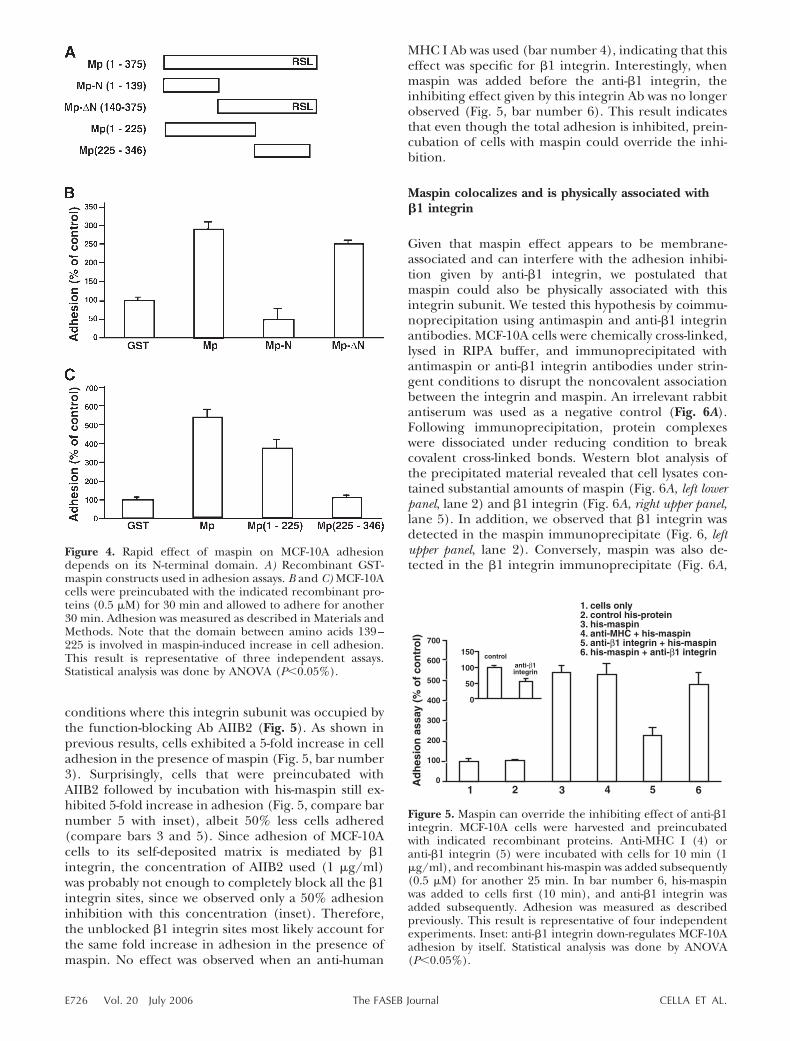
We next attempted to determine which portion ofmaspin is important for this rapid induction of adhe-sion. For this purpose we generated several maspinconstructs for the production of mutant maspin pro-teins as described in Materials and Methods. Initially,we used proteins containing only the first 139 amino-acids (Mp-N) and another fragment where this N-terminal portion had been deleted (Mp-�N) (5) (Fig.4). We observed that Mp-N failed to increase celladhesion, while the carboxyl half of the moleculeretained most of the effect observed with WT maspin(Fig. 4B). To further identify the functional domain, weconstructed two additional mutants, which containamino acids 1–225 and amino acids 225–346, respec-tively (Fig. 4A), and tested these proteins in cell adhe-sion assays. Mp(225–346) had no effect on MCF-10Aadhesion to the endogenous matrix (Fig. 4C). Mp(1–225), in contrast, retained a decreased, but significantability to sustain cell adhesion (Fig. 4C). All together,these experiments indicate that maspin RSL domain isnot involved in maspin-dependent increase in celladhesion. However, a portion of the 86 residues be-tween amino acid 139–225 appears to contain thedomain responsible for the rapid effect of maspin on
MCF-10A adhesion to the self-deposited matrix. Thesefindings further support the idea that maspin acts via adifferent mechanism in the MCF-10A model.
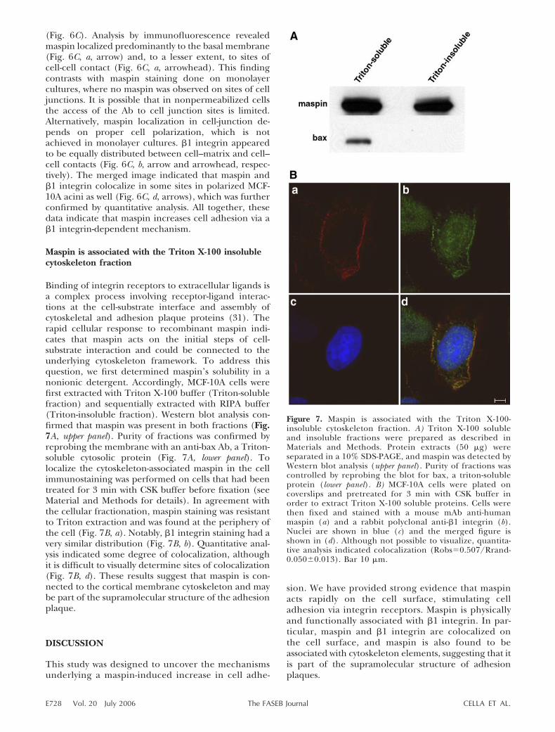
Maspin can override the inhibiting effectof anti-�1 integrin
Cells interact with the ECM proteins via transmem-brane proteins, of which the integrin superfamily of cellsurface receptors has been the most studied. MCF-10Amatrix is mainly composed of laminin-5 (19), to whichthese cells adhere predominantly via 3�1 integrin (27,28). Therefore, in order to investigate the involvementof �1 integrin in maspin-induced increase in adhesion,we examined the effect of maspin in adhesion in
Figure 3. Maspin RSL domain is not involved in maspin-induced rapid adhesion to MCF-10A endogenous matrix. A)Recombinant GST-maspin constructs used in adhesion assays:Mp(1–375 aa) – WT GST-maspin; Mp*(R3Q) – point muta-tion on the RSL domain (arginine to glutamine); Mp-�RSL(1–340 aa) – C-terminal deletion. B) MCF-10A cells werepreincubated with the indicated recombinant proteins (0.5�M) for 30 min. 2 � 104 cells/well were plated on endoge-nous matrix and adhesion was quantified as described inMaterials and Methods. C) Antibodies were preincubated withGST-maspin at the indicated dilutions for 30 min and sequen-tially incubated with cells for additional 30 min. Adhesionassays were performed as described. We observed a 2- to 3-foldincrease in cell adhesion in the presence of maspin, which didnot change significantly when cells were incubated withmutants of the RSL region or with anti-RSL Ab. Resultrepresents three independent assays. Statistical analysis wasdone by ANOVA (P�0.05%).
E725MASPIN IS PHYSICALLY AND FUNCTIONALLY LINKED TO �1 INTEGRIN
conditions where this integrin subunit was occupied bythe function-blocking Ab AIIB2 (Fig. 5). As shown inprevious results, cells exhibited a 5-fold increase in celladhesion in the presence of maspin (Fig. 5, bar number3). Surprisingly, cells that were preincubated withAIIB2 followed by incubation with his-maspin still ex-hibited 5-fold increase in adhesion (Fig. 5, compare barnumber 5 with inset), albeit 50% less cells adhered(compare bars 3 and 5). Since adhesion of MCF-10Acells to its self-deposited matrix is mediated by �1integrin, the concentration of AIIB2 used (1 �g/ml)was probably not enough to completely block all the �1integrin sites, since we observed only a 50% adhesioninhibition with this concentration (inset). Therefore,the unblocked �1 integrin sites most likely account forthe same fold increase in adhesion in the presence ofmaspin. No effect was observed when an anti-human
MHC I Ab was used (bar number 4), indicating that thiseffect was specific for �1 integrin. Interestingly, whenmaspin was added before the anti-�1 integrin, theinhibiting effect given by this integrin Ab was no longerobserved (Fig. 5, bar number 6). This result indicatesthat even though the total adhesion is inhibited, prein-cubation of cells with maspin could override the inhi-bition.
Maspin colocalizes and is physically associated with�1 integrin
Given that maspin effect appears to be membrane-associated and can interfere with the adhesion inhibi-tion given by anti-�1 integrin, we postulated thatmaspin could also be physically associated with thisintegrin subunit. We tested this hypothesis by coimmu-noprecipitation using antimaspin and anti-�1 integrinantibodies. MCF-10A cells were chemically cross-linked,lysed in RIPA buffer, and immunoprecipitated withantimaspin or anti-�1 integrin antibodies under strin-gent conditions to disrupt the noncovalent associationbetween the integrin and maspin. An irrelevant rabbitantiserum was used as a negative control (Fig. 6A).Following immunoprecipitation, protein complexeswere dissociated under reducing condition to breakcovalent cross-linked bonds. Western blot analysis ofthe precipitated material revealed that cell lysates con-tained substantial amounts of maspin (Fig. 6A, left lowerpanel, lane 2) and �1 integrin (Fig. 6A, right upper panel,lane 5). In addition, we observed that �1 integrin wasdetected in the maspin immunoprecipitate (Fig. 6, leftupper panel, lane 2). Conversely, maspin was also de-tected in the �1 integrin immunoprecipitate (Fig. 6A,
Figure 5. Maspin can override the inhibiting effect of anti-�1integrin. MCF-10A cells were harvested and preincubatedwith indicated recombinant proteins. Anti-MHC I (4) oranti-�1 integrin (5) were incubated with cells for 10 min (1�g/ml), and recombinant his-maspin was added subsequently(0.5 �M) for another 25 min. In bar number 6, his-maspinwas added to cells first (10 min), and anti-�1 integrin wasadded subsequently. Adhesion was measured as describedpreviously. This result is representative of four independentexperiments. Inset: anti-�1 integrin down-regulates MCF-10Aadhesion by itself. Statistical analysis was done by ANOVA(P�0.05%).
Figure 4. Rapid effect of maspin on MCF-10A adhesiondepends on its N-terminal domain. A) Recombinant GST-maspin constructs used in adhesion assays. B and C) MCF-10Acells were preincubated with the indicated recombinant pro-teins (0.5 �M) for 30 min and allowed to adhere for another30 min. Adhesion was measured as described in Materials andMethods. Note that the domain between amino acids 139–225 is involved in maspin-induced increase in cell adhesion.This result is representative of three independent assays.Statistical analysis was done by ANOVA (P�0.05%).
E726 Vol. 20 July 2006 CELLA ET AL.The FASEB Journal

right lower panel, lane 5), further supporting the associ-ation between maspin and �1 integrin. Although weobserved maspin on the cell surface (Fig. 1A), there isa possibility that maspin may associate with �1 integrinin the intracellular or intramembrane space. For thisreason, we chose to perform chemical cross-linkingwith DTSSP and DSS simultaneously, two cross-linkingreagents that are membrane-impermeable and -perme-able, respectively. DTSSP would cross-link maspin and�1 integrin if their interaction occurs extracellularly.DSS would cross-link them if their interaction occurs inthe intramembrane or intracellular space. With thisapproach we assured ourselves that maspin and �1integrin interaction would be cross-linked and de-tected. Notably, under reducing conditions maspin and�1 integrin migrated at their respective MW. No highMW complex was observed in Western blot analysis(data not shown), indicating that they were preferen-tially cross-linked by DTSSP, which can be cleaved byreducing agents, and not by DSS, which cannot bereduced and therefore can’t be cleaved. Since DTSSPcannot cross the plasma membrane, and thus it canonly cause cross-linking among cell surface proteins,these data together suggest that maspin associates with�1 integrin extracellularly.
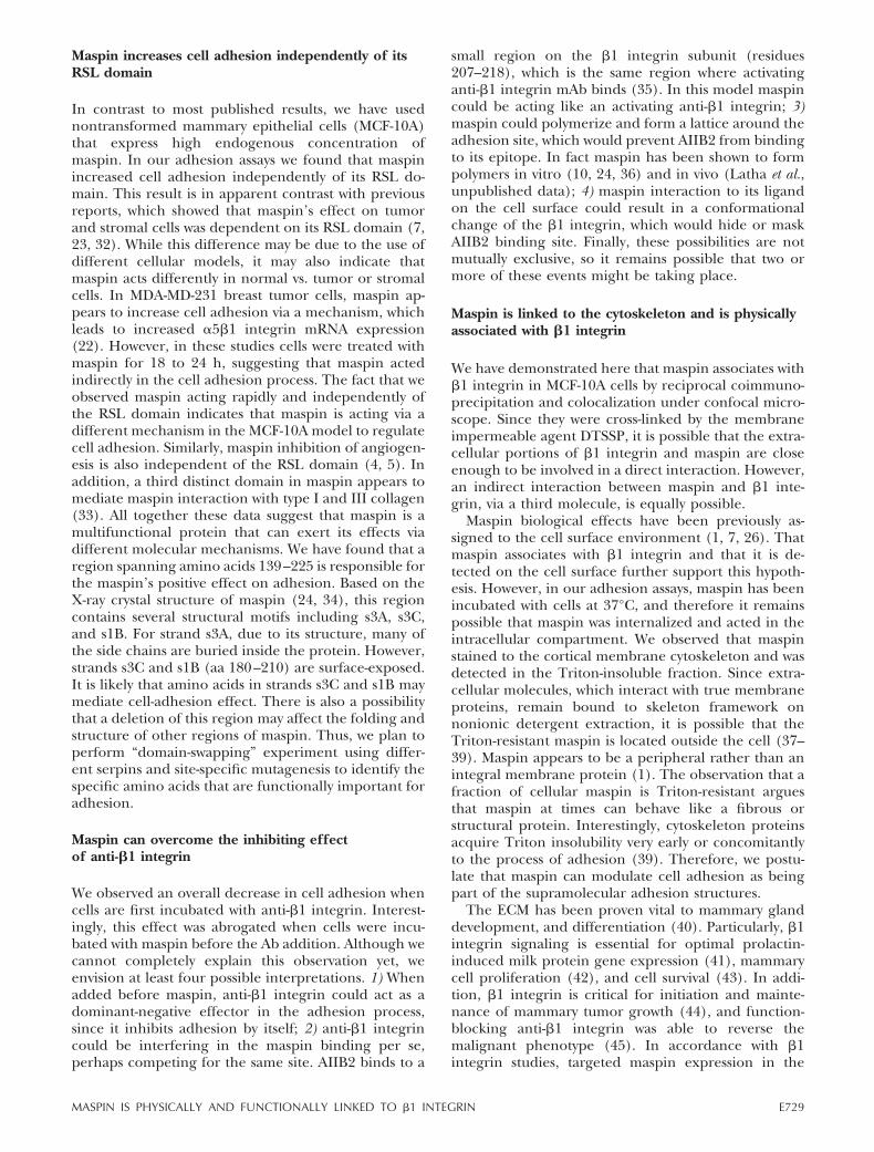
Since maspin interacts with �1 integrin, we reasonedthat these proteins would also localize in the same placein MCF-10A cells. To determine if there was colocaliza-tion, we carried out indirect double-immunofluores-cence to analyze the staining patterns of maspin and �1integrin by confocal microscopy (Fig. 6B). Cells were
first cultivated as monolayers on coverslips, fixed, per-meabilized, and stained with a mAb anti-maspin (Fig.6B, a) and a polyclonal anti-�1 integrin (Fig. 6B, b).Strong staining along the cell perimeter was detectedfor both maspin and �1 integrin (Fig. 6B, a and b,arrows). Importantly, visual inspection of the mergedimage showed some sites of their colocalization on themembrane (Fig. 6B, d, arrows), which was quantitativelyconfirmed by the Pearson’s correlation coefficient(17).
Epithelial cells grown in monolayers can displayseveral of their normal functions. They can proliferate,migrate, differentiate, and die. However, under theseconditions they cannot recapitulate the epithelial archi-tecture, which depends on the hallmark properties ofepithelial cells, i.e., their ability to polarize, formingapical and basolateral surfaces with vectorial depositionof ECM elements. Since �1 integrin expression isaltered in cells plated on plastic and proper signaltransduction depends on the spatial organization of thecell (29, 30), we were interested in confirming whethermaspin and �1 integrin colocalize in polarized epithe-lial cells in 3D structure compared to that which werecultivated in monolayers. To verify the colocalization ofmaspin and �1 integrin in 3D culture, we analyzedmaspin and �1 expression and distribution in MCF-10Aacini. MCF-10A cells were seeded in the Matrigel andallowed to develop an acinar-like structure for 15 d.Acinar structures were analyzed on frozen section byconfocal microscopy. MCF-10A cells organized to forma single polarized layer surrounding a hollow lumen
Figure 6. Maspin and �1 integrin coimmunoprecipi-tate and colocalize in MCF-10A cells grown in mono-layers and in 3D culture. A) MCF-10A cells werechemically cross-linked and lysed, and 500 �g ofprotein extracts were immunoprecipitated with theindicated antibodies. An irrelevant rabbit antiserumwas used as a negative control (lanes 3 and 6).Immunoprecipitates were separated in SDS-PAGEgels as detailed in Materials and Methods and ana-lyzed by Western blot as indicated. *Pre-�1 integrin. B)MCF-10A cells were plated on coverslips, fixed, per-meabilized, and stained with a mouse mAb anti-human maspin (a) and a rabbit polyclonal anti-�1integrin (b). Nucleus is shown in blue (c) and mergedimage is shown in (d). Arrows indicate that maspinand �1 integrin are located in the periphery of the cell(a and b, respectively) and are colocalized (d,Robs0.704/Rrand0.060�0.012); Bar 10 �m. C)MCF-10A cells were embedded in the Matrigel andallowed to form acini for 15 d. Cryosections (8 �m)were prepared and stained for maspin (a) and �1integrin (b), using the same antibodies mentionedabove. Nuclei are shown in blue (c) and mergedimage is shown in (d). Arrow and arrowhead in (a)and (b) indicate localization on the basal membraneand in sites of cell-cell contact, respectively. Arrowsin (d) indicate sites of maspin and �1 integrincolocalization (Robs0.580/Rrand0.115�0.019);Bar, 20 �m.
E727MASPIN IS PHYSICALLY AND FUNCTIONALLY LINKED TO �1 INTEGRIN
(Fig. 6C). Analysis by immunofluorescence revealedmaspin localized predominantly to the basal membrane(Fig. 6C, a, arrow) and, to a lesser extent, to sites ofcell-cell contact (Fig. 6C, a, arrowhead). This findingcontrasts with maspin staining done on monolayercultures, where no maspin was observed on sites of celljunctions. It is possible that in nonpermeabilized cellsthe access of the Ab to cell junction sites is limited.Alternatively, maspin localization in cell-junction de-pends on proper cell polarization, which is notachieved in monolayer cultures. �1 integrin appearedto be equally distributed between cell–matrix and cell–cell contacts (Fig. 6C, b, arrow and arrowhead, respec-tively). The merged image indicated that maspin and�1 integrin colocalize in some sites in polarized MCF-10A acini as well (Fig. 6C, d, arrows), which was furtherconfirmed by quantitative analysis. All together, thesedata indicate that maspin increases cell adhesion via a�1 integrin-dependent mechanism.
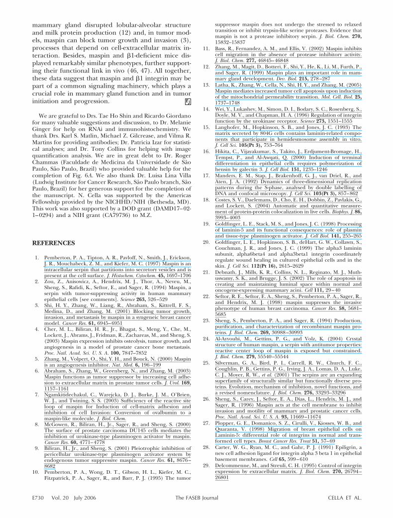
Maspin is associated with the Triton X-100 insolublecytoskeleton fraction
Binding of integrin receptors to extracellular ligands isa complex process involving receptor-ligand interac-tions at the cell-substrate interface and assembly ofcytoskeletal and adhesion plaque proteins (31). Therapid cellular response to recombinant maspin indi-cates that maspin acts on the initial steps of cell-substrate interaction and could be connected to theunderlying cytoskeleton framework. To address thisquestion, we first determined maspin’s solubility in anonionic detergent. Accordingly, MCF-10A cells werefirst extracted with Triton X-100 buffer (Triton-solublefraction) and sequentially extracted with RIPA buffer(Triton-insoluble fraction). Western blot analysis con-firmed that maspin was present in both fractions (Fig.7A, upper panel). Purity of fractions was confirmed byreprobing the membrane with an anti-bax Ab, a Triton-soluble cytosolic protein (Fig. 7A, lower panel). Tolocalize the cytoskeleton-associated maspin in the cellimmunostaining was performed on cells that had beentreated for 3 min with CSK buffer before fixation (seeMaterial and Methods for details). In agreement withthe cellular fractionation, maspin staining was resistantto Triton extraction and was found at the periphery ofthe cell (Fig. 7B, a). Notably, �1 integrin staining had avery similar distribution (Fig. 7B, b). Quantitative anal-ysis indicated some degree of colocalization, althoughit is difficult to visually determine sites of colocalization(Fig. 7B, d). These results suggest that maspin is con-nected to the cortical membrane cytoskeleton and maybe part of the supramolecular structure of the adhesionplaque.
DISCUSSION
This study was designed to uncover the mechanismsunderlying a maspin-induced increase in cell adhe-
sion. We have provided strong evidence that maspinacts rapidly on the cell surface, stimulating celladhesion via integrin receptors. Maspin is physicallyand functionally associated with �1 integrin. In par-ticular, maspin and �1 integrin are colocalized onthe cell surface, and maspin is also found to beassociated with cytoskeleton elements, suggesting that itis part of the supramolecular structure of adhesionplaques.
Figure 7. Maspin is associated with the Triton X-100-insoluble cytoskeleton fraction. A) Triton X-100 solubleand insoluble fractions were prepared as described inMaterials and Methods. Protein extracts (50 �g) wereseparated in a 10% SDS-PAGE, and maspin was detected byWestern blot analysis (upper panel). Purity of fractions wascontrolled by reprobing the blot for bax, a triton-solubleprotein (lower panel). B) MCF-10A cells were plated oncoverslips and pretreated for 3 min with CSK buffer inorder to extract Triton X-100 soluble proteins. Cells werethen fixed and stained with a mouse mAb anti-humanmaspin (a) and a rabbit polyclonal anti-�1 integrin (b).Nuclei are shown in blue (c) and the merged figure isshown in (d). Although not possible to visualize, quantita-tive analysis indicated colocalization (Robs0.507/Rrand-0.050�0.013). Bar 10 �m.
E728 Vol. 20 July 2006 CELLA ET AL.The FASEB Journal
Maspin increases cell adhesion independently of itsRSL domain
In contrast to most published results, we have usednontransformed mammary epithelial cells (MCF-10A)that express high endogenous concentration ofmaspin. In our adhesion assays we found that maspinincreased cell adhesion independently of its RSL do-main. This result is in apparent contrast with previousreports, which showed that maspin’s effect on tumorand stromal cells was dependent on its RSL domain (7,23, 32). While this difference may be due to the use ofdifferent cellular models, it may also indicate thatmaspin acts differently in normal vs. tumor or stromalcells. In MDA-MD-231 breast tumor cells, maspin ap-pears to increase cell adhesion via a mechanism, whichleads to increased 5�1 integrin mRNA expression(22). However, in these studies cells were treated withmaspin for 18 to 24 h, suggesting that maspin actedindirectly in the cell adhesion process. The fact that weobserved maspin acting rapidly and independently ofthe RSL domain indicates that maspin is acting via adifferent mechanism in the MCF-10A model to regulatecell adhesion. Similarly, maspin inhibition of angiogen-esis is also independent of the RSL domain (4, 5). Inaddition, a third distinct domain in maspin appears tomediate maspin interaction with type I and III collagen(33). All together these data suggest that maspin is amultifunctional protein that can exert its effects viadifferent molecular mechanisms. We have found that aregion spanning amino acids 139–225 is responsible forthe maspin’s positive effect on adhesion. Based on theX-ray crystal structure of maspin (24, 34), this regioncontains several structural motifs including s3A, s3C,and s1B. For strand s3A, due to its structure, many ofthe side chains are buried inside the protein. However,strands s3C and s1B (aa 180–210) are surface-exposed.It is likely that amino acids in strands s3C and s1B maymediate cell-adhesion effect. There is also a possibilitythat a deletion of this region may affect the folding andstructure of other regions of maspin. Thus, we plan toperform “domain-swapping” experiment using differ-ent serpins and site-specific mutagenesis to identify thespecific amino acids that are functionally important foradhesion.
Maspin can overcome the inhibiting effectof anti-�1 integrin
We observed an overall decrease in cell adhesion whencells are first incubated with anti-�1 integrin. Interest-ingly, this effect was abrogated when cells were incu-bated with maspin before the Ab addition. Although wecannot completely explain this observation yet, weenvision at least four possible interpretations. 1) Whenadded before maspin, anti-�1 integrin could act as adominant-negative effector in the adhesion process,since it inhibits adhesion by itself; 2) anti-�1 integrincould be interfering in the maspin binding per se,perhaps competing for the same site. AIIB2 binds to a
small region on the �1 integrin subunit (residues207–218), which is the same region where activatinganti-�1 integrin mAb binds (35). In this model maspincould be acting like an activating anti-�1 integrin; 3)maspin could polymerize and form a lattice around theadhesion site, which would prevent AIIB2 from bindingto its epitope. In fact maspin has been shown to formpolymers in vitro (10, 24, 36) and in vivo (Latha et al.,unpublished data); 4) maspin interaction to its ligandon the cell surface could result in a conformationalchange of the �1 integrin, which would hide or maskAIIB2 binding site. Finally, these possibilities are notmutually exclusive, so it remains possible that two ormore of these events might be taking place.
Maspin is linked to the cytoskeleton and is physicallyassociated with �1 integrin
We have demonstrated here that maspin associates with�1 integrin in MCF-10A cells by reciprocal coimmuno-precipitation and colocalization under confocal micro-scope. Since they were cross-linked by the membraneimpermeable agent DTSSP, it is possible that the extra-cellular portions of �1 integrin and maspin are closeenough to be involved in a direct interaction. However,an indirect interaction between maspin and �1 inte-grin, via a third molecule, is equally possible.
Maspin biological effects have been previously as-signed to the cell surface environment (1, 7, 26). Thatmaspin associates with �1 integrin and that it is de-tected on the cell surface further support this hypoth-esis. However, in our adhesion assays, maspin has beenincubated with cells at 37°C, and therefore it remainspossible that maspin was internalized and acted in theintracellular compartment. We observed that maspinstained to the cortical membrane cytoskeleton and wasdetected in the Triton-insoluble fraction. Since extra-cellular molecules, which interact with true membraneproteins, remain bound to skeleton framework onnonionic detergent extraction, it is possible that theTriton-resistant maspin is located outside the cell (37–39). Maspin appears to be a peripheral rather than anintegral membrane protein (1). The observation that afraction of cellular maspin is Triton-resistant arguesthat maspin at times can behave like a fibrous orstructural protein. Interestingly, cytoskeleton proteinsacquire Triton insolubility very early or concomitantlyto the process of adhesion (39). Therefore, we postu-late that maspin can modulate cell adhesion as beingpart of the supramolecular adhesion structures.
The ECM has been proven vital to mammary glanddevelopment, and differentiation (40). Particularly, �1integrin signaling is essential for optimal prolactin-induced milk protein gene expression (41), mammarycell proliferation (42), and cell survival (43). In addi-tion, �1 integrin is critical for initiation and mainte-nance of mammary tumor growth (44), and function-blocking anti-�1 integrin was able to reverse themalignant phenotype (45). In accordance with �1integrin studies, targeted maspin expression in the
E729MASPIN IS PHYSICALLY AND FUNCTIONALLY LINKED TO �1 INTEGRIN
mammary gland disrupted lobular-alveolar structureand milk protein production (12) and, in tumor mod-els, maspin can block tumor growth and invasion (3),processes that depend on cell-extracellular matrix in-teraction. Besides, maspin and �1-deficient mice dis-played remarkably similar phenotypes, further support-ing their functional link in vivo (46, 47). All together,these data suggest that maspin and �1 integrin may bepart of a common signaling machinery, which plays acrucial role in mammary gland function and in tumorinitiation and progression.
We are grateful to Drs. Tae Ho Shin and Ricardo Giordanofor many valuable suggestions and discussion, to Dr. MelanieGinger for help on RNAi and immunohistochemistry. Wethank Drs. Karl S. Matlin, Michael Z. Gilcrease, and Vilma R.Martins for providing antibodies; Dr. Patricia Izar for statisti-cal analyses; and Dr. Tony Collins for helping with imagequantification analysis. We are in great debt to Dr. RogerChammas (Faculdade de Medicina da Universidade de SaoPaulo, Sao Paulo, Brazil) who provided valuable help for thecompletion of Fig. 6A. We also thank Dr. Luisa Lina Villa(Ludwig Institute for Cancer Research, Sao Paulo branch, SaoPaulo, Brazil) for her generous support for the completion ofthe manuscript. N. Cella was supported by the AmericasFellowship provided by the NICHHD/NIH (Bethesda, MD).This work was also supported by a DOD grant (DAMD17–02-1–0294) and a NIH grant (CA79736) to M.Z.
REFERENCES
1. Pemberton, P. A., Tipton, A. R., Pavloff, N., Smith, J., Erickson,J. R., Mouchabeck, Z. M., and Kiefer, M. C. (1997) Maspin is anintracellular serpin that partitions into secretory vesicles and ispresent at the cell surface. J. Histochem. Cytochem. 45, 1697–1706
2. Zou, Z., Anisowicz, A., Hendrix, M. J., Thor, A., Neveu, M.,Sheng, S., Rafidi, K., Seftor, E., and Sager, R. (1994) Maspin, aserpin with tumor-suppressing activity in human mammaryepithelial cells [see comments]. Science 263, 526–529
3. Shi, H. Y., Zhang, W., Liang, R., Abraham, S., Kittrell, F. S.,Medina, D., and Zhang, M. (2001) Blocking tumor growth,invasion, and metastasis by maspin in a syngeneic breast cancermodel. Cancer Res. 61, 6945–6951
4. Cher, M. L., Biliran, H. R., Jr., Bhagat, S., Meng, Y., Che, M.,Lockett, J., Abrams, J., Fridman, R., Zachareas, M., and Sheng, S.(2003) Maspin expression inhibits osteolysis, tumor growth, andangiogenesis in a model of prostate cancer bone metastasis.Proc. Natl. Acad. Sci. U. S. A. 100, 7847–7852
5. Zhang, M., Volpert, O., Shi, Y. H., and Bouck, N. (2000) Maspinis an angiogenesis inhibitor. Nat. Med. 6, 196–199
6. Abraham, S., Zhang, W., Greenberg, N., and Zhang, M. (2003)Maspin functions as tumor suppressor by increasing cell adhe-sion to extracellular matrix in prostate tumor cells. J. Urol. 169,1157–1161
7. Ngamkitidechakul, C., Warejcka, D. J., Burke, J. M., O’Brien,W. J., and Twining, S. S. (2003) Sufficiency of the reactive siteloop of maspin for Induction of cell-matrix adhesion andinhibition of cell Invasion: Conversion of ovalbumin to amaspin-like molecule. J. Biol. Chem.
8. McGowen, R., Biliran, H., Jr., Sager, R., and Sheng, S. (2000)The surface of prostate carcinoma DU145 cells mediates theinhibition of urokinase-type plasminogen activator by maspin.Cancer Res. 60, 4771–4778
9. Biliran, H., Jr., and Sheng, S. (2001) Pleiotrophic inhibition ofpericellular urokinase-type plasminogen activator system byendogenous tumor suppressive maspin. Cancer Res. 61, 8676–8682
10. Pemberton, P. A., Wong, D. T., Gibson, H. L., Kiefer, M. C.,Fitzpatrick, P. A., Sager, R., and Barr, P. J. (1995) The tumor
suppressor maspin does not undergo the stressed to relaxedtransition or inhibit trypsin-like serine proteases. Evidence thatmaspin is not a protease inhibitory serpin. J. Biol. Chem. 270,15832–15837
11. Bass, R., Fernandez, A. M., and Ellis, V. (2002) Maspin inhibitscell migration in the absence of protease inhibitory activity.J. Biol. Chem. 277, 46845–46848
12. Zhang, M., Magit, D., Botteri, F., Shi, Y., He, K., Li, M., Furth, P.,and Sager, R. (1999) Maspin plays an important role in mam-mary gland development. Dev. Biol. 215, 278–287
13. Latha, K., Zhang, W., Cella, N., Shi, H. Y., and Zhang, M. (2005)Maspin mediates increased tumor cell apoptosis upon inductionof the mitochondrial permeability transition. Mol. Cell. Biol. 25,1737–1748
14. Wei, Y., Lukashev, M., Simon, D. I., Bodary, S. C., Rosenberg, S.,Doyle, M. V., and Chapman, H. A. (1996) Regulation of integrinfunction by the urokinase receptor. Science 273, 1551–1555
15. Langhofer, M., Hopkinson, S. B., and Jones, J. C. (1993) Thematrix secreted by 804G cells contains laminin-related compo-nents that participate in hemidesmosome assembly in vitro.J. Cell Sci. 105(Pt 3), 753–764
16. Hikita, C., Vijayakumar, S., Takito, J., Erdjument-Bromage, H.,Tempst, P., and Al-Awqati, Q. (2000) Induction of terminaldifferentiation in epithelial cells requires polymerization ofhensin by galectin 3. J. Cell Biol. 151, 1235–1246
17. Manders, E. M., Stap, J., Brakenhoff, G. J., van Driel, R., andAten, J. A. (1992) Dynamics of three-dimensional replicationpatterns during the S-phase, analysed by double labelling ofDNA and confocal microscopy. J. Cell Sci. 103(Pt 3), 857–862
18. Costes, S. V., Daelemans, D., Cho, E. H., Dobbin, Z., Pavlakis, G.,and Lockett, S. (2004) Automatic and quantitative measure-ment of protein-protein colocalization in live cells. Biophys. J. 86,3993–4003
19. Goldfinger, L. E., Stack, M. S., and Jones, J. C. (1998) Processingof laminin-5 and its functional consequences: role of plasminand tissue-type plasminogen activator. J. Cell Biol. 141, 255–265
20. Goldfinger, L. E., Hopkinson, S. B., deHart, G. W., Collawn, S.,Couchman, J. R., and Jones, J. C. (1999) The alpha3 lamininsubunit, alpha6beta4 and alpha3beta1 integrin coordinatelyregulate wound healing in cultured epithelial cells and in theskin. J. Cell Sci. 112(Pt 16), 2615–2629
21. Debnath, J., Mills, K. R., Collins, N. L., Reginato, M. J., Muth-uswamy, S. K., and Brugge, J. S. (2002) The role of apoptosis increating and maintaining luminal space within normal andoncogene-expressing mammary acini. Cell 111, 29–40
22. Seftor, R. E., Seftor, E. A., Sheng, S., Pemberton, P. A., Sager, R.,and Hendrix, M. J. (1998) maspin suppresses the invasivephenotype of human breast carcinoma. Cancer Res. 58, 5681–5685
23. Sheng, S., Pemberton, P. A., and Sager, R. (1994) Production,purification, and characterization of recombinant maspin pro-teins. J. Biol. Chem. 269, 30988–30993
24. Al-Ayyoubi, M., Gettins, P. G., and Volz, K. (2004) Crystalstructure of human maspin, a serpin with antitumor properties:reactive center loop of maspin is exposed but constrained.J. Biol. Chem. 279, 55540–55544
25. Silverman, G. A., Bird, P. I., Carrell, R. W., Church, F. C.,Coughlin, P. B., Gettins, P. G., Irving, J. A., Lomas, D. A., Luke,C. J., Moyer, R. W., et al. (2001) The serpins are an expandingsuperfamily of structurally similar but functionally diverse pro-teins. Evolution, mechanism of inhibition, novel functions, anda revised nomenclature. J. Biol. Chem. 276, 33293–33296
26. Sheng, S., Carey, J., Seftor, E. A., Dias, L., Hendrix, M. J., andSager, R. (1996) Maspin acts at the cell membrane to inhibitinvasion and motility of mammary and prostatic cancer cells.Proc. Natl. Acad. Sci. U. S. A. 93, 11669–11674
27. Plopper, G. E., Domanico, S. Z., Cirulli, V., Kiosses, W. B., andQuaranta, V. (1998) Migration of breast epithelial cells onLaminin-5: differential role of integrins in normal and trans-formed cell types. Breast Cancer Res. Treat 51, 57–69
28. Carter, W. G., Ryan, M. C., and Gahr, P. J. (1991) Epiligrin, anew cell adhesion ligand for integrin alpha 3 beta 1 in epithelialbasement membranes. Cell 65, 599–610
29. Delcommenne, M., and Streuli, C. H. (1995) Control of integrinexpression by extracellular matrix. J. Biol. Chem. 270, 26794–26801
E730 Vol. 20 July 2006 CELLA ET AL.The FASEB Journal
30. Wang, F., Weaver, V. M., Petersen, O. W., Larabell, C. A.,Dedhar, S., Briand, P., Lupu, R., and Bissell, M. J. (1998)Reciprocal interactions between beta1-integrin and epidermalgrowth factor receptor in three-dimensional basement mem-brane breast cultures: a different perspective in epithelial biol-ogy. Proc. Natl. Acad. Sci. U. S. A. 95, 14821–14826
31. Geiger, B., Bershadsky, A., Pankov, R., and Yamada, K. M. (2001)Transmembrane crosstalk between the extracellular matrix–cytoskeleton crosstalk. Nat. Rev. Mol. Cell. Biol. 2, 793–805
32. Ngamkitidechakul, C., Burke, J. M., O’Brien, W. J., and Twining,S. S. (2001) Maspin: synthesis by human cornea and regulationof in vitro stromal cell adhesion to extracellular matrix. Invest.Ophthalmol. Vis. Sci. 42, 3135–3141
33. Blacque, O. E., and Worrall, D. M. (2002) Evidence for a directinteraction between the tumour suppressor serpin maspin, andtypes I and III collagen. J. Biol. Chem.
34. Law, R. H., Irving, J. A., Buckle, A. M., Ruzyla, K., Buzza, M.,Bashtannyk-Puhalovich, T. A., Beddoe, T. C., Nguyen, K., Wor-rall, D. M., Bottomley, S. P., et al. (2005) The high resolutioncrystal structure of the human tumor suppressor maspin revealsa novel conformational switch in the G-helix. J. Biol. Chem. 280,22356–22364
35. Takada, Y., and Puzon, W. (1993) Identification of a regulatoryregion of integrin beta 1 subunit using activating and inhibitingantibodies. J. Biol. Chem. 268, 17597–17601
36. Liu, T., Pemberton, P. A., and Robertson, A. D. (1999) Three-state unfolding and self-association of maspin, a tumor-suppress-ing serpin. J. Biol. Chem. 274, 29628–29632
37. Ben-Ze’ev, A., Duerr, A., Solomon, F., and Penman, S. (1979)The outer boundary of the cytoskeleton: a lamina derived fromplasma membrane proteins. Cell 17, 859–865
38. Mautner, V., and Hynes, R. O. (1977) Surface distribution ofLETS protein in relation to the cytoskeleton of normal andtransformed cells. J. Cell Biol. 75, 743–768
39. Gonen, A., Weisman-Shomer, P., and Fry, M. (1979) Celladhesion and acquisition of detergent resistance by the cytoskel-eton of cultured chick fibroblasts. Biochim. Biophys. Acta 552,307–321
40. Bissell, M. J., and Bilder, D. (2003) Polarity determination inbreast tissue: desmosomal adhesion, myoepithelial cells, andlaminin 1. Breast Cancer Res. 5, 117–119
41. Streuli, C. H., Bailey, N., and Bissell, M. J. (1991) Control ofmammary epithelial differentiation: basement membrane in-duces tissue-specific gene expression in the absence of cell-cellinteraction and morphological polarity. J. Cell Biol. 115, 1383–1395
42. Li, N., Zhang, Y., Naylor, M. J., Schatzmann, F., Maurer, F.,Wintermantel, T., Schuetz, G., Mueller, U., Streuli, C. H., andHynes, N. E. (2005) Beta1 integrins regulate mammary glandproliferation and maintain the integrity of mammary alveoli.EMBO J. 24, 1942–1953
43. Farrelly, N., Lee, Y. J., Oliver, J., Dive, C., and Streuli, C. H.(1999) Extracellular matrix regulates apoptosis in mammaryepithelium through a control on insulin signaling. J. Cell Biol.144, 1337–1348
44. White, D. E., Kurpios, N. A., Zuo, D., Hassell, J. A., Blaess, S.,Mueller, U., and Muller, W. J. (2004) Targeted disruption ofbeta1-integrin in a transgenic mouse model of human breastcancer reveals an essential role in mammary tumor induction.Cancer Cell 6, 159–170
45. Weaver, V. M., Petersen, O. W., Wang, F., Larabell, C. A.,Briand, P., Damsky, C., and Bissell, M. J. (1997) Reversion of themalignant phenotype of human breast cells in three-dimen-sional culture and in vivo by integrin blocking antibodies. J. CellBiol. 137, 231–245
46. Stephens, L. E., Sutherland, A. E., Klimanskaya, I. V., An-drieux, A., Meneses, J., Pedersen, R. A., and Damsky, C. H.(1995) Deletion of beta 1 integrins in mice results in innercell mass failure and peri-implantation lethality. Genes Dev. 9,1883–1895
47. Gao, F., Shi, H. Y., Daughty, C., Cella, N., and Zhang, M. (2004)Maspin plays an essential role in early embryonic development.Development 131, 1479–1489
Received for publication November 23, 2005.Accepted for publication February 16, 2006.
E731MASPIN IS PHYSICALLY AND FUNCTIONALLY LINKED TO �1 INTEGRIN
The FASEB Journal • FJ Express Summary
Maspin is physically associated with �1 integrinregulating cell adhesion in mammary epithelial cells
Nathalie Cella, Alejandro Contreras, Khatri Latha, Jeffrey M. Rosen, and Ming Zhang1
Baylor College of Medicine, Department of Molecular and Cellular Biology, One Baylor Plaza,Houston, Texas, USA
To read the full text of this article, go to http://www.fasebj.org/cgi/doi/10.1096/fj.05-5500fje
SPECIFIC AIMS
The purposes of our study are to investigate the role ofmaspin in cell adhesion and to identify the moleculardomain involved in this process and the underlyingmechanism.
PRINCIPAL FINDINGS
1. Maspin rapidly modulates cell adhesion
In a different approach from most previous studies, wehave used a nontransformed mammary epithelial cellline (MCF-10A cells), which expresses high concentra-tion of maspin. Since adhesion primary involves theinteraction between the cell membrane and the sub-strate, we first investigated whether maspin was presenton the cell surface by immunofluorescence. Usingnonpermeabilized cells to prevent internalization ofthe antibody (Ab), we observed maspin staining at theperiphery of the cell and confirmed its expression onthe external side of the membrane. We next analyzed apossible role of endogenous maspin in cell adhesion aswell as exogenous addition of recombinant maspin bycell adhesion assays. We found that incubation of cellswith antimaspin or maspin down-regulation by RNAidecreased cell adhesion. Accordingly, incubation ofcells with recombinant maspin increased cell adhesion.These effects were observed within 30 min, suggestingthat maspin acts in the early steps of the cell adhesionprocess.
2. Mutation analyses reveal a new domain involved inmaspin effect on cell adhesion
Previous studies done in tumor cell models indicatethat the maspin RSL domain (reactive site loop) isessential for its positive effect on adhesion. To deter-mine the domain of maspin, we tested the effect ofvarious maspin mutants on cell adhesion. Differentlyfrom the studies in mammary tumors, we found that adomain comprising amino acid 139 to 225, and not theRSL domain, retained a positive effect on cell adhesion.
3. Maspin is physically associated with �1 integrin
Cell adhesion to extracellular matrix is mediated byintegrins to a great deal. In our model system, we usedthe endogenous matrix deposited by MCF-10A cells,which contains predominantly laminin-5. Therefore,we hypothesized that maspin could be modulating celladhesion via a �1 integrin, a major laminin-5 receptorin this cell line. Using total protein extracts, we ob-served that �1 integrin coimmunoprecipitated withmaspin. Reciprocally, maspin was also coimmunopre-cipitated with �1 integrin.
MCF-10A adhesion to its endogenous matrix couldbe inhibited 50% with a function-blocking anti-�1integrin. Interestingly, this effect was abrogated whencells were incubated with maspin before addition of theAb, indicating that maspin could override the inhibi-tion given by the anti-�1 integrin.
4. Maspin colocalizes with �1 integrin
To further confirm and extend our findings, we per-formed indirect double-immunofluorescence to ana-lyze the staining pattern of maspin and �1 integrin byconfocal microscopy. We observed a significant colocal-ization of these two molecules, predominantly at thecell membrane site, which was further confirmed byquantitative analysis. Since both integrin expressionand signaling are altered in cells grown on flat surfaces,we intend to confirm this result in cells cultivated inmatrigel, in which cells can develop into three-dimen-sional acinar structures which recapitulate typical fea-tures of the epithelial architecture. Similarly to cellsgrown on flat surfaces, maspin and �1 integrin alsocolocalized in three-dimensional culture, which wasobserved for both basement membrane and cell-cellcontact sites.
1 Correspondence: Baylor College of Medicine, Depart-ment of Molecular and Cellular Biology, ALKEK Bldg., Rm.N630, One Baylor Plaza, Houston, TX 77030, USA. E-mail:[email protected]
doi: 10.1096/fj.05-5500fje
1510 0892-6638/06/0020-1510 © FASEB
5. Maspin is associated with the Triton X-100insoluble cytoskeleton fraction
Binding of integrin receptors to extracellular ligandsinvolves receptor-ligand interactions at the cell-sub-strate interface and assembly of cytoskeletal and adhe-sion plaque proteins. The rapid cellular response torecombinant maspin indicates that maspin acts on theinitial steps of cell-substrate interaction and could beconnected to the underlying cytoskeleton framework.To test this possibility, we determined maspin’s solubil-ity in Triton X-100, a nonionic detergent. Maspin wasdetected in the soluble as well as in the insoluble Triton
X-100, indicating that a fraction of it is associated withthe cytoskeleton. In addition, immunofluorescenceanalysis revealed that the insoluble maspin fraction ispresent at the periphery of the cell, indicating thatmaspin is connected to the cortical membrane cytoskel-eton and may be part of the supramolecular structureof the adhesion plaque.
CONCLUSIONS AND SIGNIFICANCE
Our study forms the following conclusions: 1) maspinacts rapidly to increase cell adhesion, indicating that ittakes part of the early steps of the cell adhesion process;2) differently from what was observed in tumor andstromal models, we have found that maspin effect onadhesion does not involve the RSL domain; 3) maspinis physically associated with �1 integrin, suggesting thatit may modulate its ability to mediate cell adhesion. Inagreement with that, these molecules colocalize on thecell membrane site; 4) a fraction of maspin is associatedwith the Triton X-100-insoluble fraction, suggestingthat it is part of the supramolecular structure of adhe-sion plaques.
In our nontransformed cell model, we find thatmaspin acts rapidly and independently of the RSLdomain. This is in apparent contrast with studies donewith stromal and tumor cells, where maspin-inducedincrease in cell adhesion depended on RSL, and theeffect was observed after 18–24 h of incubation withrecombinant maspin. In addition, one previous studyobserved an increase in integrin mRNA expressionfollowing a long-term treatment of maspin, whereas inour model we found that maspin and �1 integrin werephysically linked. While this difference may be due tothe use of different cellular models, it may also indicatethat maspin influence �1 via two different manners:directly, modulating �1 integrin function in the cellmembrane microenvironment, and indirectly, up-regu-lating its expression. Alternatively, maspin could actdifferently in normal vs. tumor or stromal cells. Theseare interesting hypotheses that remain to be tested.Finally, it is interesting to observe that, in addition tothe new domain described here, two distinct domainsare responsible for the effect of maspin on angiogenesisand interaction with collagens, respectively, suggestingthat maspin exerts its effects via different molecularmechanisms.
Our cell fractionation analysis indicates thatmaspin is present in a soluble fraction as well as in adetergent-insoluble cytoskeleton fraction, suggestingthat maspin may be part of the adhesion plaque.Whether the maspin-insoluble fraction is intracellu-lar or the extracellular maspin pool is linked to thecytoskeleton via its interaction with �1 integrin remainsto be investigated.
�1 integrin has been proven vital for several mam-mary epithelial cell functions, including growth, dif-ferentiation, apoptosis, and cancer progression. Inagreement with these observations, targeted maspin
Figure 1. Polyclonal antimaspin Ab, but not an anti-RSL Abgenerated against the RSL domain of maspin, inhibits MCF-10A cell adhesion to the self-deposited matrix. A) MCF-10Acells were plated on coverslips and stained with the rabbitanti-RSL (AbS4A) Ab on ice, to prevent the internalization ofthe Ab. Preimmune serum revealed no staining (data notshown). This imagine is representative of two independentassays. Confocal microscope analysis revealed maspin on thecell surface. Bar, 20 �m. B) MCF-10A cells were harvested withenzyme-free dissociation buffer, and preincubated with theindicated antibodies. 2 � 104 cells/well were seeded and celladhesion was measured after 30 min using colorimetricreaction. Polyclonal anti-maspin inhibited cell adhesion by76%, whereas no effect was detected with the anti-RSL Ab.Control, preimmune serum. Each sample was measured intriplicate. Result is representative of 3 independent assays.Statistical analysis was done by a t test (P�0.05%).
1511MASPIN IS PHYSICALLY AND FUNCTIONALLY LINKED TO �1 INTEGRIN

expression in the mammary gland resulted in disruptedlobular-alveolar structure, and in tumor models maspincan block tumor growth and invasion.
In conclusion, our study provided evidence thatmaspin and �1 integrin may be part of a commonsignaling machinery, which plays a crucial role inmammary gland function and in tumor initiation andprogression.
Figure 3. Schematic diagram illustrates and summarizes ex-periments and main conclusions. A) Endogenous maspinmodulates MCF-10A cell adhesion, which involves its physicalinteraction with beta 1 integrin and with cytoskeleton. Addi-tion of anti-maspin down-regulates cell adhesion. B) MCF-10Acells in suspension were incubated with recombinant GST-maspin, which modulates beta1 integrin and cytoskeletonfunction, leading to an increase in cell adhesion to extracel-lular matrix. This activity is independent of maspin RSLdomain (not depicted).
Figure 2. Maspin and �1 integrin coimmunopre-cipitate and colocalize in MCF-10A cells grown inmonolayers and in 3D culture. A) MCF-10A cellswere chemically cross-linked, lysed, and 500 �g ofprotein extracts were immunoprecipitated with theindicated antibodies. An irrelevant rabbit antiserumwas used as a negative control (lanes 3 and 6).Immunoprecipitates were separated in SDS-PAGEgels as detailed in Material and Methods and analyzedby Western blot as indicated. *Pre-�1 integrin. B)MCF-10A cells were plated on coverslips, fixed, per-meabilized, and stained with a mouse monoclonalantibody anti-human maspin (a) and a rabbit poly-clonal anti-�1 integrin (b). Nucleus is shown in blue(c) and merged image is shown in (d). Arrows indicatethat maspin and �1 integrin are located in the periph-ery of the cell (a and b, respectively) and are colocal-ized (d, Robs�0.704/Rrand�0.060�0.012); Bar 10�m. C) MCF-10A cells were embedded in the Matrigeland allowed to form acini for 15 d. Cryosections (8�m) were prepared and stained for maspin (a) and�1 integrin (b), using the same antibodies mentionedabove. Nuclei are shown in blue (c) and mergedimage is shown in (d). Arrow and arrowhead in (a)and (b) indicate localization on the basal membraneand in sites of cell–cell contact, respectively. Arrowsin (d) indicate sites of maspin and �1 integrincolocalization (Robs�0.580/Rrand�0.115�0.019);Bar 20 �m.
1512 Vol. 20 July 2006 CELLA ET AL.The FASEB Journal