Clinical, pathological and antigenic aspects of bovine viral diarrhea virus (BVDV) type 2 isolates...
11
Veterinary and Biomedical Sciences, Department of Papers in Veterinary and Biomedical Science University of Nebraska - Lincoln Year Clinical, pathological and antigenic aspects of bovine viral diarrhea virus (BVDV) type 2 isolates identified in Brazil E. F. Flores, Universidade Federal de Santa Maria, 97105-900 Santa Maria, RS, Brazil L. H. G. V. Gil, Universidade Federal de Santa Maria, 97105-900 Santa Maria, RS, Brazil S. A. Botton, Universidade Federal de Santa Maria, 97105-900 Santa Maria, RS, Brazil R. Weiblen, Universidade Federal de Santa Maria, 97105-900 Santa Maria, RS, Brazil J. F. Ridpath, Metabolic Diseases and Immunology Research Unit, NADC, ARS/USDA, Ames, IA L. C. Kreutz, Universidade Federal de Santa Maria, 97105-900 Santa Maria, RS, Brazil C. Pilati, Centro Agroveterina ˆ Ario de Lages, Universidade de Santa Catarina (UDESC), Lages, SC, Brazil D. Driemeyer, Universidade Federal do Rio Grande do Sul, UFRGS, Porto Alegre, RS, Brazil V. Moojen, Universidade Federal do Rio Grande do Sul, UFRGS, Porto Alegre, RS, Brazil A. C. Wendelstein, Universidade Federal do Rio Grande do Sul, UFRGS, Porto Alegre, RS, Brazil This paper is posted at DigitalCommons@University of Nebraska - Lincoln.
-
Upload
independent -
Category
Documents
-
view
1 -
download
0
Transcript of Clinical, pathological and antigenic aspects of bovine viral diarrhea virus (BVDV) type 2 isolates...
Veterinary and Biomedical Sciences, Department of
Papers in Veterinary and Biomedical Science
University of Nebraska - Lincoln Year
Clinical, pathological and antigenic
aspects of bovine viral diarrhea virus
(BVDV) type 2 isolates identified in
BrazilE. F. Flores, Universidade Federal de Santa Maria, 97105-900 SantaMaria, RS, BrazilL. H. G. V. Gil, Universidade Federal de Santa Maria, 97105-900Santa Maria, RS, BrazilS. A. Botton, Universidade Federal de Santa Maria, 97105-900 SantaMaria, RS, BrazilR. Weiblen, Universidade Federal de Santa Maria, 97105-900 SantaMaria, RS, BrazilJ. F. Ridpath, Metabolic Diseases and Immunology Research Unit,NADC, ARS/USDA, Ames, IAL. C. Kreutz, Universidade Federal de Santa Maria, 97105-900 SantaMaria, RS, BrazilC. Pilati, Centro AgroveterinaArio de Lages, Universidade de SantaCatarina (UDESC), Lages, SC, BrazilD. Driemeyer, Universidade Federal do Rio Grande do Sul, UFRGS,Porto Alegre, RS, BrazilV. Moojen, Universidade Federal do Rio Grande do Sul, UFRGS,Porto Alegre, RS, BrazilA. C. Wendelstein, Universidade Federal do Rio Grande do Sul,UFRGS, Porto Alegre, RS, Brazil
This paper is posted at DigitalCommons@University of Nebraska - Lincoln.

http://digitalcommons.unl.edu/vetscipapers/88
Clinical, pathological and antigenic aspects ofbovine viral diarrhea virus (BVDV) type 2 isolates
identi®ed in Brazil
E.F. Floresa,*, L.H.G.V. Gila,b, S.A. Bottona, R. Weiblena,J.F. Ridpathc, L.C. Kreutza, C. Pilatid, D. Driemeyere,
V. Moojene, A.C. Wendelsteine
aDepartamento de Medicina VeterinaÂria Preventiva, e Departamento de Microbiologia e Parasitologia,
Universidade Federal de Santa Maria, 97105-900 Santa Maria, RS, Brazilb236-VBS, Department of Veterinary and Biomedical Sciences, University of Nebraska at Lincoln,
Lincoln, NE 68583-0905, USAcMetabolic Diseases and Immunology Research Unit, NADC, ARS/USDA, Ames, IA 50010, USAdCentro AgroveterinaÂrio de Lages, Universidade de Santa Catarina (UDESC), Lages, SC, Brazil
eFaculdade de VeterinaÂria, Universidade Federal do Rio Grande do Sul, UFRGS, Porto Alegre, RS, Brazil
Abstract
Nucleotide sequencing and phylogenetic analysis of Brazilian bovine viral diarrhea virus
(BVDV) ®eld isolates identi®ed four viruses belonging to the genotype 2. Comparison of 50 UTR
sequences from these isolates to those of North American BVDV type 2 revealed genomic
variations that correlated with the geographic origins of the isolates. Two of the Brazilian type 2
viruses were isolated from clinical cases of gastroenteric/respiratory disease and two were isolated
from healthy bovine fetuses. The clinical cases affected young animals (8- and 18-months-old) and
were characterized by diarrhea, respiratory signs, extensive oral and digestive tract erosions,
conjunctival and vulvar congestion, occasional digestive bleeding and vulvar and heart petechial
hemorrhage. Antigenic analysis of these isolates with a panel of 10 monoclonal antibodies revealed
marked antigenic differences in the major envelope glycoprotein, gp53/E2, compared to standard
laboratory and vaccine BVDV strains. In addition, virus-speci®c antisera raised to Brazilian BVDV
type 2 viruses displayed very low serological cross-reactivity with standard BVDV type 1 strains.
Differences up to 64-fold in cross-neutralization titers were observed between BVDV type 1 and
Brazilian BVDV type 2 isolates. The identi®cation of BVDV type 2 among Brazilian cattle may
have important implications for epidemiological studies, diagnostic and immunization strategies.
Furthermore, the low neutralizing activity of BVDV type 1 antisera against the recently identi®ed
Veterinary Microbiology 77 (2000) 175±183
* Corresponding author. Tel.: �55-55-220-8055; fax: �55-55-220-8742
E-mail address: ¯[email protected] (E.F. Flores).
0378-1135/00/$ ± see front matter # 2000 Elsevier Science B.V. All rights reserved.
PII: S 0 3 7 8 - 1 1 3 5 ( 0 0 ) 0 0 2 7 4 - 1
Brazilian BVDV type 2 isolates raises the question about the degree of protection conferred by
BVDV vaccines, most of them based on a single type 1 strain. # 2000 Elsevier Science B.V. All
rights reserved.
Keywords: Bovine viral diarrhea virus (BVDV); Genotypes; Antigenic diversity
1. Introduction
In the recent years, highly virulent bovine viral diarrhea virus (BVDV) strains have
been associated with severe outbreaks of acute BVD, with deaths in all age groups in
North America (Corapi et al., 1989; Alves et al., 1996; Carman et al., 1998). In addition,
severe thrombocytopenia and hemorrhagic disease have been described in some herd
epidemics (Corapi et al., 1989; Rebhun et al., 1989; Carman et al., 1998). Preliminary
studies demonstrated that the isolates associated with these outbreaks were genetically
and antigenically distinct from the classical BVDV strains. Phylogenetic analysis of a 268
nucleotide sequence within the 50 untranslated region (UTR) of the viral RNA genome
allowed the segregation of BVDV into two genotypes: BVDV type 1 and BVDV type 2.
The viruses isolated from the acute BVD/hemorrhagic disease outbreaks were classi®ed
as BVDV type 2, whereas the classical BVDV strains were classi®ed either as BVDV
type 1a or 1b (Pellerin et al., 1994; Ridpath et al., 1994). Retrospective studies have
demonstrated the presence of BVD type 2 viruses among the North American cattle
population for at least 20 years (Ridpath, unpublished). Nevertheless, BVDV type 2 have
been mainly identi®ed in the US and Canada, with a few reports of their presence in
Europe (Wolfmeyer et al., 1997).
The objective of this article is to report the main clinical, pathological and antigenic
features associated with BVDV type 2 isolates recently identi®ed in Brazil. Phylogenetic
analysis of 19 Brazilian BVDV isolates identi®ed four viruses as belonging to genotype 2.
These viruses were isolated from clinical cases of gastroenteric/respiratory disease and
from healthy fetuses, and were shown to be antigenically distinct from the standard
BVDV strains.
2. Material and methods
2.1. Clinical sampling, virus isolation and identi®cation
The standard BVDV strains (Singer, NADL, Oregon/C24v and BVDV-890) and the
BVDV-speci®c monoclonal antibodies (MAbs) were kindly provided by Dr. Ruben Donis
(Department of Veterinary and Biomedical Sciences, University of Nebraska at Lincoln,
Lincoln, NE). The origin of 19 Brazilian BVDV isolates submitted to nucleotide
sequencing and phylogenetic analysis, including those identi®ed as BVDV type 2, has
been reported previously (Gil, 1998). Virus isolation was performed through inoculation
of clinical specimens onto pestivirus-free Madin±Darby bovine kidney cells (MDBK,
American Type Culture Collection, Rockville, MD) followed by detection of viral
176 E.F. Flores et al. / Veterinary Microbiology 77 (2000) 175±183
antigens by indirect immuno¯uorescence (IFA). MDBK cells were routinely maintained
in Eagle's minimal essential medium (MEM), contained penicillin (35 mg/l), strepto-
mycin (200 mg/l), supplemented with 5% horse serum. IFA was performed in acetone-
®xed cells, using a pool of anti-BVDV Singer MAbs (Corapi et al., 1990) as the primary
antibody and an FITC-conjugated anti-mouse IgG (Sigma-Aldrich, USA) as the
secondary antibody. Samples positive for BVDV antigens were identi®ed and the
respective viruses were further ampli®ed in MDBK cells. For histological examination,
tissue samples collected at necropsy were ®xed in 10% neutral-buffered formalin,
embedded in paraf®n, sectioned at 5 mm and stained with hematoxylin±eosin using
routine methods.
2.2. Viral RNA ampli®cation, sequencing and phylogenetic analysis
Genotyping of Brazilian BVDV isolates was based on phylogenetic analysis of a highly
conserved 268-nucleotide sequence located within the 50 UTR of the viral RNA genome
(Ridpath et al., 1994). PCR ampli®cation of sequences from the 50 UTR were performed
as described by Ridpath and Bolin (1998). Both strands of each PCR product were
sequenced in duplicate as described previously (Ridpath et al., 1994). Comparison and
analysis of derived sequences were performed using the Align Plus (Scienti®c and
Educational Software, State Line, PA), GeneWorks (Intelligenetics, Montain View, CA)
and Dnasis (Hitachi Software, San Bruno, CA) software packages.
2.3. Antigenic characterization
The isolates were characterized antigenically with a panel of MAbs and by cross-
neutralization, using virus-speci®c antisera raised in lambs. Ten MAbs raised to the
prototype BVDV Singer strain (Corapi et al., 1990) were used to characterize the
Brazilian BVDV type 2 isolates. The ability of each individual MAb to recognize and
bind to viral antigens was assayed by IFA. Mock-infected cells, cells infected with the
prototype Singer strain and stained with each individual MAb and cells infected with
each isolate and stained with a pool of MAbs were used as controls. For cross-
neutralization studies, virus-speci®c antiserum for each of the following BVD viruses was
raised in lambs: reference BVDV-1 strains Singer, NADL and Oregon/C24v; BVDV-2
890; Brazilian isolates VS-63, VS-123.4, VS-260 and LV-96. Each of eight BVDV-
seronegative, 6±8-months-old lambs was inoculated intranasally and intramuscularly with
tissue culture supernatant of infected MDBK cells containing approximately 107TCID50
(median tissue culture infectious dose) of each virus. The animals were kept isolated from
each other until the collection of serum. Sera were obtained 30 days post-inoculation (PI)
and heat inactivated (568C for 30 min) prior to virus neutralization (VN) assays.
Individual serum samples were initially titrated against their homologous viruses and
subsequently titrated against each heterologous virus in a standard VN assay. Readings
were performed after 96 h of incubation. Virus growth or neutralization was monitored by
microscopic examination of cytopathic effect (CPE) for the cytopathic (CP) strains
(Singer, NADL and Oregon/C24v) or by staining the cells by IFA for the non-cytopathic
(NCP) viruses. Whenever a serum sample was titrated with different viruses, the VN tests
E.F. Flores et al. / Veterinary Microbiology 77 (2000) 175±183 177
were performed at the same time, in the same plate, using the same preparation of MDBK
cells. The cross-neutralization tests yielded 64 values, corresponding to the VN titer of
each virus±serum combination. These values are expressed as the reciprocal of the
highest dilution of serum capable to prevent viral replication.
3. Results
3.1. The Brazilian BVDV type 2 isolates
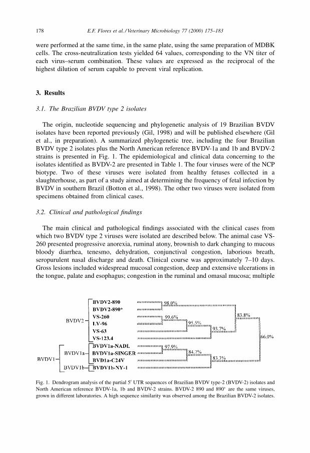
The origin, nucleotide sequencing and phylogenetic analysis of 19 Brazilian BVDV
isolates have been reported previously (Gil, 1998) and will be published elsewhere (Gil
et al., in preparation). A summarized phylogenetic tree, including the four Brazilian
BVDV type 2 isolates plus the North American reference BVDV-1a and 1b and BVDV-2
strains is presented in Fig. 1. The epidemiological and clinical data concerning to the
isolates identi®ed as BVDV-2 are presented in Table 1. The four viruses were of the NCP
biotype. Two of these viruses were isolated from healthy fetuses collected in a
slaughterhouse, as part of a study aimed at determining the frequency of fetal infection by
BVDV in southern Brazil (Botton et al., 1998). The other two viruses were isolated from
specimens obtained from clinical cases.
3.2. Clinical and pathological ®ndings
The main clinical and pathological ®ndings associated with the clinical cases from
which two BVDV type 2 viruses were isolated are described below. The animal case VS-
260 presented progressive anorexia, ruminal atony, brownish to dark changing to mucous
bloody diarrhea, tenesmo, dehydration, conjunctival congestion, laborious breath,
seropurulent nasal discharge and death. Clinical course was approximately 7±10 days.
Gross lesions included widespread mucosal congestion, deep and extensive ulcerations in
the tongue, palate and esophagus; congestion in the ruminal and omasal mucosa; multiple
Fig. 1. Dendrogram analysis of the partial 50 UTR sequences of Brazilian BVDV type-2 (BVDV-2) isolates and
North American reference BVDV-1a, 1b and BVDV-2 strains. BVDV-2 890 and 890� are the same viruses,
grown in different laboratories. A high sequence similarity was observed among the Brazilian BVDV-2 isolates.
178 E.F. Flores et al. / Veterinary Microbiology 77 (2000) 175±183
white-yellowish spots diffusely distributed on the omasal mucosa. Disseminated areas of
congestion and ulcerations covered with ®brin were observed in the small intestine, and
petechial hemorrhages were seen in the epicardium and myocardium. Microscopic
examination revealed deep ulcerations throughout the mucosa of the digestive tract;
polymorphonuclear cell in®ltrates in the mucosa of the tongue and esophagus; mono and
polymorphonuclear in®ltrates in the ruminal mucosa and submucosa; polymorphonuclear
and lymphocytic in®ltrates in the mucosa of small intestine.
The animal case LV-96 came from a herd which had previously experienced cases
clinically compatible with BVD, including a ` mucosal disease''-like disease in another
animal. The animal case LV-96 displayed retarded growth, apathy and anorexia, recurrent
periods of immunosuppressive-like disease in which diarrhea, interdigital dermatitis,
conjunctivitis, arthritis and chronic pneumonia were prominent. Signs were progressive
and the animal was euthanized after a clinical course of approximately 7 months. Gross
examination revealed digestive tract erosions and ulcerations, often covered with necrotic
plaques, including the hard palate, dental pad, tongue and esophagus; and antero-ventral
pneumonia. Microscopic examination revealed necrotic foci in the rumen, and in the
transition between omasum and reticulum. Digestive tract mucosal necrosis and
ulcerations, ®brinoid necrosis in associated blood vessels were also observed. Serum
samples collected at the onset of clinical signs and at the time of euthanasia showed an
increase in VN antibody titer (against BVDV Singer strain) from negative to 400.
3.3. Antigenic characterization
3.3.1. Reactivity with MAbs
The virus isolates were biologically cloned and cells infected with the pure population
of each virus were submitted to IFA, using 10 MAbs to the major envelope glycoprotein
(gp53/E2) of the prototype BVDV Singer strain (Corapi et al., 1990). The pro®le of
reactivity of these MAbs with each Brazilian isolate plus standard US BVDV-1 and
BVDV-2 strains is presented in Table 2. Analysis of reactivity of these MAbs revealed
marked antigenic differences in the envelope glycoprotein gp53/E2 compared to the
standard BVDV strains. The isolate VS-63 was recognized by only two MAbs, whereas
isolates VS-260, LV-96 and VS-123.4 reacted with three and ®ve MAbs, respectively. In
Table 1
Origin of Brazilian BVDV type 2 isolates
Identification Biotype Origin/clinical Specimen Year
VS-63a NCP Healthy fetus Serum 1996
VS-123.4a NCP Healthy fetus Serum 1996
VS-260a NCP Gastroenteric disease, 8-months-old heifer Spleen 1997
LV-96b NCP Chronic gastroenteric disease, 18-months-old heifer Buffy coat 1996
a Viruses isolated and identified at the Virology Laboratory of the Department of Preventive Veterinary
Medicine, Federal University of Santa Maria (UFSM), Santa Maria, RS, Brazil.b Virus isolated and identified at the Virology Laboratory of the Veterinary College, Federal University of
Rio Grande do Sul (UFRGS), Porto Alegre, RS, Brazil.
E.F. Flores et al. / Veterinary Microbiology 77 (2000) 175±183 179
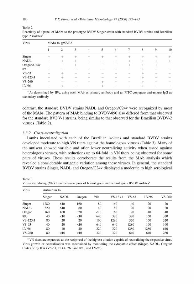
contrast, the standard BVDV strains NADL and Oregon/C24v were recognized by most
of the MAbs. The pattern of MAb binding to BVDV-890 also differed from that observed
for the standard BVDV-1 strains, being similar to that observed for the Brazilian BVDV-2
viruses (Table 2).
3.3.2. Cross-neutralization
Lambs inoculated with each of the Brazilian isolates and standard BVDV strains
developed moderate to high VN titers against the homologous viruses (Table 3). Many of
the antisera showed variable and often lower neutralizing activity when tested against
heterologous viruses, with reductions up to 64-fold in VN titers being observed for some
pairs of viruses. These results corroborate the results from the MAb analysis which
revealed a considerable antigenic variation among these viruses. In general, the standard
BVDV strains Singer, NADL and Oregon/C24v displayed a moderate to high serological
Table 2
Reactivity of a panel of MAbs to the prototype BVDV Singer strain with standard BVDV strains and Brazilian
type 2 isolatesa
Virus MAbs to gp53/E2
1 2 3 4 5 6 7 8 9 10
Singer � � � � � � � � � �NADL � � � � ÿ � � � � �Oregon/C24v � ÿ � ÿ ÿ � � � � ÿ890 � ÿ ÿ ÿ ÿ ÿ ÿ � ÿ ÿVS-63 ÿ � ÿ ÿ ÿ ÿ ÿ � ÿ ÿVS-123.4 � � ÿ � ÿ � ÿ � ÿ ÿVS-260 � ÿ ÿ ÿ � ÿ ÿ � ÿ ÿLV-96 ÿ � ÿ ÿ � ÿ ÿ � ÿ ÿ
a As determined by IFA, using each MAb as primary antibody and an FITC-conjugate anti-mouse IgG as
secondary antibody.
Table 3
Virus-neutralizing (VN) titers between pairs of homologous and heterologous BVDV isolatesa
Virus Antiserum to
Singer NADL Oregon 890 VS-123.4 VS-63 LV-96 VS-260
Singer 1280 640 160 80 160 40 20 20
NADL 320 640 80 40 80 20 20 20
Oregon 160 160 320 <10 160 20 40 40
890 40 <10 <10 640 320 320 160 320
VS-123.4 80 20 20 160 1280 320 160 320
VS-63 40 20 <10 640 640 1280 160 160
LV-96 80 10 20 320 320 1280 1280 640
VS-260 80 <10 <10 320 320 640 640 1280
a VN titers are expressed as the reciprocal of the highest dilution capable of neutralizing the respective virus.
Virus growth or neutralization was ascertained by monitoring the cytopathic effect (Singer, NADL, Oregon/
C24v) or by IFA (VS-63, 123.4, 260 and 890, and LV-96).
180 E.F. Flores et al. / Veterinary Microbiology 77 (2000) 175±183
cross-reactivity within the group. A similar degree of cross-reactivity was observed
between the Brazilian isolates, and between these isolates and the BVDV-2 890. Testing
the standard strains (antisera/virus) against the Brazilian isolates (and against BVDV-
890), revealed a low serological cross-reactivity between the two groups of viruses. These
results allowed the clear identi®cation of two serologically distinct groups of BVD
viruses: one including the standard BVDV-1 strains Singer, NADL and Oregon/C24v, and
another comprising the Brazilian isolates and BVDV-890. Among the Brazilian BVDV-2s
isolates, VS-123.4 displayed the highest serological reactivity with the standard strains,
particularly with BVDV Singer.
4. Discussion
Originally identi®ed in outbreaks of severe disease, BVDV type 2 viruses were initially
thought to be new and invariably virulent viruses. Subsequent studies demonstrated that
viruses belonging to genotype 2 circulate among North American cattle population for at
least 20 years (Carman et al., 1998; Ridpath, unpublished). These viruses are now
beginning to be identi®ed also in Europe (Wolfmeyer et al., 1997) and South America
(Gil, 1998; Canal et al., 1998; Odeon et al., 1998), indicating that they are likely to be
identi®ed in other countries as epidemiological investigation proceeds. Moreover,
evidence is being gathered showing that only a minor fraction of all type 2 BVDV isolates
are highly virulent (Ridpath et al., 1994; Donis, 1998). Nowadays, around 40% of all
BVD viruses isolated in North America are type 2 (Ridpath and Bolin, 1998). These
®ndings support the idea that genotype 2 should not be taken as synonymous of high
pathogenicity and virulence (Ridpath et al., 1994; Donis, 1998).
The origin, evolutionary history and epidemiological factors behind the emergence of
BVDV type 2 remain a matter of dispute and controversy. Sequencing data from South
American isolates are now beginning to emerge and may help in understanding the
evolution and epidemiology of these viruses (Gil, 1998; Canal et al., 1998; Odeon et al.,
1998). Preliminary phylogenetic analysis based on highly conserved 50 UTR sequences
revealed differences between North and South American BVDV type 2 isolates (Fig. 1;
Gil, 1998; Gil et al., in preparation). These ®ndings suggest that North and South
American BVDV 2 isolates may belong to two different subgenotypes. Phylogenetic
analysis of a larger number of South American isolates is obviously needed to support
this hypothesis.
The clinical cases described in the present paper were very characteristic of BVDV-
associated disease. Whereas one animal developed a long-lasting and wasting illness (LV-
96), the other heifer (VS-260) developed an acute and fatal disease. In the ®rst case, the
recent herd history had reports of clinical events compatible with BVDV infection, yet
without virological con®rmation; i.e. an acute and fatal gastroenteric disease with clinical
features resembling those of heifer LV-96 and abortions in which fever and petechial
hemorrhage were observed. The heifer LV-96 went through a long clinical course in
which recurrent periods of diarrhea and immunosuppressive-like signs were observed.
Recurrent interdigital dermatitis was also reported in this case. These features resemble
what has been described as chronic BVD (Baker, 1995). In the case VS-260, the clinico-
E.F. Flores et al. / Veterinary Microbiology 77 (2000) 175±183 181
pathological ®ndings were highly suggestive of mucosal disease (MD). However the
isolation of a NCP, without the CP counterpart, and the subsequent genotyping of this
isolate suggested that this was indeed a case of acute BVD caused by a BVDV type 2
virus.
The genotypic segregation of BVDV into genotypes 1 and 2 seems to correlate fairly
well with the antigenic differences observed between these groups of viruses. BVDV
isolates identi®ed as belonging to genotype 2 display a very low serological cross-
reactivity with BVDV type 1 (Pellerin et al., 1994; Wolfmeyer et al., 1997). Therefore, in
addition to the marked antigenic differences historically observed among BVDV (and
pestiviruses in general) (Bolin et al., 1991; Dubovi, 1992), the recent identi®cation of
BVDV type 2 allowed the clear identi®cation of two antigenically distinct groups of BVD
viruses (Pellerin et al., 1994; Wolfmeyer et al., 1997). These ®ndings have obvious
implications for diagnosis, control, vaccine development and immunization strategies.
The data from our study demonstrated a low serologic cross-reactivity between the
Brazilian type 2 and the standard BVDV type 1 strains (Table 3). The marginal capacity
of antisera to standard BVDV 1 strains to neutralize the Brazilian BVD type 2 viruses
raises the question about the degree of protection conferred by commercially available
vaccines and may indicate the need for formulation of vaccines based on Brazilian
isolates.
Acknowledgements
This work was supported by the MCT, CNPq, CAPES and FINEP grants (PRONEX in
Veterinary Virology, 215/96). E. Flores (352386/96) and R. Weiblen (520161/97-1) had
research scholarships from the Brazilian Council for Research (CNPq). We thank Dr.
Ruben Donis (Department of Veterinary and Biomedical Sciences, University of
Nebraska at Lincoln. Lincoln, NE) for providing the MAbs and the reference BVDV
strains. The technical assistance of E.A.S. Oliveira in performing the SN tests of the case
LV-96 is greatly appreciated.
References
Alves, D., Tremblay, R., Godkin, A., Anderson, N., Carman, S., McEwen, B., Hazlett, M., 1996. Update on
bovine virus diarrhea in Ontario. Can. Vet. J. 37, 177.
Baker, J.C., 1995. The clinical manifestations of bovine viral diarrhea infections. Vet. Clin. N. Am. 11, 427±444.
Bolin, S.R., Littledike, E.T., Ridpath, J.F., 1991. Serologic detection and practical consequences of antigenic
diversity among bovine viral diarrhea virus in a vaccinated herd. Am. J. Vet. Res. 52, 1033±1047.
Botton, S.A., Gil, L.H.V.G., Silva, A.M., Flores, E.F., Weiblen, R., Pituco, E.M., Roehe, P.M., Moojen, V.,
Wendelstein, A.C., 1998. CaracterizacËaÄo preliminar de amostras do võÂrus da diarreÂia viral bovina (BVDV)
isoladas no Brasil. Pesq. Vet. Bras. 18, 84±92.
Canal, C.W., Strasser, M., Hertig, C., Masuda, A., Peterhans, E., 1998. Detection of antibodies to bovine viral
diarrhea virus (BVDV) and characterization of genomes of BVDV from Brazil. Vet. Microbiol. 63, 85±97.
Carman, S., Van Dreumel, T., Ridpath, J., Hazlett, M., Alves, D., Dubovi, E.J., Tremblay, R., Bolin, S.R.,
Godkin, A., Anderson, N., 1998. Severe acute bovine viral diarrhea (BVD) in Ontario, 1993±1995. J. Vet.
Diagn. Invest. 10, 27±35.
182 E.F. Flores et al. / Veterinary Microbiology 77 (2000) 175±183
Corapi, W.V., French, T.W., Dubovi, E.J., 1989. Severe thrombocytopenia in young calves experimentally
infected with noncytopathic bovine viral diarrhea virus. J. Virol. 62, 2823±2827.
Corapi, W.V., Donis, R.O., Dubovi, E.J., 1990. Characterization of a panel of monoclonal antibodies and their
use in the study of the antigenic diversity of bovine viral diarrhea virus. Am. J. Vet. Res. 51, 1388±1394.
Donis, R., 1998. Molecular evolution of highly virulent pestivirus strains which cause acute severe BVD in
cattle. In: Proceedings of the International Symposium on Bovine Herpesvirus Types 1 and 5 and Bovine
Viral Diarrhea Virus. Santa Maria, RS, Brazil, pp. 21±31.
Dubovi, E.J., 1992. Genetic diversity and BVD virus. Comp. Immunol. Microbiol. Infect. Dis. 15, 155±165.
Gil, L.H.V.G., 1998. Sequenciamento, anaÂlise ®logeneÂtica e caracterizacËaÄo de polipeptõÂdeos naÄo-estruturais de
amostras do võÂrus da diarreÂia viral bovina (BVDV). MS Thesis. Santa Maria, RS, UFSM, 1998, 69 pp.
Odeon, A.C., Kaiser, G.G., Donis, R.O., Risatti, G., Leunda, M.R., 1998. Aislamiento y anaÂlisis molecular del
võÂrus de la diarrea viral bovina genotipo II en Argentina. In: Proceedings of the XII ReunioÂn Cientõ®co
TeÂcnica. As. Arg. Vet. Lab. Diagn. 12, 94.
Pellerin, C., van den Hurk, J., Lecomte, J., 1994. Identi®cation of a new group of bovine viral diarrhea virus
strains associated with severe outbreaks and high mortalities. Virology 203, 260±267.
Rebhun, W.C., French, T.W., Perdrizet, J.A., Dubovi, E.J., Dill, S.G., Karcher, L.F., 1989. Thrombocytopenia
associated with acute bovine virus diarrhea infection in cattle. J. Vet. Intern. Med. 3, 42±46.
Ridpath, J.F., Bolin, S.R., 1998. Differentiation of types 1a, 1b and 2 bovine viral diarrhea virus (BVDV) by
PCR. Mol. Cell Probes 12, 101±106.
Ridpath, J., Bolin, S., Dubovi, E., 1994. Segregation of bovine viral diarrhea virus into genotypes. Virology 205,
66±74.
Wolfmeyer, A., Wolf, G., Beer, M., 1997. Genomic (50UTR) and serological differences among German BVDV
®eld isolates. Arch. Virol. 142, 2049±2057.
E.F. Flores et al. / Veterinary Microbiology 77 (2000) 175±183 183