Clinical Impact of the Epithelial-Mesenchymal Transition in ...
43
HAL Id: hal-03051871 https://hal.archives-ouvertes.fr/hal-03051871 Submitted on 15 Feb 2021 HAL is a multi-disciplinary open access archive for the deposit and dissemination of sci- entific research documents, whether they are pub- lished or not. The documents may come from teaching and research institutions in France or abroad, or from public or private research centers. L’archive ouverte pluridisciplinaire HAL, est destinée au dépôt et à la diffusion de documents scientifiques de niveau recherche, publiés ou non, émanant des établissements d’enseignement et de recherche français ou étrangers, des laboratoires publics ou privés. Clinical Impact of the Epithelial-Mesenchymal Transition in Lung Cancer as a Biomarker Assisting in Therapeutic Decisions Julien Ancel, Maxime Dewolf, Gaëtan Deslée, Béatrice Nawrocky-Raby, Véronique Dalstein, Christine Gilles, Myriam Polette To cite this version: Julien Ancel, Maxime Dewolf, Gaëtan Deslée, Béatrice Nawrocky-Raby, Véronique Dalstein, et al.. Clinical Impact of the Epithelial-Mesenchymal Transition in Lung Cancer as a Biomarker Assisting in Therapeutic Decisions. Cells Tissues Organs, Karger, 2022, pp.91-109. 10.1159/000510103. hal- 03051871
-
Upload
khangminh22 -
Category
Documents
-
view
1 -
download
0
Transcript of Clinical Impact of the Epithelial-Mesenchymal Transition in ...

HAL Id: hal-03051871https://hal.archives-ouvertes.fr/hal-03051871
Submitted on 15 Feb 2021
HAL is a multi-disciplinary open accessarchive for the deposit and dissemination of sci-entific research documents, whether they are pub-lished or not. The documents may come fromteaching and research institutions in France orabroad, or from public or private research centers.
L’archive ouverte pluridisciplinaire HAL, estdestinée au dépôt et à la diffusion de documentsscientifiques de niveau recherche, publiés ou non,émanant des établissements d’enseignement et derecherche français ou étrangers, des laboratoirespublics ou privés.
Clinical Impact of the Epithelial-MesenchymalTransition in Lung Cancer as a Biomarker Assisting in
Therapeutic DecisionsJulien Ancel, Maxime Dewolf, Gaëtan Deslée, Béatrice Nawrocky-Raby,
Véronique Dalstein, Christine Gilles, Myriam Polette
To cite this version:Julien Ancel, Maxime Dewolf, Gaëtan Deslée, Béatrice Nawrocky-Raby, Véronique Dalstein, et al..Clinical Impact of the Epithelial-Mesenchymal Transition in Lung Cancer as a Biomarker Assistingin Therapeutic Decisions. Cells Tissues Organs, Karger, 2022, pp.91-109. �10.1159/000510103�. �hal-03051871�

1
Clinical impact of EMT in lung cancer as a biomarker for assisting therapeutic decisions
Julien Ancel1,2, Maxime Dewolf2, Gaëtan Deslée1,2, Béatrice Nawrocki-Raby1, Véronique
Dalstein1,3, Christine Gilles4,*,δ and Myriam Polette1,3,δ.
1 Inserm, Université de Reims Champagne Ardenne, P3Cell UMR-S1250, SFR CAP-SANTE,
51092 Reims, France
2 Service de pneumologie, Hôpital Maison Blanche, CHU de Reims, 51092 Reims, France
3 Laboratoire de pathologie, Hôpital Maison Blanche, CHU de Reims, 51092 Reims, France
4 Laboratory of Tumor and Development Biology, GIGA-Cancer, University of Liège, 4000
Liège, Belgium
Short Title: EMT in lung cancer management
δ Equal contribution
*Corresponding Author
Christine Gilles, Laboratory of Tumor and Development Biology, GIGA-Cancer, University
of Liège, 4000, Belgium.
E-mail: [email protected]

2
Keywords: non-small-cell lung cancer; epithelial–mesenchymal transition; therapy;
resistance.

3
Abstract 1
Lung cancer is one of the most common solid cancers and represents the leading cause of 2
cancer death worldwide. Over the last decade, examining epithelial-mesenchymal transition 3
(EMT) in lung cancer has gained major interest. Here, we review clinical and histological 4
features of non-small cell lung cancer (NSCLC) associated with EMT. We then aim at 5
establishing EMT potential clinical implications in current therapeutic options including 6
surgery, radiation, targeted therapy against oncogenic drivers and immunotherapy. 7
8
9
10
11
12
13
14
15
16
17
18
19
20
21
22
23
24
25
26

4
Introduction 27
Lung cancer is responsible for the highest number of cancer‐related deaths worldwide. About 28
85% of lung cancers are classified as non-small cell lung cancers (NSCLC), subsequently 29
histologically subdivided as adenocarcinoma (AC) (about half of NSCLC cases) and 30
squamous cell carcinoma (SCC) (about 30% of NSCLC cases). Regarding staging, more than 31
a half of cases are diagnosed at a metastatic or advanced stage without curative options. Over 32
the past years, innovative therapies such as molecularly targeted drugs and immunotherapies 33
have emerged and have led to overall improvements in cancer treatment. However, global 34
5‐year survival rate for NSCLC patients remains under 20 % (Bray et al., 2018; Hirsch et al., 35
2017). Even in localized stages with a surgically resectable tumor, 5-year survival rates 36
drastically drop from 60% for stage IIA disease to 36% for stage IIIA disease, according to 37
the 8th edition staging project of the International Association for the Study of Lung Cancer 38
(IASLC) (Goldstraw et al., 2016). Promising and innovative strategies are ongoing, especially 39
in neo-adjuvant (Uprety et al., 2020) and adjuvant (Broderick, 2020; NCT02595944 ; 40
NCT02486718 ; NCT02504372) contexts, that could improve the dramatic landscape of 41
NSCLCs (Kris et al., 2020; Otaibi et al., 2019). Nevertheless, such a bad record also 42
highlights the need to further refine characterization of tumor specimens in order to improve 43
clinical patient management. 44
Examining epithelial-mesenchymal transition (EMT) in lung cancer has gained major interest 45
in the last decade. EMT is a term widely used now to describe the loss of epithelial features 46
and the acquisition of mesenchymal traits by epithelial cells in various physiological and 47
pathological conditions. In cancer, EMT is considered as a dynamic and reversible process, 48
thereby generating various phenotypes. Some hybrids may have enhanced plasticity allowing 49
them to adapt to various tumor micro-environments encountered during cancer progression 50
and metastatic dissemination (Bhatia et al., 2020; Dongre and Weinberg, 2019; Yang et al., 51

5
2020). As such, EMT has been shown to endow tumor cells with many properties that may 52
provide them a selective advantage to overcome different steps of cancer progression 53
including enhanced invasive potential, enhanced survival, stimulation of angiogenesis, 54
immune escape, resistance to apoptotic signals and niching properties (Francart et al., 2018; 55
Pastushenko and Blanpain, 2019). EMT is molecularly complex, diversified and context-56
dependent. This complexity certainly contributes to slow down the implementation of EMT 57
consideration in clinical routine. EMT programs may indeed be triggered by several factors 58
such as transforming growth factor‐β (TGF‐β) (Miyazono et al., 2018), fibroblast growth 59
factor (FGF) (Katoh and Nakagama, 2014), epidermal growth factor (EGF) (Shaurova et al., 60
2020), Notch or Wnt signaling pathways (Patel et al., 2019; Yuan et al., 2014). Activated 61
signaling pathways converge onto a set of transcription factors (EMT‐TFs) such as Snail, 62
ZEB and Twist family (Zeisberg and Neilson, 2009). A dynamic balance finally represses 63
expression of epithelial related-genes such as E‐cadherin and induces expression of genes 64
coding for mesenchymal markers such as vimentin. Importantly, EMT rather supports early 65
steps of the metastatic dissemination, i.e. tumor invasion, survival in the blood stream and 66
early niching. After an eventual period of dormancy, a reversal towards more epithelial 67
phenotypes, so called mesenchymal-epithelial transition (MET), is considered to occur at 68
secondary sites to support metastatic outgrowth. Although EMT features have been described 69
for many years in numerous histological types of cancer, discussion remains regarding EMT 70
characterization in tumors. Pan-cancer studies are based on most consensual and proposed 71
canonical markers such as E-cadherin (CDH1), vimentin (VIM), N-cadherin (CDH2) and 72
fibronectin (FN1). Using NSCLC cell lines and tumor samples, some studies established 73
EMT-signatures. From these NSCLC reports, some markers such as DSP, TJP1, CLDN4, 74
ERBB3, GALNT3 and CDS1 appear commonly associated with CDH1 epithelial related-genes 75
while MMP-2, AXL, ZEB1/2, NRP-1 and TWIST clustered with VIM and FN1 mesenchymal 76

6
related-genes (Antony and Huang, 2017; Goossens et al., 2017; Karacosta et al., 2019; Shao 77
et al., 2019; Wong et al., 2018; Wushou et al., 2014; Zheng and Kang, 2014). 78
We discuss here the potential utility of examining and considering EMT in clinical practice. 79
Although some reports highlight a contribution of EMT in small cell lung cancer (Ito et al., 80
2017) and malignant pleural mesothelioma (Schramm et al., 2010) with specific implications 81
respective to their cell type origin, we will focus this review on NSCLC, the most frequent 82
lung cancer in which EMT has now been extensively analyzed. We aim at pointing to 83
clinically relevant features related to EMT process that could help clinicians to identify 84
disease contexts. We further propose to examine the beneficial contribution of EMT in 85
clinical management from early/advanced stages to metastatic conditions in order to meet 86
current clinical challenges. Considering the poor outcome of resectable lung cancer, a crucial 87
challenge is indeed to identify independent and powerful predictors of patient global 88
outcomes that are currently lacking. On another hand, advanced and metastatic conditions are 89
associated with ineluctable therapy resistances to current available strategies including 90
chemotherapies, radiotherapy, targeted therapies and immunotherapies. In these clinical 91
conditions, considering EMT as a biomarker could allow a refinement of patient management. 92
93
NSCLC biomarkers related to EMT 94
Aiming at defining the clinical relevance of EMT as a biomarker, we will first confront EMT 95
status with pathological features of lung tumor cells, and then unravel relationships bridging 96
EMT to two cornerstones of both lung carcinogenesis and clinical management that are 97
oncogenic drivers and immune profiles. 98
a. EMT and histopathological features 99
Literature emphasizes that EMT features are quite commonly observed in lung cancer both in 100
AC and in SCC (Kidd et al., 2014; Mittal, 2018). For example, Dauphin et al. described until 101
50% of tumors cells with a mesenchymal phenotype (Dauphin et al., 2013). Mesenchymal 102

7
traits were shown to be even more frequent in other lung histological types such as large cell 103
neuroendocrine carcinomas (Galván et al., 2014), sarcomatoid carcinomas (Thomas et al., 104
2012) or pleomorphic carcinomas which are typical of mesenchymal switch (Kondo et al., 105
2018; Miyahara et al., 2015). Mesenchymal features have also been associated to a low degree 106
of differentiation in NSCLC (Bian et al., 2019; Dauphin et al., 2013; Matsubara et al., 2014; 107
Z. Wang et al., 2019). Otherwise, the spatial distribution of EMT features within a tumor is 108
very heterogenous. EMT attributes have thus frequently been reported in so-called invasive 109
fronts, at the interface with extracellular matrix, correlating with invasiveness and metastatic 110
potential (Maeng et al., 2014). EMT has also been associated with hypoxic zones and 111
inflammation-rich areas (Dominguez et al., 2017), emphasizing a major contribution of 112
specific tumor microenvironments (TME) in EMT induction/regulation (Foster et al., 2014; 113
Hung et al., 2009; Yang and Wu, 2008). Accordingly, experimental data strongly support that 114
EMT may be induced/regulated by ECM components, inflammatory mediators and other 115
soluble factors secreted by stromal cells, and hypoxia (Lou et al., 2016; Mittal et al., 2016). 116
Inversely, EMT+ tumor cells have also been reported to secrete higher levels of soluble factors 117
crucially impacting the tumor microenvironment (e.g.: VEGF stimulating angiogenesis, 118
chemokines impacting immune cell recruitment). At last, among classically examined 119
clinicopathological parameters in lung cancer is also immune infiltration. Thus, EMT also 120
modulate immune cell-infiltration (De Matteis et al., 2019; Dominguez et al., 2017) through 121
well identified molecular regulatory networks (Markopoulos et al., 2019). For instance, Chae 122
et al. reported reduced CD4 T-cell and CD4/CD8 T-cell infiltration respectively in lung AC 123
and SCC with tumor cells displaying mesenchymal attributes (EMT+) (Chae et al., 2018). 124
Inversely, increased activated B-cells and regulatory T-cells were reported, although some 125
differential infiltration patterns need to be further defined between AC and SCC. 126
Additionally, EMT+ tumors were also found to over-express multiple immunosuppressive 127
cytokines such as IL-10, TGF-β, IL-6 or IL-11 (Y. N. Jiang et al., 2019; Q. Zhang et al., 2017; 128

8
Zhao et al., 2018; Zheng et al., 2019). A narrow crosstalk and regulatory loops between 129
stromal cells and EMT-derived cells are thus established, contributing to the formation of 130
particular areas favoring tumor invasion and dissemination. Main EMT-associated markers 131
and activation pathways are illustrated and summarized in Figure 1. 132
In addition to EMT determination in lung primary tumors, examining EMT in Circulating 133
Tumor Cells (CTCs) has also gain major interest. CTCs indeed today appear as promising 134
biomarkers in lung cancer (Y. Li et al., 2018; Maly et al., 2019; Milano et al., 2018). Unlike 135
tumor biopsies, CTCs allow a live assessment of disease progression and could thus help to 136
predict metastasis and monitor therapeutic response. Numerous studies today report the 137
presence of EMT-shifted hybrid CTCs and CTC clusters in NSCLC patients (G. Li et al., 138
2018; Lindsay et al., 2017; Sawabata et al., 2020). Interestingly, Manjuntha et al. observed 139
that the intensity of EMT markers staining (vimentin and fibronectin) was higher in EMT+ 140
CTCs than in patient-matched NSCLC tumor tissues (Manjunath et al., 2019). Validating the 141
importance of EMT characterization and supporting its utility for clinicians, Xu et al. 142
classified CTCs into three subpopulations from epithelial, intermediate and mesenchymal 143
phenotypes and reported that mesenchymal CTCs were more commonly found in patients in 144
the metastatic stages of different types of cancers (Wu et al., 2015). Additionally, several 145
reports associated a mesenchymal shift in CTCs as predictor of poor outcomes in NSCLC (Li 146
et al., 2017, p. 4; Liu et al., 2018). Miguel-Perez et al. thus identified mesenchymal CTCs as 147
an independent prognostic factor for relapse-free survival, with an impact on overall survival 148
in resected lung adenocarcinomas (de Miguel-Pérez et al., 2019). Moreover, similar data were 149
obtained in a prospective and controlled cohort (Manjunath et al., 2019). Among metastatic 150
stages, EMT sub-classification could allow to further refine those with poor evolution profile 151
(Y. Wang et al., 2019). EMT status in CTCs could also be useful as predictor of therapeutic 152
response (Liao et al., 2014; Milano et al., 2018; Togo et al., 2017). Despite these numerous 153
data validating the clinical relevance of examining EMT in CTCs, the variability of 154
9
methodologies used to enrich and isolate CTCs combined with the molecular complexity of 155
EMT certainly introduces biases in our comprehension of EMT-related CTC heterogeneity. It 156
seems worth mentioning that many CTC isolation devices are based on the expression of 157
specific epithelial biomarkers (such as EpCAM). Subpopulations of EMT-derived CTCs, 158
supposedly expressing lower levels of many membrane epithelial markers, may thus failed 159
being detected by such methods. To address this limitation, alternative enrichment devices 160
exploiting physical properties of CTCs are developed to isolate label-free CTCs and facilitate 161
the study of EMT heterogeneity in the CTC population (Alix-Panabières et al., 2017; Genna 162
et al., 2020; Nicolazzo et al., 2019). 163
All in all, EMT is a frequent event in NSCLC, observable both in the primary tumor site and 164
in CTCs. Its association with unfavorable clinicopathological features justifies the proposition 165
to consider EMT as a potential marker to predict patient outcomes. Methodological 166
standardization and identification of the most relevant molecular markers are nevertheless 167
necessary steps before a routine clinical utilization. 168
169
b. EMT and oncogenic drivers 170
Our growing molecular knowledge of cancer somehow shook some dogmas and continuously 171
redirects lung cancer management. Several targeted therapies constitute the current arsenal to 172
combat lung cancer, most of which are specifically directed against recognized lung cancer 173
oncogenic drivers (Figure 2). Among major genetic modifications associated with NSCLC, 174
KRAS, EGFR and ALK addictive mutations or alterations are probably the main ones 175
(Arbour and Riely, 2019). 176
-KRAS 177
To date, KRAS mutation is the most common molecular alteration encountered in NSCLC but 178
remains with no effective therapies targeting tumors harboring mutant variant of KRAS, 179
despite many clinical trials (Aran and Omerovic, 2019; Yang et al., 2019). Experimental data 180

10
suggest that KRAS mutations contribute to support mesenchymal changes, alone but often in 181
synergy with other EMT‐inducing factors (Arner et al., 2019). For instance, mutant KRAS and 182
TP53 cell lines established from a lung cancer transgenic mouse model were found to display 183
important EMT/MET plasticity. A shift toward a mesenchymal phenotype was shown to 184
depend on a well-described miR-200/ZEB regulatory loop and to promote metastasis 185
(Gibbons et al., 2009). Deciphering further KRAS mutant/EMT relationships, Singh et al. 186
showed that, within KRAS mutant cell lines, two sub-groups were distinguished based on their 187
KRAS dependency to maintain their viability. The KRAS-dependent NSCLC cell line treated 188
with the classical EMT-inducer (TGF-β1) acquired KRAS independency, contrary to untreated 189
cell line. Thus, mesenchymal switched NSCLC cell lines harbored a KRAS independency and 190
inversely, supporting a close relationship between EMT and loss of oncogene addiction 191
(Singh et al., 2009). Examining KRAS molecular status and EMT phenotype as tandem 192
biomarkers could thus harbor a particular significance and also refine patient stratification and 193
therapeutic strategies. Considering until now deceiving effectiveness for drugs targeting 194
KRAS addiction, selected patients with KRAS-mutant NSCLC harboring epithelial phenotype 195
could thus benefit from KRAS inhibitors. 196
- EGFR 197
As the second most common molecular addiction occurring in NSCLC, activating mutations 198
of the epidermal growth factor receptor (EGFR) gene are especially involved in lung 199
adenocarcinoma without smoking history. A large majority of these activating mutations (85-200
90%) occurs by exon 19 deletion (about 45%) or exon 21 L858R mutation (about 45%) 201
(Castellanos et al., 2017). In EGFR-mutated advanced lung cancer patients, many randomized 202
phase III trials have revealed that treatment with first-, second- and now third-generation 203
EGFR tyrosine kinase inhibitors (TKIs) resulted in an improved outcome compared to 204
standard chemotherapy in first line (Soria et al., 2018). Erlotinib even provided a similar 205
efficacity than chemotherapy in second line in EGFR wild type tumor with a better tolerance 206

11
(Ciuleanu et al., 2012). However, cancer progression fatally occurs after a median of 12-207
month treatment and almost all patients who strongly responded to EGFR-TKI acquire 208
resistance over time. Frequently, the mechanism of resistance is a acquired EGFR mutation 209
(Rotow and Bivona, 2017). In half of the cases, this second mutation is T790M point mutation 210
in exon 20 of the EGFR gene (Kobayashi et al., 2005) that can be triggered by osimertinib, a 211
dedicated EGFR-TKI (Carlisle and Ramalingam, 2019). Among patients with tumor 212
harboring mutation of EGFR and developing EGFR-TKIs resistance, a significant part does 213
not exhibit mechanisms of genotypic resistance. Such EGFR-independent mechanism of 214
resistance includes EMT, occurring in about 5% of cases (Bronte et al., 2018; Lim et al., 215
2018). Interestingly, tumors from patients developing resistance to TKIs exhibit mesenchymal 216
traits while maintaining their original EGFR‐activating mutation. Moreover, Sequist et al. did 217
not observe EMT features in patient developing a resistance mechanism mediated by T790M 218
EGFR mutation (Sequist et al., 2011), supporting that T790M does not drive EMT. In patients 219
harboring T790M mutation and treated with osimertinib, progression also unfortunately 220
occurs, and a C797S tertiary mutation has been identified. A tertiary resistance could be also 221
associated to and/or supported by EMT, even in non-C797S mutation-harboring patients. 222
Although MET gene amplification may more frequently drive the underlined mechanism of 223
resistance in this context (Del Re et al., 2019). Those clinical observations linking EGFR-224
TKIs resistance to the emergence of EMT phenotypes are supported by in vitro and in vivo 225
preclinical studies (Tulchinsky et al., 2019; X. Zhu et al., 2019). For example, TGF-β1, 226
Insulin‐like growth factor 1 receptor (IGF1R) and Notch-1 pathways, known to be potent 227
EMT-inducers, seem to be crucial actors in resistance mechanism (Cortot et al., 2013; Rho et 228
al., 2009; Soucheray et al., 2015; Suda et al., 2011; Xie et al., 2013; Zhou et al., 2015). This 229
phenotypical switch appears to be reversible and could thus be promising in combined 230
therapies (Witta et al., 2006). By using dasatinib, Sesumi et al. inhibited EMT induction by 231

12
TGF-β in EGFR-mutant NSCLC cell lines. For NSCLC already harboring erlotinib resistance 232
with mesenchymal features, dasatinib monotherapy failed to restore both an epithelial 233
phenotype and sensitivity to EGFR-TKIs. However, combining erlotinib and dasatinib 234
prevented EMT-mediated resistance to EGFR-TKI and resulted in T790M mutation of 235
resistance (Sesumi et al., 2017). 236
- ALK 237
Among ALK molecular alterations, ALK translocations with a fusion partner correspond to 238
the second targetable oncogenic driver to date in NSCLC (Du et al., 2018). Following the 239
ALEX trial, Alectinib became the gold standard of ALK-rearranged related TKI with an 240
increase of overall survival (Hida et al., 2017). As similarly observed in EGFR inhibitor 241
management, some ALK inhibitor resistance inevitably occurs and is mediated most of time 242
by an acquired ALK mutation (Katayama, 2018). As for resistance to EGFR-TKI, EMT has 243
been observed and proposed as a non-oncogenic resistance pathway (Peters and 244
Zimmermann, 2018). Gainor et al. explored different mechanisms of ceritinib resistance in 12 245
re-biopsies and observed that 5 specimens displayed mesenchymal traits. Interestingly, some 246
of them also harbored a concomitant second ALK-mutation of resistance (Gainor et al., 2016). 247
Similarly, Gower et al. described acquired EMT characteristics in tumor harboring ALK-TKI 248
resistance. However, reversible models of EMT did not allow to restore sensitivity to ALK 249
inhibitors, suggesting that EMT process can be associated but not required for ALK-TKI-250
resistance. Deciphering further this EMT/ALK-mutation status, Fukuda et al. performed 251
microdissection analyses in a tumor resistant to ALK-rearranged related TKI. This tumor 252
concomitantly harbored an acquired ALK mutation of resistance (L1196M) in epithelial-like 253
tumor area while no additive mutation was found in mesenchymal-switched tumor area. 254
Taken together, those observations suggest that EMT can both be independent and additive 255
mechanisms underlying ALK-TKI-resistant cancers (Fukuda et al., 2019). To investigate the 256
EMT and ALK-TKI resistance relationship, Kogita el al. established NSCLC cell line with 257
13
EML4-ALK rearrangement and showed that hypoxic condition was associated to ALK-TKI-258
resistance by an EMT-dependent signaling (Kogita et al., 2014). Beyond molecular 259
mechanisms involving hypoxia induced-factors, epithelial splicing regulatory protein 1 260
(ESRP1) (Voena et al., 2016), three other EMT-related pathways were involved in ALK 261
mutant cancers as proteoglycans, HIF-1 and FoxO signaling pathways and ECM-receptor 262
interaction as reported by Wei et al. (Wei et al., 2018). All together, these observations 263
suggest that resistance to ALK-TKI can be associated with mesenchymal features even 264
though EMT is not the sole driver of resistance. 265
- Others 266
Among other targetable oncogenic drivers, some molecular alterations seemed related to 267
EMT. For example, EMT process has been described as dysregulated in BRAF mutant 268
cancers such as primary cutaneous melanoma or papillary thyroid carcinoma (Mitchell et al., 269
2016). Possibly due to low prevalence of BRAF mutant in lung cancer (less than 1%), only 270
few works reported BRAF/EMT interplay in NSCLC (Urbanska et al., 2020). With a 271
structural similarity to ALK but lower prevalence, ROS1 alterations are also oncogenic drivers 272
targetable in clinical management (Lin and Shaw, 2017). Gou et al. described a mesenchymal 273
polarization in NSCLC cell line with CD74-ROS1 G2032R mutation, leading to increased 274
aggressiveness and interestingly supporting a resistance to ROS1-TKI (Crizotinib) (Gou et al., 275
2018). Still regarding EMT process, our laboratory observed a more frequent mesenchymal 276
switch in NSCLC cell lines and tumors harboring HER2 activation, classically related to 277
aggressiveness. Interestingly, anti-HER2 therapies allowed to restore epithelial features and 278
reduce invasiveness (Da Silva et al., 2020). Finally, c-MET molecular alterations are also 279
described in a minority of NSCLC and many preclinical and clinical trials have been designed 280
in lung cancer. However, NSCLC harbor a large heterogeneity in c-MET molecular 281
modification such as overexpression, amplification and point mutations that could explain 282

14
many controversial results to date (Drilon et al., 2017), although exon-14 skipping mutations 283
seem promising (Pasquini and Giaccone, 2018). 284
285
In conclusion, both common and uncommon oncogenic drivers seem intrinsically linked to 286
EMT processes. A large part of studies reported that mesenchymal features are associated 287
with resistance to various drugs. Whether EMT is a consequence of or a prerequisite to drug 288
resistance are two non-mutually exclusive possibilities. A better comprehension on those 289
phenomena may lead to innovative pharmacological strategies. 290
291
c. EMT and immune profile 292
Among EMT-induced properties contributing to enhance metastatic potential, the ability of 293
tumor cells to escape immune surveillance has gained major interest along with the 294
emergence of immunotherapies. Thus, numerous studies report a positive correlation between 295
a mesenchymal switch and the expression of immune checkpoint proteins. As a cornerstone in 296
immunotherapy management, PD-(L)1 has been largely explored in NSCLC context. Several 297
in vitro and preclinical studies reported an induction of PD-L1 expression by different EMT 298
pathways and EMT-TFs, and PD-L1/EMT-TFs co-expression has been reported in human 299
lung cancer specimens (Asgarova et al., 2018; F. Li et al., 2018; Noman et al., 2017). This co-300
expression has actually been observed in many histological types (Alsuliman et al., 2015; 301
Chen et al., 2017; Ock et al., 2016) and largely reported in NSCLC, from metastatic to locally 302
and resected lung cancers (Ancel et al., 2019; Kim et al., 2016), and in CTCs (Manjunath et 303
al., 2019). Other reports studying PD-1 and PD-L1 expression further corroborated this 304
association with EMT phenotypes (Kim et al., 2016; Mak et al., 2016). Additionally, EMT 305
seems to affect other immune checkpoint systems including CTLA-4, TIM-3 but also PD-306
L1/2, PD-1 and B7-H3 that were also found overexpressed, suggesting a wide range effect of 307
EMT on tumoral immune escape (Lou et al., 2016). Chae et al. observed that overexpression 308

15
of druggable immune checkpoints, such as CTLA-4 and TIM-3 (but not PD-L1 in their study 309
context), is associated with an EMT signature in NSCLC and with a lower infiltration of CD4 310
T cells (Chae et al., 2018). Thus, besides a direct effect on EMT in regulating the expression 311
of immune checkpoint protein in tumor cells, EMT also acts on immune cell infiltration, as 312
we discuss later in the text, contributing to create an immunosuppressive TME in the vicinity 313
of EMT+ areas. 314
Overall, EMT process appears as a promising biomarker intrinsically related to tumor 315
aggressiveness in NSCLC. EMT could thus help refining tumor prognostic and help clinicians 316
in the choice of pharmacological strategies, especially regarding targetable oncogenic drivers 317
and immunotherapies. 318
319
EMT process in clinical lung cancer management 320
Aiming at going beyond a descriptive level, we here below report how clinicians could 321
benefit from examining EMT in NSCLC, both in early and metastatic stages. 322
a. EMT relevance as prognosis factor in early lung cancer 323
As previously mentioned, 5-year survival rates in early stage NSCLC remain poor, even after 324
a complete resection, and relapse fatally occurs in a large number of cases. Aiming at 325
reducing this burden, adjuvant platinum-based regimens are employed, though with limited 326
effects. Many targeted therapies are currently available in lung cancer but their use is 327
restricted to advanced stages. For example, pre- and/or post-operative anti-PD-(L)1 cannot be 328
employed despite over than 50% tumor specimens harboring PD-L1 positive cells. This 329
highlights a crucial need to further refine the characterization of tumor samples in order to 330
identify patients that could benefit from a personalized strategy such as immune checkpoint 331
inhibitors. Identifying patient with worse outcomes is a key step to this end, and robust 332
prognostic markers are thus needed. Considering the extensive literature bridging EMT to 333
tumor aggressiveness, EMT has been explored, solely or in combination with other markers 334

16
through different approaches that, as we discuss later in the review, still need to be optimized 335
and validated in order to be exploitable in a clinical context. Chikaishi et al thus described a 336
non-informative EMT status based on vimentin, gamma-catenin, fibronectin and E-cadherin 337
expressions in 183 resected tumors, unable to predict patient’s outcomes (Chikaishi et al., 338
2011). These results could reflect the incorporation in the analyzed cohort of a large number 339
of stage IA tumors, known to display a specific better prognosis. Examining homogenous and 340
larger cohorts, many other studies reported a positive association between EMT+ 341
characteristics and poor outcome. The evaluation of the prognostic and predictive value of 342
EMT in early stages of NSCLC (NCT03509779) is being examined in a prospective cohort 343
(TWIST lung). In other studies, higher vimentin expression in tumor cells was proposed as a 344
predictor of metastasis occurrence (Aruga et al., 2018; Dauphin et al., 2013; Tsoukalas et al., 345
2017; Y. Wang et al., 2019). In SCC, vimentin expression failed to establish an independent 346
prognostic but high S100A expression and lack of intercellular E-cadherin allowed to predict 347
patients at a high risk of recurrence and poor prognosis (Zhang et al., 2013). Both in AC and 348
SCC, reduced membranous staining of E-cadherin and expression of vimentin were shown to 349
be independent predictors of mortality (Aruga et al., 2018; Che et al., 2015; Shao et al., 2019). 350
Overpassing the clinical challenge to collect tumor biopsies, CTCs in peripheral blood were 351
also explored as a predictor of outcomes in early stages of lung cancers. Regarding CTCs, 352
many parameters are confronted such as CTCs count, CTCs variation or CTCs employed as 353
liquid biopsy (Cabel et al., 2017; Syrigos et al., 2018). Moreover, CTCs can be informative 354
through their biomarker expression and assessing EMT seems promising. Indeed, CTCs with 355
a mesenchymal switch were associated with poor outcomes (Li et al., 2017, p. 4; Liu et al., 356
2018; Manjunath et al., 2019; de Miguel-Pérez et al., 2019). Thus, considering EMT statuses 357
in CTCs could allow to enhance CTC clinical relevance in lung cancer management (Jin et 358
al., 2017; Lowes and Allan, 2018; Wu et al., 2015). 359

17
To summarize on prognosis significance, studies examining multiple EMT markers are 360
numerous and mainly concordant, supporting an independent capacity of EMT signature to 361
predict patient outcomes. However, it appears crucial to identify some most relevant markers 362
to examine their expression in routine practice. In these conditions, vimentin and E-cadherin 363
expressions seem promising. 364
365
b. EMT implication in advanced and metastatic lung cancers 366
In addition to promoting local and distant dissemination/recurrence, EMT was proposed by 367
many authors to support resistance to therapies (Dudas et al., 2020). We explore here EMT 368
interrelation with therapeutic options currently used in advanced stages of NSCLC, such as 369
chemo/radiotherapies, targeted therapies and immune checkpoint blocking antibodies. 370
- Chemo/radio resistance 371
Extensive literature today emphasizes a role of EMT in resistance to chemotherapies currently 372
used in clinical strategy such as cisplatin, paclitaxel, gemcitabine, and vinorelbine (Fischer et 373
al., 2015; Han et al., 2016; van Staalduinen et al., 2018; Suda et al., 2017; Toge et al., 2015). 374
More particularly, mesenchymal attributes have been associated to cisplatin resistance, the 375
major first line chemotherapy in NSCLC. This is supported by numerous in vitro and in vivo 376
data (Chen et al., 2016; Guo et al., 2018; He et al., 2018; G.-B. Jiang et al., 2019). Similar 377
findings also support a role of EMT in resistance to docetaxel, a cytotoxic gold-standard drug 378
in lung cancer typicaly used in second line (Chen et al., 2014; Shen et al., 2014). Importantly, 379
the majority of these reports emphasized reversible and flexible EMT-mediated resistance 380
processes, offering targeting perspectives. Aiming at circumventing chemotherapy resistance, 381
many interesting approaches are thus being developed to adapt combination treatment 382
protocols and/or to block EMT and sensitize tumor cells to chemotherapy. For instance, 383
examining different protocols of pemetrexed/cisplatin combination treatment on lung cancer 384
cell lines, Tièche et al identified a resistant cell-subpopulation with EMT and cancer stem cell 385

18
characteristics emerging in all tested treatment settings. Interestingly, the authors observed 386
that a pretreatment with pemetrexed, before the addition of cisplatin, reduced the emergence 387
of this EMT/cancer stem cell phenotype and significantly enhanced the inhibitory effect of 388
cisplatin on lung cancer cell growth (Tièche et al., 2016). Another in vitro study reported an 389
EMT-mediated resistance to antifolate pemetrexed chemotherapy and further showed that 390
blocking EMT signaling with the flavonoid kaempferol restored pemetrexed sensitivity 391
(Liang et al., 2015). Using in vitro drug-resistant NSCLC cell models, Kurokawa et al. 392
observed that acquired cisplatin resistance reduces the sensitivity of cancer cells to a 393
subsequent treatment with gefitinib, an EGFR-TKI. Cisplatin-induced resistance to gefitinib 394
was associated with acquisition of both EMT and induction of AXL, a now well-described 395
EMT-associated tyrosine kinase receptor that may bypass EGFR signaling for survival and 396
proliferation and that has become an attractive therapeutic target (Kurokawa et al., 2013). 397
Regarding radioresistance, some studies have examined the relationships between EMT and 398
ionizing radiation. Radiation was thus shown to induce EMT and enhance motility and 399
invasiveness in various lung cancer cell lines (Gomez-Casal et al., 2013; Jung et al., 2007; Lu 400
et al., 2018; Yao et al., 2016). As observed for chemoresistance, radioresistance-mediated by 401
EMT seemed to be a reversible and targetable process. For instance, Notch-1-regulating 402
flavonoid compounds (Rhamnetin and Cirsiliol) were found to inhibit EMT and induce 403
radiosensitization in different NSCLC cell lines (Kang et al., 2013). PD-L1 expression was 404
also reported to be increased along with EMT after ionizing radiation. Down-regulating PD-405
L1 in radiation resistant cells was shown to alleviate radiation resistance and to decrease EMT 406
attributes, and combined radiotherapy and anti–PD-L1 antibody synergistically enhanced 407
antitumor immunity in a xenograft mouse model (Gong et al., 2017). 408
Although limited to preclinical in vitro and in vivo studies, those observations highlight the 409
interest of monitoring EMT to refine sequential therapy, line management and drug 410

19
combination, and also to identify EMT pathways as potential targets to enhance or restore 411
chemo/radio sensitivity. 412
- EGFR-TKIs resistance 413
As mentioned earlier, EMT is involved in primary and acquired resistance to anti-EGFR 414
drugs. Additionally, clinical studies and trials confirmed a potential interest for patients of 415
examining EMT in anti-EGFR therapies. 416
Only few clinical reports evaluated EMT in patients with tumors harboring activating EGFR 417
mutations. Those observations associated mesenchymal features with EGFR-TKI resistance 418
(Miyoshi et al., 2015; Poh et al., 2018; N. Zhang et al., 2017). As there are no validated 419
markers of response to EGFR inhibitors in EGFR wild-type patients, more numerous clinical 420
trials assessed EMT interest as predictor of response in this molecular context. Thus, 421
Villalobos et al. examined E-cadherin and vimentin expression in a cohort of 104 advanced 422
and metastatic NSCLC patients treated with erlotinib/bevacizumab or a chemotherapy 423
regimen and unselected regarding on their EGFR genotype. It appears that tumors with 424
mesenchymal attributes exhibited increased PFS in the chemotherapy group in comparison to 425
the EGFR-TKI group suggesting promising better efficacity of standard chemotherapy in 426
comparison to erlotinib/bevacizumab combination for mesenchymal-like tumors (Villalobos 427
et al., 2019). Additionally, based on NSCLC cell lines and validated in clinical conditions in 428
the BATTLE-1 cohort treated with erlotinib (Biomarker-integrated Approaches of Targeted 429
Therapy for Lung Cancer Elimination), Byers et al. described an EMT signature able to 430
predict EGFR-TKIs resistance. In this (BATTLE-1 NSCLC) cohort, the epithelial EMT 431
signature predicted a better disease control in patients receiving erlotinib in comparison to 432
mesenchymal-switched NSCLC. EMT signature was not associated to different response for 433
other therapies including platinum drugs, pemetrexed, docetaxel and paclitaxel (Byers et al., 434
2013). As a consequence, the ability to identify tumors that have not undergone EMT may 435
help to select patients who would most likely benefit from EGFR inhibition, particularly in 436

20
second line for patients harboring a wild type EGFR cancer. Additionally, in vitro studies 437
support that targeting EMT may reverse or prevent acquisition of therapeutic resistance to 438
EGFR inhibitors. This is for instance illustrated in an in vitro study reporting that the 439
reversion of an epithelial phenotype through forced E-cadherin expression in NSCLC cell 440
lines restored sensitivity to the EGFR inhibitor gefitinib (Witta et al., 2006). 441
In the same line of thought, examining EMT biomarkers in resistance to anti-EGFR therapy 442
may also point to EMT-induced alternative pathways that could overcome EGFR signaling 443
for cell survival and growth. This is very well illustrated by now numerous studies showing 444
that, as we mentioned earlier, AXL is frequently overexpressed in EGFR inhibitor resistance 445
(Karachaliou et al., 2018; Kim et al., 2019; Singh and Silakari, 2017; F. Wang et al., 2019; 446
Zhang et al., 2012). AXL is indeed considered as a promising target to overcome EGFR 447
resistance. AXL inhibitors have been generated with some of them assessed in clinical trials 448
such as Cabozantinib, a small size TKI multi-targeting AXL, MET, RET, KIT and VEGFR2 449
(Neal et al., 2016; Nokihara et al., 2019; Wakelee et al., 2017). In conclusion, it appears that, 450
in addition to be a biological marker of tumor aggressiveness, EMT signature could be a 451
marker of non-response to EGFR-TKIs in lung cancer tumors. 452
- Immune evasion 453
Cancer immunotherapy, including competing antibodies, checkpoint inhibitors, vaccines, and 454
adoptive cell transfer, is based on restoring the immune response towards the tumor. 455
Examining the interplay between EMT and the immune system has been proposed as a 456
promising strategy to improve immunotherapy efficiency (Horn et al., 2020; Soundararajan et 457
al., 2019). To date, current practice solely relies on blocking antibodies that have already 458
proven to be cornerstone options for patients with lung cancer (Doroshow et al., 2019). 459
Despite few contradictory results (Cooper et al., 2015; Okita et al., 2017), PD-L1 expression 460
does not seem sufficient to accurately predict response to immuno-related drugs (Duma et al., 461
2019; Xia et al., 2019), and examining potential companion biomarkers both in tumors and 462

21
CTCs could enhance predictive significance (Kloten et al., 2019). PD-L1 also remains a poor 463
prognostic indicator of overall survival (Takada et al., 2018; Woodard et al., 2016). 464
In the PACIFIC study, PD-L1 inhibition by Durvalumab showed an improvement in PFS in a 465
narrowly selected cohort of non-metastatic advanced-stages patients who received a 466
chemoradiotherapy pretreatment. Even in doing so, the response rate only reached 28.4% 467
(Antonia et al., 2018). This objective response rate (ORR) seems lower and deceiving in 468
comparison to ORR observed in metastatic stages, showing a real need to refine predictor 469
markers of response. With this in mind, EMT was proposed as a tandem marker with PD-L1 470
expression, able to predict resistance to immunotherapy (Jia et al., 2019). Thus, taking in 471
consideration both vimentin and PD-L1 expression in primary tumor (Ancel et al., 2019) or in 472
CTCs (Manjunath et al., 2019) allows to redefine patients with worse outcomes. This sub-473
group co-expressing high levels of both vimentin and PD-L1 could be associated to worse 474
ORR with immunotherapies. 475
Furthermore, Funaki et.al reported an enhanced PD-L1 expression observed after a platinum-476
based regimen treatment via a TGF-β- induced EMT in lung cell lines (Funaki et al., 2017). 477
More than aggressiveness substratum in lung cancer, EMT-PD-L1 strong correlation could 478
thus explain efficacy observed for chemotherapies in combination with immunotherapies in 479
NSCLC (Gandhi et al., 2018; Paz-Ares et al., 2018). Indeed, improved response rate observed 480
with durvalumab after a chemo-radiation in advanced stages and with pembrolizumab-481
chemotherapy association in first line metastatic stages, support its relevance. Considering 482
major role of EMT in immunosuppression exacerbating resistance to immunotherapies, many 483
reports argue for potential interest in combination of therapies to prevent and/or overcome 484
treatment resistance (Soundararajan et al., 2019). 485
486
487
488

22
Strategies in development - limitations and perspectives 489
In the light of its extensively documented implication in promoting tumor aggressiveness in 490
diverse tumor types and especially in lung cancer, EMT is thus today considered both as a 491
promising companion prognostic/predictive biomarker and as a target for anticancer therapy. 492
Along these lines, we drew some propositions, exploring how interrogating EMT status as a 493
companion biomarker or how inhibiting EMT could potentially affect cancer management in 494
different contexts. These propositions are recapitulated in Figure 3. 495
It is nevertheless important to emphasize that a major limitation to the exploitation of EMT in 496
the clinic resides in the fact that reliable EMT signatures/biomarkers still need to be validated 497
in clinical settings. In order to assess EMT polarization, mRNA expression signature (Chen et 498
al., 2019; Gordian et al., 2019; Rudisch et al., 2015; Thompson et al., 2020) or specific EMT 499
canonical markers are often analyzed. It is important to recognize that tumor cells broadly 500
interplay with stroma, and particularly with stromal mesenchymal cells including fibroblasts 501
or immune infiltrative cells that express frequently analyzed EMT markers. The examination 502
of gene expression signature on total mRNA may thus introduce critical biases. Such 503
explorative methods thus need to be further validated with concomitant analysis of tumor 504
specific marker expression or using other alternative methods such as single cell sequencing 505
(Karacosta et al., 2019; Ramirez et al., 2020), which is still restricted to the preclinical field. 506
Examining EMT by in situ approaches (immunohistochemistry in combination with epithelial 507
markers such as cytokeratins, in situ hybridization) probably allows a more accurate analysis 508
of EMT-associated gene expression modulations occurring in tumor cells. Nevertheless, 509
determining thresholds and cut-off values to score and define the extend of EMT is also a 510
thorny challenge that needs to be further evaluated in clinical trials, particularly in the context 511
of immunostaining analyses. In this line of idea to quantify the extent of EMT in tumors, 512
establishing a numerical EMT index using selected validated biomarkers is a promising 513
perspective of current EMT research (Fici et al., 2017). 514

23
In addition, the tumor material to be analyzed for EMT is also a subject of discussion. In early 515
cancer contexts, pathological examination of whole surgically resected tumors facilitates the 516
appreciation of tumor heterogeneity in its entirety (Neelakantan et al., 2015). In metastatic 517
stages, EMT characterization on biopsy samples is limited to fewer tumoral territories and 518
mostly in non-pretreated condition. Furthermore, EMT being recognized as a dynamic 519
process associated to tumor invasion and early dissemination, one can hypothesize that 520
metastases would rather contain tumor cells that reverted to more epithelial phenotypes 521
through mesenchymal-epithelial transitions. In order to develop a personalized medicine and 522
to adapt treatments in a real-time manner taking EMT into account, it thus seems pertinent to 523
propose that EMT characterization should be performed on lung primary tumor biopsies and 524
on CTCs issued from timely and repeated liquid biopsies. 525
526
Concerning the exploitation of EMT as a therapeutic target, there are no dedicated EMT 527
inhibitors used in the clinic. However, existing drugs impacting RTK known to be involved in 528
EMT (such the anti TGF-, or Notch and Snail inhibitors) have been used for this purpose in 529
preclinical models (Feng et al., 2020). Elaborating EMT inhibitors is a very active sector and 530
many other anti-EMT compounds are being generated, some of which have been tested in 531
NSCLC context (Otsuki et al., 2018). Most of them are still in preclinical development and 532
we chose here to illustrate those confronted to clinical phases. RO4929097, a gamma 533
secretase inhibitor designed to target Notch signaling has been employed in early phases for 534
ovarian (Diaz-Padilla et al., 2015) and pancreatic (De Jesus-Acosta et al., 2014) cancers with 535
limited results. Phase II trials unfortunately also failed to demonstrate its efficacy on 536
advanced and metastatic NSCLC (NCT01193868) as well as in recurrent or refractory 537
NSCLC (NCT01070927) alone or in combination with erlotinib (NCT01193881). To date, 538
drug production has been stopped. A well-designed and randomized phase II study of 132 539
patients, evaluated the outcome of erlotinib combined or not to entinostat (isoform selective 540

24
HDACi), described as a potential inhibitor of EMT. Erlotinib combined with entinostat did 541
not improve PFS based on a 4-month follow-up in global population. Interestingly, OS was 542
longer in patients with high E-cadherin levels assessed at the time of diagnostic, with a safety 543
profile and demonstrating the need to identify biomarker predictive of response to improve 544
patient stratification (Witta et al., 2012). With the aim to identify biomarker of EGFR-TKIs 545
response in NSCLC, Reckamp et al. originally assessed EMT markers in serum-samples from 546
22 patients. Decreased soluble E-cadherin and MMP-9 serum levels between baseline and 547
first evaluation were correlated with better response to erlotinib and celecoxib combination 548
(Reckamp et al., 2008). However, this combination did not seem to improve outcomes in an 549
unselected population on a phase II trial (Reckamp et al., 2015). TLY3039478 also designed 550
against Notch has been tested in a phase I trial. Eight patients with advanced NSCLC were 551
recruited, demonstrating safety with a signal of efficacy based on metabolic response or tumor 552
necrosis (Massard et al., 2018). A further clinical trial based on this drug is still recruiting 553
(NCT02836600). Innovative strategies such as si-mi-RNAs could also represent an interesting 554
approach in solid tumors (Naghizadeh et al., 2019), although their vectorization process is still 555
insufficiently developed to date (Wang et al., 2014). 556
Targeting EMT in NSCLC could thus be beneficial alone but more probably in combination, 557
specifically with chemotherapies to prevent and/or overcome resistance to actual treatments. 558
Combining anti-EMT molecules with chemotherapy may also conceptually override a 559
suspected implication of mesenchymal-epithelial transition in metastatic outgrowth. 560
Combination of anti-EMTs with other targeted therapies may also be beneficial. Additionally, 561
the redundancy of EMT activation pathways (Figure 2) also constitutes a clear challenge in 562
targeting EMTs (Yin et al., 2019; Zoni et al., 2015), and pleads in favor of multi-target TKI 563
approaches that are being examined (Hellerstedt et al., 2019; de Jonge et al., 2019; Wheatley-564
Price et al., 2019). Multiple therapeutics against EMT-activating pathways (TGF, FAK, 565
FGFR, PDGFR,…) have been tested with no convincing effects (Gerber et al., 2020; 566

25
Giaccone et al., 2015; Han et al., 2018; Paik et al., 2017), although researches are still 567
ongoing with FGFR (SenthilKumar et al., 2020) or FAK inhibitors (Mak et al., 2019). Among 568
EMT-associated targetable receptor pathways, AXL seems one of the most currently 569
promising (C. Zhu et al., 2019). Accordingly, AXL small molecule inhibitors are currently 570
being tested as monotherapy or in combination with chemotherapy or anti-EGFR therapy in 571
clinical trials (Kim et al., 2020; Levin et al., 2016). 572
In another way, the high prevalence of PD-L1 expression in tumors with mesenchymal 573
attributes and data suggesting resistance to immune checkpoint inhibitors would further refine 574
patients benefiting to anti-PD-L1 therapies. Thus, among higher PD-L1 expressers, a 575
mesenchymal switch could predict resistance to immunotherapies in comparison to tumor 576
with an epithelial-like phenotype. As a hypothesis, patients with NSCLC harboring 577
EMT+/PD-L1+ markers could thus benefit from combination of immunotherapy and 578
chemotherapy. Synergistic effects of combined TGF-β inhibition and PD-L1 blockade are 579
also explored (Lind et al., 2020; Sow et al., 2019). 580
581
Conclusion 582
Numerous data emphasize a narrow relationship between EMT and lung cancer, in early to 583
advanced and metastatic stages. Examining EMT parameters as a routine biomarker is 584
foreseen to improve personalized lung cancer management. For early-resected lung cancer, 585
the detection of EMT traits could help identifying patients with worse outcomes and guide 586
clinicians towards an adaptation of clinical surveillance and adjuvant strategies. For 587
conventional therapies, immunotherapies and oncogenic drivers targeted-therapies, EMT may 588
appear as a predictive factor and/or marker of resistance and could steer clinicians to an 589
alternative therapeutic option. Specific EMT actors may also represent promising new 590
therapeutic targets to be used in combination therapy. A better characterization of most 591

26
relevant EMT actors to be considered for specific purpose seems crucial and will undoubtedly 592
facilitate and speed up the implementation of EMT consideration in clinical practice. 593
594
Disclosure Statement 595
The authors declare that they have no conflicts of interest. 596
597
Author Contributions 598
All authors, AJ, MD, GD, BNR, VD, MP and CG, participated in manuscript preparation and 599
revision. All authors read and approved the final manuscript. 600
601
Acknowledgement 602
The research effort associated with this review was funded in part by the "Partenariat Hubert 603
Curien-Tournesol". CG is a Senior Associate Researcher from the FRS-FNRS (Belgium). 604
605
Funding sources 606
This research received no external funding. 607
608
27
References 609 610
Alix-Panabières C, Mader S, Pantel K. Epithelial-mesenchymal plasticity in circulating tumor 611 cells. J Mol Med 2017;95:133–42. https://doi.org/10.1007/s00109-016-1500-6. 612 Alsuliman A, Colak D, Al-Harazi O, Fitwi H, Tulbah A, Al-Tweigeri T, et al. Bidirectional 613 crosstalk between PD-L1 expression and epithelial to mesenchymal transition: significance in 614 claudin-low breast cancer cells. Mol Cancer 2015;14:149. https://doi.org/10.1186/s12943-615 015-0421-2. 616 Ancel J, Birembaut P, Dewolf M, Durlach A, Nawrocki-Raby B, Dalstein V, et al. 617 Programmed Death-Ligand 1 and Vimentin: A Tandem Marker as Prognostic Factor in 618 NSCLC. Cancers (Basel) 2019;11. https://doi.org/10.3390/cancers11101411. 619 Antonia SJ, Villegas A, Daniel D, Vicente D, Murakami S, Hui R, et al. Overall Survival with 620 Durvalumab after Chemoradiotherapy in Stage III NSCLC. N Engl J Med 2018;379:2342–50. 621 https://doi.org/10.1056/NEJMoa1809697. 622 Antony J, Huang RY-J. AXL-Driven EMT State as a Targetable Conduit in Cancer. Cancer 623 Res 2017;77:3725–32. https://doi.org/10.1158/0008-5472.CAN-17-0392. 624 Aran V, Omerovic J. Current Approaches in NSCLC Targeting K-RAS and EGFR. Int J Mol 625 Sci 2019;20. https://doi.org/10.3390/ijms20225701. 626 Arbour KC, Riely GJ. Systemic Therapy for Locally Advanced and Metastatic Non-Small 627 Cell Lung Cancer: A Review. JAMA 2019;322:764–74. 628 https://doi.org/10.1001/jama.2019.11058. 629 Arner EN, Du W, Brekken RA. Behind the Wheel of Epithelial Plasticity in KRAS-Driven 630 Cancers. Front Oncol 2019;9:1049. https://doi.org/10.3389/fonc.2019.01049. 631 Aruga N, Kijima H, Masuda R, Onozawa H, Yoshizawa T, Tanaka M, et al. Epithelial-632 mesenchymal Transition (EMT) is Correlated with Patient’s Prognosis of Lung Squamous 633 Cell Carcinoma. Tokai J Exp Clin Med 2018;43:5–13. 634 Asgarova A, Asgarov K, Godet Y, Peixoto P, Nadaradjane A, Boyer-Guittaut M, et al. PD-L1 635 expression is regulated by both DNA methylation and NF-kB during EMT signaling in non-636 small cell lung carcinoma. Oncoimmunology 2018;7:e1423170. 637 https://doi.org/10.1080/2162402X.2017.1423170. 638 Bhatia S, Wang P, Toh A, Thompson EW. New Insights Into the Role of Phenotypic 639 Plasticity and EMT in Driving Cancer Progression. Front Mol Biosci 2020;7:71. 640 https://doi.org/10.3389/fmolb.2020.00071. 641 Bian T, Zheng L, Jiang D, Liu J, Zhang J, Feng J, et al. Overexpression of fibronectin type III 642 domain containing 3B is correlated with epithelial-mesenchymal transition and predicts poor 643 prognosis in lung adenocarcinoma. Exp Ther Med 2019;17:3317–26. 644 https://doi.org/10.3892/etm.2019.7370. 645 Bray F, Ferlay J, Soerjomataram I, Siegel RL, Torre LA, Jemal A. Global cancer statistics 646 2018: GLOBOCAN estimates of incidence and mortality worldwide for 36 cancers in 185 647 countries. CA Cancer J Clin 2018;68:394–424. https://doi.org/10.3322/caac.21492. 648 Broderick SR. Adjuvant and Neoadjuvant Immunotherapy in Non-small Cell Lung Cancer. 649 Thorac Surg Clin 2020;30:215–20. https://doi.org/10.1016/j.thorsurg.2020.01.001. 650 Bronte G, Bravaccini S, Bronte E, Burgio MA, Rolfo C, Delmonte A, et al. Epithelial-to-651 mesenchymal transition in the context of epidermal growth factor receptor inhibition in non-652 small-cell lung cancer: EMT and EGFR inhibition in lung cancer. Biol Rev 2018;93:1735–46. 653 https://doi.org/10.1111/brv.12416. 654 Byers LA, Diao L, Wang J, Saintigny P, Girard L, Peyton M, et al. An epithelial-655 mesenchymal transition gene signature predicts resistance to EGFR and PI3K inhibitors and 656 identifies Axl as a therapeutic target for overcoming EGFR inhibitor resistance. Clin Cancer 657 Res 2013;19:279–90. https://doi.org/10.1158/1078-0432.CCR-12-1558. 658
28
Cabel L, Proudhon C, Gortais H, Loirat D, Coussy F, Pierga J-Y, et al. Circulating tumor 659 cells: clinical validity and utility. Int J Clin Oncol 2017;22:421–30. 660 https://doi.org/10.1007/s10147-017-1105-2. 661 Carlisle JW, Ramalingam SS. Role of osimertinib in the treatment of EGFR-mutation positive 662 non-small-cell lung cancer. Future Oncol 2019;15:805–16. https://doi.org/10.2217/fon-2018-663 0626. 664 Castellanos E, Feld E, Horn L. Driven by Mutations: The Predictive Value of Mutation 665 Subtype in EGFR-Mutated Non-Small Cell Lung Cancer. J Thorac Oncol 2017;12:612–23. 666 https://doi.org/10.1016/j.jtho.2016.12.014. 667 Chae YK, Chang S, Ko T, Anker J, Agte S, Iams W, et al. Epithelial-mesenchymal transition 668 (EMT) signature is inversely associated with T-cell infiltration in non-small cell lung cancer 669 (NSCLC). Sci Rep 2018;8:2918. https://doi.org/10.1038/s41598-018-21061-1. 670 Che J, Yang Y, Xiao J, Zhao P, Yan B, Dong S, et al. Decreased expression of claudin-3 is 671 associated with a poor prognosis and EMT in completely resected squamous cell lung 672 carcinoma. Tumour Biol 2015;36:6559–68. https://doi.org/10.1007/s13277-015-3350-1. 673 Chen D, Huang J, Zhang K, Pan B, Chen J, De W, et al. MicroRNA-451 induces epithelial-674 mesenchymal transition in docetaxel-resistant lung adenocarcinoma cells by targeting proto-675 oncogene c-Myc. Eur J Cancer 2014;50:3050–67. https://doi.org/10.1016/j.ejca.2014.09.008. 676 Chen L, Xiong Y, Li J, Zheng X, Zhou Q, Turner A, et al. PD-L1 Expression Promotes 677 Epithelial to Mesenchymal Transition in Human Esophageal Cancer. Cell Physiol Biochem 678 2017;42:2267–80. https://doi.org/10.1159/000480000. 679 Chen Q-Y, Jiao D-M, Wang J, Hu H, Tang X, Chen J, et al. miR-206 regulates cisplatin 680 resistance and EMT in human lung adenocarcinoma cells partly by targeting MET. 681 Oncotarget 2016;7:24510–26. https://doi.org/10.18632/oncotarget.8229. 682 Chen Y-L, Zhang Y, Wang J, Chen N, Fang W, Zhong J, et al. A 17 gene panel for non-683 small-cell lung cancer prognosis identified through integrative epigenomic-transcriptomic 684 analyses of hypoxia-induced epithelial-mesenchymal transition. Mol Oncol 2019;13:1490–685 502. https://doi.org/10.1002/1878-0261.12491. 686 Chikaishi Y, Uramoto H, Tanaka F. The EMT status in the primary tumor does not predict 687 postoperative recurrence or disease-free survival in lung adenocarcinoma. Anticancer Res 688 2011;31:4451–6. 689 Ciuleanu T, Stelmakh L, Cicenas S, Miliauskas S, Grigorescu AC, Hillenbach C, et al. 690 Efficacy and safety of erlotinib versus chemotherapy in second-line treatment of patients with 691 advanced, non-small-cell lung cancer with poor prognosis (TITAN): a randomised 692 multicentre, open-label, phase 3 study. Lancet Oncol 2012;13:300–8. 693 https://doi.org/10.1016/S1470-2045(11)70385-0. 694 Cooper WA, Tran T, Vilain RE, Madore J, Selinger CI, Kohonen-Corish M, et al. PD-L1 695 expression is a favorable prognostic factor in early stage non-small cell carcinoma. Lung 696 Cancer 2015;89:181–8. https://doi.org/10.1016/j.lungcan.2015.05.007. 697 Cortot AB, Repellin CE, Shimamura T, Capelletti M, Zejnullahu K, Ercan D, et al. Resistance 698 to irreversible EGF receptor tyrosine kinase inhibitors through a multistep mechanism 699 involving the IGF1R pathway. Cancer Res 2013;73:834–43. https://doi.org/10.1158/0008-700 5472.CAN-12-2066. 701 Da Silva J, Jouida A, Ancel J, Dalstein V, Routhier J, Delepine G, et al. FHITlow 702 /pHER2high signature in non-small cell lung cancer is predictive of anti-HER2 molecule 703 efficacy. J Pathol 2020. https://doi.org/10.1002/path.5439. 704 Dauphin M, Barbe C, Lemaire S, Nawrocki-Raby B, Lagonotte E, Delepine G, et al. 705 Vimentin expression predicts the occurrence of metastases in non small cell lung carcinomas. 706 Lung Cancer 2013;81:117–22. https://doi.org/10.1016/j.lungcan.2013.03.011. 707 De Jesus-Acosta A, Laheru D, Maitra A, Arcaroli J, Rudek MA, Dasari A, et al. A phase II 708 study of the gamma secretase inhibitor RO4929097 in patients with previously treated 709

29
metastatic pancreatic adenocarcinoma. Invest New Drugs 2014;32:739–45. 710 https://doi.org/10.1007/s10637-014-0083-8. 711 De Matteis S, Canale M, Verlicchi A, Bronte G, Delmonte A, Crinò L, et al. Advances in 712 Molecular Mechanisms and Immunotherapy Involving the Immune Cell-Promoted Epithelial-713 to-Mesenchymal Transition in Lung Cancer. J Oncol 2019;2019:7475364. 714 https://doi.org/10.1155/2019/7475364. 715 Del Re M, Crucitta S, Gianfilippo G, Passaro A, Petrini I, Restante G, et al. Understanding 716 the Mechanisms of Resistance in EGFR-Positive NSCLC: From Tissue to Liquid Biopsy to 717 Guide Treatment Strategy. Int J Mol Sci 2019;20. https://doi.org/10.3390/ijms20163951. 718 Diaz-Padilla I, Wilson MK, Clarke BA, Hirte HW, Welch SA, Mackay HJ, et al. A phase II 719 study of single-agent RO4929097, a gamma-secretase inhibitor of Notch signaling, in patients 720 with recurrent platinum-resistant epithelial ovarian cancer: A study of the Princess Margaret, 721 Chicago and California phase II consortia. Gynecol Oncol 2015;137:216–22. 722 https://doi.org/10.1016/j.ygyno.2015.03.005. 723 Dominguez C, David JM, Palena C. Epithelial-mesenchymal transition and inflammation at 724 the site of the primary tumor. Semin Cancer Biol 2017;47:177–84. 725 https://doi.org/10.1016/j.semcancer.2017.08.002. 726 Dongre A, Weinberg RA. New insights into the mechanisms of epithelial-mesenchymal 727 transition and implications for cancer. Nat Rev Mol Cell Biol 2019;20:69–84. 728 https://doi.org/10.1038/s41580-018-0080-4. 729 Doroshow DB, Sanmamed MF, Hastings K, Politi K, Rimm DL, Chen L, et al. 730 Immunotherapy in Non-Small Cell Lung Cancer: Facts and Hopes. Clin Cancer Res 731 2019;25:4592–602. https://doi.org/10.1158/1078-0432.CCR-18-1538. 732 Drilon A, Cappuzzo F, Ou S-HI, Camidge DR. Targeting MET in Lung Cancer: Will 733 Expectations Finally Be MET? J Thorac Oncol 2017;12:15–26. 734 https://doi.org/10.1016/j.jtho.2016.10.014. 735 Du X, Shao Y, Qin H-F, Tai Y-H, Gao H-J. ALK-rearrangement in non-small-cell lung 736 cancer (NSCLC). Thorac Cancer 2018;9:423–30. https://doi.org/10.1111/1759-7714.12613. 737 Dudas J, Ladanyi A, Ingruber J, Steinbichler TB, Riechelmann H. Epithelial to Mesenchymal 738 Transition: A Mechanism that Fuels Cancer Radio/Chemoresistance. Cells 2020;9. 739 https://doi.org/10.3390/cells9020428. 740 Duma N, Santana-Davila R, Molina JR. Non-Small Cell Lung Cancer: Epidemiology, 741 Screening, Diagnosis, and Treatment. Mayo Clin Proc 2019;94:1623–40. 742 https://doi.org/10.1016/j.mayocp.2019.01.013. 743 Feng Y-L, Chen D-Q, Vaziri ND, Guo Y, Zhao Y-Y. Small molecule inhibitors of epithelial-744 mesenchymal transition for the treatment of cancer and fibrosis. Med Res Rev 2020;40:54–745 78. https://doi.org/10.1002/med.21596. 746 Fici P, Gallerani G, Morel A-P, Mercatali L, Ibrahim T, Scarpi E, et al. Splicing factor ratio as 747 an index of epithelial-mesenchymal transition and tumor aggressiveness in breast cancer. 748 Oncotarget 2017;8:2423–36. https://doi.org/10.18632/oncotarget.13682. 749 Fischer KR, Durrans A, Lee S, Sheng J, Li F, Wong STC, et al. Epithelial-to-mesenchymal 750 transition is not required for lung metastasis but contributes to chemoresistance. Nature 751 2015;527:472–6. https://doi.org/10.1038/nature15748. 752 Foster JG, Wong SCK, Sharp TV. The hypoxic tumor microenvironment: driving the 753 tumorigenesis of non-small-cell lung cancer. Future Oncol 2014;10:2659–74. 754 https://doi.org/10.2217/fon.14.201. 755 Francart M-E, Lambert J, Vanwynsberghe AM, Thompson EW, Bourcy M, Polette M, et al. 756 Epithelial-mesenchymal plasticity and circulating tumor cells: Travel companions to 757 metastases. Dev Dyn 2018;247:432–50. https://doi.org/10.1002/dvdy.24506. 758 Fukuda K, Takeuchi S, Arai S, Katayama R, Nanjo S, Tanimoto A, et al. Epithelial-to-759 Mesenchymal Transition Is a Mechanism of ALK Inhibitor Resistance in Lung Cancer 760

30
Independent of ALK Mutation Status. Cancer Res 2019;79:1658–70. 761 https://doi.org/10.1158/0008-5472.CAN-18-2052. 762 Funaki S, Shintani Y, Kawamura T, Kanzaki R, Minami M, Okumura M. Chemotherapy 763 enhances programmed cell death 1/ligand 1 expression via TGF-β induced epithelial 764 mesenchymal transition in non-small cell lung cancer. Oncology Reports 2017;38:2277–84. 765 https://doi.org/10.3892/or.2017.5894. 766 Gainor JF, Dardaei L, Yoda S, Friboulet L, Leshchiner I, Katayama R, et al. Molecular 767 Mechanisms of Resistance to First- and Second-Generation ALK Inhibitors in ALK -768 Rearranged Lung Cancer. Cancer Discov 2016;6:1118–33. https://doi.org/10.1158/2159-769 8290.CD-16-0596. 770 Galván JA, Astudillo A, Vallina A, Crespo G, Folgueras MV, González MV. Prognostic and 771 diagnostic value of epithelial to mesenchymal transition markers in pulmonary 772 neuroendocrine tumors. BMC Cancer 2014;14:855. https://doi.org/10.1186/1471-2407-14-773 855. 774 Gandhi L, Rodríguez-Abreu D, Gadgeel S, Esteban E, Felip E, De Angelis F, et al. 775 Pembrolizumab plus Chemotherapy in Metastatic Non-Small-Cell Lung Cancer. N Engl J 776 Med 2018;378:2078–92. https://doi.org/10.1056/NEJMoa1801005. 777 Genna A, Vanwynsberghe AM, Villard AV, Pottier C, Ancel J, Polette M, et al. EMT-778 Associated Heterogeneity in Circulating Tumor Cells: Sticky Friends on the Road to 779 Metastasis. Cancers (Basel) 2020;12. https://doi.org/10.3390/cancers12061632. 780 Gerber DE, Camidge DR, Morgensztern D, Cetnar J, Kelly RJ, Ramalingam SS, et al. Phase 2 781 study of the focal adhesion kinase inhibitor defactinib (VS-6063) in previously treated 782 advanced KRAS mutant non-small cell lung cancer. Lung Cancer 2020;139:60–7. 783 https://doi.org/10.1016/j.lungcan.2019.10.033. 784 Giaccone G, Bazhenova LA, Nemunaitis J, Tan M, Juhász E, Ramlau R, et al. A phase III 785 study of belagenpumatucel-L, an allogeneic tumour cell vaccine, as maintenance therapy for 786 non-small cell lung cancer. Eur J Cancer 2015;51:2321–9. 787 https://doi.org/10.1016/j.ejca.2015.07.035. 788 Gibbons DL, Lin W, Creighton CJ, Rizvi ZH, Gregory PA, Goodall GJ, et al. Contextual 789 extracellular cues promote tumor cell EMT and metastasis by regulating miR-200 family 790 expression. Genes Dev 2009;23:2140–51. https://doi.org/10.1101/gad.1820209. 791 Goldstraw P, Chansky K, Crowley J, Rami-Porta R, Asamura H, Eberhardt WEE, et al. The 792 IASLC Lung Cancer Staging Project: Proposals for Revision of the TNM Stage Groupings in 793 the Forthcoming (Eighth) Edition of the TNM Classification for Lung Cancer. J Thorac Oncol 794 2016;11:39–51. https://doi.org/10.1016/j.jtho.2015.09.009. 795 Gomez-Casal R, Bhattacharya C, Ganesh N, Bailey L, Basse P, Gibson M, et al. Non-small 796 cell lung cancer cells survived ionizing radiation treatment display cancer stem cell and 797 epithelial-mesenchymal transition phenotypes. Mol Cancer 2013;12:94. 798 https://doi.org/10.1186/1476-4598-12-94. 799 Gong X, Li X, Jiang T, Xie H, Zhu Z, Zhou F, et al. Combined Radiotherapy and Anti-PD-L1 800 Antibody Synergistically Enhances Antitumor Effect in Non-Small Cell Lung Cancer. J 801 Thorac Oncol 2017;12:1085–97. https://doi.org/10.1016/j.jtho.2017.04.014. 802 Goossens S, Vandamme N, Van Vlierberghe P, Berx G. EMT transcription factors in cancer 803 development re-evaluated: Beyond EMT and MET. Biochim Biophys Acta Rev Cancer 804 2017;1868:584–91. https://doi.org/10.1016/j.bbcan.2017.06.006. 805 Gordian E, Welsh EA, Gimbrone N, Siegel EM, Shibata D, Creelan BC, et al. Transforming 806 growth factor β-induced epithelial-to-mesenchymal signature predicts metastasis-free survival 807 in non-small cell lung cancer. Oncotarget 2019;10:810–24. 808 https://doi.org/10.18632/oncotarget.26574. 809 Gou W, Zhou X, Liu Z, Wang L, Shen J, Xu X, et al. CD74-ROS1 G2032R mutation 810 transcriptionally up-regulates Twist1 in non-small cell lung cancer cells leading to increased 811

31
migration, invasion, and resistance to crizotinib. Cancer Lett 2018;422:19–28. 812 https://doi.org/10.1016/j.canlet.2018.02.032. 813 Guo J, Jin D, Wu Y, Yang L, Du J, Gong K, et al. The miR 495-UBE2C-ABCG2/ERCC1 814 axis reverses cisplatin resistance by downregulating drug resistance genes in cisplatin-815 resistant non-small cell lung cancer cells. EBioMedicine 2018;35:204–21. 816 https://doi.org/10.1016/j.ebiom.2018.08.001. 817 Han B, Li K, Zhao Y, Li B, Cheng Y, Zhou J, et al. Anlotinib as a third-line therapy in 818 patients with refractory advanced non-small-cell lung cancer: a multicentre, randomised phase 819 II trial (ALTER0302). Br J Cancer 2018;118:654–61. https://doi.org/10.1038/bjc.2017.478. 820 Han M-L, Zhao Y-F, Tan C-H, Xiong Y-J, Wang W-J, Wu F, et al. Cathepsin L upregulation-821 induced EMT phenotype is associated with the acquisition of cisplatin or paclitaxel resistance 822 in A549 cells. Acta Pharmacol Sin 2016;37:1606–22. https://doi.org/10.1038/aps.2016.93. 823 He Y, Xie H, Yu P, Jiang S, Wei L. FOXC2 promotes epithelial-mesenchymal transition and 824 cisplatin resistance of non-small cell lung cancer cells. Cancer Chemother Pharmacol 825 2018;82:1049–59. https://doi.org/10.1007/s00280-018-3697-2. 826 Hellerstedt BA, Vogelzang NJ, Kluger HM, Yasenchak CA, Aftab DT, Ramies DA, et al. 827 Results of a Phase II Placebo-controlled Randomized Discontinuation Trial of Cabozantinib 828 in Patients with Non-small-cell Lung Carcinoma. Clin Lung Cancer 2019;20:74-81.e1. 829 https://doi.org/10.1016/j.cllc.2018.10.006. 830 Hida T, Nokihara H, Kondo M, Kim YH, Azuma K, Seto T, et al. Alectinib versus crizotinib 831 in patients with ALK-positive non-small-cell lung cancer (J-ALEX): an open-label, 832 randomised phase 3 trial. Lancet 2017;390:29–39. https://doi.org/10.1016/S0140-833 6736(17)30565-2. 834 Hirsch FR, Scagliotti GV, Mulshine JL, Kwon R, Curran WJ, Wu Y-L, et al. Lung cancer: 835 current therapies and new targeted treatments. The Lancet 2017;389:299–311. 836 https://doi.org/10.1016/S0140-6736(16)30958-8. 837 Horn LA, Fousek K, Palena C. Tumor Plasticity and Resistance to Immunotherapy. Trends 838 Cancer 2020;6:432–41. https://doi.org/10.1016/j.trecan.2020.02.001. 839 Hung J-J, Yang M-H, Hsu H-S, Hsu W-H, Liu J-S, Wu K-J. Prognostic significance of 840 hypoxia-inducible factor-1alpha, TWIST1 and Snail expression in resectable non-small cell 841 lung cancer. Thorax 2009;64:1082–9. https://doi.org/10.1136/thx.2009.115691. 842 Ito T, Kudoh S, Ichimura T, Fujino K, Hassan WAMA, Udaka N. Small cell lung cancer, an 843 epithelial to mesenchymal transition (EMT)-like cancer: significance of inactive Notch 844 signaling and expression of achaete-scute complex homologue 1. Hum Cell 2017;30:1–10. 845 https://doi.org/10.1007/s13577-016-0149-3. 846 Jia D, Li X, Bocci F, Tripathi S, Deng Y, Jolly MK, et al. Quantifying Cancer Epithelial-847 Mesenchymal Plasticity and its Association with Stemness and Immune Response. J Clin 848 Med 2019;8. https://doi.org/10.3390/jcm8050725. 849 Jiang G-B, Fang H-Y, Tao D-Y, Chen X-P, Cao F-L. COX-2 potentiates cisplatin resistance 850 of non-small cell lung cancer cells by promoting EMT in an AKT signaling pathway-851 dependent manner. Eur Rev Med Pharmacol Sci 2019;23:3838–46. 852 https://doi.org/10.26355/eurrev_201905_17811. 853 Jiang YN, Ni XY, Yan HQ, Shi L, Lu NN, Wang YN, et al. Interleukin 6-triggered ataxia-854 telangiectasia mutated kinase activation facilitates epithelial-to-mesenchymal transition in 855 lung cancer by upregulating vimentin expression. Exp Cell Res 2019;381:165–71. 856 https://doi.org/10.1016/j.yexcr.2019.05.011. 857 Jin X-R, Zhu L-Y, Qian K, Feng Y-G, Zhou J-H, Wang R-W, et al. Circulating tumor cells in 858 early stage lung adenocarcinoma: a case series report and literature review. Oncotarget 859 2017;8:23130–41. https://doi.org/10.18632/oncotarget.15506. 860 de Jonge MJA, Steeghs N, Lolkema MP, Hotte SJ, Hirte HW, van der Biessen DAJ, et al. 861 Phase I Study of BI 853520, an Inhibitor of Focal Adhesion Kinase, in Patients with 862

32
Advanced or Metastatic Nonhematologic Malignancies. Target Oncol 2019;14:43–55. 863 https://doi.org/10.1007/s11523-018-00617-1. 864 Jung J-W, Hwang S-Y, Hwang J-S, Oh E-S, Park S, Han I-O. Ionising radiation induces 865 changes associated with epithelial-mesenchymal transdifferentiation and increased cell 866 motility of A549 lung epithelial cells. European Journal of Cancer 2007;43:1214–24. 867 https://doi.org/10.1016/j.ejca.2007.01.034. 868 Kang J, Kim E, Kim W, Seong KM, Youn H, Kim JW, et al. Rhamnetin and cirsiliol induce 869 radiosensitization and inhibition of epithelial-mesenchymal transition (EMT) by miR-34a-870 mediated suppression of Notch-1 expression in non-small cell lung cancer cell lines. J Biol 871 Chem 2013;288:27343–57. https://doi.org/10.1074/jbc.M113.490482. 872 Karachaliou N, Chaib I, Cardona AF, Berenguer J, Bracht JWP, Yang J, et al. Common Co-873 activation of AXL and CDCP1 in EGFR-mutation-positive Non-smallcell Lung Cancer 874 Associated With Poor Prognosis. EBioMedicine 2018;29:112–27. 875 https://doi.org/10.1016/j.ebiom.2018.02.001. 876 Karacosta LG, Anchang B, Ignatiadis N, Kimmey SC, Benson JA, Shrager JB, et al. Mapping 877 lung cancer epithelial-mesenchymal transition states and trajectories with single-cell 878 resolution. Nat Commun 2019;10:5587. https://doi.org/10.1038/s41467-019-13441-6. 879 Katayama R. Drug resistance in anaplastic lymphoma kinase-rearranged lung cancer. Cancer 880 Sci 2018;109:572–80. https://doi.org/10.1111/cas.13504. 881 Katoh M, Nakagama H. FGF receptors: cancer biology and therapeutics. Med Res Rev 882 2014;34:280–300. https://doi.org/10.1002/med.21288. 883 Kidd ME, Shumaker DK, Ridge KM. The role of vimentin intermediate filaments in the 884 progression of lung cancer. Am J Respir Cell Mol Biol 2014;50:1–6. 885 https://doi.org/10.1165/rcmb.2013-0314TR. 886 Kim D, Bach D-H, Fan Y-H, Luu T-T-T, Hong J-Y, Park HJ, et al. AXL degradation in 887 combination with EGFR-TKI can delay and overcome acquired resistance in human non-888 small cell lung cancer cells. Cell Death Dis 2019;10:361. https://doi.org/10.1038/s41419-019-889 1601-6. 890 Kim N-Y, Suh Y-A, Kim S, Lee C. Bufalin down-regulates Axl expression to inhibit cell 891 proliferation and induce apoptosis in non-small-cell lung cancer cells. Biosci Rep 2020;40. 892 https://doi.org/10.1042/BSR20193959. 893 Kim S, Koh J, Kim M-Y, Kwon D, Go H, Kim YA, et al. PD-L1 expression is associated with 894 epithelial-to-mesenchymal transition in adenocarcinoma of the lung. Hum Pathol 2016;58:7–895 14. https://doi.org/10.1016/j.humpath.2016.07.007. 896 Kloten V, Lampignano R, Krahn T, Schlange T. Circulating Tumor Cell PD-L1 Expression as 897 Biomarker for Therapeutic Efficacy of Immune Checkpoint Inhibition in NSCLC. Cells 898 2019;8. https://doi.org/10.3390/cells8080809. 899 Kobayashi S, Boggon TJ, Dayaram T, Jänne PA, Kocher O, Meyerson M, et al. EGFR 900 mutation and resistance of non-small-cell lung cancer to gefitinib. N Engl J Med 901 2005;352:786–92. https://doi.org/10.1056/NEJMoa044238. 902 Kogita A, Togashi Y, Hayashi H, Sogabe S, Terashima M, De Velasco MA, et al. Hypoxia 903 induces resistance to ALK inhibitors in the H3122 non-small cell lung cancer cell line with an 904 ALK rearrangement via epithelial-mesenchymal transition. Int J Oncol 2014;45:1430–6. 905 https://doi.org/10.3892/ijo.2014.2574. 906 Kondo M, Ogino H, Ogawa H, Afroj T, Toyoda Y, Sakaguchi S, et al. A case of pulmonary 907 pleomorphic carcinoma with malignant phenotypes induced by ZEB1-associated epithelial-908 mesenchymal transition. Respir Med Case Rep 2018;25:119–21. 909 https://doi.org/10.1016/j.rmcr.2018.07.008. 910 Kris MG, Faivre-Finn C, Kordbacheh T, Chaft J, Luo J, Tsao A, et al. Making Checkpoint 911 Inhibitors Part of Treatment of Patients With Locally Advanced Lung Cancers: The Time Is 912 Now. Am Soc Clin Oncol Educ Book 2020;40:1–12. https://doi.org/10.1200/EDBK_280807. 913

33
Kurokawa M, Ise N, Omi K, Goishi K, Higashiyama S. Cisplatin influences acquisition of 914 resistance to molecular-targeted agents through epithelial-mesenchymal transition-like 915 changes. Cancer Sci 2013;104:904–11. https://doi.org/10.1111/cas.12171. 916 Levin PA, Brekken RA, Byers LA, Heymach JV, Gerber DE. Axl Receptor Axis: A New 917 Therapeutic Target in Lung Cancer. J Thorac Oncol 2016;11:1357–62. 918 https://doi.org/10.1016/j.jtho.2016.04.015. 919 Li F, Zhu T, Yue Y, Zhu X, Wang J, Liang L. Preliminary mechanisms of regulating PD‑L1 920 expression in non‑small cell lung cancer during the EMT process. Oncol Rep 2018;40:775–921 82. https://doi.org/10.3892/or.2018.6474. 922 Li G, Wang Y, Tan G. The construction of EpCAM/vimentin-PLGA/lipid immunomagnetic 923 microspheres and the isolation of circulating tumor cells from lung cancer. Int J Clin Exp 924 Pathol 2018;11:5561–70. 925 Li S, Chen Q, Li H, Wu Y, Feng J, Yan Y. Mesenchymal circulating tumor cells (CTCs) and 926 OCT4 mRNA expression in CTCs for prognosis prediction in patients with non-small-cell 927 lung cancer. Clin Transl Oncol 2017;19:1147–53. https://doi.org/10.1007/s12094-017-1652-z. 928 Li Y, Wu S, Bai F. Molecular characterization of circulating tumor cells-from bench to 929 bedside. Semin Cell Dev Biol 2018;75:88–97. https://doi.org/10.1016/j.semcdb.2017.09.013. 930 Liang S-Q, Marti TM, Dorn P, Froment L, Hall SRR, Berezowska S, et al. Blocking the 931 epithelial-to-mesenchymal transition pathway abrogates resistance to anti-folate 932 chemotherapy in lung cancer. Cell Death Dis 2015;6:e1824. 933 https://doi.org/10.1038/cddis.2015.195. 934 Liao ZJ, Guo YH, Zhao Z, Yao JT, Xu R, Nan KJ. Gemcitabine inhibits the micrometastasis 935 of non-small cell lung cancer by targeting the EpCAM-positive circulating tumor cells via the 936 HGF/cMET pathway. Int J Oncol 2014;45:651–8. https://doi.org/10.3892/ijo.2014.2464. 937 Lim SM, Syn NL, Cho BC, Soo RA. Acquired resistance to EGFR targeted therapy in non-938 small cell lung cancer: Mechanisms and therapeutic strategies. Cancer Treat Rev 2018;65:1–939 10. https://doi.org/10.1016/j.ctrv.2018.02.006. 940 Lin JJ, Shaw AT. Recent Advances in Targeting ROS1 in Lung Cancer. J Thorac Oncol 941 2017;12:1611–25. https://doi.org/10.1016/j.jtho.2017.08.002. 942 Lind H, Gameiro SR, Jochems C, Donahue RN, Strauss J, Gulley JL, et al. Dual targeting of 943 TGF-β and PD-L1 via a bifunctional anti-PD-L1/TGF-βRII agent: status of preclinical and 944 clinical advances. J Immunother Cancer 2020;8. https://doi.org/10.1136/jitc-2019-000433. 945 Lindsay CR, Faugeroux V, Michiels S, Pailler E, Facchinetti F, Ou D, et al. A prospective 946 examination of circulating tumor cell profiles in non-small-cell lung cancer molecular 947 subgroups. Ann Oncol 2017;28:1523–31. https://doi.org/10.1093/annonc/mdx156. 948 Liu D-G, Xue L, Li J, Yang Q, Peng J-Z. Epithelial-mesenchymal transition and GALC 949 expression of circulating tumor cells indicate metastasis and poor prognosis in non-small cell 950 lung cancer. Cancer Biomark 2018;22:417–26. https://doi.org/10.3233/CBM-170995. 951 Lou Y, Diao L, Cuentas ERP, Denning WL, Chen L, Fan YH, et al. Epithelial-Mesenchymal 952 Transition Is Associated with a Distinct Tumor Microenvironment Including Elevation of 953 Inflammatory Signals and Multiple Immune Checkpoints in Lung Adenocarcinoma. Clin 954 Cancer Res 2016;22:3630–42. https://doi.org/10.1158/1078-0432.CCR-15-1434. 955 Lowes LE, Allan AL. Circulating Tumor Cells and Implications of the Epithelial-to-956 Mesenchymal Transition. Adv Clin Chem 2018;83:121–81. 957 https://doi.org/10.1016/bs.acc.2017.10.004. 958 Lu J, Zhong Y, Chen J, Lin X, Lin Z, Wang N, et al. Radiation Enhances the Epithelial- 959 Mesenchymal Transition of A549 Cells via miR3591-5p/USP33/PPM1A. Cell Physiol 960 Biochem 2018;50:721–33. https://doi.org/10.1159/000494238. 961 Maeng Y-I, Kim K-H, Kim J-Y, Lee S-J, Sung W-J, Lee C-K, et al. Transcription factors 962 related to epithelial mesenchymal transition in tumor center and margin in invasive lung 963 adenocarcinoma. Int J Clin Exp Pathol 2014;7:4095–103. 964

34
Mak G, Soria J-C, Blagden SP, Plummer R, Fleming RA, Nebot N, et al. A phase Ib dose-965 finding, pharmacokinetic study of the focal adhesion kinase inhibitor GSK2256098 and 966 trametinib in patients with advanced solid tumours. Br J Cancer 2019;120:975–81. 967 https://doi.org/10.1038/s41416-019-0452-3. 968 Mak MP, Tong P, Diao L, Cardnell RJ, Gibbons DL, William WN, et al. A Patient-Derived, 969 Pan-Cancer EMT Signature Identifies Global Molecular Alterations and Immune Target 970 Enrichment Following Epithelial-to-Mesenchymal Transition. Clin Cancer Res 2016;22:609–971 20. https://doi.org/10.1158/1078-0432.CCR-15-0876. 972 Maly V, Maly O, Kolostova K, Bobek V. Circulating Tumor Cells in Diagnosis and 973 Treatment of Lung Cancer. In Vivo 2019;33:1027–37. https://doi.org/10.21873/invivo.11571. 974 Manjunath Y, Upparahalli SV, Avella DM, Deroche CB, Kimchi ET, Staveley-O’Carroll KF, 975 et al. PD-L1 Expression with Epithelial Mesenchymal Transition of Circulating Tumor Cells 976 Is Associated with Poor Survival in Curatively Resected Non-Small Cell Lung Cancer. 977 Cancers (Basel) 2019;11:E806. https://doi.org/10.3390/cancers11060806. 978 Markopoulos GS, Roupakia E, Marcu KB, Kolettas E. Epigenetic Regulation of Inflammatory 979 Cytokine-Induced Epithelial-To-Mesenchymal Cell Transition and Cancer Stem Cell 980 Generation. Cells 2019;8. https://doi.org/10.3390/cells8101143. 981 Massard C, Azaro A, Soria J-C, Lassen U, Le Tourneau C, Sarker D, et al. First-in-human 982 study of LY3039478, an oral Notch signaling inhibitor in advanced or metastatic cancer. Ann 983 Oncol 2018;29:1911–7. https://doi.org/10.1093/annonc/mdy244. 984 Matsubara D, Kishaba Y, Yoshimoto T, Sakuma Y, Sakatani T, Tamura T, et al. 985 Immunohistochemical analysis of the expression of E-cadherin and ZEB1 in non-small cell 986 lung cancer. Pathol Int 2014;64:560–8. https://doi.org/10.1111/pin.12214. 987 de Miguel-Pérez D, Bayarri-Lara CI, Ortega FG, Russo A, Moyano Rodriguez MJ, Alvarez-988 Cubero MJ, et al. Post-Surgery Circulating Tumor Cells and AXL Overexpression as New 989 Poor Prognostic Biomarkers in Resected Lung Adenocarcinoma. Cancers (Basel) 2019;11. 990 https://doi.org/10.3390/cancers11111750. 991 Milano A, Mazzetta F, Valente S, Ranieri D, Leone L, Botticelli A, et al. Molecular Detection 992 of EMT Markers in Circulating Tumor Cells from Metastatic Non-Small Cell Lung Cancer 993 Patients: Potential Role in Clinical Practice. Anal Cell Pathol (Amst) 2018;2018:3506874. 994 https://doi.org/10.1155/2018/3506874. 995 Mitchell B, Dhingra JK, Mahalingam M. BRAF and Epithelial-Mesenchymal Transition: 996 Lessons From Papillary Thyroid Carcinoma and Primary Cutaneous Melanoma. Adv Anat 997 Pathol 2016;23:244–71. https://doi.org/10.1097/PAP.0000000000000113. 998 Mittal V. Epithelial Mesenchymal Transition in Tumor Metastasis. Annu Rev Pathol 999 2018;13:395–412. https://doi.org/10.1146/annurev-pathol-020117-043854. 1000 Mittal V, El Rayes T, Narula N, McGraw TE, Altorki NK, Barcellos-Hoff MH. The 1001 Microenvironment of Lung Cancer and Therapeutic Implications. Adv Exp Med Biol 1002 2016;890:75–110. https://doi.org/10.1007/978-3-319-24932-2_5. 1003 Miyahara S, Hamasaki M, Hamatake D, Yamashita S-I, Shiraishi T, Iwasaki A, et al. 1004 Clinicopathological analysis of pleomorphic carcinoma of the lung: diffuse ZEB1 expression 1005 predicts poor survival. Lung Cancer 2015;87:39–44. 1006 https://doi.org/10.1016/j.lungcan.2014.11.007. 1007 Miyazono K, Katsuno Y, Koinuma D, Ehata S, Morikawa M. Intracellular and extracellular 1008 TGF-β signaling in cancer: some recent topics. Front Med 2018;12:387–411. 1009 https://doi.org/10.1007/s11684-018-0646-8. 1010 Miyoshi S, Kato T, Katayama H, Ito R, Mizuno Y, Okura T, et al. A case of EGFR mutant 1011 lung adenocarcinoma that acquired resistance to EGFR-tyrosine kinase inhibitors with MET 1012 amplification and epithelial-to-mesenchymal transition. Onco Targets Ther 2015;8:783–7. 1013 https://doi.org/10.2147/OTT.S78911. 1014

35
Naghizadeh S, Mohammadi A, Baradaran B, Mansoori B. Overcoming multiple drug 1015 resistance in lung cancer using siRNA targeted therapy. Gene 2019;714:143972. 1016 https://doi.org/10.1016/j.gene.2019.143972. 1017 Neal JW, Dahlberg SE, Wakelee HA, Aisner SC, Bowden M, Huang Y, et al. Erlotinib, 1018 cabozantinib, or erlotinib plus cabozantinib as second-line or third-line treatment of patients 1019 with EGFR wild-type advanced non-small-cell lung cancer (ECOG-ACRIN 1512): a 1020 randomised, controlled, open-label, multicentre, phase 2 trial. Lancet Oncol 2016;17:1661–1021 71. https://doi.org/10.1016/S1470-2045(16)30561-7. 1022 Neelakantan D, Drasin DJ, Ford HL. Intratumoral heterogeneity: Clonal cooperation in 1023 epithelial-to-mesenchymal transition and metastasis. Cell Adh Migr 2015;9:265–76. 1024 https://doi.org/10.4161/19336918.2014.972761. 1025 Nicolazzo C, Gradilone A, Loreni F, Raimondi C, Gazzaniga P. EpCAMlow Circulating 1026 Tumor Cells: Gold in the Waste. Dis Markers 2019;2019:1718920. 1027 https://doi.org/10.1155/2019/1718920. 1028 Nokihara H, Nishio M, Yamamoto N, Fujiwara Y, Horinouchi H, Kanda S, et al. Phase 1 1029 Study of Cabozantinib in Japanese Patients With Expansion Cohorts in Non-Small-Cell Lung 1030 Cancer. Clin Lung Cancer 2019;20:e317–28. https://doi.org/10.1016/j.cllc.2018.12.018. 1031 Noman MZ, Janji B, Abdou A, Hasmim M, Terry S, Tan TZ, et al. The immune checkpoint 1032 ligand PD-L1 is upregulated in EMT-activated human breast cancer cells by a mechanism 1033 involving ZEB-1 and miR-200. Oncoimmunology 2017;6. 1034 https://doi.org/10.1080/2162402X.2016.1263412. 1035 Ock C-Y, Kim S, Keam B, Kim M, Kim TM, Kim J-H, et al. PD-L1 expression is associated 1036 with epithelial-mesenchymal transition in head and neck squamous cell carcinoma. 1037 Oncotarget 2016;7:15901–14. https://doi.org/10.18632/oncotarget.7431. 1038 Okita R, Maeda A, Shimizu K, Nojima Y, Saisho S, Nakata M. PD-L1 overexpression is 1039 partially regulated by EGFR/HER2 signaling and associated with poor prognosis in patients 1040 with non-small-cell lung cancer. Cancer Immunol Immunother 2017;66:865–76. 1041 https://doi.org/10.1007/s00262-017-1986-y. 1042 Otaibi Z, Kamran A, Wegner RE, Colonias A, Weksler B, Finley GG. Trends in 1043 immunotherapy use and survival impact in stage IV non-small cell lung cancer. JCO 1044 2019;37:e20715–e20715. https://doi.org/10.1200/JCO.2019.37.15_suppl.e20715. 1045 Otsuki Y, Saya H, Arima Y. Prospects for new lung cancer treatments that target EMT 1046 signaling. Dev Dyn 2018;247:462–72. https://doi.org/10.1002/dvdy.24596. 1047 Paik PK, Shen R, Berger MF, Ferry D, Soria J-C, Mathewson A, et al. A Phase Ib Open-Label 1048 Multicenter Study of AZD4547 in Patients with Advanced Squamous Cell Lung Cancers. 1049 Clin Cancer Res 2017;23:5366–73. https://doi.org/10.1158/1078-0432.CCR-17-0645. 1050 Pasquini G, Giaccone G. C-MET inhibitors for advanced non-small cell lung cancer. Expert 1051 Opin Investig Drugs 2018;27:363–75. https://doi.org/10.1080/13543784.2018.1462336. 1052 Pastushenko I, Blanpain C. EMT Transition States during Tumor Progression and Metastasis. 1053 Trends Cell Biol 2019;29:212–26. https://doi.org/10.1016/j.tcb.2018.12.001. 1054 Patel S, Alam A, Pant R, Chattopadhyay S. Wnt Signaling and Its Significance Within the 1055 Tumor Microenvironment: Novel Therapeutic Insights. Front Immunol 2019;10:2872. 1056 https://doi.org/10.3389/fimmu.2019.02872. 1057 Paz-Ares L, Luft A, Vicente D, Tafreshi A, Gümüş M, Mazières J, et al. Pembrolizumab plus 1058 Chemotherapy for Squamous Non-Small-Cell Lung Cancer. N Engl J Med 2018;379:2040–1059 51. https://doi.org/10.1056/NEJMoa1810865. 1060 Peters S, Zimmermann S. Management of Resistance to First-Line Anaplastic Lymphoma 1061 Kinase Tyrosine Kinase Inhibitor Therapy. Curr Treat Options Oncol 2018;19:37. 1062 https://doi.org/10.1007/s11864-018-0553-x. 1063 Poh M-E, Liam C-K, Rajadurai P, Chai C-S. Epithelial-to-mesenchymal transition (EMT) 1064 causing acquired resistance to afatinib in a patient with epidermal growth factor receptor 1065

36
(EGFR)-mutant lung adenocarcinoma. J Thorac Dis 2018;10:E560–3. 1066 https://doi.org/10.21037/jtd.2018.06.122. 1067 Ramirez D, Kohar V, Lu M. Toward Modeling Context-Specific EMT Regulatory Networks 1068 Using Temporal Single Cell RNA-Seq Data. Front Mol Biosci 2020;7:54. 1069 https://doi.org/10.3389/fmolb.2020.00054. 1070 Reckamp KL, Gardner BK, Figlin RA, Elashoff D, Krysan K, Dohadwala M, et al. Tumor 1071 response to combination celecoxib and erlotinib therapy in non-small cell lung cancer is 1072 associated with a low baseline matrix metalloproteinase-9 and a decline in serum-soluble E-1073 cadherin. J Thorac Oncol 2008;3:117–24. https://doi.org/10.1097/JTO.0b013e3181622bef. 1074 Reckamp KL, Koczywas M, Cristea MC, Dowell JE, Wang H-J, Gardner BK, et al. 1075 Randomized phase 2 trial of erlotinib in combination with high-dose celecoxib or placebo in 1076 patients with advanced non-small cell lung cancer. Cancer 2015;121:3298–306. 1077 https://doi.org/10.1002/cncr.29480. 1078 Rho JK, Choi YJ, Lee JK, Ryoo B-Y, Na II, Yang SH, et al. Epithelial to mesenchymal 1079 transition derived from repeated exposure to gefitinib determines the sensitivity to EGFR 1080 inhibitors in A549, a non-small cell lung cancer cell line. Lung Cancer 2009;63:219–26. 1081 https://doi.org/10.1016/j.lungcan.2008.05.017. 1082 Rotow J, Bivona TG. Understanding and targeting resistance mechanisms in NSCLC. Nat 1083 Rev Cancer 2017;17:637–58. https://doi.org/10.1038/nrc.2017.84. 1084 Rudisch A, Dewhurst MR, Horga LG, Kramer N, Harrer N, Dong M, et al. High EMT 1085 Signature Score of Invasive Non-Small Cell Lung Cancer (NSCLC) Cells Correlates with 1086 NFκB Driven Colony-Stimulating Factor 2 (CSF2/GM-CSF) Secretion by Neighboring 1087 Stromal Fibroblasts. PLoS ONE 2015;10:e0124283. 1088 https://doi.org/10.1371/journal.pone.0124283. 1089 Sawabata N, Susaki Y, Nakamura T, Kawaguchi T, Yasukawa M, Taniguchi S. Cluster 1090 circulating tumor cells in surgical cases of lung cancer. Gen Thorac Cardiovasc Surg 2020. 1091 https://doi.org/10.1007/s11748-020-01308-3. 1092 Schramm A, Opitz I, Thies S, Seifert B, Moch H, Weder W, et al. Prognostic significance of 1093 epithelial-mesenchymal transition in malignant pleural mesothelioma. Eur J Cardiothorac 1094 Surg 2010;37:566–72. https://doi.org/10.1016/j.ejcts.2009.08.027. 1095 SenthilKumar G, Fisher MM, Skiba JH, Miller MC, Brennan SR, Kaushik S, et al. FGFR 1096 Inhibition Enhances Sensitivity to Radiation in Non-Small Cell Lung Cancer. Mol Cancer 1097 Ther 2020;19:1255–65. https://doi.org/10.1158/1535-7163.MCT-19-0931. 1098 Sequist LV, Waltman BA, Dias-Santagata D, Digumarthy S, Turke AB, Fidias P, et al. 1099 Genotypic and histological evolution of lung cancers acquiring resistance to EGFR inhibitors. 1100 Sci Transl Med 2011;3:75ra26. https://doi.org/10.1126/scitranslmed.3002003. 1101 Sesumi Y, Suda K, Mizuuchi H, Kobayashi Y, Sato K, Chiba M, et al. Effect of dasatinib on 1102 EMT-mediated-mechanism of resistance against EGFR inhibitors in lung cancer cells. Lung 1103 Cancer 2017;104:85–90. https://doi.org/10.1016/j.lungcan.2016.12.012. 1104 Shao B, Bjaanæs MM, Helland Å, Schütte C, Conrad T. EMT network-based feature selection 1105 improves prognosis prediction in lung adenocarcinoma. PLoS ONE 2019;14:e0204186. 1106 https://doi.org/10.1371/journal.pone.0204186. 1107 Shaurova T, Zhang L, Goodrich DW, Hershberger PA. Understanding Lineage Plasticity as a 1108 Path to Targeted Therapy Failure in EGFR-Mutant Non-small Cell Lung Cancer. Front Genet 1109 2020;11:281. https://doi.org/10.3389/fgene.2020.00281. 1110 Shen W, Pang H, Liu J, Zhou J, Zhang F, Liu Lele, et al. Epithelial-mesenchymal transition 1111 contributes to docetaxel resistance in human non-small cell lung cancer. Oncol Res 1112 2014;22:47–55. https://doi.org/10.3727/096504014X14098532393473. 1113 Singh A, Greninger P, Rhodes D, Koopman L, Violette S, Bardeesy N, et al. A gene 1114 expression signature associated with “K-Ras addiction” reveals regulators of EMT and tumor 1115 cell survival. Cancer Cell 2009;15:489–500. https://doi.org/10.1016/j.ccr.2009.03.022. 1116

37
Singh PK, Silakari O. Chemotherapeutics-resistance “arms” race: An update on mechanisms 1117 involved in resistance limiting EGFR inhibitors in lung cancer. Life Sci 2017;186:25–32. 1118 https://doi.org/10.1016/j.lfs.2017.08.001. 1119 Soria J-C, Ohe Y, Vansteenkiste J, Reungwetwattana T, Chewaskulyong B, Lee KH, et al. 1120 Osimertinib in Untreated EGFR-Mutated Advanced Non-Small-Cell Lung Cancer. N Engl J 1121 Med 2018;378:113–25. https://doi.org/10.1056/NEJMoa1713137. 1122 Soucheray M, Capelletti M, Pulido I, Kuang Y, Paweletz CP, Becker JH, et al. Intratumoral 1123 Heterogeneity in EGFR-Mutant NSCLC Results in Divergent Resistance Mechanisms in 1124 Response to EGFR Tyrosine Kinase Inhibition. Cancer Res 2015;75:4372–83. 1125 https://doi.org/10.1158/0008-5472.CAN-15-0377. 1126 Soundararajan R, Fradette JJ, Konen JM, Moulder S, Zhang X, Gibbons DL, et al. Targeting 1127 the Interplay between Epithelial-to-Mesenchymal-Transition and the Immune System for 1128 Effective Immunotherapy. Cancers (Basel) 2019;11. 1129 https://doi.org/10.3390/cancers11050714. 1130 Sow HS, Ren J, Camps M, Ossendorp F, Ten Dijke P. Combined Inhibition of TGF-β 1131 Signaling and the PD-L1 Immune Checkpoint Is Differentially Effective in Tumor Models. 1132 Cells 2019;8. https://doi.org/10.3390/cells8040320. 1133 van Staalduinen J, Baker D, Ten Dijke P, van Dam H. Epithelial-mesenchymal-transition-1134 inducing transcription factors: new targets for tackling chemoresistance in cancer? Oncogene 1135 2018;37:6195–211. https://doi.org/10.1038/s41388-018-0378-x. 1136 Suda K, Rozeboom L, Rivard CJ, Yu H, Ellison K, Melnick MAC, et al. Therapy-induced E-1137 cadherin downregulation alters expression of programmed death ligand-1 in lung cancer cells. 1138 Lung Cancer 2017;109:1–8. https://doi.org/10.1016/j.lungcan.2017.04.010. 1139 Suda K, Tomizawa K, Fujii M, Murakami H, Osada H, Maehara Y, et al. Epithelial to 1140 mesenchymal transition in an epidermal growth factor receptor-mutant lung cancer cell line 1141 with acquired resistance to erlotinib. J Thorac Oncol 2011;6:1152–61. 1142 https://doi.org/10.1097/JTO.0b013e318216ee52. 1143 Syrigos K, Fiste O, Charpidou A, Grapsa D. Circulating tumor cells count as a predictor of 1144 survival in lung cancer. Crit Rev Oncol Hematol 2018;125:60–8. 1145 https://doi.org/10.1016/j.critrevonc.2018.03.004. 1146 Takada K, Toyokawa G, Shoji F, Okamoto T, Maehara Y. The Significance of the PD-L1 1147 Expression in Non-Small-Cell Lung Cancer: Trenchant Double Swords as Predictive and 1148 Prognostic Markers. Clin Lung Cancer 2018;19:120–9. 1149 https://doi.org/10.1016/j.cllc.2017.10.014. 1150 Thomas VT, Hinson S, Konduri K. Epithelial-mesenchymal transition in pulmonary 1151 carcinosarcoma: case report and literature review. Ther Adv Med Oncol 2012;4:31–7. 1152 https://doi.org/10.1177/1758834011421949. 1153 Thompson JC, Hwang W-T, Davis C, Deshpande C, Jeffries S, Rajpurohit Y, et al. Gene 1154 signatures of tumor inflammation and epithelial-to-mesenchymal transition (EMT) predict 1155 responses to immune checkpoint blockade in lung cancer with high accuracy. Lung Cancer 1156 2020;139:1–8. https://doi.org/10.1016/j.lungcan.2019.10.012. 1157 Tièche CC, Peng R-W, Dorn P, Froment L, Schmid RA, Marti TM. Prolonged pemetrexed 1158 pretreatment augments persistence of cisplatin-induced DNA damage and eliminates resistant 1159 lung cancer stem-like cells associated with EMT. BMC Cancer 2016;16:125. 1160 https://doi.org/10.1186/s12885-016-2117-4. 1161 Toge M, Yokoyama S, Kato S, Sakurai H, Senda K, Doki Y, et al. Critical contribution of 1162 MCL-1 in EMT-associated chemo-resistance in A549 non-small cell lung cancer. Int J Oncol 1163 2015;46:1844–8. https://doi.org/10.3892/ijo.2015.2861. 1164 Togo S, Katagiri N, Namba Y, Tulafu M, Nagahama K, Kadoya K, et al. Sensitive detection 1165 of viable circulating tumor cells using a novel conditionally telomerase-selective replicating 1166
38
adenovirus in non-small cell lung cancer patients. Oncotarget 2017;8:34884–95. 1167 https://doi.org/10.18632/oncotarget.16818. 1168 Tsoukalas N, Aravantinou-Fatorou E, Tolia M, Giaginis C, Galanopoulos M, Kiakou M, et al. 1169 Epithelial-Mesenchymal Transition in Non Small-cell Lung Cancer. Anticancer Res 1170 2017;37:1773–8. https://doi.org/10.21873/anticanres.11510. 1171 Tulchinsky E, Demidov O, Kriajevska M, Barlev NA, Imyanitov E. EMT: A mechanism for 1172 escape from EGFR-targeted therapy in lung cancer. Biochim Biophys Acta Rev Cancer 1173 2019;1871:29–39. https://doi.org/10.1016/j.bbcan.2018.10.003. 1174 Uprety D, Mandrekar SJ, Wigle D, Roden AC, Adjei AA. Neoadjuvant Immunotherapy for 1175 Non-Small Cell Lung Cancer - Current Concepts and Future Approaches. Journal of Thoracic 1176 Oncology 2020:S1556086420304251. https://doi.org/10.1016/j.jtho.2020.05.020. 1177 Urbanska EM, Sørensen JB, Melchior LC, Costa JC, Santoni-Rugiu E. Changing ALK-TKI-1178 Resistance Mechanisms in Rebiopsies of ALK-Rearranged NSCLC: ALK- and BRAF-1179 Mutations Followed by Epithelial-Mesenchymal Transition. Int J Mol Sci 2020;21. 1180 https://doi.org/10.3390/ijms21082847. 1181 Villalobos M, Czapiewski P, Reinmuth N, Fischer JR, Andreas S, Kortsik C, et al. Impact of 1182 EMT in stage IIIB/IV NSCLC treated with erlotinib and bevacizumab when compared with 1183 cisplatin, gemcitabine and bevacizumab. Oncol Lett 2019;17:4891–900. 1184 https://doi.org/10.3892/ol.2019.10153. 1185 Voena C, Varesio LM, Zhang L, Menotti M, Poggio T, Panizza E, et al. Oncogenic ALK 1186 regulates EMT in non-small cell lung carcinoma through repression of the epithelial splicing 1187 regulatory protein 1. Oncotarget 2016;7:33316–30. https://doi.org/10.18632/oncotarget.8955. 1188 Wakelee HA, Gettinger S, Engelman J, Jänne PA, West H, Subramaniam DS, et al. A phase 1189 Ib/II study of cabozantinib (XL184) with or without erlotinib in patients with non-small cell 1190 lung cancer. Cancer Chemother Pharmacol 2017;79:923–32. https://doi.org/10.1007/s00280-1191 017-3283-z. 1192 Wang F, Liu X, Bartholdy BA, Cheng H, Halmos B. Blockade of AXL activation overcomes 1193 acquired resistance to EGFR tyrosine kinase inhibition in non-small cell lung cancer. Transl 1194 Cancer Res 2019;8:2425–38. https://doi.org/10.21037/tcr.2019.09.61. 1195 Wang Y, Liu Y, Zhang L, Tong L, Gao Y, Hu F, et al. Vimentin expression in circulating 1196 tumor cells (CTCs) associated with liver metastases predicts poor progression-free survival in 1197 patients with advanced lung cancer. J Cancer Res Clin Oncol 2019;145:2911–20. 1198 https://doi.org/10.1007/s00432-019-03040-9. 1199 Wang Y, Xia H, Zhuang Z, Miao L, Chen X, Cai H. Axl-altered microRNAs regulate 1200 tumorigenicity and gefitinib resistance in lung cancer. Cell Death Dis 2014;5:e1227. 1201 https://doi.org/10.1038/cddis.2014.186. 1202 Wang Z, Yang M-Q, Lei L, Fei L-R, Zheng Y-W, Huang W-J, et al. Overexpression of 1203 KRT17 promotes proliferation and invasion of non-small cell lung cancer and indicates poor 1204 prognosis. Cancer Manag Res 2019;11:7485–97. https://doi.org/10.2147/CMAR.S218926. 1205 Wei J, van der Wekken AJ, Saber A, Terpstra MM, Schuuring E, Timens W, et al. Mutations 1206 in EMT-Related Genes in ALK Positive Crizotinib Resistant Non-Small Cell Lung Cancers. 1207 Cancers (Basel) 2018;10. https://doi.org/10.3390/cancers10010010. 1208 Wheatley-Price P, Gadgeel S, Takahashi T, Li X, Dar M, Blumenschein GR. Phase 1b/2 1209 Randomized Study of MEDI-575 in Combination With Carboplatin Plus Paclitaxel Versus 1210 Carboplatin Plus Paclitaxel Alone in Adult Patients With Previously Untreated Advanced 1211 Non-Small-Cell Lung Cancer. Clin Lung Cancer 2019;20:e362–8. 1212 https://doi.org/10.1016/j.cllc.2018.11.012. 1213 Witta SE, Gemmill RM, Hirsch FR, Coldren CD, Hedman K, Ravdel L, et al. Restoring E-1214 cadherin expression increases sensitivity to epidermal growth factor receptor inhibitors in 1215 lung cancer cell lines. Cancer Res 2006;66:944–50. https://doi.org/10.1158/0008-5472.CAN-1216 05-1988. 1217

39
Witta SE, Jotte RM, Konduri K, Neubauer MA, Spira AI, Ruxer RL, et al. Randomized phase 1218 II trial of erlotinib with and without entinostat in patients with advanced non-small-cell lung 1219 cancer who progressed on prior chemotherapy. J Clin Oncol 2012;30:2248–55. 1220 https://doi.org/10.1200/JCO.2011.38.9411. 1221 Wong SHM, Fang CM, Chuah L-H, Leong CO, Ngai SC. E-cadherin: Its dysregulation in 1222 carcinogenesis and clinical implications. Crit Rev Oncol Hematol 2018;121:11–22. 1223 https://doi.org/10.1016/j.critrevonc.2017.11.010. 1224 Woodard GA, Jones KD, Jablons DM. Lung Cancer Staging and Prognosis. Cancer Treat Res 1225 2016;170:47–75. https://doi.org/10.1007/978-3-319-40389-2_3. 1226 Wu S, Liu S, Liu Z, Huang J, Pu X, Li J, et al. Classification of circulating tumor cells by 1227 epithelial-mesenchymal transition markers. PLoS ONE 2015;10:e0123976. 1228 https://doi.org/10.1371/journal.pone.0123976. 1229 Wushou A, Hou J, Zhao Y-J, Shao Z-M. Twist-1 up-regulation in carcinoma correlates to 1230 poor survival. Int J Mol Sci 2014;15:21621–30. https://doi.org/10.3390/ijms151221621. 1231 Xia L, Liu Y, Wang Y. PD-1/PD-L1 Blockade Therapy in Advanced Non-Small-Cell Lung 1232 Cancer: Current Status and Future Directions. Oncologist 2019;24:S31–41. 1233 https://doi.org/10.1634/theoncologist.2019-IO-S1-s05. 1234 Xie M, He C-S, Wei S-H, Zhang L. Notch-1 contributes to epidermal growth factor receptor 1235 tyrosine kinase inhibitor acquired resistance in non-small cell lung cancer in vitro and in vivo. 1236 Eur J Cancer 2013;49:3559–72. https://doi.org/10.1016/j.ejca.2013.07.007. 1237 Yang H, Liang S-Q, Schmid RA, Peng R-W. New Horizons in KRAS-Mutant Lung Cancer: 1238 Dawn After Darkness. Front Oncol 2019;9:953. https://doi.org/10.3389/fonc.2019.00953. 1239 Yang J, Antin P, Berx G, Blanpain C, Brabletz T, Bronner M, et al. Guidelines and definitions 1240 for research on epithelial-mesenchymal transition. Nat Rev Mol Cell Biol 2020. 1241 https://doi.org/10.1038/s41580-020-0237-9. 1242 Yang M-H, Wu K-J. TWIST activation by hypoxia inducible factor-1 (HIF-1): implications in 1243 metastasis and development. Cell Cycle 2008;7:2090–6. https://doi.org/10.4161/cc.7.14.6324. 1244 Yao Y-H, Cui Y, Qiu X-N, Zhang L-Z, Zhang W, Li H, et al. Attenuated LKB1-SIK1 1245 signaling promotes epithelial-mesenchymal transition and radioresistance of non-small cell 1246 lung cancer cells. Chin J Cancer 2016;35:50. https://doi.org/10.1186/s40880-016-0113-3. 1247 Yin J, Hu W, Xue X, Fu W, Dai L, Jiang Z, et al. Epigenetic activation of hepatocyte growth 1248 factor is associated with epithelial-mesenchymal transition and clinical outcome in non-small 1249 cell lung cancer. J Cancer 2019;10:5070–81. https://doi.org/10.7150/jca.30034. 1250 Yuan X, Wu H, Han N, Xu H, Chu Q, Yu S, et al. Notch signaling and EMT in non-small cell 1251 lung cancer: biological significance and therapeutic application. J Hematol Oncol 2014;7:87. 1252 https://doi.org/10.1186/s13045-014-0087-z. 1253 Zeisberg M, Neilson EG. Biomarkers for epithelial-mesenchymal transitions. J Clin Invest 1254 2009;119:1429–37. https://doi.org/10.1172/JCI36183. 1255 Zhang Hua, Liu J, Yue D, Gao L, Wang D, Zhang Hongdian, et al. Clinical significance of E-1256 cadherin, β-catenin, vimentin and S100A4 expression in completely resected squamous cell 1257 lung carcinoma. J Clin Pathol 2013;66:937–45. https://doi.org/10.1136/jclinpath-2013-1258 201467. 1259 Zhang N, Wang D, Li X, Yang Z, Zhang G, Wang Y, et al. A case report of EGFR mutant 1260 lung adenocarcinoma that acquired resistance to EGFR-tyrosine kinase inhibitors with T790M 1261 mutation and epithelial-to-mesenchymal transition. Respir Med Case Rep 2017;22:183–6. 1262 https://doi.org/10.1016/j.rmcr.2017.08.015. 1263 Zhang Q, Liu S, Parajuli KR, Zhang W, Zhang K, Mo Z, et al. Interleukin-17 promotes 1264 prostate cancer via MMP7-induced epithelial-to-mesenchymal transition. Oncogene 1265 2017;36:687–99. https://doi.org/10.1038/onc.2016.240. 1266
40
Zhang Z, Lee JC, Lin L, Olivas V, Au V, LaFramboise T, et al. Activation of the AXL kinase 1267 causes resistance to EGFR-targeted therapy in lung cancer. Nat Genet 2012;44:852–60. 1268 https://doi.org/10.1038/ng.2330. 1269 Zhao M, Liu Y, Liu R, Qi J, Hou Y, Chang J, et al. Upregulation of IL-11, an IL-6 Family 1270 Cytokine, Promotes Tumor Progression and Correlates with Poor Prognosis in Non-Small 1271 Cell Lung Cancer. Cell Physiol Biochem 2018;45:2213–24. 1272 https://doi.org/10.1159/000488166. 1273 Zheng H, Kang Y. Multilayer control of the EMT master regulators. Oncogene 1274 2014;33:1755–63. https://doi.org/10.1038/onc.2013.128. 1275 Zheng X, Lu G, Yao Y, Gu W. An Autocrine IL-6/IGF-1R Loop Mediates EMT and 1276 Promotes Tumor Growth in Non-small Cell Lung Cancer. Int J Biol Sci 2019;15:1882–91. 1277 https://doi.org/10.7150/ijbs.31999. 1278 Zhou J, Wang J, Zeng Y, Zhang X, Hu Q, Zheng J, et al. Implication of epithelial-1279 mesenchymal transition in IGF1R-induced resistance to EGFR-TKIs in advanced non-small 1280 cell lung cancer. Oncotarget 2015;6:44332–45. https://doi.org/10.18632/oncotarget.6293. 1281 Zhu C, Wei Y, Wei X. AXL receptor tyrosine kinase as a promising anti-cancer approach: 1282 functions, molecular mechanisms and clinical applications. Mol Cancer 2019;18:153. 1283 https://doi.org/10.1186/s12943-019-1090-3. 1284 Zhu X, Chen L, Liu L, Niu X. EMT-Mediated Acquired EGFR-TKI Resistance in NSCLC: 1285 Mechanisms and Strategies. Front Oncol 2019;9:1044. 1286 https://doi.org/10.3389/fonc.2019.01044. 1287 Zoni E, van der Pluijm G, Gray PC, Kruithof-de Julio M. Epithelial Plasticity in Cancer: 1288 Unmasking a MicroRNA Network for TGF-β-, Notch-, and Wnt-Mediated EMT. J Oncol 1289 2015;2015:198967. https://doi.org/10.1155/2015/198967. 1290 1291
q1292
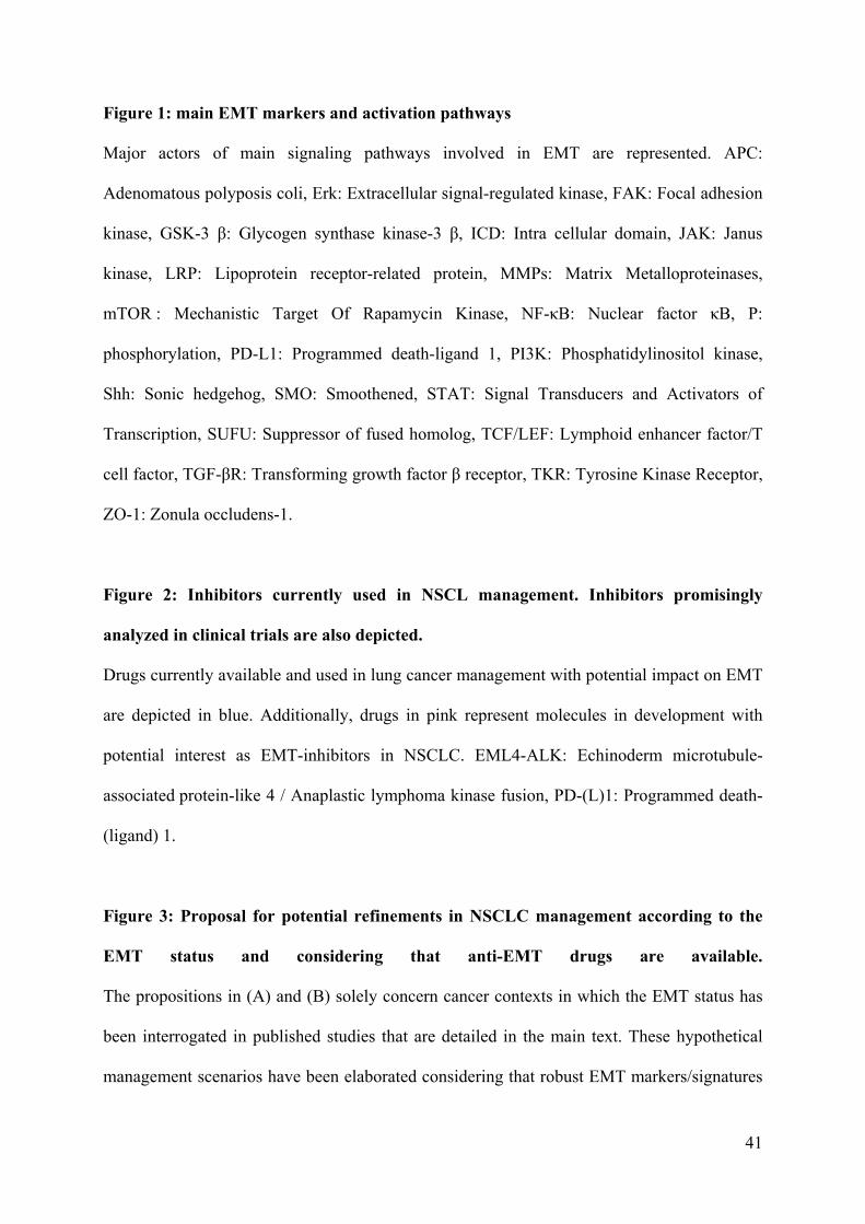
41
Figure 1: main EMT markers and activation pathways
Major actors of main signaling pathways involved in EMT are represented. APC:
Adenomatous polyposis coli, Erk: Extracellular signal-regulated kinase, FAK: Focal adhesion
kinase, GSK-3 β: Glycogen synthase kinase-3 β, ICD: Intra cellular domain, JAK: Janus
kinase, LRP: Lipoprotein receptor-related protein, MMPs: Matrix Metalloproteinases,
mTOR : Mechanistic Target Of Rapamycin Kinase, NF-κB: Nuclear factor κB, P:
phosphorylation, PD-L1: Programmed death-ligand 1, PI3K: Phosphatidylinositol kinase,
Shh: Sonic hedgehog, SMO: Smoothened, STAT: Signal Transducers and Activators of
Transcription, SUFU: Suppressor of fused homolog, TCF/LEF: Lymphoid enhancer factor/T
cell factor, TGF-βR: Transforming growth factor β receptor, TKR: Tyrosine Kinase Receptor,
ZO-1: Zonula occludens-1.
Figure 2: Inhibitors currently used in NSCL management. Inhibitors promisingly
analyzed in clinical trials are also depicted.
Drugs currently available and used in lung cancer management with potential impact on EMT
are depicted in blue. Additionally, drugs in pink represent molecules in development with
potential interest as EMT-inhibitors in NSCLC. EML4-ALK: Echinoderm microtubule-
associated protein-like 4 / Anaplastic lymphoma kinase fusion, PD-(L)1: Programmed death-
(ligand) 1.
Figure 3: Proposal for potential refinements in NSCLC management according to the
EMT status and considering that anti-EMT drugs are available.
The propositions in (A) and (B) solely concern cancer contexts in which the EMT status has
been interrogated in published studies that are detailed in the main text. These hypothetical
management scenarios have been elaborated considering that robust EMT markers/signatures

42
can be validated in clinical practice. The figure thus does not present current therapeutic
options.