An expression signature of syndecan-1 (CD138), E-cadherin ...
Upload
independentCategory
view
1download
0
The
Journ
al o
f Exp
erim
enta
l M
edic
ine
ARTICLE
JEM © The Rockefeller University Press $30.00
Vol. 204, No. 12, November 26, 2007 2935-2948 www.jem.org/cgi/doi/
2935
10.1084/jem.20071082
Notch signaling is initiated when a Notch li-gand interacts with a Notch transmembrane receptor expressed on an adjacent cell ( 1 ). This interaction triggers a series of proteolytic diges-tions that releases the Notch intracellular do-main (NotchIC), allowing it to translocate into the nucleus. Within the nucleus, NotchIC binds to the transcriptional repressor CSL, resulting in derepression and coactivation of Notch down-stream target genes and thereby regulating vari-ous cellular processes, including diff erentiation, proliferation, and apoptosis. Interestingly, in the development of cancer, Notch may act as either an oncogene or a tumor suppressor gene depend-ing on the tumor type ( 2 ).
Mammary-specifi c overexpression of constitu-tively active Notch1IC, Notch3IC, or Notch4IC in mice leads to the formation of aggressive, meta-static breast tumors ( 3, 4 ). Recent studies have also highlighted a potential role for Notch sig-naling in human breast cancer development. Expression of all four Notch receptors has been reported in human breast tumors at varying
frequencies ( 5 ). Poorly differentiated breast tumors are associated with elevated Notch1 pro-tein levels and reduced patient survival ( 6 ). Interestingly, an association between increased mRNA expression of the Notch ligand Jagged1 and reduced survival in patients with breast cancer has recently been reported, with high-level coexpression of Jagged1 and Notch1 mRNA defi ning a subset of patients with very poor outcome ( 7 ). Notch has also been re-ported to be activated downstream of Ras and Wnt in the promotion of mammary tumors through the induction of Notch ligands and/or receptors ( 8, 9 ). Notch signaling may contrib-ute to tumorigenesis by promoting mammary epithelial cell growth or inhibiting apoptosis ( 10, 11 ). However, much remains to be learned about the molecular mechanisms of Notch-mediated oncogenesis.
Numerous reports have indicated a role for epithelial-to-mesenchymal transition (EMT) in promoting the invasion and dissemination of malignant cells, particularly in breast cancer ( 12 ). Recent studies have suggested that Notch sig-naling induces a specialized type of EMT dur-ing normal heart development and that Notch
CORRESPONDENCE
Aly Karsan:
Abbreviations used: 5AZA,
5-azacytidine; cDNA, comple-
mentary DNA; CpG,
cytosine-phosphate-guanine;
EMT, epithelial-to-mesenchy-
mal transition; HA, hemag-
glutinin; HDAC, histone
deacetylase; MSP, methylation-
specifi c PCR; NaBu, sodium
butyrate; NotchIC, Notch in-
tracellular domain; qPCR,
quantitative RT-PCR; shRNA,
short hairpin RNA; siRNA,
small interfering RNA; TSA,
trichostatin A; YFP, yellow
fl uorescent protein.
K.G. Leong, K. Niessen, and I. Kulic contributed equally to
this work.
The online version of this article contains supplemental material.
Jagged1-mediated Notch activation induces epithelial-to-mesenchymal transition through Slug-induced repression of E-cadherin
Kevin G. Leong, 1,2,4 Kyle Niessen, 1,2,4 Iva Kulic, 1,2,4 Afshin Raouf, 3 Connie Eaves, 3,4,5,6 Ingrid Pollet, 1,2,5 and Aly Karsan 1,2,4,5
1 Department of Medical Biophysics, 2 Department of Pathology and Laboratory Medicine, and 3 Terry Fox Laboratory,
British Columbia Cancer Agency, Vancouver, British Columbia V5Z 1L3, Canada
4 Experimental Medicine Program, 5 Department of Pathology and Laboratory Medicine, and 6 Department of Medical Genetics,
University of British Columbia, Vancouver, British Columbia V6T 2B5, Canada
Aberrant expression of Jagged1 and Notch1 are associated with poor outcome in breast
cancer. However, the reason that Jagged1 and/or Notch overexpression portends a poor
prognosis is unknown. We identify Slug, a transcriptional repressor, as a novel Notch target
and show that elevated levels of Slug correlate with increased expression of Jagged1 in
various human cancers. Slug was essential for Notch-mediated repression of E-cadherin,
which resulted in � -catenin activation and resistance to anoikis. Inhibition of ligand-
induced Notch signaling in xenografted Slug-positive/E-cadherin – negative breast tumors
promoted apoptosis and inhibited tumor growth and metastasis. This response was associ-
ated with down-regulated Slug expression, reexpression of E-cadherin, and suppression of
active � -catenin. Our fi ndings suggest that ligand-induced Notch activation, through the
induction of Slug, promotes tumor growth and metastasis characterized by epithelial-to-
mesenchymal transition and inhibition of anoikis.

2936 JAGGED – NOTCH SIGNALING INDUCES EMT THROUGH SLUG | Leong et al.
positive human breast cancers. Jagged1-mediated activation of Notch in breast epithelial cells induces EMT through induction of Slug and subsequent repression of the cell – cell adhesion protein E-cadherin. Because Slug can be induced by factors other than Notch, we identify Notch downstream target genes of the HEY family as potential markers of pri-mary human breast tumors that have activated the Jagged1 – Notch – Slug signaling axis. In Slug-positive/E-cadherin – negative
up-regulates Snail in endothelial cells to promote mesenchy-mal transformation ( 13, 14 ). However, there is no direct or even correlative in vivo data that Notch regulates EMT in epithelial cancers.
In this paper, we identify Slug, a zinc-fi nger transcrip-tional repressor functionally linked to human breast cancer progression and metastasis ( 15 ), to be a direct downstream target gene of Notch that is up-regulated in Jagged1- and Notch1-
Figure 1. Notch activation inhibits E-cadherin expression in human breast epithelial cells through the induction of Slug. (A) Immunofl uorescent
staining for E-cadherin (red), YFP (green), and DAPI (blue) in primary human breast epithelial cells transduced with MIY, MIYNotch1IC, or MIYNotch4IC.
Bar, 50 � m. (B) qPCR for expression of E-cadherin, Slug, Snail, and Twist1 in MCF-10A cells transduced with MIY or MIYNotch1IC. Data are expressed as the
relative gene expression level, with MIY control as the comparator, and are from three independent experiments (mean + SEM). *, P ≤ 0.05. (C) Immuno-
fl uorescent staining for Slug (red), YFP (green), and DAPI (blue) in MCF-10A cells transduced with MIY or MIYNotch1IC. Bar, 50 � m. (D) qPCR for expression
of Slug and E-cadherin in MCF-10A cell lines (MIY, MIYNotch1IC, or MIYSlug) transiently transfected with siRandom or siSlug. Data from two independent
experiments are shown and are expressed as the relative gene expression level with MIY control as the comparator. n.d., not detectable.

JEM VOL. 204, November 26, 2007
ARTICLE
2937
each of the labeled oligonucleotides was competed away with unlabeled wild-type, but not mutated, double-stranded oligo-nucleotide ( Fig. 2, A and B ). In addition, lentiviral-delivered short hairpin RNAs (shRNAs) targeting two distinct sites of CSL ( Fig. 2 C ) also prevented the gel shift, indicating that these sites in the Slug promoter bind endogenous CSL and, thus, confi rming that Slug is a direct target of Notch/CSL. These results, combined with our data demonstrating the ability of Slug alone (independent of Notch1IC) to induce a spindle-shaped morphology and down-regulate E-cadherin expression in nor-mal breast epithelial cells (Fig. S2, available at http://www.jem.org/cgi/content/full/jem.20071082/DC1), identify Slug as a key player in the mechanism of Notch-induced EMT.
Jagged1/Notch1 correlates with Slug expression in human
breast cancers
To determine whether Jagged1-triggered Notch activation could induce Slug expression, Jagged1-expressing mouse en-dothelial cells (from the SVEC 4-10 cell line) were co-cultured
human breast cancer xenografts, inhibition of ligand-induced Notch signaling inhibits growth of the primary tumor and distant metastases, which correlates with reduced Slug expres-sion and reexpression of E-cadherin. E-cadherin reexpression, either through Notch inhibition or enforced expression, is associated with relocalization of � -catenin from the nucleus to the plasma membrane and reversal of � -catenin activation in xenografted breast tumors. Our fi ndings suggest a critical role for induction of EMT and inhibition of anoikis in pro-moting an aggressive phenotype in tumors exhibiting ligand-induced Notch signaling.
RESULTS
Notch activation inhibits E-cadherin expression in human
breast epithelial cells through the induction of Slug
Down-regulation of E-cadherin is one of the best markers of EMT in human breast cancer ( 12 ). To determine whether Notch activation induces EMT in human breast epithelial cells as manifested by repression of E-cadherin, the E-cadherin – positive normal human breast epithelial cell line MCF-10A was transduced with a retroviral vector (MIY) linking yellow fl uorescent protein (YFP) to activated Notch1 (Notch1IC) or activated Notch4 (Notch4IC). Hence, cells that express Notch1IC or Notch4IC also express YFP. Expression of either Notch1IC or Notch4IC caused this normal breast epithelial cell line to down-regulate E-cadherin, dissociate cell – cell con-tacts, and acquire a spindle-shaped morphology, consistent with mesenchymal transformation (Fig. S1, A – C, available at http://www.jem.org/cgi/content/full/jem.20071082/DC1). A similar ability of activated Notch to down-regulate E-cad-herin was demonstrated in primary human breast epithelial cells ( Fig. 1 A ).
To identify a potential mechanism of Notch-mediated E-cadherin silencing, expression of three known E-cadherin repressors that initiate EMT in breast cancer — Slug, Snail, and Twist1 ( 16 ) — was assessed in a normal breast epithelial cell line by quantitative RT-PCR (qPCR). In contrast to what has been reported in endothelial cells ( 13 ), Snail mRNA expression was not detected in either Notch1IC-expressing or control cells. Twist1 mRNA levels did not diff er between control and Notch1IC-expressing cells ( Fig. 1 B ). Slug mRNA expression, however, was signifi cantly increased in Notch1IC cells, which was associated with a decrease in E-cadherin mRNA expression ( Fig. 1 B ). Notch1IC-induced expression of Slug protein was confi rmed by immunofl uorescence micros-copy ( Fig. 1 C ). Knockdown of Slug, achieved by delivering small interfering RNA (siRNA) targeting Slug, into cells trans-duced with either Notch1IC or Slug was suffi cient to restore E-cadherin expression ( Fig. 1 D ).
To determine whether Slug is a direct target of Notch/CSL, we examined the human Slug promoter and identifi ed two potential CSL-binding consensus motifs ( − 846 to − 853 and − 1686 to − 1679 relative to the transcriptional start site). EMSAs using double-stranded oligonucleotides spanning these sites showed a clear gel shift of MDA-MB-231 human breast carcinoma nuclear lysates ( Fig. 2, A and B ). The binding of
Figure 2. Slug is a direct target of Notch signaling. EMSA of
nuclear lysates from MDA-MB-231 human breast cancer cells transduced
with nonspecifi c shRNA (shRand) or two different shRNAs targeting CSL
(shCSL1 and shCSL2). CSL consensus binding sites in the human Slug
promoter (A, − 846 to − 853 [TATGGGAA]; and B, − 1686 to − 1679
[TGTGGGAA]) relative to the transcriptional start site were used as the 32 P-
labeled probe, and either nonradioactive wild-type or mutated (mt) oligo-
nucleotides were used as competitors in 50-fold excess. The CSL – DNA
protein complex and the free DNA probe are identifi ed by arrows.
(C) shRNA-mediated knockdown of CSL in MDA-MB-231 cell lines was
verifi ed by RT-PCR analysis.

2938 JAGGED – NOTCH SIGNALING INDUCES EMT THROUGH SLUG | Leong et al.
or the Notch ligand Jagged1 ( 7 ). To examine whether increased Slug expression would be found in breast cancers showing increased Jagged1 or Notch1 expression, we accessed a com-pendium of 132 independent gene expression datasets repre-senting > 10,000 microarray experiments in the Oncomine database ( 17 ). Expression of both Jagged1 and Notch1 corre-lated positively with Slug in two independent breast cancer datasets ( Fig. 3, B and C ). Of signifi cance, a positive correlation between the expression of Jagged1 and Slug, but not Snail, was observed in numerous cancers other than the breast, which was consistent with our fi nding that Jagged1 is capable of inducing Slug but not Snail ( Fig. 3 D ).
with normal parental MCF-10A cells, and qPCR using human-specifi c primers was used to measure Slug expression in the human breast epithelial cells. Jagged1-induced Notch activa-tion, as demonstrated by induction of the target gene HEY1 (Fig. S2 E), resulted in increased levels of Slug mRNA levels with a concomitant decrease in E-cadherin mRNA expres-sion in these normal breast epithelial cells ( Fig. 3 A ). These fi ndings show that Jagged1 expression can activate Notch sig-naling in a juxtacrine manner to induce Slug expression and mesenchymal transformation of breast epithelial cells.
Poor prognosis for breast cancer patients has been shown to correlate with elevated levels of expression of either Slug ( 16 )
Figure 3. Jagged1 and Slug expression are correlated in human breast epithelial cells and in primary human breast cancer. (A) qPCR for ex-
pression of Slug and E-cadherin in MCF-10A parental cells co-cultured with mouse endothelial cells transduced with MIY vector control ( − Jagged1) or
MIYJagged1 (+Jagged1). Human-specifi c primers were used to avoid amplifi cation of mouse transcripts and limit analysis to MCF-10A cells. Data are
expressed as the relative gene expression level, with the empty vector control co-culture ( − Jagged1) as the comparator, and are from three independent
experiments (mean + SEM). Slug: *, P ≤ 0.05; E-cadherin: *, P < 0.0001. (B – D) Expression correlations in primary human cancers. Pearson correlation coeffi -
cients were obtained from microarray datasets deposited in the Oncomine database. For each dataset, individual correlations between two genes of
interest (as well as all possible correlations in cases where replicate probes were present in the microarray) are represented by open circles. Bars represent
the mean Pearson correlation coeffi cient. The numbers of normal and cancer specimens included in the correlation analysis for each dataset are indicated.
(B) Expression correlations between Jagged1 and Slug in primary human breast cancer. Jagged1 expression correlations with Snail are also shown. *,
P < 0.01; **, P < 0.001. (C) Expression correlations between Notch1 and Slug in primary human breast cancer. Notch1 expression correlations with Snail
are also shown. *, P < 0.01; **, P < 0.001. (D) Expression correlations between Jagged1 and Slug in various primary human cancers. Where available,
expression correlations between Jagged1 and Snail are also shown. *, P ≤ 0.05; **, P < 0.01; ***, P < 0.001. n.d., not determined.
JEM VOL. 204, November 26, 2007
ARTICLE
2939
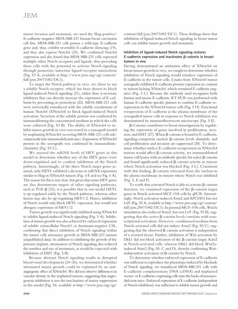
Although these fi ndings suggest that repression of E-cad-herin is mediated by Slug and not HES/HEY, HEY genes may represent surrogate markers of activation of the Jagged1 – Notch – Slug signaling axis in human breast cancer. Examination of two independent breast cancer microarray datasets confi rmed positive expression correlations between Jagged1 and HEY1, HEY2, and HEYL, respectively ( Fig. 4 C ). Moreover, the expression of HEY1, HEY2, and HEYL were all positively correlated with the expression of Slug but not Snail ( Fig. 4 D ). Thus, the HEY genes may be potential markers of human breast cancers that exhibit Notch activation and may, therefore, classify a subset of breast cancer patients that would benefi t from therapeutics specifi cally designed to target the Jagged1 – Notch – Slug pathway.
Inhibition of ligand-induced Notch activation blocks growth
and metastasis of breast tumors in vivo
To determine whether blockade of Notch ligand – receptor interaction would reverse Slug-induced EMT and inhibit breast
HEY genes are potential markers of human breast cancers
that exhibit activation of the Jagged1 – Notch – Slug
signaling axis
In response to Notch activation, transcriptional repressors of the HES and HEY families of basic helix-loop-helix proteins are induced ( 18 ). Cell type and context determine which members are induced ( 18 ). qPCR was used to analyze whether HES1 and HEY1/2/L were up-regulated in response to Notch activation in breast epithelial cells. Although HES1 levels did not diff er between vector control and Notch1IC cells, all three HEY genes were absent in control cells and up-regulated in the presence of Notch1IC ( Fig. 4 A ).
Because HEY proteins, similar to Slug, silence gene ex-pression by binding to E-boxes in target gene promoters ( 18 ), we determined whether enforced expression of any one of the HEY proteins was sufficient to down-regulate E-cadherin. In contrast to Slug (Fig. S2, B-E), none of the three HEY proteins altered E-cadherin transcript levels in these cells ( Fig. 4 B ).
Figure 4. HEY genes are potential markers of human breast cancers that exhibit activation of the Jagged1 – Notch – Slug signaling axis.
(A) qPCR for Notch target genes (HES1, HEY1, HEY2, and HEYL) in MCF-10A MIY and MIYNotch1IC cell lines. Data shown are the mean threshold cycle num-
ber (C T ) + SEM from three independent experiments. n.d., not detectable. (B) qPCR for gene expression in MCF-10A cells transduced with MIY, MIYHEY1,
MIYHEY2, or MIYHEYL. Data show relative gene expression level or threshold cycle number (C T ). n.d., not detectable. (C and D) Expression correlations in
primary human breast cancers. Pearson correlation coeffi cients were obtained from microarray datasets deposited in the Oncomine database. For each
dataset, correlations between two genes of interest (as well as all possible correlations in the event of replicate genes in the microarray) are represented
by open circles. Bars represent the mean Pearson correlation coeffi cient. The number of normal and cancer specimens included in the correlation analysis
for each dataset are indicated. n.d., not determined. (C) Expression correlations between Jagged1 and each of the HEY target genes in primary human
breast cancer. *, P ≤ 0.05; **, P < 0.001. (D) Expression correlations between each of the HEY target genes and Slug in primary human breast cancer. Where
available, expression correlations between each of the HEY target genes and Snail are also shown. *, P < 0.01; **, P < 0.001. (E) Semiquantitative RT-PCR
for Notch target genes in MDA-MB-231 MIG and MIGXNotch4HA tumor xenografts. Data are expressed as the relative gene expression level with MIG
control tumor as the comparator (mean + SEM). *, P ≤ 0.05. n.d.
2940 JAGGED – NOTCH SIGNALING INDUCES EMT THROUGH SLUG | Leong et al.
content/full/jem.20071082/DC1). These fi ndings show that inhibition of ligand-induced Notch signaling in breast tumor cells can inhibit tumor growth and metastasis.
Inhibition of ligand-induced Notch signaling restores
E-cadherin expression and inactivates � -catenin in breast
tumors in vivo
Having demonstrated an antitumor eff ect of XNotch4 on breast tumor growth in vivo, we sought to determine whether inhibition of Notch signaling would reinduce expression of E-cadherin in the tumor cells. Lysates from XNotch4 tumor xenografts exhibited E-cadherin protein expression in contrast to tumors lacking XNotch4, which remained E-cadherin neg-ative ( Fig. 5 C ). Because the antibody used recognizes both human and mouse E-cadherin, RT-PCR was performed with human E-cadherin – specifi c primers to confi rm E-cadherin re-expression in the XNotch4 tumor cells ( Fig. 5 D ). Functional reexpression of E-cadherin at the plasma membrane of the xenografted tumor cells in response to Notch inhibition was demonstrated by immunofl uorescent microscopy ( Fig. 5 E ).
� -Catenin contributes to breast tumorigenesis by regulat-ing the expression of genes involved in proliferation, inva-sion, and EMT ( 27 ). When � -catenin is bound to E-cadherin, signaling-competent nuclear � -catenin levels diminish, and cell proliferation and invasion are suppressed ( 28 ). To deter-mine whether surface E-cadherin reexpression in XNotch4 tumors would aff ect � -catenin activity, we immunoblotted tumor cell lysates with an antibody specifi c for active � -catenin and found signifi cantly reduced � -catenin activity in tumors where Notch activation was blocked ( Fig. 5 C ). Consistent with this fi nding, � -catenin relocated from the nucleus to the plasma membrane in tumors where Notch was inhibited ( Fig. 5, E and F ).
To verify that activated Notch is able to activate � -catenin function, we examined expression of the � -catenin target genes in Notch-activated MCF-10A cells by qPCR. Interest-ingly, Notch activation induced Axin2 and APCDD1 but not Lef1 (Fig. S5 A, available at http://www.jem.org/cgi/content/full/jem.20071082/DC1). In parental MCF-10A cells, Wnt3a stimulation also induced Axin2, but not Lef1 (Fig. S5 B), sug-gesting that the active � -catenin levels correlate with tran-scriptional activation. However, conditioned medium from Notch-activated cells did not induce Axin2 (Fig. S5 C), sug-gesting that the observed � -catenin activation is independent of a secreted factor. Further, inhibition of Wnt activation by Dkk1 did not block activation of the � -catenin target Axin2 in Notch-activated cells, whereas Dkk1 did block Wnt3a-induced Axin2 (Fig. S5, C and D), thereby confi rming Wnt-independent activation of � -catenin by Notch.
To determine whether enforced expression of E-cadherin was suffi cient to reproduce the phenotype induced by blockade of Notch signaling, we transduced MDA-MB-231 cells with E-cadherin complementary DNA (cDNA) and implanted vector- or E-cadherin – expressing cells onto the backs of immuno -defi cient mice. Enforced expression of E-cadherin (independent of Notch inhibition) was suffi cient to inhibit tumor growth and
tumor invasion and metastasis, we used the Slug-positive/E-cadherin – negative MDA-MB-231 human breast carcinoma cell line. MDA-MB-231 cells possess a wild-type E-cadherin gene and, thus, exhibit reversible E-cadherin silencing ( 19 ), and they also express Notch4 ( 20 ). We confi rmed Notch4 expression and also found that MDA-MB-231 cells expressed multiple other Notch receptors and ligands, thus providing these cells with the potential to activate Notch signaling through juxtacrine/autocrine ligand – receptor interactions (Fig. S3 A, available at http://www.jem.org/cgi/content/full/jem.20071082/DC1).
To target the Notch pathway in vivo, we chose to use a soluble Notch receptor, which has been shown to block ligand-induced Notch signaling ( 21 ), rather than � -secretase inhibitors that can directly increase the expression of E-cad-herin by preventing its proteolysis ( 22 ). MDA-MB-231 cells were retrovirally transduced with the soluble ectodomain of human Notch4 (XNotch4) to block ligand-induced Notch activation. Secretion of the soluble protein was confi rmed by immunoblotting the concentrated medium in which the cells were cultured (Fig. S3 B). The ability of XNotch4 to in-hibit tumor growth in vivo was tested in a xenograft model by implanting XNotch4-secreting MDA-MB-231 cells sub-cutaneously into immunodefi cient mice. Expression of XNotch4 protein in the xenografts was confi rmed by immunohisto-chemistry (Fig. S3 C).
We assessed the mRNA levels of HEY genes in this model to determine whether any of the HEY genes were down-regulated and to confi rm inhibition of the Notch pathway. Interestingly, of the three Notch target genes as-sessed, only HEYL exhibited a decrease in mRNA expression similar to Slug in XNotch4 tumors ( Fig. 4 E and see Fig. 6 A ). The reason for this is not clear, but given that some HEY genes are also downstream targets of other signaling pathways, such as TGF- � ( 23 ), it is possible that in our model HEYL is up-regulated solely by the Notch pathway, whereas other factors may also be up-regulating HEY1/2. Hence, inhibition of Notch would only block HEYL expression, but would not abrogate expression of HEY1/2.
Tumor growth was signifi cantly inhibited using XNotch4 to inhibit ligand-induced Notch signaling ( Fig. 5 A ). Inhibi-tion of tumor growth was also achieved by enforced expression of soluble extracellular Notch1 or dominant-negative CSL, confi rming that direct inhibition of Notch signaling within the tumor cells attenuates growth in MDA-MB-231 tumors (unpublished data). In addition to inhibiting the growth of the primary implant, attenuation of Notch signaling also reduced the number and size of metastases, as would be expected with inhibition of EMT ( Fig. 5 B ).
Because aberrant Notch signaling results in disrupted blood vessel development ( 24 – 26 ), we determined whether attenuated tumor growth could be explained by an anti-angiogenic eff ect of XNotch4. We did not observe diff erences in vascular density in the implanted tumors, suggesting that angio-genesis inhibition is not the mechanism of tumor suppression in this model (Fig. S4, available at http://www.jem.org/cgi/

JEM VOL. 204, November 26, 2007
ARTICLE
2941
Figure 5. Inhibition of ligand-induced Notch activation blocks breast tumor growth and metastasis, restores E-cadherin expression, and
inactivates � -catenin in vivo. (A) Tumor growth curves for MDA-MB-231 cells transduced with MIG or MIGXNotch4HA grown as xenografts on the
dorsa of immunodefi cient mice. Data are presented as the mean ± SEM of the tumor volumes. *, P < 0.01. (B) Quantitation of metastases in MDA-MB-231
MIG and MIGXNotch4HA tumor-bearing mice. Data shown represent the mean number of metastases per mouse + SEM and the mean weight of each
metastatic nodule + SEM. *, P ≤ 0.05. (C) Immunoblots for expression of XNotch4HA, E-cadherin, active � -catenin, and � -tubulin in MDA-MB-231 MIG
and MIGXNotch4HA tumors. Protein expression was quantitated by densitometry and normalized to � -tubulin. Data shown represent mean + SEM. *, P ≤
0.05. (D) RT-PCR for expression of human E-cadherin in MDA-MB-231 MIG and MIGXNotch4HA tumors. Human-specifi c primers were used to avoid am-
plification of mouse E-cadherin. (E) Immunofluorescent staining for E-cadherin (red), � -catenin (red), and DAPI (blue) in MDA-MB-231 MIG and
MIGXNotch4HA tumors. Bar, 15 � m. (F) Quantitation of the proportion of cells exhibiting nuclear � -catenin staining in MDA-MB-231 MIG and
MIGXNotch4HA tumors. Data shown represent mean + SEM. *, P ≤ 0.05. (G) Tumor growth curves for MDA-MB-231 cells transduced with MIY or MIYE-
cadherin grown as xenografts on the dorsa of immunodeficient mice. Data are presented as the mean ± SEM of the tumor volumes. *, P < 0.001.
(H) Quantitation of metastases in MDA-MB-231 MIY and MIYE-cadherin tumor-bearing mice. Data shown represent the mean number of metastases per
mouse + SEM and the mean weight of each metastatic nodule + SEM. *, P < 0.001. (I) Immunoblots for expression of E-cadherin and � -tubulin in MDA-
MB-231 MIY and MIYE-cadherin tumors. (J) Immunofl uorescent staining for E-cadherin (red), � -catenin (red), and DAPI (blue) in MDA-MB-231 MIY and
MIYE-cadherin tumors. Bar, 15 � m.
2942 JAGGED – NOTCH SIGNALING INDUCES EMT THROUGH SLUG | Leong et al.
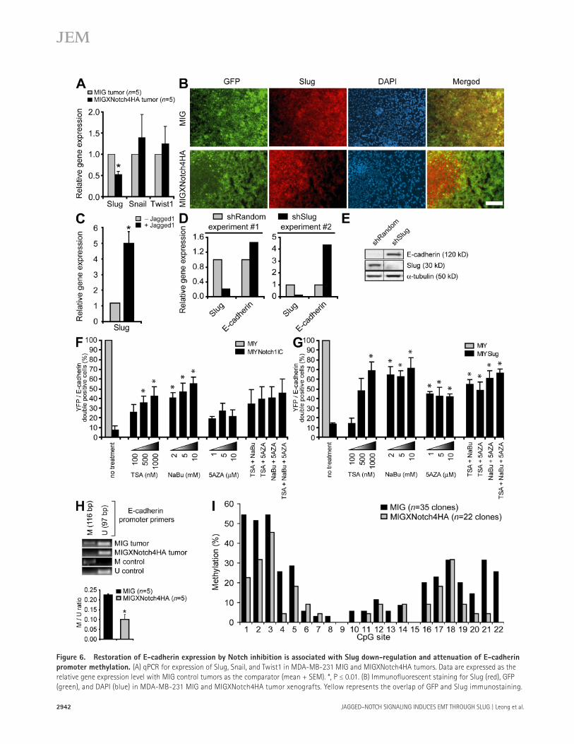
Figure 6. Restoration of E-cadherin expression by Notch inhibition is associated with Slug down-regulation and attenuation of E-cadherin
promoter methylation. (A) qPCR for expression of Slug, Snail, and Twist1 in MDA-MB-231 MIG and MIGXNotch4HA tumors. Data are expressed as the
relative gene expression level with MIG control tumors as the comparator (mean + SEM). *, P ≤ 0.01. (B) Immunofl uorescent staining for Slug (red), GFP
(green), and DAPI (blue) in MDA-MB-231 MIG and MIGXNotch4HA tumor xenografts. Yellow represents the overlap of GFP and Slug immunostaining.

JEM VOL. 204, November 26, 2007
ARTICLE
2943
Treatment of NotchIC- or Slug-expressing breast epithelial cells with the HDAC inhibitors trichostatin A (TSA) and sodium butyrate (NaBu), either alone or together, was suf-fi cient to reverse the down-regulation of E-cadherin ex-pression by Notch1IC or Slug ( Fig. 6, F and G ). A current molecular model for transcriptional gene silencing suggests that histone deacetylation is a primary event involved in the initiation of chromatin compaction, and that DNA methyla-tion functions subsequent to histone deacetylation to estab-lish a permanent state of gene inactivation ( 30 ). NotchIC- or Slug-expressing cells were thus treated with the DNA methyl-transferase inhibitor 5-azacytidine (5AZA), which also elic-ited reinduction of E-cadherin expression. However, 5AZA and TSA/NaBu together did not produce an additive eff ect on E-cadherin reexpression, suggesting that DNA methylation may occur secondary to histone deacetylation, as previously suggested ( 30 ).
The E-cadherin promoter contains numerous cytosine-phosphate-guanine (CpG) sites, which result in E-cadherin silencing when methylated on the corresponding cytosine residue ( 31 ). Given that DNA methylation has been reported to be dominant over histone deacetylation in mediating gene silencing ( 32 ), as well as our data suggesting that methyla-tion may occur as a later step in silencing the E-cadherin promoter, we sought to determine whether reduced Slug expression and reexpression of E-cadherin in our breast tumor xenografts correlated with reduced DNA methylation at the E-cadherin promoter. Using two independent methods, methylation-specifi c PCR (MSP) and genomic bisulfi te se-quencing, E-cadherin promoter methylation in XNotch4 tumors was found to be reduced compared with control tumors ( Fig. 6, H and I ). Because XNotch4 tumors did not display generalized hypomethylation of the genome (Fig. S6, A and B, available at http://www.jem.org/cgi/content/full/jem.20071082/DC1), Notch inhibition likely results in de-methylation at specifi c promoters regulated by Notch/Slug. These results suggest that inhibition of Notch signaling re-induces E-cadherin by repressing Slug and reversing E-cadherin promoter methylation.
metastasis with concomitant relocalization of � -catenin to the plasma membrane ( Fig. 5, G – J ). These data suggest that in-hibition of ligand-induced Notch activation is tumor sup-pressive, in part because of reinduction of surface E-cadherin expression. This in turn retains � -catenin at the plasma mem-brane, thereby attenuating � -catenin nuclear activity and in-hibiting tumor growth and metastasis.
Restoration of E-cadherin expression by Notch inhibition
is caused by Slug down-regulation and attenuation
of E-cadherin promoter methylation
To determine whether inhibition of ligand-induced Notch signaling restored E-cadherin expression secondary to repres-sion of Slug, expression of the genes encoding Slug, Snail, and Twist1 was assessed by qPCR in the breast tumor xeno-grafts. Although transcript levels of Snail and Twist1 did not diff er between control and XNotch4 tumors, Slug expression was signifi cantly reduced when Notch signaling was inhibited ( Fig. 6 A ). XNotch4 also blocked expression of Slug protein, because Slug was only present in areas of the tumor where XNotch4 was absent, as determined by immunofl uorescent staining of breast tumor xenografts ( Fig. 6 B ).
To directly demonstrate that Jagged1 could induce Slug in breast tumor cells, MDA-MB-231 parental cells were co- cultured with mouse stromal cells transduced with either Jagged1 or empty vector, and human Slug transcript levels were quantitated by RT-PCR using human-specifi c Slug primers. In response to Jagged1-induced Notch signaling, Slug transcripts in MDA-MB-231 cells were increased approxi-mately fi vefold ( Fig. 6 C ). To prove that Slug was responsible for E-cadherin repression in these cells, Slug was targeted using lentiviral-delivered shRNA in breast tumor cells. Knockdown of Slug was confi rmed by qPCR and immunoblotting, and was found to be suffi cient to restore expression of E-cadherin ( Fig. 6, D and E ).
Because Slug recruits histone deacetylase (HDAC) com-plexes to mediate transcriptional silencing ( 29 ), we deter-mined whether HDAC activity was required for the ability of Notch-induced Slug to down-regulate E-cadherin expression.
Bar, 100 � m. (C) qPCR for gene expression in MDA-MB-231 parental cells co-cultured with mouse endothelial cells transduced with MIY vector control
( − Jagged1) or MIYJagged1 (+Jagged1). Human-specifi c primers were used to assay Slug specifi cally in MDA-MB-231 cells and avoid amplifi cation of mouse
transcripts. Data are expressed as the relative gene expression level, with the empty vector control co-culture ( − Jagged1) as the comparator, and are from
three independent experiments (mean + SEM). *, P ≤ 0.05. (D) qPCR for expression of Slug and E-cadherin in MDA-MB-231 cells transduced with shRan-
dom or shSlug. Data from two independent experiments are shown and are expressed as the relative gene expression level with shRandom control as the
comparator. (E) Immunoblot for expression of E-cadherin, Slug, and � -tubulin in MDA-MB-231 cells transduced with shRandom or shSlug. (F) Quantita-
tion of the proportion of MCF-10A cells transduced with MIY or MIYNotch1IC that are positive for both YFP and E-cadherin. Cells were treated with TSA
(100/500/1000 nM), NaBu (2/5/10 mM), or 5AZA (1/5/10 � M) alone or in combination, and E-cadherin expression was assessed 3 d after treatment by
immunofl uorescent microscopy. Data shown represent mean + SEM of at least three independent experiments. *, P ≤ 0.05. (G) Quantitation of the propor-
tion of MCF-10A cells transduced with MIY or MIYSlug that are positive for both YFP and E-cadherin. Cells were treated with TSA (100/500/1000 nM),
NaBu (2/5/10 mM), or 5AZA (1/5/10 � M) alone or in combination. Data shown represent mean + SEM of at least three independent experiments. *, P ≤
0.05. (H) MSP to assess methylation status of the E-cadherin promoter in MDA-MB-231 tumors. Primers specifi c for methylated (M) or unmethylated (U)
E-cadherin promoter were used. Amplifi ed M and U products were quantitated by densitometry and expressed as the M/U ratio. Data shown represent
mean + SEM. *, P < 0.001. (I) Genomic bisulfi te sequencing to assess methylation status of the E-cadherin promoter in MDA-MB-231 tumors (MIG, n = 35
clones from fi ve tumors; MIGXNotch4HA, n = 22 clones from three tumors). A total of 22 CpG sites within the E-cadherin proximal promoter ( − 104 to
+118) were analyzed. Data shown represent the percentage of methylation observed at each CpG site.

2944 JAGGED – NOTCH SIGNALING INDUCES EMT THROUGH SLUG | Leong et al.
The Notch – Slug signaling axis inhibits anoikis of human
breast cells
During cancer progression and dissemination, malignant can-cer cells enter the circulation or lymphatic system and then must survive in the vasculature until they successfully extrav-asate into a tissue site. Cell death associated with abolished matrix-initiated integrin signaling has been termed anoikis. For a tumor cell to metastasize to a distant site, it needs to overcome anoikis. Activated Notch inhibits apoptosis in response to various triggers in diff erent cell types, but it has also been reported to induce apoptosis in other situations ( 11, 33, 34 ). We thus asked whether breast epithelial cells express-ing Notch1IC or Slug would exhibit a survival advantage compared with control cells when maintained under conditions that prevented adhesion. For this assay, cells were placed in suspension cultures, rather than soft agar ( 11 ), to mimic transit through the bloodstream. Both NotchIC- and Slug-expressing breast epithelial cells exhibited protection against anoikis compared with control cells ( Fig. 7 A ). Conversely, breast tumor xenografts in which Notch signaling was inhibited showed reduced Slug expression ( Fig. 6, A and B ), with a concomi-tant increase in cell death as measured by activated Caspase 3 levels ( Fig. 7 B ).
DISCUSSION
Although mouse models have indicated that mammary-spe-cifi c overexpression of a truncated constitutively active Notch can result in breast tumors, human data suggest that both Notch ligands and receptors are up-regulated in a proportion of breast cancers and that this expression is correlated with poor outcome. The implication is that juxtacrine or auto-crine Notch activation, rather than activating Notch muta-tions, may be responsible for an aggressive tumor phenotype. This is in marked contrast to T cell acute lymphoblastic leuke-mia, where more than half of the cases have activating muta-tions of Notch1 ( 35 ). In this paper, we have shown that Jagged1 activation of Notch up-regulates the transcriptional repressor Slug to promote carcinogenesis by two cellular mechanisms: (a) by facilitating cancer cell metastasis through initiation of EMT, and (b) by enhancing cell survival in the absence of cell matrix adhesion. Importantly, we have demonstrated that, in human breast cancers, expression of Jagged1 and Notch1 cor-relates positively with Slug expression.
In breast cancer patients, increased expression of either Jagged1 or Notch1 is predictive of poor overall survival ( 7 ). If both Jagged1 and Notch1 are increased, there is a further substantial reduction in overall survival ( 7 ). Our data provide an explanation for the reported dose-dependent relationship of Jagged1 expression and negative outcome in breast cancer ( 7 ), suggesting that juxtacrine or autocrine Jagged1 – Notch inter-actions induce Slug, which initiates EMT and inhibits anoikis, thereby promoting tumor invasion and metastasis. Interest-ingly, Jagged1 expression has also been associated with prostate cancer metastasis and recurrence ( 36 ), and our demonstration of positive correlations between Jagged1 and Slug expression in a wide variety of tumors raises the possibility that our fi nd-
ings may be generalized to other tumors in which Notch is activated by ligand.
Expression of the Notch target gene HEYL has previously been reported to be absent in the normal breast epithelium but present in the tumor cell compartment of invasive breast cancers ( 37 ). These fi ndings fi t with our results, which show that HEYL mRNA is undetectable in control breast epithe-lial cells but is up-regulated in cells exhibiting activation of the Notch – Slug signaling axis. Importantly, our analysis of primary human breast cancers has identifi ed HEY genes, in particular HEYL , as potential markers of human breast cancers that exhibit activation of the Jagged1 – Notch – Slug signaling axis.
The Snail gene has previously been reported to be a di-rect target gene of Notch in endothelial cells ( 13 ). However, our data suggest that this is not the case in epithelial cells, be-cause we did not observe a positive correlation between acti-vated Notch signaling and Snail induction. Rather, we observed a positive correlation between Notch activation and Slug ex-pression both in human breast epithelial cells in vitro and in primary human breast cancers. Hence, our fi ndings indicate that Slug, but not Snail, is a downstream target gene of Notch in epithelial and other tumors. Interestingly, increased levels of
Figure 7. The Notch – Slug signaling axis inhibits anoikis of
human breast cells. (A) Anoikis assay to assess cell death in MCF-10A
cell lines (MIY, MIYNotch1IC, and MIYSlug). The fraction of hypodiploid
cells was determined by fl ow cytometry, and the data representing the
mean + SEM of three independent experiments are expressed as the fold
change in hypodiploid cells relative to MIY control. MIYNotch1IC: *, P <
0.01; MIYSlug: *, P ≤ 0.05. (B) Immunohistochemical staining for activated
caspase 3 in implanted MDA-MB-231 MIG and MIGXNotch4HA tumors.
Data representing the mean + SEM are shown as the proportion of the
activated caspase 3 – stained area compared with the total tumor area.
*, P = 0.012. Bar, 25 � m.

JEM VOL. 204, November 26, 2007
ARTICLE
2945
play a role in maintaining the tumor stem cell, as well as in permitting these cells to survive the metastatic process.
Tumor cell metastasis is the primary cause of mortality in a vast majority of cancer patients. As such, there is a compel-ling need to elucidate the mechanisms of tumor cell metastasis and to develop rationally designed therapeutics to specifi cally target the metastatic process. Our fi ndings highlight the po-tential use of inhibitors of ligand-induced Notch signaling as viable and eff ective agents to block EMT and tumor metasta-sis in the treatment of human cancers that exhibit activation of the Jagged1 – Notch – Slug signaling axis. By specifi cally tar-geting ligand-induced activation, there is the potential of re-ducing the more widespread side eff ects potentially evoked by less-specifi c therapeutics, such as the � -secretase inhibitors.
MATERIALS AND METHODS Cell lines. The human breast epithelial cell line MCF-10A was cultured in
a 1:1 mixture of DMEM/F12 (Sigma-Aldrich) supplemented with 5% horse
serum (Sigma-Aldrich), 2 mM glutamine (Sigma-Aldrich), 20 ng/ml of epi-
dermal growth factor (Sigma-Aldrich), 100 ng/ml cholera toxin (Cedarlane),
10 � g/ml insulin (Sigma-Aldrich), 500 ng/ml hydrocortisone (Sigma-
Aldrich), and 100 U/ml each of penicillin and streptomycin (Invitrogen).
The human breast carcinoma cell lines MDA-MB-231 and T47D and the
mouse endothelial cell line SVEC4-10 were cultured in DMEM supple-
mented with 10% heat-inactivated calf serum (HyClone), 2 mM glutamine,
and 100 U/ml each of penicillin and streptomycin. All cells were maintained
at 37 ° C in an atmosphere of 5% CO 2 .
Isolation of primary human breast epithelial cells. Normal human
breast tissue was obtained in accordance with guidelines approved by the
University of British Columbia from anonymized discarded material from
normal premenopausal women undergoing reduction mammoplasty sur-
geries. A crude epithelial cell – enriched cell suspension was obtained enzy-
matically and cryopreserved. As required, cells were thawed, and single-cell
suspensions were prepared as previously described ( 45 ). Cells were co-
cultured with 1.2 × 10 6 X-irradiated NIH3T3 mouse fi broblasts for 1 d in
Epicult-B medium (StemCell Technologies Inc.) supplemented with 5% FCS.
Cells were harvested, and epithelial cell adhesion molecule (EpCAM) – positive
breast epithelial cells were magnetically separated using the human EpCAM
selection cocktail EasySep (StemCell Technologies Inc.). These selected cells
were cultured in Epicult-B medium at a density of 2 × 10 5 cells per dish in
35-mm dishes precoated with Vitrogen (Cohesion Technologies). To precoat
the dishes, 2 – 3 ml of Vitrogen (67 � g/ml in PBS) was used for 1 h at 37 ° C
and washed with PBS before use.
Plasmid constructs and gene transfer. Retroviral vectors (MIY and
MIG) containing an internal ribosomal entry site and either YFP (MIY) or
GFP (MIG) were used to facilitate the sorting of transduced cells. cDNA
constructs encoding the human Notch1IC, C-terminal hemagglutinin
(HA)-tagged human Notch4IC, full-length C-terminal Flag-tagged human
Slug (a gift of E. Fearon, University of Michigan, Ann Arbor, MI), full-
length human Jagged1, full-length human E-cadherin (a gift of B.M.
Gumbiner, University of Virginia, Charlottesville, VA), and N-terminal myc-
tagged full-length human HEY1/2/L (a gift of D. Srivastava, University of
California, San Francisco, San Francisco, CA) were subcloned into MIY. The
cDNA construct encoding the entire extracellular domain of human Notch4
(XNotch4; amino acids 1 – 1,443) tagged with a C-terminal HA epitope
was subcloned into MIG. The pLentilox3.7-shRandom and pLentilox3.7-shSlug
constructs were generated by inserting shRNAs targeting the sequences
5 � -GTTGCTTGCCACGTCCTAGAT-3 � (Random) and 5 � -GCATTT-
GCAGACAGGTCAAAT-3 � (Slug) into pLentilox3.7 (a gift of L. Van Parijs,
Massachusetts Institute of Technology, Cambridge, MA). Cells were transduced
Slug, but not Snail, have been associated with tumors from patients with metastatic disease or disease recurrence ( 16 ).
Although it has been reported that Slug has antiapoptotic activity in some cell types, the current study is the fi rst dem-onstration that Slug can inhibit anoikis. To initiate EMT, re-pression of E-cadherin permits loss of cell – cell cohesion, thereby disrupting apical-basal polarity and promoting cell migration. However, given that the loss of E-cadherin renders cells sus-ceptible to anoikis, Slug must independently promote cell survival in this context to allow local tumor invasion and distant metastases. Thus, our fi ndings that Notch blockade inhibits growth of the primary tumor by triggering apoptosis and re-duces distant metastases is concordant with this model of Slug-dependent EMT and cell survival.
Both Slug and Notch have been shown to inhibit p53 function, in part through the inhibition of Puma expression, which may be one mechanism of the antianoikis eff ect ( 11, 38 ). Notch has also been shown to activate the phosphatidylinositol 3 � -kinase – Akt antiapoptotic pathway, but whether there is cross talk with Slug remains to be seen ( 39, 40 ). The ability of Notch to suppress c-Jun N-terminal kinase activation and activate phosphatidylinositol 3 � -kinase – Akt may explain the inhibition of p53 by Notch ( 11, 33, 39 ). Both Notch and Slug have also been shown to induce Bcl-2 ( 33, 41 ); thus, there are multiple potential anoikis – apoptosis pathways that Notch may regulate through Slug.
It is also possible that Slug-mediated repression of E-cad-herin, which we have shown results in activation of � -catenin, may be responsible for an antianoikis eff ect. Indeed, even modest overexpression of active � -catenin has been shown to prevent anoikis ( 42 ). Of interest, � -catenin has been reported to induce Slug promoter activity ( 43 ). Hence, Notch induc-tion of Slug is potentially enhanced by a feed-forward loop. Initially, Notch/CSL would directly induce Slug. Subsequent Slug-mediated E-cadherin repression, via interaction with E2-boxes in the E-cadherin promoter ( 12 ), would then release � -catenin from the plasma membrane, which would accu-mulate in the nucleus and further activate the Slug promoter. However, the human Slug promoter also contains several cis elements predicted to bind the HEY / HES family of Notch-induced transcriptional repressors (unpublished data). Thus, these factors may act to attenuate the positive feedback loop described. Of note, our data showed that enforced expression of E-cadherin mimicked the eff ects of blocking Notch signal-ing in vivo, suggesting that a major function of Notch-induced Slug in this model may be to repress E-cadherin.
Recent evidence has demonstrated that breast cancers may be initiated in and propagated by a minority cell population that have been designated tumor stem cells ( 44 ). Notch signal-ing appears to play an important role in normal mammary stem cell self-renewal ( 44 ). Thus, the Notch pathway may be selec-tively activated in more primitive cancer stem cells. A mouse transgenic model of activated Notch4-driven breast cancer in-dicates that these tumors are highly metastatic ( 3 ). Interestingly, Slug has been shown to have antiapoptotic activity in hema-topoietic progenitor cells ( 38 ); thus, the Notch – Slug axis may
2946 JAGGED – NOTCH SIGNALING INDUCES EMT THROUGH SLUG | Leong et al.
annealing temperatures are described in Tables S1 and S2. Entire MSP reactions
were assessed in 2% TAE-agarose gels containing ethidium bromide. Bands
corresponding to methylated and unmethylated PCR products were quanti-
tated by densitometry. Data are expressed as a ratio of methylated over un-
methylated PCR products (M/U ratio) and represent the mean ratio + SE from
fi ve control tumors and fi ve XNotch4 tumors. For genomic bisulfi te sequenc-
ing, bisulfi te-modifi ed genomic DNA isolated from tumor tissue was amplifi ed
by PCR using primers spanning the E-cadherin proximal promoter. Primer
sequences and annealing temperatures are described in Tables S1 and S2. PCR
products were purifi ed and cloned into the pDrive cloning vector (QIAGEN),
and individual clones were sequenced. Five control tumors (a total of 35 MIG
clones) and three XNotch4 tumors (a total of 22 XNotch4 clones) were
analyzed, and data were expressed as the percentage of methylation per CpG
site (total number of methylated clones/total number of clones).
Immunostaining. Breast epithelial cells were stained with mouse mono-
clonal antibody to extracellular E-cadherin (Chemicon) or rabbit polyclonal
antibody to Slug (Santa Cruz Biotechnology, Inc.). 7- � m-thick tumor cryo-
sections were stained with rabbit polyclonal antibody to Slug (Santa Cruz
Biotechnology, Inc.), mouse monoclonal antibodies to E-cadherin (BD Bio-
sciences) or � -catenin (BD Biosciences), and rabbit monoclonal antibody to
activated caspase-3 (BD Biosciences). For immunohistochemistry, a biotin-
ylated goat anti – rabbit antibody (Vector Laboratories) followed by horseradish
peroxidase – conjugated streptavidin (Vector Laboratories) were used, and
nuclei were counterstained with hematoxylin. For immunofl uorescence, the
fl uorochrome-conjugated secondary antibodies goat anti – rat Alexa Fluor
594 (Invitrogen) and goat anti – mouse Alexa Fluor 594 (Invitrogen) were
used, and nuclei were counterstained with DAPI (Sigma-Aldrich). Immuno-
fl uorescence was detected with an imaging microscope (Axioplan II; Carl
Zeiss, Inc.), and images were captured with a digital camera (1350EX;
QImaging). To quantify the proportion of cells exhibiting nuclear � -catenin
staining, at least six random fi elds at 200 × magnifi cation were analyzed per
tumor using Northern Eclipse software. Data are presented as the mean per-
centage of nuclear � -catenin staining (total number of nuclear � -catenin –
positive cells/total number of cells) + SEM from four control tumors and
four XNotch4 tumors. To quantify the proportional area of tumors showing
activated caspase 3, entire tumor sections were analyzed using Northern
Eclipse software. Data are presented as the mean percentage of the activated
caspase 3 – stained area (total area positive for activated caspase 3/total area of
tumor section) + SEM from 14 control tumors and 12 XNotch4 tumors.
siRNA transfection. MCF-10A MIY and MIYNotch1IC cells were fl ow
sorted, and YFP-positive cells were transiently transfected with 100 nM
siRNA targeting the sequences 5 � -GTTGCTTGCCACGTCCTAGAT-3 �
(siRandom) or 5 � -GCATTTGCAGACAGGTCAAAT-3 � (siSlug) using
DharmaFECT 1 transfection reagent (Dharmacon Inc.). Cells were harvested
4 d after transfection.
Co-culture. 10 6 parental human cells (MCF-10A or MDA-MB-231) were
co-cultured with 10 6 mouse SVEC4-10 endothelial cells transduced with
MIY control vector or MIYJagged1 and plated in 100-mm tissue culture
dishes. Cells were harvested after 3 d of co-culture. qPCR was performed
with human-specifi c primers to avoid amplifi cation of mouse transcripts.
Anoikis assay. MCF-10A cell lines were fl ow sorted, and YFP-positive
cells were plated into 60-mm tissue culture plates (8 × 10 5 cells per plate)
coated with 1% agarose. After 24 h of incubation at 37 ° C, cells were fi xed/
permeabilized in 70% ethanol and stained with propidium iodide (Sigma-
Aldrich), and the proportion of cells with hypodiploid DNA content was
determined by fl ow cytometry. Data are expressed as the proportion of hy-
podiploid cells relative to MIY control.
Tumorigenicity assays. Female nonobese diabetic/severe combined
immunodefi cient mice were obtained from the Animal Resource Centre of
the British Columbia Cancer Research Centre. For MDA-MB-231 tumor
with empty vector control or vector containing cDNA inserts, and trans-
duced cells were sorted based on YFP or GFP expression using a cell sorter
(FACS 440; Becton Dickinson).
Immunoblotting. Cultured cells or tumor tissue were lysed and analyzed
by SDS-PAGE and immunoblotting with rabbit polyclonal antibody to Slug
(Santa Cruz Biotechnology, Inc.), or mouse monoclonal antibodies to HA
(Sigma-Aldrich), E-cadherin (BD Biosciences), � -catenin (BD Biosciences),
active � -catenin (Millipore), and � -tubulin (Sigma-Aldrich). Protein expression
was quantitated by densitometry.
RNA isolation, RT-PCR, and qPCR. Total RNA isolation was per-
formed using TRI zol reagent (Invitrogen) or an RNeasy kit (QIAGEN),
according to the manufacturers ’ recommendations. First-strand cDNA was
synthesized using Superscript II reverse transcriptase (Invitrogen). After ribo-
nuclease H treatment (Invitrogen), PCR was performed. Control reactions
omitting reverse transcriptase were performed in each experiment. Primer
sequences and annealing temperatures are described in Tables S1 and S2 (avail-
able at http://www.jem.org/cgi/content/full/jem.20071082/DC1). For RT-
PCR, entire PCR samples were assessed in 1.5% TAE-agarose gels containing
ethidium bromide. Control human cDNA was generated from pooled total
RNA isolated from the following human cells: human mammary epithelial
cells; vascular smooth muscle cells; cervical cancer cells, SiHa; colon cancer
cells, WiDr; and kidney epithelial cells, 293T. Control mouse cDNA was
generated from pooled total RNA isolated from the following mouse cells:
Lewis lung carcinoma cells; endothelial cells, SVEC4-10; and fi broblasts,
NIH3T3. For qPCR, reactions were run on a real-time PCR system (ABI
Prism 7900; Applied Biosystems). Gene expression was detected with SYBR
green (Applied Biosystems), and relative gene expression was determined by
normalizing to GAPDH using the � C T method.
EMSA. Nuclear lysates were collected from shRandom, shCSL1, and
shCSL2 overexpressing MDA-MB-231 cells for the CSL EMSA assays.
The binding reaction (10 mM TrisHCl, 50 mM NaCl, 1 mM EDTA
[pH 8], 4% glycerol, 2 � g PolydI-dC binding buff er, and 10 � g of nuclear
protein) was performed by preincubating with either 50-fold excess wild-type
(Slug 850, [forward] GGGCCCTTTTTCCCATAAAAAAAAAG and [reverse]
GGGAAAAAGGGTATTTTTTTTTCGGG; Slug 1680, [forward] TGTGT-
GTTTTGTGGGAAATGGAG and [reverse] CTCCATTTCCCACAAAA) or
mutant (Slug 800, [forward] GGGCCCTTTGCAGCATAAAAAAAAAAG
and [reverse] GGGAAACGTCGTATTTTTTTTTTCGGG; Slug 1600,
[forward] TGTGTGTTTTGTGCTGCATGGAG and [reverse] CTC-
CATGCAGCACAAAA) nonradioactive duplex oligos for 15 min on ice,
and then adding a 150,000-cpm 32 P-labeled double-stranded probe and incu-
bating for 30 min at room temperature. Binding reactions were run on 5%
Tris-Borate EDTA gels and exposed to a phosphorimager plate for 12 – 16 h.
Inhibition of histone acetylation and DNA methylation. MCF-10A
cell lines were fl ow sorted, and YFP-positive cells were plated into fourwell
chamber slides at 7 × 10 5 cells per well and allowed to adhere and grow for 48 h.
Cells were then treated with TSA (100/500/1000 nM), NaBu (2/5/10 mM),
and/or 5AZA (1/5/10 � M) for 72 h. After immunofl uorescent staining for
E-cadherin, at least six random fi elds at 200 × magnifi cation were analyzed
per well using Northern Eclipse software (Empix Imaging). Data are pre-
sented as the percentage of YFP/E-cadherin double-positive cells + SEM.
Methylation assays. Genomic DNA was isolated from cultured cells or
tumor tissue using a DNeasy tissue kit (QIAGEN) according to the manu-
facturer ’ s recommendations. 1 � g of genomic DNA was bisulfi te modifi ed
using a CpGenome DNA modifi cation kit (Chemicon) and eluted in 25 � l
Tris-EDTA buff er, according to the manufacturer ’ s recommendations.
MSP was performed using � 120 ng of bisulfi te-modifi ed DNA, 400 nM of
5 � and 3 � primers, 0.2 mM of 2 ’ -deoxynucleoside 5 � -triphosphates (Invitrogen),
1 × PCR buff er, and 0.625 U of HotStarTaq DNA polymerase (QIAGEN),
according to the manufacturer ’ s recommendations. Primer sequences and

JEM VOL. 204, November 26, 2007
ARTICLE
2947
REFERENCES 1 . Mumm , J.S. , and R. Kopan . 2000 . Notch signaling: from the outside in.
Dev. Biol. 228 : 151 – 165 . 2 . Leong , K.G. , and A. Karsan . 2006 . Recent insights into the role of
Notch signaling in tumorigenesis. Blood . 107 : 2223 – 2233 . 3 . Gallahan , D. , C. Jhappan , G. Robinson , L. Hennighausen , R. Sharp , E.
Kordon , R. Callahan , G. Merlino , and G.H. Smith . 1996 . Expression of a truncated Int3 gene in developing secretory mammary epithelium specifi -cally retards lobular diff erentiation resulting in tumorigenesis. Cancer Res. 56 : 1775 – 1785 .
4 . Hu , C. , A. Dievart , M. Lupien , E. Calvo , G. Tremblay , and P. Jolicoeur . 2006 . Overexpression of activated murine Notch1 and Notch3 in trans-genic mice blocks mammary gland development and induces mammary tumors. Am. J. Pathol. 168 : 973 – 990 .
5 . Callahan , R. , and S.E. Egan . 2004 . Notch signaling in mammary devel-opment and oncogenesis. J. Mammary Gland Biol. Neoplasia . 9 : 145 – 163 .
6 . Parr , C. , G. Watkins , and W.G. Jiang . 2004 . The possible correlation of Notch-1 and Notch-2 with clinical outcome and tumour clinicopatho-logical parameters in human breast cancer. Int. J. Mol. Med. 14 : 779 – 786 .
7 . Reedijk , M. , S. Odorcic , L. Chang , H. Zhang , N. Miller , D.R. McCready , G. Lockwood , and S.E. Egan . 2005 . High-level coexpression of JAG1 and NOTCH1 is observed in human breast cancer and is as-sociated with poor overall survival. Cancer Res. 65 : 8530 – 8537 .
8 . Ayyanan , A. , G. Civenni , L. Ciarloni , C. Morel , N. Mueller , K. Lefort , A. Mandinova , W. Raff oul , M. Fiche , G.P. Dotto , and C. Brisken . 2006 . Increased Wnt signaling triggers oncogenic conversion of hu-man breast epithelial cells by a Notch-dependent mechanism. Proc. Natl. Acad. Sci. USA . 103 : 3799 – 3804 .
9 . Weijzen , S. , P. Rizzo , M. Braid , R. Vaishnav , S.M. Jonkheer , A. Zlobin , B.A. Osborne , S. Gottipati , J.C. Aster , W.C. Hahn , et al . 2002 . Activation of Notch-1 signaling maintains the neoplastic phenotype in human Ras-transformed cells. Nat. Med. 8 : 979 – 986 .
10 . Pece , S. , M. Serresi , E. Santolini , M. Capra , E. Hulleman , V. Galimberti , S. Zurrida , P. Maisonneuve , G. Viale , and P.P. Di Fiore . 2004 . Loss of negative regulation by Numb over Notch is relevant to human breast carcinogenesis. J. Cell Biol. 167 : 215 – 221 .
11 . Stylianou , S. , R.B. Clarke , and K. Brennan . 2006 . Aberrant activation of notch signaling in human breast cancer. Cancer Res. 66 : 1517 – 1525 .
12 . Vincent-Salomon , A. , and J.P. Thiery . 2003 . Host microenvironment in breast cancer development: epithelial-mesenchymal transition in breast cancer development. Breast Cancer Res. 5 : 101 – 106 .
13 . Timmerman , L.A. , J. Grego-Bessa , A. Raya , E. Bertran , J.M. Perez-Pomares , J. Diez , S. Aranda , S. Palomo , F. McCormick , J.C. Izpisua-Belmonte , and J.L. de la Pompa . 2004 . Notch promotes epithe-lial-mesenchymal transition during cardiac development and oncogenic transformation. Genes Dev. 18 : 99 – 115 .
14 . Noseda , M. , G. McLean , K. Niessen , L. Chang , I. Pollet , R. Montpetit , R. Shahidi , K. Dorovini-Zis , L. Li , B. Beckstead , et al . 2004 . Notch activation results in phenotypic and functional changes consistent with endothelial-to-mesenchymal transformation. Circ. Res. 94 : 910 – 917 .
15 . Come , C. , V. Arnoux , F. Bibeau , and P. Savagner . 2004 . Roles of the tran-scription factors snail and slug during mammary morphogenesis and breast carcinoma progression. J. Mammary Gland Biol. Neoplasia . 9 : 183 – 193 .
16 . Martin , T.A. , A. Goyal , G. Watkins , and W.G. Jiang . 2005 . Expression of the transcription factors snail, slug, and twist and their clinical signifi -cance in human breast cancer. Ann. Surg. Oncol. 12 : 488 – 496 .
17 . Rhodes , D.R. , J. Yu , K. Shanker , N. Deshpande , R. Varambally , D. Ghosh , T. Barrette , A. Pandey , and A.M. Chinnaiyan . 2004 . ONCOMINE: a cancer microarray database and integrated data-mining platform. Neoplasia . 6 : 1 – 6 .
18 . Iso , T. , L. Kedes , and Y. Hamamori . 2003 . HES and HERP families: multi-ple eff ectors of the Notch signaling pathway. J. Cell. Physiol. 194 : 237 – 255 .
19 . van de Wetering , M. , N. Barker , I.C. Harkes , M. van der Heyden , N.J. Dijk , A. Hollestelle , J.G. Klijn , H. Clevers , and M. Schutte . 2001 . Mutant E-cadherin breast cancer cells do not display constitutive Wnt signaling. Cancer Res. 61 : 278 – 284 .
20 . Imatani , A. , and R. Callahan . 2000 . Identifi cation of a novel NOTCH-4/INT-3 RNA species encoding an activated gene product in certain human tumor cell lines. Oncogene . 19 : 223 – 231 .
implantation, 5 × 10 6 cells were injected subcutaneously into the dorsa of mice.
Once tumors were palpable, tumor volume (0.523 × length × width ×
height) was measured weekly using calipers. For each time point, MDA-
MB-231 tumor data are presented as the mean tumor volume ± SEM from
(a) 12 MIG tumors and 11 MIGXNotch4HA tumors, and (b) 12 MIY tu-
mors and 12 MIYE-cadherin tumors. Tumor-growth curves for one experi-
ment are presented and are representative of three independent experiments,
with 8 – 16 mice per tumor group for each experiment. Mice bearing MDA-
MB-231 tumors (MIG and MIGXNotch4HA, or MIY and MIYE-cadherin)
were killed at the same time, and the total number and total weight (in mil-
ligrams) of metastases were determined. Data are presented as the mean
number of metastases per mouse + SEM and the mean weight of each meta-
static nodule (in milligrams) + SEM. Metastasis data were determined by an-
alyzing (a) 25 MIG tumors and 27 MIGXNotch4HA tumors, and (b) 12 MIY
tumors and 12 MIYE-cadherin tumors. Animal experiments were approved
by the University of British Columbia Institutional Animal Care and Ethics
Committee, and all animals were handled according to institutional animal
care procedures.
Microarray data analysis. Pearson correlation coeffi cients were obtained
from publicly available microarray datasets deposited in the Oncomine data-
base (available at www.oncomine.org): breast 1 ( 46 ), breast 2 ( 47 ), adrenal ( 48 ),
brain ( 49 ), endocrine ( 50 ), gastric ( 51 ), lung ( 52 ), ovarian ( 53 ), renal ( 54 ),
salivary gland ( 55 ), sarcoma 1 ( 56 ), and sarcoma 2 ( 57 ). For each dataset,
individual correlations between two genes of interest (as well as all possible
correlations in the event of replicate genes in the microarray) were deter-
mined, as well as the mean Pearson correlation coeffi cient. Microarray data
accession numbers are shown in Table S3 (available at http://www.jem
.org/cgi/content/full/jem.20071082/DC1).
Statistical analysis. To determine statistical signifi cance, a one-way analysis
of variance with a Tukey test for multiple comparisons was performed using
the statistics program Statistical Package for Social Scientists (version 11.0;
SPSS Inc.). Statistical signifi cance was taken at P ≤ 0.05.
Online supplemental material. Tables S1 and S2 provide all primer se-
quences used in this study. Table S3 provides microarray accession number
information. Fig. S1 shows Notch-induced E-cadherin repression in MCF-
10A cells. Fig. S2 shows Notch-induced Slug expression in MCF-10A cells.
Fig. S3 demonstrates the expression of Notch ligands and receptors in MDA-
MB-231 cells, and secreted XNotch4HA protein. Fig. S4 shows a lack
of a vascular eff ect by XNotch4HA. Fig. S5 demonstrates Notch-induced
� -catenin activation independent of Wnt activation. Fig. S6 reveals that
inhibition of Notch does not cause a generalized hypomethylation of the
genome. Online supplemental material is available at http://www.jem.org/
cgi/content/full/jem.20071082/DC1.
We thank F. Wong and D. McDougal for assistance with fl ow cytometry and cell
sorting. We also thank P.L. Olive for critical review of the manuscript.
This research was supported by grants to A. Karsan from the National Cancer
Institute of Canada with funds from the Canadian Cancer Society and the Cancer
Research Society, and to C. Eaves from Genome Canada. K.G. Leong was supported
by a Doctoral Research Award from the Canadian Institutes of Health Research and
a Predoctoral Fellowship Award from the Department of the Army (DAMD17-01-1-
0164). The U.S. Army Medical Research Acquisition Activity was the awarding and
administering acquisition offi ce. K. Niessen is the recipient of a Senior Graduate
Studentship from the Michael Smith Foundation for Health Research, and I. Kulic
is supported by a Postgraduate Scholarship from the Natural Sciences and
Engineering Research Council of Canada and a Junior Graduate Studentship from
the Michael Smith Foundation for Health Research. A. Raouf is supported by a
Post-doctoral Research Award from the Canadian Institutes of Health Research.
A. Karsan is a Senior Scholar of the Michael Smith Foundation for Health Research.
The authors declare that they have no competing fi nancial interests.
Submitted: 30 May 2007
Accepted: 16 October 2007

2948 JAGGED – NOTCH SIGNALING INDUCES EMT THROUGH SLUG | Leong et al.
40 . Androutsellis-Theotokis , A. , R.R. Leker , F. Soldner , D.J. Hoeppner , R. Ravin , S.W. Poser , M.A. Rueger , S.K. Bae , R. Kittappa , and R.D. McKay . 2006 . Notch signalling regulates stem cell numbers in vitro and in vivo. Nature . 442 : 823 – 826 .
41 . Bermejo-Rodriguez , C. , M. Perez-Caro , P.A. Perez-Mancera , M. Sanchez-Beato , M.A. Piris , and I. Sanchez-Garcia . 2006 . Mouse cDNA microarray analysis uncovers Slug targets in mouse embryonic fi bro-blasts. Genomics . 87 : 113 – 118 .
42 . Weng , Z. , M. Xin , L. Pablo , D. Grueneberg , M. Hagel , G. Bain , T. Muller , and J. Papkoff . 2002 . Protection against anoikis and down-regula-tion of cadherin expression by a regulatable beta-catenin protein. J. Biol. Chem. 277 : 18677 – 18686 .
43 . Conacci-Sorrell , M. , I. Simcha , T. Ben-Yedidia , J. Blechman , P. Savagner , and A. Ben-Ze ’ ev . 2003 . Autoregulation of E-cadherin ex-pression by cadherin – cadherin interactions: the roles of � -catenin sig-naling, Slug, and MAPK. J. Cell Biol. 163 : 847 – 857 .
44 . Liu , S. , G. Dontu , and M.S. Wicha . 2005 . Mammary stem cells, self-renewal pathways, and carcinogenesis. Breast Cancer Res. 7 : 86 – 95 .
45 . Stingl , J. , C.J. Eaves , I. Zandieh , and J.T. Emerman . 2001 . Characterization of bipotent mammary epithelial progenitor cells in normal adult human breast tissue. Breast Cancer Res. Treat. 67 : 93 – 109 .
46 . van ‘ t Veer , L.J. , H. Dai , M.J. van de Vijver , Y.D. He , A.A. Hart , M. Mao , H.L. Peterse , K. van der Kooy , M.J. Marton , A.T. Witteveen , et al . 2002 . Gene expression profi ling predicts clinical outcome of breast cancer. Nature . 415 : 530 – 536 .
47 . Zhao , H. , A. Langerod , Y. Ji , K.W. Nowels , J.M. Nesland , R. Tibshirani , I.K. Bukholm , R. Karesen , D. Botstein , A.L. Borresen-Dale , and S.S. Jeff rey . 2004 . Diff erent gene expression patterns in invasive lobular and ductal carcinomas of the breast. Mol. Biol. Cell . 15 : 2523 – 2536 .
48 . Giordano , T.J. , D.G. Thomas , R. Kuick , M. Lizyness , D.E. Misek , A.L. Smith , D. Sanders , R.T. Aljundi , P.G. Gauger , N.W. Thompson , et al . 2003 . Distinct transcriptional profi les of adrenocortical tumors uncov-ered by DNA microarray analysis. Am. J. Pathol. 162 : 521 – 531 .
49 . Freije , W.A. , F.E. Castro-Vargas , Z. Fang , S. Horvath , T. Cloughesy , L.M. Liau , P.S. Mischel , and S.F. Nelson . 2004 . Gene expression profi l-ing of gliomas strongly predicts survival. Cancer Res. 64 : 6503 – 6510 .
50 . Jain , S. , M.A. Watson , M.K. DeBenedetti , Y. Hiraki , J.F. Moley , and J. Milbrandt . 2004 . Expression profi les provide insights into early malig-nant potential and skeletal abnormalities in multiple endocrine neoplasia type 2B syndrome tumors. Cancer Res. 64 : 3907 – 3913 .
51 . Chen , X. , S.Y. Leung , S.T. Yuen , K.M. Chu , J. Ji , R. Li , A.S. Chan , S. Law , O.G. Troyanskaya , J. Wong , et al . 2003 . Variation in gene expres-sion patterns in human gastric cancers. Mol. Biol. Cell . 14 : 3208 – 3215 .
52 . Garber , M.E. , O.G. Troyanskaya , K. Schluens , S. Petersen , Z. Thaesler , M. Pacyna-Gengelbach , M. van de Rijn , G.D. Rosen , C.M. Perou , R.I. Whyte , et al . 2001 . Diversity of gene expression in adenocarci-noma of the lung. Proc. Natl. Acad. Sci. USA . 98 : 13784 – 13789 .
53 . Schaner , M.E. , D.T. Ross , G. Ciaravino , T. Sorlie , O. Troyanskaya , M. Diehn , Y.C. Wang , G.E. Duran , T.L. Sikic , S. Caldeira , et al . 2003 . Gene expression patterns in ovarian carcinomas. Mol. Biol. Cell . 14 : 4376 – 4386 .
54 . Higgins , J.P. , R. Shinghal , H. Gill , J.H. Reese , M. Terris , R.J. Cohen , M. Fero , J.R. Pollack , M. van de Rijn , and J.D. Brooks . 2003 . Gene expression patterns in renal cell carcinoma assessed by complementary DNA microarray. Am. J. Pathol. 162 : 925 – 932 .
55 . Frierson , H.F. , Jr ., A.K. El-Naggar , J.B. Welsh , L.M. Sapinoso , A.I. Su , J. Cheng , T. Saku , C.A. Moskaluk , and G.M. Hampton . 2002 . Large scale molecular analysis identifi es genes with altered expression in sali-vary adenoid cystic carcinoma. Am. J. Pathol. 161 : 1315 – 1323 .
56 . Linn , S.C. , R.B. West , J.R. Pollack , S. Zhu , T. Hernandez-Boussard , T.O. Nielsen , B.P. Rubin , R. Patel , J.R. Goldblum , D. Siegmund , et al . 2003 . Gene expression patterns and gene copy number changes in der-matofi brosarcoma protuberans. Am. J. Pathol. 163 : 2383 – 2395 .
57 . Segal , N.H. , P. Pavlidis , W.S. Noble , C.R. Antonescu , A. Viale , U.V. Wesley , K. Busam , H. Gallardo , D. DeSantis , M.F. Brennan , et al . 2003 . Classifi cation of clear-cell sarcoma as a subtype of melanoma by genomic profi ling. J. Clin. Oncol. 21 : 1775 – 1781 .
21 . Small , D. , D. Kovalenko , D. Kacer , L. Liaw , M. Landriscina , C. Di Serio , I. Prudovsky , and T. Maciag . 2001 . Soluble Jagged 1 represses the function of its transmembrane form to induce the formation of the Src-dependent chord-like phenotype. J. Biol. Chem. 276 : 32022 – 32030 .
22 . Marambaud , P. , J. Shioi , G. Serban , A. Georgakopoulos , S. Sarner , V. Nagy , L. Baki , P. Wen , S. Efthimiopoulos , Z. Shao , et al . 2002 . A presenilin-1/gamma-secretase cleavage releases the E-cadherin intracellular domain and regulates disassembly of adherens junctions. EMBO J. 21 : 1948 – 1956 .
23 . Zavadil , J. , L. Cermak , N. Soto-Nieves , and E.P. Bottinger . 2004 . Integration of TGF-beta/Smad and Jagged1/Notch signalling in epithe-lial-to-mesenchymal transition. EMBO J. 23 : 1155 – 1165 .
24 . Krebs , L.T. , Y. Xue , C.R. Norton , J.R. Shutter , M. Maguire , J.P. Sundberg , D. Gallahan , V. Closson , J. Kitajewski , R. Callahan , et al . 2000 . Notch signaling is essential for vascular morphogenesis in mice. Genes Dev. 14 : 1343 – 1352 .
25 . Leong , K.G. , X. Hu , L. Li , M. Noseda , B. Larrivee , C. Hull , L. Hood , F. Wong , and A. Karsan . 2002 . Activated Notch4 inhibits angiogenesis: role of beta 1-integrin activation. Mol. Cell. Biol. 22 : 2830 – 2841 .
26 . Zeng , Q. , S. Li , D.B. Chepeha , T.J. Giordano , J. Li , H. Zhang , P.J. Polverini , J. Nor , J. Kitajewski , and C.Y. Wang . 2005 . Crosstalk between tumor and endothelial cells promotes tumor angiogenesis by MAPK activation of Notch signaling. Cancer Cell . 8 : 13 – 23 .
27 . Hatsell , S. , T. Rowlands , M. Hiremath , and P. Cowin . 2003 . Beta-catenin and Tcfs in mammary development and cancer. J. Mammary Gland Biol. Neoplasia . 8 : 145 – 158 .
28 . Wong , A.S. , and B.M. Gumbiner . 2003 . Adhesion-independent mech-anism for suppression of tumor cell invasion by E-cadherin. J. Cell Biol. 161 : 1191 – 1203 .
29 . Tripathi , M.K. , S. Misra , S.V. Khedkar , N. Hamilton , C. Irvin-Wilson , C. Sharan , L. Sealy , and G. Chaudhuri . 2005 . Regulation of BRCA2 gene expression by the SLUG repressor protein in human breast cells. J. Biol. Chem. 280 : 17163 – 17171 .
30 . Bachman , K.E. , B.H. Park , I. Rhee , H. Rajagopalan , J.G. Herman , S.B. Baylin , K.W. Kinzler , and B. Vogelstein . 2003 . Histone modifi cations and silencing prior to DNA methylation of a tumor suppressor gene. Cancer Cell . 3 : 89 – 95 .
31 . Berx , G. , K. Staes , J. van Hengel , F. Molemans , M.J. Bussemakers , A. van Bokhoven , and F. van Roy . 1995 . Cloning and characterization of the hu-man invasion suppressor gene E-cadherin (CDH1). Genomics . 26 : 281 – 289 .
32 . Cameron , E.E. , K.E. Bachman , S. Myohanen , J.G. Herman , and S.B. Baylin . 1999 . Synergy of demethylation and histone deacetylase inhibition in the re-expression of genes silenced in cancer. Nat. Genet. 21 : 103 – 107 .
33 . MacKenzie , F. , P. Duriez , F. Wong , M. Noseda , and A. Karsan . 2004 . Notch4 inhibits endothelial apoptosis via RBP-Jkappa-dependent and -independent pathways. J. Biol. Chem. 279 : 11657 – 11663 .
34 . Zweidler-McKay , P.A. , Y. He , L. Xu , C.G. Rodriguez , F.G. Karnell , A.C. Carpenter , J.C. Aster , D. Allman , and W.S. Pear . 2005 . Notch signaling is a potent inducer of growth arrest and apoptosis in a wide range of B cell malignancies. Blood . 106 : 3898 – 3906 .
35 . Weng , A.P. , A.A. Ferrando , W. Lee , J.P. Morris IV, L.B. Silverman , C. Sanchez-Irizarry , S.C. Blacklow , A.T. Look , and J.C. Aster . 2004 . Activating mutations of NOTCH1 in human T cell acute lymphoblastic leukemia. Science . 306 : 269 – 271 .
36 . Santagata , S. , F. Demichelis , A. Riva , S. Varambally , M.D. Hofer , J.L. Kutok , R. Kim , J. Tang , J.E. Montie , A.M. Chinnaiyan , et al . 2004 . JAGGED1 expression is associated with prostate cancer metastasis and recurrence. Cancer Res. 64 : 6854 – 6857 .
37 . Parker , B.S. , P. Argani , B.P. Cook , H. Liangfeng , S.D. Chartrand , M. Zhang , S. Saha , A. Bardelli , Y. Jiang , T.B. St Martin , et al . 2004 . Alterations in vascular gene expression in invasive breast carcinoma. Cancer Res. 64 : 7857 – 7866 .
38 . Wu , W.S. , S. Heinrichs , D. Xu , S.P. Garrison , G.P. Zambetti , J.M. Adams , and A.T. Look . 2005 . Slug antagonizes p53-mediated apoptosis of hematopoietic progenitors by repressing puma. Cell . 123 : 641 – 653 .
39 . Mungamuri , S.K. , X. Yang , A.D. Thor , and K. Somasundaram . 2006 . Survival signaling by Notch1: mammalian target of rapamycin (mTOR)-dependent inhibition of p53. Cancer Res. 66 : 4715 – 4724 .
Copyright © 2022 FDOKUMEN