Anti-tau antibody reduces insoluble tau and decreases brain atrophy
Upload
independentCategory
view
2download
0
Chronic exposure of neural cells to elevated intracellular sodiumdecreases mitochondrial mRNA expression
Krish Chandrasekarana,*, Li-Ing Liub, Kimmo HatanpaÈaÈb, Umesha Shettyb,Zara Mehrabiana, Peter D. Murraya, Gary Fiskuma, Stanley I. Rapoportb
aDepartment of Anesthesiology, University of Maryland School of Medicine, MSTF 5-34, 685 West Baltimore Street,
Baltimore, MD 21201, USAbSection on Brain Physiology and Metabolism, National Institute on Aging, NIH, Bethesda, MD 20892, USA
Received 14 December 2000; received in revised form 21 March 2001; accepted 29 March 2001
Abstract
Regulation of expression of mitochondrial DNA- (mtDNA-) encoded genes of oxidative phosphorylation can occur rapidly in
neural cells subjected to a variety of physiological and pathological conditions. However, the intracellular signal(s) involved in
regulating these processes remain unknown. Using mtDNA-encoded cytochrome oxidase subunit III (COX III), we show that
its mRNA expression in a differentiated rat pheochromocytoma cell line PC12S is decreased by chronic exposure to agents that
increase intracellular sodium. Treatment of differentiated PC12S cells either with ouabain, an inhibitor of Na/K-ATPase, or
with monensin, a sodium ionophore, decreased the steady-state levels of COX III mRNA by 50%, 3±4 h after addition of the
drugs. No signi®cant reduction in mtDNA-encoded 12S rRNA or nuclear DNA-encoded b-actin mRNA were observed.
Removal of the drugs restored the normal levels of COX III mRNA. Determination of half-lives of COX III mRNA, 12S
rRNA, and b-actin mRNA revealed a selective decrease in the half-life of COX III mRNA from 3.3 h in control cells to 1.6 h in
ouabain-treated cells, and to 1 h in monensin-treated cells. These results suggest the existence of a mechanism of posttran-
scriptional regulation of mitochondrial gene expression that is independent of the energetic status of the cell and may operate
under pathological conditions. q 2001 Elsevier Science B.V. and Mitochondria Research Society. All rights reserved.
Keywords: Neural cells; Mitochondrial mRNA; Intracellular sodium
1. Introduction
Neurons depend on a high rate of mitochondrial
oxidative metabolism to produce ATP (Erecinska
and Silver, 1989). ATP is needed for ion pumping
to restore the cellular membrane potential after depo-
larization. The mitochondrial respiratory chain
consists of ®ve multisubunit oxidative phosphoryla-
tion (OXPHOS) enzyme complexes. Four of these
OXPHOS complexes, including Complex IV (cyto-
chrome oxidase (COX)), are bipartite in nature,
consisting of subunits derived from both mitochon-
drial DNA (mtDNA) and nuclear DNA (nDNA).
Mitochondrial DNA encodes 13 polypeptides, all of
which are necessary for electron transport and
OXPHOS. The large number of remaining subunits
is speci®ed by the nuclear genome. To form active
enzyme complexes, both mtDNA- and nDNA-
Mitochondrion 1 (2001) 141±150
1567-7249/01/$20.00 q 2001 Elsevier Science B.V. and Mitochondria Research Society. All rights reserved.
PII: S1567-7249(01)00010-1
www.elsevier.com/locate/mito
* Corresponding author. Tel.: 11-410-706-3418; fax: 11-410-
706-2550.
E-mail address: [email protected]
(K. Chandrasekaran).

encoded subunits are required (Attardi and Schatz,
1988).
Neuronal activity and energy demand in¯uence the
expression of mitochondrial DNA-encoded genes. For
example, under conditions of decreased neuronal
activity induced by afferent impulse blockade, neuro-
nal mitochondrial gene expression and COX enzyme
activity are decreased (Wong-Riley, 1989; Wong-
Riley et al., 1997). Removal of the afferent impulse
blockade restores basal mitochondrial gene expres-
sion and COX activity (Wong-Riley, 1989; Wong-
Riley et al., 1997). Such regulation occurs mainly at
the transcriptional level (Wong-Riley et al., 1997;
Zhang and Wong-Riley, 2000a,b). Using the in orga-
nello method, it was shown that high intramitochon-
drial ATP levels suppress transcription of mtDNA,
explaining how energy demand can regulate mtDNA
transcription (Gaines and Attardi, 1984; Enriquez et
al., 1996a,b). Apart from transcriptional control, the
primary regulation of mitochondrial gene expression
is based on differences in RNA stability. Thus,
although mitochondrial gene expression is a major
component in the regulation of energy metabolism
of the cell, the contribution of transcriptional and
posttranscriptional mechanisms to the overall regula-
tion of mitochondrial gene expression is not known
(Kagawa and Ohta, 1990).
The goal of our present study was to probe the
mechanism of regulation of mitochondrial gene
expression under conditions of chronic exposure to
two drugs that increase intracellular sodium ([Na]i)
but by two different independent mechanisms.
Ouabain reduces cellular Na1 ef̄ ux by inhibiting the
Na/K-ATPase and would be expected to reduce cellu-
lar energy demand. Monensin is a Na1 ionophore,
which in contrast to ouabain, would increase the cellu-
lar energy demand through futile cycling of Na1 across
the plasma membrane. These expected effects on
energy metabolism were con®rmed by measurements
of cellular ATP/ADP ratios and compared to the levels
of mtDNA-encoded cytochrome oxidase subunit
(COX III) mRNA in cultures of nerve growth factor-
(NGF-) induced differentiated PC12 neural cells. Both
the levels and half-lives of COX III mRNA, mtDNA-
encoded 12S rRNA, and nDNA-encoded b-actin
mRNA were quanti®ed. Our results indicate a selective
decrease in mtDNA-encoded mRNA stability in both
ouabain- and monensin-treated cells, suggesting a
Na1-mediated mechanism of posttranscriptional regu-
lation that is independent of the energetic status of
the cell in neuronal cultures. A part of this work has
been published as abstract (Liu et al., 1999).
2. Methods and materials
2.1. Cell culture
A morphological variant of rat pheochromocytoma
PC12 cells (PC12S) that has the ability to grow in
tissue culture dishes without polylysine treatment
was used in experiments (Fukuyama et al., 1993).
PC12S cells were maintained in Dulbecco's modi®ed
Eagle's medium (DMEM) containing 2 mM gluta-
mine, 7.5% heat inactivated fetal calf serum, 7.5%
heat inactivated horse serum, and penicillin±strepto-
mycin. Differentiation was induced by the addition of
NGF (Life Technologies, MD, USA) at 50 mg/ml to
the cell culture medium. We showed previously that
the morphology of PC12S cells resembles that of
sympathetic neurons after addition of NGF for 5
days (Fukuyama et al., 1993). Differentiated PC12S
maintained in NGF for 10 days was used throughout
the experiments.
2.2. Chemicals
All reagents and chemicals used were of the highest
grade available from Sigma Chemical Co. (St. Louis,
MO, USA). Stock solutions of actinomycin D and
ouabain were prepared in water, whereas monensin
was dissolved in 95% ethanol. When ethanol was
used as a solvent, appropriate control experiments
were conducted using the vehicle alone. Ethanol
concentrations were always ,0.1%.
2.3. Experimental procedure
Cells grown in 60 £ 15 mm dishes were treated
either with ouabain, at a ®nal concentration of
1 mM, or with monensin, at a ®nal concentration of
100 nM. At timed points over a 6-h period, cells were
washed with Dulbecco's phosphate buffered saline
(DPBS) without calcium and magnesium and total
RNA was isolated using the TRlzol reagent as recom-
mended by the manufacturer (Life Technologies, MD,
K. Chandrasekaran et al. / Mitochondrion 1 (2001) 141±150142

USA). Total RNA was subjected to Northern blot
analysis as described below.
The reversibility of the effect of ouabain and
monensin on mitochondrial gene expression was eval-
uated by exposing the differentiated PC12S cells to
the drugs for a period of 6 h. The cells were then
washed three times with DPBS without calcium and
magnesium, fresh DMEM growth medium with NGF
was added, and total RNA was isolated at various
times over a 24-h period and processed for Northern
blot analysis.
The effect of ouabain or monensin on the stability
of mtDNA- and nDNA-encoded transcripts was deter-
mined by adding the transcriptional inhibitor actino-
mycin D to the cultures at a ®nal concentration of
5 mg/ml. After 1 h, either vehicle or ouabain or
monensin was added. Total RNA was isolated at
various times over an 8-h period and processed for
Northern blot analysis.
2.4. RNA analysis
Ten mg of total RNA was run on a 1.2% formalde-
hyde agarose gel and transferred on to a GeneScreen
Plus membrane as described by the manufacturer
(Dupont, New England Nuclear, MA, USA). Prehy-
bridization and hybridization were done with Hybri-
dizol reagent (Hybridizol I and II mixed in the ratio of
4:1, Oncor, MD, USA). The blots were prehybridized
at 428C for 16 h, then [32P]-labeled cytochrome
oxidase subunit III (COX III) probe was added and
hybridized for 48 h at 428C (Chandrasekaran et al.,
1994). The blots were washed with increasing strin-
gency and the ®nal wash was performed at 658C with
0.2 £ SSC (1 £ SSC� 150 mM sodium chloride and
15 mM sodium citrate) and 1% sodium dodecylsulfate
(SDS). The blots were exposed to X-ray ®lm (Bio-
max MS, Kodak, NY, USA) with an intensifying
screen for 45 min to 2 days at 2708C. Probe was
removed from the blots by placing them in boiling
DEPC-treated water for 10 min. The blots were then
rehybridized with a [32P]-labeled control b-actin
probe as described above. Finally, the blots were
hybridized with 12S rRNA probe. The level of RNA
hybridized was quanti®ed using an image analysis
program (NIH image 1.57 program written by
Wayne Rasband, NIH). To maintain measured inten-
sities within the linear range, the blots hybridized with
different probes were exposed for different periods.
The level of RNA was quanti®ed from autoradio-
grams of lower exposure than was used for photogra-
phy. Ratios of COX III mRNA to b-actin mRNA and
12S rRNA to b-actin mRNA were calculated (Chan-
drasekaran et al., 1994).
2.5. Probe preparation and labeling
Cytochrome oxidase subunit III, 12S rRNA and b-
actin probes were prepared by isolating the cDNA
insert from the plasmid clones (American Type
Culture Collection, VA, USA). The cDNA fragments
were gel puri®ed and labeled with [32P] dCTP using a
random primed labeling kit (Pharmacia, NJ, USA).
The labeled probes were puri®ed using probe puri®-
cation columns (Pharmacia, NJ, USA). Northern blots
hybridized with either probe showed a single band of
expected size, verifying the speci®city of the probes.
2.6. Estimation of half-lives of COX III mRNA, 12S
rRNA, and b -actin mRNA
Ten mg of total RNA from cells treated with either
vehicle or ouabain or monensin in the presence of
actinomycin D was subjected to Northern blot analy-
sis. The blots were hybridized with COX III, 12S
rRNA, and b-actin probes and the levels of the respec-
tive RNA species were quanti®ed. Levels of b-actin
mRNA are expressed as the percentage of b-actin
mRNA remaining at each experimental time
compared to zero time. Levels of COX III mRNA
and 12S rRNA were calculated as the ratio of the
respective species to the level of b-actin mRNA. At
each experimental time, the RNA ratios are expressed
as a percentage of the ratio at time zero. The half-lives
were determined from the equation t1/2� 0.301/slope
of the best-®t line (log10 remaining RNA versus time).
2.7. Measurement of ATP/ADP ratio
Differentiated PC12S cells were treated with vehi-
cle or ouabain or monensin for various periods as
described. Nucleotides were extracted using hot
methanol (Shryock et al., 1986). Brie¯y, the cells
were scraped in 5 ml of hot (758C) 80% methanol
containing 0.5 mM EDTA. The extract was centri-
fuged at 8000 £ g for 10 min at 48C. The supernatant
was transferred to a fresh glass tube and evaporated to
K. Chandrasekaran et al. / Mitochondrion 1 (2001) 141±150 143
dryness. The residue was dissolved in 1 ml distilled
water, 0.5 ml chloroform was added, vortexed, and
the samples were centrifuged at 2000 £ g for 4 min
at 108C. Fifty ml of the aqueous solution was injected
on HPLC columns. The adsorbosphere nucleotide±
nucleoside column (7 mm, 250 £ 4.6 mm, Allteck)
with two-solvent system was used for separation of
ATP and ADP. Solvent A contained 60 mM
NH4H2PO4, 5 mM tetrabutyl-ammonium phosphate,
pH 5.0 and solvent B contained methanol with
5 mM tetrabutyl-ammonium phosphate. The gradient
of HPLC was from 90 to 64% of solvent A during a
period of 20 min, then maintained for 10 min, then
returned to 90% of solvent A for 10 min. The ¯ow
rate was 1 ml/min and the nucleotides were detected
at 259 nM. The resolution of ADP and ATP were
determined using external ATP and ADP standards.
The peak areas were used to calculate the ratio of ATP
to ADP.
2.8. Measurement of intracellular sodium
PC12 cells were differentiated with NGF (50 ng/
ml) for 5 days. In order to measure the intracellular
sodium concentration rather than the in¯ux, the
culture medium was replaced with a medium contain-
ing NGF and 22Na (5 mCi or 185 KBq per ml) for
another 5 days. Measurement of intracellular 22Na
showed that an equilibration between added radioac-
tive label with the cold sodium in the medium was
achieved within 24 h. The cells were then treated for
various time periods with either the vehicle or monen-
sin (®nal concentration: 100 nM). The reaction was
terminated by aspiration of the medium, and the
cells were quickly washed twice with ice-cold
DPBS and digested for 1 h in 0.2 ml of 1 M NaOH
at room temperature. Cell digests were assayed for22Na contents by scintillation counter.
2.9. Statistical analysis and replication of results
The results presented are representative of at least
three to ®ve independent experiments. Where indi-
cated, statistical analysis was carried out using a
one-way analysis of variance (ANOVA) followed by
Tukey's test for multiple comparisons. The differ-
ences were considered signi®cant when P , 0:05:
3. Results
3.1. Treatment of differentiated PC12S cells with
ouabain or monensin alters the ratio of ATP/ADP
Ouabain, an inhibitor of the plasma membrane Na/
K-ATPase, and monensin, an Na1 ionophore, are
useful tools for elevating [Na]i (Pressman and
Fahim, 1982). This effect of monensin on differen-
tiated PC12S cells was con®rmed by incubating cell
cultures in the presence of 22Na. Exposure of cell
cultures for 3 h to 100 nM monensin resulted in a
500% increase in 22Na, from 30 ^ 5 to
150 ^ 12 pmol/mg protein. Exposure of cell cultures
to ouabain (1 mM) for 3 h resulted in a 300% increase
in 22Na, from 30 ^ 5 to 85 ^ 9 pmol/mg protein
(Pressman and Fahim, 1982).
The effect of ouabain and monensin on cellular
ATP/ADP ratios was tested. Addition of ouabain
caused a rapid and nearly 100% increase in the
ATP/ADP ratio followed by a return to a ratio that
was still signi®cantly higher ( < 50%) than that of
vehicle-treated cells (Fig. 1). Addition of 100 nM
monensin resulted in a sustained, approximately
40% decrease in ATP/ADP ratio. These results
suggested that in differentiated PC12S cells, the Na/
K-ATPase is one of the major consumers of ATP.
K. Chandrasekaran et al. / Mitochondrion 1 (2001) 141±150144
Fig. 1. Time course and extent of ouabain- and monensin-induced
ATP/ADP ratio changes in differentiated PC12S cells. Extracts
were prepared with hot methanol from cells that were treated with
the drugs for various time periods. The nucleotide phosphates were
resolved using HPLC and the ratio of ATP to ADP was determined
from their peak areas. Each point is the mean ^ SEM of four sepa-
rate experiments.

Inhibition of the sodium pump by ouabain reduces the
consumption of ATP and thereby increases the ATP/
ADP ratio. In contrast, in¯ux of sodium ions by the
ionophore, monensin, activates the pump, causing an
increased consumption of ATP and a decreased ATP/
ADP ratio. However, measurements of cell viability
using trypan blue exclusion indicated no decrease in
viability (.95% trypan blue exclusion) following at
least 6 h exposure to either ouabain or monensin.
Microscopic examination of the cells indicated that
within 1 h after the addition of ouabain or monensin
there was cell swelling. This was likely due to
increased intracellular sodium ion [(Na1)i], caused
either by an inhibition of Na/K-ATPase (ouabain) or
by an in¯ux of sodium ions (monensin), accompanied
by a passive in¯ux of Cl2 and shifts in water content.
3.2. Chronic treatment of differentiated PC12S cells
with ouabain decreases mtDNA-encoded COX III
mRNA levels
To examine the effects of ouabain and monensin on
mtDNA-encoded COX subunit III gene expression, it
was ®rst necessary to ascertain the steady-state level
of COX III mRNA in PC12S cells treated with vehi-
cle. PC12S cells were differentiated with NGF for 10
days. The cells were treated with the vehicle (water or
ethyl alcohol (0.01%)) for various periods of time,
total cellular RNA was isolated, and 10 mg aliquots
were subjected to Northern analysis as described in
Section 2. Blots of RNA were probed with mtDNA-
derived cDNAs encoding COX III and 12S rRNA as
well as nDNA-derived cDNA encoding b-actin.
Levels of b-actin mRNA were determined to ensure
that equivalent amounts of RNA were loaded and
transferred into each lane in Northern blot analyses.
The results showed that there was no evidence of any
signi®cant modulation of steady-state transcript levels
of COX III, 12S rRNA, and b-actin genes in vehicle-
treated PC12S cells (not shown).
Treatment of differentiated PC12S cells with the
Na/K-ATPase inhibitor ouabain decreased the
steady-state levels of mtDNA-encoded COX III
mRNA (Fig. 2a,c). Mitochondrial DNA-encoded
12S rRNA, however, was unaffected by ouabain treat-
ment (Fig. 2a,b). To ensure that the quantity of 12S-
speci®c radiolabeled probes were not limiting in this
experiment, serial dilutions of RNA from these cells
were subjected to dot-blot analysis with speci®c
probes, and analysis con®rmed that the steady-state
quantity of 12S rRNA was indeed unaffected (data
not shown). There was also no evidence of any signif-
icant ouabain-induced modulation of steady-state
nDNA-encoded b-actin mRNA levels (Fig. 2a).
K. Chandrasekaran et al. / Mitochondrion 1 (2001) 141±150 145
Fig. 2. The time course of changes in b-actin mRNA, 12S rRNA,
and COX III mRNA levels in ouabain-treated cells. The b-actin
mRNA, 12S rRNA, and COX III mRNA levels of differentiated
PC12S cells exposed to ouabain (®nal concentration 1 mM) for
the indicated periods were determined by Northern blot analysis
and quanti®ed by image analysis of autoradiograms. The relative
changes in b-actin mRNA was related to zero time samples. The
ratio of COX III mRNA to b-actin mRNA and 12S rRNA to b-actin
mRNA ratio was calculated at each time point and was then related
to the ratio of zero time samples. Each point is the mean ^ SEM of
three to ®ve separate experiments. The asterisk indicates the signif-
icant difference from zero time samples �P , 0:05�:
These results taken together with the elevation of the
ATP/ADP ratio by ouabain shown in Fig. 1 suggested
that inhibition of the sodium pump by ouabain
decreased the consumption of ATP (energy demand)
and consequently decreased the levels of mtDNA-
encoded COX III mRNA.
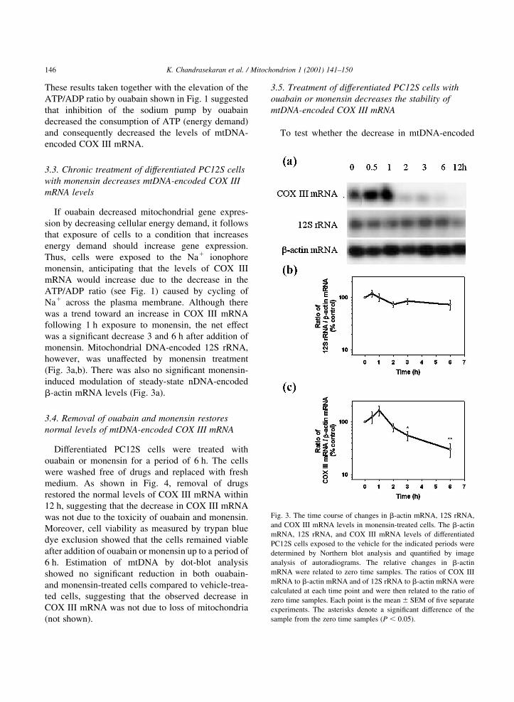
3.3. Chronic treatment of differentiated PC12S cells
with monensin decreases mtDNA-encoded COX III
mRNA levels
If ouabain decreased mitochondrial gene expres-
sion by decreasing cellular energy demand, it follows
that exposure of cells to a condition that increases
energy demand should increase gene expression.
Thus, cells were exposed to the Na1 ionophore
monensin, anticipating that the levels of COX III
mRNA would increase due to the decrease in the
ATP/ADP ratio (see Fig. 1) caused by cycling of
Na1 across the plasma membrane. Although there
was a trend toward an increase in COX III mRNA
following 1 h exposure to monensin, the net effect
was a signi®cant decrease 3 and 6 h after addition of
monensin. Mitochondrial DNA-encoded 12S rRNA,
however, was unaffected by monensin treatment
(Fig. 3a,b). There was also no signi®cant monensin-
induced modulation of steady-state nDNA-encoded
b-actin mRNA levels (Fig. 3a).
3.4. Removal of ouabain and monensin restores
normal levels of mtDNA-encoded COX III mRNA
Differentiated PC12S cells were treated with
ouabain or monensin for a period of 6 h. The cells
were washed free of drugs and replaced with fresh
medium. As shown in Fig. 4, removal of drugs
restored the normal levels of COX III mRNA within
12 h, suggesting that the decrease in COX III mRNA
was not due to the toxicity of ouabain and monensin.
Moreover, cell viability as measured by trypan blue
dye exclusion showed that the cells remained viable
after addition of ouabain or monensin up to a period of
6 h. Estimation of mtDNA by dot-blot analysis
showed no signi®cant reduction in both ouabain-
and monensin-treated cells compared to vehicle-trea-
ted cells, suggesting that the observed decrease in
COX III mRNA was not due to loss of mitochondria
(not shown).
3.5. Treatment of differentiated PC12S cells with
ouabain or monensin decreases the stability of
mtDNA-encoded COX III mRNA
To test whether the decrease in mtDNA-encoded
K. Chandrasekaran et al. / Mitochondrion 1 (2001) 141±150146
Fig. 3. The time course of changes in b-actin mRNA, 12S rRNA,
and COX III mRNA levels in monensin-treated cells. The b-actin
mRNA, 12S rRNA, and COX III mRNA levels of differentiated
PC12S cells exposed to the vehicle for the indicated periods were
determined by Northern blot analysis and quanti®ed by image
analysis of autoradiograms. The relative changes in b-actin
mRNA were related to zero time samples. The ratios of COX III
mRNA to b-actin mRNA and of 12S rRNA to b-actin mRNA were
calculated at each time point and were then related to the ratio of
zero time samples. Each point is the mean ^ SEM of ®ve separate
experiments. The asterisks denote a signi®cant difference of the
sample from the zero time samples �P , 0:05�:

COX III mRNA in ouabain- or monensin-treated cells
is due to increased degradation or decreased synthesis,
we determined the half-life of COX III mRNA, 12S
rRNA, and b-actin mRNA in control, ouabain-, and
monensin-treated differentiated PC12S cells. Actino-
mycin D has been previously shown to inhibit total
cellular transcription in PC12 cells by more than 90%.
To estimate half-lives, actinomycin D was added to
differentiated PC12S cells and after 1 h, vehicle,
ouabain, or monensin was added. Total cellular
RNA was isolated at various times over an 8-h period.
Throughout this period, cell viability was not compro-
mised and there was no substantial reduction in total
RNA yield in the presence of actinomycin D. Fig. 5a
shows autoradiograms of RNA isolated and probed
with COX III and b-actin. Clearly, mRNA encoding
COX III was signi®cantly affected, decreasing the
estimated t1/2 from 3.3 h in control cells to 1.6 h in
ouabain-treated cells and to 1 h in monensin-treated
cells (Fig. 4c), respectively. No such decrease in half-
lives were observed with mtDNA-encoded 12S rRNA
(Fig. 4b) and nDNA-encoded b-actin mRNA (Fig. 4a)
in both ouabain- and monensin-treated cells. Also, the
estimated t1/2 of COX III mRNA in control cells was
much shorter (3.3 h) when compared to the t1/2 of 12S
rRNA (17 h) and b-actin mRNA (.17 h).
K. Chandrasekaran et al. / Mitochondrion 1 (2001) 141±150 147
Fig. 4. Effect of the removal of ouabain and monensin on the ratio of
COX III mRNA to b-actin mRNA. Differentiated PC12S cells
exposed to ouabain and monensin for a period of 6 h. The cells
were then washed free of drugs and were maintained in normal
culture medium. The COX III mRNA and b-actin mRNA levels
for the indicated periods were determined by Northern blot analysis
and quanti®ed by image analysis of autoradiograms. The relative
change in COX III mRNA/and b-actin mRNA ratio was related to
COX III mRNA/and b-actin mRNA ratio of zero time samples.
Each point is the mean ^ SEM of three separate experiments.
Fig. 5. Estimation of half-lives for b-actin mRNA, 12S rRNA, and
COX III mRNA in control, ouabain-, and monensin-treated differ-
entiated PC12S cells. Total cytosolic RNA was isolated from
control, ouabain-, and monensin-treated cells at indicated time
points after termination of transcription by actinomycin D, and
10 mg aliquots were subjected to Northern blot analysis as described
in Section 2. Hybridization was quanti®ed by image analysis of
autoradiograms. Semi-log plots of transcript remaining versus
time is depicted in the case of b-actin mRNA. In the cases of 12S
rRNA and COX III mRNA, the ratios of 12S rRNA to b-actin
mRNA and COX III mRNA to b-actin mRNA were calculated.
The relative change in the ratio was related to the ratio of zero
time samples. Each point is the mean ^ SEM of three separate
experiments.

4. Discussion
The results of the present study demonstrate that
chronic treatment with both ouabain and monensin
induced a selective decrease in mtDNA-encoded
COX III mRNA expression in differentiated PC12S
cells. No signi®cant decreases were observed with
mtDNA-encoded 12S rRNA and with nDNA-encoded
b-actin mRNA in both ouabain- and monensin-treated
cells. Moreover, estimation of mtDNA by dot-blot
analysis showed no signi®cant reduction in both
ouabain- and monensin-treated cells compared to
vehicle-treated cells. The observed decrease in COX
III mRNA levels in both ouabain- and monensin-trea-
ted cells is, therefore, not due to either loss of mito-
chondria or a general breakdown of RNA. On the
other hand, a similar reduction in both ouabain- and
monensin-treated cells was observed with mtDNA-
encoded COX subunit-I mRNA and -II mRNA (data
not shown). Thus, the effect of ouabain and monensin
appears to be speci®c for mtDNA-encoded mRNA
and not for rRNA. This may relate to differences in
the synthesis and stability between mtDNA-encoded
mRNA and rRNA. A similar decrease in COX III
mRNA with ouabain and monensin was observed
also in undifferentiated and differentiated PC12 cells
(T. Tom, personal communication), in a human neuro-
blastoma cell line SHSY5Y and in monensin-treated
rat primary cerebellar granule neurons. Thus, it is
unlikely that the choice of the cell culture could
account for the ouabain- and monensin-induced
response.
Addition of ouabain to differentiated PC12S cells
led to a rapid and prolonged rise in the ATP/ADP
ratio. In neural tissues, a major portion of the energy
derived from metabolism is used to restore ionic
gradients to resting levels and the Na/K-ATPase is
one of the major consumers of ATP (Mata et al.,
1980; Sokoloff, 1981; Erecinska and Silver, 1989).
Consistent with these observations, our results show
that an inhibition of Na/K-ATPase by ouabain
decreases ATP consumption and increases the ATP/
ADP ratio in differentiated PC12S cells.
We originally hypothesized that ouabain would
reduce mitochondrial gene expression as a direct
consequence of an elevated ATP/ADP ratio. In orga-
nello transcription experiments using isolated mito-
chondria show that mitochondrial RNA synthesis
can be regulated in response to changes in intramito-
chondrial ATP levels (Gaines et al., 1987; Enriquez et
al., 1996b). High levels of intramitochondrial ATP
suppress mtDNA transcription possibly by inhibiting
mitochondrial RNA polymerase, presenting a
mechanism by which energy demand could regulate
mtDNA transcription (Enriquez et al., 1996b).
Although the results demonstrating a reduction in
COX III mRNA levels associated with increased
ATP/ADP levels in ouabain-treated cells is consistent
with these mechanisms of gene regulation, the obser-
vation that the estimated t1/2 of COX III mRNA was
decreased from 3.1 h in control cells to 1.6 h in
ouabain-treated cells suggested that RNA degradation
rather than synthesis was the primary site of control
by ouabain. This result suggests that both transcrip-
tional and posttranscriptional mechanisms are likely
to be involved in the maintenance of a steady level of
mtDNA-encoded COX III mRNA.
The unexpected result of this study was the
observed decrease in COX III mRNA in monensin-
treated cells. Monensin is a Na1-selective ionophore
that causes Na1 in¯ux with a corresponding ef¯ux of
H1 or K1 in numerous cell types including PC12 cells
(Pressman and Fahim, 1982; Shier and DuBourdieu,
1992). Under our experimental conditions, we
observed a 500% increase in 22Na after exposure of
PC12S cell cultures for 3 h to 100 nM monensin (not
shown). Ionic or pharmacological interventions that
increase [Na1]i levels cause signi®cant induction of
Na/K-ATPase activity and functional Na/K-pump
sites (Wolitzky and Fambrough, 1986; Lingrel and
Kuntzweiler, 1994). Addition of monensin to neuro-
nal cells enhances cellular energy metabolism and
increases ATP utilization by Na/K-ATPase, presum-
ably to restore ionic gradients to resting levels (Mata
et al., 1980). Accordingly, we found that the addition
of monensin to differentiated PC12S cells decreased
the ATP/ADP ratio, likely due to increased ATP
consumption. We anticipated that with elevated
ATP consumption, there would be an up-regulation
of COX III mRNA. Though there was an increase in
COX III mRNA levels in the ®rst hour after addition
of monensin, the levels of COX III mRNA subse-
quently showed a signi®cant decrease. We interpret
these ®ndings to suggest that the initial increase repre-
sents a mechanism of increased expression due to
increased energy demand. The decrease in COX III
K. Chandrasekaran et al. / Mitochondrion 1 (2001) 141±150148

mRNA following chronic exposure to monensin
suggests the presence of an alternate mechanism for
the regulation of mitochondrial gene expression that is
independent of the energetic status of the cell and that
overrides the normal regulation by energy demand.
This decrease in mitochondrial gene expression
under conditions of increasing energy demand is not
unique to this system. For example, levels of mtDNA-
encoded cytochrome oxidase subunit I (COX I)
mRNA decreases within hours in CA1 neurons of
gerbils after transient forebrain ischemia (Abe et al.,
1993). This early decrease in COX I mRNA occurs in
the absence of a decrease in mtDNA, suggesting
impaired mitochondrial gene expression occurring at
the level of transcription and/or turnover of mitochon-
drial mRNA (Abe et al., 1993). In this model system,
the decrease in COX I mRNA occurs when the energy
demand is high on these cells not only to restore ionic
gradients to resting levels but also to maintain neuro-
nal activity (Arai et al., 1986; Abe et al., 1993). A
disproportionate decrease in mtDNA-encoded COX
subunit mRNA in the absence of changes in
mtDNA-encoded 12S rRNA is also observed in the
brains of Alzheimer's disease (AD) patients (Chan-
drasekaran et al., 1994,1998; HatanpaÈaÈet al., 1996).
One explanation for the observed decrease in COX
III mRNA but not of 12S rRNA is the relatively short
half-life of COX III mRNA. Decay of COX III
mRNA, 12S rRNA, and b-actin mRNA in the
presence and absence of ouabain or monensin were
calculated after inhibition of de novo mitochondrial
and nuclear transcription by the addition of actinomy-
cin D to the cell culture medium. The estimated t1/2s of
COX III mRNA in control cells were 3.1 h whereas
the estimated t1/2s of 12S rRNA and of b-actin mRNA
were greater than 30 h. Thus, the half-lives of
mtDNA-encoded mRNAs are short. These estimated
half-lives are similar to the results reported in other
cell culture systems (Gelfand and Attardi, 1981;
Chrzanowska-Lightowlers et al., 1994). In ouabain-
and monensin-treated cells, the estimated t1/2
decreased to 1.6 and 1 h, respectively. Thus, there is
a threefold decrease in the stability of COX III mRNA
in drug-treated cells. This posttranscriptional mechan-
ism operating in both ouabain- and monensin-treated
cells is likely to be responsible for the accelerated
degradation of mtDNA-encoded mRNA. The precise
mechanism that mediates a decrease in the stability of
COX III mRNA remains unknown. RNA binding
proteins that function to protect mRNA from degrada-
tion are well documented (Jackson, 1993). The
presence of an abundant mitochondrial RNA-binding
protein has been known (Dekkeret al., 1991). Both
ouabain and monensin induce an increase in intracel-
lular sodium ion concentration [Na1]i in a number of
cell cultures including PC12 cells (Pressman and
Fahim, 1982; Boonstra et al., 1983; Shier and
DuBourdieu, 1992; Blaustein and Lederer, 1999).
Increased [Na1]i causes subsequent changes in intra-
cellular Ca21, osmolarity, pH, or a combination of the
above. The mechanism by which the increased [Na1]i
and associated cellular changes in¯uence the stability
of mtDNA-encoded mRNA remains to be determined.
In summary, we suggest that there are likely to be at
least two mechanisms involved in the regulation of
mitochondrial gene expression. A physiological
mechanism of transcriptional regulation level that
allows mtDNA to synthesize the optimal level of
mRNA in response to energetic demands (Enriquez
et al., 1996b). The results presented here may repre-
sent a second mechanism of regulation that operates at
the level of stability of mRNA that is independent of
the energetic status of the cell and that may operate
under pathologic conditions. This mechanism is likely
to be pathological because this overrides the normal
regulation by energy requirement, causes accelerated
degradation of transcripts, and is counterproductive to
the actual energy demand of the cell. The results
provide a potential approach to determine the mole-
cular components of this mechanism. It is also of
interest to determine whether this mechanism contri-
butes to neuronal death in acute and chronic neurode-
generative diseases. To the best of our knowledge, the
present study is the ®rst to demonstrate that mitochon-
drial gene expression is also regulated at the level of
mRNA stability under conditions of chronic exposure
to elevated intracellular sodium.
Acknowledgements
This work was supported by the grants from the US
Army DAMD17-99-1-9483 to G.F., from NIH
AG16966A to K.C., and from AHA 0051001U to
K.C.
K. Chandrasekaran et al. / Mitochondrion 1 (2001) 141±150 149

References
Abe, K., Kawagoe, J., Kogure, K., 1993. Early disturbance of a
mitochondrial DNA expression in gerbil hippocampus after
transient forebrain ischemia. Neurosci. Lett. 153 (2), 173±176.
Arai, H., Passonneau, J.V., Lust, W.D., 1986. Energy metabolism in
delayed neuronal death of CA1 neurons of the hippocampus
following transient ischemia in the gerbil. Metab. Brain Dis. 1
(4), 263±278.
Attardi, G., Schatz, G., 1988. Biogenesis of mitochondria. Annu.
Rev. Cell Biol. 4, 289±333.
Blaustein, M.P., Lederer, W.J., 1999. Sodium/calcium exchange: its
physiological implications. Physiol. Rev. 79 (3), 763±854.
Boonstra, J., Moolenaar, W.H., Harrison, P.H., Moed, P., van der
Saag, P.T., de Laat, S.W., 1983. Ionic responses and growth
stimulation induced by nerve growth factor and epidermal
growth factor in rat pheochromocytoma (PC12) cells. J. Cell
Biol. 97 (1), 92±98.
Chandrasekaran, K., Giordano, T., Brady, D.R., Stoll, J., Martin,
L.J., Rapoport, S.I., 1994. Impairment in mitochondrial cyto-
chrome oxidase gene expression in Alzheimer disease. Brain
Res. Mol. Brain Res. 24 (1±4), 336±340.
Chandrasekaran, K., HatanpaÈaÈ, K., Brady, D.R., Stoll, J., Rapoport,
S.I., 1998. Downregulation of oxidative phosphorylation in
Alzheimer disease: loss of cytochrome oxidase subunit mRNA
in the hippocampus and entorhinal cortex. Brain Res. 796 (1±2),
13±19.
Chrzanowska-Lightowlers, Z.M., Preiss, T., Lightowlers, R.N.,
1994. Inhibition of mitochondrial protein synthesis promotes
increased stability of nuclear-encoded respiratory gene tran-
scripts. J. Biol. Chem. 269 (44), 27322±27328.
Dekker, P.J., Papadopoulou, B., Grivell, L.A., 1991. Properties of
an abundant RNA-binding protein in yeast mitochondria.
Biochimie 73 (12), 1487±1492.
Enriquez, J.A., Perez-Martos, A., Lopez-Perez, M.J., Montoya, J.,
1996a. In organello RNA synthesis system from mammalian
liver and brain. Methods Enzymol. 264, 50±57.
Enriquez, J.A., Fernandez-Silva, P., Perez-Martos, A., Lopez-Perez,
M.J., Montoya, J., 1996b. The synthesis of mRNA in isolated
mitochondria can be maintained for several hours and is inhib-
ited by high levels of ATP. Eur. J. Biochem. 237 (3), 601±610.
Erecinska, M., Silver, I.A., 1989. ATP and brain function. J. Cereb.
Blood Flow Metab. 9 (1), 2±19.
Fukuyama, R., Chandrasekaran, K., Rapoport, S.I., 1993. Nerve
growth factor-induced neuronal differentiation is accompanied
by differential induction and localization of the amyloid precur-
sor protein (APP) in PC12 cells and variant PC12S cells. Brain
Res. Mol. Brain Res. 17 (1±2), 17±22.
Gaines, G., Attardi, G., 1984. Highly ef®cient RNA-synthesizing
system that uses isolated human mitochondria: new initiation
events and in vivo-like processing patterns. Mol. Cell Biol. 4
(8), 1605±1617.
Gaines, G., Rossi, C., Attardi, G., 1987. Markedly different ATP
requirements for rRNA synthesis and mtDNA light strand tran-
scription versus mRNA synthesis in isolated human mitochon-
dria. J. Biol. Chem. 262 (4), 1907±1915.
Gelfand, R., Attardi, G., 1981. Synthesis and turnover of mitochon-
drial ribonucleic acid in HeLa cells: the mature ribosomal and
messenger ribonucleic acid species are metabolically unstable.
Mol. Cell Biol. 1 (6), 497±511.
HatanpaÈaÈ, K., Brady, D.R., Stoll, J., Rapoport, S.I., Chandrasekaran,
K., 1996. Neuronal activity and early neuro®brillary tangles in
Alzheimer's disease. Ann. Neurol. 40 (3), 411±420.
Jackson, R.J., 1993. Cytoplasmic regulation of mRNA function:
the importance of the 3 0 untranslated region. Cell 74 (1), 9±
14.
Kagawa, Y., Ohta, S., 1990. Regulation of mitochondrial ATP
synthesis in mammalian cells by transcriptional control. Int. J.
Biochem. 22 (3), 219±229.
Lingrel, J.B., Kuntzweiler, T., 1994. Na 1 ,K(1)-ATPase. J. Biol.
Chem. 269 (31), 19659±19662.
Liu, L.I., Rapoport, S.I., Chandrasekaran, K., 1999. Regulation of
mitochondrial gene expression in differentiated PC12 cells.
Ann. N. Y. Acad. Sci. 893, 341±344.
Mata, M., Fink, D.J., Gainer, H., Smith, C.B., Davidsen, L., Savaki,
H., Schwartz, W.J., Sokoloff, L., 1980. Activity-dependent
energy metabolism in rat posterior pituitary primarily re¯ects
sodium pump activity. J. Neurochem. 34 (1), 213±215.
Pressman, B.C., Fahim, M., 1982. Pharmacology and toxicology of
the monovalent carboxylic ionophores. Annu. Rev. Pharmacol.
Toxicol. 22, 465±490.
Shier, W.T., DuBourdieu, D.J., 1992. Sodium- and calcium-depen-
dent steps in the mechanism of neonatal rat cardiac myocyte
killing by ionophores. I. The sodium-carrying ionophore,
monensin. Toxicol. Appl. Pharmacol. 116 (1), 38±46.
Shryock, J.C., Rubio, R., Berne, R.M., 1986. Extraction of adenine
nucleotides from cultured endothelial cells. Anal. Biochem. 159
(1), 73±81.
Sokoloff, L., 1981. Localization of functional activity in the central
nervous system by measurement of glucose utilization with
radioactive deoxyglucose. J. Cereb. Blood Flow Metab. 1 (1),
7±36.
Wolitzky, B.A., Fambrough, D.M., 1986. Regulation of the (Na11
K 1 )-ATPase in cultured chick skeletal muscle. Modulation of
expression by the demand for ion transport. J. Biol. Chem. 261
(21), 9990±9999.
Wong-Riley, M.T., 1989. Cytochrome oxidase: an endogenous
metabolic marker for neuronal activity. Trends Neurosci. 12
(3), 94±101.
Wong-Riley, M.T., Mullen, M.A., Huang, Z., Guyer, C., 1997.
Brain cytochrome oxidase subunit complementary DNAs: isola-
tion, subcloning, sequencing, light and electron microscopic in
situ hybridization of transcripts, and regulation by neuronal
activity. Neuroscience 76 (4), 1035±1055.
Zhang, C., Wong-Riley, M.T., 2000a. Depolarizing stimulation
upregulates GA-binding protein in neurons: a transcription
factor involved in the bigenomic expression of cytochrome
oxidase subunits. Eur. J. Neurosci. 12 (3), 1013±1023.
Zhang, C., Wong-Riley, M.T., 2000b. Synthesis and degradation of
cytochrome oxidase subunit mRNAs in neurons: differential
bigenomic regulation by neuronal activity. J. Neurosci. Res.
60 (3), 338±344.
K. Chandrasekaran et al. / Mitochondrion 1 (2001) 141±150150
Copyright © 2022 FDOKUMEN


![Biochemical acclimation, stomatal limitation and precipitationpatterns underlie decreases in photosynthetic stimulation of soybean(Glycine max) at elevated [CO2] and temperatures under](https://static.fdokumen.com/doc/165x107/631fe62c02108aeec703411e/biochemical-acclimation-stomatal-limitation-and-precipitationpatterns-underlie.jpg)