Statistical Detection of Chromosomal Homology Using Density ...
Upload
independentCategory
view
0download
0
Sheth et al. Molecular Cytogenetics 2013, 6:24http://www.molecularcytogenetics.org/content/6/1/24
CASE REPORT Open Access
Chromosomal imbalance letter: Phenotypicconsequences of combined deletion 8pter andduplication 15qterFrenny Sheth1*, Joris Andrieux2, Stuti Tewari1, Harsh Sheth3, Manisha Desai1, Pritti Kumari1, Nidhish Nanavaty1
and Jayesh Sheth1
Abstract
Exact breakpoint determination by oligonucleotide array-CGH has improved the analysis of genotype-phenotypecorrelations in cases with chromosome aberrations allowing a more accurate definition of relevant genes,particularly their isolated or combined impact on the phenotype in an unbalanced state. Chromosomal imbalanceshave been identified as one of the major causes of mental retardation and/or malformation syndromes and theyare observed in ~2-5% of the cases. Here we report a female child born to non-consanguineous parents andhaving multiple congenital anomalies such as atrial septal defect and multiple ventricular septal defects, convergentstrabismus, micropthalmia, seizures and mental retardation, with her head circumference and stature normal for herage. Cytogenetic study suggested 46,XX,add(8)(p23). Further analysis by array-CGH using 44K oligonucleotide probeconfirmed deletion on 8p23.3p23.1 of 7.1 Mb and duplication involving 15q23q26.3 of 30 Mb size leading to 46,XX,der(8)t(8;15)(p23.3;q23)pat.arr 8p23.3p23.1(191,530-7,303,237)x1,15q23q26.3(72,338,961-102,35,195)x3. The uniquephenotypic presentation in our case may have resulted from either loss or gain of a series of contiguous geneswhich may have resulted in a direct phenotypic effect and/or caused a genetic regulatory disturbance. Doublesegmental aberrations may have conferred phenotypic variability, as in our case, making it difficult to predict thecharacteristics that evolved as a result of the global gene imbalance, caused by the concomitant deletion andduplication.
Keywords: Chromosomal imbalance, 8p23 deletion, 15q23 duplication, IGF1R, GATA4, MCPH1
IntroductionChromosomal rearrangements are frequently observedin patients with multiple congenital anomalies (MCA),dysmorphism and developmental delay with/withoutmental retardation [1-3]. Inheritance of a balancedtranslocation from either or both parents is often re-sponsible for structural chromosomal defects leading tosegmental duplication or deletion of the chromosomepair in an affected individual [4]. The paradigm shift indiagnostics with the implementation of next generationin silico softwares and array based comparative genomichybridization (aCGH) technology, chromosome break-point determination, analyses of critical regions involved
* Correspondence: [email protected]’s Institute of Human Genetics, FRIGE House, Jodhpur Gam Road,Satellite, Ahmedabad 380 015, IndiaFull list of author information is available at the end of the article
© 2013 Sheth et al.; licensee BioMed Central LCommons Attribution License (http://creativecreproduction in any medium, provided the or
in genetic disorders and copy number evaluation hashelped to correlate chromosomal region alteration andthe resulting phenotype [5,6].Segmental deletion of chromosome 8p [7,8] and dupli-
cation involving chromosome 15q [9,10] independentlyare well characterized and accurately compared with theclinical features seen in the affected individuals. How-ever, segmental aneusomies simultaneously coveringlarge regions on both the chromosomes, leading to aphenotypic presentation, have rarely been described.Partial deletion of 8p23 is a relatively frequent deletion
syndrome characterized by major congenital anomalies,especially congenital heart defects, seizures, behavioralabnormalities and postnatal growth deficiency [8]. Facialdysmorphism may be subtle and mental retardation lesssevere than in those with deletions associated with moreproximal breakpoints [8].
td. This is an Open Access article distributed under the terms of the Creativeommons.org/licenses/by/2.0), which permits unrestricted use, distribution, andiginal work is properly cited.

Figure 1 Clinical presentation of the proband at 8 years of age.
Sheth et al. Molecular Cytogenetics 2013, 6:24 Page 2 of 7http://www.molecularcytogenetics.org/content/6/1/24
Duplication of the distal long arm of chromosome 15has been reported in various studies, which mainlyhighlighted it as an overgrowth syndrome characterizedby prenatal overgrowth, macrocephaly, tall stature andcraniosynostosis [9]. However, such cases exclusively in-volve the distal most segment i.e. 15q25. It has been
Figure 2 Partial karyotype of the father showing breakpoints on both
proposed by Roggenbuck et al. 2004, that the aforemen-tioned distal 15q trisomy syndrome may be the result ofthe disruption of the gene linked to 15q25 region, ratherthan partial trisomy for the region. Their study reported3 cases sharing features like ptosis, small size and devel-opmental delay [10].Here we present a female child with deletion 8p23.
3p23.1 as well as duplication 15q23q26.3. The aberrantchromosome 8 was inherited from the phenotypically nor-mal father who was the carrier of a balanced translocation46,XY,t(8;15)(p23;q23). We describe her phenotype, atbirth, at 4 years and 8 years of age. In addition, we de-scribe the phenotypic consequences of the concomitanteffect of segmental deletion and duplication of the same.
Clinical presentationA 4 year-old female child was referred for congenitalheart defects, dysmorphic facial features and develop-mental delay evident since her birth. She was the firstchild born to a non-consanguineous couple. Duringpregnancy, her mother had developed oligohydramniosand pre-eclampsia in the third trimester. She had a his-tory of two spontaneous 1st trimester abortions, one be-fore and one after the birth of proband. The baby wasborn full term by caesarian section. Her birth weightwas 3.5 kg and she did not cry at birth (Apgar score 4–6).She received oxygen for respiratory distress in the NICUfor 6 days. Initially, she had feeding difficulties but eventu-ally started breastfeeding at 1½month. Loud holosystolicmurmur was evident from the 2nd day of life, and the 2D
derivative chromosomes.

Sheth et al. Molecular Cytogenetics 2013, 6:24 Page 3 of 7http://www.molecularcytogenetics.org/content/6/1/24
echocardiograph showed atypical muscular atrial septaldefect (ASD) and multiple ventricular septal defects(VSD), with no patent ductus arteriosus (PDA). TORCHinvestigations were normal.At the age of 4 years, the head circumference was 47 cm
(10th percentile), height 94.5 cm (15th percentile) andweight 14.5 kg (25th percentile) [11]. The prominent dys-morphic features were ptosis, downward slanted palpebralfissure, microphthalmia, left convergent strabismus, wide
Figure 3 Breakpoint characterization by 44K oligonucleotide array-CGb: 30 Mb duplication at 15q [arr 15q23q26.3(chr15:72,338,961-102,351,195)x
nasal base, long philtrum, open mouth, low set ears, shortneck, micrognathia, puffy cheeks, short fingers, bilateral1st incurved finger and absence of thenar eminence[Figure 1]. Developmental delay was evident from the firstyear as social smile was absent even at 6 months. Shelearnt sitting at 10 months and walking at 20 months.Speech was absent and her developmental quotient atpresentation was of a 2 year old child. She had 3 attacks ofpartial seizures during the first 20 months and was kept
H. a: 7.1 Mb deletion at 8p [arr 8p23.3p23.1(191,530-7,303,237)x1] and3].

Sheth et al. Molecular Cytogenetics 2013, 6:24 Page 4 of 7http://www.molecularcytogenetics.org/content/6/1/24
on carbamazepine 150 mg/day and sodium valproate100 mg/day till she was 5 years old. Since then, she hasbeen free of epileptic symptoms. Sequential completehemogram was suggestive of chronic iron deficiencyanemia with hemoglobin levels of 8–8.5 gm%. 2D echo-cardiograph again at 3 years showed multiple small mus-cular VSDs and a single ASD of 4.2 × 7.0 mm.On re-examination at the age of 8 years, the head cir-
cumference was 49.5 cm (10th percentile), height 120 cm(25th percentile) and weight 28 kg (75th percentile) [11].Mental retardation was evident. She could not speak butresponded to commands. The bladder and bowel controlhad not yet been achieved. Apart from these, no otherdevelopmental abnormalities were noted. She couldcarry out routine activities independently. It is to benoted that she had normal for age head circumference,height and weight.
ResultsMetaphase chromosome analysisAfter obtaining institutional ethical committee approvaland informed written consent form, metaphase chromo-some preparations were obtained from PHA stimulatedlymphocyte cultures according to the standard proced-ure with slight modifications [12]. Chromosome analysiswas carried out by GTG-banding at 550-band levelaccording to ISCN 2013 nomenclature in both patientand parents (50 metaphases, each). Patient’s karyotypepattern showed additional genetic material on the shortarm of #8p i.e.46,XX,add(8)(p23). Parental chromosomalinvestigation revealed that the father was a carrier of abalanced translocation 46,XY,t(8;15)(p23;q23) [Figure 2].
Figure 4 Metaphase showing signals of both BAC clones RP11-95F11normal #15 and on derivative #8p [thin arrow]. Whereas, clone RP11-13
This suggests that the child had inherited der(8) from thefather thus leading to an unbalanced genetic makeup.
Array-CGHGenomic DNA was extracted from peripheral bloodlymphocytes using standard SDS-proteinase K extractionmethod [13]. Extracted genomic DNA concentrationwas determined with NanoDrop ND-1000 spectropho-tometer (NanoDrop Technologies, Berlin, Germany).Evaluation of gene copy number was performed by 44koligonucleotide array-Comparative Genomic Hybridiza-tion (aCGH) by following manufacturer's recommenda-tions (Human Genome CGH microarray 44B kit, AgilentTechnologies Inc., Santa Clara, CA, USA). Female gen-omic DNA (Promega Corporation, Madison, WI, USA)was used as a sex-matched reference, which was analyzedwith the CGH-analysis software v3.4 (Agilent Technolo-gies Inc., Santa Clara, CA, USA) by applying Z-score seg-mentation algorithm with a window size of 10 points toidentify chromosome aberrations. Analysis was performedusing 3-points filter and 0.2 variation which lead to con-firmation of partial deletion 8p region of 7.1 Mb [arr cgh8p23.3p23.1(191,530-7,303,237)(hg19-NCBI build37)x1] andpartial 15q duplication of 30 Mb [arr cgh 15q23q26.3(chr15:72,338,961-102,351,195)x3], i.e. 46,XX,der(8)t(8;15)(p23.3;q23)pat.arr 8p23.3p23.1(191,530-7,303,237)x1,15q23q26.3(72,338,961-102,351,195)x3 [Figure 3a,b].
Fluorescence in situ hybridization (FISH)FISH analysis was performed using BAC clones RP11-139L10 covering 8p23→ pter, RP11-95F11 and RP11-100A1 spanning 15q23→ qter. Nick Translation Kit
and RP11-100A1 spanning 15q25.3 to 15q26.3 region on two9L10 covering 8p23.3 is seen only on normal #8p [broad arrow].

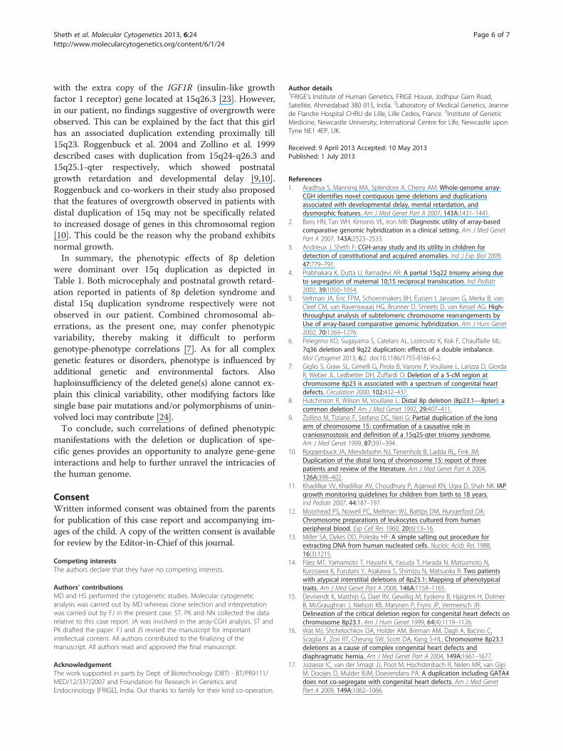
Table 1 Comparison of phenotypes having partial 8pdeletion and partial 15q duplication and its correlationwith our case
Clinical features Partial 8pdeletion
Ourpatient
Partial 15qduplication
Mental retardation + ++ +
Post natal growth deficiency + - +
Microcephaly + - +
Speech delay + +++ +
Congenital heart disease + +++ +\-
Seizures + + +
Hypotonia - - +
Behavioural abnormalities + - -
Downslanted palpebral fissure +/- + +
Strabismus - ++ +
Ptosis - +++ +
Wide nasal base + ++ +
Short neck + ++ +
Bulbous nose + + -
Low set ears - + +
Puffy cheeks + + +
Midline crease in lower lip - + +
Thin upper lip + - -
Long philtrum - + +
High arched palate - + +
Micrognathia + + +
Pectus excavatum - - +
Scoliosis - - +
Depressed sternum + - -
Short fingers + + +
Inguinal and diaphragmatic hernia + - -
Widely spaced nipples + ++ -
Sheth et al. Molecular Cytogenetics 2013, 6:24 Page 5 of 7http://www.molecularcytogenetics.org/content/6/1/24
(Vysis, Abbott Molecular, USA) was used for labeling.The two clones - RP11-95F11 and RP11-100A1 were la-beled with Spectrum Orange and Spectrum Green re-spectively whereas, third BAC clone was labeled usingboth fluorochromes in 1:1 ratio labeling. FISH signalswere observed using Olympus BX-51 microscope (Olympus,Germany) and pseudo-coloring was carried out usingAdobe Photoshop [Figure 4].
DiscussionThis case report presents a 4 year old girl who was diag-nosed with double segmental chromosomal aberrations46,XX,der(8)t(8;15)(p23;q23)pat. The derivative #8 wasinherited from the father who was a carrier of balanced8;15 chromosomal translocation which led to partial dele-tion of 8p23.3p23.1 and duplication of 15q23q26.3 in thechild. Inheriting unbalanced chromosomal rearrange-ments that originated from a translocation event in eitherparent are often associated with mental retardation and/orcongenital malformations [5].The distal 8p deletion is of a relatively frequent occur-
rence with distinct clinical features [10]. Congenitalheart defects, behavioral problems, mild to moderatemental retardation/developmental delay, strabismus andmild facial and digital anomalies are frequent phenotypiccharacteristics seen in these patients [9,14,15]. The se-verity of mental retardation, pre and postnatal growth de-ficiency including microcephaly and facial dysmorphismbecomes more pronounced as the deletion site involvesmore proximal regions [15].Cytogenetic evidence suggests that the haploin-
sufficiency of ≥1 gene located in 8p23 behaves as a dom-inant mutation, thus impairing heart differentiation andleading to a wide spectrum of congenital heart defects(CHDs) [8]. The gene responsible for the heart defectsin this syndrome has been identified to be GATA4 on8p23.1 [14]. Haploinsufficiency of GATA4 is thought toplay a critical role in the development of these birth de-fects [16,17]. This data is in concordance with the find-ings in our patient who has atrial septal defect andmultiple ventricular septal defects since birth.Microcephaly is another feature frequently seen in pa-
tients with distal 8p deletion [15]. The MCPH1 gene,mapped to chromosome 8p23, has been implicated to bea candidate gene for primary microcephaly [18,19]. Al-though, in our patient, microcephaly was not noted, thisfinding has also been reported by other studies [10,20].This gene has been shown to have a high penetrance withlarge deletion, as has been detected in the present case.However, the possibility of non-penetrance and/or non-expressivity as the cause behind the absence of microceph-aly cannot be entirely excluded [15]. The concomitantpresence of duplication of distal 15q leading to a normalfor age head circumference could also be a possibility.
Our proband had mild mental retardation with verylittle speech development and facial dysmorphic featureslike wide nasal base, puffy cheeks, low set ears,micrognathia are in concordance with other cases of 8pdeletion syndrome [9]. But majority of her facial fea-tures, including those mentioned above, like ptosis,down slanted palpebral fissures, long philtrum, openmouth, mid crease in the lower lip are similar to the dis-tinguishable facial characteristics seen in cases of distalduplication of 15q [21].Since the first report of 15q22→ qter duplication by
Fujimoto et al. 1974 [22], at least 71 other cases of simi-lar or smaller distal 15q imbalances have been described[23]. The phenotype-genotype correlations observed inthese cases resulted in the delineation of the 15q over-growth syndrome, which is caused by the increased dos-age of the genes that are present between 15q25-q26.3.The findings of overgrowth have reliably been associated
Sheth et al. Molecular Cytogenetics 2013, 6:24 Page 6 of 7http://www.molecularcytogenetics.org/content/6/1/24
with the extra copy of the IGF1R (insulin-like growthfactor 1 receptor) gene located at 15q26.3 [23]. However,in our patient, no findings suggestive of overgrowth wereobserved. This can be explained by the fact that this girlhas an associated duplication extending proximally till15q23. Roggenbuck et al. 2004 and Zollino et al. 1999described cases with duplication from 15q24-q26.3 and15q25.1-qter respectively, which showed postnatalgrowth retardation and developmental delay [9,10].Roggenbuck and co-workers in their study also proposedthat the features of overgrowth observed in patients withdistal duplication of 15q may not be specifically relatedto increased dosage of genes in this chromosomal region[10]. This could be the reason why the proband exhibitsnormal growth.In summary, the phenotypic effects of 8p deletion
were dominant over 15q duplication as depicted inTable 1. Both microcephaly and postnatal growth retard-ation reported in patients of 8p deletion syndrome anddistal 15q duplication syndrome respectively were notobserved in our patient. Combined chromosomal ab-errations, as the present one, may confer phenotypicvariability, thereby making it difficult to performgenotype-phenotype correlations [7]. As for all complexgenetic features or disorders, phenotype is influenced byadditional genetic and environmental factors. Alsohaploinsufficiency of the deleted gene(s) alone cannot ex-plain this clinical variability, other modifying factors likesingle base pair mutations and/or polymorphisms of unin-volved loci may contribute [24].To conclude, such correlations of defined phenotypic
manifestations with the deletion or duplication of spe-cific genes provides an opportunity to analyze gene-geneinteractions and help to further unravel the intricacies ofthe human genome.
ConsentWritten informed consent was obtained from the parentsfor publication of this case report and accompanying im-ages of the child. A copy of the written consent is availablefor review by the Editor-in-Chief of this journal.
Competing interestsThe authors declare that they have no competing interests.
Authors’ contributionsMD and HS performed the cytogenetic studies. Molecular cytogeneticanalysis was carried out by MD whereas clone selection and interpretationwas carried out by FJ in the present case. ST, PK and NN collected the datarelative to this case report. JA was involved in the array-CGH analysis. ST andPK drafted the paper. FJ and JS revised the manuscript for importantintellectual content. All authors contributed to the finalizing of themanuscript. All authors read and approved the final manuscript.
AcknowledgementThe work supported in parts by Dept. of Biotechnology (DBT) - BT/PR9111/MED/12/337/2007 and Foundation for Research in Genetics andEndocrinology [FRIGE], India. Our thanks to family for their kind co-operation.
Author details1FRIGE’s Institute of Human Genetics, FRIGE House, Jodhpur Gam Road,Satellite, Ahmedabad 380 015, India. 2Laboratory of Medical Genetics, Jeannede Flandre Hospital CHRU de Lille, Lille Cedex, France. 3Institute of GeneticMedicine, Newcastle University, International Centre for Life, Newcastle uponTyne NE1 4EP, UK.
Received: 9 April 2013 Accepted: 10 May 2013Published: 1 July 2013
References1. Aradhya S, Manning MA, Splendore A, Cherry AM: Whole-genome array-
CGH identifies novel contiguous gene deletions and duplicationsassociated with developmental delay, mental retardation, anddysmorphic features. Am J Med Genet Part A 2007, 143A:1431–1441.
2. Baris HN, Tan WH, Kimonis VE, Iron MB: Diagnostic utility of array-basedcomparative genomic hybridization in a clinical setting. Am J Med GenetPart A 2007, 143A:2523–2533.
3. Andrieux J, Sheth F: CGH-array study and its utility in children fordetection of constitutional and acquired anomalies. Ind J Exp Biol 2009,47:779–791.
4. Prabhakara K, Dutta U, Ramadevi AR: A partial 15q22 trisomy arising dueto segregation of maternal 10;15 reciprocal translocation. Ind Pediatr2002, 39:1050–1054.
5. Veltman JA, Eric FPM, Schoenmakers BH, Eussen I, Janssen G, Merkx B, vanCleef CM, van Ravenswaaij HG, Brunner D, Smeets D, van Kessel AG: High-throughput analysis of subtelomeric chromosome rearrangements byUse of array-based comparative genomic hybridization. Am J Hum Genet2002, 70:1269–1276.
6. Pelegrino KO, Sugayama S, Catelani AL, Lezirovitz K, Kok F, Chauffaille ML:7q36 deletion and 9q22 duplication: effects of a double imbalance.Mol Cytogenet 2013, 6:2. doi:10.1186/1755-8166-6-2.
7. Giglio S, Graw SL, Gimelli G, Pirola B, Varone P, Voullaire L, Larizza D, GiordaR, Weber JL, Ledbetter DH, Zuffardi O: Deletion of a 5-cM region atchromosome 8p23 is associated with a spectrum of congenital heartdefects. Circulation 2000, 102:432–437.
8. Hutchinson R, Wilson M, Voullaire L: Distal 8p deletion (8p23.1––8pter): acommon deletion? Am J Med Genet 1992, 29:407–411.
9. Zollino M, Tiziano F, Stefano DC, Neri G: Partial duplication of the longarm of chromosome 15: confirmation of a causative role incraniosynostosis and definition of a 15q25-qter trisomy syndrome.Am J Med Genet 1999, 87:391–394.
10. Roggenbuck JA, Mendelsohn NJ, Tenenholz B, Ladda RL, Fink JM:Duplication of the distal long of chromosome 15: report of threepatients and review of the literature. Am J Med Genet Part A 2004,126A:398–402.
11. Khadilkar VV, Khadilkar AV, Choudhury P, Agarwal KN, Ugra D, Shah NK: IAPgrowth monitoring guidelines for children from birth to 18 years.Ind Pediatr 2007, 44:187–197.
12. Moorhead PS, Nowell PC, Mellman WJ, Battips DM, Hungerford DA:Chromosome preparations of leukocytes cultured from humanperipheral blood. Exp Cell Res 1960, 20(6):13–16.
13. Miller SA, Dykes DD, Polesky HF: A simple salting out procedure forextracting DNA from human nucleated cells. Nucleic Acids Res 1988,16(3):1215.
14. Páez MT, Yamamoto T, Hayashi K, Yasuda T, Harada N, Matsumoto N,Kurosawa K, Furutani Y, Asakawa S, Shimizu N, Matsuoka R: Two patientswith atypical interstitial deletions of 8p23.1: Mapping of phenotypicaltraits. Am J Med Genet Part A 2008, 146A:1158–1165.
15. Devriendt K, Matthijs G, Dael RV, Gewillig M, Eyskens B, Hjalgrim H, DolmerB, McGraughran J, Nielson KB, Marynen P, Fryns JP, Vermeesch JR:Delineation of the critical deletion region for congenital heart defects onchromosome 8p23.1. Am J Hum Genet 1999, 64(4):1119–1126.
16. Wat MJ, Shchelochkov OA, Holder AM, Breman AM, Dagli A, Bacino C,Scaglia F, Zori RT, Cheung SW, Scott DA, Kang S-HL: Chromosome 8p23.1deletions as a cause of complex congenital heart defects anddiaphragmatic hernia. Am J Med Genet Part A 2004, 149A:1661–1677.
17. Joziasse IC, van der Smagt JJ, Poot M, Hochstenbach R, Nelen MR, van GijnM, Dooijes D, Mulder BJM, Doevendans PA: A duplication including GATA4does not co-segregate with congenital heart defects. Am J Med GenetPart A 2009, 149A:1062–1066.
Sheth et al. Molecular Cytogenetics 2013, 6:24 Page 7 of 7http://www.molecularcytogenetics.org/content/6/1/24
18. Wang Y, Su B: Molecular evolution of microcephalin, a gene determininghuman brain size. Hum Mol Genet 2004, 13:11.
19. Jackson AP, Eastwood H, Bell SM, Adu J, Toomes C, Carr IM, Roberts E,Hampshire DJ, Crow YJ, Mighell AJ, Karbani G, Jafri H, Rashid Y, Mueller RF,Markham AF, Woods CG: Identification of microcephalin, a proteinimplicated in determining the size of the human brain. Am J Hum Genet2002, 71(1):136–142.
20. Fryns JP, Kleczkowska A, Moerman PH, Van Den Berghe K, Van Den BergheH: The fetal phenotype of 15q2 duplication. Ann Genet 1988, 31:123–125.
21. Schnatterly P, Bono KL, Robinow M, Wyandt HE, Kardon N, Kelly TE: Distal15q trisomy: phenotypic comparison of nine cases in an extendedfamily. Am J Hum Genet 1984, 36:444–451.
22. Fujimoto A, Towner JW, Ebbin AJ, Kahlstrom EJ, Wilson MG: Inheritedpartial duplication of chromosome no. 15. J Med Genet 1974, 11:287–290.
23. Gutiérrez-Franco ML, Madariaga-Campos ML, Vásquez-Velásquez AI, MatuteE, Guevara-Yáñez R, Rivera H: A girl with 15q overgrowth syndrome anddup(15)(q24q26.3) That included telomeric sequences. Korean J Lab Med2010, 30(3):318–324.
24. Devriendt K, Vermeesch J: Chromosomal phenotypes and submicroscopicabnormalities. Hum Genomics 2004, 1(2):126–133.
doi:10.1186/1755-8166-6-24Cite this article as: Sheth et al.: Chromosomal imbalance letter:Phenotypic consequences of combined deletion 8pter and duplication15qter. Molecular Cytogenetics 2013 6:24.
Submit your next manuscript to BioMed Centraland take full advantage of:
• Convenient online submission
• Thorough peer review
• No space constraints or color figure charges
• Immediate publication on acceptance
• Inclusion in PubMed, CAS, Scopus and Google Scholar
• Research which is freely available for redistribution
Submit your manuscript at www.biomedcentral.com/submit
Copyright © 2022 FDOKUMEN