Characterizing Brain Cortical Plasticity and Network Dynamics Across the Age-Span in Health and...
27
Characterizing Brain Cortical Plasticity and Network Dynamics Across the Age-Span in Health and Disease with TMS-EEG and TMS-fMRI Alvaro Pascual-Leone, Berenson-Allen Center for Noninvasive Brain Stimulation, Division of Cognitive Neurology, Department of Neurology, Beth Israel Deaconess Medical Center, Harvard Medical School, 330 Brookline Ave, Boston, MA 02215, USA; Institut Universitari de Neurorehabilitació Guttmann, Universidad Autónoma de Barcelona, Barcelona, Spain Catarina Freitas, Berenson-Allen Center for Noninvasive Brain Stimulation, Division of Cognitive Neurology, Department of Neurology, Beth Israel Deaconess Medical Center, Harvard Medical School, 330 Brookline Ave, Boston, MA 02215, USA Lindsay Oberman, Berenson-Allen Center for Noninvasive Brain Stimulation, Division of Cognitive Neurology, Department of Neurology, Beth Israel Deaconess Medical Center, Harvard Medical School, 330 Brookline Ave, Boston, MA 02215, USA Jared C. Horvath, Berenson-Allen Center for Noninvasive Brain Stimulation, Division of Cognitive Neurology, Department of Neurology, Beth Israel Deaconess Medical Center, Harvard Medical School, 330 Brookline Ave, Boston, MA 02215, USA Mark Halko, Berenson-Allen Center for Noninvasive Brain Stimulation, Division of Cognitive Neurology, Department of Neurology, Beth Israel Deaconess Medical Center, Harvard Medical School, 330 Brookline Ave, Boston, MA 02215, USA Mark Eldaief, Berenson-Allen Center for Noninvasive Brain Stimulation, Division of Cognitive Neurology, Department of Neurology, Beth Israel Deaconess Medical Center, Harvard Medical School, 330 Brookline Ave, Boston, MA 02215, USA Shahid Bashir, Berenson-Allen Center for Noninvasive Brain Stimulation, Division of Cognitive Neurology, Department of Neurology, Beth Israel Deaconess Medical Center, Harvard Medical School, 330 Brookline Ave, Boston, MA 02215, USA Marine Vernet, Berenson-Allen Center for Noninvasive Brain Stimulation, Division of Cognitive Neurology, Department of Neurology, Beth Israel Deaconess Medical Center, Harvard Medical School, 330 Brookline Ave, Boston, MA 02215, USA © Springer Science+Business Media, LLC 2011 [email protected]. This is one of several papers published together in Brain Topography on the “Special Issue: Brain Imaging across the Lifespan”. Conflict of interest None. NIH Public Access Author Manuscript Brain Topogr. Author manuscript; available in PMC 2012 June 13. Published in final edited form as: Brain Topogr. 2011 October ; 24(3-4): 302–315. doi:10.1007/s10548-011-0196-8. NIH-PA Author Manuscript NIH-PA Author Manuscript NIH-PA Author Manuscript
-
Upload
independent -
Category
Documents
-
view
0 -
download
0
Transcript of Characterizing Brain Cortical Plasticity and Network Dynamics Across the Age-Span in Health and...
Characterizing Brain Cortical Plasticity and Network DynamicsAcross the Age-Span in Health and Disease with TMS-EEG andTMS-fMRI
Alvaro Pascual-Leone,Berenson-Allen Center for Noninvasive Brain Stimulation, Division of Cognitive Neurology,Department of Neurology, Beth Israel Deaconess Medical Center, Harvard Medical School, 330Brookline Ave, Boston, MA 02215, USA; Institut Universitari de Neurorehabilitació Guttmann,Universidad Autónoma de Barcelona, Barcelona, Spain
Catarina Freitas,Berenson-Allen Center for Noninvasive Brain Stimulation, Division of Cognitive Neurology,Department of Neurology, Beth Israel Deaconess Medical Center, Harvard Medical School, 330Brookline Ave, Boston, MA 02215, USA
Lindsay Oberman,Berenson-Allen Center for Noninvasive Brain Stimulation, Division of Cognitive Neurology,Department of Neurology, Beth Israel Deaconess Medical Center, Harvard Medical School, 330Brookline Ave, Boston, MA 02215, USA
Jared C. Horvath,Berenson-Allen Center for Noninvasive Brain Stimulation, Division of Cognitive Neurology,Department of Neurology, Beth Israel Deaconess Medical Center, Harvard Medical School, 330Brookline Ave, Boston, MA 02215, USA
Mark Halko,Berenson-Allen Center for Noninvasive Brain Stimulation, Division of Cognitive Neurology,Department of Neurology, Beth Israel Deaconess Medical Center, Harvard Medical School, 330Brookline Ave, Boston, MA 02215, USA
Mark Eldaief,Berenson-Allen Center for Noninvasive Brain Stimulation, Division of Cognitive Neurology,Department of Neurology, Beth Israel Deaconess Medical Center, Harvard Medical School, 330Brookline Ave, Boston, MA 02215, USA
Shahid Bashir,Berenson-Allen Center for Noninvasive Brain Stimulation, Division of Cognitive Neurology,Department of Neurology, Beth Israel Deaconess Medical Center, Harvard Medical School, 330Brookline Ave, Boston, MA 02215, USA
Marine Vernet,Berenson-Allen Center for Noninvasive Brain Stimulation, Division of Cognitive Neurology,Department of Neurology, Beth Israel Deaconess Medical Center, Harvard Medical School, 330Brookline Ave, Boston, MA 02215, USA
© Springer Science+Business Media, LLC 2011
This is one of several papers published together in Brain Topography on the “Special Issue: Brain Imaging across the Lifespan”.
Conflict of interest None.
NIH Public AccessAuthor ManuscriptBrain Topogr. Author manuscript; available in PMC 2012 June 13.
Published in final edited form as:Brain Topogr. 2011 October ; 24(3-4): 302–315. doi:10.1007/s10548-011-0196-8.
NIH
-PA Author Manuscript
NIH
-PA Author Manuscript
NIH
-PA Author Manuscript

Mouhshin Shafi,Berenson-Allen Center for Noninvasive Brain Stimulation, Division of Cognitive Neurology,Department of Neurology, Beth Israel Deaconess Medical Center, Harvard Medical School, 330Brookline Ave, Boston, MA 02215, USA; Department of Neurology, Massachusetts GeneralHospital; Partner’s Neurology Program, Harvard Medical School, Boston, MA, USA
Brandon Westover,Berenson-Allen Center for Noninvasive Brain Stimulation, Division of Cognitive Neurology,Department of Neurology, Beth Israel Deaconess Medical Center, Harvard Medical School, 330Brookline Ave, Boston, MA 02215, USA
Andrew M. Vahabzadeh-Hagh, andBerenson-Allen Center for Noninvasive Brain Stimulation, Division of Cognitive Neurology,Department of Neurology, Beth Israel Deaconess Medical Center, Harvard Medical School, 330Brookline Ave, Boston, MA 02215, USA; Department of Neurology, Children’s Hospital, Boston,Harvard Medical School, Boston, MA, USA
Alexander RotenbergDepartment of Neurology, Children’s Hospital, Boston, Harvard Medical School, Boston, MA,USA
AbstractBrain plasticity can be conceptualized as nature’s invention to overcome limitations of the genomeand adapt to a rapidly changing environment. As such, plasticity is an intrinsic property of thebrain across the life-span. However, mechanisms of plasticity may vary with age. Thecombination of transcranial magnetic stimulation (TMS) with electroencephalography (EEG) orfunctional magnetic resonance imaging (fMRI) enables clinicians and researchers to directly studylocal and network cortical plasticity, in humans in vivo, and characterize their changes across theage-span. Parallel, translational studies in animals can provide mechanistic insights. Here, weargue that, for each individual, the efficiency of neuronal plasticity declines throughout the age-span and may do so more or less prominently depending on variable ‘starting-points’ and different‘slopes of change’ defined by genetic, biological, and environmental factors. Furthermore,aberrant, excessive, insufficient, or mistimed plasticity may represent the proximal pathogeniccause of neurodevelopmental and neurodegenerative disorders such as autism spectrum disordersor Alzheimer’s disease.
KeywordsCortical brain plasticity; Transcranial magnetic stimulation; Electroencephalography; Functionalmagnetic resonance imaging; Lifespan
Plasticity as an Intrinsic Property of Human BrainThe world we live in changes rapidly. Afferent inputs and efferent demands to the brain shiftquicker than the time needed to implement genetic or even epigenetic changes. Brainplasticity can be conceptualized as nature’s invention to overcome limitations of the genomeand adapt to the rapidly changing environment (Fig. 1; Pascual-Leone et al. 2005). As such,plasticity represents an intrinsic property of the nervous system retained throughout life thatenables modification of function and structure in response to environmental demands via thestrengthening, weakening, pruning, or adding of synaptic connections and by promotingneurogenesis. This means that the brain does not remain static but, instead, continues tochange as the obligatory consequence of each sensory input, motor act, association, rewardsignal, action plan, and awareness (Pascual-Leone et al. 2005).
Pascual-Leone et al. Page 2
Brain Topogr. Author manuscript; available in PMC 2012 June 13.
NIH
-PA Author Manuscript
NIH
-PA Author Manuscript
NIH
-PA Author Manuscript

However, it is worth remembering William James who defined plasticity is the “possessionof a structure weak enough to yield to an influence, but strong enough not to yield all atonce”. Excess plasticity can be equally deleterious as insufficient plasticity (Fig. 2).Plasticity is essential to the establishment and maintenance of brain circuitry, it can bebeneficial for the individual enabling acquisition of new skills and adaptation after an injury,but it can also account for the symptoms of disease. Normal plasticity mechanisms can serveto compound the pathological consequences of a specific genetic mutation or sustainedenvironmental insult, and aberrant plasticity mechanisms can act on a previously normalbrain to induce pathological manifestations of disease. Early altered or mistimed plasticitymay set the stage for otherwise innocuous processes to become pathogenic (Gogolla et al.2009). A deficit in plasticity will render the brain unable to adjust to changing demands. Onthe other hand, if the brain is too plastic, structural connections may become unstable andfunctional systems necessary for cognition and behavior may be compromised.
The brain is highly interconnected. Therefore, plasticity plays out across the multiple levelsof nervous system complexity, from cellular through microcircuits to circuits and large-scalenetworks. Activity in lower levels may influence activity in higher levels, and vice versa.Changes in local plasticity can be compensated for by circuit and network adaptations insuch a way that behavior may not deteriorate (in fact, some behaviors may even beparadoxically improved). Alternatively, local changes may be compounded by furthermaladaptive circuit and network dynamics giving rise to disability and the symptoms ofdisease. Thus, changes in local plasticity may constitute the first in a chain of eventsculminating in ‘circuitopathies’ in which symptoms are the consequence of dysfunctions ofneural circuits and networks. If so, measures of cortical plasticity may provide very earlylocal and network biomarkers of neuropsychiatric disease.
In the present article we discuss concepts of plasticity as they might evolve across the age-span and contribute to normal development, life-long cognitive abilities, and themanifestation of developmental or neurodegenerative disorders. We argue that, in allindividuals, the efficacy of the mechanisms of plasticity changes over the lifespan, but that itdoes so from variable starting-points and with variable slopes depending on a number ofgenetic factors, environmental factors, and their complex interaction (Fig. 3). Empiricaldetermination of each individual’s slope of changing brain plasticity across the lifespan ispossible with real-time integration of transcranial magnetic stimulation (TMS) withelectroencephalography (EEG) and functional magnetic resonance imaging (fMRI) (Freitaset al. 2011a, b). Such methods might ultimately provide early predictors of individual riskfor age-related cognitive decline, diagnostic biomarkers for neurodevelopmental andneurodegenerative disorders, and enable plasticity-based interventions to optimize outcomesfor each individual.
Plasticity Changes Across the Lifespan: Animal Models and IndirectEvidence in Humans
Animal studies building on pioneering work from Barnes in the late 1970s (Barnes 1979)have demonstrated an age-associated decline in synaptic plasticity that correlates withneurocognitive impairments (Rosenzweig and Barnes 2003). For example, once induced,hippocampal long-term potentiation (LTP) decays faster in older rats, and this appears to beassociated with a greater degree of forgetfulness (Barnes and McNaughton 1980; Kelly et al.2006). Moreover, deficits in the balance between LTP and longterm depression (LTD) resultin impaired learning and memory (Larson et al. 1986; Roman et al. 1987; Bliss et al. 2003).
However, despite these and other findings in animal models, evidence in humans hasremained largely indirect. Brain imaging studies with structural MRI, fMRI, diffusion tensor
Pascual-Leone et al. Page 3
Brain Topogr. Author manuscript; available in PMC 2012 June 13.
NIH
-PA Author Manuscript
NIH
-PA Author Manuscript
NIH
-PA Author Manuscript

imaging (DTI), and positron emission tomography (PET) can provide evidence supportingthe claim that plasticity changes across the lifespan. But, for the most part, these methodsassess the consequences of plasticity and fail to directly assess the mechanisms of plasticitythemselves.
Structural neuroimaging can assess the integrity of brain structures and provide indirectmeasures of plasticity phenomena. For example, cross-sectional studies have consistentlyidentified age-associated morphometric brain changes encompassing regional corticalthinning, volumetric subcortical reductions, and ventricular enlargement (e.g., Fjell et al.2009; Walhovd et al. 2005, 2009). Longitudinal studies have demonstrated annual atrophyrates for brain volume, hippocampus and entorhinal cortex (e.g., Scahill et al. 2003; Fotenoset al. 2005), and atrophy in cortical brain regions over different periods of time (Raz et al.2005; Driscoll et al. 2009). Cortical thickness decreases over the lifespan are estimated at0.5% a year (Thompson et al. 2007). These changes affect different neural systemsdifferently: motor and visual cortices show regional thinning, whereas non-limbic temporalregions and parietal areas are relatively spared in normal aging (Salat et al. 2004; Raz et al.2004). Furthermore, DTI can address structural changes in white matter structure(myelination) and connectivity. For example, DTI has demonstrated that white-matterconnections, largely in fronto-striatal areas, have reduced myelination as age increases (Salatet al. 2005). It is assumed that such structural, morphometric MRI and DTI measures mustbe associated with cognitive and behavioral consequences (Fjell and Walhoyd 2010).
There are changes in cognitive task performance associated with aging and, as a corollary tothis, changes in the neural activation resulting from these cognitive tasks. It has been widelyestablished that normal human aging in humans is associated with decrements in cognitiveperformance across several domains including processing speed, working memory, episodicmemory, attentional control, inhibitory control, and executive function (e.g., Gazzaley andD’Esposito 2007; Reuter-Lorenz and Park 2010; Park and Reuter-Lorenz 2009). In parallel,a host of neuroimaging investigations have described altered patterns of brain activation inhealthy elders as they perform cognitive tasks. One such pattern is that of a reduction inprefrontal hemispheric asymmetry in elderly individuals, referred to as the HAROLD model(hemispheric asymmetry reduction in older adults) (Cabeza et al. 2002). According to theHAROLD model, the older brain displays less localizable and more bilateral activationduring certain cognitive tasks. A second pattern is a shift in evoked neural activity fromposterior to anterior cortex, a model referred to by Davis et al. as PASA (posterior-anteriorshift in aging) (2008). The PASA model posits that the aging brain is more likely to recruitprefrontal, rather than occipito-temporal, cortex in the service of task execution.
In addition to life-span changes in task-related brain activation patterns, resting-state fMRIis revealing age-related differences in the functional connectivity across large-scale brainnetworks. One such large-scale brain functional network, the default mode network (DMN)has been shown to undergo notable modifications with advancing age in health and disease(Buckner et al. 2008). Older individuals reportedly exhibit significantly lower DMN activityin the posterior cingulate as well as a tendency toward lower activity in all other DMNregions as compared to younger subjects (Koch et al. 2010). Functional connectivity withinthe DMN also seems to be reduced in older adults (Grady et al. 2010). During performanceof a working memory task, the pattern of deactivation of the DMN also seems to be affectedby aging, with older individuals not only showing decreased connectivity but also decreasedability to suppress lowfrequency oscillations of the DMN (Sambataro et al. 2010). Age-specific changes in activation and connectivity are also seen in the task-positive network(TPN), though the functional significance of this remains uncertain (Grady et al. 2010;Sambataro et al. 2010). During memory encoding and recognition, age-related changesappear to occur mainly in the long-range connections with widespread reductions associated
Pascual-Leone et al. Page 4
Brain Topogr. Author manuscript; available in PMC 2012 June 13.
NIH
-PA Author Manuscript
NIH
-PA Author Manuscript
NIH
-PA Author Manuscript

with aging in the fronto-temporal and temporo-parietal regions, and a few age-relatedincreases in the posterior parietal regions (Wang et al. 2010). During developmental years,children and young adults appear to have similar patterns of functionally connected regions,but with differences in the size of functionally connected regions as well as in the strength offunctional connectivity between brain regions (Jolles et al. 2010).
The apparent tendency for the aged brain to activate bifrontal cortex more anterior (asopposed to posterior) cortical regions during certain cognitive tasks as well as the observedchanges in resting-state functional brain connectivity are often explained on the basis ofcompensatory strategies. It has been theorized that these changes represent plasticity-basedmodifications to compensate for deficits associated with normal aging. However, there isalso increasing discussion that age-related changes in brain activation patterns may alsoreflect a break-down of optimized brain function, an age-related increase in neural noise thatmight ultimately be maladaptive. Further longitudinal studies are needed and, in addition,greater mechanistic understanding of the nature of age-related changes in brain activity andstructure is warranted. In relation to plasticity, direct experimental measures of plasticitymechanisms in humans that might be translated to animal models are desirable.
Direct Empirical Measures of Mechanisms of Plasticity in HumansTranscranial Magnetic Stimulation (TMS)
TMS is a non-invasive procedure used to create electric currents in discrete brain regions(Kobayashi and Pascual-Leone 2003; Wagner et al. 2007; Horvath et al. 2011). TMS isbased on Faraday’s principle of electromagnetic induction and features application ofrapidly changing magnetic field pulses to the scalp via a copper wire coil connected to amagnetic stimulator. These brief pulsed magnetic fields painlessly pass through the skull andcan create electric currents of sufficient magnitude in discrete brain regions to depolarizeneurons. When applied to the motor cortex, this depolarization results in a series ofdescending (direct and indirect) cortico-spinal waves that can sum-up at the spinalsegmental level, depolarize alpha motor neurons, and lead to the contraction of contralateralmuscles. This contraction, known as a motor evoked potential (MEP), can be measuredusing electromyography (EMG). Applied to non-motor cortical regions, TMS evokes a localfield potential that can be recorded with EEG, and represents a measure of cortical reactivityto TMS (Fig. 4). In addition, beyond the targeted cortical brain region, TMS exertstransynaptic distributed network effects that can be revealed combining TMS withneuroimaging.
Trains of repeated TMS pulses (rTMS) at various stimulation frequencies and patterns caninduce a lasting modification of activity in the targeted brain region which can outlast theeffects of the stimulation itself. Such lasting changes presumably represent alterations inplasticity mechanisms. One rTMS protocol, known as theta burst stimulation (TBS), mimicsparadigms used to induce LTP and LTD in animal models (Huang et al. 2005). TBS consistsof bursts of 3 pulses at 50 Hz repeated at intervals of 200 ms. When applied to the motorcortex, this protocol can induce robust and consistent responses across subjects, and lead toenhancement or depression of MEP amplitudes depending on stimulation parameters. AfterTBS is applied to the motor cortex in an intermittent fashion (iTBS), TMS-evoked potentialsshow increased amplitude. Conversely, after TBS is applied continuously (cTBS), TMS-evoked potentials show a marked decrease in amplitude.
TBS is safe if appropriate safety guidelines are followed (Oberman et al. 2011) and variouslines of evidence support the notion that such TMS protocols can indeed provide a measureof synaptic plasticity in humans (Cárdenas-Morales et al. 2010). Physiologic andpharmacologic studies of TBS in humans show involvement of glutamatergic and
Pascual-Leone et al. Page 5
Brain Topogr. Author manuscript; available in PMC 2012 June 13.
NIH
-PA Author Manuscript
NIH
-PA Author Manuscript
NIH
-PA Author Manuscript

GABAergic mediators (Huang et al. 2008; Stagg et al. 2009), and the effects and their time-course are consistent with LTP- or LTD-phenomena. Furthermore, Tokay et al. (2009)demonstrated that high-frequency magnetic stimulation induces LTP in rat hippocampalslices and that stimulation-induced LTP was prevented by the presence of a selective N-methyl-d-aspartate receptor (NMDAR) blocker. These findings further suggested thatpresynaptic involvement of the induced LTP may be excluded based on the lack of changesin paired-pulse ratio and the afferent fiber volleys. However, studies to date are limited andfurther understanding of the mechanistic effects of TMS is of obvious and crucialimportance.
Combining TMS with EEG and fMRIReal-time integration of single- and paired-pulse TMS with EEG can provide informationabout local cortical reactivity and distributed network dynamics in health and in disease(Thut et al. 2005; Thut and Pascual-Leone 2010). Contrasting such measures before andserial following various rTMS protocols (for example TBS) can provide insights into LTP-and LTD-like cortical plasticity and network dynamics in humans in vivo across corticalregion.
Using EEG as the outcome measure enables the implementation of a variety of sophisticatedtechniques to identify and characterize connectivity networks between different brainregions (Sameshima and Baccala 1999; Kaminski et al. 2001; Stam and van Dijk 2002;Kramer et al. 2009). Graph theoretic techniques can be utilized to derive an understanding ofsome of the important properties of these networks (Bullmore and Sporns 2009). Severalstudies have shown that these network architectures vary during normal aging(Micheloyannis et al. 2009; Boersma et al. 2011), and may be abnormal in patients with avariety of neuropsychiatric conditions including schizophrenia (Micheloyannis et al. 2006;Medkour et al. 2010), depression (Fingelkurts et al. 2007; Leistedt et al. 2009), ASD(Murias et al. 2007), epilepsy (Bettus et al. 2008; van Dellen et al. 2009; Douw et al. 2010),TBI (Cao and Slobounov 2010; Sponheim et al. 2011), and AD (Stam et al. 2007; Jelles etal. 2008; de Haan et al. 2009). The combination of such EEG approaches with TMS allowsclinicians and researchers to fully characterize dynamic properties of local and distributedbrain cortical activity by offering a behaviorally independent input of quantifiable andparametrically scalable magnitude applicable across ages, individuals, and neural states.
Esser et al. (2006) demonstrated that after trains of high-frequency (5 Hz) rTMS, EEGresponses to single TMS pulses—TMS-evoked potentials (TEP)—are significantlypotentiated. Conversely, amplitude of TEPs is significantly decreased after low-frequency(0.6 Hz) rTMS (Van der Werf and Paus 2006). These findings are consistent with thedifferential effects of high and low frequency rTMS on cortical excitability and plasticity(Valero-Cabre et al. 2007). A number of studies have shown that the cortical responses tovarious sensory stimuli are also altered by different rTMS protocols; for example, Ishikawaet al. (2007) demonstrated that the amplitude of somato-sensory evoked potentials is alteredafter TBS, with the effects persisting for up to 50 min. Repetitive TMS protocols also inducequantifiable changes in various EEG metrics of intrinsic cortical activity, such as changes inEEG power in various frequency bands (Okamura et al. 2001; Schutter et al. 2001; Griskovaet al. 2007, 2009; Brignani et al. 2008; Fuggetta et al. 2009), or synchrony and coherencebetween different cortical regions (Jing and Takigawa 2000; Strens et al. 2002; Oliviero etal. 2003; Schindler et al. 2008). Julkunen et al. (2008, 2011) assessed navigated TMS-evoked EEG responses in patients with AD, and showed prominent changes in functionalconnectivity (and reactivity) in AD subjects. In particular, TMS-evoked response at 30-50ms decreased significantly in AD patients over widespread brain regions, thus suggestingdysfunction of a large-scale sensorimotor network or less network synchronization inpatients with AD.
Pascual-Leone et al. Page 6
Brain Topogr. Author manuscript; available in PMC 2012 June 13.
NIH
-PA Author Manuscript
NIH
-PA Author Manuscript
NIH
-PA Author Manuscript

TMS-EEG local cortical and network plasticity measures can be further elucidated byobtaining simultaneous neuroimaging with fMRI. Functional MRI allows for theinvestigation of whole brain response to and recovery from patterned stimulation. Forexample, combining TMS with resting-state fMRI allows clinicians and researchers toexperimentally manipulate local cortical responses with patterned stimulation and observingchange in network responses (Halko et al. 2010).
Direct Evidence of Changing Plasticity Mechanisms in Humans Across the Life-SpanDirect evidence of brain plasticity changes in humans throughout the lifespan in health anddisease can be measured via real-time combination of TMS with EMG, EEG, and/or fMRI(Fig. 4). Moreover, measures of TMS before and serially following cTBS or iTBS canprovide measurements of LTP- or LTD-like plasticity.
Preliminary experimental evidence reveals a decline in corticomotor plasticity across thehuman lifespan (Freitas et al. 2011a, b). In a cross-sectional study of 36 healthy volunteersthroughout the adult age-span ranging from 19 to 81 years, we found the duration andmagnitude of corticospinal excitability modulation by cTBS was inversely and significantlycorrelated with age (Fig. 5). These data provide direct experimental evidence that, inhumans, LTD-like plasticity becomes increasingly less efficient with advancing age. Suchdecreasing plasticity in the motor cortex with advancing age may be associated with thedecrement of hand motor function (e.g., longer reaction time) observed during normal agingin both men and women (e.g., Carmeli et al. 2003) and to the age-related deficits in motorlearning (e.g., Brown et al. 2009). TMS studies using EEG instead of EMG measures willenable assessment of non-motor cortical regions and provide direct experimentalassessments of age-related changes in mechanisms of plasticity.
Advantage of Translation of Method to AnimalsThrough the use of TMS in controlled animal models of healthy and diseased states, theunderlying mechanisms and the role of TMS in evaluating neural plasticity may be morethoroughly defined. Animal models allow for more precise pharmacologic manipulation,improving our ability to correlate specific neurochemical changes with TMS effects. Inaddition, the use of rodent models provides for the potential to evaluate the role of TMSunder specific genetic backgrounds, i.e., transgenic mice. In turn, TMS may also improveanimal studies by allowing a noninvasive quantitative neurophysiologic measure that can beobtained in real-time and evaluated serially in longitudinal studies within the same subjects.In translational animal research, such as in neuroprotective stroke research, failure to bringthe successes of animal studies to clinical fruition may be, in part, the result of differentoutcome markers between animal and human investigations (Gladstone et al. 2002). TMSmeasures of neuronal excitability and plasticity may serve as a unifying outcome measureamongst animal and human research to better facilitate clinical translation.
The use of TMS in animal studies has been met with some difficulty. Namely, conventionalTMS coils, designed for human application, generate relatively broad volumes of electricalcurrent which may activate multiple cortical and subcortical regions when applied to the rat.As such, the focality of TMS in animals is brought into question and obscures the origin andinterpretation of resultant TMS metrics. Furthermore, TMS animal studies require anesthesiato both minimize animal discomfort and suppress spontaneous muscle activation. Thepresence of neuro-chemically-active anesthetics may further obscure TMS findings (Luft etal. 2001; Rotenberg et al. 2010).
Yet, rat TMS studies have made important advancements in recent years. TMS in rats andmice has been shown to elicit stable motor evoked potentials (MEPs) with similar
Pascual-Leone et al. Page 7
Brain Topogr. Author manuscript; available in PMC 2012 June 13.
NIH
-PA Author Manuscript
NIH
-PA Author Manuscript
NIH
-PA Author Manuscript
recruitment curves as seen in humans (Rotenberg et al. 2010). In addition, when placed overthe scalp in an eccentric orientation, TMS is able to evoke MEPs lateralized to thecontralateral limb. These findings suggest a resolution of rodent TMS to at least onehemisphere. Further, the longer latency of TMS-evoked MEPs when compared with MEPsfrom cervical electrical stimulation, as well as the similarities in latency and morphologywith MEPs from focal cortical electrical stimulation, favors a presumed cortical origin ofTMS (Luft et al. 2001, 2002; Rotenberg et al. 2010; Schlag et al. 2001; Zandieh et al. 2003).Studies have also begun to demonstrate the role of animal TMS as a marker of neuronalplasticity. Luft and colleagues demonstrated the effect of motor learning, simulated bysomato-sensory afferent stimulation, on the enhancement of cortical motor excitability (Luftet al. 2002). More recent research has translated paired-pulse TMS techniques to murinemodels, providing measures of cortical inhibition as an indicator of neuronal plasticity(Vahabzadeh-Hagh et al. 2010). TMS-EEG studies in animal models can also parallel thoseconducted in humans (Ives et al. 2006). Although many variables remain, rodent TMSresearch is proving to be a viable model through which many TMS questions may be furtherinvestigated.
Individual Differences in PlasticityTMS-based experimental methods to examine cortical plasticity mechanisms in humansenable longitudinal studies that are critically needed to further examine brain changes acrossthe lifespan. Longitudinal studies are particularly critical as individual differences in brainplasticity are likely quite large, and understanding the factors that condition such individualdifferences will offer unique and novel targets to promote individual brain health andwellbeing across the lifespan.
Important factors to consider that likely contribute to differences in mechanisms of plasticityinclude genetic and epigenetic mechanisms (e.g., polymorphisms, genetic expression),hormonal factors (e.g., gender, menstrual cycle), impact of morbidities (e.g. diabetes,cancer, or infections), and lifetime experiences (e.g., traumatic brain injury, exposure totoxins, stress, sleep deprivation, substance abuse, poor cognitive reserve, poor nutrition,sedentariness, etc.). Therefore, dissimilar ‘starting points’ for different individuals anddistinct lifelong ‘slopes of change’ in plasticity might be postulated (Fig. 3). Beyond that,Fig. 6 schematically illustrates how different genetic and environmental factors may impacteach individual’s brain plasticity ‘slope’ throughout the age-span, inducing distributedneural network responses that may prove adaptive or maladaptive, and might determine age-related cognitive abilities and even the risk of late-life dementia.
The Role of Genetic DifferencesA number of genetic factors have been identified that relate to regulation of human brainplasticity (Pearson-Fuhrhop et al. 2009). For example, the brain derived neurotrophic factor(BDNF) gene plays an important role in neuroplasticity. The BDNF gene contains afunctional single-nucleotide polymorphism (rs6265) that results in the substitution of valineto methionine (Val66Met), leading to reduced mature BDNF expression. The BDNFVal66Met polymorphism has been shown to differentially modulate human corticalplasticity and the response to training (Kleim et al. 2006), brain stimulation (Cheeran et al.2008) and motor learning (Fritsch et al. 2010). Thus, the BDNF example illustrates the factthat individuals with a certain genetic predisposition may show a different response tointerventions that modulate brain plasticity. Furthermore, the BDNF Val66Metpolymorphism has also been shown to distinctively influence both brain structure andfunction by exerting a more global, rather than local, effect. For instance, brain allometryshows that BDNF 66Met carriers display larger total lobar volumes, while regionaldifferences might only reflect the proportion of the volumes occupied by different brain
Pascual-Leone et al. Page 8
Brain Topogr. Author manuscript; available in PMC 2012 June 13.
NIH
-PA Author Manuscript
NIH
-PA Author Manuscript
NIH
-PA Author Manuscript
regions relative to the total lobar volumes (Toro et al. 2009). Similarly, the BDNF Val66Metpolymorphism appears to differently influence whole-brain—but not regional-activationpatterns (McHughen et al. 2010). The functional relevance of such effects remains unclear.Yet, it is conceivable that the presence of the Val66Met polymorphism may have distinctrepercussions on the mechanisms of plasticity during normal development and aging.Accordingly, individuals carrying this polymorphism may have distinct starting points andplasticity slopes throughout the age-span. Such differences may, eventually, contribute tolate-life neuro-degeneration and dementia. Indeed, it has been suggested that the presence ofthe BDNF Val66Met polymorphism renders higher susceptibility to AD, althoughconflicting results have been found (e.g., Desai et al. 2005; Matsushita et al. 2005; Saarela etal. 2006; Tsai et al. 2006; Fehér et al. 2009). Recently, the low-activity Met66 allele wasshown to be an additional risk factor for rapid disease progression during the preclinicalperiod of AD (Hashimoto et al. 2009) and may also constitute a risk factor for thedevelopment of psychotic symptoms in AD (Pivac et al. 2010).
Another common genetic mutation that illustrates the impact of genetic polymorphisms ondistinct aspects of brain plasticity is the apolipoprotein E (APOE) susceptibility gene locatedon chromosome 19 and its ε4 allele, which is thought to raise the risk of AD (e.g., Corder etal. 1993) in a dose-dependent manner (Bertram et al. 2007; Bertram and Tanzi 2009). Wolkand Dickerson (2010) have shown that the presence of APOE-ε4 differentially influenceslarge-scale brain networks and this contributes to the clinical phenotype of AD. Thus, itseems that the presence of APOE-ε4 may influence network plasticity and the slope ofplasticity throughout the lifespan.
In addition, mutations in a number of other genes involved in synaptic plasticity have beenshown to confer an increased risk for certain mental disorders that are associated withchanges in cortical local and network plasticity. Examples can be found in candidate genesfor autism spectrum disorders (ASD), including Neuroligin 3 and 4, which are implicated insynaptogenesis (Jamain et al. 2003), CNTNAP2, which contributes to neural migration(Durand et al. 2007), SHANK3, which encodes a protein involved in dendritic development(Durand et al. 2007), and, more recently, c3orf58, NHE9, and PCDH10, also criticallyinvolved in synaptic development and plasticity (Morrow et al. 2008).
The interface between genes and environment may translate into differential geneticexpression between individuals and also throughout the lifespan. For instance, Lu et al.(2004) used transcriptional profiling to identify a set of genes whose expression decreaseswith increasing age; among the genes most affected were those involved with synapticplasticity, including the gene coding for a subunit of the Glu R1 AMPA receptor and genescoding for subunits of the NMDA and GABAA receptors. Additionally, these authors foundthat genes involved with the synaptic calcium signaling system (which is critical for LTP)were downregulated with advancing age, including calmodulin 1 and CAM kinase IIα.Moreover, as argued by some (Bray 2008), individual differences in gene expression arelikely to be responsible for much of the human diversity.
The Role of Environmental and Other Personal Differences—Beyond geneticfactors, and certainly contributing to genetic expression via epigenetic mechanisms orinteractions with susceptible genes, environmental factors also contribute to shapedifferences in the efficacy and functional consequences of plasticity mechanisms across thelifespan. This includes such diverse factors as educational experience, family upbringing andother social interactions, cultural background, hormonal factors (e.g., gender), morbidities(e.g. diabetes, cancer, infections, or depression), nutritional factors, exposure to toxins,stress, sleep deprivation, substance abuse, physical exercise, as well as unexpected insults(e.g., traumatic brain injuries). Physical exercise is increasingly recognized as a powerful
Pascual-Leone et al. Page 9
Brain Topogr. Author manuscript; available in PMC 2012 June 13.
NIH
-PA Author Manuscript
NIH
-PA Author Manuscript
NIH
-PA Author Manuscript

influence on plasticity across the lifespan (e.g. Voss et al. 2011). Nutrition also has asignificant influence on the aging process (Woo 2011), possibly due in part to an impact onbrain plasticity slopes across the lifespan. The richness of one’s environment (Volkers andScherder 2011) as well as cultural influences (Park and Gutchess 2002) impact the agingprocess, perhaps via modified cortical plasticity mechanisms.
Another important factor potentially contributing to inter-individual variability in efficacy ofplasticity and its lifespan slope is intelligence. Indeed, brain plasticity and intellectual abilityboth seem to be partly driven by shared genes (Brans et al. 2010). In their longitudinal studyof adult twins, Brans et al. (2010) showed that increased thickening in the medial temporallobe and attenuated thinning of the frontal cortex, until age 35, were associated with higherIQ scores and further showed that genes implicated in cortical thickness overlapped withthose involved in the level of intelligence. This may be related to early life experience andcontribute to the construct of cognitive reserve.
Epidemiological studies suggest that early-life abuse and neglect increase susceptibility topsychopathology. Reviews of large-scale research studies conclude that there is clearly anexcess of early traumatic events in individuals who experience psychosis, schizophrenia, andother psychiatric conditions in adulthood (Larkin and Read 2008; Read et al. 2005). Indeed,the quality of each individual’s social environment can have profound influences on thedevelopment and activity of neural systems, with repercussions on a variety of behavioraland physiological responses (Curley et al. 2011). If so, changes in brain plasticity inindividuals living in dysfunctional environments may be distinct from the changes of thosein protective and supportive ones.
At the opposite end of the lifespan, one might consider the construct of cognitive reserve(Stern 2003, 2009), which allows cognitive function to be maintained—or minimallydisrupted—in older age and can enable individuals to sustain a greater amount ofneuropathological insults before they manifest signs and symptoms of cognitive decline. Anumber of lifestyle factors including education, work complexity, social network, andleisure activities seem to contribute to this reserve (Fratiglioni and Wang 2007; Scarmeasand Stern 2003). For instance, highly-educated individuals, even those with neuropathologicAlzheimer’s disease (AD), seem to have a reduced risk of clinical manifestation of dementia(Roe et al. 2007), perhaps related to greater white matter integrity (Teipel et al. 2010). Inmild AD patients with the same degree of cognitive deterioration, highly educated patientshave been found to have more advanced pathological and functional brain changes(Kemppainen et al. 2008). This suggests that the clinical manifestation of advancing ADpathology is delayed in individuals with higher educational attainment (Stern et al. 1992;Alexander et al. 1997), presumably due to more efficient compensatory, plasticity-basedmechanisms against the underlying pathology. In the proposed model of plasticity across thelifestyle, adaptive network plasticity might represent the neurobiological substrate ofcognitive reserve.
Link to PathologyAcquired brain insults, such as traumatic brain injury, or certain systemic diseases likediabetes, depression, or cancer may impact the capacity for plasticity and the slopes ofchange in plasticity thereafter. For instance, former athletes with a history of sportsconcussion show altered cognitive and physiological responses more than three decadespost-concussion (De Beaumont et al. 2009). These findings suggest that the efficiency ofplasticity mechanisms may become impaired and the slope of decline in brain plasticity maybe more pronounced in those having sustained a sports concussion. Indeed, recent evidencedemonstrates reorganization of functional connectivity after acquired brain injury
Pascual-Leone et al. Page 10
Brain Topogr. Author manuscript; available in PMC 2012 June 13.
NIH
-PA Author Manuscript
NIH
-PA Author Manuscript
NIH
-PA Author Manuscript

(Castellanos et al. 2010) that, while initially adaptive, may ultimately limit compensatoryadaptations for age-related changes in brain plasticity mechanisms.
In addition to such instances, though, dissimilar starting points and different slopes ofchange in brain plasticity across the lifespan may represent the proximal cause forneuropsychiatric diseases, either during early developmental or aging years. Consistent withsuch notion, early findings suggest that the mechanisms of plasticity are aberrantly altered inneurodevelopmental and neurodegenerative disorders such as ASD and AD, respectively.
Autism Spectrum Disorders (ASD)In a recent study, we have found compelling direct evidence for abnormal plasticity inindividuals with fragile X and ASD (Oberman et al. 2010). As compared with neurotypical,age-, gender- and IQ-matched controls, adult subjects with ASD showed greater and longer-lasting modulation of MEPs following TBS (Fig. 7a). The return to baseline latencyfollowing TBS was on average between 80 and 90 min in the ASD group compared to 25–30 min in the control group. Additionally, the return to baseline latency was significantlycorrelated with performance on behavioral motor learning tasks. This finding was replicatedin a second cohort of individuals and suggests that ASD is associated with a hyperplasticstate.
Alzheimer’s Disease (AD)Recently, we collected proof-of-principle data supporting the existence of aberrantmechanisms of plasticity in patients in early stages of AD as compared with cognitivelynormal, age-matched elderly individuals. Results show that the duration and magnitude ofthe modulation of corticospinal excitability by cTBS is significantly shorter in individualswith early AD than in controls (Fig. 7b; unpublished data). This finding suggests that ahypoplastic state may underlay the cognitive and behavioral decline in AD. Our findingscorroborate and extend those of others (for review, Freitas et al. 2011b). For example,Battaglia et al. (2007) studied neocortical (motor) LTP-like plasticity in AD and healthyindividuals using a paired associative stimulation (PAS) protocol and found it to besignificantly reduced in AD patients. Furthermore, Inghilleri et al. (2006) tested the effectsof cortical motor modulation induced by suprathreshold high-frequency (5 Hz) rTMS andfound the amplitude of MEPs progressively decreased in patients while increasing incontrols. This suggests impaired LTP-like plasticity.
ConclusionsThe integrity of the neurophysiological mechanisms underlying brain plasticity play animportant role throughout the lifespan—during developmental and aging years—in healthand also in disease. In health, local cortical and network plasticity might keep a fine tunedbalance which optimizes functionality. The mechanisms of plasticity, and thus the balancebetween local and network plasticity, change over the lifespan (Fig. 8). In the young brain,local cortical plasticity appears to be higher, and cross-sectional studies suggest that itdecays with age. Longitudinal studies are critically needed as individual factors arepresumed to be critical. On the other hand, in the older healthy brain, network dynamicsappears to incorporate non-functionally connected regions and may provide a criticalneurobiological substrate to cognitive reserve. Hypo- or hyperplastic mechanisms may setthe stage for abnormal circuit development, network compensation, and, accordingly,pathological behavior. Mismatched balance between local cortical plasticity and networkplasticity is also likely to be an important contributor to pathophysiology in developmental(e.g., ASD) and neurodegenerative diseases (e.g., AD). Likewise, a progressive dampeningin the efficiency of plasticity mechanisms that may be induced by a number of genetic,
Pascual-Leone et al. Page 11
Brain Topogr. Author manuscript; available in PMC 2012 June 13.
NIH
-PA Author Manuscript
NIH
-PA Author Manuscript
NIH
-PA Author Manuscript

lifestyle, and environmental factors may trigger for a cascade of events potentially leading tocognitive deterioration and even dementia in the absence of successful networkcompensatory strategies.
Empirically measuring plasticity may provide critical predictors and early biomarkers ofdisease. Additionally, in the setting of longitudinal studies, plasticity measures, as thoseproposed here, may allow for the assessment of the impact of genetic, biological, andenvironmental factors on changes in brain plasticity throughout the lifespan. Ultimately,modulating brain plasticity may minimize, delay or even prevent symptoms of brain disease.Such measures of cortical brain plasticity may thus serve to guide plasticity-basedtherapeutic interventions aimed at promoting brain health and cognitive function across theage-span.
AcknowledgmentsWork on this study was supported by grants from the National Center for Research Resources: Harvard-ThorndikeGeneral Clinical Research Center at BIDMC (NCRR MO1 RR01032) and Harvard Clinical and TranslationalScience Center (UL1 RR025758), NIH grant K24 RR018875, Center for Integration of Medicine and InnovativeTechnology (CIMIT), Neuronix and Nexstim to APL. CF was supported by a post-doctoral grant from theFoundation for Science and Technology, Portugal (SFRH/BPD/ 66846/2009), co-funded by the European SocialFund. LO was supported by NIH fellowship F32MH080493 and 1KL2RR025757-01. APL serves on the scientificadvisory boards for Nexstim, Neuronix, Starlab Neuroscience, Allied Mind, Neosync, and Novavision, and is aninventor on patents and patent applications related to noninvasive brain stimulation and the real-time integration oftranscranial magnetic stimulation with electroencephalography and magnetic resonance imaging.
ReferencesAlexander GE, Furey ML, Grady CL, Pietrini P, Brady DR, Mentis MJ, Schapiro MB. Association of
premorbid intellectual function with cerebral metabolism in Alzheimer’s disease: implications forthe cognitive reserve hypothesis. Am J Psychiatry. 1997; 154(2):165–172. [PubMed: 9016263]
Barnes CA. Memory deficits associated with senescence: a neurophysiological and behavioral study inthe rat. J Comp Physiol Psychol. 1979; 93(1):74–104. [PubMed: 221551]
Barnes CA, McNaughton BL. Physiological compensation for loss of afferent synapses in rathippocampal granule cells during senescence. J Physiol. 1980; 309:473–485. [PubMed: 7252877]
Battaglia F, Wang HY, Ghilardi MF, Gashi E, Quartarone A, Friedman E, et al. Cortical plasticity inAlzheimer’s disease in humans and rodents. Biol Psychiatry. 2007; 62:1405–1412. [PubMed:17651702]
Bertram L, Tanzi RE. Genome-wide association studies in Alzheimer’s disease. Hum Mol Genet.2009; 18(R2):R137–R145. [PubMed: 19808789]
Bertram L, McQueen MB, Mullin K, Blacker D, Tanzi RE. Systematic meta-analyses of Alzheimerdisease genetic association studies: The AlzGene database. Nat Genet. 2007; 39:17–23. [PubMed:17192785]
Bettus G, Wendling F, Guye M, Valton L, Régis J, Chauvel P, Bartolomei F. Enhanced EEGfunctional connectivity in mesial temporal lobe epilepsy. Epilepsy Res. 2008; 81(1):58–68.[PubMed: 18547787]
Bliss TV, Collingridge GL, Morris RG. Introduction. Long-term potentiation and structure of the issue.Philos Trans R Soc Lond B Biol Sci. 2003; 358(1432):607–611. [PubMed: 12740102]
Boersma M, Smit DJ, de Bie HM, Van Baal GC, Boomsma DI, de Geus EJ, Delemarre-van de WaalHA, Stam CJ. Network analysis of resting state EEG in the developing young brain: structure comeswith maturation. Hum Brain Mapp. 2011; 32(3):413–425. [PubMed: 20589941]
Brans RG, Kahn RS, Schnack HG, van Baal GC, Posthuma D, van Haren NE, Lepage C, Lerch JP,Collins DL, Evans AC, Boomsma DI, Hulshoff Pol HE. Brain plasticity and intellectual ability areinfluenced by shared genes. J Neurosci. 2010; 30(16):5519–5524. [PubMed: 20410105]
Bray NJ. Gene expression in the etiology of schizophrenia. Schizophr Bull. 2008; 34(3):412–418.[PubMed: 18334509]
Pascual-Leone et al. Page 12
Brain Topogr. Author manuscript; available in PMC 2012 June 13.
NIH
-PA Author Manuscript
NIH
-PA Author Manuscript
NIH
-PA Author Manuscript

Brignani D, Manganotti P, Rossini PM, Miniussi C. Modulation of cortical oscillatory activity duringtranscranial magnetic stimulation. Hum Brain Mapp. 2008; 29(5):603–612. [PubMed: 17557296]
Brown RM, Robertson EM, Press DZ. Sequence skill acquisition and off-line learning in normal aging.PLoS One. 2009; 4(8):e6683. [PubMed: 19690610]
Buckner RL, Andrews-Hanna JR, Schacter DL. The brain’s default network: anatomy, function, andrelevance to disease. Ann NY Acad Sci. 2008; 24:1–38. [PubMed: 18400922]
Bullmore E, Sporns O. Complex brain networks: graph theoretical analysis of structural and functionalsystems. Nat Rev Neurosci. 2009; 10(4):312.
Cabeza R, Anderson ND, Locantore JK, McIntosh AR. Aging gracefully: compensatory brain activityin high-performing older adults. Neuroimage. 2002; 17(3):1394–1402. [PubMed: 12414279]
Cao C, Slobounov S. Alteration of cortical functional connectivity as a result of traumatic brain injuryrevealed by graph theory, ICA, and sLORETA analyses of EEG signals. IEEE Trans Neural SystRehabil Eng. 2010; 18(1):11–19. [PubMed: 20064767]
Cárdenas-Morales L, Nowak DA, Kammer T, Wolf RC, SchönfeldtLecuona C. Mechanisms andapplications of theta-burst rTMS on the human motor cortex. Brain Topogr. 2010; 22(4):294–306.[PubMed: 19288184]
Carmeli E, Patish H, Coleman R. The aging hand. J Gerontol A Biol Sci Med Sci. 2003; 58(2):146–152. [PubMed: 12586852]
Castellanos NP, Paúl N, Ordóñez VE, Demuynck O, Bajo R, Campo P, Bilbao A, Ortiz T, del-Pozo F,Maestú F. Reorganization of functional connectivity as a correlate of cognitive recovery inacquired brain injury. Brain. 2010; 133(Pt. 8):2365–2381. [PubMed: 20826433]
Cheeran B, Talelli P, Mori F, Koch G, Suppa A, Edwards M, et al. A common polymorphism in thebrain-derived neurotrophic factor gene (BDNF) modulates human cortical plasticity and theresponse to rTMS. J Physiol. 2008; 586:5717–5725. [PubMed: 18845611]
Corder EH, Saunders AM, Strittmatter WJ, Schmechel DE, Gaskell PC, Small GW, et al. Gene dose ofapolipoprotein E type 4 allele and the risk of Alzheimer’s disease in late onset families. Science.1993; 261:921–923. [PubMed: 8346443]
Curley JP, Jensen CL, Mashoodh R, Champagne FA. Social influences on neurobiology and behavior:epigenetic effects during development. Psychoneuroendocrinology. 2011; 36(3):352–371.[PubMed: 20650569]
Davis SW, Dennis NA, Daselaar SM, Fleck MS, Cabeza R. Que PASA? The posterior-anterior shift inaging. Cereb Cort. 2008; 18(5):1201–1209.
De Beaumont L, Théoret H, Mongeon D, Messier J, Leclerc S, Tremblay S, Ellemberg D, LassondeM. Brain function decline in healthy retired athletes who sustained their last sports concussion inearly adulthood. Brain. 2009; 132(Pt. 3):695–708. [PubMed: 19176544]
de Haan W, Pijnenburg YA, Strijers RL, van der Made Y, van der Flier WM, Scheltens P, Stam CJ.Functional neural network analysis in frontotemporal dementia and Alzheimer’s disease usingEEG and graph theory. BMC Neurosci. 2009; 10:101. [PubMed: 19698093]
Desai P, Nebes R, DeKosky ST, Kamboh MI. Investigation of the effect of brain-derived neurotrophicfactor (BDNF) polymorphisms on the risk of late-onset alzheimer’s disease (AD) and quantitativemeasures of AD progression. Neurosci Lett. 2005; 379(3):229–234. [PubMed: 15843069]
Douw L, van Dellen E, de Groot M, Heimans JJ, Klein M, Stam CJ, Reijneveld JC. Epilepsy is relatedto theta band brain connectivity and network topology in brain tumor patients. BMC Neurosci.2010; 11:103. [PubMed: 20731854]
Driscoll I, Davatzikos C, An Y, Wu X, Shen D, Kraut M, Resnick SM. Longitudinal pattern ofregional brain volume change differentiates normal aging from MCI. Neurology. 2009; 72(22):1906–1913. [PubMed: 19487648]
Durand CM, Betancur C, Boeckers TM, Bockmann J, Chaste P, Fauchereau F, Nygren G, Rastam M,Gillberg IC, Anckarsäter H, Sponheim E, Goubran-Botros H, Delorme R, Chabane N, Mouren-Simeoni MC, de Mas P, Bieth E, Rogé B, Héron D, Burglen L, Gillberg C, Leboyer M, BourgeronT. Mutations in the gene encoding the synaptic scaffolding protein SHANK3 are associated withautism spectrum disorders. Nat Genet. 2007; 39(1):90–98.
Pascual-Leone et al. Page 13
Brain Topogr. Author manuscript; available in PMC 2012 June 13.
NIH
-PA Author Manuscript
NIH
-PA Author Manuscript
NIH
-PA Author Manuscript

Esser SK, Huber R, Massimini M, Peterson MJ, Ferrarelli F, Tononi G. A direct demonstration ofcortical LTP in humans: a combined TMS/EEG study. Brain Res Bull. 2006; 69(1):86–94.[PubMed: 16464689]
Fehér A, Juhász A, Rimanóczy A, Kálmán J, Janka Z. Association between BDNF Val66Metpolymorphism and Alzheimer disease, dementia with Lewy bodies, and Pick disease. AlzheimerDis Assoc Disord. 2009; 23(3):224–228. [PubMed: 19812463]
Fingelkurts AA, Fingelkurts AA, Rytsälä H, Suominen K, Isometsä E, Kähkönen S. Impairedfunctional connectivity at EEG alpha and theta frequency bands in major depression. Hum BrainMapp. 2007; 28(3):247–261. [PubMed: 16779797]
Fjell AM, Walhoyd KB. Structural brain changes in aging: courses, causes and cognitiveconsequences. Rev Neurosci. 2010; 21(3):187–221. [PubMed: 20879692]
Fjell AM, Westlye LT, Amlien I, Espeseth T, Reinvang I, Raz N, Agartz I, Salat DH, Greve DN,Fischl B, Dale AM, Walhovd KB. High consistency of regional cortical thinning in aging acrossmultiple samples. Cereb Cortex. 2009; 19(9):2001–2012. [PubMed: 19150922]
Fotenos AF, Snyder AZ, Girton LE, Morris JC, Buckner RL. Normative estimates of cross-sectionaland longitudinal brain volume decline in aging and AD. Neurology. 2005; 64(6):1032–1039.[PubMed: 15781822]
Fratiglioni L, Wang HX. Brain reserve hypothesis in dementia. J Alzheimers Dis. 2007; 12(1):11–22.[PubMed: 17851191]
Freitas C, Perez J, Knobel M, Tormos JM, Oberman L, Eldaief M, Bashir S, Vernet M, Peña-GómezC, Pascual-Leone A. Changes in cortical plasticity across the lifespan. Front Aging Neurosci.2011a; 3:5. [PubMed: 21519394]
Freitas C, Mondragón-Llorca H, Pascual-Leone A. Noninvasive brain stimulation in Alzheimer’sdisease: Systematic review and perspectives for the future. Exp Gerontol. 2011b; 46(8):611–627.[PubMed: 21511025]
Fritsch B, Reis J, Martinowich K, Schambra HM, Ji Y, Cohen LG, Lu B. Direct current stimulationpromotes BDNF-dependent synaptic plasticity: potential implications for motor learning. Neuron.2010; 66(2):198–204. [PubMed: 20434997]
Fuggetta G, Rizzo S, Pobric G, Lavidor M, Walsh V. Functional representation of living and nonlivingdomains across the cerebral hemispheres: a combined event-related potential/transcranial magneticstimulation study. J Cogn Neurosci. 2009; 21(2):403–414. [PubMed: 18510439]
Gazzaley, A.; Esposito, M. Top-down modulation and normal aging. In: DeLeon, MJ.; Snider, DA.;Federoff, H., editors. Imaging and the Aging Brain. Ann NY Acad Sci. New York: 2007. p. 67-83.
Gladstone DJ, Black SE, Hakim AM. Toward wisdom from failure: lessons from neuroprotectivestroke trials and new therapeutic directions. Stroke. 2002; 33(8):2123–2136. [PubMed: 12154275]
Gogolla N, Galimberti I, Deguchi Y, Caroni P. Wnt signaling mediates experience-related regulationof synapse numbers and mossy fiber connectivities in the adult hippocampus. Neuron. 2009;62(4):510–525. [PubMed: 19477153]
Grady CL, Protzner AB, Kovacevic N, Strother SC, Afshin-Pour B, Wojtowicz M, Anderson JA,Churchill N, McIntosh AR. A multivariate analysis of age-related differences in default mode andtask-positive networks across multiple cognitive domains. Cereb Cortex. 2010; 20(6):1432–1447.[PubMed: 19789183]
Griskova I, Ruksenas O, Dapsys K, Herpertz S, Höppner J. The effects of 10 Hz repetitive transcranialmagnetic stimulation on resting EEG power spectrum in healthy subjects. Neurosci Lett. 2007;419(2):162–167. [PubMed: 17478041]
Grossheinrich N, Rau A, Pogarell O, Hennig-Fast K, Reinl M, Karch S, Dieler A, Leicht G, Mulert C,Sterr A, Padberg F. Theta burst stimulation of the prefrontal cortex: safety and impact oncognition, mood, and resting electroencephalogram. Biol Psychiatry. 2009; 65(9):778–784.[PubMed: 19070834]
Halko M, Eldaief MC, Horvath JC, Pascual-Leone A. Combining transcranial magnetic stimulationand fMRI to examine the default mode network. J Vis Exp. 2010; 46 doi:10.3791/2271.
Hashimoto R, Hirata Y, Asada T, Yamashita F, Nemoto K, Mori T, Moriguchi Y, Kunugi H, Arima K,Ohnishi T. Effect of the brain-derived neurotrophic factor and the apolipoprotein E
Pascual-Leone et al. Page 14
Brain Topogr. Author manuscript; available in PMC 2012 June 13.
NIH
-PA Author Manuscript
NIH
-PA Author Manuscript
NIH
-PA Author Manuscript

polymorphisms on disease progression in preclinical Alzheimer’s disease. Genes Brain Behav.2009; 8(1):43–52. [PubMed: 18786162]
Horvath JC, Perez JM, Forrow L, Fregni F, Pascual-Leone A. Transcranial magnetic stimulation: ahistorical review and future prognosis of therapeutically relevant ethical concerns. J Med Ethics.2011; 37(3):137–143. [PubMed: 21106996]
Huang YZ, Edwards MJ, Rounis E, Bhatia KP, Rothwell JC. Theta burst stimulation of the humanmotor cortex. Neuron. 2005; 45:201–206. [PubMed: 15664172]
Huang YZ, Rothwell JC, Edwards MJ, Chen RS. Effect of physiological activity on an NMDA-dependent form of cortical plasticity in human. Cereb Cortex. 2008; 18(3):563–570. [PubMed:17573373]
Inghilleri M, Conte A, Frasca V, Scaldaferri N, Gilio F, Santini M, et al. Altered response to rTMS inpatients with Alzheimer’s disease. Clin Neurophysiol. 2006; 117:103–109. [PubMed: 16364684]
Ishikawa S, Matsunaga K, Nakanishi R, Kawahira K, Murayama N, Tsuji S, Huang YZ, Rothwell JC.Effects of theta burst stimulation over the human sensorimotor cortex on motor and somatosensoryevoked potentials. Clin Neurophysiol. 2007; 118(5):1033–1043. [PubMed: 17382582]
Ives JR, Rotenberg A, Poma R, Thut G, Pascual-Leone A. Electroencephalographic recording duringtranscranial magnetic stimulation in humans and animals. Clin Neurophysiol. 2006; 117(8):1870–1875. [PubMed: 16793336]
Jamain S, Quach H, Betancur C, Råstam M, Colineaux C, Gillberg IC, Soderstrom H, Giros B,Leboyer M, Gillberg C, Bourgeron T. Mutations of the X-linked genes encoding neuroliginsNLGN3 and NLGN4 are associated with autism. Nat Genet. 2003; 34(1):27–29. [PubMed:12669065]
Jelles B, Scheltens P, van der Flier WM, Jonkman EJ, da Silva FH, Stam CJ. Global dynamicalanalysis of the EEG in Alzheimer’s disease: frequency-specific changes in functional interactions.Clin Neurophysiol. 2008; 119(4):837–841. [PubMed: 18258479]
Jing H, Takigawa M. Observation of EEG coherence after repetitive transcranial magnetic stimulation.Clin Neurophysiol. 2000; 111:1620–1631. [PubMed: 10964074]
Jolles DD, Kleibeuker SW, Rombouts SA, Crone EA. Developmental differences in prefrontalactivation during working memory maintenance and manipulation for different memory loads.Dev Sci. 2010; 14(4):713–724. [PubMed: 21676092]
Julkunen P, Jauhiainen AM, Westeren-Punnonen S, Pirinen E, Soininen H, Kononen M, et al.Navigated TMS combined with EEG in mild cognitive impairment and Alzheimer’s disease: apilot study. J Neurosci Methods. 2008; 172:270–276. [PubMed: 18533272]
Julkunen P, Jauhiainen AM, Könönen M, Pääkkönen A, Karhu J, Soininen H. Combining transcranialmagnetic stimulation and electroencephalography may contribute to assess the severity ofAlzheimer’s disease. Int J Alzheimers Dis. 2011:654794. [PubMed: 21629763]
Kaminski M, Ding M, Truccolo WA, Bressler SL. Evaluating causal relations in neural systems:granger causality, directed transfer function and statistical assessment of significance. BiolCybern. 2001; 85:145–157. [PubMed: 11508777]
Kelly KM, Nadon NL, Morrison JH, Thibault O, Barnes CA, Blalock EM. The neurobiology of aging.Epilepsy Res. 2006; 68(Suppl 1):S5–S20. [PubMed: 16386406]
Kemppainen NM, Aalto S, Karrasch M, Någren K, Savisto N, Oikonen V, Viitanen M, Parkkola R,Rinne JO. Cognitive reserve hypothesis: Pittsburgh Compound B and fluorodeoxyglucose positronemission tomography in relation to education in mild Alzheimer’s disease. Ann Neurol. 2008;63(1):112–118. [PubMed: 18023012]
Kleim JA, Chan S, Pringle E, Schallert K, Procaccio V, Jimenez R, Cramer SC. BDNF val66metpolymorphism is associated with modified experience-dependent plasticity in human motor cortex.Nat Neurosci. 2006; 9(6):735–737. [PubMed: 16680163]
Kobayashi M, Pascual-Leone A. Transcranial magnetic stimulation in neurology. Lancet Neurol. 2003;2:145–156. [PubMed: 12849236]
Koch W, Teipel S, Mueller S, Buerger K, Bokde ALW, Hampel H, Coates U, Reiser M, Meindl T.Effects of aging on default mode network activity in resting state fMRI: does the method ofanalysis matter. Neuroimage. 2010; 15(1):280–287. [PubMed: 20004726]
Pascual-Leone et al. Page 15
Brain Topogr. Author manuscript; available in PMC 2012 June 13.
NIH
-PA Author Manuscript
NIH
-PA Author Manuscript
NIH
-PA Author Manuscript

Kramer MA, Eden UT, Cash SS, Kolaczyk ED. Network inference with confidence from multivariatetime series. Phys Rev E Stat Nonlin Soft Matter Phys. 2009; 79:061916. [PubMed: 19658533]
Larkin W, Read J. Childhood trauma and psychosis: evidence, pathways, and implications. J PostgradMed. 2008; 54(4):287–293. [PubMed: 18953148]
Larson J, Wong D, Lynch G. Patterned stimulation at the theta frequency is optimal for the inductionof hippocampal long-term potentiation. Brain Res. 1986; 368(2):347–350. [PubMed: 3697730]
Leistedt SJ, Coumans N, Dumont M, Lanquart JP, Stam CJ, Linkowski P. Altered sleep brainfunctional connectivity in acutely depressed patients. Hum Brain Mapp. 2009; 30(7):2207–2219.[PubMed: 18937282]
Lu T, Pan Y, Kao SY, Li C, Kohane I, Chan J, Yankner BA. Gene regulation and DNA damage in theageing human brain. Nature. 2004; 429(6994):883–891. [PubMed: 15190254]
Luft AR, Kaelin-Lang A, Hauser TK, Cohen LG, Thakor NV, Hanley DF. Transcranial magneticstimulation in the rat. Exp Brain Res. 2001; 140(1):112–121. [PubMed: 11500803]
Luft AR, Kaelin-Lang A, Hauser TK, Buitrago MM, Thakor NV, Hanley DF, Cohen LG. Modulationof rodent cortical motor excitability by somatosensory input. Exp Brain Res. 2002; 142(4):562–569. [PubMed: 11845251]
Matsushita S, Arai H, Matsui T, Yuzuriha T, Urakami K, Masaki T, Higuchi S. Brain-derivedneurotrophic factor gene polymorphisms and Alzheimer’s disease. J Neural Transm. 2005; 112(5):703–711. [PubMed: 15375678]
McHughen SA, Rodriguez PF, Kleim JA, Kleim ED, Marchal Crespo L, Procaccio V, Cramer SC.BDNF val66met polymorphism influences motor system function in the human brain. CerebCortex. 2010; 20(5):1254–1262. [PubMed: 19745020]
Medkour T, Walden AT, Burgess AP, Strelets VB. Brain connectivity in positive and negativesyndrome schizophrenia. Neurosci. 2010; 169(4):1779–1788.
Micheloyannis S, Pachou E, Stam CJ, Breakspear M, Bitsios P, Vourkas M, Erimaki S, Zervakis M.Small-world networks and disturbed functional connectivity in schizophrenia. Schizophr Res.2006; 87:60–66. [PubMed: 16875801]
Micheloyannis S, Vourkas M, Tsirka V, Karakonstantaki E, Kanatsouli K, Stam CJ. The influence ofageing on complex brain networks: a graph theoretical analysis. Hum Brain Mapp. 2009; 30(1):200–208. [PubMed: 17990300]
Morrow EM, Yoo SY, Flavell SW, Kim TK, Lin Y, Hill RS, Mukaddes NM, Balkhy S, Gascon G,Hashmi A, Al-Saad S, Ware J, Joseph RM, Greenblatt R, Gleason D, Ertelt JA, Apse KA, BodellA, Partlow JN, Barry B, Yao H, Markianos K, Ferland RJ, Greenberg ME, Walsh CA. Identifyingautism loci and genes by tracing recent shared ancestry. Science. 2008; 321(5886):218–223.[PubMed: 18621663]
Murias M, Webb SJ, Greenson J, Dawson G. Resting state cortical connectivity reflected in EEGcoherence in individuals with autism. Biol Psychiatry. 2007; 62(3):270–273. [PubMed: 17336944]
Oberman L, Ifert-Miller F, Najib U, Bashir S, Woollacott I, Gonzalez-Heydrich J, Picker J, RotenbergA, Pascual-Leone A. Transcranial magnetic stimulation provides means to assess cortical plasticityand excitability in humans with fragile x syndrome and autism spectrum disorder. Front SynapticNeurosci. 2010; 2:26. [PubMed: 21423512]
Oberman L, Edwards D, Eldaief M, Pascual-Leone A. Safety of theta burst transcranial magneticstimulation: A systematic review of the literature. J Clin Neurophysiol. 2011 Epub ahead of print.
Okamura H, Jing H, Takigawa M. EEG modification induced by repetitive transcranial magneticstimulation. J Clin Neurophysiol. 2001; 18(4):318–325. [PubMed: 11673697]
Oliviero A, Strens LH, Di Lazzaro V, Tonali PA, Brown P. Persistent effects of high frequencyrepetitive TMS on the coupling between motor areas in the human. Exp Brain Res. 2003; 49:107–113. [PubMed: 12592508]
Park DC, Gutchess AH. Aging, cognition, and culture: a neuroscientific perspective. NeurosciBiobehav Rev. 2002; 26(7):859–867. [PubMed: 12470698]
Park DC, Reuter-Lorenz P. The adaptive brain: aging and neurocognitive scaffolding. Annu RevPsychol. 2009; 60:173–196. [PubMed: 19035823]
Pascual-Leone A, Amedi A, Fregni F, Merabet LB. The plastic human brain cortex. Annu Rev ofNeurosci. 2005; 28:377–401. [PubMed: 16022601]
Pascual-Leone et al. Page 16
Brain Topogr. Author manuscript; available in PMC 2012 June 13.
NIH
-PA Author Manuscript
NIH
-PA Author Manuscript
NIH
-PA Author Manuscript
Pearson-Fuhrhop KM, Kleim JA, Cramer SC. Brain plasticity and genetic factors. Top Stroke Rehabil.2009; 16(4):282–299. [PubMed: 19740733]
Pivac N, Nikolac M, Nedic G, Mustapic M, Borovecki F, Hajnsek S, et al. Brain derived neurotrophicfactor Val66Met polymorphism and psychotic symptoms in Alzheimer’s disease. ProgNeuropsychopharmacol Biol Psychiatry. 2010; 35(2):356–362. [PubMed: 21044653]
Raz N, Rodrigue KM, Head D, Kennedy KM, Acker JD. Differential aging of the medial temporallobe—a study of fiveyear change. Neurol. 2004; 62(3):433–438.
Raz N, Lindenberger U, Rodrigue KM, Kennedy KM, Head D, Williamson A, Dahle C, Gerstorf D,Acker JD. Regional brain changes in aging healthy adults: general trends, individual differencesand modifiers. Cereb Cortex. 2005; 15(11):1676–1689. [PubMed: 15703252]
Read J, van Os J, Morrison AP, Ross CA. Childhood trauma, psychosis, and schizophrenia: a literaturereview with theoretical and clinical implications. Acta Psychiatr Scand. 2005; 112(5):330–350.[PubMed: 16223421]
Reuter-Lorenz PA, Park DC. Human neuroscience and the aging mind: a new look at old problems. JGerontol B Pschol Sci Soc Sci. 2010; 65(4):405–415.
Roe CM, Xiong C, Miller JP, Morris JC. Education and Alzheimers disease without dementia: supportfor the cognitive reserve hypothesis. Neurology. 2007; 68(3):223–228. [PubMed: 17224578]
Roman F, Staubli U, Lynch G. Evidence for synaptic potentiation in a cortical network duringlearning. Brain Res. 1987; 418(2):221–226. [PubMed: 3676714]
Rosenzweig ES, Barnes CA. Impact of aging on hippocampal function: plasticity, network dynamics,and cognition. Prog Neurobiol. 2003; 69(3):143–179. [PubMed: 12758108]
Rotenberg A, Muller PA, Vahabzadeh-Hagh AM, Navarro X, López-Vales R, Pascual-Leone A,Jensen F. Lateralization of forelimb motor evoked potentials by transcranial magnetic stimulationin rats. Clin Neurophysiol. 2010
Saarela MS, Lehtimaki T, Rinne JO, Huhtala H, Rontu R, Hervonen A, Roytta M, Ahonen JP, MattilaKM. No association between the brain-derived neurotrophic factor 196G>A or 270C>Tpolymorphisms and Alzheimer’s or Parkinson’s disease. Folia Neuropathol. 2006; 44(1):12–16.[PubMed: 16565926]
Salat DH, Buckner RL, Snyder AZ, Greve DN, Desikan RSR, Busa E, Morris JC, Dale AM, Fischi B.Thinning of the cerebral cortex in aging. Cereb Cortex. 2004; 14(7):721–730. [PubMed:15054051]
Salat DH, Tuch DS, Greve DN, Van Der Kouwe AJW, Hevelone ND, Zaleta AK, Rosen BR, Fischl B,Corkin S, Rosas HD, Dale AM. Age-related alterations in white matter microstructure measuredby diffusion tensor imaging. Neurobiol Aging. 2005; 26(8):1215–1227. [PubMed: 15917106]
Sambataro F, Murty VP, Callicott JH, Tan HY, Das S, Weinberger DR, Mattay VS. Age-relatedalterations in default mode network: impact on working memory performance. Neurobiol Aging.2010; 31(5):839–852. [PubMed: 18674847]
Sameshima K, Baccala LA. Using partial directed coherence to describe neuronal ensembleinteractions. J Neurosci Methods. 1999; 94:93–103. [PubMed: 10638818]
Scahill RI, Frost C, Jenkins R, Whitwell JL, Rossor MN, Fox NC. A longitudinal study of brainvolume changes in normal aging using serial registered magnetic resonance imaging. ArchNeurol. 2003; 60(7):989–994. [PubMed: 12873856]
Scarmeas N, Stern Y. Cognitive reserve and lifestyle. J Clin Exp Neuropsychol. 2003; 25(5):625–633.[PubMed: 12815500]
Schindler K, Nyffeler T, Wiest R, Hauf M, Mathis J, Hess ChW, et al. Theta burst transcranialmagnetic stimulation is associated with increased EEG synchronization in the stimulated relativeto unstimulated cerebral hemisphere. Neurosci Lett. 2008; 436:31–34. [PubMed: 18355959]
Schlag MG, Hopf R, Redl H. Serial recording of sensory, corticomotor, and brainstem-derived motorevoked potentials in the rat. Somatosens Mot Res. 2001; 18(2):106–116. [PubMed: 11534774]
Schutter DJ, van Honk J, d’Alfonso AA, Postma A, de Haan EH. Effects of slow rTMS at the rightdorsolateral prefrontal cortex on EEG asymmetry and mood. Neuroreport. 2001; 12(3):445–447.[PubMed: 11234743]
Pascual-Leone et al. Page 17
Brain Topogr. Author manuscript; available in PMC 2012 June 13.
NIH
-PA Author Manuscript
NIH
-PA Author Manuscript
NIH
-PA Author Manuscript

Sponheim SR, McGuire KA, Kang SS, Davenport ND, Aviyente S, Bernat EM, Lim KO. Evidence ofdisrupted functional connectivity in the brain after combat-related blast injury. Neuroimage.2011; 54(Suppl 1):S21–S29. [PubMed: 20851190]
Stagg CJ, Wylezinska M, Matthews PM, Johansen-Berg H, Jezzard P, Rothwell JC, Bestmann S.Neurochemical effects of theta burst stimulation as assessed by magnetic resonancespectroscopy. J Neurophysiol. 2009; 101(6):2872–2877. [PubMed: 19339458]
Stam CJ, van Dijk BW. Synchronization likelihood: an unbiased measure of generalizedsynchronization in multivariate data sets. Physica D: Nonlinear Phenomena. 2002; 163(3):236–251.
Stam CJ, Jones BF, Nolte G, Breakspear M, Scheltens P. Small-world networks and functionalconnectivity in Alzheimer’s disease. Eur J Clin Invest. 2007; 32(Suppl 1):79–83. [PubMed:11886436]
Stern Y. The concept of cognitive reserve: a catalyst for research. J Clin Exp Neuropsychol. 2003;25(5):589–593. [PubMed: 12815497]
Stern Y. Cognitive reserve. Neuropsychologia. 2009; 47(10):2015–2028. [PubMed: 19467352]
Stern Y, Alexander GE, Prohovnik I, Mayeux R. Inverse relationship between education andparietotemporal perfusion deficit in Alzheimer’s disease. Ann Neurol. 1992; 32(3):371–375.[PubMed: 1416806]
Strens LH, Oliviero A, Bloem BR, Gerschlager W, Rothwell JC, Brown P. The effects of subthreshold1 Hz repetitive TMS on cortico-cortical and interhemispheric coherence. Clin Neurophysiol.2002; 113:1279–1285. [PubMed: 12140008]
Teipel SJ, Bokde AL, Meindl T, Amaro E Jr, Soldner J, Reiser MF, Herpertz SC, Möller HJ, HampelH. White matter microstructure underlying default mode network connectivity in the humanbrain. Neuroimage. 2010; 49(3):2021–2032. [PubMed: 19878723]
Thompson, PM.; Hayashi, KM.; Dutton, RA.; Chiang, MC.; Leow, AD.; Sowell, ER.; DeZubicaray,G.; Becker, JT.; Lopez, OL.; Aizenstein, HJ.; Toga, AW. Tracking Alzheimer’s disease. In:DeLeon, MJ.; Snider, DA.; Federoff, H., editors. Imaging and the aging brain. Ann NY Acad Sci.New York: 2007. p. 183-214.
Thut G, Pascual-Leone A. A review of combined TMS-EEG studies to characterize lasting effects ofrepetitive TMS and assess their usefulness in cognitive and clinical neuroscience. Brain Topogr.2010; 22:219–232. [PubMed: 19862614]
Thut G, Ives JR, Kampmann F, Pastor MA, Pascual-Leone A. A new device and protocol forcombining TMS and online recordings of EEG and evoked potentials. J Neurosci Methods. 2005;141:207–217. [PubMed: 15661302]
Tokay T, Holl N, Kirschstein T, Zschorlich V, Köhling R. High-frequency magnetic stimulationinduces long-term potentiation in rat hippocampal slices. Neurosci Lett. 2009; 461(2):150–154.[PubMed: 19539714]
Toro R, Chupin M, Garnero L, Leonard G, Perron M, Pike B, Pitiot A, Richer L, Veillette S, PausovaZ, Paus T. Brain volumes and Val66Met polymorphism of the BDNF gene: local or globaleffects? Brain Struct Funct. 2009; 213(6):501–509. [PubMed: 19205731]
Tsai SJ, Hong CJ, Liu HC, Liu TY, Liou YJ. The brain-derived neurotrophic factor gene as a possiblesusceptibility candidate for Alzheimer’s disease in a chinese population. Dement Geriatr CognDisord. 2006; 21(3):139–143. [PubMed: 16391475]
Vahabzadeh-Hagh AM, Muller PA, Pascual-Leone A, Jensen FE, Rotenberg A. Measures of corticalinhibition by pairedpulse transcranial magnetic stimulation in anesthetized rats. J Neurophysiol.2010; 105(2):615–624. [PubMed: 21160011]
Valero-Cabre A, Payne BR, Pascual-Leone A. Opposite impact on 14C-2-deoxyglucose brainmetabolism following patterns of high and low frequency repetitive transcranial magneticstimulation in the posterior parietal cortex. Exp Brain Res. 2007; 176(4):603–615. [PubMed:16972076]
van Dellen E, Douw L, Baayen JC, Heimans JJ, Ponten SC, Vandertop WP, Velis DN, Stam CJ,Reijneveld JC. Long-term effects of temporal lobe epilepsy on local neural networks: a graphtheoretical analysis of corticography recordings. PLoS One. 2009; 4(11):e8081. [PubMed:19956634]
Pascual-Leone et al. Page 18
Brain Topogr. Author manuscript; available in PMC 2012 June 13.
NIH
-PA Author Manuscript
NIH
-PA Author Manuscript
NIH
-PA Author Manuscript
Van Der Werf YD, Paus T. The neural response to transcranial magnetic stimulation of the humanmotor cortex: intracortical and cortico-cortical contributions. Exp Brain Res. 2006; 175(2):231–245. [PubMed: 16783559]
Volkers KM, Scherder EJ. Impoverished environment, cognition, aging and dementia. Rev Neurosci.2011; 22(3):259–266. [PubMed: 21591910]
Voss MW, Nagamatsu LS, Liu-Ambrose T, Kramer AF. Exercise, brain, and cognition across thelifespan. J Appl Physiol. 2011 Epub ahead of print.
Wagner T, Valero-Cabre A, Pascual-Leone A. Noninvasive human brain stimulation. Annu RevBiomed Eng. 2007; 9:527–565. [PubMed: 17444810]
Walhovd KB, Fjell AM, Reinvang I, Lundervold A, Dale AM, Eilertsen DE, Quinn BT, Salat D,Makris N, Fischl B. Effects of age on volumes of cortex, white matter and subcortical structures.Neurobiol Aging. 2005; 26(9):1261–1270. [PubMed: 16005549]
Walhovd KB, Westlye LT, Amlien I, Espeseth T, Reinvang I, Raz N, Agartz I, Salat DH, Greve DN,Fischl B, Dale AM, Fjell AM. Consistent neuroanatomical age-related volume differences acrossmultiple samples. Neurobiol Aging. 2009 doi:10.1016/j.neurobiolaging.
Wang L, Li Y, Metzak P, He Y, Woodward TS. Age-related changes in topological patterns of large-scale brain functional networks during memory encoding and recognition. Neuroimage. 2010;50(3):862–872. [PubMed: 20093190]
Wolk DA, Dickerson BC. Apolipoprotein E (APOE) genotype has dissociable effects on memory andattentional-executive network function in Alzheimer’s disease. Proc Natl Acad Sci USA. 2010;107:10256–10261. [PubMed: 20479234]
Woo J. Nutritional strategies for successful aging. Med Clin North Am. 2011; 95(3):477–493.[PubMed: 21549873]
Zandieh S, Hopf R, Redl H, Schlag MG. The effects of ketamine/xylazine anesthesia on sensory andmotor evoked potentials in the rat. Spinal Cord. 2003; 41(1):16–22. [PubMed: 12494316]
Pascual-Leone et al. Page 19
Brain Topogr. Author manuscript; available in PMC 2012 June 13.
NIH
-PA Author Manuscript
NIH
-PA Author Manuscript
NIH
-PA Author Manuscript

Fig. 1.Schematic representation of the concept of plasticity. Brain plasticity allows for rapidadaptation to environmental changes that occur quicker than genetic or epigenetic responsetimes
Pascual-Leone et al. Page 20
Brain Topogr. Author manuscript; available in PMC 2012 June 13.
NIH
-PA Author Manuscript
NIH
-PA Author Manuscript
NIH
-PA Author Manuscript
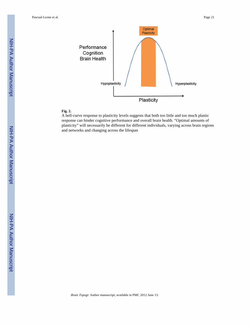
Fig. 2.A bell-curve response to plasticity levels suggests that both too little and too much plasticresponse can hinder cognitive performance and overall brain health. “Optimal amounts ofplasticity” will necessarily be different for different individuals, varying across brain regionsand networks and changing across the lifespan
Pascual-Leone et al. Page 21
Brain Topogr. Author manuscript; available in PMC 2012 June 13.
NIH
-PA Author Manuscript
NIH
-PA Author Manuscript
NIH
-PA Author Manuscript

Fig. 3.Schematic representation of individual plasticity across the lifespan. Although mechanismsof plasticity show a downward trend over the course of a typical lifetime, this trend willmanifest differently according to initial “baseline” levels, genetic factors, and environmentalinfluences. Therefore, one may conceptualize each individual has a unique “slope ofplasticity” across the lifespan
Pascual-Leone et al. Page 22
Brain Topogr. Author manuscript; available in PMC 2012 June 13.
NIH
-PA Author Manuscript
NIH
-PA Author Manuscript
NIH
-PA Author Manuscript

Fig. 4.TMS-based measures of cortical reactivity and plasticity. As recorded using eitherelectromyography (EMG) or electroencephalography (EEG), brain responses to TMS can bemeasured as motor evoked potentials (when TMS is applied to motor cortex) or localizedevoked field potentials. Such responses reflect cortical or corticospinal reactivity. Thesemeasures can be obtained before and following a given intervention, for example controlledmodulation of cortical excitability by repetitive TMS in the form of theta burst stimulation(TBS). The TBS-induced change in EMG or EEG responses to TMS provides a measure oflocal cortical plasticity. An example of TMS-EEG measures before and after continuousTBS illustrates the measurement of LTD-like plasticity
Pascual-Leone et al. Page 23
Brain Topogr. Author manuscript; available in PMC 2012 June 13.
NIH
-PA Author Manuscript
NIH
-PA Author Manuscript
NIH
-PA Author Manuscript
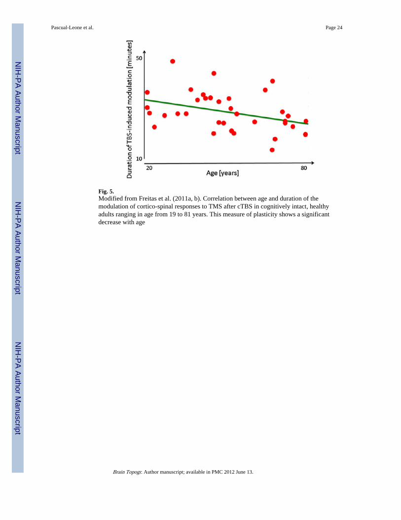
Fig. 5.Modified from Freitas et al. (2011a, b). Correlation between age and duration of themodulation of cortico-spinal responses to TMS after cTBS in cognitively intact, healthyadults ranging in age from 19 to 81 years. This measure of plasticity shows a significantdecrease with age
Pascual-Leone et al. Page 24
Brain Topogr. Author manuscript; available in PMC 2012 June 13.
NIH
-PA Author Manuscript
NIH
-PA Author Manuscript
NIH
-PA Author Manuscript

Fig. 6.Schematic representation of the influence of genetic or environmental impacts on brainplasticity. Alteration of local plasticity will trigger secondary adaptive responses acrossdiffuse neural networks that may prove ultimately adaptive or maladaptive for theindividual. Depending upon the amount and scope of such secondary responses, initial insulteffects may be alleviated or heightened
Pascual-Leone et al. Page 25
Brain Topogr. Author manuscript; available in PMC 2012 June 13.
NIH
-PA Author Manuscript
NIH
-PA Author Manuscript
NIH
-PA Author Manuscript

Fig. 7.Alterations of TMS-based measures of plasticity in nervous system pathology exemplifiedby autism spectrum disorder (ASD; a) and Alzheimer’s diseases (AD; b). a As comparedwith age-, gender- and IQ-matched controls, subjects with ASD show stronger and longer-lasting modulatory responses to continuous theta burst stimulation suggesting abnormal,hyper-plastic mechanisms. This is true for both, LTP- and LTD-like plasticity. b Ascompared with age-, gender- and IQ-matched controls, subjects with mild AD show weakerand shorter-lasting modulatory responses to continuous theta burst stimulation suggestingabnormal, hypo-plastic mechanisms
Pascual-Leone et al. Page 26
Brain Topogr. Author manuscript; available in PMC 2012 June 13.
NIH
-PA Author Manuscript
NIH
-PA Author Manuscript
NIH
-PA Author Manuscript

Fig. 8.Schematic representation of the hypothesized balance between local and network plasticity,its change across the lifespan, and its alterations by pathology
Pascual-Leone et al. Page 27
Brain Topogr. Author manuscript; available in PMC 2012 June 13.
NIH
-PA Author Manuscript
NIH
-PA Author Manuscript
NIH
-PA Author Manuscript