Characterization of a Type III secretion substrate specificity switch (T3S4) domain in YscP from...
14
Molecular Microbiology (2005) 56(1), 54–67 doi:10.1111/j.1365-2958.2005.04534.x © 2005 Blackwell Publishing Ltd Blackwell Science, LtdOxford, UKMMIMolecular Microbiology0950-382XBlackwell Publishing Ltd, 2005 ? 20055615467Original ArticleCharacterization of a T3S4 domain in YscPC. Agrain et al. Accepted 15 December, 2004. *For correspondence. E-mail guy. [email protected]; Tel. (+41) 61 267 2110; Fax (+41) 61 267 2118. † Present address: CNRS, UPR9050; E-mail: Laure.Journet@ esbs.u-strasbg.fr ‡ These authors contributed equally to this work. Characterization of a Type III secretion substrate specificity switch (T3S4) domain in YscP from Yersinia enterocolitica Céline Agrain, 1‡ Isabelle Callebaut, 2‡ Laure Journet, 1† Isabel Sorg, 1 Cécile Paroz, 1 Luís Jaime Mota 1 and Guy R. Cornelis 1 * 1 Biozentrum der Universität Basel, Basel, Switzerland. 2 Département de Biologie Structurale, Laboratoire de Minéralogie-Cristallographie Paris (CNRS/UMR 7590) Universités Paris 6 and Paris 7, France. Summary The length of the needle ending the Yersinia Ysc injec- tisome is determined by YscP, a protein acting as a molecular ruler. In addition, YscP is required for Yop secretion. In the present paper, by a systematic dele- tion analysis, we localized accurately the region required for Yop secretion between residues 405 and 500. As this C-terminal region of YscP has also been shown to control needle length it probably represents the substrate specificity switch of the machinery. By a bioinformatics analysis, we show that this region has a globular structure, an original a / b fold, a P-x-L- G signature and presumably no catalytic activity. In spite of very limited sequence similarities, this struc- ture is conserved among the proteins that are pre- sumed to control the needle length in many different injectisomes and also among members of the FliK family, which control the flagellar hook length. This region thus represents a new protein domain that we called T3S4 for T ype III s ecretion s ubstrate s pecificity s witch. The T3S4 domain of YscP can be replaced by the T3S4 domain of AscP ( Aeromonas salmonicida ) or PscP ( Pseudomonas aeruginosa ) but not by the one from FliK, indicating that in spite of a common global structure, these domains need to fit their part- ner proteins in the secretion apparatus. Introduction Injectisomes are multicomponent nanomachines involved in molecular trans -kingdom communication between bac- teria and eukaryotic cells. They are encountered in Gram- negative bacteria that are either pathogenic for animals and plants or are symbionts (Viprey et al ., 1998; Dale et al ., 2001; Dale et al ., 2002). They allow extracellular bacteria to inject proteins across the plasma membrane of eukaryotic cells and they enable intracellular bacteria to inject proteins across the host membrane limiting their compartment. For reviews see References (Cornelis and Wolf-Watz, 1997; Anderson and Schneewind, 1999; Galan and Collmer, 1999; Cornelis and Van Gijsegem, 2000; Plano et al ., 2001; Cornelis, 2002). Based on a phylogeny analysis made on three highly conserved proteins, the injectisomes can be grouped into five major families (Foultier et al ., 2002). The injectisomes from animal pathogens cluster in three families. The plas- mid encoded Ysc injectisome common to Yersinia entero- colitica, Yersinia pestis and Yersinia pseudotuberculosis represents an archetype for the largest family, which also includes the injectisomes from Pseudomonas aeruginosa (Psc), Aeromonas salmonicida (Asc), Bordetella pertus- sis , parapertussis and bronchiseptica (Bsc), Photorhab- dus luminescens (Lsc and Sct) and an unnamed injectisome from Vibrio parahemolyticus (called here Vsc). A second family includes the Shigella spp. Mxi-Spa injec- tisome, the Salmonella spp. Inv injectisome (encoded by SPI-1 in S. enterica ), the Ysa injectisome from Y. entero- colitica , an injectisome from Burkholderia pseudomallei and one from Sodalis glossinidius . A third family includes the Ssa injectisome from S. enterica (encoded by SPI-2), the Esc injectisome from enteropathogenic (EPECs) and enterohaemorrhagic (EHECs) E. coli and a chromosome- encoded injectisome from Y. pestis . The injectisomes from plant pathogens cluster in two families called Hrp1 ( Erwinia amylovora and Pseudomonas syringae ) and Hrp2 ( Xanthomonas campestris , Ralstonia solanacearum ). The amino acid sequence of many injectisome constit- uents has significant similarity to proteins from the flagel- lum with whom they share Type III secretion (T3S) (Allaoui et al ., 1994; Fields et al ., 1994; Van Gijsegem et al ., 1995; Aizawa, 2001; Blocker et al ., 2003). Both structures have a similar basal body consisting of a pair of rings that span the inner and outer bacterial membranes, hold together by a short tube. In most animal pathogens, the injectisome
-
Upload
independent -
Category
Documents
-
view
0 -
download
0
Transcript of Characterization of a Type III secretion substrate specificity switch (T3S4) domain in YscP from...
Molecular Microbiology (2005)
56
(1), 54–67 doi:10.1111/j.1365-2958.2005.04534.x
© 2005 Blackwell Publishing Ltd
Blackwell Science, LtdOxford, UKMMIMolecular Microbiology0950-382XBlackwell Publishing Ltd, 2005
? 2005
56
15467
Original Article
Characterization of a T3S4 domain in YscPC. Agrain
et al.
Accepted 15 December, 2004. *For correspondence. E-mail [email protected]; Tel. (
+
41) 61 267 2110; Fax (
+
41) 61 267 2118.
†
Present address: CNRS, UPR9050; E-mail: [email protected]
‡
These authors contributed equally to this work.
Characterization of a Type III secretion substrate specificity switch (T3S4) domain in YscP from
Yersinia enterocolitica
Céline Agrain,
1‡
Isabelle Callebaut,
2‡
Laure Journet,
1†
Isabel Sorg,
1
Cécile Paroz,
1
Luís Jaime Mota
1
and Guy R. Cornelis
1
*
1
Biozentrum der Universität Basel, Basel, Switzerland.
2
Département de Biologie Structurale, Laboratoire de Minéralogie-Cristallographie Paris (CNRS/UMR 7590) Universités Paris 6 and Paris 7, France.
Summary
The length of the needle ending the
Yersinia
Ysc injec-tisome is determined by YscP, a protein acting as amolecular ruler. In addition, YscP is required for Yopsecretion. In the present paper, by a systematic dele-tion analysis, we localized accurately the regionrequired for Yop secretion between residues 405 and500. As this C-terminal region of YscP has also beenshown to control needle length it probably representsthe substrate specificity switch of the machinery. Bya bioinformatics analysis, we show that this regionhas a globular structure, an original
aaaa
/
bbbb
fold, a P-x-L-G signature and presumably no catalytic activity. Inspite of very limited sequence similarities, this struc-ture is conserved among the proteins that are pre-sumed to control the needle length in many differentinjectisomes and also among members of the FliKfamily, which control the flagellar hook length. Thisregion thus represents a new protein domain that wecalled T3S4 for Type III secretion substrate specificityswitch. The T3S4 domain of YscP can be replaced bythe T3S4 domain of AscP (
Aeromonas salmonicida
)or PscP (
Pseudomonas aeruginosa
) but not by theone from FliK, indicating that in spite of a commonglobal structure, these domains need to fit their part-ner proteins in the secretion apparatus.
Introduction
Injectisomes are multicomponent nanomachines involvedin molecular
trans
-kingdom communication between bac-
teria and eukaryotic cells. They are encountered in Gram-negative bacteria that are either pathogenic for animalsand plants or are symbionts (Viprey
et al
., 1998; Dale
et al
., 2001; Dale
et al
., 2002). They allow extracellularbacteria to inject proteins across the plasma membraneof eukaryotic cells and they enable intracellular bacteriato inject proteins across the host membrane limiting theircompartment. For reviews see References (Cornelis andWolf-Watz, 1997; Anderson and Schneewind, 1999;Galan and Collmer, 1999; Cornelis and Van Gijsegem,2000; Plano
et al
., 2001; Cornelis, 2002).Based on a phylogeny analysis made on three highly
conserved proteins, the injectisomes can be grouped intofive major families (Foultier
et al
., 2002). The injectisomesfrom animal pathogens cluster in three families. The plas-mid encoded Ysc injectisome common to
Yersinia entero-colitica, Yersinia pestis
and
Yersinia pseudotuberculosis
represents an archetype for the largest family, which alsoincludes the injectisomes from
Pseudomonas aeruginosa
(Psc),
Aeromonas salmonicida
(Asc),
Bordetella pertus-sis
,
parapertussis
and
bronchiseptica
(Bsc),
Photorhab-dus luminescens
(Lsc and Sct) and an unnamedinjectisome from
Vibrio parahemolyticus
(called here Vsc).A second family includes the
Shigella
spp. Mxi-Spa injec-tisome, the
Salmonella
spp. Inv injectisome (encoded bySPI-1 in
S. enterica
), the Ysa injectisome from
Y. entero-colitica
, an injectisome from
Burkholderia pseudomallei
and one from
Sodalis glossinidius
. A third family includesthe Ssa injectisome from
S. enterica
(encoded by SPI-2),the Esc injectisome from enteropathogenic (EPECs) andenterohaemorrhagic (EHECs)
E. coli
and a chromosome-encoded injectisome from
Y. pestis
. The injectisomesfrom plant pathogens cluster in two families calledHrp1 (
Erwinia amylovora
and
Pseudomonas syringae
)and Hrp2 (
Xanthomonas campestris
,
Ralstoniasolanacearum
).The amino acid sequence of many injectisome constit-
uents has significant similarity to proteins from the flagel-lum with whom they share Type III secretion (T3S) (Allaoui
et al
., 1994; Fields
et al
., 1994; Van Gijsegem
et al
., 1995;Aizawa, 2001; Blocker
et al
., 2003). Both structures havea similar basal body consisting of a pair of rings that spanthe inner and outer bacterial membranes, hold togetherby a short tube. In most animal pathogens, the injectisome

Characterization of a T3S4 domain in YscP
55
© 2005 Blackwell Publishing Ltd,
Molecular Microbiology
,
56
, 54–67
ends up with a needle that protrudes outside the bacte-rium. Therefore, the injectisome is also called needle-complex (Kubori
et al
., 1998; Blocker
et al
., 1999;Kimbrough and Miller, 2000; Kubori
et al
., 2000; Sukhan
et al
., 2003; Marlovits
et al
., 2004). A long flexible pilusterminates the Hrp and Esc injectisomes (Roine
et al
.,1997; Knutton
et al
., 1998; Van Gijsegem
et al
., 2000). Inthe flagellum, the filament is connected to the basal bodyvia a hook. The length of this hook is genetically deter-mined and a protein called FliK has been shown to playthe key role in this process (Patterson-Delafield
et al
.,1973; Suzuki and Iino, 1981). Mutants deficient in FliKmake extra-long hooks (called polyhooks) and no fila-ment. Extragenic suppressive mutations restoring filamentassembly on polyhook structures (polyhook-filament phe-notype) have been mapped in
flhB
, a gene encoding amajor component of the export apparatus (Suzuki andIino, 1981; Kutsukake
et al
., 1994; Williams
et al
., 1996;Fraser
et al
., 2003a). The study of these
fliK
suppressorsmutations led to the proposal that the C-terminal domainof FlhB has two substrate specificity states and that aconformational change, mediated by FliK and accompa-nied by a proteolytic cleavage of FlhB is responsible forthe specificity-switching process allowing secretion offlagellin once assembly of the hook is completed(Minamino and Macnab, 2000; Makishima
et al
., 2001;Fraser
et al
., 2003b). The length of the injectisome-needleis also genetically determined. Mutations in
spa32
from
Shigella
, in
invJ
from
Salmonella
(SPI-1) (Kubori
et al
.,2000; Magdalena
et al
., 2002; Tamano
et al
., 2002) andin
yscP
from
Yersinia
(Journet
et al
., 2003) lead to needleswith undefinite length and no substrate secretion. Thehomologues of FlhB in injectisomes form the well-con-served YscU family (Allaoui
et al
., 1994). Introduction intoYscU of the FlhB substitutions that suppress the
fliK
phe-notype restores Yop secretion in
yscP
mutants (Edqvist
et al
., 2003). Moreover, like FlhB, YscU undergoes a pro-teolytic cleavage (Lavander
et al
., 2003). Thus, very muchlike FliK, YscP is presumably involved in the substratespecificity switch, interacting with the basal-body compo-nent YscU to stop secretion of the YscF needle subunitsand to start Yop secretion. In addition to this switch func-tion, YscP was recently shown to act as a molecular rulerdetermining the length of the needle (Journet
et al
., 2003).YscP is thus a protein with a dual function. In the molec-ular ruler model, the N-terminus and C-terminus of YscPare proposed to act as anchors of the central ruler part,one extremity being attached to the basal body and theother one to the growing tip of the needle (Journet
et al
.,2003). However, no information exists regarding theregions of YscP involved in its switch role.
Here, by systematic deletion mutagenesis of YscPand a bioinformatics analysis, we localized accuratelythe switch domain in the C-terminal domain of the pro-
tein and we show that this switch domain is globular andconserved among the proteins controlling length ininjectisomes as well as in the flagellum. In contrast tomost of the proteins of the injectisomes and flagella, therelationship between Spa32/InvJ, YscP and FliK hadremained to date unrevealed because of the too highdivergence in their sequences (in the range of 10%identity).
Results
YscP385–500 is required for Yop secretion
We showed previously that in frame deletions and inser-tions in the central part of YscP (36–96 and 222–306)defined a ruler region (Journet
et al
., 2003), and that inframe deletions in the N-terminus (16–25 and 26–35) aswell as in the C-terminus (385–424 and 467–515) identi-fied regions of the protein that are necessary for YscP toexert its needle length control function (Journet
et al
.,2003). However, the deletion mutants were not analysedfor their capacity to secrete Yops. In this work, we gener-ated new deletions in the
yscP
gene that together with theones generated before (see above) encompass almostthe entire gene (Fig. 1A) and we tested the mutants fortheir capacity to secrete Yops. The ruler region of YscP ischaracterized by a duplication of 60 central residues(Stainier
et al
., 2000), which hampers the engineering ofdeletions. For this reason, we could only construct onelarge deletion between residues 222 and 306 of YscP, andnot smaller ones as in the rest of the protein. We clonedthe mutated alleles downstream from an arabinose-inducible promoter (pBAD) and the recombinant plasmidswere introduced in
Y. enterocolitica
E40 (pLJ4036), a new
yscP
null mutant constructed for this study. In the pLJ4036pYV plasmid, the
yscP
gene is completely removed, fromstart to stop, in order to prevent any interference from left-over domains or sites. All the recombinant bacteria,incubated at 37
∞
C in Ca
2
+
-deprived medium containingarabinose, synthesized YscP proteins of the expected size(Fig. 1B). We then analysed the phenotype of all themutants with regard to Yop secretion. As shown in Fig. 1C,the deletions removing parts of the protein before residue381 and after residue 500 had no impact on Yop secretion.In contrast, deletions between amino acids 385–500affected Yop secretion. This indicates that this C-terminalregion (385–500) is required for both functions, lengthcontrol (Journet
et al
., 2003) and Yop secretion, andhence is responsible for the substrate specificity switchfunction. Interestingly, there was no strict correlationbetween the phenotype of Yop secretion deficiency andtight length control. Indeed, deletions affecting the N-terminus (YscP
D
16–25 and YscP
D
26–35) (Journet
et al
.,2003) led only to a loss of length control but not to a lossof Yop secretion.

56
C. Agrain
et al.
© 2005 Blackwell Publishing Ltd,
Molecular Microbiology
,
56
, 54–67
Fig. 1.
A. Schematic representations of the
yscP
in frame deletions mutants together with their ability to secrete Yops. The highlighted box in YscP (aa 403–492) represents the T3S4 domain.B. Western blot analysis (total cells; polyclonal antibody) of the various YscP proteins produced by
Y. enterocolitica
MRS40(pLJ4036), i.e the
yscP
–
mutant (–, control); and by
Y. enterocolitica
MRS40(pLJ4036) carrying the plasmids (listed on the left) encoding the various
yscP
+
genes.C. Yop proteins secreted by the same
Y. enterocolitica
E40 strains as in B (Coomassie-stained SDS-PAGE). Note that MRS40(pLJ4036), the
yscP
knockout background is not completely negative for Yop secretion.
Characterization of a T3S4 domain in YscP
57
© 2005 Blackwell Publishing Ltd,
Molecular Microbiology
,
56
, 54–67
The YscP C-terminal region is conserved and defines a new family of proteins
YscP and its flagellar equivalent, FliK, have similar func-tions but their global identity at the primary sequence levelis only within the 10% range. We identified the switchfunction in the C-terminus of YscP. In the model proposedfor FliK, the domain that switches the substrate specificityis also localized in the C-terminus (Williams et al., 1996;Minamino et al., 2004). We therefore then wondered if theC-terminal regions of YscP and FliK were sharing similar-ities and if we could point out an equivalent region inproteins found in other injectisomes or T3S systems.
To begin with, we used the C-terminal domain of YscP(from aa 385 to the C-terminus) as query and performedPSI-BLAST searches (Altschul et al., 1997) on the non-redundant database (nr) at the National Center for Biolog-ical Information (NCBI) using an E-value inclusion thresh-old of 0.005. By iteration 4, searches converged to identifycounterpart proteins of YscP in T3S systems from the YscPfamily: AscP, PscP, LscP and SctP. As suggested by theletter code, these proteins are encoded by genes thatoccupy the same locus as yscP in the operons encodingthe different injectisomes. In addition, we found the VscPprotein from Vibrio harveyi [GenBank identifier (gi)41834182]. All these proteins turned out to have a con-served C-terminal domain (34% identity) preceded by vari-able regions. We called the conserved domain the T3S4domain after Type III secretion substrate specificity switch.
We also found a suggestive match (E-value 0.19, 35%identity on a 117 aa overlap) with the flagellar hook-lengthcontrol protein FliK from Y. pestis (gi 45442809). Integrat-ing the Y. pestis FliK sequence within the position specificscore matrix (PSSM) led to significantly detect all themembers of the FliK family. One can note that the similar-ity between YscP and FliK proteins was directly found tobe significant using a longer sequence of YscP as query(aa 363–515). The Y. pestis FliK region (aa 251–393) wasalso used as a query for reciprocal PSI-BLAST searches(same parameters). By iteration 2, LscP from P. lumine-scens was significantly retrieved (E = 0.005), whereasother members of the YscP family were retrieved by iter-ation 3 (E-value of 4 ¥ 10-5 for Y. enterocolitica YscP).Proteins of the HrcP family were retrieved just below thethreshold value after convergence by iteration 22 (e.g. R.solanacearum HpaP using the Y. pestis YscP sequenceas query; E-value of 0.13, 18% sequence identity over90 aa) or by iteration 14 (e.g. R. solanacearum HpaPusing the V. parahaemolyticus LafE sequence as query;E-value of 0.023, 18% sequence identity over 90 aa).
Although marginal, the potential similarity betweenYscP/FliK and HrcP proteins was further supported at the2D level by using hydrophobic cluster analysis (HCA)(Gaboriaud et al., 1987; Callebaut et al., 1997) (Fig. 2A
and B and legend). The aligned sequences were alsosearched against databases using HMMER (Eddy, 1998),but such searches did not identify proteins other thanthose highlighted before. Interestingly the T3S4 domain islocalized in the C-terminus in every member of the family,suggesting that this specific position might be relevant tothe mechanism of length control (Fig. 3).
Finally, because it was anticipated that the T3S4domain, detected in the YscP, FliK and HrcP families,would also be present in the Spa32 and InvJ family, wescreened these last sequences using HCA for the pres-ence of 2D markers of the T3S4 domain, which could havebeen missed by other search methods. This led to identifyclear markers of the T3S4 domain in the Spa32/InvJsequences (Fig. 2A and B, see also below), suggestingthat these proteins could indeed share the T3S4 domainwith the YscP, FliK and HrcP families.
Characterization of the T3S4 domain
The T3S4 domain defined in the preceding sectionbetween aa 385 and 500 of YscP, has all the chara-cteristics of a globular domain, as visualized using HCA(Callebaut et al., 1997) (Fig. 2A). It contains approxi-mately one-third of hydrophobic amino acids, organizedin clusters, the lengths of which are typical of those ofregular secondary structures. Based on the YscPsequence, the limits of the globular domain can be refinedon both sides (Fig. 2A): (i) it starts at aa 403 as the pre-ceding sequence (~aa 391–403) corresponds to arepeated fragment, the first copy of which can be foundupstream in the YscP sequence (~aa 243–255) and itends up at aa 492, where a long sequence devoid ofhydrophobic amino acids starts. The latter sequence likelydefines a hinge separating the switch domain from a smallregion, which might organize as an a-helix.
The T3S4 domain (arrow in Fig. 2A and boxed inFig. 2B) has an a/b predicted fold, with a core regionhaving almost no insertion-deletion. Seven hydrophobicclusters, as defined using HCA and numbered up to thealignment of Fig. 2A and B, delineate the predicted sec-ondary structures, for which buried positions are high-lighted by conservation of hydrophobic amino acids(boxed green in Fig. 2B). A conserved proline and a con-served glycine (P-x-L-G) between the predicted b-strandsb1 and b2 seems to represent a signature of the T3S4domain. No clear conservation is observed outside fromthe hydrophobic amino acids and loop markers, renderinga catalytic function unlikely.
Strikingly, in proteins from the YscP and FliK subgroups,the T3S4 domain is followed by a clear hinge region,particularly rich in acidic residues (represented in red inFig. 2B). A hydrophobic cluster or a group of hydrophobicclusters (shaded grey on Fig. 2B) follows this hinge region

58 C. Agrain et al.
© 2005 Blackwell Publishing Ltd, Molecular Microbiology, 56, 54–67

Characterization of a T3S4 domain in YscP 59
© 2005 Blackwell Publishing Ltd, Molecular Microbiology, 56, 54–67
and ends the sequences in the YscP and FliK subgroupsrespectively. These additional secondary structure(s)(likely a-helices) might regulate the T3S4 domain function.However, deletion of this 15-residue tail from YscP had nomajor impact on the needle length (data not shown) andon Yop secretion (Fig. 1). In the group of proteins fromplant pathogens, this tail is missing and the predicted lastb-strand of the T3S4 domain ends the sequence.
Finally, additional secondary structures appeared to bepresent N-terminal to the first alignment block, suggestingthat variable sequences might complete the T3S4 domaincore.
Fold prediction methods [3DPSSM (Kelley et al., 2000),FUGUE (Shi et al., 2001)] did not highlight any known 3Dstructures, which might be compatible with the T3S4domain structure.
The closest T3S4 domains are exchangeable
The overall structure of the T3S4 domain seems thus tobe highly conserved among the different members of theYscP/FliK family. Not only the expected fold but also thesize of the domain is the same, even though the full-lengthproteins are of different sizes (Fig. 3). This suggested thatthe T3S4 domains of different proteins could beexchanged. We tried to replace the T3S4 domain of YscPby the one of AscP (Aeromonas salmonicida) (Burr et al.,2003), PscP (Pseudomonas aeruginosa) and the one ofFliK from S. enterica serovar Typhimurium and from Yers-inia pestis. To do this, we first introduced two restrictionsites on both sides of the region encoding the predictedT3S4 domains in the yscP gene cloned downstream frompBAD. The resulting YscP protein was still fully functionalfor both needle length control (data not shown) and Yopsecretion (Fig. 4), suggesting that the proposed delinea-tion of the T3S4 domain was correct and that both inser-tions were performed outside the domain. We then clonedthe region encoding the T3S4 domain from the differentproteins in the inserted sites and the recombinant plas-mids were introduced in the yscP– strain. Secretion ofYops was triggered in vitro and monitored. The YscPhybrid proteins, in which the T3S4 domain was replacedby the T3S4 domain of AscP or PscP (40% and 38%sequence identity respectively), could restore a wild-typephenotype regarding Yop secretion. So it appears thatT3S4 domains of AscP and PscP are capable of playing
Fig. 3. Schematic representation of T3S4 domain-containing pro-teins equivalent to YscP from the different TTSS.
Fig. 2. A. Comparison of the HCA plots of T3S4 domains of the YscP, FliK and HrcP families, highlighting conserved regular secondary structures. Protein sequences are shown on a duplicated alpha helical net on which the encircled hydrophobic amino acids (V, I, L, M, F, Y, W) form clusters. The positions of these clusters statistically match those of the regular secondary structures (alpha helices and beta strands). This analysis gives access to 2D signatures, which are much more conserved than 1D sequences and thus help sequence comparison at high levels of divergence. Guidelines to the use of HCA are given in the study by Gaboriaud et al. (1987) and by Callebaut et al. (1997) whereas recent publications can be found at the following URL (http://www.lmcp.jussieu.fr/~mornon/publications.html). The way to read the sequences and special symbols are indicated in the inset. Representative sequences of the three families are shown: YscP from Y. enterocolitica [GenBank identifier (gi) 17839593], FliK from Y. pestis (gi 16121061) and RspP from P. fluorescens (gi 15042139). Cluster similarities are indicated in green, sequence identities in orange. Amino acids shaded pink within the box in the YscP sequence (‘repeated sequence’) correspond to identities relative to a sequence fragment found upstream (aa 243–255). Vertical bars indicate cluster correspondences (labelled from 1 to 7). Clusters labelled 2, 3, 4 and 7 are highly representative of b-strands structures (Hennetin et al., 2003; K. Le Tuan et al. in preparation). The sequence of InvJ from S. enterica LT2 (gi 16766198) is shown at the bottom, below the YscP, FliK and RspP sequences, in order to illustrate the putative correspondence that can be identified using HCA with cognate T3S4 domains. In yellow are indicated some identity ‘clusters’ that can be identified relative to the FliK sequence, strengthening the putative relationship.B. Representative multiple alignment of T3S4 domains, as deduced from PSI-BLAST analysis and adjusted manually, in particular considering Hydrophobic Cluster Analysis (HCA) (see before). Species and protein names are given on the left; GenBank identifiers (gi) are on the right. N- and C-termini amino acid positions are also indicated. Stars indicate the C-terminal ends of the sequences. The T3S4 domains are boxed, whereas acidic and polar amino acids (D, E, N, Q, S, T) C-terminal to the T3S4 domains are coloured in red, highlighting the presence of acidic/polar regions downstream the T3S4 domains in the YscP and FliK families. These forms ‘hinge’ regions separating T3S4 domains from short C-terminal globular sequences, evidenced by the presence of hydrophobic clusters (shaded grey). The SctP protein is specific of the subspecies laumondii TTO1 of Photorhabdus luminescens. Conserved hydrophobic amino acids (V, I, L, F, M, Y, W) or amino acids that can substitute them (S, T, A, C) are boxed green, whereas other conserved positions are shaded using other colours. The secondary structure prediction, performed using JPred (Cuff and Barton, 1999), is shown beneath the alignment. H and E stand for helix and extended (b-strand) respectively.

60 C. Agrain et al.
© 2005 Blackwell Publishing Ltd, Molecular Microbiology, 56, 54–67
the switch role of YscP although their sequence identity islimited. In contrast, the protein carrying the FliK T3S4domain either from S. enterica ser Typhimurium or fromY. pestis could not complement the yscP mutant. Thus,T3S4 domains could be exchanged between proteinsfrom the same family of injectisomes. But when it comesto exchange T3S4 domains between proteins involved indifferent systems such as flagellum and injectisome, thefunction cannot be performed any more.
We then analysed if the hybrid proteins YscP-AscP andYscP-PscP conferred needle length control. We observedby electron microscopy that the length of most needleswas controlled but the control was leaky: the histogramsof length distribution show a clear wt peak around 55 nmbut some needles are longer (Fig. 4). Thus, it looks asthough in hybrid proteins, the substrate specificity switchworks but is not well controlled by the ruler.
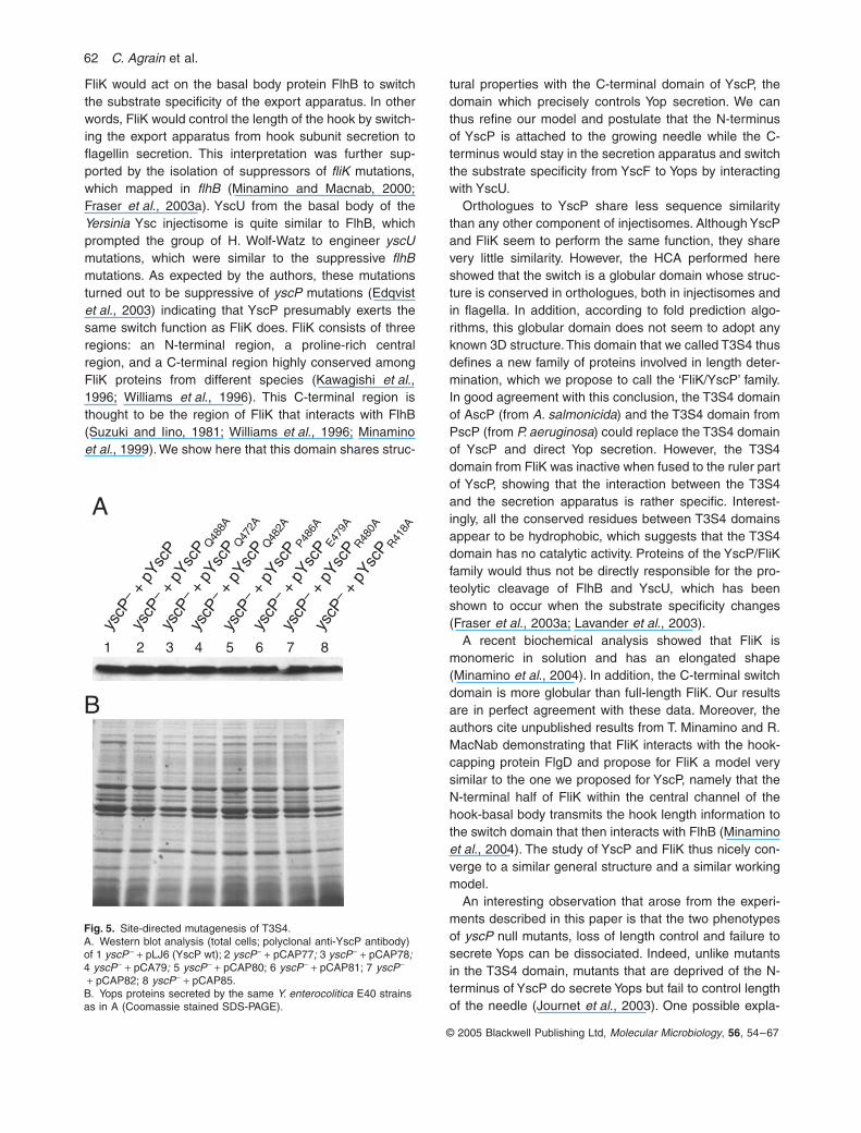
Site-directed mutagenesis of the T3S4 domain
Based on the alignment performed using HCA and Clust-alW combined, some residues appeared to be conserved.Most of them are the so-called topohydrophobic residuesthat are of great importance for the structure of domainsand therefore their mutation would dramatically affect thestructure. But there are also a few other positions con-served in all the T3S4 domains (R418, Q488), which areprobably not as critical for the structure of the domain. Wedecided to mutate them by alanine replacement to per-haps identify residues essential for the function. Thesesite-directed mutations were again engineered on theyscP+ gene cloned in the pBADMycHisA vector, down-stream from the arabinose promoter. The different con-structs obtained were then transformed in the yscP– strainand secretion was monitored at 37∞C after Ca2+ chelation.The mutants had a wild-type phenotype regarding Yopsecretion (Fig. 5). Even though these two positions wereconserved in all the T3S4 domains, they were not affectedby an alanine replacement.
Considering that swapping the T3S4 domains betweenYscP, AscP and PscP yielded functional hybrids, wedecided also to focus on positions conserved in the T3S4domains of only this subgroup of proteins (Q472, E479,R480, Q482 and P486). None of the five single alaninereplacements performed led to a Yop secretion mutantphenotype. The lack of effect of alanine substitutionsstrengthen the importance of the structure of the T3S4
domain for the function. Indeed no essential residueswere highlighted beside the hydrophobic ones. Finally, weconsidered to mutate constitutive amino acids of thedomain signature (P-x-L-G). So we performed alaninereplacements of residues P440 and L442 although wewere aware of the fact that a replacement of the prolinecould impact the local structure of the b-turn between thepredicted b1 and b2 strands. The phenotype observed forthe L442A mutant was wild-type for both functions (Fig. 6).In contrast, the P440A mutant secreted uniformly lessYops than wild-type bacteria. Hence, we also analysedneedle length control in this mutant and found an interme-diary phenotype (Fig. 6). Indeed a wt peak (55 nm) wasobserved but there were also needles completely dereg-ulated, some being extra-longs. It seems thus that thedomain still exerts its function in some individuals but notin others. The decreased efficiency may be attributed toa slight modification of T3S4 fold resulting from thereplacement of the proline, usually crucial for the turnstructure.
All the observations made are not only validating thepredicted characteristics of the structure of the T3S4domain but also pinpointing the importance of its overallstructure for the function.
Discussion
We have previously reported that YscP acts as a molec-ular ruler and that the first 35 residues as well as the last130 residues are required for the needle length control(Journet et al., 2003). We postulated from this observationthat the two ends of YscP act as anchors. One end wouldbe connected to the growing tip of the needle whereas theother end would be attached to the basal body. WhenYscP would be fully stretched, it would signal via its inter-nal anchor to the secretion apparatus that would stopexporting YscF and the needle would stop growing (Jour-net et al., 2003). Here we show that the C-terminaldomain, which is required for length control is alsorequired for Yop secretion. Thus, the C-terminal domain isrequired for the two functions of YscP: control of Yopsecretion and control of needle length. Such a dual func-tion has previously been shown for FliK, which controlssecretion of flagellin as well as the length of the hook ofthe flagellum (Williams et al., 1996; Minamino and Mac-nab, 2000; Minamino et al., 2004). To explain this pheno-type, Williams et al. (Williams et al., 1996) suggested that
Fig. 4. Swapping of the T3S4 domains.A. Coomassie stained SDS-PAGE of culture supernatants of Y. enterocolitica MRS40(pLJ4036) complemented with 1 pLJ6 (wild-type YscP, control); 2 pCA88 (introduction of a XbaI restriction site); 3 pCA89 (XbaI and BglII restriction sites); 4 pCA90 (T3S4 of AscP); 5 pCA91 (T3S4 of FliKS.t.); 6 pCA92 (T3S4 of PscP); 7 pCA93 (T3S4 of FliKY.p.). The size of YscP is indicated by an arrow.B. Histograms of the needle length measurements, and electron micrographs of yscP – + pCA90 (T3S4 of AscP) and yscP – + pCA91 (T3S4 of FliK). M, mean of the lengths; N, number of needles measured.

Characterization of a T3S4 domain in YscP 61
© 2005 Blackwell Publishing Ltd, Molecular Microbiology, 56, 54–67
62 C. Agrain et al.
© 2005 Blackwell Publishing Ltd, Molecular Microbiology, 56, 54–67
FliK would act on the basal body protein FlhB to switchthe substrate specificity of the export apparatus. In otherwords, FliK would control the length of the hook by switch-ing the export apparatus from hook subunit secretion toflagellin secretion. This interpretation was further sup-ported by the isolation of suppressors of fliK mutations,which mapped in flhB (Minamino and Macnab, 2000;Fraser et al., 2003a). YscU from the basal body of theYersinia Ysc injectisome is quite similar to FlhB, whichprompted the group of H. Wolf-Watz to engineer yscUmutations, which were similar to the suppressive flhBmutations. As expected by the authors, these mutationsturned out to be suppressive of yscP mutations (Edqvistet al., 2003) indicating that YscP presumably exerts thesame switch function as FliK does. FliK consists of threeregions: an N-terminal region, a proline-rich centralregion, and a C-terminal region highly conserved amongFliK proteins from different species (Kawagishi et al.,1996; Williams et al., 1996). This C-terminal region isthought to be the region of FliK that interacts with FlhB(Suzuki and Iino, 1981; Williams et al., 1996; Minaminoet al., 1999). We show here that this domain shares struc-
tural properties with the C-terminal domain of YscP, thedomain which precisely controls Yop secretion. We canthus refine our model and postulate that the N-terminusof YscP is attached to the growing needle while the C-terminus would stay in the secretion apparatus and switchthe substrate specificity from YscF to Yops by interactingwith YscU.
Orthologues to YscP share less sequence similaritythan any other component of injectisomes. Although YscPand FliK seem to perform the same function, they sharevery little similarity. However, the HCA performed hereshowed that the switch is a globular domain whose struc-ture is conserved in orthologues, both in injectisomes andin flagella. In addition, according to fold prediction algo-rithms, this globular domain does not seem to adopt anyknown 3D structure. This domain that we called T3S4 thusdefines a new family of proteins involved in length deter-mination, which we propose to call the ‘FliK/YscP’ family.In good agreement with this conclusion, the T3S4 domainof AscP (from A. salmonicida) and the T3S4 domain fromPscP (from P. aeruginosa) could replace the T3S4 domainof YscP and direct Yop secretion. However, the T3S4domain from FliK was inactive when fused to the ruler partof YscP, showing that the interaction between the T3S4and the secretion apparatus is rather specific. Interest-ingly, all the conserved residues between T3S4 domainsappear to be hydrophobic, which suggests that the T3S4domain has no catalytic activity. Proteins of the YscP/FliKfamily would thus not be directly responsible for the pro-teolytic cleavage of FlhB and YscU, which has beenshown to occur when the substrate specificity changes(Fraser et al., 2003a; Lavander et al., 2003).
A recent biochemical analysis showed that FliK ismonomeric in solution and has an elongated shape(Minamino et al., 2004). In addition, the C-terminal switchdomain is more globular than full-length FliK. Our resultsare in perfect agreement with these data. Moreover, theauthors cite unpublished results from T. Minamino and R.MacNab demonstrating that FliK interacts with the hook-capping protein FlgD and propose for FliK a model verysimilar to the one we proposed for YscP, namely that theN-terminal half of FliK within the central channel of thehook-basal body transmits the hook length information tothe switch domain that then interacts with FlhB (Minaminoet al., 2004). The study of YscP and FliK thus nicely con-verge to a similar general structure and a similar workingmodel.
An interesting observation that arose from the experi-ments described in this paper is that the two phenotypesof yscP null mutants, loss of length control and failure tosecrete Yops can be dissociated. Indeed, unlike mutantsin the T3S4 domain, mutants that are deprived of the N-terminus of YscP do secrete Yops but fail to control lengthof the needle (Journet et al., 2003). One possible expla-
Fig. 5. Site-directed mutagenesis of T3S4.A. Western blot analysis (total cells; polyclonal anti-YscP antibody) of 1 yscP – + pLJ6 (YscP wt); 2 yscP– + pCAP77; 3 yscP– + pCAP78; 4 yscP– + pCA79; 5 yscP– + pCAP80; 6 yscP– + pCAP81; 7 yscP–
+ pCAP82; 8 yscP– + pCAP85.B. Yops proteins secreted by the same Y. enterocolitica E40 strains as in A (Coomassie stained SDS-PAGE).
B
A
yscP
– + p
YscP
Q48
2A
yscP
– + p
YscP
Q48
8A
yscP
– + p
YscP
Q47
2A
yscP
– + p
YscP
P486
A
yscP
– + p
YscP
E479
A
yscP
– + p
YscP
R480A
yscP
– + p
YscP
R418A
yscP
– + p
YscP
1 2 3 4 5 6 7 8

Characterization of a T3S4 domain in YscP 63
© 2005 Blackwell Publishing Ltd, Molecular Microbiology, 56, 54–67
nation would be that when the YscP constructs aredeprived of one of their anchors the switch could operateat random, independently of the trigger given by thestretching of the ruler. In bacteria harbouring such con-structs, the model would predict that the needle size wouldbe variable but Yop secretion would occur from needlesthat have stopped growing. This is compatible with theobserved phenotype but it could not be taken as a prooffor the model unless one could demonstrate that onlyneedles that have stopped growing do secrete Yops.
Although the model can account for the phenotype ofmutants that do no longer tightly control the needle lengthbut still secrete Yops, it predicts that mutants with theopposite phenotype (normal length but no Yop secretion)are unlikely to occur. Indeed, an arrest in the elongationof the needle implies that the secretion of YscF is switchedoff and thus that the switch is functional. None of the yscPmutants analysed to date displayed such a phenotype. Inthe flagellum also no fliK mutants controlling hook lengthbut unable to initiate filament assembly were everdescribed (Williams et al., 1996).
According to our present study of the domain, a fullunderstanding of its functions will probably have to awaitthe determination of its 3D structure as well as that of itspotential interacting partner YscU.
Experimental procedures
Sequence analysis
Similarity searches were performed using PSI-BLAST
(Altschul et al., 1997) and HMMer(Eddy, 1998). The bidimen-
sional HCA (Gaboriaud et al., 1987; Woodcock S., 1992;Callebaut et al., 1997) was used for refining the proposedsimilarities.
Secondary structure prediction was performed using Jpred(Cuff and Barton, 1999). FUGUE (Shi et al., 2001) and 3D-PSSM (Kelley et al., 2000) were used for fold recognition.
Induction of the yop regulon and Yop secretion analysis
Bacteria were routinely grown on Luria–Bertani agar platesand in liquid LB medium. For the induction of the yop regulon,Y. enterocolitica bacteria were inoculated to an OD600 of 0.1and cultivated in brain–heart infusion (BHI; Remel) supple-mented with 4 mg ml-1 glycerol, 20 mM MgCl2 and 20 mMsodium oxalate (BHI-Ox) 2H at 22∞C, then shifted to 37∞Cand incubated for 4 h. Expression of the different yscP genescloned downstream from the pBAD promoter was induced byadding 0.2% arabinose to the culture just before the shift at37∞C, and again 2 h later. Ampicillin was used at a con-centration of 200 mg ml-1 to select for the expressionplasmids.
Proteins from the supernatant were precipitated overnightat 4∞C with trichloroacetic acid 10% (w/v) final. Electrophore-sis was carried out in 12% or 15% (w/v) polyacrylamide gelsin the presence of SDS (SDS-PAGE). Proteins secreted by3 ¥ 108 bacteria were loaded per lane. For the total bacterialcells, the proteins from 107 bacteria were loaded per lane.After electrophoresis, proteins were stained with Coomassiebrilliant blue (Pierce) or transferred by electroblotting to anitrocellulose membrane. Immunoblotting was carried outusing a polyclonal rabbit anti-YscP antibody (MIPA57).
Detection of immunoblots was performed with a secondaryantibody conjugated to horseradish peroxidase (1:2000;Dako) before development with supersignal chemilumines-cent substrate (Pierce).
B yscP
–
yscP
– p
YscP
yscP
– p
YscP
yscP
– p
YscP
yscP
–
yscP
– p
YscP
yscP
– p
YscP
yscP
– p
YscP
0
5
10
15
20
25
050 100
150
200
250
300
350
400
450
500
550
600
650
700
750
800
850
900
Needle length (nm)
Num
ber
of n
eedl
es
CyscP
– + pYscPP440A
n = 220, m = 134 ± 141
0
5
10
15
20
25
30
35
40
0 25 50 75 100
125
150
175
200
225
250
275
Needle length (nm)
Num
ber o
f nee
dles yscP
– + pYscPL442A
n = 112, m = 60 ± 8
A+ + +
+++
L442
A P4
40A
WT
WT
P440
A
L442
A
Fig. 6. Site-directed mutagenesis of the signature.A. Western blot analysis (total cells; polyclonal anti-YscP antibody) of yscP –; yscP – + pLJ6 (YscP wt); yscP– + pCAP83 (YscPL442ÆA); yscP –
+ pCAP86 (YscPP440ÆA).B. Yops proteins secreted by the same Y. enterocolitica E40 strains as in A (Coomassie stained SDS-PAGE).C. Histograms of the needle length measure-ment of the same strains as in A. Note the difference in scale of the x-axes.
64 C. Agrain et al.
© 2005 Blackwell Publishing Ltd, Molecular Microbiology, 56, 54–67
Electron microscopy
Visualization of the needle-like structures at the cell surfaceof the bacteria was performed by electron microscopy asdescribed by Hoiczyk and Blobel (Hoiczyk and Blobel, 2001).After 4 h of induction at 37∞C, bacteria were harvested at
2000 g and resuspended gently in 20 mM Tris-HCl, pH 7.5.Droplets were applied for 1 min to freshly glow-discharged,formvar-carbon coated grids, and negatively stained with 2%(w/v) uranylacetate. Bacteria were visualized in a PhilipsMorgagni 268D electron microscope at a nominal magnifica-tion of ¥44 000 or 27 000 and an acceleration voltage of
Table 1. Plasmids used in this work.
Plasmids Encoded protein Genotype or description Source or reference
pYVpYV40 Wild-type virulence plasmid from strain Y. enterocolitica E40 Sory et al. (1995)pLJ4036 pYV40 yscP– This work
ClonespBADMycHisA InvitrogenpCA1 YscPD1-15 pBADMycHisA – yscPD1-15 Journet et al. (2003)pCA18 YscPD385-424 pBADMycHisA – yscPD385-424 Journet et al. (2003)pCA5 YscPD16-25 pBADMycHisA – yscPD16-25 Journet et al. (2003)pCA6 YscPD26-35 pBADMycHisA – yscPD26-35 Journet et al. (2003)pCA7 YscPD36-45 pBADMycHisA – yscPD36-45 Journet et al. (2003)pCA79 YscPQ482ÆA Replacement of Q482 by alanine by inverse PCR on pLJ6 using
oligonucleotides 3467 and 3468This work
pCA83 YscPL442ÆA Replacement of L442 by alanine by inverse PCR on pLJ6 using oligonucleotides 3460 and 3461
This work
pCA86 YscPP440ÆA Replacement of P440 by alanine by inverse PCR on pLJ6 using oligonucleotides 3458 and 3459
pCA88 YscPW1 Introduction of a XbaI site between codons 405 and 406 of yscPentero by inverse PCR on pLJ6 using oligonucleotides 3478 and 3479
This work
pCA89 YscPW Introduction of a BglII site between codons 495 and 496 of yscPentero by inverse PCR on pCA88 using oligonucleotides 3480 and 3481
This work
pCA9 YscPD46-96 pBADMycHisA – yscPD46-96 Journet et al. (2003)pCA90 YscPWT3S4AscP Insertion of aa 149 to aa 238 from AscP (Aeromonas salmonicida),
amplified using oligonucleotides 3487 and 3488, into YscPW
This work
pCA91 YscPWT3S4FliKS.t. Insertion of aa 268 to aa 353 from FliK (Salmonella typhimurium LT2), amplified using oligonucleotides 3476 and 3477, into YscPW
This work
pCA92 YscPWT3S4PscP Insertion of aa 263 to aa 348 from PscP (Pseudomonas aeruginosa), amplified using oligonucleotides 3569 and 3570, into YscPW
This work
pCA93 YscPWT3S4FliKY.p. Insertion of aa 261 to aa 348 from FliK (Yersinia pestis), amplified using oligonucleotides 3567 and 3568, into YscPW
This work
pCAP19 YscPD425-464 Deletion from pLJ6 using oligonucleotides 3244 and 3245 This workpCAP47 YscPD97-137 Deletion of codons 97–137 of yscPentero from pLJ6 by inverse PCR using
phosphorylated oligonucleotides 3318 and 3319, followed by a ligationThis work
pCAP48 YscPD137-177 Deletion of codons 137–177 of yscPentero from pLJ6 by inverse PCR using phosphorylated oligonucleotides 3320 and 3321, followed by a ligation
This work
pCAP49 YscPD177-197 Deletion of codons 177–197 of yscPentero from pLJ6 by inverse PCR using phosphorylated oligonucleotides 3322 and 3323, followed by a ligation
This work
pCAP50 YscPD197-216 Deletion of codons 197–216 of yscPentero from pLJ6 by inverse PCR using phosphorylated oligonucleotides 3338 and 3339, followed by a ligation
This work
pCAP56 YscPD465-485 Deletion from pLJ6 using oligonucleotides 3340 and 3341 This workpCA57 YscPD485-500 Deletion from pLJ6 using oligonucleotides 3342 and 3343 This workpCAP77 YscPQ488ÆA Replacement of Q488 by alanine by inverse PCR on pLJ6 using
oligonucleotides 3462 and 3464This work
pCAP78 YscPQ472ÆA Replacement of Q472 by alanine by inverse PCR on pLJ6 using oligonucleotides 3465 and 3466
This work
pCAP80 YscPP486ÆA Replacement of P486 by alanine by inverse PCR on pLJ6 using oligonucleotides 3469 and 3470
This work
pCAP81 YscPE479ÆA Replacement of E479 by alanine by inverse PCR on pLJ6 using oligonucleotides 3472 and 3473This work
pCAP82 YscPR480ÆA Replacement of R480 by alanine by inverse PCR on pLJ6 using oligonucleotides 3474 and 3475
This work
pCAP85 YscPR418ÆA Replacement of R418 by alanine by inverse PCR on pLJ6 using oligonucleotides 3456 and 3457
This work
pLJ11 YscPD222-306 pBADMycHisA – yscPD222-306 Journet et al. (2003)pLJ6 YscPwt pBADMycHisA – yscPE40 Journet et al. (2003)pLJ7 YscPD222-381 pBADMycHisA – yscPD222-381 Journet et al. (2003)pLJC12 YscPD501-515 Cloning of the YscPD501-515 coding sequence, amplified by a PCR on
pYV40 using oligonucleotides 3064 and 3072 (introducing NcoI and EcoRI sites respectively), in NcoI and EcoRI sites of pBADMycHisA
This work

Characterization of a T3S4 domain in YscP 65
© 2005 Blackwell Publishing Ltd, Molecular Microbiology, 56, 54–67
80 kV. Sizes were measured with the ‘Soft Imaging System’software (Hamburg, Germany).
Construction of plasmids
The full list of plasmids used in this study is given in Table 1.DNA amplification for cloning purposes was made using theoligonucleotides listed in Table 2 and the Vent polymerase(Biolabs). Deletions were generated by inverse polymerasechain reaction, using the Pfu turbo polymerase (Stratagene).Both strands of each construct were sequenced using 3100-Avant genetic analyser (ABI Prism).
Acknowledgements
We thank M. Dürrenberger, G. Morson and U.M. Spornitz for
use of electron microscopy facilities. We also thank J.M.Meyer for providing Pseudomonas aeruginosa PAO1 strain,K. Hughes for Salmonella typhimurium LT2 strain and J. Freyfor Aeromona salmonicida JF2267. This work was supportedby the Swiss National Science Foundation (Grant 32-65393.01).
References
Aizawa, S.I. (2001) Bacterial flagella and type III secretionsystems. FEMS Microbiol Lett 202: 157–164.
Allaoui, A., Woestyn, S., Sluiters, C., and Cornelis, G.R.(1994) YscU, a Yersinia enterocolitica inner membrane pro-tein involved in Yop secretion. J Bacteriol 176: 4534–4542.
Altschul, S.F., Madden, T.L., Schaffer, A.A., Zhang, J.,Zhang, Z., Miller, W., and Lipman, D.J. (1997) Gapped
Table 2. Oligonucleotides used in this work.
Codes Oligonucleotides Underlined sites
3064 GATCGAATTCTTACTCCTGTTCACTGTCAC EcoRI site3072 GATCCCATGGCCAATAAAATCACCACTCGT NcoI site3244 TTCTAGTTCTACCGCTAG3245 GAAGCTTTAAGAATTTTA3318 ATTATTCTGATGGTTGTGTTG3319 TTTTTAAAGGGGGTGACTTGT3320 CGCCAATGGCATGAGAGCATT3321 AATACCAAGCCGACTGTTCAG3322 TTGAACTGGCTGAACGGAATC3323 TGGTCTGTCGGTAGGGAAACT3338 CAAAAACGCCTTGCAGAAGAA3339 AATAACCTCTGCGCCGTCAGC3340 CTGACTGGCGATCAGTTCTAC3341 CCAACACAACTTGATTTTCAA3342 AATGCGTTGTAATCGCTCAAG3343 TCACGTCAGAAGCGCCACGTC3456 CTAGTTCTACCGCTAGTGCCTGTGCTAACTGTAT3457 ATACAGTTAGCACAGGCACTAGCGGTAGAACTAG3458 CAATTACACCTTAACTTAGCTGAATTGGGGGCTATTATG3459 CATAATAGCCCCCAATTCAGCTAAGTTAAGGTGTAATTG3460 CTTAACTTACCTGAAGCGGGGGCTATTATGGTT3461 AACCATAATAGCCCCCGCTTCAGGTAAGTTAAG3462 CGCATTGAGCCAACAGCACTTGATTTTCAAGCT3464 AGCTTGAAAATCAAGTGCTGTTGGCTCAATGCG3465 GCTTTAAGAATTTTAGCGGCGGGAAGTTATGACCTTCTT3466 AAGAAGGTCATAACTTCCCGCCGCTAAAATTCTTAAAGC3467 CTTCTTGAGCGATTAGCACGCATTGAGCCAACA3468 TGTTGGCTCAATGCGTGCTAATCGCTCAAGAAG3469 CGATTACAACGCATTGAGGCAACACAACTTGATTTTCAA3470 TTGAAAATCAAGTTGTGTTGCCTCAATGCGTTGTAATCG3472 AAGTTATGACCTTCTTGCGCGATTACAACGCATTG3473 CAATGCGTTGTAATCGCGCAAGAAGGTCATAACTT3474 AGTTATGACCTTCTTGAGGCATTACAACGCATTGAGCCA3475 TGGCTCAATGCGTTGTAATGCCTCAAGAAGGTCATAACT3476 GCCCTCTAGAAGCCATGAATGGCAG XbaI site3477 GCCCAGATCTGCTACTGATACTGCT BglII site3478 TGAGACTGATGTTCTAGACAGCCTCGTCGC XbaI site3479 GCGACGAGGCTGTCTAGAACATCAGTCTCA XbaI site3480 CTGTTCACTGTCAGATCTACCGCTAGCTTG BglII site3481 CAAGCTAGCGGTAGATCTGACAGTGAACAG BglII site3487 GCCCTCTAGAAGTAATCGGGAGCTG XbaI site3488 GCCCAGATCTGTTAAAGAGATCGAG BglII site3567 GCCCTCTAGAGAGTGGCCGCAACAA XbaI site3568 GCCCAGATCTAGCCGAAACCTGTAC BglII site3569 GCCCTCTAGACTCGCGCGCCTGCTC XbaI site3570 GCCCAGATCTCTGGTTGAAGGTGAG BglII site

66 C. Agrain et al.
© 2005 Blackwell Publishing Ltd, Molecular Microbiology, 56, 54–67
BLAST and PSI-BLAST: a new generation of protein data-base search programs. Nucleic Acids Res 25: 3389–3402.
Anderson, D.M., and Schneewind, O. (1999) Type IIImachines of Gram-negative pathogens: injecting virulencefactors into host cells and more. Curr Opin Microbiol 2: 18–24.
Blocker, A., Gounon, P., Larquet, E., Niebuhr, K., Cabiaux,V., Parsot, C., and Sansonetti, P. (1999) The tripartite typeIII secreton of Shigella flexneri inserts IpaB and IpaC intohost membranes. J Cell Biol 147: 683–693.
Blocker, A., Komoriya, K., and Aizawa, S. (2003) Type IIIsecretion systems and bacterial flagella: insights into theirfunction from structural similarities. Proc Natl Acad SciUSA 100: 3027–3030.
Burr, S.E., Wahli, T., Segner, H., Pugovkin, D., and Frey, J.(2003) Association of Type III secretion genes with viru-lence of Aeromonas salmonicida subsp. salmonicida. DisAquat Organ 57: 167–171.
Callebaut, I., Labesse, G., Durand, P., Poupon, A., Canard,L., Chomilier, J., et al. (1997) Deciphering protein sequenceinformation through hydrophobic cluster analysis (HCA):current status and perspectives. Cell Mol Life Sci 53:621–645.
Cornelis, G.R. (2002) Yersinia type III secretion: send in theeffectors. J Cell Biol 158: 401–408.
Cornelis, G.R., and Van Gijsegem, F. (2000) Assembly andfunction of type III secretory systems. Annu Rev Microbiol54: 735–774.
Cornelis, G.R., and Wolf-Watz, H. (1997) The Yersinia Yopvirulon: a bacterial system for subverting eukaryotic cells.Mol Microbiol 23: 861–867.
Cuff, J.A., and Barton, G.J. (1999) Evaluation and improve-ment of multiple sequence methods for protein secondarystructure prediction. Proteins 34: 508–519.
Dale, C., Young, S.A., Haydon, D.T., and Welburn, S.C.(2001) The insect endosymbiont Sodalis glossinidius uti-lizes a type III secretion system for cell invasion. Proc NatlAcad Sci USA 98: 1883–1888.
Dale, C., Plague, G.R., Wang, B., Ochman, H., and Moran,N.A. (2002) Type III secretion systems and the evolution ofmutualistic endosymbiosis. Proc Natl Acad Sci USA 99:12397–12402.
Eddy, S.R. (1998) Profile hidden Markov models. Bioinfor-matics 14: 755–763.
Edqvist, P.J., Olsson, J., Lavander, M., Sundberg, L., Fors-berg, A., Wolf-Watz, H., and Lloyd, S.A. (2003) YscP andYscU regulate substrate specificity of the Yersinia type IIIsecretion system. J Bacteriol 185: 2259–2266.
Fields, K.A., Plano, G.V., and Straley, S.C. (1994) A low-Ca2+ response (LCR) secretion (ysc) locus lies within thelcrB region of the LCR plasmid in Yersinia pestis. J Bacte-riol 176: 569–579.
Foultier, B., Troisfontaines, P., Müller, S., Opperdoes, F., andCornelis, G.R. (2002) Characterization of the ysa pathoge-nicity locus in the chromosome of Yersinia enterocoliticaand phylogeny analysis of type III secretion systems. J MolEvol 55: 37–51.
Fraser, G.M., Gonzalez-Pedrajo, B., Tame, J.R., and Mac-nab, R.M. (2003a) Interactions of FliJ with the Salmonellatype III flagellar export apparatus. J Bacteriol 185: 5546–5554.
Fraser, G.M., Hirano, T., Ferris, H.U., Devgan, L.L., Kihara,M., and Macnab, R.M. (2003b) Substrate specificity of typeIII flagellar protein export in Salmonella is controlled bysubdomain interactions in FlhB. Mol Microbiol 48: 1043–1057.
Gaboriaud, C., Bissery, V., Benchetrit, T., and Mornon, J.P.(1987) Hydrophobic cluster analysis: an efficient new wayto compare and analyse amino acid sequences. FEBS Lett224: 149–155.
Galan, J.E., and Collmer, A. (1999) Type III secretionmachines: bacterial devices for protein delivery into hostcells. Science 284: 1322–1328.
Hennetin, J., Le, T.K., Canard, L., Colloc’h, N., Mornon, J.P.,and Callebaut, I. (2003) Non-intertwined binary patterns ofhydrophobic/nonhydrophobic amino acids are considerablybetter markers of regular secondary structures than non-constrained patterns. Proteins 51: 236–244.
Hoiczyk, E., and Blobel, G. (2001) Polymerization of a singleprotein of the pathogen Yersinia enterocolitica into needlespunctures eukaryotic cells. Proc Natl Acad Sci USA 98:4669–4674.
Journet, L., Agrain, C., Broz, P., and Cornelis, G.R. (2003)The needle length of bacterial injectisomes is determinedby a molecular ruler. Science 302: 1757–1760.
Kawagishi, I., Homma, M., Williams, A.W., and Macnab, R.M.(1996) Characterization of the flagellar hook length controlprotein fliK of Salmonella typhimurium and Escherichiacoli. J Bacteriol 178: 2954–2959.
Kelley, L.A., MacCallum, R.M., and Sternberg, M.J. (2000)Enhanced genome annotation using structural profiles inthe program 3D-PSSM. J Mol Biol 299: 499–520.
Kimbrough, T.G., and Miller, S.I. (2000) Contribution of Sal-monella typhimurium type III secretion components to nee-dle complex formation. Proc Natl Acad Sci USA 97:11008–11013.
Knutton, S., Rosenshine, I., Pallen, M.J., Nisan, I., Neves,B.C., Bain, C., et al. (1998) A novel EspA-associated sur-face organelle of enteropathogenic Escherichia coliinvolved in protein translocation into epithelial cells. EMBOJ 17: 2166–2176.
Kubori, T., Matsushima, Y., Nakamura, D., Uralil, J., Lara-Tejero, M., Sukhan, A., et al. (1998) Supramolecularstructure of the Salmonella typhimurium type III proteinsecretion system. Science 280: 602–605.
Kubori, T., Sukhan, A., Aizawa, S.I., and Galan, J.E. (2000)Molecular characterization and assembly of the needlecomplex of the Salmonella typhimurium type III proteinsecretion system. Proc Natl Acad Sci USA 97: 10225–10230.
Kutsukake, K., Minamino, T., and Yokoseki, T. (1994) Isola-tion and characterization of FliK-independent flagellationmutants from Salmonella typhimurium. J Bacteriol 176:7625–7629.
Lavander, M., Sundberg, L., Edqvist, P.J., Lloyd, S.A., Wolf-Watz, H., and Forsberg, A. (2003) Characterisation of thetype III secretion protein YscU in Yersinia pseudotubercu-losis. YscU cleavage – dispensable for TTSS but essentialfor survival. Adv Exp Med Biol 529: 109–112.
Magdalena, J., Hachani, A., Chamekh, M., Jouihri, N.,Gounon, P., Blocker, A., and Allaoui, A. (2002) Spa32regulates a switch in substrate specificity of the type III

Characterization of a T3S4 domain in YscP 67
© 2005 Blackwell Publishing Ltd, Molecular Microbiology, 56, 54–67
secreton of Shigella flexneri from needle components toIpa proteins. J Bacteriol 184: 3433–3441.
Makishima, S., Komoriya, K., Yamaguchi, S., and Aizawa, S.I.(2001) Length of the flagellar hook and the capacity of thetype III export apparatus. Science 291: 2411–2413.
Marlovits, T.C., Kubori, T., Sukhan, A., Thomas, D.R., Galan,J.E., and Unger, V.M. (2004) Structural insights into theassembly of the Type III secretion needle complex. Science306: 1040–1042.
Minamino, T., and Macnab, R.M. (2000) Domain structure ofSalmonella FlhB, a flagellar export component responsiblefor substrate specificity switching. J Bacteriol 182: 4906–4914.
Minamino, T., Gonzalez-Pedrajo, B., Yamaguchi, K., Aizawa,S.I., and Macnab, R.M. (1999) FliK, the protein responsiblefor flagellar hook length control in Salmonella, is exportedduring hook assembly. Mol Microbiol 34: 295–304.
Minamino, T., Saijo-Hamano, Y., Furukawa, Y., Gonzalez-Pedrajo, B., Macnab, R.M., and Namba, K. (2004) Domainorganization and function of Salmonella FliK, a flagellarhook-length control protein. J Mol Biol 341: 491–502.
Patterson-Delafield, J., Martinez, R.J., Stocker, B.A., andYamaguchi, S. (1973) A new fla gene in Salmonella typh-imurium – flaR – and its mutant phenotype-superhooks.Arch Mikrobiol 90: 107–120.
Plano, G.V., Day, J.B., and Ferracci, F. (2001) Type IIIexport: new uses for an old pathway. Mol Microbiol 40:284–293.
Roine, E., Wei, W., Yuan, J., Nurmiaho-Lassila, E.L., Kalkk-inen, N., Romantschuk, M., and He, S.Y. (1997) Hrp pilus:an hrp-dependent bacterial surface appendage producedby Pseudomonas syringae pv. tomato DC3000. Proc NatlAcad Sci USA 94: 3459–3464.
Shi, J., Blundell, T.L., and Mizuguchi, K. (2001) FUGUE:sequence-structure homology recognition using environ-ment-specific substitution tables and structure-dependentgap penalties. J Mol Biol 310: 243–257.
Sory, M.P., Boland, A., Lambermont, I., and Cornelis, G.R.(1995) Identification of the YopE and YopH domainsrequired for secretion and internalization into the cytosol of
macrophages, using the cyaA gene fusion approach. ProcNatl Acad Sci USA 92: 11998–12002.
Stainier, I., Bleves, S., Josenhans, C., Karmani, L., Ker-bourch, C., Lambermont, I., et al. (2000) YscP, a Yersiniaprotein required for Yop secretion that is surfaceexposed, and released in low Ca2+. Mol Microbiol 37:1005–1018.
Sukhan, A., Kubori, T., and Galan, J.E. (2003) Synthesis andlocalization of the Salmonella SPI-1 type III secretion nee-dle complex proteins PrgI and PrgJ. J Bacteriol 185: 3480–3483.
Suzuki, T., and Iino, T. (1981) Role of the flaR gene inflagellar hook formation in Salmonella spp. J Bacteriol 148:973–979.
Tamano, K., Katayama, E., Toyotome, T., and Sasakawa, C.(2002) Shigella Spa32 is an essential secretory protein forfunctional type III secretion machinery and uniformity of itsneedle length. J Bacteriol 184: 1244–1252.
Van Gijsegem, F., Gough, C., Zischek, C., Niqueux, E., Arlat,M., Genin, S., et al. (1995) The hrp gene locus ofPseudomonas solanacearum, which controls the produc-tion of a type III secretion system, encodes eight proteinsrelated to components of the bacterial flagellar biogenesiscomplex. Mol Microbiol 15: 1095–1114.
Van Gijsegem, F., Vasse, J., Camus, J.C., Marenda, M., andBoucher, C. (2000) Ralstonia solanacearum produces hrp-dependent pili that are required for PopA secretion but notfor attachment of bacteria to plant cells. Mol Microbiol 36:249–260.
Viprey, V., Del Greco, A., Golinowski, W., Broughton, W.J.,and Perret, X. (1998) Symbiotic implications of type IIIprotein secretion machinery in Rhizobium. Mol Microbiol28: 1381–1389.
Williams, A.W., Yamaguchi, S., Togashi, F., Aizawa, S.I.,Kawagishi, I., and Macnab, R.M. (1996) Mutations in fliKand flhB affecting flagellar hook and filament assembly inSalmonella typhimurium. J Bacteriol 178: 2960–2970.
Woodcock, S., M.J., and Henrissat, B. (1992) Detection ofsecondary structure elements in proteins by hydrophobiccluster analysis. Protein Eng 5: 629–635.