Characterization, chemical modifications and in vitro anticoagulant properties of an...
11
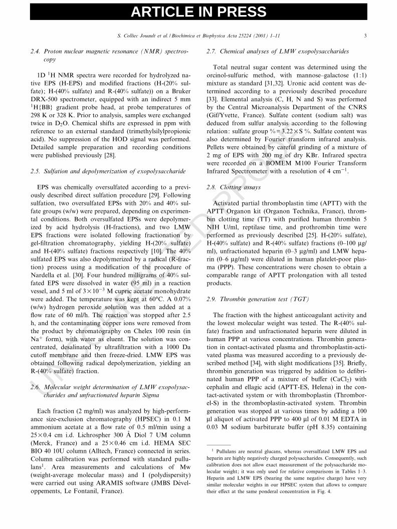
ARTICLE IN PRESS UNCORRECTED PROOF Characterization, chemical modi¢cations and in vitro anticoagulant properties of an exopolysaccharide produced by Alteromonas infernus Sylvia Colliec Jouault a ; *, Lionel Chevolot a , Dominique Helley b , Jacqueline Ratiskol a , Andre ¤e Bros b , Corinne Sinquin a , Olivier Roger a , Anne-Marie Fischer b a URM2, IFREMER/CNRS (UMR 7540, CNRS/Universite ¤ Paris 13), Laboratoire de Biochimie et Mole ¤cules Marines, De ¤partement Valorisation des Produits, BP 21105, 44311 Nantes Cedex 3, France b Laboratoire d’He ¤matologie, CHU Necker-Enfants Malades (Universite ¤ Paris V/INSERM U 428), Paris, France Received 7 March 2001; received in revised form 2 July 2001; accepted 12 July 2001 Abstract A new low-molecular-weight ‘heparin-like’ component was obtained from an exopolysaccharide produced by a mesophilic strain found in deep-sea hydrothermal vents. Data concerning the structure of the native high-molecular-weight exopolysaccharide (10 6 g/mol, 10% sulfate content) are reported for the first time. Two depolymerization processes were used to obtain low-molecular-weight (24^35U10 3 g/mol) oversulfated fractions (sulfate content 20 or 40). Nuclear magnetic resonance studies indicated that after sulfation (40%), the low-molecular- weight fraction obtained by free radical depolymerization was less sulfated in the 6-O-position than the fraction depolymerized by acid hydrolysis. The free radical depolymerized product also had sulfated residues in the 4-O-position and disulfated ones in the 2,3-O-positions. Moreover, the compounds generated by the free radical process were more homogeneous with respect to molecular mass. Also for the first time, the anticoagulant activity of the low-molecular-weight exopolysaccharide fractions is reported. When the fractions obtained after sulfation and depolymerization were compared with heparins, anticoagulant activity was detected in oversulfated fractions, but not in native exopolysaccharide. The free radical depolymerized fraction inhibited thrombin generation in both contact-activated and thromboplastin- activated plasma, showing a prolonged lag phase only in the contact-activated assay. Affinity co-electrophoresis studies suggested that a single population of polysaccharide chains binds to antithrombin and that only a subpopulation strongly interacts with heparin cofactor II. ß 2001 Published by Elsevier Science B.V. Keywords : A/nity co-electrophoresis ; Anticoagulant activity ; Depolymerization ; Heparin-like ; Serpin ; Sulfated polysaccharide ; Sulfation 1. Introduction A broad series of polysaccharides has emerged as an important class of bioactive products [1] that occur natu- rally in a great variety of animals, plants and microorgan- isms. Marine microorganisms o¡er a rich source of poly- saccharides with novel structures. Since 1994, new bacteria have been searched for near deep-sea hydrothermal vents characterized by extreme pressure and temperature condi- tions and high concentrations of toxic elements [2,3]. In- terest in mass culture of microorganisms from the marine environment has increased considerably, representing an innovative approach to the biotechnological use of under-exploited resources [4]. Exopolysaccharide (EPS)- producing microorganisms occur very widely in nature in di¡erent types of habitat [5], and some thermophilic and mesophilic EPS-producing strains have been isolated from deep-sea hydrothermal vents [2,6]. Some bacteria (e.g. Al- teromonas macleodii subsp. ¢jiensis and Vibrio diabolicus) found in these conditions have produced extracellular pol- ymers with original structures when grown in an aerobic carbohydrate-based medium [7,8]. Alteromonas infernus,a new species of bacterium isolated quite recently [9] and classi¢ed as a non-pathogenic microorganism by the Insti- tut Pasteur, secretes a water-soluble acidic heteropolysac- charide consisting of glucose, galactose, glucuronic and galacturonic acids (1:1:0.7:0.4). The composition of this high-molecular-weight polysaccharide (10 6 g/mol) di¡ers 0304-4165 / 01 / $ ^ see front matter ß 2001 Published by Elsevier Science B.V. PII:S0304-4165(01)00185-4 Abbreviations : EPS, exopolysaccharide; LMW, low molecular weight; H-EPS, hydrolyzed native EPS ; H-LMW EPS, low-molecular-weight sul- fated exopolysaccharides obtained by acid hydrolysis ; R-LMW EPS, low- molecular-weight sulfated exopolysaccharides obtained by radical depoly- merization ; HPSEC, high-performance size-exclusion chromatography ; APTT, activated partial thromboplastin time ; TT, thrombin clotting time ; PPP, platelet-poor plasma ; AT, antithrombin ; HCII, heparin cofac- tor II; ACE, a/nity co-electrophoresis * Corresponding author. Fax: +33-2-40-37-40-93. E-mail address : [email protected] (S. Colliec Jouault). Biochimica et Biophysica Acta 25224 (2001) 1^11 www.bba-direct.com
-
Upload
independent -
Category
Documents
-
view
0 -
download
0
Transcript of Characterization, chemical modifications and in vitro anticoagulant properties of an...

ARTICLE IN PRESS
UNCORRECTED PROOF
Characterization, chemical modi¢cations and in vitro anticoagulantproperties of an exopolysaccharide produced by Alteromonas infernus
Sylvia Colliec Jouault a;*, Lionel Chevolot a, Dominique Helley b, Jacqueline Ratiskol a,Andree Bros b, Corinne Sinquin a, Olivier Roger a, Anne-Marie Fischer b
a URM2, IFREMER/CNRS (UMR 7540, CNRS/Universite Paris 13), Laboratoire de Biochimie et Molecules Marines, Departement Valorisation desProduits, BP 21105, 44311 Nantes Cedex 3, France
b Laboratoire d'Hematologie, CHU Necker-Enfants Malades (Universite Paris V/INSERM U 428), Paris, France
Received 7 March 2001; received in revised form 2 July 2001; accepted 12 July 2001
Abstract
A new low-molecular-weight `heparin-like' component was obtained from an exopolysaccharide produced by a mesophilic strain found indeep-sea hydrothermal vents. Data concerning the structure of the native high-molecular-weight exopolysaccharide (106 g/mol, 10% sulfatecontent) are reported for the first time. Two depolymerization processes were used to obtain low-molecular-weight (24^35U103 g/mol)oversulfated fractions (sulfate content 20 or 40). Nuclear magnetic resonance studies indicated that after sulfation (40%), the low-molecular-weight fraction obtained by free radical depolymerization was less sulfated in the 6-O-position than the fraction depolymerized by acidhydrolysis. The free radical depolymerized product also had sulfated residues in the 4-O-position and disulfated ones in the 2,3-O-positions.Moreover, the compounds generated by the free radical process were more homogeneous with respect to molecular mass. Also for the firsttime, the anticoagulant activity of the low-molecular-weight exopolysaccharide fractions is reported. When the fractions obtained aftersulfation and depolymerization were compared with heparins, anticoagulant activity was detected in oversulfated fractions, but not in nativeexopolysaccharide. The free radical depolymerized fraction inhibited thrombin generation in both contact-activated and thromboplastin-activated plasma, showing a prolonged lag phase only in the contact-activated assay. Affinity co-electrophoresis studies suggested that asingle population of polysaccharide chains binds to antithrombin and that only a subpopulation strongly interacts with heparin cofactorII. ß 2001 Published by Elsevier Science B.V.
Keywords: A¤nity co-electrophoresis; Anticoagulant activity; Depolymerization; Heparin-like ; Serpin; Sulfated polysaccharide; Sulfation
1. Introduction
A broad series of polysaccharides has emerged as animportant class of bioactive products [1] that occur natu-rally in a great variety of animals, plants and microorgan-isms. Marine microorganisms o¡er a rich source of poly-saccharides with novel structures. Since 1994, new bacteriahave been searched for near deep-sea hydrothermal ventscharacterized by extreme pressure and temperature condi-
tions and high concentrations of toxic elements [2,3]. In-terest in mass culture of microorganisms from the marineenvironment has increased considerably, representing aninnovative approach to the biotechnological use ofunder-exploited resources [4]. Exopolysaccharide (EPS)-producing microorganisms occur very widely in nature indi¡erent types of habitat [5], and some thermophilic andmesophilic EPS-producing strains have been isolated fromdeep-sea hydrothermal vents [2,6]. Some bacteria (e.g. Al-teromonas macleodii subsp. ¢jiensis and Vibrio diabolicus)found in these conditions have produced extracellular pol-ymers with original structures when grown in an aerobiccarbohydrate-based medium [7,8]. Alteromonas infernus, anew species of bacterium isolated quite recently [9] andclassi¢ed as a non-pathogenic microorganism by the Insti-tut Pasteur, secretes a water-soluble acidic heteropolysac-charide consisting of glucose, galactose, glucuronic andgalacturonic acids (1:1:0.7:0.4). The composition of thishigh-molecular-weight polysaccharide (106 g/mol) di¡ers
0304-4165 / 01 / $ ^ see front matter ß 2001 Published by Elsevier Science B.V.PII: S 0 3 0 4 - 4 1 6 5 ( 0 1 ) 0 0 1 8 5 - 4
Abbreviations: EPS, exopolysaccharide; LMW, low molecular weight;H-EPS, hydrolyzed native EPS; H-LMW EPS, low-molecular-weight sul-fated exopolysaccharides obtained by acid hydrolysis ; R-LMW EPS, low-molecular-weight sulfated exopolysaccharides obtained by radical depoly-merization; HPSEC, high-performance size-exclusion chromatography;APTT, activated partial thromboplastin time; TT, thrombin clottingtime; PPP, platelet-poor plasma; AT, antithrombin; HCII, heparin cofac-tor II; ACE, a¤nity co-electrophoresis
* Corresponding author. Fax: +33-2-40-37-40-93.E-mail address: [email protected] (S. Colliec Jouault).
BBAGEN 25224 27-8-01
Biochimica et Biophysica Acta 25224 (2001) 1^11
www.bba-direct.com

ARTICLE IN PRESS
UNCORRECTED PROOF
in monosaccharide content and/or ratio and sulfate con-tent (10%) from other EPS isolated from deep-sea hydro-thermal bacteria and from polysaccharides of other ori-gins. In a preliminary study, highly sulfated low-molecular-weight (LMW) EPS fractions were obtainedafter combined sulfation and acidic depolymerization,without altering the osidic composition of this acidic poly-mer [10]. However, no data concerning the structure andbiological activity of these fractions were currently avail-able until now.
The preparation of therapeutic products from non-mammalian sources would avoid the risk of contaminationwith pathogenic agents potentially present in mammaliantissues [11^15]. In this respect, sulfated polysaccharidesconstitute a complex group of macromolecules known topossess a wide range of important biological properties.Heparin of porcine origin, a heterogeneous sulfated poly-saccharide from the growing family of glycosaminogly-cans, is widely used as a therapeutic anticoagulant andantithrombotic agent [16]. It is now clearly establishedthat the sulfate groups in heparin play a critical role inits antithrombotic activity [17]. However, as heparin haspharmacokinetic and biophysical limitations, attemptshave been made to develop new anticoagulant and antith-rombotic drugs [18,19]. Various studies have concernednew sulfated polysaccharides that show anticoagulant/an-tithrombotic properties and target speci¢c steps in throm-bogenesis. These polysaccharides (e.g. fucoidan, dermatansulfate and sulfated dextran derivatives) show no structur-al homology with heparin, except high sulfate content [14,15, 20^25].
The native EPS secreted by A. infernus is without anti-coagulant activity and was chemically modi¢ed to induceit. This study provides the ¢rst data on the structure of thenative EPS and the fractions obtained after sulfation anddepolymerization performed by acid hydrolysis or radicaldepolymerization. The anticoagulant activities of oversul-fated LMW EPS fractions (containing uronic acid andsulfate contents comparable to those of heparin) werecompared with those of heparin. Thrombin generationtests were performed in the presence of the most homoge-neous fraction to study the mechanism of anticoagulantactivity, and the interaction of this fraction with serpinswas analyzed by a¤nity co-electrophoresis (ACE).
2. Materials and methods
2.1. Materials
LMW heparin (Dalteparin, Fragmin; 160 anti-Xa IU/mg) was obtained from Kabi Pharmacia (St. Quentin,France). Puri¢ed human thrombin (2910 NIH units/mg),unfractionated heparin from porcine mucosa used in ACEexperiments (25^35 kDa in low-angle laser measurementsby the manufacturer, and 29 kDa as determined in our
laboratory (see Section 2.6)), dextran sulfate (8 kDa),azure A, toluidine blue and sodium Mopso were fromSigma (St. Louis, MO, USA). Unfractionated heparin(batch H108; 173 IU/mg) for anticoagulant assays wasfrom Sano¢ Winthrop (Paris, France). Reptilase-STH50and puri¢ed human heparin cofactor II (1 PEU/100 Wg)were from Diagnostica Stago (Asnie©res, France), APTTfrom Organon Technika (Fresnes, France), puri¢ed hu-man antithrombin from Chromogenix (5 units/mg), chro-mogenic substrate S-2238 from Biogenic (Maurin, France),Thromborel-S (human thromboplastin without Polybrene)from Behring (Marburg, Germany), and Chelex 100 resinand low-melt preparative grade agarose were from Bio-Rad (Ivry sur Seine, France). APTT-ES reagent (ellagicacid+cephalin) used for the thrombin generation test waskindly provided by Helena (St. Leu, France). The Stan-dard Filtron Cassette System was supplied by Pall Filtron(Northborough, MA, USA). Cetavlon (cetyltrimethylam-monium bromide) was from Merck (Darmstadt, Ger-many), and gel bond ¢lms were from Pharmacia (Uppsala,Sweden). The a¤nity co-electrophoresis system was fromOwl Scienti¢c (Woburn, MA, USA).
2.2. Production, puri¢cation and characterization of nativeexopolysaccharide
The isolation procedure and characteristics of theGY785 strain were previously reported [9]. EPS was pro-duced and puri¢ed as previously described [10], and mono-saccharide content was determined after methanolysis bygas chromatography [8]. Methylation analyses were per-formed using a modi¢cation of the Hakomori procedure[26]. Hydroxyl groups were methylated by methyl iodide inDMSO using lithium dimethylsul¢nyl as anion. Methylesters of uronic acids were reduced by lithium triethylbor-odeuteride, as previously described [27]. Methylated EPSwas then hydrolyzed with 2 M tri£uoroacetic acid for 2 hat 120³C. The derivatives were reduced by NaBD4 andacetylated prior to analysis by gas chromatography^massspectrometry (GC/MS) [8]. The main peaks were num-bered from 1 to 11. As peak response (total ionic current)depends on the structure, such analyses are more qualita-tive than quantitative.
2.3. Preparation of partially hydrolyzed exopolysaccharide(H-EPS)
Crude EPS was dissolved in 0.5 M H2SO4 at 5 mg/mland heated at 60³C for 90 min. The preparation was thenneutralized with a solution of 1 M NaOH, ultra¢ltrated(1000 Da cuto¡ membrane) to eliminate salts and smalloligosaccharides, and ¢nally freeze-dried. The molecularweight of the H-EPS was 10 000 g/mol and its polydisper-sity 1.9 (determined as described in Section 2.6). Methyl-ation analysis was performed as described above.
BBAGEN 25224 27-8-01
S. Colliec Jouault et al. / Biochimica et Biophysica Acta 25224 (2001) 1^112
ARTICLE IN PRESS
UNCORRECTED PROOF
2.4. Proton nuclear magnetic resonance (NMR) spectros-copy
1D 1H NMR spectra were recorded for hydrolyzed na-tive EPS (H-EPS) and modi¢ed fractions (H-(20% sul-fate); H-(40% sulfate) and R-(40% sulfate)) on a BrukerDRX-500 spectrometer, equipped with an indirect 5 mm1H{BB} gradient probe head, at probe temperatures of298 K or 328 K. Prior to analysis, samples were exchangedtwice in D2O. Chemical shifts are expressed in ppm withreference to an external standard (trimethylsilylpropionicacid). No suppression of the HOD signal was performed.Detailed sample preparation and recording conditionswere published previously [28].
2.5. Sulfation and depolymerization of exopolysaccharide
EPS was chemically oversulfated according to a previ-ously described direct sulfation procedure [29]. Followingsulfation, two oversulfated EPSs with 20% and 40% sul-fate groups (w/w) were prepared, depending on experimen-tal conditions. Both oversulfated EPSs were depolymer-ized by acid hydrolysis (H-fractions), and two LMWEPS fractions were isolated following fractionation bygel-¢ltration chromatography, yielding H-(20% sulfate)and H-(40% sulfate) fractions respectively [10]. The 40%sulfated EPS was also depolymerized by a radical (R-frac-tion) process using a modi¢cation of the procedure ofNardella et al. [30]. Four hundred milligrams of 40% sul-fated EPS were dissolved in water (95 ml) in a reactionvessel, and 5 ml of 3U1033 M cupric acetate monohydratewere added. The temperature was kept at 60³C. A 0.07%(w/w) hydrogen peroxide solution was then added at a£ow rate of 60 ml/h. The reaction was stopped after 2.5h, and the contaminating copper ions were removed fromthe product by chromatography on Chelex 100 resin (inNa� form), with water as eluent. The solution was con-centrated, desalinated by ultra¢ltration with a 1000 Dacuto¡ membrane and then freeze-dried. LMW EPS wasobtained following radical depolymerization, yielding anR-(40% sulfate) fraction.
2.6. Molecular weight determination of LMW exopolysac-charides and unfractionated heparin Sigma
Each fraction (2 mg/ml) was analyzed by high-perform-ance size-exclusion chromatography (HPSEC) in 0.1 Mammonium acetate at a £ow rate of 0.5 ml/min using a25U0.4 cm i.d. Lichrospher 300 Aî Diol 7 UM column(Merck, France) and a 25U0.46 cm i.d. HEMA SECBIO 40 10U column (Alltech, France) connected in series.Column calibration was performed with standard pullu-lans1. Area measurements and calculations of Mw(weight-average molecular mass) and I (polydispersity)were carried out using ARAMIS software (JMBS Devel-oppements, Le Fontanil, France).
2.7. Chemical analyses of LMW exopolysaccharides
Total neutral sugar content was determined using theorcinol-sulfuric method, with mannose^galactose (1:1)mixture as standard [31,32]. Uronic acid content was de-termined according to a previously described procedure[33]. Elemental analysis (C, H, N and S) was performedby the Central Microanalysis Department of the CNRS(Gif/Yvette, France). Sulfate content (sodium salt) wasdeduced from sulfur analysis according to the followingrelation: sulfate group % = 3.22US %. Sulfate content wasalso determined by Fourier transform infrared analysis.Pellets were obtained by careful grinding of a mixture of2 mg of EPS with 200 mg of dry KBr. Infrared spectrawere recorded on a BOMEM M100 Fourier TransformInfrared Spectrometer with a resolution of 4 cm31.
2.8. Clotting assays
Activated partial thromboplastin time (APTT) with theAPTT Organon kit (Organon Technika, France), throm-bin clotting time (TT) with puri¢ed human thrombin 5NIH U/ml, reptilase time, and prothrombin time wereperformed as previously described [25]. H-(20% sulfate),H-(40% sulfate) and R-(40% sulfate) fractions (0^100 Wg/ml), unfractionated heparin (0^3 Wg/ml) and LMW hepa-rin (0^6 Wg/ml) were diluted in human platelet-poor plas-ma (PPP). These concentrations were chosen to obtain acomparable range of APTT prolongation with all testedproducts.
2.9. Thrombin generation test (TGT)
The fraction with the highest anticoagulant activity andthe lowest molecular weight was tested. The R-(40% sul-fate) fraction and unfractionated heparin were diluted inhuman PPP at various concentrations. Thrombin genera-tion in contact-activated plasma and thromboplastin-acti-vated plasma was measured according to a previously de-scribed method [34], with slight modi¢cations [35]. Brie£y,thrombin generation was triggered by addition to de¢bri-nated human PPP of a mixture of bu¡er (CaCl2) withcephalin and ellagic acid (APTT-ES, Helena) in the con-tact-activated system or with thromboplastin (Thrombor-el-S) in the thromboplastin-activated system. Thrombingeneration was stopped at various times by adding a 100Wl aliquot of activated PPP to 400 Wl of 0.01 M EDTA in0.03 M sodium barbiturate bu¡er (pH 8.35) containing
1 Pullulans are neutral glucans, whereas oversulfated LMW EPS andheparin are highly negatively charged polysaccharides. Consequently, suchcalibration does not allow exact measurement of the polysaccharide mo-lecular weight; it was only used for relative comparisons in Tables 1^3.Heparin and LMW EPS (bearing the same negative charge) have verysimilar molecular weights in our HPSEC system that allows to comparetheir e¡ect at the same ponderal concentration in Fig. 4.
BBAGEN 25224 27-8-01
S. Colliec Jouault et al. / Biochimica et Biophysica Acta 25224 (2001) 1^11 3
ARTICLE IN PRESS
UNCORRECTED PROOF
0.15 M NaCl and 0.1 g/l BSA preincubated at 4³C. Thegenerated thrombin was quanti¢ed by adding 50 Wl of anEDTA-containing sample to 450 Wl of 1 mM S-2238. Thepercentage of inhibition of peak thrombin generation andthe lag phase preceding this peak (vt : time interval be-tween the beginning of the test, i.e. the addition of thetriggering agent, and the peak value) were calculated forvarious concentrations of each polysaccharide in plasma(plasma without polysaccharide was used as the control).Results are expressed as the concentration of polysaccha-
ride yielding 50% inhibition (IC50) and the vt correspond-ing to the IC50 value (vtIC50 ). The IC50 value was calcu-lated from the linear relation obtained by plotting thepercentage of thrombin inhibition relative to polysaccha-ride concentration. The vtIC50 value was calculated fromthe equation obtained by plotting vt as a function of poly-saccharide concentration.
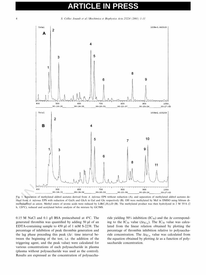
Fig. 1. Separation of methylated alditol acetates derived from A. infernus EPS without reduction (A), and separation of methylated alditol acetates de-rived from A. infernus EPS with reduction of GalA and GlcA in Gal and Glc respectively (B). OH were methylated by MeI in DMSO using lithium di-methylsul¢nyl as anion. Methyl esters of uronic acids were reduced by LiB(C2H5)3D (B). The methylated product was then hydrolyzed in 2 M TFA (2h, 120³C), reduced and acetylated before analysis of the mixture by GC/MS.
BBAGEN 25224 27-8-01
S. Colliec Jouault et al. / Biochimica et Biophysica Acta 25224 (2001) 1^114
ARTICLE IN PRESS
UNCORRECTED PROOF
2.10. Electrophoretic analysis of serpin binding
The binding of polysaccharides to human antithrombin(AT) and human heparin cofactor II (HCII) was analyzedby a¤nity co-electrophoresis according to a previouslydescribed technique [36], which allows both protein andpolysaccharidic chains to migrate freely during electropho-resis. Brie£y, the polysaccharides were electrophoresed inagarose gels through zones containing increasing amountsof AT or HCII. At neutral pH, the electrophoretic mobil-ities of polysaccharides are much higher than those ofserpins, and the binding of polysaccharides to serpins de-creases their electrophoretic mobility. As LMW EPS andunfractionated heparin showed very similar molecularweights in our HPSEC assay, they were used at thesame ponderal concentration, corresponding to the samemolar concentration (0.7 WM). Twenty micrograms of pol-ysaccharides were loaded into nine separate transverseslots close to the cathode. Each slot was opposite a rec-tangular sagittal well in which AT or HCII was cast. ATconcentrations in agarose gel were 0 (control), 43 WM and86 WM, and HCII concentrations were 0 (control), 0.75WM and 1.5 WM. The highest concentration of both serpinswas obtained after vial reconstitution with 1 ml of distilledwater, as recommended by the manufacturer. Electropho-resis was performed at 60 V and 500 mA for 3.5 h. Elec-trophoresis end points were determined by the migration
of bromophenol blue, which migrates slower than poly-saccharides. After electrophoresis, the gel was soaked inCetavlon, dried, stained with toluidine blue, bleached anddried again according to a previously described procedure[37].
3. Results
3.1. Composition and methylation studies of native EPS
GC/MS chromatograms of partially methylated acetatesderived from native EPS without reduction showed eightmain peaks (Fig. 1A) numbered 1^9 (7 is missing, butappears after reduction, as described below) and identi¢edas 1,3,5-tri-O-acetyl-2,4-di-O-methyl rhamnitol (1), 1,5-di-O-acetyl-2,3,4,6-tetra-O-methyl glucitol (2), 1,5-di-O-ace-tyl-2,3,4,6-tetra-O-methyl galactitol (3), 1,4,5-tri-O-acetyl-2,3,6-tri-O-methyl galactitol (4), 1,4,5-tri-O-acetyl-2,3,6-tri-O-methyl glucitol (5), 1,5,6-tri-O-acetyl-2,3,4-tri-O-methyl galactitol (6), 1,3,5,6-tetra-O-acetyl-2,4-di-O-meth-yl galactitol (8), 1,3,4,5,6-penta-O-acetyl-2-O-methyl ga-lactitol (9). Peaks 2 and 3 were large (the former abouttwice the size of the latter), corresponding to terminalglucose and galactose residues respectively. Such highquantities of non-reducing ends indicated that this EPSwas highly branched. Peaks 4 and 5 were due to 1,4-linked
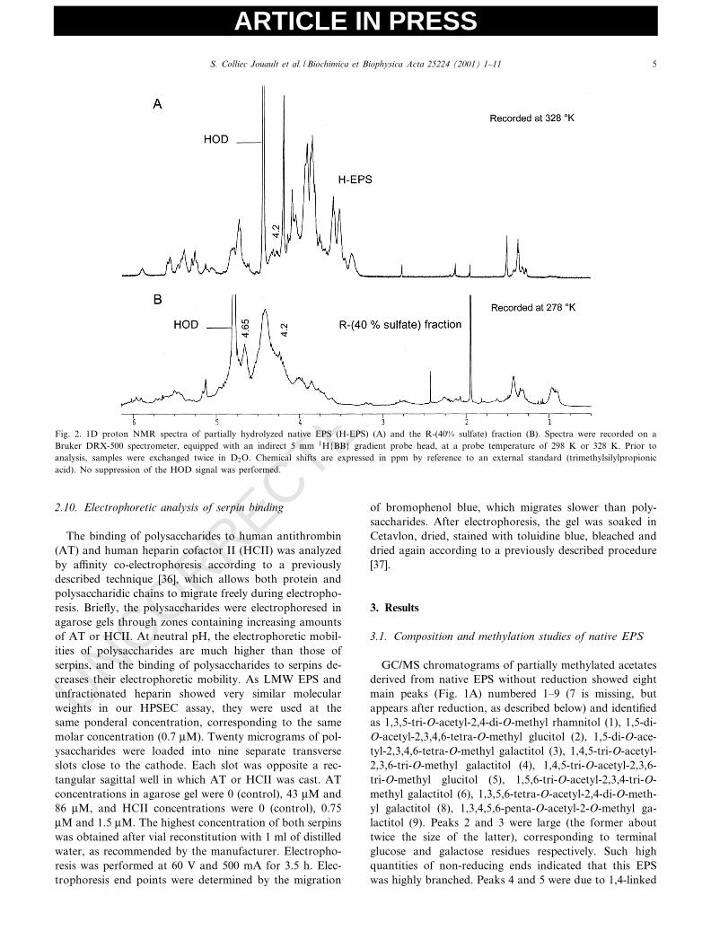
Fig. 2. 1D proton NMR spectra of partially hydrolyzed native EPS (H-EPS) (A) and the R-(40% sulfate) fraction (B). Spectra were recorded on aBruker DRX-500 spectrometer, equipped with an indirect 5 mm 1H{BB} gradient probe head, at a probe temperature of 298 K or 328 K. Prior toanalysis, samples were exchanged twice in D2O. Chemical shifts are expressed in ppm by reference to an external standard (trimethylsilylpropionicacid). No suppression of the HOD signal was performed.
BBAGEN 25224 27-8-01
S. Colliec Jouault et al. / Biochimica et Biophysica Acta 25224 (2001) 1^11 5
ARTICLE IN PRESS
UNCORRECTED PROOF
galactose and glucose respectively. The other smallerpeaks were attributed to 1,3-linked rhamnose (peak 1)and variously linked galactose residues (1,6-linked, 1,3,6-linked and 1,3,4,6-linked; peaks 6, 8 and 9 respectively).After reduction of carboxylic acid functions (Fig. 1B),three additional peaks appeared, but the main sugar resi-due was still terminal glucose. Peak 7 (1,4,5-tri-O-acetyl-2,3,6-tri-O-methyl glucitol) corresponded to two kinds ofglucuronic acid residues (1,2- or 1,4-linked), peak 9 to amixture of 1,3,4,6-linked galactose and 1,3,4-linked galact-uronic acid in equal proportions according to MS data(333/335 ratio was around 1), peak 10 to 1,3,4-linked glu-curonic acid, and peak 11 to 1,2,4-linked glucuronic acid.These results indicate that side chains were mainly borne
by uronic acids. Methylation analyses performed on poly-saccharides from various batches and on H-EPS gave sim-ilar results, although with some variations in peak inten-sities, especially for peak 1 which sometimes almostdisappeared. Nevertheless, the main peaks were always2, 3, 4 and 5 without carboxylic acid reduction, and peaks7, 9 and 10 appeared when reduction was performed.These results indicate that the bacterium secreted two pol-ysaccharides. The major one (the subject of our study) wascomposed only of glucose, galactose, glucuronic and ga-lacturonic acids, in accordance with the osidic composi-tion previously described [9], whereas the other (a minorcompound) contained signi¢cant quantities of rhamnose.
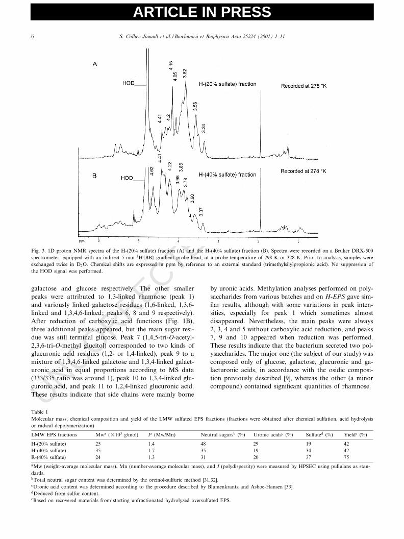
Fig. 3. 1D proton NMR spectra of the H-(20% sulfate) fraction (A) and the H-(40% sulfate) fraction (B). Spectra were recorded on a Bruker DRX-500spectrometer, equipped with an indirect 5 mm 1H{BB} gradient probe head, at a probe temperature of 298 K or 328 K. Prior to analysis, samples wereexchanged twice in D2O. Chemical shifts are expressed in ppm by reference to an external standard (trimethylsilylpropionic acid). No suppression ofthe HOD signal was performed.
Table 1Molecular mass, chemical composition and yield of the LMW sulfated EPS fractions (fractions were obtained after chemical sulfation, acid hydrolysisor radical depolymerization)
LMW EPS fractions Mwa (U103 g/mol) Ia (Mw/Mn) Neutral sugarsb (%) Uronic acidsc (%) Sulfated (%) Yielde (%)
H-(20% sulfate) 25 1.4 48 29 19 42H-(40% sulfate) 35 1.7 35 19 34 42R-(40% sulfate) 24 1.3 31 20 37 75aMw (weight-average molecular mass), Mn (number-average molecular mass), and I (polydispersity) were measured by HPSEC using pullulans as stan-dards.bTotal neutral sugar content was determined by the orcinol-sulfuric method [31,32].cUronic acid content was determined according to the procedure described by Blumenkrantz and Asboe-Hansen [33].dDeduced from sulfur content.eBased on recovered materials from starting unfractionated hydrolyzed oversulfated EPS.
BBAGEN 25224 27-8-01
S. Colliec Jouault et al. / Biochimica et Biophysica Acta 25224 (2001) 1^116
ARTICLE IN PRESS
UNCORRECTED PROOF
3.2. Nuclear magnetic resonance spectroscopy
Due to its high molecular weight, native EPS could notbe studied directly by NMR spectroscopy. A partially hy-drolyzed compound (H-EPS) was prepared, and its NMRspectrum was recorded as well as the spectra of the threeoversulfated LMW EPSs: H-(20% sulfate), H-(40% sul-fate) and R-(40% sulfate) fractions (see Section 2). The1H-1D spectrum of H-EPS is shown in Fig. 2A. Fromthe low ¢eld, signals in the H-EPS spectrum were gatheredin four clusters. The ¢rst (5.5^5.0 ppm) corresponded toanomeric protons of K-linked osidic units, and the second(4.8^4.4 ppm) to protons of L-linked anomeric or sulfatedpositions (see below). The third (4.3^3.3) was attributed toH3, H4, H5 and H6 of various sugar residues, and thefourth (1.4^1.2 ppm) to methyl groups of rhamnose resi-dues. Some impurities (above 1 ppm) also appeared. TheCOSY spectrum (not shown) displayed a cross-peak cor-responding to a sulfated 2-O-position (H2 at 4.75 ppm) ofan K-linked residue (H1 at 5.28 ppm), which was necessa-
rily a glucuronic acid as methylation analysis indicatedthat only glucuronic acids were 2-O-substituted. After sul-fation (Figs. 2B and 3A,B), there were many more reso-nances between 4.2 and 4.8 ppm (protons of sulfated po-sitions) than with H-EPS (Fig. 2A), especially for the moresulfated fractions: R-(40% sulfate) and H-(40% sulfate).For the R-(40% sulfate) fraction, there was no resonancebetween 3.5 and 3.0 ppm (corresponding to the area of H2and H3 of L-linked residues). However, H-(40% sulfate)and H-(20% sulfate) maintained signals at 3.5^3.6 ppm(H2 of L-linked galactose) and 3.3^3.4 ppm (H2 of L-linked glucose), probably because more labile sulfateswere partially eliminated during acid hydrolysis, especially2-O-sulfate that is much more susceptible to acid-catalyzedester hydrolysis [38]. On the other hand, the resonances at4.2 ppm (H6 of CH2OSO3
3 ) [39] were low in sample H-(20% sulfate), intermediate in R-(40% sulfate) and high inH-(40% sulfate), which showed the greatest sulfation atthe 6-position. It is noteworthy that H-(20% sulfate),which exhibited few additional resonances between 4.2
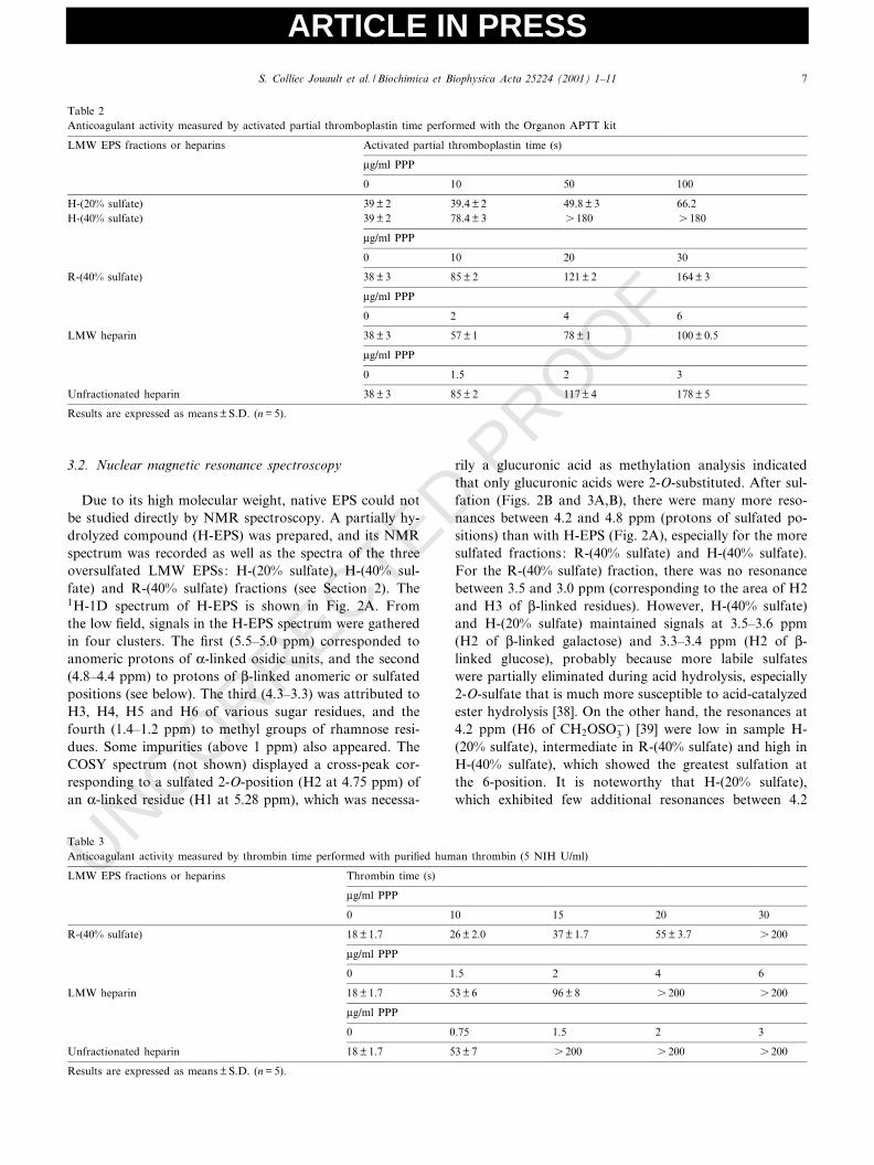
Table 3Anticoagulant activity measured by thrombin time performed with puri¢ed human thrombin (5 NIH U/ml)
LMW EPS fractions or heparins Thrombin time (s)
Wg/ml PPP
0 10 15 20 30
R-(40% sulfate) 18 þ 1.7 26 þ 2.0 37 þ 1.7 55 þ 3.7 s 200
Wg/ml PPP
0 1.5 2 4 6
LMW heparin 18 þ 1.7 53 þ 6 96 þ 8 s 200 s 200
Wg/ml PPP
0 0.75 1.5 2 3
Unfractionated heparin 18 þ 1.7 53 þ 7 s 200 s 200 s 200
Results are expressed as means þ S.D. (n = 5).
Table 2Anticoagulant activity measured by activated partial thromboplastin time performed with the Organon APTT kit
LMW EPS fractions or heparins Activated partial thromboplastin time (s)
Wg/ml PPP
0 10 50 100
H-(20% sulfate) 39 þ 2 39.4 þ 2 49.8 þ 3 66.2H-(40% sulfate) 39 þ 2 78.4 þ 3 s 180 s 180
Wg/ml PPP
0 10 20 30
R-(40% sulfate) 38 þ 3 85 þ 2 121 þ 2 164 þ 3
Wg/ml PPP
0 2 4 6
LMW heparin 38 þ 3 57 þ 1 78 þ 1 100 þ 0.5
Wg/ml PPP
0 1.5 2 3
Unfractionated heparin 38 þ 3 85 þ 2 117 þ 4 178 þ 5
Results are expressed as means þ S.D. (n = 5).
BBAGEN 25224 27-8-01
S. Colliec Jouault et al. / Biochimica et Biophysica Acta 25224 (2001) 1^11 7
ARTICLE IN PRESS
UNCORRECTED PROOFand 4.7 ppm (H of sulfated positions), lacked the peak at
4.65^4.62 present in both R-(40% sulfate) and H-(40% sul-fate) fractions (attributed to H4 of a 4-O-sulfated position[39] or to H2 and/or H3 of 2,3-O-disulfated residues[28,40]). The COSY spectrum was too complex to allowany clear distinction to be made.
3.3. Molecular weight and composition of LMW exopoly-saccharides
Two oversulfated high-molecular-weight EPSs isolatedafter chemical sulfation showed sulfate contents of 20%and 40% respectively, as determined by Fourier transforminfrared analysis and elemental analysis (data not shown).These two oversulfated EPSs were partially depolymerizedby acid hydrolysis in an attempt to isolate LMW EPSs.After fractionation by gel ¢ltration chromatography, twoLMW fractions were isolated from each hydrolyzed EPS:the H-(20%sulfate) and H-(40% sulfate) fractions respec-tively. The fractions were homogeneous in size, with apolydispersity (I) below 2, and were produced with agood yield (Table 1). Depolymerization did not alter theosidic composition of the H-LMW EPS fractions, and aslight decrease (15%) of sulfate content was observed onlywith the oversulfated EPS initially containing 40% sulfategroups. The H-LMW EPS fractions had the same sulfate/total sugar ratios as the oversulfated high molecularweight EPS. The oversulfated EPS with the highest sulfatecontent (40%) was also depolymerized by a radical pro-cess, resulting in the isolation of an LMW EPS fraction,R-(40% sulfate) fraction, which was homogeneous, withpolydispersity (I) below 1.5, and produced with a goodyield (Table 1).
3.4. Anticoagulant properties of modi¢ed exopolysaccha-rides
The in vitro anticoagulant activities of modi¢ed EPSswere measured in APTT and then compared with those ofheparins. Among the H-LMW EPSs, only the H-(40% sul-fate) fraction was able to prolong APTT appreciably. Thesame anticoagulant e¡ect was obtained for the H-(40%sulfate) fraction, LMW heparin and unfractionated hepa-rin at 10, 4 and 1.5 Wg/ml respectively. The R-(40% sulfate)fraction was as potent as the H-(40% sulfate) fraction in
prolonging APTT (10 Wg/ml were required to obtain thedoubling of APTT; Table 2). The anticoagulant e¡ect ofR-(40% sulfate) was also evaluated by TT and comparedwith that of heparins. An equivalent prolongation wasobserved for this fraction, LMW heparin and unfractio-nated heparin at 20, 1.5 and 0.75 Wg/ml respectively (Table3). The R-(40% sulfate) fraction had no signi¢cant e¡ecton reptilase time and prothrombin time (data not shown)at the tested concentrations (10^50 Wg/ml).
The e¡ect of the R-(40% sulfate) fraction on thrombingeneration was evaluated in the contact-activated andthromboplastin-activated systems and compared withthat of unfractionated heparin (Table 4). Like heparin,the R-(40% sulfate) fraction inhibited thrombin generationafter activation of intrinsic and extrinsic pathways. In thecontact-activated system, the IC50 was 58 þ 17 Wg/ml withR-(40% sulfate) as compared to 0.65 þ 0.08 Wg/ml withunfractionated heparin. R-(40% sulfate) caused a greaterdelay in contact-induced thrombin generation than didheparin, with the lag phase increasing in proportion toits concentration. The vtIC50 value was 4.8 þ 1.0 min com-pared to 1.06 þ 0.23 min with unfractionated heparin. Inthe thromboplastin-activated system, the IC50 was 53 þ 6Wg/ml with R-(40% sulfate) compared to 0.48 þ 0.07 Wg/mlwith unfractionated heparin. In contrast to contact-in-duced thrombin generation, the R-(40% sulfate) fraction,like heparin, was not e¡ective in delaying thrombin for-mation in the thromboplastin-activated system.
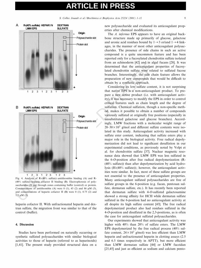
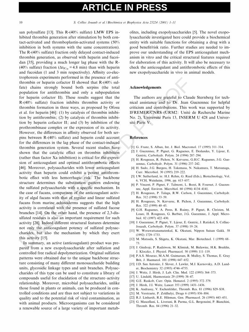
The interaction of the R-(40% sulfate) fraction withserpins was analyzed by ACE. In the presence of anti-thrombin (Fig. 4A), the patterns di¡ered for R-(40% sul-fate), unfractionated heparin and dextran sulfate (allloaded at the same concentration of 20 Wg). R-(40% sul-fate) mobility was reduced, giving a single spot at thehighest protein concentration, whereas antithrombin splitthe migrating heparin front into two distinct fronts, aspreviously shown [36]. With dextran sulfate, a part ofthe migration front was delayed at the highest proteinconcentration. In experiments performed with lower anti-thrombin concentrations, a delay was also observed,although the di¡erences were not so well clear-cut (datanot shown). In the presence of heparin cofactor II (Fig.4B), a fractionation of R-(40% sulfate) migration frontwas observed for both protein concentrations tested,which suggests that a subpopulation bound strongly to
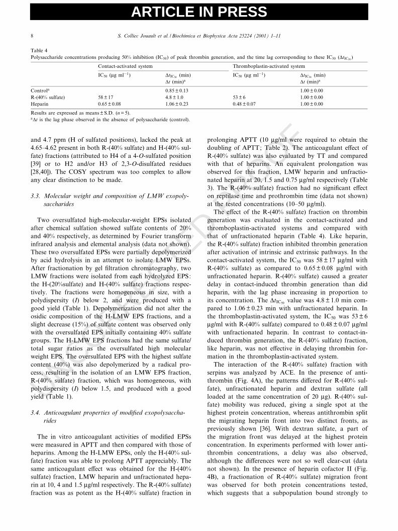
Table 4Polysaccharide concentrations producing 50% inhibition (IC50) of peak thrombin generation, and the time lag corresponding to these IC50 (vtIC50 )
Contact-activated system Thromboplastin-activated system
IC50 (Wg ml31) vtIC50 (min) IC50 (Wg ml31) vtIC50 (min)vt (min)a vt (min)a
Controla 0.85 þ 0.13 1.00 þ 0.00R-(40% sulfate) 58 þ 17 4.8 þ 1.0 53 þ 6 1.00 þ 0.00Heparin 0.65 þ 0.08 1.06 þ 0.23 0.48 þ 0.07 1.00 þ 0.00
Results are expressed as means þ S.D. (n = 5).avt is the lag phase observed in the absence of polysaccharide (control).
BBAGEN 25224 27-8-01
S. Colliec Jouault et al. / Biochimica et Biophysica Acta 25224 (2001) 1^118
ARTICLE IN PRESS
UNCORRECTED PROOF
heparin cofactor II. With unfractionated heparin and dex-tran sulfate, the migration front was similar to that of thecontrol (bu¡er).
4. Discussion
Studies have been performed on naturally occurring orsynthetic sulfated polysaccharides with similar biologicalactivities to those of heparin (referred to as heparinoids)[1,41]. The present study provided structural data on a
new polysaccharide and evaluated its anticoagulant prop-erties after chemical modi¢cations.
The A. infernus EPS appears to have an original back-bone structure made up primarily of glucose, galactoseand uronic acid residues bound by 1C3 or/and 1C4 link-ages, in the manner of most other anticoagulant polysac-charides. The presence of side chains in such an activecompound is a quite uncommon feature and has beenreported only for a fucosylated chondroitin sulfate isolatedfrom an echinoderm [42] and in algal fucans [28]. It wasdetermined that the anticoagulant properties of fucosy-lated chondroitin sulfate were related to sulfated fucosebranches. Interestingly, the side chain feature allows thepreparation of new compounds that would be di¤cult toobtain by a synthetic approach.
Considering its low sulfate content, it is not surprisingthat native EPS is a non-anticoagulant product. To pre-pare a new active product (i.e. with anticoagulant activ-ity), it was necessary to modify the EPS in order to controlcritical features such as chain length and the degree ofsulfation. Chemical sulfation, though a non-speci¢c meth-od, makes it possible to obtain a number of compoundsvariously sulfated at originally free positions (especially inunsubstituted galactose and glucose branches). Accord-ingly, LMW fractions with a molecular weight range of20^30U103 g/mol and di¡erent sulfate contents were iso-lated in this study. Anticoagulant activity increased withsulfate ester content, indicating that sulfate esters play amajor role in the biological activity. Free radical depoly-merization did not lead to signi¢cant desulfation in ourexperimental conditions, as previously noted by Volpi etal. for chondroitin sulfate [37]. Nuclear magnetic reso-nance data showed that LMW EPS was less sulfated inthe 6-O-position after free radical depolymerization (R-(40% sulfate)) than after depolymerization by acid hydro-lysis (H-(40% sulfate)) ; however, the anticoagulant activ-ities were similar. In fact, most of these sulfate groups arenot essential to the presence of anticoagulant properties.Many anticoagulant sulfated polysaccharides are free ofsulfate groups in the 6-position (e.g. fucan, pentosan sul-fate, dermatan sulfate, etc.). It has recently been reportedthat dermatan sulfate with 4-O-sulfated galactosamineshowed a strong a¤nity for HCII while dermatan sulfatesulfated in the 6-position had no anticoagulant activity atall despite its high sulfate content [43]. The free radicaldepolymerized product also had residues sulfated in the4-O-position and disulfated in the 2,3-positions, as is oftenthe case for anticoagulant sulfated polysaccharides.
Our experiments showed that anticoagulant activity washigher with 40% than 20% of sulfate esters. The LMWEPS depolymerized by the free radical process (40% sul-fate content, 24U103 g/mol) was less e¤cient than LMWheparin and unfractionated heparin in clotting assays (2.5and 6.5 times respectively in APTT), but more e¤cientthan LMW dermatan sulfate [44] or LMW fucoidan[25,45] and just as e¤cient as sodium and calcium pento-
Fig. 4. Analysis of R-(40% sulfate)^antithrombin binding (A) and R-(40% sulfate)^heparin cofactor II binding (B). Electrophoresis of poly-saccharides (20 Wg) through zones containing bu¡er (control) or protein.Concentrations of antithrombin (A) were 0 (1), 43 (2) and 86 WM (3),and concentrations of heparin cofactor II (B) were 0 (1), 0.75 (2) and1.5 WM (3).
BBAGEN 25224 27-8-01
S. Colliec Jouault et al. / Biochimica et Biophysica Acta 25224 (2001) 1^11 9
ARTICLE IN PRESS
UNCORRECTED PROOF
san polysulfate [13]. This R-(40% sulfate) LMW EPS in-hibited thrombin generation after stimulation by both con-tact-activated and thromboplastin-activated systems (50%inhibition in both systems with the same concentration).The R-(40% sulfate) fraction only delayed contact-inducedthrombin generation, as observed with heparin and fucoi-dan [35], providing a much longer lag phase with the R-(40% sulfate) fraction (vtIC50 = 4.8 min) than with heparinand fucoidan (1 and 3 min respectively). A¤nity co-elec-trophoresis experiments performed in the presence of anti-thrombin or heparin cofactor II showed that R-(40% sul-fate) chains strongly bound both serpins (the totalpopulation for antithrombin and only a subpopulationfor heparin cofactor II). These results suggest that theR-(40% sulfate) fraction inhibits thrombin activity orthrombin formation in three ways, as proposed by Ofosuet al. for heparin [46]: (1) by catalysis of thrombin inhibi-tion by antithrombin; (2) by catalysis of thrombin inhibi-tion by heparin cofactor II; and (3) by inhibition of theprothrombinase complex or the expression of its activity.However, the di¡erences in a¤nity observed for both ser-pins between R-(40% sulfate) and heparin could accountfor the di¡erences in the lag phase of the contact-inducedthrombin generation system. Several recent studies haveshown that the catalytic e¡ect on thrombin inhibition(rather than factor Xa inhibition) is critical for the expres-sion of anticoagulant and optimal antithrombotic e¡ects[46]. Moreover, polysaccharides with lower anticoagulantactivity than heparin could exhibit a potent antithrom-botic e¡ect with less hemorrhagic risk. The backbonestructure determines the substitution pattern endowingthe sulfated polysaccharide with a speci¢c mechanism. Inthe case of fucans, comparison of the anticoagulant activ-ity of algal fucans with that of regular and linear sulfatedfucans from marine echinoderms suggests that the highactivity is correlated with the presence of sulfated fucosebranches [14]. On the other hand, the presence of 2,3-dis-ulfated residues is also an important requirement for suchactivity [28]. Indeed di¡erent structural features determinenot only the anticoagulant potency of sulfated polysac-charides, but also the mechanism by which they exertthis activity [15].
In summary, an active (anticoagulant) product was pre-pared from a new exopolysaccharide after sulfation andcontrolled free radical depolymerization. Original sulfationpatterns were obtained due to the unique backbone struc-ture consisting of many di¡erent monosaccharide buildingunits, glycosidic linkage types and unit branches. Polysac-charides of this type can be used to constitute a library ofcompounds useful for elucidation of the structure^activityrelationship. Moreover, microbial polysaccharides, unlikethose found in plants or animals, can be produced in con-trolled conditions and are thus not subject to variations inquality and to the potential risk of viral contamination, aswith animal products. Microorganisms can be considereda renewable source of a large variety of important metab-
olites, including exopolysaccharides [5]. The novel exopo-lysaccharide investigated here could provide a biochemicalentity with suitable functions for obtaining a drug with agood bene¢t/risk ratio. Further studies are needed to im-prove our understanding of the EPS anticoagulant mech-anism in vitro and the critical structural features requiredfor elaboration of this activity. It will also be necessary tocheck the anticoagulant and antithrombotic e¡ects of thisnew exopolysaccharide in vivo in animal models.
Acknowledgements
The authors are grateful to Claude Sternberg for tech-nical assistance and to Dr. Jean Guezennec for helpfulcriticism and contributions. This work was supported byIFREMER/CNRS (URM2: Unite de Recherche MarineNo. 2), Universite Paris 13, INSERM U 428 and Univer-site Paris V.
References
[1] G. Franz, S. Alban, Int. J. Biol. Macromol. 17 (1995) 311^314.[2] J. Guezennec, P. Pignet, G. Raguenes, E. Deslandes, Y. Lijour, E.
Gentric, Carbohydr. Polym. 24 (1994) 287^294.[3] H. Rougeaux, R. Pichon, N. Kervarec, G.H.C. Raguenes, J.G. Gue-
zennec, Carbohydr. Polym. 31 (1996) 237^242.[4] H. Sudo, J.G. Burgess, H. Takemasa, N. Nakamura, T. Matsunaga,
Curr. Microbiol. 30 (1995) 219^222.[5] I.W. Sutherland, in: H.J. Rehm, G. Reed (Eds.), Biotechnology, Vol.
6, VCH, Weinheim, 1996, pp. 613^657.[6] P. Vincent, P. Pignet, F. Talmont, L. Bozzi, B. Fournet, J. Guezen-
nec, Appl. Environ. Microbiol. 60 (1994) 4134^4141.[7] H. Rougeaux, P. Talaga, R.W. Carlson, J. Guezennec, Carbohydr.
Res. 312 (1998) 53^59.[8] H. Rougeaux, N. Kervarec, R. Pichon, J. Guezennec, Carbohydr.
Res. 322 (1999) 40^45.[9] G.H. Raguenes, A. Peres, R. Ruimy, P. Pignet, R. Christen, M.
Loaec, H. Rougeaux, G. Barbier, J.G. Guezennec, J. Appl. Micro-biol. 82 (1997) 422^430.
[10] J. Guezennec, P. Pignet, Y. Lijour, E. Gentric, J. Ratiskol, S. Colliec-Jouault, Carbohydr. Polym. 37 (1998) 19^24.
[11] W. Worawattanamateekul, K. Okutani, Nippon Suisan Gakk. 58(1992) 1729^1733.
[12] M. Matsuda, S. Shigeta, K. Okutani, Mar. Biotechnol. 1 (1999) 68^73.
[13] J. Giedrojc, P. Radziwon, M. Klimiuk, M. Bielawiec, H.K. Breddin,J. Kloczko, J. Physiol. Pharmacol. 50 (1999) 111^119.
[14] P.A.S. Mourao, M.A.M. Guimaraes, B. Mulloy, S. Thomas, E. GrayBrit, J. Haematol. 101 (1998) 647^652.
[15] J.D. San Antonio, J. Slover, J. Lawler, M.J. Karnovsky, A.D. Land-er, Biochemistry 32 (1993) 4746^4755.
[16] J. Weitz, J. Hirsh, J. Lab. Clin. Med. 122 (1993) 364^373.[17] U. Lindahl, Haemostasis 29 (1999) 38^47.[18] G.E. Raskob, Curr. Opin. Hematol. 2 (1995) 372^379.[19] J. Hirsh, J.I. Weitz, Lancet 353 (1999) 1431^1436.[20] K. Andrassy, V. Eschenfelder, Thromb. Res. 81 (1996) S29^S38.[21] M. Verstraete, P. Zoldhelyi, Drugs 6 (1995) 856^884.[22] R.J. Linhardt, R.E. Hileman, Gen. Pharmacol. 26 (1995) 443^451.[23] G. Mascellani, L. Liverani, B. Parma, G.L. Bergonzini, P. Bianchini,
Thromb. Res. 84 (1996) 21^32.
BBAGEN 25224 27-8-01
S. Colliec Jouault et al. / Biochimica et Biophysica Acta 25224 (2001) 1^1110
ARTICLE IN PRESS
UNCORRECTED PROOF
[24] R.M. Maarou¢, M. Jozefowicz, J. Tapon Bretaudiere, J. Jozefonvicz,A.M. Fischer, Biomaterials 18 (1997) 359^366.
[25] S. Mauray, C. Sternberg, J. Theveniaux, J. Millet, C. Sinquin, J.Tapon Bretaudiere, A.M. Fischer, Thromb. Haemost. 74 (1995)1280^1285.
[26] S. Hakomori, J. Biochem. 55 (1964) 205^208.[27] W.S. York, A.G. Darvill, M. McNeil, T.T. Stevenson, P. Albersheim,
Methods Enzymol. 118 (1985) 3^40.[28] L. Chevolot, A. Foucault, F. Chaubet, N. Kervarec, C. Sinquin,
A.M. Fisher, C. Boisson-Vidal, Carbohydr. Res. 319 (1999) 154^165.[29] T. Nishino, T. Nagumo, Carbohydr. Res. 229 (1992) 355^362.[30] A. Nardella, F. Chaubet, C. Boisson Vidal, C. Blondin, P. Durand, J.
Jozefonvicz, Carbohydr. Res. 289 (1996) 201^208.[31] C. Rimington, Biochem. J. 25 (1931) 1062^1071.[32] J. Tilmans, K. Philippi, Biochemistry Z 215 (1929) 36^60.[33] N. Blumenkrantz, G. Asboe-Hansen, Anal. Biochem. 54 (1973) 484^
489.[34] F.A. Ofosu, G.J. Modi, L.M. Smith, A.L. Cerskus, J. Hirsh, M.A.
Blajchman, Blood 64 (1984) 742^747.[35] S. Mauray, E. De Raucourt, F. Chaubet, O. Maiga-Revel, C. Stern-
berg, A.M. Fischer, J. Biomater. Sci. Polym. Ed. 9 (1998) 373^387.[36] M.K. Lee, A.D. Lander, Proc. Natl. Acad. Sci. USA 88 (1991) 2768^
2772.
[37] N. Volpi, I. Sandri, T. Venturelli, Carbohydr. Res. 279 (1995) 193^200.
[38] R. Falshaw, R.H. Furneaux, G.C. Slim, in: P. Finch (Ed.), Carbo-hydrates. Structures, Syntheses and Dynamics, Kluwer AcademicPublishers, Dordrecht, 1999, pp. 107^149.
[39] R.R. Contreras, J.P. Kamerling, J. Breg, F.G. Vliegenhart, Carbo-hydr. Res. 179 (1988) 411^418.
[40] E.A. Yates, F. Santini, A. Bisio, C. Cosentino, Carbohydr. Res. 298(1997) 335^340.
[41] N. Harada, M. Maeda, Biosci. Biotechnol. Biochem. 62 (1998) 1647^1652.
[42] P.A.S. Mourao, M.S. Pereira, M.S.G. Pavao, B. Mulloy, D.M. Toll-efsen, M.C. Mowinckel, U. Abilgaard, J. Biol. Chem. 271 (1996)23973^23984.
[43] B. Mulloy, P.A. Mourao, E. Gray, J. Biotechnol. 77 (2000) 123^135.[44] R.J. Linhardt, U.R. Desai, J. Liu, A. Pervin, D. Hoppensteadt, J.
Fareed Biochem. Pharmacol. 47 (1994) 1241^1252.[45] S. Colliec, C. Boisson-Vidal, J. Jozefonvicz, Phytochemistry 35 (1994)
697^700.[46] F.A. Ofosu, G.J. Modi, J. Hirsh, M.R. Buchanan, M.A. Blajchman,
Ann. NY Acad. Sci. 485 (1986) 41^55.
BBAGEN 25224 27-8-01
S. Colliec Jouault et al. / Biochimica et Biophysica Acta 25224 (2001) 1^11 11