Detection and discovery of RNA modifications using microarrays
9
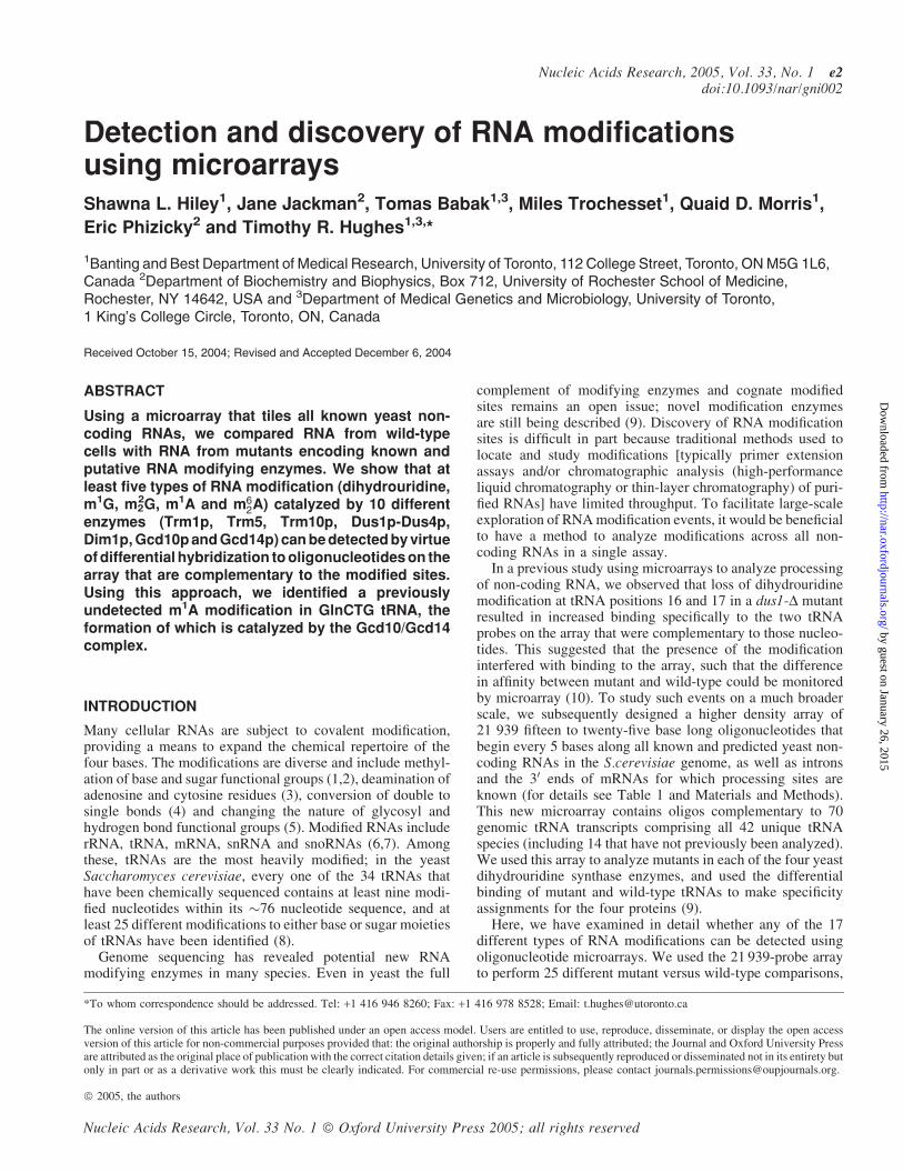
Detection and discovery of RNA modifications using microarrays Shawna L. Hiley 1 , Jane Jackman 2 , Tomas Babak 1,3 , Miles Trochesset 1 , Quaid D. Morris 1 , Eric Phizicky 2 and Timothy R. Hughes 1,3, * 1 Banting and Best Department of Medical Research, University of Toronto, 112 College Street, Toronto, ON M5G 1L6, Canada 2 Department of Biochemistry and Biophysics, Box 712, University of Rochester School of Medicine, Rochester, NY 14642, USA and 3 Department of Medical Genetics and Microbiology, University of Toronto, 1 King’s College Circle, Toronto, ON, Canada Received October 15, 2004; Revised and Accepted December 6, 2004 ABSTRACT Using a microarray that tiles all known yeast non- coding RNAs, we compared RNA from wild-type cells with RNA from mutants encoding known and putative RNA modifying enzymes. We show that at least five types of RNA modification (dihydrouridine, m 1 G, m 2 2 G, m 1 A and m 6 2 A) catalyzed by 10 different enzymes (Trm1p, Trm5, Trm10p, Dus1p-Dus4p, Dim1p, Gcd10p and Gcd14p) can be detected by virtue of differential hybridization to oligonucleotides on the array that are complementary to the modified sites. Using this approach, we identified a previously undetected m 1 A modification in GlnCTG tRNA, the formation of which is catalyzed by the Gcd10/Gcd14 complex. INTRODUCTION Many cellular RNAs are subject to covalent modification, providing a means to expand the chemical repertoire of the four bases. The modifications are diverse and include methyl- ation of base and sugar functional groups (1,2), deamination of adenosine and cytosine residues (3), conversion of double to single bonds (4) and changing the nature of glycosyl and hydrogen bond functional groups (5). Modified RNAs include rRNA, tRNA, mRNA, snRNA and snoRNAs (6,7). Among these, tRNAs are the most heavily modified; in the yeast Saccharomyces cerevisiae, every one of the 34 tRNAs that have been chemically sequenced contains at least nine modi- fied nucleotides within its 76 nucleotide sequence, and at least 25 different modifications to either base or sugar moieties of tRNAs have been identified (8). Genome sequencing has revealed potential new RNA modifying enzymes in many species. Even in yeast the full complement of modifying enzymes and cognate modified sites remains an open issue; novel modification enzymes are still being described (9). Discovery of RNA modification sites is difficult in part because traditional methods used to locate and study modifications [typically primer extension assays and/or chromatographic analysis (high-performance liquid chromatography or thin-layer chromatography) of puri- fied RNAs] have limited throughput. To facilitate large-scale exploration of RNA modification events, it would be beneficial to have a method to analyze modifications across all non- coding RNAs in a single assay. In a previous study using microarrays to analyze processing of non-coding RNA, we observed that loss of dihydrouridine modification at tRNA positions 16 and 17 in a dus1-D mutant resulted in increased binding specifically to the two tRNA probes on the array that were complementary to those nucleo- tides. This suggested that the presence of the modification interfered with binding to the array, such that the difference in affinity between mutant and wild-type could be monitored by microarray (10). To study such events on a much broader scale, we subsequently designed a higher density array of 21 939 fifteen to twenty-five base long oligonucleotides that begin every 5 bases along all known and predicted yeast non- coding RNAs in the S.cerevisiae genome, as well as introns and the 3 0 ends of mRNAs for which processing sites are known (for details see Table 1 and Materials and Methods). This new microarray contains oligos complementary to 70 genomic tRNA transcripts comprising all 42 unique tRNA species (including 14 that have not previously been analyzed). We used this array to analyze mutants in each of the four yeast dihydrouridine synthase enzymes, and used the differential binding of mutant and wild-type tRNAs to make specificity assignments for the four proteins (9). Here, we have examined in detail whether any of the 17 different types of RNA modifications can be detected using oligonucleotide microarrays. We used the 21 939-probe array to perform 25 different mutant versus wild-type comparisons, *To whom correspondence should be addressed. Tel: +1 416 946 8260; Fax: +1 416 978 8528; Email: [email protected] The online version of this article has been published under an open access model. Users are entitled to use, reproduce, disseminate, or display the open access version of this article for non-commercial purposes provided that: the original authorship is properly and fully attributed; the Journal and Oxford University Press are attributed as the original place of publication with the correct citation details given; if an article is subsequently reproduced or disseminated not in its entirety but only in part or as a derivative work this must be clearly indicated. For commercial re-use permissions, please contact [email protected]. ª 2005, the authors Nucleic Acids Research, Vol. 33 No. 1 ª Oxford University Press 2005; all rights reserved Nucleic Acids Research, 2005, Vol. 33, No. 1 e2 doi:10.1093/nar/gni002 by guest on January 26, 2015 http://nar.oxfordjournals.org/ Downloaded from
Transcript of Detection and discovery of RNA modifications using microarrays
Detection and discovery of RNA modificationsusing microarraysShawna L. Hiley1, Jane Jackman2, Tomas Babak1,3, Miles Trochesset1, Quaid D. Morris1,
Eric Phizicky2 and Timothy R. Hughes1,3,*
1Banting and Best Department of Medical Research, University of Toronto, 112 College Street, Toronto, ON M5G 1L6,Canada 2Department of Biochemistry and Biophysics, Box 712, University of Rochester School of Medicine,Rochester, NY 14642, USA and 3Department of Medical Genetics and Microbiology, University of Toronto,1 King’s College Circle, Toronto, ON, Canada
Received October 15, 2004; Revised and Accepted December 6, 2004
ABSTRACT
Using a microarray that tiles all known yeast non-coding RNAs, we compared RNA from wild-typecells with RNA from mutants encoding known andputative RNA modifying enzymes. We show that atleast five types of RNA modification (dihydrouridine,m1G, m2
2G, m1A and m62A) catalyzed by 10 different
enzymes (Trm1p, Trm5, Trm10p, Dus1p-Dus4p,Dim1p, Gcd10p and Gcd14p) can be detected by virtueof differential hybridization to oligonucleotides on thearray that are complementary to the modified sites.Using this approach, we identified a previouslyundetected m1A modification in GlnCTG tRNA, theformation of which is catalyzed by the Gcd10/Gcd14complex.
INTRODUCTION
Many cellular RNAs are subject to covalent modification,providing a means to expand the chemical repertoire of thefour bases. The modifications are diverse and include methyl-ation of base and sugar functional groups (1,2), deamination ofadenosine and cytosine residues (3), conversion of double tosingle bonds (4) and changing the nature of glycosyl andhydrogen bond functional groups (5). Modified RNAs includerRNA, tRNA, mRNA, snRNA and snoRNAs (6,7). Amongthese, tRNAs are the most heavily modified; in the yeastSaccharomyces cerevisiae, every one of the 34 tRNAs thathave been chemically sequenced contains at least nine modi-fied nucleotides within its �76 nucleotide sequence, and atleast 25 different modifications to either base or sugar moietiesof tRNAs have been identified (8).
Genome sequencing has revealed potential new RNAmodifying enzymes in many species. Even in yeast the full
complement of modifying enzymes and cognate modifiedsites remains an open issue; novel modification enzymesare still being described (9). Discovery of RNA modificationsites is difficult in part because traditional methods used tolocate and study modifications [typically primer extensionassays and/or chromatographic analysis (high-performanceliquid chromatography or thin-layer chromatography) of puri-fied RNAs] have limited throughput. To facilitate large-scaleexploration of RNA modification events, it would be beneficialto have a method to analyze modifications across all non-coding RNAs in a single assay.
In a previous study using microarrays to analyze processingof non-coding RNA, we observed that loss of dihydrouridinemodification at tRNA positions 16 and 17 in a dus1-D mutantresulted in increased binding specifically to the two tRNAprobes on the array that were complementary to those nucleo-tides. This suggested that the presence of the modificationinterfered with binding to the array, such that the differencein affinity between mutant and wild-type could be monitoredby microarray (10). To study such events on a much broaderscale, we subsequently designed a higher density array of21 939 fifteen to twenty-five base long oligonucleotides thatbegin every 5 bases along all known and predicted yeast non-coding RNAs in the S.cerevisiae genome, as well as intronsand the 30 ends of mRNAs for which processing sites areknown (for details see Table 1 and Materials and Methods).This new microarray contains oligos complementary to 70genomic tRNA transcripts comprising all 42 unique tRNAspecies (including 14 that have not previously been analyzed).We used this array to analyze mutants in each of the four yeastdihydrouridine synthase enzymes, and used the differentialbinding of mutant and wild-type tRNAs to make specificityassignments for the four proteins (9).
Here, we have examined in detail whether any of the 17different types of RNA modifications can be detected usingoligonucleotide microarrays. We used the 21 939-probe arrayto perform 25 different mutant versus wild-type comparisons,
*To whom correspondence should be addressed. Tel: +1 416 946 8260; Fax: +1 416 978 8528; Email: [email protected]
The online version of this article has been published under an open access model. Users are entitled to use, reproduce, disseminate, or display the open accessversion of this article for non-commercial purposes provided that: the original authorship is properly and fully attributed; the Journal and Oxford University Pressare attributed as the original place of publication with the correct citation details given; if an article is subsequently reproduced or disseminated not in its entirety butonly in part or as a derivative work this must be clearly indicated. For commercial re-use permissions, please contact [email protected].
ª 2005, the authors
Nucleic Acids Research, Vol. 33 No. 1 ª Oxford University Press 2005; all rights reserved
Nucleic Acids Research, 2005, Vol. 33, No. 1 e2doi:10.1093/nar/gni002
by guest on January 26, 2015http://nar.oxfordjournals.org/
Dow
nloaded from
following a protocol in which the RNA from the wild-typecell (which carries the modification) and RNA from themutant cell (which lacks the modification) are labeled andhybridized to the array in the two separate channels (Cy3and Cy5). In this way, differential hybridization caused bythe RNA modifications would be detected as a change inratio of Cy3/Cy5 signal from probes complementary tomodified nucleotides. We successfully detected the followingmodifications: dihydrouridine (catalyzed by Dus1p-Dus4p),m1G (catalyzed by Trm1p), m2
2G (catalyzed by Trm1p),m1A (catalyzed by the Gcd10p-Gcd14p complex) and m6
2A(catalyzed by Dim1p). Our results establish a simple rule:with the exception of dihydrouridine (which severely perturbsnucleotide architecture), modifications to the Watson–Cricksurface impact microarray hybridization. In addition to observ-ing hundreds of known modification events, our data suggestedseveral new modification sites, one of which we subsequentlyverified by primer extension analysis. This demonstrates thegeneral utility of microarrays as a genome-wide tool for RNAmodification detection.
MATERIALS AND METHODS
Array construction
Oligonucleotide sequences are contained in the SupplementalMaterial. Oligos were designed to be complementary to knownnon-coding RNA sequences and flanking regions and weretiled at 5 nt intervals for most RNAs (intron-containingmRNAs were tiled every 20 nt and mitochondrial RNAsevery 15 nt; see Table 1 for details). Probe lengths wereadjusted to have a melting temperature of �41�C (11). Ink-jet microarrays were manufactured by Agilent Technologies(Palo Alto, CA).
Strains
Homozygous deletion mutants (12) were obtained fromResearch Genetics. TetO7-promoter alleles were constructedas described previously (13). gcd14-1ts (14) and gcd14-D werekindly provided by Mercedes Tamame; the dim1-Y131Gstrain was provided by Denis Lafontaine. The gcd14-D strainused for primer extension analysis overexpresses IMT4, which
suppresses the lethal phenotype of the deletion (M. Tamame,personal communication).
RNA isolation and array analysis
Isogenic wild-type and mutant strains were grown in parallel at30�C in SC medium (with the exception of gcd14-1 which wasgrown in YPD+Ade) with shaking in baffled flasks (Bellco) tofinal cell concentrations matched as closely as possible to107cells/ml. TetO7-promoter strains were exposed to 10 mg/mldoxycycline for a total of 20–24 h. Cells were harvested andRNA extracted as described previously (10). An aliquot of10 mg of DNase I-treated RNA was labeled with AlexaFluor 546 or 647 according to the manufacturer’s instructions(Molecular Probes ‘Ulysis’ kit), ethanol-precipitated andhybridized to the array as described previously (15). Forma-mide was added to the final concentrations of 25 or 33%, asdescribed previously (15). Hybridizations were carried out in arotating incubator at 42�C for 16–20 h and washed asdescribed previously (15). Arrays were scanned on an Axon4000B instrument.
Image processing, array normalization anddata visualization
Scanned images were quantitated with GenePix (Axon Instru-ments). Individual channels were spatially detrended (i.e.overall correlations between spot intensity and position onthe slide removed) by high-pass filtering [(16); http://www.psi.utoronto.ca/~ofer/detrendingReport.pdf] using 10%outliers. Dye bias was corrected in each slide using theLowess smoother from the MAANOVA package (writtenby Hao Wu) with 0.3 smoother span. After these steps, thenormalized intensities were converted in log2 ratios of mutantexpression versus wild-type.
Primer extension analysis
Bulk RNA isolated from either wild-type or gcd14-D cellswas used as a template for primer extension assays using a50 32P-labeled primer (50-GGAGGTCCCACCCGG-30) that isspecific for tRNAGlnCTG RNA. Approximately 5–10 mg bulkRNA and primer (0.1 mM) were annealed by heating to 95�Cfor 3 min and then cooling to room temperature in a 5 mlreaction containing 50 mM Tris–HCl, 15 mM NaCl and10 mM DTT, pH 7.5. Then 2 ml of the annealed mixture ofprimer and RNA was used in a primer extension reaction of10 ml containing 0.4 mM of each dNTP and 4 U AMV-reversetranscriptase (20 U/ml; Promega). Sequencing reactions alsocontained 0.2 mM of each individual ddNTP (ddG, ddA, ddTor ddC). The reactions were terminated after 1 h of incubation at37�C by the addition of an equal volume of formamide/50 mMEDTA loading dye; subsequently the samples were resolvedon a 15% acrylamide/4 M urea gel and visualized byphosphorImager.
Data availability
Oligonucleotide sequences on the arrays, and all microarraydata are available at (hugheslab.med.utoronto.ca/Hiley).Spreadsheets containing the data displayed in Figures 1Band 2A are also available on the website.
Table 1. Known and predicted yeast non-coding RNAs included on the
microarray
ncRNA Number of transcripts Tiling frequency
35S pre rRNA 1 55S rRNA 1 5Genomic tRNAs 70 5snoRNAs 84 5snRNAs 6 5RNase P 1 5RNase MRP 1 5SRP RNA 1 5Telomerase RNA 1 5RUFs 8 5Introns 236 20Spliced junctions 236 5Mitochondrial genome features 44 15mRNA 30 ends 8 20
e2 Nucleic Acids Research, 2005, Vol. 33, No. 1 PAGE 2 OF 9
by guest on January 26, 2015http://nar.oxfordjournals.org/
Dow
nloaded from
RESULTS
RNA modifications detected by microarray
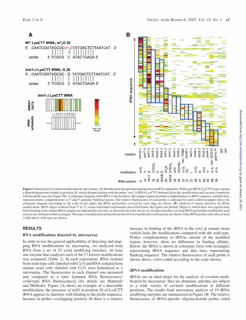
In order to test the general applicability of detecting and map-ping RNA modifications by microarray, we analyzed totalRNA from a set of 25 yeast modifying enzymes. At leastone enzyme that catalyzes each of the 17 known modificationswas examined (Table 2). In each experiment, RNA isolatedfrom wild-type cells (labeled with Cy3) and RNA isolated frommutant yeast cells (labeled with Cy5) were hybridized to amicroarray. The fluorescence in each channel was measuredand compared as a ratio [(mutant RNA fluorescence)/(wild-type RNA fluorescence)] (for details see Materialsand Methods). Figure 1A shows an example of a detectablemodification: the presence of m2
2G at position 26 of LysCTTtRNA appears to interfere with binding to the probe sequence,because in probes overlapping position 26 there is a relative
increase in binding of the tRNA in the trm1-D mutant strain(which lacks the modification) compared with the wild-type.Probes complementary to tRNAs outside of the modifiedregion, however, show no difference in binding affinity.Below, the tRNA is shown in schematic form with rectanglesrepresenting tRNA sequence and thin lines representingflanking sequence. The relative fluorescence of each probe isshown above, color-coded according to the scale shown.
tRNA modifications
tRNAs are an ideal target for the analysis of covalent modi-fication by microarray: they are abundant, and they are subjectto a wide variety of covalent modifications at differentpositions. The results from microarray analysis of 19 tRNAmodifying enzymes are summarized in Figure 1B. The relativefluorescence of tRNA-specific oligonucleotide probes (tiled
Figure 1. Detection of covalent modification by microarray. (A) Modification disrupts base pairing between RNA and probe. Wild-type tRNA LysCTT (top) containsa dimethylguanosine residue at position 26, which disrupts pairing with the probe. trm1-D tRNA LysCTT (bottom) lacks this modification and can pair completelywith the probe (see also Figure 2B). A schematic diagram of the tRNA is shown below. Rectangles represent probes complementary to tRNA sequence, and thin linesrepresent probes complementary to 50 and 30 genomic flanking regions. The relative fluorescence of each probe is indicated by color-coded rectangles above theschematic diagram (according to the scale on the right); the tRNA nucleotides covered by each oligo are shown. (B) Analysis of strains defective for tRNAmodification. tRNA oligos (ordered from 50 to 30) versus individual experiments (described below the figure) are plotted. Oligos to which there was significantlybetter binding in the mutant tRNA samples are indicated by red color, as shown by the color-bar in (A). Groups of probes covering tRNA nucleotides modified by eachenzyme are outlined in blue rectangles. The type of modification and positions known to be modified by each enzyme are shown. Only tRNA probes with ratios at least2-fold above wild type are shown.
PAGE 3 OF 9 Nucleic Acids Research, 2005, Vol. 33, No. 1 e2
by guest on January 26, 2015http://nar.oxfordjournals.org/
Dow
nloaded from
from the 50 to the 30 end of the transcripts) versus individualexperiments is plotted. The ratio of fluorescence (mutantversus wild-type) to each probe is color-coded according tothe scale shown, with red indicating more efficient binding ofthe mutant tRNA to the microarray. The nature of themodifications and the nucleotides known to be modified areshown for each experiment, and probes complementary tonucleotides known to be modified in at least one tRNA areoutlined with blue rectangles. Whereas Figure 1A shows alloligos corresponding to a single tRNA (and emphasizes the
specificity of the technique), this Figure summarizes the dif-ferential hybridization to all 70 tRNA sequences across 21experiments (and shows the ability of technique to detecttrends that emerge across all experiments). For clarity, wehave included only probes that display at least a 2-fold dif-ference in binding between mutant and wild-type in one ormore experiments. There is a concentration of red probeswithin the blue rectangles, indicating that probes complement-ary to modified nucleotides are more efficiently bound bymutant tRNAs than by their wild-type counterparts in nine
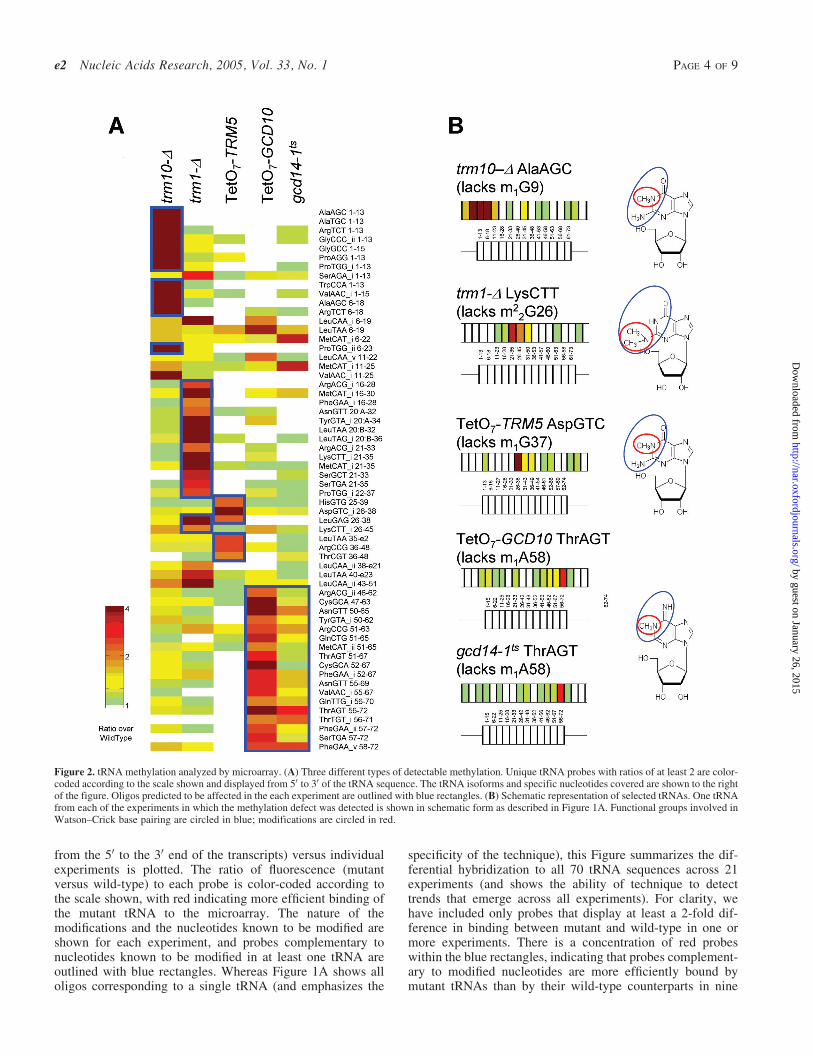
Figure 2. tRNA methylation analyzed by microarray. (A) Three different types of detectable methylation. Unique tRNA probes with ratios of at least 2 are color-coded according to the scale shown and displayed from 50 to 30 of the tRNA sequence. The tRNA isoforms and specific nucleotides covered are shown to the rightof the figure. Oligos predicted to be affected in the each experiment are outlined with blue rectangles. (B) Schematic representation of selected tRNAs. One tRNAfrom each of the experiments in which the methylation defect was detected is shown in schematic form as described in Figure 1A. Functional groups involved inWatson–Crick base pairing are circled in blue; modifications are circled in red.
e2 Nucleic Acids Research, 2005, Vol. 33, No. 1 PAGE 4 OF 9
by guest on January 26, 2015http://nar.oxfordjournals.org/
Dow
nloaded from
of the experiments: trm10-D, trm1-D, TetO7-TRM5, TetO7-GCD10, gcd14-1ts and the four dihydrouridine synthases.
Figure 2A shows a detailed view of the successfullydetected methylation modifications. Probes complementaryto known modified nucleotides are outlined in blue and theidentities of the probes are listed. The trm10-D array specif-ically detects an increase in mutant binding (i.e. positiveratios) for oligos covering tRNA nucleotides 1–13 and 6–22,both of which span position 9 where the m1G is absent in themutant. Oligos complementary to these positions in Ser andLeu tRNAs, which do not contain the m1G modification (8), donot show differential binding in mutant and wild-type, dem-onstrating that this technique is specific for individual tRNAisoforms. The same specificity is demonstrated by the exclu-sion of His and Asp tRNAs from the trm1-D array data and LystRNA from the TetO7-TRM5 data.
Gcd10p and Gcd14p form a complex to catalyze the forma-tion of m1A at tRNA position 58. Microarrays with conditionalalleles of both of these essential genes show high-ratio probesspanning position 58. The gcd14-1ts experiment producessmaller ratios and affects fewer oligos. Coupled with thefact that the TetO7-GCD10 strain has a more severe growthdefect than the gcd14-1ts strain (data not shown) indicates thatthe gcd14-1ts allele is weaker than the tetracycline-regulatedallele of GCD10.
A few high-ratio oligos exist outside of the expected tRNAregions in the trm1-D experiment and both experiments inwhich the Gcd10-Gcd14 complex was disrupted. In the caseof the Gcd10–Gcd14 complex, these probes correspond toMetCAT oligos, examined in detail in Figure 4D. Probes
with unusual behavior on the trm1-D microarray have pre-viously been observed as false-positives [see (9) Figure 3,dus2-D]. It is possible that the loss of modification causeschanges in the secondary structure of the tRNAs, whichimpacts hybridization to the array.
Selected tRNAs from each of these mutant strains are shownin detail in Figure 2B. We observe between one and three high-ratio oligos for each tRNA, suggesting that additional factorsbeyond the simple presence or absence of the modified nuc-leotide can affect binding to the array (see Discussion). How-ever, only oligos complementary to modified nucleotides showincreased binding to the mutant tRNAs, confirming the abilityof the microarray to detect the oligos complementary to themodified nucleotides with good specificity.
The site and nature of each modification is shown on thenucleotide base diagrams to the right of the tRNAs; the modi-fied functional group is outlined with a red circle, and func-tional groups involved in Watson–Crick base pairing arecircled in blue. All of the successfully-detected modificationsinvolve methylation of a functional group required for forma-tion of canonical Watson–Crick base-pairs.
Modifications to other ncRNAs
Covalent modification is an important feature of ribosomalRNA as well as tRNA. Mature ribosomal RNA in S.cerevisiaecontains over one hundred modified nucleotides; 60% of theseoccur in functionally important regions including the peptidyltransferase centre and the A, P and E sites (17). One of thesemodifications is the m6
2A formation at consecutive nucleotides
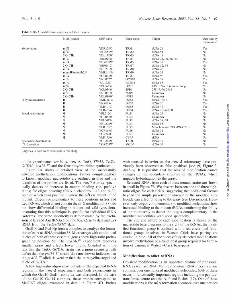
Table 2. RNA modification enzymes and their targets
Modification ORF name Gene name Target Detected bymicroarray?
Methylation m22G YDR120C TRM1 tRNA 26 Yes
m5U YKR056W TRM2 tRNA 54 No
20O CH3 YDL112W TRM3 tRNA 18 Nom5C YBL024W TRM4 tRNA 34, 40, 38, 49 Nom1G YHR070W TRM5 tRNA 37 Yes20O CH3 YBR061C TRM7 tRNA 32, 34 Nom
7G YDL201W TRM8 tRNA 46 No
mcm5U/mcm5s2U YML014W TRM9 tRNA 34 Nom
1G YOL093W TRM10 tRNA 9 Yes
m1A YNL062C GCD10 tRNA 58 Yesm1A YJL125C GCD14 tRNA 58 Yesm6
2A YPL266W DIM1 18S rRNA 30 terminal loop Yes20O CH3 YCL054W SPB1 25S rRNA 2918 Nom
5C YNL061W NOP2 Unknown No
20O CH3 YDL014W NOP1 Unknown NoDihydrouridylation D YML080W DUS1 tRNA 16/17 Yes
D YNR01W DUS2 tRNA 20 YesD YLR401C DUS3 tRNA 47 YesD YLR405W DUS4 tRNA 20:A/20:B Yes
Pseudouridylation C YPL212C PUS1 tRNA 27 NoC YGL063W PUS2 Unknown NoC YFL001W PUS3 tRNA 38, 39 NoC YNL292W PUS4 tRNA 55 NoC YLR165C PUS5 Mitochondrial 21S rRNA 2819 NoC YGR169C PUS6 tRNA 31 NoC YOR243C PUS7 Unknown NoC YLR175W CBF5 rRNA No
Adenosine deamination YGL243W TAD1 tRNA 37 Noi6A formation YOR274W MOD5 tRNA 37 No
Enzymes in bold were examined in this study.
PAGE 5 OF 9 Nucleic Acids Research, 2005, Vol. 33, No. 1 e2
by guest on January 26, 2015http://nar.oxfordjournals.org/
Dow
nloaded from
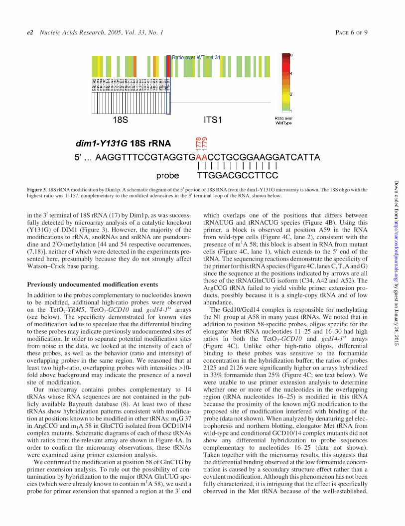
in the 30 terminal of 18S rRNA (17) by Dim1p, as was success-fully detected by microarray analysis of a catalytic knockout(Y131G) of DIM1 (Figure 3). However, the majority of themodifications to rRNA, snoRNAs and snRNA are pseudouri-dine and 20O-methylation [44 and 54 respective occurrences,(7,18)], neither of which were detected in the experiments pre-sented here, presumably because they do not strongly affectWatson–Crick base paring.
Previously undocumented modification events
In addition to the probes complementary to nucleotides knownto be modified, additional high-ratio probes were observedon the TetO7-TRM5, TetO7-GCD10 and gcd14-1ts arrays(see below). The specificity demonstrated for known sitesof modification led us to speculate that the differential bindingto these probes may indicate previously undocumented sites ofmodification. In order to separate potential modification sitesfrom noise in the data, we looked at the intensity of each ofthese probes, as well as the behavior (ratio and intensity) ofoverlapping probes in the same region. We reasoned that atleast two high-ratio, overlapping probes with intensities >10-fold above background may indicate the presence of a novelsite of modification.
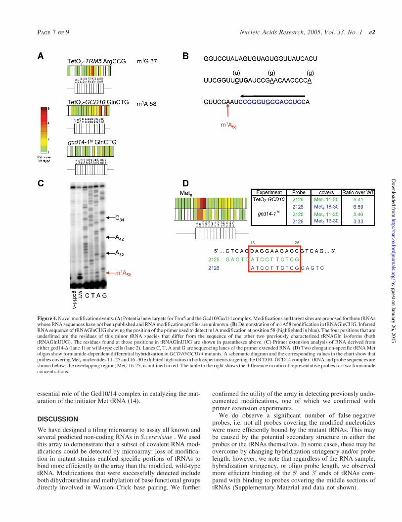
Our microarray contains probes complementary to 14tRNAs whose RNA sequences are not contained in the pub-licly available Bayreuth database (8). At least two of thesetRNAs show hybridization patterns consistent with modifica-tion at positions known to be modified in other tRNAs: m1G 37in ArgCCG and m1A 58 in GlnCTG isolated from GCD10/14complex mutants. Schematic diagrams of each of these tRNAswith ratios from the relevant array are shown in Figure 4A. Inorder to confirm the microarray observations, these tRNAswere examined using primer extension analysis.
We confirmed the modification at position 58 of GlnCTG byprimer extension analysis. To rule out the possibility of con-tamination by hybridization to the major tRNA GlnUUG spe-cies (which were already known to contain m1A 58), we used aprobe for primer extension that spanned a region at the 30 end
which overlaps one of the positions that differs betweentRNAUUG and tRNACUG species (Figure 4B). Using thisprimer, a block is observed at position A59 in the RNAfrom wild-type cells (Figure 4C, lane 2), consistent with thepresence of m1A 58; this block is absent in RNA from mutantcells (Figure 4C, lane 1), which extends to the 50 end of thetRNA. The sequencing reactions demonstrate the specificity ofthe primer for this tRNA species (Figure 4C, lanes C, T, Aand G)since the sequence at the positions indicated by arrows are allthose of the tRNAGlnCUG isoform (C34, A42 and A52). TheArgCCG tRNA failed to yield visible primer extension pro-ducts, possibly because it is a single-copy tRNA and of lowabundance.
The Gcd10/Gcd14 complex is responsible for methylatingthe N1 group at A58 in many yeast tRNAs. We noted that inaddition to position 58-specific probes, oligos specific for theelongator Met tRNA nucleotides 11–25 and 16–30 had highratios in both the TetO7-GCD10 and gcd14-1ts arrays(Figure 4C). Unlike other high-ratio oligos, differentialbinding to these probes was sensitive to the formamideconcentration in the hybridization buffer; the ratios of probes2125 and 2126 were significantly higher on arrays hybridizedin 33% formamide than 25% (Figure 4C; see text below). Wewere unable to use primer extension analysis to determinewhether one or more of the nucleotides in the overlappingregion (tRNA nucleotides 16–25) is modified in this tRNAbecause the proximity of the known m2
2G modification to theproposed site of modification interfered with binding of theprobe (data not shown). When analyzed by denaturing gel elec-trophoresis and northern blotting, elongator Met tRNA fromwild-type and conditional GCD10/14 complex mutants did notshow any differential hybridization to probe sequencescomplementary to nucleotides 16–25 (data not shown).Taken together with the microarray results, this suggests thatthe differential binding observed at the low formamide concen-tration is caused by a secondary structure effect rather than acovalent modification. Although this phenomenon has not beenfully characterized, it is intriguing that the effect is specificallyobserved in the Met tRNA because of the well-established,
Figure 3. 18S rRNA modification by Dim1p. A schematic diagram of the 30 portion of 18S RNA from the dim1-Y131G microarray is shown. The 18S oligo with thehighest ratio was 11157, complementary to the modified adenosines in the 30 terminal loop of the RNA, shown below.
e2 Nucleic Acids Research, 2005, Vol. 33, No. 1 PAGE 6 OF 9
by guest on January 26, 2015http://nar.oxfordjournals.org/
Dow
nloaded from
essential role of the Gcd10/14 complex in catalyzing the mat-uration of the initiator Met tRNA (14).
DISCUSSION
We have designed a tiling microarray to assay all known andseveral predicted non-coding RNAs in S.cerevisiae . We usedthis array to demonstrate that a subset of covalent RNA mod-ifications could be detected by microarray: loss of modifica-tion in mutant strains enabled specific portions of tRNAs tobind more efficiently to the array than the modified, wild-typetRNA. Modifications that were successfully detected includeboth dihydrouridine and methylation of base functional groupsdirectly involved in Watson–Crick base pairing. We further
confirmed the utility of the array in detecting previously undo-cumented modifications, one of which we confirmed withprimer extension experiments.
We do observe a significant number of false-negativeprobes, i.e. not all probes covering the modified nucleotideswere more efficiently bound by the mutant tRNAs. This maybe caused by the potential secondary structure in either theprobes or the tRNAs themselves. In some cases, these may beovercome by changing hybridization stringency and/or probelength; however, we note that regardless of the RNA sample,hybridization stringency, or oligo probe length, we observedmore efficient binding of the 50 and 30 ends of tRNAs com-pared with binding to probes covering the middle sections oftRNAs (Supplementary Material and data not shown).
Figure 4. Novel modification events. (A) Potential new targets for Trm5 and the Gcd10/Gcd14 complex. Modifications and target sites are proposed for three tRNAswhose RNA sequences have not been published and RNA modification profiles are unknown. (B) Demonstration of m1A58 modification in tRNAGlnCUG. InferredRNA sequence of tRNAGlnCUG showing the position of the primer used to detect m1A modification at position 58 (highlighted in blue). The four positions that areunderlined are the residues of this minor tRNA species that differ from the sequence of the other two previously characterized tRNAGln isoforms (bothtRNAGlnUUG). The residues found at those positions in tRNAGlnUUG are shown in parentheses above. (C) Primer extension analysis of RNA derived fromeither gcd14-D (lane 1) or wild-type cells (lane 2). Lanes C, T, A and G are sequencing lanes of the primer extended RNA. (D) Two elongation-specific tRNA Metoligos show formamide-dependent differential hybridization in GCD10/GCD14 mutants. A schematic diagram and the corresponding values in the chart show thatprobes covering Mete nucleotides 11–25 and 16–30 exhibited high ratios in both experiments targeting the GCD10–GCD14 complex. tRNA and probe sequences areshown below; the overlapping region, Mete 16-25, is outlined in red. The table to the right shows the difference in ratio of representative probes for two formamideconcentrations.
PAGE 7 OF 9 Nucleic Acids Research, 2005, Vol. 33, No. 1 e2
by guest on January 26, 2015http://nar.oxfordjournals.org/
Dow
nloaded from

A clear pattern emerged regarding the ability of the micro-array to detect any given modification: modifications thatinvolve the placement of one or more methyl groups on afunctional group involved in Watson–Crick base pairingwere efficiently detected, and those that involve methylationof the opposite face of the base or the sugar were not. Thesimplest explanation for this is that the presence of the methylgroup directly interferes with base pairing to the probesequences; absence of the methyl group in the mutantimproves binding. This explanation is consistent with theobservation that single-base mismatches are efficientlydetected by microarray (15,19). The exception to this is theexceptionally good detection of dihydrouridine modificationsdescribed previously (9). Dihydrouridine contains two addi-tional hydrogen atoms, at positions five and six of the base.The presence of these hydrogens not only changes the sugar-pucker of the ribose, but removes the double bond between C5and C6, seriously perturbing the architecture of the six-membered ring and moving the N3 and O4 functional groupsout of alignment for Watson–Crick base pairing (4).
If the disruption of Watson–Crick base pairing is sufficientfor the detection of modification, we would expect to havesuccessfully detected the modifications catalyzed by Tad1pand Mod5p. Tad1p catalyzes the deamination of adenosineto inosine, converting the hydrogen bond donor N6 to oxygen.Because this modification involves the substitution of NH2 forO, rather than the addition of a bulky methyl group, it is likelythat it is less disruptive to Watson–Crick base pairing. Thepresumably small difference in binding efficiency may bedetected if the RNAs were hybridized in a different combina-tion of salt and formamide concentrations or at a differenttemperature. In addition, this modification only occurs at nuc-leotide 37 of tRNA AlaAGC, a position covered by only threeprobes on the array. Mod5p catalyzes the addition of an iso-pentyl group to the N6 position of tRNA A37. It is expectedthat the attachment of this bulky group to the Watson–Crickface of adenosine would interfere with base pairing and there-fore be detectable by microarray; however, an i6A-modifiednucleotide retains one amino proton available for hydrogen-bonding, and can form a base pair. Consistent with this, thei6A modification is not a primer extension block (J. Jackmanand E. Phizicky, unpublished data). Furthermore, this modi-fication occurs in only a few tRNA isoforms (Ser, Cys and Tyr)and none of the (relatively few) oligos on the array that arecomplementary to these sequences is detected above back-ground. Taken together, these data suggest that it is the dis-ruption of base pairing potential, rather than the simplepresence of a methyl group on the Watson–Crick face ofa nucleotide, that renders a modification detectable bymicroarray.
While it is logical that the addition of one or more bulkymethyl groups to the Watson–Crick face of a nucleotide inter-feres with the ability of that nucleotide to form a base pair, thepredicted effect of pseudouridine modification on base pairingis not as straightforward. Pseudouridine contains a C–C gly-cosyl bond linking base and sugar, and an additional hydrogenbond donor in the free N1H group. Although these changes donot directly affect the Watson–Crick face of the base, they doact to increase local base stacking in both single- and double-stranded regions (5) and might be expected to increase theaffinity of the modified RNA to the microarray. We examined
the differences in hybridization between wild-type RNA andRNA from two pseudouridine synthase mutants ( pus4-D andpus7-D) and were not able to detect any differences in basepairing efficiency, positive or negative.
Historically, one of the most common ways to detect modi-fied nucleotides at specific positions was via primer extensionanalysis. For several modifications (e.g. m1G and m2
2G), thepresence of the modified nucleotide is sufficient to disruptelongation of the template and cause a primer extension stopat the site of modification. Individual modifications are assignedto specific enzymes when the stop is not present in RNA isolatedfrom strains with mutant alleles of the enzyme responsible forthe modification (20,21). Other modifications are not suffi-ciently disruptive to the polymerase and can only be detectedby primer extension after chemical modification of the modifiednucleotide. Methods have been developed to detect both pseu-douridine and dihydrouridine in this way (9,22). It may bepossible to apply a similar strategy for the detection of addi-tional modifications by microarray; the attachment of a bulkygroup, such as CMCT specifically to pseudouridine residues,may disrupt base pairing to the probe and allow detection of themodification by microarray. Similar chemical strategies may bepossible for the detection of 20OMe and other modifications,including DNA methylation, which has been shown to be animportant epigenetic silencing method (23,24). DNA replica-tion in Escherichia coli is regulated by methylation of N6 of Aresidues (which are involved in Watson–Crick base pairs) andcould in principle be detectable by microarray (25).
Recent years have seen the roles played by RNA moleculesin the cell increase from the simple translator between of DNAand protein, to include regulation of gene expression (26), bothstructural and catalytic roles in protein synthesis (27) as wellas roles in processing of other RNA molecules (2). It remainsto be seen how many newly discovered RNA classes willalso feature modifications. The method presented here fordirectly detecting the modification events provides the firstexample of a genome-wide screen for RNA modifications,and further optimization should extend the scope of thetechnique, contributing to our understanding of basic RNAfunctions in the cell.
ACKNOWLEDGEMENTS
We thank Mercedes Tamame and Denis Lafontaine for pro-viding strains and for helpful discussions, Kirsten Krause andCarol Dieckmann for manual annotations to the mitochondrialgenes and members of the Hughes laboratory for discussion ofresults. We are grateful to Dr Rick Collins for critical evalu-ation of the manuscript. This work was supported by CIHR andCFI grants to T.R.H. and a CIHR post-doctoral fellowship toS.L.H. J.E.J. and E.M.P. were suppored by NIH grant 52347to E.M.P. Funding to pay the Open Access publication chargesfor this article was provided by CIHR.
REFERENCES
1. Cheng,X. and Roberts,R.J. (2001) AdoMet-dependent methylation, DNAmethyltransferases and base flipping. Nucleic Acids Res., 29, 3784–3795.
2. Venema,J. and Tollervey,D. (1999) Ribosome synthesis inSaccharomyces cerevisiae. Annu. Rev. Genet., 33, 261–311.
e2 Nucleic Acids Research, 2005, Vol. 33, No. 1 PAGE 8 OF 9
by guest on January 26, 2015http://nar.oxfordjournals.org/
Dow
nloaded from
3. Gerber,A.P. and Keller,W. (2001) RNA editing by base deamination:more enzymes, more targets, new mysteries. Trends Biochem. Sci., 26,376–384.
4. Westhof,E., Dumas,P. and Moras,D. (1985) Crystallographicrefinement of yeast aspartic acid transfer RNA. J. Mol. Biol., 184,119–145.
5. Ofengand,J. (2002) Ribosomal RNA pseudouridines and pseudouridinesynthases. FEBS Lett., 514, 17–25.
6. Bachellerie,J.P., Cavaille,J. and Huttenhofer,A. (2002) The expandingsnoRNA world. Biochimie, 84, 775–790.
7. Ma,X., Zhao,X. and Yu,Y.T. (2003) Pseudouridylation (Psi) of U2snRNA in S.cerevisiae is catalyzed by an RNA-independent mechanism.EMBO J., 22, 1889–1897.
8. Sprinzl,M., Horn,C., Brown,M., Ioudovitch,A. and Steinberg,S. (1998)Compilation of tRNA sequences and sequences of tRNA genes.Nucleic Acids Res., 26, 148–153.
9. Xing,F., Hiley,S.L., Hughes,T.R. and Phizicky,E.M. (2004) Thespecificities of four yeast dihydrouridine synthases for cytoplasmictRNAs. J. Biol. Chem., 279, 17850–17860.
10. Peng,W.T., Robinson,M.D., Mnaimneh,S., Krogan,N.J., Cagney,G.,Morris,Q., Davierwala,A.P., Grigull,J., Yang,X., Zhang,W. et al. (2003)A panoramic view of yeast noncoding RNA processing. Cell, 113,919–933.
11. Sugimoto,N., Nakano,S., Yoneyama,M. and Honda,K. (1996) Improvedthermodynamic parameters and helix initiation factor to predictstability of DNA duplexes. Nucleic Acids Res., 24, 4501–4505.
12. Giaever,G., Chu,A.M., Ni,L., Connelly,C., Riles,L., Veronneau,S.,Dow,S., Lucau-Danila,A., Anderson,K., Andre,B. et al. (2002)Functional profiling of the Saccharomyces cerevisiae genome. Nature,418, 387–391.
13. Mnaimneh,S., Davierwala,A.P., Haynes,J., Moffat,J., Peng,W.T.,Zhang,W., Yang,X., Pootoolal,J., Chua,G., Lopez,A. et al. (2004)Exploration of essential gene functions via titratable promoter alleles.Cell, 118, 31–44.
14. Anderson,J., Phan,L., Cuesta,R., Carlson,B.A., Pak,M., Asano,K.,Bjork,G.R., Tamame,M. and Hinnebusch,A.G. (1998) The essentialGcd10p–Gcd14p nuclear complex is required for 1-methyladenosinemodification and maturation of initiator methionyl-tRNA. Genes Dev.,12, 3650–3662.
15. Hughes,T.R., Mao,M., Jones,A.R., Burchard,J., Marton,M.J.,Shannon,K.W., Lefkowitz,S.M., Ziman,M., Schelter,J.M.,Meyer,M.R. et al. (2001) Expression profiling using microarraysfabricated by an ink-jet oligonucleotide synthesizer. Nat. Biotechnol.,19, 342–347.
16. Shai,O., Morris,Q. and Frey,B.J. (2003) Spatial bias removal inmicroarray images, University of Toronto Technical ReportPSI-2003-21.
17. Decatur,W.A. and Fournier,M.J. (2002) rRNA modifications andribosome function. Trends Biochem. Sci., 27, 344–351.
18. Benne,R. and Grosjean,H. (1998) Modification and Editing of RNA.ASM Press, Washington, DC.
19. Salamon,H., Kato-Maeda,M., Small,P.M., Drenkow,J. andGingeras,T.R. (2000) Detection of deleted genomic DNA using asemiautomated computational analysis of GeneChip data. Genome Res.,10, 2044–2054.
20. Jackman,J.E., Montange,R.K., Malik,H.S. and Phizicky,E.M. (2003)Identification of the yeast gene encoding the tRNA m1Gmethyltransferase responsible for modification at position 9. RNA, 9,574–585.
21. Maden,B.E. (2001) Mapping 20-O-methyl groups in ribosomal RNA.Methods, 25, 374–382.
22. Bakin,A. and Ofengand,J. (1993) Four newly located pseudouridylateresidues in Escherichia coli 23S ribosomal RNA are all at thepeptidyltransferase center: analysis by the application of a newsequencing technique. Biochemistry, 32, 9754–9762.
23. Egger,G., Liang,G., Aparicio,A. and Jones,P.A. (2004) Epigeneticsin human disease and prospects for epigenetic therapy. Nature, 429,457–463.
24. Jiricny,J. (2002) DNA repair: bioinformatics helps reverse methylationdamage. Curr. Biol., 12, R846–R848.
25. Boye,E., Lobner-Olesen,A. and Skarstad,K. (2000) LimitingDNA replication to once and only once. EMBO Rep., 1,479–483.
26. Bartel,D.P. (2004) MicroRNAs: genomics, biogenesis, mechanism, andfunction. Cell, 116, 281–297.
27. Ban,N., Nissen,P., Hansen,J., Moore,P.B. and Steitz,T.A. (2000) Thecomplete atomic structure of the large ribosomal subunit at 2.4 Aresolution. Science, 289, 905–920.
PAGE 9 OF 9 Nucleic Acids Research, 2005, Vol. 33, No. 1 e2
by guest on January 26, 2015http://nar.oxfordjournals.org/
Dow
nloaded from