Characterization and Pathogenicity Studies on Vibrio Bacteria Isolated from Freshwater Prawn...
10
Characterization and pathogenicity studies of Vibrio parahaemolyticus isolated from diseased freshwater prawn, Macrobrachium rosenbergii (de Man) Chinmayee Priyadarshini Khuntia 1 , Basant Kumar Das 1 , Biswa Ranjan Samantaray 2 , Surya Kanta Samal 3 & Bibhutendu Kumar Mishra 1 1 Central Institute of Freshwater Aquaculture, Bhubaneswar, India 2 Krishi Vigyan Kendra, Sonepur, India 3 Regional Medical Research Center, Bhubaneswar, India Correspondence: Dr B K Das, Central Institute of Freshwater Aquaculture, PO Kausalyaganga, Bhubaneswar 751002, India. E-mail: [email protected] Abstract Vibrio parahaemolyticus, an important pathogen of freshwater prawn, Macrobrachium rosenbergii causes high mortality. Four strains of V. parahaemolyticus were isolated from the gill, haepatopancreas, hemo- lymph and carapace of diseased prawn. The species were characterized based on biochemical and serolo- gical tests. On performing an antibiotic assay, these strains were found to be highly susceptible to ce- phalaxin, tetracycline and erythomycin. During the in vitro pathogenicity test, all the strains were found to be positive to the Congo red binding assay and to be haemolytic in nature, whereas an in vivo pathogeni- city test revealed that 2 10 9 CFUs mL À1 of bacteria induced disease symptoms such as black colouration on the carapace, red discolouration of the exoskele- ton and loss of appendages within 6 days and 80% mortality. In histopathological studies, a prominent necrosis was seen in gill lamellae, and branchial ar- ches were thickened at places due to hyperplasia and haemocytic in¢ltration. Hepatopancreatic tissue showed dilation of tubules, vacuolation of hepato- cytes and marked necrosis in acinar cells. Develop- ment of an immunological technique for the detection and screening of V. parahaemolyticus infec- tion and its treatment is highly important in fresh- water prawn aquaculture. Keywords: Vibrio parahaemolyticus , Macrobra- chium rosenbergii , biochemical, serological, patho- genicity and histopthology Introduction Freshwater prawn farming has gainined importance during the past few years as a lucrative enterprise in aquaculture farming due to its high demand, market price and export value. However, this industry is fa- cing a serious threat due to microbial diseases caused by opportunistic pathogens present in culture sys- tems. Among the microorganisms causing serious losses, the best known are bacteria because of the de- vastating economic e¡ects they have on a¡ected farms. Two groups of bacteria, Leucothrix sp. and Vibrio sp., are considered to cause as the most da- mage in culture systems, leading to severe mortality in prawns (Lavilla-Pitogo1995). The genus Vibrio (Pacini 1854), belonging to the family Vibrionaceae, are Gram-negative facultative anaerobes, short to medium, comma-shaped rods found both in freshwater and marine ecosystems. They cause vibriosis, a serious threat to the aquacul- ture industry, responsible for massive mortality of cultured ¢sh as well as both penaeids and non- penaeids worldwide (Lightner & Lewis 1975; Adams 1991; Lavilla-Pitogo, Baticados, Cruz-Lacierda & De la Pena 1990; Chen, Lei & Lee 2000). Among di¡erent species of Vibrio, Vibrio parahaemolyticus was observed to be an important component of mi- cro£ora in culture systems of prawn and shrimp (Lightner & Lewis 1975; Ruangapan & Kitao 1991; Nash, Nithimathachoke, Tungmandi, Arkarymorn, Prathespi & Ruamthevessub 1992). This species has also been recognized to be an important pathogen, Aquaculture Research, 2008, 39, 301^310 doi: 10.1111/j.1365-2109.2007.01888.x r 2008 The Authors Journal Compilation r 2008 Blackwell Publishing Ltd 301
-
Upload
independent -
Category
Documents
-
view
3 -
download
0
Transcript of Characterization and Pathogenicity Studies on Vibrio Bacteria Isolated from Freshwater Prawn...

Characterization and pathogenicity studies of Vibrio
parahaemolyticus isolated from diseased freshwater
prawn, Macrobrachium rosenbergii (de Man)
Chinmayee Priyadarshini Khuntia1, Basant Kumar Das1, Biswa Ranjan Samantaray2, Surya KantaSamal3 & Bibhutendu Kumar Mishra1
1Central Institute of FreshwaterAquaculture, Bhubaneswar, India2KrishiVigyan Kendra, Sonepur, India3Regional Medical Research Center, Bhubaneswar, India
Correspondence: Dr B K Das, Central Institute of Freshwater Aquaculture, PO Kausalyaganga, Bhubaneswar 751002, India. E-mail:
Abstract
Vibrio parahaemolyticus, an important pathogen offreshwater prawn, Macrobrachium rosenbergii causeshigh mortality. Four strains of V. parahaemolyticuswere isolated from the gill, haepatopancreas, hemo-lymph and carapace of diseased prawn. The specieswere characterized based on biochemical and serolo-gical tests. On performing an antibiotic assay, thesestrains were found to be highly susceptible to ce-phalaxin, tetracycline and erythomycin. During thein vitro pathogenicity test, all the strains were foundto be positive to the Congo red bindingassayand to behaemolytic in nature, whereas an in vivo pathogeni-city test revealed that 2 � 109 CFUsmL�1of bacteriainduced disease symptoms such as black colourationon the carapace, red discolouration of the exoskele-ton and loss of appendages within 6 days and 80%mortality. In histopathological studies, a prominentnecrosis was seen in gill lamellae, and branchial ar-ches were thickened at places due to hyperplasia andhaemocytic in¢ltration. Hepatopancreatic tissueshowed dilation of tubules, vacuolation of hepato-cytes and marked necrosis in acinar cells. Develop-ment of an immunological technique for thedetection and screening ofV. parahaemolyticus infec-tion and its treatment is highly important in fresh-water prawn aquaculture.
Keywords: Vibrio parahaemolyticus, Macrobra-chium rosenbergii, biochemical, serological, patho-genicity and histopthology
Introduction
Freshwater prawn farming has gainined importanceduring the past few years as a lucrative enterprise inaquaculture farming due to its high demand, marketprice and export value. However, this industry is fa-cing a serious threat due tomicrobial diseases causedby opportunistic pathogens present in culture sys-tems. Among the microorganisms causing seriouslosses, the best knownare bacteria because of the de-vastating economic e¡ects they have on a¡ectedfarms. Two groups of bacteria, Leucothrix sp. andVibrio sp., are considered to cause as the most da-mage in culture systems, leading to severe mortalityin prawns (Lavilla-Pitogo1995).The genus Vibrio (Pacini 1854), belonging to the
family Vibrionaceae, are Gram-negative facultativeanaerobes, short to medium, comma-shaped rodsfound both in freshwater and marine ecosystems.They cause vibriosis, a serious threat to the aquacul-ture industry, responsible for massive mortality ofcultured ¢sh as well as both penaeids and non-penaeids worldwide (Lightner & Lewis 1975; Adams1991; Lavilla-Pitogo, Baticados, Cruz-Lacierda &De la Pena 1990; Chen, Lei & Lee 2000). Amongdi¡erent species of Vibrio, Vibrio parahaemolyticuswas observed to be an important component of mi-cro£ora in culture systems of prawn and shrimp(Lightner & Lewis 1975; Ruangapan & Kitao 1991;Nash, Nithimathachoke, Tungmandi, Arkarymorn,Prathespi & Ruamthevessub 1992). This species hasalso been recognized to be an important pathogen,
Aquaculture Research, 2008, 39, 301^310 doi:10.1111/j.1365-2109.2007.01888.x
r 2008 TheAuthorsJournal Compilationr 2008 Blackwell Publishing Ltd 301

haemolytic in nature (Joseph, Colwell & Kaper1982),and pathogenicity studies revealed that this patho-gen causes mortality (Lightner1993).The growing problemof diseases, which continue to
threaten the production of prawn farming throughoutthe world, has focused on the aetiological causes of vi-briosis. In the present investigation, an attempt wasmade to isolateV. parahaemolyticus fromdiseased fresh-water prawns and characterize the pathogen on thebasis of biochemical and sero-diagnosis tests. Further,Macrobrachium rosenbergii species were challengedwith the organism to manifest the disease under la-boratory conditions to determine the pathogenicity ofthis organism and antibiotic sensitivity of the patho-gen was also assessed out to discern antibiotics e¡ec-tive against vibriosis.
Material and methods
Prawns
Freshwater prawns having disease symptoms like reddiscolouration, loss of appendages, black colourationof the body and gill were collected for the study.Healthy prawns (8^10 cm) weighing 10^15 g wereacclimatized in ¢breglass rainforced plastic (FRP)tanks (500 L) for further experiments.
Isolation of bacteria
Swabbing with recti¢ed spirit (90% ethanol) to avoidcontamination, the outer surface of prawn wascleaned. Materials were collected aseptically withthe help of a platinum loop from di¡erent organs innutrient broth (Himedia Ltd., Mumbai, India) and in-cubated at 37 1C for 24 h. The growth of bacteriawasobserved by observing changes in turbidity. Colonymorphology was recorded after incubation for 2 daysat 30 1C on thiosulphate citrate bile sucrose (TCBS)agar. Cell morphology was studied in a Gram-stainedpreparation from nutrient agar plate supplied with1% (w/v) NaCl according to Hucker’s modi¢cationmethods (Barrow & Feltham1993). Motility was stu-died using a drop of broth culture on a slide andobserving under light microscopy.
Biochemical studies
Biochemical tests were conducted following the diag-nostic scheme for Vibrio species. The presence ofcatalase was tested with hydrogen peroxide, and
cytochrome oxidase was determined using oxidasediscs (Himedia).The presence of urease was detected by inoculat-
ing cultures into urease broth, followed by incuba-tion at 37 1C. Indole formation, citrate utilization, anMR test and aVP test were also performed as per thestandard protocol (MacFaddin 1980). Nitrate reduc-tion was determined by nitrate broth and carbohy-drate usage was determined by a triple sugar irontest and using sugar discs in a phenol red broth base(MacFaddin1980).Tryptone Soya agar was used as the basal medium
to test the ability of 48-h cultures to degrade casein,gelatin, starch and cellulose. Gelatin liquefactionwastested with basal medium supplemented with 0.4%(w/v) gelatin on which acidic mercuric chloride was£ooded. The ability to hydrolyse casein was deter-mined on basal medium containing 1% casein; theplates were examined for a clear zone of hydrolysisin an otherwise opaque medium after £ooding withacidic mercury chloride. Starch hydrolysis was deter-mined on the same basal medium supplementedwith1% soluble starch, followed byadditionof iodine solu-tion after incubation. A clear zones around the colo-nies indicated hydrolysis of starch. Cellulosedecomposition was studied by supplementing basalmedium with carboxy methylcellulose. Clear areaaround the colony £ooded with 1% Congo red solu-tion and HCl indicated a positive result.
Antibiogram studies
Antibiogram sensitivity was determined by theKirby^Bauer zone of di¡usion method (Bauer, Kirby,Sherris & Truck 1966). Amikacin, amoxycillion, am-picillin, bacitracin, cefuroxime, chlorotetracycline,cloxacillin, cephalaxin, cephalothin, cipro£oxacin,chloramphenicol, £umequine, gentamycin, erythro-mycin, penicillin G, thrimethoprime, nalidixic acid,tetracycline, o£oxacin, neomycin, nor£axacin andvibriostatic compound O/129 were used for theantibiotic sensitivity assay. Inhibition zone diameterwas recorded after a 48-h incubation at 37 1C.
In vitro virulence studies
The haemolytic test and Congo red binding assaywere performed to determine the virulence factor inthe isolates. The haemolytic test was performed bysupplementing the basal medium with 5% sterileblood, followed by incubation at 37 1C. The type of
Characterization and pathogenicity of V. parahaemolyticus C P Khuntia et al. Aquaculture Research, 2008, 39, 301^310
r 2008 TheAuthors302 Journal Compilationr 2008 Blackwell Publishing Ltd, Aquaculture Research, 39, 301^310

haemolysis and zone diameter was noted (Su & Lee1997). The Congo red binding assay was performedby adding 0.03% (w/v) Congo red to the basal med-ium. Deep red, raised colonies indicated the virulenceof the isolate.
In vivo virulence study
For immersion challenge, prawns weighing 10^15 gwere used.The prawns were divided into four groups,with 20 in each group. Based on in vitro virulence,bacterial inoculums (V2 strain) at concentrations of2 � 107,2 � 108 and 2 � 109 CFUsmL�1 to the ¢rst,second and third groups were used respectively. Thefourth group was used as a control. The conditionswere maintained for 14 days. Koch’s postulates wereveri¢ed from the moribund prawns.
Sero-diagnosis test
Protein concentrations of the isolates were deter-mined using the colorimetric method using Biuretreagent (Bradford1976).
Preparation of antigen
The representative isolates cultured in TCBS agarwere harvested by centrifugation at 10000 g for10min at 4 1C and a portion of the harvest was sus-pended in phosphate-bu¡ered saline (PBS,0.01M, pH7.2) and killed byheatingat 56 1C for1h. Another por-tion of the harvest was killed with 0.3% (w/v) forma-lin, washed twice and centrifuged and resuspendedin PBS. This was then dialysed against PBS using adialysis membrane (Himedia) and resuspended inPBS. For sonicated antigen, a portion of the suspen-sion of formalin-killed bacterial cells was sonicatedthree times at 50Hz for10min with a 5-min intervalbetween the cycles (Artek Sonic Dismembrator, Model150, Artek, NewYork, USA).The supernatant obtainedafter centrifugation was used as an unheated soni-cated antigen.
Immunization and preparation of antiserum
One hundred microlitres of formalin-killed bacterialcells in Freund’s complete adjuvant (FCA)were injectedintramuscularly into the hind leg of rabbits. The ani-mals were given a booster on the14th and 28th day ofimmunization with the same dose of emulsion ofFreund’s incomplete adjuvant (FIA) instead of FCA.
Antisera preparation
Blood from the rabbits was collected by an ear veinpuncture at 14 and 28 days post immunization. Ser-um was collected by centrifugation (1500 g) of theclotted blood for 15min and stored at �20 1C untilfurther use.
Agglutination test
Ona clean slide, a10:1dilution of formalin-killed bac-terial suspension was made. Then, a 10:1 dilution ofhyperimmune sera was added and the slide wasgently rocked to mix the antibody with the bacterialsuspension. The appearance of £occules or clumpingof the bacterial antigen along with the clearing of thesolutionwithin 5minwas considered to be indicativeof a positive result; to facilitate easier examination,reactionantigens were stainedwith crystal violet dye.
Microtitre plate assay
The bacterial antigenwas prepared by formalin treat-ment of bacterial cells suspended in sterile PBS to anoptical density of 0.8 at 543 nm.50:1of the sterile PBSwas taken in each well of a microtitre plate. Serialdoubling dilutions of the serumwere prepared byadd-ing 50:1dilution of serum to the ¢rst well, mixing andtransferring 50:1to the secondwell and continuing tothe respective wells. A saline control was also set upto check for auto-agglutination of 50:1 of bacterialantigen was added to each well and plates were incu-bated overnight at room temperature in a moistchamber. Irregular settling of the bacterial aggregatesat the bottomof thewellwas a positive control. Lackofagglutinationwas indicated by settling of the antigenin the formof a compact buttonat the base of thewell.Agglutination titre was determined as the reciprocalof the highest dilution showing visible agglutination.
Dot-enzyme-linked immunosorbent assay(ELISA)
Nitrocellulose paper strips of 5 � 5mm2were coatedwith a 5:1 dilution of sonicated antigen. The coatedNCP strips were dried at 65oC for 2 h in an incubator.Then, the strips were blocked in PBS containing0.05% Tween 20 (PBS-T) and 5% skim milk powderat 37 1C for 1h. Strips were washed thrice in PBS-Tand incubated with1:100 dilutions of hyper immunesera.The strips were againwashed thrice with PBS-T.Then, the strips were incubated with an anti-rabbit
Aquaculture Research, 2008, 39, 301^310 Characterization and pathogenicity of V. parahaemolyticus C P Khuntia et al.
r 2008 TheAuthorsJournal Compilationr 2008 Blackwell Publishing Ltd, Aquaculture Research, 39, 301^310 303
Horse-radish peroxidase (HRPO) conjugate (Genei,Bangalore, India) at 1:2000 dilutions in PBS-T for1hat 37 1C.The strips werewashed several times in PBS-T before placing them in a substrate solution [5mg of3,3 diaminobenzidine tetrahydrochloride (DAB),a 10:1 dilution of 30% of H2O2 and 5mL of 50mMTris bu¡er (pH 7.6)] until the development of browncolouration. The strips were washed in running tapwater to stop the reaction and then dried at roomtemperature before evaluation.
Indirect ELISA
An indirect ELISAwas performed in order to deter-mine the concentration of antibody in the hyperimmune sera and also to establish serological homo-geneity among the isolates. Sonicated whole-celllysates (antigen) were diluted with coating bu¡er ina ratio1:20 dilution and were coated into the 96-wellELISA plate (Tarsons, Chennai, India). The plate wassealed and incubated overnight at 4 1C. The plateswere then washed thrice with washing bu¡er(PBS-T) with a 5-min interval between each wash-ing. Then, diluted blocking solution (200:1) wasadded to each well to block the rest of the proteinsites, and the plate was blocked at 37 1C for 2 h. Thiswas washed thrice with washing bu¡er (PBS-T) at5-min intervals. Dilution serum (50:1) was added perwell using three wells per sample and the plate wasincubated again at 37 1C for 1h. The plate was thenwashed with PBS-T and subsequently a 50:1dilutionof a goat anti-rabbit HRPO conjugate was added tothe wells at1:5000 dilution as speci¢ed by the manu-facturer. After incubation for 1h at 37 1C and wash-ing with PBS-T, a 100:1 dilution of the substratesolutionwas added to all wells and incubated at roomtemperature until colour development. The reactionwas stopped by adding 1N H2SO4. Absorbance wasrecorded at 450 nm in an automated ELISA reader(Multiscan, MCC 340, Thermo Scienti¢c, Waltham,USA) against substrate control.
Histopathological studies
Tissues from experimental prawns as well as controlprawns were collected for histopathological studies.Tissues from the muscle, hepatopancreas and gillwere collected after the test period and preserved in10% neutral-bu¡ered formalin (NBF) for 48 h beforeprocessing it further. Tissues were then washed inrunning tap water overnight and dehydrated withan ascending order of alcohol starting from 50% to
absolute alcohol. Para⁄n blocks were made for cut-ting sections of tissue. Sections were cut at 5 mmusing a microtome (ERMA,Tokyo, Japan) and stainedby haematoxylin^eosin.
Results
Identi¢cation of isolates
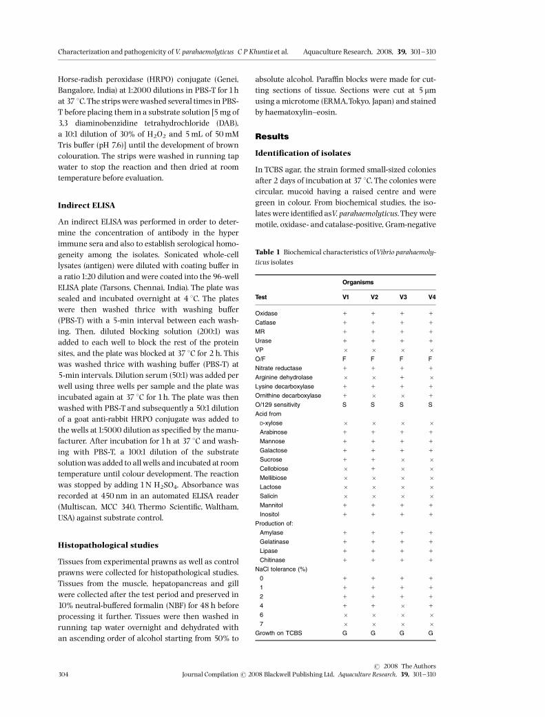
In TCBS agar, the strain formed small-sized coloniesafter 2 days of incubation at 37 1C. The colonies werecircular, mucoid having a raised centre and weregreen in colour. From biochemical studies, the iso-lates were identi¢ed asV. parahaemolyticus.They weremotile, oxidase- and catalase-positive, Gram-negative
Table 1 Biochemical characteristics ofVibrio parahaemoly-ticus isolates
Test
Organisms
V1 V2 V3 V4
Oxidase 1 1 1 1
Catlase 1 1 1 1
MR 1 1 1 1
Urase 1 1 1 1
VP � � � �O/F F F F F
Nitrate reductase 1 1 1 1
Arginine dehydrolase � � 1 �Lysine decarboxylase 1 1 1 1
Ornithine decarboxylase 1 � � 1
O/129 sensitivity S S S S
Acid from
D-xylose � � � �Arabinose 1 1 1 1
Mannose 1 1 1 1
Galactose 1 1 1 1
Sucrose 1 1 � �Cellobiose � 1 � �Mellibiose � � � �Lactose � � � �Salicin � � � �Mannitol 1 1 1 1
Inositol 1 1 1 1
Production of:
Amylase 1 1 1 1
Gelatinase 1 1 1 1
Lipase 1 1 1 1
Chitinase 1 1 1 1
NaCl tolerance (%)
0 1 1 1 1
1 1 1 1 1
2 1 1 1 1
4 1 1 � 1
6 � � � �7 � � � �
Growth on TCBS G G G G
Characterization and pathogenicity of V. parahaemolyticus C P Khuntia et al. Aquaculture Research, 2008, 39, 301^310
r 2008 TheAuthors304 Journal Compilationr 2008 Blackwell Publishing Ltd, Aquaculture Research, 39, 301^310

comma-shaped rods, which degraded D-glucose,reduced nitrate and amino acids like ornithine andlysine. All were sensitive to the vibriostatic agentO/129 (150 mg). All the strains produced acid frommannitol and were positive for indole production.They grew in1% peptone water containing 0%, 2%,4% NaCl, but did not grow in 6% and 7% of NaClpeptone water (Table 1). The strain V2 was negativeto ornithrine decarboxylase whereas strain V2 waspositive to lysine decarboxylase and did not grow at4% NaCl as compared with strain V1. Strain V4 isnon-fermentative to sucrose (Table1).
Antibiotic sensitivity test
Among the 21 antibiotics tested,V. parahaemolyticuswas found to be sensitive to 14 antibiotics. The high-est zonewas found in the case of cephalaxin, tetracy-cline and erythromycin (Table 2).
Virulence studies
In vitro pathogenicity test
The strains showed a positive reaction towardsCongo red binding assay. All strains were also foundto be of a �-haemolytic type (Table 3).
In vivo pathogenicity test
Clinical signs and symptoms. The prawns infectedwithV. parahaemolyticus did not exhibit any symptomup to 48 h, after which they became sluggish in nat-ure gradually and settled at the bottom of the tanks.Appendages and telsons were gradually lost. Theexoskeleton became reddish in colour, with blackspots on the carapace. The shells became brittle. Theinfected prawns died within 3 days. Dead and mori-bund prawns were dissected to note the grosschanges in internal organs. The gills became brown-ish and the muscle became whitish. The hepatopan-creas was pale white.
Infectivity study. The prawns challenged with2 � 107 and 2 � 108 CFUsmL�1 showed no mortal-ities during the study period while 80% mortalitywas observed in 2 � 109 CFUsmL�1 within 6 days(Fig.5).
Table 2 Antibiogram test of di¡erent isolates
Different antibiotics SymbolDisc potency(mcg) R I S V1 V2 V3 V4
Cephalexin Cp 30 11 13–14 21 S S S S
Co-Trimoxazole Co 25 10 11–15 16 S S S S
Chloramphenicol C 30 12 13–17 18 S S S S
Cefuroxine Cu 30 14 15–17 18 R R R R
Cephalothin Ch 30 14 15–17 18 S S S S
Ciprofloxacin Cf 5 15 16–20 21 S S S S
Chlorotetracyclin Ct 30 14 15–18 21 S S S S
Amoxycillin An 30 13 14–17 18 S S S S
Amikacin Ak 30 14 15–16 17 S S S S
Ampicillin A 10 13 14–16 17 R R R R
Bacitracin B 8 9–12 13 R R R R
Erythromycin-15 E-15 15 03 14–22 23 S S S S
Naldixic Acid Na 30 13 13 19 S S S S
Norfloxacin Nx 10 12 13–16 19 I I I I
Neomycin N 30 12 13–16 17 R R R R
Gentamycin G 10 12 13–14 15 S S S S
Tetracycline T 30 14 15–18 19 S S S S
Trimethoprim Tr 25 10 11–15 16 S S S S
Ofloxacine Of 2 12 13–15 16 S S S S
Penicillin G P 2 19 22–27 28 R R R R
Cloxacillin Cx 10 19 20 R R R R
Table 3 In vitro pathogenicity of di¡erent isolates in Congored binding assay and haemolytic test
Isolates Congo red binding assay Haemolytic test
V1 11 1 (7 mm)
V2 11 1 (10 mm)
V3 11 1 (8 mm)
V4 11 1 (8 mm)
Aquaculture Research, 2008, 39, 301^310 Characterization and pathogenicity of V. parahaemolyticus C P Khuntia et al.
r 2008 TheAuthorsJournal Compilationr 2008 Blackwell Publishing Ltd, Aquaculture Research, 39, 301^310 305

Sero-diagnosis
Agglutination. Di¡erent agglutination tests likeslide agglutination, tube agglutinationand colour ag-glutination showed that V. parahaemolyticus causedagglutination. Microtitre plate assay showed that thetitre was 256 after day 14, increasing to 1024 afterday 28.
Dot-ELISA and indirect ELISA. All the isolatestested positive for Dot-ELISA, which was revealed bybrown colouration of the nitrocellulose paper strips(Table 4).
The isolates were positive towards indirect ELISA,which was indicated by an increase in the OD valueat 450 nm as compared with that of the control(Table 4).
Histopathological studies
Muscle tissue showed mild to severe in¢ltration ofacinar cells into themusculature (Fig.1). Muscle bun-dles were severely deranged. Gill tissues showedmoderate necrotic changes in lamellae. Branchial ar-ches were thickened at places due to hyperplasia andsevere haemolytic in¢ltration. At times, the branchialarches were oedemateous and in¢ltrated with hae-mocytes. The cellular architecture of gill tissue wasderranged (Fig. 2).Cellular changes in hepatopancreatic tissues were
more pronounced and characterized bydilation of tu-bules, vacuolation of hepatocytes and marked necro-sis in acinar cells (Fig.3). In places, there was necrosisof acinar cells with complete desquamation of hae-mocytes. There was severe in¢ltration of cells in in-tertubular spaces. Hepatocytes were completelydevoid of cells (Fig. 4).
Table 4 Serological characteristics of di¡erent isolates
Isolates Agglutination test ELISA Dot-ELISA
V1 1 1 1
V2 1 1 1
V3 1 1 1
V4 1 1 1
Figure 1 Photomicrography of muscle tissue showingin¢ltration.
Figure 2 Photomicrography of gill tissue showing ne-crosis in gill lamellae.
Figure 3 Photomicrography of hepatopanceatic tissueshowing necrosis in acinar cells.
Figure 4 Photomicrography of hepatopanceatic tissueshowing in¢ltration of haematocytes.
Characterization and pathogenicity of V. parahaemolyticus C P Khuntia et al. Aquaculture Research, 2008, 39, 301^310
r 2008 TheAuthors306 Journal Compilationr 2008 Blackwell Publishing Ltd, Aquaculture Research, 39, 301^310

Discussion
Vibrio species exist as normal £ora in ¢sh and shell-¢sh, but has also been recognized as anopportunisticpathogen in many marine animals (Austin & Austin1993). Jaysree, Jankiram and Madhvi (2000) showedthe presence ofVibrio species inwhite spot syndromeof penaeid shrimp while Jayprakash, Rajesh Kumar,Philip and Singh (2006) revealed an association ofthis genus withM. rosenbergii.The strains were motile, oxidase- and catalase-
positive, Gram-negative, comma-shaped rods thatreduced nitrate to nitrite, grew inTCBS agar mediumand was sensitive to O/129, the vibriostatic agent(West & Colwell 1984). During this investigation, allthe strains manifested typical biochemical charac-ters with slight di¡erences. V. parahaemolyticusstrains were found to be urease positive and the abil-ity to hydrolyse urea has been proposed as a simplescreening test to predict which strain is pathogenic(Kaysner, Abeyta, Troast, Wetherington, Jinneman,Hill & Wekell 1994). From the in vitro pathogenicitystudy, it was found to be haemolytic and showed apositive response to the Congo red binding assay,which suggests that it is a pathogen, because thepathogenic strains are haemolytic in nature (Josephet al. 1982). Jana (2000) studied the haemolytic char-acteristics of V. parahaemolyticus isolated from thePenaeus monodon. She found thatV. parahaemolyticusisolated from shrimp sources including food poison-ing and healthy carriers and grown on blood agarmedium exhibited haemolysis. Haemolysin produc-tion byV. parahaemolyticus has been considered to bean important virulence factor. Further, the strainwas also able to hydrolyse gelatin, which representsanother characteristic towards pathogenicity andthis corroborates with our recent ¢ndings. Owing to
the presence of protease enzymes, it can hydrolysegelatin along with casein, elastin, collagen and hae-moglobin (Jana 2000; Marhaul 2005).Vibriosis has been experimentally induced in mar-
ine prawn by either immersing prawn in water con-taining bacteria (Sung, Kou & Song 1994; Hameed1995; Esteve & Herrera 2000; Khuntia 2002) orinjecting bacteria into muscle or haemolymph(Jiravanichpaisal & Miyazaki 1994; Lee, Yu, Chen,Yang & Liu1996). In the present study, to illustrate invivo pathogenicity, prawns were challenged by im-mersion. Prawns inoculated withV. parahaemolyticusat 2 � 109 CFUsmL�1exhibited 80%mortality with-in 7 days, whereas 2 � l07 and 2 � l08 CFUsmL�1
showed no mortalities throughout the experimentperiod. Signi¢cant mortalities were recorded inPeneaus merguiensis when challenged with108 cells mL�1 (Sae-oui,Transutapanit & Ruangpan1987). According to Martin, Rubin and Swanson(2004), 50% of mortality was recorded in case ofSicyonia ingentis at 105 CFUsmL�1within 7 days byan immersion challenge, which is low as comparedwith our study.While working with the larvae andpostlarvae, Oanh, Haoand Phuong (2001) found that104^108 cells mL�1 caused signi¢cant mortalitywhen they were challenged with Vibrio species.According to Lavilla-Pitogo, Baticados, Cruz-Lacier-da and De la Pena (1990) on postlarvae of P.monodon,signi¢cant mortalities occurred on exposure to 102
Vibrio harveyi cells mL�1within 48 h. Any infectionand mortality depend on the infectious doses andpathogens have di¡erent infectious doses for di¡er-ent organisms depending on their individual capa-city to resist stress in£uenced by their nutritionaland physiological factors, for which the mortalityrate varied from organism to organism. Vibriosis inpenaeids is generally recognized as a secondaryinfection in£uenced by factors such as stress, envir-onmental failures and a high number of potentiallypathogenic bacteria (Chen, Huang & Kou 1992;Nash et al. 1992; Moheny, Lightener & Bell 1994;Ruangapan, Tabkaew & Sangungruang 1995), butcontinuous exposure of shrimp post larvae to highnumbers of Vibrio in the environment can easilycause primary vibriosis as the bacteria can multiplyon the larval surface (Chen1993).A number of monoclonal antibodies were raised
against a number of Vibrio sp. including Vibriovulni¢cus, V. harveyi, V. parahaemolyticus and Vibrioalginolyticus to report these pathogens as causativeagents of vibriosis in marine prawn (Chen, Hanna,Altman, Smith, Moon & Hammond1992). Song, Lee,
Commulative mortality rate
0102030405060708090
1 2 3 4 5 6 7Days
% M
ort
alit
y
Set 1 (2x10⁷ CFUs/ml)Set 2 (2x10⁸ CFUs/ml)Set 3 (2x10⁹ CFUs/ml)Control
Figure 5 Cumulative mortality rate (%) trend ofMacrobrachium rosenbergii challenged with Vibrioparahaemolyticus.
Aquaculture Research, 2008, 39, 301^310 Characterization and pathogenicity of V. parahaemolyticus C P Khuntia et al.
r 2008 TheAuthorsJournal Compilationr 2008 Blackwell Publishing Ltd, Aquaculture Research, 39, 301^310 307

Lin and Chen (1992) developed an enzyme-linked im-munoassay based on monoclonal antibodies toV. vul-ni¢cus andV. harveyi. In the present study, an indirectELISAwas performed to cross check the results of ag-glutination; in our study, all the isolates were positivein the ELISA test, indicating the presence of commonantigens.The Dot-ELISAmethodwas more simple and rapid.
It also ensured con¢rmatory identi¢cation of thepathogen even when serum was diluted 100 � . Theisolates testing positive for agglutinationand indirectELISA also showed the same response to Dot ELISA.The Dot-ELISA method is simple and the require-ments are uncomplicated and therefore it can berecommended for ¢eld usage.During the in vivo experiment, prawns become
sluggish in nature gradually and settle at into thebottom of the tank after 48h. The prominent diseasesymptoms noticed in the present study after 3 days,which correlate with the symptoms of shell diseasesas black colouration along with the erosion of appen-dages, have been observed frequently recorded infreshwater prawn frommany regions when it is asso-ciated with shell diseases (Delves-Broughton &Poupard1975;Tonguithai1993).There have been numerous studies describing
morphological changes to tissues in marine prawninfected withVibrio (Lee et al. 1996; Esteve & Herrera2000; Alday-Sanz, Roque & Trunball 2002), but in-formation on experimentally induced infection withV. parahaemolyticus studies in freshwater prawns isscarce. Therefore, the results of the histopathologicalstudycould not be compared with earlier work. How-ever, muscle necrosis and hepatopancreatosis are ob-served in case of naturally infected prawns. Similarly,haemocytic in¢ltration, ¢briosis and necrosis in thehepatopancreatic cells were noticed when shrimpswere infected withV. harveyi (Lavillo-Pitogo, Liano &Paner 1998). In the present study, hepatopancreaticchanges were more pronounced, indicating a directrelationship between the host and the pathogen asthe hepatopancreas is reportedly the main targetorgan of most bacterial pathogens for their coloniza-tion and proliferation (Chen et al. 1992; Frelier, Sis,Bell & Lewis1992).Devesa, Toranzo and Basja (1985) revealed that
oxytetracycline (OTC) is intensively used in hatch-eries and grow-out ponds and is e¡ective in thetreatment of vibriosis. But according to De la Pena,Tamaki, Manoyama, Nakai and Muroga (1993),Vibrios are sensitive to chloramphenicol rather thanoxytetracycline. In the present study, we found
that this species is highly sensitive to tetracyclineand cephalaxin.The giant freshwater prawn has been widely do-
mesticated in India for over a decade. Mortalitiesare encountered due to vibriosis in all its life stages.The role of vibriosis in prawnaquaculture has grownin recent years as farmers have experienced substan-tial losses. Development of immunological techni-ques for the detection and screening of pathogensand subsequent treatment is highly important. Inthis regard, this study has immense importance forthe development of prawn culture in South eastAsian countries.
Acknowledgments
We express our heartiest gratitude to The Depart-ment of Microbiology, Orissa University of Agricul-ture and Technology, India, for this opportunity andalso for their cordial support. We are also grate toThe Director, Central Institute of Freshwater Aqua-culture (ICAR), India, for providing all the possibleassistance and sincere co-operation for conductingthis investigation.
References
Adams A. (1991) Response of penaeid shrimp to exposure toVibrio species. Fish and Shell¢sh Immunology1,59^70.
Alday-Sanz V., Roque A. & Turnbull J.F. (2002) Clearingmechanism of Vibrio vulni¢cus biotype 1 in black tigershrimp Penaeus monodon. Diseases of Aquatic Organisms48,91^99.
Austin B. & Austin D.A. (1993) Bacterial ¢sh pathogens. In:Diseases in Farmed andWild Fish, 2nd ed, pp.165^307. EllisHorwoord, Chichester, UK.
Barrow G.I. & Feltham R.K. (1993) Cowan and Steel’s Manualfor the Identi¢cation of Medical Bacteria, 2nd edn, Cam-bridge University Press, Cambridge, USA,331pp.
BauerA., KirbyW.M., Sherris J.C. & Truck M.M. (1966) Anti-biotic susceptibility testing by standard discs methods.TheAmericanJournal of Clinical Pathology 45, 493^496.
Bradford M.M. (1976) A rapid method for the quanti¢cationof the microgram quantities protein utilizing the princi-ple of protein dye binding. Annal Biochemistry 72,248^254.
Chen D., Hanna P.J., Altman K., Smith A., Moon P. & Ham-mond L.S. (1992) Development of monoclonal antibodiesthat identifyVibrio species commonly isolated from infec-tion of human, ¢sh and shell¢sh. Applied EnvironmentalMicrobiology 58,3694^3700.
Chen H.C. (1993) Studies on the Successful Culture of GrassShrimp, Penaeus Monodon. COA Fisheries Series No. 31.Council of Agriculture, China.
Characterization and pathogenicity of V. parahaemolyticus C P Khuntia et al. Aquaculture Research, 2008, 39, 301^310
r 2008 TheAuthors308 Journal Compilationr 2008 Blackwell Publishing Ltd, Aquaculture Research, 39, 301^310

ChenJ.C., Lei S.C. & Lee K.K. (2000) Lethal attribute of serineprotease secreted byVibrio alginolyticus strain in kuramaprawn Penaeus japonicus. Zool Naturforsch 55,94^99.
Chen S.N., Huang S.L. & Kou G.H. (1992) Studies on the epi-zootiology and pathogenicity of bacterial infections incultured giant prawns, Penaeus monodon, in Taiwan. In:Diseases of cultured Penaeids Shrimp in Asia and the UnitedStates (ed. by W. Fulks & K.L. Main), pp. 195^206. TheOceanic Institute, Honolulu, HI.
De la Pena L.D.,TamakiT., Manoyama K., NakaiT. & MurogaK. (1993) Characterization of the causative bacterium ofvibriosis in the Kurama prawn, Penaeus japonicus. Aqua-culture115,1^12.
Delves-Broughton J. & Paupard C.W. (1975) Disease problemsof prawns in re-circulation system in UK. Aquaculture 71,201^207.
Devesa S., Toranza A.E. & Basja J.L. (1985) First report ofvibriosis in turbot (Scopthalmus maximus) cultured inNorthwestern Spain. In: Fish and Shell¢sh Pathology(ed. byA.E. Ellis), pp.131^140. Academic Press, London.
Esteve M. & Herrera F.C. (2000) Hepatopancreatic alterationin Litopenaeus vannamei (Boone, 1939) (Crustacea: Deca-poda: Penaidae) experimental infected with aVibrio algi-nolyticus strain. Journal of Invertebrate Pathology 76,1^5.
Frelier P.F., Sis R.F., BellT.A. & Lewis D.H. (1992) Microscopicand ultra structural studies of necrotizing hepatopan-creatitis in Paci¢c white shrimp (Penaeus nannamei) cul-ture inTexas.Veterinary Pathology 29, 269^277.
Hameed A.S. (1995) Susceptibility of three Penaeus species toaVibrio camebellii-like bacterium. Journal of World Aqua-culture Society 26, 315–319.
Jana R. (2000) Studies on the characterization ofVibrio speciesisolated from shrimp Penaeus monodon using random ampli-¢ed polymorphic DNA ¢ngerprinting. MSc thesis, OrissaUniversity of Agriculture and Technology, Bhubaneswar,India.
Jayprakash N.S., Rajesh Kumar V.J., Philip R. & Singh I.S.B.(2006) Vibrios associated withMacrobrachium rosenbergii(DeMan,1879) larvae from three hatcheries on the Indiansouthwest coast. Aquaculture Research 37,351^358.
Jaysree L., Jankiram P. & Madhvi R. (2000) Characterstic,pathogenicity and antibiotic sensitivity of bacterial iso-lates from white spot diseased shrimp. Asian FisheriesScience13,327^334.
Jiravanichpaisal P. & Miyazaki T. (1994) Histopathology, bio-chemistry and pathogencity of Vibrio harveyi infectingBlack tiger parwm, Penaeus monodon. Journal of AquaticAnimal Health 6, 2735.
Joseph S.W., Colwell R.R. & Kaper J.B. (1982) Vibrio parahae-molyticus and related hallophilic Vibrios. CRC CrtiticalReviews in Microbiology10,73^124.
Kaysner C.A., Abeyta C., Troast P.A. Jr.,Wetherington J.H.,Jinneman K.C., Hilli W.E. & Wekell M.M. (1994) Urea hy-drolysis can predict the potential pathogenicity of Vibrioparahaemolyticus strains isolated in the Paci¢c Northwest.Applied and Environmental Microbiology 60,3020^3022.
Khuntia C.P. (2002) Isolation and characterization of bacterialpathogen from diseased freshwater prawn, Macrobrachiumrosenbegii (de Man). MSc thesis, Orissa University of Agri-culture andTechnology, Bhubaneswar, India.
Lavilla-Pitogo C.R. (1995) Bacterial disease of penaeidshrimp. Disease in Asian Aquaculture II (pp.107^121. FishHealth Section, Asian Fisheries Society, Manila,Phillipines.
Lavilla-Pitogo C.R., Baticados M.C.L., Cruz-Lacierda E.R. &De la Pena L.D. (1990) Occurrence of luminous bacterialdiseases of Penaeus monodon larvae in Philippines. Aqua-culture 91,1^13.
Lavilla-Pitogo C.R., Leano E.M. & Paner M.G. (1998)Mortalities of pond-cultured juvenile shrimp, Penaeusmonodon associated with dominance of luminescentVibrios in the rearing environment. Aquaculture 164,337^349.
Lee K.K, Yu S.R., Chen F.R., Yang T.I. & Liu P.C. (1996)Virulence of Vibrio alginolyticus isolated from diseasedtiger prawn, Penaeus monodon. Current Microbiology 32,229^231.
Lightner D.V. (1993) Disease of cultured shrimp. In: CRCHandbook of Mariculture (ed. by P.V. McVey), pp. 393–
486. CRC Press, Boca Raton, FL, USA.
Lightner D.V. & Lewis D.H. (1975) Asepticemia bacterial dis-ease syndrome in penaeid shrimp. Marine Fisheries Re-view 37, 25^28.
MacFaddin J.F. (1980) Biochemical Test for Identi¢cation ofMedical Bacteria. William Wilkison, Baltimore, MD, pp.1^523.
Marhaul N.P. (2005) Comparative genetic study on Vibrioalginolyticus and Vibrio parahaemolyticus isolated fromBlack tiger shrimp, Penaeus monodon. MSc thesis, OrissaUniversity of Agriculture and Technology, Bhubaneswar,India.
Martin G., Rubin N. & Swanson E. (2004) Vibrio parahaemo-lyticus and V. harveyi cause detachment of epitheliumfrom the midgut trunk of the penaid shrimp. Sicyonia in-gentis. Diseases of Aquatic Organism 60, 21^29.
Moheny L.L., Lightener D.V. & BellT.A. (1994) An epizootic ofvibriosis in Ecuadorian pond-reared Penaeus vannameiBoone (Crustacea: Decapoda). Journal of World Aquacul-ture Society 25,116^125.
Nash G.C., Nithimathachoke C., Tungmandi A., Arkary-morn P., Prathespi P. & Ruamthevessub P. (1992) Vibriosisand its control in pond reared Penaeus monodon in Thai-land. In: Diseases in Asian Aquaculture. Proceedings of theFirst Symposium on Disease in Asian Aquaculture (ed. by
R. Shari, P. Subsinghe & J.R. Arthus), pp. 143–155.
Network of Aquaculture Centers in Asia-Pacific, Bali.
Oanh D.T.T., HaoT.T.T. & Phuong N.T. (2001) Characterizationand Pathogenicity ofVibrio bacteria isolated from freshwaterprawn (Macrobrachium rosenbergii) hatcheries. isolation andidenti¢cation of Vibrio sp. from larval stages Proceeding ofthe 2000 annual workshop of JIRCAS Meskong deltaProject CanTho,Vietnam.
Aquaculture Research, 2008, 39, 301^310 Characterization and pathogenicity of V. parahaemolyticus C P Khuntia et al.
r 2008 TheAuthorsJournal Compilationr 2008 Blackwell Publishing Ltd, Aquaculture Research, 39, 301^310 309

Pacini F. (1854) Osservazione microscopiche e deduzioni pa-tologiche sul cholera Asiatica. Gazette medical ItalyTasca-na Firenze 6, 405^412.
Ruangapan L. & KitaoT. (1991) Vibrio bacteria isolated fromblack tiger shrimp Penaeus monodon (Fabricus). Journal ofFish Diseases14,383^388.
Ruangapan L.,Tabkaew R. & Sangungruang K. (1995) Bac-terial £ora of ponds with di¡erent stocking densities ofblack tiger shrimp. Penaeus monodon. Diseases in AsianAquaculture II (ed. by M. Shari¡, J.R. Arthur & R.P. Suba-singe), pp. 141^149. Fish Health Section, Asian FisheriesSociety, Manila, Phillipines.
Sae-oui D., Transutapanit A. & Ruangpan L. (1987)Vibrio harveyi a causative agent of white shrimpnauplii Penaeus merguiensis. Thai Fishery Gazette 40,177^182.
SongY.L., Lee S.P., LinY.T. & Chen C.T. (1992) Enzyme immu-noassay for shrimp Vibriosis. Diseases of Aquatic Organ-isms14, 43^50.
Su S.C. & Lee C.Y. (1997) Characterization of hemolysis of theVibrio parahaemolyticus. Journal ofMicrobiology, Immunol-ogy and Infection 30,32^42.
Sung H.H., Kou G.H. & SongY.L. (1994) Vibriosis resistanceinduced by glucan treatment in tiger shrimp (Penaeusmonodon). Fish Pathology 29,11^17.
Tonguithai K. (1993) Diseases of the freshwater prawnMacrobrachium rosenbergii in Thailand. Diseases of AsianAquaculture 1, 89–96.
West P.A. & Colwell R.R. (1984) Identi¢cation and classi¢ca-tion of Vibrionaceae an overview.Vibriosis in the Environ-ment (ed. by R.R. Colwell), pp. 285^363. JohnWiley andSons, NewYork, NY, USA.
Characterization and pathogenicity of V. parahaemolyticus C P Khuntia et al. Aquaculture Research, 2008, 39, 301^310
r 2008 TheAuthors310 Journal Compilationr 2008 Blackwell Publishing Ltd, Aquaculture Research, 39, 301^310