Gene profiling and characterization of arginine kinase-1 (MrAK-1) from freshwater giant prawn (...
9
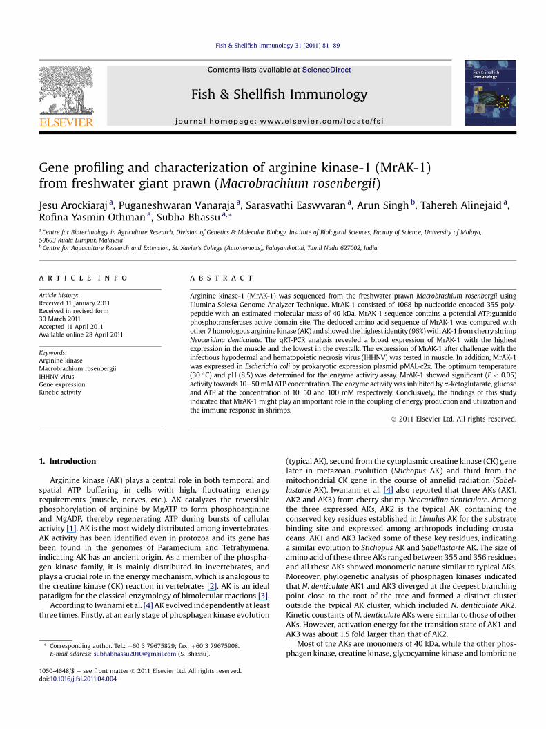
Gene profiling and characterization of arginine kinase-1 (MrAK-1) from freshwater giant prawn (Macrobrachium rosenbergii) Jesu Arockiaraj a , Puganeshwaran Vanaraja a , Sarasvathi Easwvaran a , Arun Singh b , Tahereh Alinejaid a , Rofina Yasmin Othman a , Subha Bhassu a, * a Centre for Biotechnology in Agriculture Research, Division of Genetics & Molecular Biology, Institute of Biological Sciences, Faculty of Science, University of Malaya, 50603 Kuala Lumpur, Malaysia b Centre for Aquaculture Research and Extension, St. Xavier’s College (Autonomous), Palayamkottai, Tamil Nadu 627002, India article info Article history: Received 11 January 2011 Received in revised form 30 March 2011 Accepted 11 April 2011 Available online 28 April 2011 Keywords: Arginine kinase Macrobrachium rosenbergii IHHNV virus Gene expression Kinetic activity abstract Arginine kinase-1 (MrAK-1) was sequenced from the freshwater prawn Macrobrachium rosenbergii using Illumina Solexa Genome Analyzer Technique. MrAK-1 consisted of 1068 bp nucleotide encoded 355 poly- peptide with an estimated molecular mass of 40 kDa. MrAK-1 sequence contains a potential ATP:guanido phosphotransferases active domain site. The deduced amino acid sequence of MrAK-1 was compared with other 7 homologous arginine kinase (AK) and showed the highest identity (96%) with AK-1 from cherry shrimp Neocaridina denticulate. The qRT-PCR analysis revealed a broad expression of MrAK-1 with the highest expression in the muscle and the lowest in the eyestalk. The expression of MrAK-1 after challenge with the infectious hypodermal and hematopoietic necrosis virus (IHHNV) was tested in muscle. In addition, MrAK-1 was expressed in Escherichia coli by prokaryotic expression plasmid pMAL-c2x. The optimum temperature (30 C) and pH (8.5) was determined for the enzyme activity assay. MrAK-1 showed significant (P < 0.05) activity towards 10e50 mM ATP concentration. The enzyme activity was inhibited by a-ketoglutarate, glucose and ATP at the concentration of 10, 50 and 100 mM respectively. Conclusively, the findings of this study indicated that MrAK-1 might play an important role in the coupling of energy production and utilization and the immune response in shrimps. Ó 2011 Elsevier Ltd. All rights reserved. 1. Introduction Arginine kinase (AK) plays a central role in both temporal and spatial ATP buffering in cells with high, fluctuating energy requirements (muscle, nerves, etc.). AK catalyzes the reversible phosphorylation of arginine by MgATP to form phosphoarginine and MgADP, thereby regenerating ATP during bursts of cellular activity [1]. AK is the most widely distributed among invertebrates. AK activity has been identified even in protozoa and its gene has been found in the genomes of Paramecium and Tetrahymena, indicating AK has an ancient origin. As a member of the phospha- gen kinase family, it is mainly distributed in invertebrates, and plays a crucial role in the energy mechanism, which is analogous to the creatine kinase (CK) reaction in vertebrates [2]. AK is an ideal paradigm for the classical enzymology of bimolecular reactions [3]. According to Iwanami et al. [4] AK evolved independently at least three times. Firstly, at an early stage of phosphagen kinase evolution (typical AK), second from the cytoplasmic creatine kinase (CK) gene later in metazoan evolution (Stichopus AK) and third from the mitochondrial CK gene in the course of annelid radiation (Sabel- lastarte AK). Iwanami et al. [4] also reported that three AKs (AK1, AK2 and AK3) from cherry shrimp Neocaridina denticulate. Among the three expressed AKs, AK2 is the typical AK, containing the conserved key residues established in Limulus AK for the substrate binding site and expressed among arthropods including crusta- ceans. AK1 and AK3 lacked some of these key residues, indicating a similar evolution to Stichopus AK and Sabellastarte AK. The size of amino acid of these three AKs ranged between 355 and 356 residues and all these AKs showed monomeric nature similar to typical AKs. Moreover, phylogenetic analysis of phosphagen kinases indicated that N. denticulate AK1 and AK3 diverged at the deepest branching point close to the root of the tree and formed a distinct cluster outside the typical AK cluster, which included N. denticulate AK2. Kinetic constants of N. denticulate AKs were similar to those of other AKs. However, activation energy for the transition state of AK1 and AK3 was about 1.5 fold larger than that of AK2. Most of the AKs are monomers of 40 kDa, while the other phos- phagen kinase, creatine kinase, glycocyamine kinase and lombricine * Corresponding author. Tel.: þ60 3 79675829; fax: þ60 3 79675908. E-mail address: [email protected] (S. Bhassu). Contents lists available at ScienceDirect Fish & Shellfish Immunology journal homepage: www.elsevier.com/locate/fsi 1050-4648/$ e see front matter Ó 2011 Elsevier Ltd. All rights reserved. doi:10.1016/j.fsi.2011.04.004 Fish & Shellfish Immunology 31 (2011) 81e89
Transcript of Gene profiling and characterization of arginine kinase-1 (MrAK-1) from freshwater giant prawn (...
lable at ScienceDirect
Fish & Shellfish Immunology 31 (2011) 81e89
Contents lists avai
Fish & Shellfish Immunology
journal homepage: www.elsevier .com/locate / fs i
Gene profiling and characterization of arginine kinase-1 (MrAK-1)from freshwater giant prawn (Macrobrachium rosenbergii)
Jesu Arockiaraj a, Puganeshwaran Vanaraja a, Sarasvathi Easwvaran a, Arun Singh b, Tahereh Alinejaid a,Rofina Yasmin Othman a, Subha Bhassu a,*
aCentre for Biotechnology in Agriculture Research, Division of Genetics & Molecular Biology, Institute of Biological Sciences, Faculty of Science, University of Malaya,50603 Kuala Lumpur, MalaysiabCentre for Aquaculture Research and Extension, St. Xavier’s College (Autonomous), Palayamkottai, Tamil Nadu 627002, India
a r t i c l e i n f o
Article history:Received 11 January 2011Received in revised form30 March 2011Accepted 11 April 2011Available online 28 April 2011
Keywords:Arginine kinaseMacrobrachium rosenbergiiIHHNV virusGene expressionKinetic activity
* Corresponding author. Tel.: þ60 3 79675829; fax:E-mail address: [email protected] (S. B
1050-4648/$ e see front matter � 2011 Elsevier Ltd.doi:10.1016/j.fsi.2011.04.004
a b s t r a c t
Arginine kinase-1 (MrAK-1) was sequenced from the freshwater prawn Macrobrachium rosenbergii usingIllumina Solexa Genome Analyzer Technique. MrAK-1 consisted of 1068 bp nucleotide encoded 355 poly-peptide with an estimated molecular mass of 40 kDa. MrAK-1 sequence contains a potential ATP:guanidophosphotransferases active domain site. The deduced amino acid sequence of MrAK-1 was compared withother 7 homologous arginine kinase (AK) and showed the highest identity (96%)withAK-1 from cherry shrimpNeocaridina denticulate. The qRT-PCR analysis revealed a broad expression of MrAK-1 with the highestexpression in the muscle and the lowest in the eyestalk. The expression of MrAK-1 after challenge with theinfectious hypodermal and hematopoietic necrosis virus (IHHNV) was tested in muscle. In addition, MrAK-1was expressed in Escherichia coli by prokaryotic expression plasmid pMAL-c2x. The optimum temperature(30 �C) and pH (8.5) was determined for the enzyme activity assay. MrAK-1 showed significant (P < 0.05)activity towards 10e50mMATP concentration. The enzyme activity was inhibited by a-ketoglutarate, glucoseand ATP at the concentration of 10, 50 and 100 mM respectively. Conclusively, the findings of this studyindicated that MrAK-1 might play an important role in the coupling of energy production and utilization andthe immune response in shrimps.
� 2011 Elsevier Ltd. All rights reserved.
1. Introduction
Arginine kinase (AK) plays a central role in both temporal andspatial ATP buffering in cells with high, fluctuating energyrequirements (muscle, nerves, etc.). AK catalyzes the reversiblephosphorylation of arginine by MgATP to form phosphoarginineand MgADP, thereby regenerating ATP during bursts of cellularactivity [1]. AK is the most widely distributed among invertebrates.AK activity has been identified even in protozoa and its gene hasbeen found in the genomes of Paramecium and Tetrahymena,indicating AK has an ancient origin. As a member of the phospha-gen kinase family, it is mainly distributed in invertebrates, andplays a crucial role in the energy mechanism, which is analogous tothe creatine kinase (CK) reaction in vertebrates [2]. AK is an idealparadigm for the classical enzymology of bimolecular reactions [3].
According to Iwanami et al. [4] AKevolved independently at leastthree times. Firstly, at an early stage of phosphagen kinase evolution
þ60 3 79675908.hassu).
All rights reserved.
(typical AK), second from the cytoplasmic creatine kinase (CK) genelater in metazoan evolution (Stichopus AK) and third from themitochondrial CK gene in the course of annelid radiation (Sabel-lastarte AK). Iwanami et al. [4] also reported that three AKs (AK1,AK2 and AK3) from cherry shrimp Neocaridina denticulate. Amongthe three expressed AKs, AK2 is the typical AK, containing theconserved key residues established in Limulus AK for the substratebinding site and expressed among arthropods including crusta-ceans. AK1 and AK3 lacked some of these key residues, indicatinga similar evolution to Stichopus AK and Sabellastarte AK. The size ofamino acid of these three AKs ranged between 355 and 356 residuesand all these AKs showed monomeric nature similar to typical AKs.Moreover, phylogenetic analysis of phosphagen kinases indicatedthat N. denticulate AK1 and AK3 diverged at the deepest branchingpoint close to the root of the tree and formed a distinct clusteroutside the typical AK cluster, which included N. denticulate AK2.Kinetic constants ofN. denticulate AKswere similar to those of otherAKs. However, activation energy for the transition state of AK1 andAK3 was about 1.5 fold larger than that of AK2.
Most of the AKs are monomers of 40 kDa, while the other phos-phagen kinase, creatine kinase, glycocyamine kinase and lombricine
J. Arockiaraj et al. / Fish & Shellfish Immunology 31 (2011) 81e8982
kinase are dimeric, or octameric in the case ofmitochondrial CK [5,6].In 1997 an unusual 80 kDaAKwith a two-domain structure has beenisolated from the primitive sea anemone Anthopleura sp by Suzukiet al. [7]. Interestingly, AK appears to have evolved at least twiceduring evolution of PKs:firstly at anearly stage of phosphagenkinaseevolution and secondly, from CK at later time inmetazoan evolution[8]. Recently, a few researchers have also discovered AK with 2domain structure from deep-sea clam Calyptogena kaikoi [9], clamsSolen strictus and Corbicula japonica [10] have an molecular mass of80 kDa AK. But their enzymatic propertieswere comparable to thoseof usual 40 kDa AKs.
Recently, a number of cDNA from cucumber Stichopus japoni-cus [1,3], oyster Crassostrea sp [5], kuruma prawn Marsupenaeusjaponicus [6], Callinectes sapidus [11], deep-sea clam Calyptogenakaikoi [9] clams Solen strictus and Corbicula japonica [10] chitonLiolophura japonica and turbanshell Battilus cornutus [12], Cellanagrata, Aplysia kurodai [13], horseshoe crab Limulus polyphemus[14], sea Chinese shrimp Fenneropenaeus chinensis [15], greasy-back shrimp Metapenaeus ensis [16], Litopenaeus vannamei [17]and sea anemone Anthopleura japonicus [18] encoding AKs inmajor groups of invertebrate have been made available toinvestigate AK’s structure, catalysis mechanism and evolutionaryrelationship.
A crystal structure for the transition state analog complex of themonomeric AK from horseshoe crab Limulus polyphemus hasappeared which suggests that AKs with 2 domains have a uniquesubstrate binding system [19]. These two arginine kinase formsshow very similar kinetic properties, such as both require a divalentcation (Mg2þ or Mn2þ) for activity. High concentrations of freeMg2þ will inhibit both enzymes. Although no catalytic basis fora functional difference between the two forms was found, it ispossible that in vitro differences in stability may reflect stabilitiesin vivo and thus provide a basis for regulating arginine kinase levelsin various tissues thereby giving a selective advantage to specieshaving both forms.
In recent studies, it was demonstrated that the expression ofAK correlated closely with hypoxic stress in M. japonicus [6], theimmune response to laminarin stimulation in F. chinensis [15],E. coli LPS immune response to L. vannamei [17], salinity changein C. sapidus [20], the exposure to lead in yabby, Cheraxdestructor [21], acclimation to cadmium in crab, Eriocheirsinensis [22] and the defense against the infection of virus inshrimp, Penaeus stylirostris [23] and F. chinensis [24] suggestingthat AK might play an important role in many biological eventsin crustaceans.
The Malaysian freshwater giant prawn Macrobrachium rose-nbergii is an important commercial species. However, infectiousdiseases especially, infectious hypodermal and hematopoieticnecrosis virus (IHHNV) have affected aquaculture of M. rosenbergiienormously. Thus, research into freshwater prawn defensemechanisms is important to develop disease control strategies[17,24]. Yao et al. [17] reported that AK levels changed in prawnafter immune stimulant, which suggested that the prawnimmune response maybe correlated with the change of energymetabolism that was regulated by AK [25] and some studiesshowed that AK changed significantly after prawn were infectedwith virus [23,24] and bacteria [17]. However, the detailed func-tions and characterization of AK in prawn are poorly understood.To gain insight into the characterization of AK and its role inprawn, a full length cDNA of AK-1 (designated as MrAK-1) wasidentified from the M. rosenbergii transcriptome unigenesobtained by Illumina’s Solexa sequencing technology. Theexpression profiles of AK mRNA in the muscle of M. rosenbergiiafter IHHNV challenge were studied and the enzyme activity ofrecombinant AK protein was assayed.
2. Materials and methods
2.1. Identification of M. rosenbergii arginine kinase-1
A full length MrAK-1 gene was identified from theM. rosenbergiitranscriptome unigenes obtained by Illumina’s Solexa sequencingtechnology. Briefly, unigenes obtained from the assembly of theIllumina Solexa short reads from the RNA sequencing of themuscle,gill and hepatopancrease transcriptomes of M. rosenbergii weremined for sequences which had been identified as arginine kinase 1gene through BLAST homology search against the NCBI database(http://blast.ncbi.nlm.nih.gov/Blast).
2.2. Bioinformatics analysis
The Basic Local Alignment Tool (BLAST) program was used tosearch similar nucleotide and protein sequences to theMrAK-1 [26].The open reading frame (ORF) and amino acid sequence of MrAK-1was obtained by using DNAssit 2.2. Characteristic domains ormotifswere identified using the PROSITE profile database [27]. Identity,similarity and gap percentages were calculated using FASTAprogram [28]. The N-terminal transmembrane sequence wasdetermined by DAS transmembrane prediction program (http://www.sbc.su.se/wmiklos/DAS). Signal peptide analysis was doneusing the SignalPworldwide P server (http://www.cbs.dtu.dk). Pair-wise and multiple sequence alignment were analyzed using theClustalW version 2 program [29]. The phylogenetic relationship ofthe MrAK-1 was determined using the neighbor-joining (NJ)method and MEGA 4.1 program [30].
2.3. M. rosenbergii and IHHNV challenge
Specific pathogen free (SPF) prawns (average body weight 10 g)were obtained from the Bandar Sri Sendayan, Negeri Sembilan,Malaysia. Prawns were maintained in flat-bottomed glass tanks(300 L) with aerated and filtered freshwater at 28 � 1 �C in thelaboratory. All prawns were acclimatized for 1 week before chal-lenged to IHHNV. A maximum of 15 prawns per tank were main-tained during the experiment.
For IHHNV induced mRNA expression analysis, the prawns wereinjected with IHHNV. Briefly, IHHNV infected prawn tail tissue,tested positive by nested PCR was homogenized in sterile 2% NaCl(1:10, w/v) solution and centrifuged in a tabletop centrifuge at5000 rpm for 5 min at 4 �C. The supernatant was filtered through0.45 mm filter and used for injecting (100 ml per 10 g prawn) theanimals. Samples were collected before (0 h), and after injection(3, 6, 12, 24 and 48 h) and were immediately snap-frozen in liquidnitrogen and stored at �80 �C until the total RNA was isolated.Using a sterilized syringe, the haemolymph (0.2e0.5 ml per prawn)was collected from the prawn heart and immediately centrifuged at3000 � g for 10 min at 4 �C to allow haemocyte collection for totalRNA extraction. All samples were analyzed in three duplicationsand the results are expressed as relative fold of one sample asmean � standard deviation.
2.4. RNA extraction and cDNA synthesis
Total RNAwas isolated from the tissues of each animal using TRIReagent followingmanufacturer’s protocol (Guangzhou DongshengBiotech, China). Total RNA was treated with RNase free DNA set(5 Prime GmbH, Hamburg, Germany) to remove the contaminatingDNA. The total RNA concentration was measured spectropho-metrically (NanoVue Plus Spectrophotometer, GE Healthcare UKLtd, England). First-strand cDNA was synthesized from total RNAby M-MLV reverse transcriptase (Promega, USA) following the
J. Arockiaraj et al. / Fish & Shellfish Immunology 31 (2011) 81e89 83
manufacturer’s protocol with AOLP primer (5’GGCCACGCGTCGAC-TAGTAC(T)16(A/C/G)3’).
2.5. Quantitative real-time PCR analysis of MrAK-1mRNA expression
The relative expression of MrAK-1 in the hemocytes, pleopods,walking legs, eye stalk, gill, hepatopancreas, stomach, intestine,brain and muscle were measured by qRT-PCR. qRT-PCR was carriedout using a ABI 7500 Real-time Detection System (Applied Bio-systems) in 20 ml reaction volume containing 4 ml of cDNA fromeach tissue, 10 ml of Fast SYBR� Green Master Mix, 0.5 ml of eachprimer (20 pmol/ml) and 5 ml dH2O. The qRT-PCR cycle profile was 1cycle of 95 �C for 10s, followed by 35 cycles of 95 �C for 5s, 58 �C for10s and 72 �C for 20s and finally 1 cycle of 95 �C for 15s, 60 �C for30s and 95 �C. The same qRT-PCR cycle profile was used for theinternal control gene, b-actin. The primers used in this study arepresented in Table 1. b-actin primers were designed based on ESTof1357 bp (GenBank accession No. AY651918) from M. rosenbegrii.After the PCR program, data were analyzed with ABI 7500 SDSsoftware (Applied Biosystems). To maintain consistency, the base-line was set automatically by the software. The comparative CTmethod (2�ddCT method) was used to analyse the expression level ofMrAK-1 [31]. All data were given in terms of relative mRNAexpressed as means � standard deviation. For comparison of rela-tive MrAK-1 mRNA expression, statistical analysis was performedusing one-way ANOVA and mean comparisons were performed byTukey’s Multiple Range Test using SPSS 11.5 at 5% significant level.
2.6. In vitro expression construction
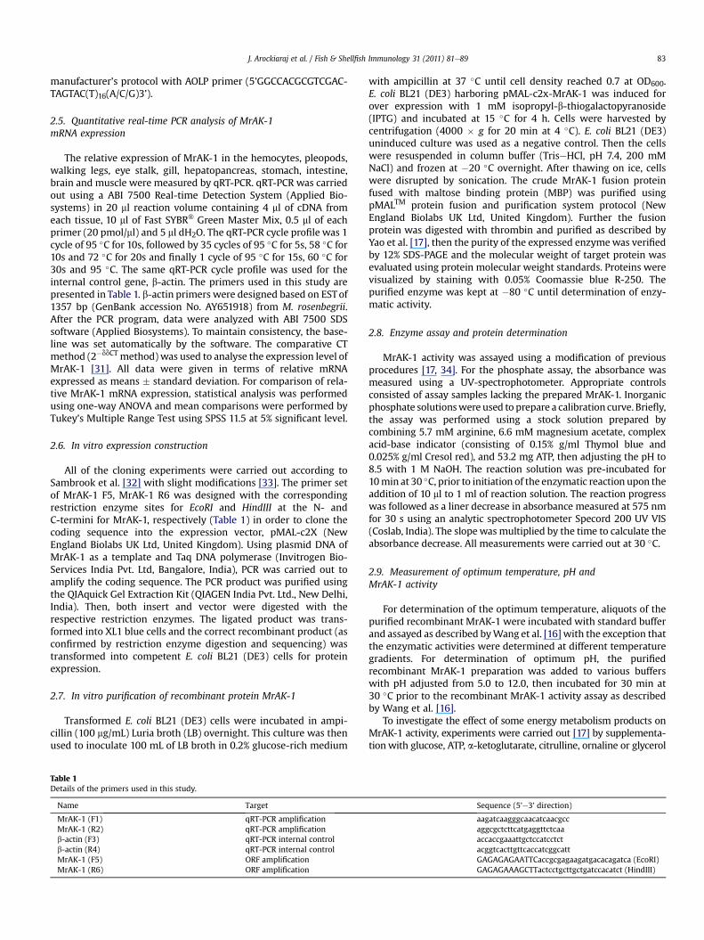
All of the cloning experiments were carried out according toSambrook et al. [32] with slight modifications [33]. The primer setof MrAK-1 F5, MrAK-1 R6 was designed with the correspondingrestriction enzyme sites for EcoRI and HindIII at the N- andC-termini for MrAK-1, respectively (Table 1) in order to clone thecoding sequence into the expression vector, pMAL-c2X (NewEngland Biolabs UK Ltd, United Kingdom). Using plasmid DNA ofMrAK-1 as a template and Taq DNA polymerase (Invitrogen Bio-Services India Pvt. Ltd, Bangalore, India), PCR was carried out toamplify the coding sequence. The PCR product was purified usingthe QIAquick Gel Extraction Kit (QIAGEN India Pvt. Ltd., New Delhi,India). Then, both insert and vector were digested with therespective restriction enzymes. The ligated product was trans-formed into XL1 blue cells and the correct recombinant product (asconfirmed by restriction enzyme digestion and sequencing) wastransformed into competent E. coli BL21 (DE3) cells for proteinexpression.
2.7. In vitro purification of recombinant protein MrAK-1
Transformed E. coli BL21 (DE3) cells were incubated in ampi-cillin (100 mg/mL) Luria broth (LB) overnight. This culture was thenused to inoculate 100 mL of LB broth in 0.2% glucose-rich medium
Table 1Details of the primers used in this study.
Name Target
MrAK-1 (F1) qRT-PCR amplificationMrAK-1 (R2) qRT-PCR amplificationb-actin (F3) qRT-PCR internal controlb-actin (R4) qRT-PCR internal controlMrAK-1 (F5) ORF amplificationMrAK-1 (R6) ORF amplification
with ampicillin at 37 �C until cell density reached 0.7 at OD600.E. coli BL21 (DE3) harboring pMAL-c2x-MrAK-1 was induced forover expression with 1 mM isopropyl-b-thiogalactopyranoside(IPTG) and incubated at 15 �C for 4 h. Cells were harvested bycentrifugation (4000 � g for 20 min at 4 �C). E. coli BL21 (DE3)uninduced culture was used as a negative control. Then the cellswere resuspended in column buffer (TriseHCl, pH 7.4, 200 mMNaCl) and frozen at �20 �C overnight. After thawing on ice, cellswere disrupted by sonication. The crude MrAK-1 fusion proteinfused with maltose binding protein (MBP) was purified usingpMALTM protein fusion and purification system protocol (NewEngland Biolabs UK Ltd, United Kingdom). Further the fusionprotein was digested with thrombin and purified as described byYao et al. [17], then the purity of the expressed enzymewas verifiedby 12% SDS-PAGE and the molecular weight of target protein wasevaluated using protein molecular weight standards. Proteins werevisualized by staining with 0.05% Coomassie blue R-250. Thepurified enzyme was kept at �80 �C until determination of enzy-matic activity.
2.8. Enzyme assay and protein determination
MrAK-1 activity was assayed using a modification of previousprocedures [17, 34]. For the phosphate assay, the absorbance wasmeasured using a UV-spectrophotometer. Appropriate controlsconsisted of assay samples lacking the prepared MrAK-1. Inorganicphosphate solutionswere used to prepare a calibration curve. Briefly,the assay was performed using a stock solution prepared bycombining 5.7 mM arginine, 6.6 mM magnesium acetate, complexacid-base indicator (consisting of 0.15% g/ml Thymol blue and0.025% g/ml Cresol red), and 53.2 mg ATP, then adjusting the pH to8.5 with 1 M NaOH. The reaction solution was pre-incubated for10min at 30 �C, prior to initiation of the enzymatic reaction upon theaddition of 10 ml to 1 ml of reaction solution. The reaction progresswas followed as a liner decrease in absorbance measured at 575 nmfor 30 s using an analytic spectrophotometer Specord 200 UV VIS(Coslab, India). The slopewas multiplied by the time to calculate theabsorbance decrease. All measurements were carried out at 30 �C.
2.9. Measurement of optimum temperature, pH andMrAK-1 activity
For determination of the optimum temperature, aliquots of thepurified recombinant MrAK-1 were incubated with standard bufferand assayed as described byWang et al. [16] with the exception thatthe enzymatic activities were determined at different temperaturegradients. For determination of optimum pH, the purifiedrecombinant MrAK-1 preparation was added to various bufferswith pH adjusted from 5.0 to 12.0, then incubated for 30 min at30 �C prior to the recombinant MrAK-1 activity assay as describedby Wang et al. [16].
To investigate the effect of some energy metabolism products onMrAK-1 activity, experiments were carried out [17] by supplementa-tion with glucose, ATP, a-ketoglutarate, citrulline, ornaline or glycerol
Sequence (5’e3’ direction)
aagatcaagggcaacatcaacgccaggcgctcttcatgaggttctcaaaccaccgaaattgctccatcctctacggtcacttgttcaccatcggcattGAGAGAGAATTCaccgcgagaagatgacacagatca (EcoRI)GAGAGAAAGCTTactcctgcttgctgatccacatct (HindIII)
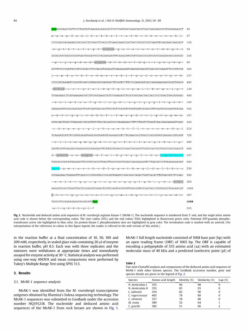
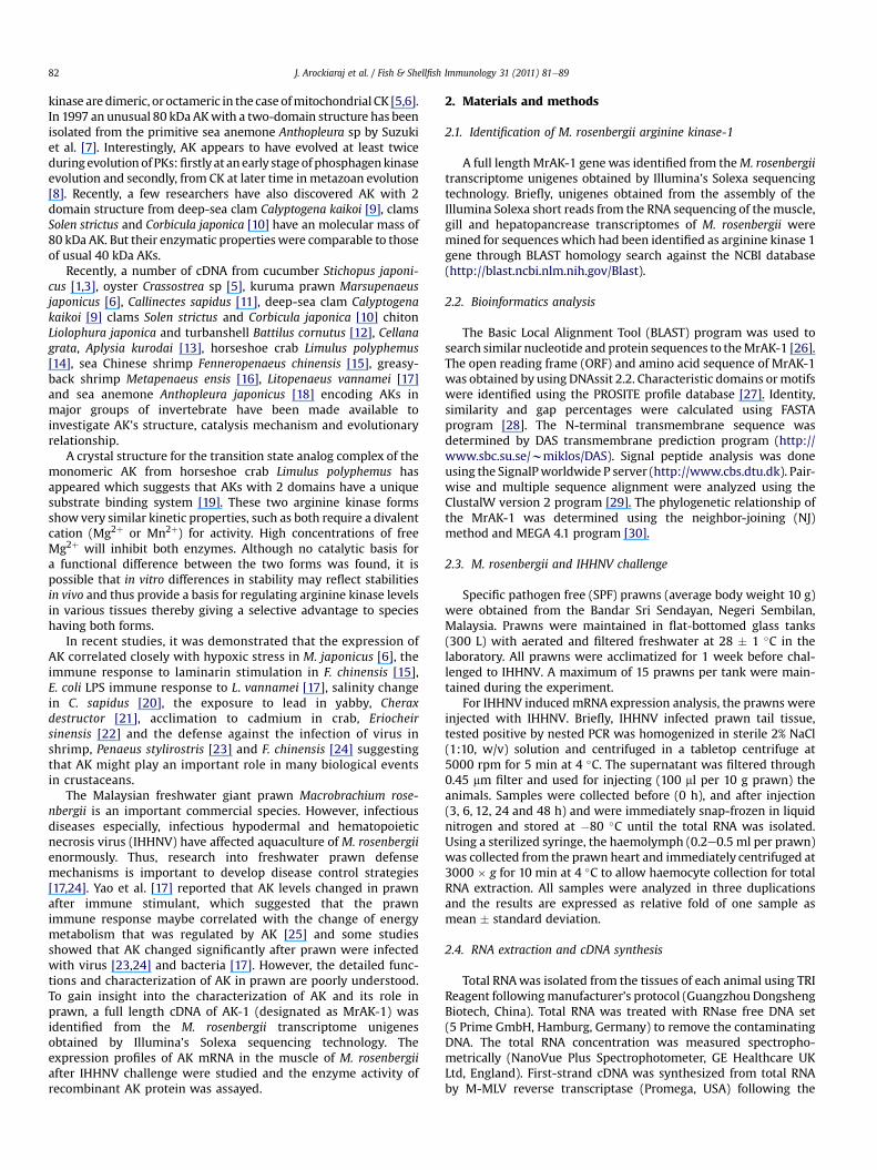
Fig. 1. Nucleotide and deduced amino acid sequences of M. rosenbergii arginine kinase-1 (MrAK-1). The nucleotide sequence is numbered from 50 end, and the single letter aminoacid code is shown below the corresponding codon. The start codon (ATG) and the end codon (TAA) highlighted in fluorescent green color. Potential ATP:guanido phospho-transferases active site highlighted in blue color. Six protein kinase C phosphorylation sites are highlighted in grey color. The termination code is marked with an asterisk. (Forinterpretation of the references to colour in this figure legend, the reader is referred to the web version of this article.)
Table 2Pair-wise ClustalWanalysis and comparisons of the deduced amino acid sequence ofMrAK-1 with other known species. The GenBank accession number, gene andspecies details are given in the legend of Fig. 2.
Species Amino acid length Identity (%) Similarity (%) Gap (%)
N. denticulata I 355 96 98 0N. denticulata II 355 85 93 0L. salmonis 354 82 90 0C. rogercresseyi 405 78 88 0C. clemensi 357 78 88 0M. ovata 389 52 64 1C. gracilis 382 51 66 2
J. Arockiaraj et al. / Fish & Shellfish Immunology 31 (2011) 81e8984
in the reaction buffer at a final concentration of 10, 50, 100 and200mM, respectively, in sealed glass vials containing 20 ml of enzymein reaction buffer, pH 8.5. Each was with three replicates and themixtures were withdrawn at appropriate times and immediatelyassayed for enzymeactivityat 30 �C. Statistical analysiswasperformedusing one-way ANOVA and mean comparisons were performed byTukey’s Multiple Range Test using SPSS 11.5.
3. Results
3.1. MrAK-1 sequence analysis
MrAK-1 was identified from the M. rosenbergii transcriptomeunigenes obtained by Illumina’s Solexa sequencing technology. TheMrAK-1 sequences was submitted to GenBank under the accessionnumber HQ191218. The nucleotide and deduced amino acidsequences of the MrAK-1 from rock bream are shown in Fig. 1.
MrAK-1 full length nucleotide consisted of 1068 base pair (bp) withan open reading frame (ORF) of 1065 bp. The ORF is capable ofencoding a polypeptide of 355 amino acid (aa) with an estimatedmolecular mass of 40 kDa and a predicted isoelectric point (pI) of
J. Arockiaraj et al. / Fish & Shellfish Immunology 31 (2011) 81e89 85
6.1. MrAK-1 aa sequence neither have a signal peptide nor a trans-membrane regions. The ExPASy motif analysis of MrAK-1 aasequence contains a potential ATP:guanido phosphotransferasesactive site (Cys263-Pro264-Thr265-Asn266-Leu267-Gly268-Thr269), 6protein kinase C phosphorylation sites and 6 caesin kinase IIphosphorylation sites (63s-y-k-d66; 89s-D-m-e92; 157t-g-m-d160;247s-g-k-d250; 304t-r-g-e307 and 327s-e-v-e330). A long ADP bindingsite is available in the aa sequence between 108 and 318. Anotherlong phosphagen binding site is available in the aa sequencebetween 51 and 262. Additionally a small substrate specificity loopis also available in the aa sequence between 300 and 310.
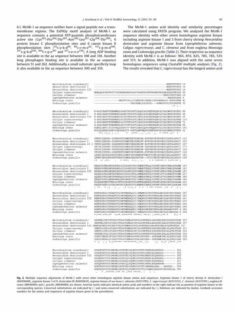
Fig. 2. Multiple sequence alignments of MrAK-1 with seven other homologous arginine(BAH56608), argininie kinase 3 of N. denticulata III (BAH56610), arginine kinase of sea louseovata (ABN49969) and C. gracilis (ABN49968) are shown. Asterisk marks indicates identicalcorresponding species. Conserved substitutions are indicated by (:) and semi-conserved sunumbers for the amino acid sequences of arginine kinase given in the parentheses.
The MrAK-1 amino acid identity and similarity percentageswere calculated using FASTA program. We analyzed the MrAK-1sequence identity with other seven homologous arginine kinaseincluding arginine kinase 1 and 3 from cherry shrimp Neocaridinadenticulate and argininie kinase from Lepeophtheirus salmonis,Caligus rogercresseyi, and C. clemensi and from euglena Monosigaovata and Codonosiga gracilis (Table 2). Their respective aa sequenceidentity with MrAK-1 is as follows: 96%, 85%, 82%, 78%, 78%, 52%and 51%. In addition, MrAK-1 was aligned with the same sevenhomologous sequences using ClustalW multiple analyses (Fig. 2).The results revealed that C. rogercresseyi has the longest amino acid
kinase amino acid sequences. Argininie kinase 1 of cherry shrimp N. denticulata IL. salmonis (ACO11785), C. rogercresseyi (ACO11152), C. clemensi (ACO15761), euglena M.amino acids and numbers to the right indicate the aa position of arginine kinase in thebstitutions are indicated by (.). Deletions are indicated by dashes. GenBank accession
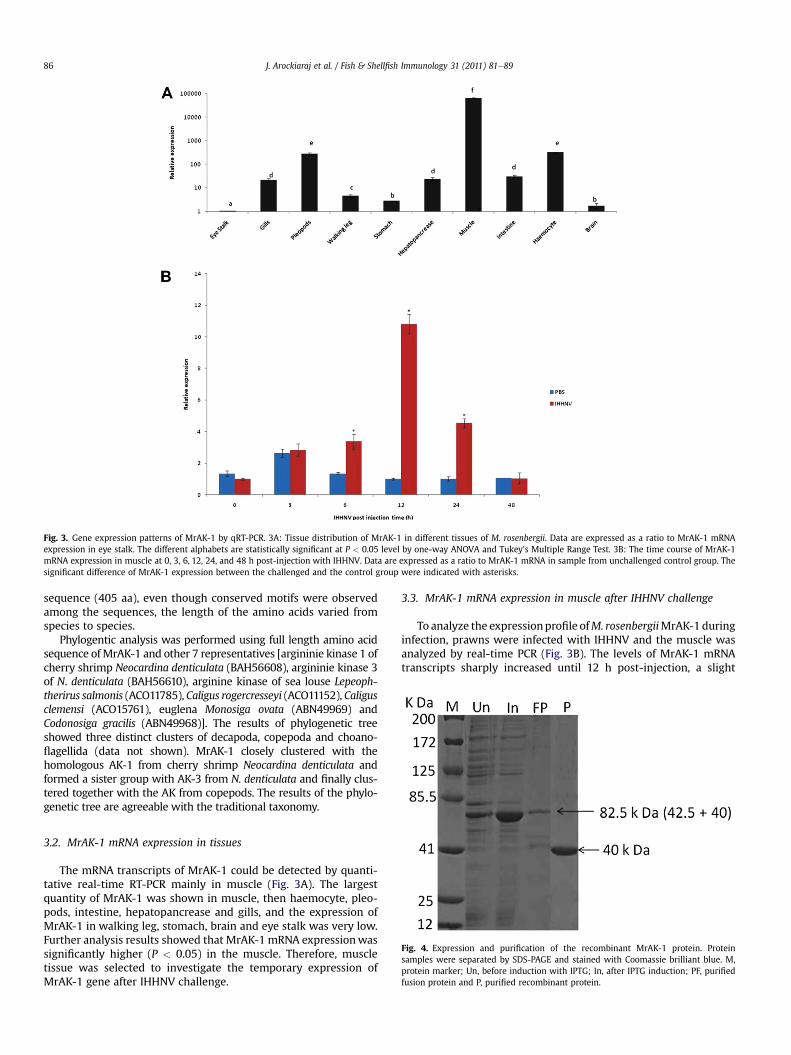
Fig. 3. Gene expression patterns of MrAK-1 by qRT-PCR. 3A: Tissue distribution of MrAK-1 in different tissues of M. rosenbergii. Data are expressed as a ratio to MrAK-1 mRNAexpression in eye stalk. The different alphabets are statistically significant at P < 0.05 level by one-way ANOVA and Tukey’s Multiple Range Test. 3B: The time course of MrAK-1mRNA expression in muscle at 0, 3, 6, 12, 24, and 48 h post-injection with IHHNV. Data are expressed as a ratio to MrAK-1 mRNA in sample from unchallenged control group. Thesignificant difference of MrAK-1 expression between the challenged and the control group were indicated with asterisks.
J. Arockiaraj et al. / Fish & Shellfish Immunology 31 (2011) 81e8986
sequence (405 aa), even though conserved motifs were observedamong the sequences, the length of the amino acids varied fromspecies to species.
Phylogentic analysis was performed using full length amino acidsequence ofMrAK-1 and other 7 representatives [argininie kinase 1 ofcherry shrimp Neocardina denticulata (BAH56608), argininie kinase 3of N. denticulata (BAH56610), arginine kinase of sea louse Lepeoph-therirus salmonis (ACO11785), Caligus rogercresseyi (ACO11152), Caligusclemensi (ACO15761), euglena Monosiga ovata (ABN49969) andCodonosiga gracilis (ABN49968)]. The results of phylogenetic treeshowed three distinct clusters of decapoda, copepoda and choano-flagellida (data not shown). MrAK-1 closely clustered with thehomologous AK-1 from cherry shrimp Neocardina denticulata andformed a sister group with AK-3 from N. denticulata and finally clus-tered together with the AK from copepods. The results of the phylo-genetic tree are agreeable with the traditional taxonomy.
Fig. 4. Expression and purification of the recombinant MrAK-1 protein. Proteinsamples were separated by SDS-PAGE and stained with Coomassie brilliant blue. M,protein marker; Un, before induction with IPTG; In, after IPTG induction; PF, purifiedfusion protein and P, purified recombinant protein.
3.2. MrAK-1 mRNA expression in tissues
The mRNA transcripts of MrAK-1 could be detected by quanti-tative real-time RT-PCR mainly in muscle (Fig. 3A). The largestquantity of MrAK-1 was shown in muscle, then haemocyte, pleo-pods, intestine, hepatopancrease and gills, and the expression ofMrAK-1 in walking leg, stomach, brain and eye stalk was very low.Further analysis results showed that MrAK-1 mRNA expressionwassignificantly higher (P < 0.05) in the muscle. Therefore, muscletissue was selected to investigate the temporary expression ofMrAK-1 gene after IHHNV challenge.
3.3. MrAK-1 mRNA expression in muscle after IHHNV challenge
To analyze the expressionprofile ofM. rosenbergiiMrAK-1duringinfection, prawns were infected with IHHNV and the muscle wasanalyzed by real-time PCR (Fig. 3B). The levels of MrAK-1 mRNAtranscripts sharply increased until 12 h post-injection, a slight
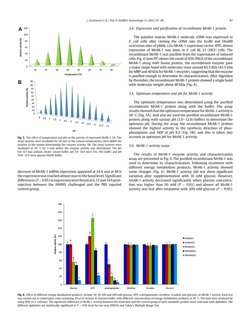
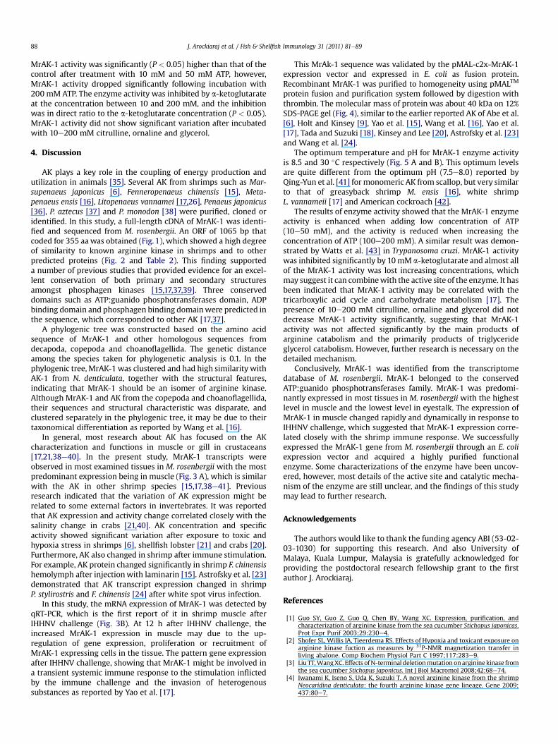
Fig. 5. The effect of temperature and pH on the activity of expressed MrAK-1. 5A: Theassay systems were incubated for 30 min in the various temperatures, then added theenzyme to the system determining the enzyme activity. 5B: The assay systems wereincubated at 30 �C for 5 min before the enzyme activity was determined. The pH6.0e6.5 was sodium citrateecitrate buffer, pH 7.0e10.0 were TriseHCl buffer and pH10.0e12.0 were glycine-NaOH buffer.
J. Arockiaraj et al. / Fish & Shellfish Immunology 31 (2011) 81e89 87
decrease of MrAK-1 mRNA expression appeared at 24 h and at 48 htheexpressionwas reachedalmostnear to thebasal level. Significantdifferences (P<0.05) in expressionwere foundat6,12and24hpost-injection between the IHHNV challenged and the PBS injectedcontrol group.
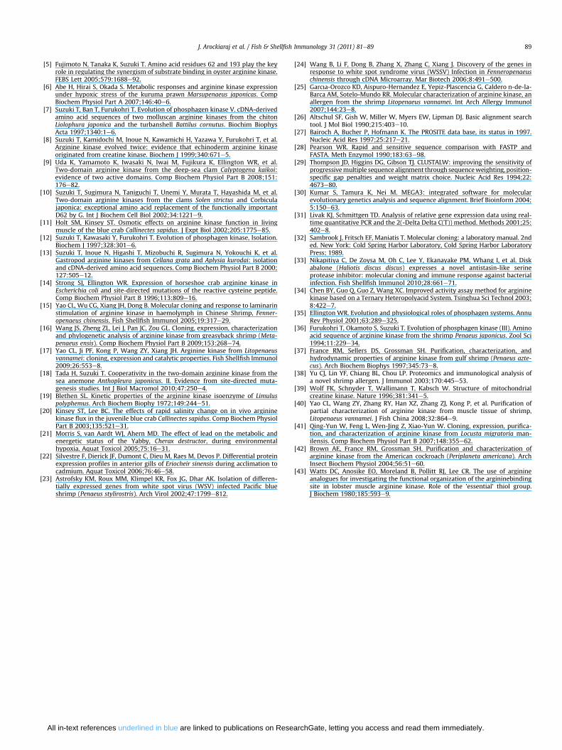
Fig. 6. Effect of different energy metabolism products, include 10, 50, 100 and 200 mM glucwas carried out in sealed glass vials containing 20 ml of enzyme in reaction buffer with diffeusing SPSS 11.5. software. The significant difference of MrAK-1 activity between the treatmedifferent alphabets are statistically significant at P < 0.05 level by one-way ANOVA and Tu
3.4. Expression and purification of recombinant MrAK-1 protein
The putative mature MrAK-1 molecule cDNA was expressed inE. coli cells after cloning the cDNA into the EcoRI and HindIIIrestriction sites of pMAL-c2x-MrAK-1 expression vector, IPTG drivenexpression of MrAK-1 was done in E. coli BL 21 (DE3) cells. Therecombinant MrAK-1 was purified from the supernatant of inducedcells. Fig. 4 (lane PF) shows the result of SDS-PAGEof the recombinantMrAK-1 along with fusion protein, the recombinant enzyme gavea major single band with molecular mass around 82.5 kDa (42.5 kDaforMBPand40kDa forMrAK-1 enzyme), suggesting that the enzymeis purified enough to determine its characterization. After digestionby thrombin, the recombinantMrAK-1 protein showed a single bandwith molecular weight about 40 kDa (Fig. 4).
3.5. Optimum temperature and pH for MrAK-1 activity
The optimum temperature was determined using the purifiedrecombinant MrAK-1 protein along with the buffer. The assayresults showed that the optimumtemperature forMrAK-1 activity is30 �C (Fig. 5A). And also we used the purified recombinant MrAK-1protein along with various pH (5.0e12.0) buffers to determine theoptimum pH. During the assay the recombinant MrAK-1 proteinshowed the highest activity in the synthesis direction of phos-phoarginine and ADP at pH 8.5 (Fig. 5B), and this is taken intoaccount as optimum pH for MrAK-1 activity.
3.6. MrAK-1 activity assay
The results of MrAK-1 enzyme activity and characterizationassay are presented in Fig. 6. The purified recombinant MrAK-1 wasused to determine its characterization. Following treatment withdifferent energy metabolism products, MrAK-1 activity showedsome changes (Fig. 6). MrAK-1 activity did not show significantvariation after supplementation with 10 mM glucose. However,mrAK-1 activity decreased significantly when glucose concentra-tion was higher than 50 mM (P < 0.05) and almost all MrAK-1activity was lost after treatment with 200 mM glucose (P < 0.05).
ose, ATP, a-ketoglutarate, citrulline, ornaline and glycerol, on MrAK-1 activity. Each testrent concentration of energy metabolism products at 30 �C. The data were analyzed bynt and the control group of each metabolic product were indicated with alphabets. Thekey’s Multiple Range Test.
J. Arockiaraj et al. / Fish & Shellfish Immunology 31 (2011) 81e8988
MrAK-1 activity was significantly (P < 0.05) higher than that of thecontrol after treatment with 10 mM and 50 mM ATP, however,MrAK-1 activity dropped significantly following incubation with200 mM ATP. The enzyme activity was inhibited by a-ketoglutarateat the concentration between 10 and 200 mM, and the inhibitionwas in direct ratio to the a-ketoglutarate concentration (P < 0.05).MrAK-1 activity did not show significant variation after incubatedwith 10e200 mM citrulline, ornaline and glycerol.
4. Discussion
AK plays a key role in the coupling of energy production andutilization in animals [35]. Several AK from shrimps such as Mar-supenaeus japonicus [6], Fenneropenaeus chinensis [15], Meta-penaeus ensis [16], Litopenaeus vannamei [17,26], Penaeus japonicus[36], P. aztecus [37] and P. monodon [38] were purified, cloned oridentified. In this study, a full-length cDNA of MrAK-1 was identi-fied and sequenced from M. rosenbergii. An ORF of 1065 bp thatcoded for 355 aa was obtained (Fig. 1), which showed a high degreeof similarity to known arginine kinase in shrimps and to otherpredicted proteins (Fig. 2 and Table 2). This finding supporteda number of previous studies that provided evidence for an excel-lent conservation of both primary and secondary structuresamongst phosphagen kinases [15,17,37,39]. Three conserveddomains such as ATP:guanido phosphotransferases domain, ADPbinding domain and phosphagen binding domainwere predicted inthe sequence, which corresponded to other AK [17,37].
A phylogenic tree was constructed based on the amino acidsequence of MrAK-1 and other homologous sequences fromdecapoda, copepoda and choanoflagellida. The genetic distanceamong the species taken for phylogenetic analysis is 0.1. In thephylogenic tree, MrAK-1 was clustered and had high similarity withAK-1 from N. denticulata, together with the structural features,indicating that MrAK-1 should be an isomer of arginine kinase.Although MrAK-1 and AK from the copepoda and choanoflagellida,their sequences and structural characteristic was disparate, andclustered separately in the phylogenic tree, it may be due to theirtaxonomical differentiation as reported by Wang et al. [16].
In general, most research about AK has focused on the AKcharacterization and functions in muscle or gill in crustaceans[17,21,38e40]. In the present study, MrAK-1 transcripts wereobserved in most examined tissues in M. rosenbergii with the mostpredominant expression being in muscle (Fig. 3 A), which is similarwith the AK in other shrimp species [15,17,38e41]. Previousresearch indicated that the variation of AK expression might berelated to some external factors in invertebrates. It was reportedthat AK expression and activity change correlated closely with thesalinity change in crabs [21,40]. AK concentration and specificactivity showed significant variation after exposure to toxic andhypoxia stress in shrimps [6], shellfish lobster [21] and crabs [20].Furthermore, AK also changed in shrimp after immune stimulation.For example, AK protein changed significantly in shrimp F. chinensishemolymph after injectionwith laminarin [15]. Astrofsky et al. [23]demonstrated that AK transcript expression changed in shrimpP. stylirostris and F. chinensis [24] after white spot virus infection.
In this study, the mRNA expression of MrAK-1 was detected byqRT-PCR, which is the first report of it in shrimp muscle afterIHHNV challenge (Fig. 3B). At 12 h after IHHNV challenge, theincreased MrAK-1 expression in muscle may due to the up-regulation of gene expression, proliferation or recruitment ofMrAK-1 expressing cells in the tissue. The pattern gene expressionafter IHHNV challenge, showing that MrAK-1 might be involved ina transient systemic immune response to the stimulation inflictedby the immune challenge and the invasion of heterogenoussubstances as reported by Yao et al. [17].
This MrAk-1 sequence was validated by the pMAL-c2x-MrAK-1expression vector and expressed in E. coli as fusion protein.Recombinant MrAK-1 was purified to homogeneity using pMALTM
protein fusion and purification system followed by digestion withthrombin. The molecular mass of protein was about 40 kDa on 12%SDS-PAGE gel (Fig. 4), similar to the earlier reported AK of Abe et al.[6], Holt and Kinsey [9], Yao et al. [15], Wang et al. [16], Yao et al.[17], Tada and Suzuki [18], Kinsey and Lee [20], Astrofsky et al. [23]and Wang et al. [24].
The optimum temperature and pH for MrAK-1 enzyme activityis 8.5 and 30 �C respectively (Fig. 5 A and B). This optimum levelsare quite different from the optimum pH (7.5e8.0) reported byQing-Yun et al. [41] for monomeric AK from scallop, but very similarto that of greasyback shrimp M. ensis [16], white shrimpL. vannameii [17] and American cockroach [42].
The results of enzyme activity showed that the MrAK-1 enzymeactivity is enhanced when adding low concentration of ATP(10e50 mM), and the activity is reduced when increasing theconcentration of ATP (100e200 mM). A similar result was demon-strated by Watts et al. [43] in Trypanosoma cruzi. MrAK-1 activitywas inhibited significantly by 10mM a-ketoglutarate and almost allof the MrAK-1 activity was lost increasing concentrations, whichmay suggest it can combinewith the active site of the enzyme. It hasbeen indicated that MrAK-1 activity may be correlated with thetricarboxylic acid cycle and carbohydrate metabolism [17]. Thepresence of 10e200 mM citrulline, ornaline and glycerol did notdecrease MrAK-1 activity significantly, suggesting that MrAK-1activity was not affected significantly by the main products ofarginine catabolism and the primarily products of triglycerideglycerol catabolism. However, further research is necessary on thedetailed mechanism.
Conclusively, MrAK-1 was identified from the transcriptomedatabase of M. rosenbergii. MrAK-1 belonged to the conservedATP:guanido phosphotransferases family. MrAK-1 was predomi-nantly expressed in most tissues in M. rosenbergii with the highestlevel in muscle and the lowest level in eyestalk. The expression ofMrAK-1 in muscle changed rapidly and dynamically in response toIHHNV challenge, which suggested that MrAK-1 expression corre-lated closely with the shrimp immune response. We successfullyexpressed the MrAK-1 gene from M. rosenbergii through an E. coliexpression vector and acquired a highly purified functionalenzyme. Some characterizations of the enzyme have been uncov-ered, however, most details of the active site and catalytic mecha-nism of the enzyme are still unclear, and the findings of this studymay lead to further research.
Acknowledgements
The authors would like to thank the funding agency ABI (53-02-03-1030) for supporting this research. And also University ofMalaya, Kuala Lumpur, Malaysia is gratefully acknowledged forproviding the postdoctoral research fellowship grant to the firstauthor J. Arockiaraj.
References
[1] Guo SY, Guo Z, Guo Q, Chen BY, Wang XC. Expression, purification, andcharacterization of arginine kinase from the sea cucumber Stichopus japonicas.Prot Expr Purif 2003;29:230e4.
[2] Shofer SL, Willis JA, Tjeerdema RS. Effects of Hypoxia and toxicant exposure onarginine kinase fuction as measures by 31P-NMR magnetization transfer inliving abalone. Comp Biochem Physiol Part C 1997;117:283e9.
[3] Liu TT,WangXC. Effects ofN-terminal deletionmutation onarginine kinase fromthe sea cucumber Stichopus japonicus. Int J Biol Macromol 2008;42:68e74.
[4] Iwanami K, Iseno S, Uda K, Suzuki T. A novel arginine kinase from the shrimpNeocaridina denticulata: the fourth arginine kinase gene lineage. Gene 2009;437:80e7.
J. Arockiaraj et al. / Fish & Shellfish Immunology 31 (2011) 81e89 89
[5] Fujimoto N, Tanaka K, Suzuki T. Amino acid residues 62 and 193 play the keyrole in regulating the synergism of substrate binding in oyster arginine kinase.FEBS Lett 2005;579:1688e92.
[6] Abe H, Hirai S, Okada S. Metabolic responses and arginine kinase expressionunder hypoxic stress of the kuruma prawn Marsupenaeus japonicas. CompBiochem Physiol Part A 2007;146:40e6.
[7] Suzuki T, Ban T, Furukohri T. Evolution of phosphagen kinase V. cDNA-derivedamino acid sequences of two molluscan arginine kinases from the chitonLiolophura japonica and the turbanshell Battilus cornutus. Biochim BiophysActa 1997;1340:1e6.
[8] Suzuki T, Kamidochi M, Inoue N, Kawamichi H, Yazawa Y, Furukohri T, et al.Arginine kinase evolved twice: evidence that echinoderm arginine kinaseoriginated from creatine kinase. Biochem J 1999;340:671e5.
[9] Uda K, Yamamoto K, Iwasaki N, Iwai M, Fujikura K, Ellington WR, et al.Two-domain arginine kinase from the deep-sea clam Calyptogena kaikoi:evidence of two active domains. Comp Biochem Physiol Part B 2008;151:176e82.
[10] Suzuki T, Sugimura N, Taniguchi T, Unemi Y, Murata T, Hayashida M, et al.Two-domain arginine kinases from the clams Solen strictus and Corbiculajaponica: exceptional amino acid replacement of the functionally importantD62 by G. Int J Biochem Cell Biol 2002;34:1221e9.
[11] Holt SM, Kinsey ST. Osmotic effects on arginine kinase function in livingmuscle of the blue crab Callinectes sapidus. J Expt Biol 2002;205:1775e85.
[12] Suzuki T, Kawasaki Y, Furukohri T. Evolution of phosphagen kinase, Isolation.Biochem J 1997;328:301e6.
[13] Suzuki T, Inoue N, Higashi T, Mizobuchi R, Sugimura N, Yokouchi K, et al.Gastropod arginine kinases from Cellana grata and Aplysia kurodai: isolationand cDNA-derived amino acid sequences. Comp Biochem Physiol Part B 2000;127:505e12.
[14] Strong SJ, Ellington WR. Expression of horseshoe crab arginine kinase inEscherichia coli and site-directed mutations of the reactive cysteine peptide.Comp Biochem Physiol Part B 1996;113:809e16.
[15] Yao CL, Wu CG, Xiang JH, Dong B. Molecular cloning and response to laminarinstimulation of arginine kinase in haemolymph in Chinese Shrimp, Fenner-openaeus chinensis. Fish Shellfish Immunol 2005;19:317e29.
[16] Wang JS, Zheng ZL, Lei J, Pan JC, Zou GL. Cloning, expression, characterizationand phylogenetic analysis of arginine kinase from greasyback shrimp (Meta-penaeus ensis). Comp Biochem Physiol Part B 2009;153:268e74.
[17] Yao CL, Ji PF, Kong P, Wang ZY, Xiang JH. Arginine kinase from Litopenaeusvannamei: cloning, expression and catalytic properties. Fish Shellfish Immunol2009;26:553e8.
[18] Tada H, Suzuki T. Cooperativity in the two-domain arginine kinase from thesea anemone Anthopleura japonicus. II. Evidence from site-directed muta-genesis studies. Int J Biol Macromol 2010;47:250e4.
[19] Blethen SL. Kinetic properties of the arginine kinase isoenzyme of Limuluspolyphemus. Arch Biochem Biophy 1972;149:244e51.
[20] Kinsey ST, Lee BC. The effects of rapid salinity change on in vivo argininekinase flux in the juvenile blue crab Callinectes sapidus. Comp Biochem PhysiolPart B 2003;135:521e31.
[21] Morris S, van Aardt WJ, Ahern MD. The effect of lead on the metabolic andenergetic status of the Yabby, Cherax destructor, during environmentalhypoxia. Aquat Toxicol 2005;75:16e31.
[22] Silvestre F, Dierick JF, Dumont C, Dieu M, Raes M, Devos P. Differential proteinexpression profiles in anterior gills of Eriocheir sinensis during acclimation tocadmium. Aquat Toxicol 2006;76:46e58.
[23] Astrofsky KM, Roux MM, Klimpel KR, Fox JG, Dhar AK. Isolation of differen-tially expressed genes from white spot virus (WSV) infected Pacific blueshrimp (Penaeus stylirostris). Arch Virol 2002;47:1799e812.
All in-text references underlined in blue are linked to publications on Rese
[24] Wang B, Li F, Dong B, Zhang X, Zhang C, Xiang J. Discovery of the genes inresponse to white spot syndrome virus (WSSV) Infection in Fenneropenaeuschinensis through cDNA Microarray. Mar Biotech 2006;8:491e500.
[25] Garcıa-Orozco KD, Aispuro-Hernandez E, Yepiz-Plascencia G, Caldero n-de-la-Barca AM, Sotelo-Mundo RR. Molecular characterization of arginine kinase, anallergen from the shrimp Litopenaeus vannamei. Int Arch Allergy Immunol2007;144:23e8.
[26] Altschul SF, Gish W, Miller W, Myers EW, Lipman DJ. Basic alignment searchtool. J Mol Biol 1990;215:403e10.
[27] Bairoch A, Bucher P, Hofmann K. The PROSITE data base, its status in 1997.Nucleic Acid Res 1997;25:217e21.
[28] Pearson WR. Rapid and sensitive sequence comparison with FASTP andFASTA. Meth Enzymol 1990;183:63e98.
[29] Thompson JD, Higgins DG, Gibson TJ. CLUSTALW: improving the sensitivity ofprogressivemultiple sequence alignment through sequenceweighting, position-specific gap penalties and weight matrix choice. Nucleic Acid Res 1994;22:4673e80.
[30] Kumar S, Tamura K, Nei M. MEGA3: integrated software for molecularevolutionary genetics analysis and sequence alignment. Brief Bioinform 2004;5:150e63.
[31] Livak KJ, Schmittgen TD. Analysis of relative gene expression data using real-time quantitative PCR and the 2(-Delta Delta C(T)) method. Methods 2001;25:402e8.
[32] Sambrook J, Fritsch EF, Maniatis T. Molecular cloning: a laboratory manual. 2nded. New York: Cold Spring Harbor Laboratory, Cold Spring Harbor LaboratoryPress; 1989.
[33] Nikapitiya C, De Zoysa M, Oh C, Lee Y, Ekanayake PM, Whang I, et al. Diskabalone (Haliotis discus discus) expresses a novel antistasin-like serineprotease inhibitor: molecular cloning and immune response against bacterialinfection. Fish Shellfish Immunol 2010;28:661e71.
[34] Chen BY, Guo Q, Guo Z, Wang XC. Improved activity assay method for argininekinase based on a Ternary Heteropolyacid System. Tsinghua Sci Technol 2003;8:422e7.
[35] Ellington WR. Evolution and physiological roles of phosphagen systems. AnnuRev Physiol 2001;63:289e325.
[36] Furukohri T, Okamoto S, Suzuki T. Evolution of phosphagen kinase (III). Aminoacid sequence of arginine kinase from the shrimp Penaeus japonicus. Zool Sci1994;11:229e34.
[37] France RM, Sellers DS, Grossman SH. Purification, characterization, andhydrodynamic properties of arginine kinase from gulf shrimp (Penaeus azte-cus). Arch Biochem Biophys 1997;345:73e8.
[38] Yu CJ, Lin YF, Chiang BL, Chou LP. Proteomics and immunological analysis ofa novel shrimp allergen. J Immunol 2003;170:445e53.
[39] Wolf FK, Schnyder T, Wallimann T, Kabsch W. Structure of mitochondrialcreatine kinase. Nature 1996;381:341e5.
[40] Yao CL, Wang ZY, Zhang RY, Han XZ, Zhang ZJ, Kong P, et al. Purification ofpartial characterization of arginine kinase from muscle tissue of shrimp,Litopenaeus vannamei. J Fish China 2008;32:864e9.
[41] Qing-Yun W, Feng L, Wen-Jing Z, Xiao-Yun W. Cloning, expression, purifica-tion, and characterization of arginine kinase from Locusta migratoria man-ilensis. Comp Biochem Physiol Part B 2007;148:355e62.
[42] Brown AE, France RM, Grossman SH. Purification and characterization ofarginine kinase from the American cockroach (Periplaneta americana). ArchInsect Biochem Physiol 2004;56:51e60.
[43] Watts DC, Anosike EO, Moreland B, Pollitt RJ, Lee CR. The use of arginineanalogues for investigating the functional organization of the argininebindingsite in lobster muscle arginine kinase. Role of the ’essential’ thiol group.J Biochem 1980;185:593e9.
archGate, letting you access and read them immediately.