Characterization and expression of the β- N-acetylhexosaminidase gene family of Tribolium castaneum
12
Insect Biochemistry and Molecular Biology Insect Biochemistry and Molecular Biology 38 (2008) 478–489 Characterization and expression of the b-N-acetylhexosaminidase gene family of Tribolium castaneum $ David G. Hogenkamp a,{ , Yasuyuki Arakane a , Karl J. Kramer b , Subbaratnam Muthukrishnan a , Richard W. Beeman b, a Department of Biochemistry, Kansas State University, 141 Chalmers Hall, Manhattan, KS 66506, USA b Grain Marketing and Production Research Center, ARS-USDA, 1515 College Avenue, Manhattan, KS 66502, USA Received 5 June 2007; received in revised form 22 July 2007; accepted 1 August 2007 Abstract Enzymes belonging to the b-N-acetylhexosaminidase family cleave chitin oligosaccharides produced by the action of chitinases on chitin into the constituent N-acetylglucosamine monomer. Four genes encoding putative chitooligosaccharidolytic b-N-acetylhex- osaminidases (hereafter referred to as N-acetylglucosaminidases (NAGs)) in the red flour beetle, Tribolium castaneum, namely TcNAG1, TcFDL, TcNAG2, and TcNAG3, and three other related hexosaminidases were identified by searching the recently completed genome [Tribolium Genome Sequencing Consortium, 2007. The first genome sequence of a beetle, Tribolium castaneum, a model for insect development and pest biology. Nature, submitted for publication]. Full-length cDNAs for all four NAGs were cloned and sequenced, and the exon–intron organization of the corresponding genes was determined. Analyses of their developmental expression patterns indicated that, although all four of the NAGs are transcribed during most developmental stages, each gene had a distinct spatial and temporal expression pattern. TcNAG1 transcripts are the most abundant, particularly at the late pupal stage, while TcNAG3 transcripts are the least abundant, even at their peak levels in the late larval stages. The function of each NAG during different developmental stages was assessed by observations of lethal phenotypes after gene-specific double-stranded RNA (dsRNA)-mediated transcript depletion as verified by real-time PCR. TcNAG1 dsRNA was most effective in interrupting all three types of molts: larval–larval, larval–pupal, and pupal–adult. Treated insects died after failing to completely shed their old cuticles. Knockdown of transcripts for the other three NAG genes resulted in phenotypes similar to those of TcNAG1 dsRNA-treated insects, but the effects were somewhat variable and less severe. Sequence comparisons with other enzymatically characterized insect homologs suggested that TcFDL, unlike the other NAGs, may have a role in N-glycan processing in addition to its apparent role in cuticular chitin turnover. These results support the hypothesis that TcNAGs participate in chitin turnover and/or N-glycan processing during insect development and that each NAG fulfills an essential and distinct function. Published by Elsevier Ltd. Keywords: b-N-Acetylhexosaminidase; Glycoside hydrolase; Chitin; Tribolium castaneum; Red flour beetle; Hexosaminidase; Chitooligosaccharide; RNA interference; Molting; Cuticle 1. Introduction The two enzymes that are responsible for the degrada- tion of chitin in insects (Kramer and Muthukrishnan, 2005) are chitinases (CHTs, EC 3.2.1.14) and b-N-acetylhexosa- minidases (EC 3.2.1.52). The latter enzymes have been traditionally referred to as N-acetylglucosaminidases (NAGs) even though many of these enzymes act on other substrates such as oligo N-acetylgalactosamines (Nagamatsu et al., 1995). CHTs belong to glycoside ARTICLE IN PRESS www.elsevier.com/locate/ibmb 0965-1748/$ - see front matter Published by Elsevier Ltd. doi:10.1016/j.ibmb.2007.08.002 Abbrevations: NAG, b-N-acetylglucosaminidase; CHT, chitinase; dsRNA, double-stranded RNA; GlcNAc, N-acetylglucosamine; HEX, hexosaminidase; RNAi, RNA interference; Tc, Tribolium castaneum; UPMGA, unweighted pair group method with arithmetic mean; RACE, rapid amplification of cDNA ends; TMS, trans-membrane segment. $ The editing of this paper was completed after Dr. David Hogen- kamp’s premature death from cancer on January 29, 2007. Working with David was a wonderful experience for all of us. This paper is dedicated to the memory of his life and work. Corresponding author. Tel.: +1 785 776 2710; fax: +1 785 537 5584. E-mail address: [email protected] (R.W. Beeman). { Deceased.
-
Upload
independent -
Category
Documents
-
view
0 -
download
0
Transcript of Characterization and expression of the β- N-acetylhexosaminidase gene family of Tribolium castaneum
ARTICLE IN PRESS
InsectBiochemistry
andMolecularBiology
0965-1748/$ - se
doi:10.1016/j.ib
Abbrevations
dsRNA, double
hexosaminidase
UPMGA, unwe
rapid amplifica$The editin
kamp’s prematu
David was a wo
the memory of�CorrespondE-mail addr
{Deceased.
Insect Biochemistry and Molecular Biology 38 (2008) 478–489
www.elsevier.com/locate/ibmb
Characterization and expression of the b-N-acetylhexosaminidase genefamily of Tribolium castaneum$
David G. Hogenkampa,{, Yasuyuki Arakanea, Karl J. Kramerb,Subbaratnam Muthukrishnana, Richard W. Beemanb,�
aDepartment of Biochemistry, Kansas State University, 141 Chalmers Hall, Manhattan, KS 66506, USAbGrain Marketing and Production Research Center, ARS-USDA, 1515 College Avenue, Manhattan, KS 66502, USA
Received 5 June 2007; received in revised form 22 July 2007; accepted 1 August 2007
Abstract
Enzymes belonging to the b-N-acetylhexosaminidase family cleave chitin oligosaccharides produced by the action of chitinases on
chitin into the constituent N-acetylglucosamine monomer. Four genes encoding putative chitooligosaccharidolytic b-N-acetylhex-
osaminidases (hereafter referred to as N-acetylglucosaminidases (NAGs)) in the red flour beetle, Tribolium castaneum, namely TcNAG1,
TcFDL, TcNAG2, and TcNAG3, and three other related hexosaminidases were identified by searching the recently completed genome
[Tribolium Genome Sequencing Consortium, 2007. The first genome sequence of a beetle, Tribolium castaneum, a model for insect
development and pest biology. Nature, submitted for publication]. Full-length cDNAs for all four NAGs were cloned and sequenced,
and the exon–intron organization of the corresponding genes was determined. Analyses of their developmental expression patterns
indicated that, although all four of the NAGs are transcribed during most developmental stages, each gene had a distinct spatial and
temporal expression pattern. TcNAG1 transcripts are the most abundant, particularly at the late pupal stage, while TcNAG3 transcripts
are the least abundant, even at their peak levels in the late larval stages. The function of each NAG during different developmental stages
was assessed by observations of lethal phenotypes after gene-specific double-stranded RNA (dsRNA)-mediated transcript depletion as
verified by real-time PCR. TcNAG1 dsRNA was most effective in interrupting all three types of molts: larval–larval, larval–pupal, and
pupal–adult. Treated insects died after failing to completely shed their old cuticles. Knockdown of transcripts for the other three NAG
genes resulted in phenotypes similar to those of TcNAG1 dsRNA-treated insects, but the effects were somewhat variable and less severe.
Sequence comparisons with other enzymatically characterized insect homologs suggested that TcFDL, unlike the other NAGs, may have
a role in N-glycan processing in addition to its apparent role in cuticular chitin turnover. These results support the hypothesis that
TcNAGs participate in chitin turnover and/or N-glycan processing during insect development and that each NAG fulfills an essential
and distinct function.
Published by Elsevier Ltd.
Keywords: b-N-Acetylhexosaminidase; Glycoside hydrolase; Chitin; Tribolium castaneum; Red flour beetle; Hexosaminidase; Chitooligosaccharide; RNA
interference; Molting; Cuticle
e front matter Published by Elsevier Ltd.
mb.2007.08.002
: NAG, b-N-acetylglucosaminidase; CHT, chitinase;
-stranded RNA; GlcNAc, N-acetylglucosamine; HEX,
; RNAi, RNA interference; Tc, Tribolium castaneum;
ighted pair group method with arithmetic mean; RACE,
tion of cDNA ends; TMS, trans-membrane segment.
g of this paper was completed after Dr. David Hogen-
re death from cancer on January 29, 2007. Working with
nderful experience for all of us. This paper is dedicated to
his life and work.
ing author. Tel.: +1785 776 2710; fax: +1 785 537 5584.
ess: [email protected] (R.W. Beeman).
1. Introduction
The two enzymes that are responsible for the degrada-tion of chitin in insects (Kramer and Muthukrishnan, 2005)are chitinases (CHTs, EC 3.2.1.14) and b-N-acetylhexosa-minidases (EC 3.2.1.52). The latter enzymes have beentraditionally referred to as N-acetylglucosaminidases(NAGs) even though many of these enzymes act onother substrates such as oligo N-acetylgalactosamines(Nagamatsu et al., 1995). CHTs belong to glycoside
ARTICLE IN PRESSD.G. Hogenkamp et al. / Insect Biochemistry and Molecular Biology 38 (2008) 478–489 479
hydrolase family 18 (GH18), whereas NAGs belong to theglycoside hydrolase family 20 (GH20) (Coutinho andHenrissat, 1999). CHTs catalyze the endohydrolysis ofchitin at random positions within the chitin polymer(Fukamizo and Kramer, 1985a, b). In contrast, NAGscatalyze the specific exohydrolysis of chitooligosaccharidesfrom the non-reducing end, generating monomers ofN-acetylglucosamine (GlcNAc; Sahai and Manocha, 1993).
Thus, insect chitin hydrolysis is carried out by a two-enzyme system composed of CHT and NAG present in themolting fluid (Fukamizo and Kramer, 1985a, b). Thepresence of these two enzymes together results in asynergistic effect, such that the rate of chitin hydrolysis isup to six times higher than the sum of the rates observedwith either enzyme alone. This enhancement of catalyticactivity by the binary chitinolytic enzyme system isdependent on the concentration ratio of CHT to NAG.The regulation of both enzymes is under strict hormonaland/or developmental control (Kramer and Muthukrish-nan, 2005). Disruption of the metabolic flux of theseenzymes could have severe detrimental effects on insectmolting and could provide a basis for the design of selectivebiopesticides.
CHT is unable to convert its chitin substrate completelyto GlcNAc monomers. Therefore, NAG is the enzymeprimarily responsible for the production of the monomerfrom chitooligosaccharides for recycling. Kinetic studieswith Manduca sexta (tobacco hornworm) CHT haverevealed that this enzyme is subject to substrate inhibitionwhen chitooligosaccharides are utilized as substrates(Koga et al., 1982, 1983; Arakane et al., 2003). Therefore,one of the potential functions of NAGs may be toprevent the accumulation of chitooligosaccharides atconcentrations that are high enough to interfere withefficient degradation of chitin by CHT (Kramer andMuthukrishnan, 2005).
Several model insect species have been utilized for thestudy of NAGs. A NAG gene from M. sexta was found tobe expressed primarily in both epidermal and midguttissues (Zen et al., 1996). The level of expression in thesetissues was highest 6–7 days after the molt to the fifthinstar. This enzyme probably functions in the degradationof chitin, with its expression being under control of thehormone 20-hydroxyecdysone. Other insect NAGs havebeen purified from a second lepidopteran species, thesilkworm, Bombyx mori (Nagamatsu et al., 1995), and adipteran species, the yellow fever mosquito, Aedes aegypti
(Filho and Shapiro, 2004). A NAG from the latter waspresent in the gut of unfed adults, but NAG activityincreased rapidly upon feeding on an artificial protein-freediet or blood. There are multiple NAGs in at least some ofthese species and, according to our knowledge, a compre-hensive study of all of the genes encoding NAG-likeproteins present in a single insect species has never beenconducted.
In this paper, we have identified four genes encodingputative NAGs and also three related b-N-acetylhexosa-
minidases from the red flour beetle, Tribolium castaneum.To begin to study the function of each NAG gene in thisinsect, we analyzed their expression patterns throughoutdevelopment in the midgut, carcass and whole insect. Wealso used RNA interference (RNAi) for selective depletionof each NAG transcript to determine the effect of NAG
suppression on development. The expression studies, aswell as the phenotypes resulting from the knockdown ofeach NAG transcript, suggest a distinct role for each ofthese enzymes during development.
2. Materials and methods
2.1. Insect cultures
The GA-1 strain (Haliscak and Beeman, 1983) ofT. castaneum was used in all experiments. Insects werereared at 30 1C under standard conditions as describedpreviously (Beeman and Stuart, 1990).
2.2. Identification of Tribolium NAGs
The amino acid sequence of a previously characterizedNAG (Zen et al., 1996) from M. sexta was used to identifypotential NAG genes in the Tribolium genome database(http://www.bioinformatics.ksu.edu/BeetleBase/) by usingthe tblastn program. Following the identification andcharacterization of the four T. castaneum NAGs, additionaltblastn searches were done with each of these sequences toensure that all of the closely related genes in the Tribolium
genome have been identified.
2.3. Cloning of T. castaneum NAGs
The RNeasys Protect Mini Kit (Qiagen) was used toisolate total RNA from Tribolium tissue samples accordingto the manufacturer’s instructions. Total RNA sampleswere treated with RNase-free DNase I for 30min at 37 1Cto remove contaminating genomic DNA. The Super-ScriptTM III First-Strand Synthesis System for RT-PCR(Invitrogen) was used to synthesize the first-strand cDNAaccording to the manufacturer’s instructions. For PCRamplification of partial cDNAs of T. castaneum NAGs
identified by ‘‘blast’’ results from the Tribolium genomedatabase, pairs of gene-specific primers were designed fromconserved regions of insect NAGs. Then, 30- and 50-RACEexperiments were done to obtain the full-length cDNA foreach TcNAG. DNA sequencing was conducted at the DNAsequencing facility at Kansas State University. The genesequences have been deposited in GenBank with accessionnumbers; TcNAG1: EF592536, TcNAG2: EF592537,TcNAG3: EF592538 and TcFDL: EF592539.
2.4. Phylogenetic analysis of NAGs
ClustalW software (www.ebi.ac.uk/clustalw/) was used toperform multiple sequence alignments prior to phylogenetic
ARTICLE IN PRESSD.G. Hogenkamp et al. / Insect Biochemistry and Molecular Biology 38 (2008) 478–489480
analysis. The MEGA 3.0 program (Kumar et al., 2004) wasused to construct the consensus phylogenetic tree usingthe unweighted pair group method with arithmetic mean(UPGMA). To evaluate the branch strength of thephylogenetic tree, bootstrap analysis of 5000 replicationswas performed.
2.5. DNA and protein sequence analyses
Protein sequence analysis tools used in this study,including translation, molecular weight, pI, and topologypredictions, were obtained from the ExPASy Proteomicswebsite (http://us.expasy.org/). TMHMM (v2.0) software(www.cbs.dtu.dk/services/TMHMM-2.0/) was used to pre-dict transmembrane segments (TMS) in the conceptualtranslation products. Signal peptide and signal anchorpredictions were conducted using the SignalP 3.0 server(www.cbs.dtu.dk/services/SignalP/). Sequence alignmentsof multiple proteins were carried out using the ClustalWprogram (http://npsa-pbil.ibcp.fr/cgi-bin/npsa_automat.pl?page=npsa_clustalw.html).
2.6. Real-time RT-PCR analysis of TcNAG expression
Analysis of the expression of all four T. castaneum NAGs
along with the internal control, TcRpS6 (encodingribosomal protein S6), was carried out using real-timeRT-PCR. Beacon Designer 2.0 software (Premier BiosoftInternational) was used to design all gene-specific primersfor quantitative PCR analysis.
Each of the four T. castaneum NAGs was clonedinto the pCRs4-TOPOs vector (Invitrogen). The concen-tration of each plasmid construct was determined withthe DNA intercalating dye Hoechst 33258 using aVersaFluorTM Fluorimeter (Bio-Rad). Sets of serialdilutions were then made from each construct and thedilutions were analyzed by real-time RT-PCR using thenucleic acid stain SYBR green (Bio-Rad) and the Bio-RadiCycler iQs real-time RT-PCR detection system at theKansas State University Gene Expression Facility. At theend of each quantitative PCR experiment, a melt curve wasgenerated to rule out the possibility of primer–dimerformation. After constructing standard curves for eachgene, the transcript copy numbers were determined alongwith the copy number of the internal housekeeping gene,TcRpS6.
2.7. Double-stranded RNA synthesis
The most dissimilar regions of the T. castaneum NAG
genes were chosen for design of double-stranded RNA(dsRNA) to minimize the likelihood of silencing otherNAGs that share similar nucleotide sequences. dsRNAswere prepared as described in Arakane et al. (2005). Thenucleotide ranges and lengths of dsRNA target regions foreach TcNAG are as follows: TcNAG1: 1148–1412 (264 bp),TcFDL: 1298–1557 (258 bp), TcNAG2: 1238–1505 (267 bp),
and TcNAG3: 1143–1363 (219 bp). Coordinates referto GenBank sequences (see Section 2.3 for GenBankaccession numbers). All primers contained linkers corre-sponding to the T7 RNA polymerase recognition (T7promoter) sequence at the 50-end. Primers for TcNAG1,TcFDL, TcNAG2, and TcNAG3 are 50-(T7)-GAACTC-CACTCCCAGC and 50-(T7)-ATTATCGAGCAAAT-TTC, 50-(T7)-GGCCCAACACTTGCAA and 50-(T7)-GTAGCCGTCGCTGATG, 50-(T7)-GAATGCAACGC-CTGAA and 50-(T7)-TAATTCTAGGAGCAAG and50-(T7)-GCAAGATACCAAAAG, and 50-(T7)-GTGCG-ATAGGACACTG, respectively. PCR amplification pro-ducts were excised from a 1.5% agarose gel, extracted withphenol/chloroform/isoamyl alcohol (25:24:1, v/v/v), etha-nol precipitated in 0.3M sodium acetate overnight, andredissolved in DEPC-treated water. The resulting dsDNAswith flanking T7 promoter sequences were then used astemplates for the in vitro transcription of the dsRNAs.RNAs were synthesized using the AmpliScribe T7-FlashTranscription Kit (Epicentre Biotechnologies, Madison,WI) according to the manufacturer’s instructions andannealed to obtain dsRNA.
2.8. Injection of dsRNAs into Tribolium
dsRNAs corresponding to each of the four NAGs wereinjected into penultimate-instar larvae, last-instar larvaeand pharate pupae of T. castaneum according to estab-lished protocols (Tomoyasu and Denell, 2004). Approxi-mately 0.2 mg of dsRNA (�0.2 ml of 1 mg/ml dsRNA) wasinjected into the dorsal side of the first or secondabdominal segment of each animal (n ¼ 20). ThedsRNA-treated animals were incubated at 30 1C until theappropriate stage or time for analysis.
3. Results
3.1. Identification of Tribolium NAGs
To identify possible NAGs and related genes in the T.
castaneum genome, the amino acid sequence from apreviously characterized NAG (glycosylhydrolase family,GH20) from M. sexta (Zen et al., 1996) was used to searchthe Tribolium genome database using the tblastn program.A total of four genes, TcNAG1, TcFDL, TcNAG2, andTcNAG3, were identified encoding proteins with highsequence similarity (51–59%) to M. sexta NAG(Table 1). These predicted proteins had a much lower(26–28%) sequence similarity to the well-characterizedhuman hexosaminidase B (Hex B), another GH20 familyenzyme with a role in GM2 ganglioside metabolism(Sandhoff et al., 1968). A search of the Tribolium genomedatabase with the Hex B sequence as query revealed threeadditional genes encoding proteins with 450% similarityto Hex B. These three genes were, therefore, annotated ashexosaminidase genes and denoted as TcHEX1, TcHEX2,and TcHEX3.
ARTICLE IN PRESS
Table 1
Sequence similarity of NAG proteins from Tribolium and Manduca
TcNAG1 TcNAG2 TcNAG3 TcFDL MsNAG HsHEXB
TcNAG1 36.8 38.1 34.2 52.1 26.5
TcNAG2 (55.4) 34.4 38.5 37.8 28.1
TcNAG3 (50.2) (51.1) 42.5 33.4 26.4
TcFDL (51.5) (55.5) (59.3) 39.0 26.9
MsNAG (69.7) (56.1) (50.0) (54.7) 27.8
HsHEXB (40.3) (40.8) (39.9) (38.7) (39.7)
Values are percentage identities (similarities) at the amino acid level.
HsHEXB is included as an outlier for comparison.
Table 2
Summary of properties of NAG proteins from Tribolium
Protein Linkage
group
Length
(residues)
MW
(kDa)
pI Signal
peptide
Signal
anchor
TcNAG1 7 598 67.3 5.2 Yes No
TcNAG2 1 593 67.4 5.5 Yes No
TcNAG3 2 582 66.5 5.6 Yes No
TcFDL 7 630 72.6 6.6 No Yes
TcNAG1
TcNAG2
TcNAG3
TcFDL
1 kb
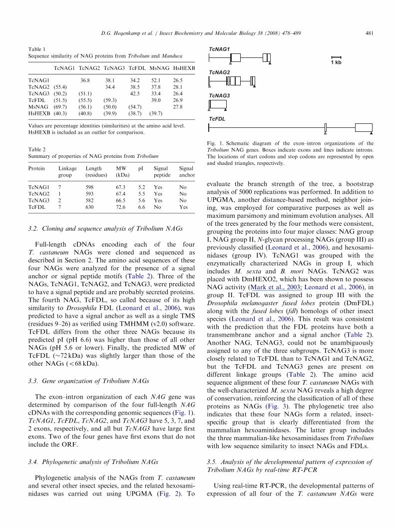
Fig. 1. Schematic diagram of the exon–intron organizations of the
Tribolium NAG genes. Boxes indicate exons and lines indicate introns.
The locations of start codons and stop codons are represented by open
and shaded triangles, respectively.
D.G. Hogenkamp et al. / Insect Biochemistry and Molecular Biology 38 (2008) 478–489 481
3.2. Cloning and sequence analysis of Tribolium NAGs
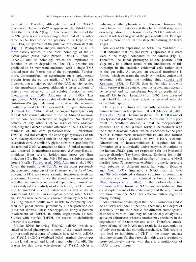
Full-length cDNAs encoding each of the fourT. castaneum NAGs were cloned and sequenced asdescribed in Section 2. The amino acid sequences of thesefour NAGs were analyzed for the presence of a signalanchor or signal peptide motifs (Table 2). Three of theNAGs, TcNAG1, TcNAG2, and TcNAG3, were predictedto have a signal peptide and are probably secreted proteins.The fourth NAG, TcFDL, so called because of its highsimilarity to Drosophila FDL (Leonard et al., 2006), waspredicted to have a signal anchor as well as a single TMS(residues 9–26) as verified using TMHMM (v2.0) software.TcFDL differs from the other three NAGs because itspredicted pI (pH 6.6) was higher than those of all otherNAGs (pH 5.6 or lower). Finally, the predicted MW ofTcFDL (�72 kDa) was slightly larger than those of theother NAGs (o68 kDa).
3.3. Gene organization of Tribolium NAGs
The exon–intron organization of each NAG gene wasdetermined by comparison of the four full-length NAG
cDNAs with the corresponding genomic sequences (Fig. 1).TcNAG1, TcFDL, TcNAG2, and TcNAG3 have 5, 3, 7, and2 exons, respectively, and all but TcNAG3 have large firstexons. Two of the four genes have first exons that do notinclude the ORF.
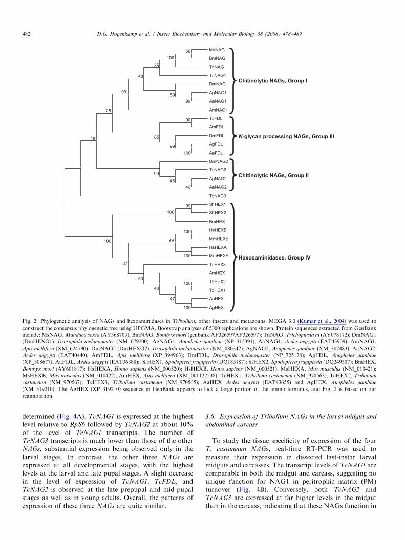
3.4. Phylogenetic analysis of Tribolium NAGs
Phylogenetic analysis of the NAGs from T. castaneum
and several other insect species, and the related hexosami-nidases was carried out using UPGMA (Fig. 2). To
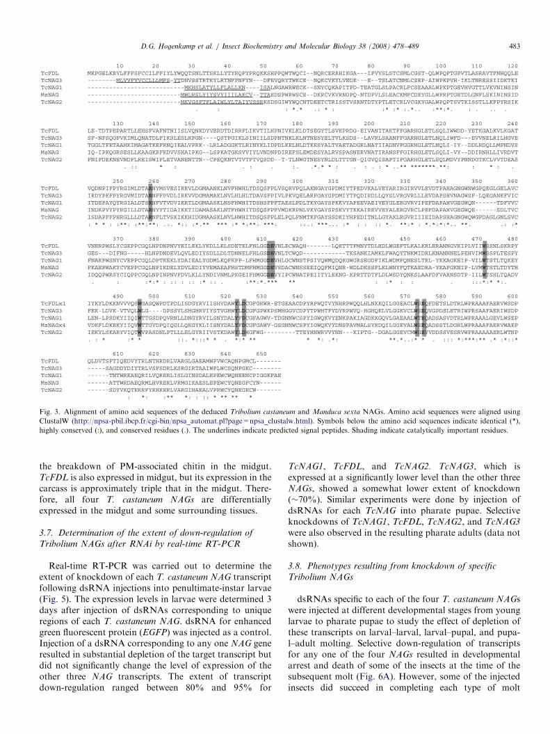
evaluate the branch strength of the tree, a bootstrapanalysis of 5000 replications was performed. In addition toUPGMA, another distance-based method, neighbor join-ing, was employed for comparative purposes as well asmaximum parsimony and minimum evolution analyses. Allof the trees generated by the four methods were consistent,grouping the proteins into four major classes: NAG groupI, NAG group II, N-glycan processing NAGs (group III) aspreviously classified (Leonard et al., 2006), and hexosami-nidases (group IV). TcNAG1 was grouped with theenzymatically characterized NAGs in group I, whichincludes M. sexta and B. mori NAGs. TcNAG2 wasplaced with DmHEXO2, which has been shown to possessNAG activity (Mark et al., 2003; Leonard et al., 2006), ingroup II. TcFDL was assigned to group III with theDrosophila melanogaster fused lobes protein (DmFDL)along with the fused lobes (fdl) homologs of other insectspecies (Leonard et al., 2006). This result was consistentwith the prediction that the FDL proteins have both atransmembrane anchor and a signal anchor (Table 2).Another NAG, TcNAG3, could not be unambiguouslyassigned to any of the three subgroups. TcNAG3 is moreclosely related to TcFDL than to TcNAG1 and TcNAG2,but the TcFDL and TcNAG3 genes are present ondifferent linkage groups (Table 2). The amino acidsequence alignment of these four T. castaneum NAGs withthe well-characterized M. sexta NAG reveals a high degreeof conservation, reinforcing the classification of all of theseproteins as NAGs (Fig. 3). The phylogenetic tree alsoindicates that these four NAGs form a related, insect-specific group that is clearly differentiated from themammalian hexoaminidases. The latter group includesthe three mammalian-like hexosaminidases from Tribolium
with low sequence similarity to insect NAGs and FDLs.
3.5. Analysis of the developmental pattern of expression of
Tribolium NAGs by real-time RT-PCR
Using real-time RT-PCR, the developmental patterns ofexpression of all four of the T. castaneum NAGs were
ARTICLE IN PRESS
Chitinolytic NAGs, Group I
Chitinolytic NAGs, Group II
N-glycan processing NAGs, Group III
Hexosaminidases, Group IV
MsNAG
BmNAG
TnNAG
TcNAG1
DmNAG
AgNAG1
AaNAG1
AmNAG1
TcFDL
AmFDL
DmFDL
AgFDL
AaFDL
DmNAG2
TcNAG2
AgNAG2
AaNAG2
TcNAG3
Sf HEX1
Sf HEX2
BmHEX
HsHEXB
MmHEXB
HsHEXA
MmHEXA
TcHEX3
AmHEX
TcHEX2
TcHEX1
AaHEX
AgHEX
100
100
56
100
100
100
100
99
99
99
99
89
100
90
35
47
48
46
43
99
46
50
99
87
85
100
28
48
Fig. 2. Phylogenetic analysis of NAGs and hexoaminidases in Tribolium, other insects and metazoans. MEGA 3.0 (Kumar et al., 2004) was used to
construct the consensus phylogenetic tree using UPGMA. Bootstrap analyses of 5000 replications are shown. Protein sequences extracted from GenBank
include: MsNAG, Manduca sexta (AY368703); BmNAG, Bombyx mori (genbank:AF326597AF326597); TnNAG, Trichoplusia ni (AY078172); DmNAG1
(DmHEXO1), Drosophila melanogaster (NM_079200); AgNAG1, Anopheles gambiae (XP_315391); AaNAG1, Aedes aegypti (EAT43909); AmNAG1,
Apis mellifera (XM_624790); DmNAG2 (DmHEXO2), Drosophila melanogaster (NM_080342); AgNAG2, Anopheles gambiae (XM_307483); AaNAG2,
Aedes aegypti (EAT40440); AmFDL, Apis mellifera (XP_394963); DmFDL, Drosophila melanogaster (NP_725178); AgFDL, Anopheles gambiae
(XP_308677); AaFDL, Aedes aegypti (EAT36388);. SfHEX1, Spodoptera frugiperda (DQ183187); SfHEX2, Spodoptera frugiperda (DQ249307); BmHEX,
Bombyx mori (AY601817); HsHEXA, Homo sapiens (NM_000520); HsHEXB, Homo sapiens (NM_000521); MsHEXA, Mus musculus (NM_010421);
MsHEXB, Mus musculus (NM_010422); AmHEX, Apis mellifera (XM_001122538); TcHEX1, Tribolium castaneum (XM_970563); TcHEX2, Tribolium
castaneum (XM_970567); TcHEX3, Tribolium castaneum (XM_970565); AaHEX Aedes aegypti (EAT43655) and AgHEX, Anopheles gambiae
(XM_319210). The AgHEX (XP_319210) sequence in GenBank appears to lack a large portion of the amino terminus, and Fig. 2 is based on our
reannotation.
D.G. Hogenkamp et al. / Insect Biochemistry and Molecular Biology 38 (2008) 478–489482
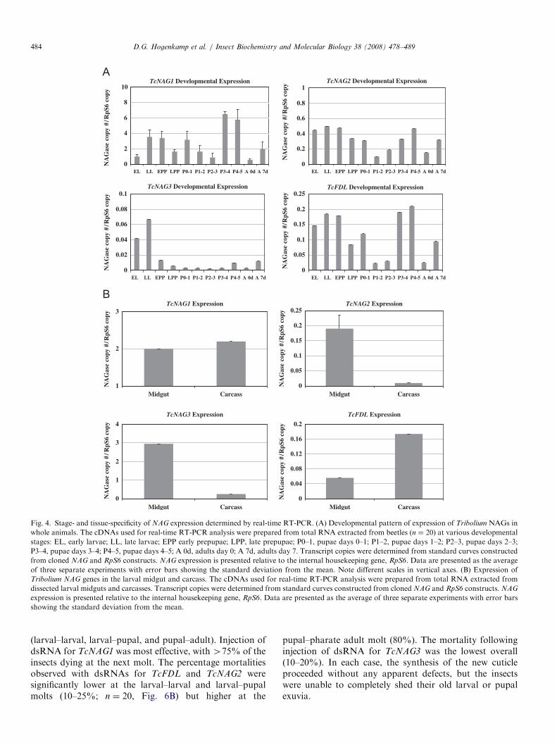
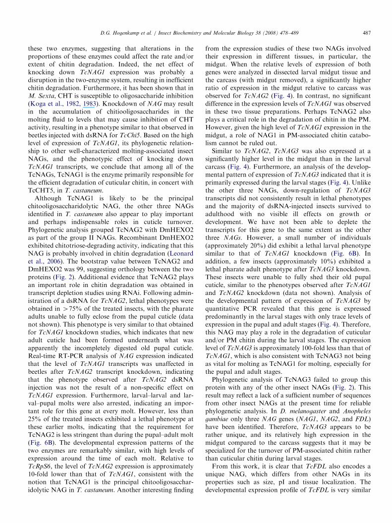
determined (Fig. 4A). TcNAG1 is expressed at the highestlevel relative to RpS6 followed by TcNAG2 at about 10%of the level of TcNAG1 transcripts. The number ofTcNAG3 transcripts is much lower than those of the otherNAGs, substantial expression being observed only in thelarval stages. In contrast, the other three NAGs areexpressed at all developmental stages, with the highestlevels at the larval and late pupal stages. A slight decreasein the level of expression of TcNAG1, TcFDL, andTcNAG2 is observed at the late prepupal and mid-pupalstages as well as in young adults. Overall, the patterns ofexpression of these three NAGs are quite similar.
3.6. Expression of Tribolium NAGs in the larval midgut and
abdominal carcass
To study the tissue specificity of expression of the fourT. castaneum NAGs, real-time RT-PCR was used tomeasure their expression in dissected last-instar larvalmidguts and carcasses. The transcript levels of TcNAG1 arecomparable in both the midgut and carcass, suggesting nounique function for NAG1 in peritrophic matrix (PM)turnover (Fig. 4B). Conversely, both TcNAG2 andTcNAG3 are expressed at far higher levels in the midgutthan in the carcass, indicating that these NAGs function in
ARTICLE IN PRESS
Fig. 3. Alignment of amino acid sequences of the deduced Tribolium castaneum and Manduca sexta NAGs. Amino acid sequences were aligned using
ClustalW (http://npsa-pbil.ibcp.fr/cgi-bin/npsa_automat.pl?page=npsa_clustalw.html). Symbols below the amino acid sequences indicate identical (*),
highly conserved (:), and conserved residues (.). The underlines indicate predicted signal peptides. Shading indicate catalytically important residues.
D.G. Hogenkamp et al. / Insect Biochemistry and Molecular Biology 38 (2008) 478–489 483
the breakdown of PM-associated chitin in the midgut.TcFDL is also expressed in midgut, but its expression in thecarcass is approximately triple that in the midgut. There-fore, all four T. castaneum NAGs are differentiallyexpressed in the midgut and some surrounding tissues.
3.7. Determination of the extent of down-regulation of
Tribolium NAGs after RNAi by real-time RT-PCR
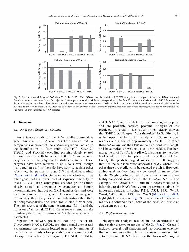
Real-time RT-PCR was carried out to determine theextent of knockdown of each T. castaneum NAG transcriptfollowing dsRNA injections into penultimate-instar larvae(Fig. 5). The expression levels in larvae were determined 3days after injection of dsRNAs corresponding to uniqueregions of each T. castaneum NAG. dsRNA for enhancedgreen fluorescent protein (EGFP) was injected as a control.Injection of a dsRNA corresponding to any one NAG generesulted in substantial depletion of the target transcript butdid not significantly change the level of expression of theother three NAG transcripts. The extent of transcriptdown-regulation ranged between 80% and 95% for
TcNAG1, TcFDL, and TcNAG2. TcNAG3, which isexpressed at a significantly lower level than the other threeNAGs, showed a somewhat lower extent of knockdown(�70%). Similar experiments were done by injection ofdsRNAs for each TcNAG into pharate pupae. Selectiveknockdowns of TcNAG1, TcFDL, TcNAG2, and TcNAG3
were also observed in the resulting pharate adults (data notshown).
3.8. Phenotypes resulting from knockdown of specific
Tribolium NAGs
dsRNAs specific to each of the four T. castaneum NAGswere injected at different developmental stages from younglarvae to pharate pupae to study the effect of depletion ofthese transcripts on larval–larval, larval–pupal, and pupa-l–adult molting. Selective down-regulation of transcriptsfor any one of the four NAGs resulted in developmentalarrest and death of some of the insects at the time of thesubsequent molt (Fig. 6A). However, some of the injectedinsects did succeed in completing each type of molt
ARTICLE IN PRESS
0
0.2
0.4
0.6
0.8
1
NA
Gas
e co
py #
/RpS
6 co
py TcNAG2 Developmental Expression
0
0.05
0.1
0.15
0.2
0.25
NA
Gas
e co
py #
/RpS
6 co
py TcFDL Developmental Expression
0
2
4
6
8
10
A
EL LL EPP LPP P0-1 P1-2 P2-3 P3-4 P4-5 A 0d A 7d
EL LL EPP LPP P0-1 P1-2 P2-3 P3-4 P4-5 A 0d A 7d
EL LL EPP LPP P0-1 P1-2 P2-3 P3-4 P4-5 A 0d A 7d
EL LL EPP LPP P0-1 P1-2 P2-3 P3-4 P4-5 A 0d A 7d
NA
Gas
e co
py #
/RpS
6 co
py
0
0.02
0.04
0.06
0.08
0.1
NA
Gas
e co
py #
/RpS
6 co
py
TcNAG3 Developmental Expression
TcNAG1 Developmental Expression
BTcNAG2 Expression
TcNAG3 Expression TcFDL Expression
TcNAG1 Expression
1
2
3
NA
Gas
e co
py #
/RpS
6 co
py
0
1
2
3
4
NA
Gas
e co
py #
/RpS
6 co
py
0
0.05
0.1
0.15
0.2
0.25
NA
Gas
e co
py #
/RpS
6 co
py
0
0.04
0.08
0.12
0.16
0.2
NA
Gas
e co
py #
/RpS
6 co
py
Midgut Carcass
Midgut Carcass Midgut Carcass
Midgut Carcass
Fig. 4. Stage- and tissue-specificity of NAG expression determined by real-time RT-PCR. (A) Developmental pattern of expression of Tribolium NAGs in
whole animals. The cDNAs used for real-time RT-PCR analysis were prepared from total RNA extracted from beetles (n ¼ 20) at various developmental
stages: EL, early larvae; LL, late larvae; EPP early prepupae; LPP, late prepupae; P0–1, pupae days 0–1; P1–2, pupae days 1–2; P2–3, pupae days 2–3;
P3–4, pupae days 3–4; P4–5, pupae days 4–5; A 0d, adults day 0; A 7d, adults day 7. Transcript copies were determined from standard curves constructed
from cloned NAG and RpS6 constructs. NAG expression is presented relative to the internal housekeeping gene, RpS6. Data are presented as the average
of three separate experiments with error bars showing the standard deviation from the mean. Note different scales in vertical axes. (B) Expression of
Tribolium NAG genes in the larval midgut and carcass. The cDNAs used for real-time RT-PCR analysis were prepared from total RNA extracted from
dissected larval midguts and carcasses. Transcript copies were determined from standard curves constructed from cloned NAG and RpS6 constructs. NAG
expression is presented relative to the internal housekeeping gene, RpS6. Data are presented as the average of three separate experiments with error bars
showing the standard deviation from the mean.
D.G. Hogenkamp et al. / Insect Biochemistry and Molecular Biology 38 (2008) 478–489484
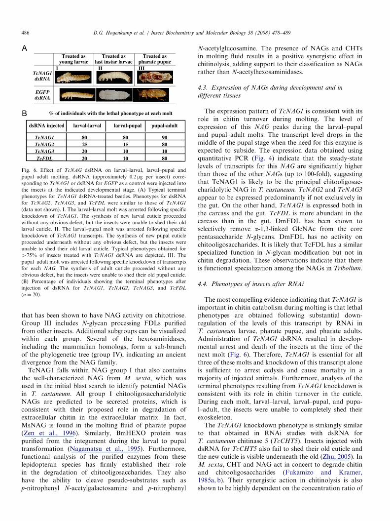
(larval–larval, larval–pupal, and pupal–adult). Injection ofdsRNA for TcNAG1 was most effective, with475% of theinsects dying at the next molt. The percentage mortalitiesobserved with dsRNAs for TcFDL and TcNAG2 weresignificantly lower at the larval–larval and larval–pupalmolts (10–25%; n ¼ 20, Fig. 6B) but higher at the
pupal–pharate adult molt (80%). The mortality followinginjection of dsRNA for TcNAG3 was the lowest overall(10–20%). In each case, the synthesis of the new cuticleproceeded without any apparent defects, but the insectswere unable to completely shed their old larval or pupalexuvia.
ARTICLE IN PRESS
Extent of Knockdown of TcNAG2
Extent of Knockdown of TcNAG3 Extent of Knockdown of TcFDL
Extent of Knockdown of TcNAG1
0
0.2
0.4
0.6
0.8
1
NA
Gas
e co
py #
/RpS
6 co
py
0
0.01
0.02
0.03
0.04
0.05
NA
Gas
e co
py #
/RpS
6 co
pyN
AG
ase
copy
#/R
pS6
copy
0
0.1
0.2
0.3
0.4
NA
Gas
e co
py #
/RpS
6 co
py
EGFP TcNAG1 TcNAG2 TcNAG3 TcFDL EGFP TcNAG1 TcNAG2 TcNAG3 TcFDL
EGFP TcNAG1 TcNAG2 TcNAG3 TcFDLEGFP TcNAG1 TcNAG2 TcNAG3 TcFDL
0
0.1
0.2
0.3
0.4
Fig. 5. Extent of knockdown of Tribolium NAGs by RNAi. The cDNAs used for real-time RT-PCR analysis were prepared from total RNA extracted
from last instar larvae three days after injection (before pupation) with dsRNAs corresponding to the four T. castaneum NAGs and the EGFP for controls.
Transcript copies were determined from standard curves constructed from cloned NAG and RpS6 constructs. NAG expression is presented relative to the
internal housekeeping gene, RpS6. Data are presented as the average of three separate experiments with error bars showing the standard deviation from
the mean. X-axis indicates dsRNA injected.
D.G. Hogenkamp et al. / Insect Biochemistry and Molecular Biology 38 (2008) 478–489 485
4. Discussion
4.1. NAG gene family in Tribolium
An extensive study of the b-N-acetylhexosaminidasegene family in T. castaneum has been carried out. Acomprehensive search of the Tribolium genome has led tothe identification of four genes (TcNAG1, TcNAG2,
TcFDL, and TcNAG3) encoding proteins closely relatedto enzymatically well-characterized M. sexta and B. mori
enzymes with chitooligosaccharidolytic activity. Theseenzymes have been referred to as NAGs even thoughmany (perhaps all) of them do have activity against othersubstrates, in particular oligo-b-N-acetylgalactosamines(Nagamatsu et al., 1995). Our searches also identified threeother genes with a lower level of sequence similarity toinsect NAGs. These latter genes encoded proteins moreclosely related to enzymatically characterized humanhexosaminidases that act on GM2 gangliosides, and weretherefore assigned to the group of hexosaminidase genes.Presumably these enzymes act on substrates other thanchitooligosaccharides and were not studied further here.The high coverage of the genome sequence (7.5� ) and theinclusion of almost all ESTs in the genome assembly makeit unlikely that other T. castaneum NAG-like genes remainundetected.
SignalP 3.0 software predicted that only one of theT. castaneum NAGs, TcFDL, contains a signal anchor anda transmembrane domain located near the N-terminus ofthe protein with only a low probability of a signal peptidecleavage. The other three enzymes, TcNAG1, TcNAG2,
and TcNAG3, were predicted to contain a signal peptideand are probably secreted proteins. Analysis of thepredicted properties of each NAG protein clearly showedthat TcFDL stands apart from the other NAGs. Firstly, itis the largest member of this family, with 630 amino acidresidues and a size of approximately 73 kDa. The otherthree NAGs are less than 600 amino acid residues in lengthand have molecular weights of less than 68 kDa. Further-more, the pI of TcFDL is4pH 6.6, in contrast to the otherNAGs whose predicted pIs are all lower than pH 5.6.Finally, the predicted signal anchor in TcFDL suggeststhat it is the sole membrane-associated NAG, whereas theother three are predicted to be secreted proteins. However,amino acid residues that are conserved in many otherfamily 20 glycosylhydrolases from other organisms arehighly conserved in all four of the Tribolium NAGs. Forexample, the active site of human b-hexosaminidase Bbelonging to the NAG family contains several catalyticallyimportant residues including R211, D354, E355, W405,W424, Y450, D452, E491, and W489 (Maier et al., 2003;highlighted residues in Fig. 2). Every one of these nineresidues is conserved in all four of the Tribolium NAGs atequivalent positions.
4.2. Phylogenetic analysis
Phylogenetic analyses resulted in the identification ofthree major clusters or groups of NAGs (Fig. 2). Group Iincludes several well-characterized lepidopteran enzymesthat are found in molting fluid and shown to possess NAGactivity, Group II NAGs include the Drosophila enzyme
ARTICLE IN PRESS
Treated as young larvae
TcNAG1dsRNA
I II III
EGFP dsRNA
Treated as last instar larvae
Treated as pharate pupae
dsRNA injected larval-larval larval-pupal pupal-adult
TcNAG1 80 80 90TcNAG2 25 15 80TcNAG3 20 10 10TcFDL 20 10 80
% of individuals with the lethal phenotype at each molt
Fig. 6. Effect of TcNAG dsRNA on larval–larval, larval–pupal and
pupal–adult molting. dsRNA (approximately 0.2 mg per insect) corre-
sponding to TcNAG1 or dsRNA for EGFP as a control were injected into
the insects at the indicated developmental stage. (A) Typical terminal
phenotypes for TcNAG1 dsRNA-treated beetles. Phenotypes for dsRNA
for TcNAG2, TcNAG3, and TcFDL were similar to those of TcNAG1
(data not shown). I. The larval–larval molt was arrested following specific
knockdown of TcNAG1. The synthesis of new larval cuticle proceeded
without any obvious defect, but the insects were unable to shed their old
larval cuticle. II. The larval–pupal molt was arrested following specific
knockdown of TcNAG1 transcripts. The synthesis of new pupal cuticle
proceeded underneath without any obvious defect, but the insects were
unable to shed their old larval cuticle. Typical phenotypes obtained for
475% of insects treated with TcNAG1 dsRNA are depicted. III. The
pupal–adult molt was arrested following specific knockdown of transcripts
for each NAG. The synthesis of adult cuticle proceeded without any
obvious defect, but the insects were unable to shed their old pupal cuticle.
(B) Percentage of individuals showing the terminal phenotypes after
injection of dsRNA for TcNAG1, TcNAG2, TcNAG3, and TcFDL
(n ¼ 20).
D.G. Hogenkamp et al. / Insect Biochemistry and Molecular Biology 38 (2008) 478–489486
that has been shown to have NAG activity on chitotriose.Group III includes N-glycan processing FDLs purifiedfrom other insects. Additional subgroups can be visualizedwithin each group. Several of the hexosaminidases,including the mammalian homologs, form a sub-branchof the phylogenetic tree (group IV), indicating an ancientdivergence from the NAG family.
TcNAG1 falls within NAG group I that also containsthe well-characterized NAG from M. sexta, which wasused in the initial blast search to identify potential NAGsin T. castaneum. All group I chitooligosaccharidolyticNAGs are predicted to be secreted proteins, which isconsistent with their proposed role in degradation ofextracellular chitin in the extracellular matrix. In fact,MsNAG is found in the molting fluid of pharate pupae(Zen et al., 1996). Similarly, BmHEXO protein waspurified from the integument during the larval to pupaltransformation (Nagamatsu et al., 1995). Furthermore,functional analysis of the purified enzymes from theselepidopteran species has firmly established their rolein the degradation of chitooligosaccharides. They alsohave the ability to cleave pseudo-substrates such asp-nitrophenyl N-acetylgalactosamine and p-nitrophenyl
N-acetylglucosamine. The presence of NAGs and CHTsin molting fluid results in a positive synergistic effect inchitinolysis, adding support to their classification as NAGsrather than N-acetylhexosaminidases.
4.3. Expression of NAGs during development and in
different tissues
The expression pattern of TcNAG1 is consistent with itsrole in chitin turnover during molting. The level ofexpression of this NAG peaks during the larval–pupaland pupal–adult molts. The transcript level drops in themiddle of the pupal stage when the need for this enzyme isexpected to subside. The expression data obtained usingquantitative PCR (Fig. 4) indicate that the steady-statelevels of transcripts for this NAG are significantly higherthan those of the other NAGs (up to 100-fold), suggestingthat TcNAG1 is likely to be the principal chitooligosac-charidolytic NAG in T. castaneum. TcNAG2 and TcNAG3
appear to be expressed predominantly if not exclusively inthe gut. On the other hand, TcNAG1 is expressed both inthe carcass and the gut. TcFDL is more abundant in thecarcass than in the gut. DmFDL has been shown toselectively remove a-1,3-linked GlcNAc from the corepentasaccharide N-glycans. DmFDL has no activity onchitooligosaccharides. It is likely that TcFDL has a similarspecialized function in N-glycan modification but not inchitin degradation. These observations indicate that thereis functional specialization among the NAGs in Tribolium.
4.4. Phenotypes of insects after RNAi
The most compelling evidence indicating that TcNAG1 isimportant in chitin catabolism during molting is that lethalphenotypes are obtained following substantial down-regulation of the levels of this transcript by RNAi inT. castaneum larvae, pharate pupae, and pharate adults.Administration of TcNAG1 dsRNA resulted in develop-mental arrest and death of the insects at the time of thenext molt (Fig. 6). Therefore, TcNAG1 is essential for allthree of these molts and knockdown of this transcript aloneis sufficient to arrest ecdysis and cause mortality in amajority of injected animals. Furthermore, analysis of theterminal phenotypes resulting from TcNAG1 knockdown isconsistent with its role in chitin turnover in the cuticle.During each molt, larval–larval, larval–pupal, and pupa-l–adult, the insects were unable to completely shed theirexoskeleton.The TcNAG1 knockdown phenotype is strikingly similar
to that obtained in RNAi studies with dsRNA forT. castaneum chitinase 5 (TcCHT5). Insects injected withdsRNA for TcCHT5 also fail to shed their old cuticle andthe new cuticle is visible underneath the old (Zhu, 2005). InM. sexta, CHT and NAG act in concert to degrade chitinand chitooligosaccharides (Fukamizo and Kramer,1985a, b). Their synergistic action in chitinolysis is alsoshown to be highly dependent on the concentration ratio of
ARTICLE IN PRESSD.G. Hogenkamp et al. / Insect Biochemistry and Molecular Biology 38 (2008) 478–489 487
these two enzymes, suggesting that alterations in theproportions of these enzymes could affect the rate and/orextent of chitin degradation. Indeed, the net effect ofknocking down TcNAG1 expression was probably adisruption in the two-enzyme system, resulting in inefficientchitin degradation. Furthermore, it has been shown that inM. Sexta, CHT is susceptible to oligosaccharide inhibition(Koga et al., 1982, 1983). Knockdown of NAG may resultin the accumulation of chitiooligosaccharides in themolting fluid to levels that may cause inhibition of CHTactivity, resulting in a phenotype similar to that observed inbeetles injected with dsRNA for TcChi5. Based on the highlevel of expression of TcNAG1, its phylogenetic relation-ship to other well-characterized molting-associated insectNAGs, and the phenotypic effect of knocking downTcNAG1 transcripts, we conclude that among all of theTcNAGs, TcNAG1 is the enzyme primarily responsible forthe efficient degradation of cuticular chitin, in concert withTcCHT5, in T. castaneum.
Although TcNAG1 is likely to be the principalchitooligosaccharidolytic NAG, the other three NAGsidentified in T. castaneum also appear to play importantand perhaps indispensable roles in cuticle turnover.Phylogenetic analysis grouped TcNAG2 with DmHEXO2as part of the group II NAGs. Recombinant DmHEXO2exhibited chitotriose-degrading activity, indicating that thisNAG is probably involved in chitin degradation (Leonardet al., 2006). The bootstrap value between TcNAG2 andDmHEXO2 was 99, suggesting orthology between the twoproteins (Fig. 2). Additional evidence that TcNAG2 playsan important role in chitin degradation was obtained intranscript depletion studies using RNAi. Following admin-istration of a dsRNA for TcNAG2, lethal phenotypes wereobtained in 475% of the treated insects, with the pharateadults unable to fully eclose from the pupal cuticle (datanot shown). This phenotype is very similar to that obtainedfor TcNAG1 knockdown studies, which indicates that newadult cuticle had been formed underneath what wasapparently the incompletely digested old pupal cuticle.Real-time RT-PCR analysis of NAG expression indicatedthat the level of TcNAG1 transcripts was unaffected inbeetles after TcNAG2 transcript knockdown, indicatingthat the phenotype observed after TcNAG2 dsRNAinjection was not the result of a non-specific effect onTcNAG1 expression. Furthermore, larval–larval and lar-val–pupal molts were also arrested, indicating an impor-tant role for this gene at every molt. However, less than25% of the treated insects exhibited a lethal phenotype atthese earlier molts, indicating that the requirement forTcNAG2 is less stringent than during the pupal–adult molt(Fig. 6B). The developmental expression patterns of thetwo enzymes are remarkably similar, with high levels ofexpression around the time of each molt. Relative toTcRpS6, the level of TcNAG2 expression is approximately10-fold lower than that of TcNAG1, consistent with thenotion that TcNAG1 is the principal chitooligosacchar-idolytic NAG in T. castaneum. Another interesting finding
from the expression studies of these two NAGs involvedtheir expression in different tissues, in particular, themidgut. When the relative levels of expression of bothgenes were analyzed in dissected larval midgut tissue andthe carcass (with midgut removed), a significantly higherratio of expression in the midgut relative to carcass wasobserved for TcNAG2 (Fig. 4). In contrast, no significantdifference in the expression levels of TcNAG1 was observedin these two tissue preparations. Perhaps TcNAG2 alsoplays a critical role in the degradation of chitin in the PM.However, given the high level of TcNAG1 expression in themidgut, a role of NAG1 in PM-associated chitin catabo-lism cannot be ruled out.Similar to TcNAG2, TcNAG3 was also expressed at a
significantly higher level in the midgut than in the larvalcarcass (Fig. 4). Furthermore, an analysis of the develop-mental pattern of expression of TcNAG3 indicated that it isprimarily expressed during the larval stages (Fig. 4). Unlikethe other three NAGs, down-regulation of TcNAG3
transcripts did not consistently result in lethal phenotypesand the majority of dsRNA-injected insects survived toadulthood with no visible ill effects on growth ordevelopment. We have not been able to deplete thetranscripts for this gene to the same extent as the otherthree NAGs. However, a small number of individuals(approximately 20%) did exhibit a lethal larval phenotypesimilar to that of TcNAG1 knockdown (Fig. 6B). Inaddition, a few insects (approximately 10%) exhibited alethal pharate adult phenotype after TcNAG3 knockdown.These insects were unable to fully shed their old pupalcuticle, similar to the phenotypes observed after TcNAG1
and TcNAG2 knockdown (data not shown). Analysis ofthe developmental pattern of expression of TcNAG3 byquantitative PCR revealed that this gene is expressedpredominantly in the larval stages with only trace levels ofexpression in the pupal and adult stages (Fig. 4). Therefore,this NAG may play a role in the degradation of cuticularand/or PM chitin during the larval stages. The expressionlevel of TcNAG3 is approximately 100-fold less than that ofTcNAG1, which is also consistent with TcNAG3 not beingas vital for molting as TcNAG1 for molting, especially forthe pupal and adult stages.Phylogenetic analysis of TcNAG3 failed to group this
protein with any of the other insect NAGs (Fig. 2). Thisresult may reflect a lack of a sufficient number of sequencesfrom other insect NAGs at the present time for reliablephylogenetic analysis. In D. melanogaster and Anopheles
gambiae only three NAG genes (NAG1, NAG2, and FDL)have been identified. Therefore, TcNAG3 appears to berather unique, and its relatively high expression in themidgut compared to the carcass suggests that it may bespecialized for the turnover of PM-associated chitin ratherthan cuticular chitin during larval stages.From this work, it is clear that TcFDL also encodes a
unique NAG, which differs from other NAGs in itsproperties such as size, pI and tissue localization. Thedevelopmental expression profile of TcFDL is very similar
ARTICLE IN PRESSD.G. Hogenkamp et al. / Insect Biochemistry and Molecular Biology 38 (2008) 478–489488
to that of TcNAG1, although the level of TcFDL
expression relative to RpS6 is approximately 30-fold lowerthan that of TcNAG1 (Fig. 5). Furthermore, the size of theTcFDL gene is considerably larger than that of the otherthree NAGs because of the presence of a large intron(413 kb) just upstream of the putative translation start site(Fig. 1). Phylogenetic analysis indicates that TcFDL ismore closely related to the insect homologs of the D.
melanogaster fused lobes protein, DmFDL, than toTcNAG1 and its homologs, which are implicated tofunction in chitin degradation. The FDL proteins arepredicted to be membrane-anchored, with a single TMSlocated near the N-terminus of the polypeptide. Further-more, ultracentrifugation experiments on a lepidopteranprotein from the culture media of Sf9 and Sf21 cellsindicated that a major portion of the NAG activity residedin the membrane fraction, although a lesser amount ofactivity was observed in the soluble fraction as well(Tomiya et al., 2006; Altmann et al., 1995). Thislepidopteran NAG was capable of effectively hydrolyzingchitotriose-PA (pyridylamino). In contrast, the recombi-nantly expressed DmFDL was unable to digest chitotriose(Leonard et al., 2006). Instead, the enzyme hydrolyzed onlythe GlcNAc residue attached to the a-1,3-linked mannoseof the core pentasaccharide of N-glycans. No cleavageactivity of any other GlcNAc residues was observedincluding the GlcNAc residue attached to the a-1,6-linkedmannose of the core pentasaccharide. Furthermore,DmFDL did not catalyze the endo-type hydrolysis of theN,N0-diacetylchitobiosyl unit in the high-mannose penta-saccharide core. A similar N-glycan substrate specificity forthe terminal GlcNAc attached to the a-1,3-linked mannosewas observed in membrane-associated b-N-acetylhexosa-minidases from several lepidopteran insect cell linesincluding Sf21, Bm-N, and Mb-0503 and a soluble enzymefrom Sf9 cells (Tomiya et al., 2006; Altmann et al., 1995).Given the similarity of TcFDL to the other previouslycharacterized homologs of the D. melanogaster fused lobes
protein, TcFDL may serve a similar function in N-glycanprocessing. However, since the membrane-associated N-acetylhexosaminidases in several lepidopteran insect celllines catalyzed the hydrolysis of chitotriose, TcFDL couldalso be involved in chitin catabolism as well unlike itscounterpart, DmFDL, in Drosophila. Indeed, when TcFDL
transcripts were selectively knocked down by RNAi, theresulting pharate adults were unable to completely shedtheir old pupal cuticle, particularly at the posterior end(data not shown). These phenotypes do imply a possibleinvolvement of TcFDL in chitin degradation as well.Studies with purified TcFDL are needed to definitivelyanswer this question.
Unlike RNAi involving TcNAG1 dsRNA, which re-sulted in lethal phenotypes in most of the treated insects,only a small percentage of animals injected with dsRNAfor TcFDL (o20%) exhibited lethal terminal phenotypesat the larval–larval, and larval–pupal molts (Fig. 6B). Thereason for this lower effectiveness of TcFDL RNAi in
inducing a lethal phenotype is unknown. However, themuch higher mortality seen at the pharate adult stage upondown-regulation of the transcripts for TcFDL indicates anessential role for this gene at the pupa–adult molt. Perhaps,its role is more critical at this stage than during the larvalstages.Analysis of the expression of TcFDL by real-time RT-
PCR indicated that this transcript is expressed at a lowerlevel in the midgut compared to the carcass (Fig. 4).Therefore, the lethal phenotype at the pharate adultstage may be a direct result of the knockdown of thistranscript in the cuticular epidermal cells rather thanin the gut cells. At ecdysis, an ecdysial membrane isformed, which separates the newly synthesized cuticle andepidermal cells from the molting fluid (Locke andKrishnan, 1973). If TcFDL does in fact play a role inchitin turnover in the cuticle, then this protein may actuallybe secreted and not membrane bound as predicted bySignalP 3.0. In fact, Leonard et al. (2006) have observedthat DmFDL, to a large extent, is secreted into theextracellular space.The crystal structures are currently available for the
human hexosaminidase b-chain, HEXB (Maier et al., 2003,Mark et al., 2003). The human b-chain of HEXB is one oftwo lysosomal b-hexosaminidases. Mutations in this generesult in Sandhoff disease, a fatal neurodegenerativedisorder. The HEXB b-chain forms a heterodimer withthe a-chain hexosaminidase, which is encoded by the geneHEXA. Homodimeric hexosaminidases are also formedfrom two HEXB b-chains or two HEXA a-chains.Dimerization of hexosaminidases is required for theformation of a catalytically active enzyme. Mutations inthe human HEXA gene are responsible for the lysosomalstorage disorder, Tay-Sachs disease. Evidence for multi-meric NAGs exists in a limited number of insects. A NAGpurified from T. castaneum exhibited a dimeric structurewith subunits of different molecular weights (Kramerand Aoki, 1987). Similarly, a NAG from B. mori
and Sf9 cells exhibited a dimeric structure, although it isprobably composed of identical subunits (Kimura,1976; Tomiya et al., 2006). If the biologically active(or more active) forms of NAGs are heterodimers, thiscould explain some of the redundancy and the requirementfor more than one NAG for effective cuticle digestionduring molting.An alternative possibility is that the T. castaneum NAGs
do not serve redundant functions. There may be a degree ofspecificity for the four NAGs for different chitooligosac-charides substrates. One may be particularly catalyticallyactive on chitotriose, whereas another may specialize in thedegradation of the tetramer and so on. Therefore, knock-down of one of the NAGs may result in the accumulationof only one particular chitooligosaccharide. This could inturn lead to inhibition of CHT in the binary enzymechitinase system. Further research in this area is required tomore definitively answer why there is a multiplicity ofNAGs in insect tissues.
ARTICLE IN PRESSD.G. Hogenkamp et al. / Insect Biochemistry and Molecular Biology 38 (2008) 478–489 489
Acknowledgments
The authors are grateful to Gerald Reeck for commentson an earlier version of this manuscript. Support wasprovided in part by the NSF Grants IBN-0316963 and0615818. Mention of a proprietary product does notconstitute a recommendation or endorsement by theUSDA. The USDA is an equal opportunity/affirmativeaction employer and all agency services are availablewithout discrimination. This is contribution 07-218-J of theKansas Agricultural Experiment Station.
There is no conflict of interest.
References
Altmann, F., Schwihla, H., Staudacher, E., Glossl, J., Marz, L., 1995.
Insect cells contain an unusual, membrane-bound b-N-acetylhexosa-
minidase probably involved in the processing of protein N-glycans.
J. Biol. Chem. 270, 17344–17349.
Arakane, Y., Muthukrishnan, S., Kramer, K.J., Specht, C.A., Tomoyasu,
Y., Lorenzen, M.D., Kanost, M.R., Beeman, R.W., 2005. The
Tribolium chitin synthase genes TcCHS1 and TcCHS2 are specialized
for synthesis of epidermal cuticle and midgut peritrophic matrix,
respectively. Insect Mol. Biol. 14, 453–463.
Arakane, Y., Zhu, Q., Matsumiya, M., Muthukrishnan, S., Kramer, K.J.,
2003. Properties of catalytic, linker and chitin-binding domains of
insect chitinase. Insect Biochem. Mol. Biol. 33, 631–648.
Beeman, R.W., Stuart, J.J., 1990. A gene for lindane+cyclodiene
resistance in the red flour beetle (Coleopetera: Tenebrionidae).
J. Econ. Entomol. 83, 1745–1751.
Coutinho, P.M., Henrissat, B., 1999. Carbohydrate-active enzymes: an
integrated database approach. In: Gilbert, H.J., Davies, G., Henrissat,
B., Svensson, B. (Eds.), Recent Advances in Carbohydrate Bioengi-
neering. The Royal Society of Chemistry, Cambridge, pp. 3–12 (http://
www.cazy.org/).
Filho, J.A.F., Shapiro, B.E., 2004. Tay-Sachs disease. Arch. Neurol. 61,
1466–1468.
Fukamizo, T., Kramer, K.J., 1985a. Mechanism of chitin oligosaccharides
hydrolysis by the binary chitinase system in insect molting fluid. Insect
Biochem. 15, 1–7.
Fukamizo, T., Kramer, K.J., 1985b. Mechanism of chitin hydrolysis by
the binary chitinase system in insect molting fluid. Insect Biochem. 15,
141–145.
Haliscak, J.P., Beeman, R.W., 1983. Status of malathion resistance in five
genera of beetles infesting farm-stored corn, wheat, and oats in the
United States. J. Econ. Entomol. 76, 717–722.
Kimura, S., 1976. The chitinase system in the cuticle of the silkworm
Bombyx mori. Insect Biochem. 6, 479–482.
Koga, D., Mai, M.S., Dziadik-Turner, C., Kramer, K.J., 1982. Kinetics
and mechanism of exochitinase and b-N-acetylhexosaminidase from
the tobacco hornworm, Manduca sexta L. (Lepidoptera: Sphingidae).
Insect Biochem. 12, 493–499.
Koga, D., Jilka, J., Kramer, K.J., 1983. Insect endochitinases: glycopro-
teins from moulting fluid, integument and pupal haemolymph of
Manduca sexta L. Insect Biochem. 13, 295–305.
Kramer, K.J., Aoki, H., 1987. Chitinolytic enzymes from pupae of the red
flour beetle, Tribolium castaneum. Comp. Biochem. Physiol. 86B,
613–621.
Kramer, K.J., Muthukrishnan, S., 2005. Chitin metabolism in insects. In:
Gilbert, L.I., Iatrou, K., Gill, S. (Eds.), Comprehensive Molecular
Insect Science. Vol. 4, Biochemistry and Molecular Biology. Elsevier
Press, Oxford, UK, pp. 111–144 (Chapter 3).
Kumar, S., Tamura, K., Nei, M., 2004. MEGA3: integrated software for
molecular evolutionary genetics analysis and sequence alignment.
Brief. Bioinform. 5, 150–163.
Leonard, R., Rendic, D., Rabouille, C., Wilson, I.B., Preat, T., Altmann,
F., 2006. The Drosophila fused lobes gene encodes an N-acetylhex-
osaminidase involved in N-glycan processing. J. Biol. Chem. 281,
4867–4875.
Locke, M., Krishnan, N., 1973. The formation of the ecdysial droplets and
the ecdysial membrane in an insect. Tissue Cell 5, 441–450.
Maier, T., Strater, N., Schuette, C.G., Klingenstein, R., Sandhoff, K.,
Saenger, W., 2003. The X-ray crystal structure of human
b-hexosaminidase B provides new insights into Sandhoff Disease.
J. Mol. Biol. 328, 669–681.
Mark, B.L., Mahuran, D.J., Cherney, M.M., Zhao, D., Knapp, S., James,
M.N.G., 2003. Crystal structure of human b-hexosaminidase B:
understanding the molecular basis of Sandhoff and Tay-Sachs Disease.
J. Mol. Biol. 327, 1093–1109.
Nagamatsu, Y., Yanagisawa, I., Kimoto, M., Okamoto, E., Koga, D.,
1995. Purification of a chitooligosaccharidolytic b-N-acetylhexosami-
nidase from Bombyx mori larvae during metamorphosis and the
nucleotide sequence of its cDNA. Biosci. Biotechnol. Biochem. 59,
219–225.
Sahai, A.S., Manocha, M.S., 1993. Chitinases of fungi and plants: their
involvement in morphogenesis and host parasite interaction. FEMS
Microbiol. Rev. 11, 317–338.
Sandhoff, K., Andreae, U., Jatzkewitz, H., 1968. Deficient hexosamini-
dase activity in an exceptional case of Tay-Sachs disease with
additional storage of kidney globoside in visceral organs. Pathol.
Eur. 3, 278–285.
Tomiya, N., Narang, S., Park, J., Abdul-Rahman, B., Choi, O., Singh, S.,
Hiratake, J., Sakata, K., Betenbaugh, M.J., Palter, K.B., Lee, Y.C.,
2006. Purification, characterization, and cloning of a Spodoptera
frugiperda Sf9 b-N-acetylhexosaminidase that hydrolyzes terminal
N-acetylglucosamine on the N-glycan core. J. Biol. Chem. 281,
19545–19560.
Tomoyasu, Y., Denell, R.E., 2004. Larval RNAi in Tribolium (Coleop-
tera) for analyzing adult development. Dev. Genes Evol. 214, 575–578.
Zen, K.C., Choi, H.K., Krishnamachary, N., Muthukrishnan, S., Kramer,
K.J., 1996. Cloning, expression, and hormonal regulation of an
insect b-N-acetylhexosaminidase gene. Insect Biochem. Mol. Biol. 26,
435–444.
Zhu, Q., 2005. Characterization of chitinase-like gene families and
proteins from Tribolium castaneum, Drosophila melanogaster and
Anopheles gambiae. Doctoral Dissertation, Kansas State University,
158pp.