characterising the microbial profiles of - Stellenbosch University
205
CHARACTERISING THE MICROBIAL PROFILES OF VARIOUS RIVER SOURCES AND INVESTIGATING THE EFFICACY OF UV RADIATION TO REDUCE MICROBIAL LOADS FOR IMPROVED CROP SAFETY By Caroline Rose Bursey Thesis presented in partial fulfilment of the requirements for the degree of Master of Science in Food Science In the Department of Food Science, Faculty of AgriSciences University of Stellenbosch Supervisor: Professor G. O. Sigge Co-supervisor: Dr C. Lamprecht March 2021
-
Upload
khangminh22 -
Category
Documents
-
view
0 -
download
0
Transcript of characterising the microbial profiles of - Stellenbosch University

CHARACTERISING THE MICROBIAL PROFILES OF
VARIOUS RIVER SOURCES AND INVESTIGATING
THE EFFICACY OF UV RADIATION TO REDUCE
MICROBIAL LOADS FOR IMPROVED CROP SAFETY
By
Caroline Rose Bursey
Thesis presented in partial fulfilment of the requirements for the
degree of
Master of Science in Food Science
In the Department of Food Science, Faculty of AgriSciences
University of Stellenbosch
Supervisor:
Professor G. O. Sigge
Co-supervisor:
Dr C. Lamprecht
March 2021

i
DECLARATION
By submitting this dissertation electronically, I declare that the entirety of the work contained
therein is my own, original work, that I am the sole author thereof (save to the extent explicitly
otherwise stated), that reproduction and publication thereof by Stellenbosch University will not
infringe any third party rights and that I have not previously in its entirety or in part submitted
it for obtaining any qualification.
March 2021
Copyright © 2021 Stellenbosch University All rights reserved
Stellenbosch University https://scholar.sun.ac.za

ii
ABSTRACT
The rivers used for the irrigation of fresh produce in the Western Cape have been under
frequent investigation in recent years. Results have frequently shown that in rivers used for
irrigation, the faecal coliform concentrations (Escherichia coli) frequently exceed the guideline
limit of 1 000 colony forming units per 100 mL. These findings present a health risk for
consumers of fresh produce. Ultraviolet (UV) radiation treatment has proven to offer some
advantages for water disinfection over conventional treatment methods such as filtration and
chemical treatments. However, this is not yet a common practice in South Africa. Knowledge
gaps exist with regard to the efficacy of UV radiation on environmental strains of pathogenic
microorganisms such as Salmonella species and Listeria monocytogenes. The aim of this
study was to investigate the effect of low-pressure (LP) UV radiation on water obtained from
various river water sources, in order to disinfect water used for irrigation purposes to ultimately
reduce the risk of causing foodborne disease outbreaks from the consumption of contaminated
fresh produce.
Four rivers in the Western Cape were sampled five times each between the wet winter
and dry summer seasons, to establish the microbial and physico-chemical profiles of the
rivers. These results were compared to the guideline limits. The samples were exposed to
three doses (20, 40 and 60 mJ.cm-2) of LP UV radiation at laboratory-scale. It was established
that LP UV radiation was effective at reducing the microbial loads to non-detectable levels.
Pathogenic microorganisms were successfully inactivated after a dose of 20 mJ.cm-2.
Heterotrophic Plate Count colony numbers were lowered more steadily, and therefore,
showed greater resistance to treatment. Thirteen strains were isolated and stored for future
experiments. It was suggested that a pre-treatment step be implemented to improve the
physical quality of the river water prior to treatment.
Isolated strains of E. coli (n = 3), Salmonella species (n = 2) and L. monocytogenes
(n = 8) were stored for further testing. The L. monocytogenes isolates (n = 8) were subjected
to lineage typing experiments, where it was established that all isolates were lineage I. This
lineage is most frequently associated with listeriosis. Extended-spectrum beta-lactamase
(ESBL) testing indicated that none of the Enterobacteriaceae isolates (n=5) were ESBL-
producers. All Enterobacteriaceae isolates showed resistance to tetracycline, ampicillin and
trimethoprim-sulfamethoxazole. Resistance of L. monocytogenes isolates (n=5) was observed
against trimethoprim-sulfamethoxazole, while four L. monocytogenes isolates showed
resistance to ampicillin, penicillin and erythromycin. Multidrug resistance was reported for 90%
of river water isolates (n=9).
Stellenbosch University https://scholar.sun.ac.za

iii
Four different bag filter pore sizes (5, 20, 50 and 100 μm) were investigated to determine
the most effective pre-treatment step to improve the UV transmission (UVT %) of the water.
This experiment was performed on the ‘worst case scenario’ river, the Mosselbank River.
Improvements in the total suspended solids, chemical oxygen demand and turbidity were
reported, however, the extremely high total dissolved solids content (728.67 mg.L-1) prevented
a larger improvement in the UVT %. It was established that the 5 μm bag filter was the most
effective pore size.
In the current study, LP UV radiation was successfully able to produce water of an
acceptable standard for the irrigation of fresh produce. The physical quality of the water did
not prevent a successful disinfection, but rather increased the exposure time required to
deliver a specific dose and therefore, decreased efficiency. It was established that LP UV
radiation is able to reduce pathogenic microorganisms to non-detectable levels. This method
of disinfection, therefore, shows promise for full-scale application of irrigation water treatment.
Stellenbosch University https://scholar.sun.ac.za

iv
UITTREKSEL
Die riviere wat vir die besproeiing van vars produkte in die Wes-Kaap gebruik word, is die
afgelope paar jaar gereeld ondersoek. Resultate het getoon dat in sommige riviere die fekale
koliforme konsentrasies (Escherichia coli) gereeld die riglynlimiet van 1 000 kolonievormende
eenhede per 100 ml oorskry. Hierdie bevindings dui op 'n gesondheidsrisiko vir die verbruikers
van vars produkte. Ultraviolet (UV) bestralingsbehandeling het bewys dat dit voordele bied
vir die ontsmetting van water bo konvensionele behandelingsmetodes soos filtrasie en
chemiese behandelings. Dit is egter nog nie 'n algemene gebruik in Suid-Afrika nie. Daar is
kennisgapings met betrekking tot die doeltreffendheid van UV-bestraling op patogene
mikroörganismes soos Salmonella-spesies en Listeria monocytogenes. Die doel van hierdie
studie was om die effek van lae druk (LP) UV-bestraling op water, verkry uit verskillende
rivierwaterbronne, te ondersoek, met die doel om water wat vir besproeiingsdoeleindes
gebruik word, te ontsmet om uiteindelik die risiko van kontaminasie vir die verbruikers van
vars produkte te verminder.
Monsters is vyf keer tussen die nat winter- en droë somerseisoene uit elkeen van vier
riviere in die Wes-Kaap geneem om die mikrobiese en fisies-chemiese profiele van die riviere
vas te stel. Hierdie resultate is vergelyk met die riglyn-limiete. Die monsters is op
laboratoriumskaal aan drie dosisse (20, 40 en 60 mJ.cm-2) LP UV-bestraling blootgestel. Daar
is vasgestel dat LP UV-bestraling effektief was om die mikrobiese lading tot nie-
waarneembare vlakke te verminder. Patogene mikroörganismes is suksesvol geïnaktiveer na
'n dosis van 20 mJ.cm-2. Die heterotrofiese kolonie-getalle het meer geleidelik verlaag en het
dus groter weerstand teen behandeling getoon. Dertien isolate is verkry en geberg vir
toekomstige eksperimente. Daar is voorgestel dat 'n voorafbehandelingsstap
geïmplementeer word om die fisiese kwaliteit van die rivierwater te verbeter voor UV
behandeling.
Isolate van E.c oli (n = 3), Salmonella species (n = 2) en L. monocytogenes (n = 8) is
gestoor vir latere toestsing. Die gebergde L. monocytogenes-isolate is onderwerp aan stam
tiperingseksperimente, waar vasgestel is dat al die isolate aan stam I behoort. Hierdie stam
word meestal met listeriose geassosieer. Uitgebreide-spektrum beta-laktamase (ESBL) toetse
het aangedui dat geen van die Enterobacteriaceae-isolate ESBL-produseerders was nie. Alle
Enterobacteriaceae-isolate (n = 5) het weerstand teen tetrasiklien, ampisillien en trimetopriem-
sulfametoksasool getoon. Weerstand van L. monocytogenes isolate (n = 5) is waargeneem
teen trimetopriem-sulfametoksasool, terwyl vier L. monocytogenes isolate weerstand getoon
het teen ampisillien, penisillien en eritromisien. Veelvuldige middelweerstand is gerapporteer
vir 90% van die rivierwater-isolate (n = 9).
Stellenbosch University https://scholar.sun.ac.za

v
Vier verskillende sakfilter poriegroottes (5, 20, 50 en 100 μm) is ondersoek om die mees
effektiewe voorbehandelingstap te bepaal om die UV-transmissie (UVT %) van die water te
verbeter. Hierdie eksperiment is uitgevoer op die ‘slegste geval scenario'-rivier, die
Mosselbankrivier. Verbeterings in die totale gesuspendeerde vastestofinhoud, chemiese
suurstofbehoefte en troebelheid is gerapporteer, maar die uiters hoë totale opgeloste
vastestofinhoud (728.67 mg L-1) het 'n groter verbetering in die UVT % verhoed. Daar is
vasgestel dat die 5 μm sakfilter die doeltreffendste poriegrootte was.
In die huidige studie kon LP UV-bestraling suksesvol aangewend word om water van 'n
aanvaarbare standaard vir die besproeiing van vars produkte te produseer. Die fisiese kwaliteit
van die water het nie 'n suksesvolle ontsmetting verhoed nie, maar het die blootstellingstyd
wat nodig was om 'n spesifieke dosis te lewer, verhoog en dus die doeltreffendheid verminder.
Daar is vasgestel dat LP UV-bestraling patogene mikroörganismes kan verminder tot nie-
waarneembare vlakke. Hierdie ontsmettingsmetode toon dus belofte vir volskaalse toepassing
op besproeiingswater.
Stellenbosch University https://scholar.sun.ac.za

vi
ACKNOWLEDGEMENTS
Sincere gratitude and appreciation is extended to the following individuals, institutions and
organisations for their invaluable contribution that ensured a successful completion of this
degree:
My supervisor, Professor Gunnar Sigge, for presenting me with this opportunity, and for the
continuous guidance and support throughout the study. Your kind and helpful nature never
went unnoticed;
My co-supervisor, Dr Corné Lamprecht, for her continual support, time and patience
throughout the study. Your comforting nature has been of irreplaceable value;
Professor Pieter Gouws and Dr Diane Rip, your incredible academic expertise and assistance
throughout my laboratory-work was invaluable;
Stellenbosch University Food Science Department staff, including Veronique Human, Megan
Arendse, Eben Brooks, Anchen Lombard and Petro du Buisson for their kindness, and
consistent willingness to assist with general queries and tasks;
Faculty of Natural and Agricultural Sciences at the University of Pretoria and Ms Zama Zulu
for the MALDI-TOF analysis performed on bacterial isolates, supported in part by the NRF
grant with reference number 74426;
Fellow Post Graduate students in the Food Science Department, your support and
entertainment throughout the past two years made a considerable difference;
The Water Research Commission (WRC) and National Research Foundation (NRF) for
providing the funds required to complete this study;
To my incredible support system of family and friends, thank you for the continuous support,
guidance, love and encouragement.
Stellenbosch University https://scholar.sun.ac.za

vii
This study was part of a solicited project (K5/2965//4) funded by the Water Research
Commission and co-funded by the Department of Agriculture, Forestry and Fisheries.
This thesis is presented in the format prescribed by the Department of Food Science at
Stellenbosch University. The structure is in the form of three research chapters, which is
prefaced by an introduction chapter with the study objectives, followed by a literature review
chapter and culminating with a chapter for elaborating a general discussion and conclusion.
Language, style and referencing format used are in accordance with the requirements of the
International Journal of Food Science and Technology. This thesis represents a compilation
of manuscripts where each chapter is an individual entity and some repetition between
chapters has, therefore, been unavoidable.
Stellenbosch University https://scholar.sun.ac.za

viii
Contents
Declaration i
Abstract ii
Uittreksel iv
Acknowledgements vi
Abbreviations ix
Chapter 1 Introduction 1
Chapter 2 Literature Review 9
Chapter 3 investigating the disinfection efficacy of low-pressure
Ultraviolet radiation on irrigation water sources, and the
impact of water quality on treatment
75
Chapter 4 Characterisation of river water isolates in terms of Ultraviolet
resistance, antimicrobial resistance and Listeria
monocytogenes lineage typing
113
Chapter 5 Investigating the effect of a bag filter on the physico-chemical
and microbial profile of the Mosselbank River for improved UV
disinfection
161
Chapter 6 General conclusions and recommendations 186
Stellenbosch University https://scholar.sun.ac.za

ix
ABBREVIATIONS
AOP Advanced Oxidation Process
AST Antibiotic Susceptibility Testing
ATCC American Type Culture Collection
BOD Biological Oxygen Demand
BHI Brain Heart Infusion Agar
BPW Buffered Peptone Water
COD Chemical Oxygen Demand
CDC Centre of Disease Control
CFU Colony Forming Units
CLSI Clinical & Laboratory Standards Institute
CPD Cyclobutane Pyrimidine Dimers
CV Clavulanic Acid
CP Cefepime
CTX Cefotaxime
CAZ Ceftazidime
DAEC Diffusely Adherent Escherichia coli
DBPs Disinfection By-products
DNA Deoxyribonucleic acid
dNTP Deoxy ribonucleotide triphosphate
DWAF Department of Water Affairs and Forestry
E. coli Escherichia coli
EAEC Enteroadhesive Escherichia coli
EC Electrical Conductivity
EHEC Enterohemorrhagic Escherichia coli
EIEC Enteroinvasive Escherichia coli
EPEC Enteropathogenic Escherichia coli
ETEC Enterotoxigenic Escherichia coli
ESBL Extended-Spectrum Beta-Lactamase
EUCAST European Committee on Antimicrobial Susceptibility Testing
FAD Flavin Adenine Dinucleotide
FAO Food and Agricultural Organisation
FDA-BAM Food and Drug Administration Bacteriological Analytical Manual
FR Franschhoek River
HIV/Aids Human Immunodeficiency Virus/ Acquired Immunodeficiency Syndrome
HPC Heterotrophic Plate Count
HUS Haemolytic-uremic Syndrome
ICU Intensive Care Unit
ISO International Organization for Standardization
kGy Kilo Gray
Stellenbosch University https://scholar.sun.ac.za

x
L-EMB Levine Eosin-Methylene Blue Agar
LP Low-pressure
MALDI-TOF Matrix-assisted Laser Desorption/Ionization Time-of-Flight
MDR Multidrug Resistant
MP Medium-pressure
MPF Minimally Processed Foods
MR Mosselbank River
NA Nutrient Agar
NER Nucleotide Excision Repair
NTU Nephelometric Turbidity Units
OD Optical Density
PCA Plate Count Agar
PCR Polymerase Chain Reaction
PR Plankenburg River
R Resistant
RTE Ready-to-Eat
S Susceptible
SA South Africa
SABS South African Bureau of Standards
spp. Species
SANS South African National Standards
STEC Shiga-toxin producing Escherichia coli
TB Tuberculosis
TDS Total Dissolved Solids
TSB Tryptic Soy Broth
TSS Total Suspended Solids
USEPA United States Environmental Protection Agency
UV Ultraviolet
UV-G Germicidal Ultraviolet
UVT % Ultraviolet Transmission Percentage
VRBG Violet Red Bile Glucose Agar
V-UV Vacuum-UV
WHO World Health Organization
XLD Xylose Lysine Deoxycholate agar
Stellenbosch University https://scholar.sun.ac.za

1
Chapter 1
INTRODUCTION
Water is a natural resource that is indispensable for the production of food. Approximately
63% of the available fresh water is used for agricultural purposes in South Africa (Donnenfeld
et al., 2018). However, population and economic growth continue to place immense pressure
on the fresh water availability, limiting the quantity available for the agricultural irrigation of
fresh produce (Hanjra & Qureshi, 2010). In South Africa, surface water, which includes rivers,
dams and lakes, is the preferred source for agricultural irrigation due to the cost and ease of
usage (Singh, 2013, Maree et al., 2016). Maree et al. (2016) indicate that of the total available
fresh water in South Africa, the surface water usage totals 77%. Zhou et al. (2012) explains
that rivers and other surface waters are frequent recipients of contaminants from the
surroundings, which results in a land-water interaction. Based on the type and extent of the
contaminant, the resulting water may have a negative impact on the functions that it is required
for. According to Donnenfeld et al. (2018), over 60% of rivers in South Africa are currently
overexploited. Apart from the concerns regarding water availability, concerns regarding water
safety and quality have increased dramatically in South Africa (Britz et al., 2013).
It has been reported that microorganism carry-over from irrigation water to crop is a
major concern in the case of food safety and can result in foodborne disease outbreaks
(Zimmer-Thomas & Slawson, 2007, Huisamen, 2012). Pachepsky et al. (2011) has indicated
that irrigation water is a major pre-harvest contributor to the contamination of fresh produce.
Other environmental sources of contamination include faecal contamination, pesticides and
other chemicals and contaminated soil (Olaimat & Holley, 2012). Pathogens that have been
commonly associated with fresh produce include pathogenic strains of Escherichia coli (E.
coli), Listeria monocytogenes, Salmonella species (spp.), viruses and parasites (Jung et al.,
2014). Fresh produce-related outbreaks have increased in the last two decades, which has
been observed globally (Herman et al., 2015). In South Africa, limited reporting of food-related
outbreaks exists, which is due to the lack of a surveillance reporting system (Laubscher, 2019).
Painter et al. (2013) reported that 46% of all foodborne outbreaks in the United States from
1998 to 2008 were traced back to produce-associated illnesses. The push towards a healthier
diet that comprises of fresh fruit and vegetables is contradicted by the threat of produce
contamination with pathogens.
The Department of Water Affairs and Forestry (DWAF) stipulates the limits for microbial
loads in irrigation water, as well as other physical characteristics (DWAF, 1996). This has
since been updated by du Plessis et al. (2017) with the Water Research Commission in the
Stellenbosch University https://scholar.sun.ac.za

2
form of the Decision Support System for risk-based and site-specific guidelines for irrigation
water. The limit for faecal coliforms is 1 000 colony forming units (cfu) per 100 mL in water
intended for the irrigation of fresh produce. Studies performed by multiple researchers in the
Western Cape (Barnes & Taylor, 2004, Paulse et al., 2009, Lamprecht et al., 2014, Olivier,
2015, Sivhute, 2019) have indicated that microbial contamination in river water continuously
exceeded the guideline limits. There is no stipulated guideline in South Africa, nor many other
countries around the world, for the presence of pathogens such as Salmonella spp. or L.
monocytogenes in irrigation water. This may result in underreporting, as there is no legislative
pressure to test for these organisms.
E. coli is frequently used as an indicator organism of faecal pollution in water, and this
is, therefore, used as a method of quantifying the level of pollution in the river water (Britz et
al., 2012). Due to the constant presence of high faecal coliform contamination, consistent
monitoring of the microbial levels in rivers used for irrigation has been performed in South
Africa, and more locally – in the Western Cape. The Plankenburg, Eerste, Mosselbank and
Krom Rivers have been analysed in a number of studies over the last decade (Lötter, 2010,
Huisamen, 2012, Olivier, 2015, Sivhute, 2019). Lötter (2010) reported faecal coliform levels of
1.6 x 10⁵ and 4.6 x 10⁵ cfu. 100 mL-1 in the Plankenburg and Mosselbank Rivers, respectively.
Huisamen (2012) reported findings of up to 7 x 10⁶ E. coli cfu. 100 mL-1 in the Plankenburg
and Eerste Rivers. In 2016, Alegbeleye et al. investigated the microbial loads in the
Plankenburg and Eerste Rivers. It was reported that average bacterial counts in the
Plankenburg River ranged between 3.1 x 10⁵ to 6.9 x 10⁸ cfu.mL-1. More recently, Sivhute
(2019) noted E. coli levels of over 3.1 x 10⁶ cfu. 100 mL-1 in the Plankenburg River. Another
concerning issue is the ability of microorganisms to acquire resistance to antimicrobials, as
well as the presence of extended-spectrum beta-lactamase (ESBL) producing
Enterobacteriaceae. Both ESBL-producers and antimicrobial resistant bacteria have
frequently been identified within surface waters around the world (Blaak et al., 2015, Vital et
al., 2018). Limited research has been recorded with regard to the persistent presence of
antimicrobial resistance genes in South Africa. However, the isolation of antimicrobial resistant
bacteria from river water isolates has increased in research (Romanis, 2013, Lamprecht et al.,
2014, Laubscher, 2019, Sivhute, 2019, Richter et al., 2019). These results provide an
indication of the consistent contamination of the rivers and emphasize the need for an effective
method of disinfection. Sigge et al. (2016) suggested that the ultimate solution for the
contaminated river water problem is treating the pollution at the source, or better yet,
prevention of the pollution itself. Based on water quality studies of local rivers, Britz et al.
(2012) has suggested that treatment strategies that result in a target microbial reduction of
Stellenbosch University https://scholar.sun.ac.za

3
3 – 4 log units should be sufficient to result in water with E. coli loads that fall within the
guideline limits.
Water disinfection includes physical, chemical and photochemical methods (National
Health and Medical Research Council (NHMRC), 2004). Lavonen et al. (2013) & Olivier (2015)
states that the efficacy of these methods of water treatment is dependent on the water quality,
which is highly variable in surface waters. The oldest method of water disinfection is the use
of filtration techniques, where particulates are physically removed from the water (Kesari et
al., 2011). Chlorine, peracetic acid and hydrogen peroxide are commonly used chemicals for
water disinfection (Jyoti & Pandit, 2004). These chemicals are associated with the
development of carcinogenic disinfection by-products (DBPs), particularly in the case of
chlorine. This has resulted in a push towards environmentally friendly methods of water
disinfection (Galvéz & Rodrígues, 2010).
Ultraviolet (UV) radiation has gained momentum as a method of disinfection due to the
reduced environmental impact, no residual chemicals, and efficacy of water treatment (Liu et
al., 2005, Guo et al., 2009). Bolton & Cotton (2008) state that UV radiation is effective at
disinfecting pathogens such as Cryptosporidium and Giardia spp. which are organisms that
are known to be resistant to chlorine disinfection. The nucleic acids of the microorganisms
absorb the UV radiation, predominantly in the region of 253.7 nm, which results in the
formation of either cyclobutane pyrimidine dimers (CPDs) or pyrimidine 6-4 pyrimidones (6-
4PPs) (Dai et al., 2012, Cutler & Zimmerman, 2011). This process results in the prevention of
transcription, resulting in mutagenesis, and ultimately leads to cell death (Cutler &
Zimmerman, 2011, Gayán et al., 2012).
Either low-pressure (LP) or medium-pressure (MP) mercury vapour lamps are utilised
to apply UV treatments (Bolton & Cotton, 2008). Most commonly seen in literature, LP lamps
and laboratory-cultured strains are utilised to determine dosage requirements of
microorganisms, particularly E. coli. This has resulted in a number of conflicting reports with
regard to UV dosage requirements, particularly for environmental or clinical isolates that show
greater resistance to disinfection than pure, laboratory-cultured organisms (Maya et al., 2003).
Dosage requirements are dependent on a number of factors, which include both intrinsic
and extrinsic characteristics (Gayán et al., 2014). Intrinsic characteristics include factors such
as cell size, presence of UV absorbing proteins, cell wall thickness and repair mechanisms,
amongst others (Koutchma, 2009). Extrinsic characteristics include the physical and chemical
properties of the water, such as the UV transmission percentage (UVT %), turbidity and
suspended solids, amongst others (Olivier, 2015, Farrell et al., 2018). Inevitably, a great deal
Stellenbosch University https://scholar.sun.ac.za

4
of variation exists with regard to microbial resistance or sensitivity towards UV disinfection,
and needs to be taken into account when determining dosage requirements (Liu, 2005).
Overall, this method of water treatment has shown to be effective for producing a safe
supply of water for the irrigation of fresh produce, as noted in previous studies (Hassen et al.,
2000, Jones et al., 2014, Sivhute, 2019). Several factors need to be taken into account for
ensuring a consistent water disinfection, which includes the variabilities in river water quality,
microbial loads present and type of UV radiation equipment employed.
Findings from previous research (Olivier, 2015 & Sivhute, 2019) indicated that the
physico-chemical characteristics of the water sample may impact the UV disinfection efficacy,
as well as increasing the exposure time required for disinfection. These studies were limited
to water sampled from rivers with relatively similar physical profiles. It was, thus,
recommended that the impact of the physico-chemical profile on UV treatment is studied
across a broader range of river water sources. Another recommendation from these research
studies included the implementation of a filtration step to improve the physical characteristics
of the water prior to UV radiation.
Previous studies have investigated the effect of UV radiation on E. coli strains (Olivier,
2015 & Sivhute, 2019, Mofidi et al., 2002, Zimmer-Thomas et al., 2007). No research has,
however, been performed in South Africa, with regard to the application of UV radiation on
environmental strains of other food pathogens such as Salmonella spp. and L.
monocytogenes, resulting in a literature gap in this area. It was, thus, recommended that the
effects of UV treatment on pathogens such as Salmonella spp. or L. monocytogenes, are also
investigated (Olivier, 2015 & Sivhute, 2019).
The greater aim of the current research study was to evaluate the efficacy of LP UV
radiation on four different river water sources that are used for agricultural irrigation, to
determine the effect of varying water quality on UV disinfection at laboratory-scale. This study
investigated the effect of UV on the food pathogens, L. monocytogenes and Salmonella spp.
in order to determine the dosage requirements for the inactivation of these organisms.
Resistance profiles of isolates obtained from the rivers were tested, including antimicrobial
resistance testing. Lastly, the transition between LP laboratory-scale UV radiation to pilot-
scale MP UV radiation was initiated through the evaluation of a pre-treatment step to improve
the physico-chemical characteristics of the river water prior to treatment. By filling these
knowledge gaps, this study intends to contribute towards the successful future application of
UV radiation in irrigation water treatment at farm-scale.
Stellenbosch University https://scholar.sun.ac.za

5
References
Alegbeleye, O. O., Opeolu, B. O & Jackson, V. A. (2016). Investigation into the bacterial
pollution levels at various sites along the Diep and Plankenburg River systems, 3
Western Cape, South Africa. Water Science Technology, 73(11), 2590 – 2599. DOI
10.2166/wst.2016.054.
Barnes, J. M. & Taylor, M. B. (2004). Health risks assessment in connection with the use of
microbiologically contaminated source waters for irrigation. WRC Report No. 1226/1/04/
University of Stellenbosch, Stellenbosch.
Blaak, H., Lynch, G., Italiaander, R., Hamidjaja, R.A., Schets, F.M. & Husman, A.M.R. De.
(2015). Multidrug-resistant and extended spectrum beta-lactamase-producing
Escherichia coli in Dutch surface water and wastewater. PLoS ONE, 10, 1–16.
Bolton, J. R. & Cotton, C. A. (2011). The Ultraviolet Disinfection Handbook. Pp. 1-149. USA:
American Water Works Association.
Britz, T. J., Sigge, G.O., Buys, E. M., Schmidt, S., Potgieter, N. & Taylor, M. B. (2012). Baseline
study on extent (types and quantities) of contamination found in irrigation water at
selected sites (K5/1773). In: Quantitative investigation into the link between irrigation
water quality and food safety (Volume 2). WRC Report no. K5/1773/4, Pretoria, South
Africa.
Britz, T.J., Sigge, G.O., Huisamen, N., Kikine, T., Ackermann, A., Lötter, M., Lamprecht, C. &
Kidd, M. (2013). Fluctuations of indicator and index microbes as indication of pollution
over three years in the Plankenburg and Eerste Rivers, Western Cape, South Africa.
Water SA, 39(4), 457-465. Accessed 02/03/2019/
Cutler, T. D. & Zimmerman, J. J. (2011). Ultraviolet Irradiation and the Mechanisms Underlying
its Inactivation of Infectious Agents. Animal Health Research Reviews, 12(1), 15-23. DOI
10.1017/S1466252311000016. Accessed 03/04/2019.
Dai, T., Vrahas, M. S., Murray, C. K. & Hamblin, M. R. (2012). Ultraviolet Irradiation: An
Alternative Antimicrobial Approach to Localized Infections. Expert Reviews of Anti-
Infective Therapy, 10(2), 185-195. DOI: 10.1586/eri.11.166. Accessed 28/03/2019.
Department of Water and Affairs and Forestry. (1996). South African Water Quality Guidelines
(2nd edition). Volume 4: Agricultural Use: Irrigation, 1-194. CSIR Environmental
Services, Pretoria.
Donnenfeld, Z., Crookes, C. & Steve Hedden. (2018). A delicate balance: Water scarcity in
South Africa. Southern Africa Report 13. Accessed 05/05/2019.
Du Plessis, M., Annandale, J., Benade, N., van der Laan,M., Jooste, S., du Preez, C., Barnard,
J., Rodda, N., Dabrowski, J., Genthe, B. & Nell, P. (2017). Risk Based, Site-Specific,
Irrigation Water Quality Guidelines; Description of Decision Support System; WRC
Report No TT 727/17; Water Research Commission: Pretoria, South Africa.
Farrell, C., Hassard, F., Jefferson, B., Leziart, T., Nocker, A. & Jarvis, P. (2018). Turbidity
composition and the relationship with microbial attachment and UV inactivation
Stellenbosch University https://scholar.sun.ac.za

6
efficiency. Science of the Total Environment, 642, 638-647. DOI
10.1016/j.scitotenv.2017.12.173. Accessed 05/04/2019.
Gálvez, J. B. & Rodríguez, S. M. (2010). Solar Energy Conversion and Photoenergy System.
[Internet Document] URL https://www.eolss.net/sample-chapters/C08/E6-106-21.pdf.
Accessed 11/04/2019.
Gayán, E., Condón, S. & Álverez, I. (2014). Biological Aspects in Food Preservation by
Ultraviolet Light: a Review. Food and Bioprocessing Technology, 7, 1-20. DOI
10.1007/s11947-013-1168-7. Accessed 03/04/2019.
Gayán, E., Serrano, M. J., Raso, J., Álvares, I. & Condón, S. (2012). Inactivation of Salmonella
enterica by UV-C Light Alone and in Combination with Mild Temperatures. Applied and
Environmental Microbiology, 78(23), 8353- 8361. DOI 10.1128/AEM.02010-12.
Accessed 01/04/2019.
Guo, M., Hu, H., Bolton, J. R. & El-Din, M. G. (2009). Comparison of Low- and Medium-
Pressure Ultraviolet Lamps: Photoreactivation of Escherichia coli and Total Coliforms in
Secondary Effluents of Municipal Wastewater Treatment Plants. Water Research, 43(3),
815-821.
Hanjra, M.A. & Qureshi, M.E. (2010). Global Water Crisis and Future Food Security in an Era
of Climate Change. Food Policy, 35, 365-377.
Hassen, A., Mahrouk, M., Ouzari, H., Cherif, M., Boudabous, A. & Damelincourt, J. J. (2000).
UV Disinfection of Treated Wastewater in a Large-Scale Pilot Plant and Inactivation of
Selected Bacteria in a Laboratory UV Device. Bioresource Technology, 74(2000), 141-
150. Accessed 27/10/2019.
Herman, K.M., Hall, A.J. & Gould, L.H. (2015). Outbreaks Attributed to Fresh Leafy
Vegetables, United States. Epidemiology and Infection, 143, 3011–21.
Huisamen, N. (2012). Assessment of microbial levels in the Plankenburg and Eerste Rivers
and subsequent carry-over to fresh produce using source tracking as indicator. MSc in
Food Science Thesis, University of Stellenbosch, South Africa.
Jones, L.A., Worobo, R.W. & Smart, C.D. (2014). UV Light Inactivation of Human and Plant
Pathogens in Unfiltered Surface Irrigation Water. Applied and Environmental
Microbiology, 80(3), 849-854.
Jung, Y., Jang, H. & Matthews, K. R. (2014). Effect of the Food Production Chain from Farm
Practices to Vegetable Processing on Outbreak Incidence. Microbial Biotechnology,
7(6), 517-527. DOI 10.1111/1751-7915.12178
Jyoti, K. K. & Pandit, A. B. (2004). Effect of Cavitation on Chemical Disinfection Efficiency.
Water Research, 38(9), 2249-2258. DOI 10.1016/j.watres.2004.02.012. Accessed
27/03/2019.
Kesari, K. K., Verma, H. N. & Behari, J. (2011). Physical Methods in Wastewater Treatment.
International Journal of Environmental Technology and Management, 14, 43-65.
Koutchma, T. (2009). Advances in Ultraviolet Light Technology for Non-Thermal Processing
of Liquid Foods. Food Bioprocessing Technology, 2(138), 138-155. DOI
10.1007/s11947-008-0178-3. Accessed 02/04/2019.
Stellenbosch University https://scholar.sun.ac.za

7
Lamprecht, C., Romanis, M., Huisamen, N., Carinus, A., Schoeman, N., Sigge, G.O. & Britz,
T.J. (2014). Escherichia coli with Virulence Factors and Multidrug Resistance in the
Plankenburg River. South African Journal of Science, 110, 1–6.
Laubscher, A. (2019). Determination of the microbiological safety of selected fresh produce of
informal retails point-of-sale. MSc Thesis, Stellenbosch University, Stellenbosch.
Lavonen, E., Gonsior, M., Tranvik, L. J., Schmitt-Kopplin, P. & Kohler, S. J. (2013). Selective
Chlorination of Natural Organic Matter: Identification of Previously Unknown Disinfection
Byproducts. Environmental Science and Technology, 47, 2264 – 2271. DOI
10.1021/es304669p.
Liu, G. (2005). An Investigation of UV Disinfection Performance under the Influence of
Turbidity and Particulates for Drinking Water Applications. MSc Thesis in Civil
Engineering. University of Waterloo: Canada.
Lötter, M. (2010). Assessment of Microbial Loads Present in Two Western Cape Rivers used
for the Irrigation of Vegetables. Master’s Thesis. Stellenbosch University, Stellenbosch.
Maree, G., Van Weele, G., Loubser, J., Govender, S. (2016). Inland Water. In: Environmental
Outlook – A Report on the State of the Environment. Pp. 133-154. Pretoria: Government
Gazette.
Maya, C., Beltrán, N., Jiménez, B. & Bonilla, P. (2003). Evaluation of the UV disinfection
process in bacteria and amphizoic amoeba inactivation. Water Science and Technology:
Water Supply, 3(4), 285-291.
Mofidi, A. A., Rochelle, P. A., Chou, C. I., Mehta, H. M. (2002). Bacterial Survival after
Ultraviolet Light Disinfection: Resistance, Regrowth and Repair. American Water Works
Association, 1-12.
NHMRC (National Health and Medical Research Council). (2004). Australian Drinking Water
Guidelines, Canberra. [Internet Document]
http://www.nhmrc.gov.au/publications/synopses/eh19syn.html. Accessed 07/03/2019.
Olaimat, A.N. & Holley, R.A. (2012). Factors influencing the microbial safety of fresh produce:
A review. Food Microbiology, 32(1), 1-19.
Olivier, F. (2015). Evaluating the Potential of Ultraviolet Irradiation for the Disinfection of
Microbiologically Polluted Irrigation Water. MSc Food Science, Stellenbosch University,
Stellenbosch.
Pachepsky, Y., Shelton, D.R., McLain, J.E.T., Patel, J. & Mandrell, D.E. (2011). Irrigation
waters as a source of pathogenic microorganisms in produce: a review. Advances in
Agronomy, 113, 73-138.
Painter, J.A., Hoekstra, R.M., Ayers, T., Tauxe, R.V., Braden, C.R., Angulo, F.J. & Griffin, P.M.
(2013). Attribution of foodborne illnesses, hospitalizations, and deaths to food
commodities by using outbreak data, United States, 1998–2008. Emerging Infectious
Diseases, 19(3), 407-415.
Paulse, A.N., Jackson, V.A. & Khan. W. (2009). Comparison of microbial contamination at
various sites along the Plankenburg- and Diep Rivers, Western Cape, South Africa.
Water SA, 35(4), 469-478.
Stellenbosch University https://scholar.sun.ac.za

8
Romanis, M. (2013). Evaluation of agricultural effluent and irrigation water as sources of
antibiotic resistant Escherichia coli. Master Thesis, University of Stellenbosch, South
Africa.
Richter, L., Du Plessis, E. M., Duvenage, S. & Korsten, L. (2019). Occurrence, Identification,
and Antimicrobial Resistance Profiles of Extended-Spectrum and AmpC β-Lactamase-
Producing Enterobacteriaceae from Fresh Vegetables Retailed in Gauteng Province,
South Africa. Foodborne Pathogens and Disease, 16(6), 421-427. DOI
10.1089/fpd.2018.2558.
Sigge, G. O, Oliver, F., Bester, C., Giddey, K. F., van Rooyen, B., Kotze, M., Blom, N.,
Bredenhann, L., Britz, T. J. & Lamprecht, C. L. (2016). Scoping Study on Different On-
Farm Treatment Options to Reduce the High Microbial Contaminant Loads of Irrigation
Water to Reduce the Related Food Safety Risk. WRC Report no. 2174/1/16, Pretoria,
South Africa.
Singh, A. (2013). Surveillance of Microbial Pathogens in the Umgeni River, Durban, South
Africa. PHD Thesis, University of Kwazulu-Natal, South Africa.
Sivhute, E. (2019). Evaluating the Disinfection Efficacy of Low-Pressure Ultraviolet Irradiation
on River Water. Masters in Food Science, Stellenbosch University, Stellenbosch.
Vital, P.G., Zara, E.S., Paraoan, C.E.M., Dimasupil, M.A.Z., Abello, J.J.M., Santos, I.T.G. &
Rivera, W.L. (2018). Antibiotic resistance and extended-spectrum beta-lactamase
production of Escherichia coli isolated from irrigation waters in selected urban farms in
Metro Manila, Philippines. Water (Switzerland), 10, 1–11.
Zhou, T., Wu, J. & Peng, S. (2012). Assessing the effects of landscape pattern on river water
quality at multiple scales: a case study of the Dongjiang River watershed, China.
Ecological Indicators, 23(2012), 166-175.
Zimmer-Thomas, J. L., Slawson, R. M. & Huck, P. M. (2007). A comparison of DNA repair and
survival of Escherichia coli O157:H7 following exposure to both low- and medium-
pressure UV irradiation. Journal of Water Health, 5, 407–415.
Stellenbosch University https://scholar.sun.ac.za

9
Chapter 2
LITERATURE REVIEW
2.1 Introduction
“When the well is dry, we know the worth of water” - Benjamin Franklin.
Although this statement was made in 1746, it still seems as if the essentiality of water is not
fully understood. Agricultural and industrial practices, and human and animal survival are all
dependent on the amount and quality of water that is available for use. Water quality is defined
as the biological, chemical and physical characteristics of the water (Bhagwan, 2008). Food
security, according to Hanjra & Qureshi (2010), is under threat as water demands, as a result
of rapid increases in urbanisation and industrialisation, continue to overshadow demands for
water used for irrigation purposes.
The state of water in South Africa is either described as “too little”, as a result of drought
conditions or over-usage or “too dirty”, as a result of pollution (Singh, 2013, Maree et al., 2016).
Microbiological studies regarding the water quality in rivers around the Western Cape have
proven that the water is not fit for irrigation without pre-treatment (Huisamen, 2012, Britz, 2012,
Britz, 2013, Omar & Barnard, 2010). Surface waters that are contaminated with pathogenic
microorganisms might result in widespread outbreaks of diarrhoeal infections, causing
developmental disabilities in children that could easily be preventable by correct water
treatment facilities and disinfection practices (World Health Organisation (WHO), 2014).
Surface waters pose a greater risk for contamination, but is often the first choice for irrigation
purposes as it of greater economic feasibility to use than groundwater (Singh, 2013, Maree et
al., 2016). Britz et al. (2013) states that a diet that contains fresh produce could prevent
illnesses such as cardiovascular diseases. Contradictorily, fresh produce has been linked to
being carriers of pathogens that result in foodborne outbreaks and oftentimes this is connected
to the irrigation water quality (Britz et al., 2013, Uyttendaele et al., 2015).
Water treatment methods such as chemical, physical and photochemical processes can
alleviate these pathogenic risks and have been employed worldwide. These methods work by
lowering, deactivating or removing organisms that can result in health risks for consumers
(National Health and Medical Research Council (NHMRC), 2004).
2.2 Adverse environmental conditions affecting water quality
South Africa is dominated by a semi-arid climate, however, rainfall patterns can vary between
100 mm per annum to over 1500 mm per annum between the eastern and western sides of
the country respectively, with a yearly average of around 450 mm (Chami & Moujabber, 2016).
South Africa has been described as the 29th driest country out of 193 countries in terms of
Stellenbosch University https://scholar.sun.ac.za

10
“Total Actual Renewable Water Resources” (TARWR) (Blignaut & Van Heerden, 2009). The
year 2015 has been reported as the driest year in data collected since 1904, as well as being
unprecedentedly hot with temperatures rising approximately 3.4°C on average for the year.
This led to the establishment of “Day Zero” in the city of Cape Town, where fresh water was
predicted to run out in April 2018 even after massive water rationing was implemented
(Masante et al., 2018). The Western Cape is described as having a Mediterranean climate,
where the province has wet winters and dry summers as well as exhibiting a semi-arid climate
towards the interior. Between 2015 and 2018, it was noted that an area in Cape Town received
35% less than the expected rainfall for that area during that period. Within July of 2017, the
wettest month in this time-frame received less than half of the normal precipitation predictions
for this month (Masante et al., 2018). As of early January 2018, water levels within the Western
Cape dams that have the capacity to store nearly 900 000 mega-litres (ML) of water were
cumulatively at 26.9 % of capacity, with 241 358 ML of water (Masante et al., 2018). Masante
et al. (2018) describe that deficits of between 70% and 80% can be noted every 10 years in
the Western Cape as well as a major increase in the frequency of heatwaves occurring in the
last 10 years resulting in negative impacts on human health as well as socio-economic
activities (Masante et al., 2018).
Droughts not only reduce the amount of water available for daily tasks, but affects the
quality of water as well. Salinity has been shown to increase in streams and rivers during
drought periods and can be attributed to evapo-concentration as well as a decrease in dilution
of highly saline groundwater systems (Mosley, 2015). Reductions in nutrient content during
droughts have been shown in rivers and streams as a result of reduction of catchment inputs,
increased uptake of dissolved nutrients by algae and macrophytes or longer water residence
times resulting in an increase in denitrification (Mosley, 2015). Donnelly et al. (1997) showed
that toxic cyanobacterial species, such as Anabaena circinalis, bloomed extensively in a
drought-period which was characterised by low river flow and phosphorous release from the
anoxic sulphate-reducing sediments within a river in Australia. Smith et al. (2015) noted a two-
to three-fold increase in Cryptosporidium oocysts and Giardia cysts in fresh water samples in
periods of extreme weather conditions, which includes both flood and drought conditions, as
compared to that of normal conditions. Indirect impacts of extreme weather conditions as well
as changing trends are often overlooked as they take months or years to present themselves
after the particular weather event occurred. These impacts can be identified in the form of
wildfires, the encouragement of the growth of invasive species and increased forest mortality
(Khan et al., 2015). Droughts in areas of Australia and the USA have led to water quality
reductions as a result of increased turbidity, compounds affecting taste and odour, and
disinfection by-products (Mosley, 2015). As a drought will naturally decrease surface water
Stellenbosch University https://scholar.sun.ac.za

11
levels, it too will reduce the amount of groundwater available due to increased pumping or
lower recharge rates (Fig. 1). This results in the quality dramatically decreasing due to
intrusion of poor quality groundwater or seawater in the case of coastal areas. This seawater
increases the bromide concentrations within the freshwater, which causes toxic disinfection
by-products to form (Kahn et al., 2015). Drought conditions, often associated with extreme
heat cause bodies of surface water to increase in temperature. These water temperature
increases, according to Lutz et al. (2013), are positively correlated to the presence of Vibrio
cholerae, especially in surface waters above 15°C. Other than V. cholerae, high water
temperatures have been associated with the proliferation of pathogenic bacterial strains.
Importantly, every 5°C increase in water temperature results in chlorine residual decay at
double the rate, reducing the residual disinfection capacity and having potentially devastating
effects on water quality (Fisher & Knutti, 2015).
Heavy rainfall as well as flooding has shown too to decrease water quality in both surface and
groundwater systems. Interestingly, flood periods are associated with an increase in
Ultraviolet (UV) absorbing compounds, such as aromatic compounds, thereby, reducing the
efficacy of UV treatment (Kahn et al., 2015). Flooding conditions increase both turbidity as
well as dissolved organic matter within water samples, which require additional treatments to
reduce to the turbidity and organic matter to acceptable levels (Göransson et al., 2013). Sewer
overflow, as a result of heavy rainfall, can cause catastrophic effects with regard to microbial
water quality (Kahn et al., 2015).
Limited research has been conducted in South Africa regarding the direct microbial
quality and water quality changes during a drought period. Dearmont et al. (1998) states that
Figure 1 The effect of drought conditions on chemical, physico-chemical as well as microbial
characteristics on water (Mosley, 2015)
Stellenbosch University https://scholar.sun.ac.za

12
a 1% increase in turbidity results in chemical costs increasing by 0.25% per litre. Athar &
Ahmad (2002) describe that an increase in the metal content in water, as a result of
contamination from mining, results in lower plant crop growth rates by between 13% and 70%
but also decrease the yield of wheat by up to 83%. An increase in salinity, to a level of 1 200
mg.L-1, which was noted in the Middle Vaal River in South Africa, can have direct costs of up
to R183 million per annum (Nieuwoudt et al., 2004). Water quality reduction as a direct result
of human impact can be attributed to a number of factors. The most notable in South Africa
being that of faecal contamination from poor sanitation in rural areas and informal settlements,
agricultural run-off (fertilisers and pesticides) and acid mine drainage (Colvin et al., 2016).
2.3 Current state of fresh water supplies and future requirements in South Africa
A report by Schreiner et al. (2018) describes the water requirements per sector in South Africa.
Agriculture, the sector that places the greatest demand on water in the country, requires
approximately 60% of fresh water. Other sectors placing pressure on the fresh water supply
include the municipal sector (27%), power generation (4.3%) and mining and industrial
demands requiring 3.3% and 3%, respectively (Schreiner et al., 2018). The report continues
to explain that 7% of formal employment in South Africa is from the agricultural sector, which
directly or indirectly impacts 8.5 million individuals. The agricultural industry contributes to 3%
of the national GDP.
From the total amount of rainfall received in South Africa per annum, only 9% reaches
rivers and surface waters and 4% recharges the groundwater supplies (Colvin et al., 2016).
Colvin et al. (2016) states South Africa was one of the first countries in the world to implement
water allocations per capita, which has allowed the maintenance of a sustainable water supply.
Periods of drought in recent years placed incredible pressure on the fresh water supply and
resulted in water restrictions of 50 litres per person per day to prevent depletion. South African
water supplies currently provide 235 litres of fresh water per capita per day, whereas the global
average currently allows for 175 litres per capita per day (Donnenfeld et al., 2018).
Furthermore, it has been estimated that 60% of rivers in South Africa are currently being
overexploited, where only 33% of rivers can be considered to be in a good condition
(Donnenfeld et al., 2018). Population growth in South Africa in the next 10 years will result in
a predicted 32% increase in fresh water demand in the country, with a population increase of
3.3 million individuals (Donnenfeld et al., 2018). As of April 2017, over 5.3 million households
did not have access to a reliable and safe water supply in South Africa (DWAF, 2019). Data
shows that municipal requirements are predicted to increase from 27.4% to 31.5% by the year
2035, attributed to the predicted population increase as well as the rapid urbanisation of the
population. South Africa’s fresh water withdrawals for 2017 can be seen in Fig. 2 as compared
Stellenbosch University https://scholar.sun.ac.za

13
to the predicted withdrawals for 2035, where agricultural demands remains responsible for the
largest usage per sector.
A current projection states that South Africa will face a 17% water deficit by 2030
(DWAF, 2019). This has led to research into the development of a Master Plan by the
Department of Water Affairs and Forestry (DWAF) which aims to build a secure water future
in South Africa. Ground water usage currently supplies only 15% of the fresh water in South
Africa (Colvin et al., 2016). This National Water and Sanitation Master Plan (DWAF, 2019)
aims to increase the ground water usage, which is greatly limited by the geology of the country,
as well as the implementation of desalination processes. A decrease in the demand on
unreliable surface water and to reduce consumer demand on fresh water from 235 litres to
175 litres per capita per day by 2040, are but a few of the methods that will be employed to
alleviate the water deficit issue (Britz et al., 2012, DWAF, 2019). In South Africa, poor water
quality standards have been attributed to poorly maintained infrastructure and equipment in
treatment facilities. Reasons include faulty operating procedures, lack of routine maintenance
and operator errors (Council for Scientific and Industrial Research (CSRI), 2007).
2.3.1 Western Cape Rivers and their Microbiological State
According to Sousa et al. (2007), surface waters are unpredictable in microbial loads and
physico-chemical characteristics. This could be attributed to the variations in climate and
seasonal changes as well as upstream commercial or recreational activities resulting in
contaminants flowing into the water source (Sousa et al., 2007). Numerous studies have been
performed at Stellenbosch University over the last nine years, with conclusions that indicate
that the water quality of the rivers used for irrigation in the Western Cape are of an
unacceptable standard, and place great risk for carry-over of microorganisms from
Figure 2 Water demands per sector for South Africa in 2017 (left) and predicted
withdrawals for 2035 (right) (Donnenfeld et al., 2018)
Stellenbosch University https://scholar.sun.ac.za

14
contaminated water to fresh produce (Lötter, 2010, Huisamen, 2012, Olivier, 2015, Giddey,
2015, Bester, 2015, Van Rooyen, 2018, Sivhute, 2019). Barnes (2003) determined the
Plankenburg River quality over various months of the year, at four sampling points over a
period of five years. Dramatic increases in faecal coliform counts were noted in water samples
withdrawn before Kayamandi informal settlement and after it (from 329 cfu. 100 mL-1 to 4.93 x
10⁷ cfu. 100 mL-1, respectively). Lower increases were observed in the winter months,
attributed to lower river temperatures as well as increased rainfall resulting in the dilution of
the microbial load. A baseline study determined the presence of indicator and index
microorganisms in the Plankenburg and Eerste Rivers in the Western Cape, and the results
indicated that the presence of faecal indicators reached 7 log cfu. 100 mLˉ¹ (Britz et al., 2012).
Huisamen (2012) noted a colony count of between 310 to 7 x 10⁶ cfu. 100 mL-1 for faecal
coliforms in the Plankenburg River in the same year. Western Cape river microbial counts
observed in previously mentioned studies dramatically exceeded the guidelines indicated by
the Water Quality Guidelines (DWAF, 1996a). Rivers tested in the Western Cape were
contaminated, not only with faecal coliforms such as E. coli, but pathogens capable of causing
widespread water- or foodborne outbreaks such as L. monocytogenes and Salmonella
species (spp.) were identified as well (Huisamen, 2012). These high microbial loads resulted
in the water being classified as unacceptable for irrigation without treatment.
According to Zimmer & Slawson (2002) and Rodrigues et al. (2020), the usage of
contaminated river water for the irrigation of fresh produce has been linked to an increasing
amount of foodborne outbreaks. The risk of becoming infected from fresh produce and the
quantity of contaminated produce consumed has shown to have a positive correlation (Britz
et al., 2012).
2.4 Irrigated produce resulting in foodborne outbreaks
The consumption of fresh produce has increased globally in the last three decades due to the
advent of new technologies, providing consumers with the convenience and ease of opening
a pre-washed bagged salad or freshly cut fruit. Predictions of fresh produce market demands
in South and East Africa are expected to quadruple by the year 2040, and the total market
size for perishable foods is set to increase eight-fold in the same time period (Tschirley et al.,
2014, Grace, 2015). Ironically, consumer consumption of fresh produce such as fruit and
vegetables forms part of a healthy lifestyle, however, contamination of fresh produce with
pathogens has led to increasing public health concerns over the past two decades. This is
attributed to the fact that fresh produce is not processed further than initial washing, in any
way that is able to eliminate pathogens effectively (Jung et al., 2014). Jung et al. (2014)
describes that most likely sources of contamination of fresh produce is attributed to irrigation
Stellenbosch University https://scholar.sun.ac.za

15
water, soil, field workers, processing plants and retail handling which have all proven to
compromise the safety of the food product in some way. Hsu et al. (2006) & Jung et al. (2014)
denote that the growth of pathogens such as L. monocytogenes, E. coli O157:H7 and
Salmonella spp. can be controlled by maintaining a consistent cold chain at refrigerated
temperatures, however, this method is insufficient to ensure complete consumer safety.
Widespread foodborne outbreaks of salmonellosis and pathogenic strains causing E. coli-
related infections have routinely been associated with fresh produce worldwide. In 2016, 720
individuals across the United States became infected with Salmonella poona which was linked
to cucumbers grown in Mexico. This outbreak resulted in 204 hospitalisations and six deaths
(United States Food and Drug Administration (U.S. FDA), 2016). Investigations into the cause
of outbreak led to wastewater management concerns as well as the design of the cucumber
pre-wash area (United States Food and Drug Administration (U.S. FDA), 2016). The outbreak
with the highest death-toll was caused by E. coli in 2011, where an O104:H4 outbreak related
to fenugreek sprouts affected 4000 individuals. This resulted in 850 cases of haemolytic
uremic syndrome (HUS) and claimed the lives of 54 individuals (Frank et al., 2011). Further
investigations suggested that contamination occurred during the sprouting stage, most likely
due to the water quality (Frank et al., 2011). Michino et al. (1999) reported on the largest E.
coli O157:H7 outbreak ever recorded, in which 12 000 cases were reported and lead to 12
deaths. This outbreak was related to raw radish sprouts in Japan (Michino et al., 1999). A
listeriosis outbreak in the United States in 2011 resulted in 31 deaths as a result of cantaloupe
that was contaminated with L. monocytogenes (Centre for Disease Control (CDC), 2011).
Between the years of 1973 and 2010, almost 2000 cases of salmonellosis were reported
across the United States, which resulted in three deaths. This was as a result of Salmonella
enterica infection from tomatoes (Bennett et al., 2014). Trace-back investigations showed that
a majority of the outbreaks were as a result of farm-level contamination (Bennett et al., 2014).
A study that took place in the Eastern Cape in 2012 investigated the prevalence of foodborne
pathogens in ready-to-eat foods found in roadside cafeterias. Nyenje et al. (2012) describes
that these roadside cafeterias provide food security for low-income urban workers as well as
the livelihood of individuals in developing countries. The results of this investigation showed
that of the investigated food products which included vegetables, rice, pies and meat stews;
vegetables had the highest microbial counts, specifically Enterobacteriaceae. These counts
were as high as 6.8 log which equals over 6 million cfu.g-1 (Nyenje et al., 2012). These high
counts, were associated with the irrigation water quality used during crop watering, lack of
running water in the cafeteria, refrigerators, lack of hygiene from the food handlers and wash
buckets filled with unsanitary water that are used to clean utensils and equipment (Nyenje et
al., 2012). Statistics provided by the WHO (2008) indicate that 1.4 million child deaths
worldwide are as a result of diarrhoea, 860 000 child deaths due to malnutrition and two billion
Stellenbosch University https://scholar.sun.ac.za

16
intestinal nematode infections could be entirely preventable through adequate sanitation and
reinforced hygienic practices. Water quality, used for drinking or crop irrigation is, therefore,
of utmost importance to ensure consumer safety.
2.5 Indicator organisms used as a measure of water quality and food
Shtawa (2016) states that the concern over the microbiological quality of water available for
both irrigation and domestic purposes is ever-growing, with a staggering one-third of intestinal
infections worldwide being as a result of waterborne diseases. Furthermore, 40% of all
diarrhoea-related deaths worldwide are as a result of poor sanitation, hygiene and water
quality (Shtawa, 2016). Contamination of irrigation water can occur as a result of numerous
factors, including animals defecating in the rivers or individuals in rural areas with limited
access to proper toilet facilities using bushes close to the rivers as toilet areas which ends up
in rivers after rainfall. These faecal coliforms, from both humans and animals, now in the rivers
used for irrigation purposes, are then passed onto crops often without further treatment. This
is of major concern as many rural households and subsistence farmers are dependent on
minimally processed foods (MPF) as their major daily intake of food. These foods are not
processed with any chemicals or heat treatment before consumption, resulting in the
consumption of contaminated fruit and vegetables and possibly leading to illnesses (Britz et
al., 2012). In some water-scarce countries, the use of grey and domestic wastewater which
often includes human sewage, is utilised as irrigation water to reduce the requirement on fresh,
clean water for irrigation purposes. Incorrect handling and treatment of this water can result in
extremely high microbial loads contaminating food and water sources (Steele & Odumeru,
2004).
The term “indicator organism” is one that is used to describe organisms whose presence
or absence describes a specific feature of concern, therefore, one that is used to determine
microbiological criteria for food safety, and suggests a possible microbial hazard or pathogen
(Forsythe, 2010). The criteria are used to ensure product quality, safety, hygiene and a
possible prediction of shelf-life (Montville et al., 2012a). According to Forsythe (2010), the
criteria to be considered an indicator organism state that it should be one that is easy to detect;
be present when the concerning pathogen is present as well as having the same growth rates
and requirements for survival as the pathogen; amongst others. Many indicator organisms
exist for contamination in different kinds of food sources, which are primarily food spoilage
organisms. Savichtcheva & Okabe (2006) describe total and faecal coliforms as well as E. coli
as being indicators of faecal contamination that could indicate the possible presence of enteric
pathogens. This is in contrast to an index organism which is used to describe the behaviour
as well as the presence of a particular organism in an environment, noting that an organism
can be both an indicator and an index organism (McEgan et al., 2013). The presence of an
Stellenbosch University https://scholar.sun.ac.za

17
index organism can be used to indicate the probability of a pathogen in a sample, for example,
the presence of E. coli in a water sample may be an indication of Salmonella spp. presence.
This, however, has its limitations as the pathogen may not necessarily be present even when
it is assumed to be. The tests for the index organisms are generally simpler and cheaper to
conduct as compared to the test for the pathogen (McEgan et al., 2013).
It therefore, can be noted that the presence of faecal coliforms can be an effective
indication of poor water quality, due to their ability to survive in ubiquitous environments, and
not limited to organisms present in the gut of warm-blooded animals. However, not all strains
can impart negative characteristics on human or animal health. The advantage of testing for
Enterococci spp. as opposed to E. coli is that these organisms are able to remain alive for
longer than E. coli and therefore will prevent the chance of obtaining false negatives when
testing (Wiley et al., 2014).
In the U.S., the test for faecal contamination was historically determined by the presence
of total coliforms. The European Union (EU) has increased the testing for Enterococci spp. as
an indicator of faecal contamination. After countless outbreaks worldwide and the need for
uniformity with regard to testing methods for contamination, Boehm and Sassoubre (2014),
state that the US, EU and WHO have collaborated in adopting the test for Enterococci spp. as
an indicator of contamination and water quality for water used recreationally and drinking water
as it allows for greater specificity. However, testing for E. coli remains an acceptable test for
faecal contamination (Ricci et al., 2017).
2.5.1 Enterobacteriaceae family
Enterobacteriaceae is a large family of approximately 20 genera that are genetically and
biologically similar, including both pathogenic and non-pathogenic organisms. These
organisms can be found in a variety of environments. The physiological diversity of this large
family proves difficult to provide specific characteristics for survival, however, several intrinsic
parameters have been indicated by Baylis et al. (2011). Enterobacteriaceae can be classified
as being psychrotrophic or mesophilic, with water activity requirements being limited to 0.95
(Baylis et al., 2011). The term water activity describes availability of water in the food,
indicating the amount of water that is not bound or immobilised by surrounding particles.
Therefore, manipulating the water activity of a food product can be an effective measure of
inactivating these microorganisms (Montville et al., 2012a). The wide pH range, pH 3.8 – 9.0,
that has been indicated for the survival of Enterobacteriaceae can be attributed to the variety
in environmental demands for survival of a diverse family. Facultative anaerobes are able to
grow both on the surface as well as the interior of foods, without being inhibited by the growth
of strict aerobes. It is interesting to note that the proliferation of Enterobacteriaceae can inhibit
Stellenbosch University https://scholar.sun.ac.za

18
the growth of aerobic spoilage microorganisms (Baylis et al., 2011).
Enterobacteriaceae rely on the fermentation of glucose for survival, with a few
exceptions, such as Aeromonas spp. and E. coli being able to ferment both glucose and
lactose (Baylis et al., 2011). Within the Enterobacteriaceae family resides the group coliforms.
Historically, coliforms were the primary test performed to determine whether contamination by
faecal matter has occurred, however, it has been noted that some coliforms are found in other
environments, such as plants (Odonkor & Ampofo, 2013). This indicates that faecal
contamination might not have occurred if a positive test for coliforms has been noted.
Coliforms have no specific taxonomic grouping, however, can be described as showing β-
galactosidase activity when chromogenic media, such as violet red bile glucose agar, is used,
as well as producing acid and gas by traditional testing methods.
2.5.2 E. coli pathogenesis, characteristics and its presence within water sources
E. coli are Gram-negative, catalase-positive and oxidase-negative rod-shaped organisms that
are an integral component in the functioning of the intestine of humans and animals, forms
part of the facultative anaerobic flora, as well as being incapable of forming spores (Levine,
1987). Shtawa (2016) explains that E. coli can be considered to be a more specific indication
of faecal contamination as compared to other faecal coliforms, due to the fact that the faecal
coliform test is non-specific and includes thermotolerant non-faecal coliforms. The enzyme, β-
glucoronidase, is considered to be specific to E. coli and is absent in faecal thermotolerant
coliforms. The presence of this enzyme confirms a presumptive positive test for E. coli.
This organism has been widely classified as an indicator organism for the possible
contamination with faecal matter and therefore, an indication of the efficacy of the sanitation
and disinfection procedures present (Montville et al., 2012b, Britz et al., 2012). Just the
presence of E. coli is, however, not a direct indication of pathogenic organisms in a food or
water sample, but it does increase the risk of the presence of other faecal-borne bacteria such
as Salmonella spp. (Shtawa, 2016).
Pathogenic and non-pathogenic strains are shed together with faeces and can result in
surrounding water supplies becoming contaminated, thereby, obtaining the reputation of
indicating faecal contamination in water supplies (Olivier, 2015, Mahmud et al., 2019). Non-
pathogenic strains of E. coli are able to colonise as soon as a few hours after birth in the
gastrointestinal tract of infants. These strains multiply within the gut and function to prevent
the growth of pathogenic strains as well as producing B-vitamins for the body, therefore
indicating that the impact of E. coli is not always negative (Forsythe, 2010, Olivier, 2015).
Intestinally pathogenic strains can be divided into six main categories, namely Enteroinvasive
E. coli (EIEC); Enteroaggregative E. coli (EAEC); Diffusely Adherent E. coli (DAEC);
Stellenbosch University https://scholar.sun.ac.za

19
Enterotoxigenic E. coli (ETEC); Enterohemorrhagic E. coli (EHEC) and Enteropathogenic E.
coli (EPEC) with the linking factor being that these strains all cause diarrhoea (Clements et
al., 2012). Omar & Barnard (2010) note that these pathogenic E. coli strains have been
identified in South African surface waters, sewage treatment facilities and other wastewater
treatment facilities. Kaper et al. (2004) state that pathogenic strains of E. coli are able to inhabit
areas of the body that non-pathogenic strains are incapable of, such as the urethra and small
intestine due to specific adherence factors. These defining factors are most frequently fibrillae
or fimbriae, but can include outer-membrane proteins and non-fimbrial proteins.
ETEC is infamously termed the cause of traveller’s diarrhoea, as well as leading cause
of diarrhoea in children in developing countries (Dai et al., 2008). This virotype has shown to
produce two proteinaceous enterotoxins which result in intestinal secretions (Kaper et al.,
2004). Furthermore, Kaper et al. (2004) noted that there is a relationship between a higher
prevalence of ETEC in developing countries and lower levels of colon cancer. The STa single-
peptide toxin, produced by this strain, has been shown to inhibit the proliferation of colon
cancer cells via a signalling cascade (Pitari et al., 2003). Surface waters in both rural and
urban areas of Bangladesh have tested positive for ETEC strains and other developing
countries worldwide (Qadri et al., 2005). ETEC infections result in symptoms such as
diarrhoea, vomiting and abdominal pain (Percival et al., 2004).
EAEC, according to Clements et al. (2012), is the second-most common cause of
travellers’ diarrhoea. This pathotype has been reported as an opportunistic enteric pathogen
targeting patients with AIDS, as well as showing a high prevalence in children in developing
countries (Clements et al., 2012, Wanke et al., 1998). EAEC produces heat stable
enterotoxins, and has the ability to form biofilms in the colon or small intestine, which further
increases the pathogenicity (Clements et al., 2012, Jensen et al., 2014). Multidrug resistance
was reported in a study performed in southern India. Of the 64 EAEC strains tested, 75% were
multidrug resistant (Raju & Ballal, 2009). The E. coli O104:H4 outbreak in Germany caused
by bean sprouts was due to Shiga-toxin producing EAEC, which resulted in 54 deaths (Jensen
et al., 2014).
EPEC was the first pathogenic type of E. coli to be researched in detail (Kaper et al.,
2004), with a model of pathogeneses more complicated than other pathotypes. It is reported
that a dose of 108-1010 organisms are required to induce an infection (Percival et al., 2004).
The adhesion of EPEC to epithelial cells via filaments, flagella and intimin, activates a
secretion system. Odonkor & Ampofo (2013) describe this pathotype as having an array of
virulence factors similar to those of Shigella. EPEC is able to stimulate an inflammatory
response and is moderately invasive with the cause of diarrhoea being as a result of the
intestinal cell ultrastructure changes due to attachment and effacement of the pathotype. Low
Stellenbosch University https://scholar.sun.ac.za

20
percentages of EPEC have been noted in the Plankenburg and Berg Rivers in the Western
Cape, in a study performed by Ndlovu et al. (2015).
EIEC is known for closely resembling Shigella organisms, in that the bacteria are non-
motile, unable to ferment lactose as well as in their genetic and pathogenic characteristics.
This pathotype is renowned for its invasive capacity within the epithelial cells resulting in cell
death via apoptosis (Levine, 1987, Schoeman et al., 2013). The resulting illness is therefore,
of great similarity to Shigellosis, which presents itself as high fevers and profuse diarrhoea.
E. coli O157:H7 is the most infamously known strain from the EHEC group, and is also
known as Shiga-toxin producing E. coli (STEC), which is known for its ability to cause bloody
diarrhoea (Odonkor & Ampofo, 2013). Bridle (2014) noted that 47 deaths were recorded from
a verocytotoxigenic E. coli strain in Europe, where the source was found to be sprouts
contaminated by water of poor quality. EHEC is able to cause HUS and kidney failure, both of
which are fatal. Food sources as well as contaminated water sources have both shown to be
transmitters of this strain of E. coli. The feature of concern for this pathotype is the extremely
low infectious dose, where less than 100 cells are required for infection (Kaper et al., 2004).
Afimbrial or fimbrial adhesions are responsible for the attachment of Diffusely Adherent
E. coli (DAEC), which is reported as a non-toxin producing pathotype (Clements et al., 2012).
DAEC is responsible for causing acute diarrhoea in children below the age of five, and is not
responsible for causing diarrhoea in adults (Clements et al., 2012, Servin, 2014). Abdominal
pain, dehydration, fever and watery or bloody diarrhoea are commonly reported symptoms of
this strain (Abbasi et al., 2016). Servin (2014) adds that DAEC asymptomatically adds to the
intestinal microbiota strains in adults and children.
A seventh pathotype has been defined, and is associated with inflammatory bowel
diseases (IBD), which includes Crohn’s disease and is called the Adherent-Invasive E. coli
(AIEC) (Servin, 2014). Lee et al. (2019) state that AIEC can be internalised into macrophages,
as well as being able to survive and replicate within the macrophage due to a host autophagy
defect, where AIEC induces the release of tumour necrosis factors through the activation of
the infected macrophage. Lee et al. (2019) continues, stating that it remains unclear whether
AIEC strains are intestinal inflammation triggers or if their presence is a consequence of
inflammation in patients with IBD.
Extra-intestinal pathogenic (ExPEC) E. coli such as uropathogenic (UPEC), neonatal
meningitis associated (NEMEC) and sepsis associated (SEPEC) E. coli occur in the
gastrointestinal tract of animals and humans but do not cause gastroenteritis or other common
symptoms, but target organs such as the central nervous system and urinary tract (Olivier,
2015). Manges et al. (2019) state that ExPEC strains are infamously known for the ability to
Stellenbosch University https://scholar.sun.ac.za

21
acquire new and detrimental antimicrobial resistance genes. A study was performed on the
evolution of drug resistance profiles of E. coli isolates obtained from clinical sources as well
as animal meat between the years 1950 – 2002 in America (Tadesse et al., 2012). A 7.2%
multidrug resistance was noted in E. coli isolates in the years 1950 – 1959, which is shadowed
by the alarming statistic that 63.6% of isolates obtained from 2000 – 2002 were resistant to
multiple antimicrobials. When considering the increase in resistance per antimicrobial, the
resistance of E. coli isolates from animal sources to chloramphenicol was reported to have an
average of 0.3% increase per year. Tadesse et al. (2012) describes that gentamycin
resistance in human isolates was only reported from the late 1990’s and onwards, with 0% of
isolates showing resistance in 1970 – 1979 to 28.1% of isolates showing resistance in 2000 –
2002. A 1.28% increase in resistance per year was reported for isolates against gentamycin.
2.6 Listeria species classification and pathogenesis
A major concern in the food industry is L. monocytogenes. This Gram-positive organism has
become infamous for its high mortality rate, of between 20-30%, and for targeting individuals
that are immunocompromised (i.e. pregnant women, babies and the elderly etc.) where it
causes listeriosis (Farber & Peterkin, 1991, Forsythe, 2010). Within the genus Listeria, there
are 20 recognised species, however, L. monocytogenes and L. ivanovii are of greatest
concern as they are the only species with the ability to negatively impact human health
(Montville et al., 2012c). Ninety-five percent of the 13 L. monocytogenes serotypes belong to
three serotypes, namely, 1/ 2a; 1/ 2b and 4b (Montville et al., 2012c). L. monocytogenes is
ubiquitous and hardy, and its ability to survive in conditions between 0°C and 42°C further
increases concern as refrigeration temperatures of 4°C are not sufficient to inhibit growth
(Forsythe, 2010). The infectious dose of this bacteria, albeit difficult to define, has been
established as 100 cfu per gram or mL of foodstuff, however, the immunocompromised as
well as the elderly, young and pregnant are said to be more susceptible where fewer cells may
result in infection (Gouws & Liedemann, 2005, Allerberger & Wagner, 2010). Mateus et al.
(2013) states that the infectious dose is strain and host susceptibility dependent.
L. monocytogenes has the ability to spread from cell to cell, without release from the cell which
is of great concern in blood-brain, placental and intestinal barriers (Dhama et al., 2015).
Mateus et al. (2013) states that in adults, the most common clinical form of listeriosis is
meningitis. L. monocytogenes is responsible not only for systemic diseases, but can too result
in localised infections. Localised listerial infections are normally identified in patients that suffer
from underlying diseases and can present itself as skin infections, hepatitis, pleuritis and
conjunctivitis amongst others (Doganay, 2003).
Stellenbosch University https://scholar.sun.ac.za

22
Pamer (2004), noted that L. monocytogenes was first isolated from a rabbit colony and
dates back to 1926 in the United Kingdom. The next reporting of this disease came from South
Africa, which was then named Tiger River Disease, and presented itself as necrotising hepatic
infections. L. monocytogenes exists in the environment as a saprophyte, but is capable of
transitioning to a pathogen when ingested by humans or animals (Freitag et al., 2009). This
indicates the adaptability of the bacteria and the ability to distinguish between environmental
cues when inside or outside host cells (Freitag et al., 2009). Pathogenicity of this bacteria
inside a host cell is as a result of increased gene expression and bacterial replication (Freitag
et al., 2009). Environments where L. monocytogenes have been identified include animal feed,
sewerage, water, soil, plants as well as processed and ready-to-eat (RTE) food products such
as dairy, meat products and vegetables (Kathariou, 2002). L. monocytogenes is responsible
for infecting the epithelial cells of the intestine, through infection via the gastrointestinal tract
(Pamer, 2004). Once within the epithelial cell, the bacteria travel through the bloodstream to
organs where they are introduced via hepatic and splenic macrophages (Pamer, 2004). Via a
series of gene expressions and protein-activated actions, the microorganism is able to
replicate within the host cell. These pathogen-containing cells are now blood-borne and are
transported to various places around the body, most notably the brain where meningitis may
occur or to the foetus, in the case of pregnant women, where L. monocytogenes can lead to
septic abortions (Kathariou, 2002, Pamer, 2004, Forsythe, 2010, Montville, 2012c). Serovar
4b, which results in 37- 64% of cases, is most notable for its devastating effect on pregnant
women and infants. Reduced gastric acidity in individuals over the age of 50 increases
susceptibility of individuals to this disease (Forsythe, 2010). This severe disease has
historically, it has been treated with ampicillin alone or in combination with gentamycin or
streptomycin (Caplan et al., 2014, Chen et al., 2020). In recent years, studies have shown
increases in resistance profiles to these antimicrobials, which has resulted in constant and
consistent monitoring in the health sector (Caplan et al., 2014). Caplan et al. (2014) reports
that high levels of resistance of L. monocytogenes isolates from food and clinical sources to
Cephalosporins are concerning as these antimicrobials are frequently used to treat infections
when the cause is not known.
2.6.1 Worldwide and South African outbreaks of listeriosis
In Denmark and France, Schjørring et al. (2017) noted a cross-border outbreak as a result of
smoked salmon which left seven individuals with listeriosis. It has been tallied that between
1 905 and 2 527 cases of listeriosis have been reported between 2013 and 2017 within the
EU countries. The most notorious, however, is the recent outbreak in South Africa. Within the
time period of January 2017 to March 2018, 978 laboratory-confirmed cases of foodborne
Stellenbosch University https://scholar.sun.ac.za

23
listeriosis were reported, the highest percentage of sufferers being from Gauteng (59%). Of
the known 674 outcomes, 212 individuals died as a result of this illness in the world’s largest
outbreak ever recorded (WHO, 2018). A study performed by Ijabadeniyi et al. (2011) showed
that 53% of water samples tested for L. monocytogenes were positive from the Olifants and
Wilge Rivers and Loskop canal in South Africa. These rivers are utilised for the irrigation of
crops such as cauliflower and broccoli (Ijabadeniyi et al., 2011). This study further described
a direct relationship between COD and L. monocytogenes in irrigation water, where higher
COD values resulted in high L. monocytogenes results, however the mechanism is poorly
understood.
2.7 Salmonella spp. and the presence in the food industry
Salmonella spp. falls within the Enterobacteriaceae family and include approximately 2 600
serotypes that are able to adapt to survive within a multitude of hosts (Eng et al., 2015). These
facultative anaerobic rod-shaped organisms are Gram-negative and are responsible for major
burdens on both human and animal health, as well as the economic health of the food industry
(Hardy, 2004). Wiley et al. (2014), describes Salmonella spp. as being mixed-acid fermenters
which result in the production of lactate, acetate, succinate as well as ethanol. Theobald Smith
and Dr Daniel Salmon were the first individuals to discover the genus Salmonella spp. in 1855
which was identified in the form of swine fever from the intestines of pigs (Eng et al., 2015).
Montville et al. (2012d) indicates that over 2 600 serovars of Salmonella have been identified,
with S. enterica subspecies containing the greatest number of serovars at approximately
1 454. S. enterica is responsible for causing enteric fever, also referred to as Typhoid as the
serotype is either Typhi or Paratyphi. There are no non-human or environmental reservoirs of
this strain, and therefore contraction is primarily due to contaminated drinking water, food or
poor hygienic practices (Ericsson et al., 2005).
Besides S. enterica, non-typhoidal salmonellosis is caused by all other serotypes of
Salmonella, which are found in abundance in the environment and animal hosts (Ericsson et
al., 2005). Two disease syndromes following the contamination of non-typhoidal Salmonella
spp. are noted by Zhang et al. (2003), namely, enterocolitis and bacteraemia. The more
notable of these two syndromes is enterocolitis, often referred to as gastroenteritis, which
accounts for 99% of Salmonella spp. infections in warm-blooded animals as well as humans
(Eng et al., 2015). Within the years of 1939 to 1954, the prevalence of food poisoning caused
by Salmonella spp. soared exponentially from 100 recorded causes to over 6 000 in 1954 in
England and Wales. Although much knowledge has been gained over the past century
regarding this organism, it still remains a major problem today (Hardy, 2004). Liu et al. (2018)
suggested that 93.8 million cases of gastroenteritis are recorded annually, worldwide and
Stellenbosch University https://scholar.sun.ac.za

24
causes approximately 155 000 deaths. Antimicrobial resistance is not only a concern in the
human health industry, but is dramatically affecting the food industry around the world. A study
performed on antimicrobial resistance profiles of microbial isolates from bovine carcasses in
Spain and Croatia found that 54.5 – 55.6% of Salmonella isolates were multidrug resistant. A
total of 66% of Salmonella isolates from poultry and pork carcasses were multidrug resistant
in Thailand (Kidsley et al., 2018).
Salmonella spp. are able to attach and invade the epithelial intestinal cells, where
numerous genes are responsible for the invasion, the genetic make-up of the cell determines
the fate of developing enteric fever. Thereafter, the bacteria multiplies in endocytotic vacuoles
which is unlike other genera such as E. coli and Shigella that duplicate in the cytoplasm of
host cells (Montville et al., 2012d). Non-virulent strains of Salmonella spp. are unable to invade
the host cell which is crucial for pathogenesis. Normal host cell immune responses are blocked
as the Salmonella spp. use a specific secretion system to produce effector proteins that alter
the physical structure of the vacuole, resulting in cell lysosome protection prevention (Eng et
al., 2015).
2.7.1 Presence of Salmonella spp. in water sources
Salmonella spp. are not a stranger to surface waters, including dams, rivers, lakes and ponds
that are all used for irrigation. Its presence is generally associated together with E. coli and
occur on food crops that are irrigated with water of poor quality (Montville et al., 2012d). A
study performed by Liu et al. (2018), indicated that trace-back evidence of Salmonella
outbreaks showed that 32.7% of the 53.4% of all foodborne outbreaks were as a result poor
irrigation water quality. It also reported that the ability of Salmonella spp. to survive in aquatic
environments can be attributed to a number of mechanisms, which includes the ability of the
bacteria to enter a state of being viable but non-culturable (Liu et al., 2018). The resulting
effect of the three syndromes caused by Salmonella spp. can be observed in Table 1.
2.7.2 Antibiotic resistance of Salmonella spp.
Antibiotic resistance of Salmonella spp. has been noted since the early 1960s, where an
individual developed resistance to chloramphenicol, and has become a cause for major
concern in recent years (Eng et al., 2015). The genes that determine resistance to antibiotics
are often on the same plasmids as those that determine Salmonella spp. virulence (Montville
et al., 2012d). Antibiotics such as ampicillin, chloramphenicol and cephalosporin’s are
routinely given for individuals with Salmonella-caused syndromes, and in areas where
resistances have been identified, fluoroquinolones are provided instead (Acheson &
Hohmann, 2001, Dyson et al., 2019). Individuals in certain areas of South Asia have been
Stellenbosch University https://scholar.sun.ac.za

25
identified as being non-susceptible to fluoroquinolones but this drug has proven to be
susceptible in most countries of the world (Ryan & Andrews, 2019). It was identified in 2018
that 75% of strains of Salmonella spp. from African countries can be regarded as multidrug
resistant. This study continued to note that extreme drug-resistance (XDR) strains of S. typhi
were recorded in over 5 000 individuals worldwide (Ryan & Andrews, 2019).
Table 2 can be used as a general guide of the infectious dose, symptoms and symptoms
for specific pathogens as well as the most likely sources of contamination as determined by
previous outbreaks.
Stellenbosch University https://scholar.sun.ac.za

26
Table 1 Symptoms and descriptions of diseases caused by infection with Salmonella spp.
Syndrome Description Symptoms Method of
identification
Prevention & cure Reference
Enteric fever
S. enterica -
serovar Typhi or
Paratyphi
Typhoid and paratyphoid
clinical symptoms are
indistinguishable, incubation
period of 7- 28 days
Low-high grade
fever, abdominal
pain, diarrhoea,
headaches and
possibility
hepatomegaly,
splenomegaly and
myalgia.
Blood samples or
urine testing in early
stages or stools from
onset of symptoms
Typhoid
vaccinations,
antibiotics, avoid
unpasteurised
dairy products and
undercooked fish
and meat
Montville et al.,
(2012d), Zhang et al.,
(2003), Eng et al.,
(2015), Ryan &
Andrews (2019).
Enterocolitis
S. typhimurium,
S. newport,
S. enteritidis
Inflammation of the digestive
tract, lasts 3- 4 days on
average
Abdominal pain,
vomiting, diarrhoea
and nausea
Blood samples or
urine testing in early
stages or stools from
onset of symptoms
Cook meats
thoroughly, ensure
general hygiene,
boil water before
drinking in high
risk areas
antibiotics, fluid
and electrolyte
replacement
Srikanth & Cherayil
(2007), Montville et al.,
(2012d)
Bacteraemia
S. dublin,
S. heidelberg,
S. choleraesuis
Bacteria enter bloodstream,
almost all serotypes can
cause this, targets
immunocompromised,
complication of
gastroenteritis
High fever, severe
conditions can lead
to septic shock,
abscess formation,
pneumonia
Blood sampling, urine
and stool tests
Intravenous
antibiotics, good
hygiene practise,
ensure eggs and
meat products are
sufficiently cooked
Eng et al., (2015),
Mileva et al., (2016),
Cianflone, (2009).
Stellenbosch University https://scholar.sun.ac.za

27
Table 2 Microorganisms under investigation in this study and their effect on human health as well as sources of contamination
Microorganism Infectious dose Incubation period Symptoms Source of contamination Reference
E. coli
(O157:H7)
10 – 100 CFU 3 - 4 days Watery diarrhoea, low
grade fever, stomach
cramps
Raw meat (beef), dairy
products, fresh produce,
faecal matter and water
sources
Baylis et al.
(2011), Rahal et
al., (2012)
L.monocytogenes
(listeriosis)
10 – 10 million
CFU in healthy
individuals
0.1 – 10 million
CFU in high risk
individuals
1 - 4 weeks
Febrile gastroenteritis
from L.
monocytogenes
18 – 20 hours
Fever, vomiting,
diarrhoea, weakness,
confusion
RTE foods, dairy products,
poultry, vegetables and water
sources
Schweon, (2015)
Salmonella spp.
(enteric fever)
20 – 106 CFU
depending on
health of
individual
12 - 36 hours Headache, abdominal
pain, diarrhoea or
constipation, fever
Eggs, poultry, pork, beef,
dairy, fruits and vegetables
and faecal matter
Forsythe, (2010),
Eng et al., (2015),
Andino & Hanning
(2015)
Stellenbosch University https://scholar.sun.ac.za

28
2.8 Irrigation, domestic and drinking water guidelines in South Africa
Physical, biological, chemical and aesthetic attributes are used to describe the quality of water
used in different environments and, therefore, the fitness for use (DWAF, 1996a). The
microbial quality of irrigation and drinking water within South Africa has become a cause for
major concern, studies show (Huisamen, 2012, Britz, 2012, Britz, 2013, Omar & Barnard,
2010). E. coli counts of between 230 and 7 x 10⁶ CFU. 100 mL⁻1 were identified in the
Plankenburg and Eerste Rivers in the Western Cape in a study performed by Huisamen
(2012). The crops irrigated with water from these rivers were investigated and noted that
various microorganisms such as L. monocytogenes, Staphylococcus aureus, E. coli and
Klebsiella spp. were present. Therefore, the need to meet requirements and guidelines for
water safety and quality is ever increasing. In 1996, DWAF developed a set of guidelines for
water safety in use in specific areas. These guidelines include sections for water used
recreationally, agriculturally, domestically as well as industrially used water. Water guidelines
differ between countries, and can be attributed to the economic status of the country, as well
as the perceived risks associated with certain organisms, based on the history of
contamination and outbreaks in each country (Steele & Odumeru, 2004). High treatment cost,
lack of facilities, equipment and knowledge in some developing countries result in water of low
quality, and ultimately lower standards as compared to developed countries. In Tables 3 and
4, the South African microbiological and physico-chemical guidelines for water used for
irrigation purposes, domestic use and fresh produce can be found, and need to be met in order
to be deemed acceptable for use.
Table 3 Guidelines associated with the microbiological aspects of water in South Africa
obtained from DWAF Vol 1 & 4 (1996a & c) & Department of Health (2002)
NS – not specified
Knowledge gaps exist in South Africa with regard to the prevalence of Salmonella spp.
and L. monocytogenes in irrigation water. This can result in underreporting of these organisms
as there is no legislative pressure to test for them. The inclusion of the food pathogens,
Salmonella spp. and L. monocytogenes, in the current study is as a result of the risk that is
Microbial load
Microorganism Agricultural irrigation Domestic use Fresh Produce
E. coli < 1000 cfu.100mL⁻¹ < 10 cfu.100mL⁻¹ 0 cfu.g⁻¹
L. monocytogenes NS NS 0 cfu.g⁻¹
Salmonella spp. NS NS 0.25 g⁻1
Protozoan
pathogens < 1 cyst < 1 cyst 0
Stellenbosch University https://scholar.sun.ac.za

29
associated with these organisms in South African water systems. L. monocytogenes has been
identified as an organism found naturally in the environment (Olaimat & Holley, 2012).
Salmonella spp. and E. coli are the primary organisms found in faecal matter of cattle and
sheep and therefore, contamination of water sources is incredibly likely (Olaimat & Holley,
2012). E. coli is an organism that has been widely used as an indicator organism in water and
food sources for the presence of faecal contamination. A report by Sigge et al. (2016)
describes the importance of disinfection of food pathogens in addition to E. coli, such as L.
monocytogenes, Salmonella spp. and protozoan pathogens need to be considered in the food
industry to ensure a reduction in foodborne outbreaks.
Total dissolved solids (TDS) and electrical conductivity (EC) are interconnected
attributes, with a directly proportional relationship. Electrical conductivity of a water sample
describes the ability of water to conduct an electric current, and this relies on the presence
certain substances such as sulphates, nitrates, sodium and calcium ions which carry an
electric charge. Crops are, however, sensitive to soil salinity and therefore, a control measure
needs to be maintained with regard to the salt content which will accumulate in the soil. The
conversion between TDS (mg.L-¹) and EC (mS.m-¹) for most waters can be obtained by
multiplying the EC value by 6.5. (DWAF, 1996a).
Table 4 Guidelines associated with physio-chemical aspects of water used for agricultural
irrigation and domestic purposes in South Africa, obtained from DWAF (1996a) & DWAF
(1996c)
NS – not specified
The chemical oxygen demand (COD) is defined as the oxygen equivalent of the total
organic matter within a sample that may show susceptibility to oxidation as a result of a strong
chemical oxidant (DWAF, 1996a). This aspect is important to consider in water samples as
CODs higher than the stipulated value can cause damage to machinery as well as interfere
with treatment efficacy (DWAF, 1996a). This value should be interpreted with the turbidity of
the sample. Currently, there is no stipulation within the Water Quality Guidelines (WQG)
Quality Agricultural
Irrigation
Domestic use
& drinking water
pH 6.5 – 8.4 6.0 – 9.0
TDS (mg.L-1) 260 0 – 450
EC (mS.m-¹) 40 0 – 70
TSS (mg.L-¹) 50 50
Turbidity (NTU) NS 0 – 1
Alkalinity (mg CaCO3.L-¹) <120 50 – 100
Stellenbosch University https://scholar.sun.ac.za

30
(DWAF, 1996a) in South Africa regarding the COD of a water sample for either agricultural
irrigation water or domestically-used water. Therefore, the WQG for Industrial Use (Vol 3) were
consulted instead (DWAF, 1996b). This document states that acceptable limits for COD in
irrigation water is < 75 mg O2.L-1 as irrigation water falls within category 4 Utility Water of
Industrial Processes, which allows for the discharge of an effluent. Drinking water COD limits
were determined to be 0 – 30 mg O2.L-1 from Category 3 Product Water in the WQG for
Industrial Use (DWAF, 1996b).
The turbidity of a water sample is as a result of the suspended solids in the water. These
measurements are dependent on the particle size distribution of the matter that is suspended
in the solution. Settling within the water, is as a result of the coagulation of matter due to the
neutralisation of electrical charges by salts of Al (III) and Fe (III). Turbidity is of great
importance to individuals in the agricultural industry as high turbidity values can result in
damage to irrigation equipment such as sprinklers as well as negatively affecting the
appearance of crops (DWAF, 1996a). Turbidity is measured in nephelometric turbidity units
(NTU), with water of one NTU being transparent and 1 000 NTU being muddy and opaque.
Water hardness or alkalinity values can be used to determine the likelihood of corrosion
to pipes as well as the ability to form scale build-up. Various indices can be utilised to
determine the value at which water is saturated with calcium carbonate (amongst other salts),
i.e. the Langelier index, the Aggressiveness index or the Ryznar index (DWAF, 1996a). In
drinking water regulations, however, the total hardness is expressed as the mg CaCO3.L¯¹.
The guidelines for agricultural irrigation water express total hardness by using either of the
three aforementioned indices (DWAF, 1996a).
The WQG (DWAF, 1996a) provide a general guide of approved and allowed water
quality constituents, and with which the fitness for use can be decided based on the
constituents falling within these suggested ranges. Any constituent that is not contained within
the suggested limits or ranges, deems the water unfit for use in the specific environment. A
more holistic approach is now under investigation, which is site-specific, risk-based and takes
a variety of considerations into account for deciding the fitness for use of water samples. This
is in the form of a Decision Support System (DSS) for the testing and use of irrigation water,
that will ultimately be used for domestic and recreational water use as well as irrigation water
(Du Plessis et al., 2017). These new guidelines have the aim of providing both general and
site-specific recommendations of the quality of water used for agricultural irrigation purposes
and uses tiers as a form of hierarchy.
Tier 1 is the first port of call for considering water quality, is equivalent of the WQG as
described by DWAF (1996a), and provides a water quality assessment, albeit conservative,
Stellenbosch University https://scholar.sun.ac.za

31
that indicates areas of concern in the sample. If there is a potential problem indicated by Tier
1 testing, Tier 2 testing should be used to determine fitness for use in a site-specific and in-
depth manner by using the DSS. This is a sophisticated model that incorporates a variety of
factors such as soil-water balance and chemistry to determine water quality in different climatic
and water management environments. Thereafter, significant expertise is required for Tier 3
testing, which is of a specialised nature. Tier 3 investigations do not fall within the DSS,
however, some guidance for testing is provided (Du Plessis et al., 2017). In terms of microbial
contamination of water samples, the revised guidelines describes the risk of a specific
pathogen by indicating the amount of infections per 1 000 individuals per annum for Tier 1
investigations, with lettuce assumed to be the most sensitive crop for contamination. Tier 2
uses a more specific approach through the use of crop specific parameters. This is opposed
to the Irrigation Water Guidelines which state the amount of colonies that would cause an
infection after consumption (DWAF, 1996). It is, therefore, acknowledged that there are newer
and updated standards provided by DWAF. However, due to the fact that these new guidelines
do not include specific limits for the parameters under investigation, for the current study, the
older standards (DWAF, 1996a, DWAF, 1996b, DWAF, 1996c) were used so as to have
values to compare to as a standard reference.
2.9 Treatment methods for irrigation water
The NHMRC (2004) states that physical, chemical or photochemical methods can be
employed for the disinfection of water samples, which is aimed at lowering, deactivating or
removing organisms that may negatively impact the health of individuals. This is, therefore,
done to meet the guidelines provided by the country, to reduce the risk of widespread
outbreaks and impact the economic status of the country due to illness, death, inability to work
and huge losses due to recalls of fresh produce contaminated by irrigation water. The WHO
(2010) stated that the use of contaminated water in developing countries for agricultural
irrigation purposes is a common occurrence and needs to be addressed to reduce health risks.
2.9.1 Physical methods utilised for water disinfection
Physical methods are referred to as filtration methods that are utilised for the separation of
solid particles and other particulates from liquids and has been classified as the oldest method
of disinfection for water (Kesari et al., 2011). Slow sand filtration and ultrafiltration are the most
commonly used examples of physical disinfection methods for irrigation water. The efficacy of
physical treatment methods is limited by the physical size of the pores in the filter medium,
however, a major advantage being that these filtration techniques impart no negative flavour
on the water being treated (Okpara et al., 2011).
Stellenbosch University https://scholar.sun.ac.za

32
Slow sand filtration as a physical treatment method
Slow sand filtration (SSF) has been described as an effective measure of water disinfection in
developing countries due its simplicity and affordability (Keraita et al., 2008). WHO (2010)
state that SSF methods are generally used as filtration methods for water that is irrigated with
a drip irrigation system. This is achieved by passing water through a bed of sand or pumice,
to remove both physical and biological pathogens (Zheng & Dunets, 2014). The extent to
which the water is filtered is dependent on the size of the sand particles, with smaller sizes
increasing the filtration efficacy. This, however, also directly increases the need for frequent
cleaning by backwashing (Haman & Zazueta, 1994). Haman & Zazueta (1994) states that the
efficacy of filtration is inversely proportional to the filtration flow rate. The system consists of a
water layer, schmutzdecke layer, filter bed and then a layer of gravel of specific depths which
each play a specific role. Microorganism levels are reduced through the action of the
schmutzdecke layer, which acts as an active biofilm (Zheng & Dunets, 2014). Olivier (2015)
and Hendricks (2006) state that the microorganisms within the water will be outcompeted for
resources by the active biofilm which is effective at reducing levels of bacteria, cysts, viruses
and algae by between 2- 4 log reductions. Zheng and Dunets (2014) recommend the use of a
pre-filtration step before slow sand filtration due to high particle levels continuously blocking
the filter pores.
Advantages and Disadvantages of the SSF method
Advantages of this method include:
Simple technology
No harmful chemicals
Low operating costs
Disadvantages of this method:
Decreased performance due to variability in filter porosity
High installation costs
Frequent clogging, resulting in constant maintenance requirements
(Zheng & Dunets, 2014)
Membrane filtration as a method of physical and biological disinfection
Membrane filtration includes microfiltration (MF), ultrafiltration (UF) and nanofiltration (NF) and
has been used for decades as a means of treating water for various uses by using membranes
with pores of specified sizes (Zularisam et al., 2006). The membrane, in this case, is a
synthetic and polymeric barrier that is selective for certain constituents within the sample,
which is dependent on the application (Zeman & Zydney, 2017). According to Leiknes (2009),
Stellenbosch University https://scholar.sun.ac.za

33
the use of MF has shown efficacy in the removal of pathogenic microorganisms and some
viruses whereas NF is effective for the removal of all viruses from water. Most applicable to
the requirement at hand is the use of UF which is able to treat water to a point where it falls in
line with water quality guidelines and is the most common type of membrane used for this
application. Ultrafiltration is effective at removing inorganic particles, microorganisms as well
as dissolved organic matter (Zularisam et al., 2006).
Important factors to consider with the use of membrane filters are the thickness and
surface area of the membrane, the total inorganic matter of the water sample, as well as
membrane fouling (Pellegrino, 2011). Fouling is described by Pellegrino (2011) as a decline
in flux as a result of accumulation or adsorption of matter on the surface and within the pores
of the membrane as a result of solutes or precipitates. This is the largest obstacle to overcome
when using membrane filtration methods, as reduction in efficacy increases costs as
productivity is reduced (Zularisam et al., 2006).
The advantages of using this method of disinfection include the elimination of possibly
harmful chemicals and therefore, prevents the development of harmful by-products, and an
effective pre-treatment procedure for water samples (Jacangelo & Noack, 2005). Limitations
of this process include reduced flux due to fouling, irregular pore sizes preventing filtration
uniformity and high capital costs (Momba et al., 2008).
Glass bead media filters as a method of water purification
Glass bead filters have become an ever-increasing interest in the field of water treatment as
an alternative to slow sand filtration techniques (Klaus, 2015). This is due to the fact that slow
sand filtration methods have shown several limitations such as decreased flow rates, high
levels of particle accumulation causing caking and extensive backwashing requirements to
maintain effective flow rates, which can also lead to loss of filter media. Most importantly, sand
filtration methods prove to be difficult to prevent bacterial growth within the media due to sand
being an excellent substrate for growth (Al-Aibi et al., 2018). Investigations into activated glass
bead media filters showed exciting results; as solids within the water are held back within the
glass beads by weak electric forces such as London and Van Der Vaal’s Forces, backwashing
would release all particles with ease compared to the sticking of particles within sand filters
(Al-Aibi et al., 2018). Furthermore, activated glass bead filters are able to improve water clarity
by reductions in NTU of approximately 25%, water usage for backwashing can be reduced by
up to 23% and lastly, up to 20% less media is required (by weight) as compared to sand
filtration (Al-Aibi et al., 2018). Klaus (2015) describes the use of this technique as having
enhanced performance and lifetime cycles as compared to sand filtration methods.
Stellenbosch University https://scholar.sun.ac.za

34
In conclusion, there are many advantages as well as limitations to using physical
disinfection for water. Physical methods are generally seen as a pre-treatment for water and
wastewater treatment. It is, therefore, advisable to use them in conjunction with another
method of treatment for optimal results.
2.9.2 Chemical methods utilised for the disinfection of water samples on a large scale
The requirements for an effective disinfectant are described by Tchobanoglous et al. (2003)
as guaranteeing the maximum disinfection of pathogenic organisms, without the generation of
toxic by-products. These compounds should be inexpensive, with simple application resulting
in water of a consistent and specific quality (Tchobanoglous et al., 2003). Linley et al. (2012)
note that the demand for non-toxic, environmentally friendly and potent chemicals has never
been higher, not limited to water treatment methods but also for medical, food and industrial
applications. Chemical treatment methods are generally applied to water samples alone or in
combination with physical treatment methods such as filtration, Ultraviolet treatment or
advanced oxidation processes (AOPs). According to Comninellis et al. (2008), the term AOPs
can be defined as the oxidation of an aqueous phase, based on highly reactive species,
resulting in the destruction of a pollutant. Chemical treatment methods are often accompanied
by the formation of carcinogenic by-products and therefore, the need to investigate and utilise
chemicals that reduce the formation of disinfection by-products (DBPs) in the environment is
ever-increasing (Jyoti & Pandit, 2004). Peracetic acid, chlorine, ozone and hydrogen peroxide
are the four most notable chemicals that are employed for water disinfection purposes (Jyoti
& Pandit, 2004).
Chlorine – introduction & mode of action
Chlorine has been an integral component of the disinfection and purification industry for two
centuries, and was the gold standard process for treating wastewaters to improve public
health. Within the last 60 years, however, the environmental impact of this chemical began to
cause concern worldwide (Whitby & Scheible, 2004). By the 1980s, great emphasis was
placed on legislation and regulations regarding the use of chlorine due to hazards, by-product
formation and residuals in the treated water samples (Whitby & Scheible, 2004). Hrušková et
al. (2018) states chlorine in the form of gaseous chlorine, sodium or calcium hypochlorite,
chloramines and chlorine dioxide have been utilised for disinfection purposes, each with their
own set of advantages and limitations.
The bactericidal effect of chlorine can be attributed to the disruption of protein synthesis,
alteration of cytoplasmic membranes and decreases in nutrient transport (Virto et al., 2005).
Okpara et al. (2011) continues to explain that chlorine, in sufficient doses, is able to disinfect
water samples of microorganisms within 30 minutes. Chlorine is able to oxidise organic
Stellenbosch University https://scholar.sun.ac.za

35
compounds within water samples, and reacts with water molecules to form hypochlorous acid
(HOCl), and then OClˉ- by dissociation which are both responsible for the antimicrobial
properties of chlorine. Trials by Lavonen et al. (2013) showed that a decreased water pH
increases the bactericidal efficacy of the chlorine, which is as a result of the formation of
hypochlorous acid, which enhances the ability of hypochlorous acid to diffuse through cell
membranes.
Richardson et al. (2000) denotes that the biggest drawback of chlorine is the production
of DBPs. Over 600 DBPs have been reported in literature, with four most notable being
chloroform, bromodichloromethane, bromoform, 3-chloro-4-(dichloromethyl)-4-oxobutenoic
acid and trihalomethanes. These DBPs have proven to be carcinogenic for laboratory animals
(Richardson et al., 2007). Benson et al. (2017) explains that not only are some DBPs
carcinogenic, but they have shown to be cytotoxic, genotoxic, mutagenic and teratogenic. The
formation of these DBPs is dependent on the temperature, pH, water source, type of
disinfectant utilised and exposure time (Benson et al., 2017). One of the most severe
drawbacks of the formation of the DBP’s is the development of cross-resistant as well as
antimicrobial reisistant bacteria (Li & Gu, 2019). This occurs via genetic mutations or horizontal
gene transfer.
Momba et al. (2008) provides an indication of the dosage requirements for effective
treatment, which is 2-3 mg.L-1 which is able to cause a 3-log reduction in the microbial load.
The use of chlorine for disinfection proves to be advantageous for the reduction in foul smells
and tastes within water samples, which is achieved through oxidation reactions (Momba et al.,
2008), however increased chlorine concentrations can result in undesirably tasting water.
Dechlorination is utilised for the removal of residual chlorine causing undesirable tastes,
however, increases the cost of chlorination by up to 50% (Solomon et al., 1998). Chlorine
treatment is cost effective, with low installation costs further increasing the attractiveness of
the compound.
The limitations of this treatment method include; DBPs, negative environmental impact,
highly corrosive compounds, microbial resistances to the compound causing decreased
disinfection efficacy, slow treatment time and the requirement for holding tanks increasing
expenses (Solomon et al.,1998).
Peracetic acid as an effective disinfectant
In an attempt to reduce the production of harmful DBPs, research was conducted to identify
an alternative to chlorine for the disinfection of water samples. Peracetic acid (PAA) therefore,
showed potential as a disinfectant that has little to no potential for DBP formation (Zhang et
al., 2019). Other than being an effective disinfectant for water treatment, PAA can be used for
Stellenbosch University https://scholar.sun.ac.za

36
disinfection in ion exchangers, cooling towers and membrane hollow fibres. Other applications
include that of the medical industry, beverage and food processing industries (Kitis, 2004,
Zanetti et al., 2007). Kitis (2004) states that the oxidation potential of PAA is greater than that
of chlorine as well as considered to have greater disinfectant power than hydrogen peroxide
due to its efficacy of disinfecting a wide spectrum of microorganisms at low concentrations.
This low pH (<2), pungent compound is completely soluble in water and is produced from the
following reaction:
CH3CO2H + H2O2 CH3CO3H + H2O
(Kitis, 2004).
This bactericidal, fungicidal and sporicidal compound quickly became an attractive
disinfectant, however, suspended solids decrease the efficacy of this compound by shielding
the bacteria from the disinfectant as well as by consuming the PAA, thereby, reducing the
concentration available for microbial disinfection. It is therefore, important to ensure that there
is sufficient disinfectant to achieve the targeted application, whilst avoiding high residuals
(Kitis, 2004, Henao et al., 2018). The main factor to consider is the dosage (mg.L.min-1) which
determines the overall disinfection efficacy. A study performed by Chhetri et al. (2014)
regarding the efficacy of disinfectants in wastewater treatment noted that with the use of PAA,
E. coli levels were reduced to undetectable levels, however, Enterococci disinfection efficacy
was weaker, indicating that these organisms are more resistant to the chemical. However,
dosages of approximately 5 - 10 mg.L-1 were sufficient for 3 – 5 log reductions in coliforms,
faecal coliforms and E. coli.
The poorly understood mode of action of PAA is noted by Kitis (2004) as being based
on the release of an active oxygen molecule which affects the sulfhydryl and sulphide bonds,
located within the microorganism’s enzymes, and proteins by oxidising them. This, then
disrupts the chemiosmotic function of the lipoprotein cytoplasmic membranes or causes
rupturing of the cell walls. Decomposition by-products of PAA include acetic acid, hydrogen
peroxide, oxygen and water and has shown to produce no mutagenic by-products following
reactions with organic material. De Luca et al. (2008) states that the use of PAA is primarily in
wastewater disinfection and less so for the treatment of agricultural irrigation water, and that
this could be attributed to the fact that it’s a relatively new compound in the industry, only
gaining momentum in the past 20 years.
Hydrogen peroxide and its application as a disinfectant
Hydrogen peroxide has gained momentum as a disinfectant since its discovery as a chemical
in 1818, but only later regarded as an effective antimicrobial and antiseptic compound (Litter
et al., 2014). Drogui et al. (2001) describe hydrogen peroxide as a compound of high redox
Stellenbosch University https://scholar.sun.ac.za

37
potential, with disinfection properties being a result of direct molecular action as well as free
radicals that are formed from catalytic reactions. The free radicals, in the presence of oxygen,
cause numerous oxidative reactions to cascade which results in the destruction and
decomposition of enzymes and organic matter within the water samples. This, ultimately leads
to the rupturing of cell walls, which results in cell death (Litter et al., 2014). Hydrogen peroxide
is produced in one of two ways, either chemically or via electrolysis. The efficiency of this
disinfectant is dependent on a number of factors that include; the cell structure, dosages,
treatment time as well as environmental conditions (Litter et al., 2014). Giddey (2015) and
Kumar & Pandit (2013) highlight several disadvantages of the use of this chemical, which
include the danger if in contact with skin, and the flammable and explosive nature which is
due to its strong oxidant ability. Furthermore, high levels of organic matter in the sample may
limit disinfection efficacy.
A study performed by Drogui et al. (2001) on the dosage requirements for hydrogen
peroxide in municipal sewerage effluents noted that a dosage of 15 mg.L-1 is required to
remove organic compounds and decrease turbidity. Ronen et al. (2010) noted that a
concentration of 125 mg.L-1, with a contact time of 35 min, is required for a 99% reduction of
faecal coliforms. Van Haute et al. (2015) and Giddey (2015) describe that post-harvest
washing of fruits and vegetables with water containing specific doses of hydrogen peroxide
showed potential due to the inability of the formation of DBPs, as well as no long term
residuals. Hydrogen peroxide used for disinfection in the drinking water industry has proved
to be relatively unsuccessful due to the high concentrations required, and therefore, high costs
to ensure safety for consumption. This chemical is primarily used for the production of potable,
but not drinkable water, according to Kumar & Pandit (2013). Even then, the use of this
chemical as the primary disinfectant is limited, which has resulted in the use of combination
treatments to produce water with specific requirements for safety purposes. Research shows
that the combination of hydrogen peroxide and ozone, UV light or metal ions can increase the
disinfectant capacity of this chemical (Giddey, 2015, Kumar & Pandit, 2013, Van Haute et al.,
2015). Van Haute et al. (2015) noted that the inclusion of a commercial metal ion formulation,
which contains Cu2+, Zn2+ and Ag2+ provided an improved stability of hydrogen peroxide in
water samples and showed potential for use in off-line processes. Giddey (2015) noted that
when hydrogen peroxide was used in combination with Cu2+, a 3.9 log reduction in faecal
coliforms occurred. While the use of hydrogen peroxide alone, at the same concentration,
allowed for a 0.9 log reduction in faecal coliforms in the same water sample. This study also
concluded that the use of Ag2+ with hydrogen peroxide was not as effective as Cu2+ with
hydrogen peroxide. A study performed by Raffellini et al. (2011) noted that
E. coli cells that are attached to other components such as suspended organic material within
Stellenbosch University https://scholar.sun.ac.za

38
the water sample were more resistant to H2O2 treatments, which increases concerns for
treatment efficacy and dosage requirements to overcome this downfall. Giddey (2015) noted
that the application of UV radiation in combination with hydrogen peroxide increased the log
reductions as compared to using UV radiation alone. The application of this chemical in
combination with ozone was described by Kumar & Pandit (2013) as an advanced oxidation
process.
The efficacy of ozone as a disinfectant
Ozone’s application in the water industry as a disinfectant began in 1886 in France, which is
considered to be the cradle of this chemical. Within 14 years of initial research, over 700 water
treatment plants were under operation in France. Most notably, in 1906, a water treatment
plant was able to treat over 22 000 m3 of water per day after slow sand filtration (Loeb et al.,
2012). According to Loeb et al. (2012), the use of ozone remains the first choice treatment for
drinking water in France. Ozone treatment in the U.S. has two notable applications for water
treatment, namely; the removal of harmful by-products in the primary disinfection step as well
as microorganism inactivation in secondary disinfection (Rojas-Valencia, 2011). Ozone (O3)
is produced when an oxygen molecule (O2) is treated by an energy source (voltages up to 20
kV) causing its dissociation and collision with another oxygen molecule to form the unstable
gas (O3) (Boner & Weston, 1999). Once the ozone dissolves in water, hydroxyl and hydrogen-
peroxy radicals form which both have a greater oxidizing capacity; further increasing the
disinfection efficacy (Boner & Weston, 1999).
Ozone is an effective treatment for viruses in water as it is able to oxidise envelope
proteins and cause cell structure modifications, a process that is relatively simple due to the
uncomplicated structure of the virus envelope. Fungi can be eliminated by ozone in water
samples, via an irreversibly damaged cell structure. A bactericidal and bacteriostatic effect is
noted by ozone, at low concentrations and treatment times (Rojas-Valencia, 2011). This is as
a result of the oxidation of the protoplasm and membrane, followed by an alteration of the
buffering mechanism of the cell leading to cell death. Spores develop in unfavourable
conditions and are able to withstand environmental stress with increased resistances as
compared to fungi and bacteria. Increased dosages of ozone are thus required to overcome
this resistance. Protozoan pathogens are major concerns within water samples, and can be
effectively treated using ozone, most notably Giardia lamblia and Cryptosporidium spp. The
latter being almost ten times more resistant to treatment by ozone than the former (Rojas-
Valencia, 2011).
Disinfection with ozone has been described as more effective than chlorine with regard
to the destruction of bacteria and viruses, as well as a shorter contact time required. The
Stellenbosch University https://scholar.sun.ac.za

39
development of harmful DBPs is prevented and no residuals remain in the water. The use of
ozone treatment in water increases the dissolved oxygen concentration, which eliminates the
need for aeration downstream (Boner & Weston, 1999). A study performed by Raub et al.
(2007) noted that not only does ozone treatment have a positive effect on producing irrigation
water of an acceptable quality but increases the soil quality when crops are watered with
ozone-treated water. Raub et al. (2007) indicated that an increase in water penetration, crop
vigour and less free-standing water was achieved through the use of ozone. Limitations of this
method include that disinfection efficacy can be impaired with high amounts of particulates,
biological oxygen demand (BOD) and COD in the water sample, shielding the bacteria from
direct treatment. This is an expensive treatment method that is not user-friendly, requiring
complicated equipment, energy input and storage equipment due to the high corrosivity of the
chemical (Boner & Weston, 1999).
2.9.3 Photochemical methods of water disinfection
Photochemistry involves chemical reactions occurring between atoms or molecules that have
been excited by the absorption of electromagnetic radiation within the wavelengths of 170 nm
and 700 nm (Pfoertner & Oppenlander, 2012). Photochemical processes are versatile and
efficient methods of disinfection, which are able to effectively inactivate a wide variety of
pathogenic and indicator organisms within food and water sources (McEvoy & Zhang, 2010).
This treatment method has gained exponential momentum as knowledge around the effect of
harmful chemicals that form DBPs were identified. Alternatives became imperative to reduce
the harmful effect on the environment, as well as animal and human health and therefore,
moving towards a sustainable future. The push towards a “green” future in technology will
further encourage the use of photochemical processes (Galvéz & Rodrígues, 2010).
Advanced oxidation processes (AOPs) involve the utilisation of Ultraviolet technology in
combination with hydrogen peroxide, ozone or other chemicals and medium-pressure mercury
lamps (Pfoertner & Oppenländer, 2012). The first evidence of photochemical AOPs was
introduced by Glaze in 1987 where it was reported that hydroxyl radicals could be generated
using photons of energy provided by a light source, in a sufficient amount to affect the
purification of water samples (O’ Shea & Dionysiou, 2012). The use of AOPs has successfully
shown to decrease the production of DBPs in water samples and therefore, provides a greater
advantage over the use of chemicals alone (Otturan & Aaron, 2014). The main advantage of
this method include that the organic components are oxidised to carbon dioxide, which is a
non-toxic compound. Electron beam technology provides a method of sanitisation that is not
only effective for water treatment, but an extensive list of applications. Kurilova et al. (2015)
state that electron beams achieve the bactericidal effect through the use of ionising radiation
to directly (physically) and indirectly (chemically) cause microorganism destruction.
Stellenbosch University https://scholar.sun.ac.za

40
Photochemical treatment methods, are thus, an effective measure of ensuring water quality
and safety.
Advanced oxidation processes in water disinfection
Initially only hydroxyl radicals, and later the introduction of sulfate radicals were discovered as
effective molecules for water decontamination. This disinfection method was first introduced
in 1980 to produce potable water from effluent, through the use of potent oxidising agents
(Deng & Zhao, 2015). This process can be described using the term AOPs where the primary
goal is the destruction of organic and inorganic compounds within water samples using highly
reactive and non-selective radicals (Sherchan et al., 2014). Deng & Zhao (2015) explain that
the use of AOPs for the inactivation and destruction of pathogenic and indicator organisms is
limited due to the short half-life of the radicals, therefore indicating that this method of
inactivation cannot be effectively employed as the primary method of disinfection for water
samples and should be used in combination with other methods to ensure consistent microbial
disinfection. Hydroxyl radicals are responsible for targeting the cell membranes, proteins and
lipids of the microbial cell, which results in damage to genetic material ultimately leading to
the inability of cells to perform normal transcription, translation and replication (Linley et al.,
2012). Equation 1 – 3 can be used to describe the process of hydroxyl radical formation as
indicated by Oturan & Aaron (2014):
H2O2 + UV 2 •OH (1)
•OH + H2O2 H2O2 + HO2• (2)
HO2• + H2O2 •OH + H2O + O2 (3)
Chemical, photochemical, sonochemical and electrochemical AOPs have been
described by Oturan & Aaron (2014). Chemical AOPs involves the utilisation of Fenton’s
regent, which is a combination of H2O2 and Fe2+ resulting in the decomposition of various
organic water contaminants. Advantages of this method include no energy input requirements,
relatively inexpensive chemicals as well as simple operating procedures. High costs due to
transport and storage of chemicals, accumulation of iron sludge and over-mineralisation of the
water from Fe(III)-carboxylic acid complexes that cannot be destroyed by the hydroxyl
radicals, are but a few of the limitations of this treatment method (Oturan & Aaron, 2014).
Braun & Oliveros (1997) describe photochemical AOPs as using Ultraviolet light
combined with hydrogen peroxide or other chemicals such as ozone (O3) or PAA, to reduce
dissolved and dispersed organic compounds with great efficiency. Ultraviolet radiation
between 200 – 300 nm is required to initiate the production of the radicals. The use of either
low- or medium-pressure UV radiation will be sufficient for this application (Oturan & Aaron,
Stellenbosch University https://scholar.sun.ac.za

41
2014). The use of photochemical AOPs has also successfully shown to decrease the
production of DPBs in water samples (Oturan & Aaron, 2014). Van Rooyen (2018) describes
that not only are photochemical AOPs effective for reducing organic matter, but also for the
reduction of pharmaceutical, pesticides and negatively tasting compounds in water samples.
Sherchan et al. (2014) noted that a complete microbial inactivation was experienced in the
treatment of a water sample where a combination of UV and H2O2 was utilised. Another study
performed by Sanches et al. (2010) described the three modes of action for this treatment
method, the first being that the germicidal effect of the process is as a result of direct photolysis
due to the UV radiation. The second being the oxidative damage that is as a result of the
chemical used (be it O3, H2O2 or PAA). Lastly, the oxidation by photocatalysis from the
hydroxyl radical formation. Oturan & Aaron (2014) state that the use of photochemical AOPs
for water treatment is more effective as well as of greater economic value than chemical AOPs.
Vulhunen & Sillanpää (2010) as well as Oturan & Aaron (2014) describe the use of
ultrasound (US) as a sonochemical process of enhancing the efficiency of conventional AOPs
in a direct or indirect mechanism. The direct method, also called sonication, utilises the
formation of cavitation bubbles that form and collapse at extremely high temperatures and
pressures which allows for the formation of highly reactive radicals, leading to the oxidation or
reduction of organic and inorganic molecules. The indirect mechanism of this process involves
the combination of water and dioxygen molecules, at high US frequencies, that undergo a
process of homolytic fragmentation to produce hydroxyl-, hydrogen peroxide- and oxygen-
radicals (Oturan & Aaron, 2014). Huang et al. (2011) describe that in the United States, little
effort is made to reuse secondary effluents to create potable water to be used in agricultural
irrigation systems. In a study performed by Huang et al. (2011) which was performed on
Cryptosporidium spp. in wastewater showed that the level of pathogen destruction was directly
proportional to the US energy intensity, as a result of the destruction of the oocysts via
shattering following the collapse of the cavitation bubbles. A 3-log reduction of pathogenic
protozoans was noted with a treatment time of nine minutes at 413 W.cm-2. Babu et al. (2016)
explain that hybrid AOPs, which is the combination of conventional AOPS with ultrasound
technology, resulted in superior efficacy of disinfection, with the outcomes being synergistic
and additive. This hybrid method of water treatment is simple to operate, environmentally
benign and is commercially viable in both small- and large-scale processing plants.
Electron beam technology
Comet Group (2019) states the use of electron beam technology is an energy-efficient
alternative to traditional practices used in industry that reduces the demand on harmful
chemicals which negatively impact the environment. This type of technology has been widely
used since the 1950s for applications such as wire insulation as well as cross-linking of
Stellenbosch University https://scholar.sun.ac.za

42
polyethylene films but has extended to multipurpose applications such as sterilisation in the
medical industry, water and wastewater disinfection, welding and food preservation amongst
others (Waite et al., 1998).
In this process, high energy electrons come into contact with the material, which react
instantaneously and causes the electrons to slow down to energies lower than 50 eV. These
slow electrons are then able to interact with the material to produce molecules, electrons and
positive ions of excited states within the materials. Electrons aren’t able to escape the pull of
positive ions formed in materials that have a low dielectric constant which results in a chemical
reaction occurring when the electrons are attracted back to the positive ions. This is termed
direct radiolysis. Water, a material with a high dielectric constant, causes a reaction referred
to as indirect radiolysis, occurs when electrons are able to resist the pull of positive ions,
ultimately leaving them able to react with the water molecules and components within the
water sample (Waite et al., 1998). This results in the decomposition of pollutants due to the
interactions with reactive species (Han et al., 2001).
A study performed by Maruthi et al. (2011a) described the four methods of
microorganism inactivation through the use of electron beam technology, namely; critical
enzyme inactivation, cell wall damage, change in permeability of the cell, and alteration in the
colloidal nature of cell protoplasm. Maruthi et al. (2011a) denotes the factors that affect the
efficacy of this method include the dosage, dose distribution, quality of radiation and exposure
patterns. Characteristics of the organism to take into account when using this technology
include growth phase, microbial load and sensitivity to treatment. The study concluded that
dosages of approximately 3 kGy are effective at producing adequately disinfected water from
sewerage water samples, which is fit for use in both industrial and agricultural practices.
Maruthi et al. (2011b) added that a dosage of 1.5 kGy was sufficient for the destruction of total
coliforms from water samples contaminated with faeces to undetectable levels. Salmonella
and Shigella counts were reduced significantly with the application of a 0.75 kGy treatment
with an electron beam. The ability of electron beam technology to effectively reduce BOD and
COD will ultimately increase water quality (Maruthi et al., 2011b).
This method of disinfection is advantageous as it operates at ambient temperatures and
pressures and is not affected by the solid content in the water sample. The greatest advantage
of this method is the reduction in dependence on chemicals for purification that could damage
and pollute the environment downstream, as well as not producing any radioactive material
(International Atomic Energy Agency (IAEA), 2012). A water treatment plant in the Republic
of Korea has combined the use of electron beam technology with a biological treatment to
treat more than 10 000 m3 of wastewater per day that is used industrially and in agriculture
(IAEA, 2012). Several limitations of this technology have been proposed and these include;
Stellenbosch University https://scholar.sun.ac.za

43
worker safety when working with highly reactive electrons, high set-up and maintenance costs,
no long-term operating history of this technology and the limitation of penetration of a few
centimetres (Maruthi, et al., 2011b). Another limitation is the challenge of employing this
treatment method in a farm-environment.
2.10 Ultraviolet light radiation as a method of irrigation water treatment
2.10.1 Introduction to Ultraviolet light radiation
The use of sunlight to inactivate microorganisms was first discovered in 1877 by Downes and
Blunt who noted that the germicidal ability was dependent on dosage, wavelength and
microorganism sensitivity (Reed, 2010). Whitby & Scheible (2004) denote that UV radiation
and its application in the water treatment industry was first introduced in 1910 and has since
gained exponential momentum due to the reliability, efficacy as well as efficiency as a water
treatment method. The discovery of UV radiation, however, spanned over three centuries
when scientists noted how sunlight was able to change the colour of crystals or chemical-
soaked paper (Hockberger, 2002). Consumer demand to lower carbon footprints and lead
more environmentally sustainable lives has resulted in investigations into alternative
technologies for disinfection purposes to lower dependence on chemicals and other harmful
substances (Koutchma, 2009). This pressure from consumers has resulted in the investigation
into the use of radiation for disinfection purposes as an alternative to both chemicals and
thermal processing. UV radiation falls within the wavelengths of 100 - 400 nm in the
electromagnetic spectrum and is divided into four spectral areas (Table 5).
Cutler & Zimmerman (2011) describe vacuum UV as being of the highest energy-
containing spectral area of UV. Dohan & Masschelein (1987) denote that this type of UV
radiation can be used to produce ozone for use in water treatment via the production of free
radicals. The United States Environmental Protection Agency (USEPA) (1999) states that this
type of technology is impractical due to the rapid dissipation of this energy over very short
distances.
Table 5 The four spectral regions of UV radiation with the corresponding wavelength bands
(Dai et al., 2012)
Spectral area Wavelength band (nm)
Vacuum UV 100-200
UVC 200-280
UVB 280-315
UVA 315-400
Stellenbosch University https://scholar.sun.ac.za

44
UV-A and UV-B radiation are most notoriously associated with skin damage from sun
exposure as a result of cell injury. Experiments on fruit flies and other experimental animals
that were treated with light within the UV-A and UV-B range, resulted in the formation of
tumours with the UV-A rays being the most carcinogenic (Hockberger, 2002). The damaging
effects of UV-A and UV-B radiation include that of photo-aging and photo-carcinogenesis.
According to Gabros & Zito (2019), the ozone layer around the Earth is able to absorb all of
the UV-C radiation from the sun, 90% of UV-B and minimal amounts of UV-A radiation. The
reduction in the ozone layer, due to carbofluoro-carbons and other chemicals, results in the
penetration of harmful rays causing damage to human, animals and vegetation on Earth. UV-
A radiation is therefore, responsible for the harmful effects of the sun as it is able to penetrate
deep within the skin to produce free-radical oxygen species, resulting in damaged cell DNA.
UV-B, however, is responsible for causing sunburn which could lead to non-melanoma skin
cancers but plays a role in the synthesis of vitamin D within the body, whereas UV-A is
responsible for the tanning of human skin (Cutler & Zimmerman, 2011, Gabros & Zito, 2019).
Broad spectrum sunscreens are able to absorb both UV-B and UV-A radiation (Gabros & Zito,
2019).
The UV-C portion of the spectrum is strongly absorbed by the nucleic acids of the
microorganisms, and therefore, has application in the inactivation of pathogenic and indicator
organisms in both food and water samples. So much so, that at 253.7 nm, a peak can be
identified for its germicidal ability, formally known as the germicidal spectrum and therefore,
UV-C radiation is often referred to as UV-G (Dai et al., 2012). Most UV-C radiation studies
have been performed in vitro and ex vivo and have shown great success in the germicidal
ability over the past 100 years, however, there is a gap within in vivo animal studies as well
as clinical studies (Dai et al., 2012). Gayán et al. (2012) explain that UV-C radiation is effective
for the disinfection across most Gram-positive organisms, yeasts, spores, viruses and moulds.
Gram-negative organisms show an even less resistance than Gram-positive organisms, to
this treatment method, due to structural differences. However, the efficacy of disinfection
varies across dose and exposure time requirements for different groups of microorganisms.
The resistances and lethal dosages vary widely amongst different organisms and strain-to-
strain. There are three considerations to take into account for quality assurance of this
treatment method, the first includes that extensive knowledge must be obtained regarding the
resistance of the microorganisms under investigation to the UV light, which will aid in the
decision making process for dosage requirements. Secondly, surveillance and inspection
must be conducted throughout the application of UV treatment to ensure sample meets
specifications, which includes the use of a calibrated UV sensor. Lastly, a broad knowledge of
Stellenbosch University https://scholar.sun.ac.za

45
the available commercial UV equipment should be ensured to ascertain which is best suited
for the application (Sommer et al., 2008).
Sommer et al. (2008) describes the two types of UV radiation that are employed for
water disinfection purposes. The first involves using lamps with low-pressure that produce
emissions of a quasi-monochromatic nature at a wavelength of 253.7 nm. Quasi-
monochromatic light can be defined as a source that provides radiation that is confined to a
specific wavelength or narrow waveband as well as behaving similarly to an ideal
monochromatic light (Sharma, 2006). Secondly, using lamps of medium-pressure which emit
light of a polychromatic nature. Due to the broader wavelength range used in medium-
pressure radiation, it is difficult to ascertain the exact cause of inactivation due to multiple
structural changes caused by the radiation (Sommer et al., 2008).
2.10.2 Basic principles and equipment required for UV radiation
Three types of UV equipment designs used for disinfection purposes dominate the commercial
industry, namely; open-channel systems, closed channel systems or closed-pipe systems.
The most commonly used design for wastewater treatment are the open channel reactors,
whereas closed-pipe systems are employed for the treatment of drinking water (Bolton &
Cotton, 2008, Linhaber & Jaramillo, 2012). In both closed-pipe and channel systems, UV
lamps are placed inside of covered UV-transmitting quartz that is submerged in the flow of the
water (Linhaber & Jaramillo, 2012). The function of this quartz sleeve is to protect the lamps
from damage as well as maintaining a constant temperature for effective lamp functioning
(International Water Guard (IWG), 2002). The quartz ensures sufficient UV-C transmission,
as regular glass has shown to block a proportion of radiation in this wavelength range. The
use of medium-pressure (MP), low-pressure (LP) and low-pressure high output (LPHO)
mercury lamps are most regularly employed for disinfection purposes in water and food
industries, with their differences based on the vapour pressure at which they operate, as well
as wavelength bands emitted. Mercury is the metal of choice for this application due to the low
vapour pressure and the ability to activate with ease as compared to other metals (Koutchuma,
2009). Pulsed UV radiation utilises pulsed xenon lamps which is considered to be a rapid and
effective method of microbial inactivation due to the intense, short pulses across a wide
wavelength spectrum (Gayán et al., 2014). Low-pressure lamps emit single or narrow
wavelength bands, targeted at the wavelength of maximum DNA absorbance, located at 253.7
nm on the electromagnetic spectrum (Kowalski, 2009). In open-channel systems, these lamps
are placed perpendicularly or in parallel to the flow (Bolton & Cotton, 2008). Medium-pressure
lamps emit a wider polychromatic spectrum of wavelengths between 200 and 600 nm, with
only between 15 – 23% of emissions at the maximal DNA absorbance wavelength of 253.7
Stellenbosch University https://scholar.sun.ac.za

46
nm (Kowalski, 2009). According to Zimmer & Slawson (2002), the increased pressure and
intensity of this radiation from a MP lamp reduces the number of lamps that are required. The
lamps are always placed in a manner to provide the highest intensity. As the UV light
propagates through the sample, it interacts with particles within the water resulting in it being
reflected, absorbed, refracted or scattered. This interaction with matter determines the efficacy
of microorganism inactivation (Koutchuma, 2009).
2.10.3 Mechanism of inactivation by UV radiation
The mechanism of microbial inactivation by UV radiation is as a result of lethal DNA damage
(Reed, 2010). The nucleotides and proteins of a microbial cell are the only components that
are able to absorb UV radiation in significant amounts in this specific wavelength range (Bolton
& Cotton, 2008). Figure 3 shows the relationship between absorbance of radiation by
nucleotides and proteins.
Proteins are responsible for most of the absorption of UV radiation below 230 nm, whereas
nucleotides dominate absorbance above 230 nm. Due to the fact that water is able to absorb
light below 230 nm, higher dosages of UV radiation will be required for disinfection at lower
wavelengths. Therefore, maximum disinfection efficacy can be obtained by utilising
wavelengths higher than 230 nm, as water absorbance at these wavelengths is decreased,
and the DNA of an organism can be effectively targeted. This results in the requirement for
higher UV doses to disrupt protein activity within microorganisms as compared to doses
required for DNA and RNA disruption (Bolton & Cotton, 2008). DNA contains phosphate
groups, sugar moieties and either a pyrimidine or purine molecule. Due to the fact that the
phosphate and sugar molecules absorb UV radiation below 210 nm, the purine and pyrimidine
Figure 3 Nucleotide and protein peaks at various wavelengths measured using a
spectrophotometer (Bolton & Cotton, 2008)
Stellenbosch University https://scholar.sun.ac.za

47
molecules are responsible for absorption at higher wavelengths, and are thus targeted (Gayán
et al., 2014).
The small increase in absorption at 280 nm by proteins can be attributed to the aromatic
amino acids such as tryptophan, tyrosine and phenylalanine, with tryptophan absorbing the
strongest at 280 nm. These aromatic compounds; along with pyridines, pyrimidines, and
flavins, contain conjugated double bonds that hold two electron pairs. The two molecular
bonds occurring within these structures are Sigma (σ) and Pi (π) orbitals. Within conjugated
ring structures of these compounds; large, non-localized π-orbitals dominate, which are stable
and exhibit a longer wave function than σ-orbitals (Cutler & Zimmerman, 2011). The
absorption of the photon energy by these bonds, results in the promotion of the electrons to
high energy levels, and leads to the conversion to vibrational energy. This unstable state must
return to ground state either via the dissipation of this gained energy or through bond rotations
(Cutler & Zimmerman, 2011).
Within RNA, uracil and cytosine are the targets of UV inactivation whereas thymine and
cytosine are targeted in DNA. Damage to the DNA occurs following the absorption of high
energy photons and the formation of one of six possible photoproducts. These include
cyclobutane pyrimidine dimers (CPDs) and to a lesser extent, pyrimidine 6-4 pyrimidones (6-
4PPs) as described by Cutler & Zimmerman (2011) and Dai et al. (2012). It is interesting to
note that pyrimidines are ten times more sensitive to UV energy than purines, therefore, these
photoproducts are of primary target (Gayán et al., 2013). Gayán et al. (2014) states that CPDs
are formed when a pyrimidine molecule absorbs a photon of energy, causing it to covalently
bond via the carbon-5 and -6 of an adjacent pyrimidine molecule forming a ring structure.
Unstable products such as oxetane and azetidine are produced in the formation of 6-4PP
molecules, when the carbon-5 and -6 of a pyrimidine molecule reacts with the fourth carbon
on a carbonyl or imino group for the 3’ neighbour. Photoproducts include thymine-thymine,
uracil-uracil, cytosine-cytosine, cytosine-thymine, uracil-thymine and uracil-cytosine dimers,
with the first two dimers requiring the least energy to form. Thymine dimers are produced with
the highest yield, and form after the hydrogen bond linkage between bases is lost, resulting in
carbon-5 and -6 becoming cross-linked. This ultimately results in the prevention of further
transcription and replication, resulting in mutagenesis and ultimately leading to cell death
(Reed, 2010, Cutler & Zimmerman, 2011, Gayán et al., 2013). Bolton & Cotton (2008) noted
that once a certain number of dimers is formed within microbial cells, DNA replication will be
inhibited and this, then, is the primary UV disinfection mechanism.
Stellenbosch University https://scholar.sun.ac.za

48
2.10.4 Factors that influence the efficacy of this treatment method
Absorption of light is defined by Koutchma (2009) as the transformation of photon energy to
other energy forms following the interaction with a substance. The higher the level of
absorbance by substances, other than the target organism within the sample, the lower the
dose delivery for the intended microbial inactivation. Hassen et al. (1999) and Gayán et al.
(2011) describe the factors that affect the efficacy of this treatment method in disinfection
applications as lamp ageing, turbidity, TSS, COD, and the UV transmittance (UVT %). Liu
(2005) describes that temperature and pH have no direct effect on disinfection efficacy.
However, this statement may come as a contradiction to a study performed by Gayán et al.
(2012) on the effect of UV radiation on Salmonella spp. in combination with mild heat
treatments (UV-H). This study showed that temperatures of 50 – 60°C increased the lethality
of the radiation by between 1.18 ± 0.06 and 6.62 log10, respectively. The inclusion of heat
proved successful in comparison to the application of UV radiation at room temperature which
hardly achieved a reduction of 0.64 ± 0.08 log10 for the same Salmonella strains. The
mechanism of increased inactivation with this combination method is as a result of bacterial
cell envelope damage due to the applied heat. The pH of the sample may play a role in
negatively impacting treatment, as an increased pH may result in dissolved metals
precipitating out of solution, leading to an increase in turbidity, which results in decreased
inactivation efficacy (Farrell et al., 2018). The COD provides an indication of the level of
organic pollution within a water sample. Total organic carbon and phenols contribute to the
absorption coefficient of water. Since both organic and inorganic compounds are able to
absorb UV light in water, it is noted that both COD and TDS are inversely proportional to the
UVT % (Olivier, 2015). Suspended solids, according to Abdul-Halim & Davey (2016), reduce
the efficacy of this treatment method either by shielding the microorganisms from the UV
radiation or by absorbing the UV radiation, thereby, reducing the energy available for microbial
inactivation. When UV light is absorbed by suspended solids, it is no longer available for the
inactivation of microorganisms and should be taken into account when determining the dosage
requirements (Liu, 2005). Water that has a high turbidity impacts disinfection efficiency by
lowering the UVT %, whereas lower turbidity improves the disinfection capacity by increasing
the UVT %. This is due to the fact that, much alike suspended solids, microorganism
aggregates can be enclosed within the particulates in the turbid samples, which increases the
microorganisms resistance to UV penetration dramatically (Farrell et al., 2018). Liu (2005)
reported that an increase in turbidity from 1 to 10 NTU would reduce the average dosage by
between 5% and 33%. Olivier (2015) states that the age of the lamp used in the UV system
may affect the disinfection efficiency as a result of microorganism’s potential to repair
themselves following treatment with older lamps. High levels of dissolved substances such as
Stellenbosch University https://scholar.sun.ac.za

49
iron, calcium and hydrogen sulphide negatively impact this treatment method by the formation
of a thin coat around the unit, which decreases the UV intensity and therefore reduces output
power (USEPA, 1999, IWG, 2002).
The UV dose can be expressed as the product of intensity and exposure time (J.m-2 or
mJ.cm-2 or mW.s.cm-2). Intensity is affected by both water quality and the output of the lamps
within the system. Intensity decreases with an increase in turbidity and suspended solids as
the UV light becomes obstructed or absorbed by other particulates in the water. Optimal
disinfection results may be achieved by using a pre-filtration step to remove these particulates
within the water (IWG, 2002). The output of the lamp can be controlled with effective cleaning
of the quartz sleeve that contains it. The exposure time is directly related to the flow rate of
water passing through the system. The IWG (2002) states that a longer period of UV
application, the higher the dose applied becomes. Equation 4 denotes the method of
calculating the average intensity of the UV light (Hallmich & Gehr, 2010). This value is required
to calculate the required time of exposure to UV radiation, which can be seen in Equation 5.
𝐼avg,λ (𝑚𝑊.𝑐𝑚−2) = 𝐼0λ [1− ℯ𝑑ln(UVT(λ))
−𝑑ln(UVT(λ))] …. [4]
In this equation, 𝐼(avg,λ .) refers to the average UV light intensity over sample depth (d); UVT
(λ) is the UV transmission of the sample measured at a wavelength of λ (254 nm), using an
optical path length of 1 cm, and I0(λ) refers to the UV light intensity measured at the sample
surface. The value obtained is then inserted into Equation 5, which provides the exposure time
required to achieve a specific dose (Hallmich & Gehr, 2010).
𝐷𝑒𝑠𝑖𝑟𝑒𝑑 𝑑𝑜𝑠𝑒 (𝑚𝐽. 𝑐𝑚−2) = 𝐴𝑣𝑒𝑟𝑎𝑔𝑒 𝑖𝑛𝑡𝑒𝑛𝑠𝑖𝑡𝑦 (𝑚𝑊. 𝑐𝑚−2) × 𝐸𝑥𝑝𝑜𝑠𝑢𝑟𝑒 𝑡𝑖𝑚𝑒 (𝑠)…[5]
Cutler & Zimmerman (2011) and Gayán et al. (2014) describe the use of UV radiation for
disinfection purposes as exhibiting first order kinetics. This can be explained as the amount of
irradiance equals the amount of product (which in this case is inactivated microorganisms)
within a given period of time. Simply put, “one hit kinetics” describes that the inactivation of
microorganisms is due to a single event, such as one photon reacting per organism, where
each organism has an equal probability of death. However, this is not always the case as
multiple photons might need to hit one target to overcome the capability of the organism to
repair its DNA, as well as variations in resistances in environmental strains resulting in higher
dosage requirements.
2.10.5 Ultraviolet dosage requirements
The UV dose can be defined as the measurement of the energy per unit area that falls upon
a surface and is most commonly expressed as mJ.cm-2 (Johnson et al., 2010). Gayán et al.
Stellenbosch University https://scholar.sun.ac.za

50
(2014) explains that dosage requirements are dependent on microbial sensitivities and may
be attributed to intrinsic and extrinsic factors. Cell wall structure and thickness, the presence
of UV absorbing proteins, cell and genome size and most importantly, the ability of the
microorganism to repair the genetic damage are amongst the many intrinsic factors that
determine the sensitivity to UV light (Koutchuma, 2009). Bolton & Cotton (2008) describe that
UV sensitivity differences between different organisms result in difficulty in determining
dosage requirements to achieve a predetermined log reduction in mixed microbial populations.
To overcome this limitation, UV dose-response curves have been developed, using collimated
beam tests, to effectively treat samples to achieve certain log reduction, taking resistant
microorganisms into consideration. As a general rule of thumb, as described by Gayán et al.
(2011), Gram-negative microorganisms are more sensitive to UV radiation than Gram-positive
organisms, yeasts, spores, moulds and viruses. Therefore, it is of great importance to
determine the microbial type and load present within the water source in order to apply an
effective dose to achieve the required result. Hoffman et al. (2004) indicates that Adenovirus-
40 requires a dosage of 120 mJ.cm-2 to achieve a 4-log reduction, which is of importance due
to the fact that this virus has been detected worldwide within sewerage systems. One should
always select a dosage that will inactivate even the most resistant organism present in the
water sample.
Many conflicting reports have been made regarding the doses required for the
inactivation of protozoan pathogens such as Cryptosporidium parvum and Giardia lamblia.
The USEPA (1999) reports that for a 2-3 log reduction of these organisms, dosages of 41
mJ.cm-2 and 63.3 mJ.cm-2 are required for Cryptosporidium parvum and Giardia lamblia,
respectively, and states that these organisms are highly resistant to treatment. Contrastingly,
to achieve a 3-log reduction in both C. parvum and G. lamblia, UV doses of <5 mJ.cm-2 and
<20 mJ.cm-2 respectively have been suggested, and is has been noted that these organisms
are highly susceptible to UV treatment (Campbell et al., 2002, Amoah et al., 2005, Hijnen et
al., 2006). This is incredibly significant as chemical disinfection techniques, such as the use
of chlorine, have shown limited application for the inactivation of protozoan (oo)cysts (Hijnen
et al., 2006). Table 6 provides a summary of the estimated dosages required to achieve a 1-
log reduction (D10) in specified microorganisms within certain groups.
Stellenbosch University https://scholar.sun.ac.za

51
Table 6 Dosage requirements for different microorganisms groups as well as specific
organisms treated with UV radiation at 253.7 nm to achieve a 1-log reduction (Amoah et al.,
2005, Koutchuma, 2009, Gayán et al., 2014, USEPA, 1999)
Microorganism group Average UV dose
required (mJ.cm-2)
Enterobacteriaceae
E. coli O157: H7
Salmonella typhi
2.0 – 8.0
3.5
1.9
Others from Bacteria domain
Listeria monocytogenes
Pseudomonas aeruginosa (ATCC
10145)
Staphylococcus aureus (ATCC 6538)
2.6
4.6
5.6
Cocci and micrococci
Enterococcus faecalis
Enterococcus faecium
1.5 – 20.0
4.2
3.4
Spore formers
Clostridium perfringens spores
Bacillus subtilis spores
4.0 – 30.0
16.7
16.9
Protozoan pathogens
Cryptosporidium parvum
Giardia lamblia
5 – 120.0
<5
<10
Yeast 2.3 – 8.0
Fungi 30.0 - 300
Algae 300.0 – 600.0
Viruses
PRD-1 (Phage) – S. typhimurium 9.9
MS-2 (Phage) – E. coli 15597 20
Gayán et al. (2011) explain that the UV dose that is absorbed by microorganisms proves
difficult to accurately measure, which is highly dependent on the exposure time and radiation.
The magnitude of this dependence is determined by the flow of the water through the
equipment. It has been reported that a turbulent flow will increase the efficiency of treatment
as compared to laminar flow (Gayán et al., 2011). Gayán et al. (2014) state that larger sized
cells have a greater resistance to UV radiation due to an increased probability of the energy-
Stellenbosch University https://scholar.sun.ac.za

52
containing photons being absorbed by other components within the cell, and therefore, not
causing damage to the DNA. This can be used to explain the much higher dosage
requirements from larger cells such as moulds and yeasts as compared to bacteria. Olivier
(2015) states that complex samples such as irrigation water that contains a broad microbial
population with various pathogenic strains may reduce the efficacy of this treatment method,
and needs to be taken into account when evaluating the disinfection efficacy. The USEPA
(1999) suggests that a dose of between 21-36 mJ.cm-2 should be sufficient in the inactivation
of bacterial and viral pathogens depending on water quality parameters such as turbidity and
COD.
2.10.6 Microbial resistance and photo-repair mechanisms
A combination of intrinsic and extrinsic parameters are responsible for the ability of
microorganisms to resist inactivation due to UV radiation. Dai et al. (2012) state that fast
multiplication rates allow microorganisms to rapidly adapt to environmental stresses, which
result in favourable competitive advantages that will eventually spread throughout the entire
population. Gayán et al. (2014) described that the main parameters that determine microbial
resistances to UV treatment are wavelength and UV dosages. A higher lethal efficiency is
achieved at wavelengths that are close to the absorption peaks of DNA. Intrinsic factors
include the species and strain of the microorganism. Increased resistances in bacterial spores
can be attributed to their relatively dehydrated state and the thick spore protein-coating
preventing the formation of pyrimidine dimers (Riesenman & Nicholson, 2000). A study
performed by Sommer et al. (2000) noted that widely-diverse doses are required for
enteropathogenic E. coli inactivation, which exhibited 13 different DUV values for the same log
reduction. Gayán et al. (2014) state that, in general, pathogenic bacterial strains are more
resistant to UV radiation than non-pathogenic strains. A study performed by Gayán et al.
(2012) on the effect of UV radiation on different Salmonella serovars with regard to their
individual resistances, noted that Salmonella typhimurium STCC 878 was the most resistant
strain, requiring 18.03 mJ.cm-2 to achieve a 4-log reduction. The most resistant microorganism
to date is that of Deinococcus radiodurans, an extremophilic bacterium, which requires a dose
of between 19.7 and 145 mJ.cm-2 for inactivation (Koutchuma, 2009). According to a study
performed by Dai et al. (2008), excessive repetition of UV radiation may result in resistances
developed by microorganisms. Kalisvaart (2004) explain there are two mechanisms of DNA
repair, the first is that which requires light and therefore, termed photoreactivation. The other
is that which does not, namely, dark-repair. The use of either mechanism is dependent on the
biological organisation level as well as the kind of damage inflicted. The ability of
microorganisms to repair their DNA after replication errors or endogenous and exogenous
DNA-damaging agents is due to various enzymatic repair pathways.
Stellenbosch University https://scholar.sun.ac.za

53
Mismatch repair (MMR), base excision repair (BER) as well as nucleotide excision repair
(NER) are the repair mechanisms that have been adopted to repair damage to DNA without
the dependence of light. These are all based on the principle of the splicing out of the damaged
region and the insertion of new bases and ligation of the damaged pieces (Friedberg, 2003,
Rastogi et al., 2010, Gayán et al., 2013). Nucleotide excision repair is one of the most versatile
and flexible repair mechanisms found in organisms and is highly conserved in eukaryotes
(Rastogi et al., 2010). Two modes of NER have been established; repair of damage over an
entire genome and repair of transcription-blocking lesions that are present in transcribed DNA
strands (De Laat et al., 1999). Nucleotide excision repair involves the direct removal of the
nucleotides containing lesions, and can remove a broad spectrum of lesions through a variety
of cascade reactions carried out by the UVRABC exinuclease enzyme. This excision method
removes large lesions such as CPDs, 6-4PPs as well as some forms of oxidative damage
(Rastogi et al., 2010).
Base excision repair (BER) arises from hydrolytic deamination and involves the removal
of the damaged base, resulting in an apurinic or apyrimidinic site that is subsequently removed
(De Laat et al., 1999). This repair mechanism is dependent on glycosylase enzymes for the
recognition of specific lesions within the nucleotide bases, which are then removed. The
efficiency and specificity of this repair process is dependent on the different forms of this
enzyme which are responsible for the removal of differently affected bases (Rastogi et al.,
2010). Mismatch repair (MMR) mechanisms are used to repair mistakes made in the
replication processes, performed by DNA polymerase enzymes. This is achieved by a group
of proteins that detect and correct base errors which occurs when bases are incorrectly
inserted, deleted and incorporated within the DNA strand.
Water is treated within enclosed treatment systems, but is often pumped into holding
tanks or drained into surface waters before use in irrigation systems. This exposure to sunlight
may affect the quality of the water post-treatment, where microorganisms that have been
previously deactivated are able to repair themselves and cause an increase in pathogenicity
(Quek & Hu, 2013). Photoreactivation is a light-mediated enzymatic repair mechanism that
involves the action of the photolyase enzyme, which binds to the CPD lesions for removal.
The photolyase enzyme contains FAD as a cofactor and a chromophore as a light harvesting
antenna (Thoma, 1999). Together with the chromophore, which is responsible for the
conversion of light energy to chemical energy, the enzyme directly reverts the damaged DNA
to its undamaged form (Clancy, 2008). In the case of CPDs, this enzyme functions by binding
to the DNA, flipping the pyrimidine dimer out of the DNA strand and into a hole that contains
the FAD molecule. Light-initiated electron transfer reactions cause the cyclobutane ring to be
split. Other enzymes required for photoreactivation include endonuclease, polymerase and
Stellenbosch University https://scholar.sun.ac.za

54
ligase (Kalisvaart, 2004). This type of repair is not limited to bacteria, but extends to algae,
protozoa, vertebrates and mammals (Das, 2001).
Kalisvaart (2004) states that for the initiation of the photoreactivation process, exposure
to light between 310 and 480 nm is required for a few minutes, depending on the organism.
Organisms have developed such diverse repair mechanisms to environmental stresses that
below the lethal UV dose, the potentially lethal effects of this treatment method can be
avoided. In water treatment systems, often indicator organisms are able to reactivate easier
than the pathogens, which results in an overestimation of the pathogen numbers (Zimmer &
Slawson, 2002). Zimmer (2003) noted that pathogenic E. coli O157:H7 was able to undergo
dark repair as well as photoreactivation when treated with LP lamps, but showed undetectable
repair mechanisms when treated with MP lamps. Quek & Hu (2013) state the possible reasons
for this include increased dimer formation, damage to critical replication enzymes or amino
acids and damage to the photolyase enzyme occur from MP UV radiation as compared to
monochromatic light used in LP UV radiation. The damage to the photolyase was reported in
a study by Quek & Hu (2008) were it was determined that MP UV radiation caused the
oxidation of the FAD molecule within the enzyme. A study performed by Oguma et al. (2005)
investigated the effect of various wavelengths using MP UV radiation to determine dimer repair
ability in vivo. It was noted that dimer repair was not affected by single wavelengths and
therefore, the action of simultaneous exposure to multiple wavelengths enabled for
photoreactivation suppression (Oguma et al., 2005). Quek & Hu (2013) performed a study on
photolyase to determine the effect of radiation, prior to the UV treatment, on the enzyme
activity at different wavelengths. This study utilised one photolyase enzyme which was
exposed to UV radiation and one that was not. A decreased dimer repair rate was noted
(between 8 and 25%) in the exposed photolyase experiment as compared to the unexposed
enzyme experiment. For applications such as irrigation systems, it is important to take the
contribution of natural sunlight into account when determining photoreactivation ability. Guo
et al. (2009) performed a study in which a lamp emitting a spectrum representing that of natural
sunlight was utilised to determine the ability of sunlight to initiate photoreactivation post-
treatment. It was reported that the E. coli strain CGMCC 1.3373 showed 50% and 20%
photoreactivation after initial UV treatment using 5 mJ.cm-2 using LP and MP UV systems,
respectively. This provides an indication that, at low UV doses and more specifically the use
of LP UV systems, sunlight may play a significant role in microbial repair (Guo et al., 2009).
Areas where sunlight is plentiful and temperatures between 23-37 °C are a cause for concern
for photoreactivation (Quek & Hu, 2008).
Furthermore, it was established that higher wavelengths result in increased ability for
photolyase repair as compared to lower wavelengths. This could be attributed to photolyase
Stellenbosch University https://scholar.sun.ac.za

55
absorbing strongly below 300 nm, with maximum absorbance peaks noted at 280 m and
emission peaks at 266 nm and 365 nm, and therefore, damage to the enzyme could alters its
repair mechanisms. In the study performed by Quek & Hu (2013), wavelengths of 254 nm
exhibited the lowest dimer repair rate, as compared to wavelengths of 365 nm which showed
to have increased dimer repair rates. This may be due to the fact that the enzyme utilises light
energy between 300 and 500 nm to perform its repair and therefore, wavelengths of 365 nm
are likely to aid in the process of photoreactivation (Quek & Hu, 2013). The study concluded
by noting that shorter wavelengths have a greater ability at preventing dimer repair, however,
filtered MP UV radiation at 254 nm has a greater effect than LP UV radiation at 254 nm due
to the filtered MP UV including peaks at 266 nm (Quek & Hu, 2013).
2.10.7 Advantages and limitations of this treatment method
Several advantages of the use of UV technology for disinfection purposes have been proposed
by Bolton & Cotton (2008). These include that this treatment method is effective at not only
inactivating bacteria, but extremely effective for the disinfection of Cryptosporidium and
Giardia spp. The equipment is relatively simple to use, and can be adjusted according to site-
specific water quality and target organisms. This is a rapid treatment method, requiring only a
few seconds as compared to upwards of 30 minutes for certain chemicals. Possibly the largest
advantages of this treatment method include that there are no DBPs formed and therefore,
can be considered to exert a small footprint on the environment as well as no residuals remain
in the water (Bolton & Cotton, 2008).
Disinfection residuals are maintained in chlorine-treatment systems via the addition of
chlorine or chloramines after initial treatment, this however, is not possible for UV treatment
systems and therefore, can be seen as a limitation (Bolton & Cotton, 2008). Difficulties in the
monitoring of the UV dosages result in reliance on sensor readings or water flow rates and
thereby, providing some level of inaccuracy. Mercury lamps provide some level of hazard due
to possibility of breakages. Highly turbid water samples may result in decreased water
treatment efficiency and therefore, the requirement for pre-treatment methods such as
filtration becomes essential (IWG, 2002). Lastly, limited water disinfection can occur when the
lamp is warming up or if there are power interruptions and therefore, would affect water
treatment capacity (Bolton & Cotton, 2008). These limitations could be overcome by using a
solar power installation or the utilisation of a battery back-up (Bolton & Cotton, 2008).
It can, therefore, be concluded that the use of UV technology is an attractive and
effective treatment method for water. The prevention of DBPs, the reduction of requirements
on harmful chemicals further increase the attractiveness of this method. This treatment
method has proven to be successful as a treatment method for water, even being able to
Stellenbosch University https://scholar.sun.ac.za

56
produce water of a high enough standard for drinking. Photoreactivation is of concern in
surface waters that are exposed to sunlight as it provides the radiation required for organisms
to repair and reduce the expectant disinfection result. This can be overcome by utilising MP
UV lamps as well as dosages that would completely inactivate the photolyase enzyme. This
treatment method should be utilised in combination with a pre-treatment step in the case of
highly turbid water samples for effective treatment.
2.11 Concluding remarks
The promotion of healthy eating has resulted in an increased consumption of fresh produce
around the world (Alegbeleye et al., 2018). Due to the fact that fresh produce is mostly eaten
raw or after minimal processing, there is a potential health risk for the consumption of
pathogens (Alegbeleye et al., 2018). Contamination of fresh produce has frequently been
associated with irrigation water of poor microbial quality (Pachepsky et al., 2011).
Microorganism carry-over from irrigation water to crop is a major concern in the case of food
safety, since it can result in foodborne outbreaks (Huisamen, 2012). Surface waters are the
preferred source of water supply for irrigation purposes in South Africa due to the fact that it
is more economically feasible to obtain than ground waters, however, the risk of contamination
of surface waters as compared to ground waters is substantially higher (Singh, 2013, Maree
et al., 2016). The microbial and physico-chemical characteristics of surface waters vary widely.
This can be attributed to a number of factors including upstream commercial and recreational
activities, resulting in the contamination of water (Sousa et al., 2007). These factors need to
be taken into account when determining the appropriate treatment to ensure a consistently
safe water supply. Continued investigations into the efficacy of different water treatment
methods are imperative to make accurate recommendations for optimised water disinfection,
and to ensure consumer safety.
Conventional water treatment methods that have been used for centuries include those
of chemical or physical treatment methods. Chemical disinfection includes the use of chlorine,
peracetic acid, hydrogen peroxide and ozone, amongst others. High levels of organic matter
in the water may dramatically decrease the treatment efficacy, and therefore, a filtration pre-
treatment step is required. Further limitations in the use of chemicals for water treatment
include the formation of DBPs, unwanted odours and tastes, microbial resistances to
chemicals as well as the negative impact on the downstream environment (Solomon et al.,
1998, Kumar & Pandit, 2013, Giddey, 2015).
Filtration methods are used to physically remove contaminants from the water source.
This is often seen as a pre-treatment step that should be used in conjunction with a chemical
or photochemical treatment, as well as constant need for maintenance and back-washing. The
Stellenbosch University https://scholar.sun.ac.za

57
efficacy of physical and chemical treatment methods are dependent on a number factors which
includes; pH, dissolved solids as well as the turbidity of the water supply (Momba et al., 2008).
Photochemical methods of water treatment include using advanced oxidation
processes, electron beam technology and Ultraviolet radiation to disinfect water. These
methods were introduced as the need for a sustainable and environmentally friendly method
of water treatment became imperative (Galvéz & Rodrígues, 2010). Advanced oxidation
processes and electron beam technology have high treatment set-up and maintenance costs,
but prove to be an effective water treatment method, nonetheless (Maruthi, et al., 2011b).
Ultraviolet radiation is now a well-established method of disinfection and is routinely
used in multiple sectors. As with all treatment methods, there are both advantages and
limitations of this treatment method. Short-contact time, ease of operating and limited microbial
resistance are possibly the main advantages of UV radiation. Ultraviolet radiation is effective
for the treatment of organisms such as Cryptosporidium and Giardia spp. which have shown
resistances to chemical treatments such as chlorine (Bolton & Cotton, 2008). This method of
treatment does not result in the formation of DBPs, however, as there are no residuals that
remain in the water (as there is with chemical treatments), the chance of recontamination
downstream is a possibility. Photoreactivation of microorganisms is another limitation of UV
radiation, however, the use of MP lamps and higher dosages are effective measures to combat
this possibility.
The efficacy of UV radiation has routinely been tested at laboratory-scale, using LP
and/or MP lamps and most often using laboratory-cultured, reference strains of E. coli only
(Zimmer-Thomas et al., 2007). The need for investigations regarding efficacy of this treatment
with the goal of upscaling to farm-level treatment is imperative to understand how effective
this treatment would be against naturally occurring microbial populations.
Knowledge gaps also exist in South Africa regarding the efficacy of UV radiation on the
disinfection of irrigation water containing food pathogens such as L. monocytogenes and
Salmonella spp. This could be attributed to the fact that limits related to these organisms are
not specified in the Irrigation Water Guidelines (DWAF, 1996a). These pathogens are
frequently present in surface waters, and therefore, might pose a significant risk to irrigation
water safety. They do, however, often go undetected as there is no legislative pressure to test
for them.
One way in which this potential threat to irrigation water safety can be addressed is by
investigating how these pathogens respond to UV light in water with different physico-chemical
profiles. Optimising UV radiation processes under varying conditions is essential to improve
disinfection efficacy and address any potential threat to irrigation water safety.
Stellenbosch University https://scholar.sun.ac.za

58
Discrepancies in literature exist with regard to specific UV dose requirements. These,
can be attributed to variations in physico-chemical characteristics of the surface waters
investigated, as well as a lack of insight into environmental pressures that might cause genetic
mutations and increased resistances of specific strains and the variety of microbial populations
naturally present in the water that varies over time (USEPA, 1999, Campbell et al., 2002,
Amoah et al., 2005, Hijnen et al., 2006, Gayán et al., 2011, Gayán et al., 2014). Therefore,
the current study was performed in order to address some of these knowledge gaps, and
provide recommendations to those treating irrigation water with UV radiation technologies.
References
Abbasi, P., Kargar, M., Doosti, A., Mardaneh, J., Ghorbani-Dalini, S. & Dehyadegari, M. A.
(2016). Molecular Detection of Diffusely Adherent Escherichia coli Strains Associated
with Diarrhoea in Shiraz, Iran. Journal of Paediatric Infections, 5(2). DOI
10.5812/pedinfect.37629. Accessed 01/04/2020.
Abdul- Halim, N. & Davey, K. R. (2016). Impact of Suspended Solids on Fr 13 Failure of UV
Radiation of Inactivation of Escherichia coli in Potable Water Production with Turbulent
Flow in an Annular Reactor. Journal of Chemical Engineering Science, 143, 55-62. DOI
10.1016/j.ces.2015.12.017
Acheson, D. & Hohmann, E. L. (2001). Nontyphoidal Salmonellosis. Clinical Infectious
Diseases, 32(2), 263- 269. DOI 10.1086/318457. Accessed 25/02/2019.
Al-Aibi, S. M., Al-Rukabie, J. S., Sharif, A. O., Aqar, D. Y., Mahood, H. B., Alaswad, S. O. &
Monjezi, A. A. (2018). Removal Efficiency of Solid Particles from Raw Water Using AFM
Grade-2 Media: Case Study. Journal of Chemical Engineering Processing Technology,
9(3), 1-8. DOI 10.4172/2157-7048.1000384.
Alegbeleye, O. O., Singleton, I. & Sant’Ana, A. S. (2018). Sources and Contamination Routes
of Microbial Pathogens to Fresh Produce during Field Cultivation: A Review. Food
Microbiology, 73(2018), 177-208. DOI 10.1016/j.fm.2018.01.003. Accessed 14/08/2019.
Allerberger, F. & Wagner, M. (2010). Listeriosis: a resurgent foodborne infection. Clinical
Microbiology and Infection, 16(1), 16-23. DOI 10.1111/j.1469-0691.2009.03109.x.
Accessed 01/04/2020.
Amoah, K., Craik, S., Smith, D.W. and Belosevic, M. 2005. Inactivation of Cryptosporidium
oocysts and Giardia cysts by Ultraviolet light in the presence of natural particulate
matter, AQUA, Journal of Water Supply Research and Technology, 54(3): 165-178.
Andino, A. & Hanning, I. (2015). Salmonella enterica: Survival, Colonization and Virulence
Differences among Serovars. Scientific World Journal, 2015, 1- 16. DOI
10.1155/2015/520179. Accessed 06/03/2019.
Athar, R. & Ahman, M. (2002). Heavy Metal Toxicity: Effect on Plant Growth and Metal Uptake
by Wheat, and on Free Living Azotobacter. Water, Air and Soil Pollution, 138, 165-180.
Accessed 12/04/2019.
Babu, S. G. Ashokkumar, M. & Neppolian, B. (2016). The Role of Ultrasound on Advanced
Oxidation Processes. In: Sonochemistry: From Basic Principles to Innovative
Stellenbosch University https://scholar.sun.ac.za

59
Applications (edited by J. C. Colmenares & Q. G. Chatel). Pp. 117-148. Switzerland:
Springer.
Barnes, J.M. (2003). The Impact of Water Pollution from Formal and Informal Urban
Developments along the Plankenburg River on Water Quality and Health Risk. PhD
Dissertation in Epidemiology, University of Stellenbosch, South Africa.
Baylis, C., Uyttendaele, M., Joosten, H. & Davies, A. (2011). The Enterobacteriaceae and
Their Significance to the Food Industry. International Life Science Institute, 1- 47.
Bennett, S, D., Littrell, K. W., Hill, T. A. & Mahovic, M. (2014). Multistate Foodborne Disease
Outbreaks Associated with Raw Tomatoes, United States, 1990-2010: A Recurring
Public Health Problem. Journal of Epidemiology and Infection, 143(7), 1352-1359.
DOI https://doi.org/10.1017/S0950268814002167. Accessed 14/08/2019.
Benson, N. U., Akintokun, O. A. & Adedapo, A. E. (2017). Disinfection Byproducts in Drinking
Water and Evaluation of Potential Health Risks of Long-Term Exposure in Nigeria.
Journal of Environmental and Public Health, 2017, 1-10. DOI 10.1155/2017/7535797.
Accessed 13/03/2019.
Bester, C. (2015). Investigating the Efficacy of Sodium Hypochlorite, Calcium Hypochlorite
and Peracetic Acid on Environmental Escherichia coli Strains. Master’s Thesis.
Stellenbosch University, Stellenbosch.
Bhagwan, J. (2008). Mind-maps and collective knowledge of the water sector, research and
development technology. Water Research Commission (WRC), Pretoria.
Blignaut, J. & Van Heerden, J. (2009). The Impact of Water Scarcity on Economic
Development Initiatives. Water South Africa, 35(4), 415-420.
Boehm, A. B. & Sassoubre, L. M. (2014). Enteroccoci as Indicators of Environmental Faecal
Contamination. In: Enterococci: From Commensals to Leading Causes of Drug
Resistant Infection. (ed. by M. S. Gilmore, D. B. Clewell, Y. Ike. & N. Shankar). Pp. 1-
21. Boston: Massachusetts Eye and Ear Infirmary.
Bolton, J. R. & Cotton, C. A. (2011). The Ultraviolet Disinfection Handbook. Pp. 1-149. USA:
American Water Works Association.
Boner, M. & Weston, R. F. (1999). Wastewater Technology Fact Sheet: Ozone Disinfection.
United States Environmental Protection Agency (USEPA) Report No. EPA 832-F-99-
063. Accessed 27/03/2019.
Braun, A. M. & Oliveros, E. (1997). How to Evaluate Photochemical Methods for Water
Treatment. Water Science and Technology, 35(4), 17-23. DOI 10.1016/S0273-
1223(97)00004-8. Accessed 25/03/2019.
Bridle, H. (2014). Overview of Waterborne Pathogens. In: Waterborne Pathogens: Detection
Methods and Applications (edited by H. Bridle). Pp. 9 – 38. London: Academic Press.
Britz, T. J., Sigge, G.O., Buys, E. M., Schmidt, S., Potgieter, N. & Taylor, M. B. (2012). Baseline
study on extent (types and quantities) of contamination found in irrigation water at
selected sites (K5/1773). In: Quantitative investigation into the link between irrigation
water quality and food safety (Volume 2). WRC Report no. K5/1773/4, Pretoria, South
Africa.
Britz, T.J., Sigge, G.O., Huisamen, N., Kikine, T., Ackermann, A., Lotter, M., Lamprecht, C. &
Kidd, M. (2013). Fluctuations of indicator and index microbes as indication of pollution
Stellenbosch University https://scholar.sun.ac.za

60
over three years in the Plankenburg and Eerste Rivers, Western Cape, South Africa.
Water SA, 39(4), 457-465.
Campbell, A.T. & Wallis, P. (2002). The effect of UV radiation on human-derived Giardia
lamblia cysts. Water Research, 36(4), 963- 969.
Caplan, M. E., Mateescu, L. A., Dimov, T. V., Rafila, A. & Borcan, A. M. (2014). Antibiotic
Susceptibility Profiles of Listeria monocytogenes Strains Isolates from Food Products
and Clinical Samples. Journal of the Romanian Association of Laboratory Medicine,
22(2), 255-261. DOI 10.2478/rrlm-2014-0014.
CDC (Centers for Disease Control and Prevention) (2011). Multistate outbreak of Listeriosis
linked to whole cantaloupes from Jensen farms, Colorado. [Internet document]. URL
http://www.cdc.gov/listeria/outbreaks/cantaloupes-jensen-farms/120811/index.html.
Accessed 26/03/2014.
Chami, D. E. & Moujabber, M. E. (2016). Drought, Climate Change and Sustainability of Water
in Agriculture: A Roadmap towards the NWRS2. South African Journal of Science,
112(9/10), 1-4. DOI 10.17159/sajs.2016/20150457. Accessed 14/02/2019.
Chhetri, R. K., Thornberg, D., Berner, J., Gramstad, R., Öjstedt, U., Sharma, A. K. & Andersen,
H. R. (2014). Chemical Disinfection of Combined Sewerage Overflow Waters Using
Performic Acid or Peracetic Acids. Science of the Total Environment, 490(2014), 1065-
1072. DOI 10.1016/j.scitotenv.2014.05.079. Accessed 15/03/2019.
Chen, Y., Chen. M., Wang, J., WU, Q., Cheng, J., Zhang, J., Sun, Q., Xue, L., Zeng, H., Lei,
T., Pang, R., Ye., Q., Wu., S., Zhang, S., Wu., H., Li, W. & Kou, X. (2020). Heterogeneity,
Characteristics, and Public Health Implications of Listeria monocytogenes in Ready-to-
Eat Foods and Pasteurised Milk in China. Frontiers in Microbiology, 11(642), 1-10. DOI
10.3389/fmicb.2020.00642
Cianflone, N. F. C. (2009). Salmonellosis and the GI Tract: More than Just Peanut Butter.
Current Gasteroenterol Reports, 10(4), 424- 431.
Clancy, S. (2008). DNA damage & repair: mechanisms for maintaining DNA integrity. Nature
Education, 1(1), 103.
Clements, A., Young, J. C., Constantinou, N., Frankel, G. (2012). Infection Strategies of
Enteric Pathogenic Escherichia coli. Gut Microbes, 3(2), 71-87. DOI
10.4161/gmic.19182. Accessed 01/04/2020.
Colvin, C., Muruven, D., Lindley, D., Gordon, H., Schachtschneider, K. (2016). Water: Facts
and Futures – Rethinking South Africa’s Water Future.
Comet Group. (2019). eBeam – Beam of Change. [Internet Document]
https://www.ebeamtechnologies.com/home. Accessed 13/03/2019.
Comninellis, C., Kapalka, A., Malato, S., Parsons, S. A., poulios, I. & Mantzavinos, D. (2008).
Advanced Oxidation Processes for Water Treatment: Advances and Trends for R&D.
Journal of Chemical Technology and Biotechnology, 83, 769-776.
Council for Scientific and Industrial Research (CSRI). (2007). The State of Municipal
Infrastructure in South Africa and its Operation and Maintenance; An Overview. [Internet
Document] URL
http://www.cidb.org.za/publications/Documents/State%20of%20Municipal%20Iinfrastru
cture%20in%20South%20Africa.pdf.
Stellenbosch University https://scholar.sun.ac.za

61
Cutler, T. D. & Zimmerman, J. J. (2011). Ultraviolet Radiation and the Mechanisms Underlying
its Inactivation of Infectious Agents. Animal Health Research Reviews, 12(1), 15-23. DOI
10.1017/S1466252311000016. Accessed 03/04/2019.
Dai, T., Tegos, G.P., Rolz-Cruz, G., Cumbie, W.E. & Hamblin, M.R. (2008). Ultraviolet C
inactivation of dermatophytes: implications for treatment of onychomycosis. British
Journal of Dermatology, 158(6), 1239–1246.
Dai, T., Vrahas, M. S., Murray, C. K. & Hamblin, M. R. (2012). Ultraviolet Radiation: An
Alternative Antimicrobial Approach to Localized Infections. Expert Reviews of Anti-
Infective Therapy, 10(2), 185-195. DOI: 10.1586/eri.11.166. Accessed 28/03/2019.
Das, T. K. Ultraviolet Disinfection Application to a Wastewater Treatment Plant. Clean
Production Processes, 3, 69-80. DOI 10.1007/s100980100108. Accessed 09/04/2019.
De Laat, W. L., Jaspers, N. G. J. & Hoeijmakers, J. H. J. (1999). Molecular Mechanism of
Nucleotide Excision Repair. Genes and Development, 13, 768-785.
De Luca, G., Sacchetti, R., Zanetti, F. & Leoni, E. (2008). Comparative Study on the Efficiency
of Peracetic Acid and Chlorine Dioxide at Low Doses in the Disinfection of Urban
Wastewaters. Annals of Agricultural and Environmental Medicine, 15, 217-224.
Dearmont, D., McCarl, B. A., Tolman, D. A. (1998). Costs of water treatment due to diminished
water quality: A case study in Texas. Water Resources Research, 34(4), 849-853.
Deng, Y. & Zhao, R. (2015). Advanced Oxidation Processes (AOPs) in Wastewater Treatment.
Current Pollution Reports, 1(3), 167- 176. DOI 10.1007/s40726-015-0015-z. Accessed
25/03/2019.
Department of Health. (2002). Guidelines for Environmental Officers on the Interpretation of
Microbiological Analysis Data of Department of Health Directorate. [Internet
document] https://pdfs.semanticscholar.org/285f/0dbac1fc870a3586a22d58220f891af
86ca3.pdf. Accessed 12/04/2019.
Department of Water Affairs and Forestry. (1996a). South African Water Quality Guidelines
(2nd edition). Volume 4: Agricultural Use: Irrigation, 1-194. CSIR Environmental
Services, Pretoria.
Department of Water Affairs and Forestry. (1996b). South African Water Quality Guidelines
(2nd edition). Volume 3: Industrial Use, 1-123. CSIR Environmental Services, Pretoria.
Department of Water Affairs and Forestry. (1996c). South African Water Quality Guidelines
(2nd edition). Volume 1: Domestic Use, 1- 130. CSIR Environmental Services, Pretoria.
Department of Water Affairs and Forestry. (2019). National Water and Sanitation Master Plan,
Volume 1. [Internet Document]
https://www.gov.za/sites/default/files/gcis_document/201911/national-water-and-
sanitation-master-plandf.pdf. Accessed 19/06/2019.
Dhama, K., Karthik, K., Ruchi, T., Shabbir, M. Z., Barbuddhe, S., Malik, S. V. S. & Sing, R. K.
(2015). Listeriosis in animals, its public health significance (food-borne zoonosis) and
advances in diagnosis and control: a comprehensive review. Veterinary Quarterly, 35(4),
211-235. DOI 10.1080/01652176.2015.1063023. Accessed 01/04/2020.
Doganay, M. (2003). Listeriosis: Clinical presentation. FEMS Immunology and Medical
Microbiology, 35(3), 173-175. DOI 10.1016/S0928-8244(02)00467-4. Accessed
01/04/2020.
Stellenbosch University https://scholar.sun.ac.za

62
Dohan, J. M. & Masschelein, W. J. (1987). The Photochemical Generation of Ozone: Present
State-of-the-Art. Ozone Science and Engineering, 9, 315-334.
Donnelly, T. H., Grace, M. R. & Hart, B. T. (1997). Algal Blooms in the Darling-Barwon River,
Australia. Journal of Water, Air and Soil Pollution, 99, 487-496.
Donnenfeld, Z., Crookes, C. & Steve Hedden. (2018). A delicate balance: Water scarcity in
South Africa. Southern Africa Report 13. Accessed 05/05/2019.
Drogui, P., Elmaleh, S., Rumeau, M., Bernard, C. & Rambaud, A. (2001). Hydrogen Peroxide
Production by Water Electrolysis: Application to Disinfection. Journal of Applied
Electrochemistry, 31, 877-882. Accessed 20/03/2019.
Du Plessis, M., Annandale, J., Benade, N., van der Laan, M., Jooste, S., du Preez, C., Barnard,
J., Rodda, N., Dabrowsi, J. & Nell, P. (2017). Risk Based, Site-Specific, Irrigation Water
Quality Guidelines. WRC Report No TT 727/17.
Dyson, Z. A., Klemm, E. J., Palmer, S. & Dougan, G. (2019). Antibiotic Resistance and
Typhoid. Clincal Infectious Diseases, 68(2). DOI 10.1093/cid/ciy1111.
Eng, S., Pusparajah, P., Ab Mutalib, N.K., Ser, H.L., Chan, K.G. & Lee, L. H. (2015).
Salmonella: A review on pathogenesis, epidemiology and antibiotic resistance. Frontiers
in Life Science, 8(3), 284- 293. DOI 10.1080/21553769.2015.1051243. Accessed
21/02/2019.
Ericsson, C. D., Hatz, C., Basnyat, B., Maskey, A. P., Zimmerman, M. D. & Murdoch, D. R.
(2005). Enteric (Typhoid) Fever in Travellers. Clinical Infectious Diseases, 41(10), 1467-
1472. DOI 10.1086/497136. Accessed 01/04/2020.
Farber, J. M. & Peterkin, P. I. (1991). Listeria monocytogenes, a food-borne pathogen.
Microbiology Reviews, 55(3), 476- 511.
Farrell, C., Hassard, F., Jefferson, B., Leziart, T., Nocker, A. & Jarvis, P. (2018). Turbidity
composition and the relationship with microbial attachment and UV inactivation
efficiency. Science of the Total Environment, 642, 638-647. DOI
10.1016/j.scitotenv.2017.12.173. Accessed 05/04/2019.
Fisher, E. M. & Knutti, R. (2015). Anthropogenic Contribution to Global Occurrence of Heavy-
Precipitation and High-Temperature Extremes. Nature and Climate Change, 5, 560-564.
Forsythe, S. J. (2010). The Microbiology of Safe Food. Pp. 1 - 476. Nottingham Trent
University: Wiley- Blackwell.
Frank, C., Werber, D., Cramer, J. P., Askar, M., Faber, N. & Heiden, M. (2011). Epidemic
Profile of Shiga-Toxin-Producing Escherichia coli O104:H4 Outbreak in Germany. New
England Journal of Medicine, 365, 1771-1780.
Freitag, N. E., Port, G. C. & Miner, M. D. (2009). Listeria monocytogenes – from saprophyte
to intracellular pathogen. Nature Reviews Microbiology, 7(9), 623-628. DOI
10.1038/nrmicro2171. Accessed 01/04/2020.
Friedberg, E. C. (2003). DNA Damage and Repair. Nature, 421, 435-440.
Gabros, S. & Zito, P. M. (2019). Sunscreens and Photoprotection. StatPearls. [Internet
Document]. https://www.ncbi.nlm.nih.gov/books/NBK537164/. Accessed 28/03/2019.
Gálvez, J. B. & Rodríguez, S. M. (2010). Solar Energy Conversion and Photoenergy System.
[Internet Document] URL https://www.eolss.net/sample-chapters/C08/E6-106-21.pdf.
Accessed 11/04/2019.
Stellenbosch University https://scholar.sun.ac.za

63
Gayán, E., Condon, S. & Alverez, I. (2014). Biological Aspects in Food Preservation by
Ultraviolet Light: a Review. Food and Bioprocessing Technology, 7, 1-20. DOI
10.1007/s11947-013-1168-7. Accessed 03/04/2019.
Gayán, E., Manas, P., Alvarez, I. & Condon, S. (2013). Mechanism of the Synergistic
Inactivation of Escherichia coli by UV-C Light at Mild Temperatures. Applied and
Environmental Microbiology, 79(14), 4465-4473. DOI 10.1128/AEM.00623-13.
Accessed 01/04/2019.
Gayán, E., Serrano, M. J., Raso, J., Alvares, I. & Condon, S. (2012). Inactivation of Salmonella
enterica by UV-C Light Alone and in Combination with Mild Temperatures. Applied and
Environmental Microbiology, 78(23), 8353- 8361. DOI 10.1128/AEM.02010-12.
Accessed 01/04/2019.
Gayán, E., Serrano, M. J., Raso, J., Alvares, I. & Condon, S. (2011). Inactivation of Salmonella
enterica by UV-C Light Alone and in Combination with Mild Temperatures. Applied and
Environmental Microbiology, 78(23), 8353- 8361. DOI 10.1128/AEM.02010-12.
Accessed 01/04/2019.
Giddey, K. F. (2015). Investigating the Efficacy of Hydrogen Peroxide against Environmental
Escherichia coli Strains. Masters in Food Science, Stellenbosch University,
Stellenbosch.
Göransson, G., Larson, M., & Bendz, D. (2013). Variation in turbidity with precipitation and
flow in a regulated river system-river. Hydrology: Earth Systems Sciences, 17, 2529–
2542, https://doi.org/10.5194/hess-17-2529-2013. Accessed 05/05/2019.
Gouws, P. A., & Liedemann, I. (2005). Evaluation of Diagnostic PCR for the Detection of
Listeria monocytogenes in Food Products. Journal of Food Technology and
Biotechnology, 43(2), 201- 205.
Grace, D. (2015). Food Safety in Low and Middle Income Countries. International Journal of
Environmental Research and Public Health, 12, 10490-10507. DOI
10.3390/ijerph120910490.
Guo, M., Hu, H., Bolton, J. R. & El-Din, M. G. (2009). Comparison of Low- and Medium-
Pressure Ultraviolet Lamps: Photoreactivation of Escherichia coli and Total Coliforms in
Secondary Effluents of Municipal Wastewater Treatment Plants. Water Research, 43(3),
815-821.
Hallmich, C. & Gehr, R. (2010). Effect of pre- and post-UV disinfection conditions on
photoreactivation of fecal coliforms in wastewater effluents. Water Research, 44, 2885-
2893.
Haman, D. Z. & Zazueta, F. S. (1994). Media Filters for Trickle Irrigation in Florida. Department
of Agriculture and Biological Engineering, Document AE57. Accessed 05/03/2019.
Han, B., Ko, J. Kim, J., Kim, Y. & Chung, W. (2001). Industrial Wastewater Treatment with
Electron Beam. Japan Atomic Energy Research Institute, Pp. 101- 111.
Hanjra, M.A. & Qureshi, M.E. (2010). Global water crisis and future food security in an era of
climate change. Food Policy, 35, 365-377.
Hardy, A. (2004). Salmonella: A Continuing Problem. Journal of Postgraduate Medicine,
80(947), 541- 545.
Stellenbosch University https://scholar.sun.ac.za

64
Hassen, A., Mahrouk, M., Ouzari, H., Cherif, M., Boudabous, A. & Damelincourt, J. J. (1999).
UV disinfection of treated wastewater in a large-scale pilot plant and inactivation of
selected bacteria in a laboratory UV device. Bioresource Technology, 74, 141-150.
Henao, L. D., Cascio, M., Turolla, A. & Antonelli, M. (2018). Effects of Suspended Solids on
Peracetic Acid Decay and Bacterial Inactivation Kinetics: Experimental assessment and
Definition of Predictive Models. Science of the Total Environment, 643(2018), 936-945.
DOI 10.1016/j.scitotenv.2018.06.219. Accessed 15/03/2019.
Hendricks, D. W. (2006). Slow Sand Filtration. In: Water Treatment Unit Processes: Physical
and Chemical (edited by Taylor & Francis Group). Pp. 661-662. CRC Press: New York.
Hijnen, W. A. M., Beerendonk, E. F. & Medema, G. J. (2006). Inactivation credit of UV radiation
for viruses, bacteria and protozoan (oo)cysts in water: a review. Water Research, 40, 3-
22. DOI 10.1016/j.watres.2005.10.030. Accessed 10/04/2019.
Hockberger, P. E. (2002). A History of Ultraviolet Photobiology for Humans, Animals and
Microorganisms. Photochemistry and Photobiology, 76(6), 561-579.
Hoffman, R., Andrews, B. & Lachmaniuk, P. (2004). Guidelines for Ultraviolet Disinfection of
Drinking Water: Considerations for Ontario. Journal of Toxicology and Environmental
Health, 67, 1805-1812. DOI 10.1080/15287390490492476. Accessed 16/04/2019.
Hrušková, T., Sasákovám N., Gregova, G. & Papajová, I. (2018). Disinfection of Water Used
for Human and Animal HruConsumption. Disinfection, Chapter 2, 9-31.
Hsu, W. Y., Simonne, A. & Jitareerat, P. (2006). Fates of Seeded Escherichia coli O157:H7
and Salmonella on Selected Fresh Culinary Herbs during Refrigerated Storage. Journal
of Food Protection, 69, 1997-2001.
Huang, C. P., Chiu, P., Myoda, S. P., Kim, I. K. & Sung, M. (2011). Treatment of Wastewaters
for Water Reuse by a Catalytic Sonochemical Process. In: Desalination and Water
Purification Research and Development Program Report No. 48. Pp. 1-116. Denver,
Colorado: University of Delaware.
Huisamen, N. (2012). Assessment of microbial levels in the Plankenburg and Eerste Rivers
and subsequent carry-over to fresh produce using source tracking as indicator. MSc in
Food Science Thesis, University of Stellenbosch, South Africa.
IAEA (International Atomic Energy Agency). (2012). Addressing the Water Shortage with
Electron Beam Wastewater Treatment. [Internet Document] http://www-
naweb.iaea.org/na/news-na/na-water-shortage.html. Accessed 12/03/2019.
Ijabadeniyi, O. A., Debusho, L. K., van der Linde, M. & Buys, E. M. (2011). Irrigation Water as
a Potential Preharvest Source of Bacterial Contamination of Vegetables. Journal of Food
Safety, 31(4), 452- 461. https://doi.org/10.1111/j.1745-4565.2011.00321.x. Accessed
21/02/2019.
IWG (International Water-Guard). (2002). Disinfection of Water by Ultraviolet Light. [Internet
Document].file:///C:/Users/User/Downloads/INTERNATIONALWATERGUARDnyDisinf
ectionbyUltravioletLight%20(3).pdf. Accessed 05/04/2019.
Jacangelo, J. G. & Noack, R. K. (2005). System Concepts. In: Microfiltration and Ultrafiltration
Membranes for Drinking Water, 1st ed. (edited by M. Christensen). Pp. 104-146. USA:
American Water Works Association (AWWA).
Jensen, B. H., Olsen, K. E. P., Struve, C., Krogfelt, K. A. & Petersen, A. M. (2014).
Epidemiology and Clinical Manifestations of Enteroaggregative Escherichia coli. Clinical
Stellenbosch University https://scholar.sun.ac.za

65
Microbiology Reviews, 27(3), 614-630. DOI 10.1128/CMR.00112-13. Accessed
01/04/2020.
Johnson, K. M., Kumar, A., Ponmurugan, P. & Gananamangai, B. M. (2010). Ultraviolet
Radiation and its Germicidal Effect in Drinking Water Purification. Journal of Phytology,
2(5), 12-19.
Jung, Y., Jang, H. & Matthews, K. R. (2014). Effect of the Food Production Chain from Farm
Practices to Vegetable Processing on Outbreak Incidence. Microbial Biotechnology,
7(6), 517-527. DOI 10.1111/1751-7915.12178
Jyoti, K. K. & Pandit, A. B. (2004). Effect of Cavitation on Chemical Disinfection Efficiency.
Water Research, 38(9), 2249-2258. DOI 10.1016/j.watres.2004.02.012. Accessed
27/03/2019.
Khan, S. J., Deere, D., Leusch, F. D. L., Humpage, A., Jenkins, M. & Cunliffe, D. (2015).
Extreme weather events: Should drinking water management systems adapt to
changing risk profiles? Journal of Water Research, 85, 124-136. DOI
10.1016/j/waters.2015.08.018. Accessed 17/04/2019.
Kalisvaart, B. F. (2004). Re-Use of Wastewater: Preventing the Recovery of Pathogens by
Using Medium-Pressure UV Lamp Technology. Water Science and Technology, 50(6),
337-344.
Kaper, J. B., Nataro, J. P. & Mobley, H. L. T. (2004). Pathogenic Escherichia coli. Nature
Reviews: Microbiology, 2(2), 123- 140. DOI: 10.1038/nrmicro818. Accessed
20/02/2019.
Kathariou, S. (2002). Listeria monocytogenes Virulence and Pathogenicity, a Food Safety
Perspective. Journal of Food Protection, 65(11), 1811- 1829.
Keraita, B., Drechsel, P. Konradsen, F. & Vreugdenhil, R. C. (2008). Potential of Simple Filters
to Improve Microbial Quality of Irrigation Water used in Urban Vegetable Farming in
Ghana. Journal of Environmental Science and Health, 43, 749-755.
Kesari, K. K., Verma, H. N. & Behari, J. (2011). Physical Methods in Wastewater Treatment.
International Journal of Environmental Technology and Management, 14, 43-65.
Kidsley, A. K., Abraham, S., Bell, J. M.,O’Dea, M., Laird, T. J., Jordan, D., Mitchell, P.,
McDevitt, C. A. & Trott, D. J. (2018). Antimicrobial Susceptibility of Escherichia coli and
Salmonella spp. Isolates From Healthy Pigs in Australia: Results of a Pilot National
Survey. Frontiers in Microbiology, 9(1207), 1-11. DOI 10.3389/fmicb.2018.01207.
Kitis, M. (2004). Disinfection of Wastewater with Peracetic Acid: A Review. Environment
International, 30(2004), 47-55. DOI 10.1016/S0160-4120(03)00147-8. Accessed
15/03/2019.
Klaus, R. (2015). Glass Bead Filter Packs in Water Wells for Higher Efficiency and Reduced
O & M Costs. In: Uranium – Past and Future Challenges (eds. B. Merkel, A. Arab).
Switzerland: Springer.
Koutchma, T. (2009). Advances in Ultraviolet Light Technology for Non-Thermal Processing
of Liquid Foods. Food Bioprocessing Technology, 2(138), 138-155. DOI
10.1007/s11947-008-0178-3. Accessed 02/04/2019.
Kowalski, W. (2009). Ultraviolet Germicidal Irradiation Handbook. Pp. 17-50. Springer, Berlin.
Stellenbosch University https://scholar.sun.ac.za

66
Kumar, J. K. & Pandit, A. B. (2013). Drinking Water Disinfection Techniques. Pp. 39-40. United
States: CRC Press.
Kurilova, A. A., Poloskov, A. V., Chubik, M. V. & Ponomarev, D. V. (2015). Application of
Electron Beam for Wastewater Disinfection. Procedia Chemistry, 15, 187-192. DOI
10.1016/j.proche.2015.10.030. Accessed 11/04/2019.
Lavonen, E., Gonsior, M., Tranvik, L. J., Schmitt-Kopplin, P. & Kohler, S. J. (2013). Selective
Chlorination of Natural Organic Matter: Identification of Previously Unknown Disinfection
Byproducts. Environmental Science and Technology, 47, 2264 – 2271. DOI
10.1021/es304669p.
Lee, J. G., Han, D. S., Jo, S. V., Lee, A. R., Park, C. H., Eun, C. S. & Lee, Y. (2019).
Characteristics and pathogenic role of adherent-invasive Escherichia coli in
inflammatory bowel disease: Potential Impact on Clinical Outcomes. PLOS ONE, 14(4).
DOI 10.1371/journal.pone.0216165 Accessed 01/04/2020.
Leiknes, T. (2009). The Effect of Coupling Coagulation and Flocculation with Membrane
Filtration in Water Treatment: a Review. Journal of Environmental Sciences, 21(2009),
8- 12. DOI 10.1016/S1001-0742(09)60003-6. Accessed 08/03/2019.
Levine, M. M. (1987). Escherichia coli That Cause Diarrhea : Enterotoxigenic,
Enteropathogenic, Enteroinvasive, Enterohemorrhagic, and Enteroadherent. Published
by : Oxford University Press Stable. Journal of Infectious Diseases, 155(3), 377–389.
Li, D. & Gu, A. Z. (2019). Antimicrobial Resistance: A New Threat from Disinfection Byproducts
and Disinfection of Drinking Water? Current Opinion in Envionmental Science & Health,
7, 83-91. https://doi.org/10.1016/j.coesh.2018.12.003.
Libhaber, M. & Jaramillo, A. O. (2012). Sustainable Treatment and Reuse of Municipal
Wastewater. Pp. 449-455. London: IWA Publishing.
Linley, E., Denyer, S. P., McDonnell, G., Simons, C. & Maillard, J.V. (2012). Use of Hydrogen
Peroxide as a Biocide: New Consideration of its Mechanisms of Biocidal Action. Journal
of Antimicrobial Chemotheraphy, 67(7), 1589-1596. DOI 10.1093/jac/dks129. Accessed
26/03/2019.
Litter, M. I., Candal, R. J. & Meichtry, J. M. (2014). Advanced Oxidation Technology. Pp. 261-
262. Florida: CRC Press.
Liu, G. (2005). An Investigation of UV Disinfection Performance under the Influence of
Turbidity and Particulates for Drinking Water Applications. MSc Thesis in Civil
Engineering. University of Waterloo: Canada.
Liu, H., Whitehouse, C. A. & Li, B. (2018). Presence and Persistence of Salmonella in Water:
The Impact on Microbial Quality of Water and Food Safety. Frontiers in Public Health,
6(159), 1-13. DOI 10.3389/fpubh.2018.00159. Accessed 25/02/2019.
Loeb, B. L., Thompson, C. M., Drago, J., Takahara, H. & Baig, S. (2012). Worldwide Ozone
Capacity for Treatment of Drinking Water and Wastewater: A Review. Ozone: Science
and Engineering, 34(1), 64-77. DOI 10.1080/01919512.2012.640251. Accessed
27/03/2019.
Lötter, M. (2010). Assessment of Microbial Loads Present in Two Western Cape Rivers used
for the Irrigation of Vegetables. Master’s Thesis. Stellenbosch University, Stellenbosch.
Stellenbosch University https://scholar.sun.ac.za

67
Lutz, C., Erken, M., Noorian, P., Sun, S. & McDougald, D. (2013). Environmental Reservoirs
and Mechanisms of Persistence of Vibrio cholerae. Frontiers in Microbiology, 4(375), 1-
15. DOI 10.3389/fmicb.2013.00375. Accessed 17/04/2019.
Mahmud, Z. H., Islam., M. S., Imran, K. M., Hakim, S. A. I., Worth, M., Ahmed, A. A., Hossan,
S., Haider, M., Islam, R., Hossain, F., Johnston, D. & Ahmed, N. (2019). Occurrence of
Esherichia coli and Faecal Coliforms in the Drinking Water at Source and Household
Point-of-Use in Rohingya camps, Bangladesh. Gut Pathogens, 11(52). DOI
https://doi.org/10.1186/s13099-019-0333-6.
Manges, A. R., Geum, H., Guo, A., Edens, T. J., Fibke, C. D. & Pitout, J. D. D. (2019). Global
Extraintestinal Pathgenic Escherichia coli (ExPEC) Lineages. Clinical Microbiology
Reviews, 32(3), 1-25.
Maree, G., Van Weele, G., Loubser, J., Govender, S. (2016). Inland Water. In: Environmental
Outlook – A Report on the State of the Environment. Pp. 133-154. Pretoria: Government
Gazette.
Maruthi, Y. A., Das, N. L., Hossain, K., Sarma, K. S. S., Rawat, K. P. & Sabharwal, S. (2011a).
Application of Electron Beam Technology in Improving Sewerage Water Quality: and
Advanced Technique. African Journal of Environmental Science and Technology, 5(7),
545-552.
Maruthi, Y. A., Das, N. L., Hossain, K., Sarma, K. S. S., Rawat, K. P. & Sabharwal, S. (2011b).
Disinfection and Reduction of Organic Load of Sewerage Water by Electron Beam
Radiation. Journal of Applied Water Science, 1(1-2), 49- 56. DOI 10.1007/s13201-011-
0008-z. Accessed 13/03/2019.
Masante, D., MCormick, N., Vogt, J., Carmona-Moreno, C., Cordano, E. & Ameztoy, I. (2018).
Drought and Water Crisis in Southern Africa. Joint Research Centre of the European
Commission, Report No. 111596. DOI 10.2760/81873. Accessed 14/02/2019.
Mateus, T., Silva, J., Maia, R. L. & Teixeira, P. (2013). Listeriosis during Pregnancy: A Public
Health Concern. ISRN Obstetrics and Gynaecology, 2013, 1-6. DOI
10.1155/2013/851712. Accessed 01/04/2020.
McEgan, R., Mootian, G., Goodridge, L. D., Schaffner, D. W. & Danyluk, M. D. (2013).
Predicting Salmonella Populations from Biological, Chemical and Physical Indicators in
Florida Surface Waters. Applied and Environmental Microbiology, 79(13), 4094- 4105.
McEvoy, J. G. & Zhang, Z. (2010). Applications of Photocatalytic Disinfection. International
Journal of Photoenergy, 2010, 1-11. DOI 10.1155/2010/764870. Accessed 11/04/2019.
Michino, H., Araki, K., Minami, S., Tayaka, S., Sakai, N., Ono, A. & Yanagawa, H. (1999).
Massive outbreak of Escherichia coli O157:H7 infection in school children in Sakai City,
Japan, associated with consumption of white radish sprouts. American Journal of
Epidemiology, 150, 787-796.
Mileva, S., Gospodinova, M. & Todorov, I. (2016). Salmonella enteritidis primary bacteremia
in previously healthy patient from Taiwan: case report. Therapeutic Advances in
Infectious Disease, 3(5), 128-132. DOI 10.1177/2049936116661949. Accessed
25/02/2019.
Momba, M. N. B., Ob, C. L. & Thompson, P. (2008). Improving Disinfection Efficiency in Small
Drinking Water Treatment Plants. WRC Report no. 1531/1/08, Pretoria, South Africa.
Stellenbosch University https://scholar.sun.ac.za

68
Montville, T. J., Matthews, K. R. & Kniel, K. E. (2012a). Food Microbiology: An Introduction.
Pp. 25- 31. Washington, DC: ASM Press.
Montville, T. J., Matthews, K. R. & Kniel, K. E. (2012b). Food Microbiology: An Introduction.
Pp. 95- 99. Washington, DC: ASM Press.
Montville, T. J., Matthews, K. R. & Kniel, K. E. (2012c). Food Microbiology: An Introduction.
Pp. 189- 204. Washington, DC: ASM Press.
Montville, T. J., Matthews, K. R. & Kniel, K. E. (2012d). Food Microbiology: An Introduction.
Pp. 205- 219. Washington, DC: ASM Press.
Mosley, L. M. (2015). Drought impacts on the water quality of freshwater systems; review and
integration. Earth-Science Reviews, 140, 203-214. DOI
10.1016/j.earscirev.2014.11.010. Accessed 20/05/2019.
Ndlovu, T., Le Roux, M., Khan, W. & Kahn, S. (2015). Co-Detection of Virulent Escherichia
coli Genes in Surface Water Sources. PLOS One, 10(2), 1- 12. DOI
10.1371/journal.pone.0116808. Accessed 20/02/2019.
NHMRC (National Health and Medical Research Council). (2004). Australian Drinking Water
Guidelines, Canberra. [Internet Document]
http://www.nhmrc.gov.au/publications/synopses/eh19syn.html. Accessed 07/03/2019.
Nieuwoudt, W.L., Backeberg, G.R. and Du Plessis, H.M. (2004). The value of water in the
South African Economy: Some Implications. Agrekon. 43(2), 162-183.
Nyenje, M. E., Odjadjare, C. E., Tanih, N. F., Green, E. & Ndip, R. N. (2012). Foodborne
Pathogens Recovered from Ready-to-Eat Foods from Roadside Cafeterias and Retail
Outlets in Alice, Eastern Cape Province, South Africa: Public Health Implications.
International Journal of Research & Public Health, 9, 2608-2619. DOI
10.3390/ijerph9082608.
O’Shea, K. E. & Dionysiou, D. D. (2012). Advanced Oxidation Processes for Water Treatment.
The Journal of Physical Chemistry Letters, 3, 2112-2113. DOI 10.1021/jz300929x.
Accessed 11/04/2019.
Odonkor, S. T., & Ampofo, J. K. (2013). Escherichia coli as an indicator of bacteriological
quality of water: an overview. Microbiology Research, 4(1), 2, 5-11.
https://doi.org/10.4081/mr.2013.e2
Oguma, K., Katayama, H. & Ohgaki, S. (2005). Spectral impact of inactivating light on
photoreactivation of Escherichia coli. Journal of Environmental Engineering and
Science, 4, 51–56.
Okpara, C. G., Oparaku, N. F. & Ibeto, C. N. (2011). An Overview of Water Disinfection In
Developing Countries and Potentials of Renewable Energy. Journal of Environmental
Science and Technology, 4, 18-30. DOI 10.3923/jest.2011.18.30.
Olaimat, A.N. & Holley, R.A. (2012). Factors influencing the microbial safety of fresh produce:
A review. Food Microbiology, 32(1), 1-19.
Olivier, F. (2015). Evaluating the Potential of Ultraviolet Radiation for the Disinfection of
Microbiologically Polluted Irrigation Water. MSc Food Science, Stellenbosch University,
Stellenbosch.
Omar, K. B. & Barnard, T. G. (2010). Occurrence of Pathogenic Escherichia coli in South
African wastewater treatment plants as detected by multiplex PCR. Water SA, 36(2),
172 – 176.
Stellenbosch University https://scholar.sun.ac.za

69
Oturan, M. A. & Aaron, J. J. (2014). Advanced Oxidation Processes in Water/Wastewater
Treatment: Principles and Applications; A Review. Critical Reviews in Environmental
Science and Technology, 44(23), 2577-2641. DOI 10.1080/10643389.2013.829765.
Accessed 25/03/2019.
Pachepsky, Y., Shelton, D.R., McLain, J.E.T., Patel, J. & Mandrell, D.E. (2011). Irrigation
waters as a source of pathogenic microorganisms in produce: a review. Advances in
Agronomy, 113, 73-138.
Pamer, E. G. (2004). Immune Responses to Listeria monocytogenes. Nature Reviews
Immunology: Infectious Disease Service New York, 4, 812- 823. DOI 10.1038/nri1461.
Accessed 21/02/2019.
Pellegrino, J. (2011) Filtration and Ultrafiltration Equipment and Techniques. Separation and
Purification Methods, 29(1), 91-118. DOI 10.1081/SPM-100100004. Accessed
08/03/2019.
Percival, S., Chalmers, R., Embrey, M., Hunter, P., Sellwood, J. & Wyn-Jones, P. (2004).
Escherichia coli. In: Microbiology of Waterborne Diseases: Microbiological Aspects &
Risk. Pp. 71-90. New York: Academic Press.
Pfoertner, K. H. & Oppenländer, T. (2012). Photochemistry. Ullmann’s Encyclopaedia of
Industrial Chemistry. Pp. 1-45. DOI 10.1002/14356007.a19_573.pub2. Accessed
11/04/2019.
Pitari, G. M., Zingman, L. V., Hodgson, D. M., Alekseev, A. E., Kazerounian, S.,
Bienengraeber, M.m Hajnoczky, G., Terzic, A. & Waldman, S. A. (2003). Bacterial
enterotoxins are associated with resistance to colon cancer. Proc. Natl Acad. Sci. USA,
100(5), 2695–2699. DOI 10.1073/pnas.0434905100. Accessed 20/02/2019.
Qadri F., Svennerholm, A., Faruque, A. S. G. & Sack, B. R. (2005). Enterotoxigenic
Escherichia coli in Developing Countries: Epidemiology, Microbiology, Clinical Features,
Treatment and Prevention. Clinical Microbiology Reviews, 3(18), 465-483. DOI
10.1128/CMR.18.3.465–483.2005. Accessed 20/02/2019.
Quek, P. H. & Hu, J. (2008). Effects of UV Radiation on Photolyase and Implications with
Regards to Photoreactivation following Low- and Medium-Pressure UV Disinfection.
Applied Environmental Microbiology, 74(1), 327-328. DOI 10.1128/AEM.00013-07.
Accessed 09/04/2019.
Quek, P. H. & Hu, J. (2013). Effects of wavelengths of medium-pressure Ultraviolet radiation
on photolyase and subsequent photoreactivation. Water Science & Technology: Water
Supply, 13(1), 158 – 165.
Raffellini, S., Schenk, M., Guerrero, S. & Alzamora, S.M. (2011). Kinetics of Escherichia coli
inactivation employing hydrogen peroxide at varying temperatures, pH and
concentrations. Food Control, 22, 920–932.
Rahal, E. A., Kazzi, N., Nassar, F. J. & Matar, G. M. (2012). Esherichia coli O157: H7 – Clinic
Aspects and Novel Treatment Approaches. Frontiers in Cellular and Infection
Microbiology, 2(138), 1- 7. DOI 10.3389/fcimb.2012.00138. Accessed 06/03/2019.
Raju, B. & Ballal, M. (2009). Multidrug resistant enteroaggregative Escherichia coli diarrhoea
in rural southern Indian population. Scandinavian Journal of Infectious Diseases, 41(2),
105-108. DOI 10.1080/00365540802641856. Accessed 01/04/2020.
Stellenbosch University https://scholar.sun.ac.za

70
Rastogi, R. P., Kumar, A., Tyagi, M. B. & Sinha, R. P. (2010). Molecular Mechanisms of
Ultraviolet Radiation-Induced DNA Damage and Repair. Journal of Nucleic Acids, 2010,
1-32. DOI 10.4061/2010/592980. Accessed 08/04/2019.
Raub, L., Amrhein, C. & Matsumoto, M. (2007). The Effects of Ozonated Irrigation Water on
Soil Physical and Chemical Properties. Ozone Science and Engineering, 23(1), 65-76.
DOI 10.1080/01919510108961989. Accessed 27/03/2019.
Reed, N. G. (2010). The History of Ultraviolet Germicidal Radiation for Air Disinfection. Public
Health Reports, 125(1), 15-27. DOI 10.1177/003335491012500105. Accessed
01/04/2019.
Ricci, A., Allende, A., Bolton, D., Chemaly, M., Davies, R., Escamez. R. D. F., Girones, R.,
Herman, L., Koutsoumanis, K., Lindqvist, R., Robertson, L., Ru, G., Sanaa, M., Simons,
M., Skandamis, P., Snary., E., Speybroeck, N., Kuile, B. T., THrelfall, J., Wahlstrom, H.,
Andersen, K. K., Uyttendaele, M., Valero, A., Felicio, M. T. D. S., Messens, W. &
Norrung, B. (2017). Guidance on the Requirements for the Development of
Microbiological Criteria. European Food Safety Authority Journal, 15(11), DOI
https://doi.org/10.2903/j.efsa.2017.5052.
Richardson, S. D., Plewa, M. J., Wagner, E. D., Schoeny, R., DeMarini, D. M. (2007).
Occurrence, genotoxicity, and carcinogenicity of regulated and emerging disinfection by-
products in drinking water: a review and roadmap for research. Mutation Research:
Reviews in Mutation Research, 636(1-3), 178-242. DOI 10.1016/j.mrrev.2007.09.001.
Accessed 13/03/2019.
Richardson, S. D., Thruston, A. D., Caughran, T. V., Chen, P. H., Collette, T. W., Schenck, K.
M., Lykins, B. W., Rav-Acha, C. & Glezer, V. (2000). Identification of New Drinking Water
Disinfection By-Products from Ozone, Chlorine Dioxide, Chloramine and Chlorine.
Journal of Water, Air and Soil Pollution, 123(1-4), 95-102.
Riesenman, P. J. & Nicholson, W. L. (2000). Role of the Spore Coat Layers in Bacillus subtilis
Spore Resistance to Hydrogen Peroxide, Artificial UV-C, UV-B and Solar UV Radiation.
Applied and Environmental Microbiology, 66(2), 620-626. DOI 10.1128/AEM.66.2.620-
626.2000. Accessed 09/04/2010.
Rodrigues, C., Da Silva, A. L. B. R., & Dunn, L. L. (2020). Factors Impacting the Prevalence
of Foodborne Pathogens in Agricultural Water Sources in the Southeatern United
States, Water, 12(51), 1 – 18. DOI: 10.3390/w12010051.
Rojas-Valencia, M. N. (2011). Research on Ozone Application as Disinfectant and Action
Mechanisms on Wastewater Microorganisms. In: Science against Microbial Pathogens:
Communicating Current Research and Technological Advances. (Edited by A. Mendez-
Vilas). Pp. 263-271. Spain: Formatex Research Centre.
Ronen, Z., Guerrero, A. & Gross, A. (2010). Greywater disinfection with the environmentally
friendly Hydrogen peroxide Plus (HPP). Chemosphere, 78, 61-65.
Ryan, E. T. & Andrews, J. (2019). Treatment and prevention of enteric (typhoid and
paratyphoid) fever. [Internet Document] https://www.uptodate.com/contents/treatment-
and-prevention-of-enteric-typhoid-and-paratyphoid-fever. Accessed 21/02/2019.
Sanches, S., Crespo, M. C. & Pereira, V. J. (2010). Drinking Water Treatment of Priority
Pesticides Using Low Pressure UV Photolysis and Advanced Oxidation Processes.
Water Research, 44(6), 1809-1818. DOI 10.1016/j.watres.2009.12.001.
Stellenbosch University https://scholar.sun.ac.za

71
Savichtcheva, O. & Okabe, S. (2006). Alternative indicators of faecal pollution: relations with
pathogens and conventional indicators, current methodologies for direct pathogen
monitoring and future application perspectives. Water Research, 40, 2463- 2476.
Schreiner, B. G., Mungatana, E. D. & Baleta, H. (2018). Impacts of Drought Induced Water
Shortages in South Africa: Sector Policy Briefs. WRC Report No. 2604/2/18, Pretoria,
South Africa.
Schjørring, S., Lassen, S. G., Jensen, T., Moura, A., Kjeldgaard, J. S., Müller, L., Thielke, S.,
Leclercq, A., Maury, M. M., Tourdjman, M., Donguy, M.P., Lecuit, M., Ethelberg, S. &
Nielsen, E. M. (2017). Cross-border outbreak of listeriosis caused by cold-smoked
salmon, revealed by integrated surveillance and whole genome sequencing (WGS),
Denmark and France, 2015 to 2017. Euro Surveillance, 22(50), 1-5. DOI
https://doi.org/10.2807/1560-7917.ES.2017.22.50.17-00762. Accessed 21/02/2019
Schoeman, N. A., Britz, T. J., Sigge, G. O. & Lamprecht, C. (2013). Prevalence,
Characterization and Potential Origin of Escherichia coli Found in Surface and Ground
Waters Utilised for Irrigation of Fresh Produce. MSc Food Science, University of
Stellenbosch, Stellenbosch.
Schweon, S. J. (2015). A Closer Look at Listeriosis. Journal of Nursing Education, 45(10),
68-69. DOI 10.1097/01.NURSE.0000471429.31828.19. Accessed 06/03/2019.
Servin, A. L. (2014). Pathogenesis of Human Diffusely Adhering Escherichia coli Expressing
Afa/Dr Adhesins (Afa/Dr DEAC): Current Insights and Future Challenges. 27(4), 823-
869. DOI 10.1128/CMR.00036-14. Accessed 01/04/2020.
Sharma, K. K. (2006). Optics: Principles and Applications. Pp. 80-81. California: Elsevier.
Sherchan, S.P., Snyder, S.A., Gerba, C.P. & Pepper, I.L. (2014). Inactivation of MS2 coliphage
by UV and hydrogen peroxide: comparison by cultural and molecular methodologies.
Journal of Environmental Science and Health. Part A, Toxic/hazardous substances &
environmental engineering, 49, 397–403.
Shtawa, A. (2016). E. coli as a Gauge of Bacteriological Quality of Water: an Overview. Global
Journal for Research Analysis, 5(6), 144- 147.
Sigge, G. O, Oliver, F., Bester, C., Giddey, K. F., van Rooyen, B., Kotze, M., Blom, N.,
Bredenhann, L., Britz, T. J. & Lamprecht, C. L. (2016). Scoping Study on Different On-
Farm Treatment Options to Reduce the High Microbial Contaminant Loads of Irrigation
Water to Reduce the Related Food Safety Risk. WRC Report no. 2174/1/16, Pretoria,
South Africa.
Singh, A. (2013). Surveillance of Microbial Pathogens in the Umgeni River, Durban, South
Africa. PHD Thesis, University of Kwazulu-Natal, South Africa.
Sivhute, E. (2019). Evaluating the Disinfection Efficacy of Low-Pressure Ultraviolet Irradiation
on River Water. Masters in Food Science, Stellenbosch University, Stellenbosch.
Smith, B. A., Ruthman, T., Sparling, E., Auld, H., Comer, N., Young, I., Lammerding, A. M. &
Fazil, A. (2015). A risk modelling framework to evaluate the impacts of climate change
and adaption on food and water safety. Food Research International, 68(2015), 78-85.
DOI http://dx.doi.org/10.1016/j.foodres.2014.07.006. Accessed 04/04/2019.
Solomon, C., Casey, P., Mackne, C. & Lake, A. (1998). Chlorine Disinfection. U.S.
Environmental Protection Agency No. CX824652. Accessed 13/03/2019.
Stellenbosch University https://scholar.sun.ac.za

72
Sommer, R., Cabaj, A., Hirschmann, G. & Haider, T. (2008). Disinfection of Drinking Water by
UV Radiation: Basic Principles – Specific Requirements – International
Implementations. Ozone: Science and Engineering, 30, 43-48. DOI
10.1080/01919510701759181. Accessed 02/04/2019.
Sommer, R., Lhotsky, M., Haider, T. & Cabaj, A. (2000). UV Inactivation, Liquid-Holding
Recovery, and Photoreativation of Escherichia coli O157 and Other Pathogenic
Escherichia coli Strains in Water. Journal of Food Protection, 63(8), 1015-1020.
Sousa, R., Dias, S. & Antunes, C. (2007). Subtidal macro-benthic structure in the lower Lima
estuary, NW of Iberian Peninsula. Annales Zoologici Fennici, 44, 303–313.
Srikanth, C. V. & Cherayil, B. J. (2007). Intestinal Innate Immunity and the Pathogenesis of
Salmonella Enteritis. Immunology Resolutions, 37(1), 61- 78.
Steele, M. & Odumeru, J. (2004). Irrigation water as a source of foodborne pathogens on fruits
and vegetables. Journal of Food Protection, 67(12), 2839-2849.
Tadesse, D. A., Zhao, S., Tong, E., Ayers, S., Singh, A., Bartholomew, M. J. & McDermott, P.
F. (2012). Antimicrobial Drug Resistance in Escherichia coli from Humans and Food
Animals, United States, 1950-2002. Emerging Infectious Diseases, 18(5), 741-749. DOI:
10.3201/eid1805.111153
Tchobanoglous, G., Burton, F.L., Stensel, H.D., 2003. Wastewater Engineering, treatment and
Reuse Metcalf & Eddy Inc. McGraw Hill, New York.
Thoma, F. (1999). Light and Dark In Chromatin Repair: Repair of UV-Induced DNA Lesions
by Photolyase and Nucleotide Excision Repair. The EMBO Journal, 18(23), 6586-6598.
Tschirley, D., Reardon, T., Dolislager, M. & Snyder, J. (2014). The Rise of a Middle Class in
East and Southern Africa: Implications for Food System Transformation. World Institute
for Development Economics, Helsinki: Finland.
United States Environmental Protection Agency (USEPA). (1999). Ultraviolet Radiation. EPA
Guidance Manual – Alternative Disinfectants and Oxidants. Pp. 1-24. Accessed
06/04/2019.
United States Food and Drug Administration (U. S. FDA). (2016). FDA Investigated Multistate
Outbreak of Salmonella Poona linked to cucumbers. [Internet Document].
https://www.fda.gov/food/outbreaks-foodborne-illness/fda-investigated-multistate-
outbreak-salmonella-poona-linked-cucumbers. Accessed 06/08/2019.
Uyttendaele, M., Jaykus, L.A., Amoah, P., Chiodini, A., Cunliffe, D., Jacxsens, L., Holvoet, K.,
Korsten, L., Lau, M., McClure, P., Medema, G., Sampers, I. & Rao Jasti, P. (2015).
Microbial hazards in irrigation water: Standards, norms, and testing to manage
use of water in fresh produce primary production. Comprehensive Reviews in Food
Science and Food Safety, 14, 336–356.
Van Haute, S., Tryland, I., Veys, A. & Sampers, I. (2015). Wash Water Disinfection of a Full-
Scale Leafy Vegetables Washing Process with Hydrogen Peroxide and the Use of a
Commercial Metal Ion Mixture to Improve Disinfection Efficiency. Food Control, 50, 173-
183. DOI 10.1016/j.foodcont.2014.08.028. Accessed 20/03/2019.
Van Rooyen, B. B. (2018). Evaluation of the Efficacy of Chemical, Ultraviolet (UV) and
Combination Treatments on Reducing Microbial Loads in Water Prior to Irrigation.
Masters in Food Science, Stellenbosch University, Stellenbosch.
Stellenbosch University https://scholar.sun.ac.za

73
Virto, R., Manas, P., Alvarez, I., Condon, S & Raso, J. (2005). Membrane Damage and
Microbial Inactivation by Chlorine in the Absence and Presence of a Chlorine-
Demanding Substrate. Applied Environmental Microbiology, 71(9), 5022 – 5028. DOI
10.1128/AEM.71.9.5022-5028.2005
Vulhunen, S. & Sillanpää, M. (2010). Recent Developments in Photochemical and Chemical
AOPs in Water Treatment: A Mini Review. Reviews in Environmental Science and
Biotechnology, 9(4), 323-330. DOI 10.1007/s11157-010-9216-5. Accessed
26/03/2019.
Waite, T. D., Kurucz, C. N. Cooper, W. J. & Brown, D. (1998). Full Scale Electron Beam
Systems for Treatment of Water, Wastewater and Medical Waste. Radiation Technology
for Conservation of the Environment, 187-201.
Wanke, C. A., Weber, M. H., Zbinden, R., Watson, D. A. & Acheson, D. (1998).
Enteroaggregative Escherichia coli as a potential cause of diarrhoeal disease in adults
infected with human immunodeficiency virus. Journal of Infectious Diseases, 178(10),
185-190.
Whitby, G. E. & Scheible, O. K. (2004). The History of UV and Wastewater. American
Ultraviolet Company IUVA News, 6(3), 15-26. Accessed 13/03/2019.
Wiley, J. M., Sherwood, L. M. & Woolverton, C. J. (2014). Prescott’s Microbiology. Pp. 530-
532.Great Britain: Ashford Colour Press Ltd.
World Health Organisation (WHO). (2004). Intensified Control of Neglected Diseases. [Internet
Document] URL
https://apps.who.int/iris/bitstream/handle/10665/68529/WHO_CDS_CPE_CEE_2004.4
5.pdf;jsessionid=45201FA8605CC29A643299117FD07A61?sequence=1. Pp. 1-66.
World Health Organization (WHO). (2010). Options for Simple On-Farm Treatment in
Developing Countries. [Internet Document]
https://www.who.int/water_sanitation_health/wastewater/FLASH_OMS_WSHH_Guida
nce_note4_20100729_17092010.pdf. Accessed 07/03/2019.
World Health Organisation (WHO). (2014). Preventing Diarrhoea through Better Water,
Sanitation and Hygiene. Switzerland. Accessed 07/04/2019.
World Health Organization. (2018). Listeriosis – South Africa. [Internet Document]. URL
https://www.who.int/csr/don/28-march-2018-listeriosis-south-africa/en/. Accessed
21/02/2019.
Zanetti, D., De Luca, G., Sacchetti, R. & Stampi, S. (2007). Disinfection Efficiency of Peracetic
Acid (PAA): Inactivation of Coliphages and Bacterial Indicators in a Municipal
Wastewater Plant. Environmental Technology, 28, 1265-1271.
Zeman, L. J. & Zydney, A. L. (2017). Microfiltration and Ultrafiltration: Principles and
Applications. Pp. 1-15. New York: Marcel Dekker, Inc.
Zhang, C., Brown, P. J. B., Miles, R. J., White, T. A., Grant, D. G., Stalla, D. & Zhiqiang, H.
(2019). Inhibition of Regrowth of Planktonic and Biofilm Bacteria after Peracetic Acid
Disinfection. Water Research, 149(2019), 640-649.
Zhang, S., Kingsley, R. A., Santos, R. L., Andrews- Polymenis, H., Raffatellu, M., Figeiredo,
J., Nunes, J., Tsolis, R. M., Adams, L. G. & Bäumler, A. J. (2003). Molecular
Pathogenesis of Salmonella enterica Serotype Thphimurium-Induced Diarrhoea.
Stellenbosch University https://scholar.sun.ac.za

74
Infection and Immunity, 7(1), 1- 12. DOI 10.1128/IAI.71.1.1-12.2003. Accessed
21/02/2019.
Zheng. Y. & Dunets, S. (2014). Slow Sand Filtration. [Internet Document]
http://www.ces.uoguelph.ca/water/PATHOGEN/SlowSand.pdf. Accessed 07/03/2019.
Zimmer, J. L. & Slawson, R. M. (2002). Potential Repair of Escherichia coli DNA following
Exposure to UV Radiation from Both Medium- and Low-Pressure UV Sources Used in
Drinking Water Treatment. Applied and Environmental Microbiology, 68(7), 3293-3299.
DOI 10.1128/AEM.68.7.3293–3299.2002. Accessed 04/04/2019.
Zimmer, J.L. (2003). Repair potential of selected microorganisms following UV radiation used
in drinking water treatment. IOW Proceedings “Wasser Berlin 2003”, pp. 113–130.
Zimmer-Thomas, J. L., Slawson, R. M. & Huck, P. M. (2007). A comparison of DNA repair and
survival of Escherichia coli O157:H7 following exposure to both low- and medium-
pressure UV irradiation. Journal of Water Health, 5, 407–415.
Zularisam, A. W., Ismail, A. F. & Salim, R. (2006). Behaviours of Natural Organic Matter in
Membrane Filtration for Surface Water Treatment: a Review. Desalination, 194(2006),
211-231. DOI 10.1016/j.desal.2005.10.030. Accessed 08/03/2019.
Stellenbosch University https://scholar.sun.ac.za

75
Chapter 3
INVESTIGATING THE DISINFECTION EFFICACY OF LOW-PRESSURE
ULTRAVIOLET RADIATION ON IRRIGATION WATER SOURCES, AND THE
IMPACT OF WATER QUALITY ON TREATMENT
Abstract
The disinfection ability of Ultraviolet (UV) radiation was tested against four selected rivers used
for irrigation purposes. Microbial and physico-chemical tests were performed on water
samples to establish a profile of the four rivers. Microbial tests included the quantification of
Escherichia coli (E. coli), Enterobacteriaceae, Heterotrophic Plate Counts (HPC), and the
presence of Listeria monocytogenes (L. monocytogenes) and Salmonella species (spp.). The
physico-chemical results aided in the correlation between water quality and treatment efficacy.
The four selected rivers were deemed unacceptable for agricultural irrigation without pre-
treatment, based on the microbial results obtained. The river water samples were exposed to
three doses of low-pressure (LP) UV radiation (20, 40, 60 mJ.cm-2). The dosages were applied
by exposing the sample to three consecutive doses of 20 mJ.cm-2. Microbial populations, and
the presence or absence of two selected pathogens, were recorded before UV radiation, and
then after each of the respective doses.
The efficacy of this treatment method for the disinfection of river water, was noted in the
log reductions of microbial counts after UV radiation was applied. The largest log reduction
was reported for the HPC test in the Mosselbank River, where a 5.2 log reduction was noted
(1 780 000 cfu.mL⁻1 to none detectable) after 60 mJ.cm⁻2 of UV radiation was applied.
Extreme faecal pollution was noted in these rivers, where the highest recorded E. coli count
of 2.79 x 10⁵ CFU.mL⁻1 was noted in the Mosselbank River. Log reductions for E. coli counts
over the three UV doses, ranged between 2.9 – 4.4 log, 1.3 – 3.6 log, 1.6 – 2.1 log and 2.3 –
3.9 log, for the Mosselbank, Franschhoek, Eerste and Plankenburg Rivers, respectively.
Heterotrophic plate count log reductions ranged between 0.9 – 5. 2 log, 1.1 – 4.5 log, 1.3 –
3.2 log and 2.6 – 4.3 log, for the Mosselbank, Franschhoek, Eerste and Plankenburg Rivers,
respectively. The log reductions for Enterobacteriaceae, for all three UV doses, ranged
between 2.9 – 4.1 log, 1.3 – 3.6 log, 1.5 – 2.0 log and 2.1 – 4.2 log for the Mosselbank,
Franschhoek, Eerste and Plankenburg Rivers, respectively. No microbial growth was detected
on the agar plates at the highest log reduction value for each river. The presence/absence
testing of the pathogens, L. monocytogenes and Salmonella spp. proved that LP UV radiation
is effective for the disinfection at laboratory-scale, even after the lowest dose applied (20
mJ.cm⁻2). The significant reduction in microbial loads, as well as the absence of pathogens
after UV radiation applied, indicates the efficacy of this treatment method.
Stellenbosch University https://scholar.sun.ac.za

76
Water quality did not impact treatment efficacy, but significantly impacted the exposure
times required to deliver a specific dose. Water was deemed safe for use after UV radiation
was applied, and met the guidelines (Department of Water Affairs and Forestry (DWAF), 1996)
for the irrigation of fresh produce. The doses of LP UV radiation selected proved successful
for disinfection.
Introduction
Fresh water sources, according to Warriner et al. (2009), are often associated with the
presence of waterborne pathogens. The risk of carry-over of these bacteria, viruses and
protozoa from irrigation water to crop is a major food safety concern (Huisamen, 2012).
Pachepsky et al. (2011) states that this risk is higher in developing countries. In the South
African context, this could be attributed to the highly polluted rivers, as well as the scarcity of
a consistent water supply. The microbial loads present in certain rivers in the Western Cape
have consistently exceeded microbial guidelines in the Irrigation Water Guidelines (DWAF,
1996), and are therefore, deemed unfit for irrigation purposes without pre-treatment (Barnes,
2003, Kahn et al., 2015, Britz et al., 2013, Olivier, 2015, Sivhute, 2019). These findings are
not only prevalent for the Western Cape, but similar findings have been reported in surface
waters across South Africa (Gemmell & Schmidt, 2013, Chigor et al., 2013, Bezuidenhout,
2013).
Microbial guidelines for irrigation water quality, set out by the Department of Water
Affairs and Forestry (DWAF) (1996a), state that faecal coliforms present in the water may not
exceed 1 000 colony forming units (cfu) per 100 mL. The need for an effective, efficient and
economically feasible treatment method is, therefore, required to ensure crop safety. Britz et
al. (2012) and Britz et al. (2013), state that in order to obtain water that is safe for irrigation
purposes, a 3 – 4 log reduction of microbial load is suggested for local rivers, considering the
level of contamination that has been noted in previous studies.
Ultraviolet (UV) radiation has become an attractive method of water disinfection in recent
years. This is due to a shift to using disinfection methods that do not negatively impact the
environment (Koutchma, 2009). UV radiation has proven to be successful for the inactivation
of a broad range of microorganisms, including parasites such as Cryptosporidium and Giardia
species (spp.), which have shown resistance to chemical disinfection methods, such as
chlorine (Hijnen et al., 2006). Residual chemicals present in water after treatment, as well as
the formation of carcinogenic disinfection by-products (DBPs) after chlorine use, further
increases the attractiveness of UV radiation as a disinfection method (Solomon et al., 1998,
Whitby & Scheible, 2004, Momba et al., 2008).
Stellenbosch University https://scholar.sun.ac.za

77
The efficacy of UV radiation has been frequently tested at laboratory-scale, where
laboratory-cultured, reference strains of E. coli are predominantly used, and most often low-
pressure (LP) UV systems (Zimmer-Thomas et al., 2007). Limited research has been
performed in South Africa with regard to the presence of pathogens such as Salmonella spp.
and L. monocytogenes in irrigation water, as well as the response of these microorganisms to
UV radiation. These pathogens are frequently present in surface waters (Falardeau et al.,
2017, Omar & Barnard, 2010), and therefore, might pose a significant risk to irrigation water
safety. They do, however, often go undetected as there is no legislative pressure to test for
them. Discrepancies exist in literature with regard to specific doses required for inactivation
for different microbial groups. This could be attributed to the variations in environmental or
clinical sources of the test strain, as well as the possibility of genetic mutations and increased
resistances of specific strains and mixed microbial populations (United States Environmental
Protection Agency (USEPA), 1999, Campbell et al., 2002, Amoah et al., 2005, Hijnen et al.,
2006, Gayán et al., 2011, Gayán et al., 2014).
The aim of the research presented in this study was to investigate the efficacy of
laboratory-scale LP UV treatment on the microbial loads present in various rivers used for
irrigation purposes. The four selected rivers were sampled five times each, and three doses
of LP UV radiation were applied. The microbial quality was reported before and after the UV
treatment, where counts for E. coli, Enterobacteriaceae, HPC, Salmonella spp. and L.
monocytogenes were performed. The water quality and the understanding of the impact of
this on treatment efficacy was determined by testing the physico-chemical characteristics.
These tests included chemical oxygen demand (COD), alkalinity, pH, total dissolved solids
(TDS), total suspended solids (TSS), electrical conductivity (EC), and turbidity before
treatment was applied. The first of the two-fold objective of this study was to provide a broad
understanding of the microbial and physico-chemical profile of the four selected rivers. The
second was to identify how the microorganisms present in these rivers respond to UV
radiation. Microbial isolates from river water before and after UV radiation were retained for
further analysis in following chapters.
Materials and Methods
General materials and methods
Site selection and sampling method
The first of the four rivers that were selected for this study was the Plankenburg River,
Stellenbosch (-33.932712, 18.85174). This river acted as an experimental control as various,
previous studies have consistently indicated extremely high microbial loads. The Plankenburg
Stellenbosch University https://scholar.sun.ac.za

78
River joins the Jonkershoek River downstream to form the Eerste River. The Eerste River,
Vlottenburg (-33.980080, 18.775718) was selected as it is routinely used for the irrigation of
fresh produce and fruit by commercial farmers. The Mosselbank River sampling site in
Kraaifontein (-33.819729, 18.703042) was situated downstream of the sewerage treatment
works, which, together with the storm water run-off becomes a water source for large-scale
commercial farmers. Lastly, the Franschhoek River, Franschhoek (-33.898283, 19.094181)
was sampled at the confluence of both the Berg and Stiebeuel Rivers. This river water is used
for the irrigation of grapes but also for large-scale commercial tomato famers. All four rivers
shared common characteristics, where they were geographically distributed in the Western
Cape, used for irrigation purposes and some being notorious for their high microbial loads
(Barnes, 2003, Kahn et al., 2015, Britz et al., 2013, Olivier, 2015, Sivhute, 2019).
The South African National Standards (SANS) method 5667-6 for sampling (SANS,
2006) was utilised to obtain water samples from four rivers around the Stellenbosch area. The
water was obtained using a sterile 1 L Schott bottle and transported on ice. All microbiological
tests were performed within six hours of sampling, and all physico-chemical tests were
performed within 24 hours of sampling (APHA, 2005).
Physico-chemical analysis of river water samples
River water was sampled and transported to the laboratory for microbial and physico-chemical
analysis. The results were compared to the guidelines provided for water intended for fresh
produce irrigation (Table 1).
Table 1 Suggested limits for physio-chemical parameters of irrigation water (DWAF, 1996a)
1 Not stipulated. The Water Quality Guidelines for Domestic Use (DWAF, 1996c) states that for turbidity values exceeding 10 NTU, the water carries an associated risk of disease, therefore this was used as the guideline limit.
2 Not stipulated. Industrial Water Guidelines were consulted (DWAF, 1996b). A value of < 120 mg.L CaCO3
-1 was selected as these guidelines state little to minor impairment of crop quality.
Water Quality Irrigation water
TDS 260 mg.m-¹
Turbidity 1 10 NTU
Alkalinity 2 < 120 mg.L CaCO3-1
UVT % Not stipulated
Chemical oxygen demand 3 < 75 mg O2.L-1
pH 6.5 – 8.4
TSS 50 mg.m-¹
Electrical Conductivity 40 mS.m-¹
Stellenbosch University https://scholar.sun.ac.za

79
3 Not stipulated. The Guidelines for Industrial Use (DWAF, 1996b) were consulted and an acceptable limits for COD in irrigation water is < 75 mg O2.L-1 was accepted.
Total Dissolved Solids
The Total Dissolved Solids (TDS) of the water sample was determined using a (TDS)-3 meter
(HM Digital). This handheld meter is used to determine the total amount of mobile charged
ions, which is directly proportional to the electrical conductivity of the sample. The meter
expresses the reading in parts per million (ppm) which equates to mg.L⁻¹. Each sample was
analysed in duplicate, after which an average value was obtained.
Turbidity
The turbidity of river water samples was measured using a portable Orion AQ3010 Turbidity
Meter (Thermo Scientific, USA). Values were expressed in Nephelometric Turbidity Units
(NTU), prior to use, the instrument was calibrated with samples of known turbidity, starting
with the standard of 800 NTU. All samples were then analysed in duplicate.
Alkalinity
The alkalinity value of each water sample was obtained by performing a titration according to
Standard Methods (APHA, 2005). A solution of 0.1 N H2SO4 was prepared and titrated into a
beaker containing 50 mL of sample. The titration was performed until a pH of 4.3 was recorded.
The volume of H2SO4 required was used in a calculation to determine the alkalinity in units of
mg.L CaCO3-1.
Ultraviolet Transmission percentage
A Sense T254 UV Transmission percentage (%) Photometer (Berson, Netherlands) was
utilised to determine the UVT % of water samples. The procedure provided by the
manufacturer was followed, and the photometer was calibrated using distilled water which
represented a UVT % of 100%. Analyses was performed in duplicate.
Chemical Oxygen Demand
This is a measure of the quantity of oxygen that is available for consumption by oxidative
reactions in a solution. The chemical oxygen demand (COD) is measured photometrically
using the Spectroquant NOVA 60 Spectrophotometer (Merck Millipore, South Africa) and
expressed in units of mg O2.L-1. River water samples were tested in the range of 10 – 150 mg
O2.L-1. Following the standard testing procedure - three millilitres of each sample were added
to a standardised test vial, which contained the required reagents, and vortexed. Samples
then were allowed to digest at 148°C for 2 hours in a Spectroquant TR 420 thermal reactor
(Hach, USA). The samples were allowed to cool following digestion before COD values were
measured photometrically, (Merck Millipore, South Africa), in duplicate.
Stellenbosch University https://scholar.sun.ac.za

80
pH
Using a portable pH meter (WTW, Germany), according to the instructions of the
manufacturer, duplicate pH readings for each river sample were obtained. The instrument was
calibrated before use using standard pH solutions (using a three-point calibration; pH 7, pH 4
and pH 10).
Total Suspended Solids
The Standards Methods (APHA, 2005) were consulted to obtain instructions for the
measurements of total suspended solids (TSS). The TSS was determined by filtering an
amount of river water sample through Munktell glass fibre 0.6 μM filter paper (Lasec, South
Africa), which was then placed in a crucible at 105°C for 2 hours. The weights of the crucibles
were recorded before and after filtration with river water. Cooling was performed in a
desiccator for 30 minutes after heating. The respective calculation was performed to determine
the values for TSS expressed in units of mg.L-1. All tests were performed in duplicate.
Electrical Conductivity
The quantity of the dissolved salts in the river samples were determined by using a portable
HI 8733 conductivity meter (Hanna Instruments, USA). The instrument was calibrated
according to the manufacturer’s instructions. Samples were then measured in duplicate and
an average value obtained.
Microbiological analysis of water samples
Following sampling at each river, samples were analysed for the presence of indicators and
pathogens such as E. coli, Salmonella spp., L. monocytogenes and Enterobacteriaceae. The
HPC was also determined. For the enumeration of Enterobacteriaceae, E. coli and HPC a
dilution series (10-1-10-⁶) was prepared for each river water sample in test tubes containing
sterile buffered peptone water (BPW) (Oxoid, South Africa) following the standard procedures
in SANS method 6887-1 (SANS, 1999). Microbial analysis of these samples was further
conducted by using the appropriate standard pour or streak plate techniques as discussed in
the following section.
Heterotrophic Plate Count enumeration
The Heterotrophic Plate Count (HPC) was determined using Plate Count Agar (PCA) (Oxoid,
South Africa) according to the SANS method 4833-1 (SANS, 2007). Duplicate plates for all
dilutions and doses were prepared using the standard pour plate technique and the straw-
coloured colonies were counted (in the range of 10-300) following inverse incubation at 30°C
for 72 hours.
Stellenbosch University https://scholar.sun.ac.za

81
Escherichia coli identification and enumeration
The presence and number of E. coli was determined using Brilliance Coliform/ E. coli Selective
Agar (Oxoid, South Africa) following instructions provided by the International Standards
Organisation (ISO) 16654 Method (2001). Duplicate plates were prepared for all doses and
dilutions and the standard pour plate technique was used. A presumptive positive result for E.
coli is represented by purple colonies on this agar after incubation at 37°C for 24 hours. The
colonies in the range of 10 and 300 were recorded.
Enterobacteriaceae detection and enumeration
Enterobacteriaceae counts were determined using Violet Red Bile Glucose (VRBG) Agar
(Oxoid, South Africa), and the pour plate technique on duplicate plates for all dilutions and
treatments according to the ISO Method 21528-2 (ISO, 2017). Following incubation at 37°C
for 24 hours, pink coloured colonies in the range of 10 and 300 were counted.
Salmonella species identification
Salmonella spp. identification was based on presence/absence testing using selective media
and therefore, no counts were performed. Two selective agars were used, namely, Xylose
Lysine Deoxycholate Agar (XLD) and Hektoen Enteric Agar (Oxoid, South Africa), following
standard enrichment methodology described in SANS method 19250 (SANS, 2011b). The 25
mL water samples before and after each UV dose were first incubated in 225 mL sterile
buffered peptone water for 24 hours at 35°C. Following this incubation, 0.1 mL was then
transferred to 10 mL of autoclaved Rappaport Vassiliadis (RV) broth (Oxoid, South Africa),
vortexed, and incubated at 42°C for a further 24 hours. Using a sterile loop, each sample was
then streaked onto XLD and Hektoen Enteric Agar plates in duplicate. These plates were
incubated at 35 - 37°C for 24 hours. Red colonies with black centres were considered as
indicative of a presumptive positive result for Salmonella spp. on XLD Agar and blue-green
coloured colonies with or without black centres indicated Salmonella spp. on Hektoen Enteric
Agar.
Listeria monocytogenes identification
The presence of L. monocytogenes was determined by streaking Rapid’L.mono Agar Plates
with water samples that had been enriched for 24 hours at 30°C in Half-Fraser Enrichment
Broth and a Listeria Selective Supplement (BioRad, South Africa) according to ISO method
11290-1, 2017. A presumptive positive result for this test was recorded if blue/black coloured
colonies were present on the red agar plates after 24 hours of incubation at 35°C. Since L.
monocytogenes identification was based on the presence/absence of growth on the selective
agar, no counts were recorded.
Stellenbosch University https://scholar.sun.ac.za

82
Statistical Analysis
Statistical analysis was performed using Microsoft Excel 2013 (Version 15.0.5249.1001)
software. A 95% confidence interval was utilised to determine the significant differences (p
<0.05). The calculations of means, standard deviations and bar graph construction was
performed using Microsoft Excel 2013 coupled with XLStat (2020.1.3) (Addinsoft, 2020).
Collimated Beam Procedure
A collimated beam UV system was utilised for treatment of water samples at laboratory-scale.
This device emits a single beam of UV light at a wavelength of 253.7 nm, which is deemed
the germicidal wavelength of microorganisms as it targets and damages DNA (Bolton &
Cotton, 2008). A simplified design can be seen in Fig. 1 below.
This instrument utilizes an Amalgam LP mercury vapour lamp (UV-Technik, Germany)
that has an output power of 40 W and an arc length of 25 cm. The intensity of the UV light is
determined at the surface of the sample before each treatment is applied, and is measured
using an ILT1400 radiometer (International Light Technologies, USA) which is coupled with a
XRL140T254 detector (International Light Technologies, USA).
Figure 1 indicates the design of the bench-scale collimator device used to perform the
laboratory-scale UV experiments (Berson, Netherlands) (Olivier, 2015)
Stellenbosch University https://scholar.sun.ac.za

83
Research Design - Establishment of a microbial and physico-chemical profile of the four rivers
and investigating the effect of LP UV treatment on microbial population.
The experimental design of this study was structured in order to meet the two previously stated
objectives. Firstly, the physico-chemical analyses and microbial tests conducted before UV
treatment were used to establish a microbial and physico-chemical profile for each river over
a number of sampling occasions. Secondly, the microbial analyses before and after UV
radiation provided an indication of the efficacy of this method for water disinfection, as well as
to determine indicator and pathogen dose responses to UV radiation. The four selected rivers
were sampled five times each over the summer irrigation period, from October 2019 – January
2020. Samples were analysed for physico-chemical parameters before UV treatment.
Microbial analyses were conducted before and after three different UV doses; 20, 40 & 60
mJ.cm-2. The dosages were applied by exposing the sample to three consecutive doses of 20
mJ.cm-2, resulting in the final sample receiving a dosage of 60 mJ.cm-2.
For each sample the UVT % of the river water was first established. The UV light
intensity of the UV lamp was then measured at the surface of the sample and then calculations
were performed to determine the exposure time required for each UV dose. The UV lamp was
allowed to heat up for 10 minutes before the light intensity value was recorded. As the sample
depth in the beaker decreases with each dose, the exposure time decreases too, and
therefore, the calculation for exposure time needs to be performed at each sample depth.
These sample depth changes were determined before each experiment with sterile water to
minimise time taken between dose applications. The physico-chemical tests performed for
each river water sample aid in the understanding of how the physical water characteristics
may impact the disinfection efficacy.
The required volume for the microbial tests before UV treatment were aseptically
removed, directly from the sampling bottle (Fig. 2), after which a dilution series was prepared
(Fig. 3). A volume of 153 mL of river water was needed for all microbial tests, conducted after
all three UV doses were applied. This volume of water was transferred to a sterile 500 mL
beaker containing a magnetic stirrer. The magnetic stirrer was set on a medium speed, this
selected speed was utilised for all tests that followed. After dose one was applied, the required
amount of water was removed for the respective microbial tests, as indicated in Figures 2 &
3. The beaker was placed back into the collimated beam device, and the same procedure was
repeated for doses two and three. No evaporation of water during the application of UV
treatment was noted. The results were compared with the Irrigation Water Guidelines (DWAF,
1996a), where possible, to determine the suitability of this water for use, as well as to
determine the efficacy of this treatment method. Microbial strains that survived UV radiation
and pathogens identified from the river water before radiation, were isolated for future study.
Stellenbosch University https://scholar.sun.ac.za

84
Figure 2 Schematic diagram of the post-sampling process, preparing samples for UV radiation, and
the respective water sample quantities required for microbial tests
Figure 3 Schematic diagram of the dilution series process that was performed in this study before
and after UV radiation, to determine the efficacy of disinfection of microorganisms present in river
water with UV radiation at various doses
10-1
10-2
10-3
10-4
10-5
1 mL each for E. coli, HPC
and Enterobacteriaceae test
Repeat for doses 1, 2 & 3
Plate in respective
agars and incubate for specified time
10-6
1 mL from each treatment into 9 mL sterile BPW
Repeat
1 L Schott bottle
with river water
Measure UVT &
light intensity of
UV lamp,
calculate times
required for
various doses 153 mL river
water in sterile
beaker
25 mL into
225 mL
sterile ½
Fraser broth
25 mL into 225 mL sterile BPW
1 mL for indicator
dilution series
For “before treatment”
Sample in
beaker
LP UV lamp
Apply dose 1
25 mL into 225
mL sterile ½
Fraser broth
25 mL into 225
mL sterile
BPW
1 mL for indicator
dilution series
Repeat for dose 2 and dose 3
Stellenbosch University https://scholar.sun.ac.za

85
Isolation and storage of microbial strains for future testing
For the isolation and storage of isolates, the following procedure was followed. A single colony
was removed from each of the agar plates, and streaked onto a Nutrient Agar plate and
incubated at 37°C for 24 hours (Oxoid, South Africa). After incubation, a single colony was
then removed from the agar plate using a sterile loop, and inoculated into 5 mL of sterile
Tryptone Soya Broth (Oxoid, South Africa). This inoculum was incubated at 37°C for 24 hours,
after which 800 μL of the TSB-bacterial suspension was then added to a sterile Cryovial, along
with 800 μL of a sterile 50 % (v.v-1) glycerol solution and gently mixed. This resulted in a
bacterial stock solution with a final concentration of 25 % (v.v-1) glycerol. Organisms isolated
from this study were stored in glycerol at -80°C for further analysis, as described in Chapter
4.
Stellenbosch University https://scholar.sun.ac.za
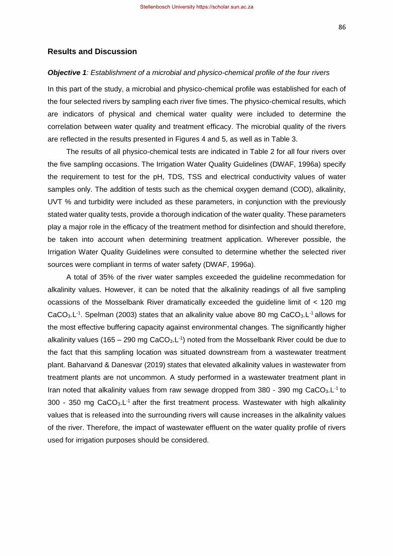
86
Results and Discussion
Objective 1: Establishment of a microbial and physico-chemical profile of the four rivers
In this part of the study, a microbial and physico-chemical profile was established for each of
the four selected rivers by sampling each river five times. The physico-chemical results, which
are indicators of physical and chemical water quality were included to determine the
correlation between water quality and treatment efficacy. The microbial quality of the rivers
are reflected in the results presented in Figures 4 and 5, as well as in Table 3.
The results of all physico-chemical tests are indicated in Table 2 for all four rivers over
the five sampling occasions. The Irrigation Water Quality Guidelines (DWAF, 1996a) specify
the requirement to test for the pH, TDS, TSS and electrical conductivity values of water
samples only. The addition of tests such as the chemical oxygen demand (COD), alkalinity,
UVT % and turbidity were included as these parameters, in conjunction with the previously
stated water quality tests, provide a thorough indication of the water quality. These parameters
play a major role in the efficacy of the treatment method for disinfection and should therefore,
be taken into account when determining treatment application. Wherever possible, the
Irrigation Water Quality Guidelines were consulted to determine whether the selected river
sources were compliant in terms of water safety (DWAF, 1996a).
A total of 35% of the river water samples exceeded the guideline recommedation for
alkalinity values. However, it can be noted that the alkalinity readings of all five sampling
ocassions of the Mosselbank River dramatically exceeded the guideline limit of < 120 mg
CaCO3.L-1. Spelman (2003) states that an alkalinity value above 80 mg CaCO3.L-1 allows for
the most effective buffering capacity against environmental changes. The significantly higher
alkalinity values (165 – 290 mg CaCO3.L-1) noted from the Mosselbank River could be due to
the fact that this sampling location was situated downstream from a wastewater treatment
plant. Baharvand & Danesvar (2019) states that elevated alkalinity values in wastewater from
treatment plants are not uncommon. A study performed in a wastewater treatment plant in
Iran noted that alkalinity values from raw sewage dropped from 380 - 390 mg CaCO3.L-1 to
300 - 350 mg CaCO3.L-1 after the first treatment process. Wastewater with high alkalinity
values that is released into the surrounding rivers will cause increases in the alkalinity values
of the river. Therefore, the impact of wastewater effluent on the water quality profile of rivers
used for irrigation purposes should be considered.
Stellenbosch University https://scholar.sun.ac.za

87
Table 2 Physico-chemical characteristics of the four selected rivers used for irrigation over five sampling occasions during the summer irrigation
period (October 2019 – January 2020)
SD – Standard deviation
Plankenburg Eerste Mosselbank Franschhoek
Parameter 1 2 3 4 5 Avg.
1 2 3 4 5 Avg.
1 2 3 4 5 Avg.
1 2 3 4 5 Avg.
SD SD SD SD
UVT % 60.3 60.0 64.5 61.2 31.2 55.4
13.7 62.2 62.0 63.9 56.1 52.1
59.3
5.0 22.2 30.0 28.3 25.0 30.2
27.1
3.5 80.0 80.0 79.2 80.2 49.1
73.7
13.7
TDS
(mg. L-1) 180 160 143 165 485
226
145 245 316 285 297 272
283
27 831 850 802 903 540
786
142 96 95 96 83 244
123
68
EC
(mS.mˉ¹) 0.30 0.19 0.17 0.22 0.55
0.28
0.16 0.34 0.43 0.32 0.37 0.34
0.36
0.04 1.0 1.16 0.91 0.99 0.63
0.93
0.20 0.13 0.11 0.14 0.13 0.26
0.15
0.06
COD (mg O₂. L⁻¹)
29 22 19 52 202 63
79 25 12 18 34 16
21
9 59 50 57 47 47
52
6 12 10 10 13 24
14
6
Turbidity
(NTU) 18.5 15.5 11.1 16.0 27
17.6
5.9 3.4 3.4 3.5 2.6 2.3
3.0
0.6 9.9 7.7 7.1 17.8 9.6
10.4
4.3 2.0 2.6 2.3 2.2 8.9
3.6
3.0
TSS
(mg.L⁻¹) 19 19 6 5 18
14
7 9 11 8 4 14
9
3 9 24 8 13 22
15
7 6 5 1 3 7
4
2
pH 7.21 6.91 6.88 6.72 6.48 7.59 7.41 7.28 7.38 7.35 7.39 7.49 7.31 7.43 7.19 6.76 6.78 6.75 6.79 7.14
Alkalinity (mg.L CaCO₃⁻¹)
75 90 63 70 168 93
43 106 106 93 100 95
100
6 290 205 220 270 165
230
50 50 45 55 45 135
66
39
Stellenbosch University https://scholar.sun.ac.za

88
The alkalinity values of the remaining three rivers fell within guideline recommendations
( <120 mg CaCO3.L-1), except for the fifth sampling ocassion of the Plankenburg (168 mg
CaCO3.L-1) and Franschhoek Rivers (135 mg CaCO3.L-1) (Table 2). The river water levels in
the Franschhoek River on this sampling ocassion appeared to be significantly lower with a
slower flow rate, and was coupled with increases in all other physico-chemical characteristics.
Most notably, a drop in UVT % from 80% to 50%, a quadrupled turbidity value (8.9 NTU) and
double the COD value (24 mg O₂.L⁻¹) – when comparing this sampling occasion to the
previous four sampling ocassions (Table 2). Similar findings were noted for the Plankenburg
River on the fifth sampling ocassion.
Water that contains high alkalinity values, and therefore, high levels of carbonates and
bicarbonate ions, results in insoluble minerals formed by calcium and magnesium ions that
remain in the soil after irrigation (Bauder et al., 2014). Scale build-up from carbonates in
irrigation water on drip or spray irrigation systems causes reduced water flow rates, and can
therefore, negatively impact irrigation efficacy (Bauder et al., 2014). Alkalinity is affected by
dissolved salts and the oxidation of organic matter which wouldn’t necessarily affect pH. It is
possible to have a water sample that has a lower pH but still ranks high in alkalinity, therefore,
still has the capacity to buffer changes in acidity levels, while being acidic (USEPA, 2011).
There is a positive correlation between the TDS and the alkalinity of the water samples (Table
2). These findings agree with a study performed by Shroff et al. (2015) where a 0.88 correlation
coefficient was reported between the TDS and alkalinity of water samples obtained from the
environment. High levels of carbonates in water samples have been associated with the
deposition or scaling on the UV lamp sleeve. This has an impact on the intensity of the UV
radiation that reaches the water (USEPA, 2003). Consistent cleaning of the lamp sleeve is
recommended for waters with high alkalinity values (USEPA, 2003).
The pH value of a water sample, according to Abdelrahim et al. (2013), has an effect on
the biological and chemical reactions that occur in water, as well as having an impact on the
solubility of metal ions. The specified pH range (DWAF, 1996a) for the irrigation water
samples, 6.5 – 8.4, is relatively wide and it is noted that all river water samples fell within this
range (Table 2). A general trend can be noted, where high alkalinity readings are associated
with higher pH values (Table 2). This correlates with a statement made by Valdez-Aguilar et
al. (2009), where high pH values in irrigation water are associated with high concentrations of
carbonates and bicarbonates. The microbial activity of water samples is directly impacted by
the water pH (Jin & Kirk, 2018). This is due to the fact that the pH is responsible for influencing
the acitivites and growth of the microbial communities.
Turbidity is determined by the amount of light that is scattered by particles within a water
sample, but this does not include any settled solids (Perlman, 2014). The guideline limits
Stellenbosch University https://scholar.sun.ac.za

89
(DWAF, 1996a) do not include a specification for the allowed turbidity for irrigation water. The
Water Quality Guidelines for Domestic Use (DWAF, 1996c) states that for turbidity values
exceeding 10 NTU, the water carries an associated risk of disease, and this guideline was
used in this study as a reference basis to comment on the water quality of the four selected
rivers. Of the four rivers studied, the Plankenburg River consistently had higher turbidity values
(11.1 – 26.9 NTU), all of which exceeded the guideline limit. The other rivers all had values
below 10 NTU (except on one occasion for the Mosselbank River) (Table 2). Interestingly, the
Plankenburg River also had the most consistent presence of pathogenic microorganisms of
all the rivers investigated (Table 3). Rapid clogging of irrigation equipment can be attributed
to the TSS and turbidity of a water sample and needs to be taken into account to ensure that
the UV treatment is applied consistently to all water samples (Ribeiro et al., 2004). A study
performed by Ribeiro et al. (2004) noted a positive relationship between turbidity and TSS
readings in irrigation water in Brazil. Furthermore, high-coefficient correlations were noted
between the TSS and bacterial and algae concentrations in irrigation water. Similar findings
were noted for increased turbidity levels (Ribeiro et al., 2004).
The total solids are the sum of the settleable solids, the suspended solids (TSS) and the
dissolved solids (TDS) (USEPA, 1999). The TSS of a water sample includes sand, silt, clay,
mineral precipitates as well as biological matter, according to Butler & Ford (2018), which
includes all particles that would be held back by a 2 μm filter (USEPA, 1999). The Mosselbank
River had consistently high dissolved solids content (540 – 903 mg.Lˉ¹) compared to the other
three rivers (average 82 – 485 mg.Lˉ¹) (Table 2). This was in contrast with the TSS values of
the Mosselbank River (8.2 – 24.2 mg.Lˉ¹), that were approximately in the same range as those
observed for the other three rivers over the sampling occasions (1.3 – 19.4 mg.Lˉ¹) (Table 2).
This is an indication of a higher concentration of dissolved salts and minerals, which could
provide an explanation for the higher alkalinity readings in the Mosselbank River. This
emphasises the potential impact that effluent from the wastewater treatment plant, situated
upstream from the sampling location, may have on the river water profile.
A linear relationship between the turbidity and TSS has been proposed by Hannouche
et al. (2011). High turbidity, which is a reflection of high levels of TDS and TSS in the sample,
implies a high level of organic and inorganic pollution (Johnson et al., 2010). The relationship
between TDS and TSS has been reported as not constant or proportional over time (Butler &
Ford, 2018). The variability in these two factors is, therefore, too great to allow for a direct
relationship to be established. Higher TDS readings are noted in the Eerste River (average of
283 mg.Lˉ¹) compared to the Plankenburg River (average of 226 mg.Lˉ¹), while the opposite
is reported for the TSS readings (averages of 9 and 14 mg.L-1 for these two rivers,
respectively) (Table 2). This could mean that the higher concentration of dissolved solids in
Stellenbosch University https://scholar.sun.ac.za

90
the Eerste River does not have a considerable influence on the UVT % values (which
remained relatively similar across the five sampling occasions of these two rivers). This could
indicate that the suspended solids may play a greater role in the scattering of light for the
Plankenburg River, or that the organic matter is resulting in the scattering of light.
A report by Islam et al. (2017) states that the TDS of a water sample correlates positively
with the electrical conductivity (EC) and directly affects the pH. As the TDS value increases,
the electrical conductivity increases, too, and a more acidic pH is reported (Islam et al., 2017).
Bauder et al. (2014) states that the electrical conductivity of irrigation water is the most
influential water quality parameter with regard to crop productivity. This is due to the fact that,
with higher EC values in irrigation water, the less water is available for the plant as it is unable
to compete with the ions present in the soil solution for water (Bauder et al., 2014). Therefore,
water available for transpiration of the plant decreases as the EC increases which ultimately
leads to reduced yield potential. In this study, the high alkalinity values observed for the
Mosselbank River generally correlated with slightly higher pH values (Table 2). The stated
correlation between an increased EC and TDS values hold true across all sampling occasions
for all four rivers (Table 2). Rusydi (2018) states that the relationship between electrical
conductivity and total dissolved solids in water sources as one that correlates linearly (R² =
0.97). This can be calculated as:
TDS = 0.65 x EC (where EC is µS.mˉ¹ and TDS is mg.Lˉ¹)
When applying this calculation to the recorded values for EC and TDS (Table 2), it is
evident that the correlation is correct. All rivers have electrical conductivity values within the
specified limit (DWAF, 1996a). Lowered EC readings, according to Edokpayi et al. (2018), are
most commonly reported in the rainy seasons of an area, which is as a result of the dilution
effect from increased precipitation. Increased evaporation during the drier season can lead to
increased levels of dissolved ions in river water (Edokpayi et al., 2018). As the current study
was performed in the drier season only, with limited rainfall, this correlation could not be
observed.
Chemical Oxygen Demand (COD) is a measure of the total amount of oxygen required
to oxidise all organic material into carbon dioxide and water (USEPA, 1999). There is currently
no stipulation within the irrigation guidelines (DWAF, 1996a) in South Africa regarding the
COD of a water sample for either irrigation water or domestically-used water. Therefore, the
Guidelines for Industrial Use (DWAF, 1996b) were consulted instead. These guidelines state
that acceptable limits for COD in irrigation water is < 75 mgO2.L-1 as irrigation water falls within
Category 4 Utility Water of Industrial Processes, which allows for the discharge of an effluent,
without the clogging of equipment.
Stellenbosch University https://scholar.sun.ac.za
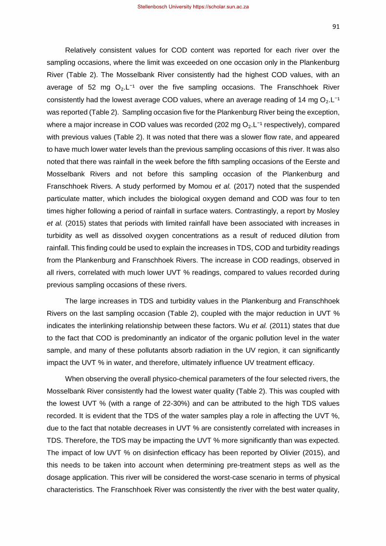
91
Relatively consistent values for COD content was reported for each river over the
sampling occasions, where the limit was exceeded on one occasion only in the Plankenburg
River (Table 2). The Mosselbank River consistently had the highest COD values, with an
average of 52 mg O₂.L⁻¹ over the five sampling occasions. The Franschhoek River
consistently had the lowest average COD values, where an average reading of 14 mg O₂.L⁻¹
was reported (Table 2). Sampling occasion five for the Plankenburg River being the exception,
where a major increase in COD values was recorded (202 mg O₂.L⁻¹ respectively), compared
with previous values (Table 2). It was noted that there was a slower flow rate, and appeared
to have much lower water levels than the previous sampling occasions of this river. It was also
noted that there was rainfall in the week before the fifth sampling occasions of the Eerste and
Mosselbank Rivers and not before this sampling occasion of the Plankenburg and
Franschhoek Rivers. A study performed by Momou et al. (2017) noted that the suspended
particulate matter, which includes the biological oxygen demand and COD was four to ten
times higher following a period of rainfall in surface waters. Contrastingly, a report by Mosley
et al. (2015) states that periods with limited rainfall have been associated with increases in
turbidity as well as dissolved oxygen concentrations as a result of reduced dilution from
rainfall. This finding could be used to explain the increases in TDS, COD and turbidity readings
from the Plankenburg and Franschhoek Rivers. The increase in COD readings, observed in
all rivers, correlated with much lower UVT % readings, compared to values recorded during
previous sampling occasions of these rivers.
The large increases in TDS and turbidity values in the Plankenburg and Franschhoek
Rivers on the last sampling occasion (Table 2), coupled with the major reduction in UVT %
indicates the interlinking relationship between these factors. Wu et al. (2011) states that due
to the fact that COD is predominantly an indicator of the organic pollution level in the water
sample, and many of these pollutants absorb radiation in the UV region, it can significantly
impact the UVT % in water, and therefore, ultimately influence UV treatment efficacy.
When observing the overall physico-chemical parameters of the four selected rivers, the
Mosselbank River consistently had the lowest water quality (Table 2). This was coupled with
the lowest UVT % (with a range of 22-30%) and can be attributed to the high TDS values
recorded. It is evident that the TDS of the water samples play a role in affecting the UVT %,
due to the fact that notable decreases in UVT % are consistently correlated with increases in
TDS. Therefore, the TDS may be impacting the UVT % more significantly than was expected.
The impact of low UVT % on disinfection efficacy has been reported by Olivier (2015), and
this needs to be taken into account when determining pre-treatment steps as well as the
dosage application. This river will be considered the worst-case scenario in terms of physical
characteristics. The Franschhoek River was consistently the river with the best water quality,
Stellenbosch University https://scholar.sun.ac.za

92
which was coupled with the lowest indicator counts (Fig. 6A) as well as pathogen presence
(Table 3). The UVT % of this river fell within the range of 49-80% (Table 2). This river will be
regarded as the best-case scenario for the rest of the study. The Eerste and Plankenburg
Rivers had UVT % readings of between 52-62%, with the exception of the last sampling event
of the Plankenburg River. These UVT % readings can be considered as adequate to ensure
effective disinfection of the water.
The UVT % of water samples is known to be greatly impacted by the turbidity as well as
the TSS (Farrell et al., 2018). This is due to the fact that, much like suspended solids,
microorganism aggregates can be enclosed within the particulates in the turbid samples,
which increases the resistance of microorganisms to UV penetration dramatically (Farrell et
al., 2018). Turbidity is seen to be a factor that most greatly influences the efficacy of UV
treatment of water, where an inverse relationship between UVT % and turbidity has been
reported (Gurol, 2005). The findings in the current study correlate with this statement, where
rivers that had the lowest turbidity readings consistently had the highest UVT %. Jones et al.
(2014) states that the relationship between turbidity and UV treatment efficiency is, however,
inconsistent. This is because factors that contribute to an increase in turbidity show great
variability in the ability to block or absorb UV radiation. Clarke and Bettin (2006) state that a
link between low turbidity values and the absence of pathogenic microorganism cannot be
guaranteed. Although there are seemingly conflicting opinions regarding the impact of turbidity
on UV treatment efficacy, it is evident that as the turbidity value increases, the exposure time
required to deliver a specific dose increases as the UVT % is decreased. Therefore, from an
economic standpoint, the turbidity does indeed play a major role in this method of water
disinfection. It is important to implement a pre-treatment step to ensure that the UV treatment
is applied to a consistent standard of water. Olivier (2015) states that inorganic as well as
organic compounds are able to absorb UV light in water samples, and therefore, the COD and
TDS are inversely proportional to the UVT %.
The factors that are responsible for impacting the efficacy of UV treatment for water
disinfection are the turbidity, suspended solids, dissolved solids, COD and UVT % of a water
sample (Hassen et al., 2000 & Gayán et al., 2014). Suspended solids, according to Abdul-
Halim & Davey (2016), reduce the efficacy of this treatment method either by shielding the
microorganisms from UV radiation or by absorbing the UV radiation, thereby, reducing the
energy available for microbial inactivation. When UV light is absorbed by suspended solids, it
is no longer available for inactivation of microorganisms and should, therefore, be taken into
account when determining the dosages for UV treatment (Liu, 2005). In order to achieve
complete disinfection of water samples, it is evident that the physico-chemical parameters play
Stellenbosch University https://scholar.sun.ac.za

93
a major role in impacting treatment, and should be consistently monitored to ensure an
effective treatment.
The observation of the microbial and physico-chemical characteristics of four rivers over
the summer irrigation period (October 2019 – January 2020), provides information about the
water profiles of these different river sources with differing upstream activities and overall
water quality. This, therefore, provides information about varied river water quality and allows
for broader UV treatment parameter optimisation that accommodates for the worst-case
scenario.
In this study, microbial indicator levels present in the four selected rivers over the five
sampling occasions were determined and are represented in Figs. 4 A-D as log cfu.mL-1.
Indicator organism tests included HPC, Enterobacteriaceae and E. coli. The error bars shown
in the following figures represent standard deviation, which indicate the variation between
duplicate plates poured, across six decimal dilutions.
As mentioned, the rivers were sampled five times each over the summer irrigation period
(October 2019 – January 2020). The microbial indicator counts were determined using a
dilution series of 10-1 – 10-6 and standard plating methods. Dilutions were plated in duplicate,
and the results were averaged. The results were recorded as log cfu.mL-1 (Figs. 4 A – D, 5, 6
A – D, 7 A – D and 8 A – D). The pathogen tests were performed on a presence/absence
basis only. The Irrigation Water Guidelines (DWAF, 1996a) states only that the E. coli counts
of an irrigation water sample should fall within 1 000 cfu.100 mL-1 to be deemed safe. This
correlates to 10 cfu.mL-1 or log 1 cfu.mL-1. There is no requirement to test for pathogens such
as Salmonella spp. and L. monocytogenes in the current guidelines. At present, few studies
exist regarding the prevalence of these pathogens in South African surface waters in South
Africa, and knowledge gaps exist on how effectively UV disinfection can be used to reduce
pathogen levels present in surface waters worldwide.
Stellenbosch University https://scholar.sun.ac.za

94
Figures 4 A – D provide an indication of the great variability in water quality, not only
between the four selected rivers but also between the sampling occasions for a river over the
summer irrigation period. These colony counts were, where possible, compared to the
Irrigation Water Guidelines (DWAF, 1996a) to determine the fitness for use. The fourth
sampling occasion of the Mosselbank River saw an E. coli count of log 5.45 ± 0.03 cfu.mL-1
which equates to 279 000 cfu.mL-1. This was the highest E. coli count recorded of all four
rivers. The lowest recorded E. coli count was sampling occasion three in the Eerste River. A
log 2.55 ± 0.01 cfu.mL-1 equates to an average of 355 cfu.mL-1. Even the lowest E. coli colony
0,0
1,0
2,0
3,0
4,0
5,0
6,0
1 2 3 4 5
Log
CFU
. mLˉ
¹
Mosselbank
HPC Enterobacteriaceae E. coli
0,0
1,0
2,0
3,0
4,0
5,0
6,0
1 2 3 4 5
Log
CFU
. mLˉ
¹
Franschhoek
HPC Enterobacteriaceae E. coli
0,0
1,0
2,0
3,0
4,0
5,0
6,0
1 2 3 4 5
Log
CFU
. mLˉ
¹
Eerste
HPC Enterobacteriaceae E. coli
0,0
1,0
2,0
3,0
4,0
5,0
6,0
1 2 3 4 5
Log
CFU
. mLˉ
¹
Plankenburg
HPC Enterobacteriaceae E.coli
Figure 4 A-D Results of indicator organism colony counts before UV treatment is applied,
expressed as log cfu.mL-¹, with standard deviation error bars included
0,0
1,0
2,0
3,0
4,0
5,0
6,0
1 2 3 4 5
Log
CFU
. mLˉ
¹
Mosselbank
HPC Enterobacteriaceae E. coli
4A 4B
4C 4D
Stellenbosch University https://scholar.sun.ac.za

95
count observed dramatically exceeded the guideline limit. This confirms the need for effective
water disinfection treatment prior to irrigation. It can be noted in Figures 4 A – D that there are
some instances where the E. coli counts are higher than the Enterobacteriaceae counts. This
could primarily be as a result of the methodology, specifically, the different agar used that
caused this result.
Figure 5 provides a visual representation of the average recorded values over the five
sampling events per river for each indicator population. This provides an overall microbial
quality indication of each of the rivers’ for the entire summer irrigation period. Increased counts
did, in some instances, correlate with worsening physico-chemical characteristics. For
instance, a significant increase in indicator colony counts was noted from the first four
sampling occasions of the Franschhoek River to the fifth sampling occasion (Fig 4A). This
correlated to a substantial UVT % decrease, and all other physico-chemical results (except
pH) almost doubled, compared to the previous four sampling occasions (Table 2). Likewise, a
similar trend was observed between sampling occasions four and five of the Plankenburg
River (Table 2). It can therefore be noted that, a tentative correlation could be made between
the physico-chemical and microbial quality characteristics of the water sampled. The
Plankenburg and Mosselbank Rivers were consistently the most contaminated rivers,
considering the microbial indicator counts (Fig. 4B & 4D). Even though the Eerste and
Franschhoek Rivers (Fig. 4C & 4A) had E. coli counts that were lower than the other two rivers,
these counts still markedly exceeded the water quality guideline limits. Therefore, on the basis
of E. coli levels only, all four rivers were deemed unacceptable for agricultural use without pre-
treatment.
Escherichia is one of many genera that fall within the Enterobacteriaceae family. It can
be noted from Figures 4 A–D that little variation existed between Enterobacteriaceae and E.
coli counts. Statistical analysis was performed to determine if there are significant differences
between the E. coli and Enterobacteriaceae counts observed across the five sampling
occasions per river. The p-values obtained in this experiment were; 0.353, 0.608, 0.811, and
0.628 for the Plankenburg, Eerste, Franschhoek and Mosselbank Rivers, respectively. At a
95.0% confidence interval, no significant difference (p<0.05) was observed between the
colony counts of Enterobacteriaceae and E. coli. A tentative conclusion could be drawn that
the Enterobacteriaceae population predominantly consisted of E. coli strains. The HPC values
vary greatly between rivers and across sampling occasions. No correlation can be made
between HPC values and Enterobacteriaceae/ E. coli counts. Due to the fact that the HPC
test is a non-selective test for the total aerobic bacteria in the water sample, it is difficult to
draw conclusions as to what kind of contamination had occurred before certain sampling
occasions where HPC counts were high, for example, sampling occasion one of the
Stellenbosch University https://scholar.sun.ac.za

96
Franschhoek River (Fig. 4A). Overall, these results provide a good baseline microbial profile
for each river, which aids in the understanding of how best to disinfect the river water.
Upstream activities, be it recreational or industrial, dumping of household waste and animal
defecation may all play a role in the microbial populations in the river waters. Changes in
weather, such as extreme winds, heat or rainfall may too play a role in these findings.
Understanding how these factors fluctuate across the irrigation season in the Western Cape
are of great importance to ensure that the most effective treatment is applied, taking the worst-
case scenario into consideration.
A report by Sigge et al. (2016) has highlighted the possibility of a potential food safety
risk that is associated with the presence of pathogens in irrigation water. Sigge et al. (2016)
proposed the importance of setting limits for important food pathogens in addition to E. coli,
such as L. monocytogenes and Salmonella spp., in irrigation water, to ensure a reduction in
foodborne outbreaks associated with fresh produce. The results of the pathogen tests done in
this study are presented in Table 3, where the presence and absence of the pathogens
Salmonella spp. and L. monocytogenes in the untreated river water samples are indicated.
The Plankenburg and Mosselbank Rivers, again, had the most consistent pathogen
presence over the five sampling occasions. The Franschhoek and Eerste Rivers were the only
rivers to have Salmonella spp. present on one sampling occasion only, which was sampling
occasion five. It must be noted that there were extremely high temperatures in the week before
testing, which may have influenced the presence of this pathogen. L. monocytogenes was
present in all but one sampling occasion of the Eerste River. The Franschhoek River had the
lowest prevalence of Salmonella spp. and L. monocytogenes.
0
1
2
3
4
5
6
HPC E. coli Enterobacteriaceae
Log
CFU
.mLˉ
¹
Plankenburg Eerste Mosselbank Franschhoek
Figure 5 Average colony counts for all indicator organisms tested across five sampling
occasions, indicated as log cfu.mL-¹ with standard deviation bars included for error
across all five sampling occasions per river
Stellenbosch University https://scholar.sun.ac.za

97
Vasquez-Boland et al. (2001) states that Listeria spp. are saprophytic organisms, that
feed on dead organic matter in the environment and are ubiquitous in nature. The presence
of Listeria spp. in general in river water is thus, not surprising. Unfortunately, L.
monocytogenes in particular carry pathogenic genes that can be activated the moment they
enter the human body (Vasquez-Boland et al., 2001). The need to control this organism in an
industry that involves minimally processed foods is therefore, of great importance to ensure
consumer safety.
Table 3 Presence and absence of Salmonella spp. and L. monocytogenes in river water
samples, prior to the application of UV radiation. Shaded blocks with a “+” sign represent a
positive test for the specific organism over the five sampling occasions
The consistent presence of Salmonella spp. and L. monocytogenes in the four rivers
across the five sampling occasions indicate that these are not irregular findings. This indicates
that these rivers are consistently contaminated and therefore, pose a great risk for farmers
and other individuals using the water without pre-treatment. McEgan et al. (2013) states that
the prevalence of indicator organisms such as E. coli in surface waters can be an indication
of the presence of pathogenic microorganisms, such as the ones tested here. This finding
correlates with the colony counts observed in the Mosselbank and Plankenburg Rivers, where
high indicator counts are correlated with a consistent pathogen presence. These findings
further exacerbate the need for guidelines in South Africa to include pathogen limits, to ensure
that these microorganisms can be treated effectively and will not pose a risk for consumers of
fresh produce irrigated with these waters.
River Pathogen tested 1 2 3 4 5
Plankenburg Salmonella spp. + + - + +
L. monocytogenes + + + + +
Eerste Salmonella spp. - - - - +
L. monocytogenes + + + + -
Mosselbank Salmonella spp. - + - + +
L. monocytogenes + + + + +
Franschhoek Salmonella spp. - - - - +
L. monocytogenes - + + - +
Stellenbosch University https://scholar.sun.ac.za

98
Objective 2: The efficacy of low-pressure UV radiation on microbial populations in river water
The effect of UV radiation on microbial counts was evaluated using a laboratory-scale
collimated beam LP UV system. These water samples were exposed to three UV doses, and
the microbial load after each applied dose was tested and recorded (Figs. 6 – 8 & Tables 3
and 5). In this study, it was observed that UV radiation is an effective treatment method, as all
microbial counts were reduced to a level that would be considered adequate for use, according
to the Irrigation Water Guidelines (DWAF, 1996a).
Table 4 Exposure times (min) calculated from the UVT % of each river water sample, UV lamp
intensity (78.8 mW.cm-2) and sample depth for the three specified UV dosages (mJ.cm-2),
based on data obtained in the first sampling occasion
Table 4 provides an indication of the exposure times required for UV radiation at three different
doses (20, 40, 60 mJ.cm-2), using a lamp intensity reading of 78.8 mW.cm-2 and the respective
UVT % reading of each river. Kahn et al. (2015) states that scattering of UV light in highly
turbid water results in the reduction of UVT %, and this leads to extended exposure times for
effective disinfection. In Table 4, water from the Mosselbank River had a UVT % of 22.2% (on
sampling occasion 1), which required an in exposure time of 22:24 (min: sec) to ensure that a
dose of 20 mJ.cm-2 was applied. In contrast, water from the Franschhoek River required an
exposure time of 6:06 to achieve the same dose, based on the UVT % of 80% (Table 4). The
time taken to apply the second dose (40 mJ.cm-2) and third dose (60 mJ.cm-2), as can be seen
in Table 4 decreases with each dose, this is due to the fact that the respective amount of
sample is removed for microbial testing after each dose. The sample depth, therefore,
decreases and reduces the time required to apply the next dose as the calculation for
exposure times includes the sample depth. It is evident that, to apply a 60 mJ.cm-2 dose, a
significant amount of time is required, which impacts efficiency and economic feasibility.
Therefore, implementing pre-treatment steps to improve UVT %, such as filtration, the
Rivers Plankenburg
River Eerste River
Mosselbank River
Franschhoek River
UVT %
60.3 62.2 22.2 80
UV doses (mJ.cm-2)
Exposure time (min: sec)
20 9:02 8:41 22:24 6:06
40 16:29 15:55 38:42 11:36
60 22:31 21:50 49:22 16:35
Stellenbosch University https://scholar.sun.ac.za

99
exposure time might be dramatically reduced and increase throughput greatly. Sivhute (2019)
states that by utilising a pre-treatment step to ensure the physical water quality is improved,
could translate to increased log reductions once UV radiation is applied. The microbial
disinfection efficacy and process efficiency are factors of great importance in any water
treatment method. Ensuring that the greatest disinfection occurs in an economically feasible
timeframe is of utmost importance. When upscaling to a pilot-scale system, a medium-
pressure (MP) lamp will deliver higher UV light intensities than the LP lamp utilised in the
collimated-beam system, and would decrease exposure time significantly.
Okpara et al. (2011) states that chlorine, in sufficient doses, is able to disinfect water
samples of microorganisms within 30 minutes. When one considers the negative
environmental impact of chlorine as a disinfection method, it is also of great importance to
take the economics into consideration. No matter the sample size, disinfection with chlorine
takes a predetermined amount of time. So, an exposure time of 22 minutes (required for the
Mosselbank River) (Table 4), as the worst-case scenario, is not nearly as long as the exposure
time required for chlorine disinfection at any scale. Ultraviolet light intensities can be increased
exponentially, especially through the use of MP UV systems, and therefore, the contact time
can be significantly reduced. Another factor is that the cost of UV radiation does not increase
linearly as it does with chlorine. Cost and time required for disinfection are two factors that,
from a farmer’s perspective, may govern the choice of which treatment method to use.
Results of indicator organism response to UV radiation
Water samples were exposed to UV light in a collimated-beam UV machine at laboratory-
scale. The colony counts before treatment were recorded, along with the counts after exposure
to the three selected UV doses. These results are shown in Figs. 6A-D – 8A-D and are
expressed as log cfu.mL-1. Table 5 provides an indication of the presence of the tested
pathogens, before and after UV treatment. These results are expressed on a
presence/absence basis.
Stellenbosch University https://scholar.sun.ac.za

100
Escherichia coli
6B
6C 6D
0,0
1,0
2,0
3,0
4,0
5,0
6,0
1 2 3 4 5
Log
CFU
.mLˉ
¹
Eerste
Before treatment 20 mJ.cmˉ² 40 mJ.cmˉ² 60 mJ.cmˉ²
0,0
1,0
2,0
3,0
4,0
5,0
6,0
1 2 3 4 5
Log
CFU
.mLˉ
¹
Mosselbank
Before treatment 20 mJ.cmˉ² 40 mJ.cmˉ² 60 mJ.cmˉ²
0,0
1,0
2,0
3,0
4,0
5,0
6,0
1 2 3 4 5
Log
CFU
.mLˉ
¹
Plankenburg
Before treatment 20 mJ.cmˉ² 40 mJ.cmˉ² 60 mJ.cmˉ²
0,0
1,0
2,0
3,0
4,0
5,0
6,0
1 2 3 4 5
Log
CFU
.mLˉ
¹
Franschhoek
Before treatment 20 mJ.cmˉ² 40 mJ.cmˉ² 60 mJ.cmˉ²
6A
Figure 6 A – D E. coli counts expressed as log cfu.mL-¹ before and after three UV radiation
doses for four rivers over five sampling occasions, where 1.0 log cfu.mL-1 is the detection limit,
the red line at 1.0 log cfu.mL-1 indicates the lowest detection limit, below which there was no
growth noted
Stellenbosch University https://scholar.sun.ac.za

101
Heterotrophic Plate Count
Figure 7 A – D Heterotrophic Plate Count colony counts expressed as log cfu.mL-¹ before and
after three UV radiation doses for four rivers over five sampling occasions, where 1.0 log
cfu.mL-1 is the detection limit, the red line at 1.0 log cfu.mL-1 indicates the lowest detection
limit, below which there was no growth noted
0,0
1,0
2,0
3,0
4,0
5,0
6,0
1 2 3 4 5
Log
CFU
.mLˉ
¹
Plankenburg
No treatment 20 mJ.cmˉ² 40 mJ.cmˉ² 60 mJ.cmˉ²
0,0
1,0
2,0
3,0
4,0
5,0
6,0
1 2 3 4 5
Log
CFU
.mLˉ
¹
Eerste
No treatment 20 mJ.cmˉ² 40 mJ.cmˉ² 60 mJ.cmˉ²
0,0
1,0
2,0
3,0
4,0
5,0
6,0
1 2 3 4 5
Log
CFU
.mLˉ
¹
Franschhoek
No treatment 20 mJ.cmˉ² 40 mJ.cmˉ² 60 mJ.cmˉ²
0,0
1,0
2,0
3,0
4,0
5,0
6,0
1 2 3 4 5
Log
CFU
.mLˉ
¹
Mosselbank
Before treatment 20 mJ.cmˉ² 40 mJ.cmˉ² 60 mJ.cmˉ²
7B
7C 7D
7A
Stellenbosch University https://scholar.sun.ac.za

102
Enterobacteriaceae
8B
8C 8D
8A
0,0
1,0
2,0
3,0
4,0
5,0
6,0
1 2 3 4 5
Log
CFU
.mLˉ
¹
Plankenburg
No treatment 20 mJ.cmˉ² 40 mJ.cmˉ² 60 mJ.cmˉ²
0,0
1,0
2,0
3,0
4,0
5,0
6,0
1 2 3 4 5Lo
g C
FU.m
Lˉ¹
Eerste
No treatment 20 mJ.cmˉ² 40 mJ.cmˉ² 60 mJ.cmˉ²
0,0
1,0
2,0
3,0
4,0
5,0
6,0
1 2 3 4 5
Log
CFU
.mLˉ
¹
Franschhoek
No treatment 20 mJ.cmˉ² 40 mJ.cmˉ² 60 mJ.cmˉ²
0,0
1,0
2,0
3,0
4,0
5,0
6,0
1 2 3 4 5
Log
CFU
.mLˉ
¹
Mosselbank
Before treatment 20 mJ.cmˉ² 40 mJ.cmˉ² 60 mJ.cmˉ²
Figure 8 A – D Enterobacteriaceae colony counts expressed as log cfu.mL-¹ before and after
three UV radiation doses for four rivers over five sampling occasions, where 1.0 log cfu.mL-1 is
the detection limit, the red line at 1.0 log cfu.mL-1 indicates the lowest detection limit, below
which there was no growth noted
Stellenbosch University https://scholar.sun.ac.za

103
The log reductions recorded for all indicator organism tests, after the lowest dose of UV
radiation applied (20 mJ.cm-2), indicate the efficacy of this treatment method for disinfection.
After 20 mJ.cm-2 of radiation is applied, the water quality, would be deemed safe for use
according to the Irrigation Water Guidelines (DWAF, 1996a), for E. coli counts. Sivhute (2019)
states that the sensitivity of a microorganism to UV radiation can be described by the degree
of log reduction that is observed following radiation. Higher log reductions would be noted for
UV sensitive organisms, than observed for UV resistant strains. The higher the initial microbial
load, the larger the log reduction value will be. Log reduction values do not provide an
indication of the initial microbial loads.
In this study, initial E. coli counts over the five sampling occasions, ranged between
7 900 – 291 000, 300 – 73 000, 350 – 1 260 and 1 870 – 74 000 cfu.mL-1, for the Mosselbank,
Franschhoek, Eerste and Plankenburg Rivers, respectively (Figs. 6A-D). The microbial
reductions after UV radiation are presented in these figures, where no detectable colonies
were present after the first dose (20 mJ.cm-2) was applied, except for two sampling occasions
of the Mosselbank River and one of the Franschhoek River (Figs. 6C & 6D, respectively). The
HPC microbial levels ranged between 2 900 – 1 780 000, 200 – 340 000, 600 – 17 500 and
12 400 – 198 000 cfu.mL-1, for the Mosselbank, Franschhoek, Eerste and Plankenburg Rivers,
respectively (Figs. 7A-D). Microbial levels after each dose of UV was applied varied between
rivers and across sampling occasions. There were no detectable microbial colonies after 60
mJ.cm-2 across all rivers. The microbial counts for Enterobacteriaceae, ranged between 7 700
– 198 000, 160 – 100 000, 240 – 1 370 and 1 270 – 174 000 cfu.mL-1 for the rivers, respectively
(Figs. 8A-D). Colony counts after UV radiation was applied was limited to one sampling
occasion each of the Franschhoek and Mosselbank Rivers (Figs. 8C & 8D, respectively). The
largest log reduction recorded was noted on sampling occasion four of the Mosselbank River,
with a 5.2 log reduction, for Heterotrophic Plate Count colony counts. This was also the
microbial test that had the highest initial colony count, of all the sampling occasions for all four
rivers. A total of 1. 78 x 106 cfu.mL-1 was recorded on this sampling occasion, before UV
treatment was applied. This finding was coupled with increased TDS content and high turbidity
readings (Table 2). This correlates with Chahal et al. (2016) that stated that increased turbidity
values support the transport of attached pathogenic microorganisms, and therefore, increased
treatment demands for disinfection.
It can be noted, that a more gradual reduction in the HPC population after UV radiation
was recorded, than for the Enterobacteriaceae and E. coli populations, particularly in the
Mosselbank River (Fig. 7D). This finding correlates with similar findings observed by Bester
(2015) and Olivier (2015). The HPC population typically represents a broad spectrum of
aerobic microorganisms, both Gram-positive and Gram-negative, that are able to grow on
Stellenbosch University https://scholar.sun.ac.za

104
Plate Count Agar (Oxoid, South Africa) at 30°C. When observing the HPC counts of the
Mosselbank River, the prevalence of microorganisms after 20 and 40 mJ.cm-2 of UV radiation
was much higher than observed for any other river, and any other indicator population (E. coli
and Enterobacteriaceae). These results could have a two-fold explanation. The overall initial
microbial concentration in the Mosselbank River was higher than the other rivers (Fig 7A-D),
therefore, even after a number of log reductions after UV radiation, the microbial count is still
higher than the other rivers. Another possible explanation is that the type of microorganisms
present in the microbial population in this river could be of a greater diversity, or show greater
resistance to UV radiation than the organisms present in the other rivers, and therefore, result
in their presence after 40 mJ.cm-2 and 60 mJ.cm-2 (Fig 7D). This could possibly be attributed
to the effluent from the upstream water treatment plant releasing resistant bacteria into the
river. These bacteria, which are clearly more resistant to UV than the Enterobacteriaceae and
E. coli colonies, could show resistance to antimicrobial agents, too.
As discussed in the physico-chemical section, the suspended solids of a water sample
reduce the efficacy of disinfection by shielding microorganisms or by the absorption of UV
radiation (Abdul-Halim & Davey, 2016). The TSS and the UVT % of water samples were also
reported to have an inverse relationship, which plays a role in the efficacy of UV disinfection.
These factors should be taken into account when determining UV dosages. Water that has a
lower turbidity, ultimately has higher UVT % readings and therefore, results in an improved
disinfection efficacy. This is due to the fact that, much like suspended solids, microorganism
aggregates can be enclosed within the particulates in the turbid samples, which increases the
microorganism’s resistance to UV penetration dramatically (Farrell et al., 2018). Liu (2005)
reported that an increase in turbidity from 1 to 10 NTU would reduce the average dosage by
between 5 and 33%.
Based on the results obtained in the log reductions for indicator organisms after UV
radiation, it can be noted that treatment efficacy is not impacted by water quality, however, the
exposure time required to deliver the required dose is dramatically impacted. This has an
impact on the efficiency or productivity of the user at farm-scale. A treatment step, possibly in
the form of a filtration step, implemented before UV radiation to remove some organic matter
could reduce exposure times required by improving the UVT %. The pathogen results obtained
following UV radiation can be seen in Table 5.
Stellenbosch University https://scholar.sun.ac.za

105
Pathogen results after UV radiation
Table 5 Presence/absence results of Listeria monocytogenes (LM) and Salmonella spp. (S)
for four rivers, over five sampling repetitions (rep.) after three UV radiation doses
0 =No treatment, 1 = 20 mJ.cmˉ², 2 = 40 mJ.cmˉ², 3 = 60 mJ.cmˉ² of UV radiation
LM – Listeria monocytogenes, S – Salmonella spp.
Table 5 indicates the presence/absence results of the pathogen tests, before and after UV
radiation at the specified doses. It is interesting to note that, on all but one occasion, the
pathogens; L. monocytogenes and Salmonella spp. were no longer present after the lowest
dose of UV radiation was applied. The presence of L. monocytogenes after 20 mJ.cm-2 in the
Plankenburg River on the second sampling occasion might be attributed to a higher initial
concentration of this organism in the river on this particular occasion. This is, however, a
speculative observation as the detection methods used for both pathogens in this study are
enrichment methods that do not allow for the determination of the actual microbial levels prior
to enrichment. When observing the physico-chemical results on this day, there is no outlying
factor or significantly differing characteristic that could contribute to this particular result. Due
to the fact that the pathogens on other sampling events of this river, as well as the other three
rivers were no longer present after the 20 mJ.cm-2 UV dose was applied (Table 5), it was
concluded that this dose was mostly effective against the prevalent levels of pathogens in the
rivers at the time of testing. The Franschhoek River had the lowest frequency of pathogen
presence across the five sampling occasions. This correlates with the findings that this river
had the best physico-chemical characteristics, of all four rivers. It can be noted, however, that
the presence of Salmonella spp. on the fifth sampling occasion, was coupled with deteriorating
water quality, including decreased UVT % and significant increases in TDS, EC, COD, turbidity
and alkalinity (Table 2).
Rep. 1 Rep. 2 Rep. 3 Rep. 4 Rep. 5
River Test 0 1 2 3 0 1 2 3 0 1 2 3 0 1 2 3 0 1 2 3
Plankenburg S + - - - + - - - - - - - + - - - + - - -
LM + - - - + + - - + - - - + - - - + - - -
Eerste S - - - - - - - - - - - - - - - - + - - -
LM + - - - + - - - + - - - + - - - - - - -
Mosselbank S - - - - + - - - - - - - + - - - + - - -
LM + - - - + - - - + - - - + - - - + - - -
Franschhoek S - - - - - - - - - - - - - - - - + - - -
LM - - - - + - - - + - - - - - - - + - - -
Stellenbosch University https://scholar.sun.ac.za

106
Gayán et al. (2014) stated that, in general, pathogenic bacterial strains are more
resistant to UV radiation than non-pathogenic strains. A study performed by Gayán et al.
(2012) on the effect of UV radiation on different Salmonella spp. with regard to their individual
resistances, and noted that Salmonella typhimurium STCC 878 was the most resistant strain,
requiring 18.03 mJ.cm-2 to achieve a 4-log reduction. In the current study, it can be noted from
the outlying result in the Plankenburg River, that when deciding on an effective dose to ensure
complete microbial disinfection and water safety, that a dose of >20 mJ.cm-2 may be required
if pathogens are present at higher initial levels, or if more resistant strains are present.
Thirteen microbial strains were isolated from these tests and stored according to the
procedure previously mentioned for further investigation in Chapter 4.
Conclusions
In this study, the utilisation of UV radiation as a method of disinfection of several river waters
with varying water quality has, thus, been proven successful. The results of the indicator and
pathogen testing in this study indicated that these rivers are not acceptable for agricultural use
without pre-treatment. The indicator counts, specifically E. coli, notably exceeded the guideline
limits on all sampling occasions. This means that transfer of E. coli from the water to fresh
produce may be highly likely during agricultural irrigation. This presents a significant problem
because pathogenic strains of E. coli and other pathogenic microorganisms may result in
foodborne disease outbreaks following the consumption of contaminated produce. The
baseline microbial and physico-chemical profiles of the rivers that were established, enabled
the researcher to investigate the efficacy of UV disinfection over a broader range of rivers. A
tentative correlation was made between the physico-chemical characteristics of a water
sample and the microbial population levels, where deteriorating physico-chemical
characteristics were coupled with higher microbial loads as well as a more consistent
pathogen presence.
The Mosselbank River consistently had the poorest physico-chemical characteristics as
well as the highest indicator population counts, and pathogen presence. This river could be
considered as the ‘worst-case scenario’ for further UV treatment optimisation studies. If the
dose requirements of UV radiation are based on ensuring that this river falls within the
guideline specifications, there is a high probability that water from the other selected rivers will
also be deemed safe after treatment.
The efficacy of UV radiation in the disinfection of microorganisms present in river water
was confirmed in this study, where even at the lowest dose applied, the E. coli counts fell well
within guideline limits. The HPC test results indicated a slower drop in microbial loads –
Stellenbosch University https://scholar.sun.ac.za

107
specifically in the Mosselbank River – which is indicative of a diverse and possibly more
resistant microbial population in this river. Enterobacteriaceae levels decreased immediately
after the lowest UV dose was applied. The recommended UV dose to ensure sufficient
microbial disinfection for indicator organisms in rivers with similar profiles as those included in
this study can thus be suggested to be between 20 – 40 mJ.cm-2.
The pathogen results of this study indicate the efficacy of this treatment method against
Salmonella spp. and L. monocytogenes after the lowest UV dose applied. Taking the outlying
result observed in the Plankenburg River, where L. monocytogenes was present after 20
mJ.cm-2 of UV radiation on one occasion into consideration, the recommended dose to ensure
microbial disinfection for pathogenic microorganisms can, therefore, be suggested as between
20 – 40 mJ.cm-2. The presence/absence testing of these pathogens does not provide an
indication of the microbial load present. A recommendation would be to include a study on the
pathogenic microorganisms isolated from the rivers at known initial concentrations to observe
the response to UV radiation.
These recommendations, based on the results of the experiments in the current study,
fall in line with USEPA (1999) suggesting that a dose of between 21-36 mJ.cm-2 should be
sufficient in the inactivation of bacterial and viral pathogens depending on water quality
parameters such as turbidity and COD.
The organisms isolated and stored from these experiments will be utilised in Chapter 4
to determine the effect of an increased microbial concentration on UV disinfection efficacy, as
well as to determine the resistance profiles between the river water isolates and isolates from
other environmental and clinical sources.
Overall, the rivers examined in this study proved to be highly diverse considering their
physico-chemical and microbial profiles. The results of the UV radiation experiments provide
an indication that this treatment method is effective in ensuring microbial disinfection, and
therefore, capable of providing a safe water supply for the irrigation of crops. It can, therefore,
be concluded that UV radiation is effective at laboratory-scale and can be utilised to ensure
river water disinfection of both indicator organisms as well as pathogenic microorganisms.
This study provides sufficient information regarding the presence of selected microbial
populations in the river waters as well as UV dose responses to facilitate an investigation into
river water disinfection for use at farm-scale.
Stellenbosch University https://scholar.sun.ac.za

108
References
AbdelRahim, K. A. A., Hassanein, A. M., Sabry Younis, M., Abd El-Azeiz Heikal, A. (2013).
Correlation between Physico-chemical and Microbiological Characteristics of River Nile
Water in Sohag Governorate, Egypt. Journal of Pure and Applied Microbiology, 7, 63-
77. Accessed 14/04/2020.
Abdul-Halim, N. & Davey, K.R. (2016). Impact of suspended solids on Fr 13 failure of UV
irradiation for inactivation of Escherichia coli in potable water production with turbulent
flow in an annular reactor. Chemical Engineering Science, 143, 55–62.
Addinsoft. (2020). XLSTAT statistical and data analysis solution. New York, USA.
https://www/xlstat.com.
APHA (American Public Health Association) (2005). Standard Methods for the Examination of
Water and Wastewater, 21st ed. Washington, DC: American Public Health Association
Press.
Amoah, K., Craik, S., Smith, D.W. and Belosevic, M. 2005. Inactivation of Cryptosporidium
oocysts and Giardia cysts by Ultraviolet light in the presence of natural particulate
matter, AQUA, Journal of Water Supply Research and Technology, 54(3): 165-178.
Baharvand, S. & Daneshvar, M. R. M. (2019). Impact assessment of treating wastewater on
the physicochemical variables in the environment: a case of Kermanshah wastewater
treatment plant in Iran. Environmental Systems Research, 8(18), 1 – 11. DOI
10.1186/s40068-019-0146-0.
Barnes, J.M. (2003). The Impact of Water Pollution from Formal and Informal Urban
Developments along the Plankenburg River on Water Quality and Health Risk. PhD
Dissertation in Epidemiology, University of Stellenbosch, South Africa.
Bauder, T. A., Waskom, R. M., Sutherland, P. L. & Davis, J. G. (2014). Irrigation Water Quality
Criteria- Soil and Crop Science. Colorado State University: CSU.
Bezuidenhout, C. (2013). A Large-Scale Study of Microbial and Physico-Chemical Quality of
Selected Groundwaters and Surface Waters in the North West Province, South Africa.
WRC Report 1966/1/13.
Bolton, J. R. & Cotton, C. A. (2008). The Ultraviolet Disinfection Handbook. Pp. 1-149. USA:
American Water Works Association.
Britz, T. J., Sigge, G.O., Buys, E. M., Schmidt, S., Potgieter, N. & Taylor, M. B. (2012). Baseline
study on extent (types and quantities) of contamination found in irrigation water at
selected sites (K5/1773). In: Quantitative investigation into the link between irrigation
water quality and food safety (Volume 2). WRC Report no. K5/1773/4, Pretoria, South
Africa.
Britz, T.J., Sigge, G.O., Huisamen, N., Kikine, T., Ackermann, A., Lotter, M., Lamprecht, C. &
Kidd, M. (2013). Fluctuations of indicator and index microbes as indication of pollution
over three years in the Plankenburg and Eerste Rivers, Western Cape, South Africa.
Water SA, 39(4), 457-465.
Butler, B A. & Ford, R. G. (2018). Evaluating relationships between total dissolved solids
(TDS) and total suspended solids (TSS) in a mining-influenced watershed. Mine Water
Environment, 37(1), 18-30. DOI 10.1007/s10230-017-0484-y.
Campbell, A.T. & Wallis, P. (2002). The effect of UV irradiation on human-derived Giardia
lamblia cysts. Water Research, 36(4), 963- 969.
Stellenbosch University https://scholar.sun.ac.za

109
Chahal, C., van den Akker, B., Young, F., Franco, C., Blackbeard, J. & Monis, P. (2016).
Pathogen and Particle Associations in Wastewater. Advanced Applications in
Microbiology, 97, 63-119. DOI 10.1016/bs.aambs.2016.08.001
Chigor, V. N., Sibanda, T. & Okoh, A. I. (2013). Studies on the bacteriological qualities of the
Buffalo River and three source water dams along its course in the Eastern Cape
Province of South Africa. Environmental Science and Pollution Research, 20, 4125-
4136. DOI 10.1007/s11356-012-1348-4.
Clarke, S. & Bettin, W. (2006). Chlorine dioxide disinfection in the use of the individual water
purification devices. Technical Information Paper, 11, 1-15.
DWAF (Department of Water Affairs and Forestry). (1996a). South African Water Quality
Guidelines (2nd edition). Volume 4: Irrigation water use, 1-123. CSIR Environmental
Services, Pretoria.
DWAF (Department of Water Affairs and Forestry). (1996b). South African Water Quality
Guidelines (2nd edition). Volume 3: Industrial water use, 1-123. CSIR Environmental
Services, Pretoria.
DWAF (Department of Water Affairs and Forestry). (1996c). South African Water Quality
Guidelines (2nd edition). Volume 1: Domestic Use, 1- 130. CSIR Environmental
Services, Pretoria.
Edokpayi, J. N., Odiyo, J. O., Popoola, E. O. & Msagati, T. A. M. (2018). Evaluation of
Microbiological and Physico-chemical Parameters of Alternative Source of Drinking
Water: A Case Study of Nzhelele River, South Africa. The Open Microbiology Journal,
12, 18-27. DOI 10.2174/1874285801812010018. Accessed 14/04/2020.
Falardeau, J., Johnson, R. P., Pagotto, F., Wang, S. (2017). Occurrence, characterisation,
and potential predictors of verotoxigenic Escherichia coli, Listeria monocytogenes, and
Salmonella in surface water used for produce irrigation in the Lower Mainland of British
Columbia, Canada. PLOS One, 12(9), 1-22. DOI 10.1007/s11356-012-1348-4.
Farrell, C., Hassard, F., Jefferson, B., Leziart, T., Nocker, A. & Jarvis, P. (2018). Turbidity
composition and the relationship with microbial attachment and UV inactivation
efficiency. Science of the Total Environment, 642, 638-647. DOI
10.1016/j.scitotenv.2017.12.173. Accessed 05/04/2019.
Gayán, E., Condon, S. & Álvarez, I. (2014). Biological Aspects in Food Preservation by
Ultraviolet Light: A Review. Food and Bioprocessing Technology, 7, 1-20. DOI
10.1007/s11947-013-1168-7
Gayán, E., Manas, P., Alvarez, I. & Condon, S. (2012). Mechanism of the Synergistic
Inactivation of Escherichia coli by UV-C Light at Mild Temperatures. Applied and
Environmental Microbiology, 79(14), 4465-4473. DOI 10.1128/AEM.00623-13.
Accessed 01/04/2019.
Gayán, E., Serrano, M. J., Raso, J., Alvares, I. & Condon, S. (2011). Inactivation of Salmonella
enterica by UV-C Light Alone and in Combination with Mild Temperatures. Applied and
Environmental Microbiology, 78(23), 8353- 8361. DOI 10.1128/AEM.02010-12.
Accessed 01/04/2019.
Gemmell, M. E., Schmidt, S. (2013). Is the microbiological quality of the Msunduzi River
(KwaZulu-Natal South Africa) suitable for domestic, recreational, and agricultural
Stellenbosch University https://scholar.sun.ac.za

110
purposes? Environmental Science and Pollution Research, 20, 6551-6562. DOI
10.1007/s11356-013-1710-1.
Gurol, M.D. (2005). Facts and myths about irrigation water. [Internet document]
ftp://ftp.ecn.purdue.edu/vmerwade/class/GDT/Reports_Books/facts%20and%20myths
%20a bout%20irrigation%20water.pdf. Accessed 07/04/2020.
Hannouche, A., Ghassan, C., Ruban, G., Tassin, B. & Lemaire B. J. (2011). Relationship
between turbidity and total suspended solids concentration within a combined sewer
system. Water Science and Technology, 64(12), 2445-52. DOI
10.2166/wst.2011.779ff.hal-00722662ff
Hassen, A., Mahrouk, M., Ouzari, H., Cherif, M., Boudabous, A. & Damelincourt, J. J. (2000).
UV Disinfection of Treated Wastewater in a Large-Scale Pilot Plant and Inactivation of
Selected Bacteria in a Laboratory UV Device. Bioresource Technology, 74(2000), 141-
150. Accessed 27/10/2019.
Hijnen, W. A. M., Beerendonk, E. F. & Medema, G. J. (2006). Inactivation credit of UV radiation
for viruses, bacteria and protozoan (oo)cysts in water: a review. Water Research, 40, 3-
22. DOI 10.1016/j.watres.2005.10.030. Accessed 10/04/2019.
Huisamen, N. (2012). Assessment of microbial levels in the Plankenburg and Eerste Rivers
and subsequent carry-over to fresh produce using source tracking as indicator. MSc in
Food Science Thesis, University of Stellenbosch, South Africa.
Islam, R., Faysal, S. M., Ruhul Amin, M., Juliana, F., Islam, M. J., Alam, J., Hossain, M.,
Asaduzzaman, M. (2017). Assessment of pH and Total Dissolved Solids (TDS) in the
Commercially Available Bottled Drinking Water. Journal of Nursing and Health Sciences,
6(5), 35-40. DOI 10.9790/1959-0605093540.
International Standards Organisation (ISO). (2017). ISO Method 21528-2:2017. Microbiology
of the food chain – Horizonal Method for the Detection and Enumeration of
Enterobacteriaceae – PART 2: Colony-count technique. ISO: Switzerland.
International Standards Organisation (ISO). (2001). ISO Method 16654:2001. Microbiology of
the food and animal feeding stuffs – Horizonal Method for the Detection of Escherichia
coli O157. ISO: Switzerland.
International Standards Organisation (ISO). (2017). ISO Method 11290-1:2017. Microbiology
of Food and Animal Feeding Stuffs: Horizontal Method for the Detection and
Enumeration of L. monocytogenes. ISO: Switzerland.
Jin, Q. & Kirk, M. F. (2018). pH as a Primary Control in Environmental Microbiology: 1.
Thermodynamic Perspective. Frontiers in Environmental Science, 6(21), 1-15. DOI
10.3389/fenvs.2018.00021.
Johnson, K.M., Kumar, M.R.A., Ponmurugan, P. & Gananamangai, B.M. (2010). Ultraviolet
Radiation and its germicidal effects. The Journal of Phytology, 2, 12–19.
Jones, L.A., Worobo, R.W. & Smart, C.D. (2014). UV light inactivation of human and plant
pathogens in unfiltered surface irrigation water. Applied and Environmental
Microbiology, 80(3), 849-854.
Khan, A.A., Khedikar, I.P. & Shrivastava, R.R. (2015). Inactivation of water containing
Escherichia coli by using Ultraviolet light. Journal of Mechanical and Civil Engineering,
12, 2278–1684.
Stellenbosch University https://scholar.sun.ac.za

111
Koutchma, T. (2009). Advances in Ultraviolet Light Technology for Non-Thermal Processing
of Liquid Foods. Food Bioprocessing Technology, 2(138), 138-155. DOI
10.1007/s11947-008-0178-3. Accessed 02/04/2019.
Liu, G. (2005). An Investigation of UV Disinfection Performance under the Influence of
Turbidity and Particulates for Drinking Water Applications. MSc Thesis in Civil
Engineering. University of Waterloo: Canada.
McEgan, R., Mootian, G., Goodridge, L. D., Schaffner, D. W. & Danyluk, M. D. (2013).
Predicting Salmonella Populations from Biological, Chemical and Physical Indicators in
Florida Surface Waters. Applied and Environmental Microbiology, 79(13), 4094-4105.
Momba, M. N. B., Ob, C. L. & Thompson, P. (2008). Improving Disinfection Efficiency in Small
Drinking Water Treatment Plants. WRC Report no. 1531/1/08, Pretoria, South Africa.
Momou, J., Akoua-Koffi, C., Traoré, K. S., Akré, D. S. & Dosso, M. (2017). Effects of rainfalls
variability and physical-chemical parameters on enteroviruses in sewage and lagoon in
Yopougon, Côte d’Ivoire. Applied Water Science, 7(2017), 2075-2087. DOI
10.1007/s13201-016-0388-1.
Mosley, L. M. (2015). Drought impacts on the water quality of freshwater systems; review
and integration. Earth-Science Reviews, 140, 203-214. DOI
10.1016/j.earscirev.2014.11.010. Accessed 20/05/2019.
Okpara, C. G., Oparaku, N. F. & Ibeto, C. N. (2011). An Overview of Water Disinfection In
Developing Countries and Potentials of Renewable Energy. Journal of Environmental
Science and Technology, 4, 18-30. DOI 10.3923/jest.2011.18.30.
Olivier, F. (2015). Evaluating the Potential of Ultraviolet Irradiation for the Disinfection of
Microbiologically Polluted Irrigation Water. MSc Food Science, Stellenbosch University,
Stellenbosch.
Pachepsky, Y., Shelton, D.R., McLain, J.E.T., Patel, J. & Mandrell, D.E. (2011). Irrigation
waters as a source of pathogenic microorganisms in produce: a review. Advances in
Agronomy, 113, 73-138.
Perlman, H. (2014). Sediment and Suspended Sediment. In The USGS Water Science
School. Retrieved from http://water.usgs.gov/edu/sediment.html
Ribeiro, T. A. P., Paterniani, J. E. S., Airoldi, R. P. d. S., da Silva, M. J. M. (2004). Water
Quality and Head Loss in Irrigation Filters. Science and Agriculture, 61(6), 563-572.
Rusydi, A. F. (2018). Correlation between conductivity and total dissolved solids in various
types of water: A review. Earth and Environmental Science, 118(2018), 1-6. DOI
10.1088/1755-1315/118/1/012019.
SANS (South African National Standards) (1999). Method 6887-1. Microbiology of Food and
Animal Feeding Stuffs: Preparation of test samples, initial suspension and decimal
dilutions for microbiological examination Part 1: General rules for the preparation of the
initial suspension and decimal dilutions. Pretoria: South Africa.
SANS (South African National Standards) (2006). Method 5667-6. Water quality: Sampling
Part 6: Guidance on sampling of rivers and streams. Pretoria: South Africa.
SANS (South African National Standards) (2007). Method 4833-1. Microbiology of Food and
Animal Feeding Stuffs: Horizontal methods for the enumeration of coliforms: Colony-
count technique at 30°C. Pretoria: South Africa.
Stellenbosch University https://scholar.sun.ac.za

112
SANS (South African National Standards) (2011a). Method 5221. Microbiological Analysis of
Water – General Test Methods. Pretoria: South Africa.
SANS (South African National Standards) (2011b). Method 19250. Water Quality: Detection
of Salmonella spp. Pretoria: South Africa.
Shroff, P., Vashi, R. T., Champaneri, V. A. & Patel, K. K. (2015). Correlation study among
water quality parameters of groundwater of Valsad distric of South Gujarat (India).
Journal of Fundamental and Applied Sciences. 7(3), 340 – 349. DOI 10.4314/jfas.v7i3.3.
Sigge, G. O, Oliver, F., Bester, C., Giddey, K. F., van Rooyen, B., Kotze, M., Blom, N.,
Bredenhann, L., Britz, T. J. & Lamprecht, C. L. (2016). Scoping Study on Different On-
Farm Treatment Options to Reduce the High Microbial Contaminant Loads of Irrigation
Water to Reduce the Related Food Safety Risk. WRC Report no. 2174/1/16, Pretoria,
South Africa.
Sivhute, E. (2019). Evaluating the Disinfection Efficacy of Low-Pressure Ultraviolet Irradiation
on River Water. Masters in Food Science, Stellenbosch University, Stellenbosch.
Solomon, C., Casey, P., Mackne, C. & Lake, A. (1998). Chlorine Disinfection. U.S.
Environmental Protection Agency No. CX824652. Accessed 13/03/2019.
Spelman, F. R. (2003). Handbook of Water and Wastewater Treatment Plant Operations.
Lewis Publishers, CRC, London.
United States Environmental Protection Agency (USEPA). (1999). Ultraviolet Radiation. EPA
Guidance Manual – Alternative Disinfectants and Oxidants. Pp. 1-24. Accessed
06/04/2019.
United States Environmental Protection Agency (USEPA). (2011). pH and Alkalinity. Volunteer
Estuary Monitoring Manual: A Methods Manual. 2nd ed. Pp. 1-11. Accessed 28/10/2019.
United States Environmental Protection Agency (USEPA). (2003). Wastewater Technology
Fact Sheet: Disinfection for Small Systems. EPA Report 832-F-03-024.
Valdez-Aguilar, L. A., Grieve, C. M. & Poss, J. (2009). Salinity and Alkaline pH in Irrigation
Water Affect Marigold Plants: I. Growth and Shoot Dry Weight Partitioning. Horticultural
Science, 44(6), 179-1725.
Vazquez-Boland, J.A., Kuhn, M., Berche, P. Chakraborty, T., Dominguez-Bernal, G., Goebel,
W., González-Zorn, B., Wehland, J. & Kreft, J. (2001). Listeria pathogenesis and
molecular virulence determinants. Clinical Microbiology Reviews, 14(3), 584-640. DOI
10.1128/CMR.14.3.584-640.2001
Warriner, K., Huber, A., Namvar, A., Fan, W. & Dunfield, K. (2009). Recent advances in the
microbial safety of fresh fruits and vegetables. Advances in Food Nutrition Research,
57, 155-208.
Whitby, G. E. & Scheible, O. K. (2004). The History of UV and Wastewater. American
Ultraviolet Company IUVA News, 6(3), 15-26. Accessed 13/03/2019.
Wu, G-Q., Bi, W-H., Lü, J-M. & Fu, G-W. (2011). Determination of chemical oxygen demand
in water using near-infrared transmission and UV absorbance method. Spectroscopy
and Spectral Analysis, 31(6), 1486-1489.
Zimmer-Thomas, J. L., Slawson, R. M. & Huck, P. M. (2007). A comparison of DNA repair and
survival of Escherichia coli O157:H7 following exposure to both low- and medium-
pressure UV irradiation. Journal of Water Health, 5, 407–415.
Stellenbosch University https://scholar.sun.ac.za

113
Chapter 4
CHARACTERISATION OF RIVER WATER ISOLATES IN TERMS OF
ULTRAVIOLET RESISTANCE, ANTIMICROBIAL RESISTANCE AND LISTERIA
MONOCYTOGENES LINEAGE TYPING
Abstract
In this chapter, the antimicrobial resistance profiles of microbial strains isolated from the four
rivers studied in the previous chapter (Chapter 3) were determined. The individual responses
of the isolates to Ultraviolet (UV) radiation were also tested. The results provided insight into
the microorganisms that currently exist in the rivers as well as the variances between river
water isolates.
In Study A, the lineage type of the Listeria monocytogenes (L. monocytogenes) river
water isolates was determined in order to predict the source of contamination. All river water
isolates were classified as belonging to lineage I, which is associated with clinical infections
and animals. This lineage is of great concern as it is responsible for over 95% of listeriosis
infections worldwide.
In Study B, the antimicrobial resistance profiles of the river water isolates were
determined against several antimicrobial agents. It was concluded that all E. coli (n = 3) and
Salmonella spp. (n = 2) isolates were resistant to tetracycline, trimethoprim-sulfamethoxazole
and ampicillin. A total of 80% of L. monocytogenes river water isolates (n = 4) were resistant
to ampicillin, penicillin and erythromycin. Complete resistance (n = 5) was observed for L.
monocytogenes isolates against trimethoprim-sulfamethoxazole. Multidrug resistance was
reported in all but one of the river water isolates (n = 9). Extended Spectrum Beta-Lactamase
(ESBL) testing indicated that none of the Enterobacteriaceae river water isolates were ESBL-
producers.
In Study C, the same UV doses as studied in the previous chapter (Chapter 3) (20, 40
& 60 mJ.cm-2) were utilised to determine the effect that a high initial microbial load
(approximately 8 log cfu.mL-1) may have on UV disinfection efficacy. The initial load was
spiked to higher than what was found naturally in the river water. It was established that a UV
dose of 60 mJ.cm-2 was not sufficient to reduce the Escherichia coli (E. coli) microbial load to
acceptable levels (1 000 cfu.100 mL-1). The Franschhoek River E. coli isolate was the most
resistant strain tested, with an average cell concentration of log 5.2 after an UV dose of 60
mJ.cm-2 was applied. This is in comparison to the other four E. coli isolates, which presented
an average cell concentration of log 3.1 following a dose of 60 mJ.cm-2.
L. monocytogenes test results indicated that the Mosselbank River isolate was sensitive
to UV radiation after 40 mJ.cm-2, even at high initial microbial loads. The reference
Stellenbosch University https://scholar.sun.ac.za

114
L. monocytogenes isolates were more resistant to UV radiation compared to the Mosselbank
River water isolate, as microbial presence was noted after the highest dose was applied to the
reference strains. Salmonella species (spp.) river isolates were equally as sensitive to UV
radiation than the other strains tested and required at least 40 mJ.cm-2 of UV radiation for
disinfection.
The resistance profiles and characteristics of the selected river water isolates were
successfully determined in this study. The isolates carry resistance genes to multiple
antimicrobials and therefore, pose a potential risk for human and animal health. It is evident
that the microbial loads, as well as the food pathogen presence, in the rivers that have been
studied in the Western Cape is a cause for concern as it could result in the contamination of
fresh produce.
Introduction
In the Western Cape, South Africa, high levels of faecal contamination have been reported in
river water intended for the irrigation of fresh produce (Britz et al., 2013). This ongoing issue
has resulted in the constant monitoring of rivers such as the Plankenburg and Eerste Rivers
(Paulse et al., 2012, Lamprecht et al., 2014, Sivhute, 2019). The rivers analysed continuously
exceeded the guideline limits for irrigation water (Department of Water Affairs and Forestry
(DWAF), 1996), creating a risk for the consumers of the fresh produce irrigated with these
waters. Investigations into effective methods of water disinfection for irrigation water have
been performed, in rivers around the Western Cape in a number of studies (Schoeman et al.,
2013, Giddey, 2015, Olivier, 2015, Sivhute, 2019).
Listeria monocytogenes can be characterised by 13 different serotypes with four
different lineages (Pirone-Davies et al., 2018). The investigation into which lineage a particular
isolate belongs to can provide an indication of a possible contamination source. It has been
reported that lineage I isolates are responsible for over 95% of the listeriosis cases worldwide,
and that lineage I isolates are, for instance, more virulent than lineage II.
Antibiotics have been one of the greatest discoveries of human history (Aslan et al.,
2018). However, a growing resistance to these antimicrobials raised concern in the clinical
and agricultural industries, with over 20 000 resistance genes reported since 1930 (Aslan et
al., 2018). Blaak et al. (2015) states that contamination of surface water with bacteria that
carry antimicrobial resistance genes may be as a result of animal defecation, agricultural run-
off or sewer discharge. This surface water is then used for irrigation purposes, resulting in the
contamination of crops with resistant bacteria, which are ingested by consumers. This poses
a major public health concern, as antimicrobial resistant bacteria can cause infections that are
Stellenbosch University https://scholar.sun.ac.za

115
incredibly difficult to treat (Blaak et al., 2015). Isolates were regarded as multidrug resistant
(MDR) if they showed resistance to at least one antimicrobial in three or more classes (Basak
et al., 2016). The investigation into the presence of extended-spectrum beta-lactamase
(ESBL)-producing Enterobacteriaceae began when an increase in resistance to commonly-
used antimicrobials – β-lactams – was reported (Shaikh et al., 2015). These ESBL-producers
synthesize an enzyme that is able to hydrolyse β-lactam antimicrobials that renders them
inactive. The mutation of these β-lactamase enzymes causes resistance to even newly
developed β-lactam antimicrobials (Shaikh et al., 2015). Blaak et al. (2015) states that an
Enterobacteriaceae strain that contains one or more ESBL-genes, can disseminate those
genes into the environment.
Ultraviolet (UV) radiation has been successfully used as a water treatment method,
specifically for drinking water since the 1980s in Europe (Hijnen et al., 2006). This method of
disinfection gained interest after the discovery that it is effective against Cryptosporidium spp.
and Giardia spp., both of which have shown resistance to chlorine (Hijnen et al., 2006). Gayán
et al. (2014) states that larger-sized cells may provide greater resistance to UV radiation than
smaller ones. This is due the fact that there may be an increase in the probability of the
photons of UV radiation being absorbed by components in the cell other than the DNA. This
is the reason for yeasts and moulds requiring higher doses for inactivation (Gayán et al.,
2014). Investigations into the efficacy of this method of disinfection have normally occurred at
laboratory-scale, with pure cultures of E. coli strains (Zimmer et al., 2007). No research,
specifically in South Africa, has been reported for the use of UV radiation to disinfect naturally
occurring environmental isolates of food pathogens such as L. monocytogenes and
Salmonella spp. present in irrigation water.
In the previous chapter (Chapter 3), four rivers were investigated to determine the
microbial and physico-chemical profile over the summer irrigation period. Escherichia coli, L.
monocytogenes and Salmonella spp. isolates were obtained from the rivers. It was established
from the microbial and physico-chemical characteristics of the rivers under investigation, that
these rivers were unacceptable for agricultural irrigation without pre-treatment, as all
exceeded the irrigation water guideline limit of 1 000 cfu of E. coli per 100 mL (DWAF, 1996).
The consistent presence of food pathogens such as Salmonella spp. and L. monocytogenes
further raised concern for the safety of consumers of fresh produce irrigated with this water. A
deeper investigation into the specific microbial resistance profiles of these isolates could
provide a more complete understanding of the potential health risk that the level of
contamination in these rivers pose.
In this three-part study, the microbial characteristics of these river isolates were
analysed, including their responses to UV radiation and antimicrobial resistance profiles.
Stellenbosch University https://scholar.sun.ac.za

116
Reference strains from multiple sources were included for comparative purposes.
Research Design
This chapter is divided into three separate studies. The first involves the analysis of the
L. monocytogenes river isolates through Polymerase Chain Reaction (PCR) and Restriction
Fragment Length Polymorphism (RFLP) to determine the lineage type of each isolate. The
second study investigates the antimicrobial resistance profiles of all isolates and ESBL
presence of Enterobacteriaceae isolates. The final study investigates of the effect of low-
pressure (LP) UV radiation on selected bacterial isolates at high initial concentrations using
three UV doses.
Study A – Characterisation of L. monocytogenes isolates from river sources using PCR-Restriction Fragment Length Polymorphism.
L. monocytogenes is said to be ubiquitous in the natural environment (Pirone-Davies et al.,
2018). Environmental strains of L. monocytogenes isolates have shown to belong to any one
of four lineages (Pirone-Davies et al., 2018). The objective of Study A was to determine the
lineage type of each of the L. monocytogenes isolates which are listed in Table 1. PCR
analysis allows for the confirmation of the species, which is observed after viewing the gel
following electrophoresis. Following this step, PCR-restriction fragment length polymorphism
(RFLP) is performed to determine the lineage type of each isolate. The lineage of L.
monocytogenes enables for predictions of the source of contamination to be made. The RFLP
method of lineage type characterisation has been described by Rip & Gouws (2020).
Study B – ESBL and Antimicrobial Susceptibility Testing of isolates.
The purpose of Study B was to screen the Enterobacteriaceae isolates for the presence of
ESBL producers, as well as to determine the antimicrobial resistance profiles of all isolates
(including L. monocytogenes isolates) (Table 1). The standard test procedures were followed
as set out by the European Committee on Antimicrobial Susceptibility Testing (EUCAST,
2020) for ESBL testing and the Clinical Laboratory Standards Institute (CLSI, 2016) for
antimicrobial susceptibility testing. Both ESBL and antimicrobial susceptibility testing was
performed using the disc diffusion method. The choice of the antimicrobials that were included
in this study was based on literature, and comprised of commonly used antimicrobials in the
agricultural and clinical sectors. These procedures were performed twice per strain. Strain
viability confirmation and preparation for testing are described in the general materials and
methods section. An organism is considered to be an ESBL-producer if the inhibition zone
diameters are ≥5 mm larger with clavulanic acid than the discs without clavulanic acid
(EUCAST, 2020). An organism is considered to be resistant to an antimicrobial agent if the
Stellenbosch University https://scholar.sun.ac.za

117
inhibition zone diameters are smaller than the prescribed diameter, and susceptible to that
antimicrobial if the inhibition zone diameter is larger than the prescribed diameter (EUCAST,
2020).
Study C – Comparison of resistance profiles of selected river water isolates to reference strains using low-pressure UV radiation at high initial microbial loads.
In Study C, the resistance profiles of selected river water isolates were compared to the
resistance profiles of certain American Type Culture Collection (ATCC), clinical and
environmental strains of the same species, against low-pressure (LP) UV radiation. This study
also served to determine the efficacy of three doses of UV radiation for disinfection against an
increased microbial concentration in autoclaved river water. The river water isolates and
reference strains utilised in this study are listed in Table 1. Each microbial strain was tested
three times.
Stellenbosch University https://scholar.sun.ac.za

118
Methods and Materials
This chapter is divided into three separate studies. The following section is divided into two
parts, which includes the general materials and methods of the three studies and then the
research design summary of each study.
General Materials and Methods
Microbial cultures
In the previous chapter (Chapter 3), isolates were obtained from the river water (before and
after UV radiation) by streaking a single colony from the respective selective agar plates onto
Nutrient Agar (Oxoid, South Africa) and incubating for 24 hours at 37°C. A single colony from
the Nutrient Agar plate was inoculated into 5 mL Tryptone Soya Broth (TSB) (Oxoid, South
Africa) and incubated for a further 24 hours at 37°C. Following this incubation period, 800 μL
of this TSB-bacterial suspension was placed in a sterile Cryovial with 800 μL of sterile 50 %
(v.v-1) glycerol solution and gently mixed. This resulted in a bacterial stock solution with a final
concentration of 25% (v.v-1) glycerol, which was stored at -80°C until future use. The isolates
are listed in Table 1, along with sampling dates and UV treatments applied before isolation
(where applicable).
Confirmation of strain viability
Prior to each experiment, the viability of test isolates was confirmed by streaking onto the
appropriate selective agars. This was achieved by inoculating 100 μL of bacterial suspension
that had been stored in glycerol (25 % v.v-1) at -80°C into 5 mL sterile tryptone soy broth (TSB)
(Oxoid, South Africa) and incubating at 37°C for 24 hours. A loop full of the each of the E. coli
inoculums were then streaked out onto Levine’s Eosin Methylene-Blue Lactose Sucrose agar
(L-EMB) (Oxoid, South Africa) and incubated at 36°C for 24 hours. The presence of E. coli
was confirmed with metallic green-coloured colonies on the L-EMB agar. A loop full of each of
the Salmonella spp. strains was streaked onto Xylose Lysine Deoxycholate Agar (XLD)
(Oxoid, South Africa) and inversely incubated at 37°C for 24 hours. The appearance of red
colonies with black centres indicated a positive test for Salmonella spp. A loop full of each of
the suspensions of L. monocytogenes strains was streaked onto Rapid’L.mono Agar plates
(BioRad, South Africa) and incubated at 30°C for 24 hours. A positive test for L.
monocytogenes was considered to be the appearance of blue/black colonies on the agar after
incubation.
Stellenbosch University https://scholar.sun.ac.za

119
Table 1 Microorganisms utilised in the three studies of Chapter 4, including their source, known antimicrobial resistance profiles and UV dose
applied before isolation
Isolate code Source & isolation date Known resistance Treatment applied
Study used
E. coli FR-01 Franschhoek River (16/10/2019) None 20 mJ.cm-2 UV B & C
E. coli FR-02 Franschhoek River (09/10/2019) None 20 mJ.cm-2 UV B & C
E. coli MR-01 Mosselbank River (22/01/2020) None 20 mJ.cm-2 UV B & C
E. coli CTX-TEM Fresh produce aminoglycosides, flouroquinolones, trimethoprim- sulfamethoxazole,
None B & C
E. coli BAA-2469 ATCC BAA-2469 (NDM +) All – except nitrofurantoin & tigecycline
None B & C
E. coli ATCC 35218 ATCC 35218 ampicillin, penicillin, Cephalosprins
None B & C
Salmonella MR-02 Mosselbank River (22/01/2020) None None B & C
Salmonella PR-02 Plankenburg River (22/01/2020) None None B & C
Salmonella – poultry Poultry source None None B & C
S. braenderup S. braenderup H9812 (clinical source) None None B & C
Stellenbosch University https://scholar.sun.ac.za

120
Table 1 Continued
Isolate code Source & isolation date Known resistance Treatment
applied Study used
L. monocytogenes PR-03 Plankenburg River (22/01/2020) None 20 mJ.cm-2 UV A & B
Listeria MEN09 MEN-09 (Lineage I – food production equipment)
tetracycline, erythromycin, gentamycin
None A & B & C
Listeria MEN32 MEN-32 (Lineage II – raw beef) tetracycline, erythromycin, gentamycin
None A & B
ATCC 23074 ATCC 23074 None None B & C
L. monocytogenes FR-01 Franschhoek River (25/10/2019) None None A & B
L. monocytogenes FR-02 Franschhoek River (25/10/2019) None None A
L. monocytogenes FR-03 Franschhoek River (22/01/2020) None None A
L. monocytogenes MR-01 Mosselbank River (25/10/2019) None None A & B
L. monocytogenes MR-03 Mosselbank River (22/01/2020) None None A & B & C
L. monocytogenes PR-01 Plankenburg River (16/10/2019) None None A & B
L. monocytogenes PR-02 Plankenburg River (08/11/2019) None None A
Stellenbosch University https://scholar.sun.ac.za

121
Identification of isolates using MALDI-TOF
Following isolation from agar plates, the isolates were purified and prepared for species
identification with a MicroFlex LT Matrix-Assisted Laser Desorption/ Ionisation Time of Flight
(MALDI-TOF) mass spectrometer (Bruker Daltonics, Germany). The MALDI Biotyper 3.0
software was utilised to determine the identity of each isolate by comparing spectra with
reference strains in the database. The similarity of a reference strain to the tested isolate was
correlated using a logarithmic score, which was interpreted according to the manufacturer’s
guidelines (Zulu, Z. 2020, Researcher, Faculty of Natural and Agricultural Sciences, University
of Pretoria, Pretoria). A log value of ≥ 2.300 indicates the species identification with a high
level of confidence. An intermediate log value score of ≥ 2.000 lindicates a probable species
identifcation. A log value of between 1.700 and 1.999 indicates only the identification of a
genus. Any scores below 1.700 does not allow for any identification to be made. This is often
as a result of a mixed culture being present, or that the isolate under investigation is not
present in the current database and therefore cannot be idenitified (Zvezdanova et al., 2020).
The results of these anaylses can be seen in Tables 8 and 9.
Polymerase Chain Reaction and RFLP analysis
Isolates, listed in Table 1 for Study A, were removed from the -80°C freezer, and 20 µL of
each isolate was inoculated into 10 mL of autoclaved TSB (Oxoid, South Africa). This was
incubated at 37°C for 24 hours. The isolation of DNA was achieved by following the protocol
set out in the Zymo Research DNA Extraction Kit (Inqaba Biotec, South Arica). The resulting
isolated and purified DNA in dissolved form was stored according to the protocol for the PCR
analysis.
Polymerase Chain Reaction
The PCR analysis was performed using the method which was described by Rip & Gouws
(2020). The PCR process was performed in order to confirm that the river water isolates were
in fact L. monocytogenes prior to enzyme digestion. The hly specific primers were selected for
this experiment (IDT Technologies, South Africa). This gene is responsible for the virulence of
L. monocytogenes as it encodes for listeriolysin O. This pore-forming cytotoxin exhibits
haemolytic activity (De Mello et al., 2008). The primer sequences utilised can be seen in Table
2. The PCR amplification reaction was performed using 25 μL volumes. The Multiplex
TEMPase 2x Master Mix (Lasec, South Africa) was utilised for these experiments. This Master
Mix consisted of TEMPase Hot Start DNA Polymerase, 0.4 mM of each dNTP and 3.0 mM
MgCl2 (at 1X). The 25 μL amplification reaction consisted of 12.5 μL Master Mix, 10.5 μL RNA
free water (Inqaba Biotechnology, South Africa), 0.5 μL of each of the primers (final
concentration of 0.2 μM) and 1 μL of the previously extracted DNA (Rip & Gouws, 2020). A
positive and negative control was included in this experiment. The negative control consisted
Stellenbosch University https://scholar.sun.ac.za
122
of nuclease free water, and the positive control was a L. monocytogenes MEN 09 lineage I
isolate (Rip, D. 2020, Lecturer and Researcher, Stellenbosch University, Stellenbosch,
personal communication, 28 January). The thermal cycler utilised for this experiment was the
T100 Thermal Cycler (BioRad, South Africa). The conditions selected were: initial denaturation
for 3 min at 94°C followed by 30 cycles of denaturation at 94°C for 40 s. Primer annealing was
performed at 55°C for 40 s, and extension took place at 72°C for 40 s. The final extension
lasted for 5 min at 72°C (Rip & Gouws, 2020). The PCR products were then cooled to 4°C
and stored at -18°C until electrophoresis was performed for visualisation. The process of DNA
extraction, PCR amplification and gel electrophoresis was completed twice.
Table 2 Forward and reverse primer sequence utilised for L. monocytogenes PCR testing (Rip & Gouws, 2020)
Oligosaccharide Sequence (5’ – 3’ orientation)
Forward primer - hlyF CAT TAG TGG AAA GAT GGA ATG
Reverse primer - hlyR GTA TCC TCC AGA GTG ATC GA
Electrophoresis visualisation
A 2% agarose gel was prepared that contained 10 000 x EZ-Vision® Bluelight DNA dye (VWR,
South Africa). The wells were loaded with 6X TriTrack DNA Loading Dye (Thermo Scientific,
South Africa). The PCR amplicon size was compared to a 100 base pair DNA ladder (Thermo
Scientific, South Africa). The DNA fragment separation conditions were 85 V for 120 minutes.
The DNA fragments were visualised using the Bio-Rad Gel doc XR+ System (Bio-Rad, South
Africa), which was combined with Image Lab Software version 5.2.1.
RFLP Analysis
Once the isolates, listed in Table 1, were confirmed as L. monocytogenes from the above
electrophoresis step, the remaining PCR products obtained from the PCR amplification
process were utilised for the RFLP analysis. Following the method described by Rip and
Gouws (2020), a 10 μL restriction digest was made, which contained 5 μL of the PCR product,
2.5 μL RNA free water, 1.5 μL Fast Digest NdeI Enzyme (Thermo Scientific, South Africa) and
1 μL 1X final concentration restriction buffer (Thermo Scientific, South Africa). This enzyme
will cut lineage I isolates only (Rip & Gouws, 2020). This was allowed to digest for 25 min at
37°C, and then immediately placed on ice to halt the reaction. A positive and negative control
were included in this reaction. The positive control used was L. monocytogenes MEN09
lineage I isolate (Rip, D. 2020, Lecturer and Researcher, Stellenbosch University,
Stellenbosch, personal communication, 28 January). The negative control used was an uncut
DNA sample obtained from a PCR product of an isolate before digesting with the enzyme. The
Stellenbosch University https://scholar.sun.ac.za

123
procedure for electrophoresis visualisation was then followed, as described above. However,
the conditions for separation were 80 V for 120 minutes. The RFLP procedure was completed
twice. If the characteristic band sizes were not observed for lineage I from the isolates tested,
the procedure described above would have been repeated with the Fast Digest HaeII enzyme
under different digestion conditions (Thermo Scientific, South Africa) for lineage II isolates.
Preparation of strains for ESBL and antimicrobial susceptibility testing
Bacterial isolates used in this section are indicated in Table 1. Bacterial strains (stored in 25%
v.v-1 glycerol) were removed from the -80°C freezer, defrosted, and a 20 μL inoculum was
suspended in five mL of sterile TSB and was then incubated for 24 hours at 37°C. Following
viability confirmation on respective selective agars, Salmonella and E. coli strains were
streaked onto Nutrient Agar (NA) (Oxoid, South Africa) and inversely incubated at 37°C for 24
hours. The L. monocytogenes strains were streaked onto Brain Heart Infusion agar (BHI)
(Oxoid, South Africa) and incubated inversely at 37°C for 24 hours.
McCartney bottles containing 25 mL sterile distilled H2O (dH2O) were inoculated with a
single colony from each respective agar plate (NA and BHI) to reach an approximate cell
density correlating to the 0.5 McFarland Standard (BioMèrieux, South Africa). A confirmatory
test for bacterial cell optical density (OD) was then performed using a Spectroquant Prove 600
Spectrophotometer (Merck, South Africa), zeroed using sterile distilled water. The absorbance
of each suspension was then measured at 600 nm, and adjusted to ensure a final absorbance
reading of 0.2 was achieved. This procedure remained the same for both ESBL and
antimicrobial susceptibility testing.
ESBL testing procedure
For ESBL testing of the Enterobacteriaceae strains, Mueller-Hinton agar for non-fastidious
organisms (Oxoid, South Africa) was prepared in duplicate for each strain, before it was
inoculated using a sterile cotton swab, that had been soaked in the respective suspensions.
Excess fluid was removed from the swab by pressing it against the walls of the swab tube.
Inoculation was performed by moving the swab in horizontal, vertical, and diagonal directions
to ensure that the surface was completely covered to form a consistent lawn over the agar
(EUCAST, 2020). Using a disc dispenser (Thermo Scientific, South Africa), discs of
ceftazidime [30 μg], cefotaxime [30 μg] and cefepime [30 μg], each alone and in combination
with clavulanic acid [10 μg] were dispensed onto each plate, in duplicate for each strain
(Davies Diagnostics, South Africa). These plates were then inversely incubated for 24 hours
at 37°C. Zone diameters were then measured with a sterile ruler. Enterobacteriaceae strains
are deemed to be ESBL producers if the inhibition zone diameter is ≥ 5 mm larger with the
clavulanic acid containing discs compared to the discs without clavulanic acid (EUCAST,
Stellenbosch University https://scholar.sun.ac.za

124
2020). Klebsiella pneumoniae 700603 was included as a positive control during ESBL-testing,
as stipulated in the EUCAST (2020) procedure.
Antimicrobial Susceptibility Testing procedure
Antimicrobial susceptibility testing (AST) was then performed on the same strains tested for
ESBL production, again using the disc diffusion method and included the L. monocytogenes
strains (Table 1) (CLSI, 2016, EUCAST, 2020). For Enterobacteriaceae, Mueller-Hinton agar,
for non-fastidious organisms, was prepared and then inoculated in the same manner as
described for the ESBL testing. A disc dispenser (Thermo Scientific, South Africa) was then
utilised to dispense antimicrobial discs onto the surface of the plate. Each strain was tested in
duplicate. The antimicrobial discs (Thermo Scientific, South Africa) used for E. coli and
Salmonella spp. strains are presented in Table 3. The 15-15-15 minute rule was followed
according to the procedure set out by EUCAST, (2020). This rule can be described as the
amount of time (15 min) suggested for culture suspension (0.5 McFarland standard), another
15 min to apply the antimicrobial discs, and the final 15 min for the time allotted to place the
plates in incubator after disc application. Plates were inversely incubated for 18 hours at 37°C,
and then inhibition zone diameters were measured using a ruler. E. coli ATCC 35218 and E.
coli ATCC 25922 were included as positive and negative controls, respectively, for
antimicrobial resistant Enterobacteriaceae. The E. coli ATCC 35218 strain has a known
resistance to ampicillin and other antibiotics. According to EUCAST (2020), this is as a result
of the presence of the TEM-1 β-lactamase enzyme. The E. coli ATCC 25922 strain is
susceptible to most antibiotics.
Table 3 Antimicrobials used in this study for the testing of antimicrobial resistance in
Enterobacteriaceae isolates
Antibiotic class Antibiotic (& abbreviation) [Disc] (μg)
Penicillin Ampicillin (AMP 10) 10
Chloramphenicol Chloramphenicol (C 30) 30
Fluoroquinolones Ciprofloxacin (CIP 5) 5
Aminoglycoside Gentamycin (GM 10) 10
Streptomycin (S 10) 10
Sulphonamide Trimethoprim-
sulfamethoxazole
(STX 25)
25
Tetracycline Tetracycline (TE 30) 30
Quinolone Nalidixic acid (NA 30) 30
Stellenbosch University https://scholar.sun.ac.za

125
Nalidixic acid and streptomycin were not prescribed in the EUCAST (2020) or CLSI (2016)
manuals for antimicrobial susceptibility testing of Enterobacteriaceae. The inclusion of these
two antimicrobials was based the multitude of literature sources that included these
antimicrobials in the testing specifically of Salmonella spp. (Mensah et al., 2019, Kebede et
al., 2016, Maguire et al., 2019, Revolledo & Ferrerira, 2010). These two antimicrobials, as well
as the others indicated in Table 3, are routinely used in the investigation of antimicrobial
resistances in Salmonella spp. isolated from environmental sources. Nalidixic acid and
streptomycin discs were only used in Salmonella spp. isolate testing and were not used in E.
coli isolate testing.
Antimicrobial susceptibility testing on L. monocytogenes isolates was performed on
Mueller-Hinton Agar with a supplement of 5% defibrinated horse blood and 20 mg. L-1 β-NAD
(MH-F) (Thermo Scientific, South Africa). These plates are suitable for growing this particular
fastidious microorganism. These pre-set plates were inoculated in duplicate in the same
manner as the Enterobacteriaceae, however, different discs (Thermo Scientific, South Africa)
were utilised (Table 4), based on EUCAST (2020) procedure. Again, the 15-15-15 minute rule
was followed. Gentamycin was not included in the EUCAST (2020) procedure, however, this
antimicrobial is routinely used in the determination of antimicrobial resistances of L.
monocytogenes isolates obtained from clinical and environmental sources (Lyautey et al.,
2007, Caplan et al., 2014). The combination of ampicillin and gentamycin, or just ampicillin
alone is commonly used in clinical practices as the first-line drugs of choice for listeriosis
treatment (Chen et al., 2020). Streptococcus pneumoniae ATCC 49619 was utilised as the
control strain in this experiment (EUCAST, 2020). All plates were inversely incubated for 18
hours at 37°C, and then inhibition zone diameters were measured using a ruler.
Table 4 Antimicrobial discs used in L. monocytogenes isolates resistance testing
The breakpoint zone diameters (mm) were measured after incubation, and the diameters were
compared to the EUCAST Breakpoint Tables for Enterobacteriaceae and for L.
monocytogenes (EUCAST, 2020). If the inhibition zone diameter is larger than the prescribed
Antibiotic class Antibiotic (& abbreviation) [Disc] (μg)
Penicillin Ampicillin (AMP 2)
Penicillin (P 1)
2
1
Carbapenems Meropenem (MEM 10) 10
Macrolides Erythromycin (E 15) 15
Aminoglycoside Gentamycin (GM 10) 10
Sulphonamide Trimethoprim-sulfamethoxazole (STX 25) 25
Stellenbosch University https://scholar.sun.ac.za

126
diameter, that isolate is deemed to be susceptible to treatment with that antimicrobial. If the
inhibition zone diameter is smaller than the specified diameter, that isolate is deemed to be
resistant to that particular antimicrobial (EUCAST, 2020). All isolates were tested in duplicate,
and this procedure was performed twice per isolate. The breakpoint zone diameters are
presented in Tables 5 and 6.
Table 5 Break points diameters for antimicrobial agents for Enterobacteriaceae used in this
study (CLSI, 2016, EUCAST, 2020)
S – Susceptible; R – Resistant
Table 6 Breakpoint zone diameters for antimicrobials used for L. monocytogenes isolates
tested in this study (CLSI, 2016, EUCAST, 2020)
S –Susceptible; R – Resistant
Antimicrobial Abbreviation
Breakpoint zone diameter
(mm)
S > R <
Ampicillin AMP 14 14
Chloramphenicol C 17 17
Ciprofloxacin CIP 25 22
Gentamycin GM 17 17
Streptomycin S 24 23
Trimethoprim-sulfamethoxazole
STX 14 11
Tetracycline TE 15 11
Nalidixic Acid NA 18 17
Antimicrobial Abbreviation
Breakpoint
zone diameter (mm)
S > R <
Ampicillin AMP 13 13
Penicillin P 16 16
Meropenem MEM 26 26
Erythromycin E 25 25
Gentamycin GM 19 19
Trimethoprim-sulfamethoxazole STX 29 29
Stellenbosch University https://scholar.sun.ac.za

127
Preparation of microbial cultures for UV radiation
All river water isolates and standard reference cultures (Table 1) were stored in
glycerol (25% v.v-1) at -80°C until required. Bacterial strains were prepared for UV radiation in
the following manner. Each strain was removed from the -80°C freezer, defrosted, and a 100
μL inoculum was suspended in 10 mL sterile TSB and was then incubated for 24 hours at
37°C. Following viability confirmation (as described in the previous section), Salmonella
strains were streaked onto Nutrient Agar (NA) (Oxoid, South Africa) and inversely incubated
at 37°C for 24 hours. The Salmonella river water and reference strains utilised in this
experiment can be seen in Table 1. The L. monocytogenes strains were streaked onto Brain
Heart Infusion Agar (BHI) (Oxoid, South Africa) and incubated inversely at 37°C for 24 hours.
The L. monocytogenes reference and river water strains used in this experiment are
summarised in Table 1. The E. coli isolates were utilised directly from a 24 h incubated
inoculum in TSB. The E. coli reference and river water isolates utilised in this experiment can
be seen in Table 1.
Escherichia coli isolate preparation for UV radiation
One mL of the 10 mL E. coli inoculum was removed using a sterile pipette and added to a
sterile 1.5 mL Eppendorf tube. This procedure was repeated five times per strain. All five
Eppendorf tubes were then centrifuged using a Sigma 1-14 Microfuge (Sigma, South Africa),
for one minute at 10 000 x g. The supernatant of each tube was removed and the pellet was
re-suspended in one mL of autoclaved Plankenburg River water, and vortexed to ensure a
uniform mixture. Standardisation was performed by aseptically transferring one mL of bacterial
suspension to a sterile cuvette. The bacterial cell optical density (OD) was performed using a
Spectroquant Prove 600 Spectrophotometer (Merck, South Africa), zeroed using the same
autoclaved river water as was used for the suspensions. The absorbance was then measured
at 600 nm, to ensure an absorbance reading of 0.2 was achieved. This one mL of E. coli
inoculum that was used in the spectrophotometer was discarded, and the remaining four mL
was utilised in the experiment. These aliquots came from the same initial suspension, which
was adequately vortexed, so all had the same concentration. Only one mL of sample is
required for E. coli testing for each treatment applied (i.e. per dilution series). As there are four
treatments, i.e. before treatment, and three different UV doses, a total of four mL is required.
The contents of the first Eppendorf was then placed into nine mL of sterile buffered peptone
water (BPW) in a test tube for the dilution series for before UV treatment. The contents of the
remaining three Eppendorf tube (total of three mL) were then transferred to a sterile petri dish
for UV treatment. This method was adapted from a similar method by Zimmer & Slawson
(2002).
Stellenbosch University https://scholar.sun.ac.za

128
Salmonella spp. and L. monocytogenes preparation for UV radiation
For Salmonella spp. and L. monocytogenes UV susceptibility testing, the preparation process
for UV treatment remained the same for both bacteria and is described as follows. On the day
of experimental testing, a single colony from each respective agar plates (following a 24 h
incubation) was selected using a sterile loop and transferred to 100 mL autoclaved
Plankenburg River water in a Schott bottle to yield a cell density equivalent to a 0.5 McFarland
standard (BioMèrieux, South Africa). The test for bacterial cell optical density (OD) was
performed using a Spectroquant Prove 600 Spectrophotometer (Merck, South Africa), in the
same manner as the E. coli testing. The quantity of 100 mL was prepared as each dose
(including before treatment) requires 25 mL of bacterial suspension. This is due to the fact that
L. monocytogenes and Salmonella spp. testing requires 25 mL of sample for the enrichment
step (ISO method 11290-1, 2017 & SANS method 19250, 2011). As there are four treatments,
i.e. before treatment, and then three different UV doses, a total of 100 mL was required per
strain. This method was adapted from a similar method by Zimmer & Slawson (2002).
Ultraviolet treatment of bacterial strains
Prior to UV treatment, the Ultraviolet Transmission percentage (UVT %) of each prepared
bacterial suspension was measured using the Sense T254 UV Transmission (%) Photometer
(Berson, Netherlands). The light intensity of the UV lamp was measured with the ILT1400
radiometer (International Light Technologies, USA) which is coupled with a XRL140T254
detector (International Light Technologies, USA).
The exposure time necessary to apply each dose was calculated, using the known
sample depth. For the pathogen testing (Salmonella spp. & L. monocytogenes), an amount of
25 mL of the bacterial strain was removed from the Schott bottle for the microbial test before
treatment was applied, leaving 75 mL of the bacterial strain for the three doses of UV radiation
(3 x 25 mL). The remaining 75 mL in Schott bottles was then transferred to a sterile beaker
containing a sterile magnetic stirrer. Each bacterial strain was then subjected to UV radiation
at the three doses (20, 40 & 60 mJ.cm-2) while being stirred with the magnetic stirrer, on a
medium speed. For E. coli testing, one mL was required per dose of UV to perform the dilution
series for analysis. Following each UV dose application, the respective microbial tests were
performed. This process is described in Fig. 1. Due to experimental challenges, L.
monocytogenes MR-03 was not tested with the same batch of Plankenburg River water as the
rest of the isolates. L. monocytogenes ATCC 23074 was tested in both samples of
Plankenburg River water in order to determine the effect of the physico-chemical changes of
the two river water batches on the treatment efficacy. The physico-chemical characteristics of
the river water used for these two isolates are listed in Table 7 for Test 2. The physico-chemical
Stellenbosch University https://scholar.sun.ac.za

129
characteristic results of the sterile Plankenburg River water used for the testing of the
remaining isolates in Study C can be seen in Table 7 under Test 1. The methods for the
physico-chemical analysis can be seen in Chapter 3.
Table 7 Physico-chemical analysis of sterile Plankenburg River water utilised in Study C
Physico-chemical parameter Result Test 1
Result Test 2
UVT % 59.7 80
TDS (mg.L-1) 123 176
EC (mS.mˉ¹) 0.16 0.22
COD (mg O₂.L⁻¹) 14 12
Turbidity (NTU) 15 7.01
TSS (mg.L⁻¹) 13 9
pH 7.17 7.08
Alkalinity (mg.L CaCO₃⁻¹) 80 70
Figure 1 Experimental design of Study C for the application of three doses of UV radiation to
bacterial isolates
Single colony was
removed from each
pathogen plate
Inoculated into sterile
river water to reach
0.5 McFarland Std.
Calculate
exposure time
for doses
Transfer remaining
amount into sterile
beaker/petri dish
Remove 25 mL &
perform respective
microbial test for
“before treatment” for
pathogen testing
Apply dose 1, remove respective
amount for microbial test and
place back in collimated beam
for dose 2
Place in
collimated beam
Repeat for dose 3
Remove 1 mL &
perform serial dilution
for “before treatment”
for E. coli testing
Stellenbosch University https://scholar.sun.ac.za
130
Salmonella spp. detection
In order to determine the efficacy of UV treated Salmonella spp., 25 mL from each treated
Salmonella sample was inoculated into 225 mL sterile BPW and incubated at 30°C for 24
hours. Following incubation, 0.1 mL of this solution was inoculated into 10 mL of sterile
Rappaport Vassiliadis (RV) broth (Oxoid, South Africa), and incubated at 42°C for 24 hours.
A loop full of each inoculum was then streaked onto XLD (Oxoid, South Africa) plates in
duplicate, using a sterile loop, and inversely incubated at 37°C for 24 hours (SANS 19250,
SANS, 2011). Salmonella spp. was indicated by the presence of red colonies with a black
centre the agar. This process was repeated three times per strain.
L. monocytogenes detection
Each L. monocytogenes strain was treated with the specified UV doses, after which 25 mL of
each sample was inoculated into 225 mL sterile ½ Fraser Broth containing a Listeria selective
supplement (Oxoid, South Africa). This was incubated at 30°C for 24 hours. Following this, a
sterile loop was then used to streak each strain, in duplicate, onto Rapid’L.mono Agar plates
(BioRad, South Africa), and incubated at 37°C for 24 hours (ISO Method 11290-1, 2017). L.
monocytogenes was indicated by the presence of blue/black coloured colonies. This process
was repeated three times per strain.
Escherichia coli detection and enumeration
After each E. coli strain was exposed to the UV light (using the same doses applied to the
Salmonella and L. monocytogenes strains), one mL of the suspension was removed from the
petri dish and inoculated into a test tube containing nine mL of sterile BPW and a dilution
series was prepared up to a dilution of 1 x 10⁻⁶. This was repeated after the application of
doses two and three. This process was also performed before treatment to determine initial
microbial loads. One mL of each dilution was then plated with the pour plate technique, in
duplicate, with approximately 20 mL of Brilliance Chromogenic E. coli/ Coliform Agar (Oxoid,
South Africa) and inversely incubated at 37°C for 24 hours (ISO Method 16654:2001). Purple
coloured colonies between 10 and 300 were counted. This process was repeated three times
per strain.
A summary of the three studies in this chapter can be seen in Figure 2.
Stellenbosch University https://scholar.sun.ac.za

131
Figure 2 Summary of research design of three studies in Chapter 4
Isolated surviving organisms (stored at -80 °C) (Chapter 3)
Compare resistances of river water isolates listed
in Table 1 to reference strains of the same
species in sterile river water
Utilisation of same UV doses (20, 40, 60 mJ.cm-2)
in a LP collimated beam UV system
Determine the effect of increased microbial
concentration on disinfection efficacy
Study A
Antimicrobial Susceptibility Testing (AST) on all
bacterial strains tested (Table 1) and Extended-
Spectrum β-Lactamase (ESBL) testing on all
Enterobacteriaceae isolates listed in
Table 1.
Study B
Characterisation of the L. monocytogenes
isolates from river sources, using the PCR-
Restriction Fragment Length Polymorphism
procedure, to determine lineage type.
Study C
Methods & Materials
Viability confirmation
Preparation for UV radiation
UV radiation
Microbial testing & analyses
Methods & Materials
Viability confirmation
Preparation for ESBL & AST
ESBL and AST testing procedures
and analyses
Methods & Materials
Isolate preparation
DNA isolation
PCR and Electrophoresis
RFLP Analysis
Stellenbosch University https://scholar.sun.ac.za

132
Results and Discussion
Study A: RFLP Characterisation of L. monocytogenes isolates
Pirone-Davies et al. (2018) reports that infection from L. monocytogenes is difficult to control
due to the widespread dissemination of this organism in the natural environment. Chen et al.
(2017) indicates that L. monocytogenes is capable of surviving normal as well as severe
environmental conditions, which adds to the complexity of this organism. L. monocytogenes
has been isolated from a wide variety of environmental sources, including food products, river
water, industrial effluent, soil, human faeces and animals (Chen et al., 2017).
L. monocytogenes can be divided into four phylogenetic lineages, which vary in their
evolutionary, ecological and virulence characteristics. The determination of lineage type
provides an indication of the source of contamination (Chen et al., 2017).
L. monocytogenes consists of four different lineages (Chen et al., 2017). Within these
four lineages, are thirteen recognised L. monocytogenes serotypes (Pirone-Davies et al.,
2018). These thirteen serotypes are based on the flagellar (H) and somatic (O) antigens
(Jamshidi & Zeinali, 2019). Lineage I includes the following serotypes; 1/2b, 3b, 3c and 4b.
Lineage II includes 1/2a, 1/2c and 3a and lineage III serotypes includes 4a and 4c (Chen et
al., 2017). Listeriosis is predominantly caused by the 1/2a (lineage II), 1/2b (lineage I) and 4b
(lineage I) serotypes, and are reportedly responsible for over 95% of clinical cases (Burall et
al., 2017).
In order to determine the lineage of the river isolates in this study, the first step was to
perform PCR analysis on all river water isolates for species confirmation. Following
confirmation, isolates were subjected to PCR-RFLP analysis to determine lineage types. The
procedure set out by Rip and Gouws (2020) was followed. The PCR and RFLP procedures
were performed in duplicate for each strain. The results from the repeated experiment were
identical.
All isolates were confirmed as L. monocytogenes with PCR analysis, where the hlyF and
hlyR genes (730 base pairs fragment) was analysed, which represents the undigested
amplification product. These isolates were then analysed for lineage type using the RFLP
method set out by Rip and Gouws (2020).
Following species confirmation, the PCR products obtained from the thermal cycler were
digested with a lineage I enzyme (NdeI). This 10 μL restriction digest contained isolate PCR
product, enzyme, buffer and nuclease free water, which was digested according to the supplier
instructions. This enzyme would cut DNA from lineage I isolates into 390 and 341 base-pair
fragments. The enzyme-digested PCR products were separated using agarose gel
Stellenbosch University https://scholar.sun.ac.za

133
electrophoresis and the results can be seen in Fig. 3. The 100 bp ladder, (lane 1, Fig. 3), was
also loaded with DNA loading dye. This was visualised using the Bio-Rad Gel doc XR+ System
(Bio-Rad, South Africa), which was combined with Image Lab Software version 5.2.1. The
negative control, lane 2, is an undigested amplicon. The positive control, lane 3, is
L. monocytogenes MEN09, which is a lineage I isolate (Fig. 3).
Dreyer et al. (2016) state that lineage I strains are most commonly reported in clinical
infections and are linked to animals, whereas lineage II is associated with the environment as
well as foodborne outbreaks. Lineages III and IV are isolated very rarely, but according to
Dreyer et al. (2016), are associated predominantly with animals. Pirone-Davies et al. (2018)
states that L. monocytogenes isolates from lineage I are more virulent, on average, than
lineage II. Den Bakker et al. (2008) & Pirone-Davies et al. (2018) indicate that lineage I isolates
exhibit more clonal characteristics than lineage II, which indicates that genetic traits required
for fitness are under strong selection.
Based on the results of the lineage typing (Fig. 3), it was determined that all river water
isolates were from lineage I. This was confirmed by the fact that the NdeI FastDigest enzyme
(Thermo Scientific, South Africa) cut the PCR amplicons into band sizes of 390 and 341 base
pairs. These findings correlated with the procedure set out by Rip & Gouws (2020). Kayode et
al. (2019) performed a study which characterised listeriosis outbreaks in the Southern African
region. This study gathered information regarding all studies performed in Southern Africa that
included the detection of L. monocytogenes from different sources. Most notably, 11 reports
of L. monocytogenes prevalence in wastewater and river water sources were included from
various locations in South Africa. Kayode et al. (2019) state that the presence of L.
Figure 3 Lineage typing results of L. monocytogenes river water isolates, using the RFLP
procedure, on a 2% agarose gel. Lane 1 = 100 bp ladder, Lane 2 = undigested amplicon, Lane
3 = L. monocytogenes MEN 09 (Lineage 1), Lane 4 = PR-01, Lane 5 = PR-02, Lane 6 = PR-
03, Lane 7 = MR-01, Lane 8, MR-03, Lane 9 = FR-01, Lane 10 = FR-03, Lane 11 = FR-02
500 bp
1 2 3 4 5 6 7 8 9 10 11
390 bp
341 bp
Stellenbosch University https://scholar.sun.ac.za

134
monocytogenes in water could be attributed to food-processing factory effluent, sewer
discharge, vegetation and run-off from land or dump sites. This consistent presence of L.
monocytogenes in river water all over country indicates that this pathogen is capable of
surviving adverse environmental conditions and is resilient to treatment processes designed
to disinfect it (Kayode et al., 2019). A study performed on L. monocytogenes isolates from
soil and surface water in Austria noted that 33.3% of isolates were associated with lineage I
serotypes and 66.67% were associated with lineage II serotypes (Linke et al., 2014). A study
performed by Lyautey et al. (2007) reported that lineage I isolates dominated (61%) during the
summer, whereas lineage II dominated (77%) during autumn in Canadian river water isolates.
It is evident from these previously mentioned studies, that lineage I isolates are commonly
found in river water, and the findings in the current study correlate with this.
Study B: Extended Spectrum β-Lactamase and Antimicrobial Susceptibility Testing of isolates
The reduction in effectiveness of antimicrobials in the clinical, industrial and environmental
industries is an ever-increasing concern, which has affected the healthcare systems for both
humans and animals (Shaikh et al., 2015, Blaak et al., 2015). The use of the words
antimicrobials and antibiotics are reported as interchangeable terms in literature. The ability
of microorganisms to adapt themselves to survive antimicrobials is determined by three main
mechanisms; intrinsic resistance, the acquisition of antibiotic resistance genes through
horizontal gene transfer and mutations (Zhang et al., 2017). Increased usage as well as the
misuse of these drugs are two reasons for the dramatic increases in the development of
resistances in recent years (Shaikh et al., 2015). The World Health Organization (WHO)
(2014) has declared that antimicrobial resistant bacteria are emerging pollutants of water.
The isolates indicted in Table 1 for Study B were tested for antimicrobial resistance to a
variety of antimicrobial agents (Tables 3 and 4). The procedures set out by the European
Committee on Antimicrobial Susceptibility Testing (EUCAST, 2020) for ESBL testing and the
Clinical & Laboratory Standards Institute (CLSI, 2016) for antimicrobial susceptibility testing
were followed. The river water isolates listed in Table 1 have not been screened for
antimicrobial resistance before. Due to experimental limitations and technical breakdowns in
2020, the MALDI-TOF strain confirmation tests could only be done much later than the
antimicrobial analysis. The selection of isolates used in this section of work was thus based
on the presumptive identification of the strains prior to testing. This was based on the typical
growth on the respective selective agars. In the case of the L. monocytogenes isolates, the
isolate selection was made based on PCR confirmation (Study A) and the growth on the
fastidious Mueller-Hinton agar.
Stellenbosch University https://scholar.sun.ac.za

135
Antimicrobial Susceptibility Testing
Antimicrobial susceptibility testing was performed on all Enterobacteriaceae and L.
monocytogenes isolates indicated in Table 1 for Study B. The breakpoint zone diameters for
each antimicrobial tested are presented in Table 5 for Enterobacteriaceae and Table 6 for L.
monocytogenes. Isolates were classified as resistant if the breakpoint zone diameter
measured across the antimicrobial disc was smaller than the breakpoint diameter, and
deemed susceptible to that particular antimicrobial if the zone diameter was larger than that
suggested by EUCAST (2020). Positive and negative controls were included for both
Enterobacteriaceae and L. monocytogenes isolates which were stipulated in the EUCAST
procedures. These controls included E. coli ATCC 35218 (positive control), and E. coli 29522
(negative control) for Enterobacteriaceae. Streptococcus pneumoniae ATCC 49619 was
included as the quality control for L. monocytogenes testing. All tests were performed twice,
with duplicate plates prepared for each isolate. All results were averaged, and indicated in
Tables 8 and 9 below. The results of MALDI-TOF analysis are indicated the respective
columns in Tables 8 and 9. Isolates noted with a N/A in Tables 8 and 9 were not tested with
MALDI-TOF analysis as these isolates are identification-confirmed reference strains. Figure 4
shows a MH-F plate which has been inoculated with L. monocytogenes following disc
dispensing.
Blaak et al. (2015) explains that the ingestion of microorganisms that have resistance to
antimicrobials introduces both direct and indirect risks for humans. The direct risk is the
introduction of infections that are incredibly difficult to treat. Indirectly, the ingestion of resistant
microorganisms that are harmless to a healthy individual, are able to colonise the gut or skin
even in healthy humans, and results in an asymptomatic carriage of that bacteria. This may
result in gene transfer from the resistant bacteria to the naturally-occurring commensal
Figure 4 A Mueller-Hinton-F plate, with the antimicrobial discs dispensed, prior to incubation
to test for L. monocytogenes antimicrobial resistance
Stellenbosch University https://scholar.sun.ac.za

136
bacteria in the body, resulting in commensal bacteria carrying the resistance genes, which
could cause opportunistic infections in immunocompromised individuals (Blaak et al., 2015).
Table 8 Antimicrobial susceptibility testing results of Enterobacteriaceae isolates, and
indication of the multidrug resistance (MDR) of the isolates to the antimicrobials tested
Bacteria Antimicrobial MALDI-TOF
ID confirmation
AMP STX GM C TE CIP NA S MDR
Yes/No
E. coli ATCC 35218 R R S R S R - - Yes N/A
E. coli ATCC 29522 S S S S S S - - No N/A
E. coli FR-01 R R S S R S - - Yes Confirmed
E. coli FR-02 R R R S R I - - Yes Confirmed
E. coli MR-01 R R S S R R - - Yes Confirmed
E. coli BAA-2469 R R R R R R - - Yes N/A
Salmonella MR-02 R R S S R S S R Yes Confirmed
Salmonella PR-02 R R S S R S R R Yes Confirmed
S. braenderup BAA-664 R S S S S S S S No N/A
Salmonella - poultry S S S S S S S S No Confirmed
*MR – Mosselbank River, PR – Plankenburg River, FR – Franschhoek River **AMP – ampicillin, STX –
trimethoprim sulfamethoxazole, GM – gentamycin, C – chloramphenicol, TE – tetracycline, CIP – ciprofloxacin,
NA – nalidixic acid, S – streptomycin R – Resistant, S – Susceptible, I – Intermediate. N/A – not tested during
MALDI-TOF analysis
Blaak et al. (2015) states that surface water is a major concern for the presence of
antimicrobial resistant microorganisms mainly through the contamination of water by animal
faeces, agricultural runoff, or through the discharge of improperly treated sewage wastewater
into surrounding rivers and dams. Through recreational activities as well as the irrigation of
crops with antimicrobial resistant microorganisms, humans may be exposed to this bacteria.
The results presented in Tables 8 and 9 provide an indication of the antimicrobial
resistance profiles of the river water isolates (from Chapter 3) as well as the other ATCC,
clinical and environmental strains that served as reference strains (Table 1). Alarmingly, all
but one (L. monocytogenes FR-01) of the river water isolates (90%) could be classified as
MDR based on the definition of showing resistance to at least one antimicrobial in three or
more antimicrobial classes (Basak et al., 2016). River water isolates tested for antimicrobial
susceptibility testing included E. coli (n=3), Salmonella spp. (n=2) and L. monocytogenes
(n=5). Variances were noted in resistance profiles of the isolates to certain drugs within the
same river, such as the Mosselbank L. monocytogenes isolates, MR-01 and MR-03. The
MR-01 isolate showed susceptibility to penicillin, whereas MR-03 showed resistance to this
Stellenbosch University https://scholar.sun.ac.za

137
antimicrobial (Table 9). This could indicate that there are different L. monocytogenes strains
present in the river. All Enterobacteriaceae river water isolates were resistant to ampicillin,
tetracycline and trimethoprim-sulfamethoxazole (Table 8). In Table 9, 80% of L.
monocytogenes river water isolates (n=4) showed resistance to penicillin and erythromycin.
Singer et al. (2016) states that the drivers of antimicrobial resistant bacteria in the environment
are linked to domestic and industrial discharge of biocides and other chemicals, animal
defecation, agricultural runoff and overuse of antimicrobials in both humans and animals. The
prevalence of multidrug resistant microorganisms in river water was extremely high. This
results in limited treatment options for individuals that may fall ill as a result of contamination
from these microorganisms. In 2017, the World Health Organization developed a list that
classified antimicrobials according to their importance for application in the medical industry.
The antimicrobials used in the current study falls into either the critically important or highly
important categories of this list (WHO, 2017). Therefore, the resistances reported to these
extremely important antimicrobials further raises concerns.
Table 9 Antimicrobial susceptibility testing of L. monocytogenes isolates, and indication of the
multidrug resistance (MDR) of the isolates to the antimicrobials tested
Strain/ Source
Antimicrobial
AMP STX GM P MEM E MDR
Yes/No
MALDI-TOF ID
confirmation
PCR ID confirmation
LM MR-01 R R R S S R Yes Unconfirmed Confirmed
LM MR-03 R R R R S R Yes Confirmed Confirmed
LM FR-01 S R S R S S No Unconfirmed Confirmed
LM PR-01 R R S R R R Yes Unconfirmed Confirmed
LM PR-03 R R S R S R Yes Unconfirmed Confirmed
LM MEN09 R R R R R R Yes N/A N/A
LM MEN32 R R R R R R Yes N/A N/A
LM ATCC
23074 R R R R R R Yes N/A N/A
S. pneumoniae ATCC 49619
R R R R S R Yes N/A -
*MR – Mosselbank River, PR – Plankenburg River, FR – Franschhoek River ** AMP – ampicillin, STX –
trimethoprim sulfamethoxazole, GM – gentamycin, P – penicillin, MEM – meropenem, E – erythromycin R –
Resistant, S – Susceptible, I – Intermediate N/A – not tested during MALDI-TOF analysis. LM – Listeria
monocytogenes
A study performed by Blaak et al. (2015) on the presence of antimicrobial resistant
bacteria in Dutch surface waters and wastewater indicated that 26% of surface water isolates
were resistant to at least one antimicrobial. This was slightly lower than the presence of
Stellenbosch University https://scholar.sun.ac.za

138
antimicrobial resistant bacteria found in wastewater treatment plant effluent (31%). This study
found that E. coli isolates were resistant to ampicillin, streptomycin, tetracycline and
trimethoprim-sulfamethoxazole more often than to antimicrobials such as chloramphenicol,
cefotaxime and ciprofloxacin. Sivhute (2019) analysed the antimicrobial resistance profiles of
E. coli (n = 7) isolated from the Krom, Eerste and Plankenburg Rivers in the Western Cape.
Two of these rivers were also included in the current study. Findings from Sivhute’s study
indicated that 100% of isolates were resistant to ampicillin, while 79% of the strains also
showed resistance to tetracycline. In the current study, 100% of Enterobacteriaceae river
water isolates, from the Franschhoek, Plankenburg and Mosselbank rivers (n=5), were
resistant to tetracycline. All Enterobacteriaceae isolates (n=5) and 80% of the L.
monocytogenes isolates (n=4) were resistant to ampicillin (Table 8 & 9). The
Enterobacteriaceae resistance against ampicillin, therefore, correlates with Sivhute’s (2019)
E. coli resistance patterns, where two of the same rivers were analysed. Lamprecht et al.
(2014) analysed the presence of MDR E. coli strains in the Plankenburg River over a period
of three years. The highest frequency of resistance was reported against ampicillin and
trimethoprim-sulfamethoxazole, followed by tetracycline. The fact that similar results are noted
in the current study indicate that the use of these rivers without pre-treatment may pose a
health risk for the consumers of fresh produce irrigated with this river water. In Table 1, it can
be seen that E. coli BAA-2469 (NDM-positive) is known to be resistant to all antimicrobials
except for nitrofurantoin and tigecycline (ATCC, 2019). The findings in the current study
confirms that this isolate is resistant to all of the antimicrobials tested in this study.
A study performed by Akinyemi et al. (2011) on antimicrobial resistance profiles of
Salmonella isolates from surface water sources in Nigeria showed that 80% of the 37 isolates
tested were resistant to at least two of the antimicrobials tested. Eight of the thirteen
antimicrobials used in this study by Akinyemi et al. (2011) were utilised in the current study. In
the study performed by Akinyemi et al. (2011), resistance to ampicillin, tetracycline and
streptomycin was reported most frequently (62.2%, 56.8%, and 75.7%, respectively).
Resistance to trimethoprim-sulfamethoxazole was only reported in 8.1% of the isolates tested.
The Salmonella isolates in the current study showed resistance to ampicillin, tetracycline,
streptomycin and trimethoprim-sulfamethoxazole (Table 8). According to Montville et al.
(2012d), Salmonella is not a stranger to surface waters, which are frequently used for irrigation
without pre-treatment. It has also been proposed by Ryan & Andrews (2018) that 75% of
Salmonella spp. strains isolated from African countries are MDR.
Another important factor to consider is the effect of UV radiation on antimicrobial
resistance profiles in microorganisms. Alcántara-Díaz et al. (2004) states that UV radiation is
able to completely inactivate microorganisms through DNA damage. If microorganisms are
Stellenbosch University https://scholar.sun.ac.za

139
not killed following UV radiation, mutagenesis to damaged DNA may be a result of improperly
repaired DNA or the inability to remove damaged DNA. Alcántara-Díaz et al. (2004) found that
E. coli isolates responded to physical and chemical DNA-damaging agents in a similar manner
that bacteria would respond to environmental stresses through mutation generation and
adaption. These microorganisms, with damaged DNA as a result of incomplete disinfection,
may pass this damaged DNA onto daughter cells which may further increase the resistance
profiles of microorganisms in the rivers.
Zhang et al. (2017) summarises that conflicting reports describe how UV radiation
affects antimicrobial resistant bacteria. Munir et al. (2011) noted that there wasn’t a significant
reduction in antimicrobial resistant bacteria after UV radiation was applied at full-scale in a
wastewater treatment plant. Whereas, Guo et al. (2013) and Huang et al. (2013) state that UV
radiation is sufficient for the disinfection of antimicrobial resistant bacteria in water samples.
The findings from the study performed by Guo et al. (2013) indicated an increase in
antimicrobial resistant bacteria to certain antimicrobials after 20 mJ.cm-2 of UV radiation was
applied. Rizzo et al. (2014) found that ciprofloxacin resistance decreased in E. coli isolates
after UV was applied and Pang et al. (2016) reported greater resistance against ampicillin
after 40 mJ.cm-2 of UV radiation was applied compared to the same isolates before UV
radiation was applied. Zhang et al. (2017) states that there are limited studies on the role that
genes conveying antimicrobial resistance play during UV radiation. These findings indicate
that if UV radiation does not completely eradicate the MDR microorganisms from the water
sources prior to irrigation, or if photoreactivation occurs, that the risk of microorganisms
showing even greater resistance profiles may develop.
The findings from the current study highlights that antimicrobial resistance is prevalent
in microorganisms found in river waters. The risk that these harmful microorganisms may be
passed from irrigation water to fresh produce is a concern for consumer safety. In Chapter 3,
it was determined that the Franschhoek River was the ‘best case scenario’ in terms of
microbial and physico-chemical characteristics. In the current study, it can be seen that the
isolates from this river exhibited similar antimicrobial resistance profiles as the isolates from
the Mosselbank River, which was deemed to be the ‘worst case scenario’. This indicates that
even though the microbial loads may be lower in the Franschhoek River, the presence of MDR
bacteria still poses a threat. The findings in this study correlate with literature that antimicrobial
resistance of microorganisms is a cause for concern, especially with regard to fresh produce
that may not be heat treated before consumption. These findings further exacerbate the need
for effective water disinfection steps, to reduce the risk associated with the consumption of
contaminated fresh produce.
Stellenbosch University https://scholar.sun.ac.za

140
Extended-spectrum β-Lactamase (ESBL) testing
Variation exists in literature with regard to the precise definition of an ESBL-producer
(Paterson & Bonomo, 2005; Shaikh et al., 2015). However, the most widely accepted
description is an Enterobacteriaceae strain that produces the specific enzyme, β-lactamase,
which is capable of conferring resistances to Penicillins and Cephalosporins (1st, 2nd and 3rd
generation) as well as Aztreonams. Furthermore, ESBLs are inhibited by β-lactamase
inhibitors such as clavulanic acid (Paterson & Bonomo, 2005). Table 10 indicates the isolates
tested, as well as the results obtained in the duplicate tests per isolate to determine the
presence of ESBL-producers. A microorganism is considered to be a Group 1 ESBL-producer
if the inhibition zone diameter measured around the CTX or CAZ discs that are in combination
with CV are ≥ 5 mm larger than those without CV. The isolate was considered to be a Group
2 ESBL-producer if the inhibition zone diameter around the CPM disc with CV was ≥ 5 mm
larger than the CPM disc without CV added (Poulou et al., 2014, Sivhute, 2019). A Group 2
ESBL producer includes Enterobacteriaceae that contain inducible chromosomal AmpC
(Laubscher, 2019, EUCAST, 2020). Sivhute (2019) found that 11 of 14 (79%) E. coli isolates
from the Krom, Eerste and Plankenburg Rivers were ESBL-producers. In contrast, the
Enterobacteriaceae strains that were tested as part of this study did not yield similar results.
ESBL’s can be separated into 8 main types. The SHV, TEM, CTX, OXA, PER, GES,
BES-1 and VEB-1 types all exhibit variances in the functional properties of the β-lactamase
enzyme (Shaik et al., 2015). Shaik et al. (2015) states that the first-choice method of treating
infections caused by ESBL-producing organisms is through the use of Carbapenems such as
imipenem or meropenem, amongst others. However, resistance to these Carbapenems has
emerged in recent years. Rawat & Nair (2010) states that ESBLs are encoded by the same
plasmid that carries resistance genes for antimicrobial agents such as tetracycline,
chloramphenicol and trimethoprim-sulfamethoxazole. Due to the fact that ESBL production is
plasmid-mediated, an isolate may be able to contain ESBL-producing as well as non ESBL-
producing cells within the same species (Yadav et al., 2015). Teklu et al. (2019) states that
because non ESBL-producing cells may be present even when ESBL-producing cells are
present, resistance to antimicrobials across a broader spectrum than only β-lactams may
occur. It was important to understand how the river water isolates in the current study were
resistant to multiple antimicrobial agents but were not ESBL-producers.
Thomson (2010) indicates that this could be as a result of AmpC genes. Ye et al. (2017)
states that ESBLs and AmpC-producers make up an important branch of the
Enterobacteriaceae. The difference between ESBL-producers and AmpC-producers is
summarised by Rodríguez-Baño et al. (2018). ESBL-producers are inhibited by β-lactam
inhibitors and are incapable of hydrolysing Cephamycins, whereas AmpC-producers are not
Stellenbosch University https://scholar.sun.ac.za

141
inhibited by β-lactam inhibitors, confers resistance to Cephamycins and are incapable of
effectively hydrolysing cefepime (Rodríguez-Baño et al., 2018). ESBLs are typically encoded
by plasmid genes, whereas AmpC genes can either be encoded for by plasmid genes or
chromosomal genes (Rodríguez-Baño et al., 2018). Chromosomally-mediated AmpC genes,
which have commonly been located in Gram-negative bacilli, may result in the resistance to
multiple antimicrobial agents such as Penicillins, Azetronam and Cephalosporins, therefore,
the same as ESBL-producers. Multidrug resistances are often associated with plasmid-
mediated AmpC β-lactamases in Enterobacteriaceae (Thomson, 2010). This is confirmed by
Jacoby (2009), who states that plasmids that carry the AmpC β-lactamase genes are also able
to carry other resistance genes, which would make that particular bacteria resistant to
Aminoglycosides, chloramphenicol and tetracycline, amongst others. AmpC-producers are
resistant to a broader range of antimicrobial agents than ESBL-producers (Tepeli & Zorba,
2018). The results (Table 10) show that none of the river water isolates tested were ESBL-
producers.
Table 10 Extended-spectrum β-lactamase testing of all Enterobacteriaceae isolates
Zone diameter (mm)
CTX CTX/CV CAZ CAZ/CV CPM CPM/CV ESBL
producer
Organism 1 2 1 2 1 2 1 2 1 2 1 2 Yes/ No
E. coli MR-01 11 14 11 11 11 16 15 20 17 27 19 33 No
E. coli FR-01 29 32 32 31 30 28 30 30 32 33 33 31 No
E. coli ATCC BAA-2469
11 11 11 14 11 12 13 11 14 12 15 13 No
E. coli ATCC 35218 33 28 33 32 30 28 30 29 32 31 30 32 No
E. coli ATCC 29522 32 33 31 28 28 25 29 28 31 32 32 32 No
Salmonella MR-02 31 31 34 33 28 26 29 30 31 30 31 33 No
Salmonella PR-02 29 31 31 31 30 26 33 30 31 26 32 31 No
Salmonella – poultry
30 31 28 32 24 25 27 28 28 29 32 31 No
S. braenderup ATCC BAA-664
31 32 32 33 25 12 29 25 32 27 32 34 No
K. pneumoniae 700603
18 24 34 31 13 12 26 25 26 27 30 30 Yes
*MR – Mosselbank River, PR – Plankenburg River, FR – Franschhoek River ** CTX – Cefotaxime, CAZ – Ceftazidime, CPM – Cefepime, CV – Clavulanic Acid
Correa-Martínez et al. (2019) state that AmpC-producers are less prevalent than ESBL-
producers, however, have been identified in several outbreaks around the world. A great
Stellenbosch University https://scholar.sun.ac.za

142
concern with AmpC-producers is that they are often resistant to Carbapenems, which is the
first-choice drug for ESBL-producers. Lan et al. (2017) states that there is currently no
standardised method for the detection of AmpC-producers. Over-expression of the AmpC
gene has commonly been reported in E. coli. This may result in bacteria testing negative for
being ESBL-producers which can cause negative consequences for the treatment of patients
(Lan et al., 2017). Multiplex PCR is an effective method of determining if an isolate is an AmpC-
producer (Lan et al., 2017). A study was performed by Ye et al. (2017), on the presence of
ESBL-producers and AmpC-producers in river water in China. A total of 43
Enterobacteriaceae isolates were obtained from rivers in eight different districts, where 11.6%
and 16.3% of isolates were ESBL-producers and AmpC-producers, respectively (Ye et al.,
2017). Zurfluh et al. (2013) states that surface waters are at a particularly higher risk for
harbouring resistance genes due to the fact that surface waters are the recipients of bacteria
from a multitude of sources. More locally, a study performed in Gauteng Province in 2019
analysed the presence of ESBL-producers and AmpC-producers in fresh vegetables such as
spinach, tomatoes and cucumber (Richter et al., 2019). Supermarkets, farmer’s market stalls
and street vendors were used to obtain the 545 vegetable samples. Seventy-seven isolates
were selected as they were presumptive ESBL-producers. Phenotypic analysis of these
isolates confirmed that 79.2% of those selected isolates were ESBL-producers and 41.6%
were AmpC-producers. This study concluded that these findings of the presence of multidrug
resistant microorganisms as well as ESBL- and AmpC-producers in raw vegetables in
Gauteng are the first documented for this region (Richter et al., 2019). Due to the fact that
PCR screening was not performed on the river water isolates in the current study, it cannot be
confirmed that the presence of the AmpC gene may be inhibiting the positive test for ESBL,
or if in fact, that the isolates do carry the AmpC gene at all. This means that, the organism
may still show MDR while testing negative for ESBLs.
Study B was performed to determine the resistance profiles of the selected isolates, as
well as to compare these resistances to other clinical, ATCC and environmental isolates of the
same species of bacteria. It can be concluded that the river water isolates show MDR to the
antimicrobials tested. These findings further highlight the requirement for effective treatment
methods to ensure a consistently safe supply of water for irrigation. The ESBL test results
indicate that none of the isolates were ESBL-producers. This does not rule out the possibility
of ESBL presence in these rivers, as these organisms have been identified before in previous
studies in the Krom, Plankenburg and Eerste Rivers. AmpC gene expression should be
explored in future research as there is currently a gap in literature regarding expression in
environmental Enterobacteriaceae isolates in South Africa.
Stellenbosch University https://scholar.sun.ac.za

143
Study C: Comparison of resistance profiles of river water isolates to ATCC and enviromental strains to low-pressure UV radiation.
Various strains of bacteria were exposed to selected doses of LP UV radiation, to determine
the variations in resistance profiles between isolates obtained from different sources. The
selection of river water isolates used in this study were limited to those who showed a high
similarity to strains included in the MALDI-TOF database, resulting in positive strain
confirmation results during MALDI-TOF analysis (Tables 8 and 9). This experiment also
served to determine the efficacy of LP UV radiation against an increased microbial
concentration in autoclaved river water. Table 1 includes a complete list of all bacterial isolates
used in this experiment. The river water isolates were obtained during river profiling described
in Chapter 3.
Each of the isolates listed in Table 1 for Study C, were exposed to three doses of UV
radiation (20, 40, 60 mJ.cm-2) and the microbial populations were determined before and after
each respective dose. This procedure was conducted in triplicate for all strains. Figures 5 – 9
indicate the log cell counts before treatment (0 mJ.cm-2) and after each of the doses. The
results of the three rounds of testing are indicated in the following figures, where the colony
forming units per mL have been averaged. The standard deviation (SD) (95% confidence
level) error bars show the deviation in colony counts observed across duplicate plates poured,
across six decimal dilutions, as well as the variation across the three testing occasions. Colony
counts of between 10 – 300 colonies were recorded for E. coli. Salmonella spp. and
L. monocytogenes were tested on a presence/absence basis only, and therefore, no colony
were recorded.
0
1
2
3
4
5
6
7
8
9
10
0 mJ.cmˉ² 20 mJ.cmˉ² 40 mJ.cmˉ² 60 mJ.cmˉ²
Log
CFU
.mLˉ
¹
UV radiation doses
0
1
2
3
4
5
6
7
8
9
10
0 mJ.cmˉ² 20 mJ.cmˉ² 40 mJ.cmˉ² 60 mJ.cmˉ²
Log
CFU
.mLˉ
¹
UV radiation doses
Figure 5 Response of the E. coli FR-01 isolate
to varying doses of UV radiation, showing
standard deviation error bars
Figure 6 Response of the E. coli (CTX-TEM) to
various UV radiation doses, showing standard
deviation error bars
Stellenbosch University https://scholar.sun.ac.za

144
Figures 5 – 9 indicate how the microbial loads present in the inoculums decrease as a result
of UV radiation. The initial microbial concentrations (0 mJ.cm-2) noted in this study, are much
higher than those naturally observed in the river water. This enables the researcher to
investigate the efficacy of this treatment method, even at high microbial concentrations (108
cfu.mL-1). The initial microbial concentration was based on the method described by Zimmer
and Slawson (2002) which was adapted for this study. Figure 5 and Figure 7 indicate the log
reductions observed in the E. coli river water isolates, from the Franschhoek (FR-01) and
0
1
2
3
4
5
6
7
8
9
10
0 mJ.cmˉ² 20 mJ.cmˉ² 40 mJ.cmˉ² 60 mJ.cmˉ²
Log
CFU
.mLˉ
¹
UV radiation doses
0
1
2
3
4
5
6
7
8
9
10
0 mJ.cmˉ² 20 mJ.cmˉ² 40 mJ.cmˉ² 60 mJ.cmˉ²
Log
CFU
.mLˉ
¹
UV radiation doses
0
1
2
3
4
5
6
7
8
9
10
0 mJ.cmˉ² 20 mJ.cmˉ² 40 mJ.cmˉ² 60 mJ.cmˉ²
Log
CFU
.mLˉ
¹
UV radiation doses
Figure 7 Response of the E.coli MR-01 isolate
to the indicated doses of UV radiation, showing
standard deviation error bars
Figure 8 Response of E.coli strain ATCC BAA-
2469 to the indicated doses of UV radiation,
showing standard deviation error bars
Figure 9 Response of the E. coli strain ATCC
35218 to the indicated doses of UV radiation,
showing standard deviation error bars
Stellenbosch University https://scholar.sun.ac.za

145
Mosselbank Rivers (MR-01), respectively. Figures 6, 8 and 9 are the E. coli isolates selected
from alternative sources.
There is no significant difference between the initial microbial concentrations across all
five isolates as well as all three sampling repetitions per strain (p<0.05). Any variation in results
could, thus, primarily be linked to the intrinsic characteristics of the microorganism. This
ensures the response of each strain to UV radiation can be compared directly, and
speculations regarding resistances can be made. Gayán et al. (2014) explains that intrinsic
parameters such as the cell size, cell wall thickness as well as the makeup of the genetic
material of a microorganism may impact sensitivity towards UV radiation. It has also been
reported by Hijnen et al. (2006) that microorganisms in their stationary phase show the
greatest resistance to UV radiation, as compared to the growth phase.
In order to determine the strain that was the most resistant to UV radiation, the log counts
of each strain after 60 mJ.cm-2 were compared. The strain that had an average value of 5.2
log colony counts after the highest UV dose applied was the FR-01 isolate (Fig. 5). This was
followed by the ATCC 35218 strain, with an average colony count of 4.2 log (Fig. 9). The
Mosselbank River isolate was the third-most resistant strain, where an average of 2.8 log
colony counts were reported after the highest dose applied (Fig.7). The BAA-2469 and CTX-
TEM isolates closely followed with 2.7 log and 2.0 log colony counts each, respectively (Figs.
8 & 6). The reduction in the E. coli colony counts following each dose of UV radiation can be
seen in Fig. 10.
The largest reduction in microbial load can be noted in the E. coli isolate (E. coli CTX-
TEM), where an average reduction of 6.1 log was reported after 60 mJ.cm-2 of UV radiation,
followed by the E. coli BAA-2469 isolate with an average 5.9 log reduction after the same
dosage (Fig. 10). The lowest log reduction noted, was found in the E. coli FR-01 isolate, where
an average reduction of 1.1 log was reported after 20 mJ.cm-2 of UV radiation was applied
(Fig. 10). This isolate also had the highest microbial load present after the 60 mJ.cm-2 of UV
radiation (average of 5.2 log colony counts across the 3 repetitions) (Fig. 5). This is in
comparison to a combined log reduction average of 3.1 for all four other isolates after 60
mJ.cm-2 across the three testing repetitions (Figs. 6, 7, 8 & 9).
It is interesting to note, that although the Franschhoek River was considered as the best-
case scenario for overall river quality based on the results presented in Chapter 3, these
results indicate that the E. coli isolate obtained from this river (FR-01), is the most resistant to
UV radiation of the stains tested. A study performed by Mofidi et al. (2002), found that
laboratory-cultured E. coli strains (such as ATCC strain 23229) were more sensitive to UV
radiation than pathogenic, clinical or environmental isolates (i.e. O157:H7). Hijnen et al. (2006)
Stellenbosch University https://scholar.sun.ac.za

146
state that environmental Salmonella and faecal coliforms were significantly more resistant
than microorganisms of the same species that have been laboratory-cultured, this finding
agrees with the findings in the current study.
Many conflicting reports exist in literature with regard to specific doses required for
particular log reductions of E. coli strains. A report by Chevrefils et al. (2006) compiled a table
of the suggested dose requirements that have been presented in literature. Contradiction
exists in dose requirements for microorganisms. For example, an E. coli ATCC 11229 strain
was reported by six different research papers to have seven different dose suggestions for a
three-log reduction, ranging from < 3 – 9 mJ.cm-2 (Chevrefils et al., 2006). This variation
provides a great deal of confusion for applying effective dosages for the required disinfection,
and reflects the state of knowledge that exists. The choice of either LP or medium-pressure
lamps can impact dosage requirements due to the variations in electrical output between the
lamps, and should be taken into account prior to radiation (United States Environmental
Protection Agency (USEPA), 2003).
Hijnen et al. (2006) states that mechanisms of DNA repair have evolved over time due
to microorganism exposure to UV radiation from the sun. Therefore, environmental isolates
may potentially have greater repair potential in comparison to isolates that have not previously
been exposed to sunlight. Although DNA repair was not investigated during this study, it can
be speculated that environmental strains possess certain intrinsic characteristics that
distinguish them from reference strains due to the variation in stimulus provided by the
environment (Hijnen et al., 2006). These variations in intrinsic characteristics could result in
increased resistances to disinfection treatments (Olivier, 2015). Bacteria that has previously
0
1
2
3
4
5
6
7
20 mJ.cmˉ² 40 mJ.cmˉ² 60 mJ.cmˉ²
Log
CFU
.mLˉ
¹
E. coli CTX-TEM FR-01 MR-01 ATCC 35218 BAA-2469
Figure 10 Residual log cfu. mLˉ¹ of various strains of E. coli after three
doses of UV radiation
Stellenbosch University https://scholar.sun.ac.za

147
been exposed to starvation, osmotic stress, heat or acid has shown that co-protective adaptive
responses can be triggered (Van der Veen & Abee, 2011).
Goldman & Travisano (2011) state that microorganisms that are frequently exposed to
UV radiation (such as environmental strains that are exposed to sunlight), have greater repair
mechanisms to reduce the UV damage to DNA. Their study reported on the exposure of E.
coli isolates to repeated rounds of acute UV doses for a number of days, after which the
changes in sensitivity or resistance profiles were recorded. A three-fold increase in resistance
to UV radiation was reported in the strains subjected to repeated UV exposure, compared with
duplicates of strains that were not previously exposed (Goldman & Travisano, 2011). These
adaptive responses, to repair DNA damage as a result of UV radiation, often are responsible
for dramatic alterations to other physiological traits within the cell. This results in greater
variation in UV resistance within a strain. This evolution of microorganisms’, towards an
increase in UV resistance, could result in enhanced sensitivities to other stresses, such as
osmotic, saline, chemical or antimicrobial. Goldman & Travisano (2011) state that the
microorganisms that were consistently exposed to acute UV doses showed poorer survival
during exposure to saline conditions.
The E. coli isolate containing the CTX-TEM gene has previously been identified as an
ESBL-producer, which has shown resistance to penicillins as well as third generation
Cephalosporins (Pfeifer et al., 2010). A study performed by Zhang et al. (2017) stated that E.
coli isolates that show multiple antibiotic resistances are more resistant to low doses of UV
radiation, and require at least 20 mJ.cm-2 of radiation for effective disinfection. It has been
reported that antibiotic resistant microorganisms have the potential to survive UV radiation, as
well as have better repair mechanisms than those that aren’t resistant to antimicrobials (Zhang
et al., 2017). Following UV treatment of cefotaxime-resistant E. coli, a 0.5 log increase in
bacterial counts was reported after a 3-day storage period (Silva et al. 2018). In this study, it
was observed that the ATCC 35218 E. coli isolate was the second-most UV resistant isolate,
considering the microbial counts observed after the highest dose of radiation applied (Fig. 8).
This strain is known to be resistant to ampicillin, but has also shown resistance to
antimicrobials such as Aminoglycoside and Macrolide drug classes (Romanis, 2013). E. coli
ATCC 35218 is also a TEM-1 β-lactamase producer, which can confer resistance to
Cephalosporins and penicillins (Salverda et al., 2010, WHO, 2011, ATCC, 2020). Destiani &
Templeton (2019) state that conflicting findings have been reported for the UV disinfection of
antibiotic resistant bacteria. Guo et al. (2013) states that UV radiation results in an increased
resistance to vancomycin, tetracycline and chloramphenicol, amongst others. A study
performed by Destiani and Templeton (2019) reported that following UV radiation, antibiotic
resistant E. coli strains were able to regrow and repair by up to 43% following a dosage of 10
Stellenbosch University https://scholar.sun.ac.za

148
mJ.cm-2. Guo et al. (2013) states that this is could be due to the UV radiation resulting in the
enrichment of bacterial resistance to antimicrobials. Therefore, it can be postulated that E. coli
strains that show resistances to antimicrobials, might be less sensitive to UV radiation for
disinfection.
Britz et al. (2012) and Britz et al. (2013) state that a target reduction of 3 – 4 log is
suggested for water treatment strategies in order to ensure acceptable microbial loads in river
water that is used for irrigation. This finding is based on the contamination levels that occur in
local rivers in the Western Cape. A 3 – 4 log reduction target implies that if E. coli populations
present in the river water range between 10⁵ - 10⁶ cfu. 100 mL-1, the treatment would reduce
the microbial population to a range that is considered safe according to the guideline
stipulations (1 000 faecal coliforms per 100 mL) (DWAF, 1996). The initial microbial loads
tested in this study, are much higher than that which would have naturally occurred in the river
water. Based on the 3 – 4 log reduction target, it is clear that 20 mJ.cm-2 is not sufficient to
ensure satisfactory disinfection. This finding implies that the higher the microbial load, the
higher the dosage requirement for effective disinfection. Therefore, it is suggested that a dose
of at least 40 mJ.cm-2 is required to achieve this target. A combined average log reduction of
3.6 was reported across all isolates after the application of 40 mJ.cm-2 (Figs. 5–9). However,
this reduction in the artificially concentrated microbial inoculums prepared for this study was
still not sufficient to reach levels below the guideline limit. This is as a result of the initial
concentration being experimentally adapted to 108 cfu.mL-1. When a microbial population as
high as 108 cfu.mL-1 is reported, a 6-log reduction target is suggested. Therefore, an
acceptable dose for this target is 60 mJ.cm-2 when the initial concentration is 108 cfu.mL-1. In
fact, the Mosselbank River isolate (Fig. 7) was the only isolate that had counts fall within the
guideline stipulation after 60 mJ.cm-2. Interestingly, again, the Mosselbank River was
perceived as the ‘worst-case scenario’ of the selected rivers. Although the initial
concentrations used in this study were considered to be unnaturally high, Sivhute (2019)
reported E. coli counts of up to log 6.5 cfu.mL-1 tested in the Plankenburg River. Therefore,
high concentrations have been reported before, occurring naturally in the tested rivers, which
indicates a consistently high health risk.
Ultraviolet radiation has been successfully utilised in the disinfection of pathogens such
as Cryptosporidium and Giardia spp., as well as L. monocytogenes and S. enterica in water
sources (Jones et al., 2014). The pathogen isolates selected in this study (Salmonella spp.
and L. monocytogenes) were exposed to the same treatment as the E. coli isolates. A variation
in isolate sources was included to determine the environmental influence on UV resistance
profiles. The results of the presence/absence testing of Salmonella spp. and L.
monocytogenes isolates (all done in triplicate) can be seen in Table 11 and Table 12,
Stellenbosch University https://scholar.sun.ac.za

149
respectively. Due to experimental challenges, all Listeria strains could not be evaluated
simultaneously. All experiments, except for L. monocytogenes ATCC 23074 and
L. monocytogenes MR-03, evaluated in Study C were tested using the same batch of
autoclaved river water. These two isolates were tested with a separate batch of Plankenburg
River water. All physico-chemical characteristics of river water used are listed in Table 7. As
the cultivation and experimental conditions were kept consistent during this study, extrinsic
parameters such as the growing conditions, growth phase and recovery conditions would not
significantly impact the treatment efficacy. A “+” sign in Tables 11 & 12 is indicative of a
positive presence of that particular pathogen, and a “-” indicates that that pathogen was not
present after testing.
Table 11 Presence or absence testing of Salmonella spp. isolates after various doses of UV radiation
Salmonella strain
MR-02 Isolate PR-02 Isolate Salmonella - Poultry source
S. braenderup
Repetition 1 2 3 1 2 3 1 2 3 1 2 3
UV dose
0 mJ.cmˉ² + + + + + + + + + + + +
20 mJ.cmˉ² + + + + + + + + + + + +
40 mJ.cmˉ² + + + + + + + + + + + +
60 mJ.cmˉ² + - - + - - + + + + + +
MR – Mosselbank River, PR – Plankenburg River
The isolates were treated in the same manner, with the absorbance values recorded
prior to the application of UV radiation in order to determine the initial bacterial concentration,
as the E. coli isolates. This ensures that the initial microbial concentration remains consistent
across each repetition as well as between strains for direct comparisons to be made. Initial
microbial loads were 108 cfu.mL-1 for all isolates. The results indicated in Table 11 & 12 provide
an indication of the pathogen sensitivity to UV radiation. Gayán et al. (2011) states that Gram-
negative microorganisms show greater sensitivity to UV radiation than Gram-positive
microorganisms. This general rule of thumb, did not agree with the findings in this study. The
L. monocytogenes (Gram-positive) river water isolate (MR-03) and the Salmonella spp. river
water isolates (MR-02 & PR-02) (Gram-negative) both required at least 40 mJ.cm-2 of radiation
for disinfection. In Table 12, Test 1 refers to the test with the first batch of sterile Plankenburg
River, and Test 2 refers to the second batch. The physico-chemical characteristics of these
two water samples can be seen in Table 7.
Stellenbosch University https://scholar.sun.ac.za

150
Table 12 Presence or absence testing of L. monocytogenes isolates after various doses of UV radiation
L. monocytogenes strain
MR-03 ATCC 23074 Test 1
ATCC 23074 Test 2
MEN09 (Lineage I)
MRA32 (Lineage II)
Repetition 1 2 3 1 2 3 1 2 3 1 2 3 1 2 3
UV dose
0 mJ.cmˉ² + + + + + + + + + + + + + + +
20 mJ.cmˉ² + + + + + + + + + + + + + + +
40 mJ.cmˉ² + + + + + + + + + + + + + + +
60 mJ.cmˉ² - - - + + + + + + + + + + + +
MR – Mosselbank River.
L. monocytogenes ATCC 23074 was tested twice, in two different samples of Plankenburg River water
indicated by Test 1 and Test 2.
When comparing the resistance profiles of the Salmonella river water isolates (MR-02 &
PR-02) to the Salmonella reference strains tested, it can be noted that the river water isolates
showed greater sensitivity to UV radiation (Table 11). The prevalence of Salmonella spp. after
the highest dose applied in the first testing repetition of the river water isolates could be
attributed to slightly higher initial microbial concentrations in the first test compared to the
second and third tests. In the first test, MR-02 had an absorbance reading of 0.24 in
comparison to 0.198 and 0.211 on the second and third rounds, respectively. For the first
testing repetition of the PR-02 isolate, an absorbance reading of 0.23 was recorded, in
comparison to a reading of 0.197 and 0.207 in the second and third rounds, respectively.
These isolates were obtained from the river water (Table 1) before UV radiation was applied,
as the Salmonella spp. were completely inactivated after 20 mJ.cm-2 (Chapter 3). The
prevalence of Salmonella spp. now, after 40 and 60 mJ.cm-2 shows that the increased
microbial concentrations were too high for effective disinfection to occur. The highest dose (60
mJ.cm-2) was also not sufficient for the disinfection of the high levels of Salmonella poultry
isolate or the Salmonella braenderup isolate in the water, indicating that these organisms are
more resistant to UV radiation than the river water isolates (Table 11).
Bucur et al. (2018) states that L. monocytogenes has shown greater resistance to UV
radiation than other pathogens such as E. coli. Furthermore, other stresses such as osmotic
stress, temperature or pressure do not impact or increase the UV resistance of this pathogen
in any way (Bucur et al., 2018). This is because the UV resistance of this organism does not
depend on the stress sigma factor (SigB). The MR-03 isolate from the Mosselbank River was
inactivated after a dose of 40 mJ.cm-2 of UV radiation was applied (Table 12). The dosage
requirements for inactivation of the pathogenic microorganisms studied are relatively similar
Stellenbosch University https://scholar.sun.ac.za

151
across the selection of external isolates. For the river water isolates of Salmonella spp. and L.
monocytogenes, the dosage requirements for inactivation is almost identical.
Zimmer-Thomas et al. (2007) state that DNA repair following UV radiation can occur via
light dependent (photoreactivation) or light independent reactions. This study by Zimmer-
Thomas et al. (2007) concluded that microbial repair following UV radiation is more
pronounced at UV doses below 20 mJ.cm-2, which was observed in E. coli O157:H7 isolates
that were studied. Gayán et al. (2015) states that exposing microorganisms to sublethal
environmental conditions may result in the triggering of adaptive responses that cause
increases in the resistance profile of the microorganism. The study by Gayán et al. (2015)
concluded that UV resistance is not dependent on the SigB factor, and that sublethal UV
radiation did not increase resistance. Due to the fact that there was minimal growth on the
agar plates after UV radiation in Chapter 3, no isolates were stored for testing in this section.
A recommendation for future research would be to determine the resistance profile of isolates
to UV radiation following survival after being exposed to UV radiation prior. When comparing
the results obtained in Study B to the results shown in the current study, it can be seen that
microorganisms that show resistance to antimicrobials, also show resistance to UV radiation.
The results of this experiment indicate that UV radiation is not sufficient to inactivate
pathogens such as Salmonella spp. and L. monocytogenes at high concentrations, even at
the highest dose applied. These concentrations (± 108 cfu.mL-1) can be considered as
unnaturally high concentrations that would not necessarily be found occurring in the
environment. Based on the UV sensitivities of river water isolates, a dose of 60 mJ.cm-2 for
Salmonella spp. is advised, and for L. monocytogenes isolates, a recommended dose is 40
mJ.cm-2. It is, however, recommended that more river water isolates be tested to provide a
trend of the dosage requirements for inactivation. These findings also indicate that
environmental isolates of these pathogens are not more resistant to UV radiation than the
other non-environmental isolates. The limitation of testing for the presence/absence of an
organism is that it is not known what the log reductions are, or how quickly they are reduced,
making it difficult to make suggestions for the most economic treatment conditions.
Stellenbosch University https://scholar.sun.ac.za

152
Conclusions
In this study, the river water isolates obtained in Chapter 3 were characterised further. It was
determined in Chapter 3 that the water quality of the rivers analysed were unsuitable for
irrigation without pre-treatment. In the current study, the resistance profiles of the isolates were
analysed and compared to other isolates from various sources.
In Study A, the lineage typing results of the L. monocytogenes river water isolates
indicated that every test isolate came from lineage I. This lineage is the most frequent lineage
associated with listeriosis infections and is reported to be more virulent than lineage II.
According to literature, the source of lineage I L. monocytogenes strains has been associated
with animals. This indicates that contamination of the river water with L. monocytogenes is
possibly due to animal presence upstream.
In Study B, the antimicrobial resistance profiles as well as the presence of ESBL-
producers were tested. It was determined that all but one of the river water isolates were MDR
(90%), from the Enterobacteriaceae and L. monocytogenes analysis. The ESBL testing
indicated that none of the river water isolates were ESBL-producers. These findings do not
rule out the possibility of ESBL-producers in these rivers, as these organisms have been
isolated before in previous studies in two of the same rivers. The negative test for ESBL-
producers in the current study could be because that organism was missed on that sampling
occasion, or the possibility of over-expression of the AmpC gene in the isolates resulted in a
negative outcome for ESBL-producer test. This phenomenon has been reported in literature.
However, it is recommended that future research investigates this possibility further, especially
in environmental isolates. The multidrug resistance reported in the river water isolates is
alarming. Treatment options for individuals or animals that fall ill as a result of these
pathogenic organisms becomes limited by the amount of resistances developed against
commonly used antimicrobials. The presence of antimicrobial resistant bacteria, as noted in
the rivers in the current study show that even if microbial loads are low, there may still be a
great risk involved with the transference of antimicrobial resistant bacteria in the water to fresh
produce.
In Study C, it was determined that UV radiation was not sufficient to disinfect E. coli
suspensions in river water when the microbial concentration is high (approximately 108
cfu.mL-1), even at the highest dose applied. The concentrations of the microbial inoculums
used in this study were higher than the microbial loads commonly reported in these rivers. It
was determined that the Franschhoek River E. coli isolate exhibited the greatest resistance to
UV radiation. This was an interesting finding as this river was deemed to be the ‘best-case
scenario’ in Chapter 3, in terms of the microbial and physico-chemical profile of the river water.
Stellenbosch University https://scholar.sun.ac.za

153
The L. monocytogenes results from Study C indicated that the UV radiation was able to
reduce microbial loads in river water isolates to below detectable levels after 40 mJ.cm-2. This
indicated that UV radiation was effective at disinfecting L. monocytogenes suspensions, even
if the initial microbial concentration is elevated. The Salmonella spp. results of Study C
indicated that approximately 40 mJ.cm-2 was sufficient for disinfection. The presence of
Salmonella spp. after 60 mJ.cm-2 in the first repetition could be attributable to a slightly
elevated initial microbial concentration on that testing round. For presence/absence testing, it
is difficult to determine the rate at which the microbial concentration decreased after UV
radiation.
In this chapter, it can be seen that the food pathogens investigated are potentially
dangerous if ingested from contaminated fresh produce by consumers. The microorganisms
that showed resistance to multiple antimicrobial agents in Study B (Salmonella MR-02 & PR-
02, L. monocytogenes MR-03 and E. coli MR-01 & FR-01) also showed resistance to UV
radiation in Study C. This provides an indication that microorganisms that are able to withstand
one external stress, may show greater capabilities of resistance to others, such as
antimicrobials. It is recommended that more river water isolates, that includes
Enterobacteriaceae and L. monocytogenes, be tested to provide a trend of the resistance
profiles over various seasons. E. coli river water isolates were not screened for pathogenicity,
which is a recommendation for future research.
It can be concluded that the prevalence of these pathogenic microorganisms in the river
water raises concern for the safety of users. Not only is this a risk for farmers using this water
for irrigation purposes, but also for individuals that may use this water for other purposes. As
previously discussed, the risk of microorganism carry-over from contaminated irrigation water
to crop is a cause for concern, as many fresh produce-related disease outbreaks have been
traced back to the irrigation water quality. This risk is heightened by the fact that minimally
processed foods such as fresh produce are not heat treated prior to consumption. Specifically
for L. monocytogenes, the ability to survive in a range of environmental conditions, further
increases the concern for possible contamination.
The consistent presence of these pathogenic microorganisms in river water, throughout
the testing period, as well as the high indicator loads reported in the same rivers prior to the
current study, indicate that the use of these rivers is unsuitable for the irrigation of fresh
produce without treatment. This could possibly be an indication of a constant contamination
issue, from animal matter, human wastewater effluent as well as naturally occurring
organisms. Based on the microbial resistance profiles of the river water isolates in this study,
the need for an effective treatment method is confirmed. Ultraviolet radiation has successfully
Stellenbosch University https://scholar.sun.ac.za

154
been used at reducing the indicator and pathogenic microorganism present in irrigation water
to safe or non-detectable levels.
References
Akinyemi, K. O., Iwalokun, B. A., Foli, F., Oshodi, K., Coker, A. O. (2011). Prevalence of
multiple drug resistance and screening of enterotoxin (stn) gene in Salmonella enterica
serovars from water sources in Lagos, Nigeria. Public Health, 125(2011), 65-71. DOI:
10.1016/j.puhe.2010.11.010
Alcántara-Díaz, D., Brena-Valle, M. & Serment-Guerroro, J. (2004). Divergent Adaption of
Escherichia coli to Cyclic Ultraviolet Light Exposures. Mutagenesis, 19(5), 349-354. DOI:
10.1093/mutage/geh039.
American Type Culture Collection (ATCC). (2020). Escherchia coli (ATCC 35218). [Internet
Document] www.atcc.org.
American Type Culture Collection (ATCC). (2019). Multidrug–resistant and Antimicrobial
Testing Reference Strains. [Internet Document]
https://www.atcc.org/~/media/PDFs/Marketing%20Material/Microbiology/Multidrug-
Resistant%20and%20Antimicrobial%20Testing%20Reference%20Strains.ashx
Aslan, A., Cole, Z., Bhattacharya, A. & Oyibo, O. (2018). Presence of Antibiotic-Resistant
Escherichia coli in Wastewater Treatment Plant Effluents Utilised as Water Reuse for
Irrigation. Water, 10(805), 1-11. DOI: 10.3390/w10060805.
Basak, S., Singh, P. & Rajurkar, M. (2016). Multidrug Resistance and Extensively Drug
Resistant Bacteria: A Study. Journal of Pathogens, (2016), 1-5. DOI
10.1155/2016/4065603.
Blaak, H., Lynch, G., Italiaander, R., Hamidjaja, R. A., Schets, F. M. & de Roda Husman, A.,
M. (2015). Multidrug-Resistant and Extended Spectrum Beta-Lactamase-Producing
Escherichia coli in Dutch Surface Water and Wastewater. PLOS One, 10(6), 1-16. DOI
10.1371/journal.pone.0127752.
Britz, T. J., Sigge, G. O., Buys, E. M., Schmitdt, S., Potgieter, N., Taylor, M. B. (2012).
Quantitative Investigation into the Link between Irrigation Water Quality and Food
Safety. WRC Report No. 1773/1/12.
Britz, T. J., Sigge, G. O., Kikine, T., Acermann, A., Lötter, M., Lamprecht, C., & Kidd, M. (2013).
Fluctuations of indicator and index microbes as indication of pollution over three years
in the Plankenburg and Eerste Rivers, Western Cape, South Africa.
Bucur, F. I., Grigore-Gurgu, L., Crauwels, P., Riedel, C. U. & Nicolau, A. I. (2018). Resistance
of Listeria monocytogenes to Stress Conditions Encountered in Food and Food
Processing Environments. Frontiers in Microbiology, 9, 1-18. DOI:
10.3389/fmicb.2018.02700.
Burall, L. S., Grim, C. J., Mammel, M. K. & Datta, A. R. (2017). A Comprehensive Evaluation
of the Genetic Relatedness of Listeria monocytogenes Serotype 4b Variant Strains.
Frontiers in Public Health, 5(241), 1-10. DOI: 10.3389/fpubh.2017.00241.
Stellenbosch University https://scholar.sun.ac.za

155
Chen, J., Regan, P., Laksanalamai, P., Healey, S. & Hu, Z. Prevalence and methodologies for
detection, characterisation and subtyping of Listeria monocytogenes and L. ivanovii in
foods and environmental sources (2017). Food Science and Human Wellness, 6(3), 97-
120. DOI 10.1016/j.fshw.2017.06.002.
Chen, Y., Chen. M., Wang, J., WU, Q., Cheng, J., Zhang, J., Sun, Q., Xue, L., Zeng, H., Lei,
T., Pang, R., Ye., Q., Wu., S., Zhang, S., Wu., H., Li, W. & Kou, X. (2020). Heterogeneity,
Characteristics, and Public Health Implications of Listeria monocytogenes in Ready-to-
Eat Foods and Pasteurised Milk in China. Frontiers in Microbiology, 11(642), 1-10. DOI
10.3389/fmicb.2020.00642
Chevrefils, G., Caron, E., Wright, H., Sakamoto, G., Payment, P. Barbeau, B. & Cairns, B.
(2006). UV Dose Required to Achieve Incremental Log Inactivation of Bacteria, Protozoa
and Viruses. IUVA News, 8(1), 38-45.
Clinical Laboratory Standards Institute (CLSI). (2016). M100 Performance Standards for
Antimicrobial. [Internet document]. URL https://clsi.org/media/1930/m100ed28.pdf.
Correa-Martínez, C. L., Idelevich, E. A>, Sparbier, K., Kostrzewa, M. & Becker, K. (2019).
Rapid Detection of Extended-Spectrum β-Lactamases (ESBL) and AmpC β-Lactamases
in Enterobacterales: Development of a Screening Panel Using the MALDI-TOF MS-
Based Direct-on-Target Microdroplet Growth Assay. Frontiers in Microbiology, 10(13),
1-7. DOI: 10.3389/fmicb.2019.00013.
De Mello, J. F., Einsfeldt, K., Frazzon, A. P. G., Da Costa, M. & Frazzon, J. (2008). Molecular
analysis of the iap gene of Listeria monocytogenes isolated from ceeses in Rio Grade
do Sul, Brazil. Brazil Journal of Microbiology, 39(1), 169-172. DOI 10.1590/S1517-
838220080001000033.
Den Bakker, H. C., Didelot, X., Fortes, E. D., Nightingale, K. K. & Wiedmann, M. (2008).
Lineage specific recombination rate and microevolution in Listeria monocytogenes.
BMC Evolutionary Biology, 8(277), 1-13. DOI 10.1186/1471-2148-8-277.
Destiani, R. & Templeton, M. R. (2019). Chlorination and Ultraviolet disinfection of antibiotic-
resistant bacteria and antibiotic resistance genes in drinking water. AIMS Environmental
Science, 6(3), 222-241. DOI
Dreyer, M., Aguilar-Bultet, L., Rupp, S., Guldimann, C., Stephen, R., Schock, A., Otter, A.,
Schüpbach, G., Brisse, S., Lecuit, M., Frey, J. & Oevermann, A. (2016). Listeria
monocytogenes sequence type 1 is predominant in ruminant rhombencephalitis.
Scientific Reports, 6(36419), 1-11. DOI: 10.1038/srep36419.
DWAF (Department of Water Affairs and Forestry). (1996). South African Water Quality
Guidelines (2nd edition). Volume 4: Irrigation water use, 1-123. CSIR Environmental
Services, Pretoria.
EUCAST. (2020) European Committee on Antimicrobial Susceptibility Testing. Breakpoint
tables for interpretation of MICs and zone diameters. [Internet document]. URL
http://www.eucast.org/fileadmin/src/media/PDFs/EUCAST_files/Breakpoint_tables/v_8.
1_Breakpoint_Tables.pdf.
Stellenbosch University https://scholar.sun.ac.za
156
Gayán, E., Condon, S. & Alverez, I. (2014). Biological Aspects in Food Preservation by
Ultraviolet Light: a Review. Food and Bioprocessing Technology, 7, 1-20. DOI
10.1007/s11947-013-1168-7. Accessed 03/04/2019.
Gayán, E., Serrano, M. J., Pagán, R., Álvarez, I., Condón, S. (2015). Environmental and
biological factors influencing the UV-C resistance of Listeria monocytogenes. Food
Microbiology, 46(2015), 246-253. DOI 10.1016/j.fm.2014.08.011.
Gayán,E., Serrano, M. J., Raso, J., Alvares, I. & Condon, S. (2011). Inactivation of Salmonella
enterica by UV-C Light Alone and in Combination with Mild Temperatures. Applied and
Environmental Microbiology, 78(23), 8353- 8361. DOI 10.1128/AEM.02010-12.
Accessed 01/04/2019.
Giddey, K. F. (2015). Investigating the Efficacy of Hydrogen Peroxide against Environmental
Escherichia coli Strains. MSc Food Science, Stellenbosch University, Stellenbosch.
Goldman, R. P. & Travisano, M. (2011). Experimental evolution of Ultraviolet radiation
resistance in Escherichia coli. Evolution, 65(12), 3486-3498. DOI 10.1111/j.1558-
5646.2011.01438.x.
Guo, M. T., Yuan, Q. B. & Yang, J. (2013). Microbial selectivity of UV treatment on antibiotic-
resistant heterotrophic bacteria in secondary effluents of a municipal wastewater
treatment plant. Water Research, 47, 6388-6394.
Hijnen, W. A. M., Beerendonk, E. F. & Medema, G. J. (2006). Inactivation credit of UV radiation
for viruses, bacteria and protozoan (oo)cysts in water: A review. Water Research,
40(2006), 3-22. DOI 10.1016/j.watres.2005.10.030.
Huang, J., Hu, H., Wu, Y., Wei, B. & Lu, Y. (2013). Effect of chlorination and Ultraviolet
disinfection on tetA-mediated tetracycline resistance of Escherichia coli. Chemosphere,
90, 2247-2253.
International Standards Organisation (ISO). (2001). ISO Method 16654:2001. Microbiology of
the food and animal feeding stuffs – Horizonal Method for the Detection of Escherichia
coli O157. ISO: Switzerland.
International Standards Organisation (ISO). (2017). ISO Method 11290-1:2017. Microbiology
of Food and Animal Feeding Stuffs: Horizontal Method for the Detection and
Enumeration of L. monocytogenes. ISO: Switzerland.
Jacoby, G. A. (2009). AmpC β-Lactamases. Clinical Microbiology Reviews, 22(1), 161-182.
DOI 10.1128/CMR.00036-08.
Jamshidi, A. & Zeinali, T. (2019). Significance and Characteristics of Listeria monocytogenes
in Poultry Products. International Journal of Food Science, 2019, 1-7. DOI:
10.1155/2019/7835253
Jones, L.A., Worobo, R.W. & Smart, C.D. (2014). UV light inactivation of human and plant
pathogens in unfiltered surface irrigation water. Applied and Environmental
Microbiology, 80(3), 849-854.
Kayode, A. J., Igbinosa, E. O. & Okoh, A. I. (2019). Overview of Listeriosis in the Southern
African Hemisphere – Review. Journal of Food Safety, 40(1), 1-22. DOI:
10.1111/jfs.12732
Stellenbosch University https://scholar.sun.ac.za

157
Kebede, A., Kemal, J., Alemayehu, H. & Mariam, S. H. (2016). Isolation, Identification, and
Antibiotic Susceptibility Testing of Salmonella from Slaughtered Bovines and Ovines in
Addis Ababa Abattoir Enterprise, Ethiopia: A Cross-Sectional Study. International
Journal of Bacteriology, 2016, 1-8. DOI 10.1155/2016/3714785.
Lamprecht, C., Romanis, M., Huisamen, N., Carinus, A., Schoeman, N., Sigge, G. O., Britz,
T. J. (2014). Escherichia coli with virulence factors and multidrug resistance in the
Plankenburg River. South African Journal of Science, 110(9/10), 1-6. DOI:
10.1590/sajs.2014/20130347.
Lan, N. P. H., Hien, N. H., Phuong, T. L. T., Thanh, D. P., Thieu, N. T. V., Ngoc, D. T. T.,
Tuyen, H. T., Vinh, P. V., Ellington, M. J., Thwaites, G. E., Chau, N. V. V., Baker, S. &
Boinett, C. J. (2017). Phenotypic and genotypic characteristics of ESBL and AmpC
producing organisms associated with bacteraemia in Ho Chi Minh City, Vietnam.
Antimicrobial Resistance & Infection Control, 6(105), 1-9. DOI 10.1186/s13756-017-
0265-1.
Laubscher, A. (2019). Determination of the microbiological safety of selected fresh produce of
informal retailers point-of-sale. MSc Food Science, Stellenbosch University,
Stellenbosch.
Linke, K., Rückerl, I., Brugger, K., Karpiskova, R., Walland, J., Murl-Klinger, S., Tichy, A.,
Wagner, M. & Stessl, B. (2014). Reservoirs of Listeria Species in Three Environmental
Ecosystems. Applied and Environmental Microobiology, 80, 18, 5583-5592.
Lyautey, E., Lapen, D. R., Wilkes, G., McCleary, K., Pagotto, F., Tyler, K., Hartmann, A.,
Piveteau, P., Rieu, A., Robertson, W. J., Medeiros, D. T., Edge, T. A., Gannon, V. &
Topp. E. (2007). Distribution and Characteristics of Listeria monocytogenes Isolates
from Surface Waters of the South Nation River Watershed, Ontario, Canada. Applied
and Environmental Microbiology, 73(17), 5401-5410. DOI: 10.1128/AEM.00354-07.
Maguire, F., Rehman, M. A., Carrillo, C., Diarra, M. S. & Beiko, R. G. (2019). Identification of
Primary Antimicrobial Resistance Drivers in Agricultural Nontyphoidal Salmonella
enterica Serovars by Using Machine Learning. Clinical Science and Epidemiology, 4(4),
1-17. DOI 10.1128/sSystems.00211-19.
Mensah, N., Tang, Y., Cawthraw, S., AbuOun, M., Fenner, J, Thomson, N. R., Matherm A. E.,
Petrovska-Holmes, L. (2019). Determining antimicrobial susceptibility in Salmonella
enterica serovar Typhimurium through whole genome sequencing: a comparison
against multiple phenotypic susceptibility testing methods. BMC Microbiology, 19(148),
1-10. DOI 10.1186/s12866-019-1520-9
Mofidi, A. A., Rochelle, P. A., Chou, C. I. & Mehta, H. M. (2002). Bacterial Survival After
Ultraviolet Light Disinfection: Resistance, Regrowth and Repair. In Proceedings of the
American Water Works Association Water Quality Technology Conference. Seattle; US.
Montville, T. J., Matthews, K. R. & Kniel, K. E. (2012). Food Microbiology: An Introduction.
Pp. 205- 219. Washington, DC: ASM Press.
Munir, M., Wong, K. & Xagoraraki, I. (2011). Release of antibiotic resistant bacteria and genes
in the effluent and biosolids of five wastewater utilities in Michigan. Water Research, 45,
681-693.
Stellenbosch University https://scholar.sun.ac.za

158
Olivier, F. (2015). Evaluating the Potential of Ultraviolet Irradiation for the Disinfection of
Microbiologically Polluted Irrigation Water. MSc Food Science, Stellenbosch University,
Stellenbosch.
Pang, Y., Huang, J., Xi, J., Hu, H. & Zhu, Y. (2016). Effect of Ultraviolet irradiation and
chlorination on ampicillin resistant Escherichia coli and its ampicillin resistance gene.
Frontiers in Environmental Science, 10, 522–530.
Paterson, D. L. & Bonomo, R. A. (2005). Extended-Spectrum -Lactamases: a Clinical Update.
Clinical Microbiology Reviews, 18(4), 657-686. DOI 10.1128/CMR.18.4.657–686.2005.
Paulse, A. N., Jackson, V. A., Khan, S. & Khan, W. (2012). Isolation and identification of
bacterial pollutants from the Berg and Plankenburg Rivers in the Western Cape, South
Africa. Water SA, 14, 56-70.
Pfeifer, Y., Cullik, A. & Witte, W. (2010). Resistance to cephalosporins and carbapenems in
Gram-negative bacterial pathogens. International Journal of Medical Microbiology, 300,
371–379.
Pirone-Davies, C., Chen, Y., Pightling, A., Ryan, G., Wang, Y., Yao, K., Hoffmann, M. & Allard,
M. W. (2018). Genes significantly associated with lineage II food isolates of Listeria
monocytogenes. BMC Genomics, 19(708), 1-11. DOI 10.1186/s12864-018-5074-2.
Poulou, A., Grivakou, E., Vrioni, G., Koumaki, V., Pittaras, T., Pournaras, S. & Tsakris, A.
(2014). Modified CLSI extended-spectrum β-lactamase (ESBL) confirmatory test for
phenotypic detection of ESBLs among Enterobacteriaceae producing various β-
lactamases. Journal of Clinical Microbiology, 52, 1483–1489.
Rawat, D. & Nair, D. (2010). Extended-Spectrum β-Lactamases in Gram-Negative Bacteria.
Journal of Global Infectious Diseases. 2(3), 263-274. DOI 10.4103/0974-777X.68531.
Revolledo, L. & Ferreira, A. J. P. (2010). Salmonella antibiotic-mutant strains reduce fecal
shedding and organ invasion in broiler chicks. Poultry Science, 89(2010), 2130-2140.
DOI: 10.3382/ps.2010-00920.
Richter, L., Du Plessis, E. M., Duvenage, S. & Korsten, L. (2019). Occurrence, Identification,
and Antimicrobial Resistance Profiles of Extended-Spectrum and AmpC β-Lactamase-
Producing Enterobacteriaceae from Fresh Vegetables Retailed in Gauteng Province,
South Africa. Foodborne Pathogens and Disease, 16(6), 421-427. DOI
10.1089/fpd.2018.2558.
Rip, D. I. & Gouws, P. (2020). PCR–Restriction Fragment Length Polymorphism and Pulsed-
Field Gel Electrophoresis Characterization of Listeria monocytogenes Isolates from
Ready-to-Eat Foods, the Food Processing Environment, and Clinical Samples in South
Africa. Journal of Food Protection, 83(3), 518-533. DOI 10.4315/0362-028X.JFP-19-
301.
Rizzo, L., Della Sala, A., Fiorentino, A. & Li Puma, G. (2014). Advanced treatment of urban
wastewater by UV radiation: effect on antibiotics and antibiotic-resistant E. coli strains.
Chemosphere, 92, 171-176.
Rodríguez-Baño, J., Gutiérrez-Gutiérrez, B., Machuca, I. & Pascuala, A. (2018). Treatment of
Infections Caused by Extended-Spectrum-Beta Lactamase-, AmpC-, and
Stellenbosch University https://scholar.sun.ac.za

159
Carbapenemase-Producing Enterobacteriaceae. Clinical Microbiology Reviews, 31(2),
1-42. DOI 10.1128/CMR.00079-17.
Romanis, M. (2013). Evaluation of agricultural effluent and irrigation water as sources of
antibiotic resistant Escherichia coli. MSc Food Science, Stellenbosch University,
Stellenbosch.
Ryan, E. T. & Andrews, J. (2019). Treatment and prevention of enteric (typhoid and
paratyphoid) fever. [Internet Document] https://www.uptodate.com/contents/treatment-
and-prevention-of-enteric-typhoid-and-paratyphoid-fever. Accessed 21/02/2019.
Salverda, M. L. M., De Visser, J. A. G. M. & Barlow, M. (2010). Natural Evolution of the TEM-
1-B-Lactamase: Experimental Reconstruction and Clinical Relevance. FEMS
Microbiology Reviews, 34(6), 1015-1036. DOI 10.1111/j.1574-6976.2010.00222.x.
SANS (South African National Standards) (2011). Method 19250. Water Quality: Detection of
Salmonella spp. Pretoria: South Africa.
Schoeman, N. A. (2013). Prevalence, characterisation and potential origin of Escherichia coli
found in surface and ground waters utilized for irrigation of fresh produce. Masters in
Food Science, Stellenbosch University, Stellenbosch.
Shaikh, S., Fatima, J., Shakil, S., Rizvi, S. M. D. & Kamal, M. A. (2015). Antibiotic Resistance
and Extended Spectrum Beta-Lactamases: Types, Epidemiology and Treatment. Saudi
Journal of Biological Sciences, 22(2015), 90-101.
Silva, I., Tacāo, M., Tavares, R. D. S., Miranda, R., Araújo, S., Manaia, C. M., Henriques, I.
(2018). Fate of cefotaxime-resistant Enterobacteriaceae and ESBL-producers over a
full-scale wastewater treatment process with UV disinfection. Science of the Total
Environment, 639, 1028-1037. DOI 10.1016/j.scitotenv.2018.05.229.
Singer, A. C., Shaw, H., Rhodes, V., Hart, A. (2016). Review of Antimicrobial Resistance in
the Environment and Its Relevance to Environmental Regulators. Frontiers in
Microbiology, 7(1728), 1-22. DOI: 10.3389/fmicb.2016.01728
Sivhute, E. (2019). Evaluating the Disinfection Efficacy of Low-Pressure Ultraviolet Irradiation
on River Water. Masters in Food Science, Stellenbosch University, Stellenbosch.
Teklu, D. S., Negeri, A. A., Legese, M. H, Bedada, T. L., Woldemariam, H. K. & Tullu, K. D.
(2019). Extended-Spectrum Beta-Lactamase Production and Multidrug Resistance
among Enterobacteriaceae isolated in Addis Ababa, Ethiopia. Antimicrobial Resistance
and Infection Control, 8(39), 1-12. DOI 10.1186/s13756-019-0488-4.
Tepeli, S. O. & Zorba, N. N. D. (2018). Frequency of Extended Spectrum β-Lactamase
(ESBL)- and AmpC β-Lactamase-producing Enterobacteriaceae in a cheese production
process. Journal of Dairy Science, 101(4), 2906-2914. DOI 10.3168/jds.2017-13878.
Thomson, K. S. (2010). Extended-Spectrum β-Lactamase, AmpC, and Carbapenemase
Issues. Journal of Clinical Microbiology, 48(4), 1019-1025. DOI 10.1128/JCM.00219-10.
United States Environmental Protection Agency (USEPA). (2003). Ultraviolet Disinfection
Guidance Manual. Pp. 1-478. USEPA, Washington, DC, USA.
Stellenbosch University https://scholar.sun.ac.za

160
Van Der Veen, S. & Abee, T. (2010). Importance of SigB for Listeria monocytogenes Static
and ContinuousFlow Biofilm Formation and Disinfectant Resistance. Applied and
Environmental Microbiology, 76(23), 7854-7860. DOI 10.1128/AEM.01519-10.
World Health Organisation (WHO). (2017). WHO List of Critically Important Antimicrobials for
Human Medicine. [Internet Document]. URL
https://www.who.int/foodsafety/publications/cia2017c.pdf?ua=1. Accessed 15/10/2020.
World Health Organisation. (2011). Establishment of national laboratory-based surveillance of
antimicrobial resistance, India. [Internet Document]
https://apps.who.int/iris/bitstream/handle/10665/205019/B4692.pdf?sequence=1&isAllo
wed=y.
World Health Organisation. (2014). Global Action Plan on Antimicrobial Resistance. [Internet
Document] URL https://www.who.int/publications/i/item/global-action-plan-on-
antimicrobial-resistance. Accessed 10/10/2020.
Yadav, K. K., Adhikari, N., Khadka, R., Pant, A. D. & Shah, B. (2015). Multidrug Resistance
Enterobacteriaceae and Extended-Spectrum β-Lactamase producing Escherichia coli:
a cross-sectional study in National Kidney Center, Nepal. Antimicrobial Resistance and
Infection Control, 4(42), 1-7. DOI 10.1186/s13756-015-0085-0.
Ye, Q., Wu Q., Zhang, S., Zhang, J., Yang, G., Wang, H., Huang, J., Chen, M., Xue, L. &
Wang, J. (2017). Antibiotic-Resistant Extended Spectrum β-Lactamase- and Plasmid-
Mediated AmpC-Producing Enterobacteriaceae Isolated from Retail Food Products and
the Pearl River in Guangzhou, China. Frontiers in Microbiology, 8(96), 1-12. DOI
10.3389/fmicb.2017.00096.
Zhang, C. M., Xu, L. M., Wang, X. C., Zhuang, K. & Liu, Q. Q. (2017). Effects of Ultraviolet
disinfection on antibiotic-resistant Escherichia coli from wastewater: inactivation,
antibiotic resistance profiles and antibiotic resistance genes. Journal of Applied
Microbiology, 123, 295-306. DOI 10.1111/jam.13480.
Zimmer, J. L. & Slawson, R. M. (2002). Potential Repair of Escherichia coli DNA following
Exposure to UV Radiation from Both Medium- and Low-Pressure UV Sources Used in
Drinking Water Treatment. Applied and Environmental Microbiology, 68(7), 3293-3299.
DOI 10.1128/AEM.68.7.3293–3299.2002. Accessed 04/04/2019.
Zimmer-Thomas, J. L., Slawson, R. M. & Huck, P. M. (2007). A comparison of DNA repair and
survival of Escherichia coli O157:H7 following exposure to both low- and medium-
pressure UV irradiation. Journal of Water Health, 5, 407–415.
Zurfluh, K., Hächler, H., Nüesch-Inderbinen, M. & Stephan, R. (2013). Characteristics of
extended-spectrum β-lactamase- and carbapenemase-producing Enterobacteriaceae
isolates from rivers and lakes in Switzerland. Applied and Environmental Microbiology,
79, 3021–3026.
Zvezdanova, M. E., Arroyo, M. J., Mendez, G., Guinea, J., Mancera, L., Munoz, P., Rodriguez-
Sanchez, B. & Escribano, P. (2020). Implementation of MALDI-TOF Mass Spectrometry
and Peak Analysis: Applicatin to the Discriminitation of Cryptococcus neoforans Species
Complex and Their Interspecies Hybrids. Journal of Fungi, 6(4), 1 -12. DOI
10.3390/jof6040330.
Stellenbosch University https://scholar.sun.ac.za

161
Chapter 5
INVESTIGATING THE EFFECT OF A BAG FILTER ON THE PHYSICO-
CHEMICAL AND MICROBIAL PROFILE OF THE MOSSELBANK RIVER FOR
IMPROVED UV DISINFECTION
Abstract
The effect of a bag filter was tested on water obtained from the Mosselbank River in an attempt
to improve the overall characteristics of the water prior to disinfection with Ultraviolet (UV)
radiation. Microbial and physico-chemical analyses were performed before filtration, and after
using bag filters with four different pore sizes (5, 20, 50 and 100 μm). Microbial tests included
Escherichia coli (E. coli), Enterobacteriaceae and Heterotrophic Plate Count detection and
enumeration. The physico-chemical tests performed aided in the determination of the efficacy
of the bag filter at removing suspended pollutants in the river water. Following filtration, low-
pressure (LP) UV radiation was applied to the water samples. Microbial analyses, which
included the aforementioned indicator tests with the addition of Salmonella species (spp.) and
Listeria monocytogenes tests, were performed before and after UV radiation to determine the
disinfection efficacy.
The bag filter results indicated a significant reduction (p < 0.05) in the physico-chemical
results of the total suspended solids, turbidity, chemical oxygen demand and total dissolved
solids content of the river water, after the smallest bag filter (5 μm) was used. The total
dissolved solids content, even after bag filtration was still significantly higher than the guideline
limit of 260 mg.L-1. A greater increase in the Ultraviolet transmission percentage (UVT %) was
expected through the reduction in total suspended solids, turbidity, total dissolved solids and
chemical oxygen demand in the water.
The microbial loads in the river were not expected to be significantly reduced through
the bag filter, however, notable reductions were reported in all indicator tests before filtration
vs. after the smallest bag filter (5 μm). A 0.48, 0.43 and 0.4 log reduction was noted for E. coli,
Enterobacteriaceae and Heterotrophic Plate Count colonies, respectively, from before
treatment to after filtration with the 5 μm bag filter. These reductions in colony counts were not
significant (p > 0.05), however, the microbial content of the water was still not acceptable for
use following filtration, according to the guideline limits.
It was determined that the 5 μm bag filter provided the best results. This bag filter was
selected for future experiments. Disinfection, through the application of LP UV radiation, was
performed at three doses (20, 40 & 60 mJ.cm-2). No E. coli nor Enterobacteriaceae colonies
were reported after the lowest of UV (20 mJ.cm-2) was applied. The Heterotrophic Plate Count
colonies were lowered more gradually, which required 60 mJ.cm-2 for complete disinfection.
Stellenbosch University https://scholar.sun.ac.za

162
Neither L. monocytogenes nor Salmonella spp. were present after the lowest dose of UV
radiation was applied. The significant reduction in microbial loads, as well as absence of
pathogen presence after the application of UV radiation, again, confirmed the efficacy of LP
UV radiation as a method of river water disinfection.
Overall, the bag filter did improve the characteristics of the river water, but it was
concluded that the extremely high total dissolved solids content of this river prevented a
greater improvement in the UVT %. As a result, the UV exposure times were not impacted in
this study through the use of a bag filter. The water was, however, deemed safe for use
following the application of UV radiation, as it met the guidelines for microbial limits for the
irrigation of fresh produce.
Introduction
Water quality is defined by the microbial, physical and chemical characteristics (Bhagwan,
2008). The physico-chemical characteristics are the non-microbiological parameters used to
determine the acceptability of water for use. The Irrigation Water Guidelines (Department of
Water Affairs and Forresty (DWAF), 1996a) provide an indication of the acceptable microbial
and physico-chemical characteristics for use in agricultural irrigation. More recently, du Plessis
et al. (2017) and the Water Research Commission developed a set of site-specific, risk-based
guidelines for irrigation water quality that forms a Decision Support System. These guidelines
do not contain specified limits for all of the parameters under investigation, and it was,
therefore, decided that the DWAF (1996a) guidelines would be followed so as to have values
to compare to as a standard reference. These characteristics, as well as others, were analysed
in the four rivers (described in Chapter 3), to establish a comprehensive profile of the rivers.
The Mosselbank River was deemed to be the worst-case scenario, of the four rivers, due to
the high microbial loads and consistent pathogen presence. It also had the worst physico-
chemical characteristics of the rivers tested. Even though this river had the worst profile, it was
determined (based on the results presented in Chapter 3) that Ultraviolet (UV) radiation was
effective at reducing the microbial loads present in this river to acceptable levels, in spite of
the poor physico-chemical characteristics. When using low-pressure (LP) UV radiation at
laboratory-scale, the factor that greatly determines the exposure time required to apply a
specific dose, is the Ultraviolet transmission percentage (UVT %). This parameter is affected
by various factors in water, which include the total suspended solids (TSS), total dissolved
solids (TDS), turbidity, chemical oxygen demand (COD) and sample depth (Hassen et al.,
2000). The TSS and turbidity of a water sample have an inverse relationship with the UVT %,
according to a study performed by Farrell et al. (2018).
Stellenbosch University https://scholar.sun.ac.za

163
In an attempt to reduce the exposure times required to apply a specific dose of UV
radiation, a pre-treatment step was implemented to improve the physico-chemical
characteristics of the river water. Bag filters with four different pore sizes were selected for use
in this experiment, where the effect on flow rate, microbial load and physico-chemical
characteristics were analysed on three separate sampling occasions. Bag filters are intended
to remove the physical pollutants present in a water sample (Kesari et al. 2011), however, the
efficacy of the filtration is governed by the pore sizes of the filter (Okpara et al., 2011).
Koutchma (2009) states that the application of UV radiation has become an attractive
method of water disinfection due to the efficacy of microorganism disinfection. UV radiation is
also environmentally friendly in comparison to other methods, such as chemical disinfection
(Hijnen et al., 2006). In Chapter 3, it was determined that LP UV radiation at laboratory-scale
was effective at reducing the microbial loads to an acceptable level, at the doses investigated.
Medium-pressure (MP) lamps are preferred in pilot-scale applications due to the high power
output generated and increased intensity of the UV lamps, in comparison to LP UV lamps
(United States Environmental Protection Agency (USEPA), 2003). This, therefore, results in a
greater environmental footprint in comparison to MP UV (USEPA, 2003).
In this section of work, the first step towards the future transition between laboratory-
scale LP UV and a MP pilot-plant is made by introducing the bag filtration step. With only one
factor changing from the LP UV protocol followed in Chapter 3, the effect of filtration on the
profile of the river water can be determined. The aim was to select the most effective bag filter
for future application in a pilot plant-based research project that will follow on this Masters’
project (as part of WRC project K5/2965//4.) Comparisons between the microbial loads that
were identified during sampling in the dry, summer season and wet, rainy winter season could
provide an indication of the effect of the environmental conditions on the microbial loads
present.
The aim of the research presented in this study was to investigate, firstly, the effect of
bag filtration on the microbial and physico-chemical profiles of the river water. Secondly, to
determine the effect of LP UV radiation, at the same three doses as previously analysed
(Chapter 3), on the microbial loads present following filtration. Physico-chemical and microbial
analyses were performed before and after bag filtration at each different pore size. Microbial
tests included the quantification of Escherichia coli (E. coli), Enterobacteriaceae and
Heterotrophic Plate Count (HPC) loads. Following UV radiation, the testing for the presence
of L. monocytogenes and Salmonella spp. was included, along with the indicator organism
tests. Where possible, the microbial and physico-chemical characteristics were compared to
the guideline limits for agricultural irrigation to determine acceptability for use (DWAF, 1996a).
Stellenbosch University https://scholar.sun.ac.za

164
Materials and Methods
In this chapter, the materials and methods section is divided into two parts. The first being the
general materials and methods used. This is followed by the research design of the chapter.
General materials and methods
Sampling method
The Mosselbank River in Kraaifontein (-33.819729, 18.703042) is situated downstream from
a sewerage treatment plant and is the source of water for large-scale commercial farmers.
One thousand litres of water was obtained from this river in a tank, using a Honda L20xH
petrol pump (Honda, South Africa) and transported back to the Department of Food Science
for testing. The water was then run through four different bag filter pore sizes (5, 20, 50 & 100
μm) (Darlly, South Africa). Water samples were taken for microbial and physico-chemical
analyses before treatment, and then after each different bag filter was used.
All microbiological tests were performed within six hours of sampling, and all physico-chemical
tests were performed within 24 hours of sampling (American Public Health Association
(APHA), 2005).
Physico-chemical analysis of river water samples
The physico-chemical characteristics of the river water were analysed before treatment and
then after each bag filter was used. The results of these analyses were compared to the
guideline limits obtained from the Irrigation Water Guidelines (DWAF, 1996a). These guideline
limits can be observed in Table 1 of Chapter 3. All physico-chemical tests were performed in
duplicate and average values were obtained.
Total Dissolved Solids
A (TDS)-3 meter (HM Digital) was used to determine the total dissolved solids (TDS) content
of the river water. The meter gives a reading that is expressed as parts per million (ppm) which
equates to mg.L⁻¹.
Turbidity
A portable Orion AQ3010 Turbidity Meter was used to determine the turbidity of the water
(Thermo Scientific, USA). The turbidity is expressed in Nephelometric Turbidity Units (NTU).
The instrument was calibrated prior to use, following the protocol from the supplier.
Alkalinity
A titration was performed, according to Standard Methods (APHA, 2005), to determine the
alkalinity of the water samples. Fifty mL of water was placed in a beaker and titrated with a
solution of 0.1 N H2SO4 until a pH reading of 4.3 was reached. The volume of H2SO4 required
Stellenbosch University https://scholar.sun.ac.za

165
to reach this pH was recorded which is used to determine the alkalinity in units of mg.L
CaCO3-1.
Ultraviolet Transmission percentage
The Ultraviolet Transmission percentage (UVT %) of the river water was determined using a
Sense T254 UV Transmission (%) Photometer (Berson, Netherlands). The instrument was
calibrated using distilled water, as per protocol instructions.
Chemical Oxygen Demand
The Spectroquant Nova 60 Chemical Oxygen Demand (COD) cell test was used to determine
the COD of the water samples (Merck Millipore, South Africa). Three mL of river water was
pipetted into a vial containing the test reagents and digested for 2 hours at 148°C in a thermal
reactor (Spectroquant TR 420) (Merck Millipore, South Africa). The COD values were obtained
by using the Spectroquant Nova 60 Spectrophotometer (Merck Millipore, South Africa) and
expressed in units of mg O2.L-1. The vials used from Merck Millipore (South Africa) are for
samples that have COD readings in the range of 10 – 150 mg O2.L-1. There is no specified
limit for COD in the Irrigation Water Guidelines (DWAF, 1996a), so the guidelines for water
used for industrial-use was consulted instead (DWAF, 1996b).
pH
The pH of the river water was measured using a portable pH meter (WTW, Germany). The
meter was calibrated first using standard pH solutions, according to the instructions provided
by the supplier.
Total Suspended Solids
The APHA (2005) standard methods were consulted for the determination of the Total
Suspended Solids (TSS) content. An amount of river water was filtered through a Munktell
glass fibre 0.6 μm filter paper (Lasec, South Africa). The filter paper was dried in crucible at
105°C for 2 hours. The weights of the crucible and filter paper were recorded before filtration,
and then after heating. The respective calculation was performed to determine the TSS value
which is expressed in units of mg.L-1.
Electrical conductivity
A portable HI 8733 conductivity meter was used to determine the electrical conductivity (EC)
of the water samples (Hanna Instruments, USA). The EC of the water samples is expressed
as mS.m-1. The instrument was calibrated prior to use.
Microbial analysis of river water samples
Following sampling and bag filtration, microbial analysis was performed to determine the
microbial profile of the river water by enumerating the following; E. coli, Enterobacteriaceae
and HPC. Microbial enumeration and pathogen detection of L. monocytogenes and
Stellenbosch University https://scholar.sun.ac.za

166
Salmonella spp. were also performed following disinfection with UV radiation. Enumeration
methods included a dilution series (10⁻1-10⁻⁶) that was prepared according to the SANS
method 5221 and SANS method 6887-1 (SANS, 2011a, SANS, 1999). All methods were
conducted in duplicate, and are discussed in the following sections.
Escherichia coli identification and enumeration
Brilliance Coliform/ E. coli Selective Agar was used to determine the presence and number of
E. coli colonies in the water samples (Oxoid, South Africa). The instructions from (ISO Method
16654:2001) were followed. A presumptive positive test for E. coli was considered in the
presence of purple colonies on this agar following incubation. The colonies in the range of
10 – 300, per plate, were counted and recorded.
Enterobacteriaceae identification and enumeration
The presence and enumeration of Enterobacteriaceae was determined by using Violet Red
Bile Glucose Agar (Oxoid, South Africa), according to the procedure set out in the International
Standards Organisation (ISO) Method 21528-2 (ISO, 2017). Pink coloured colonies were
counted and recorded in the range of 10 – 300 per plate, following incubation.
Heterotrophic Plate Count enumeration
The Heterotrophic Plate Count (HPC) of the water samples was determined using Plate Count
Agar (Oxoid, South Africa), as prescribed in the SANS method 4833-1 (SANS, 2007).
Following incubation at 30°C for 72 hours, straw coloured colonies were enumerated and
recorded when colony counts were in the range of 10 – 300 per plate.
Salmonella species detection
Salmonella spp. were identified by following the SANS method 19250 (SANS, 2011). Before
and after each dose of UV radiation, a 25 mL water sample was inoculated into 225 mL sterile
buffered peptone water (Oxoid, South Africa) and incubated at 30°C for 24 hours. Then, 0.1
mL of this incubated suspension was transferred to 10 mL sterile Rappaport Vassiliadis (RV)
broth (Oxoid, South Africa) and incubated. Two selective agars, Xylose Lysine Deoxycholate
Agar (XLD) and Hektoen Enteric Agar were prepared according to supplier instructions (Oxoid,
South Africa). The streak plate technique was used for this analysis, and plates were
incubated. A presumptive positive of Salmonella spp. was considered to be the presence of
red colonies with black centre on the XLD agar, and blue-green coloured colonies with or
without black centres on the Hektoen Enteric Agar.
Listeria monocytogenes detection
The presence of L. monocytogenes was determined by following the procedure set out in the
ISO method 11290-1 (2017). A 25 mL water sample, before and after each dose of UV
radiation, was inoculated into 225 mL sterile half Fraser broth together with a Listeria Selective
Stellenbosch University https://scholar.sun.ac.za

167
Supplement (BioRad, South Africa) and incubated at 30°C for 24 hours. Rapid’L.mono Agar
Plates were streaked following the enrichment incubation (BioRad, South Africa). A
presumptive positive result for L. monocytogenes was considered to be the presence of
blue/black coloured colonies on the red agar plates, following incubation at 37°C for 24 hours.
Collimated beam procedure
The collimated beam system was utilised for UV radiation of water samples (Berson,
Netherlands). This emits a beam of light from a mercury vapour lamp at 253.7 nm. A simplified
diagram of this instrument can be seen in Figure 1. The LP mercury vapour lamp had an output
power of 40 W and an arc length of 25 cm (UV-Technik, Germany). Prior to UV radiation, the
light intensity of the UV lamp is determined at the surface of the sample, which is measured
using an ILT1400 radiometer (International Light Technologies, USA) which is coupled with a
XRL140T254 detector (International Light Technologies, USA).
The light intensity, sample depth and UVT % are used in the following equations to determine
how long each sample needs to be exposed to UV light to receive a specific dose (Hallmich &
Gehr, 2010).
𝐼avg,λ (𝑚𝑊.𝑐𝑚−2) = 𝐼0λ [1− ℯ𝑑ln(UVT(λ))
−𝑑ln(UVT(λ))] [1]
Desired dose (mJ.cm-2) = Average intensity (mW.cm-2) x Exposure time (s) [2]
Figure 1 Bench-scale collimated beam device used to perform the laboratory-scale UV
experiments (Berson, Netherlands) (Olivier, 2015)
Stellenbosch University https://scholar.sun.ac.za

168
Bag filtration procedure
Four bag filters of varying pore sizes were used in this study (5, 20, 50 & 100 μm). The bag
filter fits into the pilot-scale plant. A sample was collected in a sterile container, before filtration,
and then water was allowed to run through the bag filters for a set amount of time before
samples were taken after filtration through each bag filter. These five samples (one before and
four bag filter samples after filtration) were then analysed for the physico-chemical
characteristics and indicator organism microbial loads.
UV treatment procedure
The application of UV radiation was performed in the same manner as the method reported in
Chapter 3. The UV lamp was allowed to heat up for 10 minutes prior to the UV light intensity
measurement being recorded. The UVT % of the water samples were then recorded and the
relevant calculations were performed to determine the exposure times required for each dose
of UV. The amount of water required for one treatment (dose) for all microbial tests is 51 mL.
An amount of 25 mL was required each for Salmonella spp. and Listeria monocytogenes
testing, and 1 mL was required for the dilution series for all indicator analysis. The amount of
water for the ‘before’ treatment test, is removed from the water sample. As there are three UV
doses, a measured sample of water (153 mL for all three doses) was placed in a sterile beaker.
This beaker was placed on a magnetic stirrer, on a medium speed, in the collimated beam UV
cabinet for the allocated time for the first dose (20 mJ.cmˉ²). The 51 mL aliquot was then
removed, in a sterile manner, and the beaker was returned to the cabinet for dose two (40
mJ.cm ˉ²) and dose three (60 mJ.cm ˉ²), repeating the same process. As the amount of water
in the beaker decreases with each dose, so the time required to apply a dose decreases too.
This sample depth reduction was taken into account in the equation used to calculate
exposure times. Full microbial analysis, including pathogen testing, was then performed for
the ‘before’ treatment test, and after each dose of UV radiation was applied.
Research Design
The aim of this study was to attempt to improve the physico-chemical characteristics of river
water, prior to UV treatment, through the use of a bag filter, in order to increase the UVT %
and enhance disinfection efficacy. The experimental design of this study was structured
around two main objectives. Firstly, the effect of four different bag filter pore sizes (5, 20, 50
& 100 μm) on the physico-chemical and microbial profile from water samples of the
Mosselbank River was determined over three sampling occasions. Secondly, the effect of UV
radiation at three different doses (20, 40 & 60 mJ.cm-2) on the microbial populations was
determined following bag filtration. This river was selected as it was determined to be the
Stellenbosch University https://scholar.sun.ac.za

169
‘worst-case scenario’ irrigation source of the analysed rivers in Chapter 3 based on the
microbial loads, consistent pathogen presence and poor physico-chemical water quality.
On each sampling occasion, water samples were obtained before (n=1) and then after
each bag filter was used (n=4). These five samples were tested for their physico-chemical
characteristics and indicator organism enumeration was performed. The results obtained in
these experiments were compared to the Irrigation Water Guidelines (DWAF, 1996a), to
determine the acceptability for agricultural irrigation.
Stellenbosch University https://scholar.sun.ac.za

170
Results and Discussion
Objective 1: The effect of a bag filter on the physico-chemical and microbial profiles of the Mosselbank River
Kesari et al. (2011) states that filtration methods are utilised for separating solid particles from
liquid. This is the oldest method of water treatment. Okpara et al. (2011) indicates this method
of treatment is limited by the pore sizes of the filter. In this section of the study, the effect of a
pre-treatment step was determined through monitoring the changes in the physico-chemical
characteristics and microbial loads. Four different bag filters were used, which had varying
pore sizes (5, 20, 50 and 100 μm). The Mosselbank River was sampled on three separate
occasions and the physico-chemical characteristics and indicator organism loads were
determined before and after each different bag filter. The results of the physico-chemical
analysis are presented in Table 1. The microbial analysis results are presented in Figures 2,
3 & 4. Results were averaged across the three sampling occasions.
Table 1 Physico-chemical results of river water before treatment and after bag filters of varying
pore sizes, with standard deviation showing variation across the three sampling occasions for
each bag filter
SD – standard deviation, N/A – not available
Characteristic Bag filter pore size
Before 100 μm 50 μm 20 μm 5 μm Guideline
UVT %
SD
27.80
11.28
28.50
11
28.80
10.7
29.23
11.04
29.63
10.6 N/A
TDS (mg.Lˉ¹)
SD
728.67
25.54
727.17
24.5
725.23
23.43
722.67
24.46
715.33
25.58
260
EC (mS.mˉ¹)
SD
0.85
0.02
0.86
0.01
0.84
0.03
0.84
0.03
0.83
0.03 40
COD (mg O₂.L⁻¹)
SD
60.50
30.2
55.83
24.5
56.50
31.05
54.17
27.4
44.50
21.5 75
Turbidity (NTU)
SD
28.68
1.55
24.78
2.43
24.80
1.55
23.55
1.06
22.68
2.3 10
TSS (mg.Lˉ¹)
SD
42.39
9.06
33.81
13.07
31.45
14.9
30.58
19
28.67
13.13 50
pH 7.36 7.36 7.37 7.36 7.34 6.5-8.4
Alkalinity (mg.L
CaCO₃⁻¹)
SD
153.33
30.1
161.67
30.14
158.33
31.12
163.33
25.7
158.3
33.3 120
Stellenbosch University https://scholar.sun.ac.za

171
The four bag filter pore sizes (5, 20, 50 & 100 μm) were selected in an attempt to not extend
treatment time significantly by reducing the flow rate of the water through the system. If the
flow rate of the water through the bag filter is slow, this will increase the time required for
treatment application. From an economic feasibility standpoint, the most effective and efficient
treatment will be favoured.
Hassen et al. (2000) and Gayán et al. (2012) indicate that the factors that have the
greatest effect on the efficiency of this treatment method are the suspended solids, COD, and
turbidity and UVT %. These are the interlinking factors that affect the transmission of the beam
of UV light through a water sample. The pH, alkalinity and EC values were not expected to
change significantly through the use of a bag filter. This is due to the fact that these parameters
are mostly influenced by the dissolved solids content. According to Broséus et al. (2009),
common methods of lowering the TDS content in water include reverse osmosis and ion
exchange. Traditional bag filters are not commonly employed to lower the TDS content as the
pore sizes are too large and there is no electrochemical attraction between the particles and
the filter.
The results presented in Table 1 show how the use of a bag filter impacts the physico-
chemical characteristics of the river water. The total dissolved solids content of the river water
correlate with similar findings in Chapter 3, where extremely high values were noted on all
sampling occasions. The Irrigation Water Guidelines (DWAF, 1996a) indicate that an
acceptable TDS value is 260 mg.Lˉ¹. The values obtained in this study, even after filtration,
dramatically exceeded this guideline limit. A reduction in the TDS content from 728.67 to
715.33 mg.Lˉ¹ was noted from before treatment to after the bag filter with the smallest pore
size was used. There was a significant difference between the TDS content before and after
filtration (5 μm) (p < 0.05). It was not expected that there would be a significant reduction in
the TDS content. This is because the pore sizes of the bag filters are too large to physically
remove dissolved solids. The small reduction in TDS content could be attributed to dissolved
solids being bound or attached to the suspended solids that got filtered out. The dissolved
solids content may be too small to be removed by filtration, however, they still impact the
turbidity and ultimately the UVT % of a water sample (Fondriest Environmental, 2014).
The sampling point of this river was situated downstream from a wastewater treatment
plant, which could have contributed to the high TDS content reported in this river. The
dissolved solids could possibly be from the upstream activities, industrial effluent as well as
the reagents used in the treatment plant for adjusting the water pH prior to releasing the water
into the river. Üstün (2009) states that even though wastewater treatment plants are intended
to remove pollutants from the water, a large portion of the dissolved metal content in the water
remains or is even increased in a water treatment plant. Iloms et al. (2020) state that toxic
Stellenbosch University https://scholar.sun.ac.za

172
heavy metals such as Hg, Cd, Ar and Pb are persistent in wastewater treatment systems due
to the fact that they aren’t biodegradable as well as being recalcitrant in nature. The World
Health Organisation (WHO) (2004) & Patoczka (2007) suggests that sodium hydroxide and
carbonates are commonly used for pH adjustment after water treatment. Patoczka (2007)
performed a study on the effect of various chemicals on the TDS content in water samples.
The addition of sodium bicarbonates for neutralisation (pH > 8.3) increased the TDS level by
0.476 per mg.L-1 of chemical added. Lime (Ca(OH)2) is also a commonly used neutraliser in
water treatment, which increased the TDS content by 1.35 per mg.L-1 added (Patoczka, 2007).
These chemicals also affect the alkalinity of the water following treatment, where the addition
bicarbonates and hydroxides is positively correlated with an increase in alkalinity. Patoczka’s
study continued with an investigation into wastewater treatment plants which measured the
change in TDS content following treatment. Overall, increases in TDS were reported across
the treatment plants from influent to effluent (Patoczka, 2007). Patoczka (2007) concluded
that the addition of chemicals will typically cause an increase in TDS content, and that
chemical softeners will cause significant impact on the wastewater TDS content. These
factors, therefore, may be responsible for the high TDS content present in the Mosselbank
River water in the current study.
The measurement of the TSS content was performed by filtering the water through a 0.6
μm glass fibre filter paper. According to the American Public Health Association (APHA, 2005)
standard methods, the total suspended solids present in a water sample is the amount of
inorganic and organic material retained by a filter with pore size less than 2 μm. The TSS plays
a major role in the scattering of light, according to Farrell et al. (2018), which has a negative
impact on the UVT %. Suspended solids present in the water may shield microorganisms from
UV radiation or absorb the radiation, resulting in ineffectively treated water (Abdul-Halim &
Davey, 2016). In this study, there was a significant reduction in the TSS levels in the river
water through the use of a bag filter (p < 0.05), which was expected. In contrast to the small
1.83% improvement observed for the TDS content, there was a 32.4% improvement in TSS
following the use of the 5 μm bag filter. In a river with a lower TDS content, and a similar TSS
content the Mosselbank River, the effect of the bag filter on the UVT % just through the
removal of the suspended solids could potentially be much greater than observed in this study
(Table 1). According to the guideline limits, the TSS content before and after filtration are
acceptable for irrigation as they are less than 50 mg.L-1 (Table 1) (DWAF, 1996a).
The COD values obtained before and after filtration show a 26.4 % decrease after the
smallest filter (5 μm) was used. The COD value provides an indication of the level of organic
pollution present in a water sample, expressed as the amount of oxygen required to oxidise
all organic matter to carbon dioxide and water (Wu et al., 2011). A reduction in COD was noted
Stellenbosch University https://scholar.sun.ac.za

173
from 60.5 mg O₂.L⁻¹ before bag filtration to 44.5 mg O₂.L⁻¹ after the 5 μm bag filter was used,
which was significant (p < 0.05). This provides an indication of the interlinking relationship
between the COD and TSS content in water. Hohenblum et al. (2017) investigated the
possibility of a correlation between the COD and TSS content of water from wastewater
treatment plants. A general relationship was established that the COD value tends to be 1.5
times the total suspended solids value. When applying this ratio to the results in Table 1, a
similar ratio existed in the river water, which could indicate that the wastewater treatment plant
effluent had a noticeable impact on the river profile. Hohenblum et al. (2017) further indicates
that there is a greater relationship between the COD and suspended solids in a water sample
than between the COD and turbidity. This is due to the fact that the COD reading does not
include inorganic solids present in the suspended solids. According to the guideline limits for
industrial use, which specifies irrigation water having a limit of < 75 mg O2.L-1 (DWAF, 1996b),
this COD value of this river water is acceptable.
The turbidity of a water sample provides an indication of how much a beam of light would
be scattered due to suspended particles (Hohenblum et al., 2017). Butler & Ford (2018)
indicate that there is a positive relationship between the TSS and turbidity of a water sample.
Rügner et al. (2013) states that the relationship between the turbidity and TSS content is,
however, dependent on a number of factors which includes the size, density, type and shape
of the suspended solids present in the water. Therefore, relationships between factors are
dependent on the upstream activities which are site-specific for each river. Both the TSS and
the turbidity are known to have an inverse relationship with the UVT % (Farrell et al., 2018). A
reduction in the turbidity from 28.68 to 22.68 NTU was reported from before filtration to after
the smallest bag filter (5 μm). It was reported that an increase in turbidity from 1 – 10 NTU
could reduce the applied dosage by up to 33 % (Liu, 2005). The turbidity exceeded the
guideline limit for domestic use (DWAF, 1996c) of < 10 NTU on all testing occasions.
The expectation was that the UVT % would be improved by a significant amount through
the removal of suspended particles causing the scattering of light. In Table 1, a 1.83 %
increase in UVT % was reported from before filtration, to after the smallest pore size (5 μm)
was used. According to the USEPA (2003), an acceptable UVT % for effective disinfection in
practice would be 75%. Based on the UVT % readings observed in the current study, an
average UVT % of 28.8% is unacceptable. This result indicated that even though the COD,
suspended solids and turbidity values were significantly reduced through the bag filter, the
high TDS content present was responsible for affecting the UVT %. A study performed by
Shahawy et al. (2018) indicated in principal component analysis experiments on the
relationships between physico-chemical characteristics in river water; that TDS, TSS and the
volatile suspended solids are positively correlated and these three components all correlate
Stellenbosch University https://scholar.sun.ac.za

174
negatively with the UVT %. Therefore, even though the parameters such as the TSS, turbidity
and COD are decreasing with the pre-treatment step, the initial extremely high TDS content
was still not reduced enough to have a positive impact the UVT %.
The experiments performed in this study were performed during June and July 2020,
which is the rainy season in the Western Cape, South Africa. The establishment of the
microbial and physico-chemical profile was performed in the dry, summer season (Chapter 3).
When comparing the physico-chemical results between these two seasons for this river, the
effect of the change in season and weather on the physical water quality can be determined.
Parameters such as the TSS and turbidity increased in the wet season, but TDS and alkalinity
decreased compared to the dry season. The UVT %, COD, pH and EC remained relatively
similar during both seasons. These findings further highlight the interlinking relationship
between the physico-chemical characteristics of the river water, where factors such as TSS
and turbidity may increase in one season or the TDS in another season and the UVT % stays
relatively consistent.
In terms of application, the lifespan of a bag filter is entirely site-specific and depends
on the physical quality of the water that requires filtration (Fileder, 2020). The benefit of using
this type of filter as a pre-treatment step is that, once full of suspended solids, it can be
removed and cleaned before being returned and re-used. A decreasing flow rate is an
indication that the bag filter needs to be cleaned (Fileder, 2020). The cost of a bag filter at the
time of application in the current study (2020), was R353, 05 (Hans van Kamp, 2021, East
Midlands Water Company, personal communication, 25 February). This, compared to
membrane or sand filtration, is a more affordable pre-treatment step.
Overall, the bag filter did improve some of the physico-chemical characteristics of the
river water. The expectation that the UVT % would be significantly improved was not met,
which could be attributed to the extremely high TDS content in this river. The 5 μm bag filter
was the most effective of the four sizes tested, at improving the physico-chemical
characteristics of the water and did not reduce the flow rate of the water. This bag filter pore
size was selected for pilot-plant experiments.
Microbial indicator levels present before and after each bag filter were determined and
are represented in Figures 2, 3 & 4 as log cfu.mL-1. Indicator organism tests included E. coli,
Enterobacteriaceae and HPC. These tests were performed in duplicate for each dilution (10-1
– 10-6) and an average reading was obtained. Plates were recorded with colony numbers
between 10 and 300 colonies. Figure 2, 3 & 4 provide the results averaged across three
sampling occasions.
Stellenbosch University https://scholar.sun.ac.za
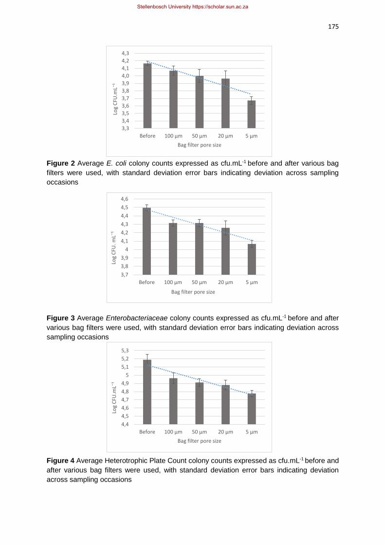
175
Figure 2 Average E. coli colony counts expressed as cfu.mL-1 before and after various bag
filters were used, with standard deviation error bars indicating deviation across sampling
occasions
Figure 3 Average Enterobacteriaceae colony counts expressed as cfu.mL-1 before and after
various bag filters were used, with standard deviation error bars indicating deviation across
sampling occasions
Figure 4 Average Heterotrophic Plate Count colony counts expressed as cfu.mL-1 before and
after various bag filters were used, with standard deviation error bars indicating deviation
across sampling occasions
4,4
4,5
4,6
4,7
4,8
4,9
5
5,1
5,2
5,3
Before 100 μm 50 μm 20 μm 5 μm
Log
CFU
.mLˉ
¹
Bag filter pore size
3,3
3,4
3,5
3,6
3,7
3,8
3,9
4,0
4,1
4,2
4,3
Before 100 μm 50 μm 20 μm 5 μm
Log
CFU
.mLˉ
¹
Bag filter pore size
3,7
3,8
3,9
4
4,1
4,2
4,3
4,4
4,5
4,6
Before 100 μm 50 μm 20 μm 5 μm
Log
CFU
. mLˉ
¹
Bag filter pore size
Stellenbosch University https://scholar.sun.ac.za

176
Huey et al. (2010) performed a study on the correlation between physico-chemical
characteristics and the microbial content present in various surface waters. It was reported
that there was a positive correlation (R2 = 0.747 and R2 = 0.711) between the turbidity and
TSS vs. the E. coli counts, respectively. It was concluded that these physico-chemical
parameters are linked to the microbial loads present (Huey et al., 2010).
In this study, the microbial populations were not expected to be significantly reduced
through the use of a bag filter. This is due to the size of a bacteria being smaller than the pore
sizes of the bag filters. An E. coli cell is approximately 1 – 2 μm in length and 0.5 μm in
diameter (El-Hajj & Newman, 2015). Any reductions in microbial counts could possibly be
attributed to bacteria being bound or attached to the suspended solids that got filtered out with
the bag filter. Christensen et al. (2001) indicates that suspended solids present in water
provide a medium for both the accumulation as well as transportation of bacteria.
A study performed by Berger et al. (1996) investigated how bacteria are able to colonise
the suspended solids present in the Danube River, Austria. It was established that between 3
and 17 bacteria were able to colonise one suspended particle. This number is reportedly
underestimated by up to 45% due to bacteria inside each particle being difficult to see under
a microscope. It was also established that 39% of the suspended particles in this river were
colonised by bacteria (Berger et al., 1996). Therefore, it is highly probable that the reduction
in colony numbers observed in this study were as a result of the bacteria being attached to
the suspended solids that were removed through filtration.
Colony counts were recorded before and after each pore size to determine if the bag
filter was able to remove any bacteria. Figures 2, 3 & 4 provide the results for the indicator
organisms tested. In Fig. 2, the E. coli results indicate a 0.43 log reduction from before filtration
to after the 5 μm bag filter. The reduction in colony counts after filtration were not sufficient for
this river water to be considered safe for use, according to the guideline limits (DWAF, 1996a).
This reduction in E. coli colonies was not statistically significant (p = 0.113). The
Enterobacteriaceae counts were reduced too, by 0.475 log counts (Fig. 3). There are no
specified limits for Enterobacteriaceae in the guidelines (DWAF, 1996a). There was not a
significant reduction in the Enterobacteriaceae counts after filtration through the 5 μm bag filter
(p = 0.379). When considering the E. coli log reductions following the three larger bag filters
(100, 50 & 20 μm), a reduction of 0.10, 0.17 and 0.20 log cfu.mL-1 were reported, respectively.
Similarly, Enterobacteriaceae log reductions that were noted following the three larger bag
filters included 0.18, 0.18 and 0.24 log cfu.mL-1 reductions were reported, respectively.
Heterotrophic Plate Count log reductions following the use of the three larger bag filters
resulted in a 0.23, 0.28 and 0.3 reduction in log cfu.mL-1, respectively. A higher initial microbial
Stellenbosch University https://scholar.sun.ac.za

177
load was reported for the HPC test (Fig. 4). The HPC test provides an indication of the total
microbial population present in a sample, so these results were expected. A reduction in
colony counts of 0.4 log were reported from before bag filtration to after the smallest filter was
used. There are no specified limits for the HPC in the guidelines (DWAF, 1996a). There was
not a significant reduction in the HPC after filtration through the 5 μm bag filter (p = 0.316).
For all indicator organism tests, the reductions were not significant where average p-values
across the three sampling occasions were 0.146, 0.245 and 0.282 for E. coli,
Enterobacteriaceae and HPC, respectively.
The current study was performed in the winter, rainy season of the Western Cape. The
initial profiling of this river was performed in the dry, summer season (Chapter 3). When
comparing the indicator microbial loads between the two seasons, the impact of the
environmental conditions on the microbial loads can be noted. An average E. coli count of log
4.64 cfu.mL-1 was recorded across the five sampling occasions in the summer, whereas an
average of 4.15 cfu.mL-1 was noted for the three sampling occasions in the winter season.
Enterobacteriaceae results show an average of 4.72 log cfu.mL-1 in the summer vs. 4.50 log
cfu.mL-1 in the winter. Heterotrophic Plate Count (HPC) results indicate an average of 4.87 log
cfu.mL-1 in the summer and an average of 5.17 cfu.mL-1 in the winter season. These results
remained relatively similar over the two seasons and provide an indication of consistent
contamination of this river. This is inspite of the water being heavily diluted in the wet, winter
season. In a study by Tornevi et al. (2014), the effect of rainfall on the microbial populations
present in a Swedish river was investigated. It was reported that precipitation events of more
than 15 mm in 24 hours resulted in a three-fold increase in E. coli colonies. This finding did
not correlate with the findings in the current study.
It is evident that the bag filter was able to remove some of the microorganisms present
in the water sample. Even though this was not the intended aim of this experiment, it is a
welcomed finding. Any reduction in microbial counts, even prior to treatment with UV radiation,
aids in the pursuit of safe water. The 5 μm bag filter produced the best results with regards to
reductions in microbial counts and physico-chemical improvement.
Stellenbosch University https://scholar.sun.ac.za

178
Objective 2: Results of microorganism response to UV radiation after bag filtration
Three doses of UV radiation (20, 40 & 60 mJ.cm-2) were applied to samples of water after
filtration through the 5 μm bag filter. For the indicator organism analysis, plates were poured
in duplicate and an average value was obtained. The results for indicator organism tests can
be seen in Fig. 5, where the red line indicates the lowest detectable limit. Pathogen testing
was performed on a presence/absence basis, and results for these experiments are indicated
in Table 2.
In Fig. 5, it can be seen that, on all three sampling occasions, there were no E. coli or
Enterobacteriaceae colonies recorded after the first dose of UV was applied (20 mJ.cm-2). The
results also indicate no growth after the second (40 mJ.cm-2) and third (60 mJ.cm-2) doses
were applied. These microorganisms therefore, are sensitive to disinfection with UV radiation.
Based on the initial microbial loads present over the three sampling occasions, average
reductions of 3.5 and 3.15 log cfu.mL-1 were observed for Enterobacteriaceae and E. coli
counts, respectively. The lowest dose (20 mJ.cm-2) was required to reduce microbial numbers
of these microorganisms to below detectable limits for the three doses applied (Fig. 5). A more
gradual reduction in the HPC colonies was reported after UV radiation. Similar results were
reported in the profiling of this river (Chapter 3). An average reduction in colonies for each
dose of UV radiation was calculated as 2.34, 3.72 and 4.07 log cfu.mL-1 for 20, 40 & 60
mJ.cm-2, respectively (Fig. 5). These findings are in agreement with a study performed by
Sharrer et al. (2005) who investigated the effect of various doses of UV radiation on the
microbial populations present. Heterotrophic Plate Counts and total coliform bacteria were
analysed. In their study, a dose of 18 mJ.cm-2 was applied to a sample of water, which
inactivated 100% of coliform bacteria present. The same dose applied, resulted in only a 98%
reduction in the HPC of this water, which was a 1.7 log reduction (Sharrer et al., 2005). Higher
initial HPC microbial loads were reported in the current study in comparison to the study
performed by Sharrer et al. (2005). Higher doses of UV radiation were also applied in the
current study than this study. After 20 mJ.cm-2 an average reduction of 2.6 log cfu.mL-1 was
recorded. This provides an indication of bacteria more resistant to UV radiation than
Enterobacteriaceae being present in the microbial population, in the current study. Mofidi et
al. (2002) performed a study on the levels of microbial survival and regrowth following high
doses of UV radiation in water samples, specifically looking at E. coli and HPC counts.
Following a dose of 140 mJ.cm-2 and then in dark storage at 20°C for 7 days, the HPC returned
to the level it was prior to UV radiation. The E. coli results showed a 1-2 log increase in cfu.mL-
1 following 24 hours of dark storage following a dose of 5 mJ.cm-2 (Mofidi et al., 2002).
Stellenbosch University https://scholar.sun.ac.za

179
Figure 5 Indicator organism test results on three separate sampling occasions, after UV
radiation was applied at three doses, with standard deviation error bars included for variation
between duplicate plates poured, across six decimal dilutions. The red line indicates the lowest
detectable limit of microbial presence
Although there was a more gradual reduction in the HPC colonies, there was only one
sampling occasion where colonies were present after 60 mJ.cm-2 of UV radiation. No E. coli
or Enterobacteriaceae colonies were present after the lowest dose of UV (20 mJ.cm-2) was
applied. This indicates that the UV radiation was effective at reducing the microbial loads
present to an acceptable or safe level. Microbial recovery following UV radiation was, however,
not investigated in this study. Since the E. coli and Enterobacteriaceae levels observed in the
Mosselbank river appear to be constant, considering the results observed in this chapter and
in Chapter 3, it is proposed that microbial recovery post UV be investigated in future.
The current study was performed in the wet, winter season. The profiling of this river
was performed between the hot, dry, summer season and the wet, rainy season (Chapter 3).
When comparing the effect of UV radiation on the microbial populations present in this river,
conclusions can be drawn regarding the resistance profiles of microorganisms in different
environmental conditions. This was of interest, specifically for the HPC colonies, as these
organisms show a greater resistance to UV radiation. Figure 6 provides an indication of the
HPC colony counts after UV radiation from summer to winter. In the winter analyses, the bag
filter was included in treatment, which should be taken into consideration.
0
1
2
3
4
5
6
1 2 3 1 2 3 1 2 3
Enterobacteriaceae E.coli HPC
Log
CFU
.mLˉ
¹
Before 20 mJ.cmˉ² 40 mJ.cmˉ² 60 mJ.cmˉ²
Stellenbosch University https://scholar.sun.ac.za

180
The results in Fig. 6 show that the effect of the environmental conditions on the microbial loads
present in this river water was not significant (p = 0.953). However, when taking the reduction
in colony numbers through the use of the bag filter (average of 0.4 log cfu.mL-1), there are
slightly higher microbial loads present in the winter season. The pathogens, L. monocytogenes
and Salmonella spp. were tested before and after filtration, as well as after each dose of UV
radiation. The results of these tests can be seen in Table 2.
Table 2 Presence absence testing results of pathogen before and after filtration, as well as
after each dose of UV radiation
No colony counts are reported, as this test was performed on a presence/absence basis only,
therefore it is not possible to determine if there were any reductions in colony numbers due to
the bag filter. Consistent pathogen presence was reported on all sampling occasions of this
river (Table 2). The efficacy of UV radiation was successful, as neither pathogen was present
after the first dose of UV was applied. This agrees with findings in Chapter 3, where no
Salmonella spp. L. monocytogenes
Treatment 1 2 3 1 2 3
Before + + + + + +
5 μm filter + + + + + +
20 mJ.cmˉ² - - - - - -
40 mJ.cmˉ² - - - - - -
60 mJ.cmˉ² - - - - - -
0
1
2
3
4
5
6
7
Before treatment 20 mJ.cmˉ² 40 mJ.cmˉ² 60 mJ.cmˉ²
Log
cfu
.mLˉ
¹
UV (summer) UV + Bag Filter (winter)
Figure 6 Comparison of HPC loads in the summer season versus the winter season,
indicated as log cfu.mL-1, with standard deviation error bars included
Stellenbosch University https://scholar.sun.ac.za

181
pathogens could be detected after the lowest dose of UV (20 mJ.cm-2) was applied. Yaun et
al. (2003) indicate that 14 mJ.cm-2 of LP UV radiation is sufficient to cause a 5-log reduction
in Salmonella spp. Similarly, a dosage requirement of 16.9 mJ.cm-2 resulted in a 5-log
inactivation of L. monocytogenes, according to Matak et al. (2005). The results in the current
study confirms the dosage requirements from these previously mentioned studies that a
dosage of 20 mJ.cm-2 is required to inactivate the pathogens under investigation.
The consistent presence of pathogens in this river confirms that it is unacceptable for
agricultural irrigation without treatment. The 5 μm bag filter was not expected to remove the
pathogens, as the pore sizes of the bag filter are larger than the size of a bacteria, however
the results indicate that some of the HPC, E. coli and Enterobacteriaceae cfu’s were removed.
However, UV radiation was effective at removing these pathogens, and lowering indicator
organism loads to an acceptable level. This water would be deemed microbial safe, according
to the Irrigation Water Guidelines (DWAF, 1996a) for agricultural irrigation.
Conclusions
In this study, the effect of a bag filter on the microbial and physico-chemical profile of the
Mosselbank River were analysed. It was determined, through the significant reductions in the
physico-chemical characteristics, that the 5 μm pore size bag filter provided the greatest
results without reducing the flow rate of the water. The bag filter was effective at reducing the
COD, TSS and turbidity levels in the water samples. Greater increases in the UVT % were
expected through the use of the bag filter, however, the river has an exceptionally high TDS
content, which could be preventing an improvement in UVT %. The bag filter did reduce the
TDS content very slightly, but this was still not enough for the level to be considered acceptable
according to the guideline limit of 260 mg.L-1.
Microorganism reduction wasn’t expected through the use of a bag filter, as even the
smallest bag filter has pores larger the size of a bacteria. An Enterobacteriaceae reduction of
log 0.475 was reported from before filtration to after the smallest bag filter. The E. coli results
showed a 0.43 log reduction after the same bag filter. The HPC test results indicated a 0.4 log
reduction after the smallest bag filter. The reductions could be as a result of bacteria being
bound or attached to the suspended solids that got filtered out of the water through the bag
filter. Following filtration with the 5 μm bag filter, three doses of UV radiation were applied to
the water samples using a LP collimated beam UV system. It was determined that the lowest
dose applied (20 mJ.cm-2) was sufficient for reducing the E. coli and Enterobacteriaceae
colonies to non-detectable levels. The HPC colonies, were lowered more steadily. This is an
indication that there are bacteria in the microbial population that are less sensitive to
disinfection with UV radiation. A dose of 60 mJ.cm-2 was required to lower the HPC colonies
present in the river water to a non-detectable level. There are no guideline limits for
Stellenbosch University https://scholar.sun.ac.za

182
Enterobacteriaceae or HPC, however, in terms of the E. coli counts, the water would be
deemed microbially safe for agricultural irrigation following treatment with UV radiation.
Overall, the bag filter was successful at improving most of the physico-chemical
characteristics. The extremely high TDS content prevented a larger increase in the UVT %.
The bag filter was also successful at removing some of the microorganisms present in the
river water. The UV results show, again, that this method of disinfection is effective, at
laboratory-scale, at producing a consistently safe supply of water. A recommendation for
future research would be to repeat this experiment on a river with a different physico-chemical
profile. This would provide an indication of the impact of the bag filter on the UVT % of a river
with lower TDS content and a higher TSS content. Microbial recovery post-UV radiation was
not observed in this study, and due to the consistent presence of E. coli and
Enterobacteriaceae in the river water observed in this chapter as well as the other rivers in
Chapter 3, it is recommended that this area of research be analysed in future.
References
Abdul-Halim, N. & Davey, K. R. (2016). Impact of Suspended Solids on Fr 13 Failure of UV
Irradiation of Inactivation of Escherichia coli in Potable Water Production with Turbulent
Flow in an Annular Reactor. Journal of Chemical Engineering Science, 143, 55-62. DOI
10.1016/j.ces.2015.12.017
APHA (American Public Health Association) (2005). Standard Methods for the Examination of
Water and Wastewater, 21st ed. Washington, DC: American Public Health Association
Press.
Berger, B., Hoch, H., Kavka, G. & Herndl, G. J. (1996). Bacterial colonization of suspended
solids in the River Danube. Aquatic Microbial Ecology, 10, 37-44.
Bhagwan, J. (2008). Mind-maps and collective knowledge of the water sector, research and
development technology. Water Research Commission (WRC), Pretoria.
Broséous, R., Cigana, J., Barbeau, B., Daines-Martinez, C. & Suty, H. (2009). Removal of
total dissolved solids, nitrates and ammonium ions from drinking water using charge-
barrier capacitive deionisation. Desalination, 249(2009), 217-223. DOI
i:10.1016/j.desal.2008.12.048
Butler, B A. & Ford, R. G. (2018). Evaluating relationships between total dissolved solids (TDS)
and total suspended solids (TSS) in a mining-influenced watershed. Mine Water
Environment, 37(1), 18-30. DOI 10.1007/s10230-017-0484-y.
Christensen, V. G., Ziegler, A. C., Jian, X. (2001). Continuous turbidity monitoring and
regression analysis to estimate total suspended solids and faecal coliform bacteria loads
in real time. Proceedings of the Seventh Federal Interagency Sedimentation
Conference, Nevada.
DWAF (Department of Water Affairs and Forestry). (1996a). South African Water Quality
Guidelines (2nd edition). Volume 4: Irrigation water use, 1-123. CSIR Environmental
Services, Pretoria.
Stellenbosch University https://scholar.sun.ac.za

183
DWAF (Department of Water Affairs and Forestry). (1996b). South African Water Quality
Guidelines (2nd edition). Volume 3: Industrial water use, 1-123. CSIR Environmental
Services, Pretoria.
DWAF (Department of Water Affairs and Forestry). (1996c). South African Water Quality
Guidelines (2nd edition). Volume 1: Domestic Use, 1- 130. CSIR Environmental
Services, Pretoria.
Du Plessis, M., Annandale, J., Benade, N., van der Laan, M., Jooste, S., du Preez, C., Barnard,
J., Rodda, N., Dabrowski, J., Genthe, B. & Nell, P. (2017). Risk Based, Site-Specific,
Irrigation Water Quality Guidelines; Description of Decision Support System; WRC
Report No TT 727/17; Water Research Commission: Pretoria, South Africa.
El-Hajj, Z. W. & Newman, E. B. (2015). How Much Territory can a Single E. coli Cell Control?
Frontiers in Microbiology, 6(309), 1-12. DOI 10.3389/fmicb.2015.00309
Farrell, C., Hassard, F., Jefferson, B., Leziart, T., Nocker, A. & Jarvis, P. (2018). Turbidity
composition and the relationship with microbial attachment and UV inactivation
efficiency. Science of the Total Environment, 642, 638-647. DOI
10.1016/j.scitotenv.2017.12.173. Accessed 05/04/2019.
Fileder. (2020). A Guide to Filtration and Water Treatment Technologies. [Internet Document]
URL: https://www.fileder.co.uk/fileder-app/app-uploads/2020/05/A-Guide-to-Filtration-
and-Water-Treatment-Technologies.pdf. Accessed 15/02/2021.
Fondriest Environmental. (2014). Turbidity, Total Suspended Solids and Water Clarity -
Fundamentals of the Environmental Measurements. [Internet Document]
https://www.fondriest.com/environmentalmeasurements/parameters/waterquality/turbid
ity-total-suspended-solids-water-clarity/. Accessed 20/08/2020.
Gayán, E., Serrano, M. J., Raso, J., Alvares, I. & Condon, S. (2012). Inactivation of Salmonella
enterica by UV-C Light Alone and in Combination with Mild Temperatures. Applied and
Environmental Microbiology, 78(23), 8353- 8361. DOI 10.1128/AEM.02010-12.
Accessed 01/04/2019.
Hallmich, C. & Gehr, R. (2010). Effect of pre- and post-UV disinfection conditions on
photoreactivation of fecal coliforms in wastewater effluents. Water Research, 44, 2885-
2893.
Hassen, A., Mahrouk, M., Ouzari, H., Cherif, M., Boudabous, A. & Damelincourt, J. J. (2000).
UV disinfection of treated wastewater in a large-scale pilot plant and inactivation of
selected bacteria in a laboratory UV device. Bioresource Technology, 74, 141-150.
Hijnen, W. A. M., Beerendonk, E. F. & Medema, G. J. (2006). Inactivation credit of UV radiation
for viruses, bacteria and protozoan (oo)cysts in water: a review. Water Research, 40, 3-
22. DOI 10.1016/j.watres.2005.10.030. Accessed 10/04/2019.
Hohenblum, P., Epema, O., Annys, J., Furtmann, K., Lundin, E., Harward, C., Strub, M.,
Mainz, C., Palfrey, R. & Jönsson, J. (2017). COD Substitution Scoping Study. European
Commission. Contract No. 07.0201/2015/SFRA/714792/ENV.C.2. Vienna, Austria.
Huey, G. M. & Meyer, M. L. (2010). Turbidity as an Indicator of Water Quality in Diverse
Watersheds of the Upper Pecos River Basin. Water, 2, 273-284. DOI
10.3390/w2020273.
Iloms E., Ololade, O. O., Ogola, H. J. O. & Selvarajan, R. (2020). Investigating Industrial
Effluent Impact on Municipal Wastewater Treatment Plant in Vaal, South Africa.
Stellenbosch University https://scholar.sun.ac.za

184
Environmental Research and Public Health, 17(1096), 1-18. DOI:
10.3390/ijerph17031096.
International Standards Organisation (ISO). (2017). ISO Method 21528-2:2017. Microbiology
of the food chain – Horizonal Method for the Detection and Enumeration of
Enterobacteriaceae – PART 2: Colony-count technique. ISO: Switzerland.
International Standards Organisation (ISO). (2001). ISO Method 16654:2001. Microbiology of
the food and animal feeding stuffs – Horizonal Method for the Detection of Escherichia
coli O157. ISO: Switzerland.
International Standards Organisation (ISO). (2017). ISO Method 11290-1:2017. Microbiology
of Food and Animal Feeding Stuffs: Horizontal Method for the Detection and
Enumeration of L. monocytogenes. ISO: Switzerland.
Kesari, K. K., Verma, H. N. & Behari, J. (2011). Physical Methods in Wastewater Treatment.
International Journal of Environmental Technology and Management, 14, 43-65.
Koutchma, T. (2009). Advances in Ultraviolet Light Technology for Non-Thermal Processing
of Liquid Foods. Food Bioprocessing Technology, 2(138), 138-155. DOI
10.1007/s11947-008-0178-3. Accessed 02/04/2019.
Liu, G. (2005). An Investigation of UV Disinfection Performance under the Influence of
Turbidity and Particulates for Drinking Water Applications. MSc Thesis in Civil
Engineering. University of Waterloo: Canada.
Matak, K. E., Churey, J. J., Worobo, R. W., Hovingh, E., Hackney, C. R., & Pierson, M. D.
(2005). Efficacy of UV Light for the Reduction of Listeria monocytogenes in Goat’s Milk.
Journal of Food Protection, 68(10), 2212-2216.
Mofidi, A. A., Rochelle, P. A., Chou, C. I., Mehta, H. M. (2002). Bacterial Survival after
Ultraviolet Light Disinfection: Resistance, Regrowth and Repair. American Water Works
Association, 1-12.
Okpara, C. G., Oparaku, N. F. & Ibeto, C. N. (2011). An Overview of Water Disinfection In
Developing Countries and Potentials of Renewable Energy. Journal of Environmental
Science and Technology, 4, 18-30. DOI 10.3923/jest.2011.18.30.
Olivier, F. (2015). Evaluating the Potential of Ultraviolet Irradiation for the Disinfection of
Microbiologically Polluted Irrigation Water. MSc Food Science, Stellenbosch University,
Stellenbosch.
Patoczka, J. (2007). Impact of Chemicals Addition in Water/Wastewater Treatment on TDS
Concentration and Sludge Generation. Proceedings of the Water Environment
Federation, 7, 5209-5214. DOI 10.2175/193864706783763264.
Rügner, H., Schwientek, M., Beckingham, B., Kuch, B. & Grathwohl, P. (2013). Turbidity as a
proxy for total suspended solids (TSS) and particle facilitated pollutant transport in
catchments. Environmental Earth Sciences, 69, 373-380. DOI 10.1007/s12665-013-
2307-1.
SANS (South African National Standards) (1999). Method 6887-1. Microbiology of Food and
Animal Feeding Stuffs: Preparation of test samples, initial suspension and decimal
dilutions for microbiological examination Part 1: General rules for the preparation of
the initial suspension and decimal dilutions. Pretoria: South Africa.
SANS (South African National Standards) (2006). Method 5667-6. Water quality: Sampling
Part 6: Guidance on sampling of rivers and streams. Pretoria: South Africa.
Stellenbosch University https://scholar.sun.ac.za

185
SANS (South African National Standards) (2007). Method 4833-1. Microbiology of Food and
Animal Feeding Stuffs: Horizontal methods for the enumeration of coliforms: Colony-
count technique at 30°C. Pretoria: South Africa.
SANS (South African National Standards) (2011a). Method 5221. Microbiological Analysis of
Water – General Test Methods. Pretoria: South Africa.
SANS (South African National Standards) (2011b). Method 19250. Water Quality: Detection
of Salmonella spp. Pretoria: South Africa.
Shahawy, A. E. L., El-Shatoury, S., Bayomi, S. & El-Monayeri, D. (2018). Water Disinfection
Using Artificial Ultraviolet Rays Technology. Unconventional Water Resources and
Agricultural in Egypt, 1-72. DOI 10.3390/w2020273.
Sharrer, M. J., Summerfelt, S. T., Bullock, G. L., Gleason, L. E. & Taeuber, J. (2005).
Inactivation of bacteria using Ultraviolet irradiation in a recirculating salmonid culture
system. Aquaculture Engineering, 33(2), 135-149. DOI 10.1016/j.aquaeng.2004.12.001.
Tornevi, A., Bergstedt, O. & Forsberg, B. (2014). Precipitation Effects on Microbial Pollution
in a River: Lag Structures and Seasonal Effect Modification. PLoS One, 9(50, 1-10.
DOI 10.1371/journal.pone.0098546.
United States Environmental Protection Agency (USEPA). (2003). Ultraviolet Disinfection
Guidance Manual. Washington, DC.
Üstün, G. E. (2009). Occurrence and removal of metals in urban wastewater treatment plants.
Journal of Hazardous Materials, 172(2-3), 833-838. DOI
10.1016/j.jhazmat.2009.07.073.
World Health Organisation (WHO). (2004). Chemicals from water treatment and distribution.
Guidelines for drinking water quality – third edition. Geneva, Switzerland.
Wu, G-Q., Bi, W-H., Lü, J-M. & Fu, G-W. (2011). Determination of chemical oxygen demand
in water using near-infrared transmission and UV absorbance method. Spectroscopy
and Spectral Analysis, 31(6), 1486-1489.
Yaun, B. R., Sumner, S. S., Eifert, J. D. & Marcy, J. E. (2003). Response of Salmonella and
E. coli O157:H7 to UV energy. Journal of Food protection, 66(6), 1071-1073.
Stellenbosch University https://scholar.sun.ac.za

186
Chapter 6
GENERAL CONCLUSIONS AND RECOMMENDATIONS
General conclusions
Researchers have established that pathogenic microorganisms are commonly the cause of
foodborne disease outbreaks (Steele & Odumeru, 2004, Pachepsky et al., 2011, Huisamen,
2012). Pachepsky et al. (2011) states that ever-increasing evidence points towards raw fresh
produce as the major factor that contributes towards gastrointestinal illnesses. Gerba & Choi
(2006) state that irrigation water plays a key role in pathogen transport to consumers of fresh
produce via the faecal-oral route. Contamination of surface water occurs more frequently in
developing countries which results in decreased water quality and increased risk of disease
transmission. In South Africa, surface water is the preferred water source for the irrigation of
fresh produce (Singh, 2013). Maree et al. (2016) notes that of the available fresh water in
South Africa, surface water usage totals 77%. Surface waters in the Western Cape have been
the subject of frequent investigation due to the constant presence of faecal contamination,
which renders some sources unsuitable for irrigation without pre-treatment (Barnes & Taylor,
2004, Paulse et al., 2009, Lamprecht et al., 2014, Olivier, 2015, Sivhute, 2019). These
contaminated surface waters need to be treated to such an extent that it is safe for agricultural
irrigation, without the risk of transferring the pathogenic microorganisms to the consumer of
the fresh produce.
Locally, and internationally, Escherichia coli (E. coli) is frequently used as an indicator
of contamination of water, specifically faecal contamination (Britz et al., 2012). This
microorganism is included in the guidelines of most countries with limits which need to be
complied with in order for the water to be deemed safe for consumption and use. However,
foodborne disease outbreaks are not only caused by pathogenic strains of E. coli, but also by
other pathogens such as Salmonella and Listeria monocytogenes (L. monocytogenes) (Jung
et al., 2014). In South Africa, the microbial guidelines for agricultural irrigation are limited to
1 000 colony forming units (cfu) of faecal coliforms (E. coli) per 100 mL of water (Department
of Water Affairs and Forestry (DWAF), 1996). The potential for bacteria to survive and
proliferate in/on the crop becomes a major concern in the case of food safety, particularly in
South Africa where a large percentage of the population is immunocompromised as a result
of HIV/AIDS (Britz et al., 2012). GreenCape (2020) states that the Western Cape contributes
21% of the country’s total agricultural income, and 44% of all exported agricultural products.
From a financial perspective, the public consequences of foodborne disease outbreaks linked
to contaminated crops, as well as product recalls, can negatively affect the agricultural industry
and cause a crippling effect on the economy.
Stellenbosch University https://scholar.sun.ac.za

187
Based on the frequently observed high microbial loads present in Western Cape rivers,
Britz et al. (2012) has indicated that a 3 – 4 log reduction of faecal coliforms in water intended
for agricultural irrigation should be sufficient to meet the guideline limits in water with initial
E. coli counts in the range of 106 cfu.100 mL-1. Physical, chemical and photochemical
treatment processes are commonly used to disinfect water, each having their own advantages
and disadvantages. The treatment explored in this study, Ultraviolet (UV) radiation, has been
investigated by several researchers worldwide (Quek & Hu, 2008, Bolton & Cotton, 2008,
Farrell et al., 2018, Gayán et al., 2014). This method of disinfection has proven to be
successful across a broad range of microorganisms, with little negative after effects and is
therefore, attractive due to the push towards more environmentally friendly treatment methods
(Galvéz & Rodrígues, 2010). This is a major advantage over other treatment options,
specifically due to the fact that chlorine has become infamous for the production of
carcinogenic disinfection by-products (DBPs) in water, as well as the residual chlorine that
remains after treatment (Whitby & Scheible, 2004). As chemical treatment methods have been
the preferred treatment option for many years (Whitby & Scheible, 2004), environmental
microorganisms have developed resistances to this disinfection method, and either require
higher concentrations to achieve the same effect or an alternate treatment is required (Momba
et al., 2008). Physical treatment methods (slow sand filtration or membrane filtration) can be
time consuming and expensive and are limited to the physical size of the pores in the filter,
resulting in an often ineffective treatment (Okpara et al., 2011). Physical methods are often
used as a pre-treatment step in water disinfection. Most photochemical treatment options,
such as Advanced Oxidation Processes or Electron Beam Technology, are expensive and are
not user-friendly (Maruthi et al., 2011). Ultraviolet radiation is a simple, yet effective method of
disinfection in many industries including water treatment. However, this method of disinfection
is not frequently implemented at farm-scale due to chemical treatment methods being the
established practice (Olivier, 2015). Studies on the effect of UV radiation used for microbial
disinfection commonly involve laboratory-cultured strains of E. coli only and low–pressure (LP)
UV radiation. This has resulted in a great deal of confusion with regard to dosage requirements
for environmental or clinical strains which are often reported as more resistant than laboratory-
cultured strains (Hijnen et al., 2006, Chevrefils et al., 2006). No research has been performed
in South Africa on the effect of UV radiation on environmental strains of Salmonella and L.
monocytogenes in both water and food. The overall aim of this study was, therefore, to
investigate the efficacy of LP UV radiation on water from four different rivers that have varying
physical and microbial characteristics, in order to reduce microbial loads for improved crop
safety.
Stellenbosch University https://scholar.sun.ac.za

188
The study consisted of three research chapters. The first (Chapter 3) involved the
establishment of physico-chemical and microbial profiles of the four selected rivers
(Mosselbank, Eerste, Plankenburg and Franschhoek Rivers). The rivers were sampled in the
dry, summer season (October 2019 – January 2020). Three doses of LP UV radiation (20, 40
& 60 mJ.cm-2) were applied to the water samples. Microbial tests included Heterotrophic Plate
Count (HPC), Enterobacteriaceae and E. coli counts, as well as presence/absence tests for
Salmonella species and L. monocytogenes. It was established in this chapter that the rivers
under investigation frequently exceeded the Irrigation Water Guideline limits for E. coli (DWAF,
1996). The food pathogens, Salmonella and L. monocytogenes, were also present on multiple
sampling occasions. Low-pressure UV radiation was effective at reducing the microbial loads
to what would be considered safe according to the Irrigation Water Guidelines. No pathogens
could be detected after treatment at a dose of 40 mJ.cm-2. The Mosselbank River was deemed
the ‘worst-case scenario’ in terms of microbial loads and physico-chemical characteristics,
while the Franschhoek River was classified as the ‘best-case scenario’. It was noted that the
physical quality of the water did not affect the disinfection capability of the UV radiation,
provided that the correct UV dose was applied. In water of low physico-chemical quality,
treatment was, however, negatively impacted since extended exposure time was necessary
to apply the required dose by LP UV. The turbidity, total suspended solids, total dissolved
solids and chemical oxygen demand had the greatest influence on reducing the UV
transmission percentage (UVT %) and thereby, increasing exposure time required to apply a
specific dose of UV. It was suggested that the use of a pre-treatment step would be
advantageous in the case of river waters with high turbidity and suspended solids, which could
potentially improve the UVT % and reduce exposure times, ultimately making the process
more efficient. Microbial isolates were retained from this study and stored at -80°C for future
research.
In the second research chapter (Chapter 4), the isolates from the river waters were
analysed in depth in a three-part study. The first involved the lineage-typing of L.
monocytogenes river water isolates using the PCR-Restriction Fragment Length
Polymorphism method set out by Rip & Gouws (2020). It was determined that all L.
monocytogenes river water isolates belonged to lineage I. This lineage is considered to be the
most common cause of listeriosis worldwide (Pirone-Davies et al., 2018). The isolates
(Enterobacteriaceae and L. monocytogenes) were then subjected to antimicrobial
susceptibility testing. Enterobacteriaceae were also tested for the presence of extended-
spectrum beta-lactamase (ESBL) producers. A total of 90% (n=9) of the river water isolates
tested were multidrug resistant. Ampicillin resistance was reported for 100% of the
Enterobacteriaceae isolates (n=5) and 80% (n=4) of the L. monocytogenes isolates. None of
Stellenbosch University https://scholar.sun.ac.za

189
the Enterobacteriaceae isolates were ESBL-producers. In the third study, the isolates were
inoculated into sterile river water at concentrations higher than would naturally be found in the
environment (approximately 108 cfu.mL-1) and exposed to the same three doses of UV
radiation as before (20, 40 & 60 mJ.cm-2). It was established that LP UV radiation was not
effective at reducing the microbial levels to those that would be considered as safe, when the
initial microbial loads are higher than 108 cfu.mL-1. The most UV-resistant strain was an
E. coli isolate from the Franschhoek River. This river was described in the previous study
(Chapter 3) as the ‘best-case scenario’. The environmental L. monocytogenes and Salmonella
river isolates tested in this study required at least 40 mJ.cm-2 of UV radiation to reduce the
microbial loads to non-detectable levels. Therefore, these pathogenic microorganisms
(Salmonella and L. monocytogenes) require lower doses of UV radiation than E. coli (required
> 60 mJ.cm-2) at the investigated microbial loads. This is in contrast to literature that states
that, in general, pathogenic microorganisms are more resistant to disinfection than non-
pathogenic ones (Gayán et al., 2014). From this chapter’s results, it was concluded that the
river water isolates carry resistance genes to multiple antimicrobials as well as show
resistance to disinfection with UV radiation at high microbial loads. This was noted specifically
with regard to the E. coli isolates (FR-01 and MR-01) that required at least 60 mJ.cm-2 of UV
radiation for disinfection. It is evident that these microorganisms pose a potential risk for
human and animal health if consumed.
In the third research chapter (Chapter 5), a bag filter was tested to determine the effect
of this pre-treatment step on the physico-chemical profile of the river water prior to UV
radiation. Since the results from the first research chapter (Chapter 3) indicated that the river
with the worst physico-chemical profile was the Mosselbank River, water was sampled from
this river and run through bag filters of varying pore sizes (5, 20, 50 & 100 μm). Microbial and
physico-chemical characteristics were analysed before and after each pore size. It was noted
that the bag filter with the pore size of 5 μm yielded the best results, without compromising the
flow rate. The total dissolved solids (TDS) content (avg. 715.33 mg.L-1) even after the bag filter
with the smallest pore size was used, was not considered acceptable for agricultural irrigation
purposes according to the guideline limit of 260 mg.L-1 (DWAF, 1996). Heterotrophic Plate
Count, Enterobacteriaceae and E. coli microbial loads were reduced by 0.4, 0.475 and 0.43
log cfu.mL-1, respectively, from before treatment to after the 5 μm bag filter was used. A large
improvement (32.4%) was noted for the total suspended solids (TSS) content of the water,
following the use of the bag filter. A 1.83% improvement was reported for the UVT % of the
water sample after the 5 μm bag filter was used. It can be speculated that this increase could
have been a much larger improvement in the UVT % if the TDS content of the river water was
lower than the extreme levels recorded for the Mosselbank River. This bag filter (5 μm) was
Stellenbosch University https://scholar.sun.ac.za

190
selected as the most effective pore size for future experiments. River water samples that were
filtered through the 5 μm bag filter, were also exposed to the same three LP UV doses as
before. The lowest dose (20 mJ.cm-2) was sufficient for reducing the E. coli,
Enterobacteriaceae, Salmonella and L. monocytogenes loads to non-detectable levels. The
HPC loads lowered more steadily, and required at least 60 mJ.cm-2 to reach non-detectable
levels. This is an indication of a microbial population that is less sensitive to UV disinfection,
and agrees with previous findings in the current study and with literature (Bester, 2015, Olivier,
2015).
Recommendations for future research
Results from the first research chapter (Chapter 3) indicated that LP UV radiation is effective
at reducing the microbial loads to acceptable levels, as well as reducing pathogenic
microorganisms to non-detectable levels. It was established that the physical quality of the
river water influenced the length of exposure times required to achieve a specific dose, but did
not have a negative impact on the final level of disinfection. Essentially, it was possible to
achieve a standard of water that would be considered microbiologically safe for use, according
to the Irrigation Water Guidelines (DWAF, 1996), no matter what the initial physical quality of
the water was. It was suggested that a pre-treatment step could be beneficial for treatment
efficiency by reducing suspended solids and improving UVT %.
In the second research chapter (Chapter 4), it was established that multidrug resistance
was reported for 90% of microbial isolates (n=9). In 2014, the World Health Organisation
(WHO) developed a global action plan in response to the rapid increase in antimicrobial
resistant bacteria, in which one of the strategies included continuous research and
surveillance of antimicrobial resistance in environmental isolates in order to curb the spread
of these pathogenic microorganisms. It is recommended that more river water isolates
(Enterobacteriaceae and L. monocytogenes) be tested, to understand how these organisms
disseminate and spread resistance in river waters, and determine the possible effect of
seasonal changes on resistance patterns. None of the Enterobacteriaceae river water isolates
were ESBL-producers, however, it is recommended that more isolates are obtained from these
rivers and that testing is performed for the presence of both ESBL-producers as well as AmpC-
producing genes. This would fill the knowledge gap in this area, as there is currently no
research that has been performed on the presence of AmpC-producers in irrigation water
sources in South Africa. Thomson (2010) states that multidrug resistances are commonly
associated with plasmid mediated AmpC β-lactamases in Enterobacteriaceae. AmpC-
producers are not inhibited by β-lactam inhibitors and are able to confer resistance to
Cephamycins (Rodríguez-Baño et al., 2018). Tepeli and Zorba (2018) state that AmpC β-
lactamases are resistant to a broader range of antimicrobials than ESBL-producers. For this
Stellenbosch University https://scholar.sun.ac.za

191
reason, it is recommended that research be performed in this area. Another recommendation
for future research based on the results of the third study of the second chapter (Chapter 4)
was to determine the resistance profiles of isolates to UV radiation after previously being
exposed to UV radiation.
In the third research chapter (Chapter 5), it is recommended that river water with a
different physico-chemical profile, specifically one with a lower TDS content, should be
investigated using the same bag filter pore sizes. The results indicated that filtration improved
TSS levels more than TDS, and it might be that in water with higher a TSS and lower TDS
content, a greater improvement in UVT % might be observed. Investigating the same pore-
sized bag filters on a range of different river water profiles, therefore, enables for a more
accurate determination of the limitations for practical application of this pre-treatment
technology. This could enable the intended user of the UV radiation to apply the most effective
treatment, which takes a wider variety of profiles into consideration.
Based on the results obtained in the first and second research chapters (Chapters 3 and
4), it is recommended that HPC isolates are obtained from the river water, after UV radiation,
and identified. This would determine which strains are contributing to the UV resistance
observed in the river waters that may present a food safety risk to fresh produce consumers.
Screening for Shiga-toxin producing E. coli (STEC) testing is also recommended. An
alarmingly low infectious dose has been reported for STEC, ranging between 10 – 100 cells
required to cause infection (Etcheverría & Padola, 2013, Karmali, 2004). If E. coli cells are
able to survive UV radiation, it is important to determine if the surviving strains are STEC or
commensal E. coli so as to determine the potential risk associated with the water that carries
low E. coli loads after treatment.
In this study, the effect of microbial recovery, including photoreactivation and dark repair,
was not investigated. Zimmer & Slawson (2002) state that microbial recovery following
radiation with LP lamps is a cause for concern. Photoreactivation (light-dependent
reactivation) is particularly problematic in areas where sunlight is plentiful and temperatures
range between 23 - 37°C (Quek & Hu, 2008). This is, therefore, recommended for future
research in order to ensure that the microorganisms inactivated during treatment have not
recovered to such an extent that it might still present a health risk to the consumer following
UV treatment.
The E. coli loads present in the examined rivers called for log reductions exceeding 3-
log in order to produce water that is fit for agricultural irrigation, based on guideline limits. If
treated effectively and completely with UV radiation, the risk of microbial contamination from
irrigation water is low. Due to the fact that pathogenic microorganisms, such as L.
Stellenbosch University https://scholar.sun.ac.za

192
monocytogenes and Salmonella spp. were frequently identified in the various rivers
investigated, these pathogens may present consistent food safety risks for the consumers of
fresh produce which was irrigated with this water. It is recommended that the relevant
authorities are informed of the potential risk associated with these pathogens. Antimicrobial
resistant microorganisms that have been identified in the rivers investigated may indirectly
place great pressure on the clinical industry as treatment options can become limited for
infections caused by multidrug resistant strains. It is imperative that the occurrence of these
microorganisms be routinely determined in order to monitor their environmental dissemination.
In conclusion, at the investigated doses, LP UV radiation is able to seamlessly provide
a consistent level of disinfection and shows promise as a method for irrigation water
disinfection. Based on the results obtained in this study, the transition from LP UV treatment
at laboratory-scale to the medium-pressure UV treatment pilot-plant can be made to determine
the feasibility of upscaling.
References
Barnes, J. M. & Taylor, M. B. (2004). Health risks assessment in connection with the use of
microbiologically contaminated source waters for irrigation. WRC Report No. 1226/1/04/
University of Stellenbosch, Stellenbosch.
Bester, C. (2015). Investigating the Efficacy of Sodium Hypochlorite, Calcium Hypochlorite
and Peracetic Acid on Environmental Escherichia coli Strains. Master’s Thesis.
Stellenbosch University, Stellenbosch.
Bolton, J. R. & Cotton, C. A. (2011). The Ultraviolet Disinfection Handbook. Pp. 1-149. USA:
American Water Works Association.
Britz, T. J., Sigge, G.O., Buys, E. M., Schmidt, S., Potgieter, N. & Taylor, M. B. (2012). Baseline
study on extent (types and quantities) of contamination found in irrigation water at
selected sites (K5/1773). In: Quantitative investigation into the link between irrigation
water quality and
Chevrefils, G., Caron, E., Wright, H., Sakamoto, G., Payment, P. Barbeau, B. & Cairns, B.
(2006). UV Dose Required to Achieve Incremental Log Inactivation of Bacteria, Protozoa
and Viruses. IUVA News, 8(1), 38-45.
Department of Water Affairs and Forestry (DWAF). (1996). South African Water Quality
Guidelines (2nd edition). Volume 4: Agricultural Use: Irrigation, 1-194. CSIR
Environmental Services, Pretoria.
Etcheverría, A. I. & Padola, N. L. (2013). Shiga toxin-producing Escherichia coli. Virulence,
4(5), 366-372. DOI 10.4161/viru.24642.
Farrell, C., Hassard, F., Jefferson, B., Leziart, T., Nocker, A. & Jarvis, P. (2018). Turbidity
composition and the relationship with microbial attachment and UV inactivation
efficiency. Science of the Total Environment, 642, 638-647. DOI
10.1016/j.scitotenv.2017.12.173. Accessed 05/04/2019.
Stellenbosch University https://scholar.sun.ac.za

193
Gálvez, J. B. & Rodríguez, S. M. (2010). Solar Energy Conversion and Photoenergy System.
[Internet Document] URL https://www.eolss.net/sample-chapters/C08/E6-106-21.pdf.
Accessed 11/04/2019.
Gayán, E., Condon, S. & Alverez, I. (2014). Biological Aspects in Food Preservation by
Ultraviolet Light: a Review. Food and Bioprocessing Technology, 7, 1-20. DOI
10.1007/s11947-013-1168-7. Accessed 03/04/2019.
Gerba, C. & Choi, C. Y. (2006). Role of irrigation water in crop contamination by viruses.
Viruses in foods. Pp. 257–263. New York: Springer.
GreenCape. (2020). Sustainable Agriculture Market Intelligence Report 2020. [Internet
Document]
URLhttps://www.greencape.co.za/assets/AGRICULTURE_MARKET_INTELLIGENCE
_REPORT_WEB.pdf. Accessed 13/11/2020.
Hijnen, W. A. M., Beerendonk, E. F. & Medema, G. J. (2006). Inactivation credit of UV radiation
for viruses, bacteria and protozoan (oo)cysts in water: A review. Water Research,
40(2006), 3-22. DOI 10.1016/j.watres.2005.10.030.
Huisamen, N. (2012). Assessment of microbial levels in the Plankenburg and Eerste Rivers
and subsequent carry-over to fresh produce using source tracking as indicator. MSc in
Food Science Thesis, University of Stellenbosch, South Africa.
Jung, Y., Jang, H. & Matthews, K. R. (2014). Effect of the Food Production Chain from Farm
Practices to Vegetable Processing on Outbreak Incidence. Microbial Biotechnology,
7(6), 517-527. DOI 10.1111/1751-7915.12178.
Karmali, M. A. (2004). Infection by Shiga Toxin-Producing Escherichia coli. Molecular
Biotechnology, 26(2004), 117-122.
Lamprecht, C., Romanis, M., Huisamen, N., Carinus, A., Schoeman, N., Sigge, G.O. & Britz,
T.J. (2014). Escherichia coli with virulence factors and multidrug resistance in the
Plankenburg River. South African Journal of Science, 110, 1–6.
Maree, G., Van Weele, G., Loubser, J., Govender, S. (2016). Inland Water. In: Environmental
Outlook – A Report on the State of the Environment. Pp. 133-154. Pretoria: Government
Gazette.
Maruthi, Y. A., Das, N. L., Hossain, K., Sarma, K. S. S., Rawat, K. P. & Sabharwal, S.
(2011). Disinfection and Reduction of Organic Load of Sewerage Water by Electron
Beam Radiation. Journal of Applied Water Science, 1(1-2), 49- 56. DOI
10.1007/s13201-011-0008-z. Accessed 13/03/2019.
Momba, M. N. B., Ob, C. L. & Thompson, P. (2008). Improving Disinfection Efficiency in Small
Drinking Water Treatment Plants. WRC Report no. 1531/1/08, Pretoria, South Africa.
Okpara, C. G., Oparaku, N. F. & Ibeto, C. N. (2011). An Overview of Water Disinfection In
Developing Countries and Potentials of Renewable Energy. Journal of Environmental
Science and Technology, 4, 18-30. DOI 10.3923/jest.2011.18.30.
Olivier, F. (2015). Evaluating the Potential of Ultraviolet Irradiation for the Disinfection of
Microbiologically Polluted Irrigation Water. MSc Food Science, Stellenbosch University,
Stellenbosch.
Stellenbosch University https://scholar.sun.ac.za

194
Pachepsky, Y., Shelton, D.R., McLain, J.E.T., Patel, J. & Mandrell, D.E. (2011). Irrigation
waters as a source of pathogenic microorganisms in produce: a review. Advances in
Agronomy, 113, 73-138.
Paulse, A.N., Jackson, V.A. & Khan. W. (2009). Comparison of microbial contamination at
various sites along the Plankenburg- and Diep Rivers, Western Cape, South Africa.
Water SA, 35(4), 469-478.
Pirone-Davies, C., Chen, Y., Pightling, A., Ryan, G., Wang, Y., Yao, K., Hoffmann, M. & Allard,
M. W. (2018). Genes significantly associated with lineage II food isolates of Listeria
monocytogenes. BMC Genomics, 19(708), 1-11. DOI 10.1186/s12864-018-5074-2.
Quek, P. H. & Hu, J. (2008). Effects of UV Radiation on Photolyase and Implications with
Regards to Photoreactivation following Low- and Medium-Pressure UV Disinfection.
Applied Environmental Microbiology, 74(1), 327-328. DOI 10.1128/AEM.00013-07.
Accessed 09/04/2019.
Rip, D. I. & Gouws, P. (2020). PCR–Restriction Fragment Length Polymorphism and Pulsed-
Field Gel Electrophoresis Characterization of Listeria monocytogenes Isolates from
Ready-to-Eat Foods, the Food Processing Environment, and Clinical Samples in South
Africa. Journal of Food Protection, 83(3), 518-533. DOI 10.4315/0362-028X.JFP-19-
301.
Rodríguez-Baño, J., Gutiérrez-Gutiérrez, B., Machuca, I. & Pascuala, A. (2018). Treatment of
Infections Caused by Extended-Spectrum-Beta Lactamase-, AmpC-, and
Carbapenemase-Producing Enterobacteriaceae. Clinical Microbiology Reviews, 31(2),
1-42. DOI 10.1128/CMR.00079-17.
Singh, A. (2013). Surveillance of Microbial Pathogens in the Umgeni River, Durban, South
Africa. PHD Thesis, University of Kwazulu-Natal, South Africa.
Sivhute, E. (2019). Evaluating the Disinfection Efficacy of Low-Pressure Ultraviolet Irradiation
on River Water. Masters in Food Science, Stellenbosch University, Stellenbosch.
Steele, M. & Odumeru, J. (2004). Irrigation water as a source of foodborne pathogens on fruits
and vegetables. Journal of Food Protection, 67(12), 2839-2849.
Tepeli, S. O. & Zorba, N. N. D. (2018). Frequency of Extended Spectrum β-Lactamase
(ESBL)- and AmpC β-Lactamase-producing Enterobacteriaceae in a cheese production
process. Journal of Dairy Science, 101(4), 2906-2914. DOI 10.3168/jds.2017-13878.
Thomson, K. S. (2010). Extended-Spectrum β-Lactamase, AmpC, and Carbapenemase
Issues. Journal of Clinical Microbiology, 48(4), 1019-1025. DOI 10.1128/JCM.00219-10.
World Health Organisation. (2014). Global Action Plan on Antimicrobial Resistance. [Internet
Document] URL https://www.who.int/publications/i/item/global-action-plan-on-
antimicrobial-resistance. Accessed 10/10/2020.
Whitby, G. E. & Scheible, O. K. (2004). The History of UV and Wastewater. American
Ultraviolet Company IUVA News, 6(3), 15-26. Accessed 13/03/2019.
Zimmer, J. L. & Slawson, R. M. (2002). Potential Repair of Escherichia coli DNA following
Exposure to UV Radiation from Both Medium- and Low-Pressure UV Sources Used in
Drinking Water Treatment. Applied and Environmental Microbiology, 68(7), 3293-3299.
DOI 10.1128/AEM.68.7.3293–3299.2002. Accessed 04/04/2019.
Stellenbosch University https://scholar.sun.ac.za