Cell adhesion and neurite outgrowth are promoted by neurofascin NF155 and inhibited by NF186
MRS conf. proc., Spring 2008, Symposium GG: Mechanical Behavior of Biological Materials and Biomaterials Proceedings Volume 1097, Epaper 08_GG3-5 Characterisation of Cell Adhesion to Substrate Materials and the Resistance to Enzymatic
and Mechanical Cell-Removal
Helen J. Griffiths§, John.G. Harvey§, James Dean§, James Curran§, Athina E. Markaki†, T.William Clyne§
§Dept of Materials Science & Metallurgy, Pembroke Street, Cambridge, CB2 3QZ, UK †Dept of Engineering, Trumpington Street, Cambridge, CB2 1PZ, UK
ABSTRACT
Cell-implant adhesive strength is important for prostheses. In this paper, an investigation is described into the adhesion of bovine chondrocytes to Ti-6Al-4V-based substrates with different surface roughnesses and compositions. Cells were cultured for 2 or 5 days, to promote adhesion. The ease of cell removal was characterised, using both biochemical (trypsin) and mechanical (accelerated buoyancy and liquid flow) methods. Computational fluid dynamics (CFD) modelling has been used to estimate the shear forces applied to the cells by the liquid flow. A comparison is presented between the ease of cell detachment indicated using these methods, for the three surfaces investigated.
INTRODUCTION
Various materials are used for implants [1]. They must be both mechanically suitable and biocompatible. For some implants, strong adhesion to surrounding tissue is desirable. Others require minimal adhesion. Tailoring of cell-implant adhesion is therefore required. Various techniques have been used to characterise adhesion [2-16]. Assessment methods fall broadly into two categories: global population studies and single cell experiments – see Table I.
Table I: A Summary of techniques used to assess cell-substrate adhesion. Technique Global /
Individual Cell
Cell type Species Substrate Time of testing Detachment Forces/ Stresses Measured
Resistance to Enzymatic Removal G Osteoblast Human Ti6Al4V 24 hr – 21 days - Anselme et al
[2, 3] Neural Erythrocyte
Chicken Sheep Cell-cell 0 – 30 min ~ 0.1 nN/cell
Fibroblast Glioma Neural
Hamster Human Chicken
ECM Proteins 0 – 60 min Initial 0.1 nN/cell Post strengthening 3.6 nN/cell
McClay et al [4]
Normal forces (1) Centrifuge (2) Ultrasound
G
Osteoblast Mouse Glass 1.47-1.55 µN/cell Debavelaere-Callens et al [5]
Normal Forces (1) Micropipette I Erythrocyte Human Cell-cell 1-100pN/cell Evans et al [6, 7]
Fibroblast Endothelial Erythrocyte
Mouse Human Human
Copolymers HEMA/EMA PS/PET ± ECM proteins Polymeric/glass ± ECMPs
2 hr 5 – 30 min 10 – 60 min
0 - 0.18 Pa 0 - 9 Pa 0.02 - 1.5 Pa
Horbett et al [8] Pratt et al [9] Mohandas et al [10]
Shear Forces (1) Spinning Disc (2) Parallel Plate Flow Chamber
G Fibroblast Endothelial
Mouse Human
Glass ± ECMPs Glass/PET/PTFE ± ECMPs
0.5 – 2 hr 20 min / 1 hr
5 - 10 Pa 0.3 - 20 Pa Truskey et a [11-13]
Shear Forces (1) AFM I Fibroblast Mouse Glass 24 hr 530 – 750 Pa Yamamoto et al [14]
Focal Adhesion Area / Cell Spreading I Osteoblast
Endothelial Human Human
Polycarbonate Glass ± ECMPs hrs - days - Biggs et al [15]
Tzoneva et al [16]
There have, however, been few comparisons between results obtained using different methods and, indeed, few attempts to evaluate well-defined mechanically-based parameters which characterise adhesion. The present study is aimed in this direction, by comparing results from three global population-based methods. The shear stresses imposed on cells by liquid flow are

estimated using CFD modelling. CFD techniques are widely employed in some areas of engineering, but their previous use for exploring cell adhesion has been limited [17].
Substrates used were coated and uncoated Ti-6Al-4V. The coating method employed was Plasma Electrolytic Oxidation (PEO), which generates well-adhered, wear-resistant oxide coatings. Coatings are formed in an aqueous electrolyte, via the application of a potential of the order of a few 100 V, which causes a series of localised discharge events. The coatings exhibit fine scale interconnected porosity [18], as well as coarse porosity and surface roughness.
EXPERIMENT Substrate Preparation and Characterisation
Samples of Ti-6Al-4V, in the form of square specimens of side 1 cm, were polished with 1200 SiC grit and, in some cases, then PEO coated, using either “phosphate” or “mixed” electrolytes. Surfaces were cleaned for 30 min in an ultrasonic bath of distilled water, followed by 30 min in acetone, and sterilized at 170 °C for 3 hours. Surface roughness was measured by optical profilometry and phases identified by X-ray diffraction. Ti-6Al-4V has a native surface layer of rutile, the phosphate PEO comprises a mixture of rutile and anatase, while the mixed PEO contains rutile, anatase and aluminium titanate. Microstructures are shown in Fig.1.
Figure 1: (a-c) SEM (secondary electron) images of cells (highlighted green) adhered to substrates and
(d,e) SEM (back-scattered electron) images of sections through PEO coatings. The phosphate PEO is thinner and smoother. Roughness data: phosphate; Ra = 4.3 ± 0.12 µm: mixed; Ra = 7.5 ± 0.13 µm
Cell Culture Bovine chondrocytes were extracted from the metacarpophalangeal joints of young animals
and cultured in Dulbeccos Modified Eagles Medium, DMEM, (Sigma) supplemented with 10 % Foetal Bovine Serum (Sigma) and 1 % Penicillin-Streptomycin (Sigma). Substrates were placed in 24-well plates and cells seeded at 104 cells per well. Cells were cultured for 2 days prior to mechanical adhesion experiments and 5 days prior to enzymatic detachment. The proportion of cells remaining adhered to samples was estimated using the alamarBlue™ Assay [19], assuming the degree of fluorescence to be approximately proportional to the number of metabolising cells. Adhesion Assays
Shear forces were imposed on cells attached to substrates via forced liquid flow. The set-up (Fig.2) comprises a parallel plate flow chamber attached to a pump. The force applied to cells

was varied by controlling the flow rate. Samples were subjected to 5 min of flow. Volumetric flow rates of 3, 7 and 12 ml s-1 were used, corresponding to average velocities of 0.14, 0.34 and 0.63 m s-1. The liquid used was unsupplemented DMEM.
Figure 2: Parallel plate flow chamber used to apply shear stresses to attached cells.
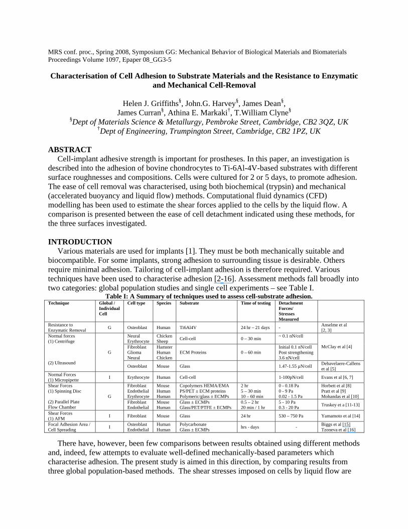
In previous work, the shear stress acting on the cells have been assumed equal to the wall shear stress [10-13, 20], [21]. This assumption underestimates the true shear force, since the cells protrude into the flow. CFD simulations have been conducted, in order to evaluate this effect. A 2-D (plane strain) mesh was employed, using the GAMBIT v.2.2.3 pre-processor, and the FLUENT programme used for the calculations. The cell was represented as a sector of a circle. The mesh was refined near the boundaries and the protrusion. The inlet velocity was set to 0.14, 0.34 or 0.63 m s-1, corresponding to experimental values. Flow in such experiments is commonly [11-13, 20] assumed laminar and fully developed. In fact, the modelling showed that only after flow distances of ~120 pipe diameters is fully developed laminar flow established. A no-slip boundary condition was imposed - i.e. liquid at the surfaces is stagnant, which has been reported experimentally for this type of set up [22]. Full details of the model are given elsewhere [23].
Normal forces were created by accelerated buoyancy. Inverted substrates, with adherent cells, were suspended in culture medium within centrifuge tubes. Different forces were applied by adjusting the centrifuge rotation frequency. Forces were calculated assuming a cell density of 2 g cm-3 (double that of surrounding fluid). Resistance to enzymatic detachment was assessed by incubating cells adhered to substrates in a dilute Trypsin-EDTA solution (1 vol% trypsin-EDTA (Sigma 10x), 99 vol% DMEM) for periods of up to an hour. Samples were washed with Hanks Buffered Salt Solution before and after enzyme exposure. REMOVAL OF CELLS BY FLOWING LIQUID
The effect of shear stress on populations of cells adhered to Ti-6Al-4V is shown in Fig.3(a). A flow rate of 7 ml s-1 was chosen for comparative studies of the PEO coatings and Ti-6Al-4V. Fig.3(b) shows that cells resist removal by shear most strongly when adhered to Ti-6Al-4V. Of the two PEO surfaces, cells were more easily removed from the smoother surface (phosphate electrolyte). A typical predicted flow pattern around a cell is shown in Fig.4(a). It can be seen that the presence of a cell is predicted to have a significant effect. The shear stresses in the region of the cell are shown in Fig.4(b).
By summing increments of force from local shear stresses on individual mesh elements, the overall force on the cell was calculated, and converted to stress by dividing by the cell area. The stresses in the experimental study were thus estimated to lie in the range 6 – 40 Pa – see Table II. When assessing adhesive strength in this way, geometrical factors may be significant. The rougher PEO surfaces showed greater resistance to cell removal under shear than the smoother PEO – see Fig.3(b). Cells have a tendency to locate in the valleys of an undulating surface, where they are shielded from the full force of the flow. However, estimated interfacial shear
strengths are likely to be lower than actual values. Firstly, cells do not adhere over their entire spread area, but over smaller areas (known as focal adhesions). These are usually a few 100 nm wide and a few microns long. The actual contact area has been estimated to be ~20 % of the nominal area [24], in which case the actual stresses on focal adhesions will be about 5 times larger than the values quoted here. Secondly, a no-slip condition was assumed at the periphery of the liquid, whereas in practice there is probably some liquid motion in this region, raising the shear force on the cells. Finally, there is also the possibility of liquid flow beneath the cells, particularly when cells are adhered to coatings with interconnected porosity. This will also raise the force experienced by the cells.
Figure 3: (a) Effect of fluid flow rate on the removal of cells adhered to Ti-6Al-4V and (b) comparison
between ease of cell removal by such shear forces from 3 different surfaces.
(a) Figure 4: (a) Predicted flow field in the vicinity of a cell (protrusion) and (b) predicted shear stresses
around the surface of the cell.
Table II: Shear stress on a cell as calculated by CFD modelling. The intermediate flow rate is was used for the comparative study.
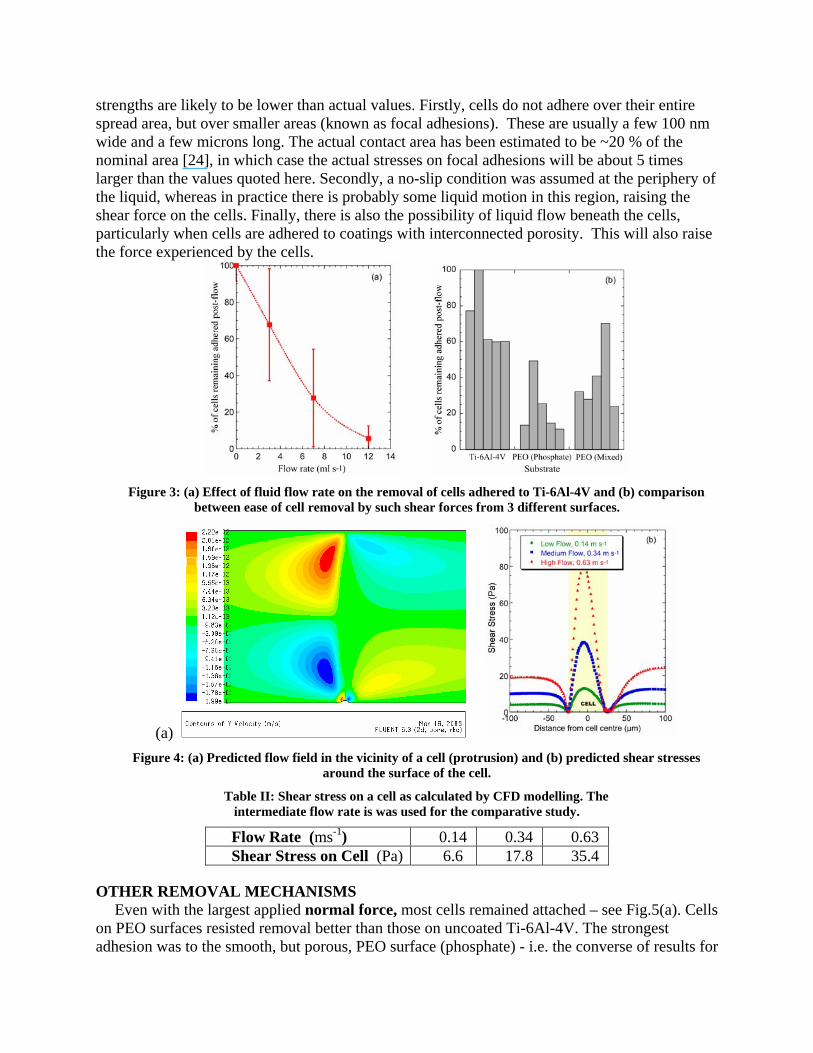
OTHER REMOVAL MECHANISMS Even with the largest applied normal force, most cells remained attached – see Fig.5(a). Cells
on PEO surfaces resisted removal better than those on uncoated Ti-6Al-4V. The strongest adhesion was to the smooth, but porous, PEO surface (phosphate) - i.e. the converse of results for
Flow Rate (ms-1) 0.14 0.34 0.63 Shear Stress on Cell (Pa) 6.6 17.8 35.4
liquid flow. Surface topology is expected to be less significant in normal stress tests, and surface chemistry probably plays a more important role. Whilst the morphology of the rougher PEO may be more desirable for the promotion of cell adhesion, the phase composition of the smoother PEO is apparently preferable. The resistance to enzymatic removal scales with the area under the % of cells remaining adhered versus time graphs. As for removal with normal forces, cells on PEO surfaces resisted detachment more strongly than those on Ti-6Al-4V – see Fig.5(b). The strongest adhesion was to the roughest surface (mixed electrolyte). Surface chemistry and topography [2, 3, 15] are known to influence cell adhesion. Not only does roughness affect adhesion, but the type of roughness also has an effect [15], with adhesion reportedly stronger when the topography is randomly patterned, rather than regular. PEO surfaces exhibit randomly-distributed porosity and roughness. In any event, more systematic work is clearly needed to clarify the relative importance of these effects in different cases.
Figure 5: Proportion of cell population remaining on different surfaces, as a function of (a) applied
(centrifugal) force, and (b) enzyme exposure time.
CONCLUSIONS • The adhesive strength of chondrocytes on Ti-6Al-4V and two oxide (PEO) coatings has been
investigated using shear flow, accelerated buoyancy and enzymatic attack. • Estimated interfacial shear stresses of up to 40 Pa, and normal stresses of up to 25 Pa, were
generated, although actual stresses were probably higher due to: (i) the true cell contact areas being smaller than nominal areas (ii) the assumed no-slip condition at the periphery of the liquid being invalid (iii) liquid flowing beneath the cells, particularly with PEO surfaces.
• PEO coatings with rougher surfaces offer more resistance to cell removal by shear forces and enzymes than smoother PEO coatings.
• While there are no strong indications that PEO surfaces offer systematically better cell adhesion than untreated Ti-6Al-4V, they certainly exhibit similar adhesive strengths and it may be possible to tailor the chemistry and pore architecture so as to optimise cell adhesion to them.
• Care is necessary when choosing a method for determining adhesive strength, since different methods are sensitive to different aspects of the adhesion. More work is needed in this area. REFERENCES
[1] B. D. Ratner, Hoffman, A.S., Schoen, F.J., Lemons, J.E., "Biomaterials Science: An Introduction to Materials in Medicine," 2nd Edition ed: Elsevier Academic Press, 2004.

[2] K. Anselme, M. Bigerelle, B. Noel, E. Dufresne, D. Judas, A. Iost, and P. Hardouin, "Qualitative and quantitative study of human osteoblast adhesion on materials with various surface roughnesses," Journal of Biomedical Materials Research, vol. 49, pp. 155-166, 2000.
[3] K. Anselme, P. Linez, M. Bigerelle, D. Le Maguer, A. Le Maguer, P. Hardouin, H. F. Hildebrand, A. Iost, and J. M. Leroy, "The relative influence of the topography and chemistry of TiAl6V4 surfaces on osteoblastic cell behaviour," Biomaterials, vol. 21, pp. 1567-1577, 2000.
[4] D. R. McClay, G. M. Wessel, and R. B. Marchase, "Inter-Cellular Recognition - Quantitation of Initial Binding Events," Proceedings of the National Academy of Sciences of the United States of America-Biological Sciences, vol. 78, pp. 4975-4979, 1981.
[5] D. Debavelaere-Callens, L. Peyre, P. Campistron, and H. F. Hildebrand, "On the use of ultrasounds to quantify the longitudinal threshold force to detach osteoblastic cells from a conditioned glass substrate," Biomolecular Engineering, vol. 24, pp. 521-525, 2007.
[6] E. Evans, D. Berk, and A. Leung, "Detachment of Agglutinin-Bonded Red-Blood-Cells .1. Forces to Rupture Molecular-Point Attachments," Biophysical Journal, vol. 59, pp. 838-848, 1991.
[7] E. A. Evans, "New Membrane Concept Applied to Analysis of Fluid Shear-Deformed and Micropipet-Deformed Red Blood-Cells," Biophysical Journal, vol. 13, pp. 941-954, 1973.
[8] T. A. Horbett, J. J. Waldburger, B. D. Ratner, and A. S. Hoffman, "Cell-Adhesion to a Series of Hydrophilic-Hydrophobic Copolymers Studied with a Spinning Disk Apparatus," Journal of Biomedical Materials Research, vol. 22, pp. 383-404, 1988.
[9] K. J. Pratt, S. K. Williams, and B. E. Jarrell, "Enhanced Adherence of Human Adult Endothelial-Cells to Plasma Discharge Modified Polyethylene Terephthalate," Journal of Biomedical Materials Research, vol. 23, pp. 1131-1147, 1989.
[10] N. Mohandas, R. M. Hochmuth, and E. E. Spaeth, "Adhesion of Red-Cells to Foreign Surfaces in Presence of Flow," Journal of Biomedical Materials Research, vol. 8, pp. 119-136, 1974.
[11] G. A. Truskey and J. S. Pirone, "The Effect of Fluid Shear-Stress Upon Cell-Adhesion to Fibronectin-Treated Surfaces," Journal of Biomedical Materials Research, vol. 24, pp. 1333-1353, 1990.
[12] G. A. Truskey and T. L. Proulx, "Quantitation of Cell Area on Glass and Fibronectin-Coated Surfaces by Digital Image-Analysis," Biotechnology Progress, vol. 6, pp. 513-519, 1990.
[13] G. A. Truskey and T. L. Proulx, "Relationship between 3t3 Cell Spreading and the Strength of Adhesion on Glass and Silane Surfaces," Biomaterials, vol. 14, pp. 243-254, 1993.
[14] A. Yamamoto, S. Mishima, N. Maruyama, and M. Sumita, "A new technique for direct measurement of the shear force necessary to detach a cell from a material," Biomaterials, vol. 19, pp. 871-879, 1998.
[15] M. J. P. Biggs, R. G. Richards, N. Gadegaard, C. D. W. Wilkinson, and M. J. Dalby, "Regulation of implant surface cell adhesion: Characterization and quantification of S-phase primary osteoblast adhesions on biomimetic nanoscale substrates," Journal of Orthopaedic Research, vol. 25, pp. 273-282, 2007.
[16] R. Tzoneva, N. Faucheux, and T. Groth, "Wettability of substrata controls cell-substrate and cell-cell adhesions," Biochimica Et Biophysica Acta-General Subjects, vol. 1770, pp. 1538-1547, 2007.
[17] N. F. Azevedo, A. R. Pinto, N. M. Reis, M. J. Vieira, and C. W. Keevil, "Shear stress, temperature, and inoculation concentration influence the adhesion of water-stressed Helicobacter pylori to stainless steel 304 and polypropylene," Applied and Environmental Microbiology, vol. 72, pp. 2936-2941, 2006.
[18] J. A. Curran and T. W. Clyne, "Porosity in Plasma Electrolytic Oxide Coatings," Acta Materialia, vol. 54, pp. 1985-1993, 2006.
[19] "alamarBlue™ Technical Datasheet," AbD Serotec Ltd, 2002, 2004. [20] R. G. Bacabac, T. H. Smit, S. C. Cowin, J. Van Loon, F. T. M. Nieuwstadt, R. Heethaar, and J. Klein-
Nulend, "Dynamic shear stress in parallel-plate flow chambers," Journal of Biomechanics, vol. 38, pp. 159-167, 2005.
[21] R. Bird, W. Stewart, and E. Lightfoot, Transport Phenomena, vol. 1. London: John Wiley & Sons, 1964. [22] R. Sabersky, A. Acosta, and E. Hauptmann, Fluid Flow a First Course in Fluid Mechanics, 2nd ed.
London: Macmillan, 1971. [23] J. Dean, H. Griffiths, A. Markaki, and T. Clyne, "An Experimental and Modelling Study of a Shear Flow
Technique for the Characterisation of Cell Adhesion," Biomaterials, in preparation. [24] J. D. Humphries, P. Wang, C. Streuli, B. Geiger, M. J. Humphries, and C. Ballestrem, "Vinculin controls
focal adhesion formation by direct interactions with talin and actin," Journal of Cell Biology, vol. 179, pp. 1043-1057, 2007.
Copyright © 2022 FDOKUMEN