A Single Immunoglobulin-like Domain of the Human Neural Cell Adhesion Molecule L1 Supports Adhesion...
15
The Rockefeller University Press, 0021-9525/97/12/1567/15 $2.00 The Journal of Cell Biology, Volume 139, Number 6, December 15, 1997 1567–1581 http://www.jcb.org 1567 A Single Immunoglobulin-like Domain of the Human Neural Cell Adhesion Molecule L1 Supports Adhesion by Multiple Vascular and Platelet Integrins Brunhilde Felding-Habermann,* Steve Silletti, ‡ Fang Mei, ‡ Chi-Hung Siu, § Paul M. Yip, § Peter C. Brooks, ‡ David A. Cheresh, ‡ Timothy E. O’Toole, i Mark H. Ginsberg, i and Anthony M.P. Montgomery ‡ *Roon Research Center for Arteriosclerosis and Thrombosis, Division of Experimental Hemostasis and Thrombosis; Department of Molecular and Experimental Medicine, ‡ Department of Immunology, The Scripps Research Institute, La Jolla, California 92037; § Banting and Best Department of Medical Research and Department of Biochemistry, University of Toronto, Ontario, Canada M5G 1L6; and i Department of Vascular Biology, The Scripps Research Institute, La Jolla, California 92037 Abstract. The neural cell adhesion molecule L1 has been shown to function as a homophilic ligand in a vari- ety of dynamic neurological processes. Here we demon- strate that the sixth immunoglobulin-like domain of hu- man L1 (L1-Ig6) can function as a heterophilic ligand for multiple members of the integrin superfamily in- cluding a v b 3 , a v b 1 , a 5 b 1 , and a IIb b 3 . The interaction be- tween L1-Ig6 and a IIb b 3 was found to support the rapid attachment of activated human platelets, whereas a cor- responding interaction with a v b 3 and a v b 1 supported the adhesion of umbilical vein endothelial cells. Muta- tion of the single Arg-Gly-Asp (RGD) motif in human L1-Ig6 effectively abrogated binding by the aforemen- tioned integrins. A L1 peptide containing this RGD motif and corresponding flanking amino acids (PSIT- WRGDGRDLQEL) effectively blocked L1 integrin in- teractions and, as an immobilized ligand, supported ad- hesion via a v b 3 , a v b 1 , a 5 b 1 , and a IIb b 3 . Whereas b 3 integrin binding to L1-Ig6 was evident in the presence of either Ca 21 , Mg 21 , or Mn 21 , a corresponding inter- action with the b 1 integrins was only observed in the presence of Mn 21 . Furthermore, such Mn 21 -dependent binding by a 5 b 1 and a v b 1 was significantly inhibited by exogenous Ca 21 . Our findings suggest that phys- iological levels of calcium will impose a hierarchy of in- tegrin binding to L1 such that a v b 3 or active a IIb b 3 . a v b 1 . a 5 b 1 . Given that L1 can interact with multiple vascular or platelet integrins it is significant that we also present evidence for de novo L1 expression on blood vessels associated with certain neoplastic or inflamma- tory diseases. Together these findings suggest an ex- panded and novel role for L1 in vascular and thrombo- genic processes. P ioneering studies on the structure and function of L1 have established this cell adhesion molecule (CAM) 1 as a member of the immunoglobulin super- family (IgSF) that plays a quintessential role in neural de- velopment (Lindner et al., 1983; Moos et al., 1988). Func- tions attributed to this neural CAM include such dynamic processes as cerebellar cell migration (Lindner et al., 1983) and neurite fasciculation and outgrowth (Lagenaur and Lemmon, 1987). Human and mouse L1 and L1-related glycoproteins in the rat (nerve growth factor–inducible, large external gly- coprotein [NILE]), chick (neuron–glial [Ng]CAM, 8D9, G4), and Drosophila (neuroglia) have been described (Gru- met et al., 1984; Bock et al., 1985; Lemmon and McLoon, 1986; Mujoo et al., 1986). These homologues share an ex- tracellular structure consisting of six Ig-like domains and five fibronectin type III–like repeats (Moos et al., 1988; Sonderegger and Rathjen, 1992). These extracellular do- mains are linked via a single transmembrane sequence to a short, highly conserved cytoplasmic domain (Reid and Hemperly, 1992). Limited structural variation within the human L1 molecule has been reported and can be attrib- uted to variable glycosylation and two alternatively spliced mini exons (Reid and Hemperly, 1992; Jouet et al., 1995). Address all correspondence to Anthony Montgomery, Department of Im- munology, R218, The Scripps Research Institute, La Jolla, CA 92037. Tel.: (619) 784-8109. Fax: (619) 784-2708. E-mail: [email protected] 1. Abbreviations used in this paper: CAM, cell adhesion molecule; GST, glutathione-S-transferase; IgSF, immunoglobulin super family; L1-Ig6, sixth immunoglobulin-like domain of human L1; NCAM, neural cell ad- hesion molecule; NILE, nerve growth factor–inducible, large external pro- tein; NgCAM, neuron–glial CAM; ORF, open reading frame; PGE 1 , pros- taglandin E 1 ; RGD, Arg-Gly-Asp; VNR, vitronectic receptor. on October 6, 2014 jcb.rupress.org Downloaded from Published December 15, 1997
-
Upload
independent -
Category
Documents
-
view
2 -
download
0
Transcript of A Single Immunoglobulin-like Domain of the Human Neural Cell Adhesion Molecule L1 Supports Adhesion...
The Rockefeller University Press, 0021-9525/97/12/1567/15 $2.00The Journal of Cell Biology, Volume 139, Number 6, December 15, 1997 1567–1581http://www.jcb.org 1567
A Single Immunoglobulin-like Domain of the HumanNeural Cell Adhesion Molecule L1 Supports Adhesionby Multiple Vascular and Platelet Integrins
Brunhilde Felding-Habermann,* Steve Silletti,
‡
Fang Mei,
‡
Chi-Hung Siu,
§
Paul M. Yip,
§
Peter C. Brooks,
‡
David A. Cheresh,
‡
Timothy E. O’Toole,
i
Mark H. Ginsberg,
i
and Anthony M.P. Montgomery
‡
*Roon Research Center for Arteriosclerosis and Thrombosis, Division of Experimental Hemostasis and Thrombosis; Department of Molecular and Experimental Medicine,
‡
Department of Immunology, The Scripps Research Institute, La Jolla, California 92037;
§
Banting and Best Department of Medical Research and Department of Biochemistry, University of Toronto,
Ontario, Canada M5G 1L6; and
i
Department of Vascular Biology, The Scripps Research Institute, La Jolla, California 92037
Abstract.
The neural cell adhesion molecule L1 has been shown to function as a homophilic ligand in a vari-ety of dynamic neurological processes. Here we demon-strate that the sixth immunoglobulin-like domain of hu-man L1 (L1-Ig6) can function as a heterophilic ligand for multiple members of the integrin superfamily in-cluding
a
v
b
3
,
a
v
b
1
,
a
5
b
1
, and
a
IIb
b
3
. The interaction be-tween L1-Ig6 and
a
IIb
b
3
was found to support the rapid attachment of activated human platelets, whereas a cor-responding interaction with
a
v
b
3
and
a
v
b
1
supported the adhesion of umbilical vein endothelial cells. Muta-tion of the single Arg-Gly-Asp (RGD) motif in human L1-Ig6 effectively abrogated binding by the aforemen-tioned integrins. A L1 peptide containing this RGD motif and corresponding flanking amino acids (PSIT-WRGDGRDLQEL) effectively blocked L1 integrin in-teractions and, as an immobilized ligand, supported ad-
hesion via
a
v
b
3
,
a
v
b
1
,
a
5
b
1
, and
a
IIb
b
3
. Whereas
b
3
integrin binding to L1-Ig6 was evident in the presence of either Ca
2
1
, Mg
2
1
, or Mn
2
1
, a corresponding inter-action with the
b
1
integrins was only observed in the presence of Mn
2
1
. Furthermore, such Mn
2
1
-dependent binding by
a
5
b
1
and
a
v
b
1
was significantly inhibited by exogenous Ca
2
1
. Our findings suggest that phys-iological levels of calcium will impose a hierarchy of in-tegrin binding to L1 such that
a
v
b
3
or active
a
IIb
b
3
. a
v
b
1
. a
5
b
1
. Given that L1 can interact with multiple vascular or platelet integrins it is significant that we also present evidence for de novo L1 expression on blood vessels associated with certain neoplastic or inflamma-tory diseases. Together these findings suggest an ex-panded and novel role for L1 in vascular and thrombo-genic processes.
P
ioneering
studies on the structure and function ofL1 have established this cell adhesion molecule(CAM)
1
as a member of the immunoglobulin super-family (IgSF) that plays a quintessential role in neural de-velopment (Lindner et al., 1983; Moos et al., 1988). Func-tions attributed to this neural CAM include such dynamicprocesses as cerebellar cell migration (Lindner et al., 1983)
and neurite fasciculation and outgrowth (Lagenaur andLemmon, 1987).
Human and mouse L1 and L1-related glycoproteins inthe rat (nerve growth factor–inducible, large external gly-coprotein [NILE]), chick (neuron–glial [Ng]CAM, 8D9,G4), and
Drosophila
(neuroglia) have been described (Gru-met et al., 1984; Bock et al., 1985; Lemmon and McLoon,1986; Mujoo et al., 1986). These homologues share an ex-tracellular structure consisting of six Ig-like domains andfive fibronectin type III–like repeats (Moos et al., 1988;Sonderegger and Rathjen, 1992). These extracellular do-mains are linked via a single transmembrane sequence to ashort, highly conserved cytoplasmic domain (Reid andHemperly, 1992). Limited structural variation within thehuman L1 molecule has been reported and can be attrib-uted to variable glycosylation and two alternatively splicedmini exons (Reid and Hemperly, 1992; Jouet et al., 1995).
Address all correspondence to Anthony Montgomery, Department of Im-munology, R218, The Scripps Research Institute, La Jolla, CA 92037. Tel.:(619) 784-8109. Fax: (619) 784-2708. E-mail: [email protected]
1.
Abbreviations used in this paper
: CAM, cell adhesion molecule; GST,glutathione-S-transferase; IgSF, immunoglobulin super family; L1-Ig6,sixth immunoglobulin-like domain of human L1; NCAM, neural cell ad-hesion molecule; NILE, nerve growth factor–inducible, large external pro-tein; NgCAM, neuron–glial CAM; ORF, open reading frame; PGE
1
, pros-taglandin E
1
; RGD, Arg-Gly-Asp; VNR, vitronectic receptor.
on October 6, 2014
jcb.rupress.orgD
ownloaded from
Published December 15, 1997

The Journal of Cell Biology, Volume 139, 1997 1568
Reflecting its designation as a neural CAM (NCAM),L1 is highly expressed on postmitotic neurons of the cen-tral and peripheral nervous systems and on pre- or nonmy-elinating Schwann cells of the peripheral nervous system(Lindner et al., 1983; Rathjen and Schachner, 1984; Mar-tini and Schachner, 1986). Although classified a neuralrecognition molecule, L1 has also been identified on non-neuronal cell types of surprisingly diverse origin. Thus, weand others, have recently described L1 on human immunecells of both myelomonocytic and lymphoid origin (Ebelinget al., 1996; Pancook et al., 1997). L1 has also been de-scribed on epithelial cells of the intestine and urogenitaltract (Thor et al., 1987; Kowitz et al., 1992; Kujat et al.,1995) and on transformed cells of both neuroectodermaland epithelial origin (Mujoo et al., 1986; Linnemann et al.,1989; Reid and Hemperly, 1992). Apart from such cellularassociations it is apparent that L1 can also be shed and in-corporated into the extracellular matrix (Martini and Schach-ner, 1986; Poltorak et al., 1990; Montgomery et al., 1996).This consequently implies a dual function for L1 both as aCAM and a substrate adhesion molecule (SAM).
In addition to having a propensity for homophilic bind-ing (Lemmon et al., 1989), L1 has recently emerged as aligand that can undergo multiple heterophilic interactions.Examples include interactions with other members of theIgSF and even components of the extracellular matrix. Thus,heterophilic ligands include TAG-1/axonin-1 (Kuhn et al.,1991; Felsenfeld et al., 1994), F3/F11 (Olive et al., 1995),laminin (Hall et al., 1997), and chondroitin sulfate proteo-glycans (Grumet et al., 1993; Friedlander et al., 1994). Sig-nificantly, L1 has also been reported to undergo multiple
cis
-type interactions with molecules as diverse as NCAM(Feizi, 1994), CD9 (Schmidt et al., 1996), and CD24 (Kad-mon et al., 1995). Interestingly such interactions in the planeof the cell membrane may serve to modify the specificityand avidity of L1 binding to ligands associated with themembranes of juxtaposed cells or the extracellular matrix(Feizi, 1994; Kadmon et al., 1995; Schmidt et al., 1996).
The presence of a single Arg-Gly-Asp (RGD) motif inthe sixth Ig-like domain of human L1, and the presence oftwo such motifs in the same domain of the murine and ratL1-homologues prompted early speculation as to whetherL1 might also function as a heterophilic ligand for mem-bers of the integrin superfamily. A number of studies havenow reported novel L1-integrin interactions (Ruppert et al.,1995; Ebeling et al., 1996; Montgomery et al., 1996; Ducz-mal et al., 1997; Pancook et al., 1997). In the first of thesestudies, Ruppert et al. (1995) describe an interaction be-tween the integrin
a
5
b
1
and murine L1. In a subsequentstudy, we report an interaction between human L1 and thevitronectin receptor
a
v
b
3
(Montgomery et al., 1996); an as-sociation that has now been described using a variety ofcell types (Ebeling et al., 1996; Pancook et al., 1997; Ducz-mal et al., 1997). Whereas an interaction between the inte-grin
a
5
b
1
and murine L1 has been documented, an interac-tion between this integrin and human L1 has not, despitethe use of cells expressing reasonable levels of this integrin(Ebeling et al., 1996). This has prompted some debateabout species-specific recognition perhaps governed bythe presence of an additional RGD site in murine L1 (Ebel-ing et al., 1996). Whereas the majority of these studies haveused purified or recombinant L1 as a substrate for inte-
grin-mediated adhesion, it is important that the interactionbetween integrin
a
5
b
1
and murine L1 has also been shownto mediate cell–cell interaction (Ruppert et al., 1995).
To date,
a
v
b
3
is the only member of the integrin super-family that has been shown to interact with human L1(Ebeling et al., 1996; Montgomery et al., 1996; Duczmal etal., 1997; Pancook et al., 1997). A primary objective of thisstudy was to determine whether recognition of human L1is a singular attribute of
a
v
b
3
or an attribute that can be ex-tended to other RGD-dependent integrins. In this regard,we demonstrate that the sixth Ig-like domain of human L1can in fact support multiple integrin interactions involving,not just
a
v
b
3
, but also the integrins
a
v
b
1
,
a
IIb
b
3
, and indeed
a
5
b
1
. A further objective of this study was to address keystructural and regulatory issues related to these L1-inte-grin interactions, including the central importance of thesingle RGD motif and the critical differential effects of spe-cific divalent cations. In this regard, we present evidencethat recognition of L1 by
a
v
b
3
,
a
v
b
1
,
a
IIb
b
3
, and
a
5
b
1
is in-deed RGD-dependent and that the binding of these differ-ent integrins is differentially regulated by physiologicallevels of calcium. Based on the novel pairing of L1 with
a
v
b
1
or
a
IIb
b
3
, we further demonstrated that this CAM cansupport the attachment of both endothelial cells (
a
v
b
1
)and activated platelets (
a
IIb
b
3
). Given the interaction be-tween L1 and the vascular integrins
a
v
b
3
and
a
v
b
1
, it is sa-lient that we also describe de novo L1 expression on bloodvessels associated with certain neoplastic or inflammatorydiseases. Based on these findings we suggest expanded andnovel roles for L1-integrin interactions in vascular andthrombogenic processes.
Materials and Methods
Antibodies
Anti-integrin antibodies used include the following: anti-hamster
a
5
b
1
mAb PB1, anti-
a
v
and b3 integrin polyclonal (anti-vitronetic receptor[VNR]), anti-avb3 mAb LM609, anti-aIIbb3 mAb LJ-CP8, anti–b3 integrinmAb 7E3, anti–b1 integrin mAb P4C10, and anti–av integrin mAb 17E6.PB1 was generated and provided by Dr. R. Juliano (University of NorthCarolina, Chapel Hill, NC), (Brown and Juliano, 1985). Anti–b1 integrinmAb P4C10 was provided by Dr. E.A. Wayner (University of Minnesota,Twin Cities, MN). The 7E3 antibody was originally generated and charac-terized by Coller et al. (1986) and the 17E6 antibody (Mitjans et al., 1995)was provided by Dr. S.L. Goodman (Merck KGaA, Darmstadt, Germany).LM609 (Cheresh and Spiro, 1987), anti-VNR, and LJ-CP8 (Niija et al.,1987) were generated within the Scripps Research Institute (La Jolla,CA). The anti–human L1 mAb 5G3 used in this study, was also generatedand characterized within the Scripps Research Institute (Mujoo et al.,1986).
PeptidesL1 peptides were synthesized on a peptide synthesizer (ABI 430A; Ap-plied Biosystems, Inc., Foster City, CA) within the Scripps Research Insti-tute Core Facility. A 15-mer peptide was selected to include the singleRGD site in human L1 (i.e., PSITWRGDGRDLQEL). Control peptideswere substituted with alanine to give PSITWRADGRDLQEL. For thepurpose of immobilization an additional batch of these peptides was madewith NH2-terminal cysteine residues. Peptides were prepared using RinkAmide MBHA or Wang resin (Calbiochem-Novabiochem, La Jolla, CA).After resin deprotection and assembly the peptides were cleaved from theresin with a cleavage cocktail (2.5% ethanedithiol, 5% thioanisole, 5%water, 87.5% trifluoroacetic acid) and subsequently purified by prepara-tive reverse phase HPLC. Peptides were characterized further by analyti-cal HPLC and mass spectroscopy.
on October 6, 2014
jcb.rupress.orgD
ownloaded from
Published December 15, 1997

Felding-Habermann et al. L1 Is a Ligand for Multiple Integrins 1569
Cell Lines and CultureThe generation and characterization of CHO cells stably transfected toexpress normal human platelet aIIbb3 (A5 cells) has been described in de-tail elsewhere (O’Toole et al., 1989, 1990; Frojmovic et al., 1991) and willbe described only briefly here. CHO cells were cotransfected with equalamounts of human aIIb and b3 expression constructs and a CDM8 vectorcontaining the neomycin resistance gene CDNeo at a ratio of 30:1 (O’Tooleet al., 1989). Transfection was performed by the calcium phosphate methodfollowed by glycerol shock. G418-resistant colonies were isolated and pos-itive clones identified by flow cytometry using subunit-specific antibodies.The generation of CHO cells transfected to express the active extracellu-lar domain of aIIbb3 (aIIbaLDb3 cells) has been described (O’Toole et al.,1994). Briefly, the cytoplasmic sequence from the aL integrin subunit wasisolated from the appropriate cDNA clone by PCR with the PCR oligonu-cleotides designed to omit the aL cytoplasmic sequence VGFFK (i.e.,aLD). As previously described, the aLD construct was ligated with a frag-ment encoding the extracellular and transmembrane domains of the aIIb
integrin subunit. Coexpression of this chimeric internal deletion mutant(aIIbaLD) with the wild-type b3 integrin subunit resulted in the stable ex-pression of the aIIbaLDb3 heterodimer bearing an active, high affinity ex-tracellular domain of human aIIbb3 (O’Toole et al., 1994). Wild-type CHO-K1cells and transfected cell lines were maintained in DME supplementedwith 10% FCS, 1% glutamine, and 1% non-essential amino acids.
A spontaneously transformed, human umbilical vein endothelial cellline designated ECV304 (Hughes, 1996) was obtained from the AmericanType Culture Collection (Rockville, MD). A stable avb3-negative variantof this line was obtained by repeated negative sorting using anti-avb3 mAbLM609. Sorting of avb3-negative unstained cells was performed using aFACstar® flow cytometer (Becton Dickinson, Co., Mountain View, CA).To obtain a stable, avb3-negative population, the cells were sorted on fiveconsecutive occasions. The ECV304 cells were maintained in M199 me-dium supplemented with 10% FCS and 1% glutamine.
Isolation of Human PlateletsBlood was collected from the antecubital vein of healthy adult donorsthrough a 19-gauge needle into syringes containing, as anticoagulant thethrombin inhibitor d-phenylalanyl-l-arginine chloromethyl ketone dihy-drochloride (PPACK; Bachem Bioscience Inc., Philadelphia, PA) (50 nMfinal concentration) and supplemented, when indicated, with prostaglan-din E1 (PGE1; Sigma Chemical Co., St. Louis, MO) (20 nM final concen-tration) to inhibit platelet activation. None of the donors had taken drugsknown to affect platelet function for the preceding 10 d. For the prepara-tion of washed platelets, the blood was supplemented with 5 U/ml of theADP scavenger apyrase (Sigma Chemical Co.) and centrifuged at 2,500 gfor 15 min at room temperature. Plasma was removed and replaced withan equivalent volume of Hepes-Tyrode’s buffer, pH 6.5 (10 mM Hepes,140 mM NaCl, 2.7 mM KCl, 0.4 mM NaH2PO4, 10 mM NaHCO3, and 5 mMdextrose), containing 1 U/ml of apyrase. The resuspended blood cells werecentrifuged again at 2,250 g for 10 min. The blood cells were washed twiceusing Hepes-Tyrode’s buffer containing 0.2 U/ml apyrase in the next stepand no apyrase in the last step. The final blood cell pellet was reconsti-tuted in Hepes-Tyrode’s buffer, pH 7.4, containing 50 mg/ml BSA to ad-just the viscosity to that of plasma, and then centrifuged at 700 g for 15min. The platelet-rich supernatant was collected and supplemented with1 mM CaCl2, 1 mM MgCl2, and 100 mM MnCl2. The platelet count was ad-justed to 100,000 platelets/ml. To analyze the effect of activation on plate-let adhesion, the platelets were stimulated with ADP and epinephrine (20 mMfinal concentration of each) immediately before adding the platelet sus-pension to the assay plates. Adhesion of non-activated platelets was stud-ied using unstimulated platelets prepared from PGE1-treated blood.
Construction and Expression of L1 Fusion ProteinsTwo wild-type and two mutant L1–glutathione-S-transferase (GST) fu-sion proteins were used in this study. The wild-type fusion proteins con-sisted of Ig-like domains 4, 5, and 6 (L1-Ig4-6) or the sixth Ig-like domainalone (L1-Ig6). The mutant fusion proteins consist of the sixth Ig-like do-main of L1 with the amino acid mutations of Arg-554 and Asp-556 to Lys-554, and Glu-556, respectively (i.e., RGD→KGE) or the single amino acidmutation Asp-556 to Ala-556 (i.e., RGD→RGA). Amino acids are num-bered as described by Bateman et al. (1996).
The generation and characterization of the L1-Ig4-6 GST fusion pro-tein used in this study has been described in detail elsewhere (Zhao and
Siu, 1995). The production of L1-Ig6 GST fusion protein (amino acids518–614) was as follows. The region of interest was amplified from full-length L1 cDNA, which was provided by Dr. J. Hemperly (Becton Dickin-son Research Center, Research Triangle Park, NC). Amplification wasperformed according to the manufacturer’s instructions for the ExpandHigh Fidelity PCR System (Boehringer Mannheim Corp., Indianapolis,IN) using an upstream sense primer specific for nucleotides 1,542–1,562 ofthe human L1 open reading frame (ORF) and containing an engineeredinternal EcoRI restriction endonuclease site (59-AAA GAA TTC ACTCAG ATC ACT-39) in conjunction with a downstream antisense primerspecific for nucleotides 1,829–1,849 of the human L1 ORF, which was alsoengineered to contain an internal EcoRI site (59-CGT GAA TTC GGCCCA GGG CTC-39). The resulting product was digested with EcoRI andsubcloned into a pGEX-1lT vector (Pharmacia Biotech Sevrage, Upp-sala, Sweden). Competent Escherichia coli strain BL21 cells (Stratagene,La Jolla, CA) were transformed with this construct and resulting colonieswere screened by PCR and examined for expression of appropriatelysized GST fusion protein by SDS-PAGE, followed by immunoblottingwith an anti-GST polyclonal antibody (Upstate Biotechnology, Inc., LakePlacid, NY). The chemiluminescent substrate PS-3 (Lumigen, Inc., South-field, MI) was used for detection. Dideoxy sequencing of positive cloneswas performed to verify the integrity of the introduced coding sequence.
The mutant L1-Ig6 RGD→RGA was generated according to the man-ufacturer’s instructions for the Quickchange Site-Directed MutagenesisKit (Stratagene) using the plasmid DNA encoding the L1-Ig6 fusion pro-tein (pGEX-1lT L1-Ig6) as template. Briefly, oligonucleotides correspond-ing to the sense and antisense sequences of bases 1,654–1,682 of the L1ORF, which included a change from A to C at base 1,666 (sense: 59-CCTGGC GTG GGG CCG GTC GAG ACC TCC AG-39; antisense: 59-CTGGAG GTC TCG ACC GGC CCC ACG CCA GG-39) were annealed to aheat-denatured template, and the construct was replicated using Pfu DNApolymerase for 18 cycles. The resulting mixture was digested with the meth-ylation-dependent endonuclease DpnI to degrade the wild-type template.Supercompetent E. coli strain XL-1 blue cells were transformed with thisconstruct by heat shock and resulting colonies were screened and se-quenced as described above.
To generate the mutant L1-Ig6 RGD→KGE the primers for an equiva-lent L1-Ig6 construct (forward primer: 59-TGG GAT CCA GAT CACTCA GGG GC-39; and the reverse primer: 59-GCG AAT TCT GGGATC CCG GCC CAG GGC TCC CCA C-39) encoding for amino acids518–614, were used in conjunction with the mutagenic primers 59-CATCAC CTG GAA GGG GGA GGG TCG AGA CC-39 and 59-GTA GTGGAC CTT CCC CCT CCC AGC TCT GG-39 in the four primer method(Higuchi, 1990). The amplified product was digested with BamHI andsubcloned into this site of pGEX-3X for expression in the E. coli strainJM101. The nucleotide sequence of the insert was confirmed by double-stranded DNA sequencing using the T7 Sequencing™ kit (Pharmacia Bio-tech Sevrage).
Purification of the recombinant fusion proteins was performed usingisopropylthio-b-d-galactoside–induced log-phase cultures essentially asdescribed by the manufacturer for the GST Gene Fusion System (Phar-macia Biotech Sevrage). Briefly, recovered bacteria were lysed by sonica-tion and incubated with detergent before clarification and immobilizationof the recombinant protein on a Sepharose 4B–coupled, glutathione affin-ity matrix (Pharmacia Biotech Sevrage). After extensive washing, theGST fusion proteins were eluted from the matrix with 10 mM reduced glu-tathione in 50 mM Tris-HCl, pH 8.0, and dialyzed extensively against PBSbefore use. The fusion proteins were subject to SDS-PAGE to confirm thecorrect mobility and to confirm purity.
Flow CytometryIntegrin expression was assessed by FACS® analysis. Subconfluent cul-tures were harvested and stained with anti-integrin mAbs at 20 mg/ml orpolyclonals diluted 1:40. The cells were then treated with an anti–mouseor anti–rabbit IgG, FITC-conjugated antibody, and were analyzed with aFACScan® flow cytometer (Becton Dickinson, Co., Mountain View, CA).Control cells were treated with secondary FITC-conjugated antibodyonly.
Adhesion AssaysAdhesion experiments were performed as detailed by Lagenaur and Lem-mon (1987) with some modifications. Purified L1-GST fusion proteins(L1-Ig6 or L1-Ig4-6) dialyzed into PBS were spotted (1-ml spots) and
on October 6, 2014
jcb.rupress.orgD
ownloaded from
Published December 15, 1997

The Journal of Cell Biology, Volume 139, 1997 1570
coated onto the bottom of 96-well Titertek plates (ICN Pharmaceuticals,Inc., Costa Mesa, CA) as described (Montgomery et al., 1996). Unless oth-erwise stated, the fusion proteins were offered at a concentration of 40 mg/ml.Treated and control wells were blocked with 5% BSA for 1–3 h at 378C.For adhesion studies involving immobilized peptides, wells were pre-coated overnight with murine IgG2a antibody at 20 mg/ml. Antibody-treated and washed wells were then incubated with the heterobifunctionalcross-linker, N-succinimidyl 3-(2-pyridyldithio)propionate (SPDP), at 30mg/ml in PBS for 45 min. These wells were then washed and peptidesadded at 100–200 mg/ml for 2–3 h. Control wells received antibody andSPDP alone. Treated and control wells were blocked with 5% BSA for1–3 h at 378C.
CHO cells were harvested using EDTA (0.526 mM) in PBS (versene;Irvine Scientific, Santa Ana, CA) and ECV304 cells with a trypsin-versenemixture (Biowhittaker, Walkerville, MD). All the cells were then given afurther wash with the EDTA solution to remove residual cations. Thecells were then resuspended in adhesion buffer consisting of HBSS (with-out calcium and magnesium) supplemented with 10 mM Hepes, and BSA(0.2–1%), with the pH adjusted to 7.4. Divalent cations were added as in-dicated in the text and included MnCl2 (0.4 mM), MgCl2 (1–2 mM), andCaCl2 (1–2 mM). Platelets were harvested and resuspended in Hepes-Tyrode’s buffer as described above. For inhibition studies, the cells andplatelets were pretreated with polyclonal antibodies (1:30 dilution), mAbs(80 mg/ml) or peptides (25 mM) for 30 min before the addition of bothcells and inhibitors to pretreated wells. Cells were added at 105/well andplatelets at 5 3 106/well, and then these plates were spun at 700 rpm togive a continuous monolayer of cells or platelets on the floor of each well.Endothelial cells and CHO cells were allowed to adhere for 30–40 min at378C, while the platelets were allowed to adhere for 10 min. At the end ofthe assay the wells were carefully washed with PBS, and non-adherentcells removed under a constant vacuum. Remaining adherent cells werefixed with 1% paraformaldehyde, and enumerated with the aid of an in-verted light microscope. Cells were counted per unit area using a 315 highpowered objective and an ocular grid with a minimum of four areascounted per well. Alternatively, adherent cells or platelets were stainedfor 20 min with 1% crystal violet in 0.1 M borate, pH 9.0. Dye was elutedwith 10% acetic acid and its absorbance determined at 600 nm. All exper-imental treatments were performed in triplicate.
ImmunohistochemistryFrozen sections of normal human skin, squamous cell carcinoma, psoriaticskin, and synovial tissue from the knee joint of patients diagnosed withrheumatoid arthritis were stained for the L1 antigen using mAb 5G3 orfor avb3 expression using mAb LM609. Frozen sections were fixed in coldacetone before removal of endogenous peroxidase with 0.03% H2O2. Sec-tions were subsequently blocked with 10% goat serum and 1% BSA inPBS. Primary antibodies were overlaid onto tissue sections at 20 mg/ml.Control sections were treated with isotype-matched murine IgG at 20 mg/ml.Sections were incubated with primary antibodies for 30–60 min at roomtemperature or overnight at 48C, and after extensive washing were treatedwith a secondary anti–mouse IgG biotinylated antibody (LSAB kit; DakoCorp., Carpinteria, CA) for 30 min. After further washing, tissue sectionswere treated with peroxidase-labeled streptavidin for an additional 30 min.To achieve red or blue-black color the sections were treated with 3-amino-9-ethylcarbazole (AEC) or Vector VIP substrate (Vector Laboratories,Burlingame, CA), respectively. For double staining, the first antigen(avb3) was revealed using one substrate (AEC), and the sections subse-quently restained for the second antigen (L1) using a different substrate(Vector VIP).
Results
Characterization of CHO-K1 Cells and Transfectants
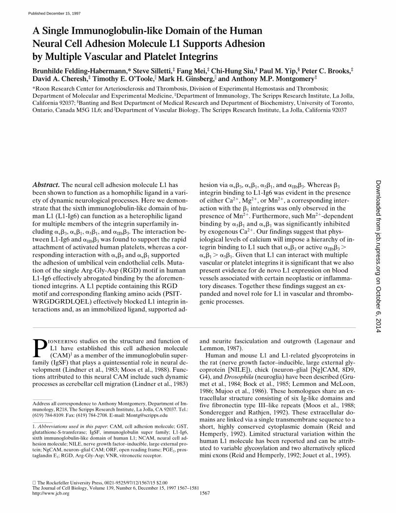
Currently, avb3 is the only integrin that has been shown tointeract with human L1 (Ebeling et al., 1996; Montgomeryet al., 1996; Duczmal et al., 1997; Pancook et al., 1997). Todetermine whether L1 is also a ligand for the platelet inte-grin aIIbb3, we used a CHO cell line (A5) genetically al-tered to express human platelet aIIbb3 (O’Toole et al.,1989, 1990; Frojmovic et al., 1991). To further determinewhether aIIbb3 needs to be in an active state to recognize
L1 we used a CHO cell line (aIIbaLDb3) transfected to ex-press aIIbb3 in a constitutively active state (O’Toole et al.,1994). Activation was achieved by chimerization of extra-cellular and transmembrane aIIb with a cytoplasmic dele-tion mutant of the aL integrin subunit (O’Toole et al.,1994). The integrin profile of these transfected cell linesand the CHO wild type was determined by flow cytometryusing anti–hamster or anti–human integrin–specific anti-bodies (Fig. 1 A). A number of salient conclusions can bedrawn from this analysis. First, both transfected cell lineshave been successfully manipulated to express high levelsof human aIIbb3 (LJ-CP8 reactivity). Second, transfectionof the human b3 integrin subunit has also resulted in apairing with endogenous hamster av and consequently ex-pression of chimeric avb3 (LM609 reactivity). Finally, andof immediate relevance for this study, wild-type and trans-fected CHO express high levels of endogenous a5b1 (PB-1reactivity) and endogenous av integrin(s) (VNR reactivity).
Wild-Type and Transfected CHO Cells Display Concentration-dependent Adhesion to L1-Ig6
We have previously demonstrated that purified full-lengthL1 and a recombinant L1 fusion protein (L1-Ig4-6) cansupport the adhesion of melanoma cells via the integrinavb3 (Montgomery et al., 1996). We further proposed thatit is the sixth Ig-like domain of human L1 that is likely tobe relevant for such adhesion by virtue of the presence ofa single RGD motif in this domain. Based on this proposi-tion, and for the purposes of this study, we generated a fu-sion protein consisting of this L1 domain alone (i.e., L1-Ig6). As a first step, this L1-Ig6 recombinant protein wastested for its ability to support adhesion by wild-type andtransfected CHO cells.
Importantly, the L1-Ig6 fusion protein supported signifi-cant concentration-dependent adhesion by all three CHOcell lines (Fig. 1 B). Furthermore, transfected CHO cellsexhibited greater adhesion than the wild-type CHO-K1cells (Fig. 1 B). These data clearly indicate the importanceof the sixth Ig-like domain of L1 for mediating cellular ad-hesion, and also suggest that transfection and expressionof b3 integrins (avb3 and/or aIIbb3) can lead to enhancedbinding to L1-Ig6. Significantly, CHO cells bearing the ac-tive aIIbb3 (aIIbaLDb3 cells) showed the greatest level ofadhesion, particularly at lower L1-Ig6 concentrations (Fig.1 B). When offered at saturating concentrations (i.e., .50mg/ml), L1-Ig6 supported .90% attachment of the aIIbaLDb3cells in contact with the substrate, resulting in a continuousmonolayer of spreading cells and these cells were resistantto detachment by the large shear forces generated duringthe washing of the 96-well plates. When offered at a satu-rating concentration, vitronectin gave an equivalent re-sponse (data not shown).
Wild-type CHO-K1 Cells Interact with L1-Ig6Using a5b1 and an av Integrin(s) and TheseHeterophilic Interactions Are Differentially Regulated by Divalent Cations
From the data presented in Fig. 1 B it is evident that thewild-type CHO-K1 cells can interact with the L1-Ig6 fu-sion protein. To characterize this wild type adhesion welooked for evidence of either b1 or av integrin involvement.
on October 6, 2014
jcb.rupress.orgD
ownloaded from
Published December 15, 1997

Felding-Habermann et al. L1 Is a Ligand for Multiple Integrins 1571
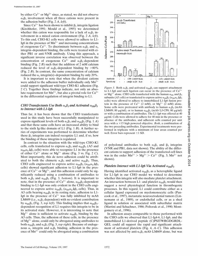
In the presence of Ca21, Mg21, and Mn21, CHO-K1 celladhesion was completely abrogated by a VNR polyclonalantibody, indicating the involvement of one or more av in-tegrins (Fig. 2 A, left). Despite high levels of a5b1 expres-sion, a function blocking mAb specific for hamster a5b1(PB1) had no impact on adhesion in this cation environ-ment (Fig. 2 A, left). However, in the presence of Mn21
alone, CHO-K1 adhesion to L1-Ig6 could only be abro-gated using a combination of VNR polyclonal antibodyand the anti-a5b1 mAb PB1 (Fig. 2 A, right). This findingindicates that in the presence of Mn21 alone, a5b1 can alsorecognize human L1-Ig6. However, it is also clear thatbinding by a5b1 must be acutely susceptible to inhibition
Figure 1. (A) Integrin profiles of wild-type CHO-K1 cells, ofCHO cells transfected with aIIb and b3 subunits (A5) and of CHOcells transfected to express active aIIbb3 (aIIbaLDb3 cells). (B) Con-centration-dependent adhesion of CHO-K1, A5, and aIIbaLDb3cells to L1-Ig6. (A) Integrin expression is represented by FACS®
histograms. Cells were treated with antibodies to hamster a5b1(monoclonal PB1), to human avb3 (mAb LM609), to humanaIIbb3 (mAb LJ-CP8), to hamster or human av or b3 integrin sub-units (polyclonal VNR), or to human b3 integrins (mAb 7E3).These cells were subsequently stained with fluorescein-conju-gated goat anti–mouse or goat anti–rabbit antibodies and wereanalyzed using a FACScan® flow cytometer. Control cells weretreated with secondary fluorescein-conjugated antibody only. (B)Wild-type or transfected cells were allowed to adhere to immobi-lized L1-Ig6 fusion protein offered at concentrations rangingfrom 1 to 100 mg/ml. After 30 min non-adherent cells were re-moved by washing and the remaining adherent cells counted perunit area with a 315 high powered objective. Experimental treat-ments were performed in triplicate with a minimum of four areascounted per well. No significant adhesion was observed on GSTalone and any minimal residual adhesion to BSA-blocked plasticalone has been subtracted from the cell counts shown. Error barsrepresent 61 SD.
Figure 2. Wild-type CHO-K1 cells adhere to L1-Ig6 using an avintegrin(s) and a5b1, and these heterophilic interactions are criti-cally and differentially regulated by exogenous Ca21. (A) Wild-type cells were allowed to adhere to immobilized L1-Ig6 fusionprotein in the presence of Ca21 (2 mM), Mg21 (2 mM), and Mn21
(0.4 mM), or in the presence of Mn21 alone (0.4 mM). Some cellswere pretreated with antibody to human avb3 (mAb LM609; 80mg/ml), to hamster a5b1 (PB1 ascites 1:30) or to VNR (polyclonalanti-VNR; 1:30), or a combination of antibodies. The level ofcontrol adhesion achieved in the absence of inhibitors is taken as100%. In the presence of mixed cations this was equivalent to 82adherent cells per field and in the presence of Mn21 alone thiswas equivalent to 255 adherent cells per field. Both, a combina-tion of the two preceding antibodies. (B) Wild-type cells were al-lowed to adhere to immobilized L1-Ig6 fusion protein in the pres-ence of Mn21 (0.4 mM) alone or in the presence of Mn21 (0.4 mM)and increasing concentrations of Ca21 (0.5–2 mM). Cells werepretreated with antibody to hamster a5b1 (PB1 ascites 1:30) or toVNR (polyclonal anti-VNR; 1:30). (C) Wild-type cells were al-lowed to adhere to immobilized L1-Ig6 fusion protein in the pres-ence of Ca21 (1 mM) alone, Mg21 (1 mM) alone or Mn21 (0.4 mM)alone. The L1-Ig6 fusion protein was offered at a concentrationof 40 mg/ml. Cells were allowed to adhere for 30–40 min in thepresence or absence of the antibodies, and adherent cells countedper unit area with a 315 high powered objective. Experimentaltreatments were performed in triplicate with a minimum of fourareas counted per well. Error bars represent 61 SD.
on October 6, 2014
jcb.rupress.orgD
ownloaded from
Published December 15, 1997
The Journal of Cell Biology, Volume 139, 1997 1572
by either Ca21 or Mg21 since, as stated, we did not observea5b1 involvement when all three cations were present inthe adhesion buffer (Fig. 2 A, left).
Since Ca21 has been shown to inhibit b1 integrin ligation(Kirchhofer, 1991; Mould et al., 1995) we determinedwhether this cation was responsible for a lack of a5b1 in-volvement in a mixed cation environment (Fig. 2 A, left).To this end, CHO-K1 cells were allowed to adhere to L1-Ig6 in the presence of Mn21 and increasing concentrationsof exogenous Ca21. To discriminate between a5b1 and avintegrin–dependent binding, the cells were treated with ei-ther PB1 or anti-VNR antibody. Using this approach, asignificant inverse correlation was observed between theconcentration of exogenous Ca21 and a5b1-dependentbinding (Fig. 2 B) such that the addition of 2 mM calciumreduced the level of a5b1-dependent binding by .80%(Fig. 2 B). In contrast, the same concentration of calciumreduced the av integrin(s)–dependent binding by only 20%.
It is important to note that when the divalent cationswere added to the adhesion buffer individually only Mn21
could support significant wild-type CHO-K1 adhesion (Fig.2 C). Together these findings indicate, not only an abso-lute requirement for Mn21, but also a pivotal role for Ca21
in the differential regulation of integrin binding to L1.
CHO Transfectants Use Both avb3 and Activated aIIbb3 to Interact with L1-Ig6
Thus far, it has been shown that the CHO transfectantsused in this study have been successfully manipulated toexpress significant levels of both avb3 and aIIbb3 (Fig. 1 A)and that these same cells have an increased ability to bindto the sixth Ig-like domain of L1 (Fig. 1 B). A further se-ries of experiments was performed to determine whetherthese b3 integrins can indeed recognize L1 and, if so, howthe binding of these integrins is regulated.
In contrast to the situation with the wild-type CHO-K1cells, cells transfected to express avb3 and aIIbb3 (A5 andaIIbaLDb3 cells) were able to recognize L1 in the presenceof either Ca21 alone or Mg21 alone (Fig. 3 vs. Fig. 2 C).Most importantly, this de novo adhesion could be attrib-uted to both the chimeric avb3 and active aIIbb3. Thus,CHO cells engineered to express active aIIbb3 (aIIbaLDb3cells) showed significant adhesion to L1-Ig6 in the pres-ence of Ca21 or Mg21, and this adhesion could only be sig-nificantly reduced using a combination of antibodies toboth avb3 and aIIbb3 (Fig. 3, bottom). It is important tonote, that in the presence of Ca21 alone, aIIbb3-dependentbinding to L1-Ig6 was only evident in the CHO cells engi-neered to express active aIIbb3 (aIIbaLDb3 cells). Thus, inA5 cells bearing aIIbb3 in its resting state, adhesion to L1-Ig6 in the presence of Ca21 was fully inhibited by mAbLM609 (i.e., avb3 dependent) with no evident contributionby aIIbb3 (Fig. 3, top left). This finding implies that aIIbb3-dependent recognition of L1 requires this integrin to be inits activated state. However, it is interesting to note thatMg21 alone is sufficient to activate aIIbb3 binding by theA5 cells. Thus, the adhesion of these cells, in the presenceof Mg21 alone, could only be abrogated using antibodies toboth aIIbb3 and avb3 (Fig. 3, top right). Because of endoge-nous av integrin and a5b1 binding, adhesion in the pres-ence of Mn21 could only be abrogated using a combination
of polyclonal antibodies to both a5b1 and b3 integrins(VNR and PB1, data not shown). The ability of the differ-ent cations to support adhesion of the transfected cell lineswas in the order Mn21 . Mg21 . Ca21 (Fig. 3; Mn21 notshown).
Platelets Interact with L1-Ig6 Via Activated aIIbb3
Having identified activated aIIbb3 as a heterophilic ligandfor L1-Ig6 in our CHO model we wished to determinewhether this integrin will also mediate platelet attachment.An interaction between L1- and platelet aIIbb3 would thensuggest a novel physiological function in thrombogenicprocesses. In this regard, L1 could contribute either as acellular ligand expressed on myelomonocytic cells (Pan-cook et al., 1997), metastatic neuroectodemal tumors (Lin-nemann et al., 1989), or endothelial cells, or as a shedligand in solution or associated with subcellular matrix(Martini and Schachner, 1986; Poltorak et al., 1990; Mont-gomery et al., 1996).
In adhesion assays comparable to those performed withthe CHO cells we observed that L1-Ig4-6, L1-Ig6, and theimmobilized L1-derived peptide (C)PSITWRGDGRDL-QEL could all support the rapid and significant attach-ment of activated platelets (Fig. 4, A–C). This adhesionwas not affected by anti-avb3 mAb LM609 alone, but was
Figure 3. Both avb3 and activated aIIbb3 can support attachmentto L1-Ig6 and such ligation can occur in the presence of Ca21
or Mg21 alone. CHO cells transfected with the human aIIb and b3subunits (A5 cells) or transfected to express active aIIbb3 (aIIbaLDb3cells) were allowed to adhere to immobilized L1-Ig6 fusion pro-tein in the presence of Ca21 (2 mM), or Mg21 (2 mM) alone.Some cells were pretreated with antibody to human avb3 (mAbLM609; 80 mg/ml), or to human aIIbb3 (mAb LJ-CP8; 80 mg/ml)or with a combination of antibodies. The L1-Ig6 was offered at 40mg/ml. Cells were allowed to adhere for 40 min in the presence orabsence of the antibodies, and adherent cells counted per unitarea with a 315 high powered objective. Both, a combination ofthe two preceding antibodies. Experimental treatments were per-formed in triplicate with a minimum of four areas counted perwell. Error bars represent 61 SD.
on October 6, 2014
jcb.rupress.orgD
ownloaded from
Published December 15, 1997

Felding-Habermann et al. L1 Is a Ligand for Multiple Integrins 1573
abrogated by the mAb 7E3, which is a potent function-blocking antibody, specific for both avb3 and aIIbb3 (Fig. 4,A–C). The affinity of activated platelets for immobilizedL1-Ig6, and the strong inhibitory effect of mAb 7E3, isalso evident from the photomicrographs presented in Fig.4, D and E. Together these findings are consistent with arole for platelet aIIbb3. The contribution of aIIbb3 was alsoconfirmed using the specific anti-aIIbb3 mAb LJ-CP8, butthis mAb was generally less effective than 7E3 (data notshown). It is important to note that these results were onlyobtained using platelets bearing active aIIbb3 as a result ofstimulation with ADP and epinephrine. Thus, unstimu-lated platelets, prepared from PGE1-treated blood, showedonly minimal adhesion to L1-Ig4-6 (Fig. 4 A). These data,
like the data obtained using the CHO cells, clearly indicatethat aIIbb3 needs to be in an active conformation to bindL1. However, as described for other peptides, recognitionof the L1-derived peptide was not fully dependent onplatelet activation (Fig. 4 C). When offered at saturatingconcentrations, the L1 fusion proteins supported the rapidattachment of a uniform monolayer of platelets such thatthe majority of platelets offered and in contact with thesubstrate appeared to attach (Fig. 4 D). When used as apositive control, vitronectin supported comparable levelsof platelet attachment (data not shown). However, as withL1-Ig4-6 significant attachment to vitronectin requiredplatelet activation.
Endothelial Cells Interact with L1-Ig6 Using avb3and avb1 and These Heterophilic Interactions Are Differentially Regulated by Divalent Cations
L1 is expressed on a variety of cell types known to interactwith endothelium (Ebeling et al., 1996; Pancook et al.,1997); and shed L1 may also associate with components ofthe subendothelial matrix (Hall et al., 1997). This distribu-tion raises the question of if and how endothelial cells in-teract with L1. To address this issue we used a transformedcell line (ECV304) derived from human umbilical vein en-dothelial cells (Hughes et al., 1996). These wild-type cellswere found to express both avb3 and a high level of b1 inte-grins (Fig. 5 A, left column). We also detected expressionof avb5 (mAb P1F6) at a median fluorescence comparableto that found for avb3 (not shown). Consistent with ourfindings using the transfected CHO cells, we observed thatendothelial avb3 can also promote adhesion to L1-Ig6 andthis adhesion is evident in the presence of calcium (Fig. 5B). Adhesion by these wild-type endothelial cells was onlymarginally inhibited by the anti–b1 integrin mAb P4C10(Fig. 5 B). A similar pattern of adhesion was obtained us-ing primary human dermal microvascular endothelial cells(Clonetics, San Diego, CA) (data not shown).
Whereas the contribution of avb3 to a variety of vascularprocesses is well documented it is also clear that this inte-grin is either absent or only marginally expressed by quies-cent endothelial cells (Brooks et al., 1994). Accordingly,we wished to determine whether L1 could also be recog-nized by avb3-negative endothelial cells. To address thiswe exploited ECV304 endothelial cells that had been re-peatedly FACS® sorted for a lack of avb3 expression. Thisapproach proved successful, resulting in the generation ofa stable population of avb3-negative cells (Fig. 5 A, rightcolumn). The levels of avb5 and b1 integrin expression inthese cells remained unchanged (not shown; and Fig. 5 A,right column). Remarkably, these avb3-negative cells alsoshowed significant adhesion to L1-Ig6 (Fig. 5 C). How-ever, in contrast to the wild-type cells, adhesion was unaf-fected by an antibody to avb3 (LM609) but was fully inhib-ited by a mAb to b1 integrins (i.e., P4C10) (Fig. 5 C).Furthermore, such adhesion was also completely abro-gated with an antibody specific for av integrins (Fig. 5 C).Together these results are consistent with an interactionbetween L1 and endothelial avb1.
Given that both avb3 and avb1 can interact with L1-Ig6,we wished to determine why avb3 is the dominant integrinin wild-type ECV304 cell adhesion (Fig. 5 B). In this re-
Figure 4. Activated aIIbb3 can mediate the attachment of plate-lets to immobilized L1 fusion proteins and the L1-derived pep-tide (C)PSITWRGDGRDLQEL. (A–C) Stimulated or unstimu-lated human platelets were allowed to adhere to immobilized L1fusion proteins (L1-Ig4-6 or L1-Ig6; A and B) or to immobilized,L1-derived RGD peptide (C). Some of the platelets were pre-treated with antibody to avb3 (mAb LM609; 80 mg/ml), or to avb3and aIIbb3 (mAb 7E3; 80 mg/ml). The platelets were allowed toadhere for 10 min in the presence or absence of the antibodiesand the adherent platelets subsequently stained with crystal vio-let for quantification. Experimental treatments were performedin triplicate. Error bars represent 61 SD. (D and E) Photomicro-graphs of stimulated platelets adhering to L1-Ig6 in the absence(D) or presence of mAb 7E3 (E). Adherent platelets werestained with crystal violet and are confined to the circular areacoated with the fusion protein. Individual platelets cannot be dis-criminated because of their small size and some aggregation. Theplatelets were allowed to adhere for 10 min in the presence or ab-sence of 7E3. Any minimal residual adhesion to BSA-blockedplastic alone has been subtracted from the OD values shown. Bar,300 mm.
on October 6, 2014
jcb.rupress.orgD
ownloaded from
Published December 15, 1997

The Journal of Cell Biology, Volume 139, 1997 1574
gard, we tested the hypothesis that exogenous calciummay favor the use of avb3 rather than avb1. We observed anumber of facts supporting this hypothesis. First, signifi-cant avb1 binding could be induced in the wild-type cells,but only in the presence of Mn21 alone (Fig. 6 A). Thus,whereas wild-type ECV304 cell adhesion in the presence
of Ca21 could be abrogated with anti-avb3 mAb LM609(Fig. 5 B), the adhesion of these cells in the presence ofMn21 alone could only be blocked using a combination ofantibodies reactive with both avb3 and avb1 (i.e., LM609and P4C10), or with a mAb reactive with both av integrins(i.e., 17E6) (Fig. 6 A, right). Second, minimal wild-type ad-hesion was observed in the presence of Ca21 or Mg21
alone and this appeared to be fully dependent upon avb3(Fig. 6 A). Finally, in an experiment analogous to that per-formed with the wild-type CHO cells (Fig. 2 B), we ob-served that the avb1-mediated (Mn21-dependent) adhe-sion of the sorted, avb3-negative endothelial cells was sig-nificantly more susceptible to inhibition by exogenousCa21 than the wild-type adhesion (Fig. 6 B). Togetherthese data indicate that in the absence of avb3 expression,quiescent endothelial cells may use avb1 to bind to L1,
Figure 5. (A) Integrin profiles of wild-type ECV304 human um-bilical vein endothelial cells or ECV304 cells repeatedly sortedfor a lack of avb3 expression. (B and C) avb3- and avb1-dependentadhesion of wild-type or sorted ECV304 endothelial cells to L1-Ig6. (A) Integrin expression is represented by FACS® histograms.Cells were treated with antibodies to av integrins (mAb 17E6), toavb3 (mAb LM609), to av or b3 integrin subunits (polyclonal VNR),or to b1 integrins (mAb P4C10). These cells were subsequentlystained with fluorescein-conjugated goat anti–mouse or goat anti–rabbit antibodies and were analyzed using a FACScan® flow cy-tometer. Control cells were treated with secondary fluorescein-conjugated antibody only. (B and C) Wild-type or sorted cellswere allowed to adhere to immobilized L1-Ig6 fusion protein of-fered at 40 mg/ml. Adhesion was performed in the presence ofCa21 (2 mM), Mg21 (2 mM), and Mn21 (0.4 mM). Some cells werepretreated with antibody to avb3 (mAb LM609; 80 mg/ml), to b1integrins (mAb P4C10; 80 mg/ml), to av integrins (MAb 17E6; 80mg/ml), or with a combination of antibodies. After 40 min non-adherent cells were removed by washing and the remainingadherent cells counted per unit area with a 315 high powered ob-jective. Both, a combination of the two preceding antibodies. Ex-perimental treatments were performed in triplicate with a minimumof four areas counted per well. Error bars represent 61 SD.
Figure 6. Usage of avb1 by wild-type ECV304 human umbilicalvein endothelial cells is dictated by a requirement for Mn21 andby inhibition by Ca21. (A) Wild-type ECV304 endothelial cellswere allowed to adhere to immobilized L1-Ig6 in the presence ofCa21 alone (1 mM), Mg21 alone (1 mM), or Mn21 alone (0.4 mM).Some cells were pretreated with antibody to avb3 (mAb LM609;80 mg/ml), to b1 integrins (mAb P4C10; 80 mg/ml), to av integrins(mAb 17E6; 80 mg/ml), or with a combination of antibodies. (B)Wild-type or sorted endothelial cells were allowed to adhere to im-mobilized L1-Ig6 fusion protein in the presence of Mn21 (0.4 mM)alone or in combination with increasing concentrations of Ca21
(0.5–2 mM). After 40 min non-adherent cells were removed bywashing and the remaining adherent cells counted per unit areawith a 315 high powered objective. Both, refers to a combinationof the two preceding antibodies. Experimental treatments wereperformed in triplicate with a minimum of four areas counted perwell. Error bars represent 61 SD.
on October 6, 2014
jcb.rupress.orgD
ownloaded from
Published December 15, 1997

Felding-Habermann et al. L1 Is a Ligand for Multiple Integrins 1575
however with the induction of avb3 expression, physiologi-cal levels of calcium are likely to favor the use of this inte-grin. It is also clear that whereas avb1-mediated adhesionis susceptible to inhibition by exogenous Ca21, this inhibi-tion is less pronounced than that seen when adhesion ismediated via a5b1 (Fig. 2 B).
The Interaction between L1-Ig6 and the b3 or b1 Integrins Is RGD-dependent
Thus far we have demonstrated that a single Ig-like do-main of L1 can support multiple integrin interactions. Thissame domain contains an RGD integrin recognition motifthat may provide the binding site for all the aforemen-tioned integrins. However it is also clear that a given RGDsite may or may not support adhesion depending on flank-ing sequences, conformational restraints and accessibility(D’Souza et al., 1991; Haas and Plow, 1994). Furthermore,although avb3, a5b1, avb1, and aIIbb3 have all been re-ported to interact with RGD motifs, a5b1 and the b3 inte-grins have also been shown to interact with non-RGD se-quences (Koivunen et al., 1994). To help address this issuewe generated a 15-mer peptide based on a sequence in thesixth Ig-like domain of L1 and inclusive of the RGD rec-ognition motif (i.e., PSITWRGDGRDQEL). This peptidewas tested for its efficacy both as an immobilized ligandand as soluble inhibitor of L1 integrin binding.
Supporting the concept of a RGD-dependent interac-tion we observed that our L1-RGD peptide, once immobi-lized, could support significant endothelial cell attachmentvia avb3, and avb1 (Fig. 7, Endothelial) and CHO cell at-tachment via endogenous a5b1 and transfected aIIbb3 (Fig.7, CHO). Specific integrin–peptide interactions were con-firmed using the function blocking antibodies indicated(Fig. 7). When offered at the same concentration, a controlpeptide (C)PSITWRADGRDQEL was ineffective as anadhesive ligand. However, it should be noted that becauseof the conservative glycine to alanine mutation (i.e.,
RGD→RAD) this control peptide could also support someattachment but only at significantly higher concentrations(not shown).
Further support for RGD-dependent interaction withL1 was obtained using the same L1-RGD peptide as a sol-uble inhibitor. At a concentration of 25 mM, the peptideeffectively abrogated endothelial cell adhesion to L1-Ig6in the presence of Mn21 alone (Fig. 8, left). We have previ-ously demonstrated that adhesion by these cells in thepresence of this cation involves both avb3 and avb1 (Fig. 6A, right). Likewise, we also observed a complete inhibitionof wild-type CHO-K1 adhesion in the presence of Mn21
(Fig. 8, middle). According to our previous data, this isconsistent with an inhibition of endogenous a5b1 and theav integrin(s) (Fig. 2 A, right). Adhesion by the CHO cellstransfected to express active aIIbb3 was less sensitive to in-hibition via the RGD peptide when used at 25 mM (Fig. 8,right), but could be fully inhibited at higher peptide con-centrations (not shown). Used at an equivalent concentra-tion our control peptide was ineffective at preventing ad-hesion. But again because of the conservative mutation ofthis peptide (i.e., RGD→RAD) this peptide could also in-hibit attachment at high concentrations (e.g., 250 mM).
Aforementioned work with the L1-RGD peptide sup-ports the concept of a RGD-dependent interaction be-tween integrins and L1. However, to address this issuedefinitively, we sought to demonstrate that mutation of theRGD site in L1-Ig6 is sufficient to abrogate integrin bind-ing. To this end, we generated additional L1-Ig6 fusionproteins containing the mutations RGD→KGE or RGD→RGA. Importantly, the conservative RGD→KGE muta-tion completely abrogated avb3- and avb1-dependent bind-ing by the endothelial cells (Fig. 9, Endothelial). This samemutation and the RGD→RGA mutation also significantlyabrogated a5b1-dependent binding by wild-type CHO cells(Fig. 9, CHO, wild-type). Some residual low level bindingto both mutations was still observed, perhaps indicatingthat the mutated sequences can still be recognized to some
Figure 7. All of the integrins characterized canrecognize the L1-derived peptide (C)PSIT-WRGDGRDLQEL. Wild-type or sorted ECV304endothelial cells and wild-type or transfected CHOcells (aIIbaLDb3 cells) were allowed to adhere toimmobilized 16-mer peptide (C)PSITWRGD-GRDLQEL or to control peptide (C)PSITWR-ADGRDLQEL. Adhesion was performed in thepresence of Ca21 (2 mM), Mg21 (2 mM), andMn21 (0.4 mM) or in the presence of Mg21 (1mM) alone, or Mn21 alone (0.4 mM). Some cellswere pretreated with antibody to human aIIbb3(mAb LJ-CP8; 80 mg/ml), to avb3 (mAb LM609;80 mg/ml), to b1 integrins (mAb P4C10; 80 mg/ml), to av integrins (mAb 17E6; 80 mg/ml), tohamster a5b1 (PB1 ascites 1:30) or to VNR (poly-clonal anti-VNR; 1:30), or with a combination ofantibodies. After 40 min, non-adherent cells wereremoved by washing and the remaining adherentcells stained with crystal violet. Both, a combina-tion of the two preceding antibodies. Experimen-tal treatments were performed in triplicate with aminimum of four areas counted per well. Errorbars represent 61 SD.
on October 6, 2014
jcb.rupress.orgD
ownloaded from
Published December 15, 1997

The Journal of Cell Biology, Volume 139, 1997 1576
degree by a5b1. Alternatively, a second a5b1-binding motifmay exist within the L1-Ig6 domain. Interestingly theRGD→KGE mutation could still be recognized to a signif-icant level by aIIbb3 (Fig. 9, CHO, Active aIIbb3). However,the alanine substitution mutant (RGA) was sufficient tofully abrogate aIIbb3-mediated binding.
L1 Expression Can Be Induced on Endothelial CellsIn Vivo
We have identified a variety of endothelial and platelet in-tegrins that can interact with the sixth Ig-like domain ofL1. Such heterophilic interactions prompted us to deter-mine whether L1 can be expressed on endothelial cells.This expression would suggest the potential for L1 integrininteractions in vascular processes such as angiogenesis andthrombosis; these are processes that require homotypic orheterotypic (platelet) interactions involving endothelialcells.
To address this issue, we looked for L1 expression onnormal or quiescent blood vessels and on activated or an-giogenic vessels associated with neoplastic or inflamma-tory diseases. In normal human skin, L1 was absent orminimally expressed by the dermal vessels (Fig. 10 G).However, significant expression of L1 was observed onvessels proximal to a squamous cell carcinoma (Fig. 10, Aand B). These proximal vessels also expressed high levelsof avb3 (Fig. 10 C); a documented marker of angiogenicblood vessels (Brooks et al., 1994). Importantly, avb3 andits heterophilic ligand L1, could be colocalized on theseangiogenic blood vessels (Fig. 10, D and E). It is importantto note, that L1 was not detected on all the angiogenic ves-sels identified by avb3 (Fig. 10, D and E); and that whereasavb3 expression was evident on intra-tumor vessels, L1 wasonly detected on angiogenic vessels in normal skin tissueperipheral to the tumor (Fig. 10 F). This pattern of expres-sion may indicate the induction of L1 expression during alimited phase of blood vessel maturation. In a preliminaryanalysis, we did not detect L1 on vessels associated with ei-
Figure 8. Integrin-dependent adhesion to L1-Ig6 can be abro-gated or reduced using soluble L1-derived peptide PSITWRG-DGRDLQEL. Wild-type ECV304 endothelial cells and wild-type or transfected CHO cells (aIIbaLDb3 cells) were allowed toadhere to immobilized L1-Ig6. Some cells were pretreated andsubsequently adhered in the presence of the soluble peptidePSITWRGDGRDLQEL (25 mM) or the control peptidePSITWRADGRDLQEL (25 mM). Some CHO cells were pre-treated with antibody to human aIIbb3 (mAb LJ-CP8; 80 mg/ml).Adhesion was performed in the presence of Mg21 (1 mM) alone,or Mn21 alone (0.4 mM). After 40 min, non-adherent cells wereremoved by washing and the remaining adherent cells countedper unit area with a 315 high powered objective. The level ofcontrol adhesion achieved in the absence of peptide inhibitorswas taken as 100%. In the case of wild-type endothelial and CHOcells this was equivalent to 188 and 160 adherent cells per field,respectively; in the case of the CHO cells bearing active aIIbb3,this was equivalent to 232 adherent cells per field. Experimentaltreatments were performed in triplicate with a minimum of fourareas counted per well. Results are expressed as a percent of theadhesion observed in the absence of peptide. Error bars repre-sent 61 SD.
Figure 9. Mutation of the RGD sequence in thesixth Ig-like domain of L1 abrogates binding byavb3, avb1, and aIIbb3 and reduces binding medi-ated by a5b1. Wild-type or sorted ECV304 en-dothelial cells and wild-type or transfected CHOcells (aIIbaLDb3 cells), were allowed to adhere toimmobilized L1-Ig6 or to L1-Ig6 containing themutated sequences RGD→KGE or RGD→RGA.Adhesion was performed in the presence of Ca21
(2 mM), Mg21 (2 mM), and Mn21 (0.4 mM), or inthe presence of Mg21 (1 mM) alone, or Mn21
alone (0.4 mM). Some cells were pretreated withantibody to human aIIbb3 (mAb LJ-CP8; 80 mg/ml). After 40 min, non-adherent cells were re-moved by washing and the remaining adherentcells counted per unit area with a 315 high pow-ered objective. The level of control adhesionachieved on the wild-type fusion protein wastaken as 100%. In the case of wild-type and
sorted endothelial cells this was equivalent to 168 and 127 adherent cells per field, respectively, in the case of the wild-type CHO cellsthis was equivalent to 82 (mixed cations) and 203 (Mn21 alone) adherent cells per field and in the case of CHO cells bearing active aIIbb3this was equivalent to 224 cells per field. Experimental treatments were performed in triplicate with a minimum of four areas countedper well. Results are expressed as a percent of the adhesion observed on wild-type L1-Ig6. Error bars represent 61 SD.
on October 6, 2014
jcb.rupress.orgD
ownloaded from
Published December 15, 1997

Felding-Habermann et al. L1 Is a Ligand for Multiple Integrins 1577
ther breast or lung tumors suggesting that induction ofvascular L1 may be tissue or organ specific.
Interestingly, expression of vascular L1 was also ob-served in synovial tissues obtained from three out of fivepatients diagnosed with rheumatoid arthritis (Fig. 10 H).In a preliminary study, L1 was also detected on vessels inpsoriatic skin (not shown). Furthermore, whereas we de-tected little or no L1 expression on cultured human der-mal microvascular endothelial cells (Clonetics) we did de-tect significant L1 levels on the surface of the ECV304endothelial cell line (data not shown). Together these find-ings may indicate that de novo L1 expression can be in-duced on endothelial cells as a result of stimulation by spe-cific, tumor-associated or inflammatory cytokines. Suchvascular L1 may then function as a receptor either for it-self or for the vascular and platelet integrins identified inthis study.
DiscussionIn this work we have detailed the interaction between asingle Ig-like domain within L1 and multiple integrins in-cluding avb3, avb1, a5b1, and aIIbb3. To our knowledge thisis the first observation that both avb1 and aIIbb3 can inter-act with a member of the IgSF. Indeed, L1 may be unique
within this family in its capacity to interact with multipleRGD-dependent integrins. Key structural and regulatoryissues have also been addressed including the central im-portance of a single RGD motif and the critical regulatoryeffect of physiological levels of calcium. Based on thesenovel integrin–CAM interactions, and the novel observa-tion that L1 can be expressed on blood vessels under vari-ous pathogenic conditions, we propose that L1 may havean expanded and unexpected role in various vascular andthrombogenic processes.
The b3 and b1 integrins identified in this study as hetero-philic receptors for the sixth Ig-like domain of L1 share acollective ability to recognize RGD motifs within their re-spective ligands (D’Souza et al., 1991; Haas and Plow, 1994;Marshall et al., 1995). In this regard, the single RGD se-quence present in L1-Ig6 (Reid and Hemperly, 1992) wouldappear to be a legitimate putative recognition motif. How-ever, it is also apparent that the conformational and se-quential environment of a given RGD site and its accessi-bility will ultimately dictate whether it can truly functionas a recognition motif for a given integrin. Furthermore, itis now widely documented that non-RGD motifs can alsobe recognized by avb3, a5b1, and aIIbb3 (Koivunen et al.,1994). To demonstrate definitively that the single RGD se-quence present in human L1 is indeed critical for binding
Figure 10. L1 expression can be induced onblood vessels. L1 expression associated withvessels proximal to a squamous cell carci-noma (A and B). L1 staining was performedusing the anti-L1 mAb 5G3 and the substrateAEC to give a red stain. L1 is evident on thesmall vessels in the dermis (arrows). Vesselsproximal to the tumor also expressed avb3: amarker of angiogenic blood vessels (C).Staining for avb3 was performed usingLM609 and the substrate AEC to give a redstain. Vascular L1 and avb3 expression couldbe colocalized on a subset of vessels (D andE). Vessels staining for avb3 are red (AEC),whereas vessels also costaining with L1 (ar-row) are darker brown to blue-black (AECand VIP). Angiogenic vessels expressing L1were detected in normal dermis between thetumor mass (left arrow) and the epidermis(right arrow) (F). The tumor and epidermisare stained for cytokeratin (F). Vessels in thedermis of normal human skin were found tobe negative or very weakly positive for L1(G). Vessels in synovial tissue derived fromthe joint of a patient with rheumatoid arthri-tis also showed evidence of L1 expression(H). L1 staining was performed using theanti-L1 antibody 5G3 and the substrate AECto give a red stain. Bars: (A) 100 mm; (B) 50mm; (C and D) 20 mm; (E) 25 mm; (F) 200mm; (G and H) 100 mm.
on October 6, 2014
jcb.rupress.orgD
ownloaded from
Published December 15, 1997

The Journal of Cell Biology, Volume 139, 1997 1578
by avb3, avb1, a5b1, and aIIbb3 we demonstrate that twomutations of this site in L1-Ig6 reduce or abrogate bindingby all four of these integrins. Furthermore we demonstratethat a L1-RGD peptide with the relevant flanking se-quences (i.e., PSITWRGDGRDLQEL) is effective bothas an immobilized substrate and as a soluble inhibitor forall of the integrins identified.
As stated, the sequence environment of a given RGDsite is important in determining the strength and specific-ity of integrin interactions (Kunicki et al., 1997). In thecase of L1, the RGD site is flanked by both a tryptophanand a glycine (i.e., WRGDG). In this regard, phage displaylibraries have identified numerous avb3 and a5b1 peptideligands that include a glycine residue COOH-terminal tothe RGD (Healy et al., 1995; and Koivunen et al., 1994).Interestingly, peptides with a strong affinity for aIIbb3 gen-erally have a large hydrophobic residue COOH-terminalto the RGD (O’Neil et al., 1992). This preference may ex-plain why at low concentrations our L1 peptide was moreefficient at supporting attachment via avb3 than via aIIbb3(data not shown). Integrin-binding peptides containing atryptophan residue NH2-terminal to the RGD have rarelybeen identified by phage display libraries, but one has beenreported with specificity for avb3 (Healy et al., 1995). It isinteresting to note, that although we detected significantavb5 expression on the ECV304 endothelial cells used inthis study (data not shown), we found no evidence for aninteraction between L1-Ig6 (or the L1-RGD peptide) andthis RGD-dependent integrin (data not shown). Thiswould suggest that the sequence environment of the RGDsite in L1 is not suitable for recognition by avb5.
The conformational or stereochemical presentation ofthe RGD site is also a key element in dictating receptorrecognition and affinity. Secondary structural analysis ofRGD recognition motifs in fibronectin (FNIII10) and Footand Mouth Disease Virus support an emerging model ofthe RGD being presented at the apex of a flexible loopthat extends outwards from the protein core. The sidechains of Arg and Asp are purported to face away fromeach other and are flexible enough to adopt the properconformation for high affinity integrin binding (Haas andPlow, 1994). By using a combination of electron micro-scopic analysis and computer-assisted modeling, Drescheret al. (1996) conclude that the RGD sites of murine L1,and the single conserved RGD motif of human L1, are ex-posed at the molecular surface in a loop or turn betweentwo b-strands. Furthermore they suggest that in the con-text of the whole molecule, the sixth Ig-like domain of L1possesses greater surface hydrophobicity than the otherIg-like domains suggesting that it, and contained RGDsite(s), can participate in intermolecular interactions.
As a general rule, all integrins require divalent cationsfor ligand recognition (D’Souza et al., 1994) and two po-tential mechanisms have been proposed for this depen-dence. First, specific cations may induce a conformationalchange in the integrin that favors ligand binding (Mouldet al., 1995). Second, the cation may be required to form aternary complex with the ligand (e.g., RGD) and the inte-grin; this complex is envisaged to be an unstable but requi-site intermediate with the cation eventually being dis-placed as the ligand–receptor complex stabilizes (D’Souzaet al., 1994). Irrespective of the mechanism, it is now well
documented that different divalent cations can dramati-cally and differentially influence integrin-binding affinityand selection. The findings of this study provide a case inpoint. Thus, whereas we observed that Ca21 is able to sup-port a limited interaction between L1 and avb3 or aIIbb3,this same cation profoundly suppressed a Mn21-dependentinteraction with a5b1 or avb1. It is important to note, thatsuch a dichotomy between these b1 integrins and the b3 in-tegrins has been documented for other ligands. Thus, Ca21
has been shown to support both avb3 or aIIbb3 attachmentto RGD peptides or vitronectin (Kirchhofer, 1991; Sue-hiro et al., 1996), whereas this same cation suppresses theattachment of a5b1 and avb1 to fibronectin and an RGDpeptide, respectively (Kirchhofer, 1991; Mould et al., 1995).Recently, Suehiro et al. (1996) described a novel classifica-tion for defining b3 ligands depending upon their ability tosupport the attachment of avb3 and/or aIIbb3 in the pres-ence of different cations. Thus, class I b3 ligands should beable to support attachment of both integrins either in thepresence of Ca21 or Mn21. A class II ligand will also sup-port attachment of aIIbb3 in the presence of either Ca21 orMn21, but avb3 attachment is suppressed by Ca21. Finally,class III ligands bind exclusively to aIIbb3 under all cationconditions, whereas class IV ligands bind exclusively toavb3. According to this classification scheme we proposethat L1 is a novel class I ligand for b3 integrins, togetherwith vitronectin, RGD peptides and disintegrin group A(Suehiro et al., 1996).
From the findings presented it is probable that Ca21 willact as a potent physiological regulator of L1–integrin in-teractions. Thus, given the presence or absence of a givenintegrin, physiological calcium concentrations are likely tofavor a hierarchy of L1–integrin interaction such that avb3or activated aIIbb3 . avb1 . a5b1. This hierarchy is likelyto be further compounded by transnegative dominance, arecently described phenomenon in which the ligation of ab3 integrin has been shown to suppress the function of a b1integrin (Diaz-Gonzalez et al., 1996). It could legitimatelybe argued from our data that physiological levels of cal-cium would effectively preclude an interaction betweena5b1 and L1-Ig6. However, it is important to note that aninteraction between a5b1 and murine L1 has been observedin the presence of calcium but only after an undefined acti-vational event after ligation of CD24 (heat stable antigen)(Kadmon et al., 1995). Thus, a5b1 may indeed have a phys-iological role in L1 binding, but it is likely to be regulatedby a requirement for additional activational signals.
The observation that L1 can support an RGD-dependentinteraction with avb3 confirms our earlier study that dem-onstrated that melanoma cells can interact with either full-length L1 or an L1 fusion protein (L1-Ig4-6) via avb3 (Mont-gomery et al., 1996). The interaction between human L1and avb3 has also been confirmed using lymphocytic celllines (Ebeling et al., 1996). The finding that human L1-Ig6can also interact with aIIbb3, avb1, and a5b1 has not (to ourknowledge) been reported, and significantly expands thepotential repertoire of heterophilic L1 interactions thatthis CAM can support and the range of cell types that maybe involved. It is important to note that, contrary to ourfindings, a previous study did not detect any significant in-teraction between human L1 and a5b1 (Ebeling et al., 1996).The most obvious explanation for this is the strict cation
on October 6, 2014
jcb.rupress.orgD
ownloaded from
Published December 15, 1997

Felding-Habermann et al. L1 Is a Ligand for Multiple Integrins 1579
requirement of this interaction and perhaps transnegativedominance by avb3. It should also be noted that our CHOcells express very high levels of a5b1. The observation thathuman L1 can interact with a5b1 is, however, in agreementwith a report demonstrating an interaction between thisintegrin and murine L1 (Ruppert et al., 1995). In this re-gard, it is noteworthy that the sixth Ig-like domain of mu-rine L1 contains an additional RGD sequence (LGD inhuman L1) and that this sequence, like the human motif,may well be available for interaction on an exposed loop(Drescher et al., 1996). It is conceivable that the absenceof this second RGD sequence in humans may make it aless favorable ligand for a5b1. However, it is also of inter-est that human L1 retains some additional non-RGD tri-peptide sequences such as NGR (fibronectin [FN]-like do-main 3), STF (FN-like domain 2), and ETA (Ig-likedomain 4), which if exposed in the right stereochemicalconfiguration, could augment the interaction between L1and a5b1. Thus, all three of these tripeptide sequenceshave been identified as important for the interaction ofnon-RGD, 7-mer peptides with a5b1 (Koivunen et al., 1993).
L1 and the integrin counter receptors identified in thisstudy are expressed on multiple cell types of diverse histo-logical origin. This suggests the potential for a plethora ofinteractions and functions that have yet to be described.This is especially true given the observation that L1 ex-pression is not strictly confined to cells of the nervous sys-tem. Thus, we and others have recently described L1 ex-pression on human cells of both myelomonocytic andlymphoid origin (Ebeling et al., 1996; Pancook et al., 1997).L1 has also been described on epithelial cells of the intes-tine and urogenital tract (Thor et al., 1987; Kowitz et al.,1992; Kujat et al., 1995) and on transformed cells of di-verse histological origin, including melanoma, neuroblas-toma, embryonal carcinoma, osteogenic sarcoma, squamouslung carcinoma, squamous skin carcinoma, pheochromo-cytoma, rhabdomyosarcoma and retinoblastoma cell lines(Mujoo et al., 1986; Linnemann et al., 1989; Reid andHemperly, 1992). Finally, in this study we have also shownthat L1 expression can be induced on certain endothelialcells. This said, however, the extent to which L1-Ig6–inte-grin pairing contributes to either homotypic or heterotypiccell–cell interaction among these diverse cell types re-mains to be determined. It is important to note that Rup-pert et al. (1995) have demonstrated that the interactionbetween murine L1 and a5b1 can promote significant ho-motypic cell aggregation. It is also of interest that L1–inte-grin pairing may be modulated by other molecules that as-sociate with L1 in the plane of the membrane such asCD24 (Kadmon et al., 1995; Ruppert et al., 1995). Con-ceivably, different cis interactions involving L1 may steri-cally or conformationally alter the accessibility of the RGDsite thereby regulating L1–integrin interactions.
In the context of L1–integrin interactions, it is importantto note, that L1 may be equally or more relevant as a sub-strate adhesion molecule. Thus, a number of studies havedescribed incorporation of shed L1 into the occluding ex-tracellular matrix (ECM). In the adrenal medulla, for ex-ample, L1 immunoreactivity has been found in the ECMadjacent to chromaffin cells (Poltorak et al., 1990). Martiniand Schachner (1986) have reported L1 expression inbasement membranes associated with murine Schwann
cells and in association with collagen fibrils of the endo-neurium, and we have previously reported L1 expressionassociated with strands of intra-tumor laminin (Montgom-ery et al., 1996). Given these reports and our current find-ings, it is likely that deposition of this CAM will signifi-cantly alter the microenvironment of a cell to favor thekind of integrin interactions described in this study.
As stated, the capacity of L1 to interact with multiple in-tegrins on cells of diverse histological origin may lead to aplethora of interactions that will add to the functional sig-nificance of this CAM. Two examples of this are providedin this study. First, we have demonstrated that L1-Ig6 cansupport significant platelet adhesion via aIIbb3. Given theexpression of L1 on endothelial cells, and myelomonocyticcells (Ebeling et al., 1996; Pancook et al., 1997), and thecapacity of L1 to associate with basement membrane com-ponents (e.g., Laminin; Hall et al., 1997), one could predicta role for L1 in thrombogenic processes that involve thesecell types or structures. Since L1 is also highly expressedon many neuroectodermal tumors, it may be that an inter-action between L1 and active aIIbb3 will contribute to tu-mor-associated thrombosis: a process thought to be impor-tant for the lodgment step of hematogenous metastasis(Felding-Habermann et al., 1996). Second, we have alsodemonstrated a novel interaction between L1-Ig6 and en-dothelial cells via either vascular avb3 or avb1. It is con-ceivable that these interactions will contribute to the roll-ing, arrest, and/or attachment of L1-expressing cells on, orto, endothelium. This possibility is particularly intriguinggiven the expression of L1 on trafficking immune cells andmetastatic tumor cell lines (Linnemann et al., 1989; Reidand Hemperly, 1992; Ebeling et al., 1996; Pancook et al.,1997). Our observation that L1 expression can be inducedon blood vessels associated with certain neoplastic or in-flammatory diseases may indicate a role for L1–integrin orL1–L1 interactions in the maturation of new blood vesselsand/or reflect an induction of de novo L1 expression by in-flammatory or tumor-associated cytokines.
Whereas the focus of this study has been on non-neuralcell types, the data presented may have some interestingimplications for neuronal processes. For example, bothavb3 and avb1 have been implicated in avian neural crestcell adhesion and migration (Delannet et al., 1994). In arecent study, Milner et al. (1996) demonstrated the impor-tance of avb1 for migration by oligodendrocyte precur-sors. Interestingly, the authors speculate that, among otherligands, L1 present within axonal tracts might serve as apotential avb1 ligand, providing a mechanism for guidingmigrating oligodendrocytes. Whereas this is merely specu-lation, our data clearly support the potential for L1–avb1interactions during neural development. At this stage, noevidence yet exists to show that L1–integrin interactionspromote neurite extension or other neuronal processes in-volving L1; these processes appear to be primarily depen-dent upon a homophilic interaction. However, consider-ation needs to be given to the integrin repertoire of thecell being tested. For example, avb1 is expressed on oligo-dendrocyte precursors but is lost on differentiation. Simi-larly, attention needs to be given to the appropriate cationenvironment. Thus, it is evident from this study that ex-pression of avb1 adhesion is dependent upon the presenceof Mn21 but is inhibited by Ca21.
on October 6, 2014
jcb.rupress.orgD
ownloaded from
Published December 15, 1997
The Journal of Cell Biology, Volume 139, 1997 1580
The findings of this study extend the range and signifi-cance of L1–integrin interactions and add to our under-standing of how these heterophilic interactions are regu-lated. In addition to the documented interaction betweenhuman L1 and avb3, we have demonstrated that this CAMis a relatively promiscuous ligand, supporting further novelheterophilic interactions with avb1, a5b1, and aIIbb3. It isfurther shown that these integrins share a collective abilityto recognize a single RGD motif in the sixth Ig-like do-main of human L1 and that the binding of these integrinsto this motif is critically and differentially regulated byphysiological levels of calcium. Based on the novel interac-tions involving avb1 and aIIbb3 we have shown that thesixth Ig-like domain of human L1 can support significantendothelial cell and platelet attachment. Based on thesefindings, and the observation that L1 expression can be in-duced on endothelial cells, we propose an expanded rolefor this CAM in vascular and thrombogenic processes.
The authors wish to thank Dr. R.A. Reisfeld of the Scripps Research In-stitute for his support and encouragement. This is Scripps manuscript No.10821-IMM.
This study was supported by National Institutes of Health RO1 GrantCA69112-01 (A.M.P. Montgomery), by National Institutes of Health R29Grant CA67988 (B. Felding-Habermann), and by the Medical ResearchCouncil of Canada (C-H. Siu). P. Yip is supported by an Ontario Gradu-ate Studentship (Canada), and S. Silletti by a National Cancer Institute re-search fellowship (1/F32/CA72192-01).
Received for publication 2 May 1997 and in revised form 22 August 1997.
References
Bateman, A., M. Jouet, J. Macfarlane, J.-S. Du, S. Kenwrick, and C. Chothia.1996. Outline structure of the human L1 cell adhesion molecule and the siteswhere mutations cause neurological disorders. EMBO (Eur. Mol. Biol. Or-gan.) J. 15:6050–6059.
Bock, E.C., C. Richter-Landsberg, A. Faissner, and M. Schachner. 1985. Dem-onstration of immunochemical identity between the nerve growth factor-inducible large external (NILE) glycoprotein and the cell adhesion moleculeL1. EMBO (Eur. Mol. Biol. Organ.) J. 4:2765–2768.
Brooks, P.C., R.A. Clark, and D.A. Cheresh. 1994. Requirement of vascular in-tegrin a-v b-3 for angiogenesis. Science. 264:569–571.
Brown, P.J., and R.L. Juliano. 1985. Selective inhibition of fibronectin-medi-ated adhesion by monoclonal antibodies to a cell surface glycoprotein. Sci-ence. 228:1448–1451.
Cheresh, D.A., and R.C. Spiro. 1987. Biosynthetic and functional properties ofan Arg-Gly-Asp-directed receptor involved in human melanoma cell attach-ment to vitronectin, fibrinogen and von Willebrand factor. J. Biol. Chem.262:17703–17711.
Coller B.S., J.D. Folts, L.E. Scudder, and S.R. Smith. 1986. Antithrombotic ef-fect of a monoclonal antibody to the platelet glycoprotein IIb/IIIa receptorin an animal experimental model. Blood. 68:783–786.
Delannet, M., F. Martin, B. Bossy, D.A. Cheresh, L.F. Reichardt, and J.-L.Duband. 1994. Specific roles of the avb1, avb3 and avb5 integrins in avianneural crest cell adhesion and migration on vitronectin. Development (Camb.).120:2687–2702.
Diaz-Gonzalez, F., J. Forsyth, B. Steiner, and M.H. Ginsberg. 1996. Trans-dom-inant inhibition of integrin function. Mol. Biol. Cell. 7:1939–1951.
Drescher, B., E. Spiess, M. Schachner, and R. Probstmeier. 1996. Structuralanalysis of the murine cell adhesion molecule L1 by electron microscopy andcomputer-assisted modelling. Eur. J. Neurosci. 8:2467–2478.
D’Souza, S.E., M.H. Ginsberg, and E.F. Plow. 1991. Arginyl-glycyl-aspartic acid(RGD): a cell adhesion motif. Trends Biochem. Sci. 16:246–250.
D’Souza, S.E., T.A. Haas, R.S. Piotrowicz, V. Byers-Ward, D.E. McGrath,H.R. Soule, C. Cierniewski, E.F. Plow, and J.W. Smith. 1994. Ligand and cat-ion binding are dual functions of a discrete segment of the integrin b3 sub-unit: cation displacement is involved in ligand binding. Cell. 79:659–667.
Duczmal, A., S. Schollhammer, S. Katich, O. Ebling, R. Schwartz-Albiez, andP. Altevogt. 1997. The L1 adhesion molecule supports avb3-mediated migra-tion of human tumor cells and activated T lymphocytes. Biochem. Biophys.Res. Commun. 232:236–239.
Ebeling, O., A. Duczmal, S. Aigner, C. Geiger, S. Schollhammer, J. T. Kems-head, P. Moller, R. Schwartz-Albiez, and P. Altevogt. 1996. L1 adhesionmolecule on human lymphocytes and monocytes: expression and involve-
ment in binding to avb3 integrin. Eur. J. Immunol. 26:2508–2516.Feizi, T. 1994. Evidence for carbohydrate-mediated interactions between the
neural-cell-adhesion molecules NCAM and L1. Trends Biochem. Sci. 19:233–234.
Felding-Habermann, B., R. Habermann, E. Saldivar, and Z.M. Ruggeri. 1996.Role of b3 integrins in melanoma cell adhesion to activated platelets underflow. J. Biol. Chem. 271:5892–5900.
Felsenfeld, D.P., M.A. Hynes, K.M. Skoler, A.J. Furley, and T.M. Jessell. 1994.TAG-1 can mediate homophilic binding, but neurite outgrowth on TAG-1requires an L1-like molecule and b1 integrins. Neuron. 12:675–690.
Friedlander, D.R., P. Milev, L. Karthikeyan, R.K. Margolis, R.U. Margolis, andM. Grumet. 1994. The neuronal chondroitin sulfate proteoglycan neurocanbinds to the neural cell adhesion molecules Ng-CAM/L1/NILE and N-CAM,and inhibits neuronal adhesion and neurite outgrowth. J. Cell Biol. 125:669–680.
Frojmovic, M.M., T.E. O’Toole, E.F. Plow, J.C. Loftus, and M.H. Ginsberg.1991. Platelet glycoprotein IIb-IIIa (aIIbb3 integrin) confers fibrinogen- andactivation-dependent aggregation on heterologous cells. Blood. 78:369–376.
Grumet, M., S. Hoffman, C.M. Chuong, and G.M. Edelman. 1984. Polypeptidecomponents and binding functions of neuron-glia cell adhesion molecules.Proc. Natl. Acad. Sci. USA. 81:7989–7993.
Grumet, M., A. Flaccus, and R.U. Margolis. 1993. Functional characterizationof chondroitin sulfate proteoglycans of brain: Interactions with neurons andneural cell adhesion molecules. J. Cell. Biol. 120:815–824.
Haas, T., and E.F. Plow. 1994. Integrin-ligand interactions: a year in review.Curr. Opin. Cell Biol. 6:656–662.
Hall, H., S. Carbonetto, and M. Schachner. 1997. L1/HNK-1 carbohydrate- andb1 integrin-dependent neural cell adhesion to laminin. J. Neurochem. 68:544–553.
Healy, J.M., O. Murayama, T. Maeda, K. Yoshino, K. Sekiguchi, and M. Kiku-chi. 1995. Peptide ligands for integrin avb3 selected from random phage dis-play libraries. Biochemistry. 34:3948–3955.
Higuchi, R. 1990. Recombinant PCR in PCR Protocols: A Guide to Methodsand Applications. M.A. Innis, D.H. Gelfand, J.J. Sninsky, and T.J. White,editors. Academic Press, San Diego. 177–183.
Hughes, S.E. 1996. Functional characterization of the spontaneously trans-formed human umbilical vein endothelial cell line ECV304: use in an in vitromodel of angiogenesis. Exp. Cell Res. 225:171–185.
Jouet, M., A. Rosenthal, and S. Kenwrick. 1995. Exon 2 of the gene for neuralcell adhesion molecule L1 is alternatively spliced in B cells. Brain Res. Mol.Brain Res. 30:378–380.
Kadmon, G., F. von Bohlen und Halbach, R. Horstkorte, M. Eckert, P. Al-tevogt, and M. Schachner. 1995. Evidence for a cis interaction and coopera-tive signalling by the heat-stable antigen nectadrin (murine CD24) and thecell adhesion molecule L1 in neurons. Eur. J. Neurosci. 7:993–1004.
Kirchhofer, D., J. Grzesiaki, and M.D. Pierschbacher. 1991. Calcium as a poten-tial physiological regulator of integrin-mediated cell adhesion. J. Biol. Chem.266:4471–4477.
Koivunen, E., D.A. Gay, and E. Ruoslahti. 1993. Selection of peptides bindingto the a5b1 integrin from phage display library. J. Biol. Chem. 268:20205–20220.
Koivunen, E., B. Wang, C.D. Dickinson, and E. Ruoslahti. 1994. Peptides incell adhesion research. Methods Enzymol. 245:346–369.
Kowitz, A., G. Kadmon, M. Eckert, V. Schirrmacher, M. Schachner, and P. Al-tevogt. 1992. Expression and function of the neural cell adhesion moleculeL1 in mouse leukocytes. Eur. J. Immunol. 22:1199–1205.
Kuhn, T.B., E.T. Stoeckli, M.A. Condrau, F.G. Rathjen, and P. Sonderegger.1991. Neurite outgrowth on immobilized Axonin-1 is mediated by a hetero-philic interaction with L1(G4). J. Cell. Biol. 115:1113–1126.
Kunicki, T.J., D.S. Annis, and B. Felding-Habermann. 1997. Molecular deter-minants of arg-gly-asp ligand specificity for b3 integrins. J. Biol. Chem. 272:4103–4107.
Kujat, R., F. Miragall, D. Krause, R. Dermietzel, and K.H. Wrobel. 1995. Im-munolocalization of the neural cell adhesion molecule L1 in non-proliferat-ing epithelial cells of the male urogenital tract. Histochem. Cell Biol. 103:311–321.
Lagenaur, C., and V. Lemmon. 1987. An L1-like molecule, the 8D9 antigen, is apotent substrate for neurite extension. Proc. Natl. Acad. Sci. USA. 84:7753–7757.
Lemmon, V., and S. McLoon. 1986. The appearence of an L1-like molecule inthe chick visual pathway. J. Neurosci. 6:2987–2994.
Lemmon, V., K.L. Farr, and C. Lagenaur. 1989. L1-mediated axon outgrowthoccurs via a homophilic binding mechanism. Neuron. 2:1597–1603.
Lindner, J., F.G. Rathjen, and M. Schachner. 1983. L1 mono- and polyclonalantibodies modify cell migration in early postnatal mouse cerebellum. Na-ture. 305:427–430.
Linnemann, D., A. Raz, and E. Bock. 1989. Differential expression of cell adhe-sion molecules in variants of the K1735 melanoma cells differing in meta-static capacity. Int. J. Cancer . 43:709–712.
Marshall, J.F., D.C. Rutherford, A.C.E. McCartney, F. Mitjans, S.L. Goodman,and I.R. Hart. 1995. avb1 is a receptor for vitronectin and fibrinogen, andacts with a5b1 to mediate spreading on fibronectin. J. Cell Sci. 108:1227–1238.
Martini, R., and M. Schachner. 1986. Immunoelectron microscopic localizationof neural cell adhesion molecules (L-1, N-CAM, and MAG) and their shared
on October 6, 2014
jcb.rupress.orgD
ownloaded from
Published December 15, 1997

Felding-Habermann et al. L1 Is a Ligand for Multiple Integrins 1581
carbohydrate epitope and myelin basic protein in developing sciatic nerve. J.Cell Biol. 103:2439–2448.
Milner, R., G. Edwards, C. Streuli, and C. Ffrench-Constant. 1996. A role in mi-gration for the avb1 integrin expressed on oligodendrocyte precursors. J.Neurosci. 16:7240–7252.
Mitjans, F., D. Sanders, J. Adan, A. Sutter, J.M. Martinez, C.-S. Jaggle,J.M. Moyano, H.-G. Kreysch, J. Piulats, and S.L. Goodman. 1995. An anti-av-integrin antibody that blocks integrin function inhibits the developmentof a human melanoma in nude mice. J. Cell Science. 108:2825–2838.
Montgomery, A.M.P., J.C. Becker, C.-H. Siu, V. Lemmon, D.A. Cheresh, J.D.Pancook, X. Zhoa, and R.A. Reisfeld. 1996. Human neural cell adhesionmolecule L1 and rat homologue NILE are ligands for integrin avb3. J. CellBiol. 132:475–485.
Moos, M., R. Tacke, H. Scherer, D. Peplow, K. Früh, and M. Schachner. 1988.Neural adhesion molecule L1 as a member of the immunoglobulin super-family with binding domains similar to fibronectin. Nature. 334:701–703.
Mujoo, K., R.C. Spiro, and R.A. Reisfeld. 1986. Characterization of a uniqueglycoprotein antigen expressed on the surface of human neuroblastomacells. J. Biol. Chem. 261:10299–10309.
Mould, A.P., S.K. Akiyama, and M.J. Humphries. 1995. Regulation of integrina5b1-fibronectin interactions by divalent cations. J. Biol. Chem. 270:26270–26277.
Niija, K., E. Hodson, R. Bader, V. Byers-Ward, J.A. Koziol, E.F. Plow, andZ.M. Ruggeri. 1987. Increased surface expression of the membrane glyco-protein IIbIIIa complex induced by platelet activation: relationship to thebinding of fibrinogen and platelet aggregation. Blood. 70:475–483.
Olive, S., C. Dubois, M. Schachner, and G. Rougon. 1995. The F3 neuronal gly-cosylphosphatidyl inositol-linked molecule is localized to glycolipid-enrichedmembrane subdomains and interacts with L1 and fyn kinase in cerebellum.J. Neurochem. 65:2307–2317.
O’Neil, K.T., R.H. Hoess, S.A. Jackson, N.S. Ramachandran, S.A. Mousa, andW.F. DeGrado. 1992. Identification of novel peptide antagonists for GPIIb/IIIa from a conformationally constrained phage peptide library. Proteins. 14:509–515.
O’Toole, T.E., J.C. Loftus, E.F. Plow, A.A. Glass, J.R. Harper, and M.H. Gins-berg. 1989. Efficient surface expression of platelet GPIIb-IIIa requires bothsubunits. Blood. 74:14–18.
O’Toole, T.E., J.C. Loftus, X. Du, A.A. Glass, Z.M. Ruggeri, S.J. Shattil, E.F.Plow, and M.H. Ginsberg. 1990. Affinity modulation of the aIIbb3 integrin(platelet GPIIb-IIIa) is an intrinsic property of the receptor. Cell Regul. 1:883–893.
O’Toole, T.E., Y. Katagiri, R.J. Faull, K. Peter, R. Tamura, V. Quaranta, J.C.Loftus, S.J. Shattil, and M.H. Ginsberg. 1994. Integrin cytoplasmic domainsmediate inside-out signal transduction. J. Cell Biol. 124:1047–1059.
Pancook, J.D., R.A. Reisfeld, N. Varki, A. Vitiello, R.I. Fox, and A.M.P. Mont-gomery. 1997. Expression and regulation of the neural cell adhesion mole-cule L1 on human cells of myelomonocytic and lymphoid origin. J. Immunol.158:4413–4421.
Poltorak, M., K. Shimoda, and W.J. Freed. 1990. Cell adhesion molecules(CAMs) in adrenal medulla in situ and in vitro: Enhancement of chromaffincell L1/Ng-CAM expression by NGF. Exp. Neurol. 110:52–72.
Rathjen, F.G., and M. Schachner. 1984. Immunocytological and biochemicalcharacterization of a new neuronal cell surface component (L1 antigen)which is involved in cell adhesion. EMBO (Eur. Mol. Biol. Organ.) J. 3:1–10.
Reid, R.A., and J.J. Hemperly. 1992. Variants of human L1 cell adhesion mole-cule arise through alternate splicing of RNA. J. Mol. Neurosci. 3:127–135.
Ruppert, M., S. Aigner, M. Hubbe, H. Yagita, and P. Altevogt. 1995. The L1adhesion molecule is a cellular ligand for VLA-5. J. Cell Biol. 131:1881–1891.
Schmidt, C., V. Kunemund, E.S. Wintergerst, B. Schmitz, and M. Schachner.1996. CD9 of mouse brain is implicated in neurite outgrowth and cell migra-tion in vitro and is associated with the a6b1 integrin and the neural adhesionmolecule L1. J. Neurosci. Res. 43:12–31.
Sonderegger, P.R., and F.G. Rathjen. 1992. Regulation of axonal growth in thevertebrate nervous system by interactions between glycoproteins belongingto two subgroups of the immunoglobulin superfamily. J. Cell Biol. 119:1387–1394.
Suehiro, K., J.W. Smith, and E.F. Plow. 1996. The ligand recognition specificityof b3 integrins. J. Biol. Chem. 271:10365–10371.
Thor, G., R. Probstmeier, and M. Schachner. 1987. Characterization of the celladhesion molecules L1, N-CAM and J1 in the mouse intestine. EMBO (Eur.Mol. Biol. Organ.) J. 6:2581–2586.
Zhao, X., and C-H. Siu. 1995. Co-localization of the homophilic binding siteand the neuritogenic activity of the cell adhesion molecule L1 to the secondIg-like domain. J. Biol. Chem. 270:29413–29421.
on October 6, 2014
jcb.rupress.orgD
ownloaded from
Published December 15, 1997