Chapter 1 - PublicationsList.org
326
Complimentary Contributor Copy
-
Upload
khangminh22 -
Category
Documents
-
view
5 -
download
0
Transcript of Chapter 1 - PublicationsList.org

Complimentary Contributor Copy

Complimentary Contributor Copy
SURGERY - PROCEDURES, COMPLICATIONS, AND RESULTS
BARIATRIC SURGERY
FROM INDICATIONS TO
POSTOPERATIVE CARE
No part of this digital document may be reproduced, stored in a retrieval system or transmitted in any form orby any means. The publisher has taken reasonable care in the preparation of this digital document, but makes noexpressed or implied warranty of any kind and assumes no responsibility for any errors or omissions. Noliability is assumed for incidental or consequential damages in connection with or arising out of informationcontained herein. This digital document is sold with the clear understanding that the publisher is not engaged inrendering legal, medical or any other professional services.
Complimentary Contributor Copy

SURGERY - PROCEDURES, COMPLICATIONS,
AND RESULTS
Additional books in this series can be found on Nova’s website
under the Series tab.
Additional e-books in this series can be found on Nova’s website
under the e-book tab.
Complimentary Contributor Copy

SURGERY - PROCEDURES, COMPLICATIONS, AND RESULTS
BARIATRIC SURGERY
FROM INDICATIONS TO
POSTOPERATIVE CARE
CHRISTOS R. IAVAZZO
EDITOR
New York
Complimentary Contributor Copy

Copyright © 2013 by Nova Science Publishers, Inc.
All rights reserved. No part of this book may be reproduced, stored in a retrieval system or
transmitted in any form or by any means: electronic, electrostatic, magnetic, tape, mechanical
photocopying, recording or otherwise without the written permission of the Publisher.
For permission to use material from this book please contact us:
Telephone 631-231-7269; Fax 631-231-8175
Web Site: http://www.novapublishers.com
NOTICE TO THE READER
The Publisher has taken reasonable care in the preparation of this book, but makes no expressed or
implied warranty of any kind and assumes no responsibility for any errors or omissions. No
liability is assumed for incidental or consequential damages in connection with or arising out of
information contained in this book. The Publisher shall not be liable for any special,
consequential, or exemplary damages resulting, in whole or in part, from the readers’ use of, or
reliance upon, this material. Any parts of this book based on government reports are so indicated
and copyright is claimed for those parts to the extent applicable to compilations of such works.
Independent verification should be sought for any data, advice or recommendations contained in
this book. In addition, no responsibility is assumed by the publisher for any injury and/or damage
to persons or property arising from any methods, products, instructions, ideas or otherwise
contained in this publication.
This publication is designed to provide accurate and authoritative information with regard to the
subject matter covered herein. It is sold with the clear understanding that the Publisher is not
engaged in rendering legal or any other professional services. If legal or any other expert
assistance is required, the services of a competent person should be sought. FROM A
DECLARATION OF PARTICIPANTS JOINTLY ADOPTED BY A COMMITTEE OF THE
AMERICAN BAR ASSOCIATION AND A COMMITTEE OF PUBLISHERS.
Additional color graphics may be available in the e-book version of this book.
Library of Congress Cataloging-in-Publication Data
Library of Congress Control Number: 2013939135
Published by Nova Science Publishers, Inc. † New York
ISBN: 978-1-62808-011-7 (eBook)
Complimentary Contributor Copy

Contents
Preface vii
Chapter I Intestinal Peptides Involved in T2DM Pathophysiology 1 Dominika Stygar, Tomasz Sawczyn,
Iwona Karcz-Socha, Krystyna Zwirska-Korczala
and W. Konrad Karcz
Chapter II Adipokines and Ghrelin Influenced Glucose Metabolism 19 Tomasz Sawczyn, Dominika Stygar,
Iwona Karcz-Socha, Krystyna Zwirska-Korczala
and W. Konrad Karcz
Chapter III Surgical Anatomy and Bariatric Surgery 39 Khaled Hamdan and Omar Khan
Chapter IV To Have or Not to Have the Ring Banded
Roux-en-Y Gastric Bypass 51 W. Konrad Karcz
Chapter V Postoperative Complications after Bariatric Surgery 57 Ioannis D. Gkegkes and Christos Iavazzo
Chapter VI Hepatobiliary Complications of Bariatric Surgery 69 Omar Khan, K. M. Reddy and Khaled Hamdan
Chapter VII Intestinal Obstruction after Bariatric Surgery 81 Khaled Hamdan and Manish Chand
Chapter VIII Bariatric Surgery and Infectious Diseases 103 Fotinie Ntziora
Chapter IX Anemia after Bariatric Surgery 139 Silvia Leite Faria, Orlando Pereira Faria,
Heloisa Rodrigues de Gouvêa
and Mariane de Almeida Cardeal
Complimentary Contributor Copy

Contents vi
Chapter X Oral Health after Bariatric Surgery 151 Mattheos G. Papakiritsis
Chapter XI Diet after Bariatric Surgery 181 Silvia Leite Faria,
Orlando Pereira Faria,
Heloisa Rodrigues de Gouvêa
and Mariane de Almeida Cardeal
Chapter XII Bariatric and Metabolic Revisionary Surgery 199 W. Konrad Karcz and Simon Kuesters
Chapter XIII Body-Contouring after Bariatric Surgery 219 Franco Migliori
Chapter XIV Bariatric Brachioplasty 261 Miguel Modolin, Wilson Cintra Jr.
and Rodrigo Itocazo Rocha
Chapter XV Pregnancy Complications after Bariatric Surgery Procedures 275 Christos Iavazzo
Chapter XVI Pelvic Floor Disorders and Bariatric Surgery 283 Christos Iavazzo
Chapter XVII Bariatric Surgery: A Medicolegal Perspective 289 O. A. Khan, K. M. Reddy and K. A. Hamdan
Index 297
Complimentary Contributor Copy

Preface
This book aims to become a useful tool in the field of bariatric surgery providing the
most up-to-date research on the field, including the recent data on surgical techniques, the
current strategies for diagnosis, treatment, and follow-up, as well as extensive discussion of
the possible complications. More specifically, the following issues are discussed in the
chapters of the book:
The problem of obesity and related diseases, including type 2 diabetes mellitus is still
growing. There are many factors responsible for modern-day epidemics such as obesity and
T2DM. The crucial ones seem to be the environmental factors e.g. caloric availability, fat
consumption, low physical activity as well as genes which may interact with the environment.
Recent studies defined roles for gut peptides in the control of beta cell growth and survival.
The role of gastrointestinal endocrinology knowledge for example of incretines in bariatric
surgery is discussed.
Current topics in obesity research involve new adipokines linking obesity to related co-
morbidities. Adipocytokines participate in regulation of the energy homeostasis, glucose and
lipid metabolism, immunity as well as neuroendocrine and cardiovascular function.
Adipokines participate in various metabolic processes including the regulation of appetite
control, cardiovascular function, energy expenditure, insulin sensitivity and secretion, as well
as inflammation. Adipocyte dysfunctions belong to the primary defects in obesity and may
link obesity to several health problems including increased risk of insulin resistance, type 2
diabetes, fatty liver disease, hypertension, dyslipidemia, atherosclerosis, dementia, airway
disease, and some types of cancers.
The anatomy of the relevant parts of the gastrointestinal tract and abdominal wall and
their relevance to the bariatric procedures currently in common use are outlined.
Laparoscopic port placement with anatomical considerations, the role of hiatus and intra-
abdominal, the structure of the stomach wall and gastric motility, omental and bowel
anatomy, as well as Roux-en-Y gastric bypass (RYGB)- limbs and mesenteric defects are
discussed.
It is questioned to have or not to have the Ring Banded Roux-en-Y Gastric Bypass by
mentioning that the evolution from the band to the silicone ring was observed in both
vertical banded gastroplasty and banded gastric bypass. The silicone ring develops a
pseudocapsule which leads to less adhesion and is much easier to remove than the band.
The book deals with postoperative complications after bariatric surgery in general
including late, immediate, hepatobilliary complications, intestinal obstruction, gastrointestinal
Complimentary Contributor Copy

Christos R. Iavazzo viii
bleeding, functional and nutritional compaction. The knowledge of normal anatomy and the
surgical technique is fundamental to achieve excellent prognosis in patients who have
undergone bariatric surgery.
It also discusses in detail the hepatobiliary complications (including gallstone disease,
pancreatitis, hepatic complications) associated with bariatric surgery and the potential
strategies to prevent and manage these complications. Of these by far the most common is the
formation of gallstones and its subsequent sequelae. Although the majority of the
complications can be dealt with by general surgeons, successful management requires a broad
understanding of the anatomical and physiological changes following surgery and there
should be a low threshold for involving an appropriately experienced bariatric unit.
Moreover, an overview of the causes, clinical presentation, investigations and treatment
of postoperative intestinal obstruction after bariatric procedures with a particular emphasis on
Roux-en-Y gastric bypass (RYGB) is provided.
The risk of postoperative infection is presented in particular the risk of SSI, as shown in
multiple studies of diverse populations of patients. Stratification of infection risk for obese
surgical patients with different and specific tools (e.g., scores for obese patients) may be
necessary. From the existing data, it is clear that there are at least four strategies that should
be considered in order to decrease the risk of SSI when operating on obese patients. First,
tight perioperative glucose control is key to minimizing episodes of hyperglycemia that are
associated with a higher rate of SSI. Second, optimizing tissue oxygen tension through
increased perioperative FIO2 and appropriate resuscitation improves the perfusion of tissues
and oxygen radical-mediated defense mechanisms against infection. Third, larger doses of
prophylactic antibiotics maximize serum and tissue concentrations, providing a real (and
expected) decrease in SSI. Fourth, performing laparoscopic operations whenever feasible
certainly decreases the area at risk and has a demonstrated ability to reduce SSI. Obesity
management strategies mainly target behavioural components of the disorder, but are only
marginally effective. If infections contribute to human obesity, then entirely different
prevention and treatment strategies and public health policies could be needed to address this
subtype of the disorder.
Bariatric surgery is susceptible to the development of nutritional deficiencies, i.e. anemia,
due to low food intake, the effects of gastric restriction and malabsorption which occurs.
Anemic etiology may be due to deficiencies of iron, folate and/or vitamin B12. Orientation
concerning nutritional deficiencies that may result from bariatric surgery are essential to the
multidisciplinary team in the post-op phase, in order to maintain the good health and quality
of life of patients. Such issues are also discussed.
It also discusses how oral health is influenced by bariatric surgery. Bariatric patients are
at an increased risk for dental caries due to a smaller stomach volume and the need for
smaller, more frequent meals/snacks throughout the day. Bariatric patients are also at an
increased risk for dehydration and lactose intolerance that also contribute to caries activity
due to the occurrence of xerostomia and reduced exposure to anticariogenic factors in milk.
The medical team needs to relate to potential dental problems after bariatric surgery, and to
supply their patients with the appropriate information. Instruction regarding oral hygiene
maintenance, healthy diet patterns and regular dental health monitoring by a dentist or dental
hygienist are of paramount importance.
The need for nutritional education is discussed and advice on how to cut and chew food,
along with other eating behavior habits must be given to patients early in the preoperative
Complimentary Contributor Copy

Preface ix
period to avoid food intolerance. There is a tendency in looking for easy-to-swallow foods
instead of protein foods. This may lead the patient to an extensive loss of lean mass and to
nutritional deficiencies. Nutritional support is indispensable so that this tool may be used
successfully, with the aim to achieve a healthy weight loss and maintenance.
The number of bariatric operations performed each year is growing continuously, and it is
to be assumed that this trend will continue in the coming years. In the past, not all of our
procedures for treating obesity were well chosen. Some of them were not adequate for the
patient needs, behaviour or metabolic disease. Over time, there has been growing need for re-
operations to improve outcome. There is a growing number of patients who need revisionary
surgery due to poor weight loss after the primary operation. Patients in need of redo bariatric
surgery usually come to a reference metabolic centre. The development and improvement of
diagnostic procedures and guidelines prior to redo surgery is still one of our main objectives.
The problem of choosing the right revisionary procedure and discussing it with the patient is
of highest importance. Bariatric surgery, as a rule, leads to massive weight loss and marked
changes in body contour, which assumes bizarre shapes. This impacts the quality of life of the
individuals affected by these deformities, known as dysmorphisms. Brachial dysmorphism is
treated surgically by brachioplasty to restore a more aesthetic looking cylinder-shaped arm
contour. The surgical technique and complications are discussed.
It is mentioned that body-contouring procedures after massive weight loss following
bariatric surgery is increasing. Great “positive” thinking of these patients towards body-
contouring procedures creates enormous expectations for improvement in quality of life
similar to the expectations which motivated these patients toward bariatric surgery. The main
points of patient satisfaction are: “body image” greatly improved, self-esteem recovery,
improvement in life relationships, and sexual life resumption or improvement.
The role of bariatric surgery in gestational diabetes, hypertensive disorders during
pregnancy, as well as neonate’s development and birth weight are discussed. Moreover, the
diet, monitoring and complications of such women during pregnancy are also discussed. A
question is also raised whether conception should be postponed or not for at least 18 months
after bariatric operation to avoid complications concerning both mother and fetus mainly
associated with nutritional deficiencies due to the anatomical and physiological changes of
such operations.
Bariatric surgery can lead to significantly weight reduction of obese women, so this
method also acts as a treatment for the pelvic floor disorders that those women face by
offering improvement in incontinence and quality of life. The role of bariatric surgery
operations in the improvement of the pelvic floor disorders that many obese women are
facing is presented.
Medicolegal problems are also discussed in this edition. An introduction on the reasons
why bariatric surgery has a high incidence of medico-legal problems ( new surgery, poor
mentorship, vulnerable patient group is made, as well as a brief discussion regarding the legal
principles, issues regarding consenting, technical issues e.g., Roux en O, follow up issues and
emergency presentations following bariatric surgery.
This book aims to provide the current reliable essentials of clinical practice in the field,
but also to maintain a long “shelf life” in order to be used as a cornerstone of diagnosis and
management.
Iavazzo Christos, MD, MSc, PhD
Iaso Maternity Hospital, Athens, Greece
Complimentary Contributor Copy

Complimentary Contributor Copy

In: Bariatric Surgery ISBN: 978-1-62618-995-9
Editor: Christos R. Iavazzo © 2013 Nova Science Publishers, Inc.
Chapter I
Intestinal Peptides Involved
in T2DM Pathophysiology
Dominika Stygar1, Tomasz Sawczyn
1, Iwona Karcz-Socha
1,
Krystyna Zwirska-Korczala1 and W. Konrad Karcz
2
1Department of Physiology in Zabrze, Silesian Medical University, Poland
2Department of Abdominal Surgery, Videosurgery Division, Bariatric
and Metabolic Surgical Center, University of Freiburg, Germany
Abstract
Despite the vast progress in obesity and type 2 diabetes mellitus (T2DM) research,
many questions still needs to be answered in these fields e. g.: why people with type 2
diabetes mellitus do not feel satiety despite the high glucose level in their blood. Is the
glucose really the main factor for regulating appetite? The crucial factors responsible for
modern-day epidemics of T2DM are caloric availability, fat consumption, low physical
activity as well as genes which may interact with the environment. Gastrointestinal
factors involved in the regulation of carbohydrate homeostasis and appetite may also be
of essential importance. Gastrointestinal hormones called incretins, play a significant role
in the pathophysiology of type 2 diabetes. In this chapter we present the achievements of
modern surgery, which opened the possibility for surgical treatment of diabetes, and also
created new opportunities for research on the role of incretin hormones in the
pathophysiology of diabetes.
Introduction
Considering problems of obesity and type 2 diabetes mellitus (T2DM) from an
evolutionary point of view, we may conclude that nowadays we face the situation when a
Paleolithic man has fallen into the ‘food hedonism’ view is summarized by the notion that
`Human biology is designed for Sone Age conditions [1]. Human adaptedness to the hunter-
Complimentary Contributor Copy

Dominika Stygar, Tomasz Sawczyn, Iwona Karcz-Socha et al. 2
gatherer way of life does not seem to be favorable in modern times, when Homo sapiens need
to put no effort into the processes of acquiring and preparing food. For the first time in the
history of our species ‘the food comes to us’. The problem of obesity and related diseases,
including type 2 diabetes mellitus is still growing. According to Wild et al. the prevalence of
diabetes is epidemic, with more than 170 million people affected worldwide in the year 2000,
and it is estimated that this number will exceed 366 million by 2030 [2]. The diabetes
epidemic relates particularly to type 2 diabetes, and it takes place both in the developed
nations and the developing ones [3]. In a research with healthy human subjects, fasting
glucose level is homeostatically regulated by insulin and glucagon, that subsequently control
the hepatic glucose release as well as peripheral glucose uptake. Jean Mayer [4], more than 50
years ago, proposed that changes in blood glucose concentrations or arteriovenous glucose
differences are detected by glucoreceptors that affect energy intake. According to this theory,
the increase in blood glucose concentrations results in elevated feeling of satiety whereas a
decrease in blood glucose concentrations has the opposite effect. Basing on Mayer's
glucostatic theory and taking into consideration the disturbances in the glucose homeostasis
characteristic of people with T2DM, a few questions still remain unanswered e. g., why
people with type 2 diabetes mellitus do not feel satiety despite the high glucose level in their
blood. Is this really the main factor for regulating appetite? There are many factors
responsible for modern-day epidemics such as obesity and T2DM. The crucial ones seem to
be the environmental factors e. g. caloric availability, fat consumption, low physical activity
as well as genes which may interact with the environment. Yet there is mounting evidence
indicating that gastrointestinal factors involved in the regulation of carbohydrate homeostasis
and appetite may also be of essential importance [5]. Type-2 diabetes is a heterogeneous
disorder. It is a group of metabolic diseases characterized by hyperglycemia resulting from
defects in insulin secretion and/or insulin action. Abnormal insulin secretion, associated with
varying degrees of insulin resistance are typical for T2DM. Genetic and acquired factors lead
to impairment of beta-cell function, tissue insulin sensitivity and diabetes. Moreover,
deterioration of diabetes control and insulin secretory function occurs within years in type 2
diabetic patients despite insulin resistance remaining stable [6]. Thus, beta-cell dysfunction is
central to the development of diabetes, possibly due to a combination of decreased beta-cell
mass and insulin secretion defects. Recent studies defined roles for gut peptides in the control
of beta cell growth and survival [7]. Many research groups around the world examined roles
of gut peptides in the regulation of carbohydrate metabolism, however, the mechanisms
behind the alternations in glucose homeostasis are still elusive. Better understanding of this
phenomenon is needed to improve therapeutic and preventive strategies against the T2DM
development.
Gastrointestinal Endocrinology
and the Bariatric Surgery
The birth of gastrointestinal endocrinology dates back at the beginning of the 20th
century. In 1902, William M. Bayliss and Ernest H. Starling published ‘The mechanism of
pancreatic secretion’ [8]. The authors described the action of an unknown factor contained in
the acid extracts of intestinal mucosa, as stimulant of the pancreas exocrine secretion and they
Complimentary Contributor Copy

Intestinal Peptides Involved in T2DM Pathophysiology 3
named this factor secretin. Three years later, on the basis of experiments on the action of
secretin on pancreatic exocrine function, Starling proposed the word `hormone' for chemical
factors which influence the function of a distant organ via blood stream [9,10]. Starling had
considered the possibility that the duodenum may secrete factors which stimulate not only
exocrine but also endocrine activity of the pancreas. This assumption led to attempts to use
the extract, which potentially contains a factor affecting the endocrine function of the
pancreas, as a cure for diabetes. Researchers examined anti-diabetic action of secretin
solution through injecting of the secretin extract intravenously, but the results were negative.
Moore et al. [11] undertook similar attempts choosing an oral route of supply of duodenal
mucosa extract, but excluding three positive cases, other patients were treated with the extract
with negative results. Despite the negative results obtained in both cases, which resulted from
a mismatched group, more than from an assumed hypothesis, it was the first attempt to treat
diabetes by administering the intestinal peptides.
The discovery of insulin by Banting and Best in 1921 resulted in the systematic study on
intestinal hormones influencing carbohydrate metabolism. Before the outbreak of World War
II, scientists made a few interesting discoveries in this field. La Barre and Still [12] purified
crude secretin into a fraction which is only secretagogue (stimulating the exocrine pancreas)
and another which lowers the blood sugar level. It is also very important that they claimed
that the latter effect is mediated via stimulation of insulin secretion [10]. In 1932, La Barre
[13] used for the first time the name ‘incretin’ to describe substances extracted from the upper
gut mucosa, which produces hypoglycemia and does not stimulate pancreatic exocrine
secretion.
Significant acceleration on the study of intestinal hormones was noted with the
development of peptide chemistry and the modern technique of purification and protein
sequencing. The discovery of glucose-dependent insulinotropic polypeptide (GIP) by John C.
Brown in 1970, and of several other groups GLP-1 (glucagon-like peptide 1) in 1985 resulted
in innumerable reports that describe the body's carbohydrate metabolism and the role of
incretin hormones in its regulation [14, 15]. A plethora of interesting information in this topic
emerged with the development of bariatric surgery. In principle, bariatric procedures have
lead to weight loss, and an unintentional, but extremely important benefit of these treatments
is remission of type 2 diabetes mellitus. This phenomenon appears before weight loss, which
suggests a direct effect of the surgical intervention on the mechanisms that involve changes in
the gastrointestinal hormones that are engaged in the regulation of glucose metabolism [16,
17, 18]. This discovery made it possible to develop a metabolic surgery and opened new
avenues for type 2 diabetes mellitus research.
Gastrointestinal Tract
and the Energy Homeostasis
Energy balance is a homeostatic system, regulated through a neuro-hormonal method.
The major source of energy in humans is the carbohydrates of which glucose plays a pivotal
role for the metabolism of most cells. The gastrointestinal tract (GI) and associated visceral
organs play a crucial sensing and signaling role in the control of energy homeostasis. Through
its role in digestion, absorption, assimilation of ingested nutrients and releasing gut hormones,
Complimentary Contributor Copy
Dominika Stygar, Tomasz Sawczyn, Iwona Karcz-Socha et al. 4
the intestine influences a number of physiological processes and acts on tissues, including
exocrine glands and nervous system [19, 20, 21, 22]. The intestine responds dynamically to
the changes of internal and external stimuli. This ability called enteroplasticity is regulated on
a cellular, molecular and physiological level and is of major nutritional importance [23]. The
mechanism of intestinal adaptation is associated with a diet and the manner whereby dietary
manipulation modulates these processes is linked with cellular and molecular events [24].
Changes in the synthesis of glucose carriers and their subsequent insertion into membranes
affects the absorption of sugar from the gastrointestinal lumen into the enterocytes using
sodium-dependent D-glucose transporters (SGLT1), facultative fructose transporter (GLUT
5) or the glucose transporter type 2 (GLUT2) [24, 25, 26]. Sugar absorption in response to
sugar-rich meals and insulin is regulated by GLUT2 in enterocyte plasma membranes. Rapid
trafficking of GLUT2 to the apical membrane induced by glucose during the assimilation of a
meal is also linked with paracrine and endocrine hormone releasing, especially insulin and
GLP-2 [25, 26]. The hypothalamus is the integrating center of peripheral and central signals
of energy balance. The information about sources of energy stored as fat as well as the actual
availability of energy including this from the gut is consolidated here. In addition to
stimulation of the hypothalamus through vagal nerve, it also occurs through peptides
hormones. In response to nutrients GI releases more than 20 different regulatory peptides and
is the largest endocrine organ in the body. These peptides have numerous targets
(gastrointestinal exocrine glands, smooth muscle, afferent nerve terminals, brain) and are
involved in short-and long-term control of energy balance [17, 20]. Long lasting positive
energy balance and imbalance between the uptake of energy and energy output leads to a
disturbance of the mechanisms through which gut hormones regulate the energy homeostasis
(demand, supply, storage and utilization of energy rich substrates like carbohydrates and
lipids). The role of the gut in energy balance has been developed recently, especially in the
field of research on obesity and type II diabetes mellitus. Clinical and experimental studies
conducted on animals and humans who underwent metabolic surgery provide a plethora of
interesting information. Surgical intervention on the GI led to novel approaches to the
pathophysiology of T2DM and the role of gut peptides in this disease, indicating possible
roles of gut pathways also in the genetic etiology of diabetes. There is a considerable interest
in the development of intestinal endocrinology and understanding the role of intestinal
peptides in the pathophysiology of disorders involving imbalances in the control of ingestion
and disposal of energy.
Incretins and Hindgut Hypothesis
The hindgut hypothesis suggests that the quick transit of nutrients to the distal bowel
improves glucose metabolism by stimulating secretion of intestinal peptides. Observed
“incretin effect', also found after oral administration of glucose, results in stimulating insulin
secretion, more potently than intravenously administrated glucose, even when plasma glucose
excursions are matched [27]. Thus, among the known and investigated intestinal peptides, it
seems that two peptides, glucagon-like peptide-1 (GLP-1) and glucose-dependent
insulinotropic polypeptide (GIP) play the most important role in the mechanism of the
incretin effect. These so-called ‘incretins’ are secreted by intestinal L and K respectively.
Complimentary Contributor Copy

Intestinal Peptides Involved in T2DM Pathophysiology 5
Cells L also secrete peptide YY (PYY3-36). PYY3-36 is known as short-term central satiety
signal via Y2 receptors [28].
The L-cells are an open-type intestinal epithelial endocrine cells that directly contact
luminal nutrients through their apical surface [29]. The density of L-cells increases along the
length of the gastrointestinal tract, with the highest numbers being found in the distal ileum
and colon [19]. Reimann et al. described in details the signaling mechanisms underlying the
release of glucagon-like peptide 1 [30]. GLP-1 is a product of the proglucagon gene, spinning
10 kilobases and located on the long arm of chromosome 2, that encodes not only GLP-1 but
also glucagon, GLP-2 and other proglucagon-derived peptides (PGDPs) [31]. GLP-1 is
cleaved from the fragment of proglucagon molecule corresponding to amino acids 78-
107/108. Posttranslational processing of proglucagon in enteroendocrine L-cells and the
central nervous system (CNS) liberates proglucagon-derived peptides like GLP-1, GLP-2,
gut-derived glucagon (glicentin), peptide tyrosine-tyrosine (PYY), oxyntomodulin, and
intervening peptide-2. The mechanism that triggers the release of GLP-1 from L cells is
related to the sweet taste receptors which are components of the glucose sensor in the gut
lumenand plays key roles in the regulation of GLP-1 release and the incretin effect. Thus, the
primary physiologic stimulus for GLP-1 secretion from intestinal L-cells is mainly a mixed
meal which contains glucose, triacylglycerol (TG) as well as fructose and some proteins and
peptons [19, 32]. Additionally, other substances like hormones and neurotransmitters such as
bombesin, acetyl choline, GIP, CGRP, fatty acids and bile acids, which may enhance GLP-1
secretion by increasing cAMP concentrations or by triggering the release of Ca2+
from
intracellular stores were identified [30]. GLP-1 has been shown to enhance insulin gene
transcription and all the steps of insulin biosynthesis [33]. Moreover, GLP-1 upregulates the
genes for the cellular machinery involved in insulin secretion, such as the glucokinase and
GLUT-2 (also known as SLC2A2) genes [34]. GLP-1 inhibits glucagon secretion, which in
turn results in inhibition of hepatic glucose production. GLP-1 stimulates β cell proliferation
and inhibits its apoptosis. This incretin also enhances the differentiation of new β cells from
pancreatic progenitor cells [35]. Furthermore GLP-1 reduces appetite and gastrointestinal
motility, lowering postprandial glucose excursions [36]. In normoglycemic human subjects,
plasma GLP-1 levels are between 5 and 15 pmol·l-1
in the basal state, rising from 20 up to 60
pmol·l-1
after oral glucose intake [37, 38, 39]. Postprandial GLP-1 release starts
approximately 10 to 15 min after a meal ingestion. The plasma concentration of GLP-1
increases in the second hour after food intake, and then slowly declines to the baseline. When
administered intravenously in normal subjects and in diabetic patients, the plasma half-life
(t1/2) of exogenous GLP-1 is only about 1-2 minutes. Dipeptidyl peptidase-4 (DPP-4)
enzyme is thought to be primarily responsible for the degradation of glucagon-like peptide-1
(GLP-1) and glucose-dependent insulinotropic peptide (GIP) [40] present in the blood [41],
intestine [42], liver [43], brain and kidneys [44]. DPP-4 is a member of a family of ubiquitous
atypical serine proteases with many physiological functions in nutrition, metabolism, the
endocrine and immune systems, cancer growth, bone marrow mobilization and cell adhesion
[40]. DPP-4 rapidly inactivates the GLP-1 and other incretines by cleaving two amino acid
residues at the N-terminus, where the main degradation product is GLP-1-(9-36) amide.
Degradation of GLP-1 begins one minute after its release due to the presence of dipeptidyl
peptidase IV in the capillary blood vessels adjacent to L cells in the intestinal mucosa, which
means that this peptide serves mainly as a paracrine signal in the gastrointestinal tract [45].
GLP-1 also acts on the nervous system, while its secretion is partly controlled by the nervous
Complimentary Contributor Copy

Dominika Stygar, Tomasz Sawczyn, Iwona Karcz-Socha et al. 6
system. Described incretin is a typical peptide of the brain-gut axis, which may act as an
endocrine and paracrine signal. It also plays a role as a neurotransmitter of the autonomic
nervous system and a neurotrophic factor. The mechanisms involved in controlling and
coordinating L cells and in the formation of a pulsatile secretion pattern of GLP-1 depend on
the parasympathetic nervous system in vivo in a man [46]. Also peripheral cholinergic
transmission dependent on vagal efferent inputs is involved in the regulation of GLP-1 release
[47]. Possibly, the muscarinic (cholinergic) receptors M1 and M2, located in the intestinal L
cells, are involved in the transmission in humans and rats [48]. However, adrenergic
transmission may also play a role in the control of ileal L-cell activity, since beta- and alpha-
adrenergic agonists have been shown to enhance GLP-1 release [49]. GLP-1 induces a
vagovagal reflex resulting in an altered function of the pancreatic islet β cells and thereby
causes insulin to be released from the pancreatic β-cell [50]. Circulating GLP-1 and CCK-8
reduce food intake by capsaicin-insensitive, nonvagal mechanisms. Vagal or capsaicin-
sensitive neurons are not necessary to reduce food intake by circulating (endocrine) GLP-1, or
cholecystokinin. Vagal participation in satiation by these peptides may be limited to paracrine
effects exerted near the sites of their secretion [51]. The presence of chyme in the duodenum
stimulates its endocrine cells to release glucose-dependent insulin-releasing polypeptide
(GIP) and cholecystokinin. The enteric neurons are stimulated to secrete gastrin-releasing
peptide and calcitonin gene-related peptide. These gastrointestinal peptides, organized in the
functional endocrinal duodeno-ileal loop, stimulate the release, and contribute to the control
of GLP-1 [52]. It suggests that the intramural enteric nervous system is involved in mediating
the ‘upper gut signal’ [19]. The β-cell stimulation by GLP-1 in the portal vein increased the
response to intraportal glucose. This effect can be inhibited by ganglionic blockade, and
insulin release may be mediated via a non-muscarinic, neural reflex of hepatic origin (36)
[45]. GLP-1 secretion is also associated with the activity of other hormones involved in
regulating energy homeostasis. Increased postpriandal leptin plasma concentrations and the
presence of leptin receptor in L cells, have been shown to be a signal for GLP-1 release in the
second phase after meal ingestion [53]. The basal activity of the L cells is under the tonic
suppressory effect of somatostatin and the secretion of GLP-1 is inhibited by somatostatin,
motilin [53], and galanin [53]. At the same time, GLP-1 augments the release of somatostatin,
suggesting the existence of a feedback mechanism between these two hormones [54]. L cells
secrete also PYY, mostly in the ileum and colon, and very high level of its secretion is
observed in the rectum. PYY3-36 next to PYY1-37 is the main form produced postprandially,
contributing to approximately 63% of circulating PYY in the fed state and 37% in the fasting
state. PYY, PP, and NPY are members of the neuropeptide family, circulating satiety factors
of tertiary structure [28]. These peptides contain 36 amino acids as well as several tyrosine
residues, and require C-terminal amidation for biologic activity. PYY3-36, the major
circulating form (11) of PYY is a truncated 34-amino acid form created by cleavage of the N-
terminal Tyr-Pro residues by dipeptidyl peptidase IV (DPPIV). The administration of PYY
increases the absorption of fluids and electrolytes from the ileum after a meal and inhibits
pancreatic and gastric secretions, gallbladder contraction, and gastric emptying [28].
Moreover, PYY reduces cardiac output, causes vasoconstriction, and reduction in glomerular
filtration rate, plasma renin, and aldosterone activity, however, the physiologic significance of
these actions has not been established. Low circulating PYY levels are proved to be an
etiological factor in the development of obesity [28]. The majority of intestinal K cells is
present in the duodenum and proximal jejunum with smaller numbers also occurring
Complimentary Contributor Copy

Intestinal Peptides Involved in T2DM Pathophysiology 7
throughout the entire small intestine [29]. GIP (gastric inhibitory polypeptide, glucose-
dependent insulinotropic polypeptide), is a 42 amino acid active form, synthesized and
secreted by K cells in intestinal epithelium. GIP as well as GLP-1 is released in response to
nutrient ingestion. It enhances glucose dependent insulin secretion and promotes nutrient
deposition [55]. The effects of GIP are mediated through their binding with specific receptors.
They belong to the 7 transmembrane-domain G-protein-coupled receptor family. GIP
receptors are expressed in the pancreatic islets, gut, adipose tissue, heart, pituitary, adrenal
cortex and in several regions of the brain [32]. GIP and its receptor have also been identified
in the rodent CNS, including neurons, Schwann cells and Oligodendrocytes [55].
Dipeptidyl peptidase-4, rapidly convert GIP(1-42) into bioinactive GIP(3-42) a few minutes
after secretion from the gut K cell [56]. The DPP-IV enzyme is widely expressed in the
vascular endothelium of the capillaries of the villi. The majority of GIP arriving in the portal
circulation is already inactivated, which explains their short half-life. When administered
intravenously in normal subjects and in diabetic patients, the plasma half-life (t1/2) of
exogenous GIP is about 5-7 minutes [31].
GIP exhibits potent incretin activity in rodents and human subjects and the primary action
of GIP is the stimulation of glucose-dependent insulin secretion. GIP may also play a role in
adipocyte biology. There is evidence that GIP also regulates fat metabolism in adipocytes,
including enhanced insulin-stimulated incorporation of fatty acids into triglycerides,
stimulation of lipoprotein lipase activity, stimulation of fatty acids synthesis [57, 58]. Due to
these central effects on the control of appetite and food intake, GIP has been shown to cause
weight gain via its effects on adipose tissue [59]. GIP has been shown to exert proliferative
and antiapoptotic actions on islet β cells. GIP improved survival of rat INS-1 cells after serum
or glucose deprivation or following exposure to wortmannin or streptozotocin [60, 61]
Moreover, a 2-week infusion of GIP also down regulated Bax and increased Bcl-2 expression
in pancreatic b cells of ZDF rats [62]. Although the insulinotropic actions of GIP are
diminished in hyperglycemic rodents due to, partially, reduced levels of GIP receptor
expression [63], much less is known about the chronic effects of diabetes on preservation of
GIP-dependent pathways linked to cell growth and survival. GIP, but not other nutrient-
stimulated duodenal endocrine peptides, might regulate secretion of the intestinal PGDPs,
including tGLP-1, in response to nutrient ingestion in vivo in the rat. The importance of GIP
for glucose homeostasis has been studied using peptide antagonists of GIP action or antisera
directed against the GIP receptor in rats and mice. These experiments have demonstrated a
predominant role for GIP in the regulation of postprandial glucose clearance. The chronic
desensitization of the GIP receptor by hyperglycemia and decreased receptor expression in
the islet may cause the ineffectiveness of GIP [64]. The administration of a GIP receptor
antagonist reduced postprandial insulin release in conscious rats by 72% [65]. High glucose
levels in the diabetic range increase the degree of ubiquitination of GIP receptor. The
metabolic results states in GIP receptors ubiquitination in a ligand independent manner [64,
65]. In physiological conditions, smaller loads of rapidly absorbable nutrients would
preferentially activate GIP, whereas ingestion of larger meal containing more complex
nutrients would activate the distal incretin GLP-1 [31]. In contrast to studies with GLP-1,
endogenous GIP does not appear to be important for control of fasting glucose [65, 66].
Placement of either fat or glucose directly into the duodenal lumen significantly increased
plasma levels of GIP [67].
Complimentary Contributor Copy

Dominika Stygar, Tomasz Sawczyn, Iwona Karcz-Socha et al. 8
Although postprandial intestin endocrine cell stimulation is related to the secretion of
other peptides, which also play an important role in regulating energy homeostasis of the
body, shown above, GLP-1 and GIP seem to play the most important role in regulating the
carbohydrates homeostasis. Results of studies in humans as well as studies in rodents (mice
with double knock-out of the GIP and GLP-1 receptors, diabetic rats ZDF) consistently
showed their role in glucose homeostasis maintenance and the additive effect of these two
hormones in the incretin effect [19, 34].
Incretins and Its Role in T2DM Pathophysiology
The incretin effect is reduced or lost in obese type 2 diabetic mellitus patients (BMI 37
kg/m2). It has prompted speculations that abnormalities in the entero-insular axis may
precede, and perhaps even predispose the development of diabetes. Moreover, evidence that
the strengthening of incretin effect improves glucose tolerance in people with type 2 diabetes
mellitus [68].
Several studies have reported significant reductions in GLP-1 levels after mixed meal
ingestion in patients with type 2 diabetes [37, 69, 70]. Nevertheless, there are conflicting
reports regarding meal-induced GLP-1 responses in D2M patients in comparison with the
healthy control [37, 71]. The decreased response of GLP-1 in TD2M patients is related to
BMI and the actual diabetic state [37]. The impaired secretion of GLP-1 seems to be a
consequence of diabetes and develops as glucose intolerance progresses [72]. Different
responses in GLP-1 secretion between type 2 diabetic patients and healthy participants may
be predicted by such characteristics as age, body weight, fasting NEFA concentrations,
fasting glucagon concentrations and insulin resistance. Different GLP-1 responses in patients
with type 2 diabetes depend on the individual balance of the mentioned factors and the rate of
gastric emptying [19]. A therapeutic strategy based on incretin hormones may restore beta
cell responsiveness to glucose in T2DM. Also intensified treatment resulting in near normal
glucose levels may lead to a partial restoration of incretin action of GLP-1 and GIP [73].
Continuous subcutaneous administration of native GLP-1 to type 2 patients lowers fasting
and postprandial glucose levels, HbA1C and also results in weight loss [74].
GIP
In patients with type 2 diabetes the impairment of GIP secretion is more evident in the
second and third hour after meal intake compared to the healthy glucose tolerant controls [37,
36]. Physiological as well as supraphysiological concentrations of human GIP, do not
stimulate insulin secretion in type-2 diabetic patients as it was observed in normal subjects
[74]. An intravenous infusion of GIP in patients with type 2 diabetes, under hyperglycemic
conditions, elicited 46% of the insulin responses found in the healthy control subjects [75].
The loss of GIP activity is more evident during its continuous infusion than after an
intravenous bolus administration of the peptide. The lack of glucose-lowering activity of GIP
in type 2 diabetes may also be related to its stimulation of glucagon release [75]. The absence
of GIP effect on glucagon secretion is due to the increased glucose concentrations. Glucagon
Complimentary Contributor Copy

Intestinal Peptides Involved in T2DM Pathophysiology 9
secretion is stimulated by GIP only under the basal glucose concentrations, whereas insulin
secretion is enhanced under the hyperglycemic conditions showing a role for GIP in the
feedback control of glucose homeostasis [76]. Nevertheless, the reduced incretin effect in
type-2 diabetic patients is most likely to be explained by the reduced insulinotropic
effectiveness of GIP [74]. Meier et al. 2010 [75], suggested that the inability of GIP to
augment insulin secretion during hyperglycemia is primarily due to the lack of glucose-
potentiation of insulin release in patients with diabetes. Moreover, potentially unequal
insulinotropic efficacy of GIP and GLP-1 in patients with type 2 diabetes is due to an
additional mechanism of action rather than due to a specific defect in GIP signaling. On the
basis of such reasoning, the reduction of the incretin effect in patients with diabetes may
simply be an epi-phenomenon of chronic hyperglycemia, independent of any primary defect
in GIP or GLP-1 action. Reducing hyperglycemia and enhancing β-cell function in general
terms may therefore also improve the incretin effect, independently of specific interventions
related to circulating levels of GIP or GLP-1 [75].
Peptide YY
The circulating levels of PYY3-36 increase within 15 minutes after a meal intake, with a
highest level 60 min-120 min postpriandally, and then slowly declines to baseline for up to 6h
[77, 78]. PYY reduces food intake and body weight. Circulating PYY3-36 concentrations are
decreased in obese people in comparison to their non-obese counterparts [79, 80]. The PP-
fold acts via the Y family of G protein-coupled receptors. Moreover, it is proved that low
circulating levels of PYY play a role in the etiology of type 2 diabetes and associated obesity.
It is also known, that females produce a larger PYY response to a meal than male participants
[62]. Nevertheless, other studies show no significant differences in PYY plasma concen-
trations between lean and obese subjects [62, 81]. Intravenous infusion of PYY3-36 in diet-
induced insulin-resistant mice improve insulin sensitivity, as assessed by hyperinsulinemic-
euglycemic clamp [82].
Moreover, a 4-week subcutaneous infusion of PYY3-36 by osmotic pump in diabetic fatty
Zucker rats also improved glycemic indices [83]. The pattern of PYY secretion may be a
physiologic satiety signal, acting to terminate the meal and stimulating coordinated
gastrointestinal responses to aid digestion and absorption. The release of PYY occurs before
nutrients have reached the distal gastrointestinal tract and the predominant sites of PYY
secretion, this implies that release of PYY occurs via a neural reflex, possibly through the
vagus nerve.
The impaired postprandial PYY release may impair satiety and help maintain obesity, if
not it acts as a primary driver of initial development of obesity. PYY3-36 modulates the
activities of orexigenic neuropeptide Y (NPY) neurons and anorexigenic proopiomelanocortin
(POMC) neurons in the hypothalamus to inhibit food intake. Reduced PYY signaling is a
primary cause of obesity, it is certainly true that retained PYY sensitivity in the obese makes
it an attractive therapeutic target [28]. Besides its known effects as a short-term regulator of
energy homeostasis, PYY levels also reflect long-term changes in body composition.
Batterham and colleagues proved the effects of PYY3-36 on satiety, food intake and body
weight in animals and humans [84, 85]. They showed that PYY3-36 acts on the arcuate nucleus
Complimentary Contributor Copy

Dominika Stygar, Tomasz Sawczyn, Iwona Karcz-Socha et al. 10
in the hypothalamus to reduce food intake and body weight inhibiting NPY neurons and
stimulating POMC expressing neurons via Y2 receptors [84].
Moreover, peripherally administered PYY3-36 activates neurons in the area postrema and
nucleus tractus solitaries, via regions of the brainstem, to reduce food intake by inducing an
aversive response [86]. Boey et al. 2006 [87] showed that, fasting serum PYY3-36 levels in
female patients with genetic predispositions to type 2 diabetes were lower when compared to
the healthy counterparts. Association between PYY3-36 and insulin secretion with low
circulating levels of PYY predisposing to high insulin secretion. Moreover, fasting serum
PYY3-36 levels in type 2 diabetes melitius, were positively correlated with insulin sensitivity
and adiponectin, which may predict adiposity and insulin sensitivity.
In Searching of the Missing Protein(s)
Described in the previous section incretins play a significant role in the pathophysiology
of type 2 diabetes. Modern surgery opened the possibility of surgical treatment of diabetes,
and also opened new opportunities for research on the role of incretin hormones in the
pathophysiology of diabetes. Changes in gastrointestinal hormones have been analyzed for
those bariatric procedures that alter food transit and also for those in which rearrangement of
intestine is not related to the exclusion of part of the intestine with the passage of food, but
with an earlier delivery of food to the hindgut through the transposition of ileum to proximal
part of jejunum (Ileal transposition) [88].
In the latter case, the increase level of GLP-1 in plasma is could be explained by early
stimulation of L cells. It is possible that after malabsorptive/restrictive operations like Roux-
en- Y gastric bypass (RYGB), BPD (bilipancreatic diversion) or DJB (duodeno-jejunal
bypass) expedited delivery of nutrient chyme to the distal intestine occurred, enhancing
stimulation of L cells and GLP-1 releasing which results in glucose tolerance amelioration
[89, 90, 91, 92]. However, the effect of improving glucose tolerance can be achieved only by
bypassing a short segment of the proximal intestine. Rubino et al. in an experiment carried
out on Goto-Kazaki diabetic rats indicated that the exclusion of a short segment of proximal
intestine from the food passage resulted in improved glucose tolerance [16]. To confirm the
effect obtained by using duodenal-jejunal bypass or gastrojejunostomy, after four weeks they
conducted the second operation restoring duodenal transit which resulted in reestablished
glucose intolerance. Despite the fact that the level of incretin in the experiment was not
examined, this study suggests that the proximal small bowel contributes to the alterations of
glucose metabolism in type 2 diabetes. These findings have led researchers to formulate the
`foregut hypothesis' which holds that in normal condition stimulation of proximal intestine
triggers the mechanism which involves at least two factors secreted by intestinal endocrine
cells. One of them is the glucose dependent insulinotropic polypeptide (GIP) which increases
insulin secretion, and the second one is an unknown factor which controls insulin action to
prevent hypoglycemia [16]. Thus, the authors postulated the existence of another factor which
is involved in the regulation of glucose homeostasis by acting antagonistically to the incretin
hormones.
Complimentary Contributor Copy

Intestinal Peptides Involved in T2DM Pathophysiology 11
The imbalance between the mentioned factors (incretin and `anti-incretin'), caused by
chronic stimulation with particular nutrients may play a significant role in causing insulin
resistance and type 2 diabetes [16]. Unfortunately, the experiment described above does not
show incretin levels data, making it more difficult to substantiate the interpretations, however,
it entitles to seek new factors involved in glucose homeostasis maintenance. Extremely
interesting is the fact that healthy Wistar rats that underwent duodenal-jejunal bypass (DJB)
had impaired glucose tolerance [17, 93]. Although the mechanism responsible for the
aggravation of glucose homeostasis in normal non-diabetic Wistar rats is still unknown, these
findings indicate that the relevant factor could be hidden in the rearrangement part of the
intestine [17, 93].
Diabetes Treatment and Incretins
The main goal of T2DM treatment is to reduce the risk of diabetic complications and
premature death. Before 2007 combinations of the five medicines: biguanides, sulfonylureas,
meglitinides, thiazolidinediones and alpha-glucosidase inhibitors were commonly used in the
T2DM treatment. Apart from the oral therapy, the modification of the sedentary lifestyle and
over nutrition was and is very important [94]. Hyperglycemia plays a key role in the
pathogenesis of T2DM, inducting insulin resistance and β-cell failure [95]. Incretin mimetic
and DPP-4 inhibitors are the new hypoglycemic agents in the management of T2DM [96].
These groups of medicines reduced successfully fasting and postprandial glucose
concentrations and gyrated hemoglobin [96, 97]. Infusion of GLP-1 improves the glycemic
control and influences the β-cell function as well as weight and cardiovascular risk factors
[97]. GLP-1 prevents β-cell glucolipotoxicity [35], regulates cell proliferation in the pancreas
[98], inhibits the cell apoptosis and improves glucose responsiveness of isolated human islets
[99]. Exogenous infusion of GLP-1 improves left ventricular function in patients with chronic
heart failure [100], myocardial infarction [101], and reduce the blood pressure [102]. The
mechanism of the glycemic effects of DPP-4 inhibitors is still not clear. Inhibition of DPP-4
does not increase the “total' volume of circulating GLP-1 [103].
On the other hand, there are studies describing modest increase in GLP-1 in circulation
(probably it depends on how specific assays were used) [104, 105]. It is possible that other
mechanisms independent of GLP-1 decide about the efficiency of DPP-4 inhibitors in
treatment of T2DM, for example pituitary adenylate cyclase-activating polypeptide [105,
106]. Currently, degradation-resistant GLP-1 receptor agonists and inhibitors of the enzyme
DPP-4 are widely used in the treatment of T2DM. Exenatide and liraglutide are injectable
drugs [97]. Exenatide has a 53% homology to human GLP-1. We have to use it twice daily, 5
µg per dose or the 10 µg. Nevertheless, nausea, vomiting, diarrhea and abdominal pain are the
main side effects [107]. Liraglutide has 97% homology to native GLP-1 for one daily
subcutaneous administration. Common side effects are nausea and gastrointestinal symptoms
[108]. Action of GLP-1 agonists are characterized by the low risk of hypoglycemia [107,
108]. Sitagliptin, saxagliptin and vildagliptin, are oral agents that belong to DPP-4 inhibitors.
Application of DPP-4 inhibitors improves glycemic control by increasing GLP-1
longevity. The selective DPP-4 inhibitors e. g. sitagliptin, alogliptin, saxagliptin, and
vildagliptin, applied in the treatment of Type 2 diabetes, increase the level of active incretin
Complimentary Contributor Copy

Dominika Stygar, Tomasz Sawczyn, Iwona Karcz-Socha et al. 12
hormones, decrease level of circulating glucagon thus preserve or enhanc β-cell function [40].
GLP-1 has been the basis of two novel classes of glucose lowering agents, incretin mimetics
like GLP-1 receptor agonists and inhibitors of protease dipeptidyl peptidase (DPP)-4 [40].
The analysis of the treatment with metformin type 2 diabetic patients has shown to
enhance GLP-1 responses [19].
References
[1] Williams GC, Nesse RM (1991) The dawn of Darwinian medicine. Q. Rev. Biol., 66:1–
22.
[2] Wild S, Roglic G, Green A, Sicree R, King H (2004) Global prevalence of diabetes:
estimates for the year 2000 and projections for 2030. Diabetes Care, 27:1047–1053.
[3] Zimmet P, Alberti KG, Shaw J (2001) Global and societal implications of the diabetes
epidemic. Nature, 414:782–787.
[4] Mayer J (1955) Regulation of energy intake and the body weight. Ann. NY Acad. Sci.,
63:1543.
[5] Ferrannini E (1998) Insulin resistance versus insulin deficiency in non-insulin-
dependent diabetes mellitus: problems and prospects. Endocr. Rev., 19:477–490.
[6] Marchetti P, Dotta F, Lauro D, Purrello F (2008) An overview of pancreatic beta-cell
defects in human type 2 diabetes: Implications for treatment. Regul. Peptides, 146:4–
11.
[7] Drucker DJ (2003) Glucagon-Like Peptides: Regulators of Cell Proliferation,
Differentiation, and Apoptosis. Mol. Endocrinol., 17:161–171.
[8] Bayliss WM, Starling EH (1902) The mechanism of pancreatic secretion. J. Physiol.,
28:325– 353.
[9] Starling EH (1905) The Cronian Lectures on the chemical correlation of the functions
of the body. Lancet, 2:339–583.
[10] Creutzfeld W (2005) The [pre-] history of the incretin concept. Regul. Peptides,
128:87–91.
[11] Moore B, Edie ES, Abram JH (1906) On the treatment of diabetes mellitus by acid
extract of duodenal mucous membrane. Biochem. J., 28–38.
[12] La Barre J, Still EU (1939) Studies of the physiology of secretin. Am. J. Physiol.,
91:649–653.
[13] La Barre J (1932) Sur les possibilités d’un traitment du diabéte par l’ incrétine. Bull.
Acad. Med. Belg., 12:620–634.
[14] Mojsov S, Weir GC, Habener JF (1987) Insulinotropin: glucagon-like peptide-1 (7–36)
co-encoded in the glucagon gene is a potent stimulator of insulin release in the perfused
rat pancreas. J. Clin. Invest., 79:616–619.
[15] Kreymann B, Williams G, Ghatei MA, Bloom SR (1987) Glucagon-likepeptide-1 7–36:
a physiological incretin in man. Lancet, 2:1300–1304.
[16] Rubino F, Forgione A, Cummings DE, Vix M, Gnuli D, Mingrone G, Castagneto M,
Marescaux J (2006) The mechanism of diabetes control after gastrointestinal bypass
surgery reveals a role of the proximal small intestine in the pathophysiology of type 2
diabetes. Ann. Surg., 244(5):741–9.
Complimentary Contributor Copy

Intestinal Peptides Involved in T2DM Pathophysiology 13
[17] Rubino F (2008) Is Type 2 Diabetes an Operable Intestinal Disease? A provocative yet
reasonable hypothesis. Diabets Care, 31:290–296.
[18] Sánchez-Pernaute A, Herrera MA, Pérez-Aguirre ME, Talavera P, Cabrerizo L, Matía
P, et al. (2010) Single anastomosis duodeno-ileal bypass with sleeve gastrectomy
(SADI-S). One to three-year follow-up. Obes. Surg., 20(12):1720–1726.
[19] Nauck M, Vardarli I, Deacon C, Holst JJ, Meier JJ (2011) Secretion of glucagon-like
peptide-1 (GLP-1) in type 2 diabetes: what is up, what is down? Diabetologia, 54:10–
18.
[20] Murphy KG, Bloom SR (2006) Gut hormones and the regulation of energy
homeostasis. Nature, 444/7121:854–859.
[21] Kahn SE, Hull RL, Utzschneider KM (2006) Mechanisms linking obesity to insulin
resistance and type 2 diabetes. Nature, 444/7121:840–846.
[22] Cahová M, Vavrínková H, Kazdová L (2007) Glucose-fatty acid interaction in skeletal
muscle and adipose tissue in insulin resistance. Physiol. Res., 56(1):1–15.
[23] Thomson AB, Wild G (1997) Adaptation of intestinal nutrient transport in health and
disease. Part I. Dig. Dis. Sci., 42(3):453–69.
[24] Thomson AB, Wild G (1997) Adaptation of intestinal nutrient transport in health and
disease. Part II. Dig. Dis. Sci., 42(3):470–88.
[25] Ait-Omar A, Monteiro-Sepulveda M, Poitou C, Le Gall M, Cotillard A, Gilet J, et al.
(2011) GLUT2 accumulation in enterocyte apical and intracellular membranes: a study
in morbidly obese human subjects and ob/ob and high fat-fed mice. Diabetes,
60(10):2598–2607.
[26] Kellett GL, Brot-Laroche E, Mace OJ, Leturque A (2008) Sugar absorption in the
intestine: the role of GLUT2. Annu. Rev. Nutr., 28:35–54.
[27] Creutzfeld W (1979) The incretin concept today. Diabetologia, 16:75–85.
[28] Wren A, Bloom S (2007) Gut hormones and appetite control. Gastroenterology,
132:2116–2130.
[29] Baggio L, Drucker JD (2007) Biology of Incretins: GLP-1 and GIP. Gastroenterology,
132:2131–2157.
[30] Reimann F, Ward P, Gribble F, (2006) Signaling Mechanisms Underlying the Release
of Glucagon-Like Peptide 1. Diabetes, 55(2):78–85.
[31] Gautier JF, Fetita S, Sobngwi E., Salaün-Martin C (2005) Biological actions of the
incretins GIP and GLP-1 and therapeutic erspectives in patients with type 2 diabetes.
Diabetes Metab., 31:233–242.
[32] Cordier-Bussat M, Bernard C, Levenez F, Klages N, Laser-Ritz B, Philippe J et al.
(1998) Peptones stimulate both the secretion of the incretin hormone glucagon-like
peptide 1 and the transcription of the proglucagon gene. Diabetes, 47:1038–45.
[33] Fehmann HC, Goke R, Goke B (1995) Cell and molecular biology of the incretin
hormones glucagon-like peptide-1 and glucosedependent insulin releasing polypeptide.
Endocr. Rev., 16:390–410.
[34] Wang Y, Perfetti R, Greig NH, Holloway HW, DeOre KA, Montrose-Rafizadeh Ch et
al. (1997) Glucagon-like peptide-1 can reverse the age-related decline in glucose
tolerance in rats. J. Clin. Invest.,99:2883–2889.
[35] Buteau J, El-Assaad W, Rhodes CJ, Rosenberg L, Joly E, Prentki M (2004) Glucagon-
like peptide-1 prevents beta cell glucolipotoxicity. Diabetologia, 47:806–815.
Complimentary Contributor Copy

Dominika Stygar, Tomasz Sawczyn, Iwona Karcz-Socha et al. 14
[36] Knop FK (2009) Resolution of type 2 diabetes following gastric bypass surgery:
involvement of gut-derived glucagon and glucagonotropic signalling? Diabetologia,
52:2270–2276.
[37] Toft-Nielsen MB, Damholt MB, Madsbad S, Hilsted LM, Hughes TE, Michelsen BK,
Holst JJ (2001) Determinants of the impaired secretion of glucagon-like peptide-1 in
type 2 diabetic patients. J. Clin. Endocrinol. Metab., 86:3717–3723.
[38] Kreymann B, Williams G, Ghatei MA, Bloom SR (1987) Glucagon-like peptide-1 [7-
36]: a physiological incretin in man. Lancet, 2:1300–1304.
[39] Ørskov C, Rabenhøj L, Wettergren A, Kofod H, Holst JJ (1994) Tissue and plasma
concentrations of amidated and glycineextended glucagon-like peptide 1 in humans.
Diabetes, 43:535– 539.
[40] Kirby M, Yu DMT, O’Connor SP, Gorrell MD (2010) Inhibitor selectivity in the
clinical application of dipeptidyl peptidase-4 inhibition. Clinical Science, 118:31–41.
[41] Ajami K, Abbott CA, McCaughan GW, Gorrell MD (2004) Dipeptidyl peptidase 9 has
two forms, a broad tissue distribution, cytoplasmic localization and DPIV-like
peptidase activity. Biochim. Biophys. Acta., 1679:18–28.
[42] Maes MB, Dubois V, Brandt I, Lambeir AM, Van der Veken P, Augustyns K et al.
(2007) Dipeptidyl peptidase 8/9-like activity in human leukocytes. J. Leukocyte. Biol.,
81:1252–1257.
[43] Schade J, Stephan M, Schmiedl A, Wagner L, Niestroj AJ et al. (2008) Regulation of
expression and function of dipeptidyl peptidase 4 (DP4), DP8/9, and DP10 in allergic
responses of the lung in rats. J. Histochem. Cytochem., 56:147–155.
[44] Bjelke JR, Christensen J, Nielsen PF, Branner S, Kanstrup AB, Wagtmann N et al.
(2006) Dipeptidyl peptidases 8 and 9: specificity and molecular characterization
compared with dipeptidyl peptidase IV. Biochem. J., 396:391–399.
[45] Hansen L, Deacon CF, Ørskov C, Holst JJ (1999) Glucagon-like peptide-1-(7–36)
amide is transformed to glucagon-like peptide-1-(9–36)amide by dipeptidyl peptidase
IV in the capillaries supplying the L-cells of the porcine intestine. Endocrinology,
140(11):5356– 5363.
[46] Balks HJ, Holst JJ, von zur Mühlen A, Brabant G (1997) Rapid oscillations in plasma
glucagon-like peptide-1 (glp-1) in humans: cholinergic control of glp-1 secretion via
muscarinic receptors. J. Clin. Endocrinol. Metab., 82(3):786– 790.
[47] Anini Y, Hansotia T, Brubaker PL (2002) Muscarinic receptors control postprandial
release of glucagon-like peptide-1: in vivo and in vitro studies in rats. Endocrinology,
143:2420–26
[48] Anini Y, Brubaker PL (2003) Muscarinic receptors control glucagon-like peptide-1
secretion by human endocrine L cells. Endocrinology, 144:3244–50.
[49] Claustre J, Brechet S, Plaisancie P et al. (1999) Stimulatory effect of beta-adrenergic
agonists on ileal L cell secretion and modulation by alpha-adrenergic activation. J.
Endocrinol., 162:271–78.
[50] Nishizawa M, Nakabayashi H, Uchida K, Nakagawa A, Niijima A (1996) The hepatic
vagal nerve is receptive to incretin glucagon-like peptide-1, but not to glucose-
dependent insulinotropic polypeptide, in the portal vein. J. Auton. Nerv. Syst., 61:149–
154.
Complimentary Contributor Copy

Intestinal Peptides Involved in T2DM Pathophysiology 15
[51] Zhang J, Ritter RC (2012) Circulating GLP-1 and CCK-8 reduce food intake by
capsaicin-insensitive, nonvagal mechanisms. Am. J. Physiol. Regul. Integr. Comp.
Physiol., 302:R264–R273.
[52] Hansen L, Holst JJ (2002) The effects of duodenal peptides on glucagon-like peptide-1
secretion from the ileum: A duodeno–ileal loop? Regul. Peptides, 110:39– 45.
[53] Herrmann-Rinke C, Horsch D, McGregor GP, Göke B (1996) Galanin is a potent
inhibitor of glucagon-like peptide-1 secretion from rat ileum. Peptides, 17:571–76.
[54] Hansen L, Hartmann B, Minco H, Holst JJ (2004) Glucagon-like peptide-1 secretion is
influenced by perfusate glucose concentration and by a feedback mechanism involving
somatostatin in isolated perfused porcine ileum. Regul. Pept., 118:11–18.
[55] Buhren BA, Gasis M, Thorens B, Müller HW, Bosse F (2009) Glucose-dependent
insulinotropic polypeptide (GIP) and its receptor (GIPR): cellular localization, lesion-
affected expression, and impaired regenerative axonal growth. J. Neurosci. Res.,
87(8):1858–70.
[56] Kieffer TJ, Huang Z, McIntosh CH, Buchan AM, Brown JC, Pederson RA (1995)
Gastric inhibitory polypeptide release from a tumor-derived cell line. Am. J. Physiol.,
269:E316–E322.
[57] Yip RGC, Wolfe MM (2000) GIP biology and fat metabolism. Life Sci., 66:91–103.
[58] Parthier C, Kleinschmidt M, Neumann P, Rudolph R, Manhart S, Schlenzig D et al.
(2007) Crystal structure of the incretin-bound extracellular domain of a G protein-
coupled receptor. Proc. Natl. Acad. Sci. USA, 104(35):13942–7.
[59] Meier JJ, Gallwitz B, Siepmann N, Holst JJ, Deacon CF, Schmidt WE, Nauck MA
(2003) Gastric inhibitory polypeptide (GIP) dose-dependently stimulates glucagon
secretion in healthy human subjects at euglycaemia. Diabetologia, 46:798–801.
[60] Ehses JA, Casilla VR, Doty T, Pospisilik JA, Winter KD, Demuth HU, Pederson RA,
McIntosh CH (2003) Glucose-dependent insulinotropic polypeptide promotes beta-
(INS-1) cell survival via cyclic adenosine monophosphate-mediated caspase-3
inhibition and regulation of p38 mitogen-activated protein kinase. Endocrinology,
144:4433–4445.
[61] Trumper A, Trumper K, Trusheim H, Arnold R, Goke B, Horsch D (2001) Glucose-
dependent insulinotropic polypeptide is also a growth factor for beta (INS-1) cells by
pleiotropic signalling. Mol. Endocrinol., 15:1559–70.
[62] Kim SJ, Winter K, Nian C, Tsuneoka M, Koda Y, McIntosh CH (2005) Glucose-
dependent insulinotropic polypeptide (GIP) stimulation of pancreatic beta-cell survival
is dependent upon phosphatidylinositol 3-kinase (PI3-K)/protein kinase B (PKB)
signaling, inactivation of the forkhead transcription factor Foxo1 and downregulation of
bax expression. J. Biol. Chem., 280:22297–22307.
[63] Lynn FC, Pamir N, Ng EH, McIntosh CH, Kieffer TJ, Pederson RA (2001) Defective
glucose-dependent insulinotropic polypeptide receptor expression in diabetic fatty
Zucker rats. Diabetes, 50:1004–1011.
[64] Kazakos K (2001) Incretin effect: GLP-1, GIP, DPP4. Diabetes Res. Clin. Pract.,
93:32–36.
[65] Tseng CC, Kieffer TJ, Jarboe LA, Usdin TB, Wolfe MM (1996) Postprandial
stimulation of insulin release by glucose-dependent insulinotropic polypeptide (GIP).
Effect of a specific glucose-dependent insulinotropic polypeptide receptor antagonist in
the rat. J. Clin. Invest., 98:2440–2445.
Complimentary Contributor Copy

Dominika Stygar, Tomasz Sawczyn, Iwona Karcz-Socha et al. 16
[66] Lewis JT, Dayanandan B, Habener JF, Kieffer TJ (2000) Glucose-dependent
insulinotrophic peptide confers early phase insulin release to oral glucose in rats:
demonstration by a receptor antagonist. Endocrinology, 141:3710–3716.
[67] Roberge JN, Brubaker PL (1993) Regulation of intestinal proglucagon-derived peptide
secretion by glucose-dependent insulinotropic peptide in a novel enteroendocrine loop.
Endocrinology, 133:233–40.
[68] Nauck M, Stöckmann F, Ebert R, Creutzfeldt W (1986) Reduced incretin effect in Type
2 (non-insulin-dependent) diabetes. Diabetologia, 29:46-52.
[69] Muscelli E, Mari A, Casolaro A, Camastra S, Seghieri G, Gastaldelli A, Holst JJ,
Ferrannini E (2008) Separate impact of obesity and glucose tolerance on the incretin
effect in normal subjects and type 2 diabetic patients. Diabetes, 57:1340–1348.
[70] Vilsbøll T, Krarup T, Deacon CF, Madsbad S, Holst JJ (2001) Reduced postprandial
concentrations of intact biologically active glucagon-like peptide 1 in type 2 diabetic
patients. Diabetes, 50:609–613.
[71] Vollmer K, Holst JJ, Baller B, Ellrichmann M, Nauck MA, Schmidt WE, Meier JJ
(2008) Predictors of Incretin Concentrations in Subjects With Normal, Impaired, and
Diabetic Glucose Tolerance. Diabetes, 57:678–687.
[72] Holst JJ, Vilsbřll T, Deacon CF (2009) The incretin system and its role in type 2
diabetes mellitus. Mol. Cell Endocrinol., 297:127–136.
[73] Hojberg PV, Zander M, Vilsboll T, Knop FK, Krarup T, Volund A (2008). Near
normalisation of blood glucose improves the potentiating effect of GLP-1 on glucose-
induced insulin secretion in patients with T2DM. Diabetologia, 51:632–640.
[74] Nauck MA, Heimesaat MM, Ørskov C, Holst JJ, Ebert R, Creutzfeldt W (1993)
Preserved incretin activity of glucagonlike peptide 1 [7-36 amide] but not of synthetic
human gastric inhibitory polypeptide in patients with type-2 diabetes mellitus. J. Clin.
Invest., 91:301–307.
[75] Meier JJ, Nauck MA (2010) Is the diminished incretin effect in type 2 diabetes just an
epi-phenomenon of impaired β-Cell function? Diabetes, 59:1117– 1125.
[76] Meier JJ, Hücking K, Holst JJ, Deacon CF, Schmiegel WH, Nauck MA (2001)
Reduced insulinotropic effect of gastric inhibitory polypeptide in first-degree relatives
of patients with type 2 diabetes. Diabetes, 50:2497–2504.
[77] Adrian TE, Ferri GL, Bacarese-Hamilton AJ, Fuessl HS, Polak JM, Bloom SR (1985)
Human distribution and release of a putative new gut hormone, peptide YY.
Gastroenterology, 89:1070–1077.
[78] Le Roux CW, Batterham RL, Aylwin SJ, Patterson M, Borg CM, Wynne KJ et al.
(2006) Attenuated peptide YY release in obese subjects is associated with reduced
satiety. Endocrinology, 147:3–8.
[79] Roth CL, Enriori PJ, Harz K, Woelfle J, Cowley MA, Reinehr T (2005) Peptide YY is a
regulator of energy homeostasis in obese children before and after weight loss. J. Clin.
Endocrinol. Metab., 90:6386–6391.
[80] Alvarez BM, Borque M, Martinez-Sarmiento J, Aparicio E, Hernandez C, Cabrerizo L,
Fernandez-Represa JA (2002) Peptide YY secretion in morbidly obese patients before
and after vertical banded gastroplasty. Obes. Surg., 12:324–327.
[81] Stock S, Leichner P, Wong ACK, Ghatei MA, KieVer TJ, Bloom SR, Chanoine,J,
(2005) Ghrelin, Peptide YY, Glucose-dependent insulinotropic polypeptide, and hunger
Complimentary Contributor Copy

Intestinal Peptides Involved in T2DM Pathophysiology 17
responses to a mixed meal in anorexic, obese, and control female adolescents. J. Clin.
Endocrinol. Metab., 90:2161–2168.
[82] Van den Hoek AM, Heijboer AC, Corssmit EPM, Voshol PJ, Romijn JA, Havekes LM,
Pijl H (2004) PYY3-36 reinforces insulin action on glucose disposal in mice fed a high-
fat diet. Diabetes, 53:1949–1952.
[83] Pittner R, Moore C, Bhavsar S, Gedulin B, Smith P, Jodka C, Parkes D, Peterniti J,
Srivastava V, Young A (2004) Effects of PYY [3-36] in rodent models of diabetes and
obesity. Int. J. Obesity, 28:963–971.
[84] Batterham RL, Cowley MA, Small CJ, Herzog H, Cohen MA, Dakin CL et al. (2002)
Gut hormone PYY(3-36) physiologically inhibits food intake. Nature, 418:650–654.
[85] Batterham RL, Cohen MA, Ellis SM, Le Roux CW, Withers DJ et al. (2003) Inhibition
of food intake in obese subjects by peptide YY3-36. N. Engl. J. Med., 349:941–948.
[86] Halatchev IG, Cone RD (2005) Peripheral administration of PYY(3–36) produces
conditioned taste aversion in mice. Cell Metab., 1:159–168.
[87] Boey D, Heilbronn L, Sainsbury A, Laybutt R, Kriketos A, Herzog H, Campbell LV
(2006) Low serum PYY is linked to insulin resistance in first-degree relatives of
subjects with type 2 diabetes. Neuropeptides, 40:317–324.
[88] Wang TT, Hu SY, Gao HD, Zhang GY, Liu CZ, Feng JB, Frezza EE (2008) Ileal
Transposition Controls Diabetes as Well as Modified Duodenal Jejunal Bypass With
Better Lipid Lowering in a Nonobese Rat Model of Type II Diabetes by Increasing
GLP-1. Ann. Surg., 247(6):968–75.
[89] Cummings DE, Overduin J, Foster-Schubert KE (2004) Gastric bypass for obesity:
mechanisms of weight loss and diabetes resolution. J. Clin. Endocrinol. Metab.,
89:2608–2615.
[90] Mason EE (2005) The mechanism of surgical treatment of type 2 diabetes. Obes Surg.,
15:459–461.
[91] Patriti A, Facchiano E, Sanna A, Gulla L, Donini A (2004) The enteroinsular axis and
the recovery from type 2 diabetes after bariatric surgery. Obes Surg., 14:840–848.
[92] Mason EE (1999) Ileal transposition and enteroglucagon/GLP1 in obesity (and
diabetic?) surgery. Obes Surg 9:223–228.
[93] Reimann F (2010) Molecular mechanism underlying nutrient detection by incretin-
secreting cells. Int. Dairy J., 20(4):236–242.
[94] Nathen DM, Buse JB, Davidson MB, Heine RJ, Holman RR, Sherwin R, Zinman B
(2006) Management of hiperglycaemia in type 2 diabetes: a consensus algorithm for the
indication and adjustment of therapy, a consensus statement from the American
Diabetes Association and the European Association for the Study of Diabetes.
Diabetologia, 49:1711-1721.
[95] DeFronzo RA (2009) Banting lecture. From the triumvirate to the ominous octet: a new
paradigm for the treatment of type 2 diabetes mellitus. Diabetes, 58:773–795.
[96] Nauck M, Smith U (2009) Incretin-based therapy: how do incretin mimetics and DPP-4
inhibitors fit into treatment algorithms for type 2 diabetic patients? Best Pract. Res. Cl.
En., 23:513-523.
[97] Drucker DJ, Nauck MA (2006) The incretin system: glucagon-like peptide-1 receptor
agonists and dipeptidyl peptidase-4 inhibitors in type 2 diabetes. Lancet, 368:1696–
1705.
Complimentary Contributor Copy

Dominika Stygar, Tomasz Sawczyn, Iwona Karcz-Socha et al. 18
[98] Brubaker PL, Drucker DJ (2004) Minireview: glucagon-like peptides regulate cell
proliferation and apoptosis in the pancreas, gut, and central nervous system.
Endocrinology, 145:2653–2659.
[99] Farilla L, Bulotta A, Hirshberg B (2003) Glucagon like peptide 1 inhibits cell apoptosis
and improves glucose responsiveness of freshly isolated human islets. Endocrinology,
144:5149–5158.
[100] Nikolaidis LA, Mankad S, Sokos GG, Miske G, Shah A, Elahi D et al. (2004) Effects of
glucagon-like peptide 1 in patients with acute myocardial infarction and left ventricular
disfunction after successful reperfusion. Circulation, 109: 962–965.
[101] Sokos GG, Nikolaidis LA, Mankad S, Elahi D, Shannor RP (2006) Glucagon-like
peptide 1 infusion improves left ventricular ejection fraction and functional status in
patients with chronic heart failure. J. Card. Fail., 12: 694–699.
[102] Gutzwiller JP, Tschopp S, Bock A, Zehnder C, Huber AR, Kreyenbuehl M et al. (2004)
Glucagon-like peptide 1 induces natriuresis in healthy subjects and in insulin-resistant
obese men. J. Clin. Endocrinol. Metab., 89:3055–3061.
[103] El-Ouaghlidi A, Rehring E, Holst JJ, Schweizer A, Foley J, Holmes D et al. (2007) The
dipeptidyl peptidase 4 inhibitor vildagliptin does not reduce glucose-induced glucagon-
like peptide 1 and gastric inhibitory polypeptide secretion. J. Clin. Endocrinol. Metab.,
92:4165–4171.
[104] Green BD, Flatt PR, Bailey CJ (2006) Dipeptidyl peptidase IV (DPP IV) inhibitors: a
newly emerging drug class for the treatment of type 2 diabetes. Diab. Vasc. Dis. Res.,
3:159–165.
[105] Nauck MA, El-Ouaghlidi A (2005) The therapeutic actions of DPP-IV inhibition are
not mediated by glucagon-like peptide 1. Diabetologia, 48: 608–611.
[106] Ahren B, Hughes TE (2005) Inhibition of dipeptidyl peptidase-4 augments insulin
secretion in response to exogenously administered glucagon-like peptide 1, glucose-
dependent insulinotropic polypeptide, pituitary adenylate cyclase-activating
polypeptide, and gastrin-releasing peptide in mice. Endocrinology, 146:2055–2059.
[107] Drucker DJ, Buse JB, Taylor K Kendall DM, Trautmann M, Zhuang D, Porter L,
DURATION-1 Study Group (2008) Exenatide once weekly versus twice daily for the
treatment of type 2 diabetes: a randomized, open-label, non-inferiority study. Lancet,
372:1240–1250.
[108] Nauck MA, Hompesch M, Filipczak R, Le TDT, Zdravkovic M, Gumprecht J (2006)
Five weeks of treatment with the GLP-1 analogue liraglutide improves glycaemic
control and lowers body weight in subjects with type 2 diabetes. Exp. Clin. Endocrinol.
Diabetes, 114:417–423.
Complimentary Contributor Copy

In: Bariatric Surgery ISBN: 978-1-62618-995-9
Editor: Christos R. Iavazzo © 2013 Nova Science Publishers, Inc.
Chapter II
Adipokines and Ghrelin Influenced
Glucose Metabolism
Tomasz Sawczyn1, Dominika Stygar1, Iwona Karcz-Socha1,
Krystyna Zwirska-Korczala1 and W. Konrad Karcz2 1Department of Physiology in Zabrze, Silesian Medical University, Poland
2Department of Abdominal Surgery, Videosurgery Division, Bariatric
and Metabolic Surgical Center, University of Freiburg, Germany
Abstract
Adipose-derived secreted factors called adipocytokines or adipokines are considered
to be strongly involved in the pathogenesis of type 2 diabetes mellitus and obesity
complications. Adipocytokines play important roles in insulin resistance, participate in
regulation of the energy homeostasis, glucose and lipid metabolism. Adipokines
dysfunctions belong to the primary defects in obesity and may link obesity to several
health problems including increased risk of insulin resistance, type 2 diabetes,
hypertension and dyslipidemia. The current research in field of obesity strongly involve
new adipokines linking obesity to related co-morbidities. In this chapter, we focus on the
role of adipokines in glucose metabolism and its connections with T2DM.
Introduction
Early diagnosis of type 2 diabetes mellitus is a crucial factor in avoiding common
diabetic complications. Morbidity and mortality among T2DM patients are mostly connected
with a complicated course of the disease. Beside hyperglycemia, adipokines can be viewed as
other factors in the pathogenesis of diabetic complications [1]. According to current studies,
adipocytokines play important roles in the pathogenesis of diabetes mellitus, insulin
resistance, and are also associated with metabolic conditions such as hypertension and
dyslipidemia [2]. The adipose tissue, by changes in production of adipokines, can contribute
Complimentary Contributor Copy

Tomasz Sawczyn, Dominika Stygar, Iwona Karcz-Socha et al. 20
to insulin resistance in the liver and muscle [3, 4]. Adipocytokines participate in regulation of
the energy homeostasis, glucose and lipid metabolism, immunity as well as neuroendocrine
and cardiovascular function. Adipokines participate in various metabolic processes including
the regulation of appetite control, cardiovascular function, energy expenditure, insulin
sensitivity and secretion, as well as inflammation [2, 5-8]. Adipocyte dysfunctions belong to
the primary defects in obesity and may link obesity to several health problems including
increased risk of insulin resistance, type 2 diabetes, fatty liver disease, hypertension,
dyslipidemia, atherosclerosis, dementia, airway disease, and some types of cancers [5, 6].
Adipose tissue secretes pro-inflammatory cytokines and adipokines like TNFα, transforming
growth factor β (TGFβ) and interferon-c, C-reactive protein (CRP), interleukins (IL)-1, -6, -8,
-10, plasminogen activator inhibitor-1 (PAI-1), fibrinogen, haptoglobin, angiopoietin-related
proteins, metallothionein, complement factor 3, serum amyloid A (SAA) protein,
anandamide, and 2-AG as well as chemoattractant cytokines, such as monocyte chemotactic
protein-1 (MCP-1), progranulin and macrophage inflammatory protein-1a [1, 3, 5, 6, 8]. The
current topics in obesity research involve new adipokines linking obesity to related co-
morbidities.
Leptin
Leptin seems to play a pivotal role in the mechanism of proper control of metabolic
homeostasis, in which various molecules build an integrated network of functional
interactions. In the 1950s, the Jackson Laboratory identified two strains of mutant mice that
eat excessively and become profoundly obese. In 1994, Zhang et al. successfully identified
and cloned the ob gene and demonstrated that it is expressed selectively in adipose tissue [9,
10]. Subsequently the peptide hormone coded by ob gene was termed leptin, mutation of
which caused obesity in the ob/ob mouse. It was only a matter of time until the receptor for
leptin would be cloned. Indeed, not long thereafter, Tartaglia et al. cloned this protein, the
expression of which was demonstrated in the hypothalamus and other tissues, and the db
mutation was localized to this gene locus [11, 12]. Animals in leptin synthesis failure and
non-functional leptin receptor exhibit not only hyperphagia and obesity but also insulin
resistance and type 2 diabetes mellitus as well [9, 13]. The discoveries noted above have
given scientists a large research capacity in a field of neuroscience, pharmacology, and even
metabolic surgery (metabolic operations undertaken on fa/fa rats with non-functional leptin
receptor). Over the past few decades, the synthesis, regulation and effects of leptin have been
extensively investigated in a field of its pleiotropic role in human physiology and also in the
pathophysiology of metabolic disorders like metabolic syndrome, type 2 diabetes mellitus and
many others. In our considerations, we will focus only on the role of leptin in glucose
homeostasis, including its neural component of activity.
As noted above, leptin receptors (LepRb) have been found in the hypothalamus. The
hypothalamus is a part of central nervous system CNS which governs appetite regulation and
energy homeostasis. Via complex neuronal networks, the hypothalamus integrates the signals
from the gastrointestinal tract and adipose tissues and produces reactions responsible for the
regulation of food intake and energy metabolism [14]. Afferent signals, such as the hormones
insulin and leptin, convey information to the brain regarding long-term energy stores, while
Complimentary Contributor Copy

Adipokines and Ghrelin Influenced Glucose Metabolism 21
information regarding short-term energy availability is conveyed by nutrient signals like
glucose and free fatty acids as well as hormones like grehlin (see below). Neurons within the
arcuate nucleus of the hypothalamus (ARC) play a key role in leptin’s metabolic actions [15-
18]. The molecular mechanism underlying leptin's metabolic effect is based on IRS-PI3K
pathway activation in the hypothalamus. Pharmacological inhibition of PI3K activity in the
brain blocks the satiety caused by leptin action [19]. In the ARC, two populations of leptin
responsive neurons have been found: neuropeptide Y/agouti-related-peptide (NPY/AgRP) and
pro-opiomelanocortin neurons (POMC). Leptin inhibits the firing rate of NPY/AgRP neurons,
while at the same time depolarizing POMC [20, 21]. AgRP stimulates appetite by acting as an
antagonist of anorexigenic amelanocyte-stimulating hormone (α-MSH) at melanocortin–
receptors (MC-Rs), while POMC neurons produce several neuropeptides, including α-MSH,
which inhibit food-intake, increase energy expenditure and promote weight loss [22, 23, 24].
With respect to the functions noted above, in the condition of reduced leptin signaling (e. g.
fasting or induced weight loss) POMC neurons are inhibited while NPY/AgRP neurons are
stimulated. This leads to promotion of positive energy balance and the recovery of weight
loss [18]. Moreover, leptin action in POMC neurons normalizes glucose and glucagon levels
and improves hepatic insulin sensitivity [25]. It is noteworthy that feeding behaviors are
stimulated not only by releasing NPY, but also by reducing melanocortin signaling through
the release of endogenous melanocortin-3/4 receptor agonist [26]. The discovery of
coordinated function of the two neuronal subpopulations shows both their crucial role in the
central regulation of energy homeostasis and the pivotal role of leptin.
Besides its well established role in energy homeostasis, leptin plays an important role in
glucose homeostasis as well. Several observations indicate that leptin regulates glucose
metabolism independent of its effect on energy balance. The simplest proof is the presence of
insulin resistance and type 2 diabetes mellitus in mice with genetic leptin deficiency (ob/ob)
or leptin receptor deficiency (db/db mouse or fa/fa rat). Rossetti et al. observed that acute
intravenous leptin infusion enhanced insulin's ability to suppress hepatic glucose production
without affecting peripheral insulin action [27]. A similar effect was obtained by
intracerebroventricular (ICV) leptin administration. Acute ICV infusion of leptin inhibits the
action of insulin on the gene expression of phosphoenolpyruvate carboxykinasein (PEPCK) in
the liver and results in marked changes in the intrahepatic partitioning of glucose fluxes with
increased gluconeogenesis and decreased glycogenolysis [28]. German et al. determined that
continuous infusion of leptin directly into the brain normalizes blood glucose levels in STZ-
induced diabetic rats. In this model, in which the rats have beta cells damaged by
streptozotocin treatment, this effect cannot be explained by recovery of pancreatic beta cells.
Moreover, the authors indicated that this effect also cannot be explained by reduced food
intake and increased urinary glucose losses. They postulate that leptin action in the brain
potently suppressed hepatic glucose production while increasing tissue glucose uptake despite
persistent, severe insulin deficiency [29]. Interestingly, this effect was also observed at the
level of isolated cells and organs. Decreased activities of gluconeogenic enzymes like glucose
6-phosphatase and PEPCK were found in correlation with increased activities of glycolytic
enzymes like glucokinase and pyruvate kinase in isolated hepatocytes from fa/fa rats. Basal
lactate/pyruvate-dependent gluconeogenesis in these hepatocytes is reduced by 60%
compared to lean rats [30].
Isolated perfused livers of lean rats were used to investigate the role of leptin in direct
action on liver glucose metabolism. This allowed examination of direct hepatic leptin action
Complimentary Contributor Copy

Tomasz Sawczyn, Dominika Stygar, Iwona Karcz-Socha et al. 22
independent of its other effects that may also affect hepatic glucose metabolism [31]. Using
this approach in studies of postprandial glycogenolysis, Halaas et al. [31] pointed out that
recombinant mouse leptin suppressed epinephrine-stimulated glucose production. The authors
postulated that certain metabolic changes found in the db/db mouse and the fa/farat, which are
leptin-resistant as a result of mutations of the OB gene receptor, can also be explained by
impaired leptin action at the level of the liver [31].
Taken together, these results reveal an extensive and integrated role of leptin in the
regulation of not only energy homeostasis but of glucose homeostasis as well, and the central
nervous system is involved in both central and peripheral leptin effects [18].
Adiponectin
Adiponectin, also known as Acrp30, AdipoQ or gelatin-binding protein, is one of the
most important substances secreted by adipose tissue. Its protective effects against metabolic
syndrome and type II diabetes have been reported and adiponectin is currently considered as a
potential therapeutic target or biological marker in the disorders mentioned above and many
others including atherosclerosis, thrombosis, coronary heart disease (CHD) and infertility [32-
34]. A growing body of evidence suggests that adiponectin affects energy balance through its
regulating role in tissue insulin sensitivity as well as GLUT4 translocation, glucose uptake
and fatty acids oxidation [35,36]. Adiponectin has also been implicated in direct energy
balance regulation by increasing thermogenesis [37]. In this part of our paper, we focus on the
dependence between alterations of the adiponectin level and the effect on glucose
homeostasis.
Adiponectin is an adipokine occurring in the plasma in three different molecular weight
isoforms: a trimer (low molecular weight), a hexamer (trimer-dimer) of medium molecular
weight, and a larger molecular weight form (High Molecular Weight - HMW) [38]. It can be
assumed that different isoforms might play different roles. Indeed, there are several studies
confirming this hypothesis. Pajvani et al. 2004 [39] found that increases in the ratio of plasma
HMW adiponectin levels to total adiponectin levels correlate with improvement in insulin
sensitivity during thiazolidinedione treatment in both mice and human diabetic patients,
whereas increases in total serum adiponectin levels do not show positive correlation with
improvement in insulin sensitivity during treatment with the medication at the individual
level. Moreover, reduced levels of HMW adiponectin were reported to be associated with
upper body fat distribution, insulin resistance, impaired lipid oxidation, and dyslipidemia
affecting multiple lipoprotein subclasses [40]. Similarly, Fisher et al. 2005 showed that serum
high molecular weight complex (HMW) of adiponectin correlates better with glucose
tolerance than total serum adiponectin [41]. It is noteworthy that levels of total adiponectin,
HMW adiponectin, LMW adiponectin, and the HMW-to-total adiponectin ratio all correlated
significantly with the crucial features of central obesity and the insulin-stimulated glucose
disposal rate, as measured by hyperinsulinemic-euglycemic clamp [40]. However, the HMW
rather than total adiponectin, seems to be the more active form of the protein. Furthermore,
rare mutations: G84R and G90S, in the collagen domain are closely associated with type 2
diabetes mellitus (T2DM), and hypoadiponectynemia. Subjects with either of these two
mutations did not form HMW adiponectin multimers [42-44].
Complimentary Contributor Copy

Adipokines and Ghrelin Influenced Glucose Metabolism 23
As we mentioned above, adiponectin plays a pivotal role in energy homeostasis, hence is
one of most important substances in the integrated network of functional interactions between
the factors involved in the pathogenesis of metabolic syndrome and type 2 diabetes mellitus.
Reduced adiponectin levels are characteristic for insulin resistant individuals, and can be
caused by interactions of genetic factors themselves and environmental factors like lifestyle
changes that cause obesity, such as a high-fat diet and sedentary lifestyle [45].
The adiponectin gene (ADIPOQ) is located on the 3q27 chromosome. It has been
reported that many single nucleotide polymorphisms (SNPs) in the ADIPOQ gene, are
associated with a variety of disorders. Most of the disorders are part of metabolic syndromes
e. g. impaired glucose tolerance, type 2 diabetes, obesity, and dyslipidemia [46]. An
approximately doubled risk for developing type 2 diabetes mellitus has been found in
Japanese individuals who had two alleles of SNP 276 (G/G genotype) [43]. In the study of the
Indian population, a positive association was found between SNP45 T >G genotype and type
2 diabetes. Therefore, SNP45T >G in the adiponectin gene may be one of the risk factors for
type 2 diabetes [46]. Also, studies undertaken on different ethnic groups showed similar
associations of the adiponectin gene with susceptibility to type 2 diabetes mellitus [43, 47-
49].
In times in which high-caloric foods are readily available, this "genetic ballast" seems to
be really "cumbersome". In this context, the lifestyle should be considered as the second
factor causing obesity, insulin resistance, type 2 diabetes mellitus and hypoadiponectynemy.
It has been shown that levels of adiponectin are changed in the pathogenesis of these diseases.
Adiponectin has been shown to directly and indirectly affect insulin sensitivity through
modulation of the insulin signaling pathway and modulation of the molecules involved in
glucose and lipid metabolism [35, 50- 52]. It has been reported that an acute increase in the
level of circulating adiponectin in the plasma of diabetic mice causes a transient decrease in
the basal glucose level by inhibiting the expression of hepatic gluconeogenic enzymes and the
rate of endogenous glucose production [53]. Further experiments on insulin resistance in vivo
made on adiponectin transgenic mice and adiponectin-deficient mice provide consistent data,
and the reported discrepancies were most likely due to differences in genetic background [54,
55]. The proposed mechanism underlying the insulin-sensitizing action of adiponectin is
based on stimulation of AMP-activated protein kinase (AMPK) phosphorylation [52].
Adenosine monophosphate-activated protein kinase (AMPK) is an evolutionally well-
conserved heterotrimeric serine/threonine kinase [56]. AMPK regulates cellular and systemic
energy homeostasis [56]. High expression of AMPK increases food intake and body weight;
whereas dominant negative AMPK expression in the hypothalamus leads to reduction of food
intake and body weight [57]. This effect was abolished by the use of dominant-negative
AMPK, indicating that stimulation of glucose utilization by adiponectin occurs via activation
of AMPK. [52]. It is noteworthy that activation of AMPK increases glucose uptake in muscle
cells and provokes catabolic transformations that generate ATP, while switching off anabolic
cellular processes (e. g. glycogen synthesis) [58]. Current evidence suggests that many effects
of adiponectin are mediated through AMPK stimulation. It has been demonstrated that AMP-
kinase activation promotes depletion of cellular glycogen stores and reduces glycogen
synthesis in L6 rat skeletal muscle [36, 59]. Ceddia et al. 2005 also showed that globular
adiponectin increases GLUT4 translocation and glucose uptake in L6 rat skeletal muscle,
independent of insulin action involving AMPK [36]. Administration of physiological doses of
adiponectin reduced triglyceride accumulation in skeletal muscle and liver by enhancing fatty
Complimentary Contributor Copy

Tomasz Sawczyn, Dominika Stygar, Iwona Karcz-Socha et al. 24
acid combustion and energy dissipation in skeletal muscle [60, 61]. It has been demonstrated
that adiponectin increases fatty acid oxidation in the liver and skeletal muscle, and this
process is associated with enhanced insulin sensitivity via PPARα and AMPK stimulation
[62-65]. Adiponectin can stimulate cellular molecules involved in energy homeostasis
through binding to membrane receptors. Two different proteins have been localized as
adiponectin receptors: AdipoR1 and AdipoR2 [66]. AdipoRs are ubiquitously expressed in
liver, muscle, heart, adipose tissue, osteoblasts, pancreas and brain of humans, rodents and
various other mammals. AdipoR1 is quite ubiquitous, including an abundant expression in
skeletal muscle, whereas AdipoR2 is most abundantly expressed in the liver [45]. Expression
or suppression of both AdipoR1 and AdipoR2 receptors confirms the hypothesis that after
globular or full length adiponectin binding, they mediate increased AMPK, PPARα ligand
activates, fatty acids oxidation, and glucose uptake [62]. Thus, with respect to the molecular
mechanism underlying the insulin-sensitizing action of adiponectin, not only the adiponectin
level is important for this phenomenon, but also AdipoRs expression. Indeed, obesity
decreases not only plasma adiponectin levels but also AdipoR1/R2 expression, which leads to
reduction of adiponectin sensitivity and insulin resistance [66]. It has been also reported that
expression of adiponectin receptors in the skeletal muscle of patients with type 2 diabetes
mellitus decreased [67]. In addition, Adipor1 mRNA and Adipo R2 mRNA expression was
positively correlated with in-vivo C-peptide concentration and triglyceride concentration,
respectively [68].
Taken together, these results reveal an extensive and integrated network of functional
interactions between adiponectin and other molecules involved in glucose and lipid
homeostasis. This opens up many opportunities for therapeutic strategy in the treatment of
insulin resistance, type 2 diabetes mellitus, metabolic syndrome and cardiovascular disease.
Visfatin
Visfatin, was previously known as a pre-B cell colony-enhancing factor (PBEF), and its
function in the immune system was indicated as a growth factor for early B cells [69]. It was
identified in 2004 as an adipokine [70] and named visfatin, which suggested that it would be
predominantly produced and secreted in visceral fat tissue. However, the name turned out to
beslightly misleading. Subsequent studies have identified visfatin as a nicotinamide
phosphoribosyltransferase (NAMPT) capable of producing nicotinamide mononucleotide
(NMN), a precursor for the metabolic co-factor NADC [71]. Moreover, it has been shown
that visfatin/PBEF/NAMPT is produced not only in human leukocytes and adipocytes but
also hepatocytes and muscle [72, 73].
The discovery by Fukuhara et al. [70] that plasma concentration of visfatin correlates
strongly with the amount of visceral fat in human subjects and visfatin had insulin-mimetic
effect in rodent models of insulin resistance and obesity in vivo, caused a flurry of research on
its relationship to type 2 diabetes mellitus, insulin secretion and sensitivity and also to other
adipokines (i. e. adiponectin). However, despite significant interest, relatively little data was
published which could clearly indicate the biological function of visfatin in metabolic
disorders.
Complimentary Contributor Copy

Adipokines and Ghrelin Influenced Glucose Metabolism 25
Fukuhara et al. [70] showed that visfatin exerted an insulino-mimetic effect in vitro and
lowered the plasma glucose level in mice. They also suggested that visfatin binds to and
activates the insulin receptor. Recent data show that the NAD biosynthetic activity of
NAMPT/Visfatin is crucial for the regulation of glucose metabolism. Revollo et al. [74]
found that heterozygous mice (Nampt+/−
) and islets have defects in NMN/NAD biosynthesis
and glucose intolerance, mainly caused by insulin secretion deficiency. This impaired beta
cell function can be ameliorated by administration of nicotinamide mononucleotide (NMN),
the product of visfatin on NAD biosynthesis [74]. Thus, NAD biosynthesis could be
physiologically more important than the insulin-mimetic activity of visfatin for the regulation
of glucose metabolism through its role in amelioration of islet dysfunction. Caton et al.
investigated NAMPT/Visfatin activity on beta cells in vitro with induced inflammation and in
vivo on mice fed a fructose-rich diet (FRD) causing impaired insulin secretion and elevated
islet expression of IL1β [75]. They found that NMN restored insulin secretion in cultured islet
cells, and this effect was partially blocked by inhibition of sirtuin 1. Furthermore,
administration of exogenous NMN to FRD-fed mice alleviates inflammation-induced islet
dysfunction. Thus, the onset of beta cells failure in FRD-fed mice may occur via lowered
secretion of extracellular NAMPT [75]. Nevertheless, Brown and co-authors suggest that
visfatin can regulate insulin secretion and insulin receptor phosphorylation as well as the
expression of a number of genes associated with beta cell function in mice [76]. They found
12 of the 84 genes tested were significantly up or down-regulated by treatment with visfatin,
including insulin and sytaxin 4, which enhance glucose-stimulated insulin secretion [76].
Moreover, the specific visfatin inhibitor FK866 blocks not only increasing visfatin-induced
insulin secretion but also activation of the ERK1/2 insulin receptor. Both visfatin and NMN
induced ERK1/2 activation. These data suggest that visfatin has a role in multiple aspects of
islets biology, including a role in the regulation of insulin secretion and receptor signaling.
Taken together, the above data show that visfatin may be a compensatory mechanism or
its role in pathophysiology of diabetes is partial.
Vaspin
Visceral adipose tissue-derived serpin (vaspin) has gained interest in obesity research
after the observation that its expression in white adipose tissue (WATs) ameliorates certain
aberrations seen in the diabetic/obesity metabolic syndrome by sensitizing insulin action in a
rat model of obesity [77]. Elevated vaspin serum concentrations have been associated with
obesity, impaired insulin sensitivity, and fitness level in human subjects [78]. Administration
of vaspin to obese mice restores the mRNA of various genes expressed in WATs to normal
levels. The mRNA were mostly related to the metabolic syndrome including glucose
transporter-4, leptin, resistin, and adiponectin, improving glucose tolerance and insulin
sensitivity [77].
Wada 2008 suggests vaspin and its analogues as a new perspective for drug development
[79]. Moreover, central vaspin administration leads to reduced food intake and has sustained
blood glucose-lowering effects [80]. Vaspin was identified for first time by Hida as a serpin
A12 member of the serine protease inhibitor family [2, 77], which has been found to be
expressed in visceral adipose tissue of Otsuka Long-Evans Tokushima Fatty (OLETF) rats, an
Complimentary Contributor Copy

Tomasz Sawczyn, Dominika Stygar, Iwona Karcz-Socha et al. 26
animal model of abdominal obesity and type 2 diabetes mellitus [77]. In OLETF rats, vaspin
serum levels were reduced in parallel with aging and the development of severe
hyperglycemia, a process which could be reversed by insulin or pioglitazone treatment [1,
77]. Kempf and her co-workers investigated the association of vaspin single nucleotide
polymorphisms (SNPs) with type 2 diabetes mellitus and obesity [81]. Recently, a significant
association of vaspin SNP rs2236242 with type 2 diabetes has been reported in 2,759
participants of the KORA F3 study. The authors found that the AA genotype is associated
with an increased diabetes risk and this association seems to be independent of obesity.
Kempf and her team suggested a link between the novel adipokine vaspin and glucose
metabolism [81]. Expression of vaspin mRNA has been detected in human white adipose
tissue [82], stomach [80], liver, pancreas [83] as well as in the hypothalamus and
cerebrospinal fluid of db/db and C57BL/6 mice [80]. In addition, in mice and humans, the
skin has been shown to express relatively high vaspin mRNA levels [84]. Lean human
subjects (BMI < 25) had a vaspin mRNA expression almost undetectable in both visceral and
subcutaneous (SC), which is in accordance with the hardly detectable vaspin expression in
young OLETF rats [77]. This revealed that vaspin mRNA expression can be inducible by
increased body fat and metabolic abnormalities, which develop in older OLETF rats [77].
Vaspin mRNA expressions in visceral adipose tissue were more frequently detected in
overweight or obese patients with type 2 diabetes. Moreover, increased body fat is the
strongest predictor for decreased insulin sensitivity, and the strongest determinant of SC
vaspin gene expression. It has been proved that visceral vaspin expression is significantly
correlated with BMI, percentage of body fat, and 2 hours OGTT plasma glucose [82],
whereas subcutaneous vaspin expression has shown a significant correlation with waist-to-hip
ratio (WHR) and fasting plasma insulin concentration [82]. Further analysis revealed vaspin
mRNA expression in preadipocytes and mature adipocytes of human omental adipose tissue
and the stromal vascular fraction (SVF) as well [85]. The details of the mechanisms of vaspin
action in glucose metabolism and insulin sensitivity are still not fully understood. Recent data
indicate that vaspin inhibits a protease which plays a role in the degradation of a hormone or
molecule with direct or indirect glucose-lowering effects [1].
Vaspin serum concentrations were significantly higher in lean healthy women compared
to men [78]. Of note, the significant gender differences in circulating vaspin levels starts to
develop during pubertal progression in girls [83]. In lean individuals with normal glucose
tolerance, vaspin serum concentration increased with age [78, 83]. In adolescents, that
concentration has been shown to increase in parallel with worsening insulin resistance and
was acutely down-regulated following glucose provocation in insulin-resistant young subjects
independent of obesity [83]. Körner et al. also showed that, regardless of gender, age and
BMI, lower circulating vaspin level was associated with better insulin sensitivity, higher
systolic blood pressure and impaired endothelial function. In overweight women with
polycystic ovary syndrome,metformin decreased the vaspin serum levels concomitantly with
improvement in insulin sensitivity and a decrease in insulin resistance following 6 months of
metformin therapy [86]. The lowering effects of metformin on the circulating vaspin level
have been confirmed in drug-naïve patients with type 2 diabetes [1]. Interesting correlations
of circulating vaspin with age, gender, and BMI are attenuated in obese patients with
metabolic diseases and cardiovascular complications [2, 87]. Changes in serum vaspin
concentrations observed after Roux en-Y gastric bypass (RYBG) were significantly correlated
with the weight loss. Those changes in plasma vaspin levels were also associated with the
Complimentary Contributor Copy

Adipokines and Ghrelin Influenced Glucose Metabolism 27
decrease of circulating insulin levels and the improvement of HOMA, independent of the
reduction in BMI. Handisurya (2010) remarked that the up-regulation of circulating vaspin
levels in obesity associated with insulin resistance, represents an endogenous compensatory
mechanism to preserve glycemic control [87]. Circulating vaspin significantly correlates with
leptin serum concentrations, which indicates that vaspin closely reflects body fat mass [1]. In
contrast, several studies found no association between circulating vaspin levels and insulin
sensitivity in non-metabolic patients like non-diabetic subjects, patients on chronic
hemodialysis, first-degree relatives of D2M patients or hypothyroid patients [88, 89, 23 90,
91].
Apelin
Apelin (APJ endogenous ligand) is a peptide growth factor that binds the APJ receptor
with high affinity. It’s mRNA encodes preproproteins, consisted of 77 amino acid residues,
which is proteolytically processed to several active molecular forms, such as apelin-36, -17, -
13, and the pyroglutamated form of apelin-13. Apelin-13 contains the part of the molecule
which was demonstrated to be an endogenous ligand for the G protein-coupled receptor, APJ
[92]. Analyses have shown that the C-terminal part of this polypeptide is the bioactive region
of the apelin, and it is found to be conserved between frogs and mammals [93]. Apelin with a
length of 36 amino acids was originally isolated from bovine stomach tissue extracts. It is a
major component in the lung, testis, and uterus. The aa form of apelin13 was detected in the
mammary gland, apelin17 aa and apelin12 aa were detected in different tissues [94,95]. High
expression of apelin mRNA was detected in the mammary gland of rats during pregnancy and
lactation, reaching a maximal level around parturition. Apelin and APJ mRNA are associated
with functional effects in both the central nervous system and peripheral tissues [96]. This
adipokin is abundantly secreted especially in bovine colostrum and has been detected in
bovine milk [97]. Developmental studies of mice and frogs have shown that APJ receptor is
highly expressed in endothelial precursor cells and nascent vascular structures in embryos of
those species. Apelin is a potent angiogenic factor and acts as a mitogenic, chemotactic and
anti-apoptotic agent, and the expression of the apelin gene increases under conditions of
hypoxia [93]. The sequencing of the human, bovine, rat, and mouse preproapelin has shown
that there is a high sequence homology among the four species and has a 100% sequence
identity for the last C-terminal amino acids [92, 98]. Apelin-13 binds with high affinity and
selectivity to human cardiac tissue. Moreover, a wide distribution of the APJ receptor in the
human vasculature highlights the properties of a novel potent vasoconstrictor in the human
vasculature. Apelin ligand is coexpressed with APJ in frog vascular tissues and in developing
blood vessels of the perinatal mouse retina [93, 99]. Apelin has been shown to be involved in
the regulation of cardiovascular and fluid homeostasis, food intake, cell proliferation, and
angiogenesis [100]. Apelin receptor is expressed in insulin-responsive tissues like adipose
tissue, skeletal muscles, heart and in the liver [101]. Adipose tissue has been shown to
produce and secrete apelin and thus considered as adipokine, which seems to be involved in
obesity and carbohydrate metabolism [102].
In mice, apelin inhibited glucose-stimulated insulin secretion in pancreatic islets [103],
suggesting a connection with glucose homeostasis. Dray and colleagues have shown that both
Complimentary Contributor Copy

Tomasz Sawczyn, Dominika Stygar, Iwona Karcz-Socha et al. 28
short and long-term apelin treatment of high-fat diet (HFD) obese and insulin-resistant mice
improves insulin sensitivity, thus altering glucose metabolism mainly by an increase in
glucose uptake in skeletal muscle [101, 104]. Administration of apelin to mice with
generalized apelin deficiency (APKO) and db/db mice resulted in improved insulin
sensitivity, proving that apelin is necessary for the maintenance of insulin sensitivity in vivo.
The effects of apelin on glucose uptake and Akt phosphorylation are in part mediated by a Gi-
and AMPK-dependent pathway [105]. Long-term apelin treatment of mice deficient in apelin
signaling has reversed the changes in increased FFA, glycerol, and leptin concentrations as
well as abdominal adiposity [106]. Apelin administration inhibits isoproterenol-induced
lipolysis in cultured adipocytes as well [106]. After 4 weeks of chronic apelin treatment,
insulin-resistant and high-fat diet (HFD)-induced obese mice had significantly lower plasma
levels of glycemia, and were protected from hyperinsulinemia compared to control HFD
phosphate-buffered saline (PBS)-treated mice. Moreover, the apelin-treated mice had lower
plasma concentrations of triglycerides (TG) and reduced adiposity, whereas plasma fatty
acids (FA) levels remained unchanged. Indirect calorimetry measurements on HFD apelin-
treated obese and insulin-resistant mice showed lower respiratory exchange ratio values
(RER), which is characteristic for a better use of lipids. With a rise in energy expenditure and
promoting complete lipid use in soleus muscle of insulin-resistant mice, apelin treatment
contributes to insulin sensitivity improvement [104]. A plethora of FFAs or hyperglycemic
conditions lead to increased production of reactive oxygen species (ROS), and, thus reduced
mitochondrial biogenesis, causing mitochondrial dysfunction. Subsequently, mitochondrial
dysfunction observed in muscle and liver tissues drives decreased β-oxidation and ATP
production, resulting in insulin resistance, diabetes, and cardiovascular disease [107]. AMP-
activated protein kinase (AMPK) stimulates metabolic processes such as glucose transport
and FA oxidation, in order to supply cellular ATP and, thus mitochondria of skeletal muscles
and liver can adapt to energy demand [107, 108]. 4-week treatment of insulin resistant, obese
mice with apelin13 provoked an increase in mitochondrial biogenesis and a reduction in the
adverse alterations in the ultrastructure of both intra-myofibrillar and subsarcolemmal
mitochondria of soleus muscle, which are usually present in T2DM [104]. Apelin increases
AMPK and acetyl CoA carboxylase phosphorylation in muscle of insulin-resistant mice
[104]. These biological changes in the mitochondria of skeletal muscle were connected with
increased mitochondrial DNA and higher expression of peroxisome proliferator-activated
receptor γ co-activator 1α (PGC1α). It was also connected with nuclear respiratory factor-1
(NRF-1) and mitochondrial transcription factor A (TFAM), which increased mitochondrial
oxidative phosphorylation and mitochondrial biogenesis [109].
In humans, insulin and tumor necrosis factor-α (TNFα) have been shown to be main
regulating factors of apelin expression and secretion in adipose tissue [96, 110]. Circulating
apelin levels and its expression in adipose tissue are significantly higher in morbidly obese
patients [111], type 2 diabetes mellitus subjects [112] and obese patients with impaired
glucose level (IFG) [110]. That is why it can be involved in the pathophysiology of obesity
[113]. Fasting plasma apelin levels have correlated positively with HOMA-IR, BMI, TC,
LDL-C, FBG and fasting plasma insulin. HOMA-IR, BMI, and TC were independent related
factors influencing plasma apelin levels [112]. Furthermore, non-obese subjects, patients with
type 2 diabetes or with impaired glucose tolerance also showed higher levels of apelin than
control subjects [112]. The hypocaloric diet associated with weight loss in women with
moderate obesity resulted in a reduction of plasma apelin levels [114]. In morbidly obese
Complimentary Contributor Copy

Adipokines and Ghrelin Influenced Glucose Metabolism 29
patients with diabetes 2 mellitus or with impaired fasting glucose, bariatric surgery led to a
significant decrease in apelin levels [113]. On the other hand, no significant change in the
concentration of apelin was found in the group of morbidly obese patients with NFG [113].
These findings show that weight loss is not crucial for the reduction in apelin levels.
Moreover, the combination of insulin-sensitizing medications such as metformin and
rosiglitazone improves the glycemic profile and allows an increase in plasma apelin levels in
patients with diabetes 2 mellitus [115]. As a whole, these findings point out that the increased
levels of apelin may, in a first phase of metabolic disease, be a compensatory delay
mechanism of the insulin resistance and may play a role in the pathogenesis of type 2 diabetes
mellitus [104, 113].
Ghrelin
Ghrelin was described by Kojima, and Hosoda for the first time in 1999 as a 28-amino-
acid peptide hormone isolated from the cells of the gastric mucosa of rats, and secreted
primarily by the epithelial cells of the fundus of the stomach in humans [116-118]. Ghrelin is
a powerful orexigenic peptide that was identified secondary to its effect on the growth
hormone (GH) release from somatotroph cells of the anterior pituitary [119]. Ghrelin
stimulates the appetite via secretion in the hypothalamic neurons via orexigenic hormones
neuropeptide Y (NPY) /AgRP [120] and in hypothalamic arcuate nucleus (ARC) via ghrelin
receptor GHS-R1a [121, 122]. Adenosine monophosphate-activated protein kinase (AMPK)
is an evolutionally well-conserved heterotrimeric serine/threonine kinase [56]. AMPK
regulates cellular and systemic energy homeostasis [56]. High expression of AMPK increases
food intake and body weight; whereas dominant negative AMPK expression in the
hypothalamus leads to a reduction of food intake and body weight [123]. Hypothalamic
AMPK activity may be stimulated via central (intracerebroventricular) and peripheral
(intraperitoneal) ghrelin administration [124, 125]. The main ways of AMPK activation in the
hypothalamus by ghrelin’s signalling pathways has been described through the identification
of the endocannabinoid system [124, 125] and calmodulin kinase kinase 2 (CaMKK2) [126].
The fatty acid pathway, hypothalamic mitochondrial respiration, and uncoupling protein 2
have been considered as downstream targets of AMPK and mediators of ghrelin’s appetite
stimulating effect [127]. Plasma ghrelin levels were reduced in obese subjects compared to
lean controls, and also negatively correlated with percentage of body fat, fasting insulin and
leptin concentrations [128]. Diet-induced obesity (DIO) causes ghrelin resistance by reducing
NPY/AgRP responsiveness to plasma ghrelin and suppressing the neuroendocrine ghrelin
axis to limit further food intake [129].
Very diverse changes in serum ghrelin levels after bariatric surgery have been reported so
far. Postoperative decreases in ghrelin serum concentrations were observed following
procedures that isolated or removed the fundus of the stomach. Reduced production of ghrelin
after gastric bypass may be responsible for weight loss [130]. The RYGB mediates weight
loss and improves glucose tolerance via a few mechanisms, including impairment in ghrelin
secretion in the long-limb variants of RYGB [131]. Postoperative reduction in ghrelin and
gastric inhibitory polypeptide (GIP) levels are opposite to the increases caused by diet-
induced weight loss. These changes are thought to contribute to the greater efficacy of gastric
Complimentary Contributor Copy
Tomasz Sawczyn, Dominika Stygar, Iwona Karcz-Socha et al. 30
bypass [132]. By contrast, other reports showed that serum ghrelin levels increased or
remained unchanged after RYGB, adjustable gastric banding (AGB), vertical banded
gastroplasty (VBG), and biliopancreatic diversion with Roux-en-Y gastric bypass (BPD-
RYGBP) in a period from 6 months to 2 years after surgery. Plausibly, acute reductions of
plasma ghrelin are associated with short-term follow-up of one year or less [133]. Varying
data on serum ghrelin concentrations may be explained by a negative energy mode of the
patients after surgery, differed laboratory procedures among the study sites, and different
bariatric procedures [133].
References
[1] Blüher M (2012) Vaspin in obesity and diabetes: pathophysiological and clinical
significance. Endocrine, 41:176–182.
[2] Gülçelik NE, Usman A, Gürlek A (2009) Role of adipocytokines in predicting the
development of diabetes and its late complications. Endocrine, 36, 397–403.
[3] Hotamisligil GS (1999)Mechanisms of TNF-alpha-induced insulin resistance. Exp.
Clin. Endocrinol. Diabetes, 107:119–125.
[4] Guilherme A, Virbasius JV, Puri V, Czech MP (2008) Adipocyte dysfunctions linking
obesity to insulin resistance and type 2 diabetes. Natl. Rev. Mol. Cell Biol., 9:367–377.
[5] Blüher M (2009) Adipose tissue dysfunction in obesity. Exp. Clin. Endocrinol.
Diabetes, 117: 241–250.
[6] Bays HE (2009) ‘‘Sick fat,’’ metabolic disease, and atherosclerosis. Am. J. Med.,
122:S26–S37.
[7] Kralisch S, Blüher M, Paschke R, Stumvoll M, Fasshauer M (2007) Adipokines and
adipocyte targets in the future management of obesity and the metabolic syndrome.
Mini. Rev. Med. Chem., 7:39–45.
[8] Van Gaal LF, Mertens IL, DeBlock CE (2006) Mechanisms linking obesity with
cardiovascular disease. Nature, 444:875–880.
[9] Zhang Y, Proenca R, Maffei M, Barone M, Leopold L, Friedman JM (1994) Positional
cloning of the mouse obese gene and its human homologue. Nature, 372:425-432.
[10] Cammisoto P, Bendayan M (2012) A review on gastric leptin: the exocrine secretion of
gastric hormone. Ant. Cell Biol., 45(1): 1-16.
[11] Lee GH, Proenca R, Montez JM, Carroll KM, Darvishzadeh JG, Lee JI, Friedman JM
(1996) Abnormal splicing of the leptin receptor in diabetic mice. Nature, 379: 632–635.
[12] Tartaglia LA, Dembski M, Weng X, Deng N, Culpepper J, Devos R, Richards GJ,
Campfield LA, Clark FT, Deeds J, Muir C, Sanker S, Morlarty A, Moore KJ, Smutko
JS, Mays GG, Woolf EA, Monroe CA, Tepper RI (1995) Identification and expression
cloning of a leptin receptor, OB-R. Cell, 83: 1263–1271.
[13] Takaya K, Ogawa Y, Hiraoka J, Hosoda K, Yamori Y, Nakao K, Koletsky RJ (1996)
Nonsense mutation of leptin receptor in the obese spontaneously hypertensive Koletsky
rat. Nat. Genet., 14: 130–131.
[14] Druce M, Bloom SR (2006) The regulation of appetite. Arch. Dis. Child., 91:183–187.
[15] Bjørbæk C (2009) Central leptin receptor action and resistance in obesity. J. Investig.
Med., 57: 789–794.
Complimentary Contributor Copy
Adipokines and Ghrelin Influenced Glucose Metabolism 31
[16] Satoh N, Ogawa Y, Katsuura G, Hayase M, Tsuji T, Imagawa K, Yoshimasa Y, Nishi
S, Hosoda K, Nakao K (1997) The arcuate nucleus as a primary site of satiety effect of
leptin in rats. Neurosci. Lett., 224: 149–152.
[17] Morton GJ, Niswender KD, Rhodes CJ, Myers MG, Jr., Blevins JE, Baskin GD,
Schwartz MW (2003) Arcuate nucleus-specific leptin receptor gene therapy attenuates
the obesity phenotype of Koletsky (fa(k)/fa(k)) rats. Endocrinology, 144: 2016–2024.
[18] Morton GJ, Schwartz MW (2011) Leptin and the central nervous system control of
glucose metabolism. Physiol. Rev., 91:389-411.
[19] Niswender KD, Morrison CD, Clegg DJ, Olson R, Baskin DG, Myers MG Jr, Seeley
RJ, Schwartz MW (2003) Insulin activation of phosphatidylinositol 3-kinase in the
hypothalamic arcuate nucleus: a key mediator of insulin-induced anorexia. Diabetes,
52: 227–231.
[20] Van den Top M, Lee K, Whyment AD, Blanks AM, Spanswick D (2004)Orexigen-
sensitive NPY/AgRP pacemaker neurons in the hypothalamic arcuate nucleus. Nat.
Neurosci., 7: 493–494.
[21] Cowley MA, Smart JL, Rubinstein M, Cerdan MG, Diano S, Horvath TL, Cone RD,
Low MJ (2001) Leptin activates anorexigenicPOMC neurons through a neural network
in the arcuate nucleus. Nature, 411: 480–484.
[22] Wilson BD, Bagnol D, Kaelin CB, Ollmann MM, Gantz I, Watson SJ, Barsh GS (1999)
Physiological and anatomical circuitry between Agouti-related protein and leptin
signaling. Endocrinology, 140: 2387–2397.
[23] Cone RD (2005) Anatomy and regulation of the central melanocortin system. Nat.
Neurosci., 8: 571–578.
[24] Fan W, Boston BA, Kesterson RA, Hruby VJ, Cone RD (1997) Role of melanocor-
tinergic neurons in feeding and the agouti obesity syndrome. Nature, 385: 165–168.
[25] Berglund ED, Vianna CR, Donato J Jr., Kim MH, Chuang J, Lee CE, Lauzon DA, Lin
P, Brule LJ, Scott MM, Coppari R, Elmquist JK (2012) Direct leptin action on POMC
neurons regulates glucose homeostasis and hepatic insulin sensitivity in mice. J. Clin.
Ivest., 122(3): 1000-1009.
[26] Ollmann MM, Wilson BD, Yang YK, Kerns JA, Chen Y, Gantz I, Barsh GS (1997)
Antagonism of central melanocortin receptors in vitro and in vivo by agouti-related
protein. Science, 278: 135–138.
[27] Rossetti L, Massillon D, Barzilai N, Vuguin P, Chen W, Hawkins M, Wu J, Wang J
(1997) Short term effects of leptin on hepaticgluconeogenesis and in vivo insulin
action. J. Biol. Chem., 272: 27758–27763.
[28] Liu L, Karkanias GB, Morales JC , Hawkins M, Barzilai N, Wang J, Rossetti L (1998)
Intracerebroventricular Leptin Regulates Hepatic but Not Peripheral Glucose Fluxes. J.
Biol. Chem., 273 (47): 31160–31167.
[29] German JP, Thaler JP, Wisse BE, Shinsuke oh-I, Sarruf DA, Fischer JD, Matsen ME,
Taborsky GJ Jr., Schwartz MW, Morton GJ (2011)Leptin activates a novel CNS
mechanism for insulin-independentnormalization of severe diabetic hyperglycemia.
Endocrinology, 2: 394–404.
[30] Sanchez-Gutierrez J, Sanchez-Arias J, Lechuga CG, Valle JC, Samper B, Feliu JE
(1994) Decreased responsiveness of basal gluconeogenesis to insulin action in
hepatocytes isolated from genetically obese (fa/fa) Zucker rats. Endocrinology,
134:1868-1873.
Complimentary Contributor Copy

Tomasz Sawczyn, Dominika Stygar, Iwona Karcz-Socha et al. 32
[31] Nemecz M, Preininger K, Englisch R, Rinsinn CF,Schneider B, Waldhäusl W, RodenM
(1999) Acute Effect of Leptin on Hepatic Glycogenolysis and Gluconeogenesis in
Perfused Rat Liver. Hepatology, 29: 166-172.
[32] Ekmekci H, Ekmekci OB (2006) The role of adiponectin in atherosclerosis and
thrombosis. Clin. Appl. Thromb. Hemost., 12(2): 163-168.
[33] Bersinger RA, Wunder DM (2010) Adiponectin isoforms distribution in serum and in
follicular fluid of women undergoing treatment by ICSI. Acta. Obestet. Gynecol.
Scand., 89(6): 782-788.
[34] Empana JP (2008) Adiponectin isoforms and cardiovascular disease: the
epidemiological evidence has just begun. European Heart Journal, 29:1221-1223.
[35] Kadowaki, T, Yamauchi T (2005) Adiponectin and adiponectin receptors. Endocr. Rev.,
26:439–451.
[36] Ceddia RB, Somwar R, Maida A, Fang X, Bikopoulos G, Sweeney G (2005) Globular
adiponectin increases GLUT4 translocation and glucose uptake but reduces glycogen
synthesis in rat skeletal muscle cells. Diabetologia, 48: 132-139.
[37] Spranger J, Verma S, Göhring I, Bobbert T, Seifert J, Sindler AL, Pfeiffer A, Hileman
SM, Tschöp M, Banks WA (2006) Adiponectin does not cross the blood-brain barrier
but modifies cytokine expression of brain endothelial cells. Diabetes, 55: 141-147.
[38] Pajvani UB, Du X, Combs TP, Berg AH, Rajala MW, Schulthess T, Engel J, Brownlee
M, Scherer PE (2003) Structure–function studies of the adipocyte-secreted hormone
Acrp30/adiponectin. Implications for metabolic regulation and bioactivity. J. Biol.
Chem., 278: 9073–9085.
[39] Pajvani UB, Howkins M, Combs TP, Rajala MW, Doebber T, Berger JP, Wagner JA,
Wu M, Knopps A, Xiang AH, Utzschmeider KM, Khan SE, Olefsky JM, Buchanan
TA, Scherer PE (2004) Complex distribution, not absolute amount of adiponectin,
correlates with thiazolidinedione-mediated improvement in insulin sensitivity. J. Biol.
Chem., 279:12152–12162.
[40] Castro LC, Luo N, Wallace P, Klein RL, Garvey WT (2006) Adiponectin Multimeric
Complexes and the Metabolic Syndrome Trait Cluster. Diabetes, 55: 249-259.
[41] Fisher M, Trujillo ME, Hanif W, Barnett AH, McTernan PG, Scherer PE, Kumar S
(2005) Serum high molecular weight complex of adiponectin correlates better with
glucose tolerance than total serum adiponectin in Indo-Asian males. Diabetologia, 48:
1084–1087.
[42] Waki H, Yamauchi T, Kamon J, Ito Y, Uchida S, Kita S, Hara K, Hada Y, Vasseur F,
Fraguel P, Kimura S, Nagai R, Kadowaki T (2003) Impaired multimerization of human
adiponectin mutants associated with diabetes: molecular structure and multimer
formation of adiponectin. J. Biol. Chem., 278:40352–40363.
[43] Hara K, Boutin P, Mori Y, Tobe K, Diana C, Yasuda K, Yamauchi T, Otabe S, Okada
T, Eto K, Kadowaki H, Hagura R, Akanuma Y, Yazaki Y, Nagai R, Taniyama M,
Matsubara K, Yoda M, Nakano Y, Kimura S, Tomita M, Kimura S, Ito C, Fraguel P,
Kadowaki T (2002) Genetic variation in the gene encoding adiponectin is associated
with an increased risk of type 2 diabetes in the Japanese population. Diabetes, 51:536–
540.
[44] Tasanen K, Eble JA, Aumailley M, Schumann H, Batge J, Tu H, Bruckner P, Brucker-
Tuderman L (2000) Collagen XVII is destabilized by a glycine substitution mutation in
the cell adhesion domain Col15. J. Biol. Chem., 275:3093–3099.
Complimentary Contributor Copy
Adipokines and Ghrelin Influenced Glucose Metabolism 33
[45] Kadowaki T, Yamauchi T, Kubota N, Hara K, Kueki K, Tobe K (2006) Adiponectin
and adiponectin receptors in insulin resistance, diabetes, and metabolic syndrome. J.
Clin. Invest., 116: 1784- 1792.
[46] Biswas D, Vettriselvi V,• Choudhury J, Jothimalar R (2011) Adiponectin Gene
Polymorphism and its Association with Type 2 Diabetes Mellitus. Ind. J. Clin.
Biochem., 26(2):172–177.
[47] Nakatani K, Noma K, Nishioka J, Kasai Y, Marioka K, Katsuki A, Hori Y, Yano Y,
Sumida Y, Wada Hideo, Nobori T (2005) Adiponectin gene variation associates with
the increasing risk of Type 2 diabetes in non-diabetic Japanese subjects. Int. J. Mol.
Med., 15:173–7.
[48] Gable DR, Matin J, Whittall R, Cakmak H, Li KW, Cooper J, Miller GJ, Humpries SE
(2007) Common adiponectin gene variants show different effects on risk of
cardiovascular disease and Type 2 diabetes in European subjects. Ann. Hum. Genet.,
71(4):453–66.
[49] González-Sánchez JL, Zabena CA, Martinez-Larrad MT, Fernández-Pérez C, Pérez-
Barba M, Laakso M, Serrano-Ríos M (2005) An SNP in the adiponectin gene is
associated with decreased serum adiponectin levels and risk for impaired glucose
tolerance. Obes. Res., 13:807–12.
[50] Matsuzawa Y (1997) Pathophysiology and molecular mechanisms of visceral fat
syndrome: the Japanese experience. Diabetes Metab. Rev.,13:3–13.
[51] National Cholesterol Education Program (2001) Executive Summary of the Third
Report of the National Cholesterol Education Program (NCEP) Expert Panel on
Detection, Evaluation, and Treatment of High Blood Cholesterol in Adults (Adult
Treatment Panel III). JAMA, 285:2486–2497.
[52] Yamauchi T, Kamon J, Minokoshi Y, Ito Y, Waki H, Uchida S, Yamashita S, Noda M,
Kita S, Ueki K, Eto K, Akanuma Y, Fraguel P, Faufelle F, Ferre P, Carling D, Kimura
S, Nagai R, Kahn BB, Kadowaki T (2002) Adiponectin stimulates glucose utilization
and fatty-acid oxidation by activating AMP-activated protein kinase. Nat. Med.,
8:1288–1295.
[53] Berg AH, Combs TP, Du X, Brownlee M, Scherer PE (2001) The adipocyte-secreted
protein Acrp30 enhances hepatic insulin action. Nat. Med., 7:947–953.
[54] Kubota N, Terauchi Y, Yamauchi T, Kubota T, Moroi M, Matsui J, Eto K, Yamashita
T, Kamon J, Satoh H, Yano W, Froguel P, Nagai R, Kimura S, Kadowaki T, Noda T
(2002) Disruption of adiponectin causes insulin resistance and neointimal formation. J.
Biol. Chem.,277:25863–25866.
[55] Nawrocki, A. R (2006) Mice lacking adiponectin show decreased hepatic insulin
sensitivity and reduced responsiveness to peroxisome proliferator-activated receptor
gamma agonists. J. Biol. Chem.,281:2654–2660.
[56] Kola B, Boscaro M, Rutter GA, Grossman AB, Korbonits M (2006) Expanding role of
AMPK in endocrinology. Trends Endocrinol. Metab., 17:205–215.
[57] Minokoshi Y, Alquier T, Furukawa N, Kim YB, Lee A, Xue B et al. (2004) AMP-
kinase regulates food intake by responding to hormonal and nutrient signals in the
hypothalamus. Nature, 428:569–574.
[58] Hardie DG (2004) AMP-activated protein kinase: a key system mediating metabolic
responses to exercise. Med. Sci. Sports Exerc., 36:28–34.
Complimentary Contributor Copy

Tomasz Sawczyn, Dominika Stygar, Iwona Karcz-Socha et al. 34
[59] Wojtaszewski JF, Jorgensen SB, Hellsten Y, Hardie DG, Richter EA (2002) Glycogen-
dependent effects of 5 aminoimidazole- 4-carboxamide (AICA)-riboside on AMP-
activated protein kinase and glycogen synthase activities in rat skeletal muscle.
Diabetes, 51:284–292.
[60] Yamauchi T, Kamon J, Waki H, Terauchi Y, Kubota N, Hara K, Mori Y, Abe M,
Tanaka J, Ohsumi J, Ide T, Murakami K, Tsuboyama-KN, Ezaki O, Akanuma Y,
Gavrilova O, Vinson C, Reitman ML, Kagechika H, Shudo K, Yoda M, Nakano Y,
Tobe K, Nagai R, Kimura S, Tomita M, Froguel P, Kadowaki T (2001) Replenishment
of the fat-derived hormone adiponectin reverses insulin resistance associated with both
lipoatrophy and obesity. Nat. Med., 7:941–946.
[61] Fruebis J, Tsao TS, Javorschi S, Reed DE, Erickson MR, Yen FT, Bihain BE, Lodish
HF (2001) Proteolytic cleavage product of 30-kDa adipocyte complement-related
protein increases fatty acid oxidation in muscle and causes weight loss in mice. Proc.
Natl. Acad. Sci. USA,98:2005–2010.
[62] Yamauchi T, Kamon J, Ito Y et al. (2003) Cloning of adiponectin receptors that mediate
antidiabetic metabolic effects. Nature, 423:762–769.
[63] Fruebis J, Tsao TS, Javorschi S et al. (2001) Proteolytic cleavage product of 30-kDa
adipocyte complement-related protein increases fatty acid oxidation in muscle and
causes weight loss in mice. Proc. Natl. Acad. Sci. USA, 98:2005–2010.
[64] Tomas E, Tsao TS, Saha AK et al. (2002) Enhanced muscle fat oxidation and glucose
transport by ACRP30 globular domain: acetyl-CoA carboxylase inhibition and AMP-
activated protein kinase activation. Proc. Natl. Acad. Sci. USA, 99:16309–16313.
[65] Yoon MJ, Lee GY, Chung JJ, Ahn JH, Hong SH, Kim JB (2006) Adiponectin Increases
Fatty Acid Oxidation in Skeletal Muscle Cells by Sequential Activation of AMP-
Activated Protein Kinase, p38 Mitogen-Activated Protein Kinase, and Peroxisome
Proliferator–Activated Receptor α. Diabetes, 55: 2563-2570.
[66] Tsuchida A et al. (2004) Insulin/Foxo1 pathway regulates expression levels of
adiponectin receptors and adiponectin sensitivity. J. Biol. Chem.,279:30817–30822.
[67] Civitarese AE et al. (2004) Adiponectin receptors gene expression and insulin
sensitivity in non-diabetic Mexican Americans with or without a family history of type
2 diabetes. Diabetologia. 47:816–820.
[68] Harald S et al. (2004) Expression of adiponectin receptor mRNA in human skeletal
muscle cells is related to in vivo parameters of glucose and lipid metabolism.
Diabetes,53:2195–2201.
[69] Samal B, Sun Y, Stearns G, Xie C, Suggs S, McNiece I (1994) Cloning and
characterization of the cDNA encoding a novel human pre-B-cell colony-enhancing
factor. Mol. Cell Biol., 14:1431–1437.
[70] Fukuhara A, Matsuda M, Nishizawa M, Segawa K, Tanaka M, Kishimoto K, Matsuki
Y, Murakami M, Ichisaka T, Murakami H, Watanabe E, Takagi T, Akiyoshi M,
Ohtsubo T, Kihara S, Yamashita S, Makishima M, Funahashi T, Yamanaka S,
Hiramatsu R, Matsuzawa Y, Shimomura I (2005) Visfatin: aprotein secreted by visceral
fat that mimics the effects of insulin. Science, 307:426–430.
[71] Kim MK, Lee JH, Kim H, Park SJ, Kim SH, Kang GB, Lee YS, Kim JB, Kim KK, Suh
SW et al. (2006) Crystal structure of visfatin/pre-B cell colony-enhancing factor
1/nicotinamide phosphoribosyltransferase, free and in complex with the anti-cancer
agent FK-866. Journal of MolecularBiology, 362: 66–77.
Complimentary Contributor Copy

Adipokines and Ghrelin Influenced Glucose Metabolism 35
[72] Garten A, Petzold S, Barnikol-Oettler A, Körner A, Thasler WE, Kratzsch J, Kiess W,
Gebhardt R (2010) Nicotinamide phosphoribosyltransferase (NAMPT/ PBEF/visfatin)
is constitutively released from human hepatocytes. Biochem. Biophys. Res. Commun.,
391(1):376-81.
[73] Costford SR, Bajpeyi S, Pasarica M, Albarado DC, Thomas SC, Xie H, Church T,
Jubrias SA, Conley KE, Smith SR (2010) Skeletal muscle nampt is induced by exercise
in humans. Am. J. Physiol. Endocrinol. Metab., 98: E117-E126.
[74] Revollo JR, Korner A, Mills KF, Satoh A, Wang T, Garten A, Dasgupta B, Sasaki Y,
Wolberger C, Townsend RR et al. (2007) Nampt/PBEF/Visfatin regulates insulin
secretion in beta cells as a systemic NAD biosynthetic enzyme. Cell Metab., 6(5):363-
375.
[75] Caton PW, Kieswich J, Yaqoob MM, Holness MJ, Sugden MC (2011) Nicotinamide
mononucleotide protects against pro-inflammatory cytokine-mediated impairment of
mouse islet function. Diabetologia, 54: 3083-3092.
[76] Brown JE, Onyango DJ, Ramanjaneya M, Conner AC, Patel ST, Dunmore SJ, Randeva
HS (2010) Visfatin regulates insulin secretion, insulin receptor signalling and mRNA
expression of diabetes-related genes in mouse pancreatic beta-cells. J. Mol.
Endocrinol., 44(3):171-8.
[77] Hida K, Wada J, Eguchi J, Zhang H, Baba M, Seida A et al. (2005)Visceral adipose
tissue-derived serine protease inhibitor: a unique insulin-sensitizing adipocytokine in
obesity. Proc. Natl. Acad. Sci. USA, 102:10610–10615.
[78] Youn BS, Klöting N, Kratzsch J, Lee N, Park JW, Song ES et al. (2008) Serum vaspin
concentrations in human obesity and type 2 diabetes. Diabetes, 57:372–377.
[79] Wada J (2008) Vaspin and insulin resistance. Rinsho. Byori., 56:705–711.
[80] Klöting N, Kovacs P, Kern M, Heiker JT, Fasshauer M, Schön MR et al. (2011) Central
vaspin administration acutely reduces food intake and has sustained blood glucose-
lowering effects. Diabetologia, 54:1819–1823
[81] Kempf K, Rose B, Illig T, Rathmann W, Strassburger K, Thorand B et al. (2010)
Vaspin (SERPINA12) genotypes and risk of type 2 diabetes: results from the
MONICA/KORA studies. Exp. Clin. Endocrinol. Diabetes, 118:184–189.
[82] Klöting N, Berndt J, Kralisch S, Kovacs P, Fasshauer M, Schön M et al. (2006) Vaspin
gene expression in human adipose tissue: association with obesity and type 2 diabetes.
Biochem. Biophys. Res. Commun., 339:430–436.
[83] Körner A, Neef M, Friebe D, Erbs S, Kratzsch J, Dittrich K et al. (2011) Vaspin is
related to gender, puberty and deteriorating insulin sensitivity in children. Int. J. Obes.
Lond., 35:578–586.
[84] Meyer-Hoffert U (2009) Reddish, scaly, and itchy: how proteases and their inhibitors
contribute to inflammatory skin diseases. Arch. Immunol. Ther. Exp., 57:345–354.
[85] Fain JN, Buehrer B, Bahouth SW, Tichansky DS, Madan AK (2008) Comparison of
messenger RNA distribution for 60 proteins in fat cells vs the nonfat cells of human
omental adipose tissue. Metabolism, 57:1005–1015.
[86] Tan BL, Heutling D, Chen J, Farhatullah S, Adya R, Keay SD, Kennedy SR, Lehnert H,
Randeva HS (2008) Metformin decreases the adipokine vaspin in overweight women
with polycystic ovary syndrome concomitant with improvement in insulin sensitivity
and a decrease in insulin resistance. Diabetes, 57:1501–1507.
Complimentary Contributor Copy
Tomasz Sawczyn, Dominika Stygar, Iwona Karcz-Socha et al. 36
[87] Handisurya A, Riedl M, Vila G, Maier C, Clodi M, Prikoszovich T et al. (2010) Serum
vaspin concentrations in relation to insulin sensitivity following RYGB-induced weight
loss. Obes. Surg., 20:198–203.
[88] von Loeffelholz C, Möhlig M, Arafat AM, Isken F, Spranger J, Mai K, Randeva HS,
Pfeiffer AF, Weickert MO (2010) Circulating vaspin is unrelated to insulin sensitivity
in a cohort of nondiabetic humans. Eur. J. Endocrinol., 162:507–513.
[89] Seeger J, Ziegelmeier M, Bachmann A, Lössner U, Kratzsch J, Blüher M et al. (2008)
Serum levels of the adipokine vaspin in relation to metabolic and renal parameters. J.
Clin. Endocrinol. Metab., 93:247–251.
[90] Akbarzadeh S, Nabipour I, Jafari SM, Movahed A, Motamed N, Assadi M, et al.
[91] (2012) Serum visfatin and vaspin levels in normoglycemic first-degree relatives of
Iranian patients with type 2 diabetes mellitus. Diabetes Res. Clin. Pract., 95(1):132–8.
[92] Cinar SN, Gülçelik NE, Aydín K, Akín S, Usman A, Gürlek A (2011) Serum vaspin
levels in hypothyroid patients. Eur. J. Endocrinol., 165: 563–569.
[93] Lee DK, Cheng R, Nguyen T, Fan T, Kariyawasam AP, Liu Y et al. (2000)
Characterization of apelin, the ligand for the APJ receptor. J. Neurochem., 74, 34–41.
[94] Cox CM, D'Agostino SL, Miller MK, Heimark RL, Krieget PA (2006) Apelin, the
ligand for the endothelial G-protein-coupled receptor, APJ, is a potent angiogenic factor
required for normal vascular development of the frog embryo. Dev. Biol., 296:177–189.
[95] Kawamata Y, Habata Y, Fukusumi S, Hosoya M, Fujii R, Hinuma S et al. (2001)
Molecular properties of apelin: tissue distribution and receptor binding. Biochim.
Biophys. Acta., 1538:162–171.
[96] Masri B, Knibiehler B, Audigier Y (2005) Apelin signalling: a promising pathway from
cloning to pharmacology. Cell Signal., 17:415–26.
[97] Carpéné C, Dray C, Attané C, Valet P, Portillo MP, Churruca I et al. (2007) Expanding
role for the apelin/APJ system in physiopathology. J. Physiol. Biochem., 63:359–373.
[98] Habata Y, Fujii R, Hosoya M, Fukusumi S, Kawamata Y, Hinuma S et al. (1999)
Apelin, the natural ligand of the orphan receptor APJ, is abundantly secreted in the
colostrums. Biochim. Biophys. Acta., 1452:25–35.
[99] Tatemoto K, Hosoya M, Habata Y, Fujii R, Kakegawa T, Zou MX et al. (1998)
Isolation and characterization of a novel endogenous peptide ligand for the human APJ
receptor. Biochem. Biophys. Res. Commun., 251:471–476.
[100] Saint-Geniez M, Masri B, Malecaze F, Knibiehler B, Audigier Y (2002) Expression of
the murine msr/apj receptor and its ligand apelin is upregulated during formation of the
retinal vessels. Mech. Dev., 110:183–186.
[101] Castan-Laurell I, Dray C, Attané C, Duparc T, Knauf C, Valet Ph (2011) Apelin,
diabetes, and obesity. Endocrine, 40:1–9.
[102] Dray C, Knauf C, Daviaud D, Waget A, Boucher J, Buléon M (2008) Apelin stimulates
glucose utilization in normal and obese insulin-resistant mice. Cell Metab., 8:437–445.
[103] Rayalam S, Della-Fera MA, Krieg PA, Cox CM, Robins A, Baile CA (2008) A putative
role for apelin in the etiology of obesity. Biochem. Biophys. Res. Commun., 368:815–9.
[104] Sörhede Winzell M, Magnusson C, Ahrén B (2005) The apj receptor is expressed in
pancreatic islets and its ligand, apelin, inhibits insulin secretion in mice. Regul. Pept.,
131:12–7.
Complimentary Contributor Copy

Adipokines and Ghrelin Influenced Glucose Metabolism 37
[105] Attané C, Foussal C, Le Gonidec S, Benani A, Daviaud D, Wanecq E et al. (2012)
Apelin treatment increases complete fatty acid oxidation, mitochondrial oxidative
capacity, and biogenesis in muscle of insulin-resistant mice. Diabetes, 61:310–320.
[106] Yue P, Jin H, Aillaud M, Deng AC, Azuma J, Tomoko A (2010) Apelin is necessary for
the maintenance of insulin sensitivity. Am. J. Physiol. Endocrinol. Metab., 298:E59–
E67.
[107] Yue P, Jin H, Xu S, Aillaud M, Deng AC, Azuma J et al. (2011) Apelin decreases
lipolysis via Gq, Gi, and AMPK- dependent mechanisms. Endocrinology, 152:59–68.
[108] Kim JA, Wei Y, Sowerset JR (2008) Role of mitochondrial dysfunction in insulin
resistance. Circ. Res., 102:401–414.
[109] Hegarty BD, Turner N, Cooney GJ, Kraegen EW (2009) Insulin resistance and fuel
homeostasis: the role of AMP-activated protein kinase. Acta. Physiol., (Oxf. ) 196:129–
145.
[110] Scarpulla RC (2008) Transcriptional paradigms in mammalian mitochondrial
biogenesis and function. Physiol Rev 88:611–638.
[111] Boucher J, Masri B, Daviaud D, Gesta S, Guigne C, Mazzucotelli A et al. (2005)
Apelin, a newly identified adipokine up-regulated by insulin and obesity.
Endocrinology, 146:1764–1771.
[112] Heinonen MV, Purhonen AK, Miettinen P, Paakkonen M, Pirinen E, Alhava E, et al.
(2005) Apelin, orexin-A and leptin plasma levels in morbid obesity and effect of gastric
banding. Regul. Peptides, 130:7–13.
[113] Li L, Yang G, Li Q, Tang Y, Yang M, Yang H, Li K (2006) Changes and relations of
circulating visfatin, apelin, and resistin levels in normal, impaired glucose tolerance,
and type 2 diabetic subjects. Exp. Clin. Endocr. Diab., 114:544–548.
[114] Soriguer F, Garrido-Sanchez L, Garcia-Serrano S, Garcia-Almeida JM, Garcia-Arnes J,
Tinahones FJ et al. (2009) Apelin Levels Are Increased in Morbidly Obese Subjects
with Type 2 Diabetes Mellitus. Obes. Surg., 19:1574–1580.
[115] Castan-Laurell I, Vitkova M, Daviaud D, Dray D, Kovacikova M, Kovacova Z et al.
(2008) Effect of hypocaloric-induced weight loss in obese women on plasma apelin and
adipose tissue expression of apelin and AP. Eur. J. Endocrinol., 158:905–910.
[116] Kadoglou NP, Tsanikidis H, Kapelouzou A, Vrabas I, Vitta I, Karayannacos PE, et al.
(2010) Effects of rosiglitazone and metformin treatment on apelin, visfatin, and ghrelin
levels in patients with type 2 diabetes mellitus. Metabolism, 59:373–379.
[117] Kojima M, Hosoda H, Date Y, Nakazato M, Matsuo H, Kangawa K (1999) Ghrelin is a
growth-hormone-releasing acylated peptide from stomach. Nature, 402: 656-660.
[118] Domonvilelle CC, Bjorkqvist M, Sandvik AK, Bakke I, Zhao CM, Chen D et al. (2001)
A-like cells in the rat stomach contain ghrelin and do not operate under gastrin control.
Regulat. Peptides, 99: 141-150.
[119] Ianelli A, Dainese R, Piche T (2008) Laparoscopic sleeve gastrectomy for morbid
obesity. World J. Gastroenterol., 14:821–7.
[120] Korbonits M, Grossman AB (2004) Ghrelin: update on a novel hormonal system. Eur.
J. Endocrinol., 1:S67–S70.
[121] Seoane LM, Lopez M, Tovar S, Casanueva FF, Senaris R, Dieguez C (2003) Agouti-
related peptide, neuropeptide Y, and somatostatin-producing neurons are targets for
ghrelin actions in the rat hypothalamus. Endocrinology, 144:544–551.
Complimentary Contributor Copy
Tomasz Sawczyn, Dominika Stygar, Iwona Karcz-Socha et al. 38
[122] Cowley MA, Smith RG, Diano S, Tschop M, Pronchuk N, Grove KL et al. (2003) The
distribution and mechanism of action of ghrelin in the CNS demonstrates a novel
hypothalamic circuit regulating energy homeostasis. Neuron, 37:649–661.
[123] Egecioglu E, Stenstrom B, Pinnock SB, Tung LY, Dornonville dlC, Lindqvist A et al.
(2008) Hypothalamic gene expression following ghrelin therapy to gastrectomized
rodents. Regul. Pept., 146:176–182.
[124] Minokoshi Y, Alquier T, Furukawa N, Kim YB, Lee A, Xue B et al. (2004) AMP-
kinase regulates food intake by responding to hormonal and nutrient signals in the
hypothalamus. Nature, 428:569–574.
[125] Kola B, Hubina E, Tucci SA, Kirkham TC, Garcia EA, Mitchell SE et al. (2005)
Cannabinoids and ghrelin have both central and peripheral metabolic and cardiac
effects via AMP-activated protein kinase. J. Biol. Chem., 280:25196–25201.
[126] Kola B, Farkas I, Christ-Crain M, Wittmann G, Lolli F, Amin F et al. (2008) The
orexigenic effect of ghrelin is mediated through central activation of the endogenous
cannabinoid system. PLoS One, 3(3): e1797.
[127] Anderson KA, Ribar TJ, Lin F, Noeldner PK, Green MF, Muehlbauer MJ et al. (2008)
Hypothalamic CaMKK2 contributes to the regulation of energy balance. Cell Metab.,
7:377–388.
[128] Kola B, Korbonits M (2009) Shedding light on the intricate puzzle of ghrelin’s effects
on appetite regulation. J. Endocrinol., 202:191–198.
[129] Tschop M, Weyer C, Tataranni PA, Devanarayan V, Ravussin E, Heiman ML (2001)
Circulating ghrelin levels are decreased in human obesity. Diabetes, 50:707–709.
[130] Briggs DI, Enriori PJ, Lemus MB, Cowley MA, Andrews ZB (2010) Diet-induced
obesity causes ghrelin resistance in arcuate NPY/AgRP neurons. Endocrinology,
151:4745–4755.
[131] Geloneze B, Tambascia MA, Pilla VF, Geloneze SR, Repetto EM, Pareja JC (2003)
Ghrelin: a gut hormone: effect of gastric bypass surgery. Obes. Surg., 13:17–22.
[132] Cummings DE, Overduin J, Foster-Schubert KE (2004) Gastric bypass for obesity:
mechanisms of weight loss and diabetes resolution. J. Clin. Endocrinol. Metab.,
89:2608 –15.
[133] Blackburn GL (2005) Solutions in weight control: lessons from gastric surgery Am. J.
Clin. Nutr.,82:248S–52S.
[134] Tymitz K, Engel A, McDonough S, Hendy MP, Kerlakian G (2010) Changes in Ghrelin
Levels Following Bariatric Surgery: Review of the Literature. Obes. Surg., 21:125–130.
Complimentary Contributor Copy
In: Bariatric Surgery ISBN: 978-1-62618-995-9
Editor: Christos R. Iavazzo © 2013 Nova Science Publishers, Inc.
Chapter III
Surgical Anatomy and Bariatric Surgery
Khaled Hamdan1 and Omar Khan
2
1Brighton and Sussex University Hospitals, Brighton, England, UK
2Department of Upper GI Surgery, St George’s Healthcare NHS Trust, London, UK
Abstract
Bariatric surgery is technically demanding surgery and knowledge of the normal
anatomy of the gastrointestinal tract and the alterations after bariatric surgery are
important for all those involved in bariatric surgery and also for those who may be
involved with dealing with complications that require surgical intervention. This chapter
outlines the anatomical parts that are relevant to bariatric surgery including the abdominal
wall, hiatus, stomach duodenum and small intestine. It also describes the relevance of the
anatomical features of each part to the operative steps of procedures such as gastric
bypass, gastric banding and sleeve gastrectomy. Detailed knowledge of the anatomical
alterations particularly after gastric bypass surgery is also important in understanding and
surgically dealing with certain complications such as internal herniation through
mesenteric defects which is dealt with some details in this chapter.
Introduction
Bariatric surgery is technically demanding surgery requiring meticulous attention to
detail. This is particularly true of laparoscopic procedures which now make up the
overwhelming majority of cases internationally [1]. Sound knowledge of the normal anatomy
of the gastrointestinal tract and the anatomical alterations after bariatric surgery are important
for all those involved in bariatric surgery and also for those who may be involved with
dealing with complications that require surgical intervention. This chapter outlines the
anatomy of the relevant anatomical parts of the gastrointestinal tract and abdominal wall and
their relevance to the bariatric procedures currently in common use.
Complimentary Contributor Copy

Khaled Hamdan and Omar Khan 40
Laparoscopic Port Placement:
Anatomical Considerations
Detailed knowledge of the anatomy of the anterior abdominal wall is essential in order to
correctly place the ports and avoid injury to important structures in the anterior abdominal
wall and abdominal cavity. This knowledge of the anatomy should be coupled with a
thorough understanding of the general principles that apply to entering the peritoneal cavity
which include:
1) The ports must be placed in such a way as to prevent significant hemorrhage of the
abdominal wall
2) The ports must be placed in a suitable position to allow manipulation of the
appropriate viscera depending on the type of bariatric procedure. It is important to
appreciate that due to the weight of the abdominal wall fat, the position of the
umbilicus may not always be where it is expected and therefore this cannot be relied
upon as a useful landmark.
3) The ports act as rigid portal which can restrict the freedom of movement of
instruments- this is of particular importance in bariatric surgery due to the thickness
of the abdominal wall.
With regards to gaining access to the peritoneal cavity several techniques have described.
These include the closed Veress needle technique and the use of specially designed optical
trocars such as Visiport© and Endopath Excel© which, in combination of a laparoscope,
provide direct examination of the abdominal layers as they are traversed. Although the
traditional open Hasson technique is still being used by some surgeons this technique is
cumbersome in obese patients due to the thickness of the abdominal wall fat. Regardless of
the method of entry, it is important to ensure that the patient is in the supine position without
tilt, rotation or Trendelenburg positioning to avoid distortion of the abdominal wall
topography and disorientation related to the intra-abdominal anatomy.
The umbilicus is a typical site of insertion for both the Veress needle and open Hasson
technique. Alternatively, many prefer the Palmer’s point for inserting the veress needle or
optical ports as this has been found to be a safe and quick method for gaining entry into the
abdominal cavity [2-4]. Palmer’s point is located in the left subcostal margin between the
mid-clavicular and anterior axillary lines (Figure 1).
Access through this site is facilitated by the fact that the abdominal wall layers are
tethered against the lower rib cage. It is important to note that the Veress needle or optical
trocar should be inserted perpendicular to the abdominal wall in order to avoid damage to the
superior epigastric vessels which usually run medial to Palmer’s point. Once
pneumperitoneum is established the insertion of the rest of the trocars should be done under
direct vision to avoid injury to intra-abdominal structures.
The anticipated use of these ports should be carefully considered by the surgeon and
considerable experience is often required to ensure that the ports are positioned and angled in
such way to maximise the freedom of movement for each port and to avoid frustration
particularly in the early stages of performing bariatric procedures.
Complimentary Contributor Copy

Surgical Anatomy and Bariatric Surgery 41
Figure 1. Schematic drawing showing the correct site of insertion of veress needle or optical trocar at
Palmer’s point and the relationship of the epigastric vessels.
Despite taking the above precautions there is an appreciable risk of injury irrespective of
the technique used to gain entry the peritoneal cavity. The pattern of access injury varies with
the site and technique chosen and immediate recognition of these injuries is important to
reduce morbidity and mortality [5]. Typical injuries from using the umbilicus as a site of
entry include damage to major vascular structures and bowel laceration particularly the
transverse colon [6]. Delayed diagnosis of these injuries often results in exsanguination,
abscess formation, peritonitis and death [7]. Access at Palmer’s point can lead to damage to
the left lobe if the liver or the spleen. Injury to omental or epigastric vessels can cause
troublesome hemorrhage which, due to vasoconstriction, may not declare itself until after the
surgery is over.
The Hiatus and Intra-Abdominal Esophagus
Dissection in the vicinity of the hiatus is a prerequisite for most bariatric procedures
including gastric bypass, gastric band insertion and sleeve gastrectomy and therefore
thorough knowledge of the anatomy of this rather complex region is required.
Figure 2. Schematic drawings showing the anatomical relations of the abdominal esophagus.
Complimentary Contributor Copy
Khaled Hamdan and Omar Khan 42
The oesophagus passes through the diaphragmatic hiatus and extends downwards and to
the left to end at the gastro-esophageal junction which, in the absence of hiatus hernia, lies
below the diaphragm. The intra-abdominal esophagus is about 0. 5-3 cm in length and is
partially covered by peritoneum anteriorly and laterally on the left side. The anterior vagal
nerve and the phreno-esophageal ligament lie anteriorly and the decussation of the left and
right crura, the posterior vagal nerve and abdominal aorta lie posteriorly. To the right there
are the right crus, caudate lobe and, more postero-laterally, the inferior vena cava while on the
left there are the right crus and fundus of the stomach (Figure 2). The phrenic vessels course
on the surface of the diaphragm close to the hiatus and if care is not taken during liver
retraction and instrumentation these vessels can cause much unwanted bleeding. The Left
phrenic vessels usually pass behind the esophagus but occasionally the vein passes anterior to
it to terminate in the inferior vena cava.
The Stomach
Central to all bariatric procedures is the safe dissection, and if required, mobilisation and
transaction of the stomach and therefore it is important to be familiar with the detailed
anatomy of this part of the gastrointestinal tract. The stomach is described as having a lesser
and a greater curvature which divide the stomach into an anterior and posterior surface. The
lesser curvaturecontinues from the right border of the oesophagus and terminates at the
pylorus.
The lesser omentum is attached to the lesser curvature. The incisura angularis is a
prominent notch on the distal part of the lesser curvature. The left border of the oesophagus
joins the stomach at an acute angle called the angle of His or the incisura cardiaca. From here
the greater curvature arches over the fundus of the stomach and descends downwards and to
the right to end at the pylorus. The sulcus intermedius is a slight groove on the greater
curvature which lies roughly about 2-3 cm from the pylorus (Figure 3). The stomach is
divided into 4 distinct parts. As the esophagus descends through the hiatus it opens into the
stomach at the cardiac orifice which immediately leads to the cardia. The part of the stomach
that lies above a line that passes horizontally across the cardia is known as the fundus. The
body of the stomach lies between this line and the line joining the sulcus intermedius and
incisura cardiaca. Distal to this lies the pyloric antrum which terminates at the gastroduodenal
junction or the pyloric ring.
The pyloric ring represents the transition point between the thick wall of the stomach and
the thin wall of the first part of the duodenum. It can be easily palpated with the fingers or by
sweeping a grasper over it during laparoscopic surgery. Identification of the pyloric ring may
be further aided by the presence of the pre-pyloric vein of Mayo which course over its
anterior surface to drain into the right gastric vein.
A step common to most bariatric procedures involves dissection in the region of the
fundus and cardia. This usually begins at the angle of His and requires gentle retraction of the
fundus inferiorly and to the right and division of the peritoneum at the angle of His and
fundus. This reveals Belsey’s or sub-hiatal fat pad which overlies and often obscures the
angle of His. This fat pad usually contains an artery, often referred to as Belsey’s artery,
which forms a collateral channel between the left gastric and left inferior phrenic artery.
Complimentary Contributor Copy

Surgical Anatomy and Bariatric Surgery 43
Figure 3. Schematic drawings showing the important anatomical parts of the stomach (a) and the detailed
anatomy of the esophago-gastric region (b).
This artery runs transversely across the gastro-esophageal junction and dissection of the
fat pad therefore requires meticulous hemostasis to avoid bleeding. Some surgeons prefer to
excise Belsey’s fat pad in order to improve visualisation of the angle of His and facilitate
subsequent dissection. In the case of laparoscopic adjustable gastric banding excision of the
fat pad may help achieve better exposure of the wall of the fundus thus allowing for more
accurate placement of the sutures during gastric wall plication over the band. Furthermore,
some surgeons believe that this step may minimise the risk of postoperative acute obstruction
after gastric banding while others argue that this manoeuvre is associated with a higher
incidence of oesophageal injury [8].
Structure of the Stomach Wall and Gastric Motility
The stomach wall is composed of 4 distinct layers namely the adventia or serosa, the
muscularis externa, the submucosa and mucosa. The adventitia is a mesothelial layer that
represents the visceral peritoneal covering of the stomach. Both anterior and posterior layers
of the stomach unite to form the lesser omentum to the right and the greater omentum to the
left. The posterior surface of the stomach is covered by peritoneum except over a small area
close to the cardiac orifice which lies in opposition with the diaphragm and the upper part of
the suprarenal gland. The muscularis externa, mainly composed of smooth muscle fibres, is
further subdivided into an outer longitudinal, middle circular and inner oblique layer. The
submucosa contains elastin fibres, vessels, lymphatics and Meissner’s nerve plexus. The
mucosa is the inner most layer of the wall of the stomach and is subdivided into muscularis
mucosa, lamina propria and epithelial layer. The thickest part of the stomach wall is in the
antrum- this has implication in terms of the choice of stapler used during sleeve gastrectomy.
Gastric motility is mainly controlled by both neural and hormonal mechanisms. The
contractile waves of the stomach wall are initiated by a pacemaker that is located in the
longitudinal muscle in the area of the cardia and fundus [9]. Gastric distension activates
stretch receptors which in turn initiate depolarisation waves at a rate of 3/minute.
Complimentary Contributor Copy
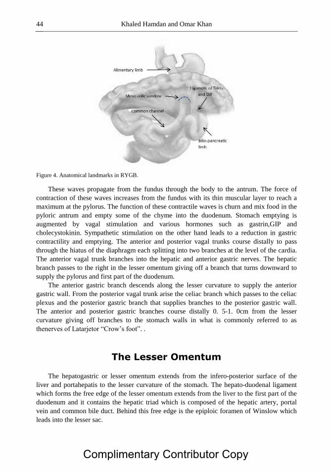
Khaled Hamdan and Omar Khan 44
Figure 4. Anatomical landmarks in RYGB.
These waves propagate from the fundus through the body to the antrum. The force of
contraction of these waves increases from the fundus with its thin muscular layer to reach a
maximum at the pylorus. The function of these contractile waves is churn and mix food in the
pyloric antrum and empty some of the chyme into the duodenum. Stomach emptying is
augmented by vagal stimulation and various hormones such as gastrin,GIP and
cholecystokinin. Sympathetic stimulation on the other hand leads to a reduction in gastric
contractility and emptying. The anterior and posterior vagal trunks course distally to pass
through the hiatus of the diaphragm each splitting into two branches at the level of the cardia.
The anterior vagal trunk branches into the hepatic and anterior gastric nerves. The hepatic
branch passes to the right in the lesser omentum giving off a branch that turns downward to
supply the pylorus and first part of the duodenum.
The anterior gastric branch descends along the lesser curvature to supply the anterior
gastric wall. From the posterior vagal trunk arise the celiac branch which passes to the celiac
plexus and the posterior gastric branch that supplies branches to the posterior gastric wall.
The anterior and posterior gastric branches course distally 0. 5-1. 0cm from the lesser
curvature giving off branches to the stomach walls in what is commonly referred to as
thenerves of Latarjetor “Crow’s foot”. .
The Lesser Omentum
The hepatogastric or lesser omentum extends from the infero-posterior surface of the
liver and portahepatis to the lesser curvature of the stomach. The hepato-duodenal ligament
which forms the free edge of the lesser omentum extends from the liver to the first part of the
duodenum and it contains the hepatic triad which is composed of the hepatic artery, portal
vein and common bile duct. Behind this free edge is the epiploic foramen of Winslow which
leads into the lesser sac.
Complimentary Contributor Copy
Surgical Anatomy and Bariatric Surgery 45
Visually, the lesser omentum is divided into a proximal denser portion, the pas condensa,
and a transparent portion, the pars flacida. This latter part is the site of dissection in the pars
flacida technique that is used in laparoscopic adjustable gastric banding. Embedded in the
lesser omentum are the left and right gastric vessels, lymph nodes, branches of the vagal
nerves including the hepatic division and the anterior and posterior gastric branches (nerves
of Latarjet) and,in 25% of individuals, an accessory left hepatic artery (see below).
The most important structure in the lesser omentumto be aware of in bariatric surgery is
the left gastric artery which is the smallest branch of the celiac axis. The artery first runs
superiorly then turns anteriorly to approach the gastro-oesophageal junction where it gives off
branches to the distal esophagus.
The main trunk of the artery courses inferiorly in the lesser omentum to run parallel to the
lesser curvature where it terminates by forming anastomoses with branches of the right gastric
artery. In a quarter of cases the left gastric artery gives rise to an accessoray left hepatic artery
which replaces the main left hepatic artery in 10% of people. The posterior gastric vessels
which are branches of the splenic vessels are reported to be present in as many as 70% of
individuals. These vessels supply the proximal part of the posterior surface of the stomach
and their hidden and often inconsistent location may lead to dangerous bleeding during the
dissection of this area.
In the case of laparoscopic gastric banding the pars flacida part of the lesser omentum is
incised to reveal the caudate lobe and the right crus. The inferior vena cava can also be seen
to lie behind the caudate lobe and to the right of the right crus and great care should be taken
not to damage it. It is often not necessary to extend pars flacida incision proximally into the
pars condensa particularly if a large aberrant or replaced hepatic artery is to be divided.
However, in some cases this artery and the accompanying hepatic branch of the anterior vagal
nerve may be divided in order to achieve better views of the right crus. Sacrificing this nerve
has not been shown to result in serious consequences although there are reports of increased
incidence of cholecystolethiasis after its division [10-12].
Once a window in the lesser omentum is created the right crus is identified and a
retrogastric tunnel is created starting at the anterior surface of the right crus at its lower limit.
A useful landmark for initiating the dissection is a small transverse fat pad on the anterior
aspect of the right crus. This useful landmark often contains a small venous tributary that runs
across the junction between the two crura. Once a small window is created posterior
dissection is then continued with utmost care to avoid unwanted bleeding or damage to the
posterior wall of the stomach. The dissection should be minimal and the grasper is gently
advanced in the direction of the angle of His. Inaccurate angulation of the grasper can lead to
serious bleeding from the spleen or to inadvertent dissection into the substance of the left crus
or the left pleural space.
In the case of RYGB dissection should remain close to the lesser curvature of the
stomach in the perigastric plane. A suitable point to start this dissection is the level of the
second visible vessel about 5cm from the gastro-oesophageal junction. It is worth noting that
the lesser omentum at this level may be quite thick and meticulous dissection close to the
stomach wall is important to prevent bleeding from the often thin walled veins. Extending this
dissection proximally should be avoided to reduce the risk of damaging the left gastric artery
and subsequent devascularisation of the stomach pouch. In addition, excessive dissection or
transection of the lesser omentum to facilitate access to the lesser sac may at least
theoretically exacerbate symptoms of dumping syndrome after RYGB by causing damage to
Complimentary Contributor Copy

Khaled Hamdan and Omar Khan 46
the anterior vagus nerve and its branches [13]. Dissection in the perigastric plane is facilitated
by getting the assistant to grasp the stomach wall close to the area being dissected and
attempting to roll the lesser curvature laterally while dissection progresses. From here the
dissection is continued into the lesser sac where posterior adhesions may be encountered.
Once this part of the stomach is transected with a linear stapler dissection is continued
proximally towards the angle of His where further posterior adhesions and the distal extent of
the fat pad are often encountered. From the anatomical point of view it is important to
appreciate that at this level the posterior wall of the fundus is often lax which can lead to
excessively large gastric pouch. This is simply avoided by careful inspection of the posterior
wall of the stomach and by applying left and downward traction on the fundus before
proceeding with dividing the stomach. It is also important to appreciate that the area behind
the gastric pouch contains fatty tissue which is adherent to the posterior wall. This often needs
to be cleared for 1-2 cm proximal to the staple line to facilitate the formation of the gastro-
jejunal anastomosis but care should be taken not to damage the left gastric artery which is in
the immediate vicinity.
The Greater Omentum
The anterior and posterior peritoneal layers of the stomach unite at the greater curvature
to form the gastrosplenic (lienogastric) and gastrocolic ligaments. The gastrocolic ligament
extends to the right to the first part of the duodenum and inferiorly to continue as the greater
omentum. The short gastric arteries which are branches of the splenic artery or one of its
divisions run in the gastrosplenic ligament to supply the fundus of the stomach. Within the
wall the fundus rich submucosal anastomoses exist between these arteries and the left gastric
and gastroepiploic. This means that the short gastric arteries can be divided with impunity
without compromising the blood supply to the fundus. Traction on the gastrosplenic ligament
and division of the short gastric vessels is required during sleeve gastrectomy and utmost care
should be taken here to avoid bleeding. In addition excessive traction on the fundus during
mobilisation of the angle of His and fundus should be avoided to prevent tearing of the
delicate capsule of the spleen.
Division of the short gastric vessels and the gastro-colic ligament along the greater
curvature to a point about 5-6 cm proximal to the pylorus provides wide access to the lesser
sac and posterior surface of the stomach during sleeve gastrectomy. Here the transverse
mesocolon and its blood vessels, namely the middle colic artery,can be damaged and a
segment of the transverse colon may be devascularised if the correct plane is not adhered to.
To avoid this upward lift on the stomach and downward traction on the transverse mesocolon
is required. Elevating or reflecting the stomach often reveals filmy gastro-pancreatic
adhesions which need to be divided in order to free the posterior wall of the stomach.
The Small Bowel
For procedures that have a malabsorbtive component, notably the RGBY and the
duodenal switch, a great deal of care and attention is required when handling and measuring
Complimentary Contributor Copy

Surgical Anatomy and Bariatric Surgery 47
the small bowl. The length of the small intestine is extremely variable ranging from 10 to 30
feet and averaging about 22 feet. The first part of the duodenum continues from the pylorus
with the common bile duct, portal vein, gastroduodenal artery and IVC passing posteriorly.
The second part of the duodenum lies anterior to the right kidney and IVC and posterior to the
transverse colon. The common bile and pancreatic ducts open into the left side of this part of
the duodenum. The third part of the duodenum passes horizontally to the left anterior to the
IVC and posterior to the superior mesenteric vein and artery. The fourth part of duodenum
crosses the spine and the aorta and ends at the duodeno-jejunal flexure to the left of the aorta.
The proximal part of the small bowel is referred to as the jejunum and the remainder is the
ileum. The jejunum commences at the duodeno-jejunal flexure (DJF) and it is differentiated
from the ileum by virtue of its origin at the DJF and by its larger diameter and thicker wall
which is caused its thick mucosal folds (valvulaeconniventes). The DJF is fixed by a
suspensory ligament (of Trietz). Accurate identification of the ligament of Trietz is essential
before proceeding with bowel division and the formation of entero-enterostomy in patients
undergoing RYGB. This requires reflection of the often fat laden omentum over the
transverse colon which itself is retracted cephalad into the upper abdomen by the assistant
(Figure 4). The length of mesentery measured from the mesenteric border of the intestine to
the root of the mesentery does not normally exceed 20-25cm. Its origin from the posterior
abdominal wall commences at the DJF and extends downwards and to the left for about 15
cm. The mesentery contains the superior mesenteric vessels, lymphatic channels, lymph
nodes and autonomic nerve fibres. The mesentery of jejunum has fewer vascular arcades and
longer and infrequent terminal branches compared to the ileum which in contrast has more
arcades and shorter and more numerous terminal vessels. In patients with extreme central
obesity the jejunal mesentery is often foreshortened and this may limit the ability of the
surgeon to pull the alimentary limb up to the level of the gastric pouch without undue tension.
In this situation the retrocolic route is the preferred technique for passing the alimentary limb
posteriorly into the lesser sac and the gastric pouch during RYGB. This is best done by
creating an opening in the anteriorly retracted mesocolon to the left of the middle colic
vessels. If the middle colic vessels cannot be identified a useful landmark for making the
opening is approximately 2-3 cm anterior and to the left of the ligament of Trietz (Figure 4).
The blood supply of the small intestines is derived from branches of the superior mesenteric
artery. The vasa recta are the terminal branches from the arches of the arcades that pass
towards the mesenteric border of the small intestine to supply the bowel wall. An important
feature of these vessels is that they do not anastomose or form collateral circulation. While
this configuration ensures excellent blood supply to the metabolically active small intestine it
means that division of these vasa recta will increase the likelihood of causing ischemia of the
affected part of the bowel wall. It is therefore important to accurately identify these vessels
and ensure that the mesentery is divided parallel and not across these vessels during RYGB.
RYGB- Limbs and Mesenteric Defects
The procedure of RYGB results in the development of several mesenteric which can
potentially lead to the development of internal hernias (Figure 5).
Complimentary Contributor Copy

Khaled Hamdan and Omar Khan 48
Figure 5. Mesenteric defects after RYGB.
Closure of these defects at the time of surgery may have a role in preventing the
development of internal herniation which is considered to be the most common cause of
intestinal obstruction after laparoscopic RYGB [14]. Understanding the origin and anatomical
location of these defects is also important for the general surgeon who may be involved in
managing patients presenting with intestinal obstruction or intermittent abdominal pain
secondary to internal herniation.
The following is a brief description of how these mesenteric defects form. The procedure
of RYGB involves the formation of 3 “limbs” – the Roux or alimentary limb which is
anastomosed to the stomach pouch, the bilio-pancreatic (BP) limb which is anastomosed to
the Roux limb and the common channel (CC) which continues to the terminal ileum and
caecum. The first step involves the division of the jejunum and its mesentery some distance
(50-100 cm) from the ligament of Trietz. This results in two free mesenteric edges; the
proximal mesenteric edge which belongs to the BP limb and the distal mesenteric edge which
belongs to the Roux limb.
The next step is forming the jejuno-jejunal anastomosis which involves joining the distal
end of the BP limb to a point 70-150 cm from the proximal end of the Roux limb. This
effectively brings the free mesenteric edge of the BP to lie against the flat surface of Roux
limb mesentery thus creating a defect which is commonly referred to as the meso-jejunal
mesenteric defect.
Once the gastric pouch is formed the gastro-jejunal anastomosis is constructed by pulling
up the Roux limb either anterior to the transverse colon (the antecolic route) or through an
opening in the mesocolon (the retrocolic route). In either case the free mesenteric edge of the
Roux limb comes to lie against the transverse mesocolon thus creating a defect known as
Peterson’s window. In the case of using the retrocolic route the opening through the
mesocolon results in another defect, the mesocolic defect, which is considered to be the most
common site for internal herniation after RYGB.
Complimentary Contributor Copy

Surgical Anatomy and Bariatric Surgery 49
References
[1] Reoch J, Mottillo S, Shimony A, Filion KB, Christou NV, Joseph L, Poirier P,
Eisenberg MJ. Safety of laparoscopic vs open bariatric surgery: a systematic review and
meta-analysis. Arch. Surg., 2011 Nov;146(11):1314-22.
[2] Altun H, Banli O, Karakoyun R, Boyuk A, Okuducu M, Onur E, Memisoglu K. Direct
trocar insertion technique for initial access in morbid obesity surgery: technique and
results. SurgLaparoscEndoscPercutan Tech. 2010 Aug;20(4):228-30.
[3] Berch BR, Torquati A, Lutfi RE, Richards WO. Experience with the optical access
trocar for safe and rapid entry in the performance of laparoscopic gastric bypass. Surg.
Endosc, 2006 Aug;20(8):1238-41.
[4] Schwartz ML, Drew RL, Andersen JN. Induction of pneumoperitoneum in morbidly
obese patients. Obes. Surg., 2003 Aug;13(4):601-4.
[5] Vilos GA, Ternamian A, Dempster J, Laberge PY, The Society of Obstetricians and
Gynaecologists of Canada. Laparoscopic entry: a review of techniques, technologies,
and complications. J. Obstet. Gynaecol. Can., 2007 May;29(5):433-65.
[6] Podnos YD, Jimenez JC, Wilson SE, Stevens CM, Nguyen NT. Complications after
laparoscopic gastric bypass: a review of 3464 cases. Arch. Surg., 2003 Sep;138(9):957-
61.
[7] Ahmad G, O'Flynn H, Duffy JM, Phillips K, Watson A. Laparoscopic entry techniques.
Cochrane Database Syst. Rev., 2012 Feb 15;2.
[8] Shen R, Ren CJ. Removal of peri-gastric fat prevents acute obstruction after Lap-Band
surgery. Obes. Surg., 2004 Feb;14(2):224-9.
[9] Rhee PL, Lee JY, Son HJ, Kim JJ, Rhee JC, Kim S, Koh SD, Hwang SJ, Sanders KM,
Ward SM. Analysis of pacemaker activity in the human stomach. J. Physiol., 2011 Dec
15;589(Pt 24):6105-18.
[10] Purdy M, Nykopp TK, Kainulainen S, Pääkkönen M. Division of the hepatic branch of
the anterior vagus nerve in fundoplication: effects on gallbladder function. Surg.
Endosc., 2009 Sep;23(9):2143-6.
[11] Tomita R, Fujisaki S, Koshinaga T, Kusafuka T. Clinical assessments in patients ten
years after pylorus-preserving gastrectomy with or without preserving both pyloric and
hepatic branches of the vagal nerve for early gastric cancer. Hepatogastroenterology,
2010 Jul-Aug;57(101):984-8.
[12] Morton JM, Bowers SP, Lucktong TA, Mattar S, Bradshaw WA, Behrns KE, Koruda
MJ, Herbst CA, McCartney W, Halkar RK, Smith CD, Farrell TM. Gallbladder
function before and after fundoplication. J. Gastrointest. Surg., 2002 Nov-
Dec;6(6):806-10; discussion 810-11.
[13] Frantzides CT, Carlson MA, Shostrom VK, Roberts J, Stavropoulos G, Ayiomamitis G,
Frantzides A. A survey of dumping symptomatology after gastric bypass with or
without lesser omental transection. Obes. Surg., 2011 Feb;21(2):186-93.
[14] Ballem N, Parikh R, Berber E, Siperstein A: Laparoscopic versus open ventral hernia
repairs: 5-year recurrence rates. Surg. Endosc., 2008; 22:1935-1940.
Complimentary Contributor Copy

Complimentary Contributor Copy
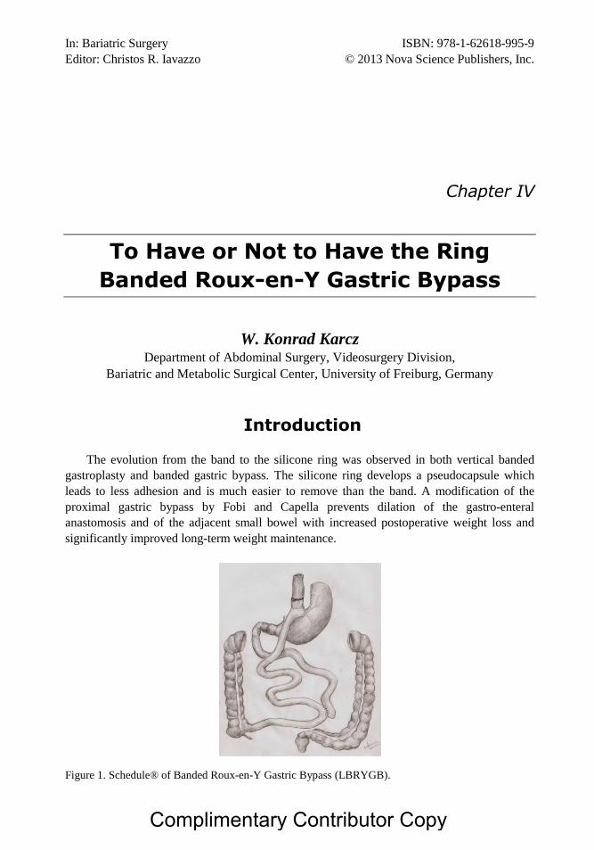
In: Bariatric Surgery ISBN: 978-1-62618-995-9
Editor: Christos R. Iavazzo © 2013 Nova Science Publishers, Inc.
Chapter IV
To Have or Not to Have the Ring
Banded Roux-en-Y Gastric Bypass
W. Konrad Karcz Department of Abdominal Surgery, Videosurgery Division,
Bariatric and Metabolic Surgical Center, University of Freiburg, Germany
Introduction
The evolution from the band to the silicone ring was observed in both vertical banded
gastroplasty and banded gastric bypass. The silicone ring develops a pseudocapsule which
leads to less adhesion and is much easier to remove than the band. A modification of the
proximal gastric bypass by Fobi and Capella prevents dilation of the gastro-enteral
anastomosis and of the adjacent small bowel with increased postoperative weight loss and
significantly improved long-term weight maintenance.
Figure 1. Schedule® of Banded Roux-en-Y Gastric Bypass (LBRYGB).
Complimentary Contributor Copy

W. Konrad Karcz 52
Figure 2. 3D volume rendering reconstruction of abdominal MSCT of a LBRYGB. Manual and semi-
automatic segmentation techniques are applied to show the pouch (cyan), the Roux-limb (orange), the
esophagus and intestine (blue), and the staple sutures (red).
We are currently waiting for results of multi-centre, prospective randomized studies that
compare the banded to the conventional gastric bypass. Safety and feasibility are demon-
strated by Fobi et al. in their published series of 50 consecutive patients with an in-creased
excess weight loss of more than 70% and no increase of postoperative complications.
It is interesting that Fobi’s modification of the gastric bypass is becoming more popular
each year (Figure 1, 2). Need we be hesitant with regard to Silastic rings? How much we can
profit from implantation and what do we have to risk? There are on the market several new
products which could be implanted on the Stomach Pouch. All modern devices are made from
silicone which made the implant safe and easy to remove.
Complications
There are many possible complications after proximal gastric bypass operations, only few
of which are seen in combination with a Silastic rings. The incidence of regurgitation and the
quality of eating is closely related to the ring size. Stubbs et al. reported that in 18 %, major
restriction in the quality of eating occurred in a group of patients treated with a 5. 5 cm ring,
whereas in groups with 6. 0 cm and 6. 5 cm rings only 8% and 4% respectively had similar
problems. 29% of the patients Regurgitation occurred more than three times a week in 29% in
the first group with 5. 5 cm and in only 14 % in the last one. The risk of developing chronic
regurgitation is related to two independent factors, namely the lower esophageal sphincter
function and the size of the silicone ring placed around the gastric pouch. The smaller the
ring, the more frequent the chronic regurgitation. Hypotonia of the lower esophageal
sphincter (the normal value 14-34 mmHg) increases the risk of chronic regurgitation. The
chance is seven times greater compared to patients with normal lower esophagus sphincter
pressure. Both LES hypotonia and a small band diameter increase the risk of frequent
regurgitation. Patients with low LES pressure ought to receive alternative bariatric
procedures, such as the Conventional RYGB or the BPD. Band erosion or migration into the
Complimentary Contributor Copy

To Have or Not to Have the Ring Banded Roux-en-Y Gastric Bypass... 53
gastric lumen after a Banded Gastric Bypass seem to be rather rare late complications. Most
authors reported an incidence of around 0. 5%. Studies with larger patient collectives report a
band erosion rate of 1. 63% (48 of 2,949 Patients). The incidence of erosion was lower (0.
92%) in primary operations. The following clinical symptoms appeared along with the
pathology: weight regain (37. 5%), stenosis or obstruction (35. 4%), pain (18. 75%), bleeding
(14. 58%). 89. 5% of patients lost the ring. In a 10-year observation of banded gastric bypass
patients, Barroso found: 26 stenoses, 9 erosions, 2 spontaneous eliminations, 3 slippages, and
two openings of the ring. The other very large group observed in the Elias Study presented
125 band’s explantations in 138 medical, band-related events. Bands can be removed
spontaneously through migration, per endoscopy, per laparoscopy or rarely by laparotomy.
Some possible causes of band erosion are cited, like: constricting band, suturing the band to
the stomach, imbricating the band with the stomach, and infection. Fobi proposed the
mechanism of Band Errosion (BE), with the first step usually being an inflammation between
the gastric wall and the band leading to band extrusion into the pouch lumen through stitch
abscess formation and extrusion of the stitch. Afterwards, a dense fibrous reaction appears
around the banded pouch.
Removal of the ring is combined with significant weight gain. Appreciable weight regain
occurred in 43. 75% patients who underwent band removal, with an average of 14% excessive
weight loss (EWL) regained. Primary replacement of the band is not recommended. As
therapy guidelines, Fobi proposed: for asymptomatic patients the treatment can be waiting for
spontaneous extrusion of the band and protone pump inhibitors (PPI) or H2-blockers. The
management of choice is endoscopic removal of the band. If endoscopic removal is not
feasible, which is especially the case when materials other than a silastic ring, such as non-
absorbable sutures, were used, the band can be removed by a laparoscopic operation with
intraoperative endoscopy. After laparoscopically performed RYGB, the erosion of non-
dissolvable materials, such as Peri-Strips or silk sutures into the gastric pouch may occur. The
most common presenting symptoms are abdominal pain, nausea, vomiting, dysphagia, and
melena. The management of choice is therapeutic endoscopy which usually allows adequate
management of most upper GI symptoms. Yu et al. show that the use of Vicryl sutures and
Seamguard avoids foreign material erosion. In summary, the introduction of the silastic ring
has reduced band-related complications. General bypass complications are comparable with
those of the banded bypass. There are no ring-related lethal complications.
Ring Implantation Manual
All silicone rings can be introduced laparoscopicaly. The GaBP®Ring (Bariatric
Solutions GmbH) (a pre-manufactured set with a prosthetic auto-locking band and a
radiopaque marker made of implant-grade silicone rubber) seems to be the most useful and
the smallest implant. It is placed around the pouch loosely about 2. 0 to 2. 5 cm from the end
point of the pouch and 4 cm from the His angle. The preparation of the small curvature could
be done with Maryland or scorpion forceps close to the stomach wall. Each silicone band
develops a pseudocapsule that leads to less adhesion and is much easier to remove than other
materials. The ring size ought to be 6. 0 to 6. 5 cm in circumference, which has a much better
Complimentary Contributor Copy

W. Konrad Karcz 54
outcome in terms of quality of eating and ring migration. The pouch size is usually estimated
at 10 to 25 ml. The ring is always fixed with non- resorbable sutures.
Mechanism of Action
Besides its restrictive role, it contributes to the reduction of undesirable side-effects, like
dumping syndrome and foul flatulence. The banded bypass causes satiety and reduces caloric
intake by delaying gastric emptying and inducing the satiation sensation even with low food
intake. The silicone ring controls the diameter and area of the gastroenterostomy.
Redo Surgery with the Ring
The Redo-surgery made by Fobi on VBG patients with preservation of the band or the
ring as a restriction lead to the development of the banded gastric bypass technique. If an
insufficient loss of weight or even a regain of weight occurs and in case of insufficient
restriction, a re-operation might be necessary. The precise diameter and anastomosis area
estimation allows the surgeon to choose the better redo surgery strategy and to select the best
operative option for his patients. It is proven that GE anastomosis dilatation is responsible for
gaining weight. If the patient is able to tolerate the restriction amplification, there are still
several surgical options. (Figure 3, 4). Endoscopic suturing with f. i. Stomafix or Anubis
(STORZ GmbH) to tighten dilated gastric pouch is technically feasible and safe and may lead
to weight loss for certain patients. Another logical therapy option is implantation of the GaBP
Ring as a redo procedure which converts the conventional to the banded bypass. An
analogous procedure with additional LA Gastric Banding to the Roux-en-Y GB was also
proposed Bessler et al. . We therefore advise using rings of at least 6. 5 cm length and 7. 0 cm
in Redo operations. The easy conversion between those two operations and better weight
reduction results will probably influence the number of banded bypass procedures.
Figure 3. CT-Scan of abdominal cavity after LBRYGB with GE anastomosis stenosis after LBRYGB,
the ring did not influence stenosis.
Complimentary Contributor Copy

To Have or Not to Have the Ring Banded Roux-en-Y Gastric Bypass... 55
Figure 4. 3D volume rendering reconstruction of abdominal MSCT showing a dilatation of the stomach
pouch (51cm3) and GE anastomosis (Diameter 3. 4cm, area 10. 1cm
2). Manual and semi-automatic
segmentation techniques are applied to show the pouch (green), the Roux-limb (orange), and the staple
sutures (red).
Weight Loss Differences
The influence of the silastic ring on the percentage of excess weight loss within the first
year was significant: between 81. 3 and 73. 5. EWL. After two years, the percentage ranged
around 80%, and at 5 years follow-up a EWL of about 75% could be found. Only a slight
weight regain of 2. 5% or 5% is observed between the second and fifth postoperative year,
whereas a weight regain by LCRYGB of over 10% in the same period is reported. The EWL
after conventional GB reached 58. 2% after five years in the best series. Bessler performed
the first prospective study that directly compared the two operation methods. After the
second or third postoperative year, the patient seems to adapt to the surgery and to suffer from
its side-effects in lower intensity, which brings a tendency for some recovery of lost weight.
But weight gain is very slight compared to the conventional gastric bypass.
References
[1] Fobi MAL. Placement of the GaBP Ring system in the Banded Gastric Bypass
Operation. Obes. Surg., 2005; 8: 1196 - 1201.
[2] Bessler M, Daud A, Kim, DiGiorgi M. Prospective randomized trial of banded versus
nonbanded gastric bypass for the super obese: early results. Surg Obes Relat Dis. 2007
Jul-Aug;3 (4): 480-4; discussion 484-5. Epub 2007 Jun 4.
[3] Stubbs et al.: What ring size should be used in association with Vertical Gastric
Bypass? Obes. Surg. Obes. Surg., 2006; 16: 1298-1303. .
[4] Fobi MAL, Lee H, Igwe D et al. Band erosion: Incidence, Etiology, Management and
Outcome after Banded Vertical Gastric Bypass. Obes. Surg., 2001; 11: 699-707.
Complimentary Contributor Copy

W. Konrad Karcz 56
[5] Barroso FL, Leite Ma, Almeida Azet al. Problems with the silicone rings in the Fobi/
Campella Operations. Oral. Presentation., 2007 IFSO Congress, Porto, Portugal.
[6] Elias OA, Garrido AB, Berti LV, Oliveira MR, Suguitani NB. Complications related to
the ring after 7000 Roux-en-Y gastric bypass with silicone ring for the obesity
treatment. Oral. Presentation., 2007 IFSO Congress, Porto, Portugal.
[7] Karcz W, Küsters S, Koter E, et al. Redo Bariatric Surgery Influenced By 3D Volume
Rendering Reconstruction of Abdominal MSCT. Submitted to Obesity Surgery June
2008.
Complimentary Contributor Copy
In: Bariatric Surgery ISBN: 978-1-62618-995-9
Editor: Christos R. Iavazzo © 2013 Nova Science Publishers, Inc.
Chapter V
Postoperative Complications
after Bariatric Surgery
Ioannis D. Gkegkes1 and Christos Iavazzo
2
1Department of Surgery, Korinthos General Hospital, Korinthos, Greece
2Iaso Maternity Hospital, Athens, Greece
Abstract
There is a variety of complications that could arise following any of the bariatric
surgical procedures that necessitate a more careful understanding of the specific anatomy
of the operation performed (Table 1). According to the type of procedure performed,
postoperative complications after surgical treatment of morbid obesity can be divided in
immediate and in delayed.
Immediate Complications
Pulmonary embolism, anastomotic leak and bleeding are the most common immediate
complications that are generally treated with by the operating team. Laboratory values and
clinical data, such as an increased C-reactive protein and the tachycardia level, could be most
of the times the only objective signs.
According to Assessment of Bariatric Surgery study, 4. 3% is the incidence of major
adverse events in early postoperative period. [1] Even though these encouraging reports,
bariatric surgery is associated with a number of complications that can be characterized as
potentially fatal. Shock secondary to hemorrhage, sepsis from an anastomotic dehiscence and
cardiopulmonary complications in high-risk comorbid population are some of the most
serious consequences. According to national studies, 0. 34% is the estimated incidence of
death following bariatric surgery, due to increased risk for thromboembolic events. [2,3]
Therefore mechanical as well as pharmacological measures for deep vein thrombosis are
normally practiced with the intention to reduce this specific risk. [4, 5]
Complimentary Contributor Copy

Ioannis D. Gkegkes and Christos Iavazzo 58
Table 1. Main postoperative complications after bariatric surgery
Mechanism of action Type of operation Type of postoperative complications
Restrictive Vertical banded
gastroplasty
-band slippage
-erosion of the band
-port access site problems Adjustable gastric
banding
Largely Restrictive/Mildly
Malabsorptive
Roux-en-Y gastric
bypass
-incisional hernias
-vomiting
-bowel obstruction
-gastrointestinal hemorrhage
-malabsorption
Mildly Restrictive/ Largely
Malabsorptive
Biliopancreatic
diversion
-incisional hernias
-bowel obstruction
-malabsorption Duodenal switch
Moreover one of the most grave complication in the surgical treatment of morbid obesity
is gastrointestinal leak, which the procedure performed, could be ranging from 1%, for gastric
banding, to 2%, for gastric bypass and sleeve gastrectomy. [6-8] Anastomotic sites and staple
lines can be in the majority of times the origin of leaks while iatrogenic bowel injuries are
less frequently responsible. [5] Conservative management is the indication only for leaks that
can be controlled by a surgical drain, in a hemodynamically stable patients, while in contrary
situations, a more aggressive approach that include surgical exploration should be indicated.
Furthermore, in 1–4% of bariatric surgeries could be complicated by episodes of early
bleeding, taking into consideration both intraluminal and intraperitoneal events. [5] Volume
resuscitation, transfusion of blood products and close clinical observation could be in the
majority of the cases essential for early recognition of hemodynamic instability that imposes
urgent surgical intervention. [9-11]
Late Complications
Late Complications after Restrictive Procedures
Laparoscopic adjustable gastric banding (LAGB) is one of the most popular bariatric
operations probably due to the fact that it is a relatively simple procedure with extremely low
rates of early morbidity and mortality (30-day) compared with other procedures such as
Roux-en-Y gastric bypass (RYGB). [12, 13] Nevertheless, the rate of reoperation as well as
the incidence of late complications could reach 10–20 per cent. Additionally, band slippage
and erosion are the two major complications of LAGB. Up to 15–20 per cent of patients could
occur the band slippage, which is more often takes place distally. [14, 15] Patients’ history
reveals frequently acid reflux, regurgitation or sense of dysphagia as well as an obstructive
symptomatology with the presence of upper abdominal pain, occurring some time after the
procedure.
In addition, a distal migration or abnormal orientation of the band can be evident in a
plain abdominal X-ray and more particularly on oblique views. Furthermore, in contrast study
Complimentary Contributor Copy

Postoperative Complications after Bariatric Surgery 59
of the upper gastrointestinal, is more likely to show evidence of malposition of the band as
well as a dilatation in the proximal pouch combined with obstruction. Generally, a well-
positioned gastric band rests below the gastro-oesophageal junction with an inclination of
about 45° according to the spine. [16]
Slipped band could presented in a relative horizontal position, a dilatation of gastric
pouch proximal to band, combined with little contrast passing through it. These radiological
findings pose the differential diagnosis between band slippage and the less severe problem of
pouch dilatation, condition with which patients often tend to present less acutely with ,
decreased satiety, acid reflux symptoms and occasionally pain located at the left upper
quadrant but no obstructive symptoms.
The first step in the management of band slippage is the without delay deflation. It can be
achieved by locating the port with two fingers while at the same time advancing a non-coring
(Huber needle) or else a spinal needle until it hits the steel plate at the back of the port. [12]
The needle is immediately withdrawn, when fluid is withdrawn and the band is completely
deflated. This procedure can also be conducted under ultrasound guidance or fluoroscopy. If
the success of this procedure has as result the symptomatic relief, it is reasonable to do
nothing proposing a review by a bariatric surgeon.
On the other hand, if this procedure is unsuccessful, the most appropriate is to consider
also the possibility of the rare but serious complication of gastric necrosis which requires
urgent exploratory laparoscopy or laparotomy. [17] The band should be removed completely,
if there is absence of an experienced surgeon. The procedure requires cautiously the division
of the fibrotic scars over the band, eviting gastric or esophageal tissue injury. An alternative
but also technically challenging procedure is to unbuckle the band and leave it at the site for
later adjustment by a bariatric surgeon. [12, 18, 19]
The erosion of gastric band takes place in up to 4 per cent of patients with LAGB and is
presented usually as a late complication caused by ischemia due to pressure on the gastric
wall. [20, 21] Patients with band erosion may be asymptomatic or with non-specific
abdominal pain, weight loss gastrointestinal bleeding as well as abdominal sepsis.
Gastrointestinal endoscopy shows partial- or full-thickness erosion, and barium swallow may
evidentiate the flow of contrast around the part of the band that has eroded into the stomach.
[22]
Contrast-enhanced computed tomography (CT) should be requested, if clinical symptoms
suggest an intra-abdominal abscess. Endoscopic band removal is the treatment of choice for
gastric band erosion. Though, when there is extensive erosion and necrosis of the stomach
wall, laparoscopic or laparotomic removal of the affected stomach should be reconsidered.
[23]
A rare (less than 0·5 per cent) but also well reported late complication after LAGB is
considered megaoesophagus. [24] Progressive dysphagia, regurgitation and often retrosternal
chest pain are some of the symptoms that patients typically mention. The diagnosis is made
by means of endoscopy or an upper gastrointestinal contrast study. When the
megaoesophagus is often caused by proximal migration of the gastric band thus mimicking
achalasia, necessitates the removal of the band in most cases.
Other late complications after vertical sleeve gastrectomy (VSG) include gastrocutaneous
fistula and tube stricture (<1 per cent). [25] The management of these types of complications
requires specialist bariatric surgery and probably may include stent insertion and conversion
to RYGB.
Complimentary Contributor Copy
Ioannis D. Gkegkes and Christos Iavazzo 60
Hepatobiliary Complications
The formation of cholesterol gallstones can be the consequence of rapid weight loss. 13
to 36 per cent of patients develop gallstones within a period of 6 months after the surgical
operation. [26, 27] In general, cholecystectomy is performed only if the patient is
symptomatic. [28, 29] In patients who have previously undergone a bypass procedure,
intraoperative cholangiography is advisable, as a result of the difficulty in reaching the biliary
tract by endoscopic retrograde cholangiopancreatography (ERCP). New endoscopic
techniques such as the transgastric approach and double balloon enteroscopy can be applied
in order to access the biliary tree in occasions where the performance of standard ERCP is
technically difficult (such as after previous RYGB). [30] The removal of common bile duct
(CBD) stones can be achieved, in the absence of experience on the novel techniques, by using
either the transcystic approach or by formal CBD exploration during surgery. [30, 31]
Intestinal Obstruction
Husain et al. referred that the incidence of bowel obstruction was found to be 4·4 per
cent, with a mean period of presentation of 313 days after RYGB. 32
Increased risk of
intestinal obstruction is correlated the surgical approach, which will affect the degree of
adhesions as well as the position of the Roux limb, in close association with the limb position,
retrocolic or antecolic. [32] The most frequent causes of bowel obstruction consist in internal
hernias (53 per cent), Roux limb compression due to scarring (20 per cent) and adhesions (14
per cent). [33] Stricture at the gastrojejunal anastomosis, incisional hernia, intestinal
intussusception and kinking of the alimentary limbcan also considered possible causes of
obstruction. The clinical symptoms of these patients are central colicky abdominal pain (82
per cent), nausea and vomiting (46–48 per cent), abdominal distension and absolute
constipation. [32] It is crucial to enquire about the volume and character of the emesis in
patients with RYGB as this may provide a clue about the site of the obstruction. Furthermore,
the type of the vomit will differ in relation to the site of obstruction. Copious amounts of
bilious vomiting are indications of obstruction at the common channel (CC) distal to the
jejunojejunal anastomosis. Dilatation of the CC proximal to the obstruction, of the
biliopancreatic limb, the gastric remnant and the Roux limb are signs that can be evident with
CT. Smaller quantity of non-bilious vomit is suggestive of obstruction at the Roux limb or
above the jejunojejunal anastomosis. CT, at this type of obstruction, will make evident a
dilated Roux limb and a collapsed CC and biliopancreatic limb. Absence or little amount of
vomit, combined with abdominal distension are generally signs of obstruction at
biliopancreatic limb. In addiction, symptoms include postprandial left upper abdominal
quadrant pain. Dilatation of the biliopancreatic limb and at the remnant of the stomach,
combined with normal CC and Roux limb caliber can also be evident with CT. Patients with a
RYGB can present a number of mesenteric defects through which even a small bowel loop is
in position to pass and provoke obstruction or in the worst case strangulation. Paroz et al.
showed that the most frequent sites for internal herniation are localized at the mesojejunal
mesenteric window (56 per cent), the Peterson’s window (27 per cent) and the mesocolic
window (17 per cent). [33] The incidence of symptomatic internal herniation variates from
1·6 to 4·1 per cent. [33,34] Failure to close the mesenteric defects, the closure technique in
Complimentary Contributor Copy

Postoperative Complications after Bariatric Surgery 61
combination with the type of suture used, and the variations of these defects as patients lose
weight are some of the factors that can assist to the development of internal hernias. [12]
Almost one-third of all patients can present with symptoms and signs of acute intestinal
obstruction. [33] The onset of symptoms is generally insidious and presents with intermittent
abdominal discomfort, mainly after eating. [34, 35]
Even if laparotomy is often indicated for the diagnosis, laparoscopy is the first option
from the invasive techniques. [34, 35] Moreover, it is important for the surgeon to identify all
limbs and great attention should be given to mesenteric defects as these should be closed with
non-absorbable sutures. The presence of ulceration at the gastrojejunal anastomosis, which
may contribute to the formation of anastomotic strictures in 27 per cent of patients, can often
be revealed by endoscopy. [36, 37]
Endoscopic balloon dilatations up to 12–15 mm can resolve gastrojejunal strictures, even
if there is a risk of perforation (2·1 per cent). [38] 8–10 per cent of patients may present
incisional hernias following open bariatric operations. [39] The utilization of laparoscopic
surgical approach can low the incidence of port-site incisional hernias, estimated to be up to
0·5 per cent. [40]
Most of the times, in obese patients, port-site hernias can be difficult to detect clinically
and ultrasonography could be useful as diagnostic method. Intestinal intussusception, a rare
cause of abdominal pain, after open or laparoscopic RYGB, is most often located at the CC of
the gastrojejunal anastomosis, due either to alterated motility or the staple line of the
anastomosis which acte as a lead point. The utilization of contrast-enhanced CT with is
extremely valuable in establishing of diagnosis while the surgical management includes the
reduction of the affected limb and resection if there is any evidence of intestinal ischaemia.
Gastrointestinal Bleeding
Gastrointestinal bleeding after RYGB is uncommon outside the postoperative period and
generally takes origin by marginal or stomal ulceration. [41] The adequate resuscitation, the
assessment of the severity of the bleeding and the close monitoring should be the first steps in
managing these patients. [42] Patients with RYGB may be chronically anaemic and thus it is
perfectly reasonable to accept lower haemoglobin levels and to withhold blood transfusion
and emergency endoscopy, provided the patient is haemodynamically stable. [43]
Endoscopy should constantly be performed immediately in patients with severe bleeding.
[44] In cases where the source of the bleeding cannot be identified, the endoscopist should
carefully examine the Roux limb, biliopancreatic limb, jejunojejunal anastomosis as well as
the gastric remnant.
The endoscopist can The utilization of a push enteroscope or paediatric colonoscope is
useful way in order to realize this. [45] An innovative technique such as double-balloon
enteroscopy is in position to facilitate the access to the biliopancreatic limb and gastric
remnant while permits therapeutic interventions. [46, 47] In cases of endoscopical failure to
control bleeding, radiological embolization of the bleeding vessel can be an indication for
surgical intervention. [12] Last but not least, if haemostasis cannot be achieved, procedures
such as excision and reconstruction of the jejunojejunal or gastrojejunal anastomoses,
excision of bleeding gastric ulcer or even resection of the gastric remnant may be considered
as alternative options.
Complimentary Contributor Copy

Ioannis D. Gkegkes and Christos Iavazzo 62
Functional Complications
Even though the patients who submit to bariatric surgery experience improvement in
functional gastrointestinal disorders, some possibly will develop exacerbations of symptoms.
[48] The basic understanding of the origin of these symptoms by the emergency surgeon is
critical. 49 per cent of bariatric patients before surgery are affected by eating disorders, such
as binge-eating. [49, 50] The first element to the diagnosis is an increase in the patient’s
weight. Malabsorption can cause diarrhoea, bile salts or dumping syndrome. [51] Dumping
syndrome habitually manifests itself as facial flushing and postprandial diarrhoea after the
consumption of carbohydrate-rich meals. [52] Moreover, these symptoms tend to improve
with time and less than 5 to10 per cent of patients continue to have chronic problems. Other
causes of diarrhoea may possibly be irritable bowel syndrome or food intolerance or bacterial
overgrowth. In these situations, the treatment of diarrhoea normally includes reduction the
amount of carbohydrate consumed, avoiding foods that usually cause intolerance, while the
use of probiotics and antibiotics is indicated where bacterial overgrowth is suspected. Many
studies report an improvement in patients’ reflux symptoms after bariatric surgery. [53, 54]
The explanation of this fact is based on the distal diversion of bile, the reduction at the
volume of parietal cells in the gastric pouch and weight loss. [55] However, some patients
may present with new or exacerbation of the reflux symptoms. [56] In cases where the initial
investigations are normal, other clinical hypotheses that should be considered are
gastroparesis and oesophageal dysmotility. [57]
Nutritional Complications
Every surgical operation that alters in drastic way the anatomic pathways of nutrient
intake may inevitably affect the intake of specific nutrients. Gastric restrictive procedures can
induce nutrient deficiencies as a result of inadequate intake as a consequence of frequent
vomiting and not due to malabsorption. Consequently, the reduction of the numerous
episodes of vomit as well as the replacement of vitamin and mineral requirements are in
position to prevent micronutrient deficiencies in these patients. [5, 58] Significantly, greater
risk for micronutrient deficiency is present in malabsorptive procedures, in which the not
proper micronutrient absorption may occur. In the digestion as well as the absorption of
vitamin B12, iron, and calcium, it is important the anatomic integrity of stomach, duodenum,
and of the proximal jejunum. In patients who have undergone gastric bypass, the deficiency
of vitamin B12 has not presented as a long-term clinical issue. The postoperative
recommendation to vitamin B12 supplement, the persistence of intrinsic factor, as the body of
the stomach is usually preserved, and the presence of a functioning terminal ileum where
vitamin B12 is absorbed, is probably the main reasons for the absence of B12 deficit in
gastric bypass. Likewise, the lack of reports on an increased incidence of pathologic fractures
demonstrates that the homeostasis of calcium, following this procedure, is preserved. [59]
Moreover, the relative young age of patients who have undergone gastric bypass, increased
estrogen production present at these patients, the stimulation of enhanced bone mass due to
obesity and the usual recommendation to calcium and vitamin D supplementation may
represent factors that also prevent pathologic fractures. On the other hand, procedures, such as
Complimentary Contributor Copy

Postoperative Complications after Bariatric Surgery 63
the biliopancreatic diversion with duodenal switch (BPD/DS), which provoke a greater degree
of malabsorption may possibly cause long-term demineralization of bones. Studies that report
an increased concentration of parathormone in circulation, despite of a careful replacement
protocol of calcium following BPD/DS, seems that at least in short term, serious bone
pathology is avoided. [60, 61]
In the early months following bariatric surgery may be presented a reduced dietary
protein intake. [5] Deficient of protein intake is considered more complicated to prevent than
are micronutrients as its replacement necessitates ingestion of actual protein in order to reach
nutritional equilibrium, while the consumption following in general the bariatric procedures
can be limited so as protein intake drops below the recommended daily standard of 0. 8 gr/kgr
(according to the ideal body weight). The early finding of deficient protein intake is alopecia,
therefore should be paid attention on the dietary protein intake during the early months
following bariatric operation.
Iron deficiency is considered the most frequent micronutrient deficiency subsequent to
gastric bypass. A degree of iron deficiency may be present preoperatively and may be
exacerbated by factors, such as the operative blood loss, the postoperative gastrointestinal
pathology, the reduced intake of iron and a degree of iron malabsorption as a result of bypass
of the stomach and duodenum, can have a great impact on iron homeostasis. As a
consequence, the supplementation of iron following bariatric surgery as well as the vigorous
monitoring of iron concentration is recommended in order to prevent grave anemia.
Conclusion
The knowledge of normal anatomy and the surgical technique is fundamental for correct
interpretation of imaging studies in patients who have undergone bariatric surgery. [62] The
type, the pathophysiology and the timing of each complication is predictable and there are
precise signs that can assist in their identification. Over the last decade, perioperative
morbidity and mortality of bariatric surgery have decreased significantly that is almost
equivalent with many well-accepted procedures such as cholecystectomy. [63] In effect, the
mortality risk of bariatric surgery is one-tenth that of coronary artery bypass surgery with
considerably greater improvement in long-term mortality. Technical advances in laparoscopic
surgery and the establishment of surgical centers of excellence have contributed at the
improvement in perioperative morbidity and mortality. [64] Consequently, extensive evidence
on safety and efficacy has imposed bariatric surgery as the standard for treatment of morbid
obesity.
References
[1] Flum DR, Belle SH, King WC, et al. Perioperative safety in the longitudinal assessment
of bariatric surgery. N. Engl. J. Med. 2009;361:445-54.
[2] Morino M, Toppino M, Forestieri P, Angrisani L, Allaix ME, Scopinaro N. Mortality
after bariatric surgery: analysis of 13,871 morbidly obese patients from a national
registry. Ann. Surg. 2007;246:1002-7; discussion 7-9.
Complimentary Contributor Copy

Ioannis D. Gkegkes and Christos Iavazzo 64
[3] Poulose BK, Griffin MR, Zhu Y, et al. National analysis of adverse patient safety for
events in bariatric surgery. Am. Surg. 2005;71:406-13.
[4] Cotter SA, Cantrell W, Fisher B, Shopnick R. Efficacy of venous thromboembolism
prophylaxis in morbidly obese patients undergoing gastric bypass surgery. Obes. Surg.
2005;15:1316-20.
[5] Elder KA, Wolfe BM. Bariatric surgery: a review of procedures and outcomes.
Gastroenterology, 2007;132:2253-71.
[6] Brethauer SA, Hammel JP, Schauer PR. Systematic review of sleeve gastrectomy as
staging and primary bariatric procedure. Surg. Obes. Relat. Dis. 2009;5:469-75.
[7] Podnos YD, Jimenez JC, Wilson SE, Stevens CM, Nguyen NT. Complications after
laparoscopic gastric bypass: a review of 3464 cases. Arch. Surg. 2003;138:957-61.
[8] Tice JA, Karliner L, Walsh J, Petersen AJ, Feldman MD. Gastric banding or bypass? A
systematic review comparing the two most popular bariatric procedures. Am. J. Med.
2008;121:885-93.
[9] Mehran A, Szomstein S, Zundel N, Rosenthal R. Management of acute bleeding after
laparoscopic Roux-en-Y gastric bypass. Obes. Surg. 2003;13:842-7.
[10] Nguyen NT, Longoria M, Chalifoux S, Wilson SE. Gastrointestinal hemorrhage after
laparoscopic gastric bypass. Obes. Surg. 2004;14:1308-12.
[11] Nguyen NT, Rivers R, Wolfe BM. Early gastrointestinal hemorrhage after laparoscopic
gastric bypass. Obes. Surg. 2003;13:62-5.
[12] Hamdan K, Somers S, Chand M. Management of late postoperative complications of
bariatric surgery. Br. J. Surg. 2011;98:1345-55.
[13] Lancaster RT, Hutter MM. Bands and bypasses: 30-day morbidity and mortality of
bariatric surgical procedures as assessed by prospective, multi-center, risk-adjusted
ACS-NSQIP data. Surg. Endosc. 2008;22:2554-63.
[14] Galvani C, Gorodner M, Moser F, et al. Laparoscopic adjustable gastric band versus
laparoscopic Roux-en-Y gastric bypass: ends justify the means? Surg. Endosc.
2006;20:934-41.
[15] Jan JC, Hong D, Pereira N, Patterson EJ. Laparoscopic adjustable gastric banding
versus laparoscopic gastric bypass for morbid obesity: a single-institution comparison
study of early results. J. Gastrointest. Surg. 2005;9:30-9; discussion 40-1.
[16] Mehanna MJ, Birjawi G, Moukaddam HA, Khoury G, Hussein M, Al-Kutoubi A.
Complications of adjustable gastric banding, a radiological pictorial review. AJR Am. J.
Roentgenol. 2006;186:522-34.
[17] Spivak H, Rubin M. Laparoscopic management of lap-band slippage. Obes. Surg.
2003;13:116-20.
[18] Chevallier JM, Zinzindohoue F, Douard R, et al. Complications after laparoscopic
adjustable gastric banding for morbid obesity: experience with 1,000 patients over 7
years. Obes. Surg. 2004;14:407-14.
[19] Tran D, Rhoden DH, Cacchione RN, Baldwin L, Allen JW. Techniques for repair of
gastric prolapse after laparoscopic gastric banding. J. Laparoendosc. Adv. Surg. Tech.
A. 2004;14:117-20.
[20] Hainaux B, Agneessens E, Rubesova E, et al. Intragastric band erosion after
laparoscopic adjustable gastric banding for morbid obesity: imaging characteristics of
an underreported complication. AJR Am. J. Roentgenol. 2005;184:109-12.
Complimentary Contributor Copy

Postoperative Complications after Bariatric Surgery 65
[21] Silecchia G, Restuccia A, Elmore U, et al. Laparoscopic adjustable silicone gastric
banding: prospective evaluation of intragastric migration of the lap-band. Surg.
Laparosc. Endosc. Percutan. Tech. 2001;11:229-34.
[22] Fobi M, Lee H, Igwe D, et al. Band erosion: incidence, etiology, management and
outcome after banded vertical gastric bypass. Obes. Surg. 2001;11:699-707.
[23] Kiewiet RM, Durian MF, van Leersum M, Hesp FL, van Vliet AC. Gallstone formation
after weight loss following gastric banding in morbidly obese Dutch patients. Obes.
Surg. 2006;16:592-6.
[24] Arias IE, Radulescu M, Stiegeler R, et al. Diagnosis and treatment of megaesophagus
after adjustable gastric banding for morbid obesity. Surg. Obes. Relat. Dis. 2009;5:156-
9.
[25] Lalor PF, Tucker ON, Szomstein S, Rosenthal RJ. Complications after laparoscopic
sleeve gastrectomy. Surg. Obes. Relat. Dis. 2008;4:33-8.
[26] Shiffman ML, Sugerman HJ, Kellum JM, Brewer WH, Moore EW. Gallstone formation
after rapid weight loss: a prospective study in patients undergoing gastric bypass
surgery for treatment of morbid obesity. Am. J. Gastroenterol. 1991;86:1000-5.
[27] Villegas L, Schneider B, Provost D, et al. Is routine cholecystectomy required during
laparoscopic gastric bypass? Obes. Surg. 2004;14:206-11.
[28] Caruana JA, McCabe MN, Smith AD, Camara DS, Mercer MA, Gillespie JA. Incidence
of symptomatic gallstones after gastric bypass: is prophylactic treatment really
necessary? Surg. Obes. Relat. Dis. 2005;1:564-7; discussion 7-8.
[29] Ukleja A, Stone RL. Medical and gastroenterologic management of the post-bariatric
surgery patient. J. Clin. Gastroenterol. 2004;38:312-21.
[30] Phillips EH, Toouli J, Pitt HA, Soper NJ. Treatment of common bile duct stones
discovered during cholecystectomy. J. Gastrointest. Surg. 2008;12:624-8.
[31] Lyass S, Phillips EH. Laparoscopic transcystic duct common bile duct exploration.
Surg. Endosc. 2006;20 Suppl 2:S441-5.
[32] Husain S, Ahmed AR, Johnson J, Boss T, O'Malley W. Small-bowel obstruction after
laparoscopic Roux-en-Y gastric bypass: etiology, diagnosis, and management. Arch.
Surg. 2007;142:988-93.
[33] Paroz A, Calmes JM, Giusti V, Suter M. Internal hernia after laparoscopic Roux-en-Y
gastric bypass for morbid obesity: a continuous challenge in bariatric surgery. Obes.
Surg. 2006;16:1482-7.
[34] Parakh S, Soto E, Merola S. Diagnosis and management of internal hernias after
laparoscopic gastric bypass. Obes. Surg. 2007;17:1498-502.
[35] Iannelli A, Buratti MS, Novellas S, et al. Internal hernia as a complication of
laparoscopic Roux-en-Y gastric bypass. Obes. Surg. 2007;17:1283-6.
[36] Carrodeguas L, Szomstein S, Zundel N, Lo Menzo E, Rosenthal R. Gastrojejunal
anastomotic strictures following laparoscopic Roux-en-Y gastric bypass surgery:
analysis of 1291 patients. Surg. Obes. Relat. Dis. 2006;2:92-7.
[37] Matthews BD, Sing RF, DeLegge MH, Ponsky JL, Heniford BT. Initial results with a
stapled gastrojejunostomy for the laparoscopic isolated roux-en-Y gastric bypass. Am.
J. Surg. 2000;179:476-81.
[38] Ukleja A, Afonso BB, Pimentel R, Szomstein S, Rosenthal R. Outcome of endoscopic
balloon dilation of strictures after laparoscopic gastric bypass. Surg. Endosc.
2008;22:1746-50.
Complimentary Contributor Copy

Ioannis D. Gkegkes and Christos Iavazzo 66
[39] Nguyen NT, Goldman C, Rosenquist CJ, et al. Laparoscopic versus open gastric
bypass: a randomized study of outcomes, quality of life, and costs. Ann. Surg.
2001;234:279-89; discussion 89-91.
[40] Schauer PR, Ikramuddin S. Laparoscopic surgery for morbid obesity. Surg. Clin. North
Am. 2001;81:1145-79.
[41] Braley SC, Nguyen NT, Wolfe BM. Late gastrointestinal hemorrhage after gastric
bypass. Obes. Surg. 2002;12:404-7.
[42] Rockall TA, Logan RF, Devlin HB, Northfield TC. Risk assessment after acute upper
gastrointestinal haemorrhage. Gut, 1996;38:316-21.
[43] Hebert PC, Wells G, Martin C, et al. Variation in red cell transfusion practice in the
intensive care unit: a multicentre cohort study. Crit. Care, 1999;3:57-63.
[44] Non-variceal upper gastrointestinal haemorrhage: guidelines. Gut, 2002;51 Suppl 4:iv1-
6.
[45] Flickinger EG, Sinar DR, Pories WJ, Sloss RR, Park HK, Gibson JH. The bypassed
stomach. Am. J. Surg. 1985;149:151-6.
[46] Kuno A, Yamamoto H, Kita H, et al. Double-balloon enteroscopy through a Roux-en-Y
anastomosis for EMR of an early carcinoma in the afferent duodenal limb. Gastrointest.
Endosc. 2004;60:1032-4.
[47] Yamamoto H, Sekine Y, Sato Y, et al. Total enteroscopy with a nonsurgical steerable
double-balloon method. Gastrointest. Endosc. 2001;53:216-20.
[48] Clements RH, Gonzalez QH, Foster A, et al. Gastrointestinal symptoms are more
intense in morbidly obese patients and are improved with laparoscopic Roux-en-Y
gastric bypass. Obes. Surg. 2003;13:610-4.
[49] Crowell MD, Cheskin LJ, Musial F. Prevalence of gastrointestinal symptoms in obese
and normal weight binge eaters. Am. J. Gastroenterol. 1994;89:387-91.
[50] Mitchell JE, Lancaster KL, Burgard MA, et al. Long-term follow-up of patients' status
after gastric bypass. Obes. Surg. 2001;11:464-8.
[51] Kaplan LM. Gastrointestinal management of the bariatric surgery patient.
Gastroenterol. Clin. North. Am. 2005;34:105-25.
[52] Abell TL, Minocha A. Gastrointestinal complications of bariatric surgery: diagnosis
and therapy. Am. J. Med. Sci. 2006;331:214-8.
[53] Frezza EE, Ikramuddin S, Gourash W, et al. Symptomatic improvement in
gastroesophageal reflux disease (GERD) following laparoscopic Roux-en-Y gastric
bypass. Surg. Endosc. 2002;16:1027-31.
[54] Tolonen P, Victorzon M, Niemi R, Makela J. Does gastric banding for morbid obesity
reduce or increase gastroesophageal reflux? Obes. Surg. 2006;16:1469-74.
[55] Mason EE, Munns JR, Kealey GP, et al. Effect of gastric bypass on gastric secretion.
Am. J. Surg. 1976;131:162-8.
[56] Mejia-Rivas MA, Herrera-Lopez A, Hernandez-Calleros J, Herrera MF, Valdovinos
MA. Gastroesophageal reflux disease in morbid obesity: the effect of Roux-en-Y
gastric bypass. Obes. Surg. 2008;18:1217-24.
[57] Salameh JR, Schmieg RE, Jr., Runnels JM, Abell TL. Refractory gastroparesis after
Roux-en-Y gastric bypass: surgical treatment with implantable pacemaker. J.
Gastrointest. Surg. 2007;11:1669-72.
[58] Chaves LC, Faintuch J, Kahwage S, Alencar Fde A. A cluster of polyneuropathy and
Wernicke-Korsakoff syndrome in a bariatric unit. Obes. Surg. 2002;12:328-34.
Complimentary Contributor Copy

Postoperative Complications after Bariatric Surgery 67
[59] Riedt CS, Brolin RE, Sherrell RM, Field MP, Shapses SA. True fractional calcium
absorption is decreased after Roux-en-Y gastric bypass surgery. Obesity (Silver Spring)
2006;14:1940-8.
[60] Marceau P, Biron S, Lebel S, et al. Does bone change after biliopancreatic diversion? J.
Gastrointest. Surg. 2002;6:690-8.
[61] Slater GH, Ren CJ, Siegel N, et al. Serum fat-soluble vitamin deficiency and abnormal
calcium metabolism after malabsorptive bariatric surgery. J. Gastrointest. Surg.
2004;8:48-55; discussion 4-5.
[62] Quigley S, Colledge J, Mukherjee S, Patel K. Bariatric surgery: a review of normal
postoperative anatomy and complications. Clin. Radiol. 2011;66:903-14.
[63] Eldar S, Heneghan HM, Brethauer SA, Schauer PR. Bariatric surgery for treatment of
obesity. Int. J. Obes. (Lond) 2011;35 Suppl 3:S16-21.
[64] DeMaria EJ, Pate V, Warthen M, Winegar DA. Baseline data from American Society
for Metabolic and Bariatric Surgery-designated Bariatric Surgery Centers of Excellence
using the Bariatric Outcomes Longitudinal Database. Surg. Obes. Relat. Dis.
2010;6:347-55.
Complimentary Contributor Copy

Complimentary Contributor Copy

In: Bariatric Surgery ISBN: 978-1-62618-995-9
Editor: Christos R. Iavazzo © 2013 Nova Science Publishers, Inc.
Chapter VI
Hepatobiliary Complications
of Bariatric Surgery
Omar Khan1, K. M. Reddy
1 and Khaled Hamdan
1Department of Upper GI Surgery, St George’s Healthcare NHS Trust, London, UK
2Brighton and Sussex University Hospitals, Brighton, England, UK
Abstract
Bariatric surgery has a number of systemic metabolic and physiological
consequences which can directly and indirectly affect the hepatobiliary system. The most
common hepatobiliary complication following bariatric surgery is the formation of
gallstones which occurs as a consequence of a number of factors including rapid weight
loss; reduced food intake leading to gallbladder stasis; changes in food preference and
malabsorption. Gallstone formation can potentially be prevented by medical treatment
with post-operative ursodeoxycholic acid or prophylactic cholecystectomy. However,
because only a small number of patients who develop gallstones become symptomatic,
the majority of surgeons currently do not perform laparoscopic cholecystectomy at the
time of bariatric surgery. Laparoscopic cholecystectomy for gallstones can be done in the
normal fashion but management of patients with choledocholithiasis or gallstone
pancreatitis can be challenging. This is particularly true of patients following gastric
bypass and duodenal switch surgery. These patients cannot undergo conventional
endoscopic retrograde cholangiopancreatography (ERCP) and as a result require hybrid
endoscopic techniques such as laparoscopic-assisted trans-gastric ERCP to allow
cannulation of the common bile duct.
Introduction
Although a plethora of bariatric operations have been described and performed, all of
these procedures are designed to restrict dietary intake and/or absorption of food. By
definition, these processes lead to alterations in both weight and nutritional uptake and these
Complimentary Contributor Copy

Omar Khan, K. M. Reddy and Khaled Hamdan 70
in turn can have consequences on the functional status of the hepatobiliary system. The
purpose of this chapter is to discuss in detail the hepatobiliary complications associated with
bariatric surgery and the potential strategies to prevent and manage these complications.
Biliary Complications
Gallstone Disease
Of all of the hepatobiliary complications following bariatric surgery, gallstone formation
and its subsequent sequalae are by far the most common and merit detailed discussion. The
details of bile salt formation and cholesterol handling by the body are well described and are
outside the scope of this chapter. In brief, bile acids (chenodeoxycholic acid and cholic acid)
are formed from cholesterol in hepatocytes. The bile acids are then conjugated with glycine or
taurine to form bile salts which results in the conversion of the acids into their ionised forms
thus increasing the hydrophilicity. [1] Bile salts in combination with lecithin then act as
emulsifying agents to allow the cholesterol to form stable vesicles and these vesicles are what
is conventionally referred to as bile. [2] Bile is stored in the gallbladder and secreted into the
intestine where it emulsifies luminal fat and hence aids lipid digestion and absorption.
Following this process the bile salts are reabsorbed predominantly at the terminal ileum and
recycled by means of the hepatic portal system.
Composition of Gallstones
Gallstones may be classified as cholesterol stones, mixed stones or pigmented stones
depending on their predominant component. The formation of cholesterol and mixed
gallstones is a multi-factorial process that has both environmental and genetic components.
The causes of lithogenesis can be categorised as follows:
1. Super-saturation of Cholesterol
This can occur as a consequence of either excess cholesterol or decreased levels of bile
acids. With reference to excess cholesterol, the bile is forced into a state of super-saturation,
as the cholesterol can no longer be incorporated into the vesicles. This causes the formation of
cholesterol-supersaturated agglomerations and the deposition of cholesterol microcrystals.
Over time there is layering on one or two core stones that go on to form compact gallstones.
2. Gallbladder Dysfunction
The contraction of the gall bladder has been shown to agitate and excrete cholesterol
crystals helping to prevent lithogenesis. A large gallbladder volume is associated with poor
motility which in turn results in impaired filling and emptying; potentially resulting in
cholestasis. [3] Ineffective or non-frequent contraction also leads to increase stasis which
provides a perfect lactogenic environment. One naturally occurring cause for the dysfunction
is age. It has been speculated that a decline in receptor sensitivity, primarily those involved in
Complimentary Contributor Copy

Hepatobiliary Complications of Bariatric Surgery 71
motor activity stimulation, i.e. cholecystokinin (CCK) and motile can cause decreased gall
bladder contractility. [4]
3. Enter hepatic Circulation
Bile acids are recycled when they are absorbed in the terminal ileum and returned to the
hepatocytes via the hepatic portal system. Therefore any condition that causes malabsorption
leading to a compromise and potentially decreased bile acid absorption results in dysphonia.
For example patients with active terminal ileal Crohn’s disease have been shown to have a
25% incidence of gallstones. [5]
Bariatric Surgical Patients and Gallstones
As a population group, bariatric patients have a high risk of developing gallstones both
before and after weight-loss surgery. The reasons for the high incidence of gallstones pre-
operatively may be attributable to:
1. Obesity
Patients with morbid obesity have been shown to have an increased dietary consumption
of cholesterol. [6] Previous studies have demonstrated that morbidly obese patients prior to
bariatric surgery have elevated amounts of super-saturated bile in both the gall bladder and
hepatic duct. [7] This in turn increases the risk of developing gallstones due to the
mechanisms previously described. In addition, the relationship between gallstone formation
and increased body mass index (BMI) is particularly strong in young females and this
demographic makes up the majority of patients undergoing bariatric surgery. One explanation
for the association between young obese females and gallstone formation may lie in their
genetics. For example a number of studies have focused on the role of the beta (3) -
adrenergic receptor (ADRB 3). This protein is excessively expressed in adipose tissue and
there is an increase in prevalence of polymorphisms of ADRB 3 in both the obese and
patients with gallstones. [8,9]
2. Low Calorie Diets and Weight Fluctuations
Weight cycling and large weight fluctuations (independent of BMI) are risk factors for
lithogenesis in both men and women. [12,13] This is due to several different pathogenic
mechanisms - at times of weight gain, there is an increase in weight and abdominal fat
accumulation resulting in super-saturation of the bile due to the increased calorie intake. [14]
In times of weight loss with very low calorie diets, there is normally a low fat content and
consequently a net reduction in gall bladder stimulated contraction leading to gall bladder
stasis. Therefore with weight cycling involving rapid weight loss and weight gain, the rate of
weight loss causes an increase in the risk of lithogenesis. [12] This is particularly relevant to
bariatric surgical patients as the majority have a history of yo-yo dieting prior to surgery. In
Complimentary Contributor Copy

Omar Khan, K. M. Reddy and Khaled Hamdan 72
addition, patients undergoing bariatric surgery typically consumea pre-operative low calorie
liver shrinking diet that may lead to gallbladder stasis. These factors may be responsible for
the high rate of asymptomatic gallstone in pre-operative bariatric patients-26% of all these
patients exhibit evidence of gallbladder disease with either cholelithiasis on preoperative
trans-abdominal ultrasound or a history of cholecystectomy. [15]
Post Bariatric Surgery
In terms of discussing the factors for gallstone formation followingbariatric surgery, it
should be noted at the outset that many patients remain obese following surgery which as
previously discussed is a risk factor for lithogenesis. However bariatric surgery itself has been
identified to carry a five-fold increase in developing symptomatic gallstones, with each form
of bariatric surgery carrying its own risk profile as shown below. [16]
Surgery Risk of post-operative symptomatic gallstone disease
Gastric banding 7% [10]
Sleeve gastrectomy 4% [13]
Roux-en-Y bypass 7% - 16% [11]
The potential mechanisms underlying these associations are not entirely clear but are
likely to include:
1. Weight Loss
By definition, the purpose of bariatric surgery is to promote weight loss. However as
discussed rapid weight loss is associated with the formation of gallstones. Moreover it has
been shown that increasing rates of weight loss results in an exponential rise in the incidence
of gallstones. [17] Hence any procedure which results in rapid weight loss, such as a duodenal
switch or a gastric bypass are most likely to result in the formation of new gallstones.
2. Food Restriction
A number of bariatric procedures have a restrictive component designed to reduce overall
dietary intake. With respect to purely restrictive procedures, adjustable gastric bands have
been shown to reduce intake which in turn leads to a decrease in gallbladder emptying and a
fall in gallbladder refilling thus encouraging stasis in the gall bladder, both of which increase
the risk of developing gallstones. [18] However, following gastric banding it is still possible
to consume foods with a high fat density, which although detrimental from a weight-loss
perspective, may paradoxically lower risk of developing gallstones by promoting gallbladder
emptying.
3. Food Preferences Changes
It has been suggested that both sleeve gastrectomy and Roux-en-Y bypass lead to
changes in food preferences. Animal models using rats have shown that following sleeve
gastrectomy and gastric bypass, there is a marked decrease in dietary fat intake. This effect
has been attributed to alterations in the rat’s food preferences towards foods with a lower
Complimentary Contributor Copy

Hepatobiliary Complications of Bariatric Surgery 73
calorie intake as well as the restrictive effects of the surgery. This change in food preference
has been replicated in Roux-en-Y bypass patients. Studies have demonstrated that this surgery
has led to both a decrease in the patients’ preoccupation with food and an increased
satisfaction with smaller portion sizes. The net consequence of these changes in food
preferences and overall decreased intake leads to highly lithogenic environment. [19]
4. Malabsorption
As discussed previously bile salts are reabsorbed in the terminal ileum. Hence any
bariatric operations that have a malabsorbative element may alter recycling of bile salts.
A) Gastric Bypass
For patient undergoing the Roux-en-Y bypass, particularly in the super obese patients
who may have a tailored long Roux limb, there is some exclusion of the proximal ileum. This
causes a net reduction in ileal absorption which may impact on the reabsorption of bile salts.
It should however be noted that the majority of the absorption of bile salts occurs in the
terminal as opposed to proximal ileum.
B) Duodenal Switch
This operation involves the exclusion of a significant portion of the terminal ileum and
hence significantly disrupts the enterohepatic circulation, consequently causing bile acid loss
and lithogenesis. [20]
Prevention of Gallstone Disease
Given the preceding discussion, it is clear that bariatric surgical patients have a high
incidence of gallstones. Decreasing the formation of gallstones and its associated
complications following surgery is therefore a clearly an important therapeutic strategy. This
may be achieved by:
Medical Prophylaxis
Ursodeoxycholic acid is a bile acid that facilitates gallstone dissolution by reducing
biliary cholesterol secretion in turn leading to lower bile cholesterol saturation. [21] Moreover
even among those patients on urosedeoxycholic acid who do develop gallstones, this
medication reduced the risk of symptomatic stone disease and hence the requirement for
cholecystectomy. [22] The main shortfall of this medical management is however patient’s
poor compliance. [21] A number of studies have examined the role of this medication
following bariatric surgery:
A) Restrictive Procedures
Miller et al. conducted a single centered, randomized, double-blind placebo controlled
trial on patients who had undergone either a vertical banded gastroplasty or adjustable gastric
banding who were given either a daily dose of 500 mg of ursodeoxycholic acid or placebo for
Complimentary Contributor Copy

Omar Khan, K. M. Reddy and Khaled Hamdan 74
6 months. The formation of gallstones in the treatment group was significantly less than with
placebo at both 12 months (3% vs. 22%) and 24 months (8% vs. 30%). In addition the
requirement for cholecystectomy was reduced in the treatment group (4. 7% vs. 12%). [23]
B) Roux-en-Y Gastric Bypass
A multi-centered, double blinded, randomised prospective trial concluded that a daily
dose of 600 mg of ursodeoxycholic acid is an effective prophylaxis treatment option for
gallstone formation following a gastric bypass. The incidence of gallstones was markedly
raised in the placebo group (32%) in comparison to the 600 mg treatment group (2%). A dose
dependent response was seen with 600 mg being most effective in lowering the incidence of
gallstones. [21] A recent meta-analysis including 5 randomised control trials with a total of
512 patients concluded that 8. 8% of the treatment group (on ursodeoxycholic acid)
developed gallstones in comparison to 27. 7% of placebo group. [24]
C) Duodenal Switch
Although there is no high quality prospective data in this group of patients, a
retrospective study of 219 duodenal switch patients looked at those taking 600 mg
ursodeoxycholic acid for 6 months post-operatively noted a very low incidence of post-
operative symptomatic gallstones. [25]
Surgical Prophylaxis
The role of prophlyactic cholecystectomy during baraitric surgery is controversial. [26]
Proponents of this procedure argue that given the high risk of developing gallstones following
baraitric surgery and the potential technical difficulties of mobilising the gallbladder due to
adhesions following surgery, the gallbladder should be routinely removed. However others
argue that cholecystectomy is a time consuming and potentially hazardous procedure with
serious complications in up to 3% of cases. [21] Moreover the technical challenge of
performing concomitant cholecystectomy is greater in the era of laparoscopic as opposed to
open surgery as port positions are optimised for the primary bariatric procedure.
The role of prophylactic cholecystectomy for particular bariatric procedures is
summarised below:
A) Gastric Banding
Although prophylactic cholecystectomy is technically feasible in patients undergoing
insertion of a gastric band, there are concerns that inadvertent bile spillage during
cholecystectomy may lead to infection of the implant. In addition, given the relatively low
incidence of symptomatic gallstone disease following banding, most authors do not advocate
prophylactic cholecystectomy. [27]
B) Roux-en-Y Gastric Bypass
As previously discussed, Roux-en-Y gastric bypass is associated with a high rate of post-
operative symptomatic gallstone disease, and as such, there have been a number of studies on
the subject of prophylactic cholecystectomy. The largest cohort comparison study on this
Complimentary Contributor Copy

Hepatobiliary Complications of Bariatric Surgery 75
subject involving 274 patients demonstrated that 18. 6% of the patients who did not have a
prophylactic cholecystectomy went on to require a cholecystectomy. Of the 134 patients that
underwent a simultaneous prophylactic cholecystectomy there was no increase in the rate for
conversion to open, operative time, hospital stay or post-operative morbidity. [28]
C) Duodenal Switch
Unlike gastric bypass, there is a smaller evidence base for the use of prophylactic
cholecystectomy in patients undergoing a duodenal switch. Michielson et al. reported that out
of 33 patients who underwent a duodenal switch without prophylactic cholecystectomy, 2
(6%) went on to develop acute cholecystitis. [29] Similarly Ren et al. reported a series of 40
consecutive patients undergoing laparoscopic biliopancreatic diversion without cholecys-
tectomy, of whom only 1 subsequently required cholecystectomy. [30]
On reviewing all of this data, it does appear that a selective approach to prophylactic
cholecystectomy is indicated. For example a strategy of pre-operative ultrasound screening
and prophylactic cholecystectomy in those cases where gallstones are detected has been
shown to lower the probability of gallbladder-related morbidity and the need for future
surgical intervention. [31]
Gallstone Disease in the Emergency Setting
For bariatric patients who do not undergo prophylactic cholecystectomy, a significant
proportion will end up as emergency admissions to hospital with complications related to
gallstones. The majority of these patients will present with either biliary colic or acute
cholecystitis and require acholecystectomy. Although there have been debates in the past as to
the benefits of urgent vs. delayed surgery, a number of groups have demonstrated the
feasibility and desirability of surgery during the patients’ index admission. Although this
principle also applies to patients who have undergone bariatric surgery, these patients do pose
additional challenges. For a start these patients may well be morbidly obese despite their
bariatric surgery. In addition there may be adhesions in the upper abdomen restricting access
to the gallbladder.
As a consequence of this, although there is good evidence to support the concept that
urgent laparoscopic cholecystectomy in bariatric patients is feasible; it should only be
performed by specialist Upper GI surgeons. In cases where the patient is too unwell to
undergo laparoscopy, radiologically-guided percutaneous cholecystostomy may be useful
either as a temporising measure to decompress the gallbladder and control sepsis prior to
medical optimisation and cholecystectomy or in some cases as a definitive treatment.
For patients who do undergo urgent cholecystectomy, we advocate the routine use of
intra-operative cholangiogram, particularly in those patients with a history of jaundice,
deranged liver function test or ultrasound findings of a dilated biliary tree. Stones discovered
at the time of on table cholangiogram may be flushed with normal saline following the
administration of IV glucagon. Should persistant filling defects remain following flushing, a
number of techniques such as trans-cystic insertion of a Fogarty catheter and
sphincterototomy may be used. The gold standard for removal of stones is trans-cystic
exploration of the common bile duct. This should be undertaken using a flexible
choledochoscope and has been shown to have a 70-80% success rate in expert hands.
Complimentary Contributor Copy

Omar Khan, K. M. Reddy and Khaled Hamdan 76
Pancreatitis
Bariatric patients with gallstone pancreatitis can pose a particular therapeutic challenge.
Those with mild pancreatitis can be managed with laparoscopic cholecystectomy and on table
cholangiogram during the index admission; and if common bile duct stones are detected intra-
operatively they should be dealt with as outlined above. The main issues relate to patients
with severe pancreatitis. A number of randomised controlled trials have demonstrated clinical
benefit from ERCP within 48 hours of the onset of symptoms, particularly if there is
concomitant obstruction of the biliary tree. Although laparoscopic gastric banding and sleeve
gastrectomy should not preclude the performance of ERCP, for patients who have previously
undergone a gastric bypass standard ERCP is not possible. Novel hybrid endoscopic
techniques such as the laparoscopic-assisted trans-gastric insertion of an endoscope through
the defunctioned stomach in order to access the biliary tree have been reported. This hybrid
approach is not however suitable for patients who have undergone duodenal switch and hence
in these patients the only techniques which can be used for instrumenting the biliary tree are
radiologically-guided percutaneous trans-hepatic approach or surgical common bile duct
exploration.
Hepatic Complications Following Bariatric Surgery
Fatty changes in the liver have been well described in the morbidly obese. This
phenomenon mimics the spectrum of alcoholic liver disease and hence has been given the
name non-alcoholic fatty liver disease (NAFLD). This umbrella term encompasses simple
steatosis, non-alcoholic steatohepatitis (characterised by hepatocyte ballooning and
inflammatory changes) and frank cirrhosis. Bariatric surgery has been shown to improve the
histological features of NAFLD through a number of mechanisms including weight loss and
resolution of the metabolic syndrome. [32] However, there have been reports of bariatric
surgery leading to severe hepatic complications. The vast majority of these relate to
jejunoileal bypass procedures. Although a number of varaitions of this operation have been
described, all essentially involved connecting the jejunum with the ileum and forming a long
bypassed small bowel loop. This resulted in massive weight loss but a number of serious
complications including kidney stones, diarrhoea, electrolyte disturbances and vitamin
deficiencies. With respect to the hepatic complications, long term follow-up of these patients
showed hepatic structural abnormalities in 29% of patients and cirrhosis in 7%. [32]
Experimental studies on rats seem to suggest that these hepatic complications were
attributable to the presence of the excluded limb, possibly through changes in the bacterial
flora. In terms of clinical studies, reversal of the bypass was shown to reduce hepatic
inflammation, fibrosis and cirrhosis. Although this operation was abandoned for precisely this
reason, there remains a cohort of unrevised patients who may develop hepatic complications
in the future.
In terms of the common currently performed bariatric procedures, gastric banding, sleeve
gastrectomy and gastric bypass have not been associated with hepatic complications. With
respect to the duodenal switch procedure, there have been some reports of liver failure
following surgery but the incidence is very rare. The few reported cases of this have been
Complimentary Contributor Copy

Hepatobiliary Complications of Bariatric Surgery 77
arrested by total parenteral nutrition and amino acid infusion or reversing the duodenal
switch. Although it is possible that in the future there may be some long-term deleterious
effect of bariatric surgery on liver function, the literature at present seems to suggest that
gastric bypass in particular has a beneficial long-term effect on liver function over and above
that seen due to weight loss.
Conclusion
Bariatric surgery has a number of effects on hepatic function and the biliary system.
Although many of these changes are beneficial, some do have the potential to result in serious
medical and surgical complications. Of these by far the most common is the formation of
gallstones and its subsequent sequelae. Although general surgeons can deal with the majority
of these complications, successful management requires a broad understanding of the
anatomical and physiological changes following surgery and there should be a low threshold
for involving an appropriately experienced bariatric unit.
References
[1] Marschall H. Einarsson C. Gallstone disease. J Intern Med. 2007; 261 (6): 529-542.
[2] Reshetnyak V. Concept of the pathogenesis and treatment of cholelithiasis. World J
Hepatol. 2012;4(2):18–34.
[3] Olokoba AB. et al. The relationship between gallstone disease and gall bladder volume.
Niger J ClinPract. 2008;11(2):89-93.
[4] Srivastava A. et al. Cholecystokinin receptor A gene polymorphism in gallstone disease
and gallbladder cancer. J Gastroenterol Hepatol. 2008;23(6):970-975.
[5] Dowling RH. Pathogenesis of gallstones. Aliment PharmacolTher. 2000;2:39-47.
[6] Völzke H. et al. Independent risk factors for gallstone formation in a region with high
cholelithiasis prevalence. Digestion. 2005;71(2):97-105.
[7] Freeman J. et al. Analysis of gallbladder bile in morbid obesity. Am J Surg. 1975;
129(2): 163-166.
[8] Klass M. et al. Arg64 variant of the beta3-adrenergic receptor is associated with
gallstone formation. Am J Gastroenterol. 2007;102(11):2482-2487.
[9] Yamakita M. Ando D. Tang S. Yamagata Z. The Trp64Arg polymorphism of the beta3-
adrenergic receptor gene is associated with weight changes in obese Japanese men: a 4-
year follow-up study. J Physiol Anthropol. 2010;29(4):133-139.
[10] Kiewiet RM. et al. Gallstone formation after weight loss following gastric banding in
morbidly obese Dutch patients. Obes Surg. 2006;16(5):592–596.
[11] Li VK. et al. Predictors of gallstone formation after bariatric surgery: a multivariate
analysis of risk factors comparing gastric bypass, gastric banding, and sleeve
gastrectomy. Surg Endosc. 2009;23(7):1640-1644.
Complimentary Contributor Copy

Omar Khan, K. M. Reddy and Khaled Hamdan 78
[12] Syngal S. et al. Long-term weight patterns and risk for cholecystectomy in women. Ann
Intern Med. 1999;130(6):471-477.
[13] Tsai J. Leitzmann F. Willett C. Giovannucci L. Weightcycling and risk of gallstone
disease in men. Arch Intern Med. 2006;166(21):2369-2374.
[14] Cereda E. et al. Weightcycling is associated with body weight excess and abdominal fat
accumulation: a cross-sectional study. Clin Nutr. 2011;30(6):718-723.
[15] Li VK. et al. Symptomatic gallstones after sleeve gastrectomy. Surg. Endosc.
2009;23(11):2488-2492.
[16] Jonas E. et al. Incidence of postoperative gallstone disease after antiobesity surgery:
population-based study from Sweden. Surg Obes Relat Dis. 2010;6(1):54-58.
[17] Weinsier L. Wilson J. Lee J. Medically safe rate of weight loss for the treatment of
obesity: a guideline based on risk of gallstone formation. Am J Med. 1995;98(2):115-7.
[18] Al-Jiffry BO. et al. Changes in gallbladder motility and gallstone formation following
laparoscopic gastric banding for morbid obesity. Can J Gastroenterol. 2003;17(3):169-
174.
[19] Shin AC. Berthoud HR. Food reward functions as affected by obesity and bariatric
surgery. Int J Obes(Lond). 2011;35(3):40-4.
[20] Scopinaro N. et al. Two years of clinical experience with biliopancreatic bypass for
obesity. Am J ClinNutr. 1980;33:506-14.
[21] Sugerman HJ. Brewer WH. Shiffman ML. et al. A multicenter, placebo-controlled,
randomized, double-blinded, prospective trial of prophylactic ursodiol for the
prevention of gall stone formation following gastric-bypass induced rapid weight loss.
Am J Surgery. 1995;169:91-97.
[22] Quesada B. et al. Management of gallstones and gallbladder disease in patients
undergoing gastric bypass. World J Gastroenterol. 2010;16(17):2075–2079.
[23] Miller K. Hell E. Lang B. Lengauer E. Gallstone formation prophylaxis after gastric
restrictive procedures for weight loss: a randomized double-blind placebo-controlled
trial. Ann Surg. 2003;238(5):697-702.
[24] Uy MC. et al. Ursodeoxycholic acid in the prevention of gallstone formation after
bariatric surgery: a meta-analysis. Obes Surg. 2008;18(12):1532-1538.
[25] Bardaro SJ. et al. Routine cholecystectomy during laparoscopic biliopancreatic
diversion with duodenal switch is not necessary. Surg Obes Relat Dis. 2007;3(5):549-
553.
[26] Mason EE. Renquist KE. Gallbladder management in obesity surgery. Obes Surg.
2002;12:222–229.
[27] Sakcak I. Avsar FM. Cosgun E. Yildiz BD. Management of concurrent cholelithiasis in
gastric banding for morbid obesity. Eur J Gastroenterol Hepatol. 2011;23(9):766-769.
[28] Tarantino I. et al. Is routine cholecystectomy justified in severely obese patients
undergoing a laparoscopic Roux-en-Y gastric bypass procedure? A comparative cohort
study. Obes Surg. 2011;21(12):1870-1878.
[29] Michielson D. Van Hee R. Hendrickx L. Complications of Biliopancreatic Diversion
Surgery as Proposed by Scopinaro in the Treatment of Morbid Obesity. Obes Surg.
1996;6(5):416-420.
[30] Ren C. Patterson E. Gagner M. Early results of laparoscopic biliopancreatic diversion
with duodenal switch: a case series of 40 consecutive patients. Obes Surg. 2000;10:514-
523.
Complimentary Contributor Copy

Hepatobiliary Complications of Bariatric Surgery 79
[31] Tucker ON. Fajnwaks P. Szomstein S. Rosenthal RJ. Is concomitant cholecystectomy
necessary in obese patients undergoing laparoscopic gastric bypass surgery? Surg
Endosc. 2008;22(11):2450-2454.
[32] Liu X et al. Resolution of non-alcoholic steatohepatits after gastric bypass surgery.
Obes Surg. 2007;17(4):486-492.
Complimentary Contributor Copy

Complimentary Contributor Copy

In: Bariatric Surgery ISBN: 978-1-62618-995-9
Editor: Christos R. Iavazzo © 2013 Nova Science Publishers, Inc.
Chapter VII
Intestinal Obstruction
after Bariatric Surgery
Khaled Hamdan1 and Manish Chand
2
1Brighton and Sussex University Hospitals, Brighton, England, UK
2The Royal Marsden Hospital, London, UK
Abstract
Intestinal obstruction is a recognised complication of abdominal surgery which
includes bariatric procedures. The symptoms and signs when related to bariatric surgery
can be subtle and knowledge of the type of surgery aids in diagnosis. Details of the
surgery will help in understanding the anatomy and the potential causes of the
obstruction. Internal hernia, adhesions, stricture and kinking of the Roux limb are all
potential causes which produce a characteristic but subtle pattern of presentation.
Technical factors such as surgical approach (laparoscopic versus open) and positioning of
the Roux limb may influence the risk of obstruction. It may be appropriate to seek
specialist advice from the local bariatric unit, however immediate treatment and initial
investigations must not be delayed or risk perforation. This chapter describes the causes,
presentation and treatment of intestinal obstruction in patients who have previously
undergone bariatric surgery.
1. Introduction
The past decade has witnessed an exponential increase in the number of bariatric
operations and as a result an increasing number of patients will present with complications
that may be directly or indirectly related to their original bariatric procedure. Many of these
patients travel long distances to have their surgery in specialized units and may not be local to
a bariatric surgical unit if complications arise. This means that regular follow-up of these
patients may not be possible and the original surgeon may not be immediately contactable. As
such, patients with complications will invariably present to primary care physicians and
Complimentary Contributor Copy

Khaled Hamdan and Manish Chand 82
surgeons who may not necessarily be familiar with bariatric surgery or have the knowledge
and experience to deal with potential complications. Therefore, it is imperative that all those
who are involved in the care of these patients should be familiar with the potential
complications that may develop after bariatric surgery.
Small bowel obstruction (SBO) is one such complication which may occur any time after
bariatric surgery and in particular after Roux-en-Y gastric bypass (RYGB). SBO is a major
cause of morbidity and mortality and can be one of the most challenging complications to
deal with. Failure to recognize and treat SBO in a timely fashion can in itself lead to serious
consequences such as short gut syndrome and in some cases, death.
This chapter provides an overview of the causes, clinical presentation, investigations and
treatment of postoperative intestinal obstruction after bariatric procedures with a particular
emphasis on Roux-en-Y gastric bypass (RYGB).
2. Presentation and Investigation –
General Considerations
The importance of obtaining a detailed history in patients with SBO cannot be
overemphasized as specific features within the history may direct the surgeon as to the
underlying cause and the most appropriate investigations. This includes the onset of
symptoms and whether the obstruction is intermittent and incomplete or acute in nature.
Furthermore, ascertaining the details of the actual procedure a patient has undergone will not
only help the surgeon in deciding the most appropriate management but also aid the
radiologist in interpreting any subsequent complex imaging. Such details include whether the
procedure was laparoscopic or open, whether the procedure was restrictive or absorptive and
whether there were any immediate post-operative concerns.
2.1. Presentation
Patients with intestinal obstruction may present at any time after surgery. This is best
illustrated by the findings of a large study in which patients developed SBO from as early as 3
days after surgery to 1215 days [1].
In the acute setting the general features of bowel obstruction after bariatric surgery are no
different to those of other general surgical procedures. The key features include colicky
abdominal pain, vomiting, abdominal distension and absolute constipation. However, it is
important to appreciate that in the bariatric population presentation may be less than typical
with symptoms that are far less specific. For example, a significant proportion of patients with
SBO secondary to internal hernia present with intermittent, undulating symptoms rather than
acute unremitting ones. In addition, symptoms of SBO such as pain and vomiting may be
wrongly attributed to non-surgical conditions that are common in bariatric patients. These
include overeating, side effects of medications, vitamin B1 or zinc deficiency and gallstones
which should all be considered in the assessment of patients presenting with vomiting after
bariatric surgery.
Complimentary Contributor Copy

Intestinal Obstruction after Bariatric Surgery 83
It is also important to appreciate that obese patients may not develop many of the clinical
signs and symptoms traditionally associated with SBO and a high index of suspicion is
required in order to prevent potentially fatal complications caused by delayed diagnosis. For
example, the clinician should be aware of the fact that signs such as abdominal tenderness and
distention may be difficult to elicit in the morbidly obese patients with SBO. Another pitfall
is the assumption that abdominal pain is a common complaint in bariatric patients and that it
most often results from functional GI disturbances as a direct consequence of the surgery. It is
true that abdominal pain in these patients may be attributed to a long list of non-surgical
causes, but one must always exclude potentially serious and life threatening conditions in
particular intestinal obstruction.
The site or level of the SBO after RYGB can produce specific symptoms which can aid
diagnosis. A full description of the anatomy of RYGB can be found in another chapter of this
book and only the salient features will be discussed here. RYGB involves the formation of
three limbs of small bowel anastomosed in a particular fashion as part of the surgery. These
are the Roux limb, the bilio-pancreatic limb (BP) and the common channel (CC). The Roux
limb is anastomosed to the stomach while the BP limb is anastomosed to the Roux limb and
the common channel continues as the conduit to the terminal ileum.
Patients with obstruction of the common channel distal to the jejuno-jejunal (JJ)
anastomosis present with abdominal pain and, if not obscured by the patient’s habitus,
prominent distention. Except in closed loop obstruction, obstruction at this site causes
copious amounts of bilious vomiting mainly because bile from the BP limb drains into the
common channel proximal to the site of obstruction. In patients in whom obstruction involves
the Roux limb proximal to the JJ anastomosis, vomiting is non-bilious as bile drains into the
non-obstructed common channel distal to the site of obstruction. Vomiting is usually induced
by eating and there may be little or no abdominal distention. Obstruction of the BP limb
causes a specific set of symptoms. The main complaint is that of abdominal pain and nausea
with little or no vomiting. Abdominal distension and absolute constipation are rarely present
which may lead to the diagnosis of intestinal obstruction being fatally missed.
2.2. Investigations
As mentioned previously, patients with SBO after RYGB may not always present with
characteristic history and physical signs of obstruction rendering the clinical diagnosis
difficult. In addition, routine investigations including blood tests and plain radiographs may
not always show any abnormalities in these patients. In one study white blood-cell count
(WBC) was normal in up to three quarters of patients with SBO and up to two-thirds of these
patients had "non-specific" findings on plain radiographs [2]. In these scenarios, radiological
studies including upper gastrointestinal (UGI) contrast studies and computed tomography
(CT) are essential in establishing the diagnosis and guiding treatment. However, the
radiological evaluation of patients with SBO after RYGB may be hampered by certain
technical challenges such as table weight limit, available scanner space and exceeding the
radiological field resulting in artifacts [3].
Generally, CT is considered to be a more accurate diagnostic tool with significantly more
cases of partial SBO being identified compared to UGI contrast studies [4]. However, in
Complimentary Contributor Copy

Khaled Hamdan and Manish Chand 84
selected cases UGI contrast studies are useful in evaluating patients with SBO particularly in
the first few months after surgery [5].
Although CT scan are highly accurate in establishing the diagnosis of acute SBO [6], the
radiological findings in patients with incomplete and intermittent SBO are subtle and
therefore can be missed by those not intimately familiar with post gastric bypass anatomy.
The diagnosis of SBO may be missed in up to 20 % of patients even in the hands of
experienced radiologists [7]. This is particularly the case in patients with internal hernias (IH)
in whom the typical radiological features of SBO are not always present at the time of
presentation [8]. For example, a study has shown that although the most common
intraoperative finding in patients with SBO was IH (35% of all causes) it was also the most
frequently missed cause of SBO on preoperative CT scans [9].
3. Causes
3.1. Incidence
The incidence of SBO after bariatric surgery ranges from 0. 5% -15%. In a large study
with a long follow up period, the incidence of SBO was reported to be as low as 1. 27% [10].
Another study reported that up to 15% of patients who were admitted with SBO had previous
bariatric surgery [11]. This wide variation in the reported incidence of SBO after bariatric
surgery may be explained by the different follow up periods reported in published studies. For
example studies with follow up periods of less than 1 year have shown low incidence of SBO
mainly due to the under reporting of IHs which tend to develop months or years after surgery
[12]. In addition, there is evidence that the incidence of SBO is greater in patients who
achieve greater than 50% excess weight loss [13]which is usually achieved after the first year
of the surgery. This is mainly because of the reopening of the mesenteric defects associated
with weight loss which in turn leads to an increase in the risk of internal herniation. These
factors mean that studies with short follow up periods do not represent the true incidence of
SBO after bariatric surgery. Other factors which contribute to this wide variation between
studies include variations in the surgical techniques used and the anatomical alterations that
follow surgery and these will be discussed in a later section [14, 15].
3.2. Causes: General Overview
The most frequently encountered causes of SBO after RYGB are adhesions, internal
hernias, anastomotic strictures and incisional hernias and these will be discussed in some
details. In a cohort of 835 patients the incidence of SBO was 5% and the most common cause
was IH (35%), followed by adhesions (25%), incisional hernias (17%) and JJ stricture (9%)
[9]. In the early post-operative period SBO most frequently results from technical problems
occurring at the time of the original surgery such as anastomotic stricture, whereas late SBO
tends to result from adhesions or internal hernias.
Other less frequent causes of SBO include kinking of the small bowel, volvulus,
intussusception, and food bolus or bezoar. Obstruction in patients who previously had
Complimentary Contributor Copy

Intestinal Obstruction after Bariatric Surgery 85
laparoscopic adjustable gastric banding (LAGB) and sleeve gastrectomy can also occur and
this will also be discussed.
It is important to emphasize that although the attending surgeon should be familiar with
procedure-specific causes and that the etiology of obstruction in bariatric patients may be
related to previous bariatric surgery, intestinal obstruction may be due to a completely
unrelated pathology. As more bariatric procedures are performed the size of the elderly
population who previously had weight loss surgery will increase. Consequently conditions
that commonly lead to intestinal obstruction in the elderly population must also be
considered. These include colorectal cancer, complicated diverticular disease, colonic
volvulus and pseudo-obstruction. It is therefore important not to get fixated on the previous
bariatric surgery as a cause but be aware of the long-term complication the procedures may
entail. In addition, it is important for the general surgeon to realize that although bariatric and
non-bariatric patients share the same etiologies of SBO there are differences in the frequency
of these causes in each group. In one study abdominal wall and internal hernias accounted for
23% of all causes of SBO in bariatric patients compared to only 8% in the non-bariatric
patients [11].
3.3. Predisposing Factors
As mentioned previously, the risk of SBO after RYGB may be influenced by the surgical
technique used and the changes in anatomy that result after surgery. The most important
factors that have been shown to increase the risk of SBO after RYGB include using the ante
colic route, laparoscopic surgery and failure to close the mesenteric defects [7, 15].
The choice of operative approach (open or laparoscopic) has been shown to influence the
risk of developing small bowel obstruction after RYGB. In fact, the type and frequency of
complications after open Roux-en-Y gastric bypass (GBP) have changed dramatically with
the development of laparoscopic technique.
It is postulated that the introduction of laparoscopy to bariatric surgery has contributed to
the marked increase in the number of bariatric operations performed worldwide [12]. This is
mainly attributed to the well proven advantages of laparoscopic surgery which include shorter
hospital stay, quicker recovery, lower incidence of wound infection, and fewer incisional
hernias. With regards to SBO, factors that may be expected to lower the incidence of this
complication after laparoscopic bariatric procedures include the well demonstrated reduction
in adhesion formation and the decrease in the risk of incisional hernias which are the two
main cases of SBO after open surgery [2, 16].
Despite these advantages several studies have shown that laparoscopic RYGB is
associated with an equal or greater incidence of SBO [14, 16-19]. Some studies have reported
the incidence of SBO after laparoscopic and open surgery to be the same [17]while the
preponderance of the evidence indicates that laparoscopic surgery is associated with a
significantly higher incidence (<15%) [14]. In a study that compared a large cohort of 25,000
patients who underwent open RYGB with patients who underwent laparoscopic surgery the
incidence of SBO was found to be significantly lower in the open group [11]. Another study
demonstrated a large difference between the two approaches with an incidence of 9. 7% in the
laparoscopic group compared to 0% in the open group [20]. Nelson et al. found a 50%
increase in the incidence of SBO in the laparoscopic group with adhesions accounting for half
Complimentary Contributor Copy

Khaled Hamdan and Manish Chand 86
of all cases of obstruction in the open group [21]. It is postulated that the increase in the
incidence of SBO after laparoscopic RYGB is related to the reduction in adhesion formation
[20]. This paradox can be explained by the fact that adhesions serve to fix small bowel loops
thereby reducing their mobility and the risk of internal herniation. This explains the
observation that the most common cause of SBO after laparoscopic bariatric surgery in most
published studies is internal hernias [14]. Open surgery, on the other hand, is known to
produce diffuse adhesions which in the case of RYGB may serve to reduce the risk internal
herniation. However, this increases the risk of adhesion obstruction which has been shown to
be the most common cause of intestinal obstruction after open RYGB [22]. The position of
the Roux limb also appears to influence the incidence of small bowel obstruction after
RYGB. Although the placement of the Roux limb in the retrocolic position may be necessary
and technically more feasible in some patients [23] this route seems to be associated with a
higher risk of SBO compared to the antecolic route [13, 24]. This is attributed to fact that the
antecolic route eliminates one of the most common sites for intestinal obstruction, the
mesocolic defect. The mesocolic defect has been sown to be the site of SBO in over 20% of
the cases either due to the development of internal herniation through the mesocolic defect or
external compression as the Roux limb traverses the mesocolic window [25]. Other studies
have identified the use of the retrocolic technique as a risk factor for intestinal obstruction
with 9. 3% of patients developing SBO mainly due to internal hernias compared to only 1.
8% in the antecolic group [26]. It has also been clearly demonstrated that switching the
placement of the Roux limb from the retrocolic route into the antecolic route resulted in a 4-
10 fold decrease in the overall incidence of small bowel obstruction [23, 26-28].
3.4. Cause of SBO after RYGB
Tucker et al. devised a simple classification system for the causes of SBO after RYGB, a
modified form of which is presented in table [3, 29].
Table 1. The ABC classification system of the causes of SBO after RYGB
Acute Chronic
Roux limb Anastomotic stricture
Internal hernia
Intussusception
Intraluminal blood clot
Mesocolic window stricture
Internal hernia
Mesocolic window stricture
BP limb JJ stricture
Intussusception
Intraluminal blood clot
Mesocolic window stricture
Volvulus
Internal Hernia
Anastomotic stricture
Common channel Internal hernia
Volvulus
Intraluminal clot
Internal hernia
Adhesions
Complimentary Contributor Copy

Intestinal Obstruction after Bariatric Surgery 87
This classification system, which is referred to as the ABC system, is based on the site of
SBOalimentary limb (A), biliopancreatic limb (B), or common channel (C)), the time of
presentation after surgery (acute if less than 30 days and late or chronic if more than 30 days),
and also according to the etiology of the obstruction.
Internal Hernias
Patients with a RYGB have several mesenteric defects that can potentially allow the
passage of small bowel loops from one abdominal compartment to another (see anatomy
chapter). As mentioned previously, herniation through these mesenteric defects is the most
common cause of SBO after laparoscopic RYGB with an incidence ranging from 1·6 and 4·1
per cent [30, 31]. Gandhi e et al. reported an overall incidence of SBO of 3. 8% after
laparoscopic RYGB with internal hernias accounting for more than half of all causes [12].
Factors that contribute to the development of internal herniation and subsequent SBO include
failure to close the mesenteric defects, closure of these defects with absorbable rather than
non-absorbable suture material, significant weight loss and laparoscopic surgery.
There is accumulating evidence to suggest that closure of the mesenteric defects
significantly reduces the incidence of IHs and SBO after RYGB [8, 22]. In one study the risk
of internal herniation was completely eliminated after the authors changed their practice to
fully closing the mesenteric defects [11]. It has also been shown that patients in whom the
mesenteric windows were routinely closed using non-absorbable sutures had significantly
fewer hernias (1. 3%) compared to those in whom the defects were not closed or closure was
done using absorbable sutures (5. 6%) [22].
As previously mentioned, abdominal adhesions form less commonly after laparoscopic
RYGB and as such small bowel loops move more freely which increases the chances of a
loop becoming trapped in one of the aforementioned mesenteric defects. The incidence of
SBO secondary to IHs is also higher in patients who are most successful in losing weight [1].
This is thought to be due to the suggestion that with weight loss there is “melting of the
mesenteric fat “ which leads to reopening of the mesenteric defects and elongation of the
bowel mesentery which in turn increase the risk of internal herniation [11].
The reported incidence of internal hernias at each of the known mesenteric defects varies
widely and, as previously mentioned, largely depends on the surgical approach (open or
laparoscopic) and whether the antecolic or the retrocolic route was used. The most frequent
site of internal herniation in patients who previously had retrocolic RYGB has been shown to
be the mesocolic defect (65%), followed by mesojejunal defect (22%) and Peterson defect
(8%) [12, 26]. In patients who previously had antecolic RYGB IHs were found to account for
more than half of all causes of SBO and all of these IHs were found to occur at the meso-
jejunal mesenteric defect [11]. This has been confirmed in two other studies which reported
that the most common location of IHs was the meso-jejunal mesenteric defect (56-
95%)followed by Petersen's window (5-27%) [8, 31, 32]. Patients with IHs usually present months or years after surgery with a mean time to
presentation of about 20 months [12]. This is in contrast to SBO secondary to other causes
such as anastomotic stricture and intussusception where presentation is more likely to be
earlier [17].
It is estimated that only a quarter to a third of patients with internal hernias present with
the classic features of acute intestinal obstruction. The diagnosis of IHs in these patients is
usually easily established and the radiological features on CT scan which include dilated
Complimentary Contributor Copy

Khaled Hamdan and Manish Chand 88
small bowel loops and swirled appearance of the mesenteric fat and blood vessels at the site
of obstruction are obvious [33]. More frequently however patients with internal hernias
present with signs of incomplete bowel obstruction and recurrent vague symptoms rather than
a single acute episode. These patients report symptoms such as intermittent abdominal pain,
bloating and nausea before they present acutely. The symptoms in these patients develop
when a loop of small bowel becomes temporarily trapped in one of the mesenteric defects.
Abdominal pain may occur shortly after eating and is occasionally relieved by vomitingor
lying on the right side, a manoeuvre that possibly allows the dislodgment of the trapped
bowel from the mesenteric defect [12]. Patients may also recall similar episodes in the past
that had spontaneously resolved. It has been shown that the development of acute SBO
secondary to IHs is nearly always preceded by at least one episode of intermittent abdominal
problems which highlights the importance of taking a thorough history from these patients
[12].
Establishing the correct diagnosis of IH is challenging and CT scans are often difficult to
interpret. Features on CT that are common to all types of internal hernias include the presence
of small bowel dilatation, volvulus and the presence of a cluster of small bowel loops [9, 31].
In addition, mesenteric swirl is considered a very helpful CT sign of internal hernia, with high
sensitivity and specificity [33]. Other CT features such as the position of cluster small bowel
loops and a shift in the position of the SMV may give a clue to the type hernia [34].
Most surgeons advocate immediate surgical exploration of the abdomen as the only
reliable means of establishing the correct diagnosis and preventing serious complications [13,
24, 35]. Furthermore, elective laparoscopic exploration in patients who presented with
intermittent abdominal pain following RYGB is likely to reveal the presence of IH in a
significant proportion of these patients. This has been highlighted in a study which
interestingly showed that patients with early symptoms who turned down early elective
intervention later presented with acute SBO and required emergency surgery [12].
Incisional and Port Site Hernias
Morbid obesity is a risk factor for wound dehiscence and the development of incisional
hernia after abdominal surgery. Incisional hernia is one of the most common complications
after open RYGB occurring in up to 26% of patients [36]. In a study which included a large
cohort of over 25000 patients the incidence of incisional hernia after open RYGB was 6. 4%
for the standard midline incision and 0. 3% for the left subcostal incision [15].
Incisional hernias can potentially lead to incarceration and or strangulation of the bowel.
Although surgical repair will be required in most patients it is often advisable to postpone this
until significant weight loss has been achieved as this makes the surgery technically easier
and improves outcome. Of course earlier repair is required if there is significant pain or the
patient develops bowel obstruction.
The development of minimally invasive surgical techniques have dramatically decrease
but not eliminated the risk of abdominal wall hernias as trocar-wound hernias still occur in
laparoscopic weight loss surgery with an incidence of less than 1% [37]. The presence of a
thick pre-peritoneal adipose tissue in obese patients means that trocar defects may still exist
even after fascial closure [38]. This predisposes to the development of Richter hernia in
which part of the circumference of the bowel herniates through the defect. Although this type
of hernia is uncommon it has a high risk of strangulation and the diagnosis may easily be
overlooked. It is therefore mandatory to close all trocar sites particularly if they are 10mm or
Complimentary Contributor Copy

Intestinal Obstruction after Bariatric Surgery 89
larger. However, closure of the fascial layer of trocar wounds may be technically difficult and
alternative techniques including closure under direct vision and the placement of a surgical
plug into the muscle layer have been suggested as means to reduce the incidence of port site
hernias [37].
Anastomotic Stricture
Anastomotic stricture at the gastro-jejunal (GJ) or JJ anastomoses is a recognizable cause
of obstruction in patients undergoing RYGB. The most common site of anastomotic stricture
after RYGB is the GJ anastomosis with a reported incidence of 6-10% [26]. Factors that
increase the likelihood of post-operative anastomotic stricture at the GJ anastomosis include
technical problems at the time of surgery such as poor blood supply and ischemia at the
anastomosis, post-operative anastomotic leak and the use of small diameter anvil [26, 39].
Marginal ulcers around the GJ anastomosis also predisposes to anastomotic strictures. They
occur in up to 15% of patients mainly as a result of tension or poor tissue perfusion at the
anstomotic site, smoking and NSAIDs. A rare but important predisposing factor is the
presence gastro-gastric fistula which leads to the presence of excessive amounts of acid in the
stomach pouch thereby increasing the risk of stomal ulceration and stricture formation.
Patients with anastomotic stricture may present in the first few days after surgery as result
of inflammatory changes and edema or later as a result of ischemia, fibrosis or chronic
ulceration. Patients with GJ anastomotic stricture present with progressive nausea,
regurgitation and reflux. They may have upper abdominal discomfort but sever pain and
abdominal distension are not usually present. A previous history of marginal ulceration or
anastomotic leak at the time of original surgery may give a clue to the diagnosis.
The incidence of obstruction at the JJ anastomosis appears to be higher after laparoscopic
RYGB compared to open surgery [20]. SBO secondary to JJ stricture tends to develop early
in the post-operative period with an incidence of about 1. 2% [39]. In one study of 1400
patients who underwent laparoscopic antecolic RYGB, stricture at the JJ anastomosis was
found to be the most common cause of SBO [4]. One of the predisposing factors to JJ
stricture is misalignment of the bowel edges during stapled closure of the enterotomy. This
leads to narrowing of the lumen of the BP limb which leads to dilatation of the BP limb,
duodenum and the excluded stomach. This problem can be minimized by using suture rather
than stapled closure of the enterotomy [40].
Patients with JJ stricture present in a manner that is indistinguishable from other causes
of SBO with nausea, vomiting and abdominal pain. Anastomotic stricture at the JJ
anastomosis that leads obstruction of the Roux limb produces non-bilious vomiting which
distinguishes it from more distal obstruction. An uncommon but serious complication of
obstruction of the BP limb secondary to stricture at the site of JJ is acute dilatation of the
gastric remnant which if not treated promptly can potentially result in gastric necrosis and
blow out, pancreatitis and biliary sepsis [41].
UGI contrast study and endoscopy are the investigations of choice for diagnosing a
suspected GJ stricture. Fluoroscopy shows narrowing at the GJ anastomosis, distended gastric
pouch, and delayed emptying of the gastric pouch. Endoscopy may reveal an underlying
cause such as marginal ulceration and may also be used for therapeutic balloon dilatation.
Balloon dilatation must be undertaken by an experienced endoscopist as the risk of
perforation can be up to 2% [42, 43]. In the case of JJ stricture the radiological features
Complimentary Contributor Copy
Khaled Hamdan and Manish Chand 90
include a dilated Roux limb, narrowing at the site of the anastomosis and possibly retrograde
flow of contrast medium into the BP limb [44].
Treatment of patients with JJ stricture depends on the timing and severity of the
symptoms. The majority of patients can be successfully treated conservatively but those who
are acutely unwell or if conservative treatment fails operative intervention is indicated [39]
Obstruction at the Mesocolic Window
In addition to internal herniation through the mesocolic window SBO in patients with
retrocolic Roux limb can be caused by extrinsic compression of the Roux limb as it traverses
mesocolic window. This is usually caused by the formation of thickened cicatrix which
results from excessive or continuous suturing during closure of the defect or hematoma at this
site [45]. Post-operative inflammatory changes and edema at this site can also cause early
post-operative obstruction but this tends to be transient and self-limiting. In one series 0. 9%
of patients developed SBO at the mesocolic window and, unlike IH, presentation tended to be
early after surgery [46]. The diagnosis is readily established using UGI contrast study or CT
scan. Imaging findings include dilatation of the Roux limb above the constriction point which
can be seen at the level of the mesocolon [44]. Endoscopic dilatation may be helpful but
patients often require division of the constricting fibrotic scar around the Roux limb [45].
Bowel Kink and Twists
SBO can also result from kinking of the small bowel at the JJ anastomosis or a twist of
the Roux limb which leads to narrowing of the bowel lumen and intermittent SBO [20].
Techniques which have been employed to reduce the risk of obstruction from kinking of the
bowel at the JJ anastomosis include suture closure of the enterotomy and placement of the so
called anti-obstruction or anti kink stich [40]. The anti- obstruction stich is placed between
the common channel and the stapled end of the BP limb immediately distal to the JJ
anastomosis limb thereby fixing the position of the staple line and preventing the common
channel from folding. This suture can be continued distally to the root of the mesentery to
close the JJ mesenteric defect.
The diagnosis of SBO caused by a kink or a twist of the small bowel is extremely
difficult to establish or distinguish from true JJ stricture and it is best to avoid these problems
by careful inspection of the JJ anastomosis and the orientation of the bowel limbs at the time
of surgery.
Intussusception
Small bowel intussusception is an uncommon cause of intestinal obstruction after RYGB
[47]. The reported overall incidence of intussusceptions after Roux-en-Y gastric bypass is 0.
1% which is higher after open surgery [48, 49]. Contributing factors include abnormal
motility of the bowel limbs and the presence of lead points at the site of suture or staple lines
[50, 51]. The condition is characterized by retrograde intussusception or internal prolapse of a
segment of small bowel proximally which occurs most commonly at the site of the JJ
anastomosis [52, 53]. The rarity of this condition coupled with the fact that presentation may
be nonspecific makes the diagnosis difficult to establish. It is therefore important to be aware
of this entity and have a high index of suspicion in order to avoid the catastrophic
complication of intestinal necrosis and perforation. A clue to the diagnosis is the presence on
Complimentary Contributor Copy

Intestinal Obstruction after Bariatric Surgery 91
CT of the classic target sign in which one bowel segment (intussusceptum) is seen to
telescope into the lumen of the receiving bowel segment (intussuscipient) [54]. Early
recognition with the aid of CT scan or immediate laparoscopy may allow the diagnosis to be
made in a timely manner. At surgery the affected segment may be simply reduced but, off
course, bowel resection of the affected segment will be required if there are signs of necrosis
[52].
Intraluminal Obstruction
Intraluminal causes of SBO include bezoars and blood clots. Bezoars are an unusual but
well documented underlying cause of SBO after bariatric surgery. Predisposing factors
include the composition of food, previous gastric surgery and gastric dysmotility [55, 56].
Bezoars can form at any site of the GIT including the gastric pouch and the small bowel [57-
60]. CT scan findings that suggest the presence of bezoar include an ovoid mass of a mottled
appearance at the site of obstruction [61] Intraluminal clots can cause obstruction at the GJ or
JJ anastomoses and the symptoms are often indistinguishable from anastomotic stricture [50].
Treatment depends on the site of bezoar or blood clot and includes endoscopic or surgical
removal.
3.5. Obstruction after Other Bariatric Procedures
Sleeve gastrectomy and LAGB involve little or no manipulation of the small bowel and
therefore are not expected to expose the patient to the risk of SBO. Although SBO after these
procedures is rare [62] several studies have reported this complication.
Although vomiting after LAGB may be the result of behavioral and eating disorders such
as overeating and bulimia, band slippage and erosion are two causes that should be
considered in this setting. Band slippage occurs when the anterior or anterolateral wall of the
fundus of the stomach prolapses upward underneath the band resulting in an enlarged pouch
above the band and a tight stoma. Patients with this condition often present with vomiting
shortly after meals with pain and discomfort in the epigastrium and left upper quadrant.
Patients with band erosion may present with signs of sepsis and occasionally infection around
the band port which results from infection tracking up the band tube.
In the case of sleeve gastrectomy stenosis or stricture formation along the staple line can
lead to abdominal discomfort and vomiting which are often triggered by eating. This
condition results from the formation of excessively narrow tube at the time of surgery or from
post-operative staple line leak and sepsis. The investigation of choice in patients presenting
with obstructive symptoms after gastric banding and sleeve gastrectomy is endoscopy and
barium contrast study. In the case of band slippage contrast study shows a dilated stomach
pouch above a horizontally lying band with little or no contrast passing through it.
The management of band slippage includes immediate deflation of the band which
relieves symptoms in the majority of the cases. If symptoms do not improve with simple band
deflation immediate intervention becomes mandatory. In the presence of an experienced
surgeon one option would be to re-site the band which can be done laparoscopically.
However, in the presence of significant edema and inflammation or if an experienced surgeon
is not available immediate re-siting of the band is not advisable. Instead, the locking
Complimentary Contributor Copy

Khaled Hamdan and Manish Chand 92
mechanism of the band can be unbuckled and the band is left in situ for later repositioning.
Alternatively, the band can be removed particularly in the presence of band erosion or sepsis.
The management of patients with obstruction after sleeve gastrectomy should be in a
specialist bariatric center. Endoscopic dilatation or stenting may be attempted but some
patients may require re-operative intervention which may include conversion to RYGB.
Complete or partial band erosion is a well-recognized complication of gastric banding.
Occasionally complete internalization and migration of the band can occur and obstruction at
the level of the duodenum or even further distally by the impacted band has been described
[63-65]. The diagnosis may be difficult to establish and the condition can lead to serious and
potentially fatal consequences secondary to bowel wall erosion and perforation [66].
Treatment involves dividing the band tube followed by removal of the band either
endoscopically or via an enterotomy [67].
There has been several reports of intestinal obstruction secondary to bowel constriction
caused by the connection tubing of the gastric band [68-70]. SBO results when a bowel loop
becomes incarcerated between and around the silicon tube and the anterior abdominal wall
[71]. Patients present with signs and symptoms of intestinal obstruction which may be
preceded by pain at the port site as a result of traction on the connection tube by affected
bowel.
4. Management
4.1. General Considerations
As the number of bariatric procedures is increasing worldwide the number of patients
who will be at risk of short and long term complication including SBO will also increase.
This is best highlighted by the findings of a study which showed that over a period of two
years 15% of all patients who were admitted by general surgeons with SBO actually had a
history of bariatric surgery [11]. Therefore, surgeons including those not performing bariatric
surgery, will be treating more patients with bowel obstruction following RYGB in the future.
SBO is arguably one of the most challenging complications to manage after bariatric
surgery. The consequences of SBO in this patient population can be catastrophic and as such
early recognition of this complication and timely operative intervention is of paramount
importance if adverse outcomes are to be minimised. Prompt diagnosis and treatment of SBO
is of paramount importance in order to prevent catastrophic and potentially fatal
complications, such as bowel infarction, peritonitis, short gut syndrome and death. Thus the
general surgeon must have an appreciation of the effect of previous bariatric surgery on the
presentation, evaluation, management and outcomes of common surgical emergencies
encountered in bariatric patients [11].
One of the concerns leveled at the management of patients who have previously
undergone bariatric surgery is the complicated underlying anatomy and that they should be
immediately treated in a specialist unit. Whilst it may be sensible to transfer a patient to a
specialist bariatric unit when stable and in need of specialist treatment, most cases of
intestinal obstruction regardless of the cause should be managed using the same basic
principles. This may also include surgical management which should not be delayed in the
Complimentary Contributor Copy

Intestinal Obstruction after Bariatric Surgery 93
attempt to seek specialist advice. Once the immediate danger has subsided and the patient is
stable, specialist bariatric advice should be sought. Most patients will be able to be entirely
managed in the local hospital with advice only needed. However, in rare cases it may be
necessary to transfer the patient for complex investigations or treatment to a specialist
bariatric unit.
4.2. Conservative Management
General surgical patients who present with SBO are managed in a manner that largely
depends on the underlying pathology and patient factors [72]. A trial of conservative
management is a widely accepted practice as an initial treatment option in carefully selected
general surgical patients [73]. This involves immediate resuscitation with intravenous fluids
and placement of a naso-gastric tube particularly if the patient is vomiting. Bladder
catheterization will aid in the accurate charting of fluid balance and therefore allows for
appropriate fluid prescription. The patient should remain “nil by mouth” and anti-emetic and
analgesic medication should be prescribed for symptom relief. Whilst these measures are
being carried out, routine investigations including blood tests and radiological imaging may
be ordered. After initial assessment and resuscitation the most important decision to make is
whether an urgent operative intervention is required which is indicated in patients with
suspected bowel ischemia, perforation or systemic illness. This approach has been shown to
be successful in up to three-quarters of patients in whom operative intervention was avoided
[74].
In certain circumstances conservative treatment may have a place in the management of
patients with SBO after RYGB. This is particularly the case in patients who are not acutely
unwell, and those with no radiological evidence of complete obstruction. However, lengthy
periods of conservative management may not be appropriate and the management algorithms
often used in general surgical patients may not be suitable in bariatric patients. In one study
non-operative management of SBO was successful in 72% of general non-bariatric patients
while 62 % of bariatric patients with SBO required surgical intervention [11]. In another
study all patients who presented with SBO after laparoscopic RGBY required surgical
intervention and none were successfully treated with simple NGT decompression and fluid
resuscitation [12]. Most non-bariatric patients with SBO can be successfully decompressed by
the insertion of NGT which is an essential step in treating these patients. However, internal
hernias and obstruction of the BP limb and sometimes the common channel distal the JJ
anastomosis results in closed loop obstruction which cannot be effectively decompressed by
the mere insertion of NGT. If the underlying anatomy is not fully understood and the
possibility of closed loop obstruction is not entertained prolonged and futile attempts at
managing SBO will result in catastrophic and fatal consequences [75, 76].
4.3. Timing of Intervention
Another factor that contributes to the concerns raised about bariatric patients presenting
with possible SBO is the difficulty in establishing the diagnosis particularly when the cause is
related to internal herniation or small incisional and port site hernias that are often difficult to
Complimentary Contributor Copy

Khaled Hamdan and Manish Chand 94
clinically detect in obese patients [11]. As mentioned previously, the presentation of patients
with internal hernias is often atypical and symptoms may be insidious and intermittent which
may be mistaken for non-surgical GI disturbances. In addition, even if the diagnosis is
considered, imaging techniques including CT scans frequently fail to demonstrate the
presence of internal hernias in a significant proportion of patients [9, 77].
These considerations have led to the increased acceptance of the notion that bariatric
patients require earlier surgical exploration compared to non-bariatric patients [12, 78]. This
is highlighted in a large study in which the majority of bariatric patients suspected of having
SBO underwent surgical intervention within the first 24 hours of admission while non-
bariatric patients underwent intervention at a later stage (> 3 days) [11]. In another study,
elective laparoscopy revealed the presence of IH in all patients presenting with abdominal
pain and that up to one-third of these patients turned out to have small bowel volvulus
emphasizing the importance of early intervention in these patients [10]. It was therefore
concluded that a high index of suspicion, based mainly on the clinical history of recurrent
colicky abdominal pain, and early surgical exploration are the only means to reduce the
number of acute complications [8].
It has been shown that morbidity in patients who present acutely with SBO can be as high
as 30% compared to no morbidity in patients undergoing elective laparoscopic exploration for
suspected IHs [12]. Similarly, lower complication rates have been reported in bariatric
patients who underwent early surgical intervention compared to non-bariatric patients who
were treated conservatively [11]. It was therefore concluded that early surgical intervention in
bariatric patients with SBO may be a safer approach than watchful waiting [11]. The
reduction in in post-operative morbidity is attributed to early intervention, the use of
laparoscopic approach and the reduced need for major bowel resection [11]. An element of
delay in establishing the diagnosis and proceeding with surgical exploration is usually present
in virtually all the patients who subsequently developed complications after surgery for SBO
[12]. In these patients the need for bowel resection, length of ITU stay and mortality are
significantly increased.
SBO may occur within the first few days after surgery or months or years later. Making a
distinction between these different patterns of presentation is useful as the etiologies and the
management options may be different. For example early SBO that results from inflammatory
changes and edema at the site of the anastomoses is usually self-limiting and a period of
conservative treatment may be tried to allow edema to subside. Nelson et al. reported that
conservative treatment is likely to be more successful in patients presenting in the early
postoperative period [21]. The majority of these patients gradually improve in which case
operative intervention will not be required. Early SBO may also be secondary to early internal
hernias or technical problems at the time of surgery such as anastomotic stricture or bowel
kinking. In these patients conservative treatment is unlikely to be successful and operative
intervention is almost always required. Up to 80% of these patients will require surgical
intervention and bowel resection indicating that conservative treatment in the early
postoperative period can be a dangerous policy [79]. Similarly, SBO occurring months or
years after RYGB as a result of adhesions or abdominal wall or internal hernias almost
always requires surgical intervention [79].
Complimentary Contributor Copy
Intestinal Obstruction after Bariatric Surgery 95
Operative Management
Laparoscopy is considered to be a reasonable and acceptable option for the treatment of
patients with SBO provided an experienced laparoscopist is available [80]. Although
laparoscopy was once considered an absolute contraindication in patients with SBO with
increasing experience it is now developing into a valuable diagnostic and therapeutic tool
particularly in patients with chronic intermittent abdominal pain [81]. The utilization of
laparoscopy in the diagnosis and treatment of patients with SBO has been reported in a
number of studies. Generally, bariatric patients with SBO are more likely to be treated
laparoscopically. This was highlighted in a study that reported that bariatric patients were 5
times more likely to undergo laparoscopic intervention for SBO compared to non-bariatric
patients (3. 3 vs. 0. 7% p>0. 001) [11]. In another study, laparoscopy identified the cause of
SBO in almost all patients (250/253 patients) and over two thirds of these patients (169/253
of patients) were managed totally laparoscopically [82]. Other studies reported that 70-85%
of patients with SBO secondary to internal hernias were successfully managed laparo-
scopically with a conversion rate of around 10% [12, 20, 32].
The main challenge of using laparoscopy in patients with SBO is establishing pneumo-
peritoneum safely and the ability to adequately inspect the abdominal cavity in the presence
of multiple dilated loops of bowel. This is not necessarily an issue related to laparoscopy in
the post-bariatric patient but specific to laparoscopy in the presence of acute intestinal
obstruction. If there are any safety concerns, a formal open laparotomy should be performed
without unnecessary delay.
Regardless of the approach, abdominal inspection should be done in a systematic manner
and a sound understanding of the surgical anatomy of bariatric procedure is essential for the
correct identification of the source of the problem. The three limbs of the RYGB must be
accurately identified. An approach to this is to start at the terminal ileum and proximally to
the JJ anastomosis, the Roux and BP limbs and the GJ anastomosis and ligament of Trietz.
One must be careful when handling the bowel whether this is with laparoscopic instruments
or manually as the distended loops of bowel may become vulnerable to trauma. If the cause
can be readily identified then this must be treated. All sites of mesenteric defects must be
carefully inspected and the presence of herniating bowel loops should be considered. If the
mesenteric defects are found to be enlarged they should be closed with non-absorbable suture
even in the absence of internal herniation.
If on surgical exploration the obstructed bowel is found to be viable the surgeon must aim
to deal with the causes of the obstruction which may involve division of adhesions or
reduction of the herniated bowel and closure of the mesenteric defect. Management is more
complicated in cases where the affected bowel is found be irreversibly ischemic with no signs
of viability. In this situation a great deal of judgment and experience will be required as the
anatomy can be confusing and the patient is often very sick.
If a significant proportion of the alimentary limb is to be resected intestinal continuity is
obviously interrupted and oral intake postoperatively is not possible. In this situation it is
advisable not to attempt any form of immediate reconstruction. Instead the affected segment
is resected and either a feeding gastrostomy through the gastric remnant or a feeding
jejunostomy distal to the ligament of Trietz should be inserted and a wide bore NGT is
inserted to decompress the gastric pouch. Definitive reconstruction, which in this situation
requires specialist input, can be attempted at a later stage when the patient’s general condition
and nutritional state improve. An alternative would be to attempt immediate restoration of
Complimentary Contributor Copy

Khaled Hamdan and Manish Chand 96
intestinal continuity by creating an anastomosis between the gastric pouch and the gastric
remnant. Although this is an attractive option it is technically challenging and in the acute
situation there is a significant risk of anastomotic leakage.
If the alimentary limb is affected and a portion of it has to be resected several options are
available depending on the length of the remaining bowel. If primary anastomosis is
considered to be unsafe an end ileostomy should be created. Reversal or reconstruction can be
attempted at a later stage. If, however, a significant portion of the common channel has to be
resected the patient may be left a short common channel which predisposes to short gut
syndrome. In this case a feeding gastrostomy or jejunostomy should be inserted to provide the
patient with adequate nutrition post operatively. Again reconstruction can be attempted at a
later stage. One has to remember that it is the surgeon’s responsibility to perform the kind of
surgery that will save the patient’s life and temporize the situation until the patient recovers
from the acute episode.
References
[1] Husain S, Ahmed AR, Johnson J, Boss T, O'Malley W: Small-bowel obstruction after
laparoscopic Roux-en-Y gastric bypass: etiology, diagnosis, and management. Archives
of surgery, 2007, 142(10):988-993.
[2] Srikanth MS, Keskey T, Fox SR, Oh KH, Fox ER, Fox KM: Computed tomography
patterns in small bowel obstruction after open distal gastric bypass. Obesity surgery,
2004, 14(6):811-822.
[3] Sunnapwar A, Sandrasegaran K, Menias CO, Lockhart M, Chintapalli KN, Prasad SR:
Taxonomy and imaging spectrum of small bowel obstruction after Roux-en-Y gastric
bypass surgery. AJR American journal of roentgenology, 2010, 194(1):120-128.
[4] Cho M, Carrodeguas L, Pinto D, Lascano C, Soto F, Whipple O, Gordon R,
Simpfendorfer C, Gonzalvo JP, Szomstein S et al.: Diagnosis and management of
partial small bowel obstruction after laparoscopic antecolic antegastric Roux-en-Y
gastric bypass for morbid obesity. Journal of the American College of Surgeons, 2006,
202(2):262-268.
[5] Merkle EM, Hallowell PT, Crouse C, Nakamoto DA, Stellato TA: Roux-en-Y gastric
bypass for clinically severe obesity: normal appearance and spectrum of complications
at imaging. Radiology, 2005, 234(3):674-683.
[6] Esmailzadeh H, Powell W, Lourie D: Use of computed tomography in diagnosis of
major postoperative gastrointestinal complications of laparoscopic Roux-en-Y gastric
bypass surgery. The American surgeon, 2004, 70(11):964-966.
[7] Higa KD, Ho T, Boone KB: Internal hernias after laparoscopic Roux-en-Y gastric
bypass: incidence, treatment and prevention. Obesity surgery, 2003, 13(3):350-354.
[8] Paroz A, Calmes JM, Giusti V, Suter M: Internal hernia after laparoscopic Roux-en-Y
gastric bypass for morbid obesity: a continuous challenge in bariatric surgery. Obesity
surgery, 2006, 16(11):1482-1487.
[9] Gunabushanam G, Shankar S, Czerniach DR, Kelly JJ, Perugini RA: Small-bowel
obstruction after laparoscopic Roux-en-Y gastric bypass surgery. Journal of computer
assisted tomography, 2009, 33(3):369-375.
Complimentary Contributor Copy

Intestinal Obstruction after Bariatric Surgery 97
[10] Rogula T, Yenumula PR, Schauer PR: A complication of Roux-en-Y gastric bypass:
intestinal obstruction. Surgical endoscopy, 2007, 21(11):1914-1918.
[11] Martin MJ, Beekley AC, Sebesta JA: Bowel obstruction in bariatric and nonbariatric
patients: major differences in management strategies and outcome. Surgery for obesity
and related diseases : official journal of the American Society for Bariatric Surgery,
2011, 7(3):263-269.
[12] Gandhi AD, Patel RA, Brolin RE: Elective laparoscopy for herald symptoms of
mesenteric/internal hernia after laparoscopic Roux-en-Y gastric bypass. Surgery for
obesity and related diseases : official journal of the American Society for Bariatric
Surgery, 2009, 5(2):144-149; discussion 149.
[13] Ahmed AR, Rickards G, Husain S, Johnson J, Boss T, O'Malley W: Trends in internal
hernia incidence after laparoscopic Roux-en-Y gastric bypass. Obesity surgery, 2007,
17(12):1563-1566.
[14] Rodriguez A, Mosti M, Sierra M, Perez-Johnson R, Flores S, Dominguez G, Sanchez
H, Zarco A, Romay K, Herrera MF: Small bowel obstruction after antecolic and
antegastric laparoscopic Roux-en-Y gastric bypass: could the incidence be
reduced?Obesity surgery, 2010, 20(10):1380-1384.
[15] Jones KB, Jr., Afram JD, Benotti PN, Capella RF, Cooper CG, Flanagan L, Hendrick S,
Howell LM, Jaroch MT, Kole K et al.: Open versus laparoscopic Roux-en-Y gastric
bypass: a comparative study of over 25,000 open cases and the major laparoscopic
bariatric reported series. Obesity surgery, 2006, 16(6):721-727.
[16] Smith SC, Edwards CB, Goodman GN, Halversen RC, Simper SC: Open vs
laparoscopic Roux-en-Y gastric bypass: comparison of operative morbidity and
mortality. Obesity surgery, 2004, 14(1):73-76.
[17] Dresel A, Kuhn JA, McCarty TM: Laparoscopic Roux-en-Y gastric bypass in morbidly
obese and super morbidly obese patients. American journal of surgery, 2004,
187(2):230-232; discussion 232.
[18] Marema RT, Perez M, Buffington CK: Comparison of the benefits and complications
between laparoscopic and open Roux-en-Y gastric bypass surgeries. Surgical
endoscopy, 2005, 19(4):525-530.
[19] Podnos YD, Jimenez JC, Wilson SE, Stevens CM, Nguyen NT: Complications after
laparoscopic gastric bypass: a review of 3464 cases. Archives of surgery, 2003,
138(9):957-961.
[20] Capella RF, Iannace VA, Capella JF: Bowel obstruction after open and laparoscopic
gastric bypass surgery for morbid obesity. Journal of the American College of
Surgeons, 2006, 203(3):328-335.
[21] Nelson LG, Gonzalez R, Haines K, Gallagher SF, Murr MM: Spectrum and treatment
of small bowel obstruction after Roux-en-Y gastric bypass. Surgery for obesity and
related diseases : official journal of the American Society for Bariatric Surgery, 2006,
2(3):377-383, discussion 383.
[22] Brolin RE, Kenler HA, Gorman JH, Cody RP: Long-limb gastric bypass in the
superobese. A prospective randomized study. Annals of surgery, 1992, 215(4):387-395.
[23] Taylor JD, Leitman IM, Hon P, Horowitz M, Panagopoulos G: Outcome and
complications of gastric bypass in super-super obesity versus morbid obesity. Obesity
surgery, 2006, 16(1):16-18.
Complimentary Contributor Copy

Khaled Hamdan and Manish Chand 98
[24] Carmody B, DeMaria EJ, Jamal M, Johnson J, Carbonell A, Kellum J, Maher J: Internal
hernia after laparoscopic Roux-en-Y gastric bypass. Surgery for obesity and related
diseases : official journal of the American Society for Bariatric Surgery, 2005,
1(6):543-548.
[25] Swartz DE, Felix EL: Acute mesenteric venous thrombosis following laparoscopic
Roux-en-Y gastric bypass. JSLS : Journal of the Society of Laparoendoscopic Surgeons
/ Society of Laparoendoscopic Surgeons, 2004, 8(2):165-169.
[26] Escalona A, Devaud N, Perez G, Crovari F, Boza C, Viviani P, Ibanez L, Guzman S:
Antecolic versus retrocolic alimentary limb in laparoscopic Roux-en-Y gastric bypass:
a comparative study. Surgery for obesity and related diseases : official journal of the
American Society for Bariatric Surgery, 2007, 3(4):423-427.
[27] Felsher J, Brodsky J, Brody F: Small bowel obstruction after laparoscopic Roux-en-Y
gastric bypass. Surgery, 2003, 134(3):501-505.
[28] Champion JK, Williams M: Small bowel obstruction and internal hernias after
laparoscopic Roux-en-Y gastric bypass. Obesity surgery, 2003, 13(4):596-600.
[29] Tucker ON, Escalante-Tattersfield T, Szomstein S, Rosenthal RJ: The ABC System: a
simplified classification system for small bowel obstruction after laparoscopic Roux-en-
Y gastric bypass. Obesity surgery, 2007, 17(12):1549-1554.
[30] Hope WW, Sing RF, Chen AY, Lincourt AE, Gersin KS, Kuwada TS, Heniford BT:
Failure of mesenteric defect closure after Roux-en-Y gastric bypass. JSLS : Journal of
the Society of Laparoendoscopic Surgeons / Society of Laparoendoscopic Surgeons,
2010, 14(2):213-216.
[31] Garza E, Jr., Kuhn J, Arnold D, Nicholson W, Reddy S, McCarty T: Internal hernias
after laparoscopic Roux-en-Y gastric bypass. American journal of surgery, 2004,
188(6):796-800.
[32] Eckhauser A, Torquati A, Youssef Y, Kaiser JL, Richards WO: Internal hernia:
postoperative complication of roux-en-Y gastric bypass surgery. The American
surgeon, 2006, 72(7):581-584; discussion 584-585.
[33] Lockhart ME, Tessler FN, Canon CL, Smith JK, Larrison MC, Fineberg NS, Roy BP,
Clements RH: Internal hernia after gastric bypass: sensitivity and specificity of seven
CT signs with surgical correlation and controls. AJR American journal of roent
genology, 2007, 188(3):745-750.
[34] Kawkabani Marchini A, Denys A, Paroz A, Romy S, Suter M, Desmartines N, Meuli R,
Schmidt S: The four different types of internal hernia occurring after laparascopic
Roux-en-Y gastric bypass performed for morbid obesity: are there any multidetector
computed tomography (MDCT) features permitting their distinction? Obesity surgery,
2011, 21(4):506-516.
[35] Cho M, Pinto D, Carrodeguas L, Lascano C, Soto F, Whipple O, Simpfendorfer C,
Gonzalvo JP, Zundel N, Szomstein Set al.: Frequency and management of internal
hernias after laparoscopic antecolic antegastric Roux-en-Y gastric bypass without
division of the small bowel mesentery or closure of mesenteric defects: review of 1400
consecutive cases. Surgery for obesity and related diseases : official journal of the
American Society for Bariatric Surgery, 2006, 2(2):87-91.
[36] Reddy RM, Riker A, Marra D, Thomas R, Brems JJ: Open Roux-en-Y gastric bypass
for the morbidly obese in the era of laparoscopy. American journal of surgery 2002,
184(6):611-615; discussion 615-616.
Complimentary Contributor Copy

Intestinal Obstruction after Bariatric Surgery 99
[37] Chiu CC, Lee WJ, Wang W, Wei PL, Huang MT: Prevention of trocar-wound hernia in
laparoscopic bariatric operations. Obesity surgery, 2006, 16(7):913-918.
[38] Cottam DR, Gorecki PJ, Curvelo M, Weltman D, Angus LD, Shaftan G: Preperitoneal
herniation into a laparoscopic port site without a fascial defect. Obesity surgery, 2002,
12(1):121-123.
[39] Lewis CE, Jensen C, Tejirian T, Dutson E, Mehran A: Early jejunojejunostomy
obstruction after laparoscopic gastric bypass: case series and treatment algorithm.
Surgery for obesity and related diseases : official journal of the American Society for
Bariatric Surgery, 2009, 5(2):203-207.
[40] Brolin RE: The antiobstruction stitch in stapled Roux-en-Y enteroenterostomy.
American journal of surgery, 1995, 169(3):355-357.
[41] Torrens AS, Born PW, Naver L: [Gastric blow-out: complication after obesity surgery].
Ugeskrift for laeger, 2009, 171(49):3624-3625.
[42] Gill RS, Whitlock KA, Mohamed R, Sarkhosh K, Birch DW, Karmali S: The role of
upper gastrointestinal endoscopy in treating postoperative complications in bariatric
surgery. Journal of interventional gastroenterology, 2012, 2(1):37-41.
[43] Ukleja A, Afonso BB, Pimentel R, Szomstein S, Rosenthal R: Outcome of endoscopic
balloon dilation of strictures after laparoscopic gastric bypass. Surgical endoscopy,
2008, 22(8):1746-1750.
[44] Chandler RC, Srinivas G, Chintapalli KN, Schwesinger WH, Prasad SR: Imaging in
bariatric surgery: a guide to postsurgical anatomy and common complications. AJR
American journal of roentgenology, 2008, 190(1):122-135.
[45] Ahmed AR, Rickards G, Johnson J, Boss T, O'Malley W: Radiological findings in
symptomatic internal hernias after laparoscopic gastric bypass. Obesity surgery, 2009,
19(11):1530-1535.
[46] Ahmed AR, Rickards G, Messing S, Husain S, Johnson J, Boss T, O'Malley W: Roux
limb obstruction secondary to constriction at transverse mesocolon rent after
laparoscopic Roux-en-Y gastric bypass. Surgery for obesity and related diseases :
official journal of the American Society for Bariatric Surgery, 2009, 5(2):194-198.
[47] Simper SC, Erzinger JM, McKinlay RD, Smith SC: Laparoscopic reversal of gastric
bypass with sleeve gastrectomy for treatment of recurrent retrograde intussusception
and Roux stasis syndrome. Surgery for obesity and related diseases : official journal of
the American Society for Bariatric Surgery, 2010, 6(6):684-688.
[48] Sandrasegaran K, Maglinte DD, Lappas JC, Howard TJ: Small-bowel complications of
major gastrointestinal tract surgery. AJR American journal of roentgenology, 2005,
185(3):671-681.
[49] Shaw D, Huddleston S, Beilman G: Anterograde intussusception following
laparoscopic Roux-en-Y gastric bypass: a case report and review of the literature.
Obesity surgery, 2010, 20(8):1191-1194.
[50] Hocking MP, McCoy DM, Vogel SB, Kaude JV, Sninsky CA: Antiperistaltic and
isoperistaltic intussusception associated with abnormal motility after Roux-en-Y gastric
bypass: a case report. Surgery, 1991, 110(1):109-112.
[51] Daellenbach L, Suter M: Jejunojejunal intussusception after Roux-en-Y gastric bypass:
a review. Obesity surgery, 2011, 21(2):253-263.
Complimentary Contributor Copy

Khaled Hamdan and Manish Chand 100
[52] Simper SC, Erzinger JM, McKinlay RD, Smith SC: Retrograde (reverse) jejunal
intussusception might not be such a rare problem: a single group's experience of 23
cases. Surgery for obesity and related diseases: official journal of the American Society
for Bariatric Surgery, 2008, 4(2):77-83.
[53] McAllister MS, Donoway T, Lucktong TA: Synchronous intussusceptions following
Roux-en-Y Gastric Bypass: case report and review of the literature. Obesity surgery,
2009, 19(12):1719-1723.
[54] Ver Steeg K: Retrograde intussusception following Roux-en-Y gastric bypass. Obesity
surgery, 2006, 16(8):1101-1103.
[55] Parameswaran R, Ferrando J, Sigurdsson A: Gastric bezoar complicating laparoscopic
adjustable gastric banding with band slippage. Obesity surgery, 2006, 16(12):1683-
1684.
[56] Robles R, Parrilla P, Escamilla C, Lujan JA, Torralba JA, Liron R, Moreno A:
Gastrointestinal bezoars. The British journal of surgery, 1994, 81(7):1000-1001.
[57] Pinto D, Carrodeguas L, Soto F, Lascano C, Cho M, Szomstein S, Rosenthal R: Gastric
bezoar after laparoscopic Roux-en-Y gastric bypass. Obesity surgery, 2006, 16(3):365-
368.
[58] Ionescu AM, Rogers AM, Pauli EM, Shope TR: An unusual suspect: coconut bezoar
after laparoscopic Roux-en-Y gastric bypass. Obesity surgery, 2008, 18(6):756-758.
[59] Pratt JS, Van Noord M, Christison-Lagay E: The tethered bezoar as a delayed
complication of laparoscopic Roux-en-Y gastric bypass: a case report. Journal of
gastrointestinal surgery : official journal of the Society for Surgery of the Alimentary
Tract, 2007, 11(5):690-692.
[60] Koulas SG, Zikos N, Charalampous C, Christodoulou K, Sakkas L, Katsamakis N:
Management of gastrointestinal bezoars: an analysis of 23 cases. International surgery,
2008, 93(2):95-98.
[61] Zissin R, Osadchy A, Gutman V, Rathaus V, Shapiro-Feinberg M, Gayer G: CT
findings in patients with small bowel obstruction due to phytobezoar. Emergency
radiology, 2004, 10(4):197-200.
[62] Himpens J, Dapri G, Cadiere GB: Laparoscopic conversion of the gastric bypass into a
normal anatomy. Obesity surgery, 2006, 16(7):908-912.
[63] Shah KG, Molmenti EP, Nicastro J: Gastric band erosion and intraluminal migration
leading to biliary and small bowel obstruction: case report and discussion. Surgery for
obesity and related diseases : official journal of the American Society for Bariatric
Surgery, 2011, 7(1):117-118.
[64] Offodile AC, 2nd, Okafor P, Shaikh SN, Lautz D, Thompson CC: Duodenal obstruction
due to erosion and migration of an adjustable gastric band: a novel endoscopic approach
to management. Surgery for obesity and related diseases : official journal of the
American Society for Bariatric Surgery, 2010, 6(2):206-208.
[65] Lantsberg L, Kirshtein B, Leytzin A, Makarov V: Jejunal obstruction caused by
migrated gastric band. Obesity surgery, 2008, 18(2):225-227.
[66] Egbeare DM, Myers AF, Lawrance RJ: Small bowel obstruction secondary to
intragastric erosion and migration of a gastric band. Journal of gastrointestinal surgery
: official journal of the Society for Surgery of the Alimentary Tract, 2008, 12(5):983-
984.
Complimentary Contributor Copy

Intestinal Obstruction after Bariatric Surgery 101
[67] Bueter M, Thalheimer A, Meyer D, Fein M: Band erosion and passage, causing small
bowel obstruction. Obesity surgery, 2006, 16(12):1679-1682.
[68] Strobos E, Antanavicius G, Josloff R: Unusual complication: small bowel obstruction
caused by tubing of gastric band. Surgery for obesity and related diseases : official
journal of the American Society for Bariatric Surgery, 2009, 5(5):637-640.
[69] Fass G, Simoens C, da Costa PM: Diffuse abdominal and port site pain caused by the
connecting tube in gastric banding. Obesity surgery 2010, 20(8):1183-1185.
[70] Egberts K, Brown WA, O'Brien PE: Systematic review of erosion after laparoscopic
adjustable gastric banding. Obesity surgery, 2011, 21(8):1272-1279.
[71] Shipkov CD, Uchikov AP, Uchikova EH: Small bowel obstruction by the silicone tube
of the gastric band. Obesity surgery, 2004, 14(9):1280-1282.
[72] Zerey M, Sechrist CW, Kercher KW, Sing RF, Matthews BD, Heniford BT:
Laparoscopic management of adhesive small bowel obstruction. The American surgeon,
2007, 73(8):773-778; discussion 778-779.
[73] Trevino C: Small bowel obstruction: the art of management. AACN advanced critical
care, 2010, 21(2):187-194.
[74] Williams SB, Greenspon J, Young HA, Orkin BA: Small bowel obstruction:
conservative vs. surgical management. Diseases of the colon and rectum, 2005,
48(6):1140-1146.
[75] Abell TL, Minocha A: Gastrointestinal complications of bariatric surgery: diagnosis
and therapy. The American journal of the medical sciences, 2006, 331(4):214-218.
[76] Macgregor AM: Small Bowel Obstruction Following Gastric Bypass. Obesity surgery,
1992, 2(4):333-339.
[77] Parakh S, Soto E, Merola S: Diagnosis and management of internal hernias after
laparoscopic gastric bypass. Obesity surgery, 2007, 17(11):1498-1502.
[78] Frezza EE, Wachtel MS: Laparoscopic re-exploration in mechanical bowel obstruction
after laparoscopic gastric bypass for morbid obesita. Minerva chirurgica, 2006,
61(3):193-197.
[79] Hwang RF, Swartz DE, Felix EL: Causes of small bowel obstruction after laparoscopic
gastric bypass. Surgical endoscopy, 2004, 18(11):1631-1635.
[80] Lauter DM: Treatment of nonadhesive bowel obstruction following gastric bypass.
American journal of surgery, 2005, 189(5):532-535; discussion 535.
[81] Chousleb E, Shuchleib S, Chousleb A: Laparoscopic management of intestinal
obstruction. Surgical laparoscopy, endoscopy and percutaneous techniques, 2010,
20(5):348-350.
[82] Chowbey PK, Panse R, Sharma A, Khullar R, Soni V, Baijal M: Elective laparoscopy
in diagnosis and treatment of recurrent small bowel obstruction. Surgical laparoscopy,
endoscopy and percutaneous techniques, 2006, 16(6):416-422.
Complimentary Contributor Copy

Complimentary Contributor Copy

In: Bariatric Surgery ISBN: 978-1-62618-995-9
Editor: Christos R. Iavazzo © 2013 Nova Science Publishers, Inc.
Chapter VIII
Bariatric Surgery
and Infectious Diseases
Fotinie Ntziora A’ Academic Department of Medicine, Laiko General Hospital
Medical School, University of Athens, Athens, Greece
Abstract
Obese patients do not appear to have a greater risk of perioperative death. However,
the risk of postoperative infection clearly is higher, in particular, their risk of SSI, as
shown in multiple studies of diverse populations of patients. As obesity is more often
considered a serious medical condition, studies with similar conclusions likely will
proliferate. Stratification of infection risk for obese surgical patients with different and
specific tools (e. g., scores for obese patients) may be necessary. From the existing data,
it is clear that there are at least four strategies that should be considered in order to
decrease the risk of SSI when operating on obese patients. First, tight perioperative
glucose control is key to minimizing episodes of hyperglycemia that are associated with a
higher rate of SSI. Second, optimizing tissue oxygen tension through increased
perioperative FIO2 and appropriate resuscitation improves the perfusion of tissues and
oxygen radical-mediated defense mechanisms against infection. Third, larger doses of
prophylactic antibiotics maximize serum and tissue concentrations, providing a real (and
expected) decrease in SSI. Fourth, performing laparoscopic operations whenever feasible
certainly decreases the area at risk and has a demonstrated ability to reduce SSI.
Infectobesity is a relatively novel, yet extremely significant concept. Obesity
management strategies mainly target behavioural components of the disorder, but are
only marginally effective. If infections contribute to human obesity, then entirely
different prevention and treatment strategies and public health policies could be needed to
address this subtype of the disorder. While inflammation is a shared and key
characteristic of both chronic inflammatory diseases and infections, infectious diseases
have long been associated directly with obesity [i. e., Canine distemper virus (CDV),
Rous-associated virus-7 (RAV-7), Borna disease virus (BDV), Scrapie agent and
adenoviruses SMAM-1 and 36] as well as the consequences of obesity such as metabolic
syndrome, diabetes and atherosclerosis [i. e. Helicobacter pylori (H. pylori), Chlamydia
Complimentary Contributor Copy

Fotinie Ntziora 104
pneumoniae (C. pneumoniae), Porphyromonas gingivalis (P. gingivalis), hepatitis B
virus (HBV), hepatitis C virus (HCV) and human immunodeficiency virus (HIV)].
Obesity and exposure to infectious agents overlap in large population segments and
therefore may mutually influence each other.
Introduction
In recent years it has been developed a new surgical assay in order to treat morbid
obesity. Novel surgical techniques have been applied to reduce the size of the gastrointestinal
tract and consequently to reduce the absorbing area for the food intake. Various infectious
diseases have been associated with bariatric surgery both as part of the surgical site infection
and as causal agents.
The increasing use of bariatric procedures in the treatment of morbidly obese patients
means that aesthetic plastic surgeons can expect to care for more and more patients who have
undergone bariatric surgery. It is important for surgeons to understand the procedures,
outcomes, and possible complications to recognize the signs and symptoms of any potential
problems. Candidates for bariatric surgery must have a body mass index (BMI) of at least 40
kg/m2 or a BMI of 35 kg/m
2 with at least one comorbidity, plus demonstrated failure of
nonsurgical means of weight control to control weight and no significant psychiatric
disorders. Surgical procedures can be categorized as restrictive or malabsorptive and include
adjustable gastric band, Roux-en-Y gastric bypass, and biliopancreatic diversion with or
without duodenal switch. There are no definitive criteria for choosing any single procedure,
although in general restrictive procedures may be more appropriate for those patients with
lower BMIs and malabsorptive procedures for those with higher BMIs. Results of bariatric
surgery are impressive and include not only significant and sustained weight loss but also
improvement or resolution of major comorbid conditions. Significant complications include
anastomotic leak, marginal ulceration, and internal herniation, as well as wound infection,
incisional hernia, hemorrhage, deep venous thrombosis, and pulmonary embolus.
Innovative procedures now under study include gastrointestinal neuromodulation, sleeve
gastrectomy, intragastric balloons, intraluminal sleeves, and other endoscopic procedures [1].
Mechanisms that Predispose Obese
Patients to Infection
Obesity increases morbidity and mortality through its multiple effects on nearly every
human system. Obesity has a clear but not yet precisely defined effect on the immune
response through a variety of immune mediators, which leads to susceptibility to infections.
Data on the incidence and outcome of specific infections, especially community-acquired
infections, in obese people are limited.
The available data suggest that obese people are more likely than people of normal
weight to develop infections of various types including postoperative infections and other
nosocomial infections, as well to develop serious complications of common infection [49].
Complimentary Contributor Copy

Bariatric Surgery and Infectious Diseases 105
Possible mechanisms have been suggested in the recent bibliography and molecul interactions
seem to play crucial role in pathogenesis.
The hormonal connection between immunity and nutrition becomes equally evident in
nutritional dysregulatory eating disorders such as obesity, which is becoming alarmingly
common in high income countries and is also spreading to transitional societies at an
unexpectedly high speed. Obesity in humans is correlated with high concentrations of leptin,
often associated with leptin resistance. Patients with obesity present with increased tumor
necrosis factor (TNF) α production, altered T cell subset ratios, repressed T cell responses,
and higher incidence of infectious diseases, all of which can be reversed by weight loss. Diet-
induced or inherite obesity in rodents causes natural killer (NK) and T cell suppression and
increased TNF-α secretion. Leptin-induced production of proinflammatory cytokines by
macrophages causes neutrophil activation and T-helper (TH) 1-derived interferon (IFN) γ
secretion.
The obese phenotype in leptin-deficient ob/ob mice is also associated with diminished
circulating T cells, reduced T cell responses, and lymphoid atrophy. Although seemingly in a
committed stage, macrophages from ob/ob mice have reduced phagocytic activity.
Furthermore, the natural ligand of the secretagogue receptor of the pituitary gland, ghrelin,
which regulates fat storage and consumption, is directly linked to immune functions by its
counteraction of leptin-induced activation of monocytes and T cells [76]. Recently, the
cytokine interleukin (IL) 18, which usually drives TH1 responses in synergy with IL-12, has
been linked to obesity. IL-18 knockout mice became obese through overeating and resistant to
insulin through increased gluconeogenesis in the liver. Consequently, intracerebrally
administered IL-18 inhibited food intake. Another study demonstrated that a differential gut
flora with distinct metabolic requirements was found in obese versus lean humans as well as
mice. Even more intriguingly, when transferred to germ-free mice, the “obese” but not the
“lean” gut flora caused an increase of total body fat. These reports add two more components
to the already complex relationship of nutrition, inflammation, immunity, and infection:
cytokine patterns and gut flora compositions.
Figure 1. Stress-impaired wound healing is mediated primarily through the hypothalamic-pituitary-adrenal,
sympathetic-adrenal medullary axes, and psychological-response-induced unhealthy behaviors (adapted from
Guo et al.,2010).
Thus, regulation of food uptake and storage is closely intermingled with immune
functions. However, a higher nutrient uptake may also be beneficial for the host response,
Complimentary Contributor Copy

Fotinie Ntziora 106
likely due to higher energy requirements, as illustrated by a recent study showing that a
cholesterol-rich diet accelerates clearance of bacilli during the treatment of tuberculosis.
Diabetes mellitus is a hormonally regulated metabolic disease which affects immunity to
infection. Neutrophils and macrophages from patients with diabetes have suppressed
functions, including phagocytosis, chemotaxis, and extravasation. T cell activation is also
affected, as evidenced by reduced delayed-type hypersensitivity reactions. Patients with
diabetes are more prone to diseases caused by Staphylococcus aureus and Mycobacterium
tuberculosis, and show higher mortality and morbidity from infections with Streptococcus.
pneumoniae and influenza virus.
In patients with diabetes, diseases due to urogenital tract and opportunistic infections by
Enterococcus, Mucor mucedo, and Candida albicans are common. Pseudomonas aeruginosa
is a frequent cause of abscesses in patients with diabetes, as are polybacterial infections
causing skin ulcers (diabetic foot), and severe osteomyelitis. There is clear evidence that
proper control of hyperglycemia improves immune functions and resistance to infection [118]
[66]. Chronic inflammation present in obesity may predispose the obese to cancer through
Fas-receptor over-expression and L-selectin under-expression in leukocytes, and elevated
Fas-ligand secretion in tumors affecting the morbidly obese. In one study leukocytes from 25
patients having gastric bypass surgery were compared to 15 normal controls preoperatively
and at 1, 3, 6, and 12 months postoperatively using flow cytometry to measure CD3, CD4,
CD8, CD56, CD62 (L-selectin), CD69, and CD95 (Fas antigen) expression on T
lymphocytes, B lymphocytes, NK cells, and neutrophils. The percentage of CD95 + T cells
was significantly elevated from controls and this difference persisted through 1 month
postoperatively. Furthermore, expression of CD95 per cell, was significantly greater in these
patients than that of the controls preoperatively, and this continued to 1 month. The reduced
expression of L-selectin combined with the elevated levels of CD95 suggested that morbid
obesity predisposes patients to sites of immune privilege. This could be the mechanism for
increased rates of cancer and wound infections seen in obesity [34].
Obesity and Clinical Infections
Nosocomial Infections
The association between obesity and postoperative infections has been the focus of recent
studies. In a retrospective review by Choban et al. the effect of obesity on nosocomial
infections in 849 surgical patients was studied. Age, American Society of Anesthesiologists
(ASA) score [83], and mortality were similar for the obese and non-obese groups. Despite a
relatively small number of patients with nosocomial infections, obese patients had a
significantly higher rate of these complications, including surgical site infections (SSI),
Clostridium difficile diarrhea, pneumonia, and bacteremia [29]. Other studies also confirmed
these finding; Garibaldi et al. [59] and, more recently, Canturk et al. [22] showed a trend
toward a higher risk of pneumonia and nosocomial infections in the obese population. A more
recent larger study by Pessaux et al., in which they evaluated the risk factors for nosocomial
infections in patients undergoing non-colorectal abdominal operations, found that obesity,
among other variables (age, underweight, cirrhosis, vertical abdominal incision, gastro-
Complimentary Contributor Copy

Bariatric Surgery and Infectious Diseases 107
intestinal anastomosis, and prolonged operative time), was an independent predictor of
postoperative infection by multivariate analysis. However, obesity did not predict risk of SSI
specifically [108, 5].
Respiratory Infections
An intact and functioning immune response is critical for protection against infectious
disease. Impairment of the immune response of the obese host would be expected to have an
impact on the response to infectious diseases.
Indeed, genetically obese animals have been shown to exhibit decreased resistance to
bacterial and viral infections including Mycobacterium abscessus, Klebsiella pneumoniae,
Streptococcus pneumoniae and Mycobacterium tuberculosis [94].
However, a separate group [146] found no differences in bacterial growth in ob/ob mice
challenged with the K. pneumoniae and S. pneumoniae strains. Db/db mice have been shown
to have increased usceptibility to Staphylococcus aureus and H. pylori. Both ob/ob and db/db
mice have been shown to have increased susceptibility to Listeria monocytogenes. Obese
Zucker rats (fa/fa) have been shown to have increased susceptibility to Candida albicans
[110].
Figure 2. Immune response to influenza infection is impaired in the obese host. The response to infection
with influenza virus results in influenza-specific effector T-cells killing infected cells and B-cells producing
neutralizing antibody to protect against further infection. Altered responses known to be a result of the
obesigenic state are shown in red. IL, interleukin; MCP, monocyte chemoattractant protein; TNF-a, tumor
necrosis factor alpha; IFN-g, interferon; NK, natural killer cell (Adopted from Karlsson et al., 2012).
Complimentary Contributor Copy

Fotinie Ntziora 108
In regards to viral infection, ob/ob mice have been found to have increased susceptibility
to viral myocarditis induced by coxsackie virus B468 as well as encephalomyocarditis virus
[81]. Similar to genetically obese models, diet-induced obese mice are more susceptible to
bacterial infection, including infection with Porphyromonas gingivalis and Staphylococcus
aureus-induced sepsis (Nilsson 2009). Influenza-infected, diet-induced obese mice had seven
times greater mortality, and increased lung pathology compared with infected lean controls.
In the lungs of influenza-infected, diet-induced obese mice, a significant decrease in the
expression of mRNA for IFN-a/-b and an increase and delay in the expression of
proinflammatory cytokines and chemokines was noted [125].
In addition, dendritic cells (DCs) from obese mice failed to efficiently present influenza
antigen to T-cells (Figure 2). Overall, it appears that diet-induced obesity can increase
susceptibility to bacterial and viral infections [82]. In a meta-analysis aiming to assess the
association between obesity and the risk of intensive care unit (ICU) admission and death
among patients hospitalized for influenza A (H1N1) viral infection a total of 3059 subjects
from six cross-sectional studies were included in this. The study concluded that severely
obese H1N1 patients (body mass index ≥ 40 kg/m2) were as twice as likely to be admitted to
ICU or die compared to H1N1 patients who were not severely obese [136].
Gastrointestinal, Liver, and Biliary Infections
Obesity and its metabolic complications are major health problems worldwide, and
increasing evidence implicates the microbiota in these important health issues. Indeed, it
appears that the microbiota function much like a metabolic "organ," influencing nutrient
acquisition, energy homeostasis, and, ultimately, the control of body weight. Moreover,
alterations in gut microbiota, increased intestinal permeability, and metabolic endotoxemia
likely play a role in the development of a chronic low-grade inflammatory state in the host
that contributes to the development of obesity and associated chronic metabolic diseases such
as nonalcoholic fatty liver disease (NAFLD). Supporting these concepts are the observations
that increased gut permeability, low-grade endotoxemia, and fatty liver are observed in
animal models of obesity caused by either high-fat or high-fructose feeding. Consistent with
these observations, germ-free mice are protected from obesity and many forms of liver injury.
Many agents that affect gut flora/permeability, such as probiotics/prebiotics, also appear to
affect obesity and certain forms of liver injury in animal model systems [55].
Infections Following Procedures
for the Management of Obesity
Types of Bariatric Surgical Procedures
Historically, the three most common types of bariatric surgical procedures include an
open and laparoscopic Roux-en-Y gastric bypass (RYGB) and laparoscopic adjustable gastric
banding (LAGB). The laparoscopic sleeve gastrectomy has become a popular procedure in
the past 4–5 years but the number of procedures that have been performed for this type of
Complimentary Contributor Copy
Bariatric Surgery and Infectious Diseases 109
surgery and the number of published reports have been small in comparison with the other
bariatric surgical procedures. A recent multicenter prospective study of 4776 bariatric
procedures showed that the combined end point of 30-day mortality, major thrombotic
complication, re-intervention and prolonged hospitalization was 1. 0% for LAGB, 4. 8% for
laparoscopic RYGB and 7. 8% for open RYGB, with overall mortality 0. 3% (52). The major
risk factors for increased complication rates in this study were obstructive sleep apnea, poor
functional status or a history of prior thrombotic events. The introduction of laparoscopic
RYGB has been associated with a significant reduction in perioperative mortality and
complications. The complication rate associated with LAGB is even lower than that seen with
laparoscopic RYGB [87].
Surgical Site Infections, Skin Infections and Wound Healing
Although risk factors for SSIs have been well described for various types of surgical
procedures, scant data exist describing risk factors for SSIs following bariatric surgery [5]. In
a retrospective review addressing the incidence of and risk factors for SSI in patients
undergoing open bariatric surgery the use of epidural analgesia and delay in the appropriate
timing of prophylactic antibiotics was associated with a higher risk of SSI [32]. The SSI
among RYGBs performed were categorized as superficial, deep incisional and organ/space
according to a study performed in order to determine the risk factors for SSI following open
RYGB surgical procedures [30]. Bivariate predictors of SSIs included morbid obesity
(defined by the authors as BMI ≥50), asthma, smoking, sleep apnea, increased duration of
surgery, and presence of urinary incontinence in the preoperative setting and needing
assistance with ambulation in the preoperative setting. In multivariate analysis, Type 2
diabetes, morbid obesity (BMI ≥50), preoperative urinary incontinence and sleep apnea were
each associated with an approximate twofold increase in SSI risk [143]. SSI following RYGB
was associated with an increased risk for emergency department visits for all causes, hospital
re-admissions, outpatient procedures and 30-day mortality [30]. Important patient-related
factors for SSI include existing infection, low serum albumin concentration, older age,
obesity, smoking, diabetes mellitus, and ischemia secondary to vascular disease or irradiation.
Surgical risk factors include prolonged procedures and inadequacies in either the surgical
scrub or the antiseptic preparation of the skin. Physiological states that increase the risk of
SSI include trauma, shock, blood transfusion, hypothermia, hypoxia, and hyperglycemia.
Parameters that may be associated independently with an increased risk of SSI, and that may
predict infection, include abdominal surgery, a contaminated or dirty operation, and more
than three diagnoses at the time of discharge.
The rise of bariatric surgery has led to an increasing number of reoperations for failed
bariatric procedures. Examples of secondary bariatric operations include vertical banded
gastroplasties, mini-gastric bypasses, non-divided bypasses, RYGBP and sleeve
gastrectomies converted to RYGBP. Other patients with jejunoileal bypasses and bilio-
pancreatic diversions had to undergo sleeve gastrectomies, re-anastomotic rings had to be
removed due to erosion, pouch trimmings, redo gastrojejunostomies, redo jejunojejuno-
stomies, remnant gastrectomies and gastrogastric fistula takedowns had to be performed for
pouch enlargements, strictures, and gastrogastric fistulas, as well as combinations of these
procedures. In a case of gastroesophageal junction leak after gastric bypass with serious
Complimentary Contributor Copy

Fotinie Ntziora 110
sepsis and hemodynamic instability minimally invasive treatment was performed without the
need for open surgery (Martin-Malagon 2010). Based on previous meta-analyses, most of the
more frequently seen complications from bariatric surgery are gastrointestinal: dumping,
vitamin/mineral deficiencies, vomiting (and nausea), staple line failure, infection, stenosis
(and bowel obstruction), ulceration, bleeding, splenic injury, and perioperative death. Other
gastrointestinal complications of bariatric surgery are indirect consequences of the surgery:
bacterial overgrowth and diarrhea [2, 141]. Serious subnutrition, associated to intestinal
bacterial overgrowth, in patients submitted to bariatric surgery has also been reported. The
patients reported asthenia, hair fallout, and edema, and one also reported diarrhea, but none
was feverish. Bacterial overgrowth is an important complication that can compromise clinical
evolution of patients submitted to intestinal surgery like gastroplasty with Y-Roux
anastomosis [92, 12]. Complications includ wound infection, intra-abdominal abscess
formation, trocar site hernias, salmonella infection associated with vomiting, and
postoperative pneumonia associated with coughing [57, 107]. Compared with open surgery,
laparoscopic surgery is associated with lower risk of wound infection and incisional hernia
[114]. However, there is no significant difference in the 30-day reoperation rate, 30-day
mortality and 30-day readmission rate between the open and laparoscopic groups [122].
Surgical site infection is one of the most common complications of bariatric surgery. The
relatively low rates of SSI published in large series of both open and laparoscopic gastric
bypass procedures underestimate the magnitude of the problem. Large series of open gastric
bypass operations have described SSI rates between 15% and 25% [95], and a pooled analysis
of outcomes in open and laparoscopic bypasses showed a rate of SSI of only 6. 6% in open
procedures [111]. Christou et al. did a retrospective review of their prospectively collected
database specifically addressing the incidence of and risk factors for SSI in patients
undergoing open bariatric surgery. The authors correlated the administration of epidural
analgesia and delay in appropriate timing of prophylactic antibiotics with a higher risk of SSI.
A high correlation between SSI and incisional hernia, another common complication after
surgery was also seen in obese patients [32]. Incisional hernia was described by Sugerman et
al. to be more common after gastric bypass in morbidly obese patients than in patients
receiving chronic corticosteroids and undergoing colectomy for inflammatory bowel disease
[127]. Laparoscopic procedures, particularly in high-volume centers, have reduced the high
risk of SSI in patients undergoing bariatric operations [103]. Schauer et al. [119] have
published the results with their first few hundred laparoscopic bariatric operations, and
reported incidences of SSI of 5% and 1. 5%, respectively. Other series have had incidences of
SSI of 1% and 9%, and the pooled analysis published by Podnos et al. showed an SSI rate of
2. 98%, all considerably lower than those described for open procedures [148, 72, 124].
The major sources of infection are microorganisms on the patient's skin and, less often,
the alimentary tract or female genital tract [27]. The most common source of pathogens
causing SSIs post-gastrointestinal surgeries is endogenous patient flora (including
staphylococcal and streptococcal species) and flora of the gastrointestinal tract (including
aerobic and anaerobic Gram-negative bacilli). As similar with other gastrointestinal surgeries,
SSI following bariatric surgery can be polymicrobial. The most common organisms causing
SSI following bariatric surgery include staphylococcal species such as Staphylococcus aureus
and coagulase-negative staphylococci. The most commonly isolated bacteria are streptococci,
Enterococcus spp., coagulase-negative staphylococci, Enterobacteriaceae, S. aureus and
Eikenella spp. The most common anaerobes isolated are Prevotella, followed by
Complimentary Contributor Copy

Bariatric Surgery and Infectious Diseases 111
Peptostreptococcus, Bacteroides and Veillonella [30]. Peritoneal contamination during
laparoscopic gastric bypass has also been reporte. Culture results were positive for
streptococcus species, anaerobes, staphylococcus species, and enterobacter. Prophylactic
intravenous antibiotics and antibiotic irrigation may reduce the risk of clinically significant
infections in this small as shown in uncontrolled series [147]. Risk factors for the
development of SSIs following bariatric surgery have to be recognized in order to improve
risk stratification for SSIs. Novel scoring systems, such as the BULCS score, have been
suggested as predictosr and risk adjusters for SSI following bariatric surgery. SSI following
bariatric surgery has been associated with receipt of antibiotic prophylaxis other than
cefazolin and comorbid conditions including sleep apnea and bipolar disorder [31]. Morbidly
obese individuals should be offered bariatric surgery before major co-morbid conditions
develop as a strategy to decrease the operative risk [75].
Obese patients are at higher risk than nonobese patients for surgical site infections and
other complications such as dehiscence, pressure ulcers, deep tissue injury, and
rhabdomyolysis [13]. Cellulitis is one of the most common infections encountered by
physicians in outpatient settings and is usually treated with oral antibiotics. The increase in
methicillin resistance among these organisms worldwide has made the treatment more
complicated and challenging. Whether empiric antibiotic therapy needs to be active against
methicillin resistant Staphylococcus aureus (MRSA) is difficult to determine but evaluation
of risk factors can help. Patients with MRSA cellulitis were significantly more obese, present
with abscesses and have lesions that involved the head and neck compared with those who
had other bacteria. The presence of abscesses may represent a manifestation of virulence
factors such as Panton-Valentine Leukocidin (PVL) or unique characteristics associated with
the new CA-MRSA strains [85]. Necrotizing fasciitis associated with bariatric surgery is
another important issue. Necrotizing fasciitis is a progressive soft tissue infection (skin,
subcutaneous tissue and fascia) caused in the main by Streptococcus pyogenes, which gains
entry into the organism through any type of wound and even through intact skin. Diagnosis is
essentially clinical, being the sum of non-specific, insidious skin lesions, associated with
intense pain and multiorgan failure. Treatment is radical surgical excision of the affected
tissues, combined antibiotic therapy and supportive care. However, mortality rates are still
very high. Several cases have been reported in the current literature; one case reported by
Heinz et al. occurred as a complication of liposuction surgery, and the second presented after
minor trauma [69].
Other cases initially presented with toxic shock-like syndrome with necrotising fasciitis
and myositis, caused by Lancefield-group-A beta-haemolytic streptococci. Radical
debridement of the skin, subcutis, fasciae and part of the pectoral muscle, plus antibiotics and
sequencially transplantation of autologous skin were necessary for the patient to survive;
however, serious difiguration remained [63, 101, 135].
Cases of patients experiencing post surgical infection with a specific pathogen have been
reported in the current litterature and some of them are presented here in summary. Two
patients who underwent abdominal body-contouring surgery in a specifc surgical center later
experienced a severe deep infection and a methicillin-resistant Staphylococcus aureus
(MRSA) superinfection resulting to a massive retractile and painful scar despite appropriate
antibiotic treatment applied [8, 7]. A rare case of group A streptococcal fasciitis complicating
tumescent liposuction was also reported highlighting the importance of early diagnosis and
treatment of this condition as by using early aggressive medical and surgical treatments, the
Complimentary Contributor Copy

Fotinie Ntziora 112
disease was prevented to progress to necrotizing fasciitis [14]. Laparoscopic adjustable gastric
banding represents a safe and effective bariatric surgical method. Nevertheless, complications
such as band infection due to colon microperforation during endoscopic polypectomy or
peritonitis by Streptococcus milleri, Streptococcus viridans or other streptococcal species
may occur [126, 33, 28]. A case of Pseudomonas aeruginosa infection of the abdominoplasty
flap that complicated the wound closure and jeopardized the aesthetic outcome has also been
reported [9].
In case series, it was reported a high prevalence of Mycobacterial infections in patients
who traveled in specific to undergo cosmetic surgery for fat removal, a practice referred to as
"lipotourism”. Mycobacterium abscessus can cause postsurgical wound infection and painful,
erythematous, draining subcutaneous abdominal nodules [58, 102]. In other case series,
microorganisms isolated from the purulent drainage obtained from wounds or fistulas
following laparoscopic gastric banding procedures were identified as Mycobacterium
fortuitum in patients exhibited signs of inflammation, microabscesses, and purulent wound
drainage within 24 months of abdominal and/or thigh liposuction or homologous fat tissue
injection [113, 99, 15, 25]. Cases of Mycobacterium chelonae infection in the buttock after
combined liposuction and lipo injection have also been reported [40, 61, 96]. These
organisms are most often known to cause skin, bone, and soft tissue infections associated
with local trauma, surgical procedures, and in patients with immunodeficiency [21]. Another
patient who underwent a gastric bypass for morbid obesity developed 1 year after the surgery
as severe vomiting that was attributed to a severe stenosis of the gastrojejunal anastomosis by
radiological studies.
The patient underwent resection of the stenotic anastomosis and histology showed an
intact mucosa and beneath it an abscess filled with numerous filamentous microorganisms
leading to the diagnosis of gastric actinomycosis [50].
Table 1. Factors affecting wound healing
Complimentary Contributor Copy

Bariatric Surgery and Infectious Diseases 113
In adult humans, optimal wound healing involves the following the events: [1] rapid
hemostasis; [2] appropriate inflammation; [3] mesenchymal cell differentiation, proliferation,
and migration to the wound site; [4] suitable angiogenesis; [5] prompt re-epithelialization (re-
growth of epithelial tissue over the wound surface); and [6] proper synthesis, cross-linking,
and alignment of collagen to provide strength to the healing tissue. Multiple factors can lead
to impaired wound healing.
In general terms, the factors that influence repair can be categorized into local and
systemic. Local factors are those that directly influence the characteristics of the wound itself,
while systemic factors are the overall health or disease state of the individual that affect his or
her ability to heal (Table 1).
Many of these factors are related, and the systemic factors act through the local effects
affecting wound healing [67]. Risk factors for wound infection included history of coronary
artery diseases, diabetes, chronic respiratory illness and malignant disease [133]. Another
important factor for SSI is inadequate dosing of antibiotic prophylaxis as supported in a large
prospective cohort study on obese patients who underwent bariatric surgery (BS) at nine
community hospitals in the USA between 2007 and 2008. Patients who received vancomycin
prophylaxis as a single agent at a dose less than 2 g were more likely to develop SSI than
patients who received other antibiotic regimens. It was thus suggested that if an antibiotic is
to be used for prophylaxis, then the appropriate dose should be calculated using actual
bodyweight rather than lean bodyweight in accordance with Infectious Disease Society of
America recommendations [56].
Obesity and Infections in Specific Populations
Children
The intestinal microbiota has been implicated in adult diseases ranging from obesity to
cancer, but there have been relatively few investigations of bacteria in surgical diseases of
infancy and childhood. Recent studies have pointed out the clinical significance of the unique
features of the pediatric microbiota and the contribution of gut microbes to common diseases
for the pediatric surgeons: necrotizing enterocolitis, obesity, and inflammatory bowel disease
[23]. A meta-analysis in obese pediatric population with average age of 16. 8 years (range, 9-
21) concluded that bariatric surgery results in sustained and clinically significant weight loss,
but also has the potential for serious complications. For laparoscopic adjustable gastric
banding (LAGB), band slippage and micronutrient deficiency were the most frequently
reported complications, with sporadic cases of band erosion, port/tube dysfunction, hiatal
hernia, wound infection, and pouch dilation. For Roux-en-Y gastric bypass (RYGB), more
severe complications have been documented, such as pulmonary embolism, shock, intestinal
obstruction, postoperative bleeding, staple line leak, and severe malnutrition [134]. Those
patients with sleep apnea have had resolution of their sleep apnea [19]. Little is known,
however, about the relationship between obesity and otitis media with effusion (OME).
Obesity may result in altered cytokine expression, gastroesophageal reflux disease, or fat
accumulation, all of which may contribute to OME. Conversely, OME may induce taste
Complimentary Contributor Copy

Fotinie Ntziora 114
changes through middle ear cavity inflammation, thus contributing to obesity. A similar
pattern of taste change has been shown in patients with gustatory nerve anesthesia [88].
Pregnancy
Obesity is a global health problem that is increasing in prevalence. The World Health
Organization characterizes obesity as a pandemic issue, with a higher prevalence in females
than males. Many pregnant patients are seen with high body mass index (BMI). Obesity
during pregnancy is considered a high-risk state because it is associated with many
complications. Compared with normal-weight patients, obese patients have a higher
prevalence of infertility. Once they conceive, they have higher rate of early miscarriage and
congenital anomalies, including neural tube defects. Besides the coexistence of preexisting
diabetes mellitus and chronic hypertension, obese women are more likely to have pregnancy-
induced hypertension, gestational diabetes, thromboembolism, macrosomia, and spontaneous
intrauterine demises in the latter half of pregnancy. Obese women also require instrument or
cesarean section delivery more often than average-weight women. Following cesarean section
delivery, obese women have a higher incidence of wound infection and disruption.
Irrespective of the delivery mode, children born to obese mothers have a higher incidence of
macrosomia and associated shoulder dystocia, which can be highly unpredictable. In addition
to being large at birth, children born to obese mothers are also more susceptible to obesity in
adolescence and adulthood. Prevention is the best way to prevent this problem. As pregnancy
is the worst time to lose weight, women with a high BMI should be encouraged to lose weight
prior to conceiving. During preconception counseling, they should be educated about the
complications associated with high a BMI. Obese women should also be screened for
hypertension and diabetes mellitus. In early pregnancy, besides being watchful about the
higher association of miscarriage, obese women should be screened with ultrasound for
congenital anomalies around 18 to 22 weeks.
The ultrasound should be repeated close to term to check on the estimated fetal weight to
rule out macrosomia. Obese pregnant women are screened for gestational diabetes around 24
to 28 weeks. During the second half of pregnancy, one needs to closely watch for signs and
symptoms of pregnancy-induced hypertension. Once in labor, an early anesthesia consultation
is highly recommended irrespective of delivery mode. When cesarean section is performed,
many obstetricians prefer an incision above the pannus to avoid skin infection.
However, the incision should be decided upon the discretion of the surgeon. Peripartum,
special attention is given to avoid thromboembolism by using compression stockings and
early ambulation [117].
There are limited data to support the use of prophylactic antibiotic before cesarean
delivery, closure of subcutaneous space >2 cm, and maintaining normothermia
intraoperatively to help reduce the incidence of postoperative wound complications. Either
primary or secondary closure of wounds is preferred to healing by secondary intention.
Antibiotics should be administered in the presence of cellulitis or systemic toxicity. Use of
vacuum-assisted wound closure devices may be useful in wound management (132). Gastric
bypass surgery, by definition, changes the absorption capability of stomach and small
intestine. The use of oral medications in patients post gastric bypass may need to be adjusted
by medical providers to account for this absorption change. Cases of peritonitis on gastric
Complimentary Contributor Copy

Bariatric Surgery and Infectious Diseases 115
banding due to Klebsiella pneumoniae complicated with fetal death have also been described
[60]. In cases of complicated infections in post gastric bypass surgery pregnant patient the
route of antibiotics administration should be taken into account [93].
Elderly
More information is needed on the role and the outcome of bariatric surgery in the
elderly. Compared with nonelderly patients, elderly patients who underwent bariatric surgery
are expected to have more comorbidities, longer lengths of stay, more overall complications,
pulmonary complications, hemorrhagic complications and wound complications. In a recent
study however it was shown that although the morbidity and mortality were higher in the
elderly, bariatric surgery in this specific population was considered as safe as other
gastrointestinal procedures because the observed mortality is better than the expected (risk-
adjusted) mortality [139]. Older patients had more pre- and post-operative comorbidities and
lost less weight than younger patients. Complications included major wound infections,
anastomotic leaks, symptomatic marginal ulcers, stomal stenoses, bowel obstructions,
incisional hernias (nonlaparoscopic), and pulmonary embolism. However the weight loss and
improvement in comorbidities in older patients were clinically significant [128].
Super Obese Patients (BMI>50kg/m2)
The safety of laparoscopic bariatric procedures in superobese patients is still debatable.
Using the American College of Surgeons National Surgical Quality Improvement Program's
participant-use file, the patients who had undergone laparoscopic Roux-en-Y gastric bypass
and laparoscopic adjustable gastric banding for morbid obesity were identified and data were
compared within each procedure after dividing the patients according to the body mass index:
<50 kg/m2 (morbidly obese group and ≥50 kg/m
2 (superobese group) in a recent study by
Kakarla et al. Overall, compared with the morbidly obese group, the superobese group had a
greater incidence of co-morbidities (e. g., hypertension and dyspnea), a significantly
increased length of stay, and a greater rate of 30-day. In the gastric bypass group, the
superobese group had a significantly greater incidence of postoperative complications,
including superficial wound infections, reintubation, pulmonary embolism, myocardial
infarction, deep vein thrombosis, septic shock, and 30-day mortality. In the laparoscopic
adjustable gastric banding group, the superobese group had a significantly greater incidence
of postoperative complications, including superficial and deep wound infections, sepsis,
septic shock and 30-day mortality [79]. These results are supported by other syudies
concluding that Roux-en-Y gastric bypass can be performed safely, even at the extremes of
weight. Postoperative complications were fewand no cases of intra-abdominal sepsis were
reported althought the operation in this group may be technically challenging. Need for ICU
and hospital LOS was greater. Weight loss was significant and appeared to be sustained in
most patients [70].
Complimentary Contributor Copy

Fotinie Ntziora 116
Critically Ill Patients and Sepsis
In contrast to the common prejudice that obese patients probably have a higher ICU
mortality than lean patients, convincing meta-analyses have revealed that this is not the case.
Mechanical ventilation may prove to be a big challenge in ICU patients not only because of
obesity-related anatomical problems that may complicate intubation, but also due to
obstructive sleep apnoea, obesity hypoventilation syndrome and increased intra-abdominal
pressure are of major relevance concerning ventilation, weaning and successful extubation.
The risk of infections is increased in obese ICU patients. Sepsis is the number one cause of
morbidity and mortality in noncoronary artery disease critical care units all over the world
and is associated with a high cost of care. An increase in morbidity in obese septic patients
compared with lean people is a cause of growing concern. Laboratory evidence suggests that
there is exaggeration in the inflammatory and prothrombogenic phenotype assumed by obese
compared with lean septic animals [136]. There seem to be interplay of obesity and sepsis; the
adipous tissue is not just a passive reservoir of energy but an active endocrine and
immunomodulating organ but the exact interactions between adipokines, inflammation and
coagulation in sepsis have to be clarified [97].
Infections and Metabolic Disorders
Novel, culture-independent, molecular and metagenomic techniques have provided new
insight into the complex interactions between the mammalian host and gut microbial species.
It is increasingly evident that gut microbes may shape the host metabolic and immune
network activity and ultimately influence the development of obesity and diabetes. Evidence
connects gut microflora to obesity and to type 1 and type 2 diabetes. Potential mechanisms
underlying this relationship may include increased nutrient absorption from the diet,
prolonged intestinal transit time, altered bile acid entero-hepatic cycle, increased cellular
uptake of circulating triglycerides, enhanced de novo lipogenesis, reduced free fatty acid
oxidation, altered tissue composition of biologically active polyunsaturated fatty acid, chronic
low-grade inflammation triggered by the endotoxin toll-like receptor 4 axis, and altered
intestinal barrier function [100].
Diabetes mellitus (DM) is a risk factor for SSI, well known complications of gastro-
intestinal surgery associated with an increased morbidity, mortality and overall cost. The type
of hypoglycemic regimen, immunosuppression, and emergency surgery has not been
associated with an increased rate of SSI. On the other hand, higher than normal glucose
control at all postoperative time intervals has been associated with SSI [121]. The impact of
abdominal fat and insulin resistance on arterial hypertension in non-obese women is also
discussed in various studies and main findings are that arterial hypertension is associated with
insulin resistance, central fat distribution, and higher leptin levels [115]. The link between
diabetes mellitus and tuberculosis has also been recognised for centuries. In recent decades,
tuberculosis incidence has declined in high-income countries, but incidence remains high in
countries that have high rates of infection with HIV, high prevalence of malnutrition and
crowded living conditions, or poor tuberculosis control infrastructure. At the same time,
diabetes mellitus prevalence is soaring globally, fuelled by obesity. There is growing
Complimentary Contributor Copy
Bariatric Surgery and Infectious Diseases 117
evidence that diabetes mellitus is an important risk factor for tuberculosis and might affect
disease presentation and treatment response. Furthermore, tuberculosis might induce glucose
intolerance and worsen glycaemic control in people with diabetes. Recent studies describe
potential mechanisms by which diabetes mellitus can cause tuberculosis, the effects of
tuberculosis on diabetic control, and pharmacokinetic issues related to the co-management of
diabetes and tuberculosis [45].
Infections and Malignancy
The human gastrointestinal tract harbors a complex and abundant microbial community
reaching as high as 1013
-1014
microorganisms in the colon. This endogenous microbiota forms
a symbiotic relationship with their eukaryotic host and homeostasis is maintained by
performing essential and non-redundant tasks (e. g. nutrition/energy and, immune system
balance, pathogen exclusion). Although this relationship is essential and beneficial to the
host, various events (e. g. infection, diet, stress, inflammation) may impact microbial
composition, leading to the formation of a dysbiotic microbiota, further impacting on health
and disease states. As an example, Crohn's disease and ulcerative colitis, collectively termed
inflammatory bowel diseases (IBD), have been associated with the establishment of a
dysbiotic microbiota. Extra-intestinal disorders such as obesity and metabolic syndrome have
also been associated with the development of a dysbiotic microbiota. Recent studies provide
evidence to the emerging relationship between the microbiota and development of colorectal
cancer and suggest that microbial manipulation (probiotic, prebiotic) may have an impact in
disease development and progression [151]. Obesity has been shown to be an independent
risk factor for other malignancies too including breast cancer, endometrial cancer, renal cell
carcinoma, esophageal adenocarcinoma, pancreatic ductal adenocarcinoma, and hepato-
cellular carcinoma.
Although HBV and HCV infections are considered as major hepatocellular carcinoma
risk factors worldwide, obesity is likely to be the primary risk factor along with other non-
viral factors, such as type 2 diabetes mellitus, alcohol, tobacco, and oral contraceptives.
Obesity also represents an independent hepatocellular carcinoma risk factor in patients with
alcoholic cirrhosis and cryptogenic cirrhosis. A follow-up study in Taiwan has implicated
synergistic effects between metabolic disorders (obesity and diabetes) and viral hepatitis, with
hepatocellular carcinoma risk increasing by more than 100-fold in HBV or HCV carriers with
obesity and diabetes. Obesity has been implicated in the genesis of metabolic syndromes
including insulin resistance and type 2 diabetes, and a spectrum of non-cancerous liver
diseases, such as NAFLD and NASH, hepatic fibrosis and cirrhosis. NAFLD is strongly
associated with type 2 diabetes mellitus and dyslipidemia. Without proper management,
NAFLD may progress to chronic liver inflammation, termed as steatohepatitis (NASH),
which is a severe condition of inflamed fatty liver that can further progress to liver fibrosis
and cirrhosis causing serious complications, including liver failure and hepatocellular
carcinoma [129].
Complimentary Contributor Copy

Fotinie Ntziora 118
Obesity of Infectious Origin
The aetiology of obesity is multifactorial. An understanding of the contributions of
various causal factors is essential for the proper management of obesity. Although it is
primarily thought of as a condition brought on by lifestyle choices, recent evidence shows
there is a link between obesity and viral infections. Numerous animal models have
documented an increased body weight and a number of physiologic changes, including
increased insulin sensitivity, increased glucose uptake and decreased leptin secretion that
contribute to an increase in body fat and support that infection may be a possible cause of
obesity [98]. In the last two decades, more than 10 adipogenic pathogens have been reported,
including human and nonhuman viruses, scrapie agents, bacteria, and gut microflora. Some of
these pathogens are associated with human obesity, but their causative role in human
obesity has not been established. The two most likely mechanisms are either a peripheral
effect on fat cell differentiation and storage or a central effect on appetite and energy
expenditure. Potential changes in specific areas within the brain involved in appetite control
have been suggested. These are not novel concepts nor are they without precedent as
encephalitis lethargica which was described by Constantin von Economo after a viral
outbreak in the winter of 1916–1917, a condition where the substantia nigra was damaged
with severe reduction in dopamine production, while other brain regions such as the cerebral
cortex remained unaffected. Thus it is possible for a virus to affect specific neuronal
pathways involved with energy balance without other obviously detrimental effects. Viruses
should thus be considered as a possible contributing factor to obesity as not to do so would
deprive us of a potential new avenue of investigating and treating the everincreasing epidemic
of obesity. Moreover the treatment of obese patients infected with the implicated viruses may
also change significantly. For the moment, studies to confirm or refute the existing data are
required, while further work to unravel the mechanism would persuade more scientists to
acknowledge the possibility that certain viruses may cause weight gain [140, 11].
If relevant to humans, “Infectobesity” would be a relatively novel, yet extremely
significant concept [106]. Obesity management strategies mainly target behavioural
components of the disorder, but are only marginally effective. A comprehensive under-
standing of the causative factors of obesity might provide more effective management
approaches. If infections contribute to human obesity, then entirely different prevention and
treatment strategies and public health policies could be needed to address this subtype of the
disorder [44].
While inflammation is a shared and key characteristic of both chronic inflammatory
diseases and infections, infectious diseases have long been associated directly with obesity [i.
e. Canine distemper virus (CDV), Rous-associated virus-7 (RAV-7), Borna disease virus
(BDV), Scrapie agent and adenoviruses SMAM-1 and 36] as well as the consequences of
obesity such as metabolic syndrome, diabetes and atherosclerosis [i. e. Helicobacter pylori
(H. pylori), Chlamydia pneumoniae (C. pneumoniae), Porphyromonas gingivalis (P.
gingivalis), hepatitis B virus (HBV), hepatitis C virus (HCV) and human immunodeficiency
virus (HIV)]. Obesity and exposure to infectious agents overlap in large population segments
and therefore may mutually influence each other.
Complimentary Contributor Copy
Bariatric Surgery and Infectious Diseases 119
Canine Distemper Virus
Canine distemper virus (CDV) was the first virus linked to obesity [91]. CDV is a
morbillivirus antigenically related to measles, which infects dogs and a wide range of
carnivores. Like all morbilliviruses, CDV is highly contagious, and transmission occurs
predominantly via aerosols. In susceptible hosts, acute febrile and multisystemic disease is
induced; neuroinvasiveness and severe immunosuppression are hallmarks of CDV infection
[68]. Lyons et al. investigated the pathological consequences of CDV infection on the central
nervous system (CNS) of mice [91]. Researchers noticed that an obesity syndrome developed
in 26% of the study animals who survived the acute CNS disease. Later studies by Bernard et
al. revealed that CDV can target hypothalamic nuclei and lead to an obesity syndrome in the
late stages of infection [16]. Griffond et al. [65] suggested that the CDV-related obesity
syndrome more probably results from an array of conditions including individual
susceptibility, leptin network alteration, imbalance between hypothalamic matrix degrading
proteases and their inhibitors and neuropeptide/neurotransmitter impairments. Currently, no
evidence exists to link CDV to human obesity.
Rous-Associated Virus Type 7
Avian leucosis viruses (AVL) are retroviruses that may induce neoplastic growth such as
B-cell lymphomas, proliferative disorders such as osteopetrosis and chronic degenerative
diseases, such as anaemia and immunosuppresion [24]. Rous associated virus 7 (RAV-7) is an
AVL that causes an obesity syndrome in chickens. Carter et al. [24] discovered that infection
of 10-day chicken embryos with RAV-7 resulted in stunting, obesity and hyperlipidemia
within 3 weeks after hatching. Histological examination of the liver of infected chickens
revealed diffuse panlobular fatty infiltrates while the thyroid and the pancreas were infiltrated
with lymphoblastoid cells. An increase in serum triglycerides and cholesterol was also
present. At present there is no report of RAV-7 human infection and whether humans are
susceptible to AVL infection is a matter that requires investigation [77].
Borna Disease Virus
Borna disease (BD) was first described more than 200 years ago in Southern Germany as
a fatal neurologic disease of horses and sheep. Borna disease virus (BDV) is a neurotropic,
enveloped, non-segmented, negative-strand RNA virus distinctive in its nuclear localization
of replication and transcription [20]. The infection of the rats with the variant BDV-obese
(BDV-ob) caused rapid increase of body weight and minimal or absent neurological signs. In
rats infected with the strain BDV-ob, inflammatory lesions in the brain were restricted mainly
to the septum, hippocampus, ventromedian hypothalamus and amygdala. These areas have
also been implicated in appetite control. Until now there has been no report of evidence
linking BDV to human obesity.
Complimentary Contributor Copy

Fotinie Ntziora 120
Scrapie Agent
Scrapie is a fatal degenerative disease affecting the CNS of sheep and goats. The clinical
symptoms of scrapie develop very slowly; the affected animals usually show behavioural
changes, tremor, rubbing and locomotor incoordination that progress to recumbence and
death. The infectious agent responsible for scrapie is an abnormal form of a physiological
constituent of the cell membrane (prion) [78]. Several authors have reported an increase in
food consumption followed by a subsequent increase in animal body weight during the pre-
clinical phase of the disease. Kim et al. [86] suggested that changes in the hypothalamic–
pituitary–adrenal axis played an important role in the development of scrapie-related obesity
This theory was supported by the following three findings: [1] the scrapie obesity effect was
augmented when the scrapie strain was injected into the hypothalamus of mice; [2] the weight
of the adrenals was significantly greater (because of cortical enlargement) in scrapie-infected
obese mice than that of uninfected controls; [3] low-scrapie infectivity titres were found in the
adrenals of the obese mice indicating that the obesity effect was probably not due to direct
action of the scrapie agent on the adrenals.
SMAM-1 Virus
SMAM-1 is an avian adenovirus that infects chickens and causes excessive fat
accumulation [41]. To assess whether SMAM-1 could be associated with human obesity,
Dhurandhar et al. [42] screened the serum of 52 obese humans in India, for antibodies against
SMAM-1 virus. Ten subjects were tested positive to SMAM-1 antibodies, while 42 subjects
did not have antibodies. The SMAM-1 positive group had significantly higher BMI and
significantly lower serum cholesterol and triglycerides values compared with the SMAM-1
negative group. These findings suggest that SMAM-1, or a serologically similar human virus
may be associated with human obesity.
Human Adenoviruses
Adenoviruses are medium-sized, non-enveloped icosahedral viruses containing double-
stranded DNA. There are more than 50 immunologically distinct types (six species: A–F) that
can cause human infections [37]. Although epidemiological characteristics of the
adenoviruses vary by type, all are transmitted by direct contact, faecal-oral transmission and
occasionally water-borne transmission. Adenoviruses most commonly cause respiratory
disease but they may cause gastroenteritis, conjunctivitis and cystitis [71]. The last decade six
human adenoviruses have been investigated in relation to obesity [43, 137].
Human Adenovirus 36
Of the many viruses screened, Adenovirus 36 (Ad-36) has been found to be a strong
candidate virus that is associated with obesity, based on evidence in various model systems as
Complimentary Contributor Copy

Bariatric Surgery and Infectious Diseases 121
well as clinical data. Oxidative stress could be a possible mechanism of how adenovirus
might lead to obesity [144].
The adiposity-promoting effect of Ad-36 was first presented in animal models. Although
the adipogenic effects of Ad-36 were demonstrated in chickens and rodents, verification in
higher mammals was necessary. For ethical reasons humans could not be infected
experimentally with Ad-36 to verify its adipogenic effect directly. In the first study with non-
human primates, the presence of spontaneously occurring antibodies to Ad-36 in rhesus
monkeys (Macaca mulatta) was observed. Moreover, a significant longitudinal association of
positive antibody status with weight gain and plasma cholesterol lowering during the 18
months after viral antibody appearance was discovered. In vitro experiments suggested that
Ad-36 downregulates leptin expression and secretion in 3T3-L1 cells, as well as leptin
secretion from primary adipocytes.
Moreover, there was some experimental evidence that Ad-36 increased de novo
lipogenesis in adipocytes. Studies determining the prevalence of Ad-36 antibodies in obese
people have been carried out by Atkinson et al. [10]. These studies showed that 30% of
obese and 11% of non-obese humans have neutralizing antibodies to Ad-36. Antibody
positive subjects were heavier compared with their antibody negative counterparts. As
expected from the animal studies, serum cholesterol and triglycerides were lower in Ad-36
antibody-positive vs. negative subjects.
On the contrary, in a study by Rogers et al. it was suggested that Ad-36 proteins may
provide novel therapeutic targets that remodel human adipose tissue to a more metabolically
favorable profile. It was shown that Ad-36 increased glucose uptake by adipose tissue
explants obtained from nondiabetic and diabetic subjects. Without insulin stimulation, Ad-36
upregulated expressions of several proadipogenic genes, adiponectin, and fatty acid synthase
and reduced the expression of inflammatory cytokine macrophage chemoattractant protein-1
in a phosphotidylinositol 3-kinase (PI3K)-dependent manner.
In turn, the activation of PI3K by Ad-36 was independent of insulin receptor signaling
but dependent on Ras signaling recruited by Ad-36. Natural Ad-36 infection in nondiabetic
and diabetic subjects was associated with significantly lower fasting glucose levels and A1C,
respectively [116]. The obesity inducing mechanism, specifically in humans was evaluated by
Vangipuram [138], realizing that glycerol-3-phosphate dehydrogenase, an adipocyte
differentiation marker, was increased in 3T3-L1 and human cells infected by Ad-36 [39].
Human Adenoviruses Ad-37, Ad-31, Ad-9, Ad-5 and Non-Adipogenic Ad-2
Whigham et al. studied three human viruses in vitro and in vivo: Ad-37 (classified as a
species D adenovirus like Ad-36), Ad-31 (species A) and Ad-2 (species C) [145].
The average food intake was not different between groups. The study of the serum lipids
revealed that the Ad-37 group had a significant increase in serum cholesterol from baseline to
death (P≤0. 01). Ad-37 had significantly decreased serum triglyceride levels at the end of the
study compared with all the other groups. In the control, Ad-31 and Ad-2 groups there were
no changes in serum cholesterol and triglyceride levels. Atkinson et al. tested obese and non-
obese volunteers for antibodies against Ad-37, Ad-31 and Ad-2 but no association of
antibodies with BMI or serum lipids was found. The prevalence of Ad-37 was very low in the
population tested and this may have caused a type II statistical error. The results of the
Complimentary Contributor Copy

Fotinie Ntziora 122
experiments using human adenoviruses demonstrate that more than one human adenovirus is
capable of producing obesity in animal models but the adipogenic property is not necessarily
shared by all human adenoviruses [140] [11].
Table 2. Human and animal obesity related viruses
(adopted from Vangipuram et al. 2007)
Hepatitis B Virus (HBV)
The safety and efficacy of bariatric surgery in patients with hepatitis B viral (HBV)
infection is not clear. In a study by Lee et al. morbidly obese patients with HBV infection did
not show difference in the outcome of bariatric surgery; however, continuing monitor of the
liver function was indicated. Morbidly obese patients positive for HBV infection were
associated with older age and higher diastolic blood pressure. The weight loss curves and
resolution of obesity-related comorbidities were similar between patients with and without
HBV infection apart from the postoperative aspartate transaminase (AST) and alanine
transaminase (ALT) that were significantly higher in HBV positive patients. During follow-
up, two patients developed fulminating hepatitis after bariatric surgery and one died [89].
Hepatitis C Virus (HCV)
Chronic HCV infection may result in liver cirrhosis and hepatocellular carcinoma and is
associated with multifaceted disease such as porphyria cutanea tarda, membranoproliferative
glomerulonephritis and cryoglobulinemia. Hepatitis C, NAFLD characterized by hepatic
steatosis, and obesity inflict significant health and economic burdens on the Western world.
Insulin resistance is the key player in these disease processes. Complex interplay between
these conditions results in the ultimate phenotype of liver disease [112]. While
epidemiological studies suggested a linkage between HCV infections and type 2 diabetes,
other studies confirmed the direct involvement of HCV infection in the development of
insulin resistance in a mouse model using mice with the HCV core gene inserted in their
genome. A high level of TNF-α was shown to be the main factor to induce insulin resistance
in HCV-transgenic mice and insulin sensitivity was restored by administration of anti-TNF-α
antibody [142].
Complimentary Contributor Copy
Bariatric Surgery and Infectious Diseases 123
Human Immunodeficiency Virus (HIV)
Insulin resistance is common in HIV-infected people and the prevalence of
hyperglycemia and diabetes is significantly higher in people with HIV infection treated with
antiretrovirals as compared with the general population. The prevalence of insulin resistance
in HIV subjects is around 35% and up to 47% when they received protease inhibitor therapy,
while the incidence of insulin resistance is only around 5% in the general population. HIV
induces an increased inflammatory state, as evident in elevated levels of adiponectin and free
fatty acids in HIV-infected individuals [142, 84]. The successful introduction in 1995 of
highly active antiretroviral therapy (HAART), a combination of potent antiretroviral agents,
has substantially decreased mortality among HIV-infected patients. HAART is the standard of
care to avoid selection of viral mutations, and the regimen is typically chosen based on the
patient's comorbidities, efficacy and tolerability in clinical trials, potential drug interactions,
adverse drug effects, and potential long-term complications. Furthermore, selection of drugs
for treatment-naïve and treatment-experienced patients take into account the benefit/risk ratio
and the HIV genotype. However, HAART is associated with a number of metabolic and
anthropometric abnormalities, including dyslipidemia and insulin resistance as well as
subcutaneous fat loss (lipoatrophy) and abdominal obesity (lipohypertrophy), all of which
may contribute to an increased risk of cardiovascular disease (CVD).
Besides the side effects from HIV treatment, there is recognition that HIV infection by
itself, or in combination with genetic and/or environmental factors, may cause metabolic
abnormalities due to the dynamic relationship between the virus and the host. In view of this,
it is noteworthy that increased mortality and morbidity rate from CVD has been reported
among HIV-infected patients. As the HIV-positive population ages, the combination of
cardiovascular risk factors commonly seen in the general population with the presence of HIV
infection and its treatment thus pose significant therapeutic and health care challenges. Soon
after PIs introduction, safety concerns for lipodystrophy and associated metabolic
complications such as insulin resistance and dyslipidemia were raised, and although several
studies have established an association between the use of PI therapy and a wide range of
adverse effects [26]. Results from cross-sectional studies have shown that compared to
healthy subjects, HIV-positive patients receiving antiretroviral therapy have increased
secretion and decreased clearance of VLDL particles, increased synthesis and reduced
catabolism of apolipoprotein B, the protein backbone of atherogenic lipoproteins, and
diminished lipoprotein lipase activity. Hypertriglyceridemia and increased levels of pro-
atherogenic remnant lipoproteins have also been noted in HIV-positive patients on HAART.
Changes in body fat distribution, often referred to as HIV/HAART-associated lipodystrophy,
are common in HIV-infected individuals and typically start to occur after 6–12 months of PI
therapy. Importantly, these body fat changes include both lipoatrophy and lipohypertrophy.
Lipoatrophy denotes a decrease in adipose tissue volume and is an HIV-specific change that
occurs with HIV/HAART therapy affecting all subcutaneous adipose tissue depots; the least
amount of subcutaneous fat loss occurs in the upper trunk, contributing to the characteristic
“buffalo hump” in HIV-treated patients. Alternatively, lipohypertrophy denotes an increase in
adipose tissue volume, and most commonly occurs in visceral adipose tissue (VAT) and
adipose tissue depots in the upper trunk, particularly in the breast and dorsocervical fat pads
[6].
Complimentary Contributor Copy

Fotinie Ntziora 124
Different approaches have been tried for the management of the HAART associated
adverse effects. These approaches can be categorized in three groups: those intrinsic to the
host, some of them modifiable and some not, those associated with antiretroviral therapy, that
are sometimes modifiable as well, and finally those related to HIV-1 infection and its
consequences, most often not modifiable. The most commonly used strategies for HALS
reversion have included host-dependent factors such as lifestyle and dietary modifications and
antiretroviral-dependent factors such as switching or avoiding the use of drugs more prone to
promote HALS. Lifestyle modifications and switching thymidine analogues have been
associated with moderate success. Pharmacological interventions have included the use of
insulin-sensitising agents and hormone therapy with disappointing results, whereas treatment
with pravastatin or pioglitazone, and uridine supplementation seem to be associated with fat
gain in preliminary studies. The only interventions with almost immediate results that may
render a patient's appearance similar to his past one have included filling techniques for facial
lipoatrophy and ultrasound-assisted liposuction for cervical fat pad hypertrophy. Among the
filling options, semipermanent reabsorbable materials and autologous fat transfer have been
associated with acceptable outcomes (38). Gastric bypass surgery may be an option for some
patients who have failed diet and therapeutic lifestyle changes, changes in ART or other
treatment modalities for HIV/ART-related lipohypertrophy and obesity.
Bariatric surgery may provide an effective treatment modality for obesity and its related
comorbidities in the HIV-infected population while not sacrificing virologic suppression
[123]. Oral administration through the bypass achieved sufficient serum concentrations of
lopinavir, whereas administration through the jejunal tube did not an HIV-infected patient
with duodenal malignant lymphoma [80]. Surgical interventions may include a combination
of ultrasonic-assisted liposuction (UAL) and suction-assisted lipectomy (SAL) of the anterior
neck, posterior neck, and trunk; direct excision of mastoid fat pads; direct excision of thigh
lipomata; facelift/necklift; browlift; fat injections; and blepharoplasty. UAL/SAL is
particularly beneficial in reducing the cervicodorsal fat pad, whereas facelift and necklift may
be necessary to adequately address anterior neck lipohypertrophy [73, 62, 109]. Roux-en-Y
gastric bypass (RYGB) can be safely performed in HIV-infected individuals. Initial results
appear to be comparable to those in noninfected controls. Well-controlled HIV infection must
not be a contraindication to bariatric surgery [51].
Helicobacter Pylori (H. pylori)
There is a high prevalence of H. pylori infection in morbidly obese patients [3, 35, 47]
[64, 150]. Aydemir et al. (Aydemir 2005) provided the first association between chronic H.
pylori infection and insulin resistance. The homeostasis model assessment of insulin
resistance was significantly higher in H. pylori-positive subjects than in H. pylori-negative
ones. Epidemiological evidence also supported the association of H. pylori seropositivity with
cardiovascular diseases and elevated parameters of metabolic syndrome [142]. Decision
analysis models suggest preventing acquisition of H. pylori, via vaccination in childhood,
could be cost-effective and may reduce incidence of gastric cancer by over 40%. As yet, no
country has adopted public health measures to treat infected individuals or prevent infection
in populations at risk [53].
The relationship between obesity and H. pylori infection is controversial. Some studies
support that H. pylori can be a 'protective' factor against the development of overweight [90].
The risk of H. pylori infection is not increased in overweight young persons. Moreover, H.
Complimentary Contributor Copy

Bariatric Surgery and Infectious Diseases 125
pylori seropositivity or CagA antibody status are not associated with the BMI or fasting
serum leptin levels. On the other hand, the speculations on the protective role of Helicobacter
pylori against gastroesophageal reflux disease (GERD) originated from epidemiological
observations. These studies have shown that the rising trend of GERD is coincident with
declining prevalence of H. pylori and peptic ulcer disease in Asia. Furthermore, most case-
control and population-based studies suggest a negative association between H. pylori
infection and GERD. It is generally believed that the preponderance of cagA+ and vacA+
virulent strains and proinflammatory interleukin-1 beta polymorphism increase the risk of
hypochlohydria and protects against the development of GERD in Asian population.
Recovery of gastric acid secretion and emergence of reflux esophagitis has been reported
after H. pylori eradication in patients with corpus gastritis and atrophic gastritis. Recent
studies have also reported that H. pylori eradication leads to recovery of ghrelin secreting
cells in the gastric corpus and a rise in plasma ghrelin levels, which may contribute to obesity
through its appetite-stimulating action and predispose to GERD. Modern lifestyle leads to a
generation with high gastric acid and ghrelin secretion rates. These physiological changes
may contribute to increased dietary calorie intake, obesity and increased prevalence of GERD
[149]. An inverse relationship between morbid obesity and H. pylori seropositivity has been
shown, leading to the hypothesis that the absence of H. pylori infection during childhood may
enhance the risk of the development of morbid obesity. In contrast, other studies showed that
obesity and/or an elevation of the BMI may be associated with an increased incidence of H.
pylori colonization, probably as a result of reduced gastric motility. In addition, the incidence
of H. pylori infection in patients undergoing Roux-en-Y gastric bypass surgery for morbid
obesity was higher than that found in all patients undergoing endoscopies and biopsy, even
though the incidence of infection was not higher in controls matched for age.
The relationship between obesity and H pylori eradication is also controversial. There are
data which demonstrate that eradication of H. pylori significantly increases the incidence of
obesity in patients with peptic ulcer disease, since it increases the level of BMI, and/or
enhances the appetite of asymptomatic patients, due to an elevation of plasma ghrelin and a
reduction of leptin levels. In fact, H. pylori infection caused a marked reduction in plasma
levels of ghrelin, as a result of a negative effect of this infection on the density of gastric
ghrelin-positive cells and an increase in plasma levels of leptin and gastrin. Since ghrelin
exerts orexigenic and adipogenic effects in contrast to leptin which exerts anorexigenic
effects, alterations in plasma levels of gastric originated appetite-controlling hormones in
children and adults infected by H. pylori may contribute to chronic dyspepsia and loss of
appetite. Consequently, H. pylori can be a “protective” factor against the development of
becoming overweight. In contrast, other studies showed that there are no differences in
plasma ghrelin levels between H. pylori+ve and H. pylori-ve patients matched for age and
BMI and that successful eradication of H. pylori had no effect on plasma ghrelin levels [104].
Mean plasma ghrelin levels fell following eradication therapy and were inversely correlated
with body weight gain. Gastric ghrelin expression increased following eradication but did not
correlate with BMI, suggesting that plasma ghrelin concentration more strongly influences
body weight change than increases in gastric ghrelin, and that increased expression of
preproghrelin mRNA in the stomach does not directly influence the total plasma ghrelin level.
The discrepancy between gastric ghrelin expression and plasma ghrelin concentration may be
reconciled by the presence of ghrelin isoforms which affect growth hormone secretagogue
Complimentary Contributor Copy

Fotinie Ntziora 126
receptors differently. Nevertheless, the regulation of gastric ghrelin secretion and the
determinants of plasma ghrelin levels have yet to be elucidated.
Obestatin, a novel hormone, and ghrelin are derived from a common precursor,
preproghrelin, yet obestatin exhibits opposite effects to those of ghrelin. Obestatin
antagonizes growth hormone secretion and reduces food intake. Its discovery in 2005 brought
an additional layer of complexity to the understanding of the function of ghrelin. The
ghrelin/obestatin balance may be a key factor determining an individual’s response to H.
pylori infection and weight gain following H. pylori eradication [17].
Chlamydia Pneumoniae (C. Pneumoniae)
Epidemiological surveys, experimental studies and clinical trials have provided strong
evidence for the association between C. pneumoniae infection and metabolic syndrome,
insulin resistance and cardiovascular disease. The current concept of the influence of C.
pneumoniae on atherosclerosis is that C. pneumoniae-infected macrophages traffic to
secondary organs including arterial endothelium, induce persistent infection and lead to the
local upregulation of proinflammatory molecules. Subsequently, infected macrophages and
smooth muscle cells transform into foam cells and result in plaque destabilization, thrombus
formation and myocardial infarction in arterial endothelium. Ased on this notion, antibiotic
treatment would reduce cardiovascular events by eliminating C. pneumoniae persistent
infection and preventing re-infection. However, clinical trials with antibiotic treatment based
on this concept failed. This failure to reduce cardiovascular events by antibiotic treatment
requires the reformulation of the current mechanistic understanding of the association
between C. pneumoniae and metabolic syndrome and cardiovascular disease [142]. Wang et
al. examined the influence of C. pneumoniae infection on progression of insulin resistance in
dependence of host genetic background and dietary fat concentration in an obese mouse
model. They concluded that murine C. pneumoniae infection enhances insulin resistance and
diabetes in a genetically and nutritionally restricted manner via circulating inflammatory
mediators such as TNF-α and proposed a new mechanism of C. pneumoniae-induced
exacerbation of insulin resistance developed in this investigation. By quantifying the levels of
C. pneumoniae and TNF-α transcripts in different organs, they concluded that the dispersal of
a small number of C. pneumoniae organisms to secondary tissues was irrelevant to
progression of insulin resistance and the early onset of type 2 diabetes. In contrast, the bulk
infection of the lung caused an increase in circulating cytokines that drove the long-term
exacerbation of insulin resistance and accelerated the onset of type 2 diabetes. It was reported
that combined pathogen burden and positive serology for both H. pylori and C. pneumoniae
showed the strongest association with insulin resistance. These data suggest that exposure to
multiple pathogens may potentiate chronic low-grade inflammation and insulin resistance and
that the mechanisms whereby the pathogens affect chronic inflammatory diseases are shared
[142].
Porphyromonas Gingivalis (P. Gingivalis)
Approximately 35% of the adults in the USA have periodontitis, an inflammation that
involves the periodontal ligament and alveolar bone, while about 75% have gingivitis, an
inflammation of the gingival tissues surrounding the teeth. Periodontal diseases are initiated
by gram-negative and anaerobic bacteria such as P. gingivalis residing in biofilms on gingival
tissues and teeth. Periodontal diseases had been thought of as localized conditions of concern
Complimentary Contributor Copy

Bariatric Surgery and Infectious Diseases 127
only to dental health professionals. Emerging evidence now suggests that periodontal diseases
also exacerbate systemic conditions such as metabolic syndrome and diabetes. The oral
infection causes elevated circulating IL-1β and TNF-α which lead to hyperlipidemia and
development of diabetes [142].
Diagnosis
Infections are usually detected in obese patients. They may be divided into common
infections such as fungal infections, pulmonary tuberculosis, pneumonia, bacteraemia, urinary
tract infections, and diabetic foot infections and specific infections. The latter occur almost
exclusively in diabetes and include rhinocerebral mucormycosis, malignant external otitis,
emphysematous pyelonephritis, perirenal abscess, emphysematous cystitis and
emphysematous cholecystitis. Radionuclide tests have been suggested for the diagnosis and
localisation of foot osteomyelitis, as well as the distinction of osteomyelitis from other
conditions, notably Charcot osteoarthropathy. Technetium-99m methylene disphosphonate
and labelled leukocyte bone scans are the main imaging techniques employed, while
emerging techniques include single-photon emission tomography/computed tomography (CT)
and positron emission tomography/CT. Nuclear medicine is also useful in the diagnosis and
follow-up of specific infections in obese patients like, malignant external otitis, rhinocerebral
mucormycosis, acute pyelonephritis, renal papillary necrosis and cholecystitis [105]. A case
of infected gastric band was diagnosed by technetium 99m-hexamethyl propilenamine oxime-
labeled leukocyte scintigraphy showing the value of integrated interpretation of anatomical
and functional imaging modalities obtained by software fusion techniques [131]. Tissue
oxygen tension has also been shown to accurately predict SSIs. In a retrospective analysis on
patients who developed an SSI after operation there was a significant difference in tissue
oxygen saturation at the surgical site between patients who developed an SSI and those who
did not [74]. Different diagnostic imaging modalities are crucial for follow-up of patients who
have undergone bariatric procedures and detection of the different postoperative
complications.
Management of Infection in Obese Patients
Given the rising incidence of obesity, bariatric surgery has proven life sustaining.
Surgical-site infections associated with bariatric surgery can be prevented with appropriate
pre-operative measures including appropriate prophylactic antibiotics, adequate control of
hyperglycemia and utilization of more laparoscopic methods for surgery. Cefazolin is the
preferred agent for surgical prophylaxis in bariatric procedures above or including the
duodenum, while cefoxitin or cefazolin in combination with metronidazole should be used for
bariatric procedures below the duodenum. For patients with IgE-mediated hypersensitivity to
penicillin or cephalosporin, antimicrobial surgical prophylaxis often includes a
fluoroquinolone or clindamycin in combination with a fluoroquinolone, an aminoglycoside or
aztreonam. Metronidazole is added if anaerobic activity is required for bariatric surgeries
involving the ileum. Intravenous is preferred over topical, subcutaneous or mechanical bowel
Complimentary Contributor Copy

Fotinie Ntziora 128
preparation as the route of delivery for bariatric antimicrobial surgical prophylaxis. Standard
doses of antimicrobial agents may result in low serum and tissue concentrations in obese
patients. Hence, the highest dose of prophylactic antimicrobial agent that can be safely
administered should be used for bariatric surgical prophylaxis. For cefazolin and cefoxitin,
this translates to a minimal initial dose of 2 g, and for metronidazole the initial dose should be
1 g. All intravenous prophylactic antimicrobial agents should be infused within 30 min to 1 h
before incision, with the exception of fluoroquinolones or vancomycin. Antimicrobial
prophylaxis should be redosed if the bariatric surgery exceeds two half-lives of the drug from
the time the first dose was administered or if the operation exceeds 3 h. The duration of
antimicrobial prophylaxis for bariatric surgery should not exceed 24 h after surgery is
completed [31].
With an aging and obese population, chronic wounds such as diabetic ulcers, pressure
ulcers, and venous leg ulcers are of an increasingly relevant medical concern in the developed
world. Identification of bacterial biofilm contamination as a major contributor to non-healing
wounds demands biofilm-targeted strategies to treat chronic wounds. While the current
standard of care has proven marginally effective, there are components of standard care that
should remain part of the wound treatment regime including systemic and topical antibiotics,
antiseptics, and physical debridement of biofilm and devitalized tissue. Emerging anti-biofilm
strategies include novel, non-invasive means of physical debridement, chemical agent
strategies, and biological agent strategies. While aging and obesity will continue to be major
burdens to wound care, the emergence of wounds associated with war require investigation
and biotechnology development to address biofilm strategies that manage multi-drug resistant
bacteria contaminating the chronic wound [4].
Malnutrition and Infection
In response to infection, the immune system first executes innate and then subsequently
acquired host defense functions of high diversity. Both processes involve activation and
propagation of immune cells and synthesis of an array of molecules requiring DNA
replication, RNA expression, and protein synthesis and secretion, and therefore consume
additional anabolic energy. Mediators of inflammation further increase the catabolic response
[118]. Malnutritiona has commonly been associated with secondary immune deficiency and
susceptibility to infection in humans. On the other hand infection itself contributes to
malnutrition. Examples of how infections can contribute to malnutrition are: [1]
gastrointestinal infection can lead to diarrhea; [2] HIV/AIDS, tuberculosis, and other chronic
infections can cause cachexia and anemia; and [3] intestinal parasites can cause anemia and
nutrient deprivation. Stimulation of an immune response by infection increases the demand
for metabolically derived anabolic energy and associated substrates, leading to a synergistic
vicious cycle of adverse nutritional status and increased susceptibility to infection. Moreover,
malnutrition affects immunity. Severe protein malnutrition in newborns and small children
causes atrophy of the thymus with reduced cell numbers and subsequently illdeveloped
peripheral lymphoid organs, i. e., lymph nodes and spleen. This causal chain leads to
longlasting immune defects characterized by leucopenia, decreased CD4 to CD8 ratio and
increased numbers of CD4/CD8 double-negative T cells, and, therefore, the appearance of
Complimentary Contributor Copy

Bariatric Surgery and Infectious Diseases 129
immature T cells in the periphery. Malnourished children present with diminished functional
T cell counts, increased undifferentiated lymphocyte numbers, and depressed serum
complement activity. Reduced antibody responses to polysaccharide antigens of encapsulated
bacteria such as Streptococcus pneumoniae and Haemophilus influenzae exacerbate
susceptibility to these pathogens. Moreover, immune defense at the epithelial barrier of the
undernourished host is compromised due to altered architecture of the gut mucosa, such as
flattened hypotrophic microvilli, reduced lymphocyte counts in Peyer’s patches, and reduced
immunoglobulin A (IgA) secretion [118].
Discussion and Conclusions
Obesity is undoubtedly a surrogate for other known risk factors for SSI, particularly
diabetes mellitus [130]. Gastric bypass patients with elevated fasting blood glucose
concentrations had a higher risk of SSI, highlighting the importance not only of diabetes
mellitus but also of perioperative hyperglycemia [36]. Obesity also has been associated with
longer operations, which is one of the few independent predictors of SSI that is commonly
significant in multiple series. The magnitude of blood loss has frequently been reported as a
risk factor for SSI. Whereas some reports cite greater blood loss in obese patients, others
report a lower incidence of reoperation for bleeding [18]. Obesity as an intrinsic risk factor
has also been postulated. In general, it is agreed that obese patients have tissue hypoperfusion
(subcutaneous adipose tissue), which may predispose to SSI through a greater risk of
ischemia/ necrosis and suboptimal neutrophil oxidative killing mainly attributed to a poor
balance between tissue oxygen demand and oxygen supply. Another mechanism may be the
tissue concentrations of prophylactic antibiotic achieved in obese patients. The importance of
antibiotic concentrations in serum and tissue during an operative procedure in prevention of
SSI has clearly been demonstrated [120]. Forse et al. observed a high rate of SSI in patients
undergoing gastric bypass and recorded low serum concentrations of antibiotic in these
patients, whilw when the dose of prophylactic antibiotic was doubled, the rate of SSI
decreased [54]. More recently, Edmiston et al. measured serum and tissue concentrations of
prophylactic antibiotics in obese patients at different intervals from incision time. As the BMI
increased, there was a significant decrease in antibiotic concentration at closure in adipose
tissue and at incision and closure in deep tissues (omentum) [46]. Obese patients need
substantially higher doses of prophylactic antibiotics to achieve therapeutic concentrations
and adequate protection against SSI. Recent studies support these data and show that in obese
patients, antibiotic dose adjustment depending on patient’s actual weight rather than ideal
weight should be preferred in order to achieve a relative benefit in patients’ outcome [48].
References
[1] Abeles D, Shikora SA. Bariatric surgery: current concepts and future directions.
Aesthet. Surg. J., 2008;28(1):79-84.
[2] Abell TL, Minocha A. Gastrointestinal complications of bariatric surgery: diagnosis
and therapy. Am. J. Med. Sci., 2006;331(4):214-8.
Complimentary Contributor Copy

Fotinie Ntziora 130
[3] Al-Akwaa AM. Prevalence of Helicobacter pylori infection in a group of morbidly
obese Saudi patients undergoing bariatric surgery: a preliminary report. Saudi. J.
Gastroenterol., 2010;16(4):264-7.
[4] Ammons CB. Anti-biofilm strategies and the need for innovations in wound care.
Recent. Pat. Antiinfect. Drug. Discov., 2010;5(1):10-17.
[5] Anaya DA, Dellinger EP. The obese surgical patient: a susceptible host for infection.
Surg. Infect. (Larchmt), 2006;7(5):473–480.
[6] Anuurad E, Bremer A, Berglund L. HIV Protease Inhibitors and Obesity. Curr. Opin.
Endocrinol. Diabetes Obes., 2010;17(5): 478–485.
[7] Araco A, Gravante G, Araco F, Grimaldi M, Bottini DJ, Cervelli V. Devastating
methicillin resistant Staphylococcus aureus wound infection following abdominoplasty
in a prior bariatric surgery patient. Obes. Surg., 2007;17(6):828-31.
[8] Araco A, Zaccheddu R, Araco F, Gravante G. Methicillin-resistant superinfection of the
wound after body-contouring abdominal surgery. Aesthetic Plast. Surg., 2008;32
(4):681-3.
[9] Araco A, Araco F, Abdullah P, Overton J, Gravante G. Pseudomonas aeruginosa
necrotizing infection of the abdominal flap in a post-bariatric patient undergoing body
contouring surgery. Obes. Surg., 2009;19(6):812-6.
[10] Atkinson RL, Dhurandhar NV, Allison DB, Bowen RL, Israel BA, Albu JB et al.
Human adenovirus-36 is associated with increased body weight and paradoxical
reduction of serum lipids. Int. J. Obes. (London), 2005; 29: 281–286.
[11] Atkinson RL. Viruses as an etiology of obesity. Mayo Clin. Proc., 2007;82(10):1192-
1198.
[12] Balsiger BM, Murr MM, Mai J, Sarr MG. Gastroesophageal reflux after intact vertical
banded gastroplasty: correction by conversion to Roux-en-Y gastric bypass. J.
Gastrointest. Surg., 2000;4(3):276-81.
[13] Baugh N, Zuelzer H, Meador J, Blankenship J. Wound wise: wounds in surgical
patients who are obese. Am. J. Nurs., 2007;107(6):40-50; quiz 51.
[14] Beeson WH, Slama TG, Beeler RT, Rachel JD, Picerno NA. Group A streptococcal
fasciitis after submental tumescent liposuction. Arch. Facial. Plast. Surg.,
2001;3(4):277-9.
[15] Behroozan DS, Christian MM, Moy RL. Mycobacterium fortuitum infection following
neck liposuction: A case report. Dermatol. Surg., 2000;26(6):588-90.
[16] Bernard A, Fevre-Montange M, Bencsik A, Giraudon P, Wild TF, Confavreux C et al.
Brain structures selectively targeted by canine distemper virus in a mouse model
infection. J. Neuropathol. Exp. Neurol., 1993; 52: 471–480.
[17] Boltin D, Niv Y. Ghrelin, Helicobacter pylori and body mass: Is There an association?
IMAJ, 2012; 14: 130–132.
[18] Bowditch MG, Villar RN. Do obese patients bleed more? A prospective study of blood
loss at total hip replacement. Ann. R. Coll Surg. Engl., 1999;81:198–200.
[19] Breaux CW. Obesity Surgery in Children. Obes. Surg., 1995;5(3):279-284.
[20] Briese T, de la Torre JC, Lewis A, Ludwig H, Lipkin WI. Borna disease virus, a
negative-strand RNA virus, transcribes in the nucleus of infected cells. Proc. Natl.
Acad. Sci. USA, 1992; 89:11486–11489.
[21] Callen EC, Kessler TL. Mycobacterium fortuitum infections associated with
laparoscopic gastric banding. Obes. Surg., 2011;21(3):404-6.
Complimentary Contributor Copy

Bariatric Surgery and Infectious Diseases 131
[22] Canturk Z, Canturk NZ, Cetinarslan B, et al. Nosocomial infections and obesity in
surgical patients. Obes. Res., 2003;11:769–775.
[23] Carlisle EM, Morowitz MJ. Pediatric surgery and the human microbiome. J. Pediatr.
Surg., 2011;46(3):577-84.
[24] Carter JK, Ow CL, Smith RE. Rous-associated virus type 7 induces a syndrome in
chickens characterized by stunting and obesity. Infect. Immun., 1983; 39: 410–422.
[25] Centers for Disease Control and Prevention (CDC). Rapidly growing mycobacterial
infection following liposuction and liposculpture--Caracas, Venezuela, 1996-1998.
MMWR Morb. Mortal. Wkly. Rep., 1998 18;47(49):1065-7.
[26] Chastain MA, Chastain JB, Coleman WP. HIV lipodystrophy: review of the syndrome
and report of a case treated with liposuction. Dermatol. Surg., 2001;27(5):497-500.
[27] Cheadle WG. Risk factors for surgical site infection. Surg. Infect. (Larchmt), 2006;7
Suppl 1:S7-11.
[28] Chen CC, Huang MT, Wei PL, Liang HH, Chen SC, Wu CH, Wang W. Severe
peritonitis due to Streptococcus viridans following adjustable gastric banding. Obes.
Surg., 2010;20(11):1603-5.
[29] Choban PS, Heckler R, Burge JC, Flancbaum L. Increased incidence of nosocomial
infections in obese surgical patients. Am. Surg., 1995;61:1001–1005.
[30] Chopra T, Zhao JJ, Alangaden G, Wood MH, Kaye KS. Preventing surgical site
infections after bariatric surgery: value of perioperative antibiotic regimens. Expert.
Rev. Pharmacoecon. Outcomes. Res., 2010;10(3):317–328.
[31] Chopra T, Marchaim D, Lynch Y, Kosmidis C, Zhao JJ, Dhar S, Gheyara N, Turner D,
Gulish D, Wood M, Alangaden G, Kaye KS. Epidemiology and outcomes associated
with surgical site infection following bariatric surgery. Am. J. Infect. Control., 2012 Feb
10. [Epub]
[32] Christou NV, Jarand J, Sylvestre JL, McLean AP. Analysis of the incidence and risk
factors for wound infections in open bariatric surgery. Obes. Surg., 2004;14(1):16–22.
[33] Cone LA, B Waterbor R, Sofonio MV. Purpura fulminans due to Streptococcus
pneumoniae sepsis following gastric bypass. Obes. Surg., 2004;14(5):690-4.
[34] Cottam DR, Schaefer PA, Shaftan GW, Angus LD. Dysfunctional immune-privilege in
morbid obesity: implications and effect of gastric bypass surgery. Obes. Surg.,
2003;13(1):49-57.
[35] Csendes A. Prevalence of Helicobacter pylori seropositivity among patients undergoing
bariatric surgery: a preliminary study. World J. Surg., 2009 Jun;33(6):1335.
[36] Czupryniak L, Strzelczyk J, Pawlowski M, Loba J. Mild elevation of fasting plasma
glucose is a strong risk factor for postoperative complications in gastric bypass patients.
Obes. Surg., 2004;14:1393–1397.
[37] De Jong JC,Wermenbol AG, Verweij-Uijterwaal MW, Slaterus KW, Wertheim-Van
Dillen P, Van Doornum GJ et al. Adenoviruses from human immunodeficiency virus-
infected individuals, including two strains that represent new candidate serotypes Ad50
and Ad51 of species B1 and D, respectively. J. Clin. Microbiol., 1999;37: 3940–3945.
[38] Del Mar Gutierrez M, Mateo G, Domingo P. Strategies in the treatment of HIV-1-
associated adipose redistribution syndromes. Expert. Opin. Pharmacother., 2007;8
(12):1871-84.
[39] De Lacerda Suplicy H, Bornschein A. Infections as the etiology for obesity. Arq. Bras.
Endocrinol. Metab., 2009;53/2:159-164.
Complimentary Contributor Copy
Fotinie Ntziora 132
[40] Dessy LA, Mazzocchi M, Fioramonti P, Scuderi N. Conservative management of local
Mycobacterium chelonae infection after combined liposuction and lipofilling. Aesthetic.
Plast. Surg., 2006;30(6):717-22.
[41] Dhurandhar NV, Kulkarni P, Ajinkya SM, Sherikar A. Effect of adenovirus infection
on adiposity in chicken. Vet. Microbiol., 1992;31: 101–107.
[42] Dhurandhar NV, Kulkarni PR, Ajinkya SM, Sherikar AA, Atkinson RL. Association of
adenovirus infection with human obesity. Obes. Res., 1997; 5: 464–469.
[43] Dhurandhar NV, Whigham LD, Abbott DH, Schultz-Darken NJ, Israel BA, Bradley
SM et al. Human adenovirus Ad-36 promotes weight gain in male rhesus and marmoset
monkeys. J. Nutr., 2002; 132: 3155–3160.
[44] Dhurandhar NV. A framework for identification of infections that contribute to human
obesity. Lancet Infect. Dis., 2011;11(12):963-969.
[45] Dooley KE, Chaisson RE. Tuberculosis and diabetes mellitus: convergence of two
epidemics. Lancet Infect. Dis., 2009;9(12):737-746.
[46] Edmiston CE, Krepel C, Kelly H, et al. Perioperative antibiotic prophylaxis in the
gastric bypass patient: Do we achieve therapeutic levels? Surgery, 2004;136:738–747.
[47] Erim T, Cruz-Correa MR, Szomstein S, Velis E, Rosenthal R. Prevalence of
Helicobacter pylori seropositivity among patients undergoing bariatric surgery: a
preliminary study. World J. Surg., 2008;32(9):2021-5.
[48] Falagas ME, Karageorgopoulos DE. Adjustment of dosing of antimicrobial agents for
bodyweight in adults. Lancet, 2010;375(9710):248-251.
[49] Falagas ME, Kompoti M. Obesity and infection. Lancet Infect. Dis., 2006;6(7):438-
446.
[50] Fernández-Aceñero MJ, Silvestre V, Fernández-Roldán R, Cortés L, García-Blanch G.
Gastric actinomycosis: a rare complication after gastric bypass for morbid obesity.
Obes. Surg., 2004;14(7):1012-5.
[51] Flancbaum L, Drake V, Colarusso T, Belsley S. Initial experience with bariatric surgery
in asymptomatic human immunodeficiency virus-infected patients. Surg. Obes. Relat.
Dis., 2005;1(2):73-6.
[52] Flum DR, Belle SH, King WC, et al. Perioperative safety in the longitudinal assessment
of bariatric surgery. N. Engl. J. Med., 2009;361(5):445–454.
[53] Ford AC, Axon AT. Epidemiology of Helicobacter pylori infection and public health
implications. Helicobacter, 2010;15 Suppl 1:1-6.
[54] Forse RA, Karam B, MacLean LD, Christou NV. Antibiotic prophylaxis for surgery in
morbidly obese patients. Surgery, 1989;106:750–756.
[55] Frazier TH, DiBaise JK, McClain CJ. Gut microbiota, intestinal permeability, obesity-
induced inflammation, and liver injury. JPEN J. Parenter. Enteral. Nutr., 2011;35(5
Suppl):14S-20S.
[56] Freeman JT, Anderson DJ, Hartwig MG, Sexton DJ. Surgical site infections following
bariatric surgery in community hospitals: a weighty concern? Obes. Surg.,
2011;21(7):836-40.
[57] Frezza EE, Reddy S, Gee LL, Wachtel MS. Complications after sleeve gastrectomy for
morbid obesity. Obes. Surg., 2009;19(6):684-7. Martin-Malagon A, Arteaga-
[58] Furuya EY, Paez A, Srinivasan A, Cooksey R, Augenbraun M, Baron M, Brudney K,
Della-Latta P, Estivariz C, Fischer S, Flood M, Kellner P, Roman C, Yakrus M, Weiss
D, Granowitz EV. Outbreak of Mycobacterium abscessus wound infections among
Complimentary Contributor Copy

Bariatric Surgery and Infectious Diseases 133
"lipotourists" from the United States who underwent abdominoplasty in the Dominican
Republic. Clin. Infect. Dis., 2008 15;46(8):1181-8.
[59] Garibaldi RA, Britt MR, Coleman ML, et al. Risk factors for postoperative pneumonia.
Am. J. Med., 1981;70:677–680.
[60] Gaudry P, Mognol P, Fortin A, Marmuse JP. Reflection on one case of acute peritonitis
due to adjustable gastric banding during pregnancy. Gynecol. Obstet. Fertil.,
2006;34(5):407-9.
[61] Giannella M, Pistella E, Perciaccante A, Venditti M. Soft tissue infection caused by
Mycobacterium chelonae following a liposculpture and lipofilling procedure. Ann. Ital.
Med. Int., 2005;20(4):245-7.
[62] Gold DR, Annino DJ Jr. HIV-associated cervicodorsal lipodystrophy: etiology and
management. Laryngoscope, 2005;115(5):791-5.
[63] Gonzalez I, Rodriguez-Ballester L, Diaz-Romero F. Gastroesophageal junction leak
with serious sepsis after gastric bypass: successful treatment with endoscopy-assisted
intraluminal esophageal drainage and self-expandable covered metal stent. Obes. Surg.,
2010;20(2):240-3.
[64] Gould J. Prevalence of Helicobacter pylori infection among patients undergoing
bariatric surgery: a preliminary study. World J. Surg., 2008;32(9):2026-7.
[65] Griffond B, Verlaeten O, Belin MF, Risold PY, Bernard A. Specific alteration of the
expression of selected hypothalamic neuropeptides during acute and late mouse brain
infection using a morbillivirus: relevance to the late-onset obesity? J. Brain. Res., 2004;
1022: 173–181.
[66] Grimble RF. Symposium 5 (Joint Nutrition Society and BAPEN Symposium): Too
many pies: metabolic competencies in obesity. The true cost of in-patient obesity:
impact of obesity on inflammatory stress and morbidity. Proceedings of the Nutrition
Society, 2010;69:511–517.
[67] Guo S, DiPietro LA. Factors affecting wound healing. J. Dent. Res., 2010;89(3):219-
229.
[68] Harder TC, Osterhaus ADME. Canine distemper virus – a morbollivirus in search of
new hosts? Trends Microbiol., 1997; 5: 120–124.
[69] Heinze S, Püschel K, Tsokos M. Necrotizing fasciitis with fatal outcome: a report of
two cases. Forensic. Sci. Med. Pathol., 2011;7(3):278-82.
[70] Helling TS. Operative experience and follow-up in a cohort of patients with a BMI > or
=70kg/m2. Obes. Surg., 2005;15(4):482-5.
[71] Hierholzer JC. Adenoviruses. In: Murray PR, Baron EJ, Pfaller MA, Tenover FC,
Yolken RH (eds). Manual of Clinical Microbiology, 6th
edn. ASM Press: Washington,
DC, 1995, pp 947–955.
[72] Higa KD, Ho T, Boone KB. Laparoscopic Roux-en-Y gastric bypass: Technique and 3-
year follow-up. J. Laparoendosc. Adv. Surg. Tech. A, 2001;11:377–382.
[73] Hultman CS, McPhail LE, Donaldson JH, Wohl DA. Surgical management of HIV-
associated lipodystrophy: role of ultrasonic-assisted liposuction and suction-assisted
lipectomy in the treatment of lipohypertrophy. Ann. Plast. Surg., 2007;58(3):255-63.
[74] Ives CL, Harrison DK, Stansby GS. Tissue oxygen saturation, measured by near-
infrared spectroscopy, and its relationship to surgical-site infections. Br. J. Surg.,
2007;94(1):87-91.
Complimentary Contributor Copy

Fotinie Ntziora 134
[75] Jamal MK, DeMaria EJ, Johnson JM, Carmody BJ, Wolfe LG, Kellum JM, Meador JG.
Impact of major co-morbidities on mortality and complications after gastric bypass.
Surg. Obes. Relat. Dis., 2005;1(6):511-6.
[76] Jeffery PL, McGuckin MA, Linden SK. Endocrine impact of Helicobacter pylori: Focus
on ghrelin and ghrelin o-acyltransferase. World J. Gastroenterol., 2011; 17(10): 1249–
1260.
[77] Johnson JA, Heneine W. Characterization of endogenous avian leukosis viruses in
chicken embryonic fibroblast substrates used in production of measles and mumps
vaccines. J. Virol., 2001;75:3605–3612.
[78] Johnson RT. Prion diseases. Lancet Neurol., 2005; 4: 635–642.
[79] Kakarla VR, Nandipati K, Lalla M, Castro A, Merola S. Are laparoscopic bariatric
procedures safe in superobese (BMI ≥50 kg/m2) patients? An NSQIP data analysis.
Surg. Obes. Relat. Dis., 2011;7(4):452-8.
[80] Kamimura M, Watanabe K, Kobayakawa M, Mihara F, Edamoto Y, Teruya K, Kikuchi
Y, Oka S. Successful absorption of antiretroviral drugs after gastrojejunal bypass
surgery following failure of therapy through a jejunal tube. Intern. Med., 2009;48
(12):1103-4.
[81] Kanda T, Takahashi T, Kudo S, Takeda T, Tsugawa H, Takekoshi N. Leptin deficiency
enhances myocardial necrosis and lethality in a murine model of viral myocarditis. Life
Sci., 2004;75:1435–47.
[82] Karlsson EA, Beck MA. The burden of obesity on infectious disease. Experimental
Biology and Medicine, 2010;235:1412-1424.
[83] Keats AS. The ASA classification of physical status: A recapitulation. Anesthesiology,
1978;49:233–236.
[84] Keithley JK, Duloy AM, Swanson B, Zeller JM. HIV infection and obesity: a review of
the evidence. J. Assoc. Nurses. AIDS Care, 2009;20(4):260-274.
[85] Khawcharoenporn T, Tice AD, Grandinetti A, Chow D. Risk factors for community-
associated methicillin-resistant Staphylococcus aureus cellulitis – and the value of
recognition. Hawai‘i medical journal, 2010;69:232-236.
[86] Kim YS, Carp RI, Callahan SM, Wisniewski HM. Adrenal involvement in scrapie-
induced obesity. Proc. Soc. Exp. Biol. Med., 1988; 189: 21–27.
[87] Lancaster RT, Hutter MM. Bands and bypasses: 30-day morbidity and mortality of
bariatric surgical procedures as assessed by prospective, multi-center, risk-adjusted
ACS-NSQIP data. Surg. Endosc., 2008;22(12):2554–2563.
[88] Lee SK, Yeo SG. Relationship between pediatric obesity and otitis media with effusion.
Curr. Allergy Asthma Rep., 2009;9(6):465-472.
[89] Lee WJ, Wang W, Lee YC, Huang MT. Clinical characteristics and outcome of
morbidly obese bariatric patients with concurrent hepatitis B viral infection. Obes.
Surg., 2008;18(5):589-94.
[90] Loffeld RJ. Helicobacter pylori, obesity and gastro-oesophageal reflux disease. Is there
a relation? A personal view. Neth. J. Med., 2005;63(9):344-347.
[91] Lyons MJ, Faust IM, Hemmes RB, Buskirk DR, Hirsch J, Zabriskie JB. A virally
induced obesity syndrome in mice. Science, 1982; 216: 82–85.
[92] Machado JD, Campos CS, Lopes Dah Silva C, Marques Suen VM, Barbosa Nonino-
Borges C, Dos Santos JE, Ceneviva R, Marchini JS. Intestinal bacterial overgrowth
after Roux-en-Y gastric bypass. Obes. Surg., 2008;18(1):139-43.
Complimentary Contributor Copy

Bariatric Surgery and Infectious Diseases 135
[93] Magee SR, Shih G, Hume A. Malabsorption of oral antibiotics in pregnancy after
gastric bypass surgery. J. Am. Board. Fam. Med., 2007;20(3):310-3.
[94] Mancuso P, Gottschalk A, Phare SM, Peters-Golden M, Lukacs NW, Huffnagle GB.
Leptin-deficient mice exhibit impaired host defense in gram-negative pneumonia. J.
Immunol., 2002;168:4018–24.
[95] Marema RT, Perez M, Buffington CK. Comparison of the benefits and complications
between laparoscopic and open Roux-en-Y gastric bypass surgeries. Surg. Endosc.,
2005;19:525–530.
[96] Meyers H, Brown-Elliott BA, Moore D, Curry J, Truong C, Zhang Y, Wallace Jr RJ.
An outbreak of Mycobacterium chelonae infection following liposuction. Clin. Infect.
Dis., 2002;34(11):1500-7.
[97] Miehsler W. Mortality, morbidity and special issues of obese ICU patients. Wien. Med.
Wochenschr., 2010;160(5-6):124-128.
[98] Mitra AK, Clarke K. Viral obesity: fact or fiction? Obes. Rev., 2010;11(4):289-296.
[99] Murillo J, Torres J, Bofill L, Ríos-Fabra A, Irausquin E, Istúriz R, Guzmán M, Castro J,
Rubino L, Cordido M. Skin and wound infection by rapidly growing mycobacteria: an
unexpected complication of liposuction and liposculpture. The Venezuelan
Collaborative Infectious and Tropical Diseases Study Group. Arch. Dermatol.,
2000;136
(11):1347-52.
[100] Musso G, Gambino R, Cassader M. Interactions between gut microbiota and host
metabolism predisposing to obesity and diabetes. Annu. Rev. Med., 2011;62:361-380.
[101] Nagelvoort RW, Hulstaert PF, Kon M, Schuurman AH. Necrotising fasciitis and
myositis as serious complications after liposuction. Ned. Tijdschr. Geneeskd., 2002
14;146(50):2430-5.
[102] Newman MI, Camberos AE, Ascherman J. Mycobacteria abscessus outbreak in US
patients linked to offshore surgicenter. Ann. Plast. Surg., 2005;55(1):107-10.
[103] Nguyen NT, Paya M, Stevens CM, et al. The relationship between hospital volume and
outcome in bariatric surgery at academic medical centers. Ann. Surg., 2004;240:586–
593.
[104] Papamichael KX, Papaioannou G, Karga H, Roussos A, Mantzaris GJ. Helicobacter
pylori infection and endocrine disorders: Is there a link? World. J. Gastroenterol.,
2009;15(22): 2701-2707.
[105] Papanas N, Zissimopoulos A, Maltezos E. The role of nuclear medicine in the diagnosis
of common and specific diabetic infections. Hell J. Nucl. Med., 2010;13(2):150-157.
[106] Pasarica M, Dhurandhar NV. Infectobesity: obesity of infectious origin. Adv. Food
Nutr. Res., 2007;52:61-102.
[107] Patel S, Szomstein S, Rosenthal RJ. Reasons and outcomes of reoperative bariatric
surgery for failed and complicated procedures (excluding adjustable gastric banding).
Obes. Surg., 2011;21(8):1209-1219.
[108] Pessaux P, Msika S, Atalla D, et al. Risk factors for postoperative infectious
complications in noncolorectal abdominal surgery: A multivariate analysis based on a
prospective multicenter study of 4718 patients. Arch. Surg., 2003;138:314–324.
[109] Piliero PJ, Hubbard M, King J, Faragon JJ. Use of ultrasonography-assisted liposuction
for the treatment of human immunodeficiency virus-associated enlargement of the
dorsocervical fat pad. Clin. Infect. Dis., 2003 15;37(10):1374-7.
Complimentary Contributor Copy
Fotinie Ntziora 136
[110] Plotkin BJ, Paulson D, Chelich A, Jurak D, Cole J, Kasimos J, Burdick JR, Casteel N.
Immune responsiveness in a rat model for type II diabetes (Zucker rat, fa/fa):
susceptibility to Candida albicans infection and leucocyte function. J. Med. Microbiol.,
1996;44:277–83.
[111] Podnos YD, Jimenez JC, Wilson SE, Stevens CM, Nguyen NT. Complications after
laparoscopic gastric bypass: A review of 3464 cases. Arch. Surg., 2003;138:957–961.
[112] Puri P, Sanyal AJ. Role of obesity, insulin resistance, and steatosis in hepatitis C virus
infection. Clin. Liver Dis., 2006;10(4):793-819.
[113] Régnier S, Martinez V, Veziris N, Bonvallot T, Meningaud JP, Caumes E. Treatment of
cutaneous infections due to Mycobacterium fortuitum: two cases. Ann. Dermatol.
Venereol., 2008;135(8-9):591-5.
[114] Reoch J, Mottillo S, Shimony A, Filion KB, Christou NV, Joseph L, Poirier P,
Eisenberg MJ. Safety of laparoscopic vs open bariatric surgery: a systematic review and
meta-analysis. Arch. Surg., 2011;146(11):1314-22.
[115] Rocha-e-Silva M. Metabolic and infectious pathologies in Brazilian medical literature:
a review. Clinics, 2010;65(8):809-814.
[116] Rogers PM, Mashtalir N, Rathod MA, Dubuisson O, Wang Z, Dasuri K, Babin S,
Gupta A, Markward N, Cefalu WT, Dhurandhar NV. Metabolically favorable
remodeling of human adipose tissue by human adenovirus type 36. Diabetes,
2008;57(9):2321-31.
[117] Satpathy HK, Fleming A, Frey D, Barsoom M, Satpathy C, Khandalavala J. Maternal.
obesity and pregnancy. Postgrad. Med., 2008;120(3):E01-9.
[118] Schaible UE, Kaufmann SHE. Malnutrition and infection: Complex mechanisms and
global impacts. PLoS Med., 2007;4(5): e115.
[119] Schauer PR, Ikramuddin S, Gourash W, et al. Outcomes after laparoscopic Roux-en-Y
gastric bypass for morbid obesity. Ann. Surg., 2000;232:515–529.
[120] Scher KS. Studies on the duration of antibiotic administration for surgical prophylaxis.
Am. Surg., 1997; 63:59–62.
[121] Sehgal R, Berg A, Figueroa R, Poritz LS, McKenna KJ, Stewart DB, Koltun WA. Risk
factors for surgical site infections after colorectal resection in diabetic patients. J. Am.
Coll Surg., 2011;212(1):29-34.
[122] Sekhar N, Torquati A, Youssef Y, Wright JK, Richards WO. A comparison of 399 open
and 568 laparoscopic gastric bypasses performed during a 4-year period. Surg. Endosc.,
2007;21(4):665-8.
[123] Selke H, Norris S, Osterholzer D, Fife KH, DeRose B, Gupta SK. Bariatric surgery
outcomes in HIV-infected subjects: a case series. AIDS Patient Care STDS,
2010;24(9):545-50.
[124] Shope TR, Cooney RN, McLeod J, Miller CA, Haluck RS. Early results after
laparoscopic gastric bypass: EEA vs GIA stapled gastrojejunal anastomosis. Obes.
Surg., 2003;13(3):355-9.
[125] Smith AG, Sheridan PA, Harp JB, Beck MA. Diet-induced obese mice have increased
mortality and altered immune responses when infected with influenza virus. J. Nutr.,
2007;137:1236–43.
[126] Stelzmueller I, Hoeller E, Wiesmayr S, Kafka R, Aigner F, Weiss H, Bonatti H. Severe
intra-abdominal infection due to Streptococcus Milleri following adjustable gastric
banding. Obes. Surg., 2005;15(4):576-9.
Complimentary Contributor Copy

Bariatric Surgery and Infectious Diseases 137
[127] Sugerman HJ, Kellum JM Jr, Reines HD, et al. Greater risk of incisional hernia with
morbidly obese than steroid-dependent patients and low recurrence with prefascial
polypropylene mesh. Am. J. Surg., 1996;171: 80–84.
[128] Sugerman HJ, DeMaria EJ, Kellum JM, Sugerman EL, Meador JG, Wolfe LG. Effects
of bariatric surgery in older patients. Ann. Surg., 2004;240(2):243-7.
[129] Sun B, Karin M. Obesity, inflammation, and liver cancer. Journal of Hepatology,
2012;56: 704–713.
[130] The Surgeon General’s Call to Action to Prevent and Decrease Overweight and
Obesity: Washington, D. C. U. S. Department of Health and Human Services. 2001.
[131] Tiktinsky E, Lantsberg S, Agranovich S, Kirshtein B, Lantsberg L. Leukocyte
scintigraphy SPECT/CT software fusion diagnosis of infected gastric band. Obes.
Surg., 2008;18(7):902-4.
[132] Tipton AM, Cohen SA, Chelmow D. Wound infection in the obese pregnant woman.
Semin. Perinatol., 2011;35(6):345-349.
[133] Topaloglu S, Avsar FM, Ozel H, Babacan M, Berkem H, Yildiz Y, Hengirmen S.
Comparison of bariatric and non-bariatric elective operations in morbidly obese patients
on the basis of wound infection. Obes. Surg., 2005;15(9):1271-6.
[134] Treadwell JR, Sun F, Schoelles K. Systematic review and meta-analysis of bariatric
surgery for pediatric obesity. Ann. Surg., 2008;248(5):763-76.
[135] Umeda T, Ohara H, Hayashi O, Ueki M, Hata Y. Toxic shock syndrome after suction
lipectomy. Plast. Reconstr. Surg., 2000;106(1):204-9.
[136] Vachharajani V, Vital S. Obesity and sepsis. J. Intensive Care Med., 2006;21(5):287-
295.
[137] Vangipuram SD, Sheele J, Atkinson RL, Holland TC, Dhurandhar NV. A human
adenovirus enhances preadipocyte differentiation. Obes. Res., 2004; 12: 770–777.
[138] Vangipuram SD, Yu M, Tian J, Stanhope KL, Pasarica M, Havel PJ, et al. Adipogenic
human adenovirus-36 reduces leptin expression and secretion and increases glucose
uptake by fat cells. Int. J. Obes., 2007;31(1):87-96.
[139] Varela JE, Wilson SE, Nguyen NT. Outcomes of bariatric surgery in the elderly. Am.
Surg., 2006;72(10):865-9.
[140] Vasilakopoulou A, le Roux CW. Could a virus contribute to weight gain? International
Journal of Obesity, 2007;31:1350–1356.
[141] Virji A, Murr MM. Caring for patients after bariatric surgery. Am. Fam. Physician.,
2006;73(8):1403-8.
[142] Wang CM, Kaltenboeck B . Exacerbation of chronic inflammatory diseases by
infectious agents: Fact or fiction? World J. Diabetes, 2010; 1(2): 27–35.
[143] Waisbren E, Rosen H, Bader AM, Lipsitz SR, Rogers SO Jr, Eriksson E. Percent body
fat and prediction of surgical site infection. J. Am. Coll Surg., 2010;210(4):381-389.
[144] Waye MM. New insights into how adenovirus might lead to obesity: An oxidative
stress theory. Free Radic. Res., 2011;45(8):880-887.
[145] Whigham LD, Israel BA, Atkinson RL. Adipogenic potential of multiple human
adenoviruses in vivo and in vitro in animals. Am. J. Physiol. Regul. Integr. Comp.
Physiol., 2006; 290: R190–4.
[146] Wieland CW, Florquin S, Chan ED, Leemans JC, Weijer S, Verbon A, Fantuzzi G, van
der Poll T. Pulmonary Mycobacterium tuberculosis infection in leptin-deficient ob/ob
mice. Int. Immunol., 2005;17:1399–408.
Complimentary Contributor Copy

Fotinie Ntziora 138
[147] Williams MD, Champion JK. Experience with routine intraabdominal cultures during
laparoscopic gastric bypass with implications for antibiotic prophylaxis. Surg. Endosc.,
2004;18(5):755-6.
[148] Wittgrove AC, Clark GW. Laparoscopic gastric bypass, Roux-en-Y, in 500 patients:
Technique and results, with 3–60 month follow-up. Obes. Surg., 2000;10:233–239.
[149] Wu JC. Does Helicobacter pylori infection protect against esophageal diseases in Asia?
Indian. J. Gastroenterol., 2011;30(4):149-153.
[150] Yang CS, Lee WJ, Wang HH, Huang SP, Lin JT, Wu MS. The influence of
Helicobacter pylori infection on the development of gastric ulcer in symptomatic
patients after bariatric surgery. Obes. Surg., 2006;16(6):735-9.
[151] Zhu Y, Michelle Luo T, Jobin C, Young HA. Gut microbiota and probiotics in colon
tumorigenesis. Cancer Lett., 2011;309(2):119-127.
Complimentary Contributor Copy

In: Bariatric Surgery ISBN: 978-1-62618-995-9
Editor: Christos R. Iavazzo © 2013 Nova Science Publishers, Inc.
Chapter IX
Anemia after Bariatric Surgery
Silvia Leite Faria1,2
, Orlando Pereira Faria1,
Heloisa Rodrigues de Gouvêa1
and Mariane de Almeida Cardeal1,2
1Gastrocirurgia , Brasilia, Brazil
2University of Brasília
Abstract
Bariatric surgery is the most effective treatment for morbid obesity, offering
satisfactory long-term weight loss and maintenance. It is, however, a procedure
susceptible to the development of nutritional deficiencies, i. e. anemia, due to low food
intake, the effects of gastric restriction and malabsorption which occurs. Anemic etiology
may be due to deficiencies of iron, folate and/or vitamin B12. The risk of iron deficiency
anemia is increased after RYGB and may occur because of reduced intake of foods
containing organic iron, a lower production of hydrochloric acid and/or the exclusion of
the major sites of iron absorption: the duodenum and proximal jejunum. Diagnosis of
anemia can be made using a complete blood count. Iron deficiency anemia (hypochromic
and microcytic) is characterized by decreased serum ferritin levels (<20ng/ml),associated
with increased iron binding capacity, a decrease in hemoglobin concentration and
decreased mean corpuscular volume. It is recommended that bariatric patients make daily
use of two multivitamins and multiminerals, thus providing for 200% RDA of iron, as a
way of preventing iron deficiency anemia. In treating anemia, ferrous sulfate can be used
at a dosage of 650mg/d. Thus, in some cases, serum ferritin may be elevated even with
low serum iron, characterizing a condition known as anemia of chronic disease, a
common framework related to obesity and early post-operative response to surgery.
Deficiency of Vitamin B12 and folic acid can lead to the development of megaloblastic
anemia (macrocytic). Bariatric patients should keep their B12 levels above 400ug/dl. With
levels between 100 and 400, patients may have subclinical deficiency and
supplementation is recommended. The diagnosis of folic acid deficiency can be done
using homocysteine and erythrocyte folate tests. Supplementation which aims at
preventing folate deficiency must reach 200% of DRIs. Orientation concerning
nutritional deficiencies that may result from bariatric surgery are essential to the
Complimentary Contributor Copy
Silvia Leite Faria, Orlando Pereira Faria, Heloisa Rodrigues de Gouvêa et al. 140
multidisciplinary team in the post-op phase, in order to maintain the good health and
quality of life of patients.
1. Introduction
Bariatric surgery is the most effective treatment for morbid obesity, offering satisfactory
long-term weight loss and maintenance [1, 2]. It is, however, a procedure susceptible to the
development of nutritional deficiencies due to low food intake, the effects of gastric
restriction and malabsorption which occurs following specific procedures, such as Roux-en-Y
Gastric Bypass (RYGB) and Biliopancreatic Diversion, with or without Duodenal Switch
(BPD / DS) [3], especially in cases where adequate nutritional monitoring is lacking [4].
However, the risk of similar deficiencies occurring in cases of purely restrictive procedures,
such as adjustable gastric banding (AGB) or sleeve gastrectomy, should not be disregarded
[5].
Since a large number of obese patients referred for bariatric surgery already suffer from
vitamin and mineral deficiencies, an increased risk of developing nutritional deficiencies in
the postoperative period of bariatric surgery merits attention [6], among which is anemia, one
of the major nutritional complications that may follow this form of surgical intervention [7].
Anemia can be defined as a state in which the concentration of hemoglobin in the blood
is abnormally low, due to the lack of one or more essential nutrients. This is quite common
among patients undergoing bariatric surgery and occurs regardless of the amount of weight
lost [8]. Studies have shown that slight post-op reduction in hemoglobin may occur, even in
the absence of nutritional deficiencies, and is usually resolved up to 12 weeks post-op. If,
after this period, abnormal hemoglobin levels persist, the possibility of nutritional
deficiencies should be investigated [9, 10].
Among the studies carried out, the prevalence of anemia after bariatric surgery presents
considerable variability due to the difficulty of long-term monitoring of the patients [11]. For
this reason in certain studies, anemic prevalence varies from 5-64% after RYGB [11], while
in those with more representative sample sizes values range from 20 to 36% among patients
one 1 year or more post-op [11, 12, 13].
Anemic etiology varies and may be due to deficiencies of iron, folate and/or vitamin B12.
Iron deficiency anemia is the most prevalent, followed by mixed-deficiency anemia (of iron
and vitamin B12) [11, 14].
2. Iron Deficiency Anemia
The risk of iron deficiency anemia (iron insufficiency) is increased after RYGB, affecting
some 52% of patients [3,14], with a higher prevalence among women of fertile age [11, 13,
15]. CABLE et al. (2011) [11] found that women at this stage are about 60% more likely to
develop anemia in relation to postmenopausal women.
Iron deficiency anemia may be associated with any form of bariatric surgery (AGB,
Sleeve, RYGB or BPD), showing different causes according to the procedure applied [3, 5].
With respect to RYGB, iron deficiency may occur because of reduced intake of foods
Complimentary Contributor Copy

Anemia after Bariatric Surgery 141
containing organic iron (heme iron polypeptide), a lower production of hydrochloric acid
and/or the exclusion of the major sites of iron absorption, i. e., the duodenum and proximal
jejunum [16]. Furthermore, anemia can develop as a result of surgical complications, namely
ulcers, gastritis and/or blood loss [17].
The reduced intake of foods containing iron can be due to the limited size of the gastric
pouch after surgery, along with intolerance to foods rich in ferrous iron (heme iron). For this
reason, patients with food intolerance to red meat have a higher risk of developing anemia [3,
16, 18]. Such intolerance can reach 50% of the patients in the postoperative period, even up to
four years after surgery and may be accompanied by nausea and vomiting. This type of
intolerance is more prevalent in patients who have undergone AGB, rather than RYGB [19,
20].
A lower production of hydrochloric acid occurs after RYGB, hindering the reduction of
ferric to ferrous iron and its subsequent absorption. It is assumed that after this procedure, a
smaller number of receptors are available at the apical membrane of enterocytes (such as
duodenal cytochrome b). These receptors are also able to reduce ferric iron and, therefore, to
execute intracellular transport [21].
In addition to the exclusion of major absorption sites in the duodenum and proximal
jejunum resulting from RYGB, malabsorptive procedures cause a slower intestinal transit
time of iron and less contact of it with the lumen, but this factor has not been correlated with
an increased incidence of iron deficiency anemia, although it results in a greater percentage of
excess weight loss [22].
2.1. Diagnosis
Diagnosis of anemia can be made using a complete blood count (CBC), which is
available for most patients, despite being a late biochemical marker [3]. Iron deficiency
anemia (hypochromic and microcytic) is characterized by decreased serum ferritin levels,
associated with increased iron binding capacity, a decrease in hemoglobin concentration and
decreased mean corpuscular volume [17].
Serum iron is also a parameter that must be evaluated at recommended intervals: at least
six months after bariatric surgery and thereafter annually [3]. The diagnosis of severe anemia
is indicated by hemoglobin concentration levels below 10g/dL [18]. The measurement of
serum ferritin reflects iron stores, but should not be used as an isolated diagnostic parameter,
since it is an acute phase protein, and may vary according to the age of the patient, and the
presence of inflammation and/or infection [3].
Thus, in some cases, serum ferritin may be elevated (≥ 200ng/ml), even with low serum
iron [13. 23, 24], characterizing a condition known as anemia of chronic disease (ACD), a
common framework related to obesity and early post-operative response to surgery. This type
of anemia is due to increased hepcidin in the body, a peptide whose production is stimulated
by interleukin-6 (IL-6), a pro-inflammatory cytokine. Hepcidin is able to block the intestinal
absorption of iron, as well as to block the mobilization of iron stores, thus maintaining high
levels of ferritin [25].
In cases of iron deficiency anemia, contrary to what happens in anemia of chronic
disease, serum ferritin falls below 40ng/ml. Ferritin should be maintained above 40ng/dL,
especially among women of childbearing age who have regular menses [15, 23]. Critical
Complimentary Contributor Copy

Silvia Leite Faria, Orlando Pereira Faria, Heloisa Rodrigues de Gouvêa et al. 142
values of iron deficiency anemia are: ferritin ≤ 10ng/ml, serum iron <50ug/ml (for women)
and <70ug/ml (for men), TIBC (total iron binding capacity) > 450ug/dL [3, 20]. Transferrin
(transferrin binding protein), a protein that maintains iron in soluble form, should also be
evaluated. Its normal iron saturation level is 30%. Thus, a saturation level of <16% is
indicative of iron deficiency anemia [24].
Clinical signs and symptoms associated with iron deficiency anemia which may help in
its diagnosis are fatigue, dizziness, paleness, headache, dyspnea, myasthenia and little
disposition for physical activity [3, 17, 20]. Some studies, though, present reference to the
correlation between anemia and pica (desire to eat non-food substances) and pagophagia
(desire to eat ice) in bariatric patients [26], which can cause irreversible oral lesions, with
resolution occurring after the reestablishment of appropriate biochemical parameters through
supplementation [27].
2.2. Prevention and Treatment
It is recommended that bariatric patients make daily use of two multivitamins and
multiminerals, thus providing for 200% RDA of iron, as a way of preventing iron deficiency
anemia [3].
For high-risk populations, such as adolescents, women of childbearing age and patients
with occult bleeding, a supplementation of 40 to 65mg of elemental iron and 320mg of
fumarate or ferrous gluconate twice daily for the prevention of anemia is recommended [9,
16, 28]. This dosage is not generally found in commercial pluri-vitamins, thus requiring
additional supplementation [4]. Only with purely restrictive procedures (e. g. AGB) is this
prophylactic supplementation unnecessary.
For more severe cases, the use of contraceptives can be a good alternative, since they are
capable of reducing the menstrual flow by about 60% or eliminating it completely, resulting
in lower risks of anemia.
As a form of prevention, dosages of elemental iron of up to 300mg per day can be
prescribed with the addition of vitamin C supplement for increased absorption of minerals.
Vitamin C is necessary to reduce ferric iron (non-heme iron) to ferrous iron (heme iron), since
after mixed and malabsorptive procedures there is less production of hydrochloric acid and
fewer receptors on the apical membrane of enterocytes, which perform this function [3].
In treating anemia, ferrous sulfate can be used at a dosage of 650mg/d [16]. When ferritin
reaches levels below 10ug/dL (indicating severe anemia), intravenous administration of iron,
in the form of iron dextran, sucrose gluconato or ferric iron, should be carried out as
recommended by the hematologist or clinical staff [3, 9]. This form of administering iron
improves the erythrocyte response, energy metabolism and the immune system [18]. In some
cases, intravenous transfusion of erythrocytes may be required [17].
Iron supplements and iron-rich foods should be taken at intervals widely spaced from the
ingestion of calcium supplements and calcium-rich foods, as well as foods high in caffeine
and tannins, since these decrease iron absorption. The intake of foods rich in ferrous iron,
such as red meat, should be encouraged [3].
In cases of anemia associated with some chronic diseases, even among post-op patients,
iron supplementation has a refractory effect, since the etiology is not from iron deficiency, but
from the inflammatory process, which might explain the ineffectiveness of iron supple-
Complimentary Contributor Copy

Anemia after Bariatric Surgery 143
mentation among some bariatric patients (10-15%) [22,28,29]. In this case, any weight loss
incurred can improve the inflammatory condition, promoting the bioavailability of iron [4,
13].
3. Anemia from Deficiency
of Vitamin B12 or Folic Acid
Vitamin B12 and folic acid play important roles in human metabolism through their
participation in the maturation of erythrocytes. Thus, deficiency of these micronutrients can
lead to the development of megaloblastic anemia (macrocytic), characterized by the
production of a reduced number of red blood cells, but of greater volume, abnormal shape and
immature status, factors which reduce their efficiency in the transport of oxygen throughout
the body. Megaloblastic anemia deficiency is related to deficiency of both of these nutrients
in about 95% of the cases studied [3]
3.1. Vitamin B12
Vitamin B12 deficiency may occur, usually between 1-9 years after RYGB among 12-
33% of patients [30]. Despite this occurrence in isolation being uncommon [31], patients
undergoing RYGB are at risk of developing disability due to incomplete digestion of food
and lower release of vitamin B12 from foods providing protein. This last factor is due to
reduced production of hydrochloric acid, responsible for transforming pepsinogen into pepsin,
which releases vitamin B12 from food [23, 32]. In addition, a recent study showed a
significant correlation between vitamin B12 and dumping syndrome [4], probably due to
losses by vomiting and diarrhea.
Intolerance to red meat contributes to reduced availability of vitamin B12 in the body,
since it is classified as a food source of this nutrient [16]. Another factor that may lead to
vitamin B12 deficiency and consequently to pernicious anemia is the lower production of the
so-called Intrinsic Factor (IF) by the parietal cells of the stomach, since this is responsible for
the absorption of vitamin B12 in the intestine (terminal ileum) [3, 23].
Given these factors, vitamin B12 absorption after RYGB is passive, in most cases, making
a specific supplementation of this nutrient necessary, since this absorption route is responsible
for only 1% of all vitamin intake in individuals who have not undergone intestinal bypass [3].
Considering the Adjustable Gastric Band (AGB) and Biliopancreatic Diversion (BPD),
changes in the exocrine functions of the stomach are insignificant. Thus, the production of
hydrochloric acid is hardly affected, and vitamin B12 deficiency in such cases is less prevalent
[3]. As for the gastric sleeve intervention, a recent study showed that 9% of the patients 1
year post-op showed deficiency of this vitamin due to reduced production of hydrochloric
acid by the stomach [5]. A person who has not been operated has a store of about 2mg of
vitamin B12, which can last for up to two years when the intake is insufficient. Thus the
prevalence of this deficiency may increase over the years, and special attention to this specific
case may be necessary [5].
Complimentary Contributor Copy

Silvia Leite Faria, Orlando Pereira Faria, Heloisa Rodrigues de Gouvêa et al. 144
3.1.1. Diagnosis
The monitoring of vitamin B12 deficiency can be done by measuring serum concentration
of the blood, with serum levels below 200ug/dl (<150pmol/L) indicating disability. In this
sense, bariatric patients should keep their B12 levels above 400ug/dl. With levels between 100
and 400, patients may have subclinical deficiency and supplementation is recommended.
Vitamin B12 deficiency with levels below 100 indicates chronic deficiency and treatment
should be initiated immediately [3]. However, half of the patients who show signs and
symptoms of this deficiency have normal serum levels of this vitamin, so additional tests are
necessary for adequate diagnosis, such as the serum level of methymalonic acid and the
degree of homocysteine concentration, which are intermediate metabolites of the Vitamin B12
path of synthesis [16]. In the diagnose of this deficiency, serum levels of homocysteine
should not exceed 13μgmol/L and those of methylmalonic acid should not exceed 0. 4mol/L.
However, high levels of homocysteine may be found in cases of folic acid deficiency. Thus,
the diagnosis of folic acid deficiency must be excluded before proceeding with the diagnose
of vitamin B12 [33]. Symptoms related to vitamin B12 deficiency include: polyneuropathy,
paresthesia and permanent neurological disorders, hallucinations, delusions and even
psychosis. If untreated, the patient can develop irreversible neurological damage [3].
3.1.2. Supplementation
As a preventive measure, daily oral supplementation of 350μg of crystalline B12 is
recommended or intranasal supplementation of 500μg weekly or intramuscular
supplementation of 1000μg monthly [3, 16, 9]. With oral dosage, since the passive absorption
of this nutrient is only about 1% of intake, on average 3,5µg are absorbed, a level superior to
the daily requirement for an adult. A oral dosage of 500μg per day can resolve deficiency
symptoms [29].
With sublingual dosages, 2000μg of the vitamin may be administered for 7-12 days. With
intramuscular doses, anemia can be treated with 1000μg administered daily for one week,
followed by weekly administration for a month and after that, each month for the rest of the
patient’s life [33, 34].
Patients should be assessed individually before prescribing supplements, as there are
various routes (intramuscular, oral, sublingual, nasal), doses and frequency of administration.
Supplements should be prescribed in manners that patients can adhere to, since anemia can
occur even with supplementation being administered [3]. Studies suggest that oral
supplementation in daily megadoses is as effective as intramuscular supplementation. Von
Drygalsky (2009) [23] compared two groups of patients receiving supplementation of 2000μg
of B12 orally in relation to about half of that dose applied intramuscularly during several days
after surgery. On the 20th day after surgery, the group that had received oral supplementation
of vitamin B12 showed serum levels significantly higher than the group supplemented
intramuscularly. However, intramuscular doses also appear to be effective, since a dose of
1000μg every three months increases significantly the concentrations of this vitamin to
adequate levels, even in late post-op phases [6]. However, in spite of the supplementation
administered by both routes being apparently effective, intramuscular doses are to be
preferred, since they do not require the discipline of daily intake by the patient, thus reducing
the risk of a possible failure due to poor adhesion to ingestion of supplements after surgery
[4].
Complimentary Contributor Copy

Anemia after Bariatric Surgery 145
3.2. Folic Acid
Folic acid deficiency is considered rare [31] and less common than vitamin B12
deficiency in patients who have undergone RYGB, reaching prevalence levels of 3-65%
(BOYLAN et al., 1998; AILLS et al., 2008) ranging from 0 to 38% in the immediate post-op
period [23]. Folic acid is considered a marker of intake of pluri-vitamins, so a deficiency can
occur due to lack of adherence to this supplementation [4] and also to a lack of food sources
in the diet (leafy vegetables, fruits, entrails, liver, dried yeast and fortified foods). This can
also occur after RYGB, even with patients who are given a multivitamin supplement, as
reported by BOYLAN et al. (1998) [35], showing a prevalence of 47% and 41% after 6 and
12 months after surgery respectively. The normal absorption site of this nutrient is in the
duodenum, but patients who have undergone malabsorptive procedures can absorb it all along
the intestine as a mechanism of adaptation. As an additional adaptive mechanism, it is
suggested that in the post-op phase an endogenous production of folate be produced by
bacteria in the initial section of the small intestine, irrespective of the achlorhydria
environment resulting from the surgery [36].
3.2.1. Diagnosis
The diagnosis of folic acid deficiency can be done using the following biochemical tests:
homocysteine and erythrocyte folate [37].
3.2.2. Supplementation
Supplementation which aims at preventing folate deficiency must reach 200% of IDRs
(with the daily ingestion of two multivitamin tablets - 800μg). In treating the deficiency, a
daily supplement of 1000mg, which should not be exceeded so as to avoid any masking of a
vitamin B12 deficiency, can be effective [3].
4. Final Thoughts
Anemia can develop after bariatric surgery, with different etiologies. Deficiency of iron,
vitamin B12 and folic acid should be investigated for the diagnosis of megaloblastic or iron
deficiency anemia. However, nutritional anemia resulting from malabsorptive procedures
may involve deficiencies of protein, selenium and copper, which require investigation when
screening for iron, vitamin B12 and folic acid present negative results [9].
The diagnosis of anemia should not be restricted to the serum form of nutrients involved.
In the case of iron deficiency anemia, for example, it is necessary to investigate the saturation
of transferrin, hemoglobin, serum ferritin and TIBC (total iron binding capacity). In cases of
megaloblastic anemia, doses of serum homocysteine are recommended. For prevention of
anemia, it is necessary to supplement with multivitamins and polyminerals. In some cases, an
additional iron supplement is required, for example for women of fertile age. The addition of
vitamin B12 post bariatric surgery is required for all patients and it may be administered by
different routes (oral, intramuscular, sublingual, nasal).
Complimentary Contributor Copy

Table 1. Overview of aspects relevant to the diagnosis, prevention and treatment of anemia after bariatric surgery
Etiology Diagnosis Prevention Treatment
Biochemical Clinical
Iron Deficiency - Ferritin: ≤ 10ng/ml;
- Serum iron <50ug/ml
(women) and <70ug/ml
(men);
- TIBC:> 450ug / dL
- Transferrin: Saturation
<16%
- Fatigue, dizziness,
paleness, headache,
dyspnea, myasthenia and
little disposition for
physical activity.
- Pica and pagophagia
– Daily use of two multivitamins and
multiminerals (providing 200% IDR).
- Populations at greatest risk *:
supplementation of 40 - 65mg of
elemental iron or 320mg of fumarate
or ferrous gluconate twice daily or
300mg of elemental iron per day, with
the addition of a Vitamin C
supplement.
- Ingestion of foods high in heme-iron
at different times from the intake of
food high in calcium and calcium
supplements.
- Ferrous sulfate: 650 mg / day
- Severe anemia **: Intravenous
Administration of iron in the form
of iron dextran, ferrous gluconate
or ferric sacarose.
- Ingestion of foods high in heme-
iron at different times from the
intake of food high in calcium and
calcium supplements.
Chronic (disease)
inflammation
- Ferritin: ≥ 200ng/ml
- Serum iron: < 50ug/ml
- - -
Vitamin B12 deficiency
- Vitamin B12 serum:
<200 ug / dl
-Homocysteine: <13μgmol
/ L and
- Methylmalonic Acid: <0.
4 mmol / L
- Polyneuropathy,
paresthesia, and
permanent neurological
disorders, hallucinations,
delusions, psychosis.
- 500μg orally daily
- 2000μg sublingual for 7 to 12
days
- 1000μg intramuscularly daily for
1 week, followed by weekly
administration for 1 month and
thereafter, monthly for the rest of
the patient’s life.
Folic acid deficiency
- Homocysteine
- Erythrocyte folate
- – Daily ingestion of two multivitamin
tablets a day: 800μg (200% of IDRs)
- 1000 mg daily
* Adolescents, women of childbearing age and patients with occult bleeding.
** hemoglobin concentration levels below 10g/dL.
Complimentary Contributor Copy
Anemia after Bariatric Surgery 147
As the occurrence of anemia does not depends on the volume of weight lost, patients who
stop losing weight, lose weight improperly or recover part of the weight lost, may believe that
supplementation is no longer needed. Another important issue is that patients presenting
anemia also present increased needs of protein which is essential for new cell growth. As
patients needs are between 60 to 120g/d, supplements may be necessary for these patients.
Thus, the need for lifelong adherence to supplementation, independent of weight loss should
be emphasized by professionals [8].
Table 1 presents a summary of the most relevant aspects for diagnosis, prevention and
treatment of anemia after bariatric surgery considered in this chapter. Orientation on
nutritional deficiencies that may result from bariatric surgery are essential to the
multidisciplinary team in the post-op phase, in order to maintain the good health and quality
of life of patients, since nutritional deficiencies, such as anemia, are eminently predictable,
preventable and treatable. Accordingly, the priority of the team should focus on stimulus to
good eating habits and adherence by the patient to the program of supplementation, with
frequent consultation of the professionals, so as to have adequate clinical and biochemical
monitoring, especially in the first two years after surgery [4].
References
[1] Buchwald H, Avidor Y, Braundwald E, et al. Batritric Surgery: a systematic review and
meta-analysis. JAMA. 2004;292(14):1724-37. [Review. Erratum in: JAMA, 2005;
293(14):1728].
[2] Valezi AC, Junior JM, de Menezes MA. Weight loss outcome after silastic ring Roux-
en-Y gastric bypass: 8 years of follow-up. Obes. Surg., 2010;20:1491–5.
[3] Aills L, Blankenship J, Buffington C, et al. ASMBS Allied Health Nutritional
Guidelines for the Surgical Weight Loss Patient. SOARD, 2008;4(Supp):73S-108S.
[4] Dalcanale L, Oliveira CPMS, Faintuch J, et al. Long-Term Nutritional Outcome After
Gastric Bypass. Obes. Surg., 2010 Feb;20(2):181-7.
[5] Arts EO, Janssen IMC, Berends FJ. The Gastric Sleeve: Losing Weight as Fast as
Micronutrients? Obes. Surg., 2011 Feb;21(2):207-11.
[6] Aasheim ET, Jonhson LK, Hofso D, et al. Vitamin status after gastric bypass and
lifestyle intervention: a comparative prospective study. Surg. Obes. Relat. Dis., 2012
Mar;8(2):169-75.
[7] Faria SL, Faria OP, Gouvêa HR, Melo TR. Nutritional Management of Weight Loss
Surgery Complications. Nutrição em Pauta, 2011;1:29-33.
[8] Dallal RM, Leighton J, Trang A. Analysis of leucopenia and anemia after gastric
bypass surgery. Surg. Obes. Relat. Dis., 2012 Mar;8(2):164-8.
[9] Mechanick JE, Kushner RF, Sugerman HJ, et al. American Association of Clinical
Endocrinologists, The Obesity Society, and American Society for Metabolic and
Bariatric Surgery Medical Guidelines for Clinical Practice for the Perioperative
Nutritional, Metabolic, and Nonsurgical Support of the Bariatric Surgery Patient. Surg.
Obes. Relat. Dis., 2008 Sep-Oct;4(5 Suppl):S109-84.
Complimentary Contributor Copy
Silvia Leite Faria, Orlando Pereira Faria, Heloisa Rodrigues de Gouvêa et al. 148
[10] Gasteyger C, Suter M, Calmes JM, Gaillard RC, Giusti V. Changes in body
composition, metabolic profile and nutritional status 24 months after gastric banding.
Obes. Surg., 2006;16:243–50.
[11] Cable CT, Colbert CY, Showalter T, et al. Prevalence of anemia after Roux-en-Y
gastric bypass surgery: what is the right number? Surg. Obes. Relat. Dis., 2011 Mar-
Apr;7(2):134-9.
[12] Amaral JF, Thompson WR, Caldwell MD, Martin HF, Randall HT. Prospective
hematologic evaluation of gastric exclusion surgery for morbid obesity. Ann. Surg.,
1985;201:186 –93.
[13] Drygalsky AV, Andris DA, Nutleman PR, et al. Anemia after bariatric surgery cannot
be explained by iron deficiency alone: results of a large cohort study. Surg. Obes. Relat.
Dis., 2011 Mar-Apr;7(2):151-6.
[14] Brolin RE, LaMarca LB, Kenler HA, Cody RP. Malabsorptive Gastric Bypass in
Patients With Superobesity J. Gastrointest. Surg., 2002;6(2):195-205. A.
[15] Fujioka K. Follow-up of Nutritional and Metabolic Problems After Bariatric Surgery.
Diab. Care, 2005;28(2):481-4.
[16] Malinowsky SS. Nutritional and Metabolic Complications of Bariatric Surgery. Am. J.
Med. Sci., 2006;331(4):. 219-225.
[17] Avgerinos DV, Llaguna OH, Seigerman M, Lefkowitz AJ, Leitman IM. Incidence and
risk factors for the development of anemia following gastric bypass surgery. World J.
Gastroenterol., 2010;16(15):1867-1870.
[18] Muñoz M, Botella-Romero F, Gómez-Ramírez S, Campos A, García-Erce JA. Iron
deficiency and anaemia in bariatric surgical patients: causes, diagnosis and proper
management. Nutr. Hosp., 2009;24:640-654.
[19] Avinoah E, Ovnat A, Charuzi I. Nutritional status seven years after Roux-en-Y gastric
bypass surgery. Surgery, 1992;111:137-142.
[20] Traina F. Deficiência de ferro no paciente submetido à ressecção gástrica ou intestinal:
prevalência, causas, repercussões clínicas, abordagem diagnóstica e prevenção. Rev.
Bras. Hematol. Hemoter., 2010;32(supl2):78-83.
[21] Wood RJ, Ronnenberg AG. Iron. In: Shils ME, Shike M, Ross AC, Caballero B,
Cousins RJ, eds. Modern nutrition in health and disease, 10th ed. Philadelphia:
Lippincott Williams and Wilkins, 2006.
[22] Brolin RE, Kenler HA, Gorman JH, Cody RP. Long-limb gastric bypass in the
superobese: a prospective randomized study. Ann. Surg., 1992;215:387-395.
[23] Drygalski AV, Andris DA. Anemia After Bariatric Surgery: More Than Just Iron
Deficiency. Nutr. Clin. Pract., 2009;24(2):217-226.
[24] Hentze MW, Muckenthaler MU, Galy B, Camaschella C. Two to Tango: Regulation of
Mammalian Iron Metabolism. Cell, 2010;142:24-38.
[25] Cottam DR, Mattar SG, Barinas-Mitchell E, et al. Chronic inflammatory hypothesis for
the morbidity associated with morbid obesity: implications and effects of weight loss.
Obes. Surg., 2005;14:589-600.
[26] Kushner RF, Shanta Retelny V. Emergence of pica (ingestion of non-food substances)
accompanying iron deficiency anemia after gastric bypass surgery. Obes. Surg., 2005;
15: 1491-1495.
[27] Moizé V, Moizé L, Lacy A and Vidal J. Pica secondary to iron deficiency 1 year after
gastric bypass. Surg. Obes. Relat. Dis., 2010 May-Jun;6(3):316-8.
Complimentary Contributor Copy

Anemia after Bariatric Surgery 149
[28] Brolin RE, Gorman JH, Gorman RC, Petschenik AJ, Bradley LB, Kenler HA, Cody RP.
Prophylactic iron supplementation after Roux-en-Y gastric bypass: a prospective,
doubleblind, randomized study. Arch. Surg., 1998;133:740-744.
[29] Brolin RE, Gorman JH, Gorman RC, et al. Are vitamin B12 and folate deficiency
clinically important after Roux-en-Y gastric bypass? J. Gastrointest. Surg., 1998;2:436-
442. – b.
[30] Folope V, Coëffier M, Déchelotte P. Nutritional deficiencies associated with bariatric
surgery. Gastroenterol. Clin. Biol., 2007;31:369-377.
[31] Brolin RE, Gorman JH, Gorman RC, et al. Are vitamin B12 and folate deficiency
clinically important after Roux-en-Y gastric bypass? J. Gastrointest. Surg.,
1998;5:436–42.
[32] Ponsky TA, Brody F, Pucci E. Alterations in gastrointestinal physiology after Roux-en-
Y gastric bypass. J. Am. Coll. Surg., 2005;201:125–31.
[33] Dali-Youcef N, Andrès E. An update on cobalamin deficiency in adults. QJM,
2009;102:17-28.
[34] Andrès E, Vogel T, Federici L, Zimmer J, Kaltenbach G. Update on oral
cyanocobalamin (vitamin B12) treatment in elderly patients. Drugs Aging,
2008;25:927-932.
[35] Boylan LM, Sugerman HJ, Driskell JA. Vitamin E, vitamin B-6, vitamin B-12, and
folate status of gastric bypass surgery patients. J. Am. Diet. Assoc., 1988;88:579–85.
[36] Russel RM, Dhar J, Dutta SK, Rosenberg IH. Influence of intraluminal pH on folate
absorption. J. Lab. Clin. Med., 1979;93:428-436.
[37] Carmel R, Green R, Rosenblatt DS, Watkins D. Update on cobalamin, folate, and
homocysteine. Hematology, 2003:62– 81.
Complimentary Contributor Copy

Complimentary Contributor Copy
In: Bariatric Surgery ISBN: 978-1-62618-995-9
Editor: Christos R. Iavazzo © 2013 Nova Science Publishers, Inc.
Chapter X
Oral Health after Bariatric Surgery
Mattheos G. Papakiritsis* Dental Private Practice, Panagouli – Athens, Greece)
Abstract
Overweight and obesity are major risk factors for a number of chronic diseases.
Once considered a problem only of high-income countries, obesity rates are rising
worldwide and affecting both the developed and developing world.
There are many treatments for obesity such as pharmaceutical and surgical. Anti-
obesity drugs may be taken to reduce appetite or inhibit fat absorption. In severe cases,
bariatric surgery is performed to reduce stomach volume and/or intestine length, leading
to earlier satiation and reduced ability to absorb nutrients from food. Bariatric surgery (BS) is the only consistently-effective long-term treatment for
morbid obesity but some bariatric surgeries can produce adverse effects. The side-effects
of bariatric surgery may be reflected in the oral cavity and can cause alterations in oral
health.
Patients who have undergone bariatric surgery are seen with increasing frequency in
dental offices and dental professionals need to be familiar with the challenges these
patients present.
The maintenance of oral health is very important before and after bariatric surgery.
Oral health is affected by many factors so dental professionals need to understand the oral
implications of obesity, including this population’s limited access to care. To address the many changing oral health needs of patients after bariatric surgery,
dental professional should assume the overall medical care while working in close
collaboration with the bariatric medical team.
* Corresponding author: Dr. Mattheos G. Papakiritsis,DDS,MSc Dental Private Practice, 67, Panagouli–Athens–
14231–Greece. [email protected].
Complimentary Contributor Copy

Mattheos G. Papakiritsis 152
Introduction
The World Health Organization (WHO) defined health as "a state of complete physical,
mental, and social well-being” and not merely the absence of disease or infirmity [1].
Overweight and obesity are defined as abnormal or excessive fat accumulation that
presents a risk to health. Body mass index (BMI) is a simple index of weight-for-height that is
commonly used to classify overweight and obesity in adults. It is defined as a person's weight
in kilograms divided by the square of his height in meters (kg/m2).
The WHO definition is:
a BMI greater than or equal to 25 is overweight
a BMI greater than or equal to 30 is obesity.
BMI provides the most useful population-level measure of overweight and obesity as it is
the same for both sexes and for all ages of adults. However, it should be considered a rough
guide because it may not correspond to the same degree of fatness in different individuals.
Overweight and obesity are major risk factors for a number of chronic diseases, including
diabetes, cardiovascular diseases and cancer. Once considered a problem only in high income
countries, overweight and obesity are now dramatically on the rise in low- and middle-income
countries, particularly in urban settings [2,3].
Obesity increases the likelihood of various diseases, particularly heart disease, type 2
diabetes, obstructive sleep apnea, certain types of cancer, and osteoarthritis [4].
Obesity is most commonly caused by a combination of excessive food energy intake, lack
of physical activity, and genetic susceptibility, although a few cases are caused primarily by
genes, endocrine disorders, medications or psychiatric illness. Evidence to support the view
that some obese people eat little yet gain weight due to a slow metabolism is limited; on
average obese people have a greater energy expenditure than their thin counterparts due to the
energy required to maintain an increased body mass [5,6].
At an individual level, a combination of excessive food energy intake and a lack of
physical activity is thought to explain most cases of obesity. A limited number of cases are
due primarily to genetics, medical reasons, or psychiatric illness. In contrast, increasing rates
of obesity at a societal level are felt to be due to an easily accessible and palatable diet,
increased reliance on cars, and mechanized manufacturing [7-11].
Obesity is a leading preventable cause of death worldwide, with increasing prevalence in
adults and children, and authorities view it as one of the most serious public health problems
of the 21st century [12].
Before the 20th century, obesity was rare [13]. In 1997 the WHO formally recognized
obesity as a global epidemic [14]. In 1980, 6% of men and 8% of women in the UK were
obese. In 2000, the respective figures had increased to 21% and 21. 4%. About 55% of the
adult population is overweight or obese [15,16].
As of 2005 the WHO estimated that at least 400 million adults (9. 8%) were obese, with
higher rates among women than men [17].
The rate of obesity also increases with age at least up to 50 or 60 years old and severe
obesity in the United States, Australia, and Canada is increasing faster than the overall rate of
obesity [18-20].
Complimentary Contributor Copy

Oral Health after Bariatric Surgery 153
Once considered a problem only of high-income countries, obesity rates are rising
worldwide and affecting both the developed and developing world [21,22].
Obesity Treatment
Dieting and physical exercise are the mainstays of treatment for obesity. Moreover, it is
important to improve diet quality by reducing the consumption of energy-dense foods such as
those high in fat and sugars, and by increasing the intake of dietary fiber. To supplement this,
or in case of failure, anti-obesity drugs may be taken to reduce appetite or inhibit fat
absorption. In severe cases, bariatric surgery is performed to reduce stomach volume and/or
intestine length, leading to earlier satiation and reduced ability to absorb nutrients from food
[23].
Pharmaceutical Therapy
Current drug treatment of obesity is directed at reducing energy/food intake either by an
action on the gastrointestinal system or via an action through the central nervous system
control of appetite and feeding [24-28].
A. Drugs Acting on the Gastrointestinal System: Pancreatic Lipase Inhibitors
Orlistat (Xenical) the most commonly used medication to treat obesity. This inhibits
pancreatic and gastric lipase thereby decreasing ingested triglyceride hydrolysis. It produces a
dose-dependent reduction in dietary fat absorption thereby leading to weight loss in obese
subjects.
This is current widely available and approved for long term use. Weight loss however is
modest with an average of 2.9 kg (6.4 lb) at 1 to 4 years and there is little information on how
these drugs affect longer-term complications of obesity [29].
B. Centrally Acting Drugs
Sibutramine promotes a sense of satiety through its central action as a serotonin and
norepinephrine re-uptake inhibitor. In addition, it may mitigate against the fall in
thermogenesis through stimulation of peripheral norepinephrine receptors [29].
On 26 November 2002, the European Court of First Instance annulled previous European
Commission decisions (2000) to withdraw the licences (Marketing Authorisations) for two
anorectic agents. In accordance with this, the Medicines Control Agency has reinstated the
relevant marketing authorisations for diethylpropion and phentermine, which can now be
prescribed [30].
Oral Side Effects due to Medications
The bariatric patient may present with oral side effects related to medication prescribed to
treat morbid obesity and comorbidities. The major side effect with the potential to cause oral
Complimentary Contributor Copy

Mattheos G. Papakiritsis 154
manifestations is xerostomia. The main symptom of xerostomia is a dry, burning mouth that
affects the patient’s ability to swallow, taste, speak, and maintain oral tissue integrity.
Demineralization of tooth enamel may occur, resulting in the rapid progression of dental
caries and overgrowth of candida albicans [31].
The medications used in treating morbid obesity may adversely affect oral health. Direct
pharmacological treatment of morbid obesity involves two basic approaches. The first occurs
through neurotransmitter medication and the second through impeding the absorption of fats.
Medications in the first category are typically either sympathomimetic amines or atypical
antidepressants. The sympathomimetic amines work by inhibiting norepinephrine, serotonin,
and dopamine as is the case with dibutramine. Diethylpropion is also a sympathomimetic
amine that is a partial reuptake blocker of norepinephrine [32].
Most commonly side effects are constipation, anorexia, insomnia; also nausea,
tachycardia, palpitations, hypertension, vasodilation, light-headedness, paraesthesia,
headache, anxiety, sweating,; rarely, blurred vision [29].
Due to the sympathetic activity of these medications, they all frequently produce
xerostomia and taste disturbance. Additionally, sibutamine also produces mouth ulcers in
approximately 3% of users [29,33].
Bupropion is an atypical antidepressant useful in appetite suppression. It acts as a weak
inhibitor of the reuptake of norepineohrine, serotonin, and dopamine. While not a
sympathomimetic amine, bupropion does produce significant sympathetic oral side effects,
probably because it is structurally related to phenylethylamines. Among these side effects is a
1% incidence of xerostomia and a 2% incidence of mouth ulcers [34].
The principle agent in medications used to impede the absorption of fats in the treatment
of morbid obesity is orlistat. Orlistat is a lipase inhibitor in the lumen of the stomach and in
the small intestine. Lipase inhibition obstructs fat digestion and subsequent absorption
producing excretion of these substances. Because orlistat is not a sympathomimetic, it
typically does not produce xerostomia. It does, however, cause a 4. 3% incidence of carious
lesions and a 4. 1% incidence of gingivitis [35].
These effects are probably due to hydrochloric acid regurgitation. A fairly significant
number of patients experience nausea and/or vomiting while taking this medication, possibly
producing this effect [36].
Morbid obesity creates systemic health problems including hypertension [37],type II
diabetes, coronary heart disease, stroke, osteoarthritis, and breast cancer. These conditions are
frequently treated with medications that produce oral side effects. Hypertension, coronary
heart disease, and stroke are treated with, among other medications, beta blockers, thiazide
diuretics, and ACE inhibitors. These commonly cause xerostomia; ACE inhibitors are linked
to swelling in the mouth [35]. Strokes are treated with oral anticoagulants, which can produce
nausea and vomiting and the attendant oral problems. Type II diabetics frequently take oral
hypoglycemics which, in addition to nausea and vomiting, can produce dysgeusia (bad taste
in the mouth). Also, the palliative treatment for osteoarthritis, the nonsteroidal anti-
inflammatory drugs (NSAIDs), can produce both xerostomia and stomatitis [38].
The risk of breast cancer in a morbidly obese woman is one to five times higher than a
normal weight woman [39]. The newer medications used to treat breast cancer—aromatase
inhibitors—may cause xerostomia included in their panel of side effects [40].
Complimentary Contributor Copy

Oral Health after Bariatric Surgery 155
Anti-Obesity Drugs and Oral Side Effects
Drug
Xer
ost
om
ia
Ora
l U
lcer
s
Ora
l
Sw
elli
ng
Dy
sgeu
sia
Sto
ma
titi
s
Glo
siti
s
Per
iod
on
tal
Dis
ease
s
Phentermine √
Sibutramine √ √ √
Orlistat √
Bupoprion √ √ √
Diethylpropion √
Phedimetrazine √
B-blockers √
Diuretics(Thiazide) √
ACE Inhibitors √ √
Ca channel blockers √
Oral hypoglycemics √
NSAIDs √ √
Arimidex √
Aromasin √
Surgical Therapy (Bariatric Surgery)
Bariatric surgery (BS) is the only consistently-effective long-term treatment for morbid
obesity [41].
The name ‘Bariatric surgery’ is derived from the Greek words baros and iatrike (bar-
iartik) denoting respectively ‘heaviness’ and ‘medicine’.
Surgery for severe obesity is associated with long-term weight loss and decreased overall
mortality. One study found a weight loss of between 14% and 25% (depending on the type of
procedure performed) at 10 years, and a 29% reduction in all cause mortality when compared
to standard weight loss measures [42].
Preliminary state-level data from the USA cite the number of bariatric surgical
procedures at approximately 130,000 in 2005, with a forecast of 218,000 in 2010 [43]. In
France, figures from 2008 indicate that the number of bariatric surgical operations (BS)
carried out that year was 13,722 [44].
Bariatric surgery includes a variety of procedures and its efficacy is based on both the
restriction of the quantities of ingested food(Vertical banded gastroplasty, Adjustable gastric
band, Sleeve gastrectomy, Intragastric balloon (gastric balloon), Gastric Plication) [and the
malabsorption of the nutrients through the shunted gut(Biliopancreatic diversion,
Endoluminal sleeve) [45-55].
Mixed procedures(Gastric bypass surgery, Sleeve gastrectomy with duodenal switch,
apply both techniques simultaneously [49].
Complimentary Contributor Copy

Mattheos G. Papakiritsis 156
According to a new technique(Implantable gastric stimulation),a device similar to a heart
pacemaker is implanted by a surgeon, with the electrical leads stimulating the external surface
of the stomach. Electrical stimulation is thought to modify the activity of the enteric nervous
system of the stomach, which is interpreted by the brain to give a sense of satiety, or fullness.
Early evidence suggests that it is less effective than other forms of bariatric surgery [56].
A factor in the success of any bariatric surgery is strict post-surgical adherence to a
healthier pattern of eating.
There are some indications of the bariatric surgery [57-60] and the effectiveness [61-71]
and complications depends on the technique, the surgeon and experienced or not-experienced
hospital units [72-75].
Bariatric Surgery and Oral Health
Some bariatric surgeries can produce adverse effects such as gastroesophageal reflux,
nausea, vomiting, malnutrition, anemia, dehydration, vitamin and mineral deficiencies
(calcium, iron, folic acid, vitamin B12 and D) among others. This occurs because in most
cases there are dysfunctional eating habits, such as overeating, eating too fast, or not chewing
food well [76-80].
In around 2% of patients with BPD/DS surgical operation there is severe malabsorption
and nutritional deficiency that requires restoration of the normal absorption. The
malabsorptive effect of BPD is so potent that those who undergo the procedure must take
vitamin and dietary minerals above and beyond that of the normal population. Without these
supplements, there is risk of serious deficiency diseases such as anemia and osteoporosis [81].
The side-effects of bariatric surgery may be reflected in the oral cavity and can cause
alterations in oral health, such as dental caries, periodontal diseases, xerostomia and dentin
hypersensitivity [79,82-84].
The oral condition of obese patients may change after bariatric surgery, because this
procedure may have side-effects capable of causing alteration in the oral cavity [79],including
nutrient deficiencies impacting healing of oral tissues and gastroesophageal reflux, resulting
in tooth erosion. Patients who have undergone bariatric surgery are seen with increasing
frequency in dental offices and dental professionals need to be familiar with the challenges
these patients present [85].
Dental Caries
Dental caries is a multifactorial disease, whose aetiology is related to the presence of a
dental plaque composed by cariogenic bacteria, which can metabolize sugars such as sucrose.
As a result of this metabolism, organic acids are produced such as lactic acid, which in turn
can induce the demineralization of dental tissues [86,87].
With time, the biofilm becomes saturated regarding minerals that are released from the
dental structure, favouring the precipitation and the formation of an initial subsurface carious
lesion [88,89].
The early sign of enamel lesion is characterized as white spot (known also as non-
cavitated lesion) as consequence of subsurface demineralization. With time and the increase
Complimentary Contributor Copy

Oral Health after Bariatric Surgery 157
of bacterial metabolism, the intact surface layer can break down leading to formation of
cavity, the spread of bacteria and progress of the lesion to dentin. Following exposure of the
dentin to the masses of bacteria in the cavity, the most superficial part of dentin will soon be
decomposed through the action of acids and proteolytic enzymes. This zone is referred to as
the zone of destruction. Beneath this zone, tubular invasion of bacteria is frequently seen. The
bacteria invasion has as consequence the pulp inflammation, which may have serious
consequences as pain, pulpar necrosis and periapical lesions [90,91].
An increase in dental caries was verified after bariatric surgery [92]. This difference may
be related to increased frequency of food ingestion by patients after bariatric surgery, capable
of resulting in increased plaque indexes, gingivitis and serious periodontal diseases [59].
Bariatric patients may be at an increased risk for caries due to the need for more frequent
and prolonged meals throughout the day. The most significant dietary factor in the etiology of
caries is the frequent consumption of sugar and other fermentable carbohydrates [93,94]
Although there is not dramatic change in the types of foods consumed before and after the
bypass surgery, there is a difference in the pattern of foods/beverages consumed following the
procedure. The primary difference included more meals/snacks daily versus main meals.
Meals/snacks include cariogenic foods such as bananas, muffins, crackers, potato chips, beer,
granola bars, pastries, and chronic sipping of fruit juice [95].
Such foods are reflective of the patient's high average sugar intake (12%). Ingesting more
than 10% of calories as sugar and consuming fermentable carbohydrates more than 4 times a
day represent an increased risk for dental caries [96].
Since the dietary "rules" for bariatric patients include eating 4 to 6 times per day [97,98]
and sipping fluids frequently throughout the day to prevent dehydration, the risk for caries
may be increased if fermentable carbohydrates are routinely consumed. Lactose intolerance is
a common condition among bariatric patients due to the diminished production of lactase. The
dietary recommendations for this condition include avoidance of cow's milk. [97] The intake
of dairy products and calcium is low at 30% and 67% of the recommended amounts,
respectively. Milk-derived factors such as calcium phosphate and casein are considered
anticariogenic since they appear to protect the enamel against demineralization [93]. Reduced
exposure to these protective effects may have added to the patient's risk of decalcification.
Dental caries is a multifactorial disease that includes not only dietary factors but also host
and bacterial factors [93]. The patient's low salivary production and buffering capacity
represent significant risk factors for dental caries [99,100].
Factors that may have contributed to the patient's low salivary production and buffering
capacity include low water consumption (60% of recommended), cigarette use [101] and
drugs [31-40].
Although it is unclear why patients first recognize the signs of dryness long time after
gastric bypass surgery, it is probable that xerostomia contributed to the increased rate of
decay due to diminished natural cleansing of the oral cavity and buffering of plaque acids
[102].
This risk is compounds by the frequent use of regular cough drops and candy to ease the
symptoms of xerostomia. There is also a high plaque index (64%) with high cariogenicity.
Cariogenic plaque represents an increased risk for dental caries when the frequency of
fermentable carbohydrate consumption is increased [96]. Meals consumed by patients contain
more fermentable carbohydrates with 12% of the calories from sucrose. In this case the dental
Complimentary Contributor Copy

Mattheos G. Papakiritsis 158
plaque index and cariogenicity are high. Also demonstrated low saliva production with higher
viscosity, which has poor buffering capacity [103].
Management of Dental Caries
Dietary recommendations for the patient included reducing sucrose consumption to less
than 10% of calories and selecting more nonfermentable carbohydrate snacks [102]. To
decrease the patient's risk for caries, recommendations were made to: a) increase complex
carbohydrates and reduce the consumption of simple sugars throughout the day [93]; b)
include cariostatic food factors such as proteins, cheeses, and phytates (phosphorus-
containing compounds found in the outer husks of cereal grains) particularly when consuming
acidogenic foods [95]; c)increase water consumption. Water recommends as a substitute to
sipping fruit juice to help reduce substrate exposure, increase hydration, and help ease the
symptoms of xerostomia [101]. d) stimulate salivary flow via fibrous foods [94]; e) increase
the frequency of brushing and flossing after meals/snacks; f) continue the daily use of 1. 1%
sodium fluoride and xylitol toothpaste and g) maintain a 4-month recare schedule to monitor
for evidence of remineralization or further demineralization [104].
Oral Side Effects due to Dehydration
Saliva has a broad range of protective functions for both oral tissues and the body at
large, and contains a wide variety of effective antimicrobial substances against bacteria,
viruses and fungi. Saliva also contains other chemoprotective factors that help to neutralize
microbial damage and render environmental toxins less harmful [105,106].
Due to reduced gastric capacity dehydration is also a common concern. Inadequate water
intake contributes to xerostomia, which can increase caries activity, periodontal disease and
tooth wear [77,78].
Saliva flow was reduced in patients before surgery and increased after RYGB, however,
the patients continued to experience hyposalivation 3 months after surgery. After 6 months
the saliva flow returned to normal. This reduction in salivary flow in patients before surgery
and newly operated patients may be associated with the treatment for diabetes in most obese
patients [107].
The reduction in the use of medication may have influenced the improvement in salivary
flow, such as hyposalivation, that was related to diabetic decompensation and the
administration of certain drugs24, 6 months after bariatric surgery [92,108].
At least 5 servings of whole fruit and vegetables were recommended to increase nutrient
intake and stimulate salivary flow via fibrous foods [109]
The use of xylitol oral care products (toothpaste, oral rinse, chewing gums, pastiles)
recommended to decrease the xerostomia and prevent oral tissue. Saliva substitutes are
usefull, too.
Complimentary Contributor Copy

Oral Health after Bariatric Surgery 159
Patient’s Diet/Poor Oral Health
Another factor related to the development of oral diseases is the patient’s diet. Whether
the patient has participated in one or more of the behaviors prevalent in eating disorders such
as restriction of food, binging and purging of food, or binging without purging, the oral
consequences are severe [31]. Poor oral health negatively affects behavioral compliance of
the post-operative bariatric surgery patient during the reintroduction of food. The patient is
required to cut all food into very small bites and is directed to perform 30 chews per bite.
During the postoperative period, chewing is required in order to reduce nausea, vomiting,
bloating, and blockage of the stoma [110].
If the patient’s oral health is compromised due to untreated dental caries, periodontal
disease causing tooth mobility, loss of all or some teeth or an ill fitting denture, the patient
may not be able to adequately start the digestion process through the chewing of food [111].
Some surgery failures relate to the inability of the patient to slowly chew food. The
patient may experience problems with eating food and turn to high caloric liquid intake and
experience a slow or minimal weight loss [112].
International guidelines generally suggest that patient criteria for elective surgery for BS
should firstly, include the control of feeding behaviour in order to eat less and more slowly
and, secondly, a functional dental status providing good chewing function [113].
This set of conditions was assumed to reduce the prevalence of physiological
complications of BS, such as vomiting, diarrhea, pain or dumping syndrome.
Patients scheduled for bariatric surgery (BS) are encouraged to chew slowly in order to
optimise the digestion process. The influence of dental status on patients’ ability to comply
with advice on chewing behaviour is poorly documented.
The relationships between mastication and digestion have previously been investigated in
different ways [114].
Increasing mastication shortens the time needed by the stomach to comminute food
particles to a diameter small enough to pass through the pylorus [115].
Mastication is also involved in maintaining good motility in the digestive tract by
enhancing physiological gastric motion through the activation of parasympathetic nervous
activity [116–121].
Moreover, adequate mastication facilitates the initial steps of digestion by stimulating
saliva production and activating the cephalic controls that initiate the assimilation of foods
[122,123].
Chewing ability in persons with obesity could affect links between nutrition and feeding
behaviour. Previous studies on feeding attitudes demonstrated that obese subjects eat faster
than their lean peers and suggested that a lack of oral stimulations could be related to
energetic metabolism [124,125].
It has been suggested that low activity of the autonomous nervous system explains a
decrease in the thermogenic response to food in individuals with obesity [126,127].
It was also shown that the palatability of the meal had an effect on the cephalic phase of
dietary thermogenesis and that this effect is significantly decreased in obese subjects
compared with non-obese ones [128].
Patients with morbid obesity who have undergone bariatric surgery are thus encouraged
to chew slowly in order to slow down food intake and optimize the digestion process. Despite
these considerations, BS is often proposed for obese patients whatever their chewing ability.
Complimentary Contributor Copy

Mattheos G. Papakiritsis 160
A study conducted in a group of 44 patients with morbid obesity scheduled for BS, showed
that 43% had healthy dental status with at least 7 pairs of functional teeth (Functional Unit:
FU), while 23% had 6 FUs, 20% had 5 FUs and 14% wore dentures In this latter study, it was
shown that patients with impaired dental status produced a food bolus with a larger particle
size than fully dentate patients. [129].
It was not known however whether the patients with impaired dental status were able to
comply with advice about increasing chewing and, if such a modification were verified,
whether an increase in chewing activity had an impact on the composition of the swallowed
bolus. The observations of both the food bolus collected just before swallowing and the
kinematic strategy developed to produce this bolus appeared to be good criteria for evaluating
mastication [130-132].
It has been demonstrated in healthy adult subjects that the granulometry of the bolus
decreases during chewing until reaching a threshold value that corresponds to swallowing
[133,134].
This threshold is a constant of each individual and depends on food rheological properties
[135,136].
It varies among individuals according to dental status and saliva characteristics and its
variability is not affected by gender [137].
In healthy fully dentate subjects, the chewing activity before swallowing differs
according to type of food, with more chewing cycles (CC) and longer chewing duration (CT)
for hard foods such as carrot and peanuts than for soft foods such as banana and apple, while
the chewing frequency (CC/CT) of each individual remains constant [138-141].
Previous studies on the chewing ability of dentally impaired subjects showed that a
decrease in the number of functionally paired teeth and oral rehabilitation with removable
dentures were linked to a decrease in CT and CC values and to an increased D50 value
[129,138].
In obese patients, the impact of potentially increased chewing activity on the
granulometry of the pre-swallowed bolus would also depend on dental status.
The influence of dental status on patients’ ability to comply with advice on chewing
behaviour is poorly documented.
A study concludes that after bariatric surgery, all the obese patients, regardless of dental
status modify their chewing kinematics. The effects of this chewing behaviour on bolus
granulometry depends on dental status and type of food [142].
Tooth Erosion/Dentin Hypersensitivity
One of the lesions related to the demineralizationis the dental erosion, which is defined as
chemical dissolution of dental tissues by a chemical process (acid or chelating agents) without
the bacterial involvement [143].
The etiology of erosion is multifactorial and not fully understood. The most important
sources of acids are those found in the diet, such as acidic foods and drinks [144] and those
originated from the stomach, like gastric acids from regurgitation and reflux disorders.
Currently, the increased consumption of acidic foods and soft drinks is becoming an
important factor for the development of erosive wear. The acidic attack leads to an
Complimentary Contributor Copy

Oral Health after Bariatric Surgery 161
irreversible loss of dental hard tissue, which is accompanied by a progressive softening of the
surface [145].
This softened zone is more susceptible to mechanical forces, such as abrasion [146]
which in turn have little or no effect on sound dental hard tissues [147].
Clinically, early enamel erosion appears as a smooth silky-shining glazed surface.
Typical for erosions of the facial aspects of teeth is a ridge of enamel that separates the defect
from the marginal gingival(cervical erosion). Occlusal erosion is characterized by rounded
cusps and concavities. Further progression of occlusal erosion lead to a distinct grooving of
the cusps, and restorations are rising above the level of the adjacent tooth surface. In cases of
severe erosion, the whole occlusal or facial morphology disappears. When the dentin is
reached, it is common report of hypersensitivity to cold, heat and osmotic pressure. Other
consequences of dental erosion are diastema, thin and fractured incisal edges, loss of vertical
dimension, opened pseudobite and prominence of aesthetic restorations [148].
Dentine hypersensitivity has been defined as a sharp, short pain arising from exposed
dentin in response to stimuli typically thermal, evaporative tactile, osmotic, chemical and
which cannot be ascribed to any other form of dental defect or pathology [149].
The short and sharp pain symptoms are thought to be derived from the hydrodynamic
challenge. The most affected patients range from 20 to 40 years-old; premolars and incisors
tend to be most sensitive teeth, being the pain localized on the facial surface. Sensitive teeth
have much greater numbers of open tubules per unit area and the average diameter of tubules
is almost 2 times greater than tubules in nonsensitive teeth [150].
Dentine hypersensitivity represents a condition of presumable multifactorial pathology.
Two processes are essential for its development:(1) dentin must be exposed through genetic
disturbance, enamel defect (lamellae and spindles), loss of enamel (erosion, abrasion,
attrition), gingival recession with rapid loss of cementum and (2) the dentin tubules must be
open to both the oral cavity and the pulp.
Diagnostic protocol for this condition consisted of Medical, Dental Dietary, Oral Hygiene
History and Intra-oral examinations with air indexing method. The treatments in the office
can be made with substances that are able to create a smear-layer on dentin surface, occluding
dentinal tubules with insoluble precipitates and stimulating the production of reparative
dentin and/or sclerotic. This can be achieved chemically with agents like potassium, calcium
and fluoride or physically [150,151].
All obese patients had some degree of dental wear, however the severity of dental wear
increased with time after bariatric surgery. Statistically significant differences were found
between before, 3 months and 6 months after bariatric surgery (p=0. 012). This increased
result may be related to chronic vomiting, because any acid below the critical pH of dental
enamel (5. 5) can dissolve hydroxyapatite crystals. Vomiting and other gastric disorders are
important risk factors for tooth wear, and after bariatric surgery, nausea and vomiting may
occur. In most cases dysfunctional eating habits, such as overeating, eating too fast, or not
chewing food well may cause these complications [76,78]
The patients show increased vomiting frequencies of at least once a week after RYGB
[92]. 37% of the patients who had undergone bariatric surgery had hypersensitive teeth.
Hypersensitivity is a complication of dental erosion when there is dentin involvement [79].
Except dental caries, acidic levels in the oral cavity, which are known to be one of the
postoperative side-effects of bariatric surgery, directly result in tooth erosion and dentine
Complimentary Contributor Copy

Mattheos G. Papakiritsis 162
hypersensitivity. Significant associations were found between reported dental hypersensitivity
and vomiting (P=0. 013), and also dental hypersensitivity and indigestion (P=0. 021) [80].
Bariatric Surgery and Periodontal Disease
Obesity is associated with a higher risk of periodontal disease. After bariatric surgery,
bone loss frequently occurs because the patients may present metabolic bone disease
(osteomalacia and osteoporosis) [17, 18]. Bypassing the antrum and duodenum results in
decreased absorption of several nutrients including iron, folate, calcium and vitamin B-12,
which can cause bone loss [7, 9]. As a result of this metabolic bone disease, some bariatric
patients may present osteoporosis that could influence the bone loss in chronic periodontitis,
and may be a co-factor in alveolar bone loss [152-155].
Periodontal disease is an infection of the tissues that hold teeth in place. It's typically
caused by poor brushing and flossing habits that allow dental plaque to build up on the teeth
and harden. In advanced stages, periodontal disease can lead to sore, bleeding gums; painful
chewing problems; and even tooth loss.
The longer plaque and tartar are on teeth, the more harmful they become. The bacteria
cause inflammation of the gums that is called gingivitis. In gingivitis, the gums become red,
swollen and can bleed easily. Gingivitis is a mild form of gum disease that can usually be
reversed with daily brushing and flossing, and regular cleaning by a dentist or dental
hygienist. This form of gum disease does not include any loss of bone and tissue that hold
teeth in place. When gingivitis is not treated, it can advance to periodontitis. In periodontitis,
gums pull away from the teeth and form pockets that become infected. The body's immune
system fights the bacteria as the plaque spreads and grows below the gum line. Bacterial
toxins and the body's natural response to infection start to break down the bone and
connective tissue that hold teeth in place. If not treated, the bones, gums, and tissue that
support the teeth are destroyed. The teeth may eventually become loose and have to be
removed.
Smoking is one of the most significant risk factors associated with the development of
gum disease. Additionally, smoking can lower the chances for successful treatment.
Hormonal changes in girls/women. These changes can make gums more sensitive and make it
easier for gingivitis to develop. People with diabetes are at higher risk for developing
infections, including gum disease. Diseases like cancer or AIDS and their treatments can also
negatively affect the health of gums. There are hundreds of prescription and over the counter
medications that can reduce the flow of saliva, which has a protective effect on the mouth.
Without enough saliva, the mouth is vulnerable to infections such as gum disease. And some
medications can cause abnormal overgrowth of the gum tissue; this can make it difficult to
keep gums clean. Finally, some people are more prone to severe gum disease than others(
genetic susceptibility).
There are two types of periodontal therapy non-surgical and surgical. Some drugs can
help but ever in combination with conservative or surgical therapy. Periodontal health should
be achieved in the least invasive and most cost-effective manner. This is often accomplished
through non-surgical periodontal treatment, including scaling and root planning (a careful
cleaning of the root surfaces to remove plaque and calculus from deep periodontal pockets
and to smooth the tooth root to remove bacterial toxins), followed by adjunctive therapy such
Complimentary Contributor Copy

Oral Health after Bariatric Surgery 163
as local delivery antimicrobials and host modulation, as needed on a case-by-case basis. Most
periodontists would agree that after scaling and root planning, many patients do not require
any further active treatment, including surgical therapy. However, the majority of patients
will require ongoing maintenance therapy to sustain health. Non-surgical therapy does have
its limitations, however, and when it does not achieve periodontal health, surgery may be
indicated to restore periodontal anatomy damaged by periodontal diseases and to facilitate
oral hygiene practices. Periodontal surgery is necessary when your periodontist determines
that the tissue around your teeth is unhealthy and cannot be repaired with non-surgical
treatment. Following are the four types of surgical treatments most commonly prescribed:
Pocket Reduction Procedures, Regenerative Procedures, Crown Lengthening and Soft Tissue
Grafts [156].
Periodontitis and obesity are both chronic health problems and the literature support an
association between the two. Regardless of the type of periodontal surgery , the prevalence of
periodontitis proved to be high (81. 45%). There is a statistically significant difference in the
prevalence of periodontitis. Differences in periodontal condition were observed in individuals
at different times of the bariatric surgery, showing a high prevalence of periodontitis in both
pre- and post-operative follow-up [157].
An improved response to non-surgical periodontal therapy is observed in obese patients
who had significant weight loss following bariatric surgery compared to obese subject who
did not have such a surgery. There is a statistically significant improvement after periodontal
therapy in the subjects had previously undergone bariatric surgery compared to the obese
group (P<0. 05) [158].
Patients who have undergone bariatric surgery need to be closely monitored after surgery
to prevent the development of oral complications, especially periodontal conditions [159].
People with diabetes are at special risk for periodontal disease. Periodontal disease can
lead to painful chewing difficulties and even tooth loss. Xerostomia(dry mouth), often a
symptom of undetected diabetes, can cause soreness, ulcers, infections, and tooth decay.
Smoking makes these problems worse.
Good blood glucose control is key to controlling and preventing mouth problems. People
with poor blood glucose control get gum disease more often and more severely than people
whose diabetes is well controlled. Daily brushing and flossing, regular dental check-ups and
good blood glucose control are the best defense against the oral complications of diabetes.
Chronic vitamin D deficiency, inadequate calcium intake, and secondary hyperpara-
thyroidism are common in obese individuals, placing them at risk for low bone mass and
metabolic bone disease [160].
Vitamin D deficiency is most commonly due to inadequate intake and is present in at
least 20% of both obese and nonobese individuals [161,162].
After bariatric surgery, vitamin D deficiency occurs more often in patients who have
undergone malabsorptive procedures than in those who had restrictive operations [163,164].
The increased incidence may be due to the length of the jejunoileal common channel [165].
Metabolic bone disease manifesting as osteopenia and secondary hyperparathyroidism
have been reported after Roux-en-Y gastric bypass surgery due to reduced calcium
absorption. The highest concentration of calcium transporters is in the duodenum. Since the
ingested food will not pass through the duodenum after a bypass procedure, calcium levels in
the blood may decrease, causing secondary hyperparathyroidism, increase in bone turnover,
Complimentary Contributor Copy

Mattheos G. Papakiritsis 164
and a decrease in bone mass. Increased risk of fracture has also been linked to bariatric
surgery [166].
Recent studies point to several potentially important periodontal risk indicators. These
include the osteopenia [167].
There is increasing evidence that osteoporosis, and the underlying loss of bone mass
characteristic of this disease, is associated with periodontal disease and tooth loss. Current
evidence including several prospective studies supports an association of osteoporosis with
the onset and progression of periodontal disease in humans. The majority of studies have
shown low bone mass to be independently associated with loss of alveolar crestal height and
tooth loss. However studies that focus on the relation of clinical attachment loss and
osteoporosis are less consistent. To date, the majority of studies on the relationship between
periodontal disease and osteoporosis have been hindered by small sample sizes, limited
control of other potential confounding factors, varying definitions of both periodontal disease
and osteoporosis, and few prospective studies where the temporality of the association can be
established. Potential mechanisms by which host factors may influence onset and progression
of periodontal disease directly or indirectly include underlying low bone density in the oral
cavity, bone loss as an inflammatory response to infection, genetic susceptibility, and shared
exposure to risk factors. Systemic loss of bone density in osteoporosis, including that of the
oral cavity, may provide a host system that is increasingly susceptible to infectious
destruction of periodontal tissue [168].
Reduced bone mineral density is a shared risk factor for periodontitis rather than a causal
factor. However, more prospective studies are required to fully determine what, if any,
relationship truly exists between periodontitis and reduced bone mineral density [169].
After bariatric surgery, patients are at even higher risk, owing to malabsorption and
decreased oral intake. Meticulous preoperative screening, judicious use of vitamin and
mineral supplements, addressing modifiable risk factors, and monitoring the absorption of
key nutrients postoperatively are essential in preventing metabolic bone disease in bariatric
surgery patients. Measurement of bone mineral density (BMD) is the best surrogate test for
future fractures, in the general population. Metabolic bone disease(osteoporosis) is
particularly important in patients about to undergo dental implants placement operation. In
this case, dentists have to check calcium levels and bone mass before and after implant
surgery.
To help increase the patient's calcium intake, 3 daily servings of dairy products with low
lactose (eg, cheese and yogurt) were recommended. An increase in dairy products may
contribute to caries protection and also help reduce the risk of osteoporosis, which represents
a significant risk among bariatric patients [170].
Nutritional Derangements and Oral Problems
Nutritional derangements due to deficiencies of micronutrients like iron, vitamin B12, fat
soluble vitamins(A,D,E,K), thiamine, and folate are especially common after malabsorptive
bariatric procedures. Seizures due to hyperinsulinemic hypoglycemia have been reported.
Seizures due to hyperinsulinemic hypoglycemia have been reported. Inappropriate insulin
secretion secondary to islet cell hyperplasia, called pancreatic nesidioblastosis, might explain
this syndrome [171].
Complimentary Contributor Copy

Oral Health after Bariatric Surgery 165
Vitamin A help in maintenance of healthy mucous membrane, formation of tooth tissues
and maintain salivary flow in the mouth. Vitamin E Help in healing and restoration of
damaged oral tissues. Vitamin K may be involved in bone formation and vitamin D related
with calcium metabolism, teeth and bone health.
Low serum vitamin D levels are associated with low bone mineral density
Supplementation with vitamin D and calcium improves bone mineral density slightly, as well
as decreases the risk falls and fractures in certain groups of people [172].
Burning mouth syndrome (BMS) is a painful, complex condition often described as a
scalding feeling in the mouth. In addition to pain, dry mouth and a bitter taste may
accompany the burning sensation. Symptoms typically worsen throughout the day and into
the evening, often subsiding overnight. Although the condition occurs more frequently in
women, anyone can be affected. For many people, the exact cause or causes of their burning
mouth is elusive. BMS has been associated with a number of conditions, such as hormonal
imbalances, oral disorders such as thrush or dry mouth, or damaged nerves (specifically,
cranial nerves associated with taste). irritating dentures, or nutritional deficiencies, but their
relationship is not clear. Treatment is tailored to the specific needs of each patient and focuses on the underlying
cause(s) of burning mouth if it can be identified, or on the painful symptoms to try to alleviate
the discomfort of BMS. Glossodynia or burning mouth syndrome (BMS) (also known as
burning tongue [132] and orodynia [173,174]. Iron-deficiency anemia (or iron-deficiency anaemia) is a common anemia that occurs
when iron loss (often from intestinal bleeding or menses) occurs, and/or the dietary intake or
absorption of iron is insufficient. In iron deficiency, hemoglobin, which contains iron, cannot
be formed [175].
Oral symptoms and signs of iron-deficiency anemia include mouth ulcers. Deficiencies in
vitamin B12, zinc [138] have been linked to oral ulceration, too [176]. Similarly, ascorbic
acid(Vitamin C) deficiencies may lead to scurvy which impairs wound healing, which can
contribute to ulcer formation.
Disorders such as iron deficiency anemia and B-vitamin deficiencies could cause
glossitis. Especially, Vitamin B9 (Folic acid) and B12 help prevent tongue inflammation,
chronic periodontitis and yeast infection. Poor hydration and low saliva in the mouth may
allow bacteria to grow more readily and cause glossitis. Also, glossitis could caused by
certain blood hypertension medications (ACE inhibitors). Angular cheilitis or angular
stomatitis is an inflammatory lesion at the labial commissure, or corner of the mouth, and
often occurs bilaterally. The condition manifests as deep cracks or splits. In severe cases, the
splits can bleed when the mouth is opened and shallow ulcers or a crust may form [177].
Although the sores of angular cheilitis may become infected by the fungus Candida
albicans (thrush), or other pathogens, studies have linked the initial onset of angular cheilitis
with nutritional deficiencies, namely riboflavin (vitamin B2) [178] and iron deficiency anemia
[179] which in turn may be evidence of poor diets or malnutrition (e. g. celiac disease). Zinc
deficiency has also been associated with angular cheilitis [180].
Zinc is absorbed in the duodenum and proximal jejunum, and absorption may be
impaired with altered GI tract anatomy. Zinc is lost primarily in the feces, with a small
amount excreted in the urine. Therefore, individuals with chronic diarrhea, malabsorption, or
steatorrhea are at risk of zinc deficiency. Manifestations of zinc deficiency include impaired
Complimentary Contributor Copy

Mattheos G. Papakiritsis 166
immune function (decreased development and activation of T lymphocytes) [181] altered
taste and impaired wound healing [182,183].
Pre and Post Surgery Dental Assessment
The maintenance of oral health is very important before and after bariatric surgery,
considering that after surgery patients need to chew slowly and for a longer time to prevent
adverse effects, such as nausea and vomiting. Therefore oral rehabilitation of patients
undergoing bariatric surgery is essential in order to reduce the side-effects after surgery and
improve the quality of life of patients before and after the intervention. Patients who have
undergone bariatric surgery are seen with increasing frequency in dental offices and dental
professionals need to be familiar with the challenges these patients present [184].
Oral health is affected by many factors so dental professionals need to understand the oral
implications of obesity, including this population’s limited access to care.
In working with obese patients, dental professionals have to collaborate with bariatric
doctors. Dental professionals understand the link between oral and systemic health and, thus,
all patients considering bariatric surgery should have their oral health status checked before
and after bariatric surgery. Oral health also plays a role in surgical outcomes so an operative
oral health assessment before and after surgery is critical. The pre-operative assessment
survey documents the patient’s oral health status and notes any conditions that may affect the
surgery. Patients preparing for surgery may present with dental caries, periodontal disease,
xerostomia, or oral conditions related to eating disorders. The assessment is important in
determining the timeline for surgery, as well as helping insure that the surgery has every
chance of producing the desired outcomes.
Oral Adverse Effects of Bariatric Surgery.
Complimentary Contributor Copy

Oral Health after Bariatric Surgery 167
The oral health assessment survey screens for dental diseases and determines the patient’s
ability to chew post-operatively. If the assessment indicates poor oral health, the patient
should be referred to a dentist for appropriate treatment prior to surgery. The results of the
assessment can also be used by the anesthesiologist to prevent dental injuries that may occur
during surgical operation. The dental professional can use the assessment inventory to
promote post-operative compliance and impact the surgical outcome for the bariatric patient.
If the patient reports post-operative problems with side effects from food intake, the screening
can be repeated to determine if oral diseases have been treated or if other complications exist
affecting the ability to masticate and swallow food.
Another factor that must be taken into consideration is the reduced access of obese
individuals in dental care due to lack of transportation and logistical problems. Dental
treatment chairs do not support the weight of a patient with a BMI of 40 or more and the
supine position required for care is not conducive to adequate respiration or comfort for the
morbidly obese [185].
Because the prevalence of extreme obesity continues to increase in, the likelihood that a
dental health professional will provide care for patients with a history of bariatric surgery will
increase, too [186].
Obese people should be referred to special dental centers that have experience and
engineering infrastructure such as transportation and special dental equipment to provide
dental services.
Bariatric Patient Pre-/Post-Operative
Oral Health Assessment Form
Patient Name: _____________________ Date:____________
Dentist:__________________________ ID No:___________
Bariatric Surgeon:__________________________________
Anaesthesiologist:___________________________________
Preoperative Alerts:_________________________________
___________________________________________________
____________________________________________________
Please examine the oral cavity with a dental mirror, explorer and probe and
with a light source and interview the patient concerning the following:
YES NO
Does report patient chewing problems?
Does report patient mouth/facial pain?
If patient has natural teeth, complete the following section:
a. Has patient obvious crown caries?
b. Has patient obvious root caries?
c. Has patient broken tooth/teeth?
d. Has patient a loose tooth/teeth?
e. Has patient calculus(tartar) on the lingual surfaces of the anterior teeth?
f. Has patient calculus(tartar) on the buccal surface of maxillary molars?
g. Has patient inflamed or bleeding gingivae?
h. Has patient periodontal pockets deeper than 3 mm?
Complimentary Contributor Copy

Mattheos G. Papakiritsis 168
(Continued)
i. Has patient halitosis(bad breath)?
j. Has patient a grey pseudomembrane?
k. Has patient thick saliva, full of bubbles?
l. Has patient dry mouth?
m. Has patient eroded tooth/teeth?
n. Has patient conservative restorations, such as fillings that appeared
higher than the chewing surface of the teeth?
o. Is patient palate red and irritated?
If patient has fixed prosthetic restoration(crown, bridge),
complete the following section:
a. Has patient obvious root caries(underneath the cervical limits of
restorations)?
b. Has patient a problem with fixed restoration such as chipped, broken,
loose, uncleanable or missing?
If patient has artificial teeth(partial/complete denture),complete the
following section(remove the appliance before the examination):
a. Has patient abnormal oral tissue(ulcers, sores, masses, thrush)?
b. Has patient a problem with partial or complete denture?
NOTE: If any answers are “YES” the patient should be referred to a dental
professional (specialist or not) for further examination and treatment prior
the bariatric surgery.
Referral Date:___________________________________
Dentist’s Name/Facility:_____________________________
Contact Information:______________________________
Comments:________________________________________
_________________________________________________
_________________________________________________
However, as the patient’s body mass decreases after bariatric surgery and the ergonomic
ability to obtain dental care increases, self-esteem and the desire for good self-care develops,
which encourages patients to seek restorative, cosmetic, and preventive dental care.
Health care providers have only recently begun to address the unique needs of the
morbidly obese and dental professionals have an important role to play on the bariatric
treatment team. The need for pre- and post-operative dental treatment planning is increasing
as the number of people seeking bariatric surgery grows. This population’s access to oral
health care also needs to be increased. Finally, seminars discussing the impact of poor oral
health on surgery outcomes and the importance of including a dental health professional on
the bariatric treatment team should be developed and presented to bariatric surgery teams
[187].
The patient’s medical management should be assessed regularly and modified
appropriately after surgery because decreased energy intake alone, in addition to weight loss,
can rapidly affect medical conditions associated with obesity.
To address the many changing dental needs of patients after bariatric surgery, dental
professional should assume the overall medical care while working in close collaboration
Complimentary Contributor Copy
Oral Health after Bariatric Surgery 169
with the bariatric medical team. Dental professional begin to educate patients pre-operatively
and continue their instruction postoperatively.
Dental Follow-up Frequency
We recommend dental evaluations 3 to 4 weeks after surgery, then quarterly for 1 year
and annually thereafter or according to the patient’s dental needs.
During this re-examination, the dentist will review oral health documenting potential
problems, compared with the preoperative state, and will proceed to restore them. It will also
inform the patient and emphasize the importance of keeping good oral hygiene. The dentist
may need to reassess the type of suggested oral hygiene measures.
In collaboration with the dietitian/nutritionist of the bariatric team will also highlight the
importance of selecting appropriate foods, consumption of which promotes oral health.
Obtaining a clinical history is important because persistent nausea, vomiting, diarrhea, or
abdominal pain requires evaluation by a physician. These conditions affect total oral health
and should take the necessary measures to overcome them.
Oral health recommendations are based on dental post-surgery assessment and should
focus on oral hygiene and diet. Rapid weight loss can lead to dehydration. Nausea and
occasional vomiting are common in the early post-operative course.
Consuming liquids (water, not acid sugar free fruit juices and milk) for hydration
between meals may help avoid common gastrointestinal (GI) symptoms and oral soft tissue
dehydration. Attention should be directed toward minimizing unplanned snacking which
adversely affects oral health by promoting tooth decay.
Patients should be continually reminded that the primary focus should be healthy
lifestyle. medical and dental outcomes rather than weight loss or body weight alone.
The dentist should investigate potential problems of the oral cavity due to certain drugs
administered postoperatively to treat certain post-surgical complications, such as NSAID's
whose use can cause the creation of oral ulcers.
Additionally, the dental professional as a member of the medical bariatric team should be
aware of the deficiency of nutrients such as iron, calcium, water soluble and fat soluble
vitamins and trace elements minerals as zinc.
The Greek philosopher Aristotle said, ". . the alteration of the body causes deterioration
of the soul. " The opposite is also true. Because obese patients also develop psychological
problems, the dentist must be in collaboration with the psychologists of the bariatric team.
This collaboration will allow the identification of possible psychological problems that affect
oral health and early treatment of these problems.
Conclusion
Bariatric surgery is an important tool in the treatment of extreme obesity.
Because the prevalence of extreme obesity continues to increase in the United States,
the likelihood that a dental health professional will provide care for patients with a
history of bariatric surgery will increase too.
Complimentary Contributor Copy

Mattheos G. Papakiritsis 170
Bariatric patients are at an increased risk for dental caries due to a smaller stomach
volume and the need for smaller, more frequent meals/snacks throughout the day.
Bariatric patients are at an increased risk for dehydration and lactose intolerance that
also contribute to caries activity due to the occurrence of xerostomia and reduced
exposure to anticariogenic factors in milk.
Due to poor bibliography, more research is needed with an adequate sample size and
controls for multiple risk factors to gain a better understanding of the relationship
between recommended meal patterns for bariatric patients and dental caries risk.
Supportive evidence would stress the need for more preventive dental care among
bariatric patients.
Preoperative nutritional counseling and oral hygiene instruction may prove especially
beneficial in helping to reduce the risk of dental caries.
The delivery of preventive care to help the patient maintain a healthy dentition and
good chewing function is especially important in a population already at nutritional
risk.
Complimentary Contributor Copy

Oral Health after Bariatric Surgery 171
The medical team needs to relate to potential dental problems after bariatric surgery,
and to supply their patients with the appropriate information.
Instruction regarding oral hygiene maintenance, healthy diet patterns and regular
dental health monitoring by a dentist or dental hygienist are of paramount
importance.
Subsequently, there is a need to better understand oral health implications associated
with bariatric surgery.
A comprehensive and team approach provides the best care to this group of patients.
References
[1] World Health Organization. (2006). Constitution of the World Health Organization
Basic Documents, Forty-fifth edition, Supplement, October 2006.
[2] http://www. who. int/topics/obesity/en
[3] http://www. who. int/mediacentre/factsheets/fs311/en/
[4] Haslam, DW; James, WP. Obesity. Lancet, 2005, 366(9492), 1197–209
[5] Kushner, Robert Treatment of the Obese Patient (Contemporary Endocrinology).
Totowa, NJ: 2007, Humana Press. pp. 158
[6] Adams, JP; Murphy, PG. Obesity in anaesthesia and intensive care. Br J Anaesth, 2000,
85(1), 91–108.
[7] Lau, DC; et al. 2006 Canadian clinical practice guidelines on the management and
prevention of obesity in adults and children summary. CMAJ, 2007, 176(8), S1–13.
[8] Bleich, S; et al. Why is the developed world obese? Annu Rev Public Health, 2008, 29,
273–95.
[9] Drewnowski, A; Specter, SE. Poverty and obesity: the role of energy density and
energy costs. Am. J. Clin. Nutr., 2004, 79(1), 6–16.
[10] Nestle, M; Jacobson, MF. Halting the obesity epidemic: a public health policy
approach. Public Health Rep, 2000, 115(1), 12–24.
[11] James, WP. The fundamental drivers of the obesity epidemic. Obes Rev 2008, 9(1), 6–
13.
[12] Barness, LA; Opitz, JM; Gilbert-Barness, E. Obesity: genetic, molecular, and
environmental aspects. Am. J. Med. Genet., 2007, 143A(24), 3016–34
[13] Haslam, D. Obesity: a medical history. Obes Rev, 2007, 8, Suppl 1, 31–6.
[14] Caballero, B. The global epidemic of obesity: An overview". Epidemiol Rev, 2007, 29,
1–5.
[15] Kopelman, PG. Obesity as a medical problem. Nature, 2000, 404, 635–43.
[16] Joint Health Survey Health Survey for England, 2001 www. doh. gov. uk/
stats/tables/trendtab06. xls
[17] Obesity and overweight". World Health Organization. http: //www. who. int/
mediacentre/factsheets/fs311/en/index. html.
[18] Sturm, R. Increases in morbid obesity in the USA, 2000–2005. Public Health, 2007,
121 (7), 492–6.
Complimentary Contributor Copy

Mattheos G. Papakiritsis 172
[19] Howard, Natasha J. et al. Severe obesity, Investigating the socio-demographics within
the extremes of body mass index. Obesity Research &Clinical Practice, 2008, 2(1), 51–
59.
[20] Tjepkema, M. Nutrition, Findings from the Canadian Community Health Survey.
Ottawa, Ontario, Statistics Canada. http://www. statcan. gc. ca/ pub/82-620-
m/2005001/article/adults-adultes/8060-eng. htm, 2005.
[21] Calle, EE; et al. Body-mass index and mortality in a prospective cohort of U. S. adults.
N. Engl. J. Med, 1999, 341 (15), 1097–105.
[22] Obesity and overweight. World Health Organization. http: // www. who. int/
mediacentre/factsheets/fs311/en/index. html. April 8, 2009.
[23] Imaz, I; et al. Safety and effectiveness of the intragastric balloon for obesity. A meta-
analysis. Obes Surg, 2008, 18(7), 841–6.
[24] Royal College of Physicians of London. Clinical management of overweight and obese
patients with particularreference to the use of drugs. London, RCP, 1998.
[25] National Institutes of Health (NIH), National Heart, Lung and Blood Institute (NLLBI).
Clinical guidelines on the identification, evaluation and treatment of overweight and
obesity, the evidence report. Washington, US Government Press, 1998.
[26] National Task Force on the Prevention and Treatment of Obesity. Long-term
pharmacotherapy in the management of obesity. JAMA, 1996, 276, 1907–15.
[27] Haddock, CK; et al. Pharmacotherapy for obesity, a quantitative analysis of four
decades of published randomised clinical trials. Int J Obes, 2002, 26, 262–73.
[28] Committee on Nutrition, Massachusetts Medical Society. Obesity treatment using drug
therapy, White Paper. Waltham MA, The Massachusetts Medical Society, 1998.
[29] Rucker, D; et al. Long term pharmacotherapy for obesity and overweight, updated
meta-analysis. BMJ, 2007, 335(7631), 1194–99.
[30] Medicines Control Agency statement on European legal action relating to anorectic
agents. www. mca. gov. uk/whatsnew/whatsnew_2002. htm#2002
[31] Doniger, S. Saliva and oral health. RDH., 2005, 25(7), 52-55.
[32] Hitner, H; Nagle, B. Battling blubber. Drug Topics., 2005, 149(16), 24-30.
[33] Ahrens, R. Weight matters, the care of overweight and obese patients. CE supplement.
America’s Pharmacist. September, 2003.
[34] Keene, JJ; Jr, Galasko, GT; Land, MF. Antidepressant use in psychiatry and medicine,
importance for dental practice. J Am Dent Assoc., 2003, 134, 71-79.
[35] Hitner, H; Nagle, B. Pharmacology, An Introduction. New York, McGraw Hill, 2005.
[36] Xenical [package insert]. Nutley, NJ, Roche Laboratories Inc, 2004
[37] Parati, Gianfranco et al. Sleep apnoea and hypertension. Current Opinion in Nephrology
& Hypertension, March 2002 - Volume 11 - Issue 2 - 201-214
[38] Nursing Drug Handbook. Springhouse, Pa, Springhouse Corp, 2005.
[39] Owens, TM. Morbid obesity, the disease and co-morbidities. Crit Care Nurs., 2003, 26,
162 165.
[40] Arimidex [package insert]. Wilmington, Del, AstraZeneca, 2004.
[41] National Institute of Health (1991) ‘‘Gastrointestinal Surgery for Severe Obesity’’ NIH.
Consensus Statement Online 1991 March 25-27. Available, http: //consensus. nih.
gov/1991/1991GISurgeryObesity084 html. htm.
[42] Sjöström, L; et al. Effects of bariatric surgery on mortality in Swedish obese subjects.
N. Engl. J. Med., 2007, 357 (8), 741–52.
Complimentary Contributor Copy

Oral Health after Bariatric Surgery 173
[43] Santry, HP; Gillen, DL; Lauderdale, DS. Trends in bariatric surgical procedures. JAMA.
2005 Oct 19, 294(15), 1909-17.
[44] Buchwald, H; Oien, DM. (2009) Metabolic/bariatric surgery Worldwide, 2008. Obes
Surg, 19, 1605–1611.
[45] Abell, TL; Minocha, A . Gastrointestinal complications of bariatric surgery, diagnosis
and therapy. Am. J. Med. Sci., 2006, 331 (4), 214–8.
[46] Intestinal 'Sleeve' Mimics Effects of Gastric Bypass. HealthDay News 2008, December
4.
[47] http://www. medpagetoday. com/Endocrinology/Diabetes/17008
[48] Schouten, R; et al. A multicenter, randomized efficacy study of the EndoBarrier
Gastrointestinal Liner for presurgical weight loss prior to bariatric surgery. Ann. Surg.,
2010, 251 (2), 236–43.
[49] Stanczyk, M; Martindale, RG; Deveney, C. Bariatric Surgery Overview. In C. D.
Berdanier, E. B. Feldman, J. Dwyer. Handbook of Nutrition and Food, 2007, Boca
Raton, FL, CRC Press. 915–926.
[50] Shikora, SA; Kim, J; Tarnoff, ME. Nutritional and gastrointestinal complications of
bariatric surgery. Nutrition in Clinical Practice, 2007, 22 (1), 29–40.
[51] Freitas, A; Sweeney, JF. Bariatric Surgery. In B. Banerjee. Nutritional Management of
Digestive Disorders, 2010, Boca Raton, FL, CRC Press. pp. 327–342.
[52] http://bariatrictimes. com/2009/06/18/laparoscopic-sleeve-gastrectomy/ Bariatrictimes.
com.
[53] Mathus-Vliegen, EM. Intragastric balloon treatment for obesity, what does it really
offer? Dig Dis, 2008, 26(1), 40–4.
[54] Rosenthal, Elisabeth (January 3, 2006). "Europeans Find Extra Options for Staying
Slim. TheNewYorkTimes. http: //query. nytimes. com/gst/ fullpage. html?res
=9400EED61030F930A35752C0A9609C8B63&sec=health&pagewanted=all.
[55] http://thehealthclinic. org. uk/procedures-2/weight-loss-surgery/gastric-balloon
[56] Pardo, JV; et al. (November 2007). Weight loss during chronic, cervical vagus nerve
stimulation in depressed patients with obesity, an observation. Int J Obes, 2007, 31
(11), 1756–9.
[57] Snow, V; et al. Pharmacologic and surgical management of obesity , a clinical practice
guideline from the American College of Physicians. Ann. Intern. Med., 2005, 142 (7),
525–31.
[58] Maggard, MA; et al. Meta-analysis, surgical treatment of obesity. Ann. Intern. Med.,
2005, 142 (7), 547–59.
[59] Fajnwaks, et al. Outcomes of bariatric surgery in patients with BMI less than 35 kg/m2.
Surgery for Obesity and Related Diseases, 2008, 4 (3), 329.
[60] Robinson, MK. Editorial, Surgical treatment of obesity—weighing the facts. N. Engl. J.
Med., 2009, 361 (5), 520–1.
[61] http://www. medscape. com/viewarticle/706394
[62] Maggard, MA; et al. (2005). Meta-analysis, surgical treatment of obesity. Ann. Intern.
Med., 2005, 142 (7), 547–59.
[63] O'Brien, PE; et al. (May 2006). Treatment of mild to moderate obesity with
laparoscopic adjustable gastric banding or an intensive medical program, a randomized
trial. Ann. Intern. Med., 2006, 144 (9), 625–33.
Complimentary Contributor Copy

Mattheos G. Papakiritsis 174
[64] Sjöström, L; et al. Effects of bariatric surgery on mortality in Swedish obese subjects.
N. Engl. J. Med., 2007, 357 (8), 741–52.
[65] Adams, TD; et al. Long-term mortality after gastric bypass surgery. N. Engl. J. Med.,
2007, 357(8), 753–61.
[66] Colquitt, JL; et al. Surgery for obesity. Cochrane Database Syst Rev, 2009, (2),
CD003641
[67] Sjöström, L; et al. Effects of bariatric surgery on mortality in Swedish obese subjects.
N. Engl. J. Med, 2007, 357 (8), 741–52.
[68] Adams, TD; et al. Long-term mortality after gastric bypass surgery. N. Engl. J. Med.,
2007, 357 (8), 753–61.
[69] Hazzan, D; et al. Laparoscopic bariatric surgery can be safe for treatment of morbid
obesity in patients older than 60 years. Surg Obes Relat Dis, 2006, 2 (6), 613–6.
[70] Vetter, ML. Mechanisms by which Bariatric Surgerymay Ameliorate Type 2 Diabetes.
CML – Diabete, 2010, 27(1), 1–12.
[71] Maciejewski, ML; et al. Survival among high-risk patients after bariatric surgery.
JAMA, 2011, 305 (23), 2419–26.
[72] Encinosa, WE; et al. Healthcare utilization and outcomes after bariatric surgery.
Medical care, 2006, 44 (8), 706–12.
[73] Snow, V; et al. Pharmacologic and surgical management of this is not gastric bypass
obesity in primary care, a clinical practice guideline from the American College of
Physicians. Ann. Intern. Med., 2005, 142 (7), 525–31.
[74] Flum, DR; et al. Perioperative safety in the longitudinal assessment of bariatric surgery.
N. Engl. J. Med., 2009, 361(5), 445–54.
[75] Nguyen, NT; et al. Result of a national audit of bariatric surgery performed at academic
centers, a 2004 University HealthSystem Consortium Benchmarking Project. Arch
Surg, 2006, 141 (5), 445–9.
[76] Arasaki, CH; et al. Incidence of regurgitation after the banded gastric bypass. Obes
Surg., 2005, 15, 1408-17.
[77] Hague, AL; Baechle, M. Advanced caries in a patient with a history of bariatric
surgery. J Dent Hyg., 2008, 82, 22-7.
[78] Shikora, SA; Kim, JJ; Tarnoff, ME. Nutrition and gastrointestinal complications of
bariatric surgery. Nutr Clin Pract., 2007, 22, 29-40.
[79] Heling, I; et al. Dental complications following gastric restrictive bariatric surgery.
Obes Surg., 2006, 16, 1131-4.
[80] Suter, M; et al. European experience with laparoscopic Roux-en-Y gastric bypass in
466 obese patients. Br J Surg., 2006, 93, 726-32.
[81] Abell, TL; Minocha, A. Gastrointestinal complications of bariatric surgery, diagnosis
and therapy. Am. J. Med. Sci., 2006, 331 (4), 214–8.
[82] Mandel, L; Da Silva, K. Parotid hypertrophy and bariatric surgery, case report. J Oral
Maxillofac Surg., 2008, 66, 572-4.
[83] Archer-Dubon, C; et al. Palatal ulcers due to vomiting after gastric band tightening.
Obes Surg., 2007, 17, 556-8.
[84] Greenway, SE; Greenway, FL. Root surface caries, a complication of the jejunoileal
bypass. Obes Surg., 2000, 10, 33-6.
[85] Moravec, LJ; Boyd, LD. Bariatric surgery and implications for oral health, a case
report. J Dent Hyg., 2011, Summer, 85(3), 166-76. Epub 2011 Aug 8.
Complimentary Contributor Copy

Oral Health after Bariatric Surgery 175
[86] Kidd, EA; Fejerskov, O. What constitutes dental caries? Histopathology of carious
enamel and dentin related to the action of cariogenic biofilms. J Dent Res., 2004, 83,
35-8.
[87] Takahashi, N; Nyvad, B. Caries ecology revisited, microbial dynamics and the caries
process. Caries Res., 2008, 42, 409-18.
[88] Caufield, PW; Griffen, AL. Dental caries, an infectious and transmissible disease.
Pediatr Clin North Am., 2000, 47, 1001-19.
[89] Groeneveld, A; Arends, J. Influence of pH and demineralization time on mineral
content, thickness of surface layer and depth of artificial caries lesions. Caries Res.,
1975, 9, 36-44.
[90] Caufield, PW; Li, Y; Dasanayake, A. Dental caries, an infectious and transmissible
disease. Compend Contin Educ Dent., 2005, 26, 10-6.
[91] Kidd, EA; Fejerskov, O. What constitutes dental caries? Histopathology of carious
enamel and dentin related to the action of cariogenic biofilms. J Dent Res., 2004, 83,
C35-8.
[92] Juliane Avansini Marsicano et al. Interfaces between bariatric surgery and oral health.
A longitudinal survey1. Acta Cirúrgica Brasileira, 2011, 26 (Suppl. 2), 79
[93] Moynihan, PJ. Dietary advice in dental practice. Br Dent J., 2002, 193(10), 563-8.
[94] Touger-Decker, R; Mobley, CC. Position of the American Dietetic Association, Oral
health and nutrition. J Am Diet Assoc., 2003, 103, 615-25.
[95] Hornick, B. Diet and nutrition implications for oral health. J Dent Hyg., 2002, 76, 67-
78.
[96] Sheiham, A. Dietary effects on dental disease. Public Health Nutr., 2001, 4, 569-91.
[97] Parkes, E. Nutritional management of patients after bariatric surgery. Am J Med Sci.,
2006, 331(4), 207-13.
[98] Shah, M; Simha, V; Garg, A. Review, Long-term impact of bariatric surgery on body
weight, comorbidities, and nutritional status. J Clin Endocrinol Metab., 2006, 91, 4223-
31.
[99] Fontana, M; Zero, DT. Assessing patients' caries risk. J Am Dent Assoc., 2006, 137(9),
1231-9.
[100] Sheiham, A. Dietary effects on dental disease. Public Health Nutr., 2001, 4(2B), 569-
91.
[101] Trudgill, NJ; et al. Impact of smoking cessation on salivary function in healthy
volunteers. Scand J Gastroenterol., 1998, 33, 568-71.
[102] Guggenheimer, J; Moore, PA. Xerostomia Etiology, recognition and treatment. J Am
Dent Assoc., 2003, 134, 61-9.
[103] Matsumoto, Y; Yoshii, E. Evaluation of a new plaque acidogencity test. J Dent Res.,
2005, 84.
[104] Anusavice, KW. Present and Future Approaches for the Control of Caries. J Dent
Educ., 2005, 69(5), 538-54.
[105] Gandara, BK; Truelove, EL. Diagnosis and management of dental erosion. J Contemp
Dent Pract., 1999, 1, 16-23.
[106] Turner, MD; Ship, JA. Dry mouth and its effects on the oral health of elderly people. J
Am Dent Assoc., 2007, 138, 15-20.
Complimentary Contributor Copy

Mattheos G. Papakiritsis 176
[107] Cercato, C; et al. A randomized double-blind placebocontrolled study of the long-term
efficacy and safety of diethylpropion in the treatment of obese subjects. Int J Obes.,
2009, 33, 857-65.
[108] von Bültzingslöwen, I; et al. Salivary dysfunction associated with systemic diseases,
systematic review and clinical management recommendations. Oral Surg Oral Med
Oral Pathol Oral Radiol Endod., 2007, 103, S57 e1-15.
[109] Moynihan, P. Foods and factors that protect against dental caries. Brit Nutr Found Nutr
Bull., 2000, 25, 281-6.
[110] Palmer, C. Diet and Nutrition in Oral Health. Upper Saddle River, NJ, Prentice Hall,
2003.
[111] Boan, J. Postoperative management of the bariatric surgery patient. Cortlandt Forum.,
2005, 22, 17-21.
[112] Perugini, RA; Mason, R; Czerniach, DR. Predictors of complications and suboptimal
weight loss after laparoscopic Roux-en-Y gastric by pass, a series of 188 patients. Arch
Surg., 2003, 138, 541-545.
[113] Agence Nationale d’Accreditation et d’Evaluation en Sante´ (2001) Chirurgie de
L’obesite´ morbide de l’adulte. Available, http: //www. has-sante. fr/portail/upload/
docs/application/pdf/chirurgie_obesite_rap. pdf.
[114] N’gom, PI; Woda, A. Influence of impaired mastication on nutrition. J Prosthet Dent,
2002, 87, 667–673.
[115] Pera, P; et al. Influence of mastication on gastric emptying. J Dent Res, 2002, 81, 179–
181.
[116] Lipsitz, LA; et al. Hemodynamic and autonomic nervous system responses to mixed
meal ingestion in healthy young and old subjects and dysautonomic patients with
postprandial hypotension. Circulation, 1993, 87, 391–400.
[117] Kaneko, H; et al. Possibility of postprandial electrogastrography for evaluating
vagal/nonvagal cholinergic activity in humans, through simultaneous analysis of
postprandial heart rate variability and serum immunoreactive hormone levels. Am J
Gastroenterol, 1995, 90, 603–609.
[118] Momose, T; et al. Effect of mastication on regional cerebral blood flow in humans
examined by positronemission tomography with o-labelled water and magnetic
resonance imaging. Archs Oral Biol, 1997, 42, 57–61.
[119] Farella, M; et al. Cardiovascular responses in humans to experimental chewing of gums
of different consistencies. Arch Oral Biol, 1999, 44, 835–842.
[120] De Araujo, IE; Rolls, ET. Representation in the human brain of food texture and oral
fat. J Neurosci, 2004, 24, 3086–3093.
[121] Hasegawa, Y; et al. Circulatory response and autonomic nervous system activity during
gum chewing. Eur J Oral Sci, 2009, 117, 470–473.
[122] Mattes, RD. Physiological responses to sensory stimulation by food, nutritional
implications. J Am Diet Assoc, 1997, 97, 406–410.
[123] Power, ML; Schulkin, J. Anticipatory physiological regulation in feeding biology,
cephalic phase responses. Appetite, 2008, 50, 194–206.
[124] Bellisle, F; Le Magnen, J. The structure of meals in humans , eating and drinking
patterns in lean and obese subjects. Physiol Behav, 1981, 27, 649–658.
[125] Bellisle, F; et al. Obesity and food intake in children , evidence for a role of metabolic
and/or behavioral daily rythms. Appetite, 1988, 11, 111–118.
Complimentary Contributor Copy
Oral Health after Bariatric Surgery 177
[126] De Jonge, L; Bray, GA. The thermic effect of food and obesity, a critical review. Obes
Res, 1997, 5, 622–631.
[127] De Jonge, L; Garrel, DR. Role of the autonomic nervous system in the thermogenic
response to food in lean individuals. Am J Physiol, 1997, 272, 775–780.
[128] Hashkes, PJ; Gartside, PS; Blondheim, SH. Effect of food palatability on early
(cephalic) phase of diet-induced thermogenesis in noobese and obese man. Int J Obes
Relat Metab Disord, 1997, 21, 608–613.
[129] Veyrune, JL; et al. Impact of morbid obesity on chewing ability. Obes Surg, 2008, 18,
1467–1472.
[130] Prinz, JF; Lucas, PW. An optimization model for mastication and swallowing in
mammals. Proc Biol Sci, 1997, 264, 1715–1721.
[131] van der Bilt, A. Assessment of mastication with implications for oral rehabilitation,
areview. J Oral Rehabil, 2011, Jan 17. doi, 101111/j.
[132] Woda, A. et al. The masticatory normative indicator. J Dent Res, 2010, 89, 281–285.
[133] Mishellany, A; Woda, A; Labas, R; Peyron, MA. The challenge of mastication,
preparing a bolus suitable for deglutition. Dysphagia, 2006, 21, 87–94.
[134] Woda, A; Mishellany, A; Peyron, MA. The regulation of masticatory function and food
bolus formation. J Oral Rehabil, 2006a, 33, 840–849.
[135] Agrawal, KR; Lucas, PW; Bruce, IC; Prinz, JF. . Food properties that influence
neuromuscular activity during human mastication. J Dent Res, 1998, 77, 1931–1938.
[136] Woda, A; et al. Adaptation of healthy mastication pertaining to the individual or to the
food. Physiol Behav, 2006b, 89, 28–35.
[137] Mishellany-Dutour, A; et al. Is the goal of mastication reached in young dentates, aged
dentates, and aged denture wearers? Brit J Nut, 2008, 99, 121–128.
[138] Mishellany-Dutour, A; et al. Is the goal of mastication reached in young dentates, aged
dentates, and aged denturewearers? Brit J Nut, 2008, 99, 121–128.
[139] Slagter, AP; et al. Human jawelevator muscle activity and food comminution in the
dentate and edentulous state. Arch Oral Biol, 1993, 195-205.
[140] Peyron, MA; Lassauzay, C; Woda, A. Effects of increased hardness on jaw movement
and muscle activity during chewing of visco-elastic model foods. Exp Brain Res, 2002,
142, 41–51.
[141] Peyron, MA; et al. Influence of age on adaptability of human mastication. J
Neurophysiol, 2004, 92, 773–9.
[142] Anne Espe´rance Godlewski et al. Effect of Dental Status on Changes in Mastication in
Patients with Obesity following Bariatric Surgery. PLoS ONE. July, 2011, Volume
6(7), e22324.
[143] Magalhães, AC; et al. Insights into preventive measures for dental erosion. J Appl Oral
Sci., 2009, 17, 75-86.
[144] Lussi, A; Jaeggi, T; Zero, D. The role of diet in the etiology of dental erosion. Caries
Res., 2004, 38, 34-44.
[145] Lussi, A. Erosive tooth wear - a multifactorial condition of growing concern and
increasing knowledge. Monogr Oral Sci., 2006, 20, 1-8.
[146] Rios, D; et al. Influence of toothbrushing on enamel softening and abrasive wear of
eroded bovine enamel, an in situ study. Braz Oral Res., 2006, 20, 148-54.
[147] Addy, M; Hunter, ML. Can tooth brushing damage your health? Effects on oral and
dental tissues. Int Dent J., 2003, 53, 177-86.
Complimentary Contributor Copy
Mattheos G. Papakiritsis 178
[148] Ganss, C; Lussi, A. Diagnosis of erosive tooth wear. Monogr Oral Sci., 2006, 20,
32-43.
[149] Walters, PA. Dentinal hypersensitivity, a review. J Contemp Dent Pract., 2005, 6,
107-17.
[150] West, NX. Dentine hypersensitivity. Monogr Oral Sci., 2006, 20, 173-89.
[151] West, NX. Dentine hypersensitivity, preventive and therapeutic approaches to
treatment. Periodontol, 2000-2008, 48, 31-41.
[152] Compher, CW; Badellino, KO; Boullata, JI. Vitamin D and the bariatric surgical
patient, a review. Obes Surg., 2008, 18, 220-4.
[153] de Prisco, C; Levine, SN. Metabolic bone disease after gastric bypass surgery for
obesity. Am J Med Sci., 2005, 329, 57-61.
[154] Nicopoulou-Karayianni, K; et al. Tooth loss and osteoporosis, the OSTEODENT Study.
J Clin Periodontol., 2009, 36, 190-7.
[155] Ashley, J; Karels, Brigette, R. Cooper. Obesity and its role in oral health. IJAHSP,
2007, 5(1), 2
[156] http: //www. perio. org.
[157] Pataro, AL; et al. Influence of Obesity and Bariatric Surgery on the Periodontal
Condition" J Periodontol., 2011, Jul 1.
[158] Lakkis, D; et al. Response to Periodontal Therapy in Subjects Who Had Weight Loss
Following Bariatric Surgery and Obese Counterparts, A Pilot Study. J Periodontol.,
2011 Oct 20.
[159] de Moura-Grec, PG; et al., Alveolar bone loss and periodontal status in a bariatric
patient, a brief review and case report, Eur J Gastroenterol Hepatol., 2012, Jan, 24(1),
84-9.
[160] Williams, SE; et al.l. Perioperative management of bariatric surgery patients, focus on
metabolic bone disease. Cleve Clin J Med., 2008, May, 75(5), 333-4, 336, 338
[161] Hamoui, N; Anthone, G; Crookes, PF. Calcium metabolism in the morbidly obese.
Obes Surg., 2004, 14, 9-12.
[162] Thomas, MK; Lloyd-Jones, DM; Thadhani, RI; et al. Hypovitaminosis D in medical
inpatients. N Engl J Med., 1998, 338, 777-783.
[163] Bloomberg, RD; Fleishman, A; Nalle, JE; Herron DM; Kini, S. Nutritional deficiencies
following bariatric surgery, what have we learned? Obes Surg., 2005, 15, 145-154.
[164] Alvarez-Leite, JI. Nutrient deficiencies secondary to bariatric surgery. Curr Opin Clin
Nutr Metab Care., 2004, 7, 569-575.
[165] Hamoui, N; Kim, K; Anthone, G; Crookes, PF. The significance of elevated levels of
parathyroid hormone in patents with morbid obesity before and after bariatric surgery.
Arch Surg., 2003, 138, 891-897.
[166] Bariatric Surgery Linked to Increased Fracture Risk. Science Daily. http: //www.
sciencedaily. com/releases/2011/06/110604182010. htm.
[167] Genco, RJ. Current View of Risk Factors for Periodontal Diseases. J Periodont,
October, 1996, 67,(10), 1041-1049
[168] Wactawski-Wende, J. Periodontal diseases and osteoporosis, association and
mechanisms. Ann Periodontol., 2001, 6(1), 197-208.
[169] Megson, E; Kapellas, K; Bartold, PM. Relationship between periodontal disease and
osteoporosis. Int J Evid Based Healthc., 2010, 8(3), 129-39.
Complimentary Contributor Copy

Oral Health after Bariatric Surgery 179
[170] Malinowski, SS. Nutritional and metabolic complications of bariatric surgery. Am J
Med Sci., 2006, 331(4), 219-25.
[171] Chauhan, V; et al. Metabolic, renal, and nutritional consequences of bariatric surgery,
implications for the clinician. South. Med. J., 2010, 103(8), 775–785.
[172] Cranney, A; et al. Effectiveness and safety of vitamin D in relation to bone health.
Evidence report/technology assessment, 2007, (158), 1–235.
[173] James, William, D. et al. Andrews' Diseases of the Skin, clinical Dermatology. 2006,
Saunders Elsevier. p. 63.
[174] Rapini, Ronald, P; Bolognia, Jean, L; Jorizzo, Joseph, L. Dermatology, 2-Volume Set.
2007, St. Louis, Mosby.
[175] Brady, PG. Iron deficiency anemia, a call for aggressive diagnostic evaluation. South.
Med. J., 2007, 100(10), 966–7.
[176] Orbak, R; Cicek, Y; Tezel, A; Dogru, Y. Effects of zinc treatment in patients with
recurrent aphthous stomatitis. Dental Materials Journal,, 2003, 22(1), 21–9.
[177] James, William, D; Berger, Timothy, G; et al. Andrews' Diseases of the Skin, clinical
Dermatology. 2006, Saunders Elsevier. pp. 60-65. Medline Plus (2005-08-01).
Riboflavin (vitamin B2) deficiency (ariboflavinosis). National Institutes of Health. http:
//www. nlm. nih. gov/ medlineplus/druginfo/natural/patient-riboflavin. html.
[179] Lu, S; Wu, H. Initial diagnosis of anemia from sore mouth and improved classification
of anemias by MCV and RDW in 30 patients. Oral Surg Oral Med Oral Pathol Oral
Radiol Endod, 2004, 98(6), 679–85.
[180] Gaveau, D; et al. Cutaneous manifestations of zinc deficiency in ethylic cirrhosis]. ".
Ann Dermatol Venereol., 1987, 114 (1), 39–53
[181] Shankar, AH; Prasad, AS. Zinc and immune function, the biological basis of altered
resistance to infection. Am J Clin Nutr., 1998, 68(2, suppl), 447S-463S
[182] Fujioka, K. Follow-up of nutritional and metabolic problems after bariatric surgery.
Diabetes Care., 2005, 28, 481-484.
[183] Shuster, MH; Vasquez, JA. Nutritional concerns related to Roux-en-Y gastric bypass,
what every clinician needs to know. Crit Care Nurs Q., 2005, 28, 227-260.
[184] Moravec, LJ; Boyd, LD. Bariatric surgery and implications for oral health, a case
report. J Dent Hyg., 2011, 85(3), 166-76.
[185] Al-Zahrani, MS; Bissada, NF; Borawskit, EA. Obesity and periodontal disease in
young, middle-aged, and older adults. J Periodontol., 2003, 74, 610-615.
[186] Santry, HP; Gillen, DL; Lauderdale, DS. Trends in bariatric surgical procedures. J Am
Med Assoc., 294, 15, 1909-17.
[187] Dimensions of Dental Hygiene. November, 2006, 4(11), 14-16.
Complimentary Contributor Copy

Complimentary Contributor Copy

In: Bariatric Surgery ISBN: 978-1-62618-995-9
Editor: Christos R. Iavazzo © 2013 Nova Science Publishers, Inc.
Chapter XI
Diet after Bariatric Surgery
Silvia Leite Faria1,2
, Orlando Pereira Faria1,
Heloisa Rodrigues de Gouvêa1 and Mariane de Almeida Cardeal
1,2
1Gastrocirurgia de Brasília, Brazil
2University of Brasilia, Brazil
Abstract
The part nutrition plays is seen as a vital component for managing post-operative
bariatric surgery patients. Nutritional therapy must encompass relevant aspects in weight
loss, as well as contribute to maintaining long-term nutritional status. Nutritional care
after bariatric surgery does have particular details employed to achieve certain goals. The
assessment and monitoring of nutritional status in the postoperative period follows a
routine which should occur in an individualized, and prophylactic way for maintaining
the health of these patients. In the first postoperative month the use of a multiphase diet is
encouraged by many surgical weight loss programs. The role of nutritional education in
bariatric surgery continues through the following months as an instrument used to
improve the surgical outcome, weight loss and the maintenance of weight loss in the long
term. The assessment of weight loss must be carried out every month. In terms of excess
weight loss, 70% is expected in the end of the first year. In addition to weight loss and the
rate at which it occurs, the quality of weight loss must be monitored. One year after the
surgery, patients may be close to a healthy weight. The rate of weight loss and/or weight
stabilization has diminished. Two years after the surgery, some patients may present a
small weight regain. Nutritional education and advice on how to cut and chew food,
along with other eating behavior habits must be given to patients early in the preoperative
period to avoid food intolerance. There is a tendency in looking for easy-to-swallow
foods instead of protein foods. This may lead the patient to an extensive loss of lean mass
and to nutritional deficiencies. Concerning protein consumption, related to bariatric
surgery, a minimum of 60 grams per day is recommended. Also, Bariatric patients should
consume a low glycemic load diet. Poly-vitamins and polyminerals are routinely
prescribed due to three important factors: change in the absorption site, intake restriction
and lesser contact of foods with digestive enzymes. Calcium supplementation must be
carried out by the use of calcium citrate. Iron supplementation must be carried out in the
Complimentary Contributor Copy

S. L. Faria, O. P. Faria, H. R. de Gouvêa et al. 182
form of iron with chelated amino acid or in the form of iron fumarate. Nutritional support
is indispensable so that this tool may be used successfully, with the aim to achieve a
healthy weight loss and maintenance.
1. Introduction
Morbid obesity is a refractory disease related to diets and medication, but which usually
responds well to bariatric surgery [1]. Currently, surgical treatment for obesity is divided into
two large groups: restrictive surgeries and mixed surgeries, which associate restriction with
more or less malabsorptive components [2]. Exclusively malabsorptive surgeries have been
abandoned since the 1970s due to high incidence of long-term metabolic and nutritional
complications. In the postoperative period, weight loss occurs up to approximately 12 to 18
months after surgery [1]. The consensus about the success criteria is linked to the loss and
maintenance of at least 50% of excess weight [3]. Weight loss brings about an improvement
in the quality of life of individuals and an improvement or cure of obesity-related co-
morbidities. Malnutrition is a risk factor associated with all bariatric procedures [2,4,5]. The
consumption restriction imposed by surgical procedures has as consequence a reduced
caloric, and macro- and micronutrient intake. The risk of nutritional deficiency varies
depending on the degree of restriction or malabsorption brought about by the surgery, as well
as on the specific area of the intestine excluded from the digestion process. Besides that, the
obese population has a risk factor of being malnourished even prior to bariatric surgery. Four
important reasons lead to this condition among the obese population: 1) They usually
consume a high caloric diet with a low level of nutrients; 2) They present high prevalence of
Eating Disorders (ED); 3) They frequently follow yo-yo diets and fad diets and 4) They
present high prevalence of co-morbidities which can lead to a high oxidative stress.
It is essential that any preexisting nutritional deficiency be determined, that an
appropriate dietary intervention be elaborated for correction and that an eating plan be created
in the postoperative period to help the patient achieve success of the surgery. Anthropometric
and biochemical evaluation, prescription of supplemental vitamins, minerals and protein and
guidance about the appropriate dietary intake form part of nutritional monitoring in the
postoperative period [4]. Thus, the part nutrition plays is seen as a vital component for
managing post-operative patients [4]. Nutritional therapy related to bariatric surgery must
encompass relevant aspects in weight loss, as well as contribute to maintaining long-term
nutritional status.
2. Assessment and Monitoring
of Nutritional Status
The assessment and monitoring of nutritional status in the postoperative period follows a
routine which should occur in an individualized, and prophylactic way for maintaining the
health of these patients. This routine includes appointments with professional dieticians,
during which anthropometric, evaluations will be made, i. e., measurements of weight and
height, calculation of BMI and of the percentage of excess weight lost along with the
Complimentary Contributor Copy

Diet after Bariatric Surgery 183
assessment of quality weight loss by measuring body composition and assessing, related
metabolic and biochemical aspects.
Table 1. Recommendations regarding the frequency of nutritional counseling and the
frequency and types of biochemical tests to be carried out during the years after surgery
Type of Nutritional monitoring Recommended frequency
Appointments with the dietitian
First six months
- With nutritional or metabolic co-morbidities
- Without nutritional or metabolic co-
morbidities
Once every 30-60 days
Once every 30-60 days
Second semester
- With nutritional or metabolic co-morbidities
- Without nutritional or metabolic co-
morbidities
Every 3 – 6 months
Only once
Second year
- With nutritional or metabolic co-morbidities
- Without nutritional or metabolic co-
morbidities
Every six months
Every six months
Thereafter Annually
Biochemical exams During the first year: once or twice a
semester.
Thereafter: Annually
Dual-energy x-ray absorptiometry should be
performed annually to monitor bone density.
- Complete Blood count: platelets
- Electrolytes
- Glucose in fasting period
- Ferritin, transferrin and serum iron
- Vitamin B12
- Liver Function
- Protein (total and fractions)
- Serum vitamin A
- Ionic calcium
- Serum magnesium
- Creatinine
- Urea
- Serum Zinc
- 25-hydroxyvitamin D
Optionall:
- Intact PTH
- Thiamine
- RBC folate
Source: Rhode and MacLean (2000) [10] and Heber et al. (2010) [5].
PTH = parathyroid hormone; RBC = red blood cell.
Table 1 shows the recommendation on the frequency of appointments and biochemical
tests that should be carried out during the years after surgery. Bioimpedance analysis is a fast,
noninvasive, and relatively cheap method for assessing body composition. A low-level
Complimentary Contributor Copy

S. L. Faria, O. P. Faria, H. R. de Gouvêa et al. 184
electrical current passes through the patient’s body, and impedance (opposition to the flow of
current) is measured with a bioimpedance analyzer. There are validated formulas for
assessing body composition in obese patients [6]. Such an assessment must be made starting
from the first phase of evaluation, so the evolution of weight loss quality can be followed.
Another good device to evaluate body composition is DXA, very precise but more
expensive than Bioelectrical impedance. Another exam that may be used, in the post-
operative moment is indirect calorimetry (IC), to evaluate the resting metabolic rate (RMR).
The RMR value is positively related to the amount of lean mass and its increase is related to a
lower possibility of weight regain [7]. In cases when the professional doesn´t have IC
available to measure RMR, Mifflin´s formula can be used with good results.
3. Postoperative – 1st Month (RYGB)
Nutritional care after bariatric surgery does have particular details employed to achieve
certain goals: to ensure adequate energy intake and nutrients required to support tissue
healing after surgery; to support the preservation of lean body mass during extreme weight
loss; to minimize reflux, early satiety, and dumping syndrome while maximizing weight loss
and, ultimately, weight maintenance [4]. The diet in this first fifteen days must be of liquid
consistency in order not to disrupt the healing period, which requires the protection of
anastomoses’ integrity [2, 6]. It is important to maintain relative gastric rest. Any solids or
larger pieces of food may increase the intragastric pressure, which increases the tension in
surgical anastomoses. There are two stages of liquid diets. Table 2 shows these stages with
their respective characteristics. The use of straws for consuming liquids should be avoided in
all stages since this habit favors aerophagia and worsens the occurrence of gas in the
postoperative period, in which the patient may experience gas as a result of air inflated by the
surgeon into the torso to provide greater mobility within the abdomen in cases of laparoscopic
surgery. After RYGB, the new gastric pouch holds up to 20 ml to 30 ml [8]. In the first and
second day (first stage), the diet to be prescribed must be a clear liquid diet, left to the
surgeon’s discretion. The patient is, thus, guided to consume small, frequent sips of liquids
(20 ml every 3 minutes). Liquids allowed are: water, teas, coconut water, isotonic drinks, diet
or light gelatin and strained soup broth. The patient must avoid drinks with some sort of
secretagogue — such as mate tea, black tea, chocolate flavored drinks and coffee — since
they increase the production of stomach acid, which could disrupt a relative gastric rest that is
important for healing. It is important that the patient consume two liters of liquids every day,
being one of water and one of other liquids, thus avoiding dehydration. Extreme
temperatures—i. e. very hot or very cold liquids—are not well tolerated. After two days
(second stage), the diet must be a full liquid diet with two oral supplements included: one of
vitamins and minerals and the other of proteins. The goal is to avoid precocious nutritional
deficiencies and/or extensive loss of lean mass. Since the use of pills must be avoided in the
first two months, the poly-vitamin supplement prescribed in this stage must be in liquid or
chewable form and the protein supplement must be dissolved in water, milk or soy extract
without sugar. Soy extract is preferred to skim milk because patients may develop lactose
intolerance, abdominal distension and flatulence in the first post-operative month. Liquids
should be without sugar to prevent the occurrence of dumping syndrome. In this stage the
Complimentary Contributor Copy
Diet after Bariatric Surgery 185
patient can include milk and alternatives, artificially sweetened yogurt, strained cream soups,
cream cereals, and sugar-free puddings.
Table 2. Liquid and pureed diet stages in the 1st postoperative month with respective
duration and characteristics
Period Type of diet and duration Characteristics
First stage Clear liquid
Duration: 1-2 days
Composed of clear liquids. Includes: water, teas
without caffeine, coconut water, isotonic
drinks, diet or light gelatin, and strained soup
broth.
Second stage Full liquid
Duration: 10-14 days
Milk and alternatives, artificially sweetened
yogurt, strained cream soups, cream cereals,
and sugar-free puddings and protein supplement
dissolved in water or soy extract without sugar.
Third stage Pureed diet
Duration:10- 14 days
Scrambled eggs and egg substitute, pureed
meat, flaked fish and meat alternatives, pureed
fruits and vegetables, soft cheeses, and hot
cereal.
The consumption of water, coconut water, teas, and isotonic drinks is maintained. In this
stage , the patient can begin to control the size of each sip, which must remain small, and its
frequency. It is important, though, to achieve a minimum of at least two liters of total liquids
and always to seek to increase water consumption in order to avoid dehydration. A daily poly-
vitamin supplement that meets the Dietary Reference Intake (DRI) should be used. A
nutritional deficiency of complex B vitamins may occur, mainly of thiamin after ten days
postoperative [9]. Therefore, at this moment, complex B vitamins are also prescribed in a
liquid form—100 times the Recommended Dietary Allowance (RDA) [10]. Protein in form of
food supplement must be prescribed to be taken at least once a day, supplying 10 grams of
protein at least. There are several types of supplement in the market and the nutritionist must
calculate the quantity of powder to be dissolved daily. After an average of two weeks post-
surgery, is prescribed a pureed diet (third stage). This phase consists of foods that have been
blended or liquefied with adequate fluid, resulting in foods that range from milkshake to
pudding to mash potato consistency. In addition, foods, such as scrambled eggs and canned
fish (tuna or salmon), can be incorporated into the diet [4]. However, the pureed diet has
some disadvantages that we should consider: in cases where the patient becomes used to
having puree-like food after one month post-op, this habit does not teach the patient to chew
efficiently. The introduction of meat and raw vegetables is harder in this case. And besides
that, these foods do not help in the sensation of satiety and can present a high caloric value,
which could be detrimental to weight loss. Table 2 shows the duration of each phase of this
routine. After one month patients should be encouraged to eat solid food, which can
contribute to satiety sensation.
In cases of patients submitted to gastric banding, a restrictive procedure, the diet
evolution is faster than the one described above, which is usually applied to patients
submitted to Roux-en-Y Gastric Bypass (RYGB). The sample liquid phase is not necessary,
Complimentary Contributor Copy
S. L. Faria, O. P. Faria, H. R. de Gouvêa et al. 186
the clear liquid stage has a duration of 2 days, the full liquid stage must last 13 days and,
thereafter, the patient starts a diet with solid foods.
Some surgical weight loss programs present other types of consistency evolution in the
first postoperative month. Some services suggest 1 month of liquid diet. To sum up, there are
different ways of prescribing a diet after bariatric surgery. However, the use of a multiphase
diet is encouraged by many surgical weight loss programs [4].
4. Postoperative between 2nd and 12th Months
(RYGB)
The role of nutritional education in bariatric surgery continues through the following
months as an instrument used to improve the surgical outcome, weight loss and the
maintenance of weight loss in the long term [4]. It is known that frequent nutritional
assessment and dietary management has correlated significantly with the success of the
surgery [11].
Thus, the nutritional follow-up must include a routine of:
Food intake analysis (emphasis on the quantity of daily proteins).
Analysis of percentage of excess weight loss.
Analysis of quality of weight loss (lean mass x body fat).
Clinical-nutritional exams (signs and symptoms of nutritional deficiencies).
Analysis of possible surgical complications related to food intake (dumping, food
being cramped, vomiting, reflux. ).
Prescription of daily vitamin and mineral supplementation.
Protein supplementation.
Lab tests must be solicited every three months.
Solid foods are included in the diet after one month postoperative, in cases of RYGB
procedure [12]. The food volume consumed reaches 50 to 80 grams and increases gradually.
In the first 15 days of solid food consumption, the preparations with drier, hard meats (such as
beef jerky) and insoluble fiber (raw salad and fruit peels) should be avoided. Oil must be
restricted to 1 tablespoon a day in this period. This phase allows the intake of foods which
will theoretically pass easily from the gastric pouch through the gastrojejunostomy into the
jejunum or through the adjustable gastric band [4].
After this adaptation period, all foods are allowed, following the orientations of a healthy
post-bariatric diet: Promote anti-obesity foods containing omega-3 fatty acids; high fiber; lean
quality protein sources; whole fruits and vegetables; foods rich in phytochemicals and
antioxidants; and low-fat dairy (calcium). Dietitians must also discourage pro-obesity
processed foods containing refined carbohydrates as well as trans and saturated fatty acids [4,
13, 14]. During the second post-operative month, patients must maintain the poly-vitamin
supplement and they should increase their protein supplement to a level of 20 grams a day.
In the sixth month, the patient consumes around 120 to 200 grams per meal, taking more
than 20 minutes for each. By the end of the first year, most patients consume around 200 to
Complimentary Contributor Copy
Diet after Bariatric Surgery 187
250 grams per meal. In general, intake revolves around 1,000 kcal in the first year [15]. The
quantity of foods consumed depends on the duration of the meal, on chewing, on the
consistency of food—liquid and pasty foods empty the gastric pouch faster—and on the type
of food—those foods that crumble in the mouth are more rapidly consumed (crackers,
peanuts, cashew nuts, etc.).
Weight Loss during the First Postoperative Year
The results of weight loss after bariatric surgery are to be expressed in percentage of
excess weight loss [16]. The assessment of weight loss must be carried out every month. In
terms of excess weight loss, 20% is expected in the 1st month, 30% in the 2
nd, 50% in the 6
th,
and 70% by the end of the first year.
Most patients have unreal expectations concerning weight loss in the postoperative period
[17]. In general, they expect to lose more weight than desirable. The first step is to demystify
the treatment. For that reason, precise, clearly founded information on the treatment must be
provided, stressing that there is no “magic;” the success in the loss of excess weight and
maintenance of healthy weight requires behavioral changes that contribute to a reasonable
weight, one which is compatible with reality.
In addition to weight loss and the rate at which it occurs, the quality of weight loss must
be monitored. A patient is expected to lose up to 80% of body fat and 20% of lean mass.
When the patient loses more than 20% of lean mass, it may be an indicator of malnutrition.
The patient may recover lost lean mass in this first year through a hyperproteic diet (usually
based on protein supplements) and regular physical activity. Since weight loss in this period
is fast and calorie intake is low, protein supplementation in this first year is mandatory.
5. Orientations of How to Optimize Food Intake
Nutritional education and advice on how to cut and chew food, along with other eating
behavior habits must be given to patients early in the preoperative period to avoid food
intolerance. A classic example is meat intake, that requires a lot of chewing and special cuts,
i. e., in small pieces and against the fiber, so that the meat can be well chewed and swallowed.
All patients undergoing RYGB must receive orientation for efficient chewing. The
patient must take more than 30 minutes in each meal [8] and chew foods exhaustively—20 to
25 times for each bite. In order to reduce the food intake speed, patients must be oriented
using techniques having cognitive and behavioral bases, such as resting flatware between
bites. Vomiting may occur due to lack of time for an efficient chewing and is one of the ten
most common nutritional complications after bariatric surgery [18]. Patients must be advised
not to drink any liquid during meals and to chew every piece of food exhaustively, efficiently
and slowly [18, 19]. They must also avoid placing a large amount of food in the mouth at the
same time. If the patients prefer, they are also encouraged to make use of dessert flatware
during meals.
Complimentary Contributor Copy

S. L. Faria, O. P. Faria, H. R. de Gouvêa et al. 188
6. Macronutrient Intake
Protein - Due to a necessary adaptation to chewing efficiency and gastric capacity, a
stage of low protein intake occurs in the first postoperative year [15]. In this stage, the
nutritionist must make an effort towards teaching the patient to consume the proper daily
quantity of protein, in association with the protein supplement intake, to reach protein
requirements [15]. There is a tendency in looking for easy-to-swallow foods (pasta, crackers,
purees, juices) instead of protein foods. This may lead the patient to an extensive loss of lean
mass and to nutritional deficiencies, usually manifested as anemia and alopecia. A greater
loss of lean mass may influence weight loss, since a reasonable quantity of muscle favors
higher energy expenditure. It is known that 500 grams of muscle “burn” 40 kcal and 500
grams of fat “burn” 6 kcal. A 5 kg lean mass loss reflects a reduction of 400 kcal in the basal
energy expenditure and a 5 kg fat loss reflects a 72 kcal reduction in the basal energy
expenditure of an individual.
Concerning protein consumption, related to bariatric surgery, a minimum consumption of
60 grams per day is recommended [5, 8, 20], and the ideal values are 80 grams per day for
women and 100 grams per day for men21
. There is also a recommendation based on body
weight of 1. 5 grams of protein per ideal body weight [15]. We must emphasize the
consumption of proteins of high biological value (animal origin proteins), since due to the
gastric restriction, foods that contain all essential amino acids in a small portion are to be
prioritized [20]. Patients must be oriented to consume first animal protein. Thereafter, they
consume cereals, grains, fruits and vegetables. The emphasis on food consumption is no
longer on caloric value, but on the quantity of protein and quality of the menu. A protein rich
diet helps by inducing satiety, stimulating weight loss, improving body composition,
maintaining FFM, regulating blood glucose levels, and reducing the level of serum
triglycerides [20].
Carbohydrates and Fats - Recently, Moize et al. (2010) [22], proposed a food pyramid
model for bariatric surgery, in which a carbohydrates (CHO) intake of between 40-45% of
daily Total Energy Intake (TEI) was recommended. Considering their caloric intake, such a
recommendation represents an absolute CHO value of between 100 to 130g/d, adhering to the
minimum amount recommended by the DRI [23] and American Diabetes Association (ADA)
[24].
Another important factor related to CHO intake is the glycemic index of foods and
glycemic load (GL) of meals, since these variables can hinder weight loss of RYGB patients
in a later postoperative period [25]. These findings highlight the importance not only of the
quantity, but also the quality of CHO to be consumed by the bariatric population.
Considering that patients, six months after surgery, can ingest, on average, 850 kcal / day
[26, 27] and following the recommendation of a minimum of 60g of protein per day [5, 8,
20], they should consume about 40-45% of CHO, which represents an average consumption
of 90 grams.
After the first year, when TEI nears 1300 kcal/day [13, 27], protein consumption levels
should be increased. We can, therefore, establish that 40% of their diet should be supplied by
CHO, reflecting a diet of nearly 120 g /d of CHO. Also, bariatric patients should consume a
low GL diet.
Complimentary Contributor Copy

Diet after Bariatric Surgery 189
As for fat, considering the priority of protein consumption and the care related to CHO
consumption, recommendations related to fat must complete the remainder of calories
supplied by these macronutrients. Findings in the literature show that RYGB patients usually
consume about 30% of lipids [13, 27]. This usual level of consumption is seen to be suitable,
since the recommended protein intake would supply 30% and CHO would supply 40% of
TEI, as recommended in the preceding paragraphs.
7. Micronutrient Supplementation
Poly-vitamins and polyminerals are routinely prescribed due to three important factors:
change in the absorption site, intake restriction—which leads to lesser consumption of foods
and consequent reduction in nutrient intake—and lesser contact of foods with digestive
enzymes, which also leads to malabsorption of vitamins and minerals, as shown in figure 1.
Vitamin B12 is administered in a 100 µg to 350 µg daily dose of crystalline B12 taken
orally or in three doses of 5,000 µg every six months or 1000 µg every month taken
intramuscularly [4, 10]. This via is preferred because RYGB patients have a significant
decrease of hydrochloric acid production and pepsinogen which is necessary for the pepsin
production and the release of vitamin B12 from protein. Moreover, they have decreased
availability of intrinsic factor, which is necessary to absorb vitamin B12 in the terminal ileum
[4]. It is known that daily intake of a poly-vitamin and polymineral that meet 200% of DRI
corrects most nutritional deficiencies, except for those that had their absorption site changed.
These need exceeded doses [10] and need a supplementation apart. Table 3 shows
recommended supplementation for bariatric patients. Calcium supplementation must be
carried out by the use of calcium citrate and its doses must be divided twice a day (i. e. 750
mg twice a day). This supplementation should be divided twice a day to avoid the occurrence
of kidney stones. A dose of vitamin D3 (800 UI daily) should be added.
Source: Rhode, 2000 [10].
Figure 1. Causes of nutritional deficiency in the postoperative period of bariatric surgery.
Complimentary Contributor Copy

S. L. Faria, O. P. Faria, H. R. de Gouvêa et al. 190
Table 3. Nutrient supplementation after bariatric surgery
Nutrient AGB RYGB Comment
Multivitamin-mineral
supplement
100% of DRI 200% of DRI/d
B12 - 1000 μg/m Intramuscular injection
350-500μg/d Oral tablet (crystalline
form)
B complex B-50 dosage/d B-50 dosage/d
Calcium 1500mg 1500-2000mg/d (calcium citrate with
vitamin D3)
Vitamin D3 800UI/d
50000UI/w In cases of deficiency
Iron - 18-27mg/d
50-100mg/d
(menstruating women)
Elemental iron. To be taken
with a vitamin C
supplement
Vit. C 500mg With iron supplement
150-300mg/d Fumarate
Fat soluble vitamin - -
B1 20-30mg/d Oral doses. Early
neuropathy
50-100mg/d Intravenous. Protracted
vomiting
Essential fatty acids 1g/d 1g/d In the form of flaxseed oil
Source: Rhode and MacLean (2000) [10]; Aills et al. (2008) [4].
Table 4. Signs and symptoms related to nutritional deficiency
in bariatric surgery patients
Body
region
Signs and symptoms Related nutrients
Hair Loss of gloss, dryness, brittleness, easily plucked Protein and Zinc
Face Nasolabial seborrhea, facial edema B2, Iron and Protein
Eyes Conjunctival pallor Iron
Tongue Glossitis, magenta tongue, atrophy of the papillae B2, B3, B12
Nails Koilonychias, brittleness Iron
Nervous
system
Psychomotor changes, formication of hands and feet B1, B6 and B12
Adapted from: Duarte, 2002.
In cases of vitamin D deficiency, 50,000 IU ergocalciferol should be taken orally, once
weekly, for 8 weeks [4]. In women of fertile age, ferritin levels must not continue below40
µg/dl [12]. Iron supplementation must be carried out in the form of iron with chelated amino
acid or in the form of iron fumarate to enhance the absorption in the absence of hydrochloric
acid.
The intake must reach 10 times the Recommended Dietary Allowance (RDA) (150
mg/day), and can reach up to 300 mg/day in the cases of iron deficiency. The concurrent
intake of vitamin C (500 mg/day) helps in the absorption of this mineral. In the case of ferritin
Complimentary Contributor Copy

Diet after Bariatric Surgery 191
levels below 10 µg/dl, it may be necessary to prescribe intravenous supplementation of iron.
Moreover, the nutritionist must pay attention to signs and symptoms of nutritional
deficiencies and also to the quality of weight loss (fat and lean mass).
Table 4 shows the main signs and symptoms of nutritional deficiencies found in bariatric
surgery patients during the postoperative period. Lab tests in the first year are important
instruments for monitoring the nutritional status. They must be done every three months and
must include CBC, protein (total and fractions), transferrin, ferritin, serum iron,
parathormone, serum vitamin A, serum 1. 25 cholecalciferol, serum vitamin B1, serum
vitamin B12, ionic calcium, serum magnesium, serum zinc, urea, creatinine, and glycaemia
[10] (Table 1).
8. Nutritional Complications
after Bariatric Surgery
Hair loss - A common complaint among patients in the first postoperative year is
alopecia. It usually occurs after the third postoperative month and may be associated with
deficiency of protein, iron, vitamin A, zinc, selenium, essential fatty acids, and biotin [28].
Protein intake must be monitored and supplementation for zinc, selenium, and vitamin A
must be given in order to achieve bariatric requirements (200% of DRI)4. Essential fatty acids
supplementation must be carried out with one capsule of flaxseed oil taken daily [10]. Biotin
supplementation must be carried out through a daily dose of 2. 5 mg [28].
Dumping Syndrome - Moreover, people with preference for sweets, who have undergone
RYGB may more frequently present the dumping syndrome, which is defined as a series of
symptoms — sweating, tachycardia, tremors, diarrhea — which may occur after consumption
of highly caloric meals. In surgeries with a higher malabsorptive component (Scopinaro or
Duodenal-Switch), patients may present unsatisfactory weight loss.
Therefore, to avoid dumping syndrome, patients should avoid sugar intake, have frequent
and small meals with a low glycemic index and have an adequate fiber intake. The soluble
fiber form gels with carbohydrates, delay glucose absorption and prolong the intestinal transit
time.
There is also the case of late postprandial dumping, occurring with reactive hypo-
glycemia due to the large amount of insulin released. To avoid it, hypoglycemic foods, such
as those high in caffeine (tea, coffee and soft drinks) as well as wine, beer and alcoholic
beverages, in general should be avoided [18, 29].
9. Long-Term Postoperative Procedures –
After 12 Months Postoperative (RYGB)
One year after the surgery, patients may be close to a healthy weight. The rate of weight
loss and/or weight stabilization has diminished. According to a meta-analysis on results of
bariatric surgery, RYGB patients showed an average excess weight loss (EWL) of 61. 6% up
Complimentary Contributor Copy

S. L. Faria, O. P. Faria, H. R. de Gouvêa et al. 192
to 2 years after the surgery1. It is, therefore, important to help patients to cultivate habits that
favor reaching or maintaining a healthy weight [31].
Two years after the surgery, some patients may present a small weight regain (2 kg to 7
kg). This condition may cause distress to some patients. The patient must be informed that
this small regain is normal and may be controlled with proper diet orientation and practice of
regular physical activity.
Possible causes of weight regain are: anatomical and physiological adaptations that take
place over time after the surgery (gastric pouch dilatation) causing part of the early satiation
triggered by the stomach’s reduced volume to be lost; inadequate eating habits (consumption
of highly caloric liquids and the presence of abnormal eating patterns, such as binge-eating
and snack-eating patterns); progressive increase in the diet’s total calorie intake.
Figure 2. Adaptation of the food pyramid for RYGB patients, one year after surgery.
A diet with low glycemic load, rich in protein food sources, fiber supplement (15g a day)
and dairy products (high calcium content) and omega-3 supplementation may prevent weight
regain and promote weight loss [18]. However, the literature shows that patients may not
tolerate well fibrous, dry and fatty foods (e. g., meat), and raw vegetables [31], which may
lead to a reduced protein and fiber intake.
Patients must not reduce the consumption of proteic foods, to avoid the risk of nutritional
deficiency. There is also a tendency for patients to consume large quantities of carbohydrates,
a behavior that is associated with minor sensation of satiety [30].
Complimentary Contributor Copy

Diet after Bariatric Surgery 193
It is estimated that a percentage of body fat, from 24% to 31% for women and from 16%
to 24% for men, is healthy for this population. A lesser percentage of lean mass is associated
to lesser energy expenditure, which makes it difficult to maintain weight in the long term.
Figure 2 shows an adaptation made of the pyramid published by the U. S. Department of
Agriculture (2005) [32]. It was an adaptation for post-bariatric patients. The main
modifications in the pyramid for the general population proposed here take into account the
fact that bariatric patients require more proteins, regular use of vitamin, mineral and protein
supplementation, and a limitation imposed by gastric restriction concerning food intake [8].
Figure 2 shows food consumption in household measures and the sum, in grams, of foods
in each group. The patients must meet with their nutritionist every three months and the
routine blood tests must be carried out every six months as of the second postoperative year.
In order to guide protein consumption, patients may be provided with a table of foods that
are a protein source of high biological value (animal protein: groups of meats and dairy
products). A portion of proteic food must contain at least 7 to 10 grams of protein. This table
may be calculated and provided to the patient aiming to facilitate the intake of daily protein
quota. Patients themselves may calculate how many portions they have consumed and
whether it is necessary to take a protein supplement each day. Average protein intake among
RYGB patients one year after surgery ranges from 50 to 60 grams a day. If a patient
consumes 2 slices of cheese (30 g), 1 pot of yoghurt (200 ml), 1 medium steak (100 g), and 1
boiled egg (60 g), this patient will have consumed 53 grams of protein. Since the ideal is 80–
100 grams every day, this patient must take some supplement that will provide the necessary
amount of protein so the proper intake of this nutrient is reached. The nutritionist must help
their patients in choosing the supplement (form and flavor) and the recommended daily dose.
10. Abnormal Eating Patterns
It is common to find some sort of change in the eating patterns among patients after
bariatric surgery. This fact may be related to the deliberate use of fad diets, to the use of
medications and formulas for weight loss, as well as to environmental and genetic factors.
Among the obese population, there is prevalence of change in the eating patterns reaching
from 65% to 85%.
There is no consensus but some papers show that the eating patterns may interfere in
weight loss in the postoperative period. After bariatric surgery, patients generally improve
their eating habits (increasing the number of meals, reducing the consumption of snacks,
increasing the consumption of meals of low caloric value and decreasing their "nibbling"
between meals), however, over time, poor eating habits can appear again [33].
We know the snack-eating pattern may present slower or unsatisfactory weight loss when
compared to a normal eating pattern [34]. This occurs because the snack-eating pattern
presents higher calorie intake, due to the consumption of foods that do provide satiety,
reaching almost two times the energy intake of a person who presents a normal eating pattern
[30, 34]. This pattern is found, according to the literature, in an individual who consumes
salty snacks—crackers, packaged snack foods, peanuts—between meals, twice a week or
more, in portions that add up to 150 kcal or more [27].
Complimentary Contributor Copy

S. L. Faria, O. P. Faria, H. R. de Gouvêa et al. 194
The cross-sectional study written by Larsen et al. [35] found that the prevalence of binge
eating (usually, more than two times a week, with a fast bulky intake of food and no
compensatory actions) was significantly lower in bariatric groups when compared with pre-op
groups. Scholtz et al. [36] found that subjects who developed an eating disorder (including
binge eating disorder - BED) postoperatively were more likely not to succeed after RYGB
surgery, presenting a loss of excess weight of less than 40%.
11. Surgeries with a Higher
Malabsorptive Component
In surgeries with a higher malabsorptive component, the evolution in the first
postoperative month follows the same pattern seen among RYGB patients, but since gastric
restriction is not so intense, the intake volume may be increased to 50 ml in the first days.
Due to the malabsorption, fits of diarrhea may occur, which are handled with a low-residue,
hypolipidic diet. The high consumption of greasy foods may increase the incidence of
diarrhea and flatulence. Larger fecal losses of protein, along with some vitamins and
minerals, occur [37]. Table 5 shows the daily absorption of calories, fats, nitrogen, and
calcium after Scopinaro surgery. Due to losses related to this type of operation, protein intake
in Scopinaro or Duodenal-Switch surgery patients must be higher than among those who
underwent RYGB surgery. The daily recommendation is at least 90 grams of protein [37].
Concerning calcium, a daily supplementation of 2 grams of calcium citrate must take place.
Excess weight loss in malabsorptive surgeries reaches 80% of this value in long term (more
than five postoperative years). Nutritional assessments and lab tests must be performed
following the same procedure as in RYGB. Vitamin and mineral supplementation must be
taken throughout the patient’s whole life [17].
Table 5. Consumption of calories, fats, nitrogen, and calcium and average daily
absorption in Scopinaro surgery patients
Nutrient Average consumption Average absorption
Calories in 24h 3,070 58%
Fat (g/24h) 130 28%
Nitrogen (g/24h) 27 57%
Calcium (mg/24h) 1,994 26%
Source: Scopinaro (2000) [37].
Conclusion
Bariatric surgery is the only efficient treatment for severe obesity nowadays. Nutritional
support is indispensable so that this tool may be used successfully, with the aim to achieve a
healthy weight loss. Nutritional follow-up in bariatric surgery must begin from the
preoperative period, contemplating changes in eating habits.
Complimentary Contributor Copy

Diet after Bariatric Surgery 195
Diet prescription should be personalized . Diet therapy adherence will depend on the
ability of the professional nutritionist to adapt recommendations related to bariatric surgery to
the individual’s lifestyle and preferences. The use of protein, vitamin and mineral
supplementation is essential in maintaining a healthy weight loss.
A fundamental dietary factor for healthy weight loss in bariatric surgery is a proper
consumption of proteins of high biological value. An inadequate protein intake is associated
with lean mass loss and malnutrition. These factors influence in long-term weight
maintenance and in the quality of life of these patients.
References
[1] Buchwald H, Avidor Y, Braundwald E, et al. Batritric surgery: a systematic review and
metaanalysis. JAMA, 2004;292(14):1724 37.
[2] Elliot K. Nutritional Consideration After Bariatric Surgery. Crit. Care Nurs. Q,
2003;26(2):133 - 138.
[3] Brolin, RE, Robertson LB, Kenler, H, Cody, R. Weight Loss and Dietary Intake After
Vertical Banded Gastroplasty and Roux-en-Y Gastric Bypass. Annals of Surgery,
1994;220:782-790.
[4] Aills L, Blankenship J, Buffington C, et al. ASMBS allied health nutritional guidelines
for the surgical weight loss patient. Surg. Obes. Relat. Dis., 2008;4(Supp):73-108.
[5] Heber D, Greenway FL, Kaplan LM, et al. Endocrine and nutritional management of
the post-bariatric surgery patient: na Endocrine Society Clinical Practice Guideline. J.
Clin. Endocrinol. Metab., 2010;95(11):4823–43.
[6] Kushner RF, Kunigk A, Alspaugh M, Andronis PT, Leitch CA e Schoeller D.
Validation of bioeletrictrial-impedance analysis as measurement of change in body
composition in obesity. Am. J. Clin. Nutr., 1990;52:219-23.
[7] Faria SL, Kelly E, Faria OP. Energy expenditure and weight regain in patients
submitted to Roux-en Y gastric bypass. Obes. Surg., 2009 Jul;19(7):856-9. Epub 2009
Apr 28.
[8] Marcason, W. What are the Dietary Guidelines Following Bariatric Surgery? Question
of the month. Journal of the American Dietetic Association, 2004;104: 487-488.
[9] Maclean LD, Rhode BM, Shizgal, HM. Nutrition following gastric operations for
mordid obesity. Ann. Surg., 1983;198(3):347-55.
[10] Rhode, BM, Maclean LD. Vitamin and Mineral Supplementation after Gastric Bypass.
In: Up date: Surgery for the morbidly obese patient. 1a ed, FD – Communications Inc.,
Toronto: 161-170, 2000.
[11] Cottam DR, Atkinson JA, Anderson A, Grace B, Fisher B. A case-controlled matched-
pair cohort study of laparoscopic Rouxen-Y gastric bypass and Lap-Band® patients in a
single U. S. Center with three-year follow-up. Obes. Surg., 2006;16:534–40.
[12] Kushner RF. Managing the obese patient after bariatric surgery: a case report of severe
malnutrition and review of the literature. Journal of Parenteral and Enteral Nutrition,
2000;24 (2):126-32.
Complimentary Contributor Copy

S. L. Faria, O. P. Faria, H. R. de Gouvêa et al. 196
[13] Faria SL, Faria OP, Lopes TC, Galvão MV, Kelly EO, Ito MK. Relation Between
Carbohydrate Intake and Weight Loss After Bariatric Surgery. Obes. Surg.,
2009b;19:708-16.
[14] Faria SL, Kelly EO, Lins RD, Faria OP. Nutritional Management of Weight Regain
After Bariatric Surgery. Obes. Surg., 2010b;20:135-139.
[15] Moize V, Geliebter A, Gluck ME, et al. Obese Patients Have Inadequate Protein Intake
Related to Protein Intolerance Up to 1 Year Following Roux-en-Y Gastric Bypass.
Obes. Surg., 2003;13:23-8.
[16] Standard Committee. Guidelines for Reporting Results in Bariatric Surgery. Obes
Surg., 1997;7:521-522.
[17] Stocker DJ. Management of bariatric surgery patient. Endocrinololy and Metabolism
Clinics North America, 2003;32:437-457.
[18] Faria SL, Faria OP, Gouvêa HR, Melo TR. Nutritional Management of Weight Loss
Surgery Complications. Nutrição em Pauta, 2011;1:29-33.
[19] Jones D. Weight LOss surgery: A Multidisciplinary approach. Pennsylvania: Matrix
Medical Communications, 2008.
[20] Faria, SL, Faria OP, Buffington C, Cardeal MA, Ito MK. Dietary Protein Intake and
Bariatric Surgery Patients: A Review. Obes. Surg., 2011b;21(11):1798-805.
[21] Woodward, BG. The surgical Experience. In: A complete guide to obesity surgery 1a ed
Trafford Publishing, Victoria, 82:115, 2001.
[22] Moize VL, Pi-Sunyer X, Mochari H, et al. Nutritional Pyramid for Post-gastric Bypass
Patients. Obes. Surg., 2010;20(8):1133-41.
[23] DRI. Institute of Medicine, Food and Nutrition Board, Dietary Reference Intakes:
energy, carbohydrate, fiber, fat, fatty acids, cholesterol, protein and amino acids.
Washington: The National Academy; 2005.
[24] Diabetes Care. American Diabetes Association - ADA. Clinical Practice
Recommedation. 2011. 4. Avaliable in:http://care. diabetesjournals. org/content/34
/Supplement_1#Introduction.
[25] Faria SL, Faria OP, Lopes TC, et al. Relation between carbohydrate intake and weight
loss after bariatric surgery. Obes. Surg., 2008;19(6):708–16.
[26] Scruggs DM, Cowan GS, Klesges L, et al. Weight Loss and Calorie Intake after
Regular and Extended Gastric Bypass. Obes. Surg., 1993;3:233-8.
[27] Brolin RE, Robertson LB, Kenler HA, et al. Weight Loss and Dietary Intake After
Vertical Banded Gastroplasty and Roux-en-Y Gastric Bypass. Ann. Surg.,
1994;220(6):782-90.
[28] Faria SL, Faria OP, Lins RD, Gouvêa HR. Hair Loss Among Bariatric Patients.
Bariatric Times, 2010;7(11):18-20.
[29] Goldemberg, L. Hyperinsulinemic Hypoglycemia Following Weight Loss Surgery. In:
Rosenthal RJ, Jones DB (Eds. ). Weight Loss Surgery: A Multidisciplinary Approach.
Edgemont: Ed. MMC (Matrix Medical Communications), 2008. p. 407-412.
[30] Hsu LKG, Benotti PN, Dwyer J, Roberts S, Saltzman E, Shikora S et al. Nonsurgical
Factors That Influence the Outcome of Bariatric Surgery : A Review. Psychosomatic
Medicine, 1998;60:338-346.
[31] Valezi AC, de Brito SJ, Junior JM, de Brito EM. Late meal pattern in obese people after
banded Roux-en-Y gastric bypass: comparision between male and female. Rev. Col.
Bras. Cir., 2008;35(6):387-91.
Complimentary Contributor Copy

Diet after Bariatric Surgery 197
[32] Department of Health and Human Services (HHS); Department of Agriculture (USDA).
Food Guide Pyramide (2005). 2005. Disponivel em http://www. sne. org
/myplate/index. php.
[33] Lang T, Hauser R, Buddeberg C, Klaghofer R. Impact of gastric banding on eating
behavior and weight. Obes. Surg., 2002;12:100 – 7.
[34] Faria SL, Kelly EO, Faria OP, Ito MK. Snack-Eating Patients Experience Lesser
Weight Loss After Roux-En-Y Gastric Bypass Surgery. Obes. Surg., 2009;19:1293-
1296.
[35] Larsen JK, van Ramshorst B, Geenen R, Brand N, Stroebe W, van Doornen LJ. Binge
eating and its relationship to outcome after laparoscopic adjustable gastric banding.
Obes. Surg., 2004 Sep;14(8):1111-7.
[36] Scholtz S, Bidlake L, Morgan J, Fiennes A, El-Etar A, Lacey JH. Long-term outcomes
following laparoscopic adjustable gastric banding: postoperative psychological sequelae
predict outcome at 5-year follow-up. Obes. Surg., 2007;17:1220 – 5.
[37] Scopinaro N, Adami GF, Marinari GM, Traverso E, Papadia F e Camerini G.
Biliopancreatic Diversion: Two Decades of Experience. In: Update: Surgery for the
morbidly obese patient. 1a ed, FD – Communications Inc., Toronto: 227-258, 2000.
Complimentary Contributor Copy

Complimentary Contributor Copy
In: Bariatric Surgery ISBN: 978-1-62618-995-9
Editor: Christos R. Iavazzo © 2013 Nova Science Publishers, Inc.
Chapter XII
Bariatric and Metabolic
Revisionary Surgery
W. Konrad Karcz and Simon Kuesters Centre for Obesity and Metabolic Surgery,
University of Freiburg, Germany
Abstract
The number of bariatric operations performed each year is growing continuously,
and it is to be assumed that this trend will continue in the coming years. Furthermore, we
will probably experience a lot of metabolic operations in the near future. Surgical
procedures for type II diabetes alone may lead to a dramatic extension of this field. In the
past, not all of our procedures for treating obesity were well chosen. Some of them were
not adequate for the patient needs, behaviour or metabolic disease. Over time, there has
been growing need for re-operations to improve outcome. We do not know how many
patients have undergone redo bariatric and metabolic surgery worldwide. Each clinic has
its own percentage of re-operations, and it is said to be between 2 and 20%. In our clinic,
there is a growing number of patients who need revisionary surgery due to poor weight
loss after the primary operation. Patients in need of redo bariatric surgery usually come to
a reference metabolic centre. The development and improvement of diagnostic
procedures and guidelines prior to redo surgery is still one of our main objectives. The
problem of choosing the right revisionary procedure and discussing it with the patient is
of highest importance.
Introduction
Primary bariatric operations are relatively safe; a meta analysis reports mortality rates of
0. 02-1% [1]. However, the re-operation rate has been as high as 56% in some series and
bariatric redo-operations are technically demanding [2]. The real number of reoperations is
not known. Revisionary operations within 30 days after primary surgery are mostly caused by
Complimentary Contributor Copy
W. Konrad Karcz and Simon Kuesters 200
technical problems and should be treated according to general surgical principals. It must be
mentioned that clinical diagnosis of in intra abdominal complications is more difficult in
obese patients. In case of any suspect early diagnosis using abdominal computed tomography
and if necessary diagnostic re-laparoscopy must be achieved. The most common indications
for late re-operation are metabolic indications: insufficient weight loss, weight regain or
recurrence of obesity-related comorbidities [2, 3]. Nevertheless, some patients come with
chronic abdominal symptoms like pain, symptoms of stenosis or dumping syndrome. Thirdly,
there is a group of patients that suffers from overly “effective” operation results, e. g. extreme
gastric restriction or malabsorption. In this chapter we present various options for redo
surgery and guidelines that should be considered when choosing the procedure.
Indications for Redo Surgery
Indications for revisionary operations can be divided into two categories: early and late
postoperative complications. The first group includes haemorrhage (intraluminal, intra
abdominal and others), suture insufficiency (pouch, remnant stomach, duodenum, intestine,
GEA, DIA, JJA and JIA), early herniations (mostly Littre`s hernia), ileus, primary implant
problems, wound infections, intra abdominal abscess etc. These complications should be
promptly and consistently solved according to general surgical principals. The second group
can be divided into three different clusters: implant-related, alimentary tract-related and
metabolic recurrence or aggravation-related problems.
Implant-related complications are directly associated with gastric banding, Endo Barrier,
rings and bandings [4-6]. The three most common problems associated with implants are
migration, dislocation and infection. Also common are port disorders in cases of adjustable
gastric banding [5].
Alimentary tract-related complications usually lead to general and unspecific symptoms
like: recurrent emesis, abdominal pain, pyrosis or diarrhoea. Correlating morphologic
diagnoses include stenosis (GEE, JJE), oesophageal dilatation, biliary or acid reflux,
ulcerations (pouch, remnant stomach, duodenum, GEA), bowel obstruction and others. The
most interesting group of late surgical complications includes appearance, recurrence or
persistence of metabolic disorders. Here we include inadequate weight loss, weight regain,
dumping syndrome [early and late], extensive malabsorption, problems related to
malabsorption of medication [7], persistence and recurrence of obesity-related diseases
(recurrence of isolated diabetes [insulin required, oral drugs required], recurrence of
metabolic syndrome or dyslipidemia, etc. ). Up to now, the most common cause for
reoperations following bariatric surgery is insufficient weight loss or weight regain. However,
the current changes and extension of indications for primary operations will probably lead to
changes of redo-surgery in this field [2, 3, 8-10].
We have identified several important indications for revisionary surgery after bariatric
and metabolic operations:
Emergency operations – according to general surgical principles.
Weight regain – more than 2 kg per month for longer than 3 months,
Complimentary Contributor Copy

Bariatric and Metabolic Revisionary Surgery 201
Insufficient restriction – dilatation of GE, pouch or sleeve, dysfunction of lower
oesophageal sphincter, band slippage.
Insufficient weight control – BMI >40 kg/m2 2 years after the primary procedure.
Poor quality of life - BAROS Score < 3 with accordant complaints [11]
Recurrence or persistence of obesity-related diseases one year after primary surgery
intestinal tract-related problems.
Contraindications of Revisionary Surgery
Two main principals should be considered: patients without a follow up history ought to
undergo 3 to 6-month observation in a reference centre before redo surgery. Detailed
evaluation and adequate diagnostics performed in the reference centre are absolutely
necessary. In our opinion, contraindications for a redo procedure are: lack of full diagnostic
evaluation, lack of approval from the associated consultants on a metabolic board, lack of
sports activity and lack of compliance. We are also using practical metabolic redo principles:
avoid combining a strong restriction with malabsoption, do not reduce the gastric pouch
volume to fewer than 10 ml and do not shorten the common channel under 50 cm. Ignoring
these principles leads to late complications and further reoperations.
Diagnostics before Revision
The patient’s eating habits is one of the most important factors which should be evaluated
in detail. Eating protocols are very useful and should be analyzed by a professional dietician
and a psychologist specialized in eating disorders. All questions concerning volume and
frequency of meals are important for the surgeon, since they provide information about the
functional stomach volume. In practice, we use the so-called Pizza-question, which gives
approximate information about the gastric pouch volume. If the patient is able to eat more
than half of a medium pizza, the restriction is no longer sufficient. Other tests, like the cottage
cheese probe described by Flanagan are also useful [12]. The second important factor is the
patient’s physical activity which should be assessed with objective measurements, for
instance resting energy expenditure (REE) or calorimetry to objectify the muscle condition
[13]. Unfortunately, the information given by the patients is always subjective. Prior to a
revisionary operation, the surgeon needs objective and detailed information about the stomach
pouch volume. As a standard method, a two dimensional X-ray with contrast medium is used
to estimate the stomach pouch volume and its function [14-18].
However, the volume of the pouch is reduced to two dimensional areas which are
measured semi-quantitatively. Furthermore, images can often only be taken in one plane due
to the obese patient’s body dimensions. It is also possible that different parts of the stomach
pouch or sleeve are not placed in an optimal position – which results in an over-projection,
especially if the pouch or sleeve is voluminous. The development of the stomach pouch
volumetry using multislice computed tomography scans (MSCT) and special software for 3D-
rendering allows precise estimation of pouch and sleeve volumes and also evaluation of
Complimentary Contributor Copy

W. Konrad Karcz and Simon Kuesters 202
related anatomical structures [19]. It also allows exact measurement of anastomoses, their
shapes and positions after gastric bypass surgery (Figure 1).
This directly influences redo surgery. Furthermore, other pathologies like fistulas or
internal herniations can be evaluated.
Figure 1. 3D volume rendering reconstruction of abdominal MSCT showing a dilatation of the stomach
pouch (55cm3) and GE anastomosis (Diameter 4. 2 cm, area 12. 2cm2). Manual and semi-automatic
segmentation techniques are applied to show the pouch (orange), the Roux-limb (green), and the staple
sutures (white).
Another essential examination is upper endoscopy with morphological inspection of the
oesophagus, the lower oesophageal sphincter, the stomach pouch, anastomoses and the upper
intestine [20, 21]. All pathological changes should undergo histopathological inspection. The
Helicobacter pylori test (f. i. HUT) needs to be performed prior to each redo surgery
procedure [22].
There is another group of functional examinations that can deliver useful information:
Oesophageal and stomach pouch / sleeve 24h manometry, pH-metry and NMR Dynamic
Stomach Movement (NMR-DSM) are the most important ones [23-25]. The lower
oesophageal sphincter (LES) is responsible for keeping the pouch closed. After food enters
into the stomach, the region of the cardia is stretched, resulting in a feeling of satiety. An
insufficient LES is responsible for gastroesophageal reflux after sleeve gastrectomy, partly for
a lack of satiety as well as oesophagus dilatation after gastric banding, VBG, sleeve or bypass
procedures.
The peristaltic NMR-DMS is able to recognize the speed of stomach emptying and
peristaltic wave after sleeve gastrectomy and the function of the pylorus. In the future, these
examinations could influence the selection of the procedure if metabolic redo surgery is
needed [25]. Blood tests can objectify the patient’s response to oral vitamin and microelement
supplementation. Results should influence the selection of redo surgery.
Complimentary Contributor Copy

Bariatric and Metabolic Revisionary Surgery 203
Redo Surgery Following Obsolete
Bariatric Operations
There are still patients who received jejunoileal bypasses (JIB) in the past and should
undergo redo surgery. In the past, we performed such revisions mainly in open technique. The
laparoscopic approach can be very difficult because of intestinal adhesions which can lead to
long operation time and intestinal lesions [26]. Almost all bariatric operations, from
physiological reconstruction to the Scopinaro-operation, were performed after JIB [27-30].
Concerning the choice of the revisionary operation, the patient’s BMI, the time of follow-up,
and individual patient needs ought to be considered. The most conservative option is to
transform the JIB into a BPD or BPD-DS. This can be done easily as an open procedure under
the following conditions: the patient has no diarrhoea, no hypoproteinemia, no liver disorder
and no deficiency of lipid-soluble vitamins. From the technical point of view, the procedure
itself could be performed as a hybrid operation with the upper part done laparoscopically at
the beginning (almost no adhesions there) and the intestinal part with open access through the
old cicatrix. In our opinion, the conversion should be performed if 20 min of laparoscopic
operation do not lead to progress.
The conventional proximal gastric bypass is a good option especially for patients with
malnutrition, severe flatulence and diarrhoea. Another popular procedure is the physiological
reconstruction of the intestine followed by sleeve gastrectomy to prevent weight regain.
Isolated reconstruction of the intestine was mostly done in patients with extreme
malabsorption.
Patients after Vertical Banded Gastroplasty (VBG) are still seen in clinical practice.
Today, the VBG is rarely performed due to disappointing long-term results and a variety of
alternative metabolic operations [2, 31]. Frequent complications with the band and the
vertical stapling line lead to a high re-operation rate after VBG [2, 32]. In the past, a common
redo operation was restapling of the vertical suture or the complete separation of the pouch
and remnant stomach with a GIA stapler. After VBG, we found two main groups of patients:
patients with strong restriction (vomiting more than once every day) and without any
restriction (mostly due to vertical stapling rupture and gastro-gastric fistula).
The anatomical and functional status of the stomach should be evaluated with
gastroscopy, contrast medium or if possible 3D MSCT. If the patient regains weight or in
cases of aggravation or recurrence of associated comorbidities, it might be necessary to
perform a conventional Roux-en-Y gastric Bypass (CRYGB) as a revisionary operation [2,
31, 33-35] (Figure 2). In patients scheduled for CRYGB after VBG, Fobi at al. left the pouch
and band in place and performed the bypass distal to the VGB-banding. This led to the
development of the primary Banded Roux-en-Y gastric bypass procedure [36, 37]. Nowadays,
patients who don’t suffer from strong restriction after VBG can be operated with a Banded
Gastric Bypass (BRYGB), but with formation of a completely new pouch.
The first reoperations after VBG were done in open fashion. Now, we prefer to perform
the operation laparoscopically. After adhaesiolysis, dissection of the greater curvature and the
opening of the bursa omentalis should be performed.
The whole fundus should then be separated from the omentum majus with cutting of the
short gastric vessels. The whole fundus should then be separated from the omentum majus
with cutting of the short gastric vessels. The adhesions in the bursa omentalis are then much
Complimentary Contributor Copy
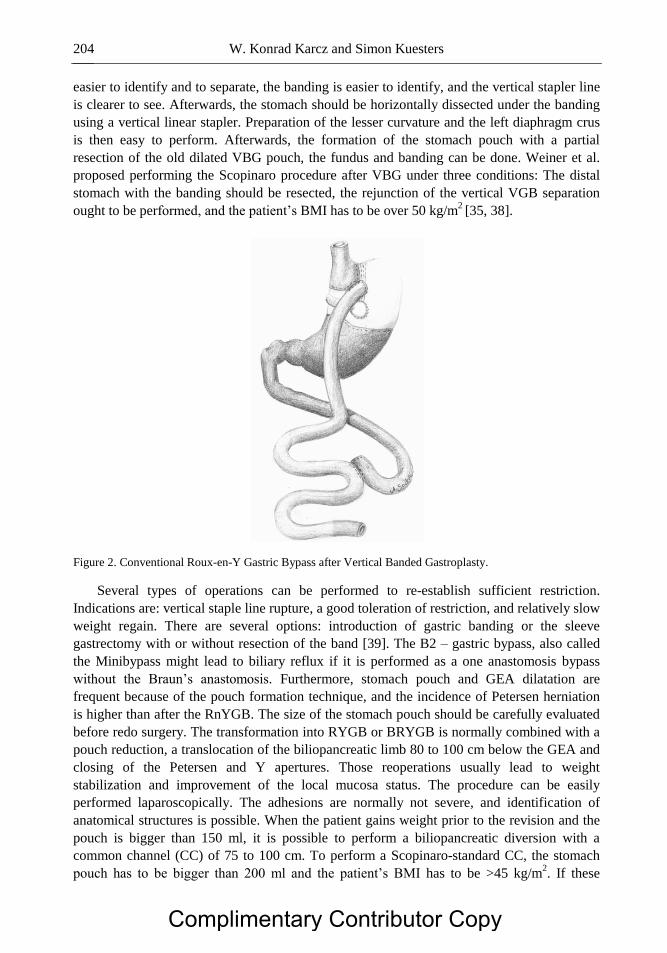
W. Konrad Karcz and Simon Kuesters 204
easier to identify and to separate, the banding is easier to identify, and the vertical stapler line
is clearer to see. Afterwards, the stomach should be horizontally dissected under the banding
using a vertical linear stapler. Preparation of the lesser curvature and the left diaphragm crus
is then easy to perform. Afterwards, the formation of the stomach pouch with a partial
resection of the old dilated VBG pouch, the fundus and banding can be done. Weiner et al.
proposed performing the Scopinaro procedure after VBG under three conditions: The distal
stomach with the banding should be resected, the rejunction of the vertical VGB separation
ought to be performed, and the patient’s BMI has to be over 50 kg/m2 [35, 38].
Figure 2. Conventional Roux-en-Y Gastric Bypass after Vertical Banded Gastroplasty.
Several types of operations can be performed to re-establish sufficient restriction.
Indications are: vertical staple line rupture, a good toleration of restriction, and relatively slow
weight regain. There are several options: introduction of gastric banding or the sleeve
gastrectomy with or without resection of the band [39]. The B2 – gastric bypass, also called
the Minibypass might lead to biliary reflux if it is performed as a one anastomosis bypass
without the Braun’s anastomosis. Furthermore, stomach pouch and GEA dilatation are
frequent because of the pouch formation technique, and the incidence of Petersen herniation
is higher than after the RnYGB. The size of the stomach pouch should be carefully evaluated
before redo surgery. The transformation into RYGB or BRYGB is normally combined with a
pouch reduction, a translocation of the biliopancreatic limb 80 to 100 cm below the GEA and
closing of the Petersen and Y apertures. Those reoperations usually lead to weight
stabilization and improvement of the local mucosa status. The procedure can be easily
performed laparoscopically. The adhesions are normally not severe, and identification of
anatomical structures is possible. When the patient gains weight prior to the revision and the
pouch is bigger than 150 ml, it is possible to perform a biliopancreatic diversion with a
common channel (CC) of 75 to 100 cm. To perform a Scopinaro-standard CC, the stomach
pouch has to be bigger than 200 ml and the patient’s BMI has to be >45 kg/m2. If these
Complimentary Contributor Copy

Bariatric and Metabolic Revisionary Surgery 205
conditions are not fulfilled, the standard BPD-S operation with a short CC should not be
performed.
Revisionary Operations after Restrictive Surgery
Almost all bariatric procedures include gastric restriction either by building a small pouch
a sleeve or upper stomach formation. The mechanical control and the neural sensation of
satiety are the two main components of gastric restriction. In cases of insufficient weight loss
or recurrent metabolic comorbidities, the anatomic aspects of stomach pouch or sleeve inlet,
outlet and volume have to be evaluated [40]. Many revisionary operations focus on the
stomach pouch and its outlet but in the case of LES dysfunction, there will be no success if
the revision is limited to the pouch size and pouch outlet.
The most popular metabolic operations based on restriction are the Laparoscopic
Adjustable Gastric Banding (LAGB), the Laparoscopic Sleeve Gastrectomy (LSG) and the
Laparoscopic Roux-en-Y Gastric Bypass, if it is not performed as a distal bypass. If there is
pouch dilation, the re-establishment of restriction leads to weight reduction and improvement
of the metabolic disorders in most cases [18, 41].
Introduction of Malabsorption
Insufficient restriction quickly leads to metabolic disturbances, weight regain and
recurrence of associated diseases, especially in procedures based solely on food limitation. It
is not easy to decide which method ought to be chosen in these cases. It is also not easy to
decide in favour of a revisionary malabsorptive surgery.
The most important question that has to be answered is: Is the patient capable of
accepting the disadvantages of malabsorption? Artificially shortening of the intestine leads to
chronic physiological changes. The recovery of sufficient restriction should therefore be
considered first. An accurate diagnostic investigation may help to understand the mechanical
situation of the intestinal tract, but is it a sufficient argument for introducing a malabsorptive
component? All patients, who qualify for malabsorptive surgery, ought to be discussed by an
interdisciplinary board following independent psychological, dietetic and metabolic
consultation. After patient education concerning the disadvantages of malabsorption,
preoperative diagnostics and interdisciplinary consensus, the indication for malabsorptive
surgery can be made.
It has to be noted that there are clear contraindications of secondary malabsorptive
operations, such as a BMI under 35 after the primary metabolic procedure, a pre-existing
malabsorptive state, malnutrition in vegetarians, pre-existing diarrhoea, psychiatric diseases,
and apparent lack of compliance. All patients with a history of major abdominal surgery with
consecutive malabsorption, like after ileum or colon resections for any reason, history of drug
or alcohol abuse, history of recent or chronic steroid medication, autoimmune disease,
inflammatory bowel disease, liver cirrhosis (CHILD B + C), serious active viral or bacterial
diseases (e. g. HIV, Hepatitis B and C, Tbc etc), pregnancy, history of any neoplasms in the
last five years, need of long-term anticoagulation for any reason and immunosuppression
Complimentary Contributor Copy
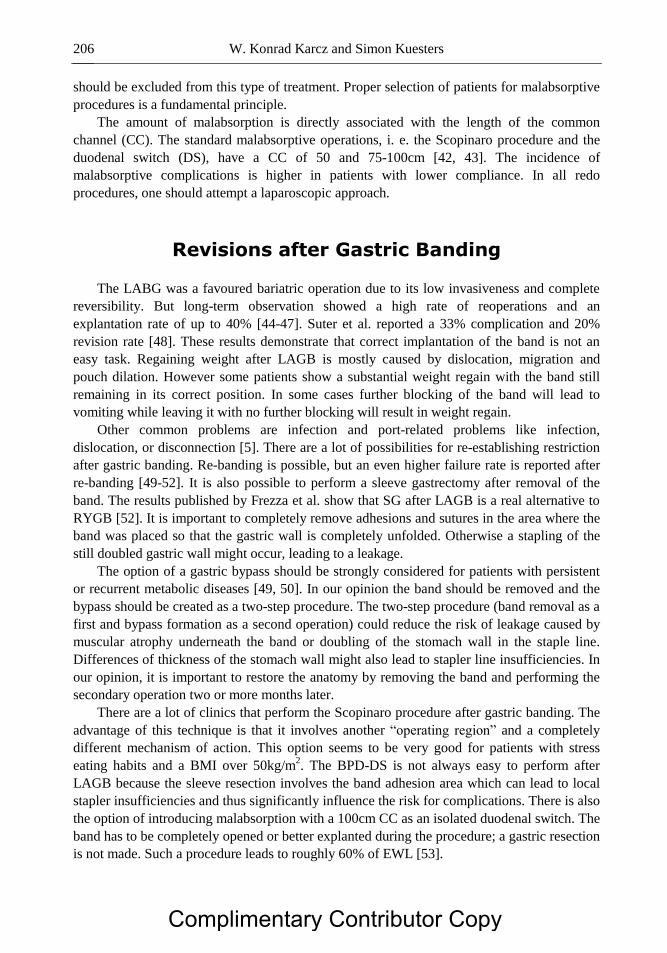
W. Konrad Karcz and Simon Kuesters 206
should be excluded from this type of treatment. Proper selection of patients for malabsorptive
procedures is a fundamental principle.
The amount of malabsorption is directly associated with the length of the common
channel (CC). The standard malabsorptive operations, i. e. the Scopinaro procedure and the
duodenal switch (DS), have a CC of 50 and 75-100cm [42, 43]. The incidence of
malabsorptive complications is higher in patients with lower compliance. In all redo
procedures, one should attempt a laparoscopic approach.
Revisions after Gastric Banding
The LABG was a favoured bariatric operation due to its low invasiveness and complete
reversibility. But long-term observation showed a high rate of reoperations and an
explantation rate of up to 40% [44-47]. Suter et al. reported a 33% complication and 20%
revision rate [48]. These results demonstrate that correct implantation of the band is not an
easy task. Regaining weight after LAGB is mostly caused by dislocation, migration and
pouch dilation. However some patients show a substantial weight regain with the band still
remaining in its correct position. In some cases further blocking of the band will lead to
vomiting while leaving it with no further blocking will result in weight regain.
Other common problems are infection and port-related problems like infection,
dislocation, or disconnection [5]. There are a lot of possibilities for re-establishing restriction
after gastric banding. Re-banding is possible, but an even higher failure rate is reported after
re-banding [49-52]. It is also possible to perform a sleeve gastrectomy after removal of the
band. The results published by Frezza et al. show that SG after LAGB is a real alternative to
RYGB [52]. It is important to completely remove adhesions and sutures in the area where the
band was placed so that the gastric wall is completely unfolded. Otherwise a stapling of the
still doubled gastric wall might occur, leading to a leakage.
The option of a gastric bypass should be strongly considered for patients with persistent
or recurrent metabolic diseases [49, 50]. In our opinion the band should be removed and the
bypass should be created as a two-step procedure. The two-step procedure (band removal as a
first and bypass formation as a second operation) could reduce the risk of leakage caused by
muscular atrophy underneath the band or doubling of the stomach wall in the staple line.
Differences of thickness of the stomach wall might also lead to stapler line insufficiencies. In
our opinion, it is important to restore the anatomy by removing the band and performing the
secondary operation two or more months later.
There are a lot of clinics that perform the Scopinaro procedure after gastric banding. The
advantage of this technique is that it involves another “operating region” and a completely
different mechanism of action. This option seems to be very good for patients with stress
eating habits and a BMI over 50kg/m2. The BPD-DS is not always easy to perform after
LAGB because the sleeve resection involves the band adhesion area which can lead to local
stapler insufficiencies and thus significantly influence the risk for complications. There is also
the option of introducing malabsorption with a 100cm CC as an isolated duodenal switch. The
band has to be completely opened or better explanted during the procedure; a gastric resection
is not made. Such a procedure leads to roughly 60% of EWL [53].
Complimentary Contributor Copy

Bariatric and Metabolic Revisionary Surgery 207
Revisions after Sleeve Gastrectomy
The sleeve gastrectomy was first performed in the context of BPD-DS [42;54]. The
volume of the remaining gastric tube should be roughly 100 -120 ml. This can be controlled
by using a calibration tube during the procedure [55, 56]. Nowadays, most bariatric surgeons
use a 32 or 34 F Bogie to form a proper-sized sleeve [56, 57]. A larger gastric tube seems to
correlate with a higher risk of weight regain and sleeve dilatation [55]. We do not have long
term results yet, but it is already known that 50% of all patients after SG undergo a secondary
operation. Nocca et al. reported a 6% rate of weight regain 2 years after SG [58]. We
recommend performing a stomach 3D MSCT with volumetry before reoperations [19].
Restoring restriction with a re-sleeve-operation could lead to dysfunction of stomach
motility, stenosis, and reflux with emesis or dysphagia. Such complications have to be treated
with myotomy or resection of a stenosis [59 [. Re-sleeve gastrectomy is therefore only
recommended in cases of large sleeve dilations with a sleeve volume >500 ml [60]. There are
two main causes for dilation after SG: an operative technique with a primary sleeve greater
than 200 ml and patients who keep or relapse into volume eating habits [58]. The re-sleeve
operation can be used safely for patients who still have a BMI over 50-60 and a volume of the
primary sleeve > 400ml. In these cases it is also possible to place a ring (banded sleeve
gastrectomy) around the upper part of the sleeve (approx. 6 cm below the cardia) to reduce
the risc of a re-dilatation [61]. In case of a sleeve dilation of 250-400 ml, where a re-sleeve-
resection should not be performed, duplication of the gastric wall using an extraluminal
running suture will reduce the sleeve volume and can lead to a sufficient restriction [62].
Another option for restoring restriction is the conversion into a gastric bypass. It is easy
to transform the sleeve into a conventional or banded gastric bypass. The conventional gastric
bypass does not lead to large weight changes, but the influence on metabolic disorders is
greater compared to the SG [63;64]. The gastric bypass operation can also be performed in
case of a localized stricture of the sleeve, if the stricture is located distally enough to create
the pouch for the gastric bypass.
Since the sleeve gastrectomy was first performed only in the context of BPD-DS, the
consequent secondary malabsorptive procedure after sleeve gastrectomy is the BPD with
duodenal switch. At about one third of patients after sleeve gastrectomy receive secondary
BPD in our series. The introduction of malabsorption with a CC of 75 -100cm (longer CC in
case of stronger restriction / smaller sleeve volume) with DS is an effective method. In our
series, almost all patients with super-obesity weighed less than 100kg one year after DS
performed as a second-step operation, and a mean BMI reduction of 12 points is seen in our
patients [65]. However, the indication for a second step BPD should be considered carefully,
as BPD might lead to severe complications like malnutrition and hypovitaminosis. It is also
possible to perform a single anastomosis duodeno-ileal omega bypass after the LSG (SADI-
S) [66]. The advantage of this procedure is the fact that only one anastomosis is needed and
the common channel is longer than after BPD-DS, which might reduce the risk of
malabsorption syndrome. However, long term results of the SADIS-S operation still have to
be evaluated.
Acid reflux and eosophagitis is commonly seen after sleeve gastrectomy. However, the
data in the literature differs concerning the question if reflux symptoms after sleeve
gastrectomy get worse or get better. In our experience an aggravation of reflux occurs in some
Complimentary Contributor Copy

W. Konrad Karcz and Simon Kuesters 208
patients, probably due to a partial migration of the sleeve into the thorax, thus weakening the
LES and a poor peristalsis in the sleeve itself [67]. We therefore usually perform a
“sleevepexie”, suturing the upper end of the sleeve to the left crus of the diaphragm to prevent
thoraxic migration. In case of severe reflux symptoms and eosophagitis after sleeve
gastrectomy we would fist initiate a consequent protone pump inhibitor therapy. The next step
would be a laparoscopic hiatoplastic and sleevopexie or a revision in to a gastric bypass. This
normally solves all reflux symptoms, but takes the passibility to perform a BPD-DS or
SADIS-S as a second step in case of weight regain.
Revisions after Gastric Bypass Surgery
The gastric bypass, the gold standard of bariatric surgery, leads to regain of weight in up
to 40% of patients in the long term follow up [68], but the redo surgery rate is still lower than
after VBG [2]. The long-term results presented by Chistou et al. show that 35% of patients
after RYGB still have a BMI >35 kg/m2
[69]. Other authors report better results (70, 71). It is
unclear how many patients suffer from pouch dilatation or what the prevalence of GE
dilatation is. The volume of the gastric pouch should be between 15 and 30 ml [71]. Larger
pouch volumes can be caused by inappropriate operating techniques or secondarily by eating
habits. The width of the GEA also plays an important role: if it is too small, it can lead to
stenosis and impaired emptying of the pouch, but if it is too large, a dilation of the first
jejununal loop can result [15]. In this case the first jejunum loop can contribute to the
functional stomach volume and thus enlarge it [72].
The precise evaluation of the diameter and area of the anastomosis allows the surgeon to
choose the best redo strategy for the patient. It is known that GE anastomosis dilation can
lead to insufficient weight control [73]. If the patient tolerates restriction, there are several
options for redo surgery. Endoscopic suturing to tighten a dilated gastric pouch is technically
feasible and safe with Stomafix as NOS (Natural Orifice Surgery), and may lead to weight
loss for certain patients [74]. Another logical option is to implant a GaBP Ring in a redo
operation with or without resizing of the pouch, thus creating a banded bypass [37]. An
analogous procedure with gastric banding in addition to the RnYGB was proposed by Bessler
et al. [75]. It might be an easy and effective way to control the volume and outlet of the
pouch. We recommend using rings of at least 6. 5 cm length in primary and 7. 0 cm in redo
operations [76, 77]. Some studies report a better weight reduction after banded bypasses. The
banded bypass causes satiety and reduces caloric intake by delaying gastric emptying, even
with low food intake. After 2 years, the percentage of EWL ranged around 80%, and at 5-year
follow-up, the EWL was roughly 75%. Only a slight weight regain of 2. 5% or 5% is
observed between the second and fifth postoperative year, whereas weight regain following
LCRYGB is reported to be over 10% in the same period [78]. A gastro-gastric fistula can also
lead to weight regain, patients normally report a lack of satiety and bigger meals. Normally a
fistula can be diagnosed in radiological examination (contrast swallow / MSCT). However, in
some cases the fistula might not be seen radiologically but then be diagnosed intraoperatively.
Remarkably there is a big difference in the frequency of gastro-gastral fistulas after gastric
bypass reaching from a few up to 40% in the literature [79, 80]. The dissection of the fistula,
normally with a linear stapler will lead to further weight loss.
Complimentary Contributor Copy
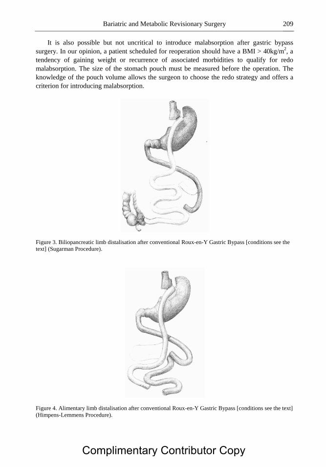
Bariatric and Metabolic Revisionary Surgery 209
It is also possible but not uncritical to introduce malabsorption after gastric bypass
surgery. In our opinion, a patient scheduled for reoperation should have a BMI > 40kg/m2, a
tendency of gaining weight or recurrence of associated morbidities to qualify for redo
malabsorption. The size of the stomach pouch must be measured before the operation. The
knowledge of the pouch volume allows the surgeon to choose the redo strategy and offers a
criterion for introducing malabsorption.
Figure 3. Biliopancreatic limb distalisation after conventional Roux-en-Y Gastric Bypass [conditions see the
text] (Sugarman Procedure).
Figure 4. Alimentary limb distalisation after conventional Roux-en-Y Gastric Bypass [conditions see the text]
(Himpens-Lemmens Procedure).
Complimentary Contributor Copy

W. Konrad Karcz and Simon Kuesters 210
Figure 5. Roux-en-Y Gastric Bypass transformed into Sleeve gastrectomy with reconstruction of the
intestinal tract.
When the functional stomach volume is bigger than 200ml (summary of stomach pouch
and first jejunal loop when the anastomosis is bigger than 2. 5 cm), it is possible to perform
the Sugerman operation with distalisation of the biliopancreatic loop or the relocation of the
alimentary loop at about 100cm from Bauhin’s Valve thus performing a distal gastric bypass
(81;82)(Figure 3,4). Both of these operations are relatively easy to perform laparoscopically.
The most difficult part is to disconnect the Roux-Y-anastomosis which can lead to stenosis or
leakage. The ends of the trans positioned loops should always be resected to avoid suturing of
the anastomosis in the scar tissue. Both operations lead to weight reduction and improvement
of the metabolic situation. In case of a small functional gastric volume severe malnutrition
symptoms can occur if malabsorption is added.
In this case another option, proposed by Gagner et al., is to change the RnYGB into a
biliopancreatic diversion with duodenal switch [83] (Figure 5, 6). Gagner published a 40 %
failure rate after RnYGB; these patients were successfully treated with a BPD-DS [68].
The transformation to a duodenal switch is technically difficult and has many potential
complications. The reconstruction of the bypassed stomach with a gastro-gastrostomy and the
following sleeve resection are the most difficult parts of the operation. The second half is very
similar to the standard duodenal switch procedure. Performing the whole transformation
during one operation is sometimes very difficult and leads to very long operating times [68,
83]. We have introduced a straight-forward criterion for when to perform a one-step
operation: if the reconstruction of the stomach and the sleeve resection takes less than 120
min, we perform the whole operation at once. Usually, it is easier to divide the operation into
two separate parts. This reduces the complication rate. The recovery of restriction and
introduction of malabsorption usually lead to an improvement of weight loss and metabolic
status in a short time [68].
Complimentary Contributor Copy

Bariatric and Metabolic Revisionary Surgery 211
Revisions after the Biliopancreatic Diversion
There are patients who underwent BPD-DS procedures without the use of gastric tubes
for sleeve calibration. Many of these patients now suffer from considerable stomach dilatation
with ensuing recurrence of metabolic disorders or weight regain in cases where the common
channel is > 100cm.
Figure 6. Roux-en-Y Gastric Bypass transformed procedure into Biliopancreatic Diversion with Duodenal
Switch.
Figure 7. Biliopancreatic Diversion with additional stomach fundus resection.
Complimentary Contributor Copy

W. Konrad Karcz and Simon Kuesters 212
The stomach volume should then be reduced to about 200ml to evoke a feeling of satiety
again [60]. A similar constellation can be observed in a small group of BPD patients who
present an upper stomach dilatation which can be treated with a fundus resection when
necessary, thus reducing the stomach pouch to 150 – 200 ml. In our series a gastric re-
resection after BPD leads to a mean reduction of 8 BMI points [65] (Figure 7).
In case of malnutritive symptoms with severe hypoalbuminemia, liver dysfunction,
severe diarrhoeas, flatulence and in cases where the patient is not able to supplement
adequately, it may be necessary to reduce malabsorption but preserve restriction. The
introduction of the restriction with reduction of malabsoption is possible by transforming the
BPD into the conventional or Banded Gastric Bypass. Analogous procedures are BPD-DS to
C- or B-RYGB transformations. The transformation of BPD to BPD-DS is technically
possible under the condition that the distal gastrectomy was not performed.
Conclusion
There is a variety of possible secondary operations after all bariatric operations. It may be
a big challenge for the surgeon to choose the right revisionary procedure in case of
insufficient weight loss. Detailed anamnesis and diagnostics are needed. If the wrong
procedure is chosen, the patient might not lose further weight or suffer from severe
complications.
However, if the right procedure is chosen there is a good chance for significant weight
loss. In our series of reoperations due to poor weight loss or weight regain a mean loss of 10
BMI points is seen one year after the revision [65].
In our opinion, the artificial control of satiety is at least partly associated with the pouch
volume. Hormonal and neural mechanisms also play an important role. Each year, several
new hormonal or neurogenic active intestinal peptides are identified. Also, the electrical
stimulation alone of vagal nerves, pylorus stimulation or stimulation of the duodenum leads
to significant changes in the intestinal tract and body homeostasis [84]. In the future,
restrictive surgery may be influenced by biologically active implants that react to
physiological impulses from the IT and communicate directly or indirectly with central
hunger regulation areas.
Prospective randomized trials are needed to compare different procedures. The group of
patients with a revisionary operation due to recurrent or persistent metabolic disorders is
growing. Reasons might be the limited spectrum of primary bariatric and metabolic
procedures performed in some clinics and the lack of good indication guidelines. A
systematic analysis of indications for different metabolic procedures needs to be discussed.
This could probably lead to a lower reoperation rate. At the moment, there seems to be no
golden standard operation in the growing area of metabolic surgery.
There are many new procedures, e. g. the duodenal bypass, the loop duodenal switch, the
ileal transposition, the banded sleeve resection and the vertical stomach separation. Vagus-,
stomach- or duodenal pacemakers and the Endo-Barrier are currently performed in small
series or are still in the process of development [84-87]
Certainly, there will be different types of new primary and revisionary operations as a
result of better understanding of the pathophysiology of metabolic processes. Furthermore,
Complimentary Contributor Copy

Bariatric and Metabolic Revisionary Surgery 213
new methods implementing bioactive implants, genetically modified “simbionts”, gene
therapy, or new drugs will undoubtedly have a significant impact on primary and revisionary
metabolic surgery in the future.
References
[1] Maggard MA, Shugarman LR, Suttorp M, Maglione M, Sugerman HJ, Livingston EH,
et al. Meta-analysis: surgical treatment of obesity. Ann. Intern. Med., 2005 Apr
5;142(7):547-59.
[2] van Gemert WG, van Wersch MM, Greve JW, Soeters PB. Revisional surgery after
failed vertical banded gastroplasty: restoration of vertical banded gastroplasty or
conversion to gastric bypass. Obes. Surg., 1998 Feb;8(1):21-8.
[3] Gawdat K. Bariatric re-operations: are they preventable? Obes. Surg., 2000
Dec;10(6):525-9.
[4] Fobi M, Lee H, Igwe D, Felahy B, James E, Stanczyk M, et al. Band erosion: incidence,
etiology, management and outcome after banded vertical gastric bypass. Obes. Surg.,
2001 Dec;11(6):699-707.
[5] Weiss HG, Kirchmayr W, Klaus A, Bonatti H, Muhlmann G, Nehoda H, et al. Surgical
revision after failure of laparoscopic adjustable gastric banding. Br. J. Surg., 2004
Feb;91(2):235-41.
[6] Fishman E, Melanson D, Lamport R, Levine A. A novel endoscopic delivery system for
placement of a duodenal-jejunal implant for the treatment of obesity and type 2
diabetes. Conf. Proc. IEEE Eng. Med. Biol. Soc., 2008;2008:2501-3.
[7] Padwal R, Brocks D, Sharma AM. A systematic review of drug absorption following
bariatric surgery and its theoretical implications. Obes. Rev., 2009 Jun 1.
[8] Martin MJ, Bennett S. Pretransplant bariatric surgery: a new indication? Surg. Obes.
Relat. Dis., 2007 Nov;3(6):648-51.
[9] Pillai AA, Rinella ME. Non-alcoholic fatty liver disease: is bariatric surgery the
answer? Clin. Liver Dis., 2009 Nov;13(4):689-710.
[10] Schulman AP, Del GF, Sinha N, Rubino F. Metabolic Surgery for the Treatment of
Type 2 Diabetes. Endocr. Pract., 2009 Jul 20;1-24.
[11] Oria HE, Moorehead MK. Bariatric analysis and reporting outcome system (BAROS).
Obes. Surg., 1998 Oct;8(5):487-99.
[12] Flanagan L. Measurement of Functional Pouch Volume following the Gastric Bypass
Procedure. Obes. Surg., 1996 Feb;6(1):38-43.
[13] de Castro CM, de Lima Montebelo MI, Rasera I, Jr., de OA, Jr., Gomes Gonelli PR,
Aparecida CG. Effects of Roux-en-Y gastric bypass on resting energy expenditure in
women. Obes. Surg., 2008 Nov;18(11):1376-80.
[14] Nishie A, Brown B, Barloon T, Kuehn D, Samuel I. Comparison of size of proximal
gastric pouch and short-term weight loss following routine upper gastrointestinal
contrast study after laparoscopic Roux-en-Y gastric bypass. Obes. Surg., 2007
Sep;17(9):1183-8.
Complimentary Contributor Copy

W. Konrad Karcz and Simon Kuesters 214
[15] Roberts K, Duffy A, Kaufman J, Burrell M, Dziura J, Bell R. Size matters: gastric
pouch size correlates with weight loss after laparoscopic Roux-en-Y gastric bypass.
Surg. Endosc., 2007 Aug;21(8):1397-402.
[16] Madan AK, Tichansky DS, Phillips JC. Does pouch size matter? Obes. Surg., 2007
Mar;17(3):317-20.
[17] Forsell P, Hellers G, Laveskog U, Westman L. Validation of Pouch Size Measurement
Following the Swedish Adjustable Gastric Banding Using Endoscopy, MRI and Barium
Swallow. Obes. Surg., 1996 Dec;6(6):463-7.
[18] Langer FB, Bohdjalian A, Felberbauer FX, Fleischmann E, Reza Hoda MA, Ludvik B,
et al. Does gastric dilatation limit the success of sleeve gastrectomy as a sole operation
for morbid obesity? Obes. Surg., 2006 Feb;16(2):166-71.
[19] Karcz WK, Kuesters S, Marjanovic G, Suesslin D, Kotter E, Thomusch O, et al. 3D-
MSCT gastric pouch volumetry in bariatric surgery-preliminary clinical results. Obes.
Surg., 2009 Apr;19(4):508-16.
[20] Munoz R, Ibanez L, Salinas J, Escalona A, Perez G, Pimentel F, et al. Importance of
routine preoperative upper GI endoscopy: why all patients should be evaluated? Obes.
Surg., 2009 Apr;19(4):427-31.
[21] de Moura AA, Cotrim HP, Santos AS, Bitencourt AG, Barbosa DB, Lobo AP, et al.
Preoperative upper gastrointestinal endoscopy in obese patients undergoing bariatric
surgery: is it necessary? Surg. Obes. Relat. Dis., 2008 Mar;4(2):144-9.
[22] Hartin CW, Jr., Remine DS, Lucktong TA. Preoperative bariatric screening and
treatment of Helicobacter pylori. Surg. Endosc., 2009 May 15.
[23] Klaus A, Weiss H. Is preoperative manometry in restrictive bariatric procedures
necessary? Obes. Surg., 2008 Aug;18(8):1039-42.
[24] Merrouche M, Sabate JM, Jouet P, Harnois F, Scaringi S, Coffin B, et al. Gastro-
esophageal reflux and esophageal motility disorders in morbidly obese patients before
and after bariatric surgery. Obes. Surg., 2007 Jul;17(7):894-900.
[25] Baumann T, Kuesters S, Grueneberger J, Marjanovic G, Zimmermann L, Schaefer AO,
et al. Time-resolved MRI after ingestion of liquids reveals motility changes after
laparoscopic sleeve gastrectomy--preliminary results. Obes. Surg., 2011 Jan;21(1):95-
101.
[26] Gentileschi P, Lirosi F, Benavoli D, Sica G, Di LN, Venza M, et al. Laparoscopic
reoperative approach after open bariatric surgery. Chir. Ital., 2009 Mar;61(2):137-41.
[27] Raftopoulos I, Courcoulas AP. Revision of jejunoileal bypass to Roux-en-Y gastric
bypass: technical considerations and outcomes from 2 cases. Surg. Obes. Relat. Dis.,
2008 Mar;4(2):198-201.
[28] Hanni CL, Pool LR, Dean RE, Cronquist JC. Treatment of jejunoileal bypass failure by
reanastomosis and gastroplasty in a single-stage procedure. Review of 47 cases. Am.
Surg., 1984 Jul;50(7):354-7.
[29] Junker K, Jensen JB, Jensen HE. Simultaneous small-intestinal reconstruction and
gastric partitioning as treatment for complications after jejunoileal bypass for morbid
obesity. Scand. J. Gastroenterol., 1981 Apr;16(3):433-6.
[30] LaFave JW, Alden JF. Gastric bypass in the operative revision of the failed jejunoileal
bypass. Arch. Surg., 1979 Apr;114(4):438-44.
[31] Schouten R, van Dielen FM, van Gemert WG, Greve JW. Conversion of vertical
banded gastroplasty to Roux-en-Y gastric bypass results in restoration of the positive
Complimentary Contributor Copy

Bariatric and Metabolic Revisionary Surgery 215
effect on weight loss and co-morbidities: evaluation of 101 patients. Obes. Surg., 2007
May;17(5):622-30.
[32] Marsk R, Jonas E, Gartzios H, Stockeld D, Granstrom L, Freedman J. High revision
rates after laparoscopic vertical banded gastroplasty. Surg. Obes. Relat. Dis., 2009
Jan;5(1):94-8.
[33] Behrns KE, Smith CD, Kelly KA, Sarr MG. Reoperative bariatric surgery. Lessons
learned to improve patient selection and results. Ann. Surg., 1993 Nov;218(5):646-53.
[34] Sanchez H, Cabrera A, Cabrera K, Zerrweck C, Mosti M, Sierra M, et al. Laparoscopic
Roux-en-Y Gastric Bypass as a Revision Procedure after Restrictive Bariatric Surgery.
Obes. Surg., 2008 Dec;18(12):1539-43.
[35] Daskalakis M, Scheffel O, Theodoridou S, Weiner RA. Conversion of Failed Vertical
Banded Gastroplasty to Biliopancreatic Diversion, a Wise Option. Obes. Surg., 2009
Aug 29.
[36] Fobi MA, Lee H. The surgical technique of the Fobi-Pouch operation for obesity (the
transected silastic vertical gastric bypass). Obes. Surg., 1998 Jun;8(3):283-8.
[37] Fobi MA. Placement of the GaBP ring system in the banded gastric bypass operation.
Obes. Surg., 2005 Sep;15(8):1196-201.
[38] Menon T, Quaddus S, Cohen L. Revision of failed vertical banded gastroplasty to non-
resectional Scopinaro biliopancreatic diversion: early experience. Obes. Surg., 2006
Nov;16(11):1420-4.
[39] Elazary R, Hazzan D, Appelbaum L, Rivkind AI, Keidar A. Feasibility of Sleeve
Gastrectomy as a Revision Operation for Failed Silastic Ring Vertical Gastroplasty.
Obes. Surg., 2008 Oct 7.
[40] Kuesters S, Marjanovic G, Karcz WK. [Redo operations after bariatric and metabolic
surgery]. Zentralbl. Chir., 2009 Feb;134(1):50-6.
[41] Muller MK, Wildi S, Scholz T, Clavien PA, Weber M. Laparoscopic pouch resizing
and redo of gastro-jejunal anastomosis for pouch dilatation following gastric bypass.
Obes. Surg., 2005 Sep;15(8):1089-95.
[42] Hess DS, Hess DW, Oakley RS. The biliopancreatic diversion with the duodenal
switch: results beyond 10 years. Obes. Surg., 2005 Mar;15(3):408-16.
[43] Marceau P, Hould FS, Potvin M, Lebel S, Biron S. Biliopancreatic diversion (doudenal
switch procedure). Eur. J. Gastroenterol. Hepatol., 1999 Feb;11(2):99-103.
[44] DeMaria EJ, Sugerman HJ, Meador JG, Doty JM, Kellum JM, Wolfe L, et al. High
failure rate after laparoscopic adjustable silicone gastric banding for treatment of
morbid obesity. Ann. Surg., 2001 Jun;233(6):809-18.
[45] Angrisani L, Furbetta F, Doldi SB, Basso N, Lucchese M, Giacomelli M, et al. Results
of the Italian multicenter study on 239 super-obese patients treated by adjustable gastric
banding. Obes. Surg., 2002 Dec;12(6):846-50.
[46] O'Brien PE, Dixon JB. Lap-band: outcomes and results. J. Laparoendosc. Adv. Surg.
Tech. A, 2003 Aug;13(4):265-70.
[47] Weiner R, Blanco-Engert R, Weiner S, Matkowitz R, Schaefer L, Pomhoff I. Outcome
after laparoscopic adjustable gastric banding - 8 years experience. Obes. Surg., 2003
Jun;13(3):427-34.
[48] Suter M, Calmes JM, Paroz A, Giusti V. A 10-year experience with laparoscopic gastric
banding for morbid obesity: high long-term complication and failure rates. Obes. Surg.,
2006 Jul;16(7):829-35.
Complimentary Contributor Copy

W. Konrad Karcz and Simon Kuesters 216
[49] Weber M, Muller MK, Michel JM, Belal R, Horber F, Hauser R, et al. Laparoscopic
Roux-en-Y gastric bypass, but not rebanding, should be proposed as rescue procedure
for patients with failed laparoscopic gastric banding. Ann. Surg., 2003 Dec;238(6):827-
33.
[50] Lanthaler M, Mittermair R, Erne B, Weiss H, Aigner F, Nehoda H. Laparoscopic
gastric re-banding versus laparoscopic gastric bypass as a rescue operation for patients
with pouch dilatation. Obes. Surg., 2006 Apr;16(4):484-7.
[51] Acholonu E, McBean E, Court I, Bellorin O, Szomstein S, Rosenthal RJ. Safety and
short-term outcomes of laparoscopic sleeve gastrectomy as a revisional approach for
failed laparoscopic adjustable gastric banding in the treatment of morbid obesity. Obes.
Surg., 2009 Dec;19(12):1612-6.
[52] Frezza EE, Torre EJ, Enriquez C, Gee L, Wachtel M, Corvala JA. Laparoscopic Sleeve
Gastrectomy After Gastric Banding Removal: A Feasibility Study. Surg Innov 2008
Dec 12.
[53] Slater GH, Fielding GA. Combining laparoscopic adjustable gastric banding and
biliopancreatic diversion after failed bariatric surgery. Obes. Surg., 2004 May;14(5):
677-82.
[54] Johnston D, Dachtler J, Sue-Ling HM, King RF, Martin G. The Magenstrasse and Mill
operation for morbid obesity. Obes. Surg., 2003 Feb;13(1):10-6.
[55] Weiner RA, Weiner S, Pomhoff I, Jacobi C, Makarewicz W, Weigand G. Laparoscopic
sleeve gastrectomy--influence of sleeve size and resected gastric volume. Obes. Surg.,
2007 Oct;17(10):1297-305.
[56] Deitel M, Crosby RD, Gagner M. The First International Consensus Summit for Sleeve
Gastrectomy (SG), New York City, October 25-27, 2007. Obes. Surg., 2008 Mar 21.
[57] Baltasar A, Serra C, Perez N, Bou R, Bengochea M, Ferri L. Laparoscopic sleeve
gastrectomy: a multi-purpose bariatric operation. Obes. Surg., 2005 Sep;15(8):1124-8.
[58] Nocca D, Krawczykowsky D, Bomans B, Noel P, Picot MC, Blanc PM, et al. A
Prospective Multicenter Study of 163 Sleeve Gastrectomies: Results at 1 and 2 Years.
Obes. Surg., 2008 Mar 4.
[59] Dapri G, Cadiere GB, Himpens J. Laparoscopic seromyotomy for long stenosis after
sleeve gastrectomy with or without duodenal switch. Obes. Surg., 2009 Apr;19(4):495-
9.
[60] Gagner M, Rogula T. Laparoscopic reoperative sleeve gastrectomy for poor weight loss
after biliopancreatic diversion with duodenal switch. Obes. Surg., 2003 Aug;13(4):649-
54.
[61] Karcz WK, Marjanovic G, Grueneberger J, Baumann T, Bukhari W, Krawczykowski
D, et al. Banded sleeve gastrectomy using the GaBP ring--surgical technique. Obes.
Facts, 2011;4(1):77-80.
[62] Brethauer SA, Harris JL, Kroh M, Schauer PR. Laparoscopic gastric plication for
treatment of severe obesity. Surg. Obes. Relat. Dis., 2011 Jan;7(1):15-22.
[63] Rosenthal R, Li X, Samuel S, Martinez P, Zheng C. Effect of sleeve gastrectomy on
patients with diabetes mellitus. Surg. Obes. Relat. Dis., 2009 Jul;5(4):429-34.
[64] Pories WJ, Swanson MS, MacDonald KG, Long SB, Morris PG, Brown BM, et al. Who
would have thought it? An operation proves to be the most effective therapy for adult-
onset diabetes mellitus. Ann. Surg., 1995 Sep;222(3):339-50.
Complimentary Contributor Copy

Bariatric and Metabolic Revisionary Surgery 217
[65] Kuesters S, Grueneberger JM, Baumann T, Bukhari W, Daoud M, Hopt UT, et al.
Revisionary bariatric surgery: indications and outcome of 100 consecutive operations at
a single center. Surg. Endosc., 2011 Dec 22.
[66] Sanchez-Pernaute A, Herrera MA, Perez-Aguirre ME, Talavera P, Cabrerizo L, Matia
P, et al. Single anastomosis duodeno-ileal bypass with sleeve gastrectomy (SADI-S).
One to three-year follow-up. Obes. Surg., 2010 Dec;20(12):1720-6.
[67] Baumann T, Grueneberger J, Pache G, Kuesters S, Marjanovic G, Kulemann B, et al.
Three-dimensional stomach analysis with computed tomography after laparoscopic
sleeve gastrectomy: sleeve dilation and thoracic migration. Surg. Endosc., 2011
Jul;25(7):2323-9.
[68] Trelles N, Gagner M. Revision bariatric surgery: laparoscopic conversion of failed
gastric bypass to biliopancreatic diversion with duodenal switch. Minerva Chir., 2009
Jun;64(3):277-84.
[69] Christou NV, Look D, Maclean LD. Weight gain after short- and long-limb gastric
bypass in patients followed for longer than 10 years. Ann. Surg., 2006 Nov;244(5):734-
40.
[70] Fobi M, Lee H, Igwe D, Felahy B, James E, Stanczyk M, et al. Gastric bypass in
patients with BMI < 40 but > 32 without life-threatening co-morbidities: preliminary
report. Obes. Surg., 2002 Feb;12(1):52-6.
[71] Madan AK, Harper JL, Tichansky DS. Techniques of laparoscopic gastric bypass: on-
line survey of American Society for Bariatric Surgery practicing surgeons. Surg. Obes.
Relat. Dis., 2008 Mar;4(2):166-72.
[72] Karcz WK, Kuesters S, Marjanovic G, Suesslin D, Kotter E, Thomusch O, et al. 3D-
MSCT Gastric Pouch Volumetry in Bariatric Surgery-Preliminary Clinical Results.
Obes. Surg., 2008 Dec 18.
[73] Naslund I. A method for measuring the size of the gastric outlet in obesity surgery.
Acta. Chir. Scand., 1984;150(5):399-404.
[74] Thompson CC, Slattery J, Bundga ME, Lautz DB. Peroral endoscopic reduction of
dilated gastrojejunal anastomosis after Roux-en-Y gastric bypass: a possible new option
for patients with weight regain. Surg. Endosc., 2006 Nov;20(11):1744-8.
[75] Gumbs AA, Margolis B, Bessler M. Laparoscopic banded-Roux-en-Y gastric bypass.
Surg. Obes. Relat. Dis., 2006 May;2(3):408-9.
[76] Crampton NA, Izvornikov V, Stubbs RS. Silastic ring gastric bypass: a comparison of
two ring sizes: a preliminary report. Obes. Surg., 1997 Dec;7(6):495-9.
[77] Stubbs RS, O'brien I, Jurikova L. What ring size should be used in association with
vertical gastric bypass? Obes. Surg., 2006 Oct;16(10):1298-303.
[78] Fobi MA, Lee H, Felahy B, Che K, Ako P, Fobi N. Choosing an operation for weight
control, and the transected banded gastric bypass. Obes. Surg., 2005 Jan;15(1):114-21.
[79] Patel S, Szomstein S, Rosenthal RJ. Reasons and Outcomes of Reoperative Bariatric
Surgery for Failed and Complicated Procedures (Excluding Adjustable Gastric
Banding). Obes. Surg., 2010 Jul 30.
[80] Fronza JS, Prystowsky JB, Hungness ES, Nagle AP. Revisional bariatric surgery at a
single institution. Am. J. Surg., 2010 Nov;200(5):651-4.
[81] Sugerman HJ, Kellum JM, DeMaria EJ. Conversion of proximal to distal gastric bypass
for failed gastric bypass for superobesity. J. Gastrointest. Surg., 1997 Nov;1(6):517-24.
Complimentary Contributor Copy

W. Konrad Karcz and Simon Kuesters 218
[82] Fobi MA, Lee H, Igwe D, Jr., Felahy B, James E, Stanczyk M, et al. Revision of failed
gastric bypass to distal Roux-en-Y gastric bypass: a review of 65 cases. Obes. Surg.,
2001 Apr;11(2):190-5.
[83] Parikh M, Pomp A, Gagner M. Laparoscopic conversion of failed gastric bypass to
duodenal switch: technical considerations and preliminary outcomes. Surg. Obes. Relat.
Dis., 2007 Nov;3(6):611-8.
[84] Wang TT, Hu SY, Gao HD, Zhang GY, Liu CZ, Feng JB, et al. Ileal transposition
controls diabetes as well as modified duodenal jejunal bypass with better lipid lowering
in a nonobese rat model of type II diabetes by increasing GLP-1. Ann. Surg., 2008
Jun;247(6):968-75.
[85] Mason EE. Ileal [correction of ilial] transposition and enteroglucagon/GLP-1 in obesity
(and diabetic?) surgery. Obes. Surg., 1999 Jun;9(3):223-8.
[86] Salvi PF, Brescia A, Cosenza UM, D'Urso R, Cardelli P, Badiali M. [Gastric pacing to
treat morbid obesity: two years experience in four patients]. Ann. Ital. Chir., 2009
Jan;80(1):25-8.
[87] Miller K, Hoeller E, Aigner F. The Implantable Gastric Stimulator for Obesity : An
Update of the European Experience in the LOSS (Laparoscopic Obesity Stimulation
Survey) Study. Treat Endocrinol., 2006;5(1):53-8.
Complimentary Contributor Copy
In: Bariatric Surgery ISBN: 978-1-62618-995-9
Editor: Christos R. Iavazzo © 2013 Nova Science Publishers, Inc.
Chapter XIII
Body-Contouring
after Bariatric Surgery
Franco Migliori EURAPS (European Association of Plastic Surgeons);
ASPS (American Society of Plastic Surgeons);
ISAPS (International Society of Aesthetic Plastic Surgery);
IFSO (International Federation for the Surgery of Obesity);
SICPRE (Italian Society of Plastic, Reconstructive and Aesthetic Surgery);
SICOB (Italian Society of Obesity Surgery);
SIUST (Italian Burns Society)
Abstract
In the last decades the rising demand for bariatric procedures worldwide has
exponentially increased the need of post-bariatric plastic surgery. Accordingly a new
chapter of plastic surgery has been born, since the standard technique usually performed
in body-contouring wasn’t perfectly suited for post-bariatric tissues due to the different
tissue structures of post-bariatric patients. The anatomical areas most frequently treated
are limbs, breast and trunk. In this chapter, the leading strategies for any of these areas,
with the main attention kept on general concepts, technical aspects and patient
management, are summarized. This field of the plastic surgery is not an easy one; it
requires good surgical experience, as well as organized and dedicated teams because it
can’t be managed by a single professional. Respecting these rules leads to great results
and great patient satisfaction.
Introduction
The increase of body-contouring (BC) procedures after massive weight loss (MWL)
following bariatric surgery (BS) is astonishing: in the U. S. A in 2004, almost 56. 000 out of
Complimentary Contributor Copy

Franco Migliori 220
140. 000 bariatric patients underwent BC surgery, in 2005 100. 000 out of 250. 000 [1]. Even
a neologism has been proposed: “Bariplasty [2]”. The expectations are increasing numbers for
at least the next two decades. This wide horizon should propose (as it already does in fact) an
integrated treatment approach [3] (bariatric surgery, plastic surgery, motivation for physical
activity, and psychological support) for the years to come. BS effects are more and more
perceived by patients as an intermediate stage quite unsatisfactory. BS dramatically reduces
weight and normalizes body-mass, but enhances dismorphisms and creates new problems
such as skin redundancy and flabbiness, sweating and eczemas, general aesthetical
uneasiness, low self-esteem: BC completes therapeutical course restoring body shape and
solving these problems. BC after BS is mainly performed on 4 anatomical areas:
Arms 7 %
Breast 38 %
Torso 40 %
Legs 15 %
Bariatric Surgery Sequelae Influencing
Body-Contouring
Among thousands of “pros and cons” described, after BS procedures, the following are
those mainly conditioning plastic surgeons performing BC.
Clinical Consequences
Significant advantages of BS are:
a) the extreme weight loss (up to 80-100 kg) within an average time interval of 12-18
months;
b) normalization of hyperglycemia and hypercholesterolemia.
Challenging associated complications, mainly in malabsorptive procedures, are:
1) Fe++
malabsorption with sideropenic anemia;
2) Ca++
malabsorption with bone demineralization;
3) in 5%of patients, protein malnutrition.
Morphological Consequences
The clinical evidenced disadvantage following BS is the inefficacy of the soft tissue scar
retraction, probably due to histological alterations, causing an overall non-esthetic aspect. The
inelastic and atrophic tissues result in deep folds and creases. The main aspects are large
dermo-adiposal folds in the arms, abdomen and inner thighs, and medium-to-severebreast
ptosis in women.
Complimentary Contributor Copy

Body-Contouring after Bariatric Surgery 221
Anatomo-Surgical Consequences
The preoperative and intraoperative challenge of BC after BS is the reduced vascular
perfusion of the tissues, mainly in the dermis, which appears thinner, and in the fat, which
appear shy potrophic, “pale” or even “gray”. The presence of BS scars in the abdominal wall
causes reduced elasticity in that specific area and a compromised dermal plexus, both
enhancing specific complications.
Histological Consequences
A personal histologic study on post-BS dermis and fat is running nowadays. Preliminary
reports, comparing post-BS tissues with healthy tissues, demonstrates:
a) in the Fat: presence of collapsed adipocytes with thicker membranes, prominent
nuclei (usually not visible) and thickened intracellular fibrous septa (Figure 1).
b) in the Dermis: thickening of the fibrous net and the hypertrophy of the vascular net.
A reduction of elastic fibers is supposed, but results of histochemical and EM study
are awaited (Figure 2).
(A) (B)
Figure 1. a) Normal Fat Tissue; b) Post-bariatric Fat Tissue.
(A) (B)
Figure 2. a) Normal Dermal Tissue; b) Post-bariatric Dermal Tissue.
Complimentary Contributor Copy

Franco Migliori 222
General Surgical Strategies
Plastic surgery is a science, and Body-Contouring is a scientific art. This means that,
despite any surgical algorithm, there is no “perfect shape” to aim to: there can be, for each
body district, some key points to respect; but the main key point remains the patient’s “body
image” as a whole. This concept includes subjective and objective evaluations, which differ
from patient to patient. The only general rule we always follow for female figure (♀ are 80-85
% of all BC after BS) is the “silhouette” image (Figure 3): prominent breast and buttocks,
narrow waist and ankles, can reasonably be considered as good-looking for every woman.
Figure 3. “Silhouette” model profile.
In our experience 67% of post-bariatric patients underwent to multiple BC procedures
(from 2 to 4 operations): there is no algorithm that determines the timing and suitability of
one procedure rather than another. Among every consideration, the psychological impact on
the patient should be considered, trying to choose more effective one as starting procedure.
As usually happens in plastic surgery procedures, the surgeon main aim is to find the
right compromise between maximal reduction of excess tissue and maximal reduction of scar
Complimentary Contributor Copy

Body-Contouring after Bariatric Surgery 223
length. Moreover, BC is quite an aggressive surgeryand the risk/benefit rate is important to be
well evaluated and understood: the same operations could fit a patient but not another.
BC procedures are not novelties in plastic surgery: some of them have been described one
century ago, most of them several decades ago (’60 and ’70), all of them far before BS was
born and set its wide success. When liposuction came in fashion (’80) the initial euphoria let
al.l plastic surgeons believe that traditional BC would have been dismissed. As euphoria
subsided and BS and MWL grew up, BC could slowly taste its cold revenge: it’s today well
accepted that liposuction is not a substitute for BC, and that both represent different solutions
for different cases in different stages [4]. The anatomo-surgical and histological features of
post-BS patients confer to their tissues an “anergic behavior”, meaning a poor or absent scar
retraction. Because of that, the surgical techniques mainly suggested are those removing all
exceeding tissue. Thus liposuction, where results depend on scar retraction, cannot be utilized
except as an ancillary technique, in rare selected cases. BC techniques of decades ago, of
course, have taken benefits by all technical and knowledge advances and have “changed their
suit”: in these pages a summary of old concepts and new criteria is proposed, looking for
maximal reshaping and minimal visible scars.
General Management
On complication of BS (mainly gastric bypass and biliopancreatic diversion) can be
vitamin B12 and Fe++
malabsorption, with consequent anemia [5]. Therefore, these levels need
to be checked, because major reconstruction will result in blood loss. In those cases, auto-
transfusion should be considered. However, auto transfusion can be performed only with a
hemoglobin rate > 11. 5, which in post-BS patient is not so frequent. Therefore, we always
perform infiltration of all the tissues to be excised with a diluted solution of adrenaline (1 cc.
/500 cc. NaCl) at least 20’ before cutting: this procedure allows a great amount of circulating
mass to be squeezed off, significantly reducing blood losses.
Another major risk in BC is deep venous thrombosis [6], with blood clots and pulmonary
embolism. We always perform prophylaxis with antiembolic compressive stockings and low-
molecular weight heparin (4000/6. 000 U. I. 12 hour before surgery, continued daily for two
weeks).
Seromas and liponecrosis can be challenging complications. There are two rules to
reduce their incidence:
avoiding any cutting device but blade: no other known instrument is less tissue-
damaging;
limiting undermining.
The extension of scars can be overwhelming in many procedures. Extreme accuracy in
suture technique and material choice can make the difference, reducing hypertrophies and
keloids occurrence. Only deep, dermal and intradermal suture should be utilized, with no
cutaneous stitch. Specific plastic surgery dedicated threads and needles have been conceived,
and it is advisable to use them in BC.
Complimentary Contributor Copy

Franco Migliori 224
Techniques
Arms
Within BC procedures performed on BS patients, brachioplasty appears to be very
exclusive, since the request is definitely lower than for other districts.
Reviewing literature [7,8,9,10,11,12,13,14] modest results are usually found, due to
modest improvement of the morphological defect, together with remarkable scars and
important complication risks.
In this particular anatomical district, the best possible result should be pursued,
optimizing cost/benefit ratio in terms of maximal correction/less visible scars. If this strategy
cannot be applied, brachioplasty should be discouraged.
A technique has therefore been developed, based on a linear scar programmed to be
hidden in the medial bicipital groove, and aiming to obtain the best possible morphological
correction. This doesn’t mean passing from a “batwing” effect to a “sweater sleeve” effect,
but to a normal arm profile. A stigmata of this “normality” should be represented by a slight
concavity in the lower arm profile proximal to the elbow.
Patients are grouped according with Strauch [8] 4-class classification of defects: post-BS
patients usually belong to classes 3 and 4. The anatomical districts interested by surgery were
area 3 (forearm) and area 4 (axilla), according with Strauch8 anatomical scheme.
Pre-Operative Markings
Special care is taken in evaluating the amount of tissue to be excided. Only the anterior
aspect of the excision is normally drawn, and the actual quantity of skin to be dismissed
intraoperatively determined. This algorithm is operator-dependent and the compulsory rule is
that markings must be drawn by the first surgeon.
Figure 4. Brachioplasty: drawing with manual tension.
Complimentary Contributor Copy

Body-Contouring after Bariatric Surgery 225
Figure 5. Brachioplasty: final drawing.
With the patient standing in a frontal view, arms abducted at shoulder level and forearms
proned (“crucifix” position), the incision line is drawn within the medial bicipital groove,
while applying with counterlateral hand a slight antero-posterior tension on the medial face of
the arm (the same tension will be applied during surgery) (Figure 4). This line continues on
the posterior axillary pillar, gently curving at 90° and becoming vertical, just posterior to and
extending as axillary hair (4-5 cm.) (Figure 5).
Surgical Technique
After a wide literature review, proposing any kind of incision shape, no acceptable
demonstration is found that linear incision (from medial epicondyle to axilla along medial
bicipital groove) is worse than other ones. A deep incision is done on that line, directly on the
muscular fascia, preserving it.
An antero-posterior undermining is gently performed with the tip of a blade, following
fascia surface for almost half circumference of the arm. All perforating vessels and nerves
long enough to be let loose are preserved until exceeding tissue extension is determined
(Figure 6).
Figure 6. Brachioplasty: suprafascial undermining.
Complimentary Contributor Copy

Franco Migliori 226
As the undermining is completed, an overcorrective tension is applied to the anterior edge
of incision and to the undermined flap, similar to that one applied while marking. The flap is
overlapped to incision edge, whose superficial projection is spot-marked with 3-4 transfixed
needles and completed with a straight line joining front incision tips. A back incision is then
performed obtaining, in this way, an excided tissue extension always rather abundant: only
perforating vessels and nerves feeding this area are tied and cut (Figure 7).
Figure 7. Brachioplasty: tissue excision completed.
Suture is evidently performed under tension, so great care is spent to provide precise and
tight edges fitting, through at least two suture layers. First stitch must fix a posterior flap
whose medial length is determined by the axillary incision angle (Figure 8). Suction drains
are placed and maintained 1-2 days post-op.
Figure 8. Brachioplasty: flap closure.
Post-Operative Care
Discharge is usually allowed within 2-3 days post-op. Elastic tubular bandages are worn
for about 1 month post-op. Drugs and devices preventing pathological scars are strongly
suggested, because hypertrophies and keloids are frequent in this area.
Complimentary Contributor Copy
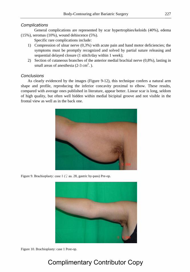
Body-Contouring after Bariatric Surgery 227
Complications
General complications are represented by scar hypertrophies/keloids (40%), edema
(15%), seromas (10%), wound dehiscence (5%).
Specific rare complications include:
1) Compression of ulnar nerve (0,3%) with acute pain and hand motor deficiencies; the
symptoms must be promptly recognized and solved by partial suture releasing and
sequential delayed closure (1 stitch/day within 1 week);
2) Section of cutaneous branches of the anterior medial brachial nerve (0,8%), lasting in
small areas of anesthesia (2-3 cm2. ).
Conclusions
As clearly evidenced by the images (Figure 9-12), this technique confers a natural arm
shape and profile, reproducing the inferior concavity proximal to elbow. These results,
compared with average ones published in literature, appear better. Linear scar is long, seldom
of high quality, but often well hidden within medial bicipital groove and not visible in the
frontal view as well as in the back one.
Figure 9. Brachioplasty: case 1 (♀ aa. 28, gastric by-pass) Pre-op.
Figure 10. Brachioplasty: case 1 Post-op.
Complimentary Contributor Copy
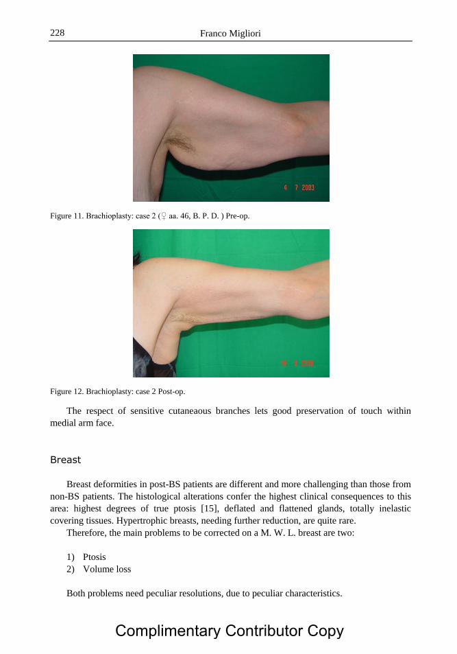
Franco Migliori 228
Figure 11. Brachioplasty: case 2 (♀ aa. 46, B. P. D. ) Pre-op.
Figure 12. Brachioplasty: case 2 Post-op.
The respect of sensitive cutaneaous branches lets good preservation of touch within
medial arm face.
Breast
Breast deformities in post-BS patients are different and more challenging than those from
non-BS patients. The histological alterations confer the highest clinical consequences to this
area: highest degrees of true ptosis [15], deflated and flattened glands, totally inelastic
covering tissues. Hypertrophic breasts, needing further reduction, are quite rare.
Therefore, the main problems to be corrected on a M. W. L. breast are two:
1) Ptosis
2) Volume loss
Both problems need peculiar resolutions, due to peculiar characteristics.
Complimentary Contributor Copy

Body-Contouring after Bariatric Surgery 229
Ptosis can be surgically minor or severe: we usually put a borderline around 6 cm. of
ptosis correction. If the difference between the pre-operative nipple-areola complex (N. A. C.)
position and the post-operative projected new position is less than this measure, a scar
spearing technique is suitable. Beyond this measure, more extended scars are needed.
Volume loss is proportional to body weight loss. The amount of residual gland
determines surgical choices. If the volume left is considered enough, from both the patient
and surgeon, a gland reshaping should be performed. Volume evaluation must be subjective,
as no granted procedure or device exists up to date. If a volume increase is considered
advisable, a mammary implant is necessary. No dedicated prosthesis has never been
conceived for the peculiar problems of a M. W. L. breast, and the existing implants cannot be
considered adequate for these patients, because of their weight (hundreds of grams) pulling
down loose M. W. L. tissues for an early ptosis recurrence: an ideal implant should be
developed on an ultralight technology to carry out a weightless device (“feather” prosthesis)
weighing only few grams. For these reasons, nowadays, the final choice should be, as far as
possible, reshaping without implants.
Surgical Strategies
The choice strategy should follow this scheme:
Suitable Volume Lacking Volume
Minor Ptosis
(N. A. C. correction <
cm. 6)
“Round block” “Round block” +
mammary implants
Severe Ptosis
(N. A. C. correction >
cm. 6)
“L technique” +
self prosthesis
“L technique” +
mammary implants
As stated above, the choices 2 and 4 are controversial. However, while choice 2 occurs in
a less ptotic breast (which means less deformed, less stressed, less loose and flabby) and can
be considered suitable, choice 4 is at high risk of early aesthetical complications (i. e.
deformities, ptosis recurrence). That’s why it is advisable to stress as much as possible the
indications of choice 3, and to limit choice 4.
N. A. C. correct vertical position is determined at the medial point of arm length: this
point is usually placed at 19-21 cm. from sternal notch. The horizontal position stands at 9-11
cm. from medio-sternal line.
Surgical Techniques
Round Block
This technique was first described by Louis Benelli [16] in late ‘80s and represents a
milestone in mammary plastic surgery, because of the great scar spearing. It is based on an
“egg” shaped periareolar skin excision, a wide undermining allowing a wide access to the
gland, which can be easily reshaped: the final circular dermic suture (“round block”)
completes the procedure. Only a periareolar scar is left.
The “round block” technique has two limits:
Complimentary Contributor Copy

Franco Migliori 230
1) it is well suited for small breasts and minor ptosis;
2) it flattens the breast and reduces N. A. C. anterior projection.
Because of this limits, clinical experience through the years has suggested the use of this
procedure for minor ptosis associated with a mammary implant. In M. W. L. breasts the
controversy is evident: the need of a scar-spearing technique on one side, the need to reduce
implants utilize on the other. At the present time, there is no escape from this compromise,
and a careful case-by-case evaluation is needed.
The technical changes to the original procedure, applied and suggested in M. W. L.
breasts are the following:
a) complete undermining of glandular upper pole (both front and rear aspects) to let
breast lifting and anchorage to fascia, muscle and/or rib periostium (at least three
stitches of threaded non-absorbable 0 or 1 suture); lower pole is left undetached, to
guarantee blood perfusion (only 1-2 cm. periareolar are undermined).
b) if implants are needed, upside-down retroglandular undermining [17], taking great
care to leave inframammary fold and 1-2 cm. of gland undetached. The implant is
topside-bottom inserted with a “mailbox posting” action; inframammary fold should
act like a barrier to prevent implant ptosis. The upper ⅓ of the implant is placed
beneath pectoralis major muscle, with “dual-plane” technique.
c) the type of implant needed (round vs. anatomical) basically depends on the kind of
aesthetical defect: major upper pole defects need round implants, major lower pole
defects need anatomical implants. Patient’s preferences are a primary factor in the
decision [18].
d) periareolar suture is always performed with “interlocking” technique smartly
conceived by Hammond [19].
L Technique
This technique was first described by De Longis [20], and represents one modern
evolution of the great family of “vertical techniques”, all descending from original Arié [21]
technique.
As the majority of vertical techniques, L technique utilizes a superior pedicle to support
N. A. C. and has been thought to suspend breast, either reducing or emphasizing volume. All
vertical techniques have the great advantage to increase N. A. C. projection.
When N. A. C. is programmed to be lifted more then6 cm., the L technique is suggested.
In M. W. L. breasts, once more, is advisable to comply with the need of volume emphasizing,
restricting as much as possible implants insertion. To do this, “auto-prosthesis” appears to be
a good compromise.
M. W. L. major ptotic breasts usually have 3 problems:
1) N. A. C. ptosis;
2) inframammary fold ptosis;
3) lack of projection (flattened shape).
Complimentary Contributor Copy

Body-Contouring after Bariatric Surgery 231
Volume is not necessarily a problem, seldom is lacking or exceeding, often can be
considered suitable. L technique with auto-prosthesis corrects all three above problems and
simulates a volume increasing.
The technical key-points are the following:
a) auto-prosthesis is a flap drawn, de-epithelialized and sculptured on an inferior
pedicle (6-8 cm. base, depending on breast width); the upper edge of the flap is
incised and detached from upper pedicled N. A. C. flap, and undermined up to
pectoralism. m. fascia;
b) auto-prosthesis is strongly anchored at 3rd
rib periostium (at least three stitches of
threaded non-absorbable 0 or 1 suture); this procedure lets inframammary fold real
lifting and fixing;
c) if an implant is unavoidable, auto-prosthesis cannot be performed and all the
mammary glandular tissue must be harvested on a superior pedicle; the implant is
bottom side – top inserted, and the flap closed on it. This procedure doesn’t prevent
ptosis recurrence, doesn’t lift inframammary fold, and adds implant weight: that’s
why should be avoided, if possible.
Post-Operative Care
Suction drains are maintained 1-2 days post-op. Drippings are very simple and light and
maintained for almost 1 week. No compression is performed. A dedicated shaping bra is worn
for 1 month day and night and further 2 weeks night-time only.
Breast reaches its final shape within 60-90 days. Scars cannot be considered stable before
6 months/1 year.
Complications
General complications are represented by scar dystrophies (13,2%), hematomas (8,7%),
wound dehiscence (3,1%).
Considering ptosis recurrence as N. A. C. sliding >2 cm., 1 year post-op, its incidence is:
Round block 3,2%
Round block + implant 27,6%
L technique + self-prosthesis 13,3%
L technique + implant 75%
Best shapes (patient and surgeon compliance) are obtained with 2 and 3 techniques.
Conclusions
Results obtained by applying the strategy algorithm shown above are usually good
(Figure 13-20), the choices 1/2 and 3 appear to be the more frequently applied and better
fitted to cover the majority of M. W. L. breast defects. Choice 4 needs experienced hands and,
anyway, has a high complication rate. In the future, the birth and the rise of new mammary
implant concepts and technologies could completely change this algorithm.
Complimentary Contributor Copy

Franco Migliori 232
Figure 13. Mastoplasty: Round-block (aa. 37, Intragastric balloon) Pre-op.
Figure 14. Mastoplasty: Round-block Post-op.
Figure 15. Mastoplasty: Round-block + prosthesis (aa. 32, BPD) Pre-op.
Complimentary Contributor Copy

Body-Contouring after Bariatric Surgery 233
Figure 16. Mastoplasty: Round-block + prosthesis Post-op.
Figure 17. Mastoplasty: L technique + autoprosthesis (aa. 35, gastric by-pass) Pre-op.
Figure 18. Mastoplasty: L technique + autoprosthesis Post-op.
Complimentary Contributor Copy
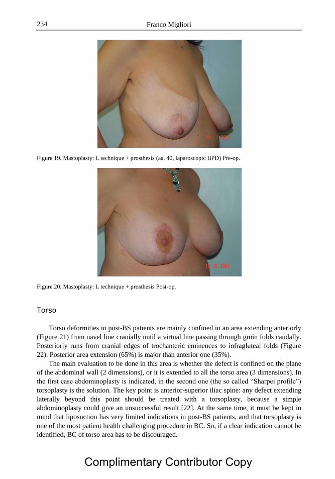
Franco Migliori 234
Figure 19. Mastoplasty: L technique + prosthesis (aa. 40, laparoscopic BPD) Pre-op.
Figure 20. Mastoplasty: L technique + prosthesis Post-op.
Torso
Torso deformities in post-BS patients are mainly confined in an area extending anteriorly
(Figure 21) from navel line cranially until a virtual line passing through groin folds caudally.
Posteriorly runs from cranial edges of trochanteric eminences to infragluteal folds (Figure
22). Posterior area extension (65%) is major than anterior one (35%).
The main evaluation to be done in this area is whether the defect is confined on the plane
of the abdominal wall (2 dimensions), or it is extended to all the torso area (3 dimensions). In
the first case abdominoplasty is indicated, in the second one (the so called “Sharpei profile”)
torsoplasty is the solution. The key point is anterior-superior iliac spine: any defect extending
laterally beyond this point should be treated with a torsoplasty, because a simple
abdominoplasty could give an unsuccessful result [22]. At the same time, it must be kept in
mind that liposuction has very limited indications in post-BS patients, and that torsoplasty is
one of the most patient health challenging procedure in BC. So, if a clear indication cannot be
identified, BC of torso area has to be discouraged.
Complimentary Contributor Copy
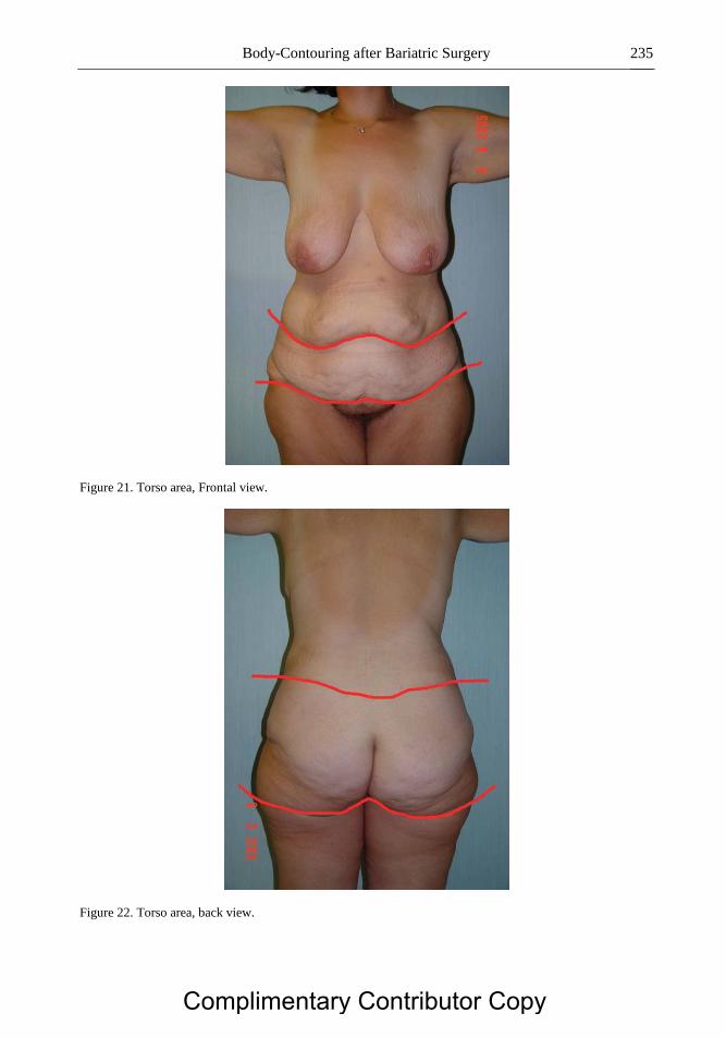
Body-Contouring after Bariatric Surgery 235
Figure 21. Torso area, Frontal view.
Figure 22. Torso area, back view.
Complimentary Contributor Copy

Franco Migliori 236
Abdominoplasty
As a result of the above-described anatomosurgical modifications and of the high
percentage of potential complications, the right abdominoplasty technique for this kind of
patient should be quick and safe. Therefore, the “vest over pants” technique described by
Planas [23, 24] with an inverted V excision, seems to be the safest to preserve a good
abdominal wall blood perfusion. Two technical variations are suggested:
1) Drawing of the upper line of incision is made with patient standing, simulating the
“vest over pants” movement and drawing the lower line. As patient will lie down the
operating table, all the draws will move upwards, underlining a frequent error:
making drawings with patient lying determines cuts in the wrong places (Figure 23-
24). As in all BC, drawings must be made with patient standing.
2) Once lines of excision are well identified, it is advisable to start with lower incision,
to perform the whole undermining and finally to cut the exceeding tissue: often the
extensive BS scars limit abdominal wall elasticity and it is safer to finally check the
amount of excision.
Figure 23. Abdominoplasty: drawings on standing position.
Figure 24. Abdominoplasty: same drawings (Figure 23) on lying position.
Complimentary Contributor Copy

Body-Contouring after Bariatric Surgery 237
If performed on the right cases, abdominoplasty provides nice improvements, mainly on
the lateral view of the abdominal wall (Figure 25-28).
Figure 25. Abdominoplasty: case 1 (♀ aa. 38, BPD) Pre-op.
Figure 26. Abdominoplasty: case 1 Post-op.
Figure 27. Abdominoplasty: case 2 (♀ aa. 42, BPD) Pre-op.
Complimentary Contributor Copy
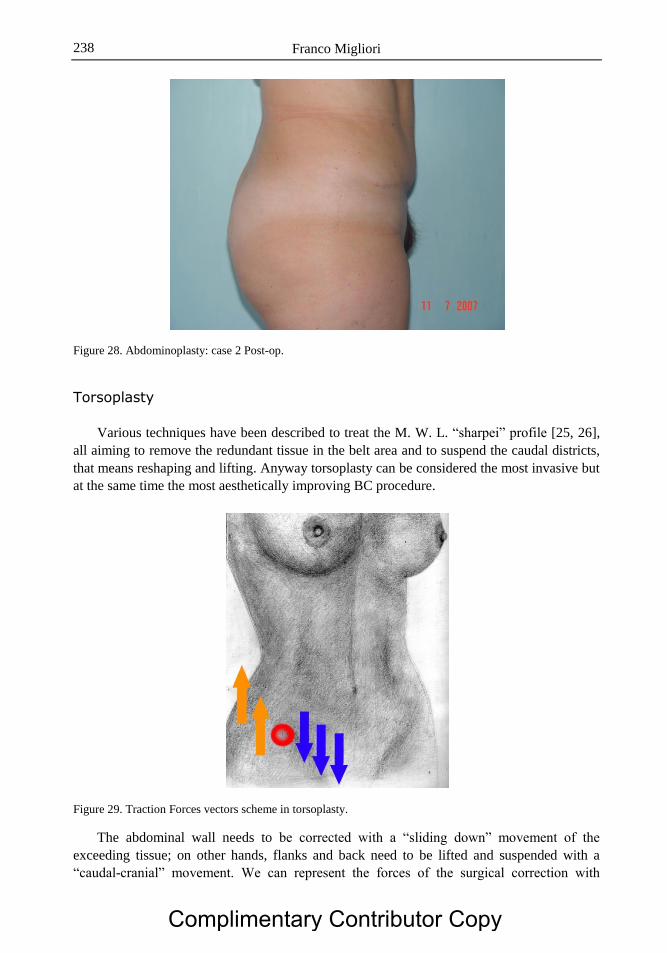
Franco Migliori 238
Figure 28. Abdominoplasty: case 2 Post-op.
Torsoplasty
Various techniques have been described to treat the M. W. L. “sharpei” profile [25, 26],
all aiming to remove the redundant tissue in the belt area and to suspend the caudal districts,
that means reshaping and lifting. Anyway torsoplasty can be considered the most invasive but
at the same time the most aesthetically improving BC procedure.
Figure 29. Traction Forces vectors scheme in torsoplasty.
The abdominal wall needs to be corrected with a “sliding down” movement of the
exceeding tissue; on other hands, flanks and back need to be lifted and suspended with a
“caudal-cranial” movement. We can represent the forces of the surgical correction with
Complimentary Contributor Copy
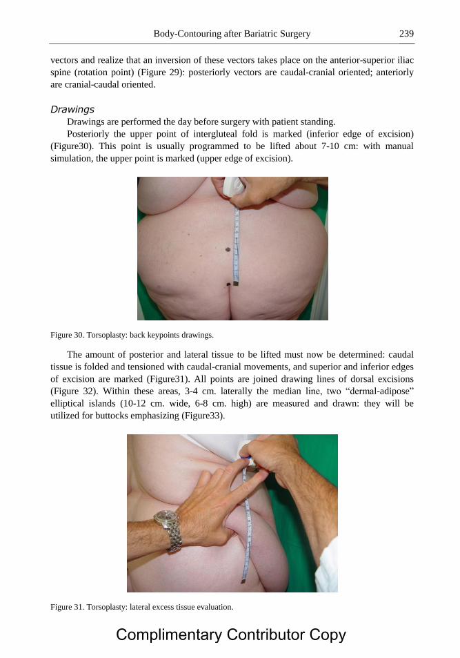
Body-Contouring after Bariatric Surgery 239
vectors and realize that an inversion of these vectors takes place on the anterior-superior iliac
spine (rotation point) (Figure 29): posteriorly vectors are caudal-cranial oriented; anteriorly
are cranial-caudal oriented.
Drawings
Drawings are performed the day before surgery with patient standing.
Posteriorly the upper point of intergluteal fold is marked (inferior edge of excision)
(Figure30). This point is usually programmed to be lifted about 7-10 cm: with manual
simulation, the upper point is marked (upper edge of excision).
Figure 30. Torsoplasty: back keypoints drawings.
The amount of posterior and lateral tissue to be lifted must now be determined: caudal
tissue is folded and tensioned with caudal-cranial movements, and superior and inferior edges
of excision are marked (Figure31). All points are joined drawing lines of dorsal excisions
(Figure 32). Within these areas, 3-4 cm. laterally the median line, two “dermal-adipose”
elliptical islands (10-12 cm. wide, 6-8 cm. high) are measured and drawn: they will be
utilized for buttocks emphasizing (Figure33).
Figure 31. Torsoplasty: lateral excess tissue evaluation.
Complimentary Contributor Copy

Franco Migliori 240
Figure 32. Torsoplasty: lateral excess tissue drawings.
Figure 33. Torsoplasty: buttocks emphasizing island Flaps markings.
Anteriorly, with a mastoplasty–like handling, we determine the upper line of excision.
Lower line is the same of a standard abdominoplasty (Figure34).
Figure 34. Torsoplasty: Frontal excess tissue drawing.
Complimentary Contributor Copy
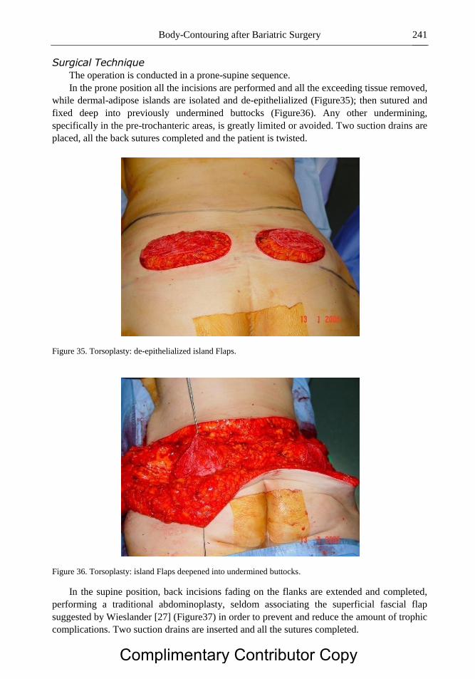
Body-Contouring after Bariatric Surgery 241
Surgical Technique
The operation is conducted in a prone-supine sequence.
In the prone position all the incisions are performed and all the exceeding tissue removed,
while dermal-adipose islands are isolated and de-epithelialized (Figure35); then sutured and
fixed deep into previously undermined buttocks (Figure36). Any other undermining,
specifically in the pre-trochanteric areas, is greatly limited or avoided. Two suction drains are
placed, all the back sutures completed and the patient is twisted.
Figure 35. Torsoplasty: de-epithelialized island Flaps.
Figure 36. Torsoplasty: island Flaps deepened into undermined buttocks.
In the supine position, back incisions fading on the flanks are extended and completed,
performing a traditional abdominoplasty, seldom associating the superficial fascial flap
suggested by Wieslander [27] (Figure37) in order to prevent and reduce the amount of trophic
complications. Two suction drains are inserted and all the sutures completed.
Complimentary Contributor Copy

Franco Migliori 242
Figure 37. Wieslander superficial Fascial Flap.
The exceeding tissue is usually cut and removed into little pieces, and any piece is gently
squeezed before cutting it: this procedure, rather than “en-bloc” excision, seems to reduce
blood losses.
Drapings are performed with non adhesive wound dressings and compressive bands.
Post-Operative Care
When the total amount of excised tissue is over 10-12 kg., one or two days of intensive
care recovery could be necessary. Usually all drains are removed within 6-8 days. Recovery
lasts about 7-10 days.
Compression is removed after one week and a girdle worn day and night for one month,
plus a further two weeks night-time only. If a small fat excess should remain in pre-
trocantheric areas, a minor liposuction can be performed between 6 months and one year after
(if no further body-contouring of thighs is scheduled).
Complications
Hematomas (5,4%) and seromas (3,8%) have dropped thanks to various preventing tricks
and procedures.
The high incidence of abdominal wall complications [28, 29] (skin dehiscence and
necrosis) (Figure 38), up to 45-50%, in this particular group of patients compared with non-
BS patients and the review of anatomical studies [30, 31] has suggested to increase the
research of causes by arteriographic study of the epigastric vessels. In five studied cases, no
reliable modifications have been found. Therefore, without any other explanation, the only
certain cause responsible for almost 70% of trophic complications following abdominoplasty
is the presence of previous scars. In more recent BS cases operated by the laparoscopic
approach (with very short abdominal scars), the percentage of complications has significantly
decreased [32].
Complimentary Contributor Copy

Body-Contouring after Bariatric Surgery 243
Figure 38. Abdominal wall complication (necrosis, 15th day post-op).
Conclusions
All patients achieve a nearly ideal torso contour (Figure 39-46), addressing very
effectively all functional and aesthetical concerns of M. W. L. patients. This BC surgery is,
without a doubt, highly challenging for both surgeon and patient: adequate learning curves,
careful patient selections and education are essential in reaching a good outcome.
Figure 39. Torsoplasty case (♀ aa. 56, BPD): Pre-op Frontal view.
Complimentary Contributor Copy

Franco Migliori 244
Figure 40. Torsoplasty case: Pre-op back view.
Figure 41. Torsoplasty case: Pre-op right view.
Figure 42. Torsoplasty case: Pre-op left view.
Complimentary Contributor Copy

Body-Contouring after Bariatric Surgery 245
Figure 43. Torsoplasty case: Post-op Frontal view.
Figure 44. Torsoplasty case: Post-op back view.
Figure 45. Torsoplasty case: Post-op right view.
Complimentary Contributor Copy

Franco Migliori 246
Figure 46. Torsoplasty case: Post-op left view.
Legs
The leg aesthetical problems in post-BS patients are mainly confined in the thigh and
more or less resume the general problem of all M. W. L. patients: sagginess of body coverage.
The loss of skin elasticity and of fat volume usually causes flabbiness of medial and/or lateral
areas of the thighs.
Except classic lipodystrophy caused by general fat deposits, thigh BC can be very useful
after BS. M. W. L. patients don’t accept the skin excess located at inner and lateral aspects of
the thighs, and the main demand for surgery is aesthetical improvement even if functional
problems can be related too, like frictions and inflammations.
The lateral defects can be corrected with a body-lift [26] or a torsoplasty extended with a
wide caudal undermining. Otherwise a flankplasty can be performed.
The inner thigh flabbiness requires specific surgical technique, the thigh lift. Depending
on the different locations of the defects, the thigh lift can be conducted as:
Semicircular thigh lift
Circular thigh lift
Longitudinal thigh lift
Pursuing the aim of scar shortening, the suggestions of Le Louarn and Pascal [33]
regarding excisions and suture are strongly advisable. To reduce possible lymphatic drainage
complications and seromas, undermining must be avoided, choosing a full-thickness “en-
block” removal of exceeding tissue.
Drawings
The preoperative drawing of the planned procedure is performed with patients standing
and the legs 20-30 cm. abducted, paying great attention to symmetry. The upper line of
incision is marked 1 cm above the inguinal fold, and 2 to 6 cm above the gluteal crease. A
Complimentary Contributor Copy

Body-Contouring after Bariatric Surgery 247
firm upward tension to the tissues to be lifted is applied, evaluating and marking the lower
edge of excision.
Markings may be helped and guided by underwear traces so that the upper line of the
drawing remains above the panty's lower edge.
If circular technique is needed, upper line continues from inguinal fold, turning back
almost at greater trochanter height and joining back upper incision line.
If vertical technique is needed, the sequence described in Figures 47-51 is performed.
Figure 47. Thighplasty:Frontal upper markings.
Figure 48. Thighplasty: back upper markings.
Complimentary Contributor Copy

Franco Migliori 248
Figure 49. Thighplasty: excess tissue evaluation.
Figure 50. Thighplasty: Frontal markings completed.
Figure 51. Thighplaty: Back markings completed.
Complimentary Contributor Copy

Body-Contouring after Bariatric Surgery 249
Surgical Techniques
The flankplasty, as described by Baroudi [34], can be useful in few cases (Figures 52-53),
but usually what can be corrected with it can be done with a torsoplasty as well. It is rare to
find a post-BS patient with isolated lateral thigh defects.
Figure 52. Flankplasty: Intra-op starting.
Figure 53. Flankplasty: Intra-op ending.
Figure 54. Thighplasty: Intra-op starting.
Complimentary Contributor Copy

Franco Migliori 250
Figure 55. Thighplasty: excision completed.
Figure 56. Thighplasty: right thigh suturing.
Figure 57. Thighplasty: right suture completed.
Complimentary Contributor Copy

Body-Contouring after Bariatric Surgery 251
Figure 58. Thighplasty: left thigh suturing.
Figure 59. Thighplasty: Intra-op ending.
The semicircular thigh lift is based on the classical Pitanguy [35], Baroudi [36] and
Lewis [37, 38] techniques.
Operation is performed in lithotomy position, alternatively adducting legs for the suturing
time (Figures 54-59). The incisions cut skin and fat tissue straight down to the fascia, which
is preserved. Removal of exceeding tissue is directly done on the fascia, with no undermining.
Firm and stable suspension is guaranteed by anchoring [33] the superficial fascia system
and the dermis of inferior flap to the lateral band of Colles’ fascia. This will successfully
prevent the two major aesthetical complications: scar caudal displacement and early
recurrence of the defect.
One drain per side is placed, and two layers suture performed to complete procedure.
Light draping is maintained for a few weeks. The use of elastic panty stockings is
recommended for several weeks post-op.
The circular thigh lift, as Baroudi [36] explains, is necessary when redundant tissue
extends to frontal and lateral aspects of the thighs, sagging down to the knees, and the
semicircular technique is not enough. The operation is conducted first in prone position, then
in lithotomy position.
Complimentary Contributor Copy

Franco Migliori 252
The anterior incision is done 2 to 4 cm above the inguinal fold as far as possible medially
and continued laterally inside underwear traces.
The only relevant technical difference with semicircular technique is lateral excision
according to the amount of tissue to be dismissed, with no undermining.
The same type of suture, draping and wearing as for semicircular lift are performed.
The longitudinal thigh lift is rarely indicated and often avoided by surgeons and patients
because of bad reputation of the results. Most deformities can effectively be reduced with the
previous procedures, leaving hidden scars. Nevertheless, some post-BS cases need vertical(or
longitudinal) reduction in order to obtain valuable results, even if bad and visible scars are
expected, anyway better than skin excess. The suggested vertical excision is posteromedial,
running straight to the popliteal region and ending as soon as the redundant tissue ends.
The main steps of the procedure are the same as for previous ones, performing no
undermining as usual. Post-op management doesn’t differ from other procedures.
Complications
General complications are represented by scar distrophies/keloids (22,7%), edema
(14,2%), seromas (11,8%), lymphorrea (7,6%), wound dehiscence (2,9%).
Specific complications include vertical scar widening (37,1%), defect recurrence
after 1 year (12,5%) and scar displacement (5%).
Conclusion
Although the characteristics of post-BS patients limit choices and chances, satisfying
results are achieved not only as cosmetic correction but also in quality of life, because in this
anatomical area functional improvements are mostly accomplished by BC (Figures 60-67).
Figure 60. Thighplasty: case 1 (♀ aa. 45, BPD)semicircular, Pre-op.
Complimentary Contributor Copy

Body-Contouring after Bariatric Surgery 253
Figure 61. Thighplasty: case 1 Post-op.
Figure 62. Thighplasty: case 2 (♀ aa. 38, BPD) semicircular, Pre-op.
Complimentary Contributor Copy

Franco Migliori 254
Figure 63. Thighplasty: case 2 Post-op.
Figure 64. Thighplasty: case 3 (♀ aa. 31, BPD) longitudinal, Front Pre-op.
Complimentary Contributor Copy

Body-Contouring after Bariatric Surgery 255
Figure 65. Thighplasty: case 3 back Pre-op.
Figure 66. Thighplasty: case 3 Front Post-op.
Complimentary Contributor Copy

Franco Migliori 256
Figure 67. Thighplasty: case 3 back Post-op.
(A)
Figure 68. (Continued).
Complimentary Contributor Copy

Body-Contouring after Bariatric Surgery 257
(B)
Figure 68. A) B. U. T. Frontpage, B) B. U. T. back page.
Outcome
Great “positive” thinking of these patients towards BC creates enormous expectations for
improvement in quality of life (similar to the expectations which motivated these patients
toward bariatric surgery) [39]. Of these operated patients, 99% declare themselves completely
satisfied with the obtained results and go on to further body contouring. These kinds of results
are not achievable by bariatric surgery alone and/or by eating behavior normalization, and
have been tested with the Body Uneasiness Test (B. U. T.) [40] (Figure 68) on 5 different
groups of these patients. On average, 2,5 operations have been performed on each patient.
Complimentary Contributor Copy

Franco Migliori 258
The main points of patient satisfaction are: “body image” greatly improved, self-esteem
recovery, improvement in life relationships, and sexual life resumption or improvement [41].
References
[1] Singh D., Forte A., Loerke C., Bell R., Thomson J. G. “P63: Prognostication of Body
Contouring after Bariatric Surgery” –Plast. Reconstr. Surg. Abstract Supplement.
118(4) Supplement:144, September 15, 2006.
[2] O'Connell J. B. “Bariplastic Surgery” –Plast. Reconstr. Surg. 113(5):1530, April 15,
2004.
[3] Huemer G. M., Dunst K. “Massive Weight Loss after Bariatric Surgery: Friend or
Foe?” –Plast. Reconstr. Surg. 114(6):1663-1664, November 2004.
[4] Baroudi R., Moraes M. “Philosophy, Technical Principles, Selection, and Indication in
BodyContouring Surgery” –Aesth. Plast. Surg. 15:1-18, 1991.
[5] Folope V., Coëffier M., Déchelotte P. “Nutritional deficiencies associated with bariatric
surgery” – Gastroenterol. Clin. Biol. 2007 Apr; 31(4):369-77.
[6] Shermak M. A., Chang D. C., Heller J. “Factors impacting thromboembolism after
bariatric body contouring surgery” – Plast. Reconstr. Surg. 2007 Apr 15;119(5):1590-6.
[7] Guerrero-Santos J. “Brachioplasty” – Aesth. Plast. Surg. 1979; 3: 1-4.
[8] Strauch B,Greenspun D, Levine J et al. “A techniqueof brachioplasty” – Plast.
Reconstr. Surg. 2004; 113: 1044-8.
[9] Bolivar de Souza Pinto E., Erazo P., Matsuda C. A. “Brachioplasty technique with the
use of molds” – Plast. Reconstr. Surg. 105:1854, 2.
[10] Lockwood T. “Brachioplasty with superficial fascial system suspension” – Plast.
Reconstr. Surg. 96:912, 1995.
[11] Goddio A. S. “A New Technique for Brachioplasty”–Plast. Reconstr. Surg. 84(1):85-
91, July 1989.
[12] Mahjouri F. “A Technique in Brachioplasty” – Plast. Reconstr. Surg. 116(2):678-679,
August 2005.
[13] Abramson D. L. “Minibrachioplasty: Minimizing Scars While Maximizing Results” –
Plast. Reconstr. Surg. 114(6):1631-1634, November 2004.
[14] Teimourian B., Malekzadeh S. “Rejuvenation of the Upper Arm” – Plast. Reconstr.
Surg. 102(2):545-551, August 1998.
[15] Brink R. “Management of true ptosis of the breast” – Plast. Reconstr. Surg. 91: 657,
1993.
[16] Benelli L. “A New PeriareolarMammaplasty: the “Round Block” Technique” – Aesth.
Plast. Surg. 14:93-100, 1990.
[17] Migliori F. “Upside-Down Augmentation Mastopexy” – Aesth. Plast. Surg. 35:593-
600, 2011.
[18] Càrdenas-Camarena L., Ramìrez-Macìas, R. “Augmentation/Mastopexy: How to Select
and Perform the Proper Technique” – Aesth. Plast. Surg. 30:21-33, 2006.
Complimentary Contributor Copy

Body-Contouring after Bariatric Surgery 259
[19] Hammond D. C., Khuthaila D. K., Kim J. “The Interlocking Gore-Tex Suture for
Control of Areolar Diameter and Shape” – Plast. Reconstr. Surg. 119(3):804-809,
March 2007.
[20] De Longis E. “Mammoplasty with an L-shaped limited scar and retropecto-
raldermopexy” – Aesth. Plast. Surg. 10:171-175, 1986.
[21] Arié G. “Una nuevatéchnica de mastoplastia” – Rev. Lat. Am. Cir. Plast. 3:22, 1957.
[22] Matarasso A. “Liposuction as an Adjunct to a Full Abdominoplasty Revisited” – Plast.
Reconstr. Surg. 106(5):1197-1202, October 2000.
[23] Planas J. “The "Vest Over Pants" abdominoplasty” – Plast. Reconstr. Surg. 61(5):694-
700, May 1978.
[24] Planas J., Bisbal J., del Cacho C., Palacin J. M. “Further advantages of the "Vest over
pants" abdominoplasty” – Aesth. Plast. Surg. 12:123-127, 1988.
[25] Lockwood T. E. “Lower body lift with superficial fascialsystem suspension” – Plast.
Reconstr. Surg. 1993; 92: 1112-6.
[26] Pascal J. F., Le Louarn C. “Remodelling body-lift withhigh lateral tension” – Aesth.
Plast. Surg 2002; 26:223-30.
[27] Wieslander J. B. “Preservation of fat under Scarpa’s fascia in the lower abdomen
reduces complications inabdominoplasties” - 10th
Congress of the European Society of
Plastic, Reconstructive and Aesthetic SurgeryE. S. P. R. A. S (Sept. 1st 2005).
[28] Nurkim M. V. et al. “Incidence of hematoma and seromain abdominoplasty with and
without the use of drains” – Rev. Soc. Bras. Cir. Plast. 2002; 17: 69-74 (abstract).
[29] vanUchelen J. H., Werker P. M. N., Kon M. “Complications of abdominoplasty in 86
patients” – Plast. Reconstr. Surg. 2001; 107: 1869-73.
[30] Huger W. E. Jr. “The anatomic rationale for abdominal lipectomy” – Ann. Surg. 1979;
45: 612-7.
[31] Hester T. R. Jr., Nahai F., Beegle P. E. et al. “Blood supply of the abdomen revisited,
with emphasis on the superficial inferior epigastric artery” – Plast. Reconstr. Surg.
1984; 47: 657-70.
[32] Scopinaro N., Marinari G. M., Camerini G. “Laparoscopic standard biliopancreatic
diversion:technique and preliminary results” – Obes. Surg. 2002;12: 362-5.
[33] Le Louarn C., Pascal J. F. “The concentric medial thighlift” – Aesth. Plast. Surg. 2004;
28: 20-23.
[34] Baroudi R., Moraes M. “Philosophy, Technical Principles, Selection, and Indication in
BodyContouring Surgery” – Aesth. Plast. Surg. 15:1-18, 1991.
[35] Pitanguy I. “Surgical reduction of the abdomen, thigh and buttocks” – Surg. Clin.
North. Am. 51:479, 1971.
[36] Regnault P., Baroudi R., da SilveiraCarvalho C. G. (1979). “Correction of Lower Limb
Lipodystrophy” –Aesth. Plast. Surg. 3:233-249.
[37] Lewis J. R. “An Atlas of Aesthetic Plastic Surgery” – 285-288 Little, Brown and Co.,
Boston,1973.
[38] Lewis J. R. “Correction of ptosis of the thighs: the thigh lift” – Plast. Reconstr. Surg.
37:494, 1966.
Complimentary Contributor Copy

Franco Migliori 260
[39] Bolton M., Pruzinsky T., Cash T. et al. (2003). “Measuring outcomesin plastic surgery:
body image and quality oflife in abdominoplasty patients” – Plast. Reconstr. Surg. ;
112: 619-25
[40] Pecori L., Serra Cervetti G. G., Marinari G. M., Migliori F., Adami G. F. “Attitudes of
morbidly obese patients to weight loss and body image following bariatric surgery and
body contouring” – Obes. Surg. 2007 Jan; 17(1) 68-73.
[41] Migliori F. et al. (2006). “Body contouring after biliopancreatic diversion” – Obes.
Surg. Dec; 16(12): 1638-44.
Complimentary Contributor Copy

In: Bariatric Surgery ISBN: 978-1-62618-995-9
Editor: Christos R. Iavazzo © 2013 Nova Science Publishers, Inc.
Chapter XIV
Bariatric Brachioplasty
Miguel Modolin, Wilson Cintra Jr.
and Rodrigo Itocazo Rocha Division of Plastic Surgery – Hospital das Clínicas –
University of Sao Paulo School of Medicine, Brazil
Introduction
Dysmorphism in the arms, following weight loss, is caused by the loss of fat content,
limited skin retraction ability resulting from smaller amounts of collagen and elastin, and
laxity due to distension of the superficial fascia.
Thus, the arms lose their cylindrical contour due to a skin fold that may extend from the
elbow to the lateral thoracic wall, going beyond the axilla, thus characterizing a deformity
known as “bat wing” (Figure 1).
This is treated surgically by brachioplasty to reduce volume thus enabling a wider range
of motion, a more aesthetic looking arm contour, the use of sleeveless clothes and, ultimately,
a better quality of life.
Surgical Technique
When the excess skin is restricted to the arm, it is resected by means of a fusiform
excision in the medial aspect from the elbow which is combined with a transverse ellipse in
the axilla thus defining the shape of a fish tail (Figure 3). If the excess skin extends to the
thorax, the elliptical marking is prolonged to this region; in the axilla, a Z-plasty is performed.
Resection is made above the superficial fascia.
Complimentary Contributor Copy

Miguel Modolin, Wilson Cintra Jr. and Rodrigo Itocazo Rocha 262
Results
Despite the long scar, patients show great satisfaction because of the improvement in
their quality of life through better hygienic care, wider range of motion, a more aesthetic
looking arm contour, and the freedom to wear sleeveless clothes which was previously
embarrassing because of their arm deformity.
Complications and Events
Complications are due to the failure to observe an accurate surgical technique, whereas
events result from adverse biological factors. Events:
1. Edema and seroma;
2. Ecchymosis;
3. Keloid scar.
Complications:
1. Hematoma;
2. Dehiscence;
3. Limphorrhea and lymphocele;
4. Necrosis;
5. Hypertrophic scar;
6. Infection;
7. Paresthesia.
Bariatric Brachioplasty
Introduction
Bariatric surgeries, as a rule, lead to massive weight loss and marked changes in body
contour, which assumes bizarre shapes.
a
Figure 1. (Continued).
Complimentary Contributor Copy
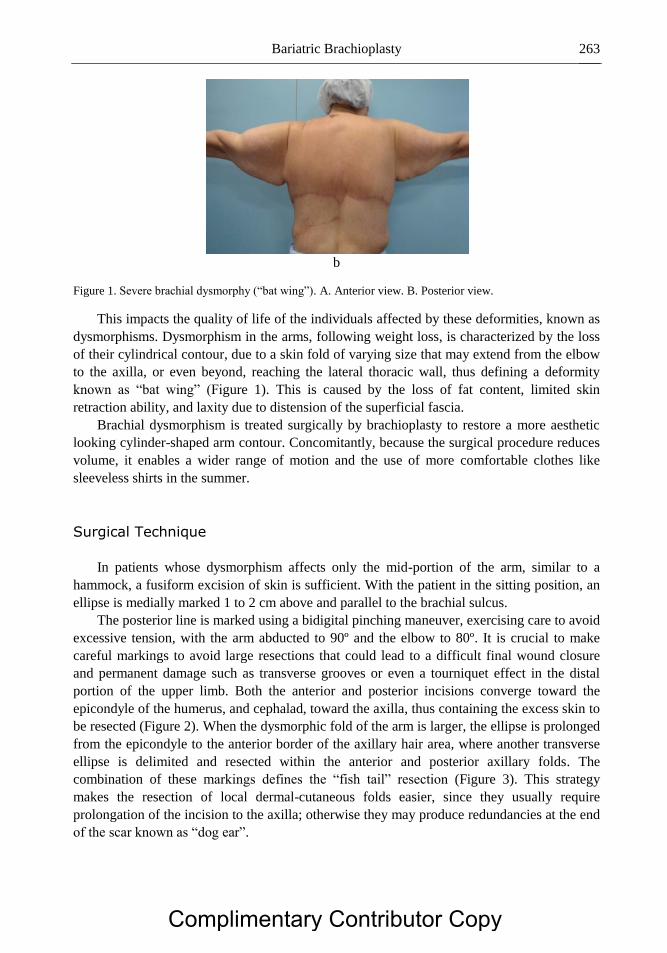
Bariatric Brachioplasty 263
b
Figure 1. Severe brachial dysmorphy (“bat wing”). A. Anterior view. B. Posterior view.
This impacts the quality of life of the individuals affected by these deformities, known as
dysmorphisms. Dysmorphism in the arms, following weight loss, is characterized by the loss
of their cylindrical contour, due to a skin fold of varying size that may extend from the elbow
to the axilla, or even beyond, reaching the lateral thoracic wall, thus defining a deformity
known as “bat wing” (Figure 1). This is caused by the loss of fat content, limited skin
retraction ability, and laxity due to distension of the superficial fascia.
Brachial dysmorphism is treated surgically by brachioplasty to restore a more aesthetic
looking cylinder-shaped arm contour. Concomitantly, because the surgical procedure reduces
volume, it enables a wider range of motion and the use of more comfortable clothes like
sleeveless shirts in the summer.
Surgical Technique
In patients whose dysmorphism affects only the mid-portion of the arm, similar to a
hammock, a fusiform excision of skin is sufficient. With the patient in the sitting position, an
ellipse is medially marked 1 to 2 cm above and parallel to the brachial sulcus.
The posterior line is marked using a bidigital pinching maneuver, exercising care to avoid
excessive tension, with the arm abducted to 90º and the elbow to 80º. It is crucial to make
careful markings to avoid large resections that could lead to a difficult final wound closure
and permanent damage such as transverse grooves or even a tourniquet effect in the distal
portion of the upper limb. Both the anterior and posterior incisions converge toward the
epicondyle of the humerus, and cephalad, toward the axilla, thus containing the excess skin to
be resected (Figure 2). When the dysmorphic fold of the arm is larger, the ellipse is prolonged
from the epicondyle to the anterior border of the axillary hair area, where another transverse
ellipse is delimited and resected within the anterior and posterior axillary folds. The
combination of these markings defines the “fish tail” resection (Figure 3). This strategy
makes the resection of local dermal-cutaneous folds easier, since they usually require
prolongation of the incision to the axilla; otherwise they may produce redundancies at the end
of the scar known as “dog ear”.
Complimentary Contributor Copy

Miguel Modolin, Wilson Cintra Jr. and Rodrigo Itocazo Rocha 264
a
b
Figure 2. Fusiform technique. A. Drawing of the fusiform marking. B. Preoperative marking in a patient.
In patients whose deformity extends to the lateral thoracic wall, the elliptical marking is
prolonged to this region containing the excess dermal-cutaneous fold. In the axilla, the wound
closure line is decomposed using Z-plasty (Figure 4). The patient is placed lying in the supine
position with his arms abducted upward, thus facilitating the surgical approach (Figure 5). In
order to reduce the operative time, the procedure is performed by two teams simultaneously.
The surgery is performed under general anesthesia and infiltration of the surgical area with
saline solution containing epinephrine to minimize bleeding. Prophylactic antibiotic therapy
with cephazolin is given during the anesthetic induction. Brachioplasty should be performed
above the superficial fascia to avoid affecting the lymphatic vessels and nerves that run in the
deeper plane. Plane closure is initiated after excision and careful hemostasis with
electrocautery. In patients undergoing the axillary approach, the superficial fascia of the arm
is fixed to the axillary fascia, thus promoting suspension of the posterior portion of the arm by
means of an interrupted suture using 4-0 polyamide (Figure 6). This works as an arm sling
that ensures tension of the tissues distended by obesity, which did not retract after weight
loss. Wound closure in the deep subcutaneous plane, including the superficial fascia, is made
with an interrupted suture using 4-0 polyamide.
Complimentary Contributor Copy

Bariatric Brachioplasty 265
a
b
Figure 3. Double-ellipse technique. A. Drawing of the “fish tail” marking. B. Preoperative marking in a
patient.
It is worth stressing that when over-resection occurs, closure of this plane – important for
reestablishing the cylindrical shape of the arm – may determine transverse depressions that
are difficult to correct. A running intradermal suture using 4-0 or 5-0 polyglecaprone is the
final suture.
Following wound closure, Z-plasty should be performed in the middle of the axilla, in
those cases in which the incision crossed it. It could be argued that this maneuver is
unnecessary given that there is remaining skin; however, precisely because of it, Z-plasty is
mandatory to define the axillary dome.
Another argument against Z-plasty is the transposition of a hairless area into a hair area
and vice-versa. Nonetheless, we should consider that Z-plasty is performed in the middle of
the axilla after considerable resection of hairy skin, making this transposition minimally
perceptible, especially in female patients who are familiar with hair removal. Moreover, Z-
plasty clearly prevents scar contracture (Figure 7).
Wound closure should be performed as fast as possible. In more extensive cases the
temporary use of staples is recommended, later replaced by definitive suture. This reduces the
edema, which can further make wound closure more difficult and frequently leads to ischemia
in the wound area or to a tourniquet effect in the arm.
Complimentary Contributor Copy

Miguel Modolin, Wilson Cintra Jr. and Rodrigo Itocazo Rocha 266
a
b
c
d
Figure 4. Prolonged ellipse technique and Z-plasty. A. Drawing of markings. B. After Z-plasty. C.
Preoperative marking in a patient. D. Immediate postoperative period showing Z-plasty.
Complimentary Contributor Copy

Bariatric Brachioplasty 267
Figure 5. Drawing showing the intraoperative positioning of the upper limbs.
Figure 6. Fixation of the superficial fascia of the arm flaps to the axillary fascia.
Figure 7. Good postoperative (1 year) scar appearance resulting from brachioplasty extended to the thorax.
Complimentary Contributor Copy

Miguel Modolin, Wilson Cintra Jr. and Rodrigo Itocazo Rocha 268
The dressing is made with sterile micropore adhesive tape woven in a latticework pattern
on the surgical wound protected by gauze greased with bactericidal ointment. Drains are not
necessary since careful hemostasis is performed and the suture leaves no dead spaces.
Compressive wrapping is not routinely used; however, the use of light compression meshes
applied from the wrist to the thorax is recommended in larger resections. Meshes that
compress only the arm should be avoided because their tourniquet effect may cause distal
edema thus affecting the blood flow in the forearm and hand.
Results
Brachioplasty performed in patients after massive weight loss is extremely rewarding
(Figures 8 and 9). A long scar is the price to pay for a rounder and more aesthetic looking arm
contour. Nonetheless, former obese patients eligible for plastic surgeries receive thorough
information regarding the scars that are to be expected; accordingly, they willingly accept
those resulting from brachioplasty. It should be pointed out that the scar resulting from
resection within the lines marked – as previously described – is located in the posterior
portion of the arm.
a
b
Figure 8. (Continued).
Complimentary Contributor Copy

Bariatric Brachioplasty 269
c
d
Figure 8. “Fish-tail” brachioplasty showing an improved arm contour. A-B. Preoperative period. C-D.
Postoperative period.
Thus, it can hardly be noticed when the upper limb is adducted. Brachioplasty provides
remarkable personal satisfaction since it improves the quality of life by allowing more
effective hygiene and a wider range of motion of the upper limbs. In many aspects it is
extremely rewarding for women since it allows them to wear sleeveless clothes, previously
embarrassing because of their arm deformities.
Complications
When brachioplasty is performed by well-trained hands, complications are not very
significant. On the other hand, even when well-performed, adverse biological factors may be
present that do not depend on the technical accuracy. These are known as events, but are
usually not severe and are easy to solve.
The most common are edema and seroma. Both result from surgical damage, and it
should be pointed out that the greater the damage the more severe the events. Edema usually
resolves within approximately 30 days, and is further improved by upper limb massage, also
Complimentary Contributor Copy

Miguel Modolin, Wilson Cintra Jr. and Rodrigo Itocazo Rocha 270
known as lymphatic drainage. Seroma is caused by a local accumulation of post-traumatic
inflammatory exudate. As a rule, drainage sessions under strict asepsis will solve the problem.
Naturally, ecchymosis may also be listed as an event following manipulation and surgical
damage; therefore, soft and delicate maneuvers are recommended to prevent them.
a
b
c
Figure 9. (Continued).
Complimentary Contributor Copy

Bariatric Brachioplasty 271
d
Figure 9. Brachioplasty using the extended ellipse technique. A-B. Preoperative period. C-D. Postoperative
period.
The hematomas which result from failure to observe careful hemostasis techniques are
more severe. When detected early, they should be drained; recurrence imposes surgical
exploration to identify the bleeding vessel and proceed to correct hemostasis. When left
inadequately treated, hematomas may lead to solid cysts that cause unattractive elevations in
the arm and are sometimes accompanied by pain, because of the compressive effect on
subjacent surfaces.
Dehiscence is a complication resulting from tension on the suture, as seen in over-
resections. Fortunately, dehiscences are partial and sometimes occur even without suture
tension. To understand this complication, it is important to remember that the skin in the
medial aspect of the arm is extremely thin. The suture may not stand the tension of post-
traumatic edema, causing the wound to break open. A partial dehiscence heals by second
intention in a short period of time, provided it is cleansed periodically, i. e., two to three times
a day. Larger dehiscences require skin grafts.
Lymphorrhea is a very disagreeable complication. Exeresis made in a deeper plane below
the superficial fascia may lead to damage of lymphatic vessels and subsequent lymphorrhea.
Lymph leakage always occurs through the most distal portion of the incision, close to the
epicondyle. Lymphorhea starts within the first days of the postoperative period and
spontaneously disappears within 30 days. On rare occasions, this lymphorrhea may
accumulate, thus forming a cyst called lymphocele. This complication requires puncture and
drainage sessions; however, the best option is surgical removal of the cyst, maintaining
suction drainage for two to three days.
Infection is an uncommon event, thanks to prophylactic measures. Some of these may
include exhaustive and thorough preoperative cleansing of the entire upper limb using
antiseptic soaps and plenty of water, a procedure that should be repeated several times.
Axillary hair removal should be performed after cleansing, approximately 30 minutes prior to
surgery. Hair removal by means of creams or wax are contraindicated, because these products
can occlude the hair follicle ostium, thus leading to formation of cysts within which bacterial
colonization will occur; this may infect the site and result in infection. Local infections may
determine dehiscence or abscess formation. In the most severe cases, surgical debridement is
Complimentary Contributor Copy

Miguel Modolin, Wilson Cintra Jr. and Rodrigo Itocazo Rocha 272
indicated. Use of antibiotics is vital and should target pathogens, as determined by culture and
antibiogram tests. Infections more frequently occur in the axilla; they usually affect small
areas and resolve with cleansing and proper antibiotic therapy.
Skin necrosis may occur, whether due to excessive tension or to infection. In these
situations, the necrotic area is of varying size. Nonetheless, it requires careful resection, but
resection of tissues of doubtful vitality should be avoided, even if this may result in an
additional debridement procedure. In smaller areas, wound healing occurs by second
intention, whereas in larger areas, skin grafting is mandatory on the raw area that remains
after debridement. In both cases, the outcome is poor because the aesthetic appearance is
affected.
Despite its size, the scar resulting from brachioplasty is well accepted by the patients.
This scar may be deformed by hypertrophy or, in more severe cases, it may turn into a keloid
scar.
Hypertrophic scars may result from suture tension. However, we should remember that
incisions are made contrary to the relaxed skin tension lines of the arm, thus favoring
hypertrophic wound healing. This is why scar massage with triamcinolone creams and the use
of silicone sheeting are recommended, even when this event is only remotely suspected. Once
the hypertrophic scar has formed, a scar resection may be performed 6 months to 1 year later,
followed by the use of triamcinolone creams plus silicone sheeting. A keloid scar is
genetically related and more common in individuals with black or yellow skin color. It is a
pathological wound healing that looks very unsightly and has symptoms of pain and itching.
To date, the best approach is resection followed by radiation therapy.
If deeper planes of dissection are reached, neurological changes may occur, the most
common of which are sensory changes in the anteromedial aspect of the forearm. This can
also result from the excessive and inadvertent use of electrocautery that leads to damage of
the anteromedial cutaneous nerve of the forearm. The nerve may also be lodged in the deep
plane closure, and this leads to severe pain. In the first situation, sensitivity is reestablished
spontaneously after 4 to 6 months. In the second, an additional surgical approach is necessary
to remove the nodule lodging the nerve.
References
[1] Aly A., Pace D., Cram A. Brachioplasty in the patient with massive weight loss. Aesth.
Surg. J., 26(1): 76-84, 2006.
[2] Baroudi R. Dermolipectomy of the upper arm. Clin. Plast. Surg., 2:485, 1975.
[3] Baroudi R. Braquioplastia in Baroudi R; Cirurgia do Contorno Corporal. Ed. Indexa,
Rio de Janeiro, Brasil, pág. 60-84, 2009.
[4] Hurwitz DJ., Holland SW. The L brachioplasty: an innovative approach to correct
excess tissue of the upper arm, axilla and lateral chest. Plast Reconstr Surg. 117(2):
403-11; discussion, 412-3, 2006.
[5] Lockwood T. Brachioplasty with superficial fascial system suspension. Plast. Reconstr.
Surg., 96(4): 912-20, 1995.
Complimentary Contributor Copy

Bariatric Brachioplasty 273
[6] Modolin M., Cintra Jr W., Faintuch J., Silva M M., Moraes Luciana B., Gemperli R.,
Ferreira MC: Improved double-fuse technique for post-bariatric brachioplasty. Rev.
Col. Bras. Cir., 38(4): 217-222, 2011.
[7] Orpheu SC., Coltro PS., Scopel GP., Gomez DS., Rodrigues CJ., Modolin ML. et al.:
Collagen and elastic content of abdominal skin after surgical weight loss. Obes. Sur.,
20(4):480-6, 2010.
[8] Pitanguy I. Correction of lipodysthophy of the lateral thoracic aspect and inner of the
arm and elbow in dermosenescence. Clin. Plast. Surg., 2(3): 477-83, 1975.
[9] Regnault P. Brachioplasty, Axilloplasty and Pre-axilloplasty. Aesth. Plast. Surg., 7:31-
36, 1983.
[10] Samra S., Sawh-Martinez R., Liu YJ., Samra F., Persing JA: Optimal placement of
brachioplasty scar: a survey evaluation. Plast. Reconstr. Surg., 126(suppl 4S):77
(Abstract), 2010.
[11] Strauch B., Greenspun D., Levine J., Baum T: A technique of brachioplasty. Plast
Reconstr Surg. 113(3): 1044-8, discussion, 1049, 2004.
[12] Symbas JD., Losken A: An outcome analysis of brachioplasty techniques following
massive weight loss. Ann. Plast. Surg., 64(5): 588-91, 2010.
Complimentary Contributor Copy

Complimentary Contributor Copy

In: Bariatric Surgery ISBN: 978-1-62618-995-9
Editor: Christos R. Iavazzo © 2013 Nova Science Publishers, Inc.
Chapter XV
Pregnancy Complications after
Bariatric Surgery Procedures
Christos Iavazzo Iaso Maternity Hospital, Athens, Greece
Abstract
Introduction: The application of bariatric surgery has recently spread among obese
women including women of reproductive age. This chapter aims to clarify the role of
bariatric surgery in gestational diabetes, hypertensive disorders during pregnancy , as
well as neonate’s development and birth weight. Moreover, the diet, monitoring and
complications of such women during pregnancy are also discussed. A question is also
raised whether conception should be postponed or not for at least 18 months after
bariatric operation to avoid complications concerning both mother and fetus mainly
associated with nutritional deficiencies due to the anatomical and physiological changes
of such operations.
Introduction
The World Health Organization characterizes obesity a global health problem with a
higher prevalence in women than men. The USA National Institute of Health recommends
bariatric surgery for obese patients with a body mass index (BMI) of at least 40 kgr/m2 and/or
patients with a BMI 35 kgr/m2 with coexisting commorbidity such as diabetes mellitus or
sleep apnea [1]. In the UK, between 2000–2003 approximately 35% of the women who died
in pregnancy, childbirth, or in the postpartum period were obese, while 30% of mothers who
had a stillbirth or neonatal death were obese [2]. Moreover, it is known that obese women
have elevated risk of preterm labour, miscarriage and fetal chromosomal anomalies, as well
as macrosomia [3]. Thromboembolism, gestational diabetes, pregnancy-induced
hypertension, and preeclampsia are also more common entities in those women [4]. They also
Complimentary Contributor Copy

Christos Iavazzo 276
have a greater incidence of dysfunctional labour, caesarian section and postpartum
hemorrhage, as well as associated perioperative morbidity [4].
Bariatric surgery is the most effective treatment for obesity, resulting in considerable
weight loss and improvement in many comorbidities [5]. The total number of bariatric
surgery operations has increased during the last decade among obese women of reproductive
age. Women aged 18-45 account for 83% of all bariatric surgery procedures [6]. There is
increasing evidence to suggest that weight loss after bariatric surgery can improve maternal
and perinatal outcomes. The most widely applied procedures of bariatric surgery are the
laparoscopic gastric banding and the Roux-en-Y operation. Women who conceive after
bariatric surgery usually seem to have uncomplicated pregnancies, but special attention is
required to some complications that can occur after these procedures. Physicians must have
concerns regarding the safety of pregnancy after bariatric surgery as the number of bariatric
surgeries being performed in women is increased.
The aim of this chapter is to discuss the related problems during pregnancy in women
who have undergone bariatric surgery.
Discussion
A careful review of the literature suggests that the optimum way to reduce obesity-
associated morbidity is by weight reduction before pregnancy [7]. Bariatric surgery is a
popular, safe and effective treatment for obesity. Weight reduction has been shown to change
the hormonal imbalance of the oligomennorheic, hyperinsulinemic and hyperandrogenemic
obese woman leading to an improved menstrual regularity, ovulation, and an increased
fecundity by normal or IVF procedures [8, 9]. In a prospective study of 17 obese patients with
PCOS, a decreased hirsutism and blood levels of androgens, as well as menstrual regularity
were found in all women after bariatric surgery [10].
Bariatric surgery results in many anatomic and physiologic changes in the abdomen and
consequently affects future pregnancies causing perinatal complications associated with
nutritional deficiencies. Common complications encountered are iron, follate, calcium,
vitamin A, vitamin B12 and vitamin K deficiencies that are causatively related with
gestational complications [11, 12]. It was shown that women with history of bariatric surgery
had iron deficiency in 80% of the pregnancies, vitamin D in 46,7%, vitamin A in 20%,
vitamin E in 13,3% and vitamin B12 in 26,7% (13). Obese women having undergone a
bariatric operation before pregnancy usually face anemia problems, for this reason iron and
multivitamin supplements should be prescribed in such women [14]. Less common
complications during pregnancy are hyperemesis, intestinal hernias, strangulation of upper
jejunum [15], hypoxic hepatitis [16]. Fetal cerebral hemorrhage caused by vitamin K
deficiency [17] and neural tube defects due to follate deficiency [18] are also serious fetal
complications presented in the literature.
Although, it is suggested that women should delay pregnancy for at least one
postoperative year, in a recent review by Karmon et al. it was suggested that future studies
should examine the safety of earlier postoperatively programmed pregnancies [19]. Sheiner et
al. showed that women who conceived the first postoperative year had no significant
differences in hypertensive disorders (15. 4% in the early vs 11. 2% in the late postoperative
Complimentary Contributor Copy

Pregnancy Complications after Bariatric Surgery Procedures 277
pregnancy; P = . 392); diabetes mellitus (11. 5% vs 7. 3%; P = . 392); perinatal outcomes,
such as congenital malformations (1. 9% vs 1. 3%; P = . 485); or bariatric complications (6.
7% vs7. 0%; P = . 392) [20] Moreover,, our findings support such a proposal [21]. The weight
loss and marked reduction in food intake following bariatric surgery does not lead to growth
or development problems for the neonate [22], however weight stabilization is strongly
recommended after bariatric surgery, before a pregnancy occurs [23]. Obstetricians should
cooperate with surgeons in order to achieve a close postoperative monitoring and control of
weight gain and appropriate nourishment [24].
According to recent reports gestational complications were less common between women
who underwent bariatric surgery. Ducarme et al. showed that among obese women, the
incidence of adverse outcomes was less in those who underwent gastric banding than in those
who did not [25]. In a recent review [26], it has been shown that pregnancy after Roux-en-Y
gastric bypass is safe with an incidence of perinatal complications similar to that of non obese
patients and lower than that of obese patients who had not undergone bariatric surgery.
Furthermore, in another study no differences in neonatal outcomes were found in pregnant
women after bariatric surgery [27].
More analytically, in a recent review, Magdaleno et al. showed an improved fertility and
a reduced risk of gestational diabetes, pregnancy-induced hypertension and preeclampsia,
macrosomia in pregnant women after bariatric surgery. However, the incidence of IUGR and
SGA infants were found to be elevated. In this study, no conclusions concerning the risk for
cesarean delivery and the best surgery-to-conception interval could be drawn [28].
Belogolovkin et al. in a retrospective cohort study showed that non-obese mothers with
bariatric surgery history were more likely to have anemia, chronic hypertension, endocrine
disorders, and SGA infants, whereas obese mothers without prior bariatric surgery were at
greater risk of having gestational diabetes, chronic hypertension, macrosomic infants, as well
as prolonged hospital stay compared to non-obese mother without prior bariatric surgery [29].
Weintraub et al. in a retrospective study showed a significant reduction in gestational diabetes
rates (17. 3% versus 11. 0%; P = 0. 009) and hypertensive disorders in pregnancy (23. 6%
versus 11. 2%; P = 0. 001) of women with a prior history of bariatric surgery [12]. Similarly,
another study showed that such women had substantially lower rates of preeclampsia and
eclampsia (odds ratio 0. 20, 95% confidence interval 0. 09 to 0. 44), chronic hypertension
complicating pregnancy (0. 39, 0. 20 to 0. 74), and gestational hypertension (0. 16, 0. 07 to 0.
37), even after adjustment for age, multiple pregnancy, surgical procedure, and preexisting
diabetes [30). In different studies, a reduction of fetal macrosomia was shown in pregnant
women with bariatric surgery especially after Roux-en-Y gastric [12, 31]. However, such
procedures could cause nutritional deficiencies which might be the cause of an increased rate
of IUGR or SGA infants in post-bariatric surgery patients compared with controls [25, 31].
The time period between bariatric surgery and pregnancy has not been shown to affect birth
weight. Regarding miscarriage rates, a decline of 33. 3% to 7. 8% was shown in women with
a history of bariatric surgery [9], while no such correlation was found for preterm labor rates
in women with or without bariatric surgery matched for BMI [32, 33]. Similar perinatal
mortality rate, as well as congenital malformations rates were found comparing all
pregnancies of patients with and without previous bariatric surgery [34]. Gastric banding is
well tolerated during pregnancy [35]. Total dysphagia, severe epigastric pain vomiting after
the first trimester of pregnancy and IUGR fetuses are the more serious complications in
Complimentary Contributor Copy

Christos Iavazzo 278
women after gastric banding surgery [36]. Similarly, Guelinckx et al. also found an increased
risk of IUGR embryos [11].
Stone et al. tried to investigate what happened in women who underwent bariatric surgery
but remained obese [37]. Those women had higher rates of cesarean section (63. 5% vs. 36.
0%, p=0. 010) and development of pregnancy-related hypertension (36. 5% vs. 8. 0%, p=0.
001) compared to non-obese women while no difference was mentioned in development of
gestational diabetes, gestational age at delivery, neonatal intensive care unit admission, or
nursery days [37].
Regarding the different types of bariatric surgery, no difference in the affect on
pregnancy outcome were found. More specifically, in a retrospective study including 449
deliveries (of which laparoscopic gastric banding (LAGB; n = 202), silastic ring vertical
gastroplasty (SRVG; n = 136), and vertical-banded gastroplasty (VBG; n = 56)-and Roux-en-
Y gastric bypass (RGB, n=55), no significant differences were noted between the groups
regarding BMI before the bariatric surgery or prepregnancy BMI, while patients following
LAGB had significantly higher BMI before delivery (36. 8 +/- 5. 9 kg compared to the SRVG
33. 4 +/- 6. 0, VBG 34. 2 +/- 5. 4, and RGB 34. 9 +/- 6. 8 groups; p < 0. 001). Following
LAGB, patients had higher weight gain during pregnancy (13. 1 +/- 9. 6 kg) compared to the
SRVG (8. 8 +/- 7. 4), VBG (8. 5 +/- 8. 0), and RGB (11. 6 +/- 9. 6; p < 0. 001) groups. The
interval between operation and pregnancy was shorter in the LAGB group (22. 8 months)
compared to the SRVG (41. 0) and the VBG (42. 1) groups and was significantly higher in
the RGB group (57. 4; p < 0. 001). Birth weight was significantly higher among newborns of
patients following RBG (3,332. 8 +/- 475. 5 g) compared to the restrictive procedures (3,104.
3 +/- 578. 7 in the LAGB, 3,086. 7 +/- 533. 1 in the SRVG, and 3,199. 2 +/- 427. 2 in the
VBG groups). It should also be noticed that no significant differences in low birth weight
(<2,500 g) or macrosomia (>4,000 g), or low Apgar scores or perinatal mortality were noted
between the groups [20]. Facchiano et al. compared laparoscopic adjustable gastric banding
and laparoscopic Roux-en-Y gastric bypass in a series of morbidly obese women with respect
to maternal and neonatal outcomes [38]. The results of the two groups were comparable
regarding age, parity, gravidity, interval from surgery to conception, weight and BMI at
conception, weight and weight gain during pregnancy, weight and BMI at 2 weeks after
pregnancy, complications during pregnancy, gestational age, method of delivery, fetal birth
weight, and fetal outcome while only the preoperative weight and BMI were greater in the
laparoscopic Roux-en-Y gastric bypass group. A higher frequency of cesarean deliveries was
found in both groups [38]. Future studies with larger numbers of participants should be
encouraged in order to reach safer conclusions regarding interactions between bariatric
surgery and pregnancy. An interesting perspective would be to compare the nutritional state
of newborns of pregnant women who did and did not undergo bariatric surgery or to compare
the outcome between women who conceived earlier to those who conceived later after
surgery.
Conclusion
The number of bariatric surgeries performed in obese women has increased the last
decade. It is believed that conception after bariatric surgery should be postponed for 18
months because the time of weight loss should not be the same with the time of pregnancy’s
Complimentary Contributor Copy

Pregnancy Complications after Bariatric Surgery Procedures 279
elevated nutritional needs. During pregnancy, daily multivitamin consumption and close
supervision are suggested in order to avoid gestational complications related with nutritional
deficiencies. Most women after bariatric surgery have successful pregnancy outcomes with
minimum complications regarding gestational diabetes and hypertension or lower birth
weight.
Further studies are required regarding the long-term maternal outcomes in pregnancies
after bariatric surgery as well as the effect on the offspring. A multidisciplinary team
management including cooperation between obstetrician, general surgeon, endocrinologist,
gastrentrologist, and dietologist is proposed.
References
[1] Malcolm K. Robinson, Editorial, Surgical treatment of obesity -- weighing the facts. N.
Engl. J. Med., 361:520, July 30, 2009.
[2] Perinatal Mortality, 2005. Confidential Enquiry into Maternal and Child Health.
CEMACH, 2007.
[3] Nohr EA, Bech BH, Davies MJ, Frydenberg M, Henriksen TB, Olsen J. Pre pregnancy
obesity and fetal death: a study within the Danish national birth cohort. Obstetrics and
Gynecology,2005;106:250–259. [PubMed].
[4] Chu SY, Kim SY, Schmid CH, et al. Maternal obesity and risk of cesarean delivery: a
meta-analysis. Obesity Reviews,2007;8(5):385–394.
[5] Buchwald H, Avidor Y, Braunwald E, Jensen MD, Pories W, Fahrbach K, et al.
Bariatric surgery: a systematic review and meta-analysis. JAMA, 2004;292:1724-37.
[6] Maggard MA, Yermilov I, Li Z, Maglione M, Newberry S, Suttorp M, et al. Pregnancy
and fertility following bariatric surgery: a systematic review. JAMA, 2008;300:2286-96.
[7] Grundy MA, Woodcock S, Attwood SE. The surgical management of obesity in young
women: consideration of the mother's and baby's health before, during, and after
pregnancy. Surg. Endosc., 2008 Oct;22(10):2107-16.
[8] Nelson SM, Fleming R. Obesity and reproduction: impact and interventions. Current
Opinion in Obstetrics and Gynecology,2007;19(4):384–389.
[9] Bilenka B, Ben-Shlomo I, Cozacov C, Gold CH, Zohar S. Fertility, miscarriage and
pregnancy after vertical banded gastroplasty operation for morbid obesity.
ActaObstetricia et GynecologicaScandinavica,1995;74(1):42–44.
[10] Escobar-Morreale HF, Botella-Carretero JI, Álvarez-Blasco F, Sancho J, San Millán JL.
The polycystic ovary syndrome associated with morbid obesity may resolve after
weight loss induced by bariatric surgery. Journal of Clinical Endocrinology and
Metabolism, 2005;90(12):6364–6369.
[11] Guelinckx I, Devlieger R, Vansant G. Reproductive outcome after bariatric surgery: a
critical review. Hum. Reprod. Update, 2009 Mar-Apr;15(2):189-201.
[12] Weintraub AY, Levy A, Levi I, Mazor M, Wiznitzer A, Sheiner E. Effect of bariatric
surgery on pregnancy outcome. Int. J. Gynaecol. Obstet.,2008 Dec;103(3):246-51.
[13] González Navarro I, Pereira Cunill JL, Serrano Aguayo P, Morales Conde S,
MartosMartínez JM, García Luna PP. Maternal and fetal outcomes in pregnancy
following bariatric surgery. Nutr. Hosp., 2011 Mar-Apr;26(2):376-83.
Complimentary Contributor Copy
Christos Iavazzo 280
[14] Love AL, Billett HH. Obesity, bariatric surgery, and iron deficiency: true, true, true and
related. Am. J. Hematol., 2008 May;83(5):403-9.
[15] Wang CB, Hsieh CC, Chen CH, Lin YH, Lee CY, Tseng CJ. Strangulation of upper
jejunum in subsequent pregnancy following gastric bypass surgery. Taiwan J. Obstet.
Gynecol., 2007 Sep;46(3):267-71.
[16] Kummer N, Cackovic M, Paidas M, Funai E. Hypoxic hepatitis in a pregnant patient: a
complication of gastric bypass surgery. Am. J. Perinatol., 2008 Sep;25(8):469-71.
[17] Van Mieghem T, Van Schoubroeck D, Depiere M, Debeer A, Hanssens M. Fetal
cerebral hemorrhage caused by vitamin K deficiency after complicated bariatric
surgery. Obstet. Gynecol., 2008 Aug;112(2 Pt 2):434-6.
[18] Moliterno JA, DiLuna ML, Sood S, Roberts KE, Duncan CC. Gastric bypass: a risk
factor for neural tube defects? Case report. J. Neurosurg. Pediatr., 2008 May;1(5):406-
9.
[19] Karmon A, Sheiner E. Pregnancy after bariatric surgery: a comprehensive review. Arch.
Gynecol. Obstet., 2008 May;277(5):381-8.
[20] Sheiner E, Edri A, Balaban E, Levi I, Aricha-Tamir B. Pregnancy outcome of patients
who conceive during or after the first year following bariatric surgery. Am. J. Obstet.
Gynecol., 2011 Jan;204(1):50. e1-6.
[21] Iavazzo C, Ntziora F, Rousos I, Paschalinopoulos D. Complications in pregnancy after
bariatric surgery. Arch. Gynecol. Obstet., 2010 Aug;282(2):225-7.
[22] Sapre N, Munting K, Pandita A, Stubbs R. Pregnancy following gastric bypass surgery:
what is the expected course and outcome? N. Z. Med. J., 2009 Nov 20;122(1306):33-42.
[23] Bienstman-Pailleux J, Gaucherand P. Laparoscopic adjustable gastric banding and
pregnancy. J. Gynecol. Obstet. Biol. Reprod. (Paris),2007 Dec;36(8):770-6.
[24] Karmon A, Sheiner E. Timing of gestation after bariatric surgery: should women delay
pregnancy for at least 1 postoperative year?Am. J. Perinatol.,2008 Jun;25(6):331-3.
[25] Ducarme G, Revaux A, Rodrigues A, Aissaoui F, Pharisien I, Uzan M. Obstetric
outcome following laparoscopic adjustable gastric banding. Int. J. Gynaecol.
Obstet.,2007 Sep;98(3):244-7.
[26] Patel JA, Patel NA, Thomas RL, Nelms JK, Colella JJ. Pregnancy outcomes after
laparoscopic Roux-en-Y gastric bypass. Surg. Obes. Relat. Dis., 2008 Jan-Feb;4(1):39-
45.
[27] Maggard MA, Yermilov I, Li Z, Maglione M, Newberry S, Suttorp M, Hilton L, Santry
HP, Morton JM, Livingston EH, Shekelle PG. Pregnancy and fertility following
bariatric surgery: a systematic review. JAMA,2008 Nov 19;300(19):2286-96.
[28] Magdaleno R Jr, Pereira BG, Chaim EA, Turato ER. Pregnancy after bariatric surgery:
a current view of maternal, obstetrical and perinatal challenges. Arch. Gynecol. Obstet.,
2011 Dec 29.
[29] Belogolovkin V, Salihu HM, Weldeselasse H, Biroscak BJ, August EM, Mbah AK,
Alio AP. Impact of prior bariatric surgery on maternal and fetal outcomes among obese
and non-obese mothers. Arch. Gynecol. Obstet.,. 2011 Nov 6.
[30] Bennett WL, Gilson MM, Jamshidi R, et al. Impact of bariatric surgery on hypertensive
disorders in pregnancy: retrospective analysis of insurance claims data. British Medical
Journal,2010;340:p. c1662.
Complimentary Contributor Copy
Pregnancy Complications after Bariatric Surgery Procedures 281
[31] Patel JA, Patel NA, Thomas RL, Nelms JK, Colella JJ. Pregnancy outcomes after
laparoscopic Roux-en-Y gastric bypass. Surgery for Obesity and Related Diseases,
2008;4(1):39–45.
[32] Richards DS, Miller DK, Goodman GN. Pregnancy after gastric bypass for morbid
obesity. Journal of Reproductive Medicine for the Obstetrician and Gynecologist,
1987;32(3):172–176.
[33] Dixon JB, Dixon ME, O’Brien PE. Birth outcomes in obese women after laparoscopic
adjustable gastric banding. Obstetrics and Gynecology,2005;106(5):965–972.
[34] Sheiner E, Levy A, Silverberg D, et al. Pregnancy after bariatric surgery is not
associated with adverse perinatal outcome. American Journal of Obstetrics and
Gynecology,2004;190(5):1335–1340.
[35] Vejux N, Campan P, Agostini A. Pregnancy after gastric banding: maternal tolerance,
obstetrical and neonatal outcomes. Gynecol. Obstet. Fertil., 2007 Nov;35(11):1143-7.
[36] Jasaitis Y, Sergent F, Bridoux V, Paquet M, Marpeau L, Ténière P. Management of
pregnancies after adjustable gastric banding. J. Gynecol. Obstet. Biol. Reprod
(Paris),2007 Dec;36(8):764-9.
[37] Stone RA, Huffman J, Istwan N, Desch C, Rhea D, Stanziano G, Joy S. Pregnancy
outcomes following bariatric surgery. J. Womens Health (Larchmt), 2011
Sep;20(9):1363-6.
[38] Facchiano E, Iannelli A, Santulli P, Mandelbrot L, Msika S. Pregnancy after
laparoscopic bariatric surgery: comparative study of adjustable gastric banding and
Roux-en-Y gastric bypass. Surg. Obes. Relat. Dis., 2011 Jun 24.
Complimentary Contributor Copy

Complimentary Contributor Copy

In: Bariatric Surgery ISBN: 978-1-62618-995-9
Editor: Christos R. Iavazzo © 2013 Nova Science Publishers, Inc.
Chapter XVI
Pelvic Floor Disorders
and Bariatric Surgery
Christos Iavazzo Iaso, Maternity Hospital, Athens, Greece
Abstract
This chapter presents the role of bariatric surgery operations in the improvement of
the pelvic floor disorders that many obese patients mainly women are facing.
Introduction
Obesity is a state of excessive body weight and adipose tissue mass. A proper diagnosis
of obesity would require the so called body mass index (BMI), which is defined as
weight/height2 (kg/m2) or assessment of body fatness by the dual energy x-ray absorption-
metry. The cut-off point for obesity is a BMI =30 kg/m2 [1]. According to the World Health
Organization [1], obesity is classified as class I for a BMI between 30 and 34. 9 kg/m2, class
II for a BMI between 35 and 39. 9 kg/m2, and class III for a BMI ≥ 40 kg/m2 [1]. Current
data estimate that there are 300 million obese adults worldwide living especially in the
developed countries [1, 2]. A rise was also noticed in the younger population (18 to 29 years)
and especially in women. The impact of obesity on quality of life is broad causing pelvic
floor disorders among other conditions.
Pelvic floor disorders (urinary or faecal incontinence and pelvic organ prolapse) are
common conditions affecting many obese women today. The exact prevalence of pelvic floor
disorders in the general population is approximately 11%, but it should be higher in obese
women [3]. In the different studies, pelvic floor disorders affect between 2% and 42% of
women, depending on the definition of the condition and the study population [4-6].
Complimentary Contributor Copy

Christos Iavazzo 284
Chronically increased abdominal pressure is the main mechanism causing the
development of pelvic floor disorders as it overts structural damage or neurologic dysfunction
predisposing to prolapse and incontinence [7-10]. Animal studies showed an association
between obesity and urinary incontinence by evaluating urethral sphincter incontinence,
urethral length or tone [11]. Elevated abdominal and intravesical pressures are also found in
patients with increased sagittal abdominal diameter and elevated BMI [7], while significant
weight loss improves stress urinary incontinence [12, 13]. The etiology of pelvic floor
disorders also includes either the direct injury and denervation to the pelvic floor musculature
or defects in the supporting system of the endopelvic fascia and ligaments [14]. Some studies
suggest that neurogenic disease caused by obesity might lead to pelvic floor disorders [11].
For example, a study proposed that the risk for abnormal median nerve conduction was 3. 5-
fold greater in obese workers [15], while a higher incidence of lumbar disk herniation is also
found in obese patients [16]. Conditions such as menopausal status, chronic constipation,
chronic cough, and heavy lifting major predisposing factors [17].
It was shown that although signs of pelvic organ prolapse are frequently observed, the
condition seldom causes symptoms especially in younger ages [18]. However, vaginal or
uterine descent at or through the introitus can become symptomatic. Symptoms may also
include a sensation of vaginal fullness or pressure, sacral back pain with standing, vaginal
spotting from ulceration of the protruding cervix or vagina, coital difficulty, lower abdominal
discomfort, and voiding and defecatory difficulties [18].
Since weight is a modifiable risk factor for incontinence, weight reduction may be an
effective treatment . Studies of weight loss have examined its effects and explored the
pathophysiologic mechanisms of improvement in pelvic floor disorders. The aim of our study
is to clarify the role of bariatric surgery procedures in improvement of pelvic floor disorders
and quality of life of obese women.
Discussion
More than 90 percent of morbidly obese women experience some degree of pelvic floor
disorders, and 50 percent of these women report that symptoms adversely impact quality of
their life [19]. In these women, obesity was found to be strongly correlated in predicting
pelvic floor disorders in the same extent as obstetric history [19]. An association between
obesity and urinary incontinence exists, while the association between obesity and other
pelvic floor disorders is less clear.
However, Chen et al. showed the presence of any pelvic floor disorder in 75% of the
obese patients compared with 44% in non-obese (P <. 0001) [20]. More obese patients
experienced stress urinary incontinence, urge urinary incontinence, and anal incontinence, but
not pelvic organ prolapse. The severity of those symptoms were higher in more obese patients
[20].
A number of studies have shown improvements in urinary incontinence after bariatric
surgery [8, 13, 21, 22]. Evidence indicates that massive weight loss (45 to 50 kg) improves
incontinence in morbidly obese women after bariatic surgery [12, 13]. Fifty per cent
reductions in incontinence frequency were observed among women who lost as little as 5% to
10% of baseline weight. Women who achieved a weight loss of 5% or greater had at least a
Complimentary Contributor Copy

Pelvic Floor Disorders and Bariatric Surgery 285
50% reduction in incontinence frequency (p =0. 03) [23]. It is reported that an average weight
loss of 69% of excess body weight could lead to significant changes in sagittal abdominal
diameter (32 to 20 cm, P < 0. 0001) and intravesical pressure (17 to 10 cmH2O, P < 0. 001)
[22]. In another study, a weight loss greater than 50% leads to a reduction in stress urinary
incontinence from 61 to 11. 6% (P < 0. 001) [12]. Similar studies reported improvement in
urodynamic parameters after bariatric surgery operations [8, 13]. Greater weight loss was
associated with greater improvement of incontinence; as from the patients who lost more than
18 BMI points, 71% regained urinary continence [24].
The role of obesity in faecal incontinence is less well defined. The prevalence of faecal
incontinence in the general population is reported to be 2 to 9% [5, 25]. Obesity appears to be
correlated with higher rates of faecal incontinence and diarrhoea.
However, in morbidly obese patients undergoing evaluation for bariatric surgery, the
prevalence of anal incontinence was notable at 32%, while incontinence of liquid stool was
21. 1% and solid stool was 8. 8% [26].
The effects of bariatric surgery on these conditions are not well defined. A systematic
review showed that faecal incontinence improved after Roux-en-Y gastric bypass in studies
with preoperative data, while the effects of bariatric surgery on diarrhoea were unclear [27]. It
should be mentioned that one of the major disadvantages of the biliopancreatic diversion with
duodenal switch operation is diarrhea. Although duodenal switch is associated with more
bowel episodes than gastric bypass, the difference is not statistically significant. More
specifically, bowel habits were found to be similar in patients who achieved 50% estimated
body weight loss with duodenal switch surgery or gastric bypass [28]. Surgically induced
weight loss has a beneficial effect on symptoms of pelvic floor disorders in morbidly obese
women.
In a questionnary based study, it was shown that the prevalence of pelvic floor disorders
symptoms improved from 87% before surgery to 65% after surgery [29]. There was also a
significant reduction in total mean distress scores after surgery, which was attributed mainly
to the significant decrease in urinary symptoms.
Moreover, reductions in the scores were noted for the other pelvic floor disorders, while
quality of life total scores also improved. Age, parity, history of complicated delivery, percent
excess body weight loss, BMI, type of weight loss procedure and presence of diabetes
mellitus and hypertension had no predictive value for postoperative outcomes in the same
study [29]. Cuichi et al. evaluated clinically and instrumentally pelvic floor disorders before
and after bariatric surgery in obese women and found that a clear association exists between
BMI and urinary incontinence [30]. Weight loss after bariatric surgery resulted in improved
urinary incontinence, fecal incontinence, and symptoms of pelvic organ prolapse. [30]. After
a mean BMI reduction of 10 kg/m(2), the prevalence of pelvic floor disorders decreased to
48% (P = . 02), with a significant improvement in quality of life. The prevalence of urinary
incontinence decreased from 61% to 9. 2% (P = . 0001) and was associated with the decrease
in postoperative BMI (P = . 04) (30). On the other hand, according to McDermott et al. the
prevalence of pelvic floor disorders did not improve greatly after surgery. More specifically,
even with significant weight loss (BMI, 43. 7 kg/m(2) to BMI, 29 kg/m(2); P < 0. 001), there
was no significant difference in the prevalence of pelvic floor symptoms before and after
surgery (94% to 81%, P = 0. 2) the first postoperative year [31]. However, significant weight
loss improved the degree of bother and quality of life related to these symptoms as it was
shown by PFDI-20 and PFIQ-7 scores [31].
Complimentary Contributor Copy
Christos Iavazzo 286
Conclusion
Bariatric surgery can lead to significantly weight reduction of obese women. This method
also acts as a treatment for the pelvic floor disorders that those women face by offering
improvement in incontinence and quality of life.
References
[1] World Health Organization Obesity and Overweight. Http://www. who. int
/dietphysicalactivity/publications/facts/obesity/en/
[2] Mokdad AH, Ford ES, Bowman BA, et al. Prevalence of obesity, diabetes, and obesity-
related health risk factors, 2001. JAMA, 2003 Jan 1;289(1):76–9.
[3] Olsen AL, Smith VJ, Bergstrom JO, Colling JC, Clark AL. Epidemiology of surgically
managed pelvic organ prolapse and urinary incontinence. Obstet. Gynecol. Apr.,
1997;89(4):501-6.
[4] Melville JH, Katon W, Delaney K, et al. Urinary incontinence in US women: a
population-based study. Arch. Intern. Med., 2005 Mar 14;165(5):537–42.
[5] Kalantar JS, Howell S, Talley NJ. Prevalence of faecal incontinence and associated risk
factors; an underdiagnosed problem in the Australian community? Med. J. Aust., 2002
Jan 21;176(2):54–7.
[6] Morrill M, Lukacz ES, Lawrence JM, et al. Seeking healthcare for pelvic floor
disorders: a population-based study. Am. J. Obstet. Gynecol., 2007 Jul;197(1):86. e1–6.
[7] Noblett KL, Jensen JK, Ostergard DR. The relationship of body mass index to intra-
abdominal pressure as measured by multichannel cystometry. Int. Urogynecol. J.
Pelvic. Floor Dysfunct., 1997;8(6):323–6.
[8] Lara MD, Kothari SN, Sugerman HJ. Surgical management of obesity: a review of the
evidence relating to the health benefits and risks. Treat. Endocrinol., 2005;4(1):55–64.
[9] Bouldin MJ, Ross LA, Sumrall CD, et al. The effect of obesity surgery on obesity
comorbidity. Am. J. Med. Sci., 2006;331(4):183–93.
[10] Lambert DM, Marceau S, Forse RA. Intra-abdominal pressure in the obese. Obes.
Surg., 2005 Oct;15(9):1225–32.
[11] Cummings JM, Rodning CB. Urinary stress incontinence among obese women: review
of pathophysiology therapy. Int. Urogynecol. J., 2000;11:41–4.
[12] Deitel M, Stone E, Kassam HA, et al. Gynecologic-obstetric changes after loss of
massive excess weight following bariatric surgery. J. Am. Coll. Nutr., 1988
Apr;7(2):147–53.
[13] Bump RC, Sugerman HJ, Fantl JA, et al. Obesity and lower urinary tract function in
women: effect of surgically induced weight loss. Am. J. Obstet. Gynecol., 1992
Aug;167(2):392–7.
[14] Bidmead J, Cardozo LD: Pelvic floor changes in the older women. Br. I. Urol., 1998;
82(Suppl 1):18-25.
[15] Nathan PA, Keniston RC, Myers LD, et al. Obesity as a risk factor for slowing of
sensory conduction of the median nerve in industry. A cross-sectional and longitudinal
study involving 429 workers. J. Occup. Med., 1992;34:379–83.
Complimentary Contributor Copy

Pelvic Floor Disorders and Bariatric Surgery 287
[16] Heliovaara M. Body height, obesity, and risk of herniated lumbar intervertebral disc.
Spine, 1987;12:469–72.
[17] Lin SY, Tee YT, Ng SC, Chang H, Lin PP, Chen GD: Changes in the extracellular
matrix in the anterior vagina of women with or without prolapse. Int. Urogynecol. J.
Pelvic. Floor Dysfunct., 2007; 18:43-8
[18] Baden WF, Walker TA. Genesis of the vaginal profile: a correlated classification of
vaginal relaxation. Clin. Obstet. Gynecol. Dec., 1972;15(4):1048-54.
[19] Wasserberg N, Haney M, Petrone P, Ritter M, Emami C, Rosca J, Siegmund K,
Kaufman HS. Morbid obesity adversely impacts pelvic floor function in females
seeking attention for weight loss surgery. Dis. Colon. Rectum., 2007 Dec;50(12):2096-
103.
[20] Chen CC, Gatmaitan P, Koepp S, Barber MD, Chand B, Schauer PR, Brethauer SA.
Obesity is associated with increased prevalence and severity of pelvic floor disorders in
women considering bariatric surgery. Surg. Obes. Relat. Dis., 2009 Jul-Aug;5(4):411-5.
[21] NIH Conference Gastrointestinal surgery for severe obesity. Consensus Development
Conference Panel. Ann. Intern. Med., 1991 Dec 15;115(12):956–61.
[22] Sugerman H, Windsor A, Bessos M, et al. Effects of surgically induced weight loss on
urinary bladder pressure, sagittal abdominal diameter and obesity co-morbidity. Int. J.
Obes. Relat. Metab. Disord., 1998 Mar;22(3):230–5.
[23] Subak LL, Johnson C, Whitcomb E, et al. Does weight loss improve incontinence in
moderately obese women? Int. Urogynecol. J. Pelvic. Floor Dysfunct., 2002;13(1):40–
3.
[24] Burgio KL, Richter HE, Clements RH, et al. Changes in urinary and fecal incontinence
symptoms with weight loss surgery in morbidly obese women. Obstet. Gynecol., 2007
Nov;110(5):1034–40.
[25] Nelson R, Norton N, Cautley E, et al. Community-based prevalence of anal
incontinence. JAMA, 1995 Aug 16;274(7):559–61.
[26] Richter HE, Burgio KL, Clements RH, et al. Urinary and anal incontinence in morbidly
obese women considering weight loss surgery. Obstet. Gynecol., 2005;106:1272–7.
[27] Poylin V, Serrot FJ, Madoff RD, Ikrumuddin S, Mellgren A, Lowry AC, Melton GB.
Obesity and bariatric surgery: a systematic review of associations with defecatory
dysfunction. Colorectal. Dis., 2011 Jun;13(6):e92-103.
[28] Wasserberg N, Hamoui N, Petrone P, Crookes PF, Kaufman HS. Bowel habits after
gastric bypass versus the duodenal switch operation. Obes. Surg., 2008
Dec;18(12):1563-6.
[29] Wasserberg N, Petrone P, Haney M, Crookes PF, Kaufman HS. Effect of surgically
induced weight loss on pelvic floor disorders in morbidly obese women. Ann. Surg.,
2009 Jan;249(1):72-6.
[30] Cuicchi D, Lombardi R, Cariani S, Leuratti L, Lecce F, Cola B. Clinical and
instrumental evaluation of pelvic floor disorders before and after bariatric surgery in
obese women. Surg. Obes. Relat. Dis., 2011 Aug 28.
[31] McDermott CD, Terry CL, Mattar SG, Hale DS. Female Pelvic Floor Symptoms Before
and After Bariatric Surgery. Obes. Surg., 2012 Jan 19.
Complimentary Contributor Copy

Complimentary Contributor Copy

In: Bariatric Surgery ISBN: 978-1-62618-995-9
Editor: Christos R. Iavazzo © 2013 Nova Science Publishers, Inc.
Chapter XVII
Bariatric Surgery:
A Medicolegal Perspective
O. A. Khan, K. M. Reddy and K. A. Hamdan Department of Upper Gastrointestinal Surgery, St George’s Hospital, London, UK
Digestive Diseases Department, Brighton and Sussex University Hospitals, Brighton, UK
Abstract
There has been a steady increase in the number of medicolegal claims following
bariatric surgery. This is due to a number of factors including high patient expectation,
the technical demands of surgery itself and the requirement for on-going lifelong follow-
up. In order to avoid medicolegal pitfalls it is essential to have a robust consenting
process; perform well-documented and technically proficient surgery and provide a
comprehensive follow up strategy. In addition there is an increasing burden on the non-
specialist surgeon to manage bariatric surgical emergencies. Although the medicolegal
standard applied to such non-specialists is lower than that expected from a specialist unit,
close collaboration with a specialist centre is strongly advised to prevent successful
medicolegal claims.
Introduction
Despite advances in the peri-and post-operative management of bariatric surgical
patients, bariatric procedures are still associated with a significant incidence of early post-
operative and long term morbidity. As a consequence of this and other factors, there has been
a steadily increasing rate of medical negligence claims in bariatric surgery - a fact which has
reflected in steadily rising insurance premium for bariatric surgeons worldwide. In this
chapter, we will explore some of the issues underlying bariatric surgical litigation, analysing
Correspondence: Omar Khan, Department of Upper GI Surgery, St George’s Healthcare NHS Trust, Blackshaw
Road, London, SW17 0QT, Tel: 0044 208 725 3180.
Complimentary Contributor Copy

O. A. Khan, K. M. Reddy and K. A. Hamdan 290
the legal principles underpinning such claims as well as a potential medical legal pitfalls
associated with bariatric surgery. This chapter will also attempt to define the role of the
general surgeon and the medicolegal ramifications of looking after post-bariatric surgery
patients who are admitted with complications. Given the wide variations in legal tradition and
statutes and in individual jurisdictions, we have chosen to use template of English law in this
chapter in order to illustrate the wider principles associated with bariatric medical legal
claims.
Legal Principles
In most jurisdictions, with the exception of gross clinical negligence, the vast majority of
clinical negligence claims comes under the purview of civil litigation. In English law, all
clinical negligent cases are considered under the principles of tort law. For any clinical
negligence case to be successful, the plaintiff must demonstrate that:
1) The doctor or hospital had a duty of care
2) This duty of care was breached
3) That this breach in turn materially contributed towards harm which was both
foreseeable and quantifiable
4) But for the breach of duty, this harm could have been avoided.
With particular reference to bariatric surgery, the main areas of contention surrounding
most medico-legal cases centre around the validity of the patient's consent; the performance
of surgery itself and the management of the patient following surgery. With respect to
question of what constitutes the valid and informed consent different jurisdictions apply
different rules. For example many states in the USA, applied the so-called “prudent patient
test” - in other words for consent to be valid, the patient would need to be given all material
information necessary for a “prudent patient” to make an appropriate decision. By contrast,
consent under English law has been governed by the so-called “Bolam principle”- namely
that provided a body of medical practitioners would agree with the consenting process, it
would be deemed to be of an acceptable standard.
Moreover this principle not only applies to consent, but all aspects of pre-, intra- and
post-operative management. This approach of peer validation has been criticised as allowing
too much leeway to medical practitioners and it should noted in the majority of legal
jurisdictions do have higher standards which medical practitioners need to attain in order to
prevent successful claims of negligence.
Complimentary Contributor Copy
Bariatric Surgery 291
Bariatric Surgery
and the Medicolegal Environment
Despite the general reduction in peri-operative morbidity and mortality following elective
surgery seen in the Western world in the last 20 years, there has been an increase in the
number of claims in the bariatric surgery. Indeed, in the United Kingdom, bariatric surgery is
now classed one of the highest litigation risks by the major medical insurance companies. The
reasons for this are multifactorial, but include:
1. Patient expectation- Bariatric patients have a high incidence of depression and
psychological disorders as compared to the general population. This can lead to
unrealistic expectations of what surgery can offered to them this consequence of this,
these patients may have an increased tendency to seek legal redress should these
expectations not be met. In addition, bariatric patients tend to be better informed
about their operations than patients undergoing most other forms of surgery typically
due to the high level of information and support groups available on the Internet.
Finally in most countries with a predominantly state-funded health care system, there
is often an underprovision of bariatric surgery in the publically-funded hospitals. As
a consequence of this, a large number of patients are forced to pay these operations
privately and as such tend to have a lower threshold for complaining should
something go wrong.
2. Nature of surgery: Bariatric surgery is a relatively new, technically-demanding and
rapidly developing branch of surgery. Although, there are now established fellowship
programmes to allow junior surgeons to develop their skills under appropriate
supervision and mentorship, a significant proportion of bariatric surgery is performed
by surgeons whose primary training and experience is in resectional upper
gastrointestinal surgery. Although a large number of these skills are transferable to
bariatric surgery, some surgeons (particularly those trained in the era of open as
opposed to laparoscopic surgery) may not have the training necessary to perform
laparoscopic bariatric surgery to a high level. Similarly bariatric operations pose
considerable challenges to anaesthetists, many of whom may not have the familiarity
and experience to deal with particular medical issues associated with the morbidly
obese. Finally many hospitals do not have the specialist equipment needed to deal
with challenges of managing bariatric patients (eg CT scanners capable of coping
with the weight of the morbidly obese).
3. Postoperative care: Unlike traditional resectional gastrointestinal surgery where the
vast majority of clinical input occurs in the immediate postoperative period, bariatric
patients tend to require continued longer-term follow-up and supervision. For
example patients with gastric bands require multiple band adjustments whilst patients
undergoing gastric bypass and duodenal switch require multivitamin supplementation
for life and regular monitoring of their micronutrient levels. In addition, a significant
number of patients represent with emergency complications related to their bariatric
procedures many years following their original surgery, a subject that will be
discussed later in this chapter. For example patients with gastric bands may present
with acute band slippage whilst gastric bypass patients may present with intestinal
Complimentary Contributor Copy

O. A. Khan, K. M. Reddy and K. A. Hamdan 292
obstruction secondary to internal hernia. The management of these conditions can be
a source of medicolegal claims, particularly as the late diagnoses of these problems
can result in significant long-term ongoing morbidity. Finally, a significant portion of
patients regain weight in the longer term following surgery and this can be a source
of dissatisfaction and potential litigation.
Burden of Medical Legal Claim in Bariatric Surgery
Despite the obvious importance of medicolegal claims in bariatric surgery, there is
surprising little in medical literature on this subject. In the largest study on this topic, Cotta et
al. [1] reviewed the case notes of 100 consecutive bariatric lawsuits. The most common
adverse events initiating litigation were leaks followed by intra-abdominal abscess, bowel
obstruction, major airway events, organ injury and pulmonary embolism. In terms of clinical
outcomes, 32 patients had a documented intra-operative complication and 72 required
additional surgery. A total of 53 of the patients died and 28 had a full recovery with
remainder having minor or major disability. Analysis by a medical malpractice lawyer found
potential negligence in 28% of cases - the most common cause of negligence being delay in
diagnosis of a complication or misinterpretation of vital signs.
Interestingly, surgeons with a low level of experience (i.e. less than one year of
experience in bariatric surgery) were most likely to be involved in lawsuits. It should be noted
that this analysis was performed on cases performed between 1997 and 2005 and as such
included a significant number of operations which are which are no longer routinely
undertaken such as vertical banded gastroplasty and open gastric bypass. In addition, none of
the patients in the cohort underwent gastric band insertion which again probably reflects the
timeframe during which this study was conducted. In a similar study Bruguera et al. [2]
reviewed the case files of 49 medicolegal bariatric cases presented to the Professional
Liability Department of the Catalonian Medical Colleges Council from 1992 to 2009. In 47%
of the cases the patients died, 21% made a complete recovery and the remainder had some
residual impairment. The most frequent causes of death were peritonitis due to suture
dehiscence (48%), and respiratory complications Malpractice was considered to have
occurred in 20% of cases and interestingly in 6% of cases the surgeons were convicted in
criminal court of criminal negligence.
In both of these studies, the finding of negligence was typically based on the failure to
detect complications in a timely fashion as supposed to the complications themselves. It
should also be noted that both studies focused on early postoperative complications associated
with bariatric surgery and did not analyse litigation associated with late complications
following surgery.
Avoiding Medicolegal Pitfalls in Bariatric Surgery
Given the rising burden of the medicolegal claims, the obvious question for any bariatric
surgeon is how these can be avoided. Although there are no hard and fast rules on this
subject, the following principles should be useful in reducing and mitigating medicolegal
claims following surgery:
Complimentary Contributor Copy

Bariatric Surgery 293
1. Consenting: It is critical that bariatric patients undergo a fully informed consenting
process. This consists of pre-operative counseling preferably by the operating
surgeon as to the risks and benefits of bariatric surgery. Although individual patient’s
medical co-morbidities must be taken into consideration in counseling patients as to
the risks of surgery, the following generic risks should be emphasised:
a) Laparoscopic gastric band insertion- In terms of the significant and serious risks
of this procedure, these include bleeding, deep vein thrombosis and pulmonary
embolism and collateral injury to the surrounding structures during port
insertion. The patient should be made aware that the band will require multiple
adjustments and be counseled as to the risks of infection of the band, tubing and
the port site as well as the risk of the band tubing fracturing or disconnecting
leading to a loss of fluid and hence restriction in the band. Finally, the patient
must be aware of the long term risks of band slippage and erosion necessitating
revision surgery.
b) Laparoscopic sleeve gastrectomy- The risks of this procedure include bleeding,
infection, collateral injury to the surrounding structures and staple line leakage.
Overall, the mortality rate from this procedure estimated to be 1 in 1000. The
procedure is irreversible and there is at present limited long-term data on the
outcomes in terms of weight regain.
c) Laparoscopic gastric bypass- The immediate postoperative risks of this
procedure include bleeding, infection, deep vein thrombosis and pulmonary
embolism and anastomotic leak. The overall mortality rate for this procedure is
approximately 1 in 200. In addition, the patients need to be aware that there is a
long term risk of internal herniation and they will require vitamin supple-
mentation and regular blood test monitoring for life.
All patients should be counseled that there is a significant chance of weight regain after
any bariatric procedure and their expected weight loss will be determined by a number of
factors including the technical performance of the operation and the patient’s willingness to
alter their behaviour in terms of the dietary intake following surgery. All patients should also
be warned that they will have a significant risk of loose overhanging skin after surgery which
may require plastic surgical intervention.
Following surgical consultation patients should be discussed in a multidisciplinary team
environment with the involvement of dietitians, psychologists, anaesthetists and physicians.
The purpose of the multidisciplinary team involvement is to assess suitability of the patient
for bariatric surgery and also in conjunction with the patient to decide which bariatric
operation best suits each individual patient. The patient should also be given a written
summary of the risks and benefits of each of the bariatric operations and allowed a cooling
period prior to surgery to allow them to take stock of their options before written consent is
reconfirmed.
2. Technical performance of the operation: There is surprisingly little in the literature
on what constitutes an appropriate medicolegal standard in the performance of
surgery. Clearly gross technical errors (eg the attachment of the biliary limb to the
gastric pouch during gastric bypass surgery – the so-called Roux en O) are
indefensible but these are relatively rare. As previously discussed, the most common
Complimentary Contributor Copy

O. A. Khan, K. M. Reddy and K. A. Hamdan 294
cause of successful litigation is failure to detect and act on the complications in a
timely manner. For example although a leak following gastric bypass surgery is not
in itself an indicator of negligence, failure to detect and act upon this would be regard
as a negligent error. This can be problematic as the signs of leak can be subtle and as
such bariatric surgeons should have very low threshold for investigating patients
whose clinical recovery appears to be delayed. In addition to surgical complications,
it should be noted that bariatric patients often have significant co-morbidities which
need to be optimally managed during their hospital stay. In particular, bariatric
patients have a high risk of deep vein thrombosis and suitable thromboprophylaxis is
an essential part of their care.
3. Post-discharge management: Following discharge from hospital, patients should be
given written advice regarding their dietary intake in the immediate postoperative
period and appropriate information regarding their vitamin supplementation.
Although these patients should ideally be followed up for life by the bariatric team
and this may not be logistically feasible, however if a patient is to be discharged it is
incumbent on the bariatric surgeon to ensure that appropriate follow-up is arranged
with their primary care physician.
Bariatric Medicolegal Issues
for the Non-Specialist
There is an increasing burden of patients presenting to their primary care physician or
emergency department requiring either routine follow-up or emergency management of
complications related to bariatric surgery.
The reasons for this recent rise in the number of patients requiring clinical input from
non-specialist, non-bariatric institutions are multifactorial and include:
1) As the number of patients who have had bariatric surgery grows, there is a growing
pool of patients within the community susceptible to the potential long-term
complications associated with bariatric surgery.
2) Given the specialist nature of bariatric surgery, there are relatively few bariatric
centres and hence patients often have travel a long distance to have their surgery in
these specialist hospitals. In addition, there is a large, well-advertised international
market for bariatric operations and hence many patients do go abroad for their
surgery. Both of these classes of patient are likely to access their local health services
as opposed to their original institution.
3) Unlike patients undergoing resections for gastrointestinal cancer, bariatric patients
are relatively young. As such there is a significant incidence of patient migration for
work or personal reasons from the area where the original surgery was performed.
4) Sub-optimal follow-up by their bariatric surgical unit can result in patients presenting
in extremis with for example metabolic complications following a failure to monitor
and adjust vitamin supplementation after gastric bypass.
Complimentary Contributor Copy

Bariatric Surgery 295
Typical emergency presentations related to bariatric surgery include early post-operative
complications such as late anastomotic leak as well as long-term complications many years
following surgery (eg gastric band slippage and internal herniation following Roux-en-Y
gastric bypass surgery). These patients present a particular clinical problem for institutions
administering emergency care. For start patients presenting as emergencies will typically will
go to their local Emergency Department, and these hospitals may not have surgeons with
familiarity in bariatric surgery nor experience in dealing with post-bariatric surgical
emergencies. In addition, the facilities in such hospitals may not be able to cater for the
special needs of morbidly obese patients. The question which then arises is- what is the
minimum medicolegal standard of care that these hospitals need to provide to such
emergency bariatric patients? Although these standards can vary from jurisdiction to
jurisdiction, under English law, the hospital will be expected to provide a standard of care
provided by a reasonably competent general surgeon in a non-specialist hospital, as opposed
to the optimum standard of care which would be afforded by a specialist bariatric unit. An
important principle is that these patients should be discussed at the earliest opportunity with a
bariatric unit (ideally the institution where the surgery was originally performed) and, where
possible and appropriate, early transfer of the patient should be arranged. However this
should not delay treatment as the majority of complications after bariatric surgery tend to be
general surgical complications (eg intestinal obstruction) and as such the management of
these complications should be within the remit and capacity of a general surgeon covering the
emergency take. Failure to recognise these problems early and intervene in a timely fashion is
one of the most common causes of preventable, major long-term disability or death in
bariatric surgical patients and hence such failures should be considered to be sub-standard
provision of care.
Conclusion
Bariatric surgery is designed in conjunction with other behavioural modifications to
improve the long-term functional outcome of patients. Unlike traditional resection of
gastrointestinal surgery, bariatric surgery requires a different paradigm
with a truly multidisciplinary approach both pre- and post-operatively to improve the
long-term functional outcomes of patients. This in turn creates a large number of medical
legal pitfalls which, in ever more litigious society are likely to result in more legal claims.
However with appropriate multidisciplinary involvement and robust protocols for the pre-,
intra- and postoperative management of these patients these risks can be mitigated and
reduced. Finally it should be noticed that we have so far focused exclusively on the
medicolegal aspects of patients enrolled within a surgical programme. However, in countries
such as the United Kingdom with a predominantly state-funded health care system, there is a
growing problem with access to publically-funded bariatric surgery. For example in the
United Kingdom, less than 1% of the patients who would benefit clinically from bariatric
surgery are in fact funded for this operation. It is likely that this rationing of bariatric surgery
will become a significant source of medicolegal claims in the future.
Complimentary Contributor Copy

O. A. Khan, K. M. Reddy and K. A. Hamdan 296
References
[1] Cottam D, Lord J, Dallal RM, Wolfe B, Higa K, McCauley K, Schauer P. Medicolegal
analysis of 100 malpractice claims against bariatric surgeons. Surg. Obes. Relat. Dis.,
2007; 3: 60-66.
[2] Bruguera M, Delgado S, Viger M, Benet J, Bruguera R, Arimany J. Medicolegal
analysis of legal claims in bariatric surgery. Cir. Esp., 2012; 90: 254-9.
Complimentary Contributor Copy

Index
#
20th century, 2, 152
21st century, 152
A
access, 40, 41, 45, 46, 49, 58, 60, 61, 75, 76, 151,
166, 167, 168, 203, 229, 294, 295
accounting, 85, 87
achalasia, 59
achlorhydria, 145
acid, 2, 6, 12, 13, 24, 29, 33, 34, 37, 58, 59, 69, 70,
71, 73, 74, 78, 89, 116, 121, 125, 139, 141, 142,
143, 144, 145, 146, 154, 160, 161, 165, 169, 184,
189, 190, 200
acidic, 160, 161
actinomycosis, 112, 132
ADA, 188, 196
adaptability, 177
adaptation(s), 4, 145, 186, 188, 192, 193
adenocarcinoma, 117
adenosine, 15
adenovirus, 120, 121, 122, 130, 132, 136, 137
adhesion(s), vii, 5, 33, 46, 51, 53, 60, 74, 75, 81, 84,
85, 86, 87, 94, 95, 144, 203, 204, 206
adipocyte, 7, 30, 32, 33, 34, 121
Adipocyte dysfunctions, vii, 20, 30
Adipokines, vii, 19, 20, 30
adiponectin, 10, 22, 23, 24, 25, 32, 33, 34, 121, 123
adipose, 7, 13, 19, 20, 22, 24, 25, 27, 28, 35, 37, 71,
88, 121, 123, 129, 131, 136, 239, 241, 283
adipose tissue, 7, 13, 19, 20, 22, 24, 25, 27, 28, 35,
37, 71, 88, 121, 123, 129, 136, 283
adiposity, 10, 28, 121, 132
adjunctive therapy, 162
adjustment, 17, 59, 129, 277
adolescents, 16, 26, 142
adrenaline, 223
adulthood, 114
adults, 125, 126, 132, 149, 152, 171, 172, 179, 283
adventitia, 43
adverse effects, 123, 124, 151, 156, 166
adverse event, 57, 292
aerosols, 119
aesthetic, ix, 104, 112, 161, 261, 262, 263, 268, 272
aetiology, 118, 156
afferent nerve, 4
age, 8, 13, 26, 62, 70, 106, 109, 113, 122, 125, 140,
141, 142, 145, 146, 152, 177, 190, 277, 278
agonist, 21
AIDS, 134, 136, 162
alanine, 122
alcohol abuse, 205
alcoholic cirrhosis, 117
alcoholic liver disease, 76
aldosterone, 6
algorithm, 17, 99, 222, 224, 231
alopecia, 63, 188, 191
ALT, 122
alters, 62
amine(s), 154
amino acid(s), 5, 27, 77, 182, 188, 190, 196
amygdala, 119
anaerobic bacteria, 126
analgesic, 93
anastomosis, 13, 46, 48, 51, 54, 55, 60, 61, 66, 83,
89, 90, 93, 95, 96, 107, 110, 112, 136, 202, 204,
207, 208, 210, 215, 217
anatomy, vii, viii, 39, 40, 41, 42, 43, 57, 63, 67, 81,
83, 84, 85, 87, 92, 93, 95, 99, 100, 163, 165, 206
anchorage, 230
anchoring, 251
androgens, 276
Complimentary Contributor Copy

Index 298
anemia, viii, 63, 128, 139, 140, 141, 142, 143, 144,
145, 146, 148, 156, 165, 179, 188, 220, 276, 277
anesthesiologist, 167
angiogenesis, 27, 113
angulation, 45
anorexia, 31, 154
antibiotic, 111, 113, 114, 126, 129, 131, 132, 136,
138, 264, 272
antibody, 107, 121, 122, 125, 129
anti-cancer, 35
anticoagulation, 205
antidepressant(s), 154
antigen, 106
anti-inflammatory drugs, 154
antiretrovirals, 123
antrum, 42, 43, 44, 162
anxiety, 154
aorta, 42, 47
apnea, 109, 113
apoptosis, 5, 11, 17, 18
appetite, vii, 1, 2, 5, 7, 13, 20, 29, 30, 38, 118, 119,
125, 151, 153, 154
appointments, 182, 183
ARC, 21, 29
Aristotle, 169
arterial hypertension, 116
artery(s), 42, 43, 44, 45, 46, 47, 63, 116, 259
ascorbic acid, 165
Asia, 125, 138
aspartate, 122
assessment, 61, 63, 66, 82, 93, 124, 132, 166, 167,
169, 174, 179, 181, 182, 184, 187, 283
assimilation, 3, 159
asthenia, 110
asthma, 109
asymptomatic, 53, 59, 72, 125, 132
atherosclerosis, vii, 20, 22, 30, 32, 103, 118, 126
ATP, 23, 28
atrophy, 105, 128, 190, 206
attachment, 164, 293
attitudes, 159
authorities, 152
autoimmune disease, 205
autonomic nervous system, 6, 176, 177
avian, 120, 134
axilla, 224, 225, 261, 263, 264, 265, 272
B
back pain, 284
bacteremia, 106
bacteria, 110, 111, 113, 118, 128, 129, 145, 156,
157, 158, 162, 165
bacterial infection, 108
bariatric unit, viii, 66, 77, 81, 92, 295
barium, 59, 91
base, 75, 231
BED, 194
beef, 186
beer, 157, 191
behavioral change, 187
behaviors, 21, 105, 159
beneficial effect, 285
benefits, 75, 97, 135, 223, 286, 292, 293
beta blocker, 154
beverages, 157, 191
bezoar, 84, 91, 100
bile, 5, 47, 62, 70, 71, 73, 74, 76, 77, 83, 116
bile acids, 5, 70
bile duct, 76
biliary tract, 60
binge eating disorder, 194
bioavailability, 143
biopsy, 125
biosynthesis, 5, 25
biotechnology, 128
biotin, 191
bipolar disorder, 111
birth weight, 277, 278, 279
black tea, 184
bleeding, viii, 42, 43, 45, 46, 53, 57, 58, 61, 64, 110,
113, 129, 142, 146, 162, 165, 167, 264, 271, 293
blepharoplasty, 124
blood, 1, 2, 3, 5, 11, 16, 21, 25, 27, 32, 35, 46, 47,
58, 61, 63, 83, 86, 88, 89, 91, 93, 109, 129, 130,
140, 141, 144, 163, 165, 183, 188, 193, 223, 230,
236, 242, 268, 276, 293
blood clot, 86, 91, 223
blood flow, 268
blood pressure, 11
blood stream, 2
blood supply, 46, 47, 89
blood transfusion, 61, 109
blood vessels, 5, 27, 46, 88
blood-brain barrier, 32
BMI, 8, 26, 28, 71, 104, 109, 114, 115, 120, 121,
125, 129, 133, 134, 152, 167, 173, 182, 201, 203,
204, 205, 206, 207, 208, 209, 212, 217, 275, 277,
278, 283, 284, 285
body composition, 9, 148, 183, 184, 188, 195
body fat, 22, 26, 27, 29, 105, 118, 123, 137, 186,
187, 193, 283
body image, ix, 222, 258, 259
body mass index (BMI), 71, 104, 108, 114, 115, 172,
275, 283, 286
body shape, 220
Complimentary Contributor Copy

Index 299
body weight, 8, 9, 12, 18, 23, 29, 63, 78, 108, 118,
119, 120, 125, 130, 169, 175, 188, 229, 283, 285
bone, 5, 62, 67, 112, 126, 127, 162, 163, 164, 165,
178, 179, 183, 220
bone form, 165
bone marrow, 5
bone mass, 62, 163, 164
bone scan, 127
bones, 63, 162
bowel, vii, 4, 10, 41, 47, 51, 58, 60, 65, 76, 82, 83,
84, 85, 86, 87, 88, 89, 90, 91, 92, 93, 94, 95, 96,
97, 98, 99, 100, 101, 110, 115, 127, 200, 285, 292
bowel obstruction, 58, 60, 65, 82, 85, 86, 88, 92, 96,
97, 98, 100, 101, 110, 115, 200, 292
brain, 4, 5, 20, 21, 24, 32, 118, 119, 133, 156
brainstem, 10
Brazil, 139, 181
breast cancer, 117, 154
brittleness, 190
buffalo, 123
bulimia, 91
burn, 188
bursa, 203
C
C. pneumoniae, 104, 118, 126
Ca2+, 5
cachexia, 128
caecum, 48
caffeine, 142, 185, 191
calcitonin, 6
calcium, 62, 67, 142, 146, 156, 157, 161, 162, 163,
164, 165, 169, 181, 183, 186, 189, 190, 191, 192,
194, 276
calculus, 162, 167
caliber, 60
calibration, 207, 211
caloric intake, 54, 188, 208
calorie, 71, 73, 125, 187, 192, 193
calorimetry, 28, 184, 201
cancer, 5, 49, 77, 106, 113, 117, 124, 152, 162, 294
candida albicans, 154
capillary, 5
capsule, 46, 191
carbohydrate, 1, 2, 3, 27, 62, 157, 158, 196
carbohydrate metabolism, 2, 3, 27
carbohydrates, 3, 8, 186, 188, 191, 192
carcinoma, 66, 117
cardiac output, 6
cardiovascular disease(s), 20, 24, 28, 30, 32, 33, 123,
124, 126, 152
cardiovascular risk, 11, 123
caries, viii, 156, 158, 164, 167, 168, 170, 174, 175
carnivores, 119
casein, 157
catabolism, 123
category a, 154
catheter, 75
CBD, 60
CD95, 106
CDC, 131
cDNA, 34
cefazolin, 111, 127
cell differentiation, 113, 118
cell line, 15
cellulitis, 111, 114, 134
central nervous system (CNS), 5, 17, 20, 22, 27, 31,
119, 153
central obesity, 22, 47
cephalosporin, 127
cerebral blood flow, 176
cerebral cortex, 118
cerebral hemorrhage, 276, 280
cerebrospinal fluid, 26
cervical erosion, 161
cervix, 284
cesarean section, 114, 278
challenges, 75, 83, 123, 151, 156, 166, 280, 291
channel blocker, 155
cheese, 164, 193, 201
cheilitis, 165
chemical, 2, 12, 128, 160, 161
chemokines, 108
chemotaxis, 106
chicken, 119, 132, 134
childhood, 113, 124, 125
children, 16, 35, 114, 125, 128, 152, 171, 176
Chlamydia, 104, 118, 126
cholangiogram, 75, 76
cholangiography, 60
cholecalciferol, 191
cholecystectomy, 60, 63, 65, 69, 72, 73, 74, 75, 77,
78, 79
cholecystitis, 75, 127
choledocholithiasis, 69
cholelithiasis, 72, 77, 78
cholestasis, 70
cholesterol, 60, 70, 71, 73, 106, 119, 120, 121, 196
cholic acid, 70
choline, 5
chromosome, 5, 23
chronic diseases, 142, 151, 152
chronic heart failure, 11, 18
chyme, 6, 10, 44
circulation, 7, 11, 47, 63, 73
Complimentary Contributor Copy

Index 300
cirrhosis, 76, 106, 117, 179
classes, 11, 224, 294
classification, 86, 87, 98, 134, 179, 224, 287
cleaning, 162
cleavage, 6, 34
clinical application, 14
clinical diagnosis, 83, 200
clinical presentation, viii, 82
clinical symptoms, 53, 59, 60, 120
clinical trials, 123, 126, 172
cloning, 30, 36
closure, 60, 87, 88, 89, 90, 95, 98, 112, 114, 129,
226, 227, 263, 264, 265, 272
clusters, 200
CNS, 7, 20, 31, 38, 119, 120
cobalamin, 149
coffee, 184, 191
colectomy, 110
colic, 46, 47, 75
collaboration, 151, 168, 169, 289
collagen, 22, 113, 261
collateral, 42, 47, 293
colon, 5, 47, 101, 112, 117, 138, 205
colonization, 125, 271
colorectal cancer, 85, 117
colostrum, 27
combustion, 24
commercial, 142
commissure, 165
common bile duct, 44, 47, 60, 65, 69, 75, 76
common presenting symptoms, 53
community, 104, 113, 132, 134, 286, 294
co-morbidities, vii, 19, 20, 115, 134, 172, 182, 183,
215, 217, 292, 294
comorbidity, 104, 286
complement, 20, 34, 129
complete blood count, 139, 141
complex carbohydrates, 158
complex interactions, 116
complexity, 126
compliance, 73, 159, 167, 201, 205, 206, 231
composition, 91, 116, 117, 160, 184
compounds, 157, 158
compression, 60, 86, 90, 114, 231, 268
computed tomography, 59, 83, 96, 98, 127, 200, 217
computer, 96
conception, ix, 275, 277, 278
conditioning, 220
conduction, 284, 286
congenital malformations, 277
Congress, 55, 56, 259
conjunctivitis, 120
connective tissue, 162
consensus, 17, 172, 182, 193, 205, 216, 287
consent, 290, 293
constipation, 60, 82, 83, 154, 284
Constitution, 171
consumption, vii, 1, 2, 62, 63, 71, 105, 120, 153,
157, 158, 160, 169, 181, 182, 185, 186, 188, 189,
191, 192, 193, 194, 195, 279
contamination, 111, 128
contour, ix, 243, 261, 262, 263, 268, 269
contraceptives, 117, 142
contracture, 265
controlled trials, 76
controversial, 74, 124, 125, 229
convergence, 132
conversion rate, 95
cooling, 293
cooperation, 279
copper, 145
coronary artery disease, 113
coronary heart disease, 22, 154
correlation(s), 12, 21, 26, 98, 110, 142, 143, 277
cortex, 7
corticosteroids, 110
cosmetic, 112, 168, 252
cost, 116, 124, 133, 162, 224
cough, 110, 157, 284
counseling, 114, 170, 183, 292
covering, 43, 228, 295
coxsackievirus, 108
cracks, 165
cranial nerve, 165
creatinine, 191
cross-sectional study, 78, 194
crown, 167, 168
CRP, 20
crust, 165
crystalline, 144, 189, 190
crystals, 70, 161
CT, 49, 54, 59, 60, 61, 83, 84, 87, 88, 90, 91, 94, 98,
100, 127, 137, 148, 160, 291
CT scan, 84, 87, 88, 90, 91, 94, 291
culture, 116, 272
cure, 3, 182
CVD, 123
cyanocobalamin, 149
cycles, 160
cycling, 71
cyst, 271
cystitis, 120, 127
cystometry, 286
cytochrome, 141
cytokines, 20, 105, 108, 126
cytometry, 106
Complimentary Contributor Copy

Index 301
D
danger, 93
data analysis, 134
database, 110
debridement, 111, 128, 272
decay, 157, 163, 169
deep venous thrombosis, 104, 223
defects, vii, 2, 12, 19, 20, 25, 39, 48, 60, 61, 75, 84,
85, 87, 88, 95, 98, 114, 128, 224, 230, 231, 246,
249, 276, 280, 284
defense mechanisms, viii, 103
deficiency(s), viii, 12, 21, 25, 28, 62, 63, 67, 76, 82,
110, 113, 128, 134, 139, 140, 141, 142, 143, 144,
145, 146, 147, 148, 149, 156, 163, 164, 165, 169,
178, 179, 182, 185, 189, 190, 191, 192, 203, 227,
258, 276, 280
deficit, 62
deflation, 59, 91
degradation, 5, 11, 26
dehiscence, 57, 111, 242, 271, 292
dehydration, viii, 156, 157, 158, 169, 170, 184, 185
delusions, 144, 146
dementia, vii, 20
dendritic cell, 108
dental care, 167, 168, 170
dental caries, viii, 154, 156, 157, 159, 161, 166, 170,
175, 176
dental implants, 164
dental injuries, 167
dental offices, 151, 156, 166
dental plaque, 156, 158, 162
dentin, 156, 157, 161, 175
dentist, viii, 162, 167, 169, 171
dentures, 160, 165
Department of Agriculture, 193, 197
Department of Health and Human Services, 137, 197
deposition, 7, 70
deposits, 246
depression, 291
deprivation, 7, 128
depth, 175
dermis, 221, 251
desensitization, 7
destruction, 157, 164
detectable, 26
detection, 17, 127
developed countries, 2, 283
diabetic patients, 2, 5, 7, 8, 12, 14, 16, 17, 22, 136
diaphragm, 42, 43, 44, 204, 208
diarrhea, 11, 106, 110, 128, 143, 169, 191, 194, 285
diastolic blood pressure, 122
diet, viii, ix, 4, 9, 17, 23, 25, 28, 29, 72, 106, 108,
116, 117, 124, 145, 152, 153, 159, 160, 169, 171,
177, 181, 182, 184, 185, 186, 187, 192, 194, 275
dietary fat, 72, 126, 153
dietary fiber, 153
Dietary Guidelines, 195
dietary intake, 69, 72, 165, 182, 293, 294
dieting, 71
differential diagnosis, 59
digestion, 3, 9, 62, 70, 143, 154, 159, 182
digestive enzymes, 181, 189
dilation, 51, 65, 99, 113, 205, 206, 207, 208, 217
direct action, 21, 120
disability, 143, 144, 292, 295
discomfort, 61, 89, 91, 165, 284
diseases, vii, 2, 23, 26, 97, 98, 99, 100, 101, 103,
104, 105, 106, 107, 108, 113, 118, 119, 126, 134,
138, 152, 156, 167, 176, 178, 200, 201, 205, 206
dislocation, 200, 206
disorder, viii, 2, 103, 118, 194, 203, 284
displacement, 251, 252
disposition, 142, 146
dissatisfaction, 291
distress, 192, 285
distribution, 14, 16, 22, 27, 32, 35, 36, 38, 116, 123
diversity, 128
dizziness, 142, 146
DNA, 120, 128
doctors, 166
dogs, 119
Dominican Republic, 133
dopamine, 118, 154
dosage, 139, 142, 144, 190
dosing, 113, 132
drainage, 112, 133, 246, 270, 271
drawing, 41, 224, 225, 236, 239, 240, 247
dressings, 242
drinking pattern(s), 176
drug interaction, 123
drug therapy, 172
drug treatment, 153
drugs, 11, 123, 124, 134, 151, 153, 157, 162, 169,
172, 200, 213
dumping, 45, 49, 54, 62, 110, 143, 159, 184, 186,
191, 200
duodenum, 3, 6, 39, 42, 44, 46, 47, 62, 63, 89, 92,
127, 139, 141, 145, 162, 163, 165, 200, 212
dyslipidemia, vii, 19, 22, 23, 117, 123, 200
dysmorphisms, ix, 263
dyspepsia, 125
dysphagia, 53, 58, 59, 207, 277
dyspnea, 115, 142, 146
Complimentary Contributor Copy

Index 302
E
eating disorders, 62, 91, 105, 159, 166, 201
ecchymosis, 270
ecology, 175
edema, 89, 90, 91, 94, 110, 190, 227, 252, 265, 268,
269, 271
education, viii, 181, 186, 187, 205, 243
EEA, 136
effusion, 113, 134
egg, 185, 193, 229
elastin, 43, 261
elderly population, 85
electrocautery, 264, 272
electrolyte, 76
elongation, 87
emboli, 61
embolism, 57
embolization, 61
embolus, 104
emergency, ix, 61, 62, 75, 88, 109, 116, 291, 294
emergency management, 294
emission, 127
emulsifying agents, 70
enamel, 154, 156, 157, 161, 175, 177
encephalitis, 118
encoding, 32, 34
endocrine, 3, 4, 5, 7, 10, 14, 116, 135, 152, 277
endocrine disorders, 135, 152, 277
endocrinologist, 279
endocrinology, vii, 2, 4, 33
endoscope, 76
endoscopic retrograde cholangiopancreatography,
60, 69
endoscopy, 53, 59, 61, 89, 91, 97, 99, 101, 133, 202,
214
endothelial cells, 32
endothelium, 7, 126
endotoxemia, 108
energy, vii, 2, 3, 6, 7, 9, 12, 13, 16, 19, 20, 21, 22,
23, 28, 29, 30, 38, 106, 108, 116, 117, 118, 128,
142, 152, 153, 168, 171, 183, 184, 188, 193, 196,
201, 213, 283
energy density, 171
energy expenditure, vii, 20, 21, 28, 118, 152, 188,
193, 201, 213
engineering, 167
England, 39, 69, 81, 171
enlargement, 120, 135
environment, vii, 1, 2, 70, 73, 145, 293
environmental aspects, 171
environmental factors, vii, 2, 23, 123
enzyme(s), 5, 7, 11, 21, 23, 35
epidemic, 2, 12, 118, 152, 171
epigastrium, 91
epinephrine, 22, 264
epithelial cells, 29
epithelium, 7
equilibrium, 63
equipment, 167, 291
ergocalciferol, 190
erosion, 53, 55, 58, 59, 64, 65, 91, 92, 100, 101, 109,
113, 156, 160, 161, 175, 177, 213, 293
erythrocytes, 142, 143
esophagus, 41, 42, 45, 52
essential fatty acids, 191
estrogen, 62
ethnic groups, 23
etiology, viii, 4, 9, 36, 65, 85, 87, 96, 130, 131, 133,
139, 140, 142, 157, 160, 177, 213, 284
eukaryotic, 117
euphoria, 223
European Commission, 153
evidence, 2, 7, 8, 22, 23, 32, 59, 61, 63, 72, 75, 84,
85, 87, 93, 106, 108, 116, 117, 118, 119, 120,
121, 124, 126, 127, 134, 156, 158, 164, 165, 170,
172, 176, 276, 286
evolution, vii, 51, 110, 184, 185, 186, 194, 230
exaggeration, 116
examinations, 161, 202
excess body weight, 285
excision, 43, 61, 111, 124, 224, 226, 229, 236, 239,
240, 242, 247, 250, 252, 261, 263, 264
exclusion, 10, 73, 117, 139, 141, 148
excretion, 154
exercise, 34, 35
exposure, viii, 7, 43, 104, 118, 126, 158, 164, 170
extracellular matrix, 287
extracts, 2, 27
extravasation, 106
extrusion, 53
exudate, 270
F
facelift, 124
facial pain, 167
faecal incontinence, 283, 285, 286
family history, 34
fascia, 111, 225, 230, 231, 251, 259, 261, 263, 264,
267, 271, 284
fasting, 2, 6, 7, 8, 10, 11, 21, 26, 28, 29, 121, 125,
129, 131, 183
fasting glucose, 2, 7, 29, 121
fat soluble, 164, 169
fatty acids, 5, 7, 21, 24, 28, 123, 186, 190, 191, 196
Complimentary Contributor Copy

Index 303
feces, 165
fermentable carbohydrates, 157
ferritin, 139, 141, 142, 190, 191
fertility, 277, 279, 280
fetus, ix, 275
fiber(s), 186, 187, 191, 192, 196, 221
fibrinogen, 20
fibrosis, 76, 89, 117
fights, 162
filtration, 6
fish, 185, 261, 263, 265
fistulas, 109, 112, 202, 208
fitness, 25
flatulence, 54, 184, 194, 203, 212
flavor, 193
flora, 76, 105, 108, 110
fluctuations, 71
fluid, 27, 59, 93, 185, 293
fluoroquinolones, 128
folate, viii, 139, 140, 145, 146, 149, 162, 164, 183
folic acid, 139, 143, 144, 145, 156
follicular fluid, 32
food intake, viii, 5, 7, 9, 10, 14, 17, 20, 21, 23, 25,
27, 29, 33, 35, 38, 54, 69, 104, 105, 121, 126,
139, 140, 153, 159, 167, 176, 186, 193, 208, 277
foramen, 44
force, 44
Ford, 132, 286
formation, viii, 6, 32, 33, 36, 41, 46, 47, 48, 53, 60,
61, 65, 69, 70, 71, 72, 73, 74, 77, 78, 83, 85, 86,
89, 90, 91, 110, 117, 126, 156, 157, 165, 177,
203, 204, 205, 206, 271
formula, 184
fractures, 62, 164, 165
France, 155
freedom, 40, 262
fructose, 4, 5, 25, 108
fruits, 145, 185, 186, 188
functional imaging, 127
fungal infection, 127
fungi, 158
fungus, 165
fusion, 127, 137
G
gallbladder, 6, 49, 69, 70, 72, 74, 75, 77, 78
gallbladder disease, 72, 78
gallstones, viii, 60, 65, 69, 70, 71, 72, 73, 74, 75, 77,
78, 82
gastrectomy, 13, 37, 39, 41, 43, 46, 49, 58, 59, 64,
65, 72, 76, 77, 78, 85, 91, 92, 99, 104, 108, 132,
140, 155, 173, 202, 203, 204, 206, 207, 210, 212,
214, 216, 217, 293
gastric mucosa, 29
gastric ulcer, 61, 138
gastrin, 6, 18, 37, 44, 125
gastritis, 125, 141
gastroenteritis, 120
gastroenterostomy, 54
gastroesophageal reflux, 66, 113, 125, 156, 202
gastrointestinal bleeding, viii, 59
gastrointestinal tract, vii, 3, 5, 9, 20, 39, 42, 99, 104,
110, 117
gastrojejunostomy, 10, 65, 186
gastroparesis, 62, 66
gender differences, 26
gene expression, 21, 26, 34, 35, 38
gene therapy, 31, 213
general anesthesia, 264
general surgeon, viii, 48, 77, 85, 92, 279, 290, 295
genes, vii, 1, 2, 5, 25, 35, 121, 152
genetic background, 23, 126
genetic components, 70
genetic factors, 23, 193
genetic predisposition, 10
genetics, 71, 152
genome, 122
genotype, 23, 26, 123
Germany, 1, 19, 51, 119, 199
gestation, 280
gestational age, 278
gestational diabetes, ix, 114, 275, 277, 278, 279
gingivae, 167
gingival, 126, 161
gingivitis, 126, 154, 157, 162
gland, 27, 43, 229, 230
glossitis, 165
glucagon, 2, 3, 4, 5, 8, 11, 12, 13, 14, 15, 16, 17, 18,
21, 75
gluconeogenesis, 21, 31, 105
glucose, vii, viii, 1, 2, 3, 4, 5, 7, 8, 10, 11, 13, 14, 15,
16, 17, 18, 19, 20, 21, 22, 23, 24, 25, 26, 27, 28,
29, 31, 32, 33, 34, 35, 36, 37, 103, 116, 118, 121,
129, 131, 137, 163, 188, 191
glucose tolerance, 8, 10, 11, 13, 16, 22, 23, 25, 26,
28, 29, 32, 33, 37
glucose-induced insulin secretion, 16
GLUT, 4, 5
GLUT4, 22, 23, 32
glycerol, 28, 121
glycine, 33, 70
glycogen, 23, 32, 34
granola, 157
Greece, ix, 57, 103, 151, 275, 283
Complimentary Contributor Copy

Index 304
growth, vii, 2, 5, 7, 15, 24, 27, 29, 37, 107, 113, 119,
125, 126, 147, 277
growth factor, 15, 24, 27
growth hormone, 29, 125, 126
guidance, 59, 182
guidelines, ix, 53, 66, 159, 171, 195, 199, 200, 212
H
H. pylori, 103, 107, 118, 124, 125, 126
HAART, 123, 124
haemostasis, 61
hair, 110, 225, 263, 265, 271
hairless, 265
half-life, 5, 7
halitosis, 168
hallucinations, 144, 146
HALS, 124
haptoglobin, 20
harbors, 117
hard tissues, 161
hardness, 177
HBV, 104, 117, 118, 122
HBV infection, 122
headache, 142, 146, 154
healing, 113, 114, 128, 156, 165, 184, 272
health, vii, viii, 13, 19, 20, 108, 113, 114, 117, 122,
123, 127, 140, 147, 148, 151, 152, 154, 162, 163,
165, 166, 167, 168, 169, 171, 173, 175, 177, 179,
181, 182, 195, 234, 275, 279, 286, 291, 294, 295
health care system, 123, 291, 295
health problems, vii, 19, 20, 108, 154, 163
health services, 294
health status, 166
heart disease, 152, 154
heart rate, 176
hedonism, 1
height, 152, 164, 182, 247, 287
Helicobacter pylori, 103, 118, 125, 130, 131, 132,
133, 134, 135, 138, 202, 214
hematologist, 142
hematoma(s), 90, 231, 259, 271
heme, 141, 142, 146
hemodialysis, 27
hemodynamic instability, 58, 110
hemoglobin, 11, 139, 140, 141, 145, 146, 165, 223
hemorrhage, 57, 58, 64, 66, 104
hemostasis, 43, 113, 264, 268, 271
hepatic fibrosis, 117
hepatic portal system, 70, 71
hepatitis, 104, 117, 118, 122, 134, 136, 276, 280
hepatitis a, 122
hepatocellular carcinoma, 117, 122
hepatocytes, 21, 24, 32, 35, 70, 71
hernia, 42, 49, 65, 82, 86, 88, 96, 97, 98, 99, 110,
200, 291
hernia repair, 49
herniated, 95, 287
HHS, 197
hiatal hernia, 113
high fat, 13, 72
highly active antiretroviral therapy, 123
high-risk populations, 142
hip replacement, 130
hippocampus, 119
hirsutism, 276
history, 1, 12, 58, 71, 75, 82, 83, 88, 89, 92, 94, 109,
113, 167, 169, 174, 201, 205, 276, 277, 284, 285
HIV, 104, 116, 118, 123, 124, 128, 130, 131, 133,
134, 136, 205
HIV/AIDS, 128
HIV-1, 124, 131
HM, 67, 134, 195, 216, 280
homeostasis, vii, 1, 2, 3, 6, 7, 8, 9, 10, 11, 13, 16, 19,
20, 21, 22, 23, 24, 27, 29, 31, 37, 38, 62, 63, 108,
117, 124, 212
homocysteine, 139, 144, 145, 149
hormone(s), 1, 3, 4, 5, 8, 10, 11, 13, 17, 20, 21, 26,
29, 32, 34, 44, 124, 125, 176
hormone levels, 176
horses, 119
hospitalization, 109
host, 105, 107, 108, 116, 117, 123, 124, 126, 128,
130, 135, 157, 163, 164
human, viii, 2, 5, 7, 8, 11, 12, 13, 14, 15, 16, 18, 20,
22, 24, 25, 26, 27, 30, 32, 34, 35, 36, 38, 49, 103,
104, 117, 118, 119, 120, 121, 122, 131, 132, 135,
136, 137, 143, 176, 177
human brain, 176
human immunodeficiency virus, 104, 118, 131, 132,
135
human subjects, 2, 5, 7, 13, 15, 24, 25, 26
Hunter, 177
hybrid, 69, 76, 203
hydrolysis, 153
hydrophilicity, 70
hydroxyapatite, 161
hygiene, viii, 163, 169, 170, 171, 269
hypercholesterolemia, 220
hyperemesis, 276
hyperglycemia, viii, 2, 7, 9, 19, 26, 31, 103, 106,
109, 123, 127, 129
hyperinsulinemia, 28
hyperlipidemia, 119, 127
hyperparathyroidism, 163
hyperplasia, 164
Complimentary Contributor Copy

Index 305
hypersensitivity, 106, 127, 156, 161, 162, 178
hypertension, vii, 19, 114, 115, 116, 154, 165, 172,
275, 277, 278, 279, 285
hypertrophy, 124, 174, 221, 272
hypoglycemia, 3, 10, 11, 164, 191
hypotension, 176
hypothalamus, 4, 9, 20, 23, 26, 29, 33, 37, 38, 119,
120
hypothermia, 109
hypothesis, 3, 4, 10, 12, 22, 24, 125, 148
hypoxia, 27, 109
I
iatrogenic, 58
IBD, 117
ideal, 63, 129, 188, 193, 229, 243
identification, 29, 47, 63, 95, 132, 169, 172, 204
identity, 27
IFN, 107, 108
ileostomy, 96
ileum, 5, 10, 14, 15, 47, 48, 62, 70, 71, 73, 76, 83,
95, 127, 143, 189, 205
image(s), 201, 222, 227
imaging modalities, 127
imbalances, 4, 165
immune defense, 129
immune function, 105, 106, 166, 179
immune response, 104, 107, 128, 136
immune system, 5, 24, 117, 128, 142, 162
immunity, vii, 20, 105, 106, 128
immunodeficiency, 112
immunoglobulin, 129
immunosuppression, 116, 119, 205
impaired immune function, 166
impairments, 119
implants, 200, 212, 213, 229, 230
improvements, 237, 252, 284
impulses, 212
in vitro, 14, 25, 31, 121, 137
in vivo, 6, 7, 14, 23, 24, 25, 28, 31, 34, 121, 137
incarceration, 88
incisional hernia, 58, 60, 61, 84, 85, 88, 104, 110,
115, 137
incisors, 161
income, 116, 151, 152, 153
indexing, 161
India, 120
individuals, ix, 23, 26, 45, 111, 123, 124, 131, 143,
152, 159, 160, 163, 165, 167, 177, 182, 263, 272
induction, 264
industry, 286
ineffectiveness, 7, 142
infancy, 113
infants, 277
infarction, 92
infectious agents, 104, 118, 137
inferior vena cava, 42, 45
inferiority, 18
infertility, 22, 114
inflammation, vii, 20, 25, 53, 76, 91, 103, 105, 106,
112, 113, 114, 116, 117, 118, 126, 128, 132, 137,
141, 146, 157, 162, 165
inflammatory bowel disease, 110, 113, 117, 205
inflammatory disease, 103, 118, 126, 137
inflammatory mediators, 126
influenza, 106, 107, 108, 136
influenza a, 108
influenza virus, 106, 107, 136
informed consent, 290, 292
infrared spectroscopy, 133
infrastructure, 116, 167
ingest, 188
ingestion, 4, 5, 7, 8, 63, 142, 144, 145, 146, 148,
157, 176, 214
inguinal, 247, 252
inhibition, 5, 14, 15, 18, 21, 25, 34, 154
inhibitor, 15, 18, 20, 25, 35, 123, 153, 154, 208
injections, 124
injury(s), 40, 41, 43, 58, 59, 108, 110, 111, 132, 284,
292, 293
INS, 7, 15
insertion, 4, 40, 41, 49, 59, 74, 75, 76, 93, 230, 292,
293
insomnia, 154
institutions, 294
insulin, vii, 2, 3, 4, 5, 7, 8, 9, 10, 11, 12, 13, 15, 16,
17, 18, 19, 20, 21, 22, 23, 24, 25, 26, 27, 28, 29,
30, 31, 32, 33, 34, 35, 36, 37, 105, 116, 117, 118,
121, 122, 123, 124, 126, 136, 164, 191, 200
insulin resistance, vii, 2, 8, 10, 11, 13, 17, 19, 20, 21,
22, 23, 24, 26, 28, 29, 30, 33, 34, 35, 36, 37, 116,
117, 122, 123, 124, 126, 136
insulin sensitivity, vii, 2, 9, 10, 20, 21, 22, 23, 25, 26,
28, 31, 32, 33, 34, 35, 36, 37, 118, 122
insulin signaling, 23
integrity, 62, 154, 184
intensive care unit, 66, 108, 278
interferon, 20, 105, 107
interferon (IFN), 105
internalization, 92
intervention, 4, 88, 90, 91, 92, 93, 94, 95, 109, 143,
147, 166, 182
intestinal obstruction, vii, viii, 48, 60, 81, 82, 83, 85,
86, 87, 90, 92, 95, 97, 101, 113, 291, 295
intestinal tract, 201, 205, 210, 212
Complimentary Contributor Copy

Index 306
intestine, 3, 5, 10, 11, 13, 14, 47, 52, 70, 143, 145,
151, 153, 182, 200, 202, 203, 205
intra-abdominal abscess, 59, 110, 292
intravenous antibiotics, 111
intravenous fluids, 93
intravenously, 3, 4, 5, 7
introitus, 284
intussusception, 60, 61, 84, 87, 90, 99, 100
inversion, 239
iron, viii, 62, 63, 139, 140, 141, 142, 145, 146, 148,
149, 156, 162, 164, 165, 169, 182, 183, 190, 191,
276, 280
irradiation, 109
irrigation, 111
irritable bowel syndrome, 62
ischemia, 47, 89, 93, 109, 129, 265
islands, 239, 241
isolation, 143
Israel, 130, 132, 137
issues, vii, viii, ix, 76, 108, 117, 135, 289, 291
J
jaundice, 75
jejunum, 6, 10, 47, 48, 62, 76, 139, 141, 165, 186,
208, 276, 280
jurisdiction, 295
K
Keloid, 262
kidney(s), 5, 47, 76, 189
kidney stones, 76, 189
knees, 251
L
laceration, 41
lactase, 157
lactation, 27
lactic acid, 156
lactose, viii, 164, 170, 184
lactose intolerance, viii, 170, 184
laparoscope, 40
laparoscopic cholecystectomy, 69, 75, 76
laparoscopic surgery, 42, 63, 85, 87, 110, 184, 291
laparoscopy, 53, 59, 61, 75, 85, 91, 94, 95, 97, 98,
101, 200
laparotomy, 53, 59, 61, 95
layering, 70
leakage, 96, 206, 210, 271, 293
leaks, 58, 115, 292
lean body mass, 184
learning, 243
lecithin, 70
legs, 246, 251
leptin, 6, 20, 21, 22, 25, 27, 28, 29, 30, 31, 37, 105,
116, 118, 119, 121, 125, 137
lesions, 111, 119, 154, 157, 160, 175, 203
leucocyte(s), 24, 106, 136
lifestyle changes, 23, 124
ligament, 42, 44, 46, 47, 48, 95, 126
ligand, 7, 24, 27, 36, 105, 106
light, 38, 154, 167, 184, 185, 231, 268
lipid metabolism, vii, 19, 20, 23, 34
lipid oxidation, 22
lipids, 4, 28, 121, 130, 189
lipodystrophy, 123, 131, 133, 246
lipolysis, 28, 37
lipoproteins, 123
liposuction, 111, 112, 124, 130, 131, 132, 133, 135,
223, 234, 242
liquid phase, 185
liquids, 169, 184, 185, 192, 214
Listeria monocytogenes, 107
litigation, 289, 290, 291, 292, 293
liver, vii, 5, 20, 21, 23, 26, 27, 28, 41, 42, 44, 72, 75,
76, 105, 108, 117, 119, 122, 132, 137, 145, 203,
205, 212, 213
liver cancer, 137
liver cirrhosis, 122, 205
liver disease, vii, 20, 76, 108, 117, 122, 213
liver failure, 76, 117
living conditions, 116
localization, 14, 15, 119
locomotor, 120
locus, 20
longevity, 11
longitudinal study, 286
loss of appetite, 125
low risk, 11
lower esophageal sphincter, 52
low-grade inflammation, 116, 126
lumen, 4, 7, 53, 89, 90, 91, 141, 154
Luo, 32, 138
lying, 88, 91, 236, 264
lymph, 45, 47, 128
lymph node, 45, 47, 128
lymphocytes, 106
lymphoid, 105, 128
lymphoid organs, 105, 128
lymphoma, 124
M
Complimentary Contributor Copy

Index 307
machinery, 5
macronutrients, 189
macrophage inflammatory protein, 20
macrophages, 105, 106, 126
macrosomia, 114, 275, 277, 278
magnesium, 183, 191
magnetic resonance, 176
magnetic resonance imaging, 176
magnitude, 110, 129
majority, viii, 6, 7, 39, 58, 69, 71, 73, 75, 76, 77, 90,
91, 94, 163, 164, 230, 231, 290, 291, 295
malabsorption, viii, 58, 62, 63, 69, 71, 139, 140, 155,
156, 164, 165, 182, 189, 194, 200, 203, 205, 206,
207, 209, 210, 212, 220, 223
malnutrition, 113, 116, 128, 156, 165, 187, 195, 203,
205, 207, 210, 220
mammals, 24, 27, 121, 177
man, 1, 6, 12, 14, 177
manipulation, 4, 40, 91, 117, 270
manufacturing, 152
marketing, 153
Maryland, 53
masking, 145
mass, ix, 2, 27, 71, 91, 104, 114, 130, 152, 164, 168,
172, 181, 184, 186, 187, 188, 191, 193, 195, 220,
223, 275, 283
mass loss, 188, 195
mastoid, 124
materials, 53, 124
matrix, 119
matter, 20, 119, 214
MCP, 20, 107
MCP-1, 20
measles, 119, 134
measurement(s), 28, 141, 182, 195, 201, 202
meat, 141, 142, 143, 185, 187, 192
medial epicondyle, 225
median, 239, 284, 286
medical, viii, 53, 69, 73, 75, 77, 101, 103, 112, 114,
128, 134, 135, 136, 151, 152, 168, 169, 171, 173,
178, 289, 290, 291, 292, 295
medical care, 151, 168
medical history, 171
medical reason, 152
medical science, 101
medication, 22, 73, 93, 153, 154, 158, 182, 200, 205
medicine, 12, 127, 135, 155, 172
melena, 53
mellitus, vii, 1, 3, 8, 12, 19, 20, 21, 22, 23, 26, 28,
106, 114, 116, 117, 129, 275, 277, 285
melting, 87
membranes, 4, 13, 221
membranoproliferative glomerulonephritis, 122
mentorship, ix, 291
mesenteric vessels, 47
mesentery, 47, 48, 87, 90, 98
messenger RNA, 35
meta analysis, 199
meta-analysis, 49, 74, 78, 108, 113, 136, 137, 147,
172, 191, 279
Metabolic, vi, 1, 19, 32, 51, 67, 116, 136, 147, 148,
163, 164, 173, 178, 179, 199, 213
metabolic changes, 22
metabolic disorder(s), 20, 24, 117, 200, 205, 207,
211, 212
metabolic disturbances, 205
metabolic responses, 34
metabolic syndrome, 20, 22, 23, 24, 25, 30, 33, 76,
103, 117, 118, 124, 126, 127, 200
metabolism, 3, 4, 5, 7, 10, 15, 19, 20, 21, 25, 26, 28,
31, 67, 135, 142, 143, 152, 156, 157, 159, 165,
178
metabolites, 144
metformin, 12, 26, 29, 37
mice, 7, 8, 9, 13, 17, 18, 20, 21, 22, 23, 25, 26, 27,
30, 31, 34, 36, 37, 105, 107, 108, 119, 120, 122,
134, 135, 136, 137
microbial community, 117
microbiota, 108, 113, 117, 132, 135, 138
micronutrients, 63, 143, 164
microorganisms, 110, 112, 117
migration, 53, 54, 58, 59, 65, 92, 100, 113, 200, 206,
208, 217, 294
miscarriage, 114, 275, 277, 279
mitochondria, 28
mitochondrial DNA, 28
mitogen, 15
model system, 108, 120
models, 17, 24, 72, 108, 118, 121, 122, 124
modifications, 124, 193, 242, 295
molds, 258
molecular biology, 13
molecular structure, 32
molecular weight, 22, 32
molecules, 20, 23, 24, 126, 128
monocyte chemoattractant protein, 107
morbidity, 41, 58, 63, 64, 75, 82, 94, 97, 104, 106,
115, 116, 123, 133, 134, 135, 148, 276, 287, 289,
290, 291
morphology, 161
mortality, 19, 41, 58, 63, 64, 82, 94, 97, 104, 106,
108, 109, 110, 111, 115, 116, 123, 134, 136, 155,
172, 174, 199, 277, 278, 290, 293
mortality rate, 111, 199, 277, 293
mortality risk, 63
motor activity, 71
Complimentary Contributor Copy

Index 308
mouth ulcers, 154, 165
MR, 34, 35, 56, 64, 132, 133
MRI, 214
mRNA, 24, 25, 26, 27, 34, 35, 108, 125
mucormycosis, 127
mucosa, 2, 3, 5, 43, 112, 129, 204
mucous membrane, 12, 165
multivariate analysis, 77, 107, 109, 135
mumps, 134
muscarinic receptor, 14
muscles, 28
mutant, 20
mutation(s), 20, 22, 30, 33, 123
mycobacteria, 135
mycobacterial infection, 131
myocardial infarction, 11, 18, 115, 126
myocardial necrosis, 134
myositis, 111, 135
N
NaCl, 223
NAD, 25, 35
National Institutes of Health, 172, 179
natural killer cell, 107
nausea, 11, 53, 60, 83, 88, 89, 110, 141, 154, 156,
159, 161, 166, 169
necrosis, 59, 89, 90, 127, 129, 157, 242, 243, 272
necrotizing fasciitis, 112
nerve, 4, 14, 42, 43, 45, 47, 49, 114, 227, 272, 284,
286
nervous system, 3, 5, 119, 156, 159
neural network, 31
neurons, 6, 9, 10, 21, 29, 31, 37, 38
neuropathy, 190
neuropeptides, 21, 133
neuroscience, 20
neurotransmitter(s), 5, 119, 154
neutrophils, 106
NHS, 39, 69, 289
nicotinamide, 24, 25, 35
nitrogen, 194
NK cells, 106
NMR, 202
nodules, 112
norepinephrine, 153, 154
North America, 196
NRF, 28
NSAIDs, 89, 154, 155
nuclei, 119, 221
nucleus, 9, 10, 21, 29, 31, 130
nutrient(s), 3, 4, 5, 7, 9, 10, 13, 17, 21, 33, 38, 62,
105, 108, 116, 128, 143, 140, 143, 144, 145, 151,
153, 155, 156, 158, 162, 164, 169, 182, 184, 189,
190, 193
nutrition, 5, 96, 105, 117, 148, 159, 176, 181, 182
nutritional assessment, 186
nutritional compaction, viii
nutritional deficiencies, viii, ix, 139, 140, 147, 165,
181, 184, 186, 188, 189, 191, 275, 276, 277, 279
nutritional status, 128, 148, 175, 181, 182, 191
O
obstruction, 43, 48, 49, 53, 59, 60, 76, 81, 82, 83, 85,
86, 87, 88, 89, 90, 91, 92, 93, 95, 97, 99, 100
obstructive sleep apnea, 109, 152
oesophageal, 43, 59, 62, 134, 200, 201, 202
oil, 190, 191
omega-3, 186, 192
omentum, 42, 43, 44, 45, 46, 47, 129, 203
operations, viii, ix, 10, 20, 52, 54, 58, 61, 69, 73, 81,
85, 99, 103, 106, 109, 110, 129, 137, 155, 163,
195, 199, 200, 203, 204, 205, 206, 208, 210, 212,
213, 215, 217, 222, 223, 257, 275, 276, 283, 285,
291, 292, 293, 294
opportunities, 1, 10, 24
optimization, 177
oral antibiotic(s), 111, 135
oral cavity, 151, 156, 157, 161, 164, 167, 169
oral diseases, 159, 167
oral health, viii, 151, 154, 156, 159, 166, 167, 168,
169, 171, 172, 174, 175, 178, 179
oral hygiene maintenance, viii, 171
oral lesion, 142
organ, 2, 4, 108, 109, 116, 283, 284, 285, 286, 292
organism, 111
organs, 3, 126
osmotic pressure, 161
osteoarthritis, 152, 154
osteoarthropathy, 127
osteomalacia, 162
osteomyelitis, 106, 127
osteoporosis, 156, 162, 164, 178
ostium, 271
otitis media, 113, 134
outpatient, 109, 111
overlap, 104, 118
overnutrition, 11
overweight, 26, 35, 124, 125, 152, 171, 172
ovulation, 276
oxidation, 22, 24, 28, 33, 34, 37, 116
oxidative stress, 137, 182
oxygen, viii, 103, 127, 129, 133, 143
Complimentary Contributor Copy

Index 309
P
pacing, 218
pain, 11, 48, 53, 58, 59, 60, 61, 82, 83, 88, 89, 91,
92, 94, 95, 101, 111, 157, 159, 161, 165, 169,
200, 227, 271, 272, 277
palate, 168
palliative, 154
pallor, 190
palpitations, 154
pancreas, 2, 3, 11, 12, 17, 24, 26, 119
pancreatitis, viii, 69, 76, 89
parallel, 26, 45, 47, 263
parasites, 128
parasympathetic nervous system, 6
parathyroid, 178, 183
parathyroid hormone, 178, 183
parity, 278, 285
participants, 8, 9, 26, 278
pasta, 188
patents, 178
pathogenesis, 11, 19, 23, 29, 77, 105
pathogens, 110, 118, 126, 129, 165, 272
pathology, 53, 63, 85, 93, 108, 161
pathophysiological, 30
pathophysiology, 1, 4, 10, 12, 20, 25, 28, 63, 212,
286
pathways, 4, 7, 29, 62, 118
pectoralis major, 230
pelvic floor disorders, ix, 283, 284, 285, 286, 287
penicillin, 127
pepsin, 143, 189
peptic ulcer disease, 125
peptidase, 5, 7, 11, 14, 17, 18
peptide(s), vii, 2, 3, 4, 5, 7, 8, 12, 13, 14, 15, 16, 17,
18, 20, 21, 24, 27, 29, 36, 37, 141, 212
perforation, 61, 81, 89, 90, 92, 93
perfusion, viii, 103, 230, 236
perinatal, 27, 276, 277, 278, 280, 281
periodontal, 126, 156, 157, 158, 159, 162, 163, 164,
166, 167, 178, 179
periodontal disease, 127, 156, 157, 158, 159, 162,
163, 164, 166, 178, 179
periodontist, 163
periodontitis, 126, 162, 163, 164, 165
perioperative glucose, viii, 103
peristalsis, 208
peritoneal cavity, 40, 41
peritoneum, 42, 43, 95
peritonitis, 41, 92, 112, 114, 131, 133, 292
permeability, 108, 132
pernicious anemia, 143
pH, 149, 161, 175, 202
phagocytosis, 106
pharmaceutical, 151
pharmacological treatment, 154
pharmacology, 20, 36
pharmacotherapy, 172
phenotype, 31, 105, 116, 122
Philadelphia, 148
phosphate, 28, 121, 157
phosphoenolpyruvate, 21
phosphorus, 158
phosphorylation, 23, 25, 28
physical activity, vii, 1, 2, 142, 146, 152, 187, 192,
201, 220
physical exercise, 153
physicians, 81, 111, 293
Physiological, 8, 31, 109, 176
physiology, 12, 20, 149
physiopathology, 36
PI3K, 21, 121
pica, 142, 148
pioglitazone, 26, 124
pituitary gland, 105
placebo, 73, 74, 78
plaque, 126, 157, 162, 175
plasma levels, 7, 28, 37, 125
plasma membrane, 4
plasminogen, 20
plastic surgeon, 104, 220, 223
platelets, 183
plexus, 43, 44, 221
pneumonia, 106, 110, 127, 133, 135
Poland, 1, 19
policy, 94, 171
polymorphism(s), 23, 26, 71, 77, 125
polypectomy, 112
polypeptide, 3, 4, 6, 10, 11, 13, 14, 15, 16, 18, 27,
29, 141
polypropylene, 137
polysaccharide, 129
polyunsaturated fat, 116
population, 23, 32, 57, 71, 78, 82, 85, 92, 104, 106,
113, 115, 118, 121, 123, 124, 125, 128, 151, 152,
156, 164, 166, 168, 170, 182, 188, 193, 283, 285,
286, 291
population group, 71
porphyria, 122
portal vein, 6, 14, 44, 47
Portugal, 55, 56
positive correlation, 22
positron, 127
positron emission tomography, 127
postoperative outcome, 285
potassium, 161
Complimentary Contributor Copy

Index 310
potato, 157, 185
precedent, 118
precipitation, 156
precursor cells, 27
preeclampsia, 275, 277
pregnancy, ix, 27, 114, 133, 135, 136, 205, 275, 276,
277, 278, 279, 280, 281
prejudice, 116
premature death, 11
premolars, 161
preoperative screening, 164
preparation, 53, 109, 128
preservation, 7, 54, 184, 228
prevention, viii, 78, 96, 103, 118, 129, 142, 145, 146,
147, 171
principles, ix, 40, 92, 200, 201, 290, 292
pro-atherogenic, 123
probability, 75
probe, 167, 201
probiotic, 117
probiotics, 62, 108, 138
professionals, 127, 147, 151, 156, 166, 168
profit, 52
progenitor cells, 5
prognosis, viii
pro-inflammatory, 20, 35, 141
prolapse, 64, 90, 283, 284, 285, 286, 287
proliferation, 5, 11, 17, 27, 113
propagation, 128
prophylactic, viii, 65, 69, 74, 75, 78, 103, 109, 110,
114, 127, 129, 142, 181, 182, 271
prophylaxis, 64, 74, 78, 111, 113, 127, 132, 136,
138, 223
prosthesis, 229, 230, 231, 232, 233, 234
protection, 107, 129, 164, 184
protective role, 125
protein synthesis, 128
proteins, 5, 20, 24, 35, 121, 158, 184, 186, 188, 193,
195
proteolytic enzyme, 157
Pseudomonas aeruginosa, 106, 112, 130
psychiatric disorders, 104
psychiatric illness, 152
psychiatry, 172
psychological problems, 169
psychologist, 169, 201
psychosis, 144, 146
ptosis, 220, 228, 229, 230, 231, 258, 259
puberty, 35
public health, viii, 103, 118, 124, 132, 152, 171
pulmonary embolism, 113, 115, 223, 292, 293
pulp, 157, 161
purification, 3
pyelonephritis, 127
pylorus, 42, 44, 46, 47, 49, 159, 202, 212
pyrosis, 200
Q
quality of life, viii, ix, 66, 140, 147, 166, 182, 195,
201, 252, 257, 261, 262, 263, 269, 283, 284, 285,
286
query, 173
R
radiation, 272
radiation therapy, 272
radiopaque, 53
RCP, 172
RE, 49, 66, 67, 97, 99, 131, 132, 148, 149, 195, 196,
214
reactions, 20, 106
reactive oxygen, 28
reality, 187
reasoning, 9
recall, 88
receptors, 4, 5, 7, 8, 9, 14, 20, 24, 31, 32, 33, 34, 43,
126, 141, 142, 153
recession, 161
recognition, 41, 58, 91, 92, 123, 134, 175
recommendations, 113, 157, 158, 169, 176, 189, 195
reconstruction, 52, 55, 61, 95, 96, 202, 203, 210,
214, 223
recovery, ix, 17, 21, 55, 85, 125, 205, 210, 242, 258,
292, 294
rectum, 6, 101
recurrence, 49, 137, 200, 203, 205, 209, 211, 229,
231, 251, 252, 271
recycling, 73
red blood cells, 143
redistribution, 131
redundancy, 220
reflux esophagitis, 125
rehabilitation, 160, 166, 177
reintroduction, 159
relatives, 16, 17, 27, 36
relaxation, 287
relevance, vii, 39, 116, 133
relief, 59, 93
remission, 3
renal cell carcinoma, 117
renin, 6
rent, 99
repair, 64, 88, 113
Complimentary Contributor Copy

Index 311
replication, 119, 128
reproduction, 279
reproductive age, 275, 276
reputation, 252
requirements, 62, 105, 106, 188, 191
researchers, 10
resection, 61, 91, 94, 112, 136, 204, 206, 207, 210,
211, 212, 263, 265, 268, 272, 295
residues, 5, 27
resistance, 2, 12, 19, 20, 23, 26, 29, 30, 37, 38, 105,
106, 107, 111, 116, 122, 123, 124, 126, 179
resolution, 17, 38, 76, 104, 113, 122, 142
respiration, 29, 167
response, 4, 6, 7, 8, 9, 10, 18, 74, 105, 107, 117, 126,
128, 139, 141, 142, 159, 161, 162, 163, 164, 176,
177, 202
responsiveness, 8, 11, 18, 29, 31, 33, 136
restoration, 8, 95, 156, 165, 168, 213, 214
retina, 27
retroviruses, 119
rhabdomyolysis, 111
riboflavin, 165, 179
rings, 52, 53, 54, 55, 109, 200, 208
risk factors, 23, 71, 77, 106, 109, 110, 111, 117, 129,
131, 148, 151, 152, 157, 161, 162, 164, 170, 286
RNA, 119, 128, 130
rodents, 7, 8, 24, 38, 105, 121
root, 47, 90, 162, 167, 168
rosiglitazone, 29, 37
routes, 144, 145
rubber, 53
rules, 157, 219, 223, 290, 292
S
safety, 63, 64, 95, 115, 122, 123, 132, 174, 176, 179,
276
saliva, 158, 159, 160, 162, 165, 168
salmon, 185
salmonella, 110
salt formation, 70
salts, 62, 70, 73
saturated fat, 186
saturated fatty acids, 186
saturation, 70, 71, 73, 127, 133, 142, 145
scaling, 162
scar tissue, 210
science, 222
scope, 70
secondary tissue, 126
secrete, 3, 4, 6, 27
secretin, 2, 3, 12
secretion, vii, 2, 3, 4, 5, 7, 8, 9, 10, 12, 13, 14, 15,
16, 18, 20, 24, 25, 27, 28, 29, 30, 35, 36, 66, 73,
105, 106, 118, 121, 123, 125, 126, 128, 137, 164
sedentary lifestyle, 11, 23
selectivity, 14, 27
selenium, 145, 191
self-esteem, ix, 168, 220, 258
self-esteem recovery, ix, 258
seminars, 168
sensation, 54, 165, 185, 192, 205, 284
sensing, 3
sensitivity, 9, 10, 22, 24, 25, 26, 28, 34, 70, 88, 98,
272
sepsis, 57, 59, 75, 89, 91, 92, 108, 110, 115, 116,
131, 133, 137
septic shock, 115
septum, 119
sequencing, 3, 27
serine, 5, 23, 25, 29, 35
serology, 126
serotonin, 153, 154
serum, viii, 7, 10, 17, 20, 22, 25, 26, 29, 32, 33, 103,
109, 119, 120, 121, 124, 125, 128, 129, 130, 139,
141, 144, 145, 146, 165, 176, 183, 188, 191
serum albumin, 109
serum ferritin, 139, 141, 145
services, 167, 186
shape, 116, 143, 222, 225, 227, 230, 231, 261, 265
sheep, 119, 120
shock, 109, 111, 113, 115, 137
shortfall, 73
showing, 8, 41, 43, 55, 106, 127, 140, 145, 163, 202,
266, 267, 269
side effects, 11, 82, 123, 153, 154, 167
signalling, 13, 15, 29, 35, 36
signals, 4, 20, 33, 38
signs, 57, 60, 63, 81, 83, 88, 91, 92, 95, 98, 104, 112,
114, 119, 142, 144, 157, 165, 186, 191, 284, 292,
293
silhouette, 222
silicon, 92
silk, 53
simulation, 239
skeletal muscle, 13, 23, 27, 28, 32, 34
skin, 26, 35, 106, 109, 110, 111, 112, 114, 220, 224,
229, 242, 246, 251, 252, 261, 263, 265, 271, 272,
273, 293
skin diseases, 35
skin grafting, 272
sleep apnea, 109, 111, 113, 275
small intestine, 6, 12, 39, 47, 114, 145, 154
smoking, 89, 109, 162, 175
smoking cessation, 175
Complimentary Contributor Copy

Index 312
smooth muscle, 4, 43, 126
smooth muscle cells, 126
snacking, 169
SNP, 23, 26, 33
society, 295
sodium, 4, 158
software, 127, 137, 201
soleus, 28
solution, 3, 223, 234, 264
specialists, 289
species, 1, 27, 28, 110, 112, 116, 120, 121, 131
sphincter, 52, 201, 202, 284
spine, 47, 59, 234, 239
spleen, 41, 45, 46, 128
Spring, 67
SS, 148, 179
SSI, viii, 103, 106, 109, 110, 113, 116, 127, 129
stabilization, 181, 191, 204, 277
staphylococci, 110
stasis, 69, 70, 71, 72, 99
state(s), 5, 7, 8, 70, 95, 107, 108, 109, 113, 114, 117,
123, 140, 152, 155, 169, 177, 205, 278, 283, 290,
291, 295
statutes, 290
steatorrhea, 165
steel, 59
stenosis, 53, 54, 91, 110, 112, 200, 207, 208, 210,
216
stent, 59, 133
sterile, 268
stillbirth, 275
stimulant, 2
stimulation, 3, 4, 6, 7, 8, 10, 15, 23, 44, 62, 71, 121,
153, 156, 173, 176, 212
stimulus, 5, 147
stock, 293
stoma, 91, 159
stomatitis, 154, 165, 179
storage, 4, 105, 118
stratification, 111
streptococci, 110, 111
stress, 117, 121, 133, 170, 206, 229, 284, 285, 286
strictures, 61, 65, 84, 89, 99, 109
stroke, 154
structure, vii, 6, 15, 34, 45, 156, 176
subcutaneous tissue, 111
submucosa, 43
substitutes, 158
substitution, 33
substrate(s), 4, 128, 134, 158
success rate, 75
sucrose, 142, 156, 157, 158
sulfate, 139, 142, 146
Sun, 34, 96, 137
supervision, 279, 291
supplementation, 62, 63, 124, 139, 142, 143, 144,
145, 146, 147, 149, 181, 186, 187, 189, 190, 191,
192, 193, 194, 195, 202, 291
suppression, 24, 105, 124, 154
surface layer, 157, 175
surgical debridement, 271
surgical intervention, 3, 39, 58, 61, 75, 93, 94, 140,
293
surgical removal, 91, 271
surgical technique, vii, viii, ix, 63, 84, 85, 88, 104,
215, 216, 223, 246, 262
survival, vii, 2, 7, 15
susceptibility, 23, 104, 107, 108, 119, 128, 136, 152,
162, 164
suture, 60, 87, 89, 90, 200, 203, 207, 223, 226, 227,
229, 230, 231, 246, 250, 251, 252, 264, 265, 268,
271, 272, 292
Sweden, 78
swelling, 154
symmetry, 246
symptoms, 11, 45, 53, 59, 60, 62, 66, 76, 81, 82, 83,
88, 90, 91, 92, 94, 97, 104, 114, 142, 144, 157,
158, 161, 165, 169, 186, 190, 191, 200, 207, 210,
212, 227, 272, 284, 285, 287
syndrome, 25, 26, 31, 33, 36, 45, 54, 62, 66, 82, 92,
96, 99, 111, 116, 119, 126, 131, 134, 137, 143,
159, 164, 165, 184, 191, 200, 207, 279
synergistic effect, 117
synthesis, 4, 7, 20, 23, 32, 113, 123, 128, 144
systolic blood pressure, 26
T
T cell(s), 105, 106, 128
T lymphocytes, 106, 166
tachycardia, 57, 154, 191
Taiwan, 117, 280
tannins, 142
target, viii, 9, 22, 91, 103, 118, 119, 272
Task Force, 172
taste aversion, 17
teams, 168, 219, 264
technetium, 127
technical change, 230
techniques, 40, 49, 52, 55, 60, 61, 69, 75, 76, 89, 94,
101, 116, 124, 127, 155, 187, 202, 208, 223, 230,
231, 238, 251, 271, 273
technology(s), 179, 229, 231
teeth, 126, 159, 160, 161, 162, 163, 165, 167, 168
tension, viii, 47, 89, 103, 127, 184, 224, 225, 226,
247, 259, 263, 264, 271, 272
Complimentary Contributor Copy

Index 313
terminals, 4
testis, 27
texture, 176
therapeutic approaches, 178
therapeutic interventions, 61
therapeutic targets, 121
therapy, 11, 17, 26, 38, 53, 54, 66, 101, 111, 123,
124, 125, 129, 134, 155, 162, 163, 173, 174, 181,
182, 195, 208, 216, 264, 272, 286
thiamin, 185
thiazide, 154
thiazide diuretics, 154
thiazolidinediones, 11
thorax, 208, 261, 267, 268
threonine, 23, 29
thrombosis, 22, 32, 57, 98, 115, 293, 294
thrombus, 126
thrush, 165, 168
thymus, 128
thyroid, 119
tissue perfusion, 89
TNF, 30, 105, 107, 122, 126, 127
TNF-alpha, 30
TNF-α, 105, 122, 126, 127
tobacco, 117
tonic, 6
tooth, 154, 156, 158, 159, 161, 162, 163, 164, 165,
167, 168, 169, 177, 178
toothbrushing, 177
topical antibiotics, 128
total iron binding capacity, 142, 145
total parenteral nutrition, 77
tourniquet, 263, 265, 268
toxicity, 114
trace elements, 169
trafficking, 4
training, 291
transcription, 5, 13, 15, 28, 119
transcripts, 126
transection, 45, 49
transferrin, 142, 145, 183, 191
transformation(s), 23, 204, 210, 212
transforming growth factor, 20
transfusion, 58, 66, 142, 223
translocation, 22, 23, 32, 204
transmission, 6, 119, 120
transplantation, 111
transport, 13, 28, 34, 141, 143
transportation, 167
transverse colon, 41, 46, 47, 48
trauma, 95, 109, 111, 112
tremor, 120
trial, 55, 73, 74, 78, 93, 173
triggers, 5, 10
triglycerides, 7, 28, 116, 119, 120, 121, 188
trimmings, 109
trochanter, 247
tuberculosis, 106, 107, 116, 127, 128, 137
tumor, 15, 28, 105, 107
tumor necrosis factor, 28, 105, 107
tumorigenesis, 138
tumors, 106
turnover, 163
twist, 90
type 2 diabetes, vii, 1, 3, 8, 9, 10, 12, 13, 16, 17, 18,
19, 20, 21, 22, 23, 24, 26, 28, 30, 32, 34, 35, 36,
37, 116, 117, 122, 126, 152, 213
tyrosine, 5
U
UK, 39, 69, 81, 152, 275, 289
ulcer, 165
ulcerative colitis, 117
ultrasonography, 61, 135
ultrasound, 59, 72, 75, 114, 124
ultrastructure, 28
unique features, 113
United, 133, 152, 169, 291, 295
United Kingdom, 291, 295
United States, 133, 152, 169
urban, 152
urea, 191
urinary bladder, 287
urinary tract, 127, 286
urinary tract infection, 127
urine, 165
USA, 15, 34, 35, 113, 126, 130, 155, 171, 275, 290
USDA, 197
uterus, 27
V
vacuum, 114
vagina, 284, 287
vagus nerve, 9, 46, 49, 173
validation, 290
vancomycin, 113, 128
variables, 106, 188
variations, 61, 84, 236, 290
vasculature, 27
vasoconstriction, 6, 41
vasodilation, 154
VAT, 123
vegetables, 145, 158, 185, 186, 188, 192
Complimentary Contributor Copy

Index 314
vein, 42, 47, 57, 115, 293, 294
Venezuela, 131
ventilation, 116
vessels, 36, 40, 41, 42, 43, 45, 46, 47, 203, 225, 226,
242, 264, 271
viral infection, 107, 108, 118, 134
viral myocarditis, 108, 134
virus infection, 136
viruses, 118, 119, 120, 121, 122, 134, 158
viscera, 40
viscosity, 158
vision, 40, 89, 154
vitamin A, 183, 191, 276
vitamin B1, viii, 62, 82, 139, 140, 143, 144, 145,
149, 156, 164, 165, 189, 191, 223, 276
vitamin B12, viii, 62, 139, 140, 143, 144, 145, 149,
156, 164, 165, 189, 191, 223, 276
vitamin B12 deficiency, 143, 144, 145
vitamin B2, 165, 179
Vitamin C, 142, 146, 165, 190
vitamin D, 62, 163, 165, 179, 189, 190, 276
vitamin D deficiency, 163, 190
vitamin E, 276
vitamin K, 276, 280
vitamin supplementation, 293, 294
vitamins, 142, 145, 164, 169, 181, 182, 184, 185,
189, 194, 203
VLDL, 123
volvulus, 84, 85, 88, 94
vomiting, 11, 53, 58, 60, 62, 82, 83, 89, 91, 93, 110,
112, 141, 143, 154, 156, 159, 161, 162, 166, 169,
174, 186, 190, 203, 206, 277
W
war, 128
Washington, 133, 137, 172, 196
water, 120, 157, 158, 169, 176, 184, 185, 271
wear, 158, 160, 161, 177, 178, 262, 269
weight changes, 77, 207
weight control, 38, 104, 201, 208, 217
weight gain, 7, 53, 55, 71, 118, 121, 126, 132, 137,
277, 278
weight reduction, ix, 54, 205, 208, 276, 284, 286
well-being, 152
White Paper, 172
WHO, 152
windows, 87
workers, 26, 284, 286
World Health Organization (WHO), 114, 152, 171,
172, 275, 283, 286
World War I, 3
worldwide, 2, 85, 92, 108, 111, 117, 151, 152, 153,
199, 219, 283, 289
wound dehiscence, 88, 227, 231, 252
wound healing, 105, 112, 113, 133, 165, 166, 272
wound infection, 85, 104, 106, 110, 112, 113, 114,
115, 130, 131, 132, 135, 137, 200
X
xerostomia, viii, 154, 156, 157, 158, 166, 170
Y
yeast, 145, 165
young women, 279
Z
zinc, 82, 165, 169, 179, 191
Complimentary Contributor Copy