Changes in β 2-adrenoceptor and other signaling proteins produced by chronic administration of ‘...
10
Pulmonary Pharmacology & Therapeutics 21 (2008) 115–124 Changes in b 2 -adrenoceptor and other signaling proteins produced by chronic administration of ‘b-blockers’ in a murine asthma model Rui Lin a , Hui Peng b , Long P. Nguyen a , Noor B. Dudekula a , Felix Shardonofsky c , Brian J. Knoll a , Sergio Parra a , Richard A. Bond a, a Department of Pharmacological and Pharmaceutical Sciences, University of Houston, Houston, TX, USA b Department of Biology and Biochemistry, University of Houston, Houston, TX, USA c Department of Pediatrics, University of Texas, Southwestern Medical Center, Dallas, TX, USA Received 1 June 2007; accepted 19 June 2007 Abstract Background: We have previously reported that chronic treatment with certain ‘b-blockers’ reduces airway hyperresponsiveness (AHR) to methacholine in a murine model of asthma. Methods: Airway resistance was measured using the forced oscillation technique in ovalbulmin-sensitized and ovalbulmin-challenged mice treated with several b-adrenoceptor (b-AR) ligands. We used the selective b 2 -AR ligand ICI 118,551 and the preferential b 1 -AR ligand metoprolol to investigate the receptor subtype mediating the beneficial effect. Expression of b-ARs was evaluated using immunofluorescence. We evaluated several signaling proteins by western blot using lung homogenates, and measured the relaxation of the isolated trachea produced by EP 2 and IP receptor agonists. Results: Four findings were associated with the decreased AHR after chronic b-blocker treatment: (1) the highly selective b 2 -AR antagonist/inverse agonist, ICI 118,551 produced the bronchoprotective effect; (2) b 2 -AR up-regulation resulted from chronic ‘b-blocker’ treatment; (3) reduced expression of certain proteins involved in regulating bronchial tone, namely, G i , phosphodiesterase 4D and phospholipase C-b1; and (4) an enhanced bronchodilatory response to prostanoid agonists for the IP and EP 2 receptors. Conclusions: These data suggest that in the murine model of asthma, several compensatory changes associated with either increased bronchodilator signaling or decreased bronchoconstrictive signaling, result from the chronic administration of certain ‘b-blockers’. r 2007 Elsevier Ltd. All rights reserved. Keywords: b-blockers; Asthma; Mouse trachea; Forced oscillation technique; Bronchial relaxation; Airway hyperresponsiveness; Prostaglandin receptors; b-Adrenoceptors; Inverse agonists 1. Introduction b 2 -Adrenoceptors (b 2 -ARs) are typical G protein- coupled receptors (GPCRs), that when activated play an important role in reversing the airway hyperresponsiveness (AHR) of asthmatic patients [1]. Typically, b 2 -ARs couple to G s proteins, activating signaling pathways that lead to bronchial smooth muscle relaxation. Thus, agonists for these receptors have been shown to be more efficacious than any other clinically used bronchodilators [2–5]. Consistent with this, short acting b 2 -AR agonists are recommended as first line therapy for asthma exacerbations, while long-acting b 2 -AR agonists (LABAs) are currently prescribed only with corticosteroids for maintenance therapy in patients refractory to other asthma controller medications [6]. However, chronic use of b 2 -AR agonists has been associated with worsening of bronchial hyperrespon- siveness to spasmogens [7], loss of asthma control [8], longer asthma exacerbations [9], and increased mortality [7,10,11]. These outcomes with b-AR agonist drugs are analogous to the results observed in congestive heart failure (CHF). This disease is characterized by low cardiac output. As a consequence, the body increases the release of adrenaline and noradrenaline to activate cardiac b-ARs in an attempt to improve cardiac function. Thus, the disease was managed ARTICLE IN PRESS www.elsevier.com/locate/ypupt 1094-5539/$ - see front matter r 2007 Elsevier Ltd. All rights reserved. doi:10.1016/j.pupt.2007.06.003 Corresponding author. Tel.: +1 713 743 1210; fax: +1 713 743 1229. E-mail address: [email protected] (R.A. Bond).
-
Upload
independent -
Category
Documents
-
view
0 -
download
0
Transcript of Changes in β 2-adrenoceptor and other signaling proteins produced by chronic administration of ‘...
ARTICLE IN PRESS
1094-5539/$ - se
doi:10.1016/j.pu
�CorrespondE-mail addr
Pulmonary Pharmacology & Therapeutics 21 (2008) 115–124
www.elsevier.com/locate/ypupt
Changes in b2-adrenoceptor and other signaling proteins produced bychronic administration of ‘b-blockers’ in a murine asthma model
Rui Lina, Hui Pengb, Long P. Nguyena, Noor B. Dudekulaa, Felix Shardonofskyc,Brian J. Knolla, Sergio Parraa, Richard A. Bonda,�
aDepartment of Pharmacological and Pharmaceutical Sciences, University of Houston, Houston, TX, USAbDepartment of Biology and Biochemistry, University of Houston, Houston, TX, USA
cDepartment of Pediatrics, University of Texas, Southwestern Medical Center, Dallas, TX, USA
Received 1 June 2007; accepted 19 June 2007
Abstract
Background: We have previously reported that chronic treatment with certain ‘b-blockers’ reduces airway hyperresponsiveness (AHR)
to methacholine in a murine model of asthma.
Methods: Airway resistance was measured using the forced oscillation technique in ovalbulmin-sensitized and ovalbulmin-challenged
mice treated with several b-adrenoceptor (b-AR) ligands. We used the selective b2-AR ligand ICI 118,551 and the preferential b1-AR
ligand metoprolol to investigate the receptor subtype mediating the beneficial effect. Expression of b-ARs was evaluated using
immunofluorescence. We evaluated several signaling proteins by western blot using lung homogenates, and measured the relaxation of
the isolated trachea produced by EP2 and IP receptor agonists.
Results: Four findings were associated with the decreased AHR after chronic b-blocker treatment: (1) the highly selective b2-AR
antagonist/inverse agonist, ICI 118,551 produced the bronchoprotective effect; (2) b2-AR up-regulation resulted from chronic ‘b-blocker’treatment; (3) reduced expression of certain proteins involved in regulating bronchial tone, namely, Gi, phosphodiesterase 4D and
phospholipase C-b1; and (4) an enhanced bronchodilatory response to prostanoid agonists for the IP and EP2 receptors.
Conclusions: These data suggest that in the murine model of asthma, several compensatory changes associated with either increased
bronchodilator signaling or decreased bronchoconstrictive signaling, result from the chronic administration of certain ‘b-blockers’.r 2007 Elsevier Ltd. All rights reserved.
Keywords: b-blockers; Asthma; Mouse trachea; Forced oscillation technique; Bronchial relaxation; Airway hyperresponsiveness; Prostaglandin receptors;
b-Adrenoceptors; Inverse agonists
1. Introduction
b2-Adrenoceptors (b2-ARs) are typical G protein-coupled receptors (GPCRs), that when activated play animportant role in reversing the airway hyperresponsiveness(AHR) of asthmatic patients [1]. Typically, b2-ARs coupleto Gs proteins, activating signaling pathways that lead tobronchial smooth muscle relaxation. Thus, agonists forthese receptors have been shown to be more efficaciousthan any other clinically used bronchodilators [2–5].Consistent with this, short acting b2-AR agonists are
e front matter r 2007 Elsevier Ltd. All rights reserved.
pt.2007.06.003
ing author. Tel.: +1713 743 1210; fax: +1 713 743 1229.
ess: [email protected] (R.A. Bond).
recommended as first line therapy for asthma exacerbations,while long-acting b2-AR agonists (LABAs) are currentlyprescribed only with corticosteroids for maintenancetherapy in patients refractory to other asthma controllermedications [6]. However, chronic use of b2-AR agonists hasbeen associated with worsening of bronchial hyperrespon-siveness to spasmogens [7], loss of asthma control [8], longerasthma exacerbations [9], and increased mortality [7,10,11].These outcomes with b-AR agonist drugs are analogous tothe results observed in congestive heart failure (CHF). Thisdisease is characterized by low cardiac output. As aconsequence, the body increases the release of adrenalineand noradrenaline to activate cardiac b-ARs in an attemptto improve cardiac function. Thus, the disease was managed
ARTICLE IN PRESSR. Lin et al. / Pulmonary Pharmacology & Therapeutics 21 (2008) 115–124116
for decades by using b-AR agonists such as dobutamine.However, clinical trials with these drugs in CHF showedthat, as in asthma, chronic b-AR agonist therapy resulted inincreased mortality [12,13].
On the other hand, b-AR antagonists/inverse agonists(‘b-blockers’) are contraindicated in asthma, because ofreports that single doses or short-term administration ofthese drugs may cause severe worsening of asthma and evenfatalities [14–16]. Again similar to asthma, ‘b-blockers’ werecontraindicated in CHF because initially they make patientsfeel worse and decreases cardiac contractility [17]. However,large clinical trials using escalating doses of certain ‘b-blockers’ to decrease the initial adverse effects, have shownthat chronic administration of certain ‘b-blockers’ increasecardiac inotropy and decrease mortality [18,19].
Thus, we hypothesized that duration of treatment is amajor determinant of the observed clinical response tob-AR drugs. Experiments with chronic use of ‘b-blockers’in asthma were the last analogy to examine between CHFand asthma. That is, that certain ‘b-blockers’ are acutelydetrimental, but chronically could be beneficial. In fact, ina murine model of asthma we have shown that nadolol andcarvedilol acutely increased AHR to methacholine, butchronically reduced it, while alprenolol, a ‘b-blocker’ withweak agonist properties, did not reduce AHR with chronicadministration [20].
Using an antigen-driven murine model of asthma, we haveexamined biochemical changes following chronic‘b-blocker’ treatment in the levels of some of the proteins inthe adenylate cyclase and phospholipase C (PLC) signalingpathways that regulate bronchial tone. We have also analyzedif chronic ‘b-blocker’ treatment can affect other Gs-coupledpathways in the airways. The results suggest that chronictreatment with certain ‘b-blockers’ induces several compen-satory changes in the signaling pathways regulating bronchialtone, and potentiates the effects of other bronchodilatingreceptors in the lung. These findings are consistent with theparadoxical effect on airway resistance (Raw) observed withchronic administration of certain b-blockers [20].
2. Materials and methods
2.1. Animals
Male, Balb/cJ mice aged 6 weeks (The Jackson AnimalLaboratory, Bar Harbor, Maine) were used throughoutthis study. Mice were housed under specific pathogen-freeconditions and given free access to water and chickenovalbumin-free chow. The mice were treated as humanelyas possible and the Institutional Animal Care and UseCommittee of the University of Houston approved allexperiments reported in this investigation.
2.2. Antigen sensitization and challenge protocols
Mice were systemically sensitized with ovalbuminadsorbed to aluminum hydroxide by three intraperitoneal
injections administered one week apart (protocol days 1, 8and 15) and challenged by five daily nasal inhalation of thesame antigen on protocol days 24–28 [21]. A group of non-ovalbumin-sensitized non-challenged mice served as con-trols. Ovalbumin-sensitized and ovalbumin-challengedmice, and non ovalbumin-sensitized non-challenged micewill be abbreviated as S/C and NS/NC mice, respectively.
2.3. Drug administration
For the determination of the b-AR subtype mediatingthe effect of chronic ‘b-blocker’ treatment, during the last 7days of the protocol, S/C mice were chronically treatedwith either ICI 118,551 (a b2-AR selective antagonist, dose:10mg/kg/d), or metoprolol (a preferential b1-AR antago-nist, at a dose of 10 or 20mg/kg/d) by subcutaneous mini-osmotic pumps implantation at day 21. Doses were chosenfrom previous reports of effectiveness in mice [22,23]. Onegroup of S/C mice was treated with dimethyl sulfoxide(DMSO) 50%, because it was the vehicle for ICI 118,551.All drugs were purchased from Sigmas (St. Louis, MO).Mini-osmotic pumpss (models 2001, Alzet, Durect Cor-poration, CA, USA) were implanted under anesthesiaobtained after an intraperitoneal injection of 4–5 ml/g of asolution containing ketamine (40mg/ml) and xylazine(6mg/ml). Absences of corneal reflex and motor responseto nociceptive stimuli were verified before the surgicalprocedures.For the remaining experiments, we used 28 days of
treatment with nadolol. For nadolol administration, S/Cmice were fed (ad libitum) from protocol days 1 to 28 withmouse chow containing the drug at a concentration of250 ppm. Previous experiments administering nadolol for28 days after the sensitization process was complete(protocol days 18–45), produced identical results toadministering nadolol from protocol days 1 to 28, meaningthat b-AR ligand treatment does not interfere with thesensitization process. Non-treated NS/NC or S/C micewere fed with normal mouse chow.
2.4. Animal preparation and measurement of pulmonary
function
On protocol day 28, mice were anesthetized with anintraperitoneal injection of 3–5 ml/g of a mixture containingketamine (42.8mg/ml), xylazine (8.6mg/ml) and acepro-mazine (1.4mg/ml). This slightly modified solution com-pared to the one described above for implantation of themini-osmotic pumpss produced a longer lasting anesthe-sia. Once the animals lost corneal reflex, the absence ofmotor response to nociceptive stimuli was verified. Subse-quently the tail vein was cannulated with a 27� 3/8, 8 in.butterfly needle (Abbott, North Chicago, IL, USA). Theanimals were ventilated (10ml/kg; 180 breaths/min)through a tracheal cannula connected to a computer-controlled small animal ventilator (Flexivent, ScientificRespiratory Equipment, Montreal). Airway resistance was
ARTICLE IN PRESSR. Lin et al. / Pulmonary Pharmacology & Therapeutics 21 (2008) 115–124 117
measured by using the forced oscillation technique [24,25].The complex input impedance of the respiratory systemwas computed as previously described [21], and the value ofthe real part of respiratory system impedance at 19.75Hzwas taken to reflect the magnitude of Raw. To induceairway constriction, a solution containing 150 mg/ml ofacetyl-a-methylcholine chloride (methacholine) (Sigmas,St. Louis, MO) was infused intravenously, using a syringeinfusion pump (Raze Scientific Instruments, Stanford, CT,USA). The methacholine infusion was started at 0.0085ml/min, and its rate was doubled stepwise up to a maximum of0.136ml/min. Each methacholine dose was administeredfor 5min, during which data were sampled at 1minintervals and then averaged. To examine the degree ofairway responsiveness of each animal, the values for Raw asa function of methacholine doses were plotted. Peak Raw isthe maximal increase in Raw measured during themethacholine dose–response curve.
2.5. Immunofluorescence
After deep anesthesia (as described above for themeasurement of pulmonary function), lungs were exsan-guinated by perfusing 10ml of phosphate-buffered saline(PBS) via the right cardiac ventricle. Fixative (10ml 4%paraformaldehyde in PBS, pH 7.0) was infused via the rightcardiac ventricle and intratracheally in situ for 10min atroom temperature. Subsequently, the lungs and heart wereremoved from the thoracic cavity, washed three times withPBS, and placed into increasing sucrose concentrations(10%, 20%, and 30% sucrose in PBS) until tissues settledto the bottom of the tubes. Tissues were embedded infrozen tissue matrix (OCTs), cut into 12 mm sectionslongitudinally, and allowed to air dry before being fixedwith 4% paraformaldehyde for 15min. Three slides wereobtained from each mouse, with 4–5 sections per slide.After three washes in PBS (10min each), all sections wereincubated in 0.1% Triton X-100 for 10min, and then 10%normal goat serum in PBS for 1 h. Two of the three slideswere then incubated overnight at 4 1C with either b2-AR orb1-AR C-terminal antibody (1:200, Santa Cruz Biotechnol-ogy). After incubation with or without primary antibodies,all slides were washed in PBS for 1 h and incubated withsecondary antibody (1:200; Cy3-goat anti-rabbit IgG,Molecular Probes) overnight at 4 1C. Tissues incubatedonly with secondary antibodies were used as negativecontrols. Images were acquired using a Quantix EEV57charge-coupled device (CCD) camera driven by ISeeImaging software (Raleigh, NC). Exposure times were thesame for all samples, and the contrast was adjusted to givethe same gray levels for background (usually o125).Morphometric analysis of the images was accomplishedusing the ISee Imaging software. For images of the lungs, agrid (2� 7 mm) was projected onto the airway epithelium,and the gray levels were measured. The extent ofimmunostaining on the airway epithelium is expressed asthe sum total of gray levels from each mouse lung divided
by the number of bronchi examined (approximately 30bronchi examined per mouse; n ¼ 3–6 mice).
2.6. Quantitative immunoblotting
To measure the expression of Gai, PDE4D, Gas, GRK2,and GRK3 in lung homogenates, frozen lung tissues werechopped and homogenized in ice-cold homogenizationbuffer (25mM Tris–HCl, 0.32M sucrose, pH7.4) using apolytron homogenizer with three 30 s bursts at a setting ofthree. One CompleteTM protease inhibitor cocktail tablet(Roche Applied Science, Indianapolis, IL) was used perevery 25ml homogenization buffer. Homogenates werecentrifuged at 1000 g for 10min at 4 1C. The supernatantwas filtered through cotton gauze and further centrifuged at40,000g for 20min at 4 1C. The supernatant from thissecond centrifugation was stored at �80 1C as cytosolicfraction. The pellet was suspended in an ice-cold 25mMTris–HCl buffer (pH7.4) and centrifuged at 40,000g for20min at 4 1C. The final pellet was suspended in 200ml ofthe same buffer and stored at �80 1C as membranousfraction. To measure the expression of PLC-b1, the tracheaand large bronchi were carefully dissected out and homo-genized. This is because changes were not observed usingwhole lung homogenates and a previous study showed thechange to occur in the airway smooth muscle cells [26].Therefore, we used the trachea and large bronchia as a moreconcentrated source of airway smooth muscle. The proteinconcentration was determined using a BCA protein assay kit(Pierce, Rockford, IL, USA). The membranous or cytosolicfractions were re-suspended in SDS (0.7mg/ml) sample bufferand heated at 95 1C for 5min. This was followed bySDS–PAGE electrophoresis through 10% Tris–HCl gels(Bio-rad, ready gel). Proteins were electroblotted toimmobilon filters (Millipore) and blocked by 5% non-fatmilk for 1 h. The blot was then incubated with anti-Gai3(Santa Cruz, 1:1000), anti-PDE4D (Febgennix, 1:500), anti-Gas (Santa Cruz, 1:1000), anti-GRK2 (Santa Cruz, 1:500),anti-GRK3 (Santa Cruz, 1:100) or anti- PLC-b1 (SantaCruz, 1:500) in 5% non-fat milk overnight. Again, the blotswere washed and blocked by non-fat milk, and incubatedwith secondary antibody (Jackson immunoresearch,1:10,000). The bands were visualized by chemiluminescence(Pierce, IL), and the digital images were collected by CCDcamera (Flurochem
TM
). The blot was then stripped and re-probed by anti-b-actin (Santa Cruz, 1:1000), which was usedas a loading control. The density of Gai3, Gas, GRK2,GRK3, and PDE4D bands were normalized to actin bandsand quantified using AlphaEase software (Alpha Innotech,San Leandro, CA). After normalizing the density of PLC-b1to b-actin, the ratio of PLC-b1/b-actin in different treatmentgroups was compared to the control group.
2.7. Isometric tension studies on tracheas
After 28 days of nadolol in food or control chow, micewere killed with a lethal dose of pentobarbital (0.1ml of
ARTICLE IN PRESSR. Lin et al. / Pulmonary Pharmacology & Therapeutics 21 (2008) 115–124118
65mg/ml). Tracheas were carefully isolated, then dissectedfree of connective tissue and placed in cold and gassed (5%CO2 in O2) Krebs–Henseleit solution (K–H) (mmol/l):NaCl 120, KCl 4.7, MgCl2 . 6H2O 1.2, NaHCO3 25,NaH2PO4
.H2O 1.2, D-Glucose 11, CaCl2 2.5. Approxi-mately equal size tracheal rings were suspended in organbaths (15ml) with one end attached to an isometric forcetransducer (Harvard Apparatus, Inc., Holliston, MA) andthe other to a tissue holder. After setting resting tension at0.5 g, the tissues were equilibrated in K–H at 37 1C andbubbled with 5% CO2 in O2 for 1 h. The bath solution waschanged every 15min. After equilibration, the tracheaswere contracted with methacholine with 3 or 10 mM(approximately the EC80 methacholine concentration)and changes in tone were recorded using a polygraph(model 7754A, Hewlett Packard, USA). After a stable levelof contraction to methacholine was achieved, the IPreceptor agonist cicaprost (Schering Akiengesellschaft) orthe EP2 receptor agonist CAY 10399 (Cayman Chemical)were cumulatively added to the baths to test their relaxanteffects. For tracheas from chronically treated mice, theK–H solution was supplemented with nadolol (10�5M) tomaintain exposure of the receptor to the drug.
2.8. Data analysis
Results were expressed as mean7S.E.M. Comparisonsof Raw dose–response curves were performed by one-wayANOVA followed by Bonferonic post-test. Western blotsdensities, and the tracheal relaxation in the organ bathexperiments between b-AR ligand-treated and non-treatedmice were performed using one-way ANOVA followed byDunnett’s multicomparison test. P-values less than 0.05were considered statistically significant.
3. Results
3.1. Effect of treatment with the preferential b-AR subtype
ligands on airway responses to methacholine
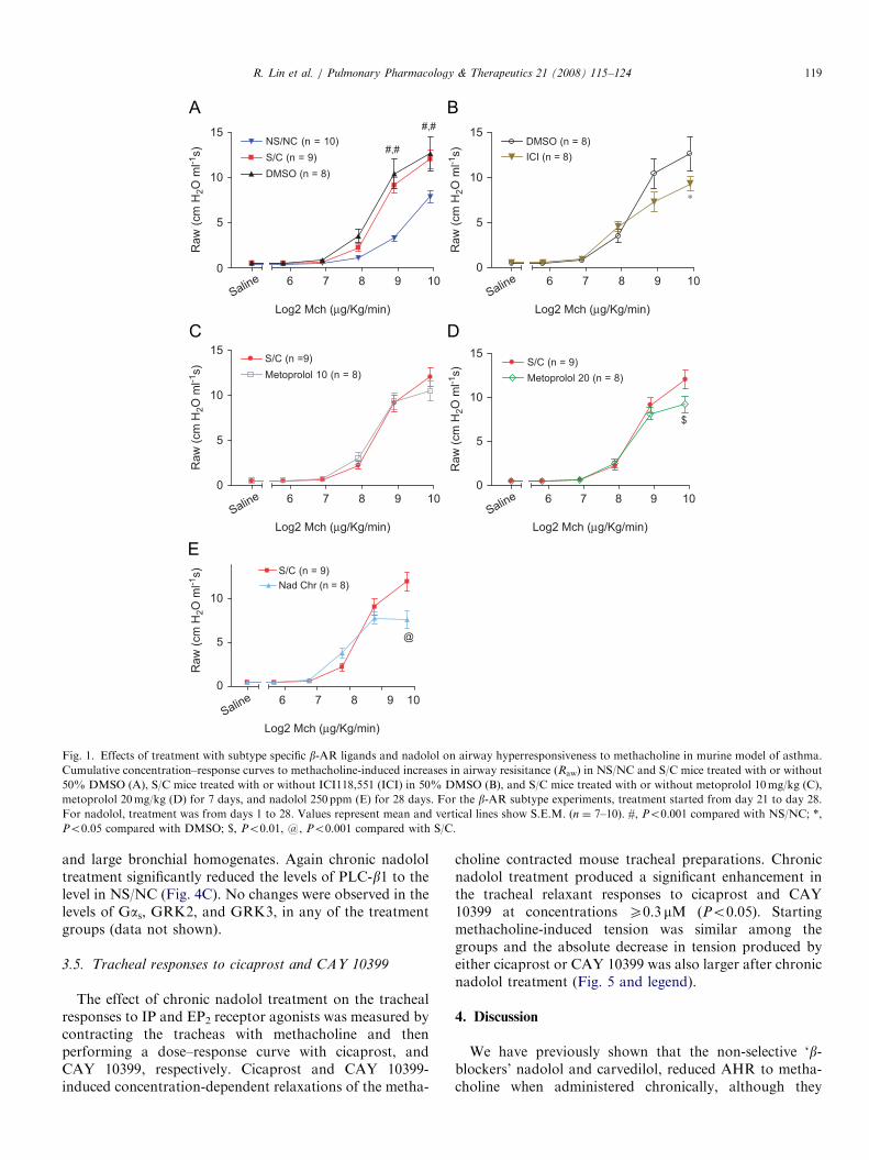
The intravenous administration of methacholine in-creased Raw in a dose-dependent manner (Fig. 1A).There was no significant difference between S/C micewithout treatment receiving regular mouse chow or treatedwith vehicle (DMSO), but both groups exhibited asignificantly greater bronchoconstrictor response to metha-choline compared to NS/NC mice (Fig. 1A). In S/Cmice treated with ICI 118,551, there was a significantreduction in the peak Raw response to methacholinecompared with S/C vehicle-treated mice (Fig. 1B). Whenthe S/C mice were treated with metoprolol 10mg/kg/d for 7days, their average methacholine dose–response relation-ship was similar to that obtained in non-treated S/Cmice (Fig. 1C). Increasing the dose of metoprolol to20mg/kg/d did result in a significant reduction in peak Raw
(Fig. 1D).
3.2. Effect of chronic nadolol treatment on airway responses
to methacholine
Treatment for 28 days with the non-selective b-ARantagonist/inverse agonist, nadolol, was again [20] able tosignificantly decrease the maximal airway constrictioninduced by methacholine (Fig. 1E). Since nadolol producedsimilar reductions in AHR to ICI 118,551, we used nadololfor further experiments because of the tremendous costdifferential (ICI 118,551 is over 500 times more expensivethan nadolol) and ease of administration (nadolol isadministered in the chow while ICI 118,551 requiresimplantation of osmotic minipumps due to its shorthalf-life).
3.3. Immunofluorescence staining for b1-ARs and b2-ARs
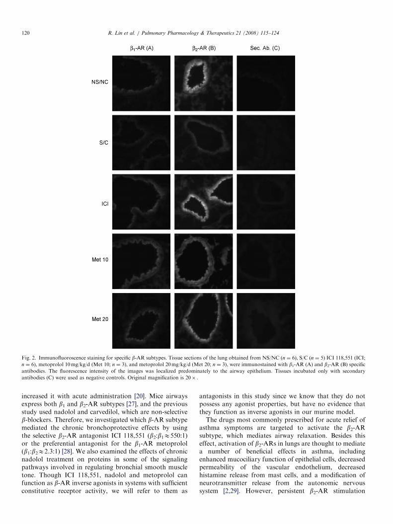
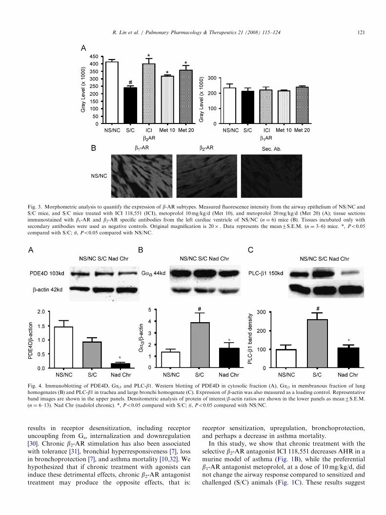
Airways immunostained for b1-ARs and b2-ARs dis-played changes in fluorescence intensity amongst thedifferent treatment groups. Results show the b1-AR (weakfluorescence intensity) and the b2-AR (strong fluorescenceintensity) expression is localized predominantly to theairway epithelium (Fig. 2, column A and B). In S/C mice,there was a decreased expression for b2-ARs compared toNS/NC mice (Fig. 2, column B). However, expression forb2-ARs was restored in S/C mice treated with ICI 181,551,and to a lesser extent with metoprolol 10mg/kg/d (Fig. 2,column B). Treatment with metoprolol 20mg/kg/d showeda further increase in b2-ARs compared to the 10mg dose(Fig. 2, column B). No appreciable difference in expressionfor b1-ARs was seen amongst the different treatmentgroups (Fig. 2, column A). Though we had previouslyreported an increase in total b-AR density using radi-oligand binding assays, these experiments show that theupregulation is mainly of the b2-AR subtype and occursprimarily in the airway epithelial cells. The bar graphshows quantified b1-AR and b2-AR fluorescence intensityin the airway epithelium (Fig. 3A). The b1-AR and b2-ARexpression in the left cardiac ventricle of the heart fromNS/NC mice were used to further assess the specificity ofthe antibodies (Fig. 3B).
3.4. Expression of proteins involved in receptor pathways
modulating bronchial tone
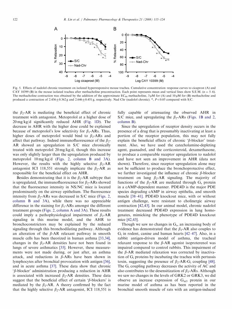
Immunoblotting studies were conducted to investigatethe expression of PDE4D, Gai3, Gas, GRK2, and GRK3 inS/C mouse lungs, with or without nadolol treatment and inNS/NC mouse lungs. In the cytosolic fraction of lunghomogenates, chronic treatment with nadolol decreasedthe expression of PDE4D; whereas, no significantdifference between NS/NC and S/C mice was identified(Fig. 4A). Gai3 expression was higher in the membranefraction of lung homogenate from S/C mice than NS/NCmice, and Gai3 expression was decreased to NS/NC levelsby chronic nadolol treatment (Fig. 4B). Compared to NS/NC mice, S/C mice had higher levels of PLC-b1 in trachea
ARTICLE IN PRESS
Saline
0
5
10
15
S/C (n = 9)
DMSO (n = 8)
NS/NC (n = 10)
6 7 8 9 10
#,#
#,#
Log2 Mch (μg/Kg/min)
Raw
(cm
H2O
ml-1
s)
Saline
0
5
10
15DMSO (n = 8)
ICI (n = 8)
∗
6 7 8 9 10
Log2 Mch (μg/Kg/min)
Saline 6 7 8 9 10
Log2 Mch (μg/Kg/min)
Saline 6 7 8 9 10
Log2 Mch (μg/Kg/min)
Saline 6 7 8 9 10
Log2 Mch (μg/Kg/min)
Raw
(cm
H2O
ml-1
s)
0
5
10
15S/C (n =9)
Metoprolol 10 (n = 8)
Raw
(cm
H2O
ml-1
s)
0
5
10
15S/C (n = 9)
Metoprolol 20 (n = 8)
$
Raw
(cm
H2O
ml-1
s)
0
5
10
S/C (n = 9)
Nad Chr (n = 8)
@
Raw
(cm
H2O
ml-1
s)
Fig. 1. Effects of treatment with subtype specific b-AR ligands and nadolol on airway hyperresponsiveness to methacholine in murine model of asthma.
Cumulative concentration–response curves to methacholine-induced increases in airway resisitance (Raw) in NS/NC and S/C mice treated with or without
50% DMSO (A), S/C mice treated with or without ICI118,551 (ICI) in 50% DMSO (B), and S/C mice treated with or without metoprolol 10mg/kg (C),
metoprolol 20mg/kg (D) for 7 days, and nadolol 250 ppm (E) for 28 days. For the b-AR subtype experiments, treatment started from day 21 to day 28.
For nadolol, treatment was from days 1 to 28. Values represent mean and vertical lines show S.E.M. (n ¼ 7–10). #, Po0.001 compared with NS/NC; *,
Po0.05 compared with DMSO; $, Po0.01, @, Po0.001 compared with S/C.
R. Lin et al. / Pulmonary Pharmacology & Therapeutics 21 (2008) 115–124 119
and large bronchial homogenates. Again chronic nadololtreatment significantly reduced the levels of PLC-b1 to thelevel in NS/NC (Fig. 4C). No changes were observed in thelevels of Gas, GRK2, and GRK3, in any of the treatmentgroups (data not shown).
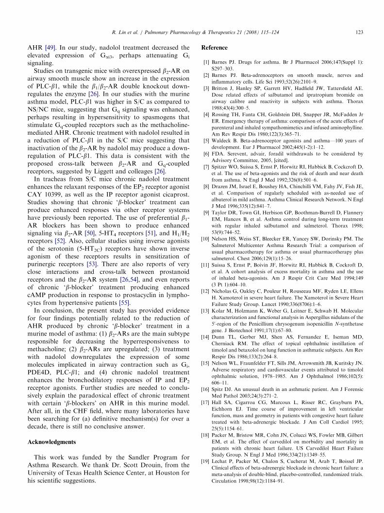
3.5. Tracheal responses to cicaprost and CAY 10399
The effect of chronic nadolol treatment on the trachealresponses to IP and EP2 receptor agonists was measured bycontracting the tracheas with methacholine and thenperforming a dose–response curve with cicaprost, andCAY 10399, respectively. Cicaprost and CAY 10399-induced concentration-dependent relaxations of the metha-
choline contracted mouse tracheal preparations. Chronicnadolol treatment produced a significant enhancement inthe tracheal relaxant responses to cicaprost and CAY10399 at concentrations X0.3 mM (Po0.05). Startingmethacholine-induced tension was similar among thegroups and the absolute decrease in tension produced byeither cicaprost or CAY 10399 was also larger after chronicnadolol treatment (Fig. 5 and legend).
4. Discussion
We have previously shown that the non-selective ‘b-blockers’ nadolol and carvedilol, reduced AHR to metha-choline when administered chronically, although they
ARTICLE IN PRESS
Fig. 2. Immunofluoroscence staining for specific b-AR subtypes. Tissue sections of the lung obtained from NS/NC (n ¼ 6), S/C (n ¼ 5) ICI 118,551 (ICI;
n ¼ 6), metoprolol 10mg/kg/d (Met 10; n ¼ 3), and metoprolol 20mg/kg/d (Met 20; n ¼ 3), were immunostained with b1-AR (A) and b2-AR (B) specific
antibodies. The fluorescence intensity of the images was localized predominately to the airway epithelium. Tissues incubated only with secondary
antibodies (C) were used as negative controls. Original magnification is 20� .
R. Lin et al. / Pulmonary Pharmacology & Therapeutics 21 (2008) 115–124120
increased it with acute administration [20]. Mice airwaysexpress both b1 and b2-AR subtypes [27], and the previousstudy used nadolol and carvedilol, which are non-selectiveb-blockers. Therefore, we investigated which b-AR subtypemediated the chronic bronchoprotective effects by usingthe selective b2-AR antagonist ICI 118,551 (b2:b1E550:1)or the preferential antagonist for the b1-AR metoprolol(b1:b2E2.3:1) [28]. We also examined the effects of chronicnadolol treatment on proteins in some of the signalingpathways involved in regulating bronchial smooth muscletone. Though ICI 118,551, nadolol and metoprolol canfunction as b-AR inverse agonists in systems with sufficientconstitutive receptor activity, we will refer to them as
antagonists in this study since we know that they do notpossess any agonist properties, but have no evidence thatthey function as inverse agonists in our murine model.The drugs most commonly prescribed for acute relief of
asthma symptoms are targeted to activate the b2-ARsubtype, which mediates airway relaxation. Besides thiseffect, activation of b2-ARs in lungs are thought to mediatea number of beneficial effects in asthma, includingenhanced mucociliary function of epithelial cells, decreasedpermeability of the vascular endothelium, decreasedhistamine release from mast cells, and a modification ofneurotransmitter release from the autonomic nervoussystem [2,29]. However, persistent b2-AR stimulation
ARTICLE IN PRESS
Fig. 3. Morphometric analysis to quantify the expression of b-AR subtypes. Measured fluorescence intensity from the airway epithelium of NS/NC and
S/C mice, and S/C mice treated with ICI 118,551 (ICI), metoprolol 10mg/kg/d (Met 10), and metoprolol 20mg/kg/d (Met 20) (A); tissue sections
immunostained with b1-AR and b2-AR specific antibodies from the left cardiac ventricle of NS/NC (n ¼ 6) mice (B). Tissues incubated only with
secondary antibodies were used as negative controls. Original magnification is 20� . Data represents the mean7S.E.M. (n ¼ 3–6) mice. *, Po0.05
compared with S/C; #, Po0.05 compared with NS/NC.
Fig. 4. Immunoblotting of PDE4D, Gai3 and PLC-b1. Western blotting of PDE4D in cytosolic fraction (A), Gai3 in membranous fraction of lung
homogenates (B) and PLC-b1 in trachea and large bronchi homogenate (C). Expression of b-actin was also measured as a loading control. Representative
band images are shown in the upper panels. Densitometric analysis of protein of interest/b-actin ratios are shown in the lower panels as mean7S.E.M.
(n ¼ 6–13). Nad Chr (nadolol chronic). *, Po0.05 compared with S/C; #, Po0.05 compared with NS/NC.
R. Lin et al. / Pulmonary Pharmacology & Therapeutics 21 (2008) 115–124 121
results in receptor desensitization, including receptoruncoupling from Gs, internalization and downregulation[30]. Chronic b2-AR stimulation has also been associatedwith tolerance [31], bronchial hyperresponsiveness [7], lossin bronchoprotection [7], and asthma mortality [10,32]. Wehypothesized that if chronic treatment with agonists caninduce these detrimental effects, chronic b2-AR antagonisttreatment may produce the opposite effects, that is:
receptor sensitization, upregulation, bronchoprotection,and perhaps a decrease in asthma mortality.In this study, we show that chronic treatment with the
selective b2-AR antagonist ICI 118,551 decreases AHR in amurine model of asthma (Fig. 1B), while the preferentialb1-AR antagonist metoprolol, at a dose of 10mg/kg/d, didnot change the airway response compared to sensitized andchallenged (S/C) animals (Fig. 1C). These results suggest
ARTICLE IN PRESS
Fig. 5. Effects of nadolol chronic treatment on isolated hyperresponsive mouse trachea. Cumulative concentration–response curves to cicaprost (A) and
CAY 10399 (B) in the mouse isolated trachea after methacholine precontraction. Each point represents mean and vertical lines show S.E.M. (n ¼ 5–6).
The methacholine contraction was obtained by the addition of the approximate EC80 methacholine, 3mM for (A) and 10 mM for (B) methacholine and
produced a contraction of 2.43670.362 g and 2.64670.451 g, respectively. Nad Chr (nadolol chronic). *, Po0.05 compared with S/C.
R. Lin et al. / Pulmonary Pharmacology & Therapeutics 21 (2008) 115–124122
the b2-AR is mediating the beneficial effect of chronictreatment with antagonist. Metoprolol at a higher dose of20mg/kg/d significantly reduced AHR (Fig. 1D). Thedecrease in AHR with the higher dose could be explainedbecause of metoprolol’s low selectivity for b1-ARs. Thus,higher doses of metoprolol would bind to b2-ARs andaffect that pathway. Indeed immunofluorescence of the b2-AR showed an upregulation in S/C mice chronicallytreated with metoprolol 20mg/kg/d, though this increasewas only slightly larger than the upregulation produced bymetoprolol 10mg/kg/d (Figs. 2, column B and 3A).However, the results with the highly selective b2-ARantagonist ICI 118,551 strongly implicate the b2-AR asresponsible for the beneficial effect on AHR.
Besides demonstrating that it is the b2-AR subtype thatis upregulated, the immunofluorescence for b2-ARs showedthat the fluorescence intensity in NS/NC mice is locatedpredominantly on the airway epithelium. The fluorescenceintensity from b2-ARs was decreased in S/C mice (Figs. 2,column B and 3A), while there was no appreciabledifference in the staining for b1-ARs amongst the differenttreatment groups (Figs. 2, column A and 3A). These resultscould imply a pathophysiological impairment of b2-ARsignaling in this murine model, and the AHR tobronchoconstrictors may be explained by the reducedsignaling through this bronchodilating pathway. Althoughan alteration of the b-AR relaxant pathway in smoothmuscle cells has been theorized in human asthma [33,34],changes in the b2-AR densities have not been found inlungs of severe asthmatics [35]. However, these measure-ments were not made during, or just after, an asthmaattack, and reductions in b-ARs have been shown inlymphocytes after bronchial provocation with antigen [36],and in acute asthma [37]. These data show that chronic‘b-blocker’ administration producing a reduction in AHRis associated with increased b2-AR densities. These datasuggest that the beneficial effect of certain ‘b-blockers’ ismediated by the b2-AR. A theory confirmed by the factthat the highly selective b2-AR antagonist, ICI 118,551 is
fully capable of attenuating the observed AHR inS/C mice, and upregulating the b2-ARs (Figs. 1B and 2,column B).Since the upregulation of receptor density occurs in the
presence of a drug that is presumably inactivating at least aportion of the receptor population, this may not fullyexplain the beneficial effects of chronic ‘b-blocker’ treat-ment. Also, we have used the catecholamine-depletingagent, guanadrel, and the corticosteroid, dexamethasone,to produce a comparable receptor upregulation to nadololand have not seen an improvement in AHR (data notshown). Therefore, since receptor upregulation alone maynot be sufficient to produce the bronchoprotective effect,we further investigated the influence of chronic b-blockertreatment on lung b2-AR signaling. The majority offunctions of the b2-AR are mediated through Gs proteinin a cAMP-dependent manner. PDE4D is the major PDEspecies degrading cAMP in airway epithelia, and smoothmuscle [38–41]. PDE4D knockout mice, with or withoutantigen challenge, were resistant to cholinergic airwaycontraction [42,43]. In our animal model, chronic nadololtreatment decreased PDE4D expression in lung homo-genates, mimicking the phenotype of PDE4D knockoutmice [42,43].Though we saw no changes in Gs, an increasing body of
evidence has demonstrated that the b2-AR also couples toGi in rodent, canine and human hearts [42–47]. Also, in arabbit antigen-driven model of asthma, the trachealrelaxant response to the b-AR agonist isoproterenol wasimpaired compared to control rabbits. This impairment ofthe b-AR mediated relaxation was corrected by inactiva-tion of Gi proteins by incubating the trachea with pertussistoxin, suggesting the presence of b2-AR-Gi coupling [48].The Gi coupling pathway decreases the activity of AC andalso contributes to the desensitization of b2-ARs. Althoughwe saw no changes in the levels of GRK2 or GRK3, we didobserve an increase expression of Gai3 protein in ourmurine model of asthma as has been reported in thebronchial smooth muscle of rats with an antigen-induced
ARTICLE IN PRESSR. Lin et al. / Pulmonary Pharmacology & Therapeutics 21 (2008) 115–124 123
AHR [49]. In our study, nadolol treatment decreased theelevated expression of Gai3, perhaps attenuating Gi
signaling.Studies on transgenic mice with overexpressed b2-AR on
airway smooth muscle show an increase in the expressionof PLC-b1, while the b1/b2-AR double knockout down-regulates the enzyme [26]. In our studies with the murineasthma model, PLC-b1 was higher in S/C as compared toNS/NC mice, suggesting that Gq signaling was enhanced,perhaps resulting in hypersensitivity to spasmogens thatstimulate Gq-coupled receptors such as the methacholine-mediated AHR. Chronic treatment with nadolol resulted ina reduction of PLC-b1 in the S/C mice suggesting thatinactivation of the b2-AR by nadolol may produce a down-regulation of PLC-b1. This data is consistent with theproposed cross-talk between b2-AR and Gq-coupledreceptors, suggested by Liggett and colleages [26].
In tracheas from S/C mice chronic nadolol treatmentenhances the relaxant responses of the EP2 receptor agonistCAY 10399, as well as the IP receptor agonist cicaprost.Studies showing that chronic ‘b-blocker’ treatment canproduce enhanced responses via other receptor systemshave previously been reported. The use of preferential b1-AR blockers has been shown to produce enhancedsignaling via b2-AR [50], 5-HT4 receptors [51], and H1/H2
receptors [52]. Also, cellular studies using inverse agonistsof the serotonin (5-HT2C) receptors have shown inverseagonism of these receptors results in sensitization ofpurinergic receptors [53]. There are also reports of veryclose interactions and cross-talk between prostanoidreceptors and the b2-AR system [26,54], and even reportsof chronic ‘b-blocker’ treatment producing enhancedcAMP production in response to prostacyclin in lympho-cytes from hypertensive patients [55].
In conclusion, the present study has provided evidencefor four findings potentially related to the reduction ofAHR produced by chronic ‘b-blocker’ treatment in amurine model of asthma: (1) b2-ARs are the main subtyperesponsible for decreasing the hyperresponsiveness tomethacholine; (2) b2-ARs are upregulated; (3) treatmentwith nadolol downregulates the expression of somemolecules implicated in airway contraction such as Gi,PDE4D, PLC-b1; and (4) chronic nadolol treatmentenhances the bronchodilatory responses of IP and EP2
receptor agonists. Further studies are needed to conclu-sively explain the paradoxical effect of chronic treatmentwith certain ‘b-blockers’ on AHR in this murine model.After all, in the CHF field, where many laboratories havebeen searching for (a) definitive mechanism(s) for over adecade, there is still no conclusive answer.
Acknowledgments
This work was funded by the Sandler Program forAsthma Research. We thank Dr. Scott Drouin, from theUniversity of Texas Health Science Center, at Houston forhis scientific suggestions.
Reference
[1] Barnes PJ. Drugs for asthma. Br J Pharmacol 2006;147(Suppl 1):
S297–303.
[2] Barnes PJ. Beta-adrenoceptors on smooth muscle, nerves and
inflammatory cells. Life Sci 1993;52(26):2101–9.
[3] Britton J, Hanley SP, Garrett HV, Hadfield JW, Tattersfield AE.
Dose related effects of salbutamol and ipratropium bromide on
airway calibre and reactivity in subjects with asthma. Thorax
1988;43(4):300–5.
[4] Rossing TH, Fanta CH, Goldstein DH, Snapper JR, McFadden Jr
ER. Emergency therapy of asthma: comparison of the acute effects of
parenteral and inhaled sympathomimetics and infused aminophylline.
Am Rev Respir Dis 1980;122(3):365–71.
[5] Waldeck B. Beta-adrenoceptor agonists and asthma—100 years of
development. Eur J Pharmacol 2002;445(1-2):1–12.
[6] FDA. Serevent, advair, foradil withdrawals to be considered by
Advisory Committee, 2005, [cited].
[7] Spitzer WO, Suissa S, Ernst P, Horwitz RI, Habbick B, Cockcroft D,
et al. The use of beta-agonists and the risk of death and near death
from asthma. N Engl J Med 1992;326(8):501–6.
[8] Drazen JM, Israel E, Boushey HA, Chinchilli VM, Fahy JV, Fish JE,
et al. Comparison of regularly scheduled with as-needed use of
albuterol in mild asthma. Asthma Clinical Research Network. N Engl
J Med 1996;335(12):841–7.
[9] Taylor DR, Town GI, Herbison GP, Boothman-Burrell D, Flannery
EM, Hancox B, et al. Asthma control during long-term treatment
with regular inhaled salbutamol and salmeterol. Thorax 1998;
53(9):744–52.
[10] Nelson HS, Weiss ST, Bleecker ER, Yancey SW, Dorinsky PM. The
Salmeterol Multicenter Asthma Research Trial: a comparison of
usual pharmacotherapy for asthma or usual pharmacotherapy plus
salmeterol. Chest 2006;129(1):15–26.
[11] Suissa S, Ernst P, Boivin JF, Horwitz RI, Habbick B, Cockroft D,
et al. A cohort analysis of excess mortality in asthma and the use
of inhaled beta-agonists. Am J Respir Crit Care Med 1994;149
(3 Pt 1):604–10.
[12] Nicholas G, Oakley C, Pouleur H, Rouseeau MF, Ryden LE, Ellens
H. Xamoterol in severe heart failure. The Xamoterol in Severe Heart
Failure Study Group. Lancet 1990;336(8706):1–6.
[13] Kolar M, Holzmann K, Weber G, Leitner E, Schwab H. Molecular
characterization and functional analysis in Aspergillus nidulans of the
50-region of the Penicillium chrysogenum isopenicillin N-synthetase
gene. J Biotechnol 1991;17(1):67–80.
[14] Dunn TL, Gerber MJ, Shen AS, Fernandez E, Iseman MD,
Cherniack RM. The effect of topical ophthalmic instillation of
timolol and betaxolol on lung function in asthmatic subjects. Am Rev
Respir Dis 1986;133(2):264–8.
[15] Nelson WL, Fraunfelder FT, Sills JM, Arrowsmith JB, Kuritsky JN.
Adverse respiratory and cardiovascular events attributed to timolol
ophthalmic solution, 1978–1985. Am J Ophthalmol 1986;102(5):
606–11.
[16] Spitz DJ. An unusual death in an asthmatic patient. Am J Forensic
Med Pathol 2003;24(3):271–2.
[17] Hall SA, Cigarroa CG, Marcoux L, Risser RC, Grayburn PA,
Eichhorn EJ. Time course of improvement in left ventricular
function, mass and geometry in patients with congestive heart failure
treated with beta-adrenergic blockade. J Am Coll Cardiol 1995;
25(5):1154–61.
[18] Packer M, Bristow MR, Cohn JN, Colucci WS, Fowler MB, Gilbert
EM, et al. The effect of carvedilol on morbidity and mortality in
patients with chronic heart failure. US Carvedilol Heart Failure
Study Group. N Engl J Med 1996;334(21):1349–55.
[19] Lechat P, Packer M, Chalon S, Cucherat M, Arab T, Boissel JP.
Clinical effects of beta-adrenergic blockade in chronic heart failure: a
meta-analysis of double-blind, placebo-controlled, randomized trials.
Circulation 1998;98(12):1184–91.
ARTICLE IN PRESSR. Lin et al. / Pulmonary Pharmacology & Therapeutics 21 (2008) 115–124124
[20] Callaerts-Vegh Z, Evans KL, Dudekula N, Cuba D, Knoll BJ,
Callaerts PF, et al. Effects of acute and chronic administration of
beta-adrenoceptor ligands on airway function in a murine model of
asthma. Proc Natl Acad Sci USA 2004;101(14):4948–53.
[21] Evans KL, Bond RA, Corry DB, Shardonofsky FR. Frequency
dependence of respiratory system mechanics during induced con-
striction in a murine model of asthma. J Appl Physiol 2003;
94(1):245–52.
[22] Costall B, Naylor RJ, Tan CC. Mechanism of action of apomorphine
on rat gastric secretion. Eur J Pharmacol 1985;116(3):279–85.
[23] Kubota T, Yamazaki N, Sudo J, Monma Y, Kaku T, Okuyama T, et
al. Protective effects of adrenoceptor-blocking agents on myocardial
injury induced by epinephrine in mice. J Toxicol Sci 1990;15(1):1–13.
[24] Fust A, Bates JH, Ludwig MS. Mechanical properties of mouse distal
lung: in vivo Versus in vitro comparison. Respir Physiol Neurobiol
2004;143(1):77–86.
[25] Wagers S, Lundblad LK, Ekman M, Irvin CG, Bates JH. The allergic
mouse model of asthma: normal smooth muscle in an abnormal lung?
J Appl Physiol 2004;96(6):2019–27.
[26] McGraw DW, Almoosa KF, Paul RJ, Kobilka BK, Liggett SB.
Antithetic regulation by beta-adrenergic receptors of Gq receptor
signaling via phospholipase C underlies the airway beta-agonist
paradox. J Clin Invest 2003;112(4):619–26.
[27] Henry PJ, Rigby PJ, Goldie RG. Distribution of beta 1- and beta 2-
adrenoceptors in mouse trachea and lung: a quantitative autoradio-
graphic study. Br J Pharmacol 1990;99(1):136–44.
[28] Baker JG. The selectivity of beta-adrenoceptor antagonists at the
human beta1, beta2 and beta3 adrenoceptors. Br J Pharmacol
2005;144(3):317–22.
[29] Broadley KJ. Beta-adrenoceptor responses of the airways: for better
or worse. Eur J Pharmacol 2006;533(1-3):15–27.
[30] Lefkowitz RJ, Hausdorff WP, Caron MG. Role of phosphorylation
in desensitization of the beta-adrenoceptor. Trends Pharmacol Sci
1990;11(5):190–4.
[31] Haney S, Hancox RJ. Tolerance to bronchodilation during treatment
with long-acting beta-agonists, a randomised controlled trial. Respir
Res 2005;6:107.
[32] Speizer FE, Doll R, Heaf P, Strang LB. Investigation into use
of drugs preceding death from asthma. Br Med J 1968;1(5588):
339–43.
[33] Szentivanyi A, Heim O, Schultze P. Changes in adrenoceptor
densities in membranes of lung tissue and lymphocytes from patients
with atopic disease. Ann NY Acad Sci 1979;332:295–8.
[34] Szentivanyi A. The conformational flexibility of adrenoceptors and
the constitutional basis of atopy. Triangle 1979;18(4):109–15.
[35] Spina D, Rigby PJ, Paterson JW, Goldie RG. Autoradiographic
localization of beta-adrenoceptors in asthmatic human lung. Am Rev
Respir Dis 1989;140(5):1410–5.
[36] Gamboa PM, de la Cuesta CG, Sanz ML, Garcia BE, Oehling A.
Decrease of beta-receptors after the antigen-specific bronchial
provocation test in bronchial asthma. Allergol Immunopathol
(Madr) 1990;18(3):115–9.
[37] Hataoka I, Okayama M, Sugi M, Inoue H, Takishima T, Shirato K.
Decrease in beta-adrenergic receptors of lymphocytes in sponta-
neously occurring acute asthma. Chest 1993;104(2):508–14.
[38] Torphy TJ, Zhou HL, Cieslinski LB. Stimulation of beta adreno-
ceptors in a human monocyte cell line (U937) up-regulates cyclic
AMP-specific phosphodiesterase activity. J Pharmacol Exp Ther
1992;263(3):1195–205.
[39] Dent G, White SR, Tenor H, Bodtke K, Schudt C, Leff AR, et al.
Cyclic nucleotide phosphodiesterase in human bronchial epithelial
cells: characterization of isoenzymes and functional effects of PDE
inhibitors. Pulm Pharmacol Ther 1998;11(1):47–56.
[40] Le Jeune IR, Shepherd M, Van Heeke G, Houslay MD, Hall IP.
Cyclic AMP-dependent transcriptional up-regulation of phospho-
diesterase 4D5 in human airway smooth muscle cells. Identification
and characterization of a novel PDE4D5 promoter. J Biol Chem
2002;277(39):35980–9.
[41] Wright LC, Seybold J, Robichaud A, Adcock IM, Barnes PJ.
Phosphodiesterase expression in human epithelial cells. Am J Physiol
1998;275(4 Pt 1):L694–700.
[42] Conti M, Richter W, Mehats C, Livera G, Park JY, Jin C. Cyclic
AMP-specific PDE4 phosphodiesterases as critical components of
cyclic AMP signaling. J Biol Chem 2003;278(8):5493–6.
[43] Giembycz M. PDE4D-deficient mice knock the breath out of asthma.
Trends Pharmacol Sci 2000;21(8):291–2.
[44] Kilts JD, Gerhardt MA, Richardson MD, Sreeram G, Mackensen
GB, Grocott HP, et al. Beta(2)-adrenergic and several other G
protein-coupled receptors in human atrial membranes activate both
G(s) and G(i). Circ Res 2000;87(8):705–9.
[45] Xiao RP. Beta-adrenergic signaling in the heart: dual coupling of the
beta2-adrenergic receptor to G(s) and G(i) proteins. Sci STKE
2001;2001(104):RE15.
[46] Xiao RP, Avdonin P, Zhou YY, Cheng H, Akhter SA, Eschenhagen
T, et al. Coupling of beta2-adrenoceptor to Gi proteins and its
physiological relevance in murine cardiac myocytes. Circ Res
1999;84(1):43–52.
[47] Xiao RP, Ji X, Lakatta EG. Functional coupling of the beta 2-
adrenoceptor to a pertussis toxin-sensitive G protein in cardiac
myocytes. Mol Pharmacol 1995;47(2):322–9.
[48] Hakonarson H, Herrick DJ, Grunstein MM. Mechanism of impaired
beta-adrenoceptor responsiveness in atopic sensitized airway smooth
muscle. Am J Physiol 1995;269(5 Pt 1):L645–52.
[49] Chiba Y, Sakai H, Misawa M. Possible involvement of G(i3) protein
in augmented contraction of bronchial smooth muscle from antigen-
induced airway hyperresponsive rats. Biochem Pharmacol 2001;
61(7):921–4.
[50] Hall JA, Kaumann AJ, Brown MJ. Selective beta 1-adrenoceptor
blockade enhances positive inotropic responses to endogenous
catecholamines mediated through beta 2-adrenoceptors in human
atrial myocardium. Circ Res 1990;66(6):1610–23.
[51] Kaumann AJ, Sanders L. 5-Hydroxytryptamine causes rate-depen-
dent arrhythmias through 5-HT4 receptors in human atrium:
facilitation by chronic beta-adrenoceptor blockade. Naunyn Schmie-
debergs Arch Pharmacol 1994;349(4):331–7.
[52] Sanders L, Lynham JA, Kaumann AJ. Chronic beta 1-adrenoceptor
blockade sensitises the H1 and H2 receptor systems in human atrium:
role of cyclic nucleotides. Naunyn Schmiedebergs Arch Pharmacol
1996;353(6):661–70.
[53] Berg KA, Stout BD, Cropper JD, Maayani S, Clarke WP. Novel
actions of inverse agonists on 5-HT2C receptor systems. Mol
Pharmacol 1999;55(5):863–72.
[54] Guo M, Pascual RM, Wang S, Fontana MF, Valancius CA,
Panettieri Jr RA, et al. Cytokines regulate beta-2-adrenergic receptor
responsiveness in airway smooth muscle via multiple PKA- and EP2
receptor-dependent mechanisms. Biochemistry 2005;44(42):13771–82.
[55] Lima DR, Turner P. Beta-blocking drugs increase responsiveness to
prostacyclin in hypertensive patients. Lancet 1982;2(8295):444.