CFfR FUNCTION, REGULATION AND CHARACTERIZATION ...
120
CFfR FUNCTION, REGULATION AND CHARACTERIZATION OF A MOUSE MODEL FOR THE L'lF508 MUTATION CFTR FUNCTIE, REGULATIE EN DE KARAKTERIZERING VAN EEN MUIS MODEL VOOR DE L'lF508 MUTATIE PROEFSCHRIFT ter verkrijging van de graad van doctor aan de Erasmus Universiteit Rotterdam op gezag van de rector magnificus Prof. Dr P.W.C. Akkermans M.A. en volgens besluit van het college van promoties de openbare verdediging zal plaats vinden op Woensdag 16 april 1997 om 13:45 uur PETER JAMES FRENCH geboren te Leiden
-
Upload
khangminh22 -
Category
Documents
-
view
0 -
download
0
Transcript of CFfR FUNCTION, REGULATION AND CHARACTERIZATION ...

CFfR FUNCTION, REGULATION
AND CHARACTERIZATION OF A MOUSE MODEL FOR THE L'lF508 MUTATION
CFTR FUNCTIE, REGULATIE
EN DE KARAKTERIZERING VAN EEN MUIS MODEL VOOR DE L'lF508 MUTATIE
PROEFSCHRIFT
ter verkrijging van de graad van doctor aan de Erasmus Universiteit Rotterdam op gezag van de
rector magnificus Prof. Dr P.W.C. Akkermans M.A.
en volgens besluit van het college van promoties
de openbare verdediging zal plaats vinden op
W oensdag 16 april 1997 om 13:45 uur
PETER JAMES FRENCH geboren te Leiden

Promotie commissie
Promotor:
Overige leden:
Co-promotor:
Prof. Dr F. G. Grosveld
Prof. Dr H. Biiller Prof. Dr B.A. Oostra Dr H.R. de Jonge
Dr J. Bijman
Dit proefschrift is mede tot stand gekornen door cen bijdrage van Smithkllnc Beecham Farma BY Nederland

aal/Nancy aan myn ouders

CONTENTS
Chapter 1 1.1 General introduction ...................................................................................................... 3 1.2 History ........................................................................................................................... 3 1.3 Clinical phenotype ......................................................................................................... 4
1.3.1 Respiratory tract ............................................................................................. 4 1.3.2 Gastrointestinal tract ...................................................................................... 4 1.3.3 Other manifestations ...................................................................................... 5
104 CFfR ............................................................................................................................. 5 1.4.1 CFfR expression ........................................................................................... 6 104.2 Regulation of CFfR expression .................................................................... 6
104.3 CFfRisoforms .............................................................................................. 7 1.404 Domains ofCFfR ......................................................................................... 7
1.404.1 The transmembrane domains ......................................................... 7
10404.2 The R domain ................................................................................. 8 10404.3 The nucleotide binding domains.: .................................................. 8 1040404 The role ofNBDI ........................................................................... 9 10404.5 The role ofNBD2 ........................................................................... 9
1.4.5 Regulation of CFfR activity ....................................................................... 10 104.5.1 cAMP regulation .......................................................................... 10 104.5.2 PKC regulation ............................................................................. 12 104.5.3 cGMP regulation .......................................................................... 12 104.504 Tyrosine kinases ........................................................................... 13 104.5.5 Other protein kinases .................................................................... 14 104.5.6 Dephosphorylation ....................................................................... 14
104.6 CFfR modulation of other ion channels ..................................................... 14
1.5 The &'508 mutation ....................................................................................... ~ ............ 16
1.5.1 The &'508 mutation is a class II mutation .................................................. 17
1.5.2 Processing ofwt and &'508 CFfR ............................................................. 18
1.5.3 Correcting the processing defect ................................................................. 19 1.6 Functions ofCFfR ...................................................................................................... 20
1.6.1 The role of CFfR in mucus synthesis ......................................................... 20 1.6.2 Mucus secretion is increased in CF ............................................................. 23 1.6.3 The role of CFfR in fluid and electrolyte transport .................................... 23
1.6.3.1 Altered fluid and electrolyte resorption ....................................... 23 1.6.3.2 Altered fluid and electrolyte secretion ......................................... 24
1.6.3.3 The pH .......................................................................................... 24 1.6.4 The role of CFTR in pathogen affinity,
internalization and killing ......................................................................... 26 1.6.5 The role of CFTR in endo- and exocytosis ................................................. 26
1.6.6 Summary ...................................................................................................... 26

1.7 Mouse models for CF .................................................................................................. 26 1.8 Scope of the thesis ....................................................................................................... 29
Chapter 2
Summary and Discussion .................................................................................................. 31 2.1 regulation of CFfR ........................................................................................ .31 2.2 functions of CFfR .......................................................................................... 33
2.3 characterization of a mouse model for the &'508 CFfR mutation ............... 36
Abbreviations ................................................................................................................................. 38 References ..................................................................................................................................... .39 Samenvatting .................................................................................................................................. 48
Chapter 3 Publications
pag 51
3.1 Isotype-specific Activation of Cystic Fibrosis Transmembrane Conductance
Regulator-Chloride Channels by cGMP-dependent Protein Kinase II (1995) P.l.French, l. Bijman, M. Edixhoven, A.B. Vaandrager, B.J. Scholte, S.M. Lohmann,
A.C. Nairn, and H.R. de longe. J. Bioi. Chell/. 270:26626-26631
pag57
3.2 Genistein Activates CFTR via a Tyrosine Kinase and Protein Phosphatase
Independent Pathway (1996) P.J. French, l. Bijman, A.G. Bot, W. Boomaars, B.l.
Scholte, and H.R. de longe. A1II. 1. Physiol. submitted
pag73
3.3 Fluid Transport in the Trachea of Normal and CF mouse (1997) P.J. French, H.P.C. Peters, l.H. van Doominck, W.H. Colledge, R. Ratcliff, M.l. Evans, B.J. Scholte
and J. Bijman. manuscript in preparation pag 83
3.4 CFTR expression and mucin secretion in cultured mouse gallbladder epithelial
ceUs (1996) R.H.P.C. Peters, P.l. French, l.H. van Doominck, G. Lamblin, R. Ratcliff,
M.J. Evans, W.H. Colledge, l. Bijman, and B.J. Scholte. Am. J. Physiol. 271:GI074-
G1083
pag93
3.5 A mouse model for the cystic fibrosis iW508 mutation (1995) l.H. van Doominck, P.J. French, E. Verbeek, R.H.P.C. Peters, H. Morreau, l. Bijman, and B.l.
Scholte. EMBO J. 14:4403-4411

pag 102
3.6 A iU'S08 Mutation in Mouse Cystic Fibrosis Transmembrane Conductance
Regulator Results in a Temperature-sensitive Processing Defect In Vivo (1996) P.J.
French, J.H. van Doominck, R.H.P.C. Peters, E. Verbeek, N.A. Ameen, c.R. Marino,
H.R. de Jonge, J. Bijman, and BJ. Scholte. 1. Clill. Illvest. 98:1304-1312
Curriculum Vitae ......................................................................................................................... 111
List of Publications ...................................................................................................................... 112 Dankwoord ................................................................................................................................... 113


3 Introduction
1.1 General introduction Cystic Fibrosis (CF) is the most common lethal autosomal recessive disease in the
caucasian population, affecting ",1:;1500 newborns (1,2). There are approximately 1000 CF
patients in the Netherlands (3). Patients are characterized by recurrent pulmonary infections,
pancreatic insufficiency and elevated sweat salt concentrations. Although improved therapy
has resulted in an average lifespan of ",29 years, CF remains a serious destructive disease
with a grim prognosis. Cystic Fibrosis is caused by mutations in the gene encoding the Cystic
Fibrosis Transmembrane Conductance Regulator (CFTR) (4·6), a cAMP regulated Cl·
channel expressed in the apical (luminal) membrane of many exocrine epithelia (5). The most
common mutation in CFTR is a single amino acid deletion at position 508, AF508, found in
",70% of all CF alleles (6). The AF508 mutation causes a defect in CFTR maturation resulting
in an incorrect targeting to the apical membrane; the mutant protein is retained and degraded
in the endoplasmic reticulum (ER) by the quality control mechanism (7).
Although the gene defective in CF patients has been identified, CF disease
pathogenesis is still a not well understood process. In order to explain the CF pathogenesis, a
detailed understanding of the CFTR protein is essential. For this, we have studied the
regulation of CFTR, its physiological functions, and the effects of the AF508 CFTR mutation.
Based on the functions of CFTR and the specific properties of CFTR mutants, strategies
towards a therapy for CF can be developed.
1.2 History Long before the first comprehensive description of CF in 1938 (8), a folkloric
anecdote existed of midwives licking the forehead of newborns. If the sweat tasted abnormally
salty, the infant was predicted to die of puimonary congestion and its side effects. This
folklore may relate to the observation of elevated sweat salt levels in CF by Di Sant-Agnese in
1953 (9). To date, the elevated sweat NaCl is still used in many clinics to help establish the
diagnosis of CF. In 1981 it was observed that CF patients have an increased potential
difference (PD) across pulmonary epithelia (10). This increased PD was also present across
CF sweat duct epithelia. This was ascribed to a chloride impermeability of the tissue,
resulting in a reduced NaCI uptake (11,12). Finally, in 1989, the CF gene was discovered, as
one of the first genes to be isolated by positional cloning (4·6). The gene product was named
the Cystic Fibrosis Transmembrane Conductance Regulator (CFTR), and encodes a cAMP
regulated Cl" channel (13-17), expressed on the apical membrane of many epithelial cells
(18-22).

Chapter 1 4
1.3 Clinical phenotype
The major life threatening clinical manifestation of CF is recurrent pulmonary
infection. However, CF is a multi organ disease affecting, besides the respiratory tract, the
gastrointestinal tract (liver, gallbladder, pancreas and intestine), the genito-urinary tract and
several other tissues, e.g. sweatgland. In addition, CF related organ insufficiency may cause
several secondary disease symptoms like diabetes and vitamin deficiencies. The more
prominent organ related disease symptoms are listed below (for comprehensive reviews see
(1,2».
1.3.1 RespiratOlY tract: At birth, no obvious pulmonary abnormalities are observed, except
for dilation of submucosal gland duct and acinus. One of the first pulmonary lesions is muCUS
obstruction and infection of conducting ainvays (bronchioles). As the disease progresses,
observed pathology include broncheolar and brocheal inflanunation, submucosal gland
hypertrophy and, hyperplasia of mucus producing goblet cells. Some small airways are
completely obstructed. In advanced stages of the disease, CF is manifested by prominent
bronchiectic cysts, brochiolar stenosis, areas of destructive emphysema and, peribroncheolar
and peribronchial fibrosis. The progressive ainvay destruction in CF is initiated by mucus
obstruction of conductive ainvays and the subsequent colonization by Pseudomonas
aeruginosa (especially of the mucoid type), Staphylococcus aureus and, Hemophilus
influenza. Several other bacterial strains have also been found. In the nose, polyp formation is
frequently observed (15-20%) associated with mucus accumulation and chronic rhinitis.
1.3.2 Gastrointestinal tract: Meconium ileus, affecting 5-10% of CF neonatals, is one of the
first major signs of gastrointestinal tract disease in CF. Less severe forms of meconium ileus
like meconium plug syndrome (large intestinal obstruction), may also be observed. Later in
life, small intestinal obstruction (meconium ileus equivalent) occurs frequently (20%) in the
CF population. Virtually all CF patients develop pancreatic abnormalities. Pancreatic
insufficiency is caused by mucus obstruction of the pancreatic duct, and the subsequent loss of
acinar cells. These are replaced by fibrous tissue. Hepatobiliary disease in CF includes focal
biliary cirrhosis and mucus plugging of intrahepatic ducts. An abnormal gallbladder is also
frequently observed. Pancreatic insufficiency and hepatobiliary disease impairs both protein
and fat digestion and so contributes to intestinal malabsorption in CF. Additionally,
independent of pancreatic and liver function, the CF intestine may exhibit defects in nutrient
absorption. Reduced nutrient uptake due to gastrointestinal malfunctioning in CF results in
growth retardation. A side effect of fat malabsorption, are deficiencies in fat soluble vitamins

5 [lIIrodllctioll
(A, D, E, and K), and their related diseases.
1.3.3 Other manifestations: Most male and part of the female CF patients are infertile. In
males the main cause of infertility is the absence of the vas deferens. In females infertility
may be secondary to malnutrition andlor lung infections, but it is also possible that thick
cervical mucus may prevent sperm passage. The fibrosis of pancreatic acini isolates and
obstructs blood flow to the pancreatic islets of Langerhans, potentially priming CF patients
for diabetes. Finally, CF patients exhibit elevated sweat NaCl, with sweat Cl- concentrations
exceeding 60 meq/l (23).
1.4CFfR The cystic fibrosis gene was cloned and characterized in 1989 (4-6). The cystic
fibrosis gene product was named the Cystic Fibrosis Transmembrane Conductance Regulator
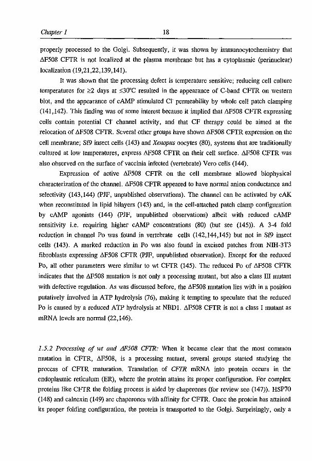
(CFTR). A model of CFTR is depicted in figure 1.1. Its encodes a protein of 1480 amino
acids consisting of two transmembrane domains (TM's), each spaoning the membrane six
times, two nucleotide binding domains (NBD's) and a highly charged cytoplasmic domain
(the R domain) containing consensus sequences for phosphorylation. The most common
mutation in CFTR, found in ",70% of all CF alleles is a single amino acid deletion of a
phenylalanine at position 508 (6F508) (6).
CFTR forms a cAMP regulated chloride channel with linear current-voltage
relationship in transfected Hela cells (13), Sf9 insect cells (15), CHO cells (14), and IEC cells
(17). Reconstitution of purified CFTR into planar lipid bilayers (16) and altered ion selectivity
upon mutation of residues lining the putative pore (24) indicated that CFTR itself is a chloride
channel, rather than being a channel regulator. The channel is expressed in the apical
membrane of epithelia involved in the transport of fluid and electrolytes (18-22) and has a
linear conductance of 8-10 pS (14,16,25).
CFTR bears homology to the protein superfamily of traffick ATPases or ATP Binding
Cassette (ABC) transporters (5,26). In general, these proteins are involved in the active
transport of substances across the membrane, for review see (26-29). Members of the ABC
transporter superfamilly include the mammalian muItidrug resistant P-glycoprotein (reviewed
in (30), the yeast STE6 gene product (31,32), the epithelial basolateral chloride conductance
regulator EBCR (33), the sulfonylurea receptor SUR, and several bacterial periplasmic
permeases (reviewed in (27».

Chapter 1 6
TM1 TM2
Figure 1.1: Topological mOOel of CFIR. showing the two transmembrane domains (FMJ alld TM2), each consisting oj 6 membrQJle spaJlning segments (MlvM12), the two nucleotide biluiing domains (NBDl and NBD2) and the regulatory domain (R).
1.4.1 CFrR expression: The expression of CFrR mRNA and protein is primarily restricted to
epithelial tissues affected in CF (5). In the gastrointestinal tract, high CFrR mRNA
expression was found in the small intestine and colon, with declining message from crypt to
villus (34,35). CFrR expression was also found in epithelial cells of the pancreatic duct
(34,35) and gallbladder (35,36), whereas low CFrR expression was observed in the stomach
(34,35). In the respiratory tract, expression of CFTR protein was found predominantly in
submucosal glands (22) and was not detectable in surface epithelium (18,22). However,
pulmonary surface epithelia do express detectable amounts of CFrR mRNA albeit at low
levels.
In addition to the gastrointestinal and respiratory tracts, CFTR expression was also
found in other secretory andlor absorptive tissues, some of which apparently are not involved
in CF pathogenesis. CFTR expression was found in salivary gland-, sweat duct- and thyroid
epithelia (for reviews see (37-39», and renal epithelia (for review see (40». In the brain, the
CFTR protein was detected in the choroid plexus and ventricular epithelium (41) and in
various other areas (42). Non secretory cells expressing CFTR include human lymphocytes
(43) and cardiac myocytes (44,45).
1.4.2 Regulation of CFrR expression: The CFTR promotor contains several potential
transcriptional regulatory elements. Among these are 2 potential API binding sites, which,
7 Introduction
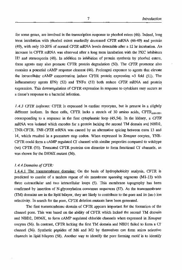
for some genes, are involved in the transcription response to phorbol esters (46). Indeed, long
term incubation with phorbol esters markedly decreased CFTR mRNA (46-49) and protein
(49), with only 10-20% of normal CFTR mRNA levels detectable after a 12 hr incubation. An
increase in CFTR mRNA was observed after a long term incubation with the PKC inhibitors
H7 and staurosporin (48). In addition to inhibition of protein synthesis by phorbol esters,
these agents may also promote CFTR protein degradation (50). The CFTR promotor also
contains a potential cAMP response element (46). Prolonged exposure to agents that elevate
the intracellular cAMP concentration induce CFTR protein expression ",3 fold (51). The
inflammatory agents IFNy (52) and TNFa (53) both reduce CFTR mRNA and protein
expression. This downregulation of CFTR expression in response to cytokines may occurs as
a tissue's response to a bacterial infection.
1.4.3 CFTR isojol1ns: CFTR is expressed in cardiac myocytes, but is present in a slightly
different isoform. In these cells, CFTR lacks a stretch of 30 amino acids, CFTR".,.l9J'
corresponding to a sequence in the first cytoplasmic loop (45,54). In the kidney, a CFTR
mRNA was isolated which encodes for a protein lacking the second TM domain and NBD2,
TNR-CFTR. TNR-CFTR mRNA was caused by an alternative spicing between exon 13 and
14, which resulted in a premature stop codon. When expressed in Xenopus oocytes, TNR
CFTR could form a cAMP regulated Cl" channel with similar properties compared to wildtype
(wt) CFTR (55). Truncated CFTR proteins can dimerize to form functional Cl" channels, as
was shown for the D836X mutant (56).
1.4.4 Domains oj CFTR:
1.4.4.1 The transmembrane domains: On the basis of hydrophobicity analysis, CFTR is
predicted to consist of a tandem repeat of six membrane spanning segments (M1-12) with
three extracellular and two intracellular loops (5). This membrane topography has been
confmned by insertion of N-glycosylation consensus sequences (57). As the transmembrane
(TM) domains are in the lipid bilayer, they are likely to contribute to the pore and its (an-) ion
selectivity. In search for the pore, CFTR deletion mutants have been generated.
The first transmembrane domain of CFTR appears important for the formation of the
channel pore. This was based on the ability of CFTR which lacked the second TM domain
and NBD2, D836X, to form cAMP regulated chloride channels when expressed in Xenopus
oocytes (56). In contrast, CFTR laCking the first TM domain and NBDI failed to form a Cl"
channel (56). Synthetic peptides of M6 and M2 by themselves can form anion selective
channels in lipid bilayers (58). Another way to identify the pore forming motif is to identify
Chapter 1 8
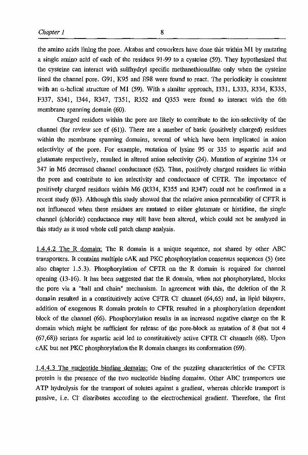
the amino acids lining the pore. Akabas and coworkers have done this within Ml by mutating
a single amino acid of each of the residues 91-99 to a cysteine (59). They hypothesized that
the cysteine can interact with sulfhydryl specific methanethiosulfate only when the cysteine
lined the channel pore. G91, K95 and E98 were found to react. The periodicity is consistent
with an a-helical structure of MI (59). With a similar approach, 1331, L333, R334, K335,
F337, S341, 1344, R347, T351, R352 and Q353 were found to interact with the 6th
membrane spanning domain (60).
Charged residues within the pore are likely to contribute to the ion-selectivity of the
channel (for review see cf (61». There are a number of basic (positively charged) residues
within the membrane spanning domains, several of which have been implicated in anion
selectivity of the pore. For example, mutation of lysine 95 or 335 to aspartic acid and
glutamate respectively, resulted in altered anion selectivity (24). Mutation of arginine 334 or
347 in M6 decreased channel conductance (62). Thus, positively charged residues lie within
the pore and contribute to ion selectivity and conductance of CFTR. The importance of
positively charged residues within M6 (R334, K355 and R347) could not be confmned in a
recent study (63). Although this study showed that the relative anion permeability of CFTR is
not influenced when these residues are mutated to either glutamate or histidine, the single
channel (chloride) conductance may still have been altered, which could not be analyzed in
this study as it used whole cell patch clamp analysis.
1.4.4.2 The R domain: The R domain is a unique sequence, not shared by other ABC
transporters. It contains multiple cAK and PKC phosphorylation consensus sequences (5) (see
also chapter 1.5.3). Phosphorylation of CFTR on the R domain is required for channel
opening (13-16). It has been suggested that the R domain, when not phosphorylated, blocks
the pore via a "ball and chain" mechanism. In agreement with this, the deletion of the R
domain resulted in a constituitively active CFTR cr channel (64,65) and, in lipid bilayers,
addition of exogenous R domain protein to CFTR resulted in a phosphorylation dependent
block of the channel (66). Phosphorylation results in an increased negative charge on the R
domain which might be sufficient for release of the pore-block as mutation of 8 (but not 4
(67,68» serines for aspartic acid led to constituitively active CFTR cr channels (68). Upon
cAK but not PKC phosphorylation the R domain changes its conformation (69).
1.4.4.3 The nucleotide binding domains: One of the puzzling characteristics of the CFTR
protein is the presence of the two nucleotide binding domains. Other ABC transporters use
ATP hydrolysis for the transport of solutes against a gradient, whereas chloride transport is
passive, i.e. Cl' distributes according to the electrochemical gradient. Therefore, the first
9 Introduction
question to be answered was whether CFfR channel opening required ATP hydrolysis or just
ATP binding. ATP and several analogues were found to directly bind to CFfR (70). Also,
mutations in the NBD' s caused an altered relation between channel activation and ATP
concentration, indicative of a direct interaction between ATP and CFfR (71). Whether CFfR
actually required ATP hydrolysis was investigated by using triphosphate analogues.
Hydrolyzable triphosphates (ATP, GTP, ITP, UTP and CTP) were able to induce channel
opening, non- or poorly hydrolyzable triphosphates (AMP-PNP, ATPyS, AMP-PCP, AMP
CPP) could not (72). Direct ATP hydrolysis was measured at NEDI in fusion with the
maltose binding protein (73). It thus appears that ATP binding and hydrolysis is required to
open the channel. ADP and AMP could not support channel activity (72,74). Both NED's
contain Walker A (GXXXXGKT/S) and B (ZZZZD, Z= hydrophobic) consensus motifs for
ATP binding (75), lying, in ABC transporters, at a distance of 133-166 aa from each other
(76). Mutations in the NED's were generated to investigate individual roles of the NBD's in
CFfR function, and are summarized below.
1.4.4.4 The role of NBD!: Mutations interfering with nucleotide binding, like G551D (77),
or hydrolysis in NBDI show decreased CFfR channel Po (71,78,79), reduced forskolin and
IBMX activation (80), or reduced the access to the activated state (81). From these
observations it can be concluded that ATP hydrolysis at NBD! is required for CFfR channel
opening.
1.4.4.5 The role of NED2: At NBD2, the situation is somewhat more complex. Disrupting or
impairing nucleotide binding, or interfering with the hydrolysis of ATP result in a low
channel Po (78,79) and decrease access to the activation state in Xenopus oocytes (81). Thus,
ATP hydrolysis at NBD2 is required for CFfR channel activation. In addition, ATP
hydrolysis at NBD2 is required for channel closure as mutations that interfere with ATP
hydrolysis, besides decreasing Po, increased the channel burst duration (78,79). Similarly,
these mutations stabilized the active state of CFfR (81).
The kinetics of CFTR opening and closure was studied in detail by Gunderson and
colleagues (79). A second open state (0,) of CFfR was detected with slightly increased
channel conductance. Mutations at NBD2 (K1250A, K1250G, K1250M, K1250T and
D!370N) interfere with the transition from 0, to 0, and caused a large increase in burst
duration. From these data, the authors hypothesized that the 0, state of CFfR is entered upon
ATP hydrolysis at NBD1. The 0, state of the channel is entered upon ATP hydrolysis at
NBD2. From the 0, state the channel can close spontaneously. Thus mutations that interfere
with ATP hydrolysis at NBD2 interfere with channel closure (79). Non hydrolyzable
nucleotides e.g. AMP-PNP (78,82), or agents that interfere with ATP hydrolysis like PPi (83)

Chapter 1 10
or VO, and BeF, (84) also induce a prolonged burst duration of CFTR when administered in
the presence of ATP. This can be explained by a model in which these agents interfere with
ATP hydrolysis at NBD2. The detailed understanding of the complex model of CFTR channel
gating provides a novel pathway for the development of pharmacotherapy for CF, as it
appears possible to increase CFTR channel activity by interfering with A TP hydrolysis at
NBD2. Increasing CFTR channel activity may be of benefit for patients with residual CFTR
activity or, in future, support CFTR expression elicited via gene transfer.
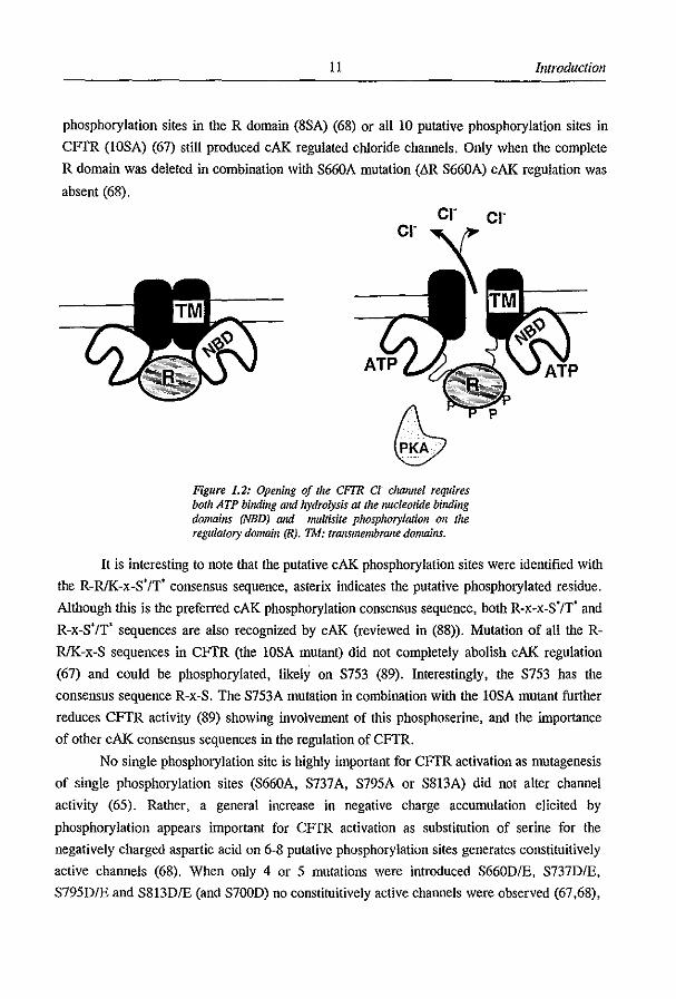
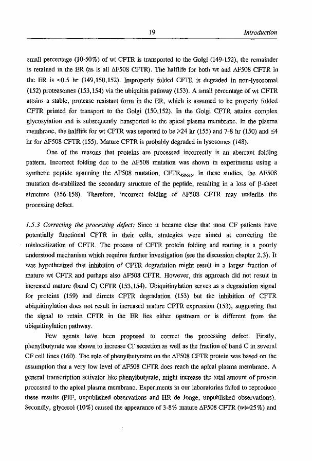
1.4.5 Regulation oj CFTR activity: The regulation of the CFTR cr channel is complex; both
multisite phosphorylation on the R domain by cAMP dependent protein kinase (cAK) and the
binding and hydrolysis of ATP on the nucleotide binding domains (NED's) are required for
channel opening (figure 1.2). Once phosphorylated by cAK, ATP alone is sufficient for
channel opening (72). It has been suggested by Gadsby and coworkers that differences in the
phosphorylation state of CFTR correspond to differences in channel activity (85), perhaps due
to the presence of a labile phosphorylation site (78). For an excellent review on CFTR
channel gating see (86). Below, an overview is given of the complex activation of CFTR by
various protein kinases and nucleotide triphosphates.
1.4.5.1 cAMP regulation: CFTR can be activated in intact cells by the second messenger
cAMP or, in excised membrane patches, by cAK (an effector enzyme of cAMP) and ATP, as
was shown in transfected Hela cells (13), Sf9 insect cells (15), CHO cells (14), IEC cells (17)
and upon reconstitution of purified CFTR in planar lipid bilayers (16). CFTR might be a
direct target for cAK as the initial analysis of the primary protein sequence of CFTR revealed
10 putative cAK consensus sequences, 9 of these are located within the R domain. (5).
Indeed, CFTR is phosphorylated by cAK both in vitro and in vivo (65,67,68). Multiple sites
are phosphorylated as the stoichiometry of phosphate incorporation in the R domain of CFTR
was 5-6 mol/mol (69,87).
A search was conducted to identify the sites phosphorylated by cAK. 13 sites
phosphorylated by cAK were identified in vitro, 4 of which (S660, S737, S795 and S813)
were also phosphorylated in vivo (65). Analysis of phosphorylation sites in CF2, a peptide
corresponding to most of the R domain (CFTR.,,.,,,), or wt CFTR revealed a similar pallern:
S660, S700, S737, S813 and either S768 or S795, or both, are phosphorylated in vitro, and at
least S660 and S700 in vivo (87). Mutation of the 4 primarily phosphorylated sites in vivo,
S660A, S737 A, S795A and S813A (the quad mutant) abolished cAMP induced chloride
secretion (65), indicating that these serines were important for cAK regulation. However, the
quad mutant expressed at high levels did reveal cAK regulated CFTR activity, albeit with
lower Po (67,68). Residual cAK regulated CFTR activation of the quad mutant indicated
other potential regulatory phosphorylation sites. Substitution of all 8 consensus serine
11 Introduction
phosphorylation sites in the R domain (8SA) (68) or all 10 putative phosphorylation sites in
CFTR (1OSA) (67) still produced cAK regulated chloride channels. Only when the complete
R domain was deleted in combination with S660A mutation (llli S660A) cAK regulation was
absent (68).
cr CI-
Figure 1.2: Opening oj the CFTR Ct chOlUIel requires both ATP binding aJuJ hydrolysis at the IUlcleotide binding domains (NED) and I1Ulltisite phosphoryiaJiOll 011 the regulatory domain (R), TM: transmembrane domains.
It is interesting to note that the putative cAK phosphorylation sites were identified with
the R-RJK-x-S'/T' consensus sequence, asterix indicates the putative phosphorylated residue.
Although this is the preferred cAK phosphorylation consensus sequence, both R-x-x-S'/T' and
R-x-S'/T' sequences are also recognized by cAK (reviewed in (88». Mutation of all the R
RJK-x-S sequences in CFTR (the 10SA mutant) did not completely abolish cAK regulation
(67) and could be phosphorylated, likely on S753 (89). Interestingly, the S753 has the
consensus sequence R-x-S. The S753A mutation in combination with the 10SA mutant further
reduces CFTR activity (89) showing involvement of this phosphoserine, and the importance
of other cAK consensus sequences in the regulation of CFTR.
No single phosphorylation site is highly important for CFTR activation as mutagenesis
of single phosphorylation sites (S660A, S737A, S795A or S813A) did not alter channel
activity (65). Rather, a general increase in negative charge accumulation elicited by
phosphorylation appears important for CFTR activation as substitution of serine for the
negatively charged aspartic acid on 6-8 putative phosphorylation sites generates constituitively
active channels (68). When only 4 or 5 mutations were introduced S660DIE, S737D/E,
S795DIE and S813DIE (and S700D) no constituitively active channels were observed (67,68),
Chapter 1 12
indicating that a large accumulation of negative charge is required.
1.4.5.2 PKC regulation: 7 putative phosphorylation sites for PKC, the Ca'+ and phospholipid
dependent protein kinase, were identified (5). PKC directly phosphorylates CFTR ill vitro
(90) and ill vivo on serines S686 and S790 (87). It was found that PKC could directly activate
CFTR at 10-15% ofcAK activity (14,90). Furthermore, Cl" secretion was induced by phorbol
myristate acetate (PMA, an activator of PKC) in CFTR transfected C127i cells (91) but not in
mouse L-cells (92). Cardiac CFTR cr channels could be submaximally activated in intact
cells by 4p-phorbo1 12,13-dibutyrate (pDBu, an activator of PKC) stimulation (93). In
addition to a being a direct activator of CFTR, PKC was reported to potentiate cAK activation
CFTR (14,94), even with CFTR mutated for all consensus cAK phosphorylation sites (IOSA)
(67). The potentiating effect was not found by Berger and coworkers (90) and thus might be
cell type dependent. Prolonged exposure to PDBu, 0.5-4 hr, did not induce a chloride current
by itself, but could potentiate the cAMP response by 31 % in pancreatic duct cells (95). In
addition, inhibition of PKC slowed the rate of CFTR rundown in the whole cell patch (95). In
summary, PKC has a twofold effect on CFTR activity. Firstly, PKC can directly activate
CFTR, but the effect is relatively small when compared to cAK activation of CFTR.
Secondly, PKC appears to have a potentiating effect on cAK activated CFTR. Both PKC
effects however, appear to be cell type dependent.
1.4.5.3 cGMP regulation: A major class of microbial enterotoxins, i.e. the family of heat
stable enterotoxins secreted by E-coli (STa), as well as the endogenous STa like peptide
hormone guanylin, result in the accumulation of the second messenger cyclic GMP (cGMP)
via the activation of a guanylyl cyclase (GC-C). The cGMP response in the enterocyte is
involved salt and water secretion across the intestinal epithelium. As the cGMP induced
chloride secretion is defective in CF epithelia, the involvement of CFTR in this pathway was
suggested (for review see (96». Several mechanisms have been proposed to link cGMP to the
CFTR cr channels, one of which is a cross-activation by cGMP of cAMP dependent protein
kinase. This was shown in T84 intestinal epithelial cells (97,98) and Cac02 cells (99). cAK
crossactivation by cGMP was based on the following observations: The cGMP response could
be inhibited by PKI, an inhibitor of cAK (98,99) (but see (100» but not with KT5823, an
inhibitor of cGKI (99,101). The cGMP response could not be evoked by specific activators of
cGKIo: and P (97). cGMP did not modulate the cAMP concentration (98). Also, the
concentrations of cGMP analogues used or the cGMP evoked (by STa) in these studies
exceeds the concentration at which cGMP cross-activates cAK. The Ka of cAK for cAMP is
0.D2 11M, for cGMP it is 4.1 11M (102).
13 IllIroductioll
cGMP may also activate CFTR via activation of cGMP dependent protein kinases
(cGK's). To date, two isoenzymes of cGK's are known, cGKI and cGKll, for review see
(103,104). Both cGKI (25,87,90) and cGKll (25) are capable of phosphorylating CFTR
(and/or CF2) ill vitro on similar sites as phosphorylated by cAK (25,87). cGK's can
phosphorylate the cAK consensus sequence R-RlK-x-SIT as the cGK consensus sequences are
R(K)-R(K)-R(K)-x-S/T and R(K)-R(K)-x-S/T (88). Although both cGK's can phosphorylate
CFTR ill vitro, cGKI proved virtually incapable of activating CFTR in excised patches of
3T3 fibroblasts (25,90), but could activate CFTR like cr channels in T84 human colonic
epithelial cells (105). cGKll could activate CFTR (25), albeit with slower kinetics than cAK.
Lin and coworkers (105) found CFTR-like Cl' channels in excised patches from T84 cells
when stimulated only by ATP and cGMP. They concluded that a membrane bound cGK was
responsible for CFTR activation. cGKll is attached to the membrane via N-tenninal
myristrylation, suggesting that cGKll is a good candidate for CFTR activation in these
experiments.
Recently, a third mechanism of cGMP regulation of CFTR was proposed in which
cGMP directly activates CFTR in an allosteric fashion (106). cGMP activation of CFTR
chloride channels was demonstrated by the two electrode voltage clamp technique following
injection of CFTR mRNA into Xenopus oocytes. This response was insensitive to both cAK
and cGKI inhibitors. A putative cyclic nucleotide binding site in CFTR was found. Upon
mutation of residues lining the cGMP binding site, altered cGMP responses were found in 217
mutations (106). However, studies in mammalian expression systems, including our own data
did not show evidence for a direct activation of CFTR by cGMP and ATP in intact cells or
excised patches (25,107).
1.4.5.4 Tyrosine kinases: In addition to regulation of CFTR by the serine/threonine protein
kinases cAK, cGKll and PKC, several groups have studied the involvement of tyrosine
kinases in the regulation of CFTR. Although no tyrosine residues on CFTR appeared to be
phosphorylated (65), genistein, a tyrosine kinase inhibitor (108,109), was able to activate
CFTR in the CFTR transfected cell lines IEC-CF7 and NIH-3T3 (110,111) and in CFTR
expressing HT291B6 adenocarcinoma and T84 colonic epithelial cells (112). Furthermore,
genistein induced a chloride secretion in the shark rectal gland (113) and enhanced the cAMP
induced chloride secretion across rat distal colon (114). In both tissues, the genistein induced
chloride secretion was dependent on the activation of CFTR-Cl' channels. Genistein did not
activate CFTR by increasing the [cAMP]; (110,111), but its action required basal cAMP
levels (112) suggesting a possible action as a protein phosphatase inhibitor (112,115). In
chapter 3.2 we provide evidence that genistein neither acts via tyrosine kinase inhibition nor
Chapter 1 14
via inhibition of a protein phosphatase. Instead, we propose that genistein directly acts on
CFrR (French et aI, submitted). Among other tyrosine kinase inhibitors tested, only
tyrphostill 47 (110) and B42 (111) but not tyrphostin 51 and 25, herbimycin, AG126 and,
cantharidin were able to activate CFrR in NIH-3T3 fibroblasts (H.R. de Jonge, unpublished
observation). Similar observations were made using other cell types (112).
1.4.5.5 Other protein kinases: The calcium-calmodulin dependent protein kinase-II (CaMKII)
could not activate CFrR in the excised patch (90), nor did CamKII inhibitors affect the STa
induced CFrR Cl" secretion (98). No in vitro phosphorylation of CFrR (90) and CF2 (87)
was observed with CaMKII. CaMKI but not CamKIlI or Casein kinase II was able to
phosphorylate CF2 in vitro (87). Whether CamKI has a role in CFrR activation is presently
unclear.
1.4.5.6 Dephosphorylation: As phosphorylation activates the channel, its counterpart,
dephosphorylation, should inactivate the channel. The investigations on CFrR
dephosphorylation do not give a consistent picture as to which protein phosphatases are
involved. The protein phosphatases 1 and 2B neither affected CFrR channel currents in the
excised patch nor did they dephosphorylate CFrR in vitro (90). Phosphatase 2A decreased the
cAK induced CFrR current by 63% in transfected NIH-3T3 fibroblasts, and this phosphatase
could also directly dephosphorylate CFrR in vitro (90). Thus, phosphatase 2A and not lor
2B is involved in CFrR dephosphorylation. In contrast, phosphatase 1 and 2A inhibitor
okadaic acid did not prevent channel rundown (deactivation) or increase Po in transfected
CHO cells (14,116) (but see below).
Alkaline phosphatase decreased CFrR channel Po (14) and directly counteracted cAK
induced CFrR phosphorylation (116). AikaJine phosphatase inhibitors (VO" lBMX or Br-t),
inhibited CFrR channel rundown upon excision of a membrane patch (116). However, these
experiments were performed in the absence of extracellular A TP, thus delayed channel
rundown could have been caused by a reduced ATP dissociation upon excision rather than
CFrR dephosphorylation. In a different study, alkaline phosphatase was found to decrease
CFrR activity, but upon removal of the phosphatase CFrR activity was restored. The authors
suggested that the ATPase activity of alkaline phosphatase caused channel deactivation (90).
In summary, it is likely that protein phosphatase 2A and perhaps alkaline phosphatase, and
not 1 or 2B, mediate the dephosphorylation of CFrR. However, further analysis is required
to accurately determine which protein phosphatase is involved in CFrR dephosphorylation.
1.4.6 CFTR modulation of other iOIl channels: CFfR has been reported to modulate other ion
conductances, which is not an uncommon principle for ABC transporters (for review see
15 Introduction
(117)). There have been several reports on a regulatory role for CFrR on the outward
rectifying chloride channel, ORCC, an ubiquitously expressed chloride channel. Initially it
was thought that defective cAK regulation of this channel was the underlying cause of cystic
fibrosis (for review see (118)). However, the cloning and characterization of the CF gene
revealed that CFrR and the ORCC are distinct proteins, see e.g. (119), encoded by separate
genes (see also (17)).
Under specific whole cell patch clamp conditions (5 mM ATP in the pipette), cAMP
induces CI' currents that are derived from both CFrR and ORCC (120). Both CUrrents are
absent in CF cells, indicating a potential cross talk between ORCC and CFrR. Similar
experiments revealed that CFrR mutant A455E but not G551D was able to regulate the
ORCC (121). A regulatory role for CFrR on the ORCC was further suggested by single
channel patch clamp experiments, showing ORCC activation by cAK and ATP only in CFrR
expressing cells, but not in CF cell lines (119,122). Reconstitution of CFrR and ORCC in
lipid bilayers showed similar results; the presence of CFrR was required for cAK activation
of the ORCC (123), and could not be replaced by the inactive CFrR mutant G551D (124).
An interesting model was proposed by Schwiebert and colleagues linking CFrR to the
ORCC (125). Conductance of ATP through CFrR activates the P,u receptor subsequently
activating the ORCC. The model was modified later when it was shown that extracellular
ATP by itself was insufficient to activate the ORCC but that the actual presence of (G551D)
CFrR was required (124). G-proteins were hypothesized to modulate the additional
interaction between CFrR and the ORCC (126). However, the model does not unify the
above mentioned data and there remain considerable controversies. Firstly, the model requires
that CFrR modulates ATP secretion. However, there is little evidence that CFrR conducts
ATP itself (127-129). A previous study which did show ATP conductivity of CFrR (130)
failed to provide convincing data that CFrR was in fact the channel studied. Secondly, in
bilayer experiments (123), the Pw receptor is likely to be absent (due to purification of the
reconstituted proteins). Finally, CFrR was not detected in patches from mouse nasal
epithelia, but still conferred cAK sensitivity to the ORCC (119). In summary, the activation
of the ORCC by CFrR via ATP is a highly controversial issue. This will remain so until the
sometimes conflicting data is explained, experiments are reproduced by independent
observers, and gaps in the theory (e.g. how does ATP exit the cell, which G-protein is
involved, and what messengers are required to activate the ORCC) are ftlled.
An increased arniloride-sensitive potential difference was found across pulmonary
epitllelia of CF patients (10). Amiloride blocks epithelial apical Na+ channels (ENaC's) and it
was concluded that the increased arniloride response in CF tissues was the result of increased
Chapter 1 16
Na+ absorption (10), reviewed in (131). Transfection of CFfR to a CF tissue, reduces the
amiloride sensitive short circuit current (Isc) to values seen in controls (132). It was
concluded that CFfR restored the Na+ hyperabsorption to normal. Subsequently, several
groups have investigated a relation between CFfR and ENaC's in more detail. Chinet and
colleagues found that a specific type of Na+ channels had a higher Po in CF patients (133).
Also, coexpression of CFfR and ENaC's led to a lower Na+ current compared to the Na+
current in absence of CFfR (134). These results suggest that the presence of CFfR has a
negative regulatory effect on the ENaC's. The most convincing evidence of an interaction
between CFTR and the ENaC came from reconstitution experiments in lipid bilayers. In these
experiments, the simple presence of CFfR reduced the channel Po of the ENaC (135).
Although CFfR might negatively regulate ENaC's, we doubt the relevance of Na+
hyperabsorption in CF. Because, in order to have net flux of Na +, anions (Cl") have to follow,
otherwise just a maximal Na + gradient is created. In the Ussing chamber, the current required
for short circuiting the tissue (the Isc) is only a measure for actively transported ions like
Na+, and not passively diffusing ions like cr (136). The short circuit conditions do not
represent the in vivo situation where actively transported Na + flux has to be followed by
passive anion flux through a shunt pathway that may utilize CFfR chloride channels.
Therefore, increased Isc is not necessary correlated with increased flux ill vivo (Bijman,
unpublished observations). Thus, tile increased amiloride sensitive Isc seen in CF, does not
imply Na + hyperabsorption ill vivo; in CF no cr ions are capable of following the Na + flux.
Thus, although CFTR might reduce the ENaC channel activity, its relevance for Na+
absorption is debatable.
1.5 The 8]'508 mutation
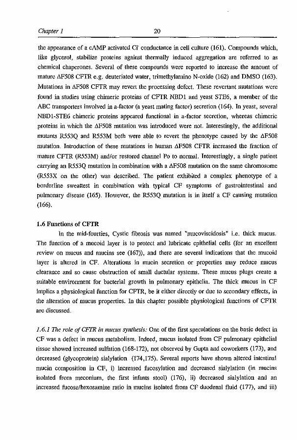
Mutations which result in a loss of channel activity in CFfR can be classified into
four major categories, class I-N (fig 1.3), reviewed in (137). Class I mutations have a
defective protein production. Examples of CFfR mutants belonging to this category are all
mutations leading to a premature stop, thus stopcodon-, frameshift-, splice- mutations as well
as promotor mutations. Class II mutations have defective processing, meaning that CFfR
protein is made, but it is not correctly targeted to the apical plasma membrane. AF508,
A1507, NI303K and several others belong to this category. NBDI appears very sensitive for
processing mutations, as all (except for N1303K) have been found in this area. Class ill
mutations have a defective regulation; CFfR is expressed in the apical membrane, but can not
be activated sufficiently. A typical example is the G551D mutation. Class N mutations have
defective conductance, meaning that CFfR is present in the apical membrane but its
17 Introduction
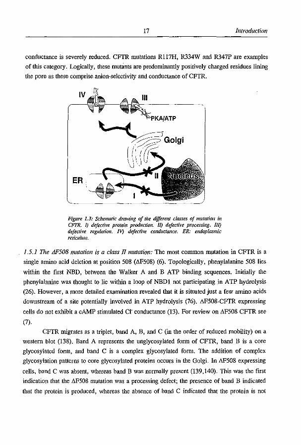
conductance is severely reduced. CFfR mutations R117H, R334W and R347P are examples
of this category. Logically, these mutants are predominantly positively charged residues lining
the pore as these comprise anion-selectivity and conductance of CFfR.
IV
~GOI9i
ER
Figure 1.3: Sdlematic drawing of the different classes oj mutation if! CFTR. J) defective protein production. II) defective processing. Ill) defective regulatioll. IV) defective conductance. ER: endoplasmic reticulum.
1.5.1 The L1F508 mutation is a class JlIIUltation: The most common m)ltation in CFfR is a
single amino acid deletion at position 508 (&'508) (6). Topologically, phenylalanine 508 lies
within the fIrst NBD, between the Walker A and B ATP binding sequences. Initially the
phenylalanine was thought to lie within a loop of NBDI not participating in ATP hydrolysis
(26). However, a more detailed examination revealed that it is situated just a few amino acids
downstream of a site potentially involved in ATP hydrolysis (76). 8F508-CFfR expressing
cells do not exhibit a cAMP stimulated cr conductance (13). For review on 8F508 CFTR see
(7).
CFfR ntigrates as a triplet, band A, B, and C (in the order of r~duced mobility) on a
western blot (138). Band A represents the unglycosylated fonn of CFTR, band B is a core
glycosylated fonn, and band C is a complex glycosylated fonn. The addition of complex
glycosylation patterns to core glycosylated proteins occurs in the Golgi. In 8F508 expressing
cells, band C was absent, whereas band B was nonnally present (139,140). This was the fIrst
indication that the &'508 mutation was a processing defect; the presence of band B indicated
that the protein is produced, whereas the absence of band C indicated that the protein is not
Chapler 1 IS
properly processed to the Golgi. Subsequently, it was shown by inununocytochemistry that
"F50S CFfR is not localized at the plasma membrane but has a cytoplasmic (perinuclear)
localization (19,21,22,139,141).
It was shown that the processing defect is temperature sensitive; reducing cell culture
temperatures for ;:,2 days at :530'C resulted in the appearance of C-band CFrR on western
blot, and the appearance of cAMP stimulated cr permeability by whole cell patch clamping
(141,142). This rmding was of some interest because it implied that "F50S CFrR expressing
cells contain potential cr charmel activity, and that CF therapy could be aimed at the
relocation of "F50S CFrR. Several other groups have shown lIF50S CFrR expression on the
cell membrane; Sf9 insect cells (143) and Xenopus oocytes (SO), systems that are traditionally
cultured at low temperatures, express lIF50S CFrR on their cell surface. lIF50S CFfR was
also observed on the surface of vaccinia infected (vertebrate) Vero cells (144).
Expression of active lIF50S CFrR on the cell membrane allowed biophysical
characterization of the charmel. lIF508 CFrR appeared to have normal anion conductance and
selectivity (143,144) (pJF, unpublished observations). The charmel can be activated by cAK
when reconstituted in lipid bilayers (143) and, in the cell-attached patch clamp configuration
by cAMP agonists (144) (pJF, unpublished observations) albeit with reduced cAMP
sensitivity i.e. requiring higher cAMP concentrations (SO) (but see (145)). A 34 fold
reduction in charmel Po was found in vertebrate cells (142,144,145) but not in Sf9 insect
cells (143). A marked reduction in Po was also found in excised patches from NIH-3T3
fibroblasts expressing lIF50S CFfR (pJF, unpublished observation). Except for the reduced
Po, all other parameters were similar to wt CFrR (145). The reduced Po of lIF508 CFrR
indicates that the lIF508 mutation is not only a processing mutant, but also a class ill mutant
with defective regulation. As was discussed before, the lIF508 mutation lies with in a position
putatively involved in ATP hydrolysis (76), making it tempting to speculate that the reduced
Po is caused by a reduced ATP hydrolysis at NBDI. "F508 CFrR is not a class I mutant as
mRNA levels are normal (22,146).
1.5.2 Processing of wI and fJF508 CFTR: When it became clear that the most common
mutation in CFrR, lIF508, is a processing mutant, several groups started studying the
process of CFrR maturation. Translation of CFTR mRNA into protein occurs in the
endoplasmic reticulum (ER), where the protein attains its proper configuration. For complex
proteins like CFrR the folding process is aided by chaperones (for review see (147)). HSP70
(14S) and calnexin (149) are chaperones with affinity for CFfR. Once the protein has attained
its proper folding configuration, the protein is transported to the Golgi. Surprisingly, only a
19 Introduction
small percentage (10-50%) of WI CFfR is transported to the Golgi (149-152), the remainder
is retained in the ER (as is all 1IF50S CFfR). The halflife for boih WI and 1IF50S CFfR in
the ER is "0.5 hr (149,150,152). Improperly folded CFfR is degraded in non-lysosomal
(152) proteasomes (153,154) via the ubiquitin pathway (153). A small percentage of wt CFfR
attains a stable, protease resistant form in the ER, which is assumed to be properly folded
CFfR primed for transport to the Golgi (150,152). In the Golgi CFfR attains complex
glycosylation and is subsequently transported to the apical plasma membrane. In the plasma
membrane, the halflife for wt CFfR was reported to be;0,24 hr (155) and 7-S hr (150) and 91
hr for 8F50S CFfR (155). Mature CFfR is probably degraded in Iysosomes (14S).
One of the reasons that proteins are processed incorrectly is an aberrant folding
pattern. Incorrect folding due to the 1IF50S mutation was shown in experiments using a
synthetic peptide spanning the 8F50S mutation, CFfR.,5Q.516' In these studies, the 8F50S
mutation de-stabilized ihe secondary structure of the peptide, resulting in a loss of fl-sheet
structure (156-15S). Therefore, incorrect folding of 8F50S CFfR may underlie the
processing defect.
1.5.3 Correcling the processing defect: Since it became clear that most CF patients have
potentially functional CFfR in their cells, strategies were aimed at correcting the
mislocalization of CFfR. The process of CFfR protein folding and routing is a poorly
understood mechanism which requires further investigation (see the discussion chapter 2.3). It
was hypothesized ihat inhibition of CFfR degradation might result in a larger fraction of
mature wt CFfR and perhaps also 8F508 CFfR However, this approach did not result in
increased mature (band C) CFfR (153,154). Ubiquitinylation serves as a degradation signal
for proteins (159) and directs CFfR degradation (153) but the inhibition of CFfR
ubiquitinylation does not result in increased mature CFfR expression (153), suggesting ihat
the signal to retain CFfR in the ER lies either upstream or is different from the
ubiquitinylation paihway.
Few agents have been proposed to correct the processing defect. Firstly,
phenylbutyrate was shown to increase C], secretion as well as the fraction of band C in several
CF cell lines (160). The role of phenylbutyratre on the 8F50S CFfR protein was based on the
assumption that a very low level of 8F508 CFfR does reach the apical plasma membrane. A
general transcription activator like phenylbutyrate, might increase the total amount of protein
processed to the apical plasma membrane. Experiments in our laboratories failed to reproduce.
these results (pJF, unpublished observations and HR de Jonge, unpublished observations).
Secondly, glycerol (10%) caused the appearance of3-S% mature 8F508 CFfR (w~5%) and
Chapter 1 20
the appearance of a cAMP activated Cl" conductance in cell culture (161). Compounds which,
like glycerol, stabilize proteins against thermally induced aggregation are referred to as
chemical chaperones. Several of these compounds were reported to increase the amount of
mature t.F508 CFrR e.g. deuteriated water, trimethylamino N-oxide (162) and DMSO (163).
Mutations in t.F508 CFTR may revert the processing defect. These revertant mutations were
found in studies using chimeric proteins of CFrR NEDI and yeast STE6, a member of the
ABC transporters involved in a-factor (a yeast mating factor) secretion (164). In yeast, several
NEDI-STE6 chimeric proteins appeared functional in a-factor secretion, whereas chimeric
proteins in which the t.F508 mutation was introduced were not. Interestingly, the additional
mutants R553Q and R553M both were able to revert the phenotype caused by the t.F508
mutation. Introduction of these mutations in human t.F508 CFrR increased the fraction of
mature CFrR (R553M) and/or restored channel Po to normal. Interestingly, a single patient
carrying an R553Q mutation in combination with a t.F508 mutation on the same chromosome
(R553X on the other) was described. The patient exhibited a complex phenotype of a
borderline sweattest in combination with typical CF symptoms of gastrointestinal and
puimonary disease (165). However, the R553Q mutation is in itself a CF causing mutation
(166).
1.6 Functions of CFfR
In the mid-fourties, Cystic fibrosis was named Itmucoviscidosis" i.e. thick mucus.
The function of a mucoid layer is to protect and lubricate epithelial cells (for an excellent
review on mucus and mucins see (167», and there are several indications that the mucoid
layer is altered in CF. Alterations in mucin secretion or properties may reduce mucus
clearance and so cause obstruction of small ductular systems. These mucus plugs create a
suitable environment for bacterial growth in puimonary epithelia. The thick mucus in CF
implies a physiological function for CFrR, be it either directly or due to secondary effects, in
the alteration of mucus properties. In this chapter possible physiological functions of CFrR
are discussed.
1.6.1 The role of CFrR il/lIIl1clIS synthesis: One of the first speculations on the basic defect in
CF was a defect in mucus metabolism. Indeed, mucus isolated from CF puimonary epithelial
tissue showed increased sulfation (168-172), not observed by Gupta and coworkers (173), and
decreased (glycoprotein) sialylation (174,175). Several reports have shown altered intestinal
mucin composition in CF, i) increased fucosylation and decreased sialylation (in mucins
isolated from meconium, the first infants stool) (176), ii) decreased sialylation and an
increased fucose/hexosamine ratio in mucins isolated from CF duodenal fluid (177), and iii)

21 llllrodllctioll
increased fucosylation without decreased sialylation (178). No increased fucosylation was
observed in CF bronchial mucus (179). Fucosylation, sialylation and sulfation appear to be
among the last steps in the biosynthesis of glycoproteins. Thus, in CF there may be a defect
in the terminal glycosylation of glycoproteins.
Changes in terminal glycosylation pattern of mucus glycoproteins as observed in CF
may alter the physical properties of mucus. Firstly, the cleavage of sialic acid from mucus
increases the elasticity of mucus (180), and reduces its viscosity (181). Secondly, decreased
sialylation may result in increased bacterial adhesion. The desialylated glycoproteins, asialo
GMI and 2 and fucoasialo-GMI are specific binding sites for many bacterial strains, whereas
the sialylated forms of GMI-3 are not (174,182,183). Decreased sialylation might therefore
result in increased bacterial adhesion to CF glycoproteins (184).
An interesting hypothesis has been postulated linking CFTR to aIterations in mucin
glycosylation. The fucosyl-, sialyl- and sulfo- transferases are localized in the distal Goigi or
trans Golgi network and have distinct pH optima (for review see (185». If CFTR is involved
in the acidification of the distal Goigi or trans Golgi network, one can imagine a different
pattern of terminal glycosylation in CF. For instance, a more basic environment would result
in increased fucosylation (PH optimum 7-8.5) and decreased sialylation (PH optimum 5.8).
Furthermore, fucosylation and sialylation are mutually exclusive processes (186).
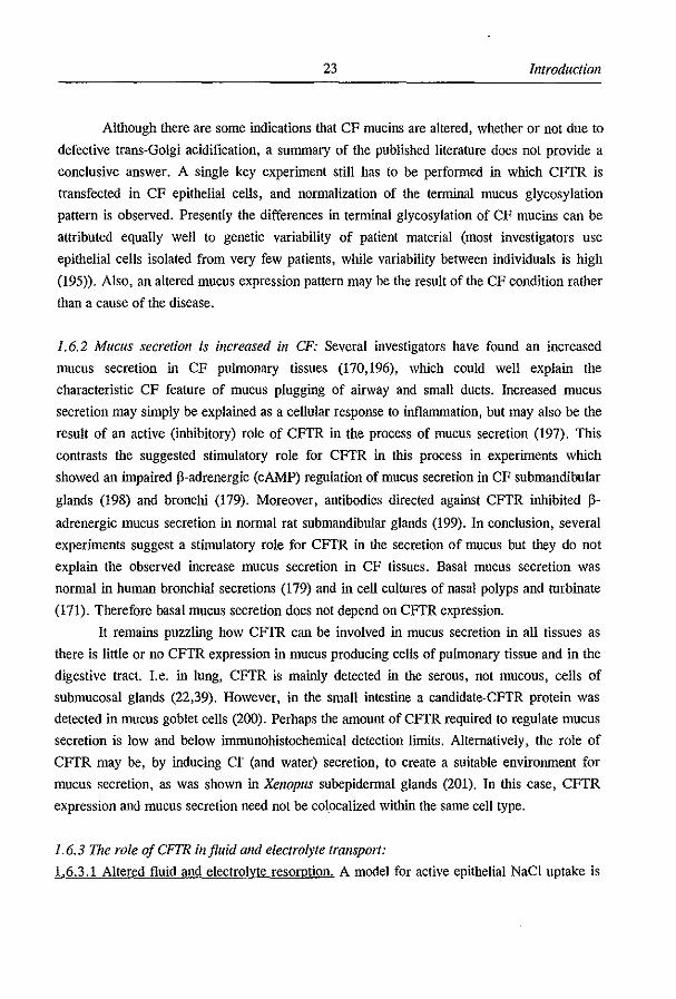
The rationale of decreased acidification of endosomal and Goigi compartments in the
absence of CFTR is excellently reviewed by AI Awqati (185). The theory is based on the
requirement of a counterion to generate a significant "'pH. In the absence of a counterion,
"'pH is limited by the potential difference (PD) created by active proton pumping (fig. 1.4).
Chloride may serve as the counterion and, via CFTR, may passively follow the electrical
gradient generated by the proton flux. The CFTR dependent acidification hypothesis requires
both the presence of CFTR in the Goigi and its involvement in the acidification process. Both
points are discussed below.
The presence of CFTR in the Golgi or trails Goigi network has not been demonstrated
directly. However de 1l0VO CFTR synthesis, derived from the ER, and the fusion of the trails
Goigi with (CFTR containing) vesicles derived from the endosomal pathway are two potential
sources of CFTR in the Goigi. The presence of CFTR has been demonstrated by
immunohistochemical analysis in clathrin coated vesicles (187) and early endosomes (188).
The functional presence of CFTR in endosomes can be monitored by the rate of
deacidification upon addition of a protonophore which is accelerated by CFTR dependent co
efflux of Cl" counterions. With this assay, the functional expression of CFTR in endosomes
has been demonstrated (189,190)

Chapler 1 22
Although several groups have examined the role of CFTR in the process of
acidification, most studies have been confmed to the study of vesicles of the endosomal
pathway, not measuring Golgi compartments. However, a direct relation between endosomal
acidification and reduced glycoprotein sialylation was shown in CHO mutants with
temperature-sensitive defective receptor-mediated endocytosis (191). A role for CFTR in
endosomai and early lysosomal acidification was suggested in studies using CF pulmonary
epithelial cells (175)' and in Swiss 3T3 fibroblasts (190)'. Furthermore, it was found that
C127 cells, expressing CFTR, acidifY when treated with cycloheximide, whereas M'508
expressors did not (192). As cellular acidification is a prerequisite for apoptotic DNA
cleavage, the investigators found that M'508 expressing cells are less primed to apoptotic
stimuli (192). In contrast to the studies listed above, other studies have failed to confmn a
role for CFTR in the acidification of both endosomal (189,193,194) and Golgi (193,194)
compartments. Thus, there is as yet no conclusive evidence for a role of CFTR in organelle
acidification.
cr ) pHz7.0 Vmz120 mV
cr pHz 5.0 VmzO mV
Figure 1.4: Tile role of a COUIIJerioll ill endosomal acidification. In absence of a cOlmlerion, prolon pumping is limited by the generated membrane potential.
, The experimenters compared endosomal acidification in CF nasal polyp cells with control tracheal cells, hardly the ideal control.
, A decreased endosomal acidification was found in t.F508 CFTR compared to wt CFTR expressing Swiss 3T3 fibroblasts. Mock transfected cells showed an acidification not different from WI CFTR transfectants, suggestive of a specific role of t.F508 CFTR.
23 Introduction
Although there are some indications that CF mucins are altered, whether or not due to
defective trans-Golgi acidification, a summary of the published literature does not provide a
conclusive answer. A single key experiment still has to be performed in which CFfR is
transfected in CF epithelial cells, and normalization of the terminal mucus glycosylation
pattern is observed. Presently the differences in terminal glycosylation of CF mucins can be
attributed equally well to genetic variability of patient material (most investigators use
epithelial cells isolated from very few patients, while variability between individuals is high
(195». Also, an altered mucus expression pattern may be the result of the CF condition rather
than a cause of the disease.
1.6.2 Mucus secretion is increased in CF: Several investigators have found an increased
mucus secretion in CF pulmonary tissues (170,196), which could well explain the
characteristic CF feature of mucus plugging of airway and small ducts. Increased mucus
secretion may simply be explained as a cellular response to inflammation, but may also be the
result of an active (inhibitory) role of CFfR in the process of mucus secretion (197). This
contrasts the suggested stimulatory role for CFfR in this process in experiments which
showed an impaired p-adrenergic (cAMP) regulation of mucus secretion in CF submandibular
glands (198) and bronchi (179). Moreover, antibodies directed against CFfR inhibited p
adrenergic mucus secretion in normal rat submandibular glands (199). In conclusion, several
experiments suggest a stimulatory role for CFfR in the secretion of mucus but they do not
explain the observed increase mucus secretion in CF tissues. Basal mucus secretion was
normal in human bronchial secretions (179) and in cell cultures of nasal polyps and turbinate
(171). Therefore basal mucus secretion does not depend on CFfR expression.
It remains puzzling how CFfR can be involved in mucus secretion in all tissues as
there is little or no CFfR expression in mucus producing cells of puhnonary tissue and in the
digestive tract. I.e. in lung, CFfR is mainly detected in the serous, not mucous, cells of
submucosal glands (22,39). However, in the small intestine a candidate-CFfR protein was
detected in mucus goblet cells (200). Perhaps the amount of CFfR required to regulate mucus
secretion is low and below immunohistochemical detection limits. Alternatively, the role of
CFfR may be, by inducing cr (and water) secretion, to create a suitable environment for
mucus secretion, as was shown in Xenopus subepidermal glands (201). In tltis case, CFfR
expression and mucus secretion need not be colocalized within the same cell type.
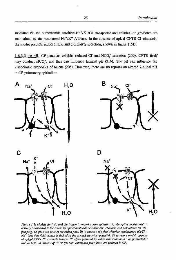
1.6.3 The role ojCFTR iI/fluid and electrolyte transport: 1.6.3.1 Altered fluid and electrolyte resorption. A model for active epithelial NaCI uptake is
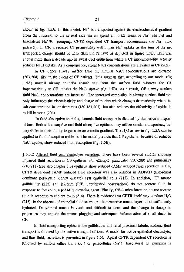
Chapter 1 24
shown in fig. 1.5A. In this model, Na+ is transported against its electrochemical gradient
from the mucosal to the serosal side via an apical amiloride sensitive Na + channel and
basolateral Na+fK+ pumping. CFTR dependent Cl' transport accompanies the Na+ flux
passively. In CF, a reduced Cl' permeability will impair Na+ uptake as the sum of the net
transported charge should be zero (Kirchhoff's law) as depicted in figure 1.5B. This was
shown more than a decade ago in sweat duct epithelium where a Cl' impenneability actually
reduces NaCI uptake. As a consequence, sweat NaCI concentrations are elevated in CF (202).
In CF upper airway surface fluid the luminal NaCI concentrations are elevated
(203,204), like in the sweat of CF patients. This suggests that, according to our model (fig
I.5A) nonnal airway epithelia absorb salt from the surface fluid whereas the Cl'
impenneability in CF impairs the NaCI uptake (fig 1.5B). As a result, CF airway surface
fluid NaCI concentrations are increased. The increased osmolality in airway surface fluid not
only influences the viscoelasticity and charge of mucins which changes dramatically when the
salt concentration in- or decreases (180,181,205), but also reduces the effectivity of epithelia
to kill bacteria (206).
In fluid absorptive epithelia, isotonic fluid transport is dictated by the active transport
of ions. Both salt absorptive and fluid absorptive epithelia may utilize similar transporters, but
they differ in their ability to generate an osmotic gradient. The H,o arrow in fig. 1.5A can be
applied to fluid absorptive epithelia. The model predicts that CF epithelia, because of reduced
NaCI uptake, show reduced fluid absorption (fig. 1.5B).
1.6.3.2 Altered fluid and electrolyte secretion. There have been several studies showing
impaired fluid secretion in CF epithelia. For example, pancreatic (207-209) and pulmonary
(210,211) (see also chapter 3.3) epithelia show reduced cAMP induced fluid secretion in CF.
CFTR dependent cAMP induced fluid secretion was also reduced in ADPKD (autosomal
dominant polycystic kidney disease) cyst epithelial cells (212). In addition, CF mouse
gallbladder (213) and jejunum (pJF, unpublished observations) do not secrete fluid in
response to forskolin, a [cAMPJ, elevating agent. Finally, CF-/- mice intestine do not secrete
fluid in response to cholera toxin (214). There is evidence that CFTR itself may conduct H,o
(215). In the absence of epithelial fluid secretion, the protective mucus layer is not sufficiently
hydrated. Dehydrated mucus is viscid and difficult to clear, and the change in rheogenic
properties may explain the mucus plugging and subsequent inflammation of small ducts in
CF.
In fluid transporting epithelia like gallbladder and renal proximal tubule, isotonic fluid
transport is directed by the active transport of ions. A model for active epithelial electrolyte,
and thus fluid, secretion is presented in figure 1.5C. Apical CFTR dependent Cl' secretion is
followed by cations either trans (K+) or paracellular (Na+). Basolateral Cl' pumping is
25 Introduction
mediated via the bumethanide sensitive Na + /K+ fCr transporter and cellular ion-gradients are
maintained by the basolateral Na+/K+ ATPase. In the absence of apical CFfR CI' channels,
the model predicts reduced fluid and electrolyte secretion, shown in figure LSD.
1.6.3.3 the pH. CF pancreas exhibits reduced cr and HCO; secretion (209). CFfR itself
may conduct HCO;, and thus can influence luminal pH (216). The pH can influence the
viscoelastic properties of mucus (205). However, there are no reports on altered luminal pH
in CF pulmonary epithelium.
c
8
o
~~cr \ l
Figure 1.5: Models for fluid alld electrolyte transport across epithelia. A) absorptive model: Na+ is actively transported 10 t"e serosa by apical amiloride sensitive Na+ chmUieis and basola/erat Na+ IK+ pumping. Ct passi~'el)' follows the cationjlolV. B) [" absence of apical chloride cOllductaJlce (CFTR), Na+ (alld thus fluid) uptake is limited by the created electrical porelUial. C) secretory model: opening of apical CFFR ct challllels induces ct ejJ1ILt fOllowed by either lranscelluiar K+ or paracellular Na+ or bOlh. In absellce ojCFIR (D) both catioll andjluidjllL\eS ore reduced ill CF.
Chapter I 26
1.6.4 The role oj CFIR in pathogen affinity, intel'llalizatioll and killing: Recurrent pulmonary
infections, a characteristic clinical manifestation of CF, may be caused by a reduced clearance
of bacteria. A reduced bacterial clearance in CF epithelia can be caused by an increased
bacterial adhesion due to the altered terminal glycosylation pattern of CF mucus (see chapter
1.6.1). Indeed, an increased Pseudomonas aemginosa adhesion to CF cells was found (217).
Increased P. ael'llginosa binding was also found in CFPAC (CF pancreatic duct cells,
homozygous for the AF508 mutation) and IB3 (CF bronchial cells, heterozygous
AF5081W1282X) cells compared to the same cell lines transfected with wt CFTR (184). P.
aeruginosa may not be the only bacteria displaying increased adhesion to CF epithelia,
Staphylococcus aureus and P. aeruginosa compete for the same binding sites (184). In
addition to increased P. aeruginosa adhesion, there may also be decreased P. aeruginosa
internalization in CF (218), with the fust predicted extracellular domain of CFTR acting as a
P. aemginosa receptor (218). Bacterial internalization may playa role in the host defence
mechanism. Finally CF epithelia may have a reduced effectivity to kill bacteria (206)
(discussed in chapter 1.6.3).
1.6.5 The role oJCFIR in elldo- and exocytosis: Bradbury and coworkers have found a CFTR
regulated inhibition of endocytosis and a stimulated exocytosis in CFPAC cells, upon
complementation with wt CFTR (219). In contrast, cAMP stimulation in T84 cells did not
induce increased apical CFTR expression (20,220). However, human nasal polyp cells did
show increased CFTR expression upon forskolin stimulation (221). Absence of CFTR
regulated endo- and exocytosis can contribute to CF disease pathogenesis as cAMP regulated
processes, i.e. fluid and electrolyte secretion, may be amplified by increased apical expression
of proteins involved in this process.
1.6.6 Summary: In this part of the thesis several hypotheses have been discussed which
associate a defect in an epithelial Cl channel to the devastating disease pathology of CF.
Apparently multiple causes may underlie the clinical manifestations of CF, as CF epithelia
may have i) an increased pathogen affinity (chapter 1.6.4) ii) increased mucus secretion
(chapter 1.6.2) iii) a reduced mucus clearance (due to altered viscoelastic properties, chapter
1.6.1 and 1.6.3) and, iv) a reduced pathogen killing (chapter 1.6.3). The combination of these
result in an environment favorable for bacterial growth and mucus plugging of small ducts.
1.7 Mouse models for CF:
Mouse models for CF can be relevant for understanding the pathogenesis of CF and
the basic physiological function of CFTR. They may also be used to aid research aimed at the

27 Introduction
development for new strategies leading to a cure for CF. Upon the cloning of the gene
encoding human CFrR (4-6) and the subsequent isolation of the mouse homologue (222), it
was possible to generate mouse models for CF. To date, most CF mouse models have been
generated for CFrR class I mutations (for CFrR mutation classification see figure 1.3) which
contain a premature stopcodon in exon 2 (223), exon 3 (224) or exon 10 (225-227). In
addition, several mouse models have been generated for the most common CFrR mutations
like (class II) LlF50B (22B-230) and (class ill) G551D (231).
Knockout CF mouse models (223-227,232) display a phenotype very similar to human
CF patients. The only knockout mouse model for CF not displaying obvious disease
symptoms was created by an insertion vector, resulting in a duplication of exon 10. The
inserted exon 10 contains a premature stop (226). Due to alternative splicing, the mutant exon
can be skipped and, as a result, some ",10% wt eftr mRNA is generated (233). Apparently
this is sufficient to prevent most of the obvious disease symptoms.
Cftr knockout animals display a general failure to thrive, amounting to 10-50%
reduced weight compared to wt animals (223-225,232). The mice show increased lethality,
especially in the first weeks of life, with only 5-10% of the animals surviving into adulthood
(223-225,232). Increased mortality is most likely caused by intestinal obstruction of the ileum
and subsequent peritonitis. Intestinal crypts are dilated and filled with eosinophilic material
(224,225,232). A wonn/ike caecum was observed (225). Other organs of the gastrointestinal
tract are not as severely affected as the intestine, but do show some signs of pathology.
Gallbladders may be ftlled with black bile in some animals, several others may show a
distended or even ruptured gallbladder (225). Variable disease symptoms have been observed
in the pancreas, varying from a dilation and block or accumulation of eosinophilic material in
several small ducts of ",50% of all CF animals (225,232), to mild infection of the main
pancreatic duct (224). There have been no obvious signs of liver disease (223,224)
In contrast to male human CF patients, male CF mice are fertile (223-225). However,
females are reported to be sterile (223). Several glandular structures have aberrant
morphology in CF mice, i) acinar dilation of the lacrimal gland (232) ii) atrophy of serous
glands and nose dorsolateral sinuses (225), iii) disruption of serous acini in salivary glands
(225) (also reported to be normal (224)), iv) dilation and accumulation of eosinophilic
secretions in the Brunner's glands (the submucosal mucoid secreting glands in the proximal
part of the duodenum) (225), reported to be normal by others (224), v) dilation of acini and
accumulation of eosinophilic material of the minor sublingual glands (224).
Perhaps the most striking difference between human CF patients and CF mouse
models is the absence of lung disease in the CF mouse (223,224,232). There may be several
Chapter 1 28
explanations for this. Firstly, the mouse respiratory tract almost completely lacks submucosal
glands (234). Mucus plugging of the submucosal glands are among the first signs of
pulmonary disease in CF in human newborns (I). If the submucosal glands are the site of
respiratory pathogenesis, their virtual absence in mice may provide an explanation for the
absence of lung disease. Secondly, absence of lung disease in CF mice may also be explained
by the presence of a large Ca'+ activated cr current in mouse pulmonary epithelium
compensating for Cftr (235), as was also recently suggested for the mouse intestine (236).
Thirdly, it may be that CF mice simply do not have long enough life span for lung disease to
develop. Finally, keeping mice under pathogen free conditions (SPF) lintits pathogen
exposure and infection. There is evidence that when CF mice with residual Cftr activity are
challenged with pathogens (S. aureus and P. cepacia) they do develop lung disease (237).
Electrophysiological measurements were performed to confmn the knockout of the
Cftr gene. Ussing chamber experiments all consistently showed in several parts of the
intestine, an absence (or reduction) of a cAMP activated cr conductance in homozygous -/
knockout mice (223,224,226,232,238,239). Cultured Cftr -/- mouse nasal and tracheal
epithelium also did not exhibit a cAMP activated cr conductance (238). In nasal epithelium ill
vivo, an increased basal PD was observed (226,239), or no response to forskolin in cultured
nasal (and tracheal) epithelium (238). These observations are consistent with an absence of a
CFTR dependent cr conductance in these tissues.
Mouse models generated with a specific point mutation (228-231) (see also chapters
3.5 and 3.6) generally show a similar disease pathology and electrophysiology, although the
phenotype is less severe than in knockout mice. Mice have a failure to thrive (228-231) and
most groups have reported an increased mortality due to intestinal obstruction (229-231). It is
important to note that most mouse models for CFTR mutations have been generated by a
procedure leaving an intronic selection marker to reside within the Cftr gene. Through the
generation of antisense mRNA by the selection marker, the expression of the desired gene
may be reduced. Alternatively, the selection marker may interfere with transcription of the
Cftr gene or the efficiency of Cftr protein processing. Reduced Cftr mRNA levels have been
found in all mutant mouse mndels thus created (229-231), but not in a mouse model for the
AF508 mutation with an undisrupted intronic sequence (228,240). Thus, except for the mouse
mndel generated in our lab (228), most CF mouse models reflect a combination of the
mutation (AF508 class IT, or G551D class llI) with reduced mRNA (and protein) expression
(class I).
Importantly, the AF508 mutation in mouse Cftr results in a temperature sensitive
processing defect (230,240), quite like human AF508 CFTR (cf (142)). These mice therefore
are excellent models for human AF508 CFTR and may facilitate studies on the processing of
29 Introduction
CFTR (see also the summary and discussion chapter 2). It has not yet heen shown that mouse
G5510 Cftr, like human G5510 CFTR, is a class ill mutation. Human G5510 CFTR
exhibits severe defective channel regulation with a reduced Po.
1.8 Scope of the thesis
Although it is widely accepted that CFTR encodes a cAMP regulated chloride
channel, it is not clear how the absence of CFTR on the apical membrane relates to the
disease symptoms of CF. One of the main characteristics in CF pathogenesis is mucus
obstruction of small airways and several ducts and several hypotheses have been postulated to
explain the mucus obstruction in relation to CFTR (discussed in chapter 1.7). A detailed
examination of CFTR regulation and the physiological functions of CFTR may help to further
understand the process of CF disease pathogenesis, and so aid the development of a therapy
for CF. Therefore, mechanisms of CFTR activation (chapter 3.1 and 3.2) and physiological
functions of CFTR (chapter 3.3 and 3.4) have been investigated and are described in this
thesis, discussed below in more detail. An alternative approach towards a therapy for CF is
aimed at the relocation of 8F508 CFTR to the apical plasma membrane. An important step to
facilitate research in this direction is the development of a mouse model for the human 8F508
CFTR mutation. The generation and characterization of a 8F508-CFTR mouse model is
described in chapters 3.5 and 3.6.
Regulation of CFTR activity: The 'classical' way to open CFTR is via activation of cAMP
dependent protein kinase (cAK) in the presence of ATP. The role of other protein kinases
have not yet been fully elucidated, but other regulatory mechanisms are likely to he present.
For instance, CFTR appears to be constituitively open in human upper airways (nasal
epithelia). A role for cGMP in the regulation of CFTR became apparent from the absence of a
cGMP induced chloride secretion across CF intestinsl epithelia. One of the pathways which
may link cGMP to CFTR activation is via the activation cGMP dependent protein kinases
(cGK's). Therefore, in chapter 3.1, we studied the role of cGK's in the activation of CFTR.
Tlils mode of regulation is of particular importance in intestinal physiology and in the
pathogenesis of diarrheal disease.
Apart from regulation of CFTR activity by the serine/threonine protein kinases cAK
and cGKIJ, several groups have studied the involvement of tyrosine kinases in the regulation
of CFTR. Indeed, genistein, a tyrosine kinase inlilbitor, could activate CFTR in a variety of
cell systems, and induce or enhance a CFTR dependent chloride secretion across intact
Chapter 1 30
epithelia (discussed in chapter 1.5.3). Although genistein is a tyrosine kinase inhibitor,
genistein has a variety of other effects which can not be attributed to tyrosine kinase
inhibition. Therefore, the mechanism of the genistein induced CFI'R activation remained to
be elucidated, and was investigated in chapter 3.2 of this thesis. Our rmding that genistein
interacts directly with the channel, and acts as an opener of both wt and &'508 CFfR
chloride channels offers perspectives for phannacotherapy for CF.
Functions ojCFTR: In chapters 3.3 and 3.4, we aimed to further understand the physiological
role of CFTR in epithelia. First, we tested the hypothesis that in CF thick and sticky mucus
(mucoviscidose) is caused by defective mucus hydration, implying a role for CFI'R in the
transepithelial transport of fluid (chapter 3.3). Second, we examined whether CF tissues show
increased mucus secretion, or whether mucus secretion is dependent on CFTR activation(chapter 3.4).
3i Summaty and Discussion
2 Summary and Discussion
Cystic Fibrosis (CF) is the most common iethal genetic disease in the caucasian
popuiation affecting'" i in 2500 newborns (i). The disease is caused by mutations in the gene
encoding the Cystic Fibrosis Transmembrane Conductaoce Reguiator, CFTR (5,6), a small
(8-i 0 pS) ATP and cAMP reguiated chloride channei which is expressed in the apicai
membrane of many epithelia (13-i6). The most common mutation in CFTR is a deletion of a
phenyialanine at position 508, &'508 (6). This deietion prevents processing of lIF508 CFTR
to its mature form. Instead, it is retained and degraded in the endoplasmic reticulum
(152-154).
Although it has been established that CFTR is a cAMP regulated cr channel, it is not
clear how a defective epithelial cr channel relates to the pathology of CF. In this thesis we
have tried to gain more insight in the role of CFTR in epithelia by studying the regulation
(chapters 3.i and 3.2) and physiological functions (chapter 3.3 and 3.4) of CFTR. In
addition, we describe a mouse model carrying the lIF508 CFTR mutation which we created
and subsequently characterized in our laboratory (chapters 3.5 and 3.6). This &'508 mouse
model may help to develop new pharmaceutical strategies based on relieving the processing
defect.
2.1 Regulation of CFrR: The 'classical' way to open CFTR chloride channels operates via the
activation of cAMP dependent protein kinase (cAK) and multisite phosphorylation of the
CFTR protein in the presence of ATP (72), but other regulatory mechanisms are also likely to
be present. In chapter 3.i and 3.2 of this thesis, we have investigated two alternative
regulatory pathways in the activation of CFTR.
In intestinal epithelia, accumulation of the second messenger cyclic GMP (cGMP)
causes fluid and electrolyte secretion through the activation of apical chloride channels. This
response is absent in CF tissues, indicating a role for CFTR in this pathway. Several
mechanisms have been proposed to link cGMP to CFTR activation, one of which is a direct
activation of CFTR by cGMP dependent kinases (cGK's). To date, two isoforms of cGMP
dependent kinases have been characterized, cGKI and cGKII. cGKI is expressed in many
tissues and is mainly localized in the cytosol whereas cGKII has a more restricted expression
pattern (brain, intestine) and is membrane bound. In the excised patch we have shown that
cGKII but not cGKI, was able to activate CFTR in cell lines stably transfected with CFTR
(Chapter 3.1). A direct interaction between cGMP dependent kinases and CFTR was shown
by in vitro phosphorylation studies. Although both kinases appear to phosphorylate CFTR in
vitro, in intact cells, the differential subcellular distribution of the kinases are likely to be the
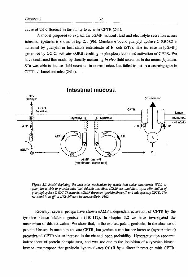
Chapter 2 32
cause of the difference in the ability to activate CFfR (241).
A model proposed to explain the cGMP induced fluid and electrolyte secretion across
intestinal epithelia is shown in fig. 2.1 (96). Membrane bound guanylyl cyclase-C (GC-C) is
activated by guanylin or heat stable enterotoxin of E. coli (STa). The increase in [cGMP];
generated by GC-C, activates cGKll resulting in phosphorylation and activation of CFTR. We
have confirmed this model by directly measuring in vivo fluid secretion in the mouse jejunum.
STa was able to induce fluid secretion in normal mice, but failed to act as a secretagogue in
CFTR -/- knockout mice (241a).
Intestinal mucosa ST.
Guanylin CI- secretion
J eFTR
lumen
membranE
ATP cell Interio
ATP
cGMP Kinase II (membrane ~ aSSOCiated)
Pn
Figure 2.1 Model depicting tile molecular mechmlism btJ which heat-stable enterotoxin (STa) or guallylin is able to provoke intestinal chloride secretion. cGMP accumulation, upon stimulation oj guanylyl ClJclase-C (Ge-C), activates cGMP dependent protein kinase II, and subsequently CFTR. TIre resflilant is all efflux ofG"followed isoosmotically by H20.
Recently, several groups have shown cAMP independent activation of CFTR by the
tyrosine kinase inhibitor genistein (110-112). In chapter 3.2 we have investigated the
mechanism of this activation. We show that, in the excised patch, genistein, in the absence of
protein kinases, is unable to activate CFfR, but genistein can further increase (hyperactivate)
preactivated CFfR via an increase in the channel open probability. Hyperactivation appeared
independent of protein phosphatases, and was not due to the inhibition of a tyrosine kinase.
Instead, we propose that genistein hyperactivates CFfR by a direct interaction with CFTR,

33 Summary and Discussion
possibly at NBD2. This hypothesis is supported by the following evidence i) genistein binds
to the ATP domain of tyrosine kinases (108), but also has affinity for other nucleotide binding
domains (242), ii) mutations or pharmacological agents that interfere with the hydrolysis of
ATP at the second nucleotide binding domain of CFfR, like genistein, increase channel burst
duration. Interestingly, genistein not only hyperactivates wt CFfR but also CFfR mutants
(lIF508) when expressed on the plasma membrane (p.J. French and HR de Jonge,
unpublished observations). Hyperactivation of mutant CFfR may be of importance for the
treatment of CF patients with residual CFfR activity. Therefore, our next step will be to
screen rectal biopsies of CF patients that exhibit a residual CFfR cr secretion in the Ussing
chamber (23) for potentiating effects of genistein. If genistein enhances the CFfR dependent
cr secretion in these patients, it may be considered to be used as a therapeutic agent. As
genistein is present in soy food, and as blood concentration may approach the level required
for CFfR activation in intact epithelial cells (110,112,113,243), simply a dietary supplement
could prove beneficial to patients with residual CFfR activity. The efficacy of a soy based
diet supplementation can be studied in our lIF508 CFfR mouse model which exhibits residual
CFfR activity (228,240). The measurement of the potential difference across nasal epithelium
can be used to monitor increases in chloride permeability.
2.2 FUI/ctions of CF1R: It is still an unresolved and intriguing problem how to relate the
absence of a cAMP regulated chloride channel to the disease symptoms of CF. In chapter 3.4
we have attempted to address this question by studying the relation between CFfR and mucus
secretion. An increased mucus secretion in CF puimonary tissue has been found by several
groups (170,244), indicating a possible function for CFfR in this process. Such an increase
may lead to the mucus plugging of small conductive airways (if the production rate exceeds
the clearance rate), creating a suitable environment for bacterial growth. Alternatively, a
stimulatory effect of CFfR on mucus secretion was suggested from experiments showing an
impaired cAMP dependent mucus secretion in CF (179,198). In this case, the impaired mucus
secretion may result in the accumulation of mucus in goblet cells and, when these lyse, mucus
is released into the lumen. This aberrantly secreted mucus may be insufficiently hydrated or
buffered, and cause obstruction due to changes in the rheological properties. A provocative
hypothesis relates defective mucus secretion in CF to the presumed intracellular role of CFfR
in endo and exocytosis (219,221), endosome fusion (245) or endosomal acidification (175).
We tested the role of CFfR in mucin secretion by investigating whether CFfR modulates
mucin secretion in primary cultures of murine gallbladder epithelium. Apparently, CF tissues
do not exhibit increased mucin secretion, nor is mucin secretion stimulated by agonists which
activate CFTR in wt cells. We conclude that CFfR does not modulate mucin secretion in
Chapter 2 34
mouse gallbladder epithelium.
Several groups have reported an abnormal . structure of mucus in CF
(168-172,174-178). An abnormal mucus structure alters the physical properties of mucus
(180,181) which may impair mucus clearance and so cause mucus plugging of small
conductive airways and ducts. In addition, the altered mucus structure in CF may enhance
bacterial adhesion (182). In the CF mouse gallbladder, our preliminary data show an
abnormal mucus composition, evidenced by a reduced periodate activated staining of mucus
(B.J. Scholte, unpublished observations). Since periodate reacts with free hydroxyl groups, it
follows that CF mucus exhibit reduced amounts of free hydroxyl groups, possibly due to an
increased mucus sulfation. This remains to be investigated.
Mucus obstruction in CF may also be caused by a defective mucus hydration.
Improper hydration of the mucoid gel layer alters its viscoelastic properties, resulting in
reduced pulmonary clearance of mucus and attached pathogens. As fluid transport is
secondary to active ion transport, and as CFTR is a chloride channel; defective chloride
permeability could result in defective fluid transport. Furthermore, CFTR is mainly expressed
in tissues involved in salt and water transport. We tested this hypothesis by directly measuring
fluid transport across intact murine trachea (chapter 3.3), gallbladder (213) and jejunum (pJF,
unpublished observations). In the trachea, we did not observe differences in the amounts of
fluid transported between normal and CF -1- mice. This can be attributed to the low
expression of CFTR in this tissue, and the high expression of other (perhaps compensatory)
chloride channels. In fact, we could not detect CFTR electophysiologically in the murine
trachea. In contrast, tissues with a high expression of CFTR, do exhibit CFTR dependent
fluid secretion. Both murine wt gallbladder (213) and small intestine (214) (pJF, unpublished
observations) secrete fluid in response to cAMP stimulation. This response is absent in CF -1-
mice, implying a role for CFTR in fluid secretion. Under fluid secretory conditions, like in
mouse gallbladder and jejunum, apical cr secretion is likely to be directed by the basolateral
Na+/K+ICI' cotransporter (fig. 1.5C). The cotransporter, using the Na+ gradient, increases
the cellular cr concentration and so creates an outward driving force for Cl". Cations, either
transcellular K+ or paracellular Na+, passively follow the apical anion secretion (see also fig.
1.5C).
Fluid secretion was also studied in a LlF508 CFTR mOuse model with residual CFTR
channel activity (228). The residual CFTR activity completely failed to induce CFTR
dependent fluid secretion in both gallbladder (R.H.P.C. Peters, unpublished observations) and
jejunum (pJF, unpublished observations), within the limits of our detection assay. As the
LlF508 CFTR mouse model has a mild phenotype compared to most knockout strains, there
apparently is no simple correlation between defective fluid secretion and CF pathology. Thus,

35 Summary alld Discllssioll
absence of significant amounts of fluid secretion is not the only determinant of disease
pathogenesis. A residual physiological function of CFTR other than fluid secretion could
explain the mild phenotype of our mouse model. What functions of CFTR are important for
the onset of the disease remains as yet uuresolved. The mild phenotype may also be explained
by the genetic background of the mouse strain (81) or breeding conditions, or by the
assumption that a very low level of fluid secretion (below the threshold of our assay) is
sufficient to prevent pathology.
In summary, several hypothesis have been proposed which link CFTR to the
accumulation and obstruction of mucus. The various presumed physiological functions of
CFTR have been combined in a model recently proposed by J. Bijman. This model is based
on the requirement of a counterion for cation dependent transport systems. In the absence of a
counterion, cation transport down its chemical gradient is lintited by the electrical gradient
that it creates. As different cell types may require cr counterions for the functioning of a
multitude of transporters, this may explain the wide variety of CF related disease symptoms.
For example, cr used as a counterion for H+, is required for endosome acidification (185),
see also fig 1.4. CFTR dependent n permeability is required for NaCI and fluid uptake in
both sweat duct and the respiratory tract (Cr is used as a counterion for Na +) see also figure
1.5A and B. The theory predicts that Na+ and fluid absorption is decreased in CF airways,
contradictory to what has been suggested before based on short circuit measurements in the
Ussing chamber (246). Cl- used as a counterion for Na+ coupled transport systems may be
required for intestinal nutrient uptake. For instance, the Na + Iglucose cotransporter was
reported to be increased in short circuit current measurements of CF intestine mounted in
Ussing chamber (247), quite like Na+ uptake in airways. Non epithelial cell types, like
cardiomyocytes and lymphocytes, may require CFTR expression to provide counterions for
their specific transport systems. In summary, the hypothesis states that CF is not simply
caused by a defect in one specific transport function of CFTR, but by a multitude of cellular
transport functions; CF is caused by a general Cl" impermeability associated with the active
transport of cations.
If the theory also applies to intestinal epithelium, it predicts a reduction in the uptake
of glucose andlor amino acids across CF intestine. This can be studied in CF mouse models
either in vivo, or ill vitro using radioiabelled ligands. In puhnonary epithelia, the model can
be verified by studying fluid absorption across cultured puhnonary epithelium as described
(210,211). Much of our present knowledge of CFTR functioning stems from short circuit
current measurements (lsc) of tissue mounted in the Ussing chamber, however, the Isc
measurements are not suitable to verify a possible defect in Na + coupled nutrient uptake in
CF because they only reflect the active transport of Na + ions and not of passively diffusing
ions like the cr counterion (136).

Chapter 2 36
2.3 Characterization of a //louse model for the &508 CF7R mutation: The most common
mutation in CFI'R found in ",70% of all CF alleles, is a deletion of a phenylalanine at position
508, &508 (6). This mutation affects correct processing to the apical plasma membrane
causing accumulation and degradation of the mutant protein in the endoplasmic reticulum
(19,21,22,139-141,149,150,152). Interestingly, t.F508 CFI'R can be functional as a CFI'R
like chloride channel when expressed on the apical membrane (80,141-143). Correction of the
processing defect therefore is an interesting area for new therapeutic strategies.
In chapter 3.5 we describe the generation and characterization of a mouse model
homozygous for the &508 mutation. We show that this mouse model has a CF phenotype,
but not as severe as the previously published KO mouse models (223-225,227,232). The mild
phenotype of our mouse model can be explained by a residual chloride secretion observed in
pulmonary (nasal), intestinal (jejunum, ileum, and caecum), and gallbladder epithelium. In a
more detailed characterization (chapter 3.6), we show that mouse &508 Cftr, like human
t.F508 CFI'R, is misprocessed. Moreover, this processing defect is temperature sensitive. A
small amount of t.F508 Cftr appears to escape the processing defect which explains the
residual cAMP induced chloride secretion observed in Ussing chamber experiments. The
channel characteristics of WI and &508 Cftr are identical. We conclude that the t.F508 Cftr
mouse model is an excellent model for the human &508 CFI'R mutation.
Two other groups have also generated mouse models for the &508 mutation
(229,230). In contrast to the t.F508 mouse model described in this thesis, these mice were
generated by a procedure leaving an intronic selection marker to reside within the mouse Cftr
gene. Interestingly, these t.F508 mouse models have a much more severe CF phenotype in
comparison to our t.F508 mouse model (229,230). The severe phenotype appears to be due to
a reduced expression of t.F508 Cftr mRNA, which is not found in our mouse model (chapter
3.6). The presence of an intronic selection marker is a likely cause of reduced t.F508-Cftr
mRNA expression. Therefore, the mouse models which have an intronic selection marker do
not reflect true processing mutants because they combine reduced CFI'R mRNA expression
(class I mutant) with a processing defect (class II mutant).
The &508 Cftr mouse model may find application, as in vivo model to test new
pharmacological approaches aimed at circumventing or correcting the processing defect. For
example, phenylbutyrate was reported to increase the amount of &508 CFTR band C in
cultured cells measured by western blot analysis (160). However, in our experiments, the use
of phenylbutyrate did not increase the amount of t.F508-Cftr expression on the
plasmamembrane of cultured mouse gallbladder epithelia (pJF, unpublished observations).
Clearly, further in vivo studies in the &508 Cftr mouse model are required to verify its

37 Summmy and DisCllssioll
potential effect.
Future studies should also focus on the basic mechanisms of protein processing and
folding. It may prove worthwhile to follow processing of M'508 CFfR in cells
overexpressing specific chaperones. A M'508 mutation in mouse Cftr results in a partial
processing defect (Chapter 3.5 and 3.6), whereas this mutation generally causes a complete
processing defect in human cells. Minor differences in mouse Cftr or processing machinery
may explain the "leaky" processing of M'508 Cftr in the mouse. Cross-species transfection
experiments could discriminate between the two, and can pinpoint the subtle but
pathophysiologically important differences. Also, mouse models exhibiting residual CFfR
activity may to help establish the amount of CFfR required for disease prevention. This is of
crucial importance for gene transfer studies.
It is interesting to note that all processing mutations (except N1303K) can be found
within a relatively small area of CFfR, comprising the amino acids 455-574 (A455E,
K464M, &507, M'508, F508R, S549I1R, A559T, D572N, P574H and N1303K
(139,140,248», although a recent report suggests that mutations in other parts of CFfR may
also cause processing defects (249). This stretch almost completely covers the first NBD of
CFfR. It would be interesting to study the Importance of this stretch in the processing of
CFfR. Several basic questions remain to be answered, for example, do chaperonins bind to
the stretch and is its topology within CFfR important. Our basic understanding of the folding
and processing of proteins may be greatly enhanced by studying proteins, like CFfR in which
a single point mutation causes a complete block in protein processing.
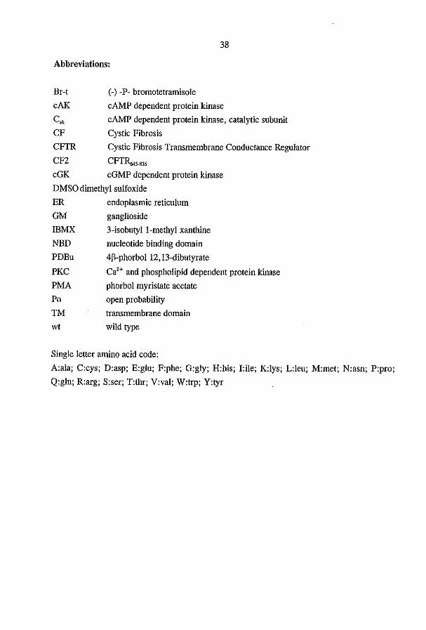
38
Abbreviatiom:
Br-t
cAK
C'" CF
CFTR
cn
(-) -P- bromotetramisole
cAMP dependent protein kinase
cAMP dependent protein kinase, catalytic subunit
Cystic Fibrosis
Cystic Fibrosis Transmembrane Conductance Regulator
CFTR.,"'35 cGK cGMP dependent protein kinase
DMSO dimethyl sulfoxide
ER endoplasmic reticulum
GM ganglioside
ffiMX 3-isobutyl I-methyl xanthine
NED
PDBu
PKC
PMA
Po
TM
wt
nucleotide binding domain
4p-phorbol 12, 13-dibutyrate
Ca2+ and phospholipid dependent protein kinase
phorbol myristate acetate
open probability
transmembrane domain wild type
Single letter amino acid code:
A:ala; C:cys; D:asp; E:glu; F:phe; G:gly; H:his; I:ile; K:lys; L:leu; M:met; N:asn; P:pro;
Q:gln; R:arg; S:ser; T:thr; V:val; W:trp; Y:tyr
39
(1) Boat, T.F., Welsh, MJ., and Beaudet, A.L. Cystic Fibrosis; Boat. T.P., Welsh, M.J., and Beaudet,
A.L., Ed., 1989; Vol. 6, pp 2649-2680.
(2) Welsh, M,J., Tsui. L.-C., Boat, T.F., and Beaudet, A.L. CyStic Fibrosis; Welsh, M.J., Tsui. L.-C.,
Boat, T.P., and Beaudet, A.L., Ed., 1995; Vol. 7, pp 3799-3876.
(3) Wildhagen, M,P., Verbeij, 1.B., Hilderink. H.B., Kooij, L. t Tijmstra, T., tcn Kate, L.P., Gerritsen, J.,
Bakker, W., Dik, I., and Habbema, P. (1996) Thorar 51,298-301
(4) Rommens. I.M., Iannuzzi, M.e., Kerem, B., Drumm. M.L., Melmer, G., Dean, M., Rozmahel, R.,
Cole, J.L.. Kennedy, D., Hidaka, N., et aI. (1989) Science 245, 1059-65
(5) Riordan, J.R., Ronunens, I.M., Kerem, B., Alon, N., Rozmahel, R. t Grzciczak, Z., Zielenski, J., Lok,
S., Plavsic, N., Chou, J.L., et aI. (1989) Science 245, 1066-73
(6) Kerem, B., Rommens, I.M., Buchanan, I.A., Markiewicz, D., Cox, T.K., Chakravarti, A., Buchwald,
M., and Tsui, L.C. (1989) Sciellce 245, 1073-80
(7) Welsh, M.I., Deuuing, G.M., Ostedgaard, L.S., and Auderson, M.P. (1993) J Cell Sci SlIppl 17,235-9
(8) Andersen, D.H. (1938) Alii J Dis Child 56, 344-399
(9) Di Sant-Agnese, P.A., Darling, R.C., Perera, G.A., and Shea, E. (1953) Pediatrics 12,549-
(10) Knowles, M., Gatzy, I., and Boucher, R. (1981) N Ellgl J Med 305, 1489-95
(1I) Quinton, P.M., and Bijman, I. (1983) N Ellgl J Med 308, 1I85-9
(12) Quinton, P.M. (1983) Namre 301, 421-2
(13) Anderson, M.P., Rich, D.P., Gregory, R.I., Smith, A.E., and Welsh, M.I. (1991) Sciellce 251,679-82
(14) Tabcharani, I.A., Chang, X.B., Riordan, I.R., and Hamahan, I.W. (1991) Namre 352,628-31
(15) Kanner, N., Hanrahan, J.W., Jensen, T.J., Naismith, A.L., Sun, S.Z., Ackerley, C.A., Reyes, E.F.,
Tsui, L.C., Rommens, I.M., Bear, C.E., et aI. (1991) Cell 64,681-91
(16) Bear, C.E., Li, C.H., Kartner, N., Bridges, RJ., Jensen, TJ., Ramjeesingh, M., and Riordan, J.R.
(1992) Cell 68,809-18
(17) Bijman, J., Dalemans, W., Kansen, M., Keulemans, J., Verbeek, E., Hoogeveen, A., De Jonge, H.,
Wilke, M., Dreyer, D., Lecocq, I.P., et aI. (1993) Am J Physiol 264, L229-35
(18) Crawford, I., Maloney, P.C., Zeitlin, P.L., Guggino, W.B., Hyde, S.C., Turley, H., Gatter, K.C.,
Harris, A., and Higgins, C.P. (1991) Proc NaIl Acad Sci USA 88,9262-6
(19) Puchel1e, E" Gaillard, D., Ploton, D., Hinnrasky, J., Fuchey, C., Boutterin, M.C., Jacquot, J., Dreyer,
D., Pavirani, A., and Dalemans, W. (1992) Am J Respir Cell Mol Biol 7,485-91
(20) Deuuing, G.M., Ostedgaard, L.S., Cheng, S.H., Smith, A.E., and Welsh, M.1. (1992) J c/illllll'est 89,
339-49
(21) Deuuing, G.M., Ostedgaard, L.S., and Welsh, M.I. (1992)J Cell Bioi liS, 551-9
(22) Engelhardt. J.F., Yankaskas, J.R., Emst, S.A., Yang, Y., Marino, C.R., Boucher, R.C., Cohn, J.A.,
and Wilson, 1.M. (1992) Nature Genet 2,240-8
(23) Hermans, C.L, Veeze, HJ., Drexhage, V.R., Halley, DJ., and van den Ouweland, A.M. (1994) Hum
Mol Genet 3, 1199-200
(24) Anderson, M.P., Gregory, RJ., Thompson, S., Souza, D.W., Paul, S., Mulligan, R.C., Smith, A.E.,
and Welsh, M.1. (1991) Sciellce 253,202-5
(25) French, P.J., Bijman, J., Edixhoven, M., Vaandrager, A.B., Scholle, B.l., Lohmann, S.M., Nairn, A.C.,
and de longe, H.R. (1995) J Bioi Chem 270,26626-31
(26) Hyde. S.C., Ems!ey, P., Hartshorn, M.L, Mimmack, M.M., Gileadi, U., Pearce, S.R., Gallagher, M.P.,
Gill, D.R., Hubbard, R.E., and Higgins, c.P. (1990) Nolllre 346,362-5
40
(27) Ames, G.F., Mimura, C.S., and Shyamala, V. (1990) Fe/ilS Microbial Rev 6,429-46
(28) Ames, G.F., and lecar, H. (1992) FasebJ 6,2660-6
(29) Higgins, C.F. (l992)Amlll Rei' Cell Bioi 8,67-113
(30) Endicott, J.A., and Ling, V. (1989) AII1/u Rev Biochem 58, 137-71
(31) Kuehler, K., Sterne, R.E., and Thorner, J. (1989) Embo J 8,3973-84
(32) McGrath, J.P., and Varshavs.-y, A. (1989) Nalllre 340,400-4
(33) Van Kuijk, M.A., Van Aubel, R.A.M.H., Busch, A.E., Lang, F., Russel, F.G.M., Bindels, RJ.M., Os,
C.H., and Deen, P.M.T. (1996) Proc Noll Acad Sci USA 93,5401-06
(34) Trezise, A.E., and Buchwald, M. (1991) Nawe 353, 434-7
(35) Strong, T.V., Boehm, K., and Collins, F.S. (1994) J Clillllll'est 93,347-54
(36) Dray·Charier, N., Paul, A" Veissiere, D., Mergey, M .. Scoazec, J.Y., Capeau, J. t Brahimi-Hom, C.,
and Housset, C. (1995) Lab Illvest 73, 828-36
(37) Riordan, J.R. (1993) Amlll Rev Physiol 55,609-30
(38) Frizzell, RA., and Morris, A.P. (1994) curr topics membr 42. 173-214
(39) Tizzano, E.F" and Buchwald, M. (1995) AmI Illlenl Med 123,305-8
(40) Schwiebert, E.M., Lopes, A.G., and Guggino, W.B. (1994) Cliff topics membr 42,265-315
(41) Hincke, M.T., Nairn, A.C., and Staines, W.A. (1995) J Neurochelll 64, 1662-8
(42) Mulberg, A.E., Resta, L.P., Wiedner. E.B., Altschu1er. S.M., Jefferson, D.M., and Broussard, D.L.
(1995) J aill IlIvesl 96, 646-52
(43) McDonald, T.V., Nghlem, P.T., Gardner, P., and Martens, C.L. (1992) J Bioi Chelll 267,3242-8
(44) Levesque, p.e., Hart, PJ., Hume, 1.R., Kenyon, J.L't and Horowitz, B. (1992) Cire Res 71, 1002-7
(45) Hart, P., Warth, J.D., Levesque, P.C., Collier, M.L., Geary, Y., Horowitz, 8., and Hume, J.R. (1996)
Proc Noll Acad Sci USA 93, 6343-8
(46) Yoshimura, K., Nakamura, H., Trapnell, B.C., Dalemans, W., Pavirani, A., Lecocq, J.P., and Crystal,
R.G. (1991) J Bioi alem 266,9140-4
(47) Trapnell, B.C., Zeitlin, P.L., Chu, C.S., Yoshimura, K., Nakamura, H., Guggino, W.B., Bargon, J.,
Banks, T.C., DalemarlS, W., Pavirani, A., et aI. (1991) J Bioi Chelll 266, 10319-23
(48) BargoD, J., Trapnell, B.C., Yoshimura, K., Dalemans, W., Pavirani, A., Lecocq, J.P., and Crystal, R.G.
(1992) J Bioi Cllem 267, 16056-60
(49) Shen, B.Q., Barthelson, R.A., Skach, W., Gruenert, D.C., Sigal, E., Mrsny, RJ., and Widdicombe,
J.H. (1993) J Bioi a"lII 268, 19070-5
(50) Breuer, W., Glickstein, H., Kartner, N., Riordan, J.R., Ausiello, D.A., and Cabantchik, I.Z. (1993) J
BioI Chem 268, 13935·9
(51) Breuer, W., Kartner, N., Riordan, J.R., and Cabantchik, Z.I. (1992)J BioI a/em 267,10465-9
(52) Besancon, F., Pnewlocki, G., Baro, I., Hongre, A.S., Escande, D., and Edelman, A. (1994) Am J
Pllysiol 267, C1398-404
(53) Nakamura, H., Yoshimura, K., Bajocchi, G., Trapnell, B.C., Pavirani, A., and Crystal, R.G. (1992)
Febs Lett 314, 366-70
(54) Horowitz, B., Tsung, S.S., Hart, P., Levesque, P.C., and Hume, J.R. (1993) Am J Physio/ 264,
H2214-20
(55) Morales, M.M., Carroll, T.P., Morita, T., Schwiebert, E.M., Devuyst, 0., Wilson, P.O., Lopes, A.G.,
Stanton, B.A., Dietz, H.C., Cutting, G.R., and Guggino, W.B. (1996) Amer J Physioi Renal Fl Elect
39, PI038-Fl048
41
(56) Sheppard, D.N., Ostedgaard, L.S., Rich, D.P., and Welsh, M.J. (1994) Cell 76, 1091·8
(57) Chang, X.B., Hou, Y.X., Jensen, T.J., and Riordan, J.R. (1994) J Bioi Chem 269, 18572·5
(58) Oblatt-Montal, M., Reddy, O.L.. Iwamoto. T., Tomich. 1.M., and Momal, M. (1994) Proc Natl Acad Sci
USA 91, 1495·9
(59) Akabas, M.H., Kaufmann, C., Cook, T.A., and Archdeacon, P. (1994) J Bioi Chelll 269, 14865·8
(60) Cheung, M., and Akabas, M.H. (1996) Biophys J 70,2688·2695
(61) [molo, K. (1993) Febs Lett 325, 100·3
(62) Sheppard, D.N., Rich, D.P., Ostedgaard, L.S., Gregory, R.J., Smith, A.E., and Welsh, M.J. (1993)
Nature 362, 160-4
(63) Hipper, A., Mall, M., Greger, R., and Kunzelmann, K. (1995) Febs Lett 374,312·6
(64) Rich, D.P., Gregory, R.J., Anderson, M.P., Manavalan, P., Smith, A.E., and Welsh, M.J. (1991)
Science 253,205-7
(65) Cheng, S.H., Rich, D.P., Marshall, J., Gregory, R.J., Welsh, M.J., and Smith, A.E. (1991) Cell 66,
1027·36
(66) Ma, J.J., Tasch, J.E., Tao, T., Zhao, J.Y., Xie, J.x., Drumm, M.L., and Davis, P.B. (1996) J Bioi
Chem 271,7351·7356
(67) Chang, X.B., Tabcharani. J.A., Hou. Y.X., Jensen, T.J., Kartner, N., Alon, N., Hanrahan, J.W., and
Riordan, J.R. (1993) J Bioi a/em 268, 11304·11
(68) Rich, D.P., Berger, H.A., Cheng, S.H., Travis, S.M., Saxena, M., Smith, A.E., and Welsh, M.J. (1993)
J Bioi Chem 268, 20259·67
(69) Dulhanty, A.M., and Riordan, J.R. (1994) Biochemistry 33,4072·9
(70) Travis, S.M., Carson, M.R., Ries, D.R., and Welsh, M.J. (1993) J Bioi Cltelll 268, 15336·9
(71) Anderson, M.P., and Welsh, M.J. (1992) Science 257, 1701-4
(72) Anderson, M.P., Berger, H.A., Rich, D.P., Gregory, R.J., Smith, A.E., and Welsh, M.J. (1991) Cell
67,775·84
(73) Ko, Y.H., and Pedersen, P.L. (1995) J Bioi Chem 270,22093·6
(74) Schultz, B.D., Venglarik, C.J., Bridges, R.J., and Frizzell, R.A. (1995) J Gm PI9'sioi 105,329·61
(75) Walker, J.E" Saraste, M., Runswick, M.J., and Gay, N.J. (1982) Emho J 1,945·51
(76) Yoshida, M" and Amano, T. (1995) Febs Lett 359, 1·5
(77) Logan, J., Hiestand, D., Daram. p" Huang. Z., Muccio, D.O., Hartman, J., Haley, B., Cook, W.1., and Sorscher, E.J. (1994) J Clill Invest 94.228-36
(78) Carson, M.R., Travis, S.M" and Welsh, M.J. (1995) J Bioi Chem 270, 1711·7
(79) Gunderson, K.L., and KopilO, R.R. (1995) Cell 82,231·9
(80) Drumm, M.L., Wilkinson, DJ., Smit, L.S., Worrell, R.T., Strong, T.V., Frizzell, R.A., Dawson. D.C.,
and Collins, F.S. (1991) Sciellce 254, 1797·9
(81) Wilkinson, DJ., Mansoura, M.K., Watson, P.Y., Smit, L.S., Collins, F.S., and Dawson, D.C. (1996) J
Gell Physiol 107, 103·19
(82) Hwang, T.C., Nagel, G., Nalm, A.C., and Gadsby, D.C. (1994) Proc NaIl Acad Sci USA 91,4698·702
(83) Carson, M.R., Winter, M.C., Travis, S.M., and Welsh, M.J. (1995) J Biol DIem 270,20466-72
(84) BaukrowilZ, T., Hwang, T.C., Nalm, A.C., and Gadsby, D.C. (1994) Nellroll 12,473·82
(85) Hwang, T.C., Hode, M., and Gadsby, D.C. (1993) J Gm Plzysiol 101,629·50
(86) Gadsby, D.C., Hwang, T.C., BaukrowilZ, T., Nagel, G., Hode, M., and Nalm, A.C. (1994) JplI J
Plzysiol 44, S183·92
42
(87) Picciotto, M.R., Cohn, J.A.. Bertuzzi, G., Greengard. P., and Nairn, A.C. (1992) J Bioi Chem 267,
12742-52
(88) Kennelly, P.l., and Krebs, E.G. (1991) J Bioi Cliem 266, 15555-8
(89) Seibert, F.S., Tabcharani, l.A., Chang, X.B., Dulhanty, A.M., Mathews, C., Hanrahan, l.W., and
Riordan, l.R. (1995) J Bioi Cliem 270, 2158-62
(90) Berger, H.A., Travis, S.M., and Welsh, M.l. (1993) J Bioi Cliem 268,203747
(91) Dechecchi, M.C., Tamanini, A" Berton, G., and Cabrini, G. (1993) J Bioi Olem 268, 11321-5
(92) Dho, S., and Foskett, 1.K. (1993) Biochim Biophys Acta 1152,83-90
(93) Collier, M.L., and Hume, l.R. (1995) Cire Res 76,317-24
(94) Bajnath, R.B., Groot, J.A., De Jonge, H.R., Kansen, M., and Bijman, J, (1993) Pjlllgers Arch 425,
100-8
(95) Winpenny, l.P., McAlroy, H.L., Gray, M.A., and Argen<, B.E. (1995) Am J Pliysiol 268, C823-8
(96) Vaandrager, A.B., and De longe, H.R. (1994) Adv Phanllacol 26,253-83
(97) Forte, L.R., Thome, P.K.. Eber, S.L., Krause, \V,J., Freeman, R.H., Francis, S.H., and Corbin, J.D.
(1992)Am J Plrysiol 263, C607-15
(98) Chao, A.C., de Sauvage, F.J., Dong, Y.J., Wagner, J.A., Goeddel, D.V., and Gardner, P. (1994) Embo
J 13, 1065-72
(99) Tien, X.Y:., Brasitus, T.A., Kaetzel, M.A., Dedman, J.R., and Nelson, DJ. (1994) J BioI Chem 269,
514
(100) Zielenski, J" Markiewicz, D., Lin, S,P., Huang, F.Y., Yang-Feng, T.L., and Tsui, L.C. (1995) Clill
Gellet 47, 125-32
(101) Hidaka, H., and Kobayashi, R. (1992) AlIIm Rev PJ/amtaco! Toxieol 32,377-97
(102) Shabb, l.B., Ng, L., and Corbin, l.D. (1990) J Bioi Cltem 265, 160314
(103) De Jonge, H.R., and Rao, M.C. cyclie nucleotide depelldellt killoses; De longe. H.R.. and Rao, M.e ..
Ed., 1990, pp 191-207.
(104) Vaandrager, A.B., and de longe, H.R. (1996) Mol Cell Bioc/tem 157, 23-30
(105) Lin, M., Nairn, A.C., and Guggino, S.E. (1992) Am J Plrysiol 262, C1304-12
(106) Sullivan, S.K., Agellon, L.B., and Schick, R. (1995) Cllrr Bioi 5, 1159-67
(107) Markert, T., Vaandrager, A.B., Gambaryan, 5., Pohler, D., Hausler, C., WaIter. U., De Jonge. H.R.,
larchau, T., and Lohmann, S.M. (1995) J ClillIlIl'est 96,822-30
(108) Akiyama, T., and Ogawara, H. (1991) Metltods Enzymol 201,362-70
(109) Akiyama, T., Ishida. l., Nakagawa,S., Ogawara, H .. Watanabe,S., Hoh, N., Shibuya, M., and Fukami,
Y. (1987) J Bioi Otelll 262, 5592-5
(110) Sears, C.L., Firoozmand, F., MeUander, A., Chambers, F.G., Bromar, I.G., Bot, A.G., Scholte, B., De longe, H.R., and Donowitz, M. (1995) Am J PJrysiol 269,0874-82
(111) lllek. B., Fischer, H. t Santos, G.F., Widdicombe. 1.H., Machen, T.E., and Reenstra, W.W. (1995) Am J
Plrysiol 268, C886-93
(112) !IIek, B., Fischer, H., and Machen, T.E. (1996) Am J Pltysiol 39, C265-C275
(1l3) Lehrich, R.W., and Forrest, l" Jf. (1995) Am J Pltysiol 269, F594-600
(114) Diener, M., and Hug, F. (1996) Ellr J Pltanllocol 299, 161-170
(ll5) ReeDSlra, W.W., Dam, A., and Raman, S. (1995) Ped Pulmonol SlIppl 12,188
(116) Becq, F., Jensen, T.1., Chang, X.B., Savoia, A., Rommens, I.M., Tsui, L.C., Buchwald. M., Riordan,
l.R., and Hanrahan, l.W. (1994) Proc Notl Aeod Sci USA 91,91604
(117) Higgins, C.F. (1995) Cell 82,693·6
(118) Welsh, M.1. (1990) Faseb J 4, 2718·25
43
(119) Gabriel, S.B., Clarke, L.L., Boucher, R.C., and Stults, M.J. (1993) Nature 363,263-8
(120) Schwiebert, E.M., Flotte, T., Cutting, G.R., and Guggino, W.B. (1994)AmJ Ph)"iol 266, CI464-77
(121) Fulmer, S.B., Schwiebert, E.M., Morales, M.M., Guggino, W.B., and Cutting, G.R. (1995) Proc Nat!
Acad Sci USA 92, 6832-6
(122) Egan, M., Flotte, T., Aftone, S., Solow, R., Zeitlin, P.L., Carter, B.I., and Guggino, W.B. (1992)
Nature 358,5814
(123) lovov, B., Ismailov, 1.1., and Benos, D.I. (1995) J Bioi alem 270, 1521-8
(124) JOVOY, B., Ismailov, I.I., Berdiev, B.K., Fuller, C.M., Sorscher, E,)., Dedman, lR., Kaetzel, M.A., and
Benos, D.J. (1995) J Bioi Chem 270,29194-200
(125) Schwiebert, E.M., Egan, M.E., Hwang, T.H., Fulmer, S.B., Allen, S.S., Cutting, G.R., and Guggino,
W.B. (1995) Cell 81, 1063-73
(126) Ismailov, I.!., Jovov, B., Fuller, C.M., Berdiev, B.K., Keeton, D.A., and Benos, DJ. (1996) J Bioi
Chem 271, 4776·80
(127) Reddy, M.M., Quinton, P.M., Haws, C., Wine, l.l., Grygorczyk, R., Tabcharard, I.A., Harnahan,
J.W., Gunderson, K.L.. and Kopito, R.R. (1996) Science 271, 1876-9
(128) Li, C., Rarnjeesingh, M., and Bear, C.E. (1996) J Bioi alem 271, 11623-6
(129) Grygorczyk, R., Tabcharard, I.A., and Harnahan, I.W. (1996) J Membr Bioi 151, 13948
(130) Reisin, LL., Prat, A.G., Abraham, E.H., Amara, J.F., Gregory, R.J., Ausiello, D.A., and Cantiello,
H.F. (1994) J Bioi Chelll 269,20584-91
(131) Quinton, P.M. (1990) FasebJ 4,2709-17
(132) Jolmson, L.G., Boyles, S.E., Wilson, I., and Boucher, R.C. (1995) J Clill [livest 95, 1377-82
(133) Chinet, T.C., Fullton, I.M., Yankaskas, I.R., Boucher, R.C., and Stutts, M.J. (1994) Am J Pliy,iol 266,
C1061-8
(134) Stutts, M.J., Canessa, C.M., Olsen, J.C., Hamrick, M., Colm, J.A., Rossier, B.C., and Boucher, R.C.
(1995) Sciellce 269,847-50
(135) Ismailov, I.I., Awayda, M.S., Jovov, B., Berdiev, B.K., Fuller, C.M., Dedman, J.R., Kaetzel, M., and
Benos, D.I. (1996) J Bioi Cliem 271,4725-32
(136) Steward, M.C., and Case, R.M. Principles of ;011 Iransport across epitlielia; Steward, M.C., and Case,
R.M., Ed., 1989.
(137) Welsh, M.J., and Smith, A.E. (1993) Cell 73, 12514
(138) Gregory, R.I., Cheng, S.H., Rich, D.P., Marshall, I., Paul, S., Hehir, K., Ostedgaard, L., Klinger,
K.W., Welsh, M.J., and Smith, A.E. (1990) NaUire 347,382-6
(139) Cheng, S.H., Gregory, R.I., Marshall, I., Paul, S., Souza, D.W., White, G.A., O'Riordan, C.R., and
Smith, A.E. (1990) Cell 63, 827-34
(140) Gregory, R.I., Rich, D.P., Cheng, S.H., Souza, D.W., Paul, S., Manavalan, P., Anderson, M.P.,
Welsh, M.I., and Smith, A.E. (1991) Mol Cell Bioi 11,3886-93
(141) Yang, Y., Devor, D.C., Engelhardt, I.F., Ernst, S.A., Strong, T.V., Collins, F.S., Cohn, J.A., Frizzell,
R.A., and Wilson, I.M. (1993) Hllm Mol Gellet 2, 1253-61
(142) Denning, G.M., Anderson, M.P., Amara, I.F., Marshall, I., Smith, A.E., and Welsh, M.I. (1992)
Nature 358,761-4
(143) Li, C., Ramjeesingh, M., Reyes, E., Jensen, T., Chang, X., Rommens, I.M., and Bear, C.E. (1993)
44
Nature Genet 3,311-6
(144) Dalemans, W., Barbry, P., Champigny. G., lallat, S., Dott, K., Dreyer, D., Crystal. R.G., Pavirani, A.,
Lecocq, J.P., and Lazdunski, M. (1991) Nan"e 354,526·8
(145) Haws, C.M., Nepomuceno, LB., Krouse, M,E., Wakelee, H., Law, T., Xia, Y., Nguyen, H" and Wine,
J.J. (1996) Amer J P/~'siol Cell Physiol 39, CI544-CI555
(146) Trapnell, B.C., Chu, C.S., Paakka, P.K.. Banks, T.e., Yoshimura, K., Ferrans, V.I., Chernick, M.S.,
and Crystal, R.G. (1991) Proc Nail Acad Sci USA 88, 6565-9
(147) Thomas, P.I., Qu, RH., and Pedersen, P.L. (1995) Trends Biochem Sci 20,456-9
(148) Yang, Y., Janich, S., Cohn, J.A., and Wilson, J.M. (1993) Proc NaIl Acad Sci USA 90,9480-4
(149) Pind, S., Riordan, J.R., and Williams, D.B. (1994) J Bioi Chem 269,12784·8
(150) Ward, C.L., and Kopito, R.R. (1994) J Bioi Chem 269,25710-8
(151) Marshall, J., Fang, S., Ostedgaard, L.S., O'Riordan, C.R., Ferrara, D., Amara, J.F., Hoppe, HA.,
Scheule, R.K., Welsh, M.J., Smith, A.E., et al. (1994) J Bioi Chem 269,2987-95
(152) Lukacs, G.L.. Mohamed, A" Kartner, N., Chang, X.B., Riordan. 1.R., and Grinstein, S. (1994) Embo J
13,6076-86
(153) Ward, C.L., Omura, S., and Kopito, R.R. (1995) Cell 83, 121-7
(154) Jensen, T.J., Leo, M.A., Pind, S., Williams, D.B., Goldberg, A,L., and Riordan, J.R. (1995) Cell 83,
129-35
(155) Lukacs, G.L., Chang, X.B., Bear, C., Kanner, N., Mohamed, A .• Riordan, 1.R., and Grinstein, S.
(1993) J Bioi Chem 268, 21592-8
(156) Thomas, P.J., Shenbagamurthi, P., Ysem, X., and Pedersen, P.L. (1991) Science 251,555-7
(157) Thomas, P.J., Ko, Y.H., and Pedersen, P.L. (1992) Febs un 312,7-9
(158) Thomas, P.I., Shenbagarnurthi, P., Sondek, I., Hullihen, I.M .. and Pedersen, P.L. (1992) J Biol alem 267, 5727-30
(159) Ciechanover, A. (1994) Cell 79, 13-21
(160) Rubenstein, R., and Zeitlin, P. (1995) Ped Puimollo/ 12,234
(161) Sato, S., Ward, C.L., Krouse, M.E., Wine, U., and Kopito, R.R. (1996) J Bioi O/Cm 271,635-8
(162) Welch, \v.I., Brown, C.R., Brown, L.Q., Biwersi, J., Verkman, A.S., Tatzell, I., and Prusiner, S.B.
(1996) Ped Pllimoll SlIppl 13, 258
(163) Behok, Z., Venglarlk, C.J., and Sorscher, E.J. (1996) Ped Pllimoll Stppl 13,259
(164) Teem, J.L., Berger, H.A., Ostedgaard, L.S., Rich, D.P., Tsui, L.C., and Welsh, M.J. (1993) Cell 73,
335-46
(165) Dork, T., Wulbrand, U., Richter, T., Neumarut, T., Wolfes, H., Wulf, B., Maass, G., and Tummler, B.
(1991) Hum Gellet 87,441·6
(166) Greil, I., Wagner, K., Eber, E., Zach, M., and Rosenkranz, W. (1995) lWeli Klin Wochellschr 107,
464-9
(167) Rose, M.C, (1992) Am J PIO'siol 263, 1A13-29
(168) Boat, T.F., Kleinerman, 1.1., Carlson, D.M., Maloney, W.H., and Matthews, L.W. (1974) Am Rev
Respir Dis 110,42841
(169) Boal, T.F., Cheng, P.W., Iyer, R.N., Carlson, D.M., and Polony, I. (1976) Arch Biochem BiopJlys 177, 95-104
(170) Frates, R., Jr., Kaizu, T.T., and Last, J.A. (1983) Pedialr Res 17,30-4
(171) Cheng, P.W., Boat, T.F., Cranfill, K., Yankaskas, J.R., and Boucher, R.C. (1989) J Clill Illvest 84,
45
68-72
(172) Zhang, Y., Doranz, B., Yankaskas, J.R., and Engelhardt, J.F. (1995) J ClillJlIl'est 96,2997-3004
(173) Gupta, R., and Jentoft, N. (1992)J BioI alem 267,3160-7
(I74) Sairnan, L., and Prince, A. (1993) J Clillllll'est 92, IgJ5-80
(175) Barasch, J" Kiss, B. t Prince. A" Saiman, L., Gruenert, D .. and al-Awqati, Q. (1991) Nature 352,70-3
(176) Clamp, J.R., and Gough, M. (I979) Clill Sci 57,445-51
(177) Dische, Z., di Sant Agnese, P., Pallavicini, C., and Youlos, J. (1959) Pediatrics. 74-91
(178) Wesley, A.. Forstner, J' t Qureshi. R., Mantle, M., and Forstner, G, (1983) Pediatr Res 17,65-9
(179) Rogers, D.P., Alton. E.W., Dewar, A" Lethem. M.I., and Barnes, P.J. (1993) E'(p LUllg Res 19,37-53
(180) Litt, M., Khan, M.A., Shih, C.K., and Wolf, D.P. (I977) BiorileoIogy 14, 127-32
(181) Mian. N., Pope. A.J., Anderson, C.E., and Kent, P.W. (1982) Biochim Bioph)'s Acla 717.41-8
(I82) Krivan, H.C., Roberts, D.D., and Ginsburg, V. (1988) Proc NatI Acod Sci USA 85, 6157-61
(183) Krivan, H.C., Ginsburg, V" and Roberts, D.O. (1988) Arch Biochem BiopJrys 260, 493-6
(184) Imuudo, L., Barasch, J., Prince, A., and Al-Awqati, Q. (1995) Proc NatI Acad Sci USA 92,3019-23
(185) al-Awqati, Q., Barasch, J., and Landry, D. (1992) J &1' BioI 172,245-66
(186) Paulson, J.C., Prieels, J.P., Glasgow, L.R., and Hill, R.L. (1978) J BioI Cllem 253,5617-24
(IgJ) Bradbury, N.A., Cohu, J.A., Venglarik, C.J., and Bridges, R.l. (1994) J Bioi Cllem 269,8296-302
(I88) Webster, P., Vanacore, L., Nairn, A.C., and Marino, C.R. (I994) Am J PlrysioI 267, C340-8
(189) Lukacs. G.L., Chang, X.B., Kartner, N., Rotstein, 0.0., Riordan, I.R., and Grinslein, S. (1992) J Bioi
alem 267, 14568-72
(I90) Biwersi, J., and Verkman, A.S. (1994) Am J PhysioI 266, C149-56
(I91) Roff, C.F., Fuchs, R., Mellman, I., and Robbins, A.R. (I986) J Cell BioI 103,2283-97
(I92) Gottlieb, R.A., and Dosanjh, A. (1996) Proc NatI Acad Sci USA 93,3587-91
(193) Dunn, K.W., Park, J., Semrad, C.B., Gelman. D.L., Shevell, T., and McGraw, T,E. (1994) J BioI Chem
269,533645
(194) Seksek, 0., Biwersi, J" and Verkman, A.S. (1996) J Biol Chem 271, 15542-15548
(195) Lamblin, G., Lhermitte, M., Degand, P., Roussel, P., and Slayter, H.S. (1979) Biochimie 61,2343
(196) Chace, K.V., Leahy, D.S., Martin, R., Carubelli, R., Flux, M., and Sachdev, G.P. (1983) Ciill Chilli
Acta 132, 143-55
(I97) McPherson, M.A., and Donner, R.L. (l994) 11ze Lancet 343,7
(I98) McPherson, M.A., Donner, R.L., Bradbury, N.A., Dodge, J.A., and Goodchild, M.C. (l986) 11ze
Lancet 2, 1007-8
(199) Mills, C.L., Pereira, M.M., Donner, R.L., and McPherson, M.A. (1992) Biochem Biop/lys Res Commull
188, 1146-52
(200) Hoogeveen, A.T., Keulemans, J" Willemsen, R., Scholle, B.1., Bijman, J., Edixhoven, M.J., De longe,
H.R., and Galjaard, H. (1991) Exp Cell Res 193,435-7
(201) Engelhardt, J.F., Smith, 5.S., Allen, E., Yankaskas, l.R., Dawson, D.C., and Wilson, J.M. (1994) Am J
PII),sioI 267, C491-500
(202) Bii,man, J., and Quinton, P.M. (I984) Am J PhysioI 247, C3-9
(203) Joris, L., Dab, 1., and Quinton, P.M. (1993) Am Rev Respir Dis 148, 1633-7
(204) Gilljam, H., Ellin, A., and Straudvik, B. (1989) ScatUi J Clill Lab lllvest 49, 1214
(205) Bansil, R., Stanley, E., and LaMont, J.T. (I995)AIIIIU Rev Ph),sioI 57,635-57
(206) Smith, J.J., Travis, S.M., Greenberg, E.P., and Welsh, M.J. (1996) Cell 85, 229-36
46
(207) Kopelman, H., Durie, P., Gaskin, K., Weizman, Z., and Forstner, G. (1985) N Ellgi J Med 312,329·34
(208) Davidson, G.P., Kirubakaran, C,P., Ratcliffe, G .. Cooper, n.M., and Robb, T.A. (1986) Acta Paediatr
Scallt! 75, 145·50
(209) Kopelman, H., Corey, M., Gaskin, K., Durie, P., Weizman, Z., and Forstner, G. (1988)
Gastroenterology 95, 349-55
(210) Jiang, C., Finkbeiner, W.E., Widdicombe, J.H., McCray, P., Jr., and Miller, S.S. (1993) Sciellce 262,
424·7
(211) Smith, J.J., Karp, P.H., and Welsh, M.J. (1994) J Clill [livest 93, 1307·11
(212) Davidow, C.J., Maser, R.L., Rome. L.A., Calvel, J.P., and Grantham, 1.1. (1996) Kidney 11ft SO,
208·218
(213) Peters, R.H.P.C., van Doominck. J.H., French, P.]., Ratcliff, R., Evans, M.J., Colledge, W.H.,
Bijman, 1., and Scholte, B.J. (1996) Hepato[ogy (in press)
(214) Gabriel, S.E .. Brigman, K.N., Koller. B.H., Boucher, R.C., and StuttS, M.L (1994) Science 266, 107-9
(215) Hasegawa, H., Skach, W., Baker, 0., Calayag, M.C., Lingappa, Y., and Yerkman, A.S. (1992) Sciellce
258, 1477·9
(216) Smith, J.1., and Welsh, M.1. (1992) J Ciin Invest 89, 1148·53
(217) Zar, H., Saiman, L., Quittell, L., and Prince, A. (1995) J Pediatr 126,230·3
(218) Pier, G.B., Grout, M., Zaidi, T.S., Olsen, J.e., Johnson, L.G., Yankaskas, J.R., and Goldberg, J.B.
(1996) Scieflce 271, 64·7
(219) Bradbury, N.A., Jilling, T., Berta, G., Sorscher, E.J., Bridges, R.J., and Kirk, K.L. (1992) Scieflce 256,
530·2
(220) Prince, L.S., Tousson, A., and Marchase. R.B. (1993) Am J Ph),siol 264, C491~8
(221) spUmont, C., Hinmasky, J., Zahm, J.M., Jacquot, J., Puchelle, E., and Kuhry, J.O. (1996) Biachem
BiopJrys Res Commull 227, 182-188
(222) Tala, F., Stamer, P., Wicking. C., Halford, S., Kruyer, H. t tench, N.J., Scambler, PJ., Hansen, C.,
Braman, J.e., Williamson, R.o et aI. (1991) Gellomics 10,301-7
(223) Hasty, P., O'Neal, W.K., Liu, K.Q., Morris, A.P., Bebok, Z., Shumyatsky, G.B., Jilling, T., Sorscher,
EJ., Bradley. A., and Beaudet, A.L. (1995) Somal Cell Mol Genet 21, 177-&7
(224) O'Neal, W.K., Hasty, P., McCray, P., Jr., Casey, B., Rivera-Perez, J" Welsh, M.J., Beaudet, A.L.,
and Bradley, A. (1993) Hum Mol Genet 2, 1561-9
(225) Snouwaert, J.N., Brigman, K.K., Latour, A.M., Malouf, N.N., Boucher, R.C., Smithies, 0., and Koller,
B.H. (1992) Scieflce 257, 1083·8
(226) Dorin, J.R., Dickinson, P., Alton. E.W., Smith, S.N., Geddes, n.M., Steveruon, B.l .. Kimber, \V.L.,
Fleming, S., Clarke, A.R., Hooper, M.L., et al. (1992) NaUire 359,211·5
(227) Colledge, W.H., Ratcliff, R. t Foster, D., Williamson, R. t and Evans, M.J. (1992) 11w LaJ/cet 340,680
(228) van Doominck, I.H., French, P.I .• Verbeek, E., Peters, R.H., Morreau, H" Bijman, J' t and Scholte,
B.1. (1995) Embo J 14,4403·11
(229) Zeiher, B.G., Eichwald. E., Zabner, J' t Smith, I.J., Puga, A.P., McCray, P., Jr., Capecchi, M.R.,
Welsh, MJ .• and Thomas, K.R. (1995) J Clill Ilivest 96,2051·64
(230) Colledge, W.H., Abella, B.S., Southern, K.W., Ratcliff, R., Jiang, C., Cheng, S.H., MacVinish, L.1.,
Anderson, I.R., Cuthbert, A.W., and Evans, M.J. (1995) A'a!ure Genet 10,445-52
(231) Delaney. S,l .• Alton, E.W., Smith, S.N., Ltum, D.P., Farley, R. t Lovelock, P.K., Thomson, S.A.,
Hume, D.A., Lamb, D., Porteous, D.1., Dorin, I.R., and Wainwright, 8.1. (1996) Embo J
47
15,955-63
(232) Ratcliff, R., Evans, MJ., Cuthbert, A.W.o MacVinish. L.J., Foster, D., Anderson, l.R., and Colledge,
W.H. (1993) Nawe Genet 4,35-41
(233) Dorin. l.R., Stevenson, B.l., Fleming, S., Alton, B.W., Dickinson, P., and Porteous, D.l. (1994) Mamm
Genome 5,465-72
(234) Pack, R.J., AI-Ugaily, L.H., Morris, G., and Widdicombe, J.G_ (1980) cell tissue res 208,65-84
(235) Clarke, L.L., Grubb, B.R., Yankaskas, J.R .. Cotton, C.U., McKenzie, A., and Boucher, R.C. (1994)
Proc Nail Acad Sci USA 91, 479-83
(236) Rozmahel, R. t Wilschanski, M., Matin, A., Plyte, S., Oliver, M., Auerbach, W., Moore, A., Forstner,
J., Durie, P., Nadeau, 1., Bear, c., and Tsui, L.C. (1996) Nature Genet 12,280-7
(237) Davidson, D.1., Dorin, I.R., Mclachlan. G., Ranaldi, V., Lamb, D., Doherty, C., Govan, I" and
Porteous, D.l. (1995) Natllre Genet 9,351-7
(238) Clarke, L.L., Grubb, B.R., Gabriel, S.E., Smithies, 0., Koller, B.H., and Boucher, R.C. (1992) Science
257, 1125-8
(239) Smith, S.N., Steel, n.M., Middleton, P.G., Munkonge, F.M., Geddes, n.M., Caplen, N.J., Porteous,
D.L, Dorin, J_R., and Alton, A.W.P.W. (1995) Am J Physiol 268, C297-C307
(240) French, P.J., Vandoorninck, J.H., Peters, R., Verbeek, E., Ameen, N.A., Marino, C.R., Dejonge, H.R.,
Bijman, J., and Scholte, BJ. (1996) J Clill Invest 98, 1304-1312 (241) Vaandrager, A., Tilly, B., Smolenski, A., Sclmeider-Rasp, S., Bot, A.G.M., Edixhoven, M., Scholte,
B.1., Jarchau, T., Walter, U., Lohmarm, S.M., Poller, W.C., and de Jonge, H.R. (1997) J
Bioi ekem 272, 4195-200
(241a) de Jonge, H.R., Ameen, N.A., Boomaars, W.E.M., French, P.J., Bijman, J., Scholte, B.J., Marino,
C.R. (1996) Ped Pllimonol sllppl 13, 218
(242) Markovits, J., Linassier, C., Fosse, P., Couprie, J., Pierre, J., Jacquemin·Sablon, A., Saucier, J.M., Le
Pecq, J.B., and Larsen, A.K. (1989) Cancer Res 49,5111-7
(243) Barnes, S. (1995) J Nutr 125, 777S-783S
(244) Chace, K.V .. Leahy, D.S., Martin, R .. Carubelli, R .. Flux, M., and Sachdev, G.P. (1983) Clbz Chim
Acta 132, 143-55
(245) Biwersi, J., Emans, N., and Verkman, A.S. (1996) Proc Nail Acad Sci Usa 93, 12484-12489
(246) Boucher, R.C., Jackson Stutts, M., Knowles, M.R., Cantley, L., and Gatzy, J.T. (1986) J Clill Illvest 78,
1245-52
(247) Baxter, P., Goldhill, J., Hardcastle, J., Hardcastle, P.T., and Taylor, C.L (1990) Gilt 31,817-20
(248) Sheppard, D.N., Ostedgaard, L.S., Winter, M.C., and Welsh, M.1. (1995) EmboJ 14,876-83
(249) Seibert (1996) Ped Pllimollol suppl 13, 226
Samenvattins 48
Samenvatting
Cystische Fibrose (CF) is een veel voorkomende erfeHjke ziekte in de westerse
samenleving. De ziekte wordt voomameHjk gekenmerkt door recidiverende pulmonaire
infecties welke de belangrijkste oorzaak vormen voor het vroegtijdig overlijden van CF
patienten. De gehele europese gemeenschap teIt ongeveer 25000 CF patienten waarvan zo'n
1000 in Nederland. Hoewel de gemiddelde leeftijd van CF patienten de laalSte decenia flink is
toegenomen tot circa 29 iaar blijft CF een zeer emstige aandoening met een slechte prognose
waarvoor tot op heden een effectieve therapie ontbreekt. Aangezien CF een erfeHjke ziekte is,
za1 er bij deze patienten een verandering zijn opgetreden in het DNA, de drager van ons erfelijk
materiaaI. Het gen 1 wat gemuteerd is in CF patienten is, na intensief onderzoek, ontdekt in
1989. Het gen bleek te coderen voor CFTR (Cystic Fibrosis Transmembrane Conductance
Regulator), een membraan-geassocieerd eiwit dat functioneert als een chloride kanaal2. Dit
kanaal komt bij CF patienten niet, of niet actief, tot expressie op de celmembraan. Momenteel is
het nog niet duidelijk waarom de afwezigheid van een chloride kanaal kan leiden tot CF. Om dit
te verklaren is een gedetailleerd onderzoek nodig naar de regulatie en fysiologische functie van
het CFTR eiwit. Ben beter inzicht in het CFTR eiwit kan nieuwe perspectieven bieden in de
ontwikkeling van een therapie voor CF.
Ben van de manieren om het CFTR-chloride kanaal te openen (te activeren) is door op
speciefieke plaalSen in het CFTR eiwit een fosfaatgroep te incorporeren. De incorporatie van
deze fosfaatgroepen (fosforylering) wordt gemedieerd door zogenaamde proteine kinasen,
waarvan er vele soarten in de eel aanwezig zijn. Ben van deze proteine kinasen is cGKlI
(cyclisch GMP afhankelijke proteine kinase type II). In hoofdstuk 3.1 wordt beschreven dat
cGKll het CFTR eiwit zowel kan fosforyleren als activeren. Activering van CFTR door cGKll
resulteert in een efflux van chloride uit de cel welke gevolgd wordt door een efflux van
kationen (natrium of kalium) en water. De rol van CFTR bij zout en water secretie in de darm
betekent dat CFTR ook een rol speel bij secretoire diarree (cholera)
1 DNA is opgebouwd uit de bouwstenen C, G, A en T. Het menselijke DNA bestaat uit zo'n 2 miljard van deze basen. De volgorde van de basen vormt de biauwdruk voor in totaal ongeveer 80000 eiwiuen. Elk stuk DNA wat eodeert voor 1 eiwit wordt een gen genoemd. 2 De ion baJans in de eel verschilt van de extracellulaire ion eoncentraties. Om de de intracellulaire ion baJans te bandhaven, besehikt elke eel over vcrseheidene ion kanaten en ion transporteurs.

49 samenvaffing
In hoofdstuk 3.2 is gekeken naar een andere vonn van CFI'R aktivering. Recentelijk
werd beschreven dat genistein, een remmer van tyrosine kinase (een andere proteine kinase),
CFI'R kon activeren. Onze data laten echter zien dat genistein niet werkt via de inhibitie van
tyrosine kinase, maar waarschijnlijk direkt aangrijpt op het CFI'R eiwit zelf. De observatie dat
genistein CFI'R kan activeren kan van groot belang zijn voor de ontwikkeling van een therapie
voor CF. Het blijkt narnelijk dat een subpopulatie van CF patienten wei CFI'R chloride kanalen
tot expressie brengt op de celmembraan (hoowel sterk venninderd in aantal of met een sterk
verJaagde activiteit). Voor deze groep patienten met residuele activiteit kan een extra activering
van het CFI'R chloride kanaal door genistein wellicht het CF ziekte proces vertragen.
Aangezien genistein aanwezig is in bepaalde voedingsmiddelen (mn in soya producten) en in
redelijke hoeveelheden opgenomen kan worden door ons lichaam, zou men deze
patientengroep kunnen behandelen met een genistein-rijk dieet. Aanvullend onderzoek zal
aantonen of dit dieet leidt tot curatieve genistein concentraties in het bloed. Voor dit onderzook
kan bv het door ons laboratorium ontwikkelde muis model voor CF gebruikt worden (zie
later).
Zoals eerder venneld is het nog onduidelijk waarom de afwezigheid van het CFI'R
cloride kanaalleidt tot CF. Om de pathogenese van CF te begrijpen is onderzoek verricht naar
de fysiologische functie(s) van CFI'R. De resultaten van dit onderzoek staan beschreven in
hoofdstuk 3.3 en 3.4 van dit proefschrift. E~n van de eerste en meest kenmerkende
ziekteverschijnselen in CF is mucus3 obstructie van ducten en lagere luchtwegen. Deze mucus
obstructie vonnt waarschijnlijk een goede voedingsbodem voor bacterien welke uiteindelijk
leidt tot pulmonaire infecties. De mucus obstructie kan op verschillende manieren worden
veroorzaakt. Twee hypothesen werden getest en staan beschreven in dit proefschrift. A1s eerste
werd gekeken naar de rol van CFI'R in vloeistoftransport. Bij een onvoldoende hydratering
van mucus wordt mucus taai en moeilijk te verwijderen. Aangezien CFI'R een chloride kanaal
is en vloeistoftransport wordt gereguleerd door ionstromen, zou een defect in dit chloride
kanaal kunnen leiden tot een defect in vloeistoftransport. De data beschreven in hoofdstuk 3.3
tonen aan dat CFI'R niet betrokken is bij het vloeistoftransport in de muizetrachea. Dit in
tegenstelling tot andere weefsels waaronder de galblaas en dunne darm van de muis waar
CFI'R wei degelijk betrokken is bij het vloeistoftransport (niet beschreven in dit proefschrift).
De observatie dat CFI'R een rol speelt in het actieve transport van vloeistof over een epithelium
(muv de muizetrachea) zou een goode onderliggende verkiaring kunnen zijn voor het ontstaan
van de CF ziekteverschijnselen.
3 Mucus bestaat uit hoogmoleculaire glycopcoteinen en wordt uitgescheiden door epitheliale cellen. Een mucus laag dient ter beschenning van de eel.

samenvatting so
Mucus obstructie in CF kan ook worden veroorzaakt door een verandering in mucus
secretie. Deze hypothese veronderstelt een actieve rol van CFrR in de secretie van mucus.
Indien bijvoorbeeld de afwezigheid van CFrR leidt tot een toename in mucussecretie kan dit
obstructie ten gevolgen hebben (als de aanvoer groter is dan de afvoer). Aan de andere kant kan
een afname in de secretie van mucus in CF leiden tot een ophoping van mucus in cellen.
Wanner deze ophoping leidt tot celdood komt mucus vrij welke onvoldoende gebufferd of
gehydrateerd is. De op deze manier in het milieu vrijgekomen mucus is taai en k1everig en kan
niet adequaat worden afgevoerd. In hoofdstuk 3.4 is het onderzoek naar de rol van CFrR bij
de secretie van mucus door gekweekte galblaascellen van de muis beschreven. Onze resultaten
laten zien dat CFrR niet betrokken is bij de secretie van mucus door deze cellen. Deze data
kunnen de CF pathogenese dan ook niet verklaren.
De meest voorkomende mutatie in het eftr gen leidt tot de deletie van een eukel
aminozuur (het CFrR eiwit is in totaal opgebouwd uit 1480 aminozuren), een fenylalanine op
positie S08 (6FS08) van het CFIR eiwit. Deze mutatie is interessant omdat het 6FS08 CFrR
nog weI codeert voor een relatief normaal gereguleerd chloride kanaal. Dit kanaal komt echter
niet tot expressie op de celmembraan. Het 6FS08 CFrR wordt na de novo synthese verhinderd
van transport uit het endoplasmatisch reticulum en wordt uiteindelijk afgebroken. De observatie
dat deze maturatie mutant nog weI kan functioneren als een relatief normaal CFIR chloride
kanaal betekent dat de meeste patienten (±90%) potentieel funtionele chloride kanalen bezitten
milS het 6FS08 CFIR maar getranslokeerd kan worden naar de celmembraan. Een muis model
voor de 6FS08 CFrR mutatie zou een belangrijke stap zijn in het onderzoek naar de maturatie
van 6FS08 CFIR en kan de basis vormen van alternatieve therapien voor CF die zich rich ten
op de relocatie van 6FS08 CFrR naar de celmembraan. In dit proefschrift wordt de generatie
en de karakterisering van een muis model voor de 6FS08 CFrR mutatie beschreven (hoofdstuk
3.S en 3.6).
51
Isotype-specific Activation of Cystic Fibrosis Transmembrane Conductance Regulator-Chloride Channels by cGMP-dependent Protein Kinase 11*
(Received for publication, April 12, 1995, and in revised form, August 18, 1995)
Pim J. French, Jan Bijman, Marcel Edixhovent Arla B. Vaandrager:t, Bob J. Scholte, Suzanne M. Lohmann§, Angus C. Nairn', and Hugo R. de Jongetl From the Departmmts ofCe1l Biokgy and tBiochemistry, Faculty of Medicine and Health &iences. Erosmus University, 3000 DR Rotlmiam, The Netherlands. the tf.,aborotory of Clinical Bioclwnistry, Medical University Clinic, 97080 Warzburg, Federal Republic ofGennany, and the 'Laboratory 4 Mokcular and Cellular Neuroscience, The RocM{eUer University, New York, New York 10021·6399
Type n cGMP.dependent protein ldnaae (cOKm iSI> toted from pig intestinal brush borders and type IacGK (cGKI) purified from bovine lung were compared tor their ability to activate the cystic fibrosis transmem. brane conductance regulator (CFI'R)·Cl- channel in ex. cised, inside-out membrane patches from NJH.3T3 fibroblasts and from a rat intestinal cell Une (IEC-CF7) stably expressing recombinant CFI'R. In both cell models, in the presence of cGl\1P and ATP. cGKll was found to tnimic the effect of the catalytic subunit of cAMP-dependent proklin kinase (cAK) on opening CFTR-Clchannels, albeit with different kinetics (z...3.min lag time, reduced rate of activation). By contrast, cGK! or a monomeric cGK! catalytic fragment was incapable of opening CFTR-Cl- channels and also failed to potentiate cGKU activation of the channels. The oAK activation but not the"cGKII activation was blocked by a oAK inhibitor peptide, The slow activation by cGKfi could not be ascribed to counteracting protein phosphatases. since neither calyculin A, a potent inhibitor of phosphatase 1 Bnd 2A, nor ATPyS (adenosine 5'.{}.(thiotriphosphate», producing stable lhiophosphorylation, was able to en· hance the activation kinetics. Channels preactivated by cGKII closed instantaneously upon removal of ATP and kinase but reopened in the presence of ATP alone. Paradoxically, inununoprecipitated CFTR or CF -2, a cloned R domain fragment of CFTR (amino acids 645-835) could be phosphorylated to a similar extent with only minor kinetic differences by both isotypes of cGK. Phosphopeptide maps of CF-2 and CFI'R, however, revealed very subtle differences in site-specificity between the cGK i&ofOl'ms. These results indicate that cGKII. in COD'
trast to cGKla, is a potential activator of chloride transport in CFI'R-expressing cell types,
Guanosine 3',S"cyclic monophosphate (cGMP) has been identified as an important intracellular mediator of salt and water secretion in intestinal epithelium (1-3). Secretagogues acting through the cOMP-signaling pathway include the family
• This work was supporwd by the Netherlands Organiz.ation for Scientific Research (NWO) and the Deutsche Forschungsgemeinschan (Grants Ko 210111-3 and SFB355). The rests of publication of this artide were defrayed in part by the payment of page charges. This artide must therefore be hereby marked ~adwrlistmml' in accordance "'ith 18 U.S.C. Section 1734 solely to indieste this fact.
n To whom reprint requests should be addressed: Dept. of Biochemistry, Cardiovascular Research Institute COEUR, Medies! Faculty, Erasmus University, P. O. Box 1738,3000 DR Rotwrdam, The Netherlands. Tel.: 31-104087324; Fax: 31-104360615.
of heat-stable enterotoxins (STS), l low molecular weight peptides secreted by enteropathogenic bacteria, and guanylin, a recently discovered endogenous ST-like peptide hormone (3. 4). Binding of ST or guanylin to the receptor domain of an intestine-specific isoform of guanylyl cyclase (GC-C) triggers cyclase activation, cGMP accumulation, and stimuJation of net fluid secretion through the activation of apical Cl- channels in parallel with inhibition of coupled NaCI transporters (3, 6, 6). The cystic fibrosis transmembrane conductance regulator (OFrR), an epithelial Cl- channel mutated in OF patients (7, 8), appears to be involved in the CI- secretory response to ST and cOMP analogues, as evidenced by the absence of this response in OF intestine (9, 10).
Several mechanisms have been proposed to link cGMP to the CFI'R-CI- channels, including (i) cGMP cross-activation of cAMP-dependent protein kinase (11-13) followed by multisitephosphorylation of CFTR (14), (ji) direct interaction of cO~fP with the CFI'R protein (15), and (iii) cGMP activation of an intestine-specific isoform of c01fP-dependent protein kinase (type n cOK; Refs. 16-19). cOKII was discovered as a cOMPsensitive 86-kDa phosphoprotein localized in intestinal brush border membranes (16), which comigrated with a cGMP receptor protein on one- and two-dimensional gels (17, 18). The intestinal isofonn is clearly distinct from the homodimeric type Ia and I{J cGK (153-156 kDa) identified in other mammalian tissues, as illustrated by differences in subcellular localization, subunit composition, isoelectric point, phosphopeptide maps, immunoreactivity, and affinity for cyclic nucleotide analogues (17-19). Recently, molecular cloning ofcOKll from mouse brain (20) and rat intestine (21) demonstrated that cGKII is a different gene product than cGKIa and 113 (22, 23).
In the present study, evidence for a functionaJ difference between cGK isoenzymes was obtained from studies of the activation ofCFl'R-CI- channels in excised membrane patches of an intestinal cell line (lEC·CFt; Ref. 24) or NlH-3T3 fibroblasts stably expressing recombinant CFTR (26). In both models, exposure of patches to a combination of cGMP and ATP failed to elicit CI- channel activity. However, the further addition of purified cGIrn, but not cOKIa. resulted in almost full activation ofOFI'R-CI- currents.
Differential activation of the CFI'R-Cl- channel by cORTI is the first example of isotype specificity in cGK regulation of
I The abbreviations used are: ST, heat-stable enterotoxin; CFTR, cystic fibrosis transmembrane conductance reguJator; CF, cystic fibrosis; eGK, tGMP-dependent. protein kinase; cAK, cA.\tP-dependent prowin kinase; PKI, Walsh inhibiwr peptide (PKI(S_24)..amide); ATP.,.S, adenosine S'..Q·(thiotriphosphate); PAGE, polyacrylamide gel electrophoresis.
52
cellular functions and provides a plausible explanation for the prominent role of cGMP as a regulator of C}- transport in intestinal epithelium in comparison to other CFI'R-expressing cen types in which cGKII expression is marginal or absent (19-21),
EXPERIMENTAL PROCEDURES
MateriaLs--Calyculln A was obtained from Calbiochem. San Diego, CA. PRJ, the Walsh inhibitor peptide (PKf(&-24}amlde) was obtained from Dr. U. Walter, Wunburg, Germany. CF·2, a cloned R domain peptide ofCFJ'R (AA645-835), was produced in bacteria and purified as described (26). Adenosine 5'-bipbosphate. sodium salt (ATP), and cGMP were obtained from Boehringer Mannheim. £,),-uPlATP was obtained from Amersham. UK. All other chemicals were from Sigma.
tAils and CFTR &prus/on Systems-Two cell types that stably express CFTR were prepaxed and maintained as describEd previowly (24, 27). IEC-CF7 cells were obtained by stable transfec:tion of the rat fetal intestine-derived IEC-6 eellline with a plasmid encoding CFTR (24); NIH..aT3 ceDs ~)[pressed CrrR after infection with a retroviral vector encoding CFTR (27).
[rolation of Protein Kfnases---cGKII was purified from the small intestine of edult pigs (donated by the Department of Experimental Cardiology, Erasmus University). 'The small intestine was dissected from anesthetized pigs, rinsed with ice-rold 0.9% NaCl, and frozen in liquid nitrogen. Brush border membrane vesiCles we~ prepared from the intestinal pieces by a freeze-thaw pto«!dure and subsequent differential M~· p~cipitation and centrifugation as described p~viou.sly (28). tOKII extraction from the vesicles and purification by affinity chromatography on 8{2.aminoothyl)-amino-cAMP.Sepharose was per· formed essentially as described (17, 18) with a slight modification. To obtain detergent-froo enzyme for use in the patch clamp eXperiments, elution ofcGKII from the affinity gel with 1 m)! cGMP was performed in the presence of 4 mM octyl glucoside Tatber than Triton X·loo. Subsequently, cGMP and octyl glucoside wer~ removed by dialysis in deterg~nt-free patcb clamp medium (see below).
The catalytic subunit of type II cAK and cGKI (characterized by antibody analysis to be primarily the fa is(lformj Ref. 29) were purified from bovine heart and bovine lung, T~spectively, as described (30,31). Th~ specific activities (unltslmg protein) of the purined protein kinases as determined by the Kemptide phosphorylation assay (32) were 4.2 (cAK), 2.0 (cGKI), and 1.6 (eGKII), respectively. A monom~ric constitutively active eGKI fragment was obtained by limited trypsinh.ation as described (33).
Pakh Clomp Technique-Patch clamp experiments were performed as d~scribed by Hamill et ai. (34). Glass {borosilicate} pipettes ""ere pulled to a resistance of 308 megohms and heat polished. Pipette potential refers to the voltage applied to the pipette interior with r~ference to th~ bath potential. Upward den«tions denote negative charge Rowing out of the pipette. A List EPC-7 amplifier was used for current amplification and voltage damping. Membrane voltage was continuously damped at -40 mV, except when performing cunentvoltage relationships. Data were monitored on an oscilloscope and stored on a VCR. Th~ Nrorded data were filtered at 50 or 100 Hz, digitized at 200 Hz, and analyzed on a personal computer. Data anal· ysis was perfonned as described by Kansen et al. (as). In view of the fact that the high density ofCFTR·CI- channels in membrane patches of the 3T3-CFTR fibroblasts hind~rs the accurate det~rmination of open state probability or numbet' of channels, channel activity in these patches was expressed in pA. rather than as the numMrofopen channels. The composition of the bath and pipette solutions was (in mM): 140 N· methyl·o-glucamine, 1 EOTA, 3 MgCI2, and 10 Hepes.HCI (pH 7.3, final C1- concentration 147 mM). 10 some ~xperiments a low CI- pipette buffer was used containing (in mM): 140 N'methyl,o-giucamine, 100 vaspartic acid, 2 MgC~, 5 CaCI..z, and 10 Hepes·HOI (pH 7.3; final CIconcentration 49 mM). Excised patches were studied in a solution ex. chan~ romparbnent(\'olume 1 ml), as described previously by Kansen et al. (36). Experiment.<; wer~ performed at room temperature.
In Vitro Phosphorylation 0{ CFTR and CP·2 and Phosphopeptide Mapping-CFTR was immunopr~cipitated from T84 cells using specific CFrR antibodies and protein A-Sepharose beads as described (26). CFTR attached to protein A-Sepharose beads (10 p.l of suspension) or CFrR antibody plus beads alone was incubated at 30 ¢C for 40 min in 100 p.lofbuffercontaining lOmM MgCl..z. 1 mM EGTA,lOm!.l Hepes, pH 7.3,50 p.\f MgATP, 20 J.l..\f cOMP, 10 Cifmmol [y-.nPlATP, and purified protein kinases (catalytic subunit of cAK, 2 miUiuniWmI; tOKI, 7.5 mi1liunitsfml; cOKII, 9.4 milliunitsfmll. The phosphorylated samples were washed, resuspended in SDS.stop solution, and analyzed on 6%
50S-PAGE as described previously (26). Phosphorylation conditions fi CF·2 (5 pM) were similar to those for CF"l'R. For kinetic experiment linear incorporation of up with time was ensured by (i) restricting U ineuba..tion time to 5 min, (m varying the CF·2 concentrations betwee 0.1 and 0.6 pM, (iii) inaea..sing the {.,...»PlATP concentration to 500 P. (1 Cifmmo1), and (iv)using equal concentrations (25 OM) of each protei kinsse. Reactions were terminated by the addition of 20 ~ of 7~ trichloroacetic acid, and protein pellets were washed three times wit 0.2 ml ofioo-cold H20, su.spend~d in 50 pi arsos.stop buffer, boiled f~ 2 min, and subjected. to 12% SDS·PAGE as described (26). ~P·Lahele CF·2 or CrrR w~re excised from the dried g~ls, washed with tw cbanges or 10% acetic add!3O% methanol and three changes of 5m methanol, aod lyophilized. In the kinetic eXperiments, the incorpora lion ofoUP into CF·2 was quantified by liquid &cintillation spedromebj For two-dimensional phosphopcptide mappingofCF.2 and CrrR, 1 m of 60 mM NH.C03, pH 8.0, containing L-l·tosylamido-2-phenyleth)' chloromethyl k~tone·treated trypsin (50 ~) was added to the driel g~l pieces and the mil:ture was incubated at37 "C for 20 h (26). 'The ge pieces were washed with 0.5 ml of50mM NH.HC03 at 37 "C for4 h, ani the coll«ted supernatants were lyophilized. Phosphopeptides were sep mated on thin layer cellulose sheets (20 X 20 em, Eastman Kodak Co. by el«trophoresis in the first dimension, followed by chromatograph: in the second dim~nsion. Dried sheet.<; were subjected. tl autoradiography.
RESULTS
kh'vation of CFTR·Cl~ Channels by Protein Kinases-Ir agreement with earlier studies of Cl- channel activation ir: excised, inside·out membrane patches from 3T3-CFfR fibro. blasts (25, 37, 38), the addition of catalytic subunit of cAK (~ milliunitslml) to the bath, in the presence of 2 m:p.r MgATP, resulf.(!d in rapid activation (lag time < 1 min) of multiple anion-seledive channels (average current increase per patch 40 :t 38 pA at. -40 mV holding potential; n ""- 15) showing characteristic properties of the CFl'R-CI- channel (i.e. C)selectivity, linear current-voltage relationship in symmetrical Cl- concentrations, 8 pS single channel conductance; results not shown}. A similar low conductance channel, oceurring at a much lower density (2-6 channelsfpatch) was activated by cAK in excised patches from the rat intestinal IEC·CF7 cell line stably expressing CFI'R (24). In addition to cAl{, the effect of cGK on CFrR-CI- channel opening was examined in excised, c~ll-free patches from both cell lines. Addition ofMgATP(2 mM) and caMP (50 I-'M) together did not activate current in 31'3· CFl'R patches during 5-15 min observation (Fig.IA). Inclusion ofthe cGKI isoform puriti~d from bovine lung(10 milliunitslml) likewise failed to open the CFI'R·Cl~ channel (Fig. lA). con· tinning earlier observations by Berger et ai. (38). However, the subsequent addition of saturating concentrations of the cGKII (10 milliunitslml) elicited a large CI- current, reaching a value after 15 min that was 78 :t 24% (n = 5) of the maximal current attained upon addition of saturating amounts of cAl{ (2 milli· unitslml) to the same patch (Fig. lA). Half-maximal cGKII· activation (48 :t. 9% of the maximal cGKII response at 15 min; n = 5) was observed in the presence of 2 milliunitslml cOKII, whereas the threshold for current activation (10 :!: 6%: n = 5) was found at 0.5 milliunitslml cGKII (data not shown). In comparison to cAK, CFI'R·CI- cUlTent activation by cGKII was a relatively slow process (time requir~d to reach half.maximal activation following addition of 10 milliunitslml cGKJI: 8 :t 1 min; for 2 milliunitslml cAK! 0.7 :t 0.2 min,n = 6;Fig.l,A and 8). This slow activation was unlikely to result from the activity of counteracting protein phosphatases, since neither calyculin A(10-7 M), a potent inhibitor of phosphatase 1 and 2A (recently implied in CFI'R regulation; Refs. 38 and 39), nOT the addi· tional presence of ATPyS (1 ml-l) to produce stable thiophosphorylation, was able to enhance the CFTR activation kinetics (Tesults not shown).
The cOKII isoform, but not cGKI, was capable of activating CFTR·CI~ channels also in excised patches from IEC-.CF7 cell.!;

53
A .. A1Pi«lIolP A~P
50 ~ ~"' -""-
" % "
" " • ,
" " " " " " UIl\' (mIn)
B .. ~.,
AlP/cOWl>
to
% " " • • , , " " ..
tlms(ml,,)
C '00
f " .. < • • " .. • "
0 0 10 15
tim. (mtn)
FIG. 1. cGKII but not cGKI activate.5 CFfR.CI- channel current in excised. inside-out membrane patches from 3T3·CFI'R fibrobla.sts. ATP(2 roM), cGMP(50 /-tM), cOKII (10 milliunit.slml). and catalytic subunit of cAR (2 milliunit.slml) were present in the cytosolic (bath) solution during the times indicated by the bars. Paml A, compariSOD of the effects of tOKI, cGKll, and cAK. The amount of CUl'Nnt activated by cGKII was 78 ± 24% (n ;; 5) oftha current measured art.er subsequent addition of cAK Pond B, a repr~ntative example of aD experiment showing that the cGK-adivated cunents rapidly returned to near base-line values upon removal oftha kinase and ATP from the bath (also shown for the cAK-activated currents in panel A), but could be restored almost instantaneously by the readdition of ATP alone. Paml C, time course of activation ofCFTR·Cl- current by cOR11 alone (e; 10 tnilliunitslml) or a combination of cORI (10 milliunlWml) and cORII ( )j n = 6. Current levels aN expressed as a percentage of the maximal CFTR-CI- current observed upon subsequent addition of 2 milliunlWmI eAK
(Fig. 2). In this low expression model, single·channel events could be monitored (Fig. 2A), which had a linear I-V relation in symmetrical Cl- concentrations, a channel conductance of 8 pS, and a rightward shift in current reversal potential upon lowering of the Cl- concentration in the pipette (Fig. 28). The mean open probability (Po) ofCFI'R·CI- channels measured at the plateau phase of activation by saturating concentrations of cOKII (0.22 ± 0.09; n = 9) did not differ significantly from the Po of cAR·activated channels (0,23 ± 0.12; n = 8),
Additional proof of the identity of the cGKII-activated channel as CFTR came from the observation that cGKII c(luld not further enhance channel activity in excised patches from 3T3-CFTR cells following their pre-phosphorylation by cAK(2 milUunitsfml) and ATP (2 mM) (results not shown). Another similarity between CFTR·Cl- channel regulation by cAK and
A
o.v --..--- . --; ... ----.1OrnV
B 0.60 - ..... 141(41 CI'
0,40 - _ 1H/141 CI' , ' 0.20 - ,,41'1
! 0,00 ------.* .. ~ .. ? -'~"~"-' ---
'0.20 -
·0.040 -
.0.60 L , I , I
-70 -50 ·30 ·10 10 30 50 70
V{mV)
FIG, 2, Biopbysical characteristics of the cOKn·activated channel in excised. inside-out membrane patches from IEC-CF7 cells. Pantl A, currcnt tracings oC cGRII-aetivated channels. Left, symmetrical 147/147 mM chloride solutions. Right, pipette buffer was replaced by a low (49 mM) ehloride buffer. Tracings were obtained at the indicated voltages. C, all channels closed; dotted iw, single-ehannel current levels. Pond B, J.V characterlsticsoCthe channel. 'The channel conductance was 8.0 ± 0,6 pS (n = 5). e, symmetrical Cl- solution (147/147 mid); ,reduction ofCl- in tho pipette to 49 mM by replacement with aspartic acid.
cOKII was the observation that the currents rapidly returned to near base-line values upon the removal of either kinase and ATP from the bath, but could be restored almost instantaneously by the readdition of 2 mM ATP alone, confinning the crucial role of ATP in CFi'R-Cl- channel functioning (Fig. IB; cf. Refs_ 25, 38, and 40),
To eliminate the possibility that cGKII activation of CFfR resulted from a contamination of the cOKII preparation with cAl(, a specific peptide inhibitor of cAl(, PKI (0.1 J.lM) was added to the bath. Under this condition CFrR·Cl- channel activation in 31'3 membrane patches by cAR. (2 mUliunitsfml) was completely abolished, whereas channel activation by cOKII (2 milliunitslml) was not significantly affected (42 ± 3% of the maximal channel activity evoked by cAK in the same patch following removal of PKI from the bath, as compared to 37 ± 5% in the absence ofPKI; n = 5), Moreover, PKI was unable to inhibit phosphorylation of Kemptide or CF-2 by cGKII (not shown).
Finally, the possibility was considered that cGKI, in spite of its failure to open CFI'R·CI- channels by itself, could interfere with the activation of CFfR·Cl- channels by the other cOK isofonn. However, neither preincubation of 3T3 patches with cGKI (10 milliunitslml. 15 min; results not shown) nor the simultanoousaddition ofcOKI (10 milliunitslmn and cGKII (10 mllliuniL"'ml) had any effect on the rate or extent ofCFI'R-Clchannel activation as compared to cOKII alone (Fig. Ie).
Phosphorylation of CFTR and CF·2 by cGK Isotypes-Phosphorylation studies carried out with CFTR immunoprecipitates (Fig, SA) and CF-2, the recombinant R domain Qf CFI'R (Fig. 38) continued previous reports that both proteins are excellent ,'n vitro substrates for cAKand cOKI (26,38), This discrepancy

54
B " " A ~ "- '. MW ~ IkDI
" " ~ • C,\K 1.0.0 1.40
200- • C ~GKI O.U O.to
~ A ~Gr:lI 0.1$ 0..0.1
~ " " '4- ~
~ " "
-, -, -. -, 4 , "
lICF-2 (VMI
FIG. 3. Phosphorylation of CFrR (ponel A) and CF·2 (panel B) by purified protein kiIlJl5e& Panel A, CFTR was immunoprecipitat«l from T84 cells and phosphorylated as desaibed under -Experimental ProeedUNS.- The reactions also ccntained: catalytic subunit of cAK (2 milliunitslm1; lanu I and 2). cGRI (7.5 milliuniWmI: lanes 3 and 4), and cGKII (9.4 milliuniWmlj lane.s 6 and 6). Lan.u 2, 4. and 6, control experiments in which CITR was omitted. 'The s~P·labeled proteins weN separated by 6% SDS-PAGE. The gel was dried and exposed to x-ray film. CFTR migrates as a broad band of 180 kDa ("band Co; see Ref. 14). 'The 86- and 74-kDa bands represent ~sidual amounts of autophosphoryiated c:GKII (lanes 6 and 6, inteet86-kDa form + 74-kDa proteolytic fragment; cf. Ref. 17) and cOKI (lanes 3 and 4, intact 74-kDa fonn), respectively, remaining following the washing sreps. Pancl B, line.weaver·B\L~k plots ofCF·2 phosphorylation by equal concentrations (26 OM) of eAR, cGKI, and cGKII. The experimental conditions needed to ensUJ\'lllnear rares of3"2P int{lrporation are specified under -Experimental Procedures.· The inset shows the kinetic (:(Instants (K",. V"".,) calculated from the Lineweaver·Burk plots. AU., arbitrary units. Data Npresent the mean of thre-e experiments.
between phosphorylation and functional studies was even more apparent from the kinetics of in vltro phosphorylation of CF-2 by cGKI and cGIrn isoenzymes (Fig. 38). CF-2 was a better substrate for cOKI than for cOKI[ (similar Km; 3-fold higher V rna,.), and the plateau level of phosphate incorporation into CFrR (Fig. SA) was also higher for cGlO (140 ± 11% of the cGKII level; n ::: 8).
Two·dimensional PJwsphopeptide Mapping 0/ CF-2 and CFl'R PhospJwr),lated b)' cAl(, eGKI, and eGKll-Up to 10 aerine residues within the R domain have been suggested to be involved in the activation ofCFTR (for review, see Ref. 39). In order to investigate whether the isotype specificity of cGK activation of the CFrR-CI- channel might be related to the pattern of specific serine residues that are phosphorylated, two-dimensional phosphopeptide mapping was performed of CF-2 and CFI'R phosphorylated by cAK, cGKI, and cGKll (Fig. 4). All three kinases phosphorylated the same peptides in CF-2 with very slight differences being observed in the intensities of phosphorylation of individual peptides (Fig. 4, A--C). All three kinaS60S also phosphorylated the CFI'R on a subset of the peptides phosphorylated in CF·2 (Fig. 4, V-F>, arguing against the location of novel cGKII sites ou~ide the R domain. Several sites, in particular those located on peptides 1, 2, and 4 (the Jatter containing serine 700; see Ref. 26), were phosphorylated to a similar level. In contrast, one prominent peptide, labeled 9 (see Ref. 26 for numbering) was preferentially phosphorylated by cAl{ and cGKII, but was phosphorylated to a much lower level by cOlO (Fig. 4, V-F, and data not shown).
DISCUSSION
In this study a recently cloned cGK isoform, cGKII (20, 21), expressed at high levels in the luminal membrane of intestinal epithelial cells (16-19, 21, 41) and to a lower extent in kidney and brain (21), is identified as a novel potential regulator of the CFI'R·CI- channel. Using a reconstitution assay consisting of a detergent-free preparation of solubilized and purified cGRll, inside-out membrane patches from CFrR·transfected intestinal cells (IEC·CF7), or NIH-3T3 fibroblasts, Mg-ATP and cGMP, the enzyme could almost fully mimic the effect of cAl{
on CFfR·CI- channel opening, albeit with a slower time course. It is possible, however, that the rate-limiting step in the in vitro assay is the anchoring of the cGKII to the membrane, and that this delayed opening does not necessarily imply a similar kinetic disadvantage for cGKII in l.iI~, consi<h''''ing its
,T , '. . . ., "~:a ••• ., "
FlO. 4. Two-dimensional tryptic phosphopeptide maps ofCF·2 and CFTR. Upper and lower panels. phosphopeptide mapsofCF-2 and CFTR, respectively. shov.ing phosphorylation by eAK (A and Dl, tGKI (B and El, and tGKIf (C and FI, respectively. Purified CF-2 and immunoprecipitated CFTR were phosphorylared for 40 min at 30 "C in the presence of cAK (2 milliuniWml), cGKI (7.5 miUiuniWm1), or cO KU (9.4 mi1liuniWml) as described under wExperimental Procedures" The l2p_
labeled proreins were separared by 6% (CFTR) or 12% (CP·2) SOS· PAGE and visualized by autoradiography. Radioactive bands ofCF-2 (pooled middle and upper bands running at,30 and 32 kDa, respectively; e{. Ref. 26) and CFTR(see Fig. 3A) were excised from gels, washed, and digested for 20 h with 50 JLg/ml L-l.tosylamido-2.phenylethyl ehloromethyl ketone-treated trypsin. Tryptic digests were separated by el«uo· phoresis in the first dimension at 400 V In 10% acetic acid, 1% pyridine. pH 3.5, and by chromatography in the second dimension using pyridine: l·butanol:warer:aeetic acid (10:15:12:3%).0, origin. Left, +; right, -. Phosphopeptides are numbered from 1 to 9, and correspond to those in Ref. 26. The results shown are repreoontative of thre(l independent experiments. In two other sets ofCF·2 maps, an additional phosphopeptide migrating closely to peptide 9 could be distinguished, indicating that the peptide 9 spot may contain more than one peptide or more than one phosphoacceptor sire in one peptide, occasionally giving rise to alternative tryptic digestion.
colocalization with CFTR in the luminal membrane (19, 41). The amount ofCI- channel current reached (78 :!: 24% of that measured after the subsequent addition of cAK) was considerably greater than the current level reported after addition {If another activating kinase, PKC, using similar assay conditions (15 :!: 8% of cAR; Ref. 38). In accordance with these functional data, the phosphopeptide maps made from CF-2 and CFI'R phosphorylated in vitro by cGKII and cAl{ were virtually iden-
55
tical and clearly different from the pattern generated by PKO (26). The plateau level of phosphate incorporation lnto CFTR reached with cAl{ and cOKII was also similar (Fig. SA). although the rate ofCF-2 phosphorylation by cORll was slower (Fig. 3B). However, such a close correlation between in vitro phosphorylation and functional data should be interpreted with caution: cGKIa. another mammalian cOK isoform expressed in many non-intestinal cell types (30, al) failed to activate CFI'R·Cl- channels in this and an earlier study (38) or to potentiate cOlill activation of the channel, in spite of its ability to phosphorylate in vitro three of the four major cAKJ cGKII sites in CFI'R (Fig. 4).
The molecular basis for the differential activation of the CFJ'R..Cl- channel by cGK isotypes is presently unclear. One possibility is that cGKIa may not recognize one or more phospboacceptor sites in CFrR that are crucial for its activation. However, the results from the tWc>-dimansional peptide mapping studies indicate that the pattern of phosphorylation of CF-2 is similar for cGKI and cGKII andessentially the same as that of cAK. Furthermore, the pattern of phosphorylation of CFI'R by the three kinases was also similar with the exception that One peptide (peptide 9) was phosphorylated to only a very low level by cOKI (Fig. 4). Although initial mutagenesis studies suggested that the multisite phosphorylation of the R domain is degenerate and that no single phosphorylation site is critical for CFrR·CI- channel function (14), more recent studies pro· vide evidence for a distinct role of subsets of phosphorylation sites in controlling the function of the two nucleotide binding domains in CFI'R (39), and for the existence of stimulatory and inhibitory phosphorylation sites in the R domain (42). However, the possible presence of a predominant inhibitory site phosphorylated by cORIa but not by cGKII or cAK seems unlikely since CFTR·CI- channel activation by cGKll or cAK was not inhibited by cGKI (Fig. le). A second possibility is that the CFl'R protein in its natural membrane environment is differentially accessible to the cOK isoenzymes and that this difference is lost following immunoprecipitation Or in CF-2 peptide studies. Conceivably, differences in size or quaternary structure between the monomeric cOKII protein (17-19) and the cOKI dimer do not playa major role because limited trypsinJzation of cORIa generating a monomeric C-terminal fragment failed to improve CFI'R·CI- channel activation. On the basis of secondary structure analysis, it has been argued that the structural determinants accounting for the tight association of the type II cGK with membranes may reside in the N·terminal region (21). Earlier topological studies in intestinal brush borders have also pointed to a role of a 16-kDa Nterminal fragment in anchoring cGKU to the microvillar cytoskeleton (17, 18). In particular the presence of a consensus sequence for N-tenninal myristoylation in cOKII (21) may have functional significance. This sequence is absent in cORIa (43), which may explain in part its inability to activate CFI'R. As a third possibility, cOKactivation of the CFI'R·CI- channel may depend on the additional phosphorylation of a CFTR-associated regulatory protein ubiquitously expressed in cells of epithelial origin (lEC·CF7) and non·epithelial cells (31'3 fibroblasts). Small phosphorylatable proteins have been recently implicated in the regulation of a different class of Cl- channels (44, 45). However, the opening of CFI'R·CI- channels in lipid bilayers by cAl{ does not require an auxiliary protein (46). Similar studies performed with the cOK isotypes are clearly needed to discriminate between a direct and indirect model of eFTR reguJation.
The demonstration of cOKII regUlation of CFTR·CI- channels in membrane patches tog".ther with the high expression level of cGKlI and virtual absence of cGKI in intestinal epithe-
Hum (17-19, 41) strongly support a model depicting cGKII as the major effector of the action of cOMP and cOMP-Hnked secretagogUes (ST, guanylin) in this tissue. Recent immunolog· ical and ion transport experiments (41) also show a tight cor· relation between cOKU expression and 8-Br-cG1IP-provoked, but not 8-Br-cAMP-provoked Cl- secretion in rat intestinal segments and human colonic cell lines ('1'84, CaCo-2). With one possible exception (47). the latter cen lines do not express detectable levels of cOlill (41) but are still able to activate CFI'R in response to ST, apparently as a consequence of excessive cOMP accwnulation followed by cross-activation of cAK (11-13l. However, in a previous study no low affinity binding of cOMP to cAMP receptors in the brush horder membrane could be detected following luminal exposure of rat small intestine to STin v"vo (48). Moreover, in contrast to 8-Br-cAMP, ST did not further enhance CI- secretion in rat small intestinal mucosa and proximal colon in vitro beyond the level reached in the presence of exogenous cOMP analogues (41). The apparent need for coexpression of cGlITI and CFl'R may also explain why cGMP activation of 01- secretion is not universally observed in other CFI'R-expressing cell types, including human airway epithelium (38).
AcJmowlulgmenf.s-We acknowledge Dr. Michael J. Welsh (Howard Hughes Medical Institute, University of Iowa College ofMedidne, Iowa City) for donating the 3T3·CFTR fibroblasts and Cecile Hanson for expert secretarial assistance.
REFERENCES
1. Field, M., Gra!, I.. If., Laird, W. J., and Smith, P. L. (1978) Prot:. Natl. Aood.. Sci. U. S. A 75. 2800-2804
2. Hughes. J. M., Murad, F., Chang, B., and Gu~rrant, R. L. (1918) Nalun 271, 7&5-756
3. Field, M.,Rao, M. C., and Chang, E. 8. (1989)N. &gL J. Mtd. 321,800_806 aod87S_883
4. Clime, M. G., Foi, K. r.,Kato,J., Moore, R. J., Hamra, F. K, Duffin, K. I..,alld Smith, C. E. (1992)Proc. NoU. N:>:ld. Sci. U. S. A 89, 9.(7_951
5. S:hultz, s., Grwn, C. K, Yueo, P. S. T., and Garbers, D. I.. (1990) CtU 63, 941-9(8
6. Vaan<lrager, A. B., S:hul~ S., De JODge, U. R., aod Gar~rs, D. I.. (1993) J. Bioi. Chem. 268, 2171_2179
7. Collin$. F. S. (1992) Science 266, 714_779 8. Welsh, M. J .• lind Smith, A. E. (1993) Celt 73, 1251-1264 9. De JOlij!.e. H. It. Bijman, J., and SinllAS8.ppel, M. (1987) P<dlatr. Pulmonol.
Suppl.I,04-57 10. Buter, P. S., GoldhllJ, J., HankuUe, P. T., and Taylor, C. J. (1988) Notun
83.5,211 11. Fortt. L. R, 'Thorne, P. K, E~r, S. 1.., Krause, W. J., Freeman, R. H., Frands,
s., aod Corbin, J. D. (1992) Am. J. Physiol. 283, COO7-C815 12. TIen, x...Y., Brasitus, T. A., Kaelul, M. A, Dedman, J. R., and Nelson, D. J.
(l99~) J. Bi~. Ckm. 249,51-54 13. Clue, A. C., de $auVIIge, F. J., !))ng, V . .J .• Wagner, J. A, Go«IdeJ, D. V., and
Gard!l<!r, P.(1994)EMBOJ. IS, 1065-1072 14. Cheng,S. H.,Ri~h, D. P., Marshall, J.,Gregory, R.J., Welsh, M.J., andSmilh.
A. E. (1991)Q/lS8, 1027-1036 15. Sullivan. S. K, Agellon, I.. B., S:hiek, ft, Gregory, R. J., aod Paul. S. (1994)
Ptdlotr. PrJmOl'd. Suppl. 10, 187-1$8 16. ~ JO!l~, H. R.{l978)Nalun 262, 590-593 17. De Jong~. H. R.(1981)Adv. CydicNudtoriIU &s.. 14,316-333 18. De JOlI.ga, H. R, and Rae, l>L 0. (l990) Tt.dbook 0( &cntory Dianhea
(l.ebenthal, E., and Thlffey. M., eds.) PI'- 191_207, Raven Press, New Ywk 19. Vundrager, A. B., and De Jong .... H. It. (1994) AdQ. Phannruxi. 28,253-283 20. Uhiu. M. D. (1993) J. Biol. Chtm. 288, 13586-13591 21. Jarchau. T .• Hllusler. C., Markert, T., P(jhler, D., Vaodeken:\thQ"e, J .• De
Jonge. H. R., Lohmann, S. M., and Wl1lter. u. (1994)Proc. Nr.a. A=J. Sci. U. S. A. 91, 9~26-9~30
22. Wem~t. W., FIO':kero, V" and Hofmann, F. (1989) FEBS utI. 251, 191-196 23. Sandberg. M., Natarajan, V .• Ronander, I .• Kalderon, D., Walter, U •• LohJD!LDIl,
S. M~ and J8hn~n. T. (l989) FEBS uti. 255, 321-329 24. Bijman, J., Dalemans, W., Kansen, M .• KeuiemBns, J .• Ve~k, E_,
H~evlN!n. A .• De Jonge, lL R., Wilke, M., Dreyu, D., Le<:-xq. J .• P., FBvirani. A, aod S:holte, B.. (1993) Am. J. Physfol. 264, L229-L235
2.5. Anderson. M. P., Berger, H. A, Rick, D. P., Gregory, R. J., Smith, A. E .• and Welsh. M. J. (1993) Ctll~, 775--784
26. Pi«iotto. M. ft. CQhn, J. A., Bertuzzi, G., GreengMd, P., aod Nairn, A C. (1992IJ. Biol. Ckm. 281, 121~2-12152
27. Andersoa, M. P., Gregory, R. J., 'Thompson,S., SoUlA, D. W., Paul, 5_, Mull~an. R. c., Smith, A E .. alld Welsh, M. J. (l99l) SatTIN 263, =-= 28. Van I»mmelen. F. S., Hamer, C. M., and De Jonge, II. R. (l9S6) Biechem. J. 238, 711_'178
29. Keilbaeh. A., Ruth, P., and Hofmann, F. (1992)Ellr. J. Bieclem. 203, 461-473 30. Kaczmarek, L. K, Jenning, K. R., Strumwasser, F., Nairn. A. C .• Walter, U.,
Wilson. F. D .• and Greengard, P. (l9S0) Proc. Natl. Acad. Sci. U. S. A. 17,
56 7481-1491
31. Walter, U., Miller, P., Wilson, F., Ml1nbs, D., 8ndCreengatd, P. (1980)J. Bioi. eMllI. 266, 3751-3763
32. Kemp, B. E., Graves, D. J., Benjamini, E., and Krebs, E. O. (1911) J. Bi~. CJum. 2.520 4888-.s94.
33. Mooken. C. E.,.nd Gill, O. N. (1980) J. BIel. CJr.~m. 265,1061_1070 34. Hamill, O. P., Marty, A.. Neher, E., SMUnl1lD. 8., and Sip"Orth, F. J. (1981)
P{f~8tN Areh. 895, 85-100 35. Kansen, 1.1., Jajoath. R., Groot J., D0 Jonge, H. R, &:bolte, It J., Hoogeveen,
A T., and Bijman. J. (1993) Pf/utgm Areh. 422. 639..54S 36. Kao.seD,M., Keulema.n.s.J., Hoogeveen, A. T.,Schoite,B.J., Vundrl!gu,A 8.,
Van der Ramp, A. W. 1.1., Sioauappel, 1.1., Bot, A G. M., De Jonge, H. R., and Bijman, J. (1992) Biccllm. BiophY8- Ada 1139.49--56
37. Gregory, R. J., ~g, S. H., Rick, D. P., MllrShall, J., Uekir, ft, OsUdgaard, 1... K1iDger, K. W., Welsh, M. J. t and Smith, A. E. (1990) Hawn 347, 382-386
38. Berger, H. A, Ttavis, S. M., and Welsh, M. J. (1993) J. BWl. eMm. 268, 2007-2047
39. Gadsby. D. C., and Nairn, A. C. (1994) Tnnd$. BilXMm. Sci. 19,513-518 ,(0. Berger, H. A., Anderson, M. P., Grego!)" R. J. Thompson, 8., HO'l'i"ard, P. W.,
Maurer, R. A,Mulligan, R.,Smith, A. E., and Welsh, M. J. (199l)J. eli, ["/.>tu. ~ 1,(22-1431
.(}. Markert, T., Vaandr.ager, A. B., Gamba!)'an, 8., Pohler, D., HAusler, C Walter, U., DeJonge, U. R.,Jarchau, T., and lIJhmann, S. At (t99S)J. elil ["/.>tu. 96, 822-830
.(2. WikilUOn,D.J.,Strong, T. V., Collin&, F. s.,and Dawson,P. C. (l99-oP(dta! PulmoMl. Suppl. 10, 179_180
43. Takio, K., Wade, R. D., Smith, S. B., KRbs, E. G., Walsh, K. A., andTitani,} (19$4) BilXMmtdry 23, .(207-.(218
.(4. Moonnan, J. R, Palmer, C. J., John, J. W., m, Durieux, M. F., and Jo~&, 1 R. (I992)J. Bioi. eMm. 20'1, U551-14554
45. Altall, 8., GulUemare, E., Leuge, P., Hono~, E., Romay, 0., Lazdunski, M and Ba.reha.min, J. (I993)Natunt' 3Sli, 850-852
46. Bear, O. E., Ii, O. H., Bartoer, N., Bridges, R. J.,Jen&en, T. J., Ramjtesing, M and Riordan, J. R. (1992) /AU 68,809_818
47. Un, M., Nairn, A. C., and GUggiDO, S. E. (1992) Am. J. Ph)"rid. 261 CI304-C1312
43. Van Dommelen, P. 8., and De Jonge, It R. (198S)Bfccllm. Bioph)"t,Acta 88E 135-142
57
Genistein Activates CFfR via a Tyrosine Kinase and Protein Phosphatase Independent
Mechanism!
Pim J. French', Jan Bijman', Alice G. Bot', Wendy E.M. Boomaars', Bob J. Scholte' and Hugo R. de Jonge'~ Departments of 'Ceil Biology and 'Biochemistry, Erasmus University. P.O. Box 1738, 3000 DR Rotterdam. the
Netherlands.
Previous studies have revealed a cAMP independent activation of CFTR·chloride
channels by the tyrosine kinase inhibitor genistein. To further investigate its mechanism
of action, we attempted to reconstitute genistein activation of CFfR in excised inside·out
membrane patches from CFTR transfected NIH·3T3 fibroblasts. In the presence or
absence of ATP, genistein was unable to open silent CFTR·cbloride channels. However,
upon CFTR prephosphorylation by the catalytic subunit of cAMP dependent protein
kinase (cAK), genistein enhanced (hyperactivated) CFTR activity 2 fold in comparison
with ATP alone. This hyperactivation of CFTR resulted from a prolonged burst duration,
causing an increased channel open probability. Genistein could also hyperactivate
partially phosphorylated CFTR, in the absence of cAK, and therefore is different from 5'·
adenylyIimido diphosphate (AMP·PNP) which required fully phosphorylated CFTR.
Therefore, subtle differences in the basal phosphorylation state of CFTR in intact cells
may contribute to the variable efficacy of genistein as a CFTR activator. Phosphatase
resistant thiophosphorylation also primed the CFTR·cbloride channel for
hyperactivation by genistein. Replacement of ATP by GTP as a hydrolyzable nucleotide
triphosphate for CFTR did not impair the ability of genistein to hyperactivate
thiophosphorylated CFI'R, despite the fact that GTP is a poor substrate for tyrosine
kinases. These fmdings argue against a possible role of protein phosphatases or tyrosine
kinases, but suggest a direct interaction of genistein with CFTR, possibly at the level of
NBD2.
Cystic Fibrosis is a common lethal autosomal recessive disease, caused by mutations in the cystic fibrosis transmembrane conductance regulator (CFTR2) (1,2). CFTR encodes an epithelial chloride channel whlch is activated upon phosphorylation by cAMP dependent protein
1 This work was supported by the Dutch Organization for Scientific Research NWO, Life Sciences SL W.
2 The abbreviations used are: CF, cystic fibrosis; CFIR cystic fibrosis transmembrane conductance regulator: cAK, cAMP dependent protein kinase; cOK, cGMP dependent protein kinase; PKC, ea2+ and phospholipid dependent protein kinase; ATPyS, adenosine 5'-O-(3-thiotriphosphate); AMP-PNP. 5'-adenylylimido diphosphate; NBD, nucleotide binding domain; Po. open probability; PKI, PKI(5-24)-amide

58
kinase (cAK) in the presence of ATP (3-5). CFfR can also be activated by a specific isoforrn of .
cGMP dependent protein kinase, cGKII (6) and to a minor extent by the Ca'+ and phospholipid dependent protein kinase, PKC (5,7).
Apart from regulation of CFTR by the serine/threonine protein kinases cAK, cGKII and PKC, several groups have studied the involvement of tyrosine kinases in the regulation of CFrR. Indeed, genistein, a tyrosine kinase inhibitor (8,9), was able to activate CFTR in CFTR
transfected cell lines (10-12) and in CFTR expressing HT291B6 adenocarcinoma and T84 colonic epithelial cells (10,13). Furthermore, genistein provoked chloride secretion in the shark rectal gland (14), and enhanced the cAMP induced chloride secretion across rat distal colon
(15). In both tissues, the genistein induced chloride secretion was dependent on the activation of CFfR-chloride channels. Genistein did not activate CFfR by increasing the [cAMP]; (10,1 I) or by activation of cAK (12), but its action was dependent on basal cAMP levels (13) and cAK activity (12), suggesting a possible action as a protein phosphatase inhibitor (12,13). Among other tyrosine kinase inhibitors tested, only tyrphostin 47 (10) and B42 (I I), but not tyrphostin 51 and 25, herbimycin, AGI26 and cantharidin (H.R. de Jonge, unpublished data) were able to activate CFTR in Nlli-3T3 fibroblasts. Similar observations were made using other cell types (13).
Although genistein is a tyrosine kinase inhibitor, the involvement of a tyrosine kinase in CFTR regulation has not been demonstrated directly. Genistein has a variety of other effects,
which can not be attributed to tyrosine kinase inhibition. For example, genistein analogues that are inactive in tyrosine kinase inhibition, like daidzein (9), were as effective as genistein in the inhibition of L-type calcium currents (16) and the inhibition of platelet activation (17). Also, the concentrations used for genistein inhibition of prostaglandin synthesis failed to correlate with a reduction in tyrosine phosphorylation (18). Furthermore, the voltage gated potassium channel in vascular smooth muscle cells was blocked by genistein through a tyrosine kinase-independent pathway (19). Finaily, genistein inhibited DNA topoisomerase IT (20), probably through a direct
interaction at the ATP site. In this study we further examined the mechanism by which genistein activates CFTR in
Nlli-3T3 fibroblasts. CFTR activation by genistein was previously demonstrated in this cell type by 125I_effIux assays and cell-attached or whole-cell patch clamp analysis (10-12). Here we
present conditions in which genistein remains capable of enhancing CFfR-chloride channel activity in excised inside-out membrane patches in the absence of cytosolic factors. These conditions ailowed us to demonstrate that genistein neither acts via the inhibition of a tyrosine kinase, as was postulated initially (10), nor by the inhibition of a protein phosphatase as was suggested later (12,13). Instead, we propose that genistein directly acts on CFrR. The possibility is discussed that genistein competes with ATP binding at one of the two nucleotide
binding domains of CFrR.
EXPERIMENTAL PROCEDURES
Mnterials: Genistein, genistin, AMPMPNP, ATP, GTP, and ATP)5 were all obtained from Sigma. PKI, the
Walsh inhibitor peptide (PKI(S·24)·amide) was obtained from Dr. U. Walter, Wfuzburg, Germany. The
catalytic subunit of type II cAMP dependent protein kinase A (cAK) was purified from bovine heart as
59
described (21).
Cells mid CFTR Expression Systems: IEC·CF1 cells were obtained by stable transfection of the rat fetal
intestine-derived IEC-6 cell line with a plasmid encoding CFIR (22). NIH-3T3 nbroblasts expressed
CFfR after infection with a retroviral vector encoding CFfR (7). Cells were maintained as described
(7,22).
Patdl Clamp Technique: Patch clamp experiments were perfonned as described by Hamill et al (23), Patch
pipettes, borosilicate glass (Clark CGlS0-TF) were pulled to a resistance of 3-5 MOlun and heat polished.
Excised inside-out patches were transferred to a solution exchange compartment (24). Pipette potential
refers to the voltage applied to the pipette interior with reference to the bath potential. Upward
defections denote negative charge flowing out of the pipette. A List EPC-7 amplifier was used for current
amplification and voltage clamping. Membrane voltage was continuously clamped at +40 or -40 mV,
except when performing current-voltage relationships. Data were filtered using an eight-pole Bessel
filter, digitized with a Digidata 1200 (Axon instruments) and stored on a personal computer. Data was
analyzed by means of pClamp, version 6.0.2 (Axon Instruments). CFTR-channel activity was observed as
anion channels (Cl' is tile only conductive ion in the Ringer's solution) \vith a linear slope conductance of
7.2±O.2 pS (n=6), activated by cAK and ATP. In Ule presence or absence of genistein CFfR exhibited
similar slope conductance and linearity. CFTR expression varied per batch and passage number, as is
apprent from a comparison of the maximal currents shown in Figs. 1 and 3. The high density of CFTR-Cl'
channels in membrane patches of Nlli-3T3-CFTR fibroblasts hinders the accurate determination of the
number of channels, therefore, channel activity in these patches was generally expressed as the average
current (after leak current substraction), rather than as the nwnber of open channels. The composition of
bath and pipeHe solutions was (mM) 140 N-Methyl-D-Glucamine, 1 EGTA, 3 MgCh and 10 Hepes-Hel
(pH 7.3, final Cl- concentration 147 mM). Experiments were performed at 37«:. Data given represent
average ± SE.
RESULTS
Genistein action requires ATP and prephosphorylated CFTR: Nlli-3T3-CFrR fibroblasts
stimulated with 100 ~ genistein displayed a CFTR like cr channel activity in cell attached
membrane patches (not shown), as previously reported (11) and in agreement with cr efflux
data (10-12). We next examined whether CFrR could be activated by genistein in excised
membrane patches using patch clamp analysis. Membrane patches were incubated for 7 minutes with 2 milliunitslml catalytic subunit of cAMP dependent protein kinase (cAK) and 2 mM ATP
to obtain maximal CFTR activation (cf. Ref. 3). Subsequent addition of 100 ~ genistein
caused a further increase in CFrR activity by 115±18% (n=1O; see Fig. IA). The increased
activity, i.e. hyperactivation, occurred within 30 seconds and was fully reversible. Since phosphorylated CFrR can be opened upon the addition of ATP alone (3), we tested whether
subsequent addition of genistein could still cause hyperactivation of CFrR. As shown in Fig.
I C, CFrR activity was reduced by ~30% upon removal of cAK, most likely as a result of partial
dephosphorylation (cf. Refs. 3,6). However, even in the absence of cAK, genistein remained
60
able to further enhance CFfR activity (64.5±II.4% increase compared with activation by A TP
alone; Fig. IC). Reversal of the order of administration, i.e. ATP plus genistein first, followed
by ATP alone provided similar results (51.4±12.0% increase in activity as compared with ATP alone; Fig. lC). These findings not only exclude the possibility that genistein hyperactivates
CFfR by increasing cAK activity, but also argue against the concept that genistein inhibits
CFTR dephosphorylation. Under these conditions, genistein action was critically dependent on
the presence of ATP, as no CFfR activity was observed in the absence of ATP (Fig. IB). In addition, genistein action required a (partially) prephosphorylated state of CFTR, as no CFfR
activity was observed in freshly excised membrane patches exposed to genistein in either the
presence or absence of 2 mM ATP (not shown). This finding also rules out the possibility that
genistein acts through activation of an endogenous, CFTR-activating, protein kinase or the
inhibition of a counteracting protein phosphatase associated with the membrane patch. In summary, we fmd that the minimal requirement for genistein activation of CFfR in excised
membrane patches is i) a cAK (pre-) phosphorylated state of CFfR and ii) the continuous
presence of ATP.
A lower concentration of genistein (20IlM) was also effective in the hyperactivation of
phosphorylated CFfR (Fig 2); the average increase in CFfR activity was 70±26% (n~3),
compared with maximal activation by cAK and ATP alone. Genistin, the 7-glucose isoflavone form of genistein is often used as an inactive analogue of genistein, like daidzein, in tyrosine
phosphorylation assays. Upon addition of lOO)lM genistin, the increase in CFfR activity, as
compared with maximal cAK pillS ATP activity, was only minor (34±14%; n~5; see Fig. 2).
DMSO, the solvent of genistein and genistin, had no effect on CFTR activity in this assay: 0.1 %
DMSO resulted in a 3.5±8.0% increase in CFTR activity ( n~3).
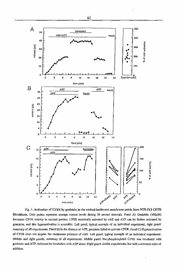
61
A soo Genistein 50
cAK+ATP wash '00
" ~ 0
=< • i "
300 30 •• g
0 • • ~
~ • 200 ~ 20 .... >;
• ~
10 100
0 0 0 3 , 9 12 " " 21 " Genis+cAK
lime (m!n)
B AT? AT? wash 35
cAK Genis
30
; 2S
0 20 ~ '"\.-. • 15
10
5 "-... 0
0 , , , • 10 12 " lime (min)
C 200 15 cAK Genistein
12 r 150
\ " =< " " 9 " ~ ~
• 0 100 ~
~ < • • , ~
~ ~
50 3
0 0 0 , , • 10 12 " .,- ". ,,~ ~~
"p ,. ,. .,. .' .' time (min) {:- (:-
Fig. I. Activation of CFfR by genistein in the excised inside-out membrane patch from NUI-3T3 CFIR
fibroblasts. Data points represent average current levels during 30 second intervals. Panel A) Genistein (I00j1M)
increases CFfR activity in excised patches. CFIR maximally activated by cAK and ATP can be further activated by
genistein, and this hyperaclivation is reversible. Left panel. typical example of an individllal experiment. right panel:
summary of aU experiments. Panel B) In the absence of A IT, genistein failed to activate CFIR. Panel C) Hyperactivation
of CFrR does not require the continuous presence of cAK. Left panel: typical example of an individual experiment.
Middle and right panels, summary of all experiments. :Middle panel: Pre-phosphorylated CFIR was incubated with
genistein and ATP. followed by incubation with ATP alone. Right panel: similar experiments. but with a reversed order of
addition.
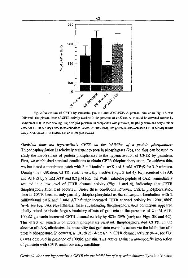
62
250
'" 200
-;; 'n ~
'" '" 0 150 15 ;I<
Fig. 2. 'Activation of CFfR by genisldn, genistin and AMp·PNP. A protocol similar to Fig. lA was
followed. The plateau level of CFTR activity reached in the presence of cAK and A1P could be elevated further by
addition of lOOIlM (see also Fig. IA) or 20J.1M genistein. In comparison with genistein, lOOJ.lM genistin had only a minor
effect on CFTR activity under these conditions. AMP-PNP (0.5 mM), like genistein, also increased CFTR activity in this
assay. Addition 0(0.1% DMSO had no effect (not shown).
Genistein does not hyperactivate CFTR via the inhibition of a protein phosphatase: Thiophosphorylation is relatively resistant to protein phosphatases (25), and thus can be used to study the involvement of protein phosphatases in the hyperactivation of CFIR by genistein. First, we established standard conditions to obtain CFfR thiophosphorylation. To achieve this,
we incubated a membrane patch with 2 milliunitslml cAK and 3 mM ATPyS for 7-9 minutes.
During this incubation, CFIR remains virtually inactive (Figs. 3 and 4). Replacement of cAK
and ATPyS by 2 mM ATP and 0.2 11M PKI, the Walsh inhibitor peptide of cAK, immediately
resulted in a low level of CFfR channel activity (Figs. 3 and 4), indicating that CFTR thiophosphorylation had occurred. Under these conditions however, critical phosphorylation sites in CFTR became only partially thiophosphorylated as the subsequent incubation with 2 milliunits/ml cAK and 2 mM ATP further increased CFIR channel activity by 1290±380% (n=4; see Fig. 3A). Nevertheless, these subsaturating thiophosphorylation conditions appeared ideally suited to obtain large stimulatory effects of genistein in the presence of 2 mM A TP.
l0011M genistein increased CFTR channel activity by 483±139% (n=4; see Figs. 3B and 4C).
This effect of genistein on protein phosphatase resistant, thiophosphorylated CFTR, in the
absence of cAK, eliminates the possibility that genistein exerts its action via the inhibition of a protein phosphatase. In contrast, a 1.0±28.2% decrease in CFfR channel activity (n=4; see Fig.
6) was observed in presence of lOOI1M genistin. This argues against a non-specific interaction
of genistein with CFIR under OUf assay conditions.
Genistein does not hyperactivate CFTR via the inhibition of a ryrosine kinase: Tyrosine kinases

63
do not efficiently use hydrolyzable nucleotide triphosphates other than ATP as a phosphate donor for phosphorylation (26). In contrast, several of these compounds were able to interact with the nucleotide binding domains (NED's) and to substitute for ATP in CFfR activity assays (3). The difference in nucleotide triphosphate specificity between CFfR and tyrosine kinases was exploited to investigate the involvement of tyrosine kinases in the hyperactivation of CFfR by genistein. Similar to previous experiments, we first thiopbosphorylated CFfR in the excised
patch by the addition of cAK and ATP)'S. After a 7-9 minute incubation we removed the kinase
and ATPyS and replaced it by 2 mM GTP and 0.2 flM PKI. We found that the addition of GTP,
in absence of ATP, is sufficient to open CFfR chloride channels (Figs. 5A and C). This confums the initial observation that GTP can open prephosphorylated CFfR at an efficiency of
~60% of maximal activation by ATP (3). Subsequent addition of lOOflM genistein caused a
rapid and large increase in channel activity, amounting to 653±222% of the activity in the presence of GTP alone (n=4; see Figs. 5B and 6). This stimulation is slightly higher than the
hyperactivation of thiophosphorylated CFfR by genistein in the presence of ATP. Under these conditions, the involvement of a tyrosine kinase was virtually excluded by the use of GTP. In conclusion, genistein does not seem to hyperactivate CFfR by means of tyrosine kinase inhibition.
Genistein increases the mean open burst time of CFTR: In a few patches, the channel frequency was sufficiently low to allow the analysis of the effects of genistein on the single channel
characteristics of CFfR. In Fig. 7, a tracing is shown of an excised patch containing a single CFfR chloride channel. Following its phosphorylation by cAK and ATP the mean open burst
time was 334±43 ms, which increased upon the addition of lOOflM genistein to lOO2±180 ms.
The increase in open burst time resulted in a marked increase in channel open probability (po) from 0.10 to 0.31. A qualitatively similar effect was observed in IEC-CF7 intestinal epithelial cells which showed an increase in Po from O.34±O.05 to 0.46±O.06 ( n=3; not shown).
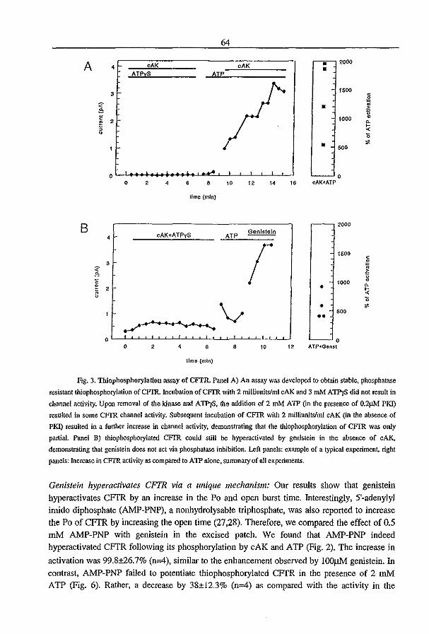
64
A cAK cAK • 2000 4 • ATPyS
3 1500
< 0
:;- • i " < 1000 0
0 2 ~
, !< Q
1;
• " ",0
0 0 0 2 4 • • ,0 t2 " 15 cAK+ATP
lime (mIn)
2000 B cAK+ATPyS ~
4 ATP
r 1500 <
3 0
" :;-~ " < '000 0 ~ e 2 • !< 5
0 1;
~v • " .00 ••
0 0 0 2 4 • • '0 t2 ATP+Genst
lima (min)
Fig. 3. Thiophosphorylation assay of CFTR. Panel A) An assay was developed to obtain stable, phosphatase
resistant thiophosphorylation ofCFIR Incubation ofCFI'R with 2 milliunitslml cAK and 3 mM ATPyS did not result in
channel activity. Upon removal of the kinase and ATPyS, the addition of 2 mM A1P (in the presence of O.2!lM PKI)
resulted in some CFIR channel activity. Subsequent incubation of CFIR with 2 milliunitslml cAK (in the absence of
PKI) resulted in a further increase in channel activity. demonstrating that the thlophosphorylation of CFIR was only
partial Panel B) thiophosphorylated CFIR could still be hyperactivated by genistein in the absence of cAlC,
demonstrating that genistein does not act via phosphatase inhibition. Lefl panels: example of a typical experiment. right
panels: Increase in CFTR activity as compared to ATP alone. summary of all experiments.
Genistein hyperacfivales CFTR via a unique mechanism: Our results show that genistein hyperactivates CFTR by an increase in the Po and open burst time. Interestingly, 5'-adenylyl imido diphosphate (AMP-PNP), a nonhydrolysable triphosphate, was also reported to increase the Po of CFTR by increasing the open time (27,28). Therefore, we compared the effect of 0.5 mM AMP-PNP with genistein in the excised patch. We found that AMP-PNP indeed hyperactivated CFTR following its phosphorylation by cAK and ATP (Fig. 2). The increase in
activation was 99.8±26.7% (n;<f), similar to the enhancement observed by lOO~ genistein. In contrast, AMP-PNP failed to potentiate thiophosphorylated CFTR in the presence of 2 mM ATP (Fig. 6). Rather, a decrease by 38±12.3% (n;<f) as compared with the activity in the
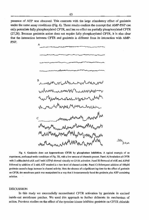
65
presence of ATP was observed. This contrasts with the large stimulatory effect of genistein
under the same assay conditions (Fig. 6). These results confinn the concept that AMP-PNP can only potentiate fully phosphorylated CFTR, and has no effect on partially phosphorylated CFfR (27,28). Because genistein action does not require fully phosphorylated CFTR, it is also clear that the interaction between CFfR and genistein is different from its interaction with AMPPNP.
2.0$
II.OpA
Ftg. 4. Genistein does not byperactivate CFfR by phosphatase inhibition. A typical example of an
experiment, perfonned under conditions of Fig. 3B, with a low amount of channels present. Panel A) Incubation of CFIR
with 2 milliunitslml cAK and 3 mM A1PyS showed virtually no CFrR activation. Panel B) Removal of cAK and A1PyS
followed by addition of2 roM AlP resulted in a low level of channel activity. Panel C) Subsequent addition of lOO}1M
genistein caused a large increase in channel activity. Note the absence of a significant lag-time for the effect of genistein
on CFIR; the membrane patch was manipulated in a way that it instantaneously faced the genistein pllts AlP containing
solution.
DISCUSSION
In this study we successfully reconstituted CFfR activation by genistein in excised inside-out membrane patches. We used this approach to further delineate its mechanism of action. Previous studies on the effect of the tyrosine kinase inhibitor ge.nistein on CFfR chloride
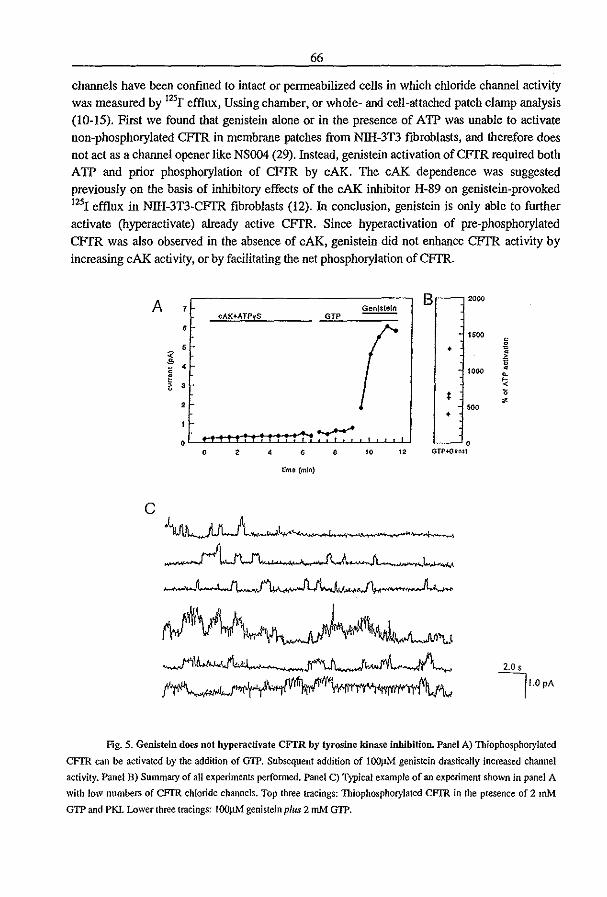
66
channels have been confined to intact or penneabilized cells in which chloride channel activity was measured by l25r effiux, Ussing chamber, or whole- and cell-attached patch clamp analysis (10-15). First we found that genistein alone or in the presence of ATP was unable to activate non-phosphorylated CFfR in membrane patches from Nlli-3T3 fibroblasts, and therefore does
not act as a channel opener like NSOO4 (29). Instead, genistein activation of CFfR required both ATP and prior phosphorylation of CFTR by cAK. The cAK dependence was suggested previously on the basis of inhibitory effects of the cAK inhibitor H-89 on genistein-provoked 1251 efflux in Nlli-3T3-CFTR fibroblasts (12). In conclusion, genistein is only able to further
activate (hyperactivate) already active CFfR. Since hyperactivation of pre-phosphorylated
CFfR was also observed in the absence of cAK, genistein did not enhance CFfR activity by increasing cAK activity, or by facilitating the net phosphorylation of CFfR.
A cAK+ATPyS GTP
• 5
~ 4
~ • 3
, --0 0 , • •
time (mIn)
C
~_"""Iw-""., I.
~
10 "
., ....
B 20"
1500
• 1000
* 500 • 0
GTP·{lin$\
-I
2.0 s IJoOPA
Fig. 5. Genistein does not hyperactivate CFfR by tyrosine kinase inhibition. Panel A) Thiophosphorylated
CFIR can be activated by the addition of G1P. Subsequent addition of lOOJ1M genistein drastically increased channel
activity. Panel B) Summary of all experiments perfonned. Panel C) Typical example of an experiment shown in panel A
with low numbers of CFTR chloride channels. Top three tracings: Thiophosphoryialed CFIR in the presence of 2 mJ\.f
GTP and PKI. Lower three tracings: IOOpM genistein pIlls 2 mM GTP.
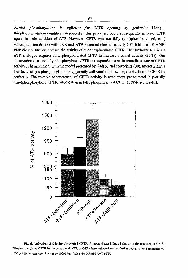
67
Partial phosphorylation is su/ficient for CFTR opening by genistein: Using
thiophosphorylation conditions described in this paper, we could subsequently activate CITR upon the sole addition of ATP. However, CITR was not fully (thio)phosphorylated, as i)
subsequent incubation with cAK and ATP increased channel activity ;0,12 fold, and ii) AMP
PNP did not further increase the activity of thiophosphorylated CITR. This hydrolysis-resistant ATP analogue requires fully phosphorylated CFfR to increase channel activity (27,28). Our observation that partially phosphorylated CFTR corresponded to an intermediate state of CFfR activity is in agreement with the model presented by Gadsby and coworkers (30). Interestingly, a low level of pre-phosphorylation is apparently sufficient to allow hyperactivation of CFTR by genistein. The relative enhancement of CFfR activity is even more pronounced in partially
(thio)phosphorylated CFTR (483%) than in fully phosphorylated CFTR (118%; see results).
1800
1500
1200 >-~ .;; +:: 900 0 CI!
0-f- 600 <{ -0
;f'- ygg 100
50
0
Fig. 6. Activation of thiophospboryJated CFTR. A protocol was followed similar to the one used in Fig. 3.
Thlophosphorylated CFIR in the presence of A 1P, or G1P where indicated can be further activated by 2 milliunilsfml
cAK or l00~M genistein, but not by l00J.lM genistin or by 0.5 mM AMP·PNP.
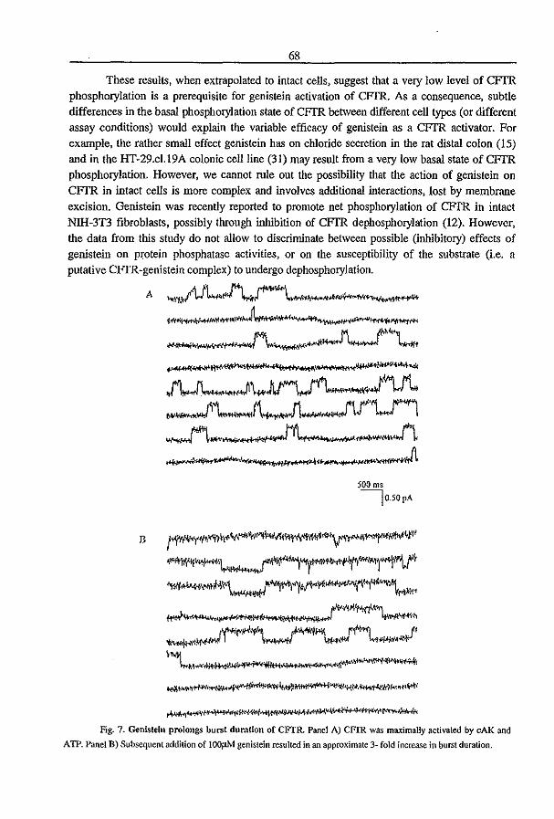
68
These results, when extrapolated to intact cells, suggest that a very low level of CFfR phosphorylation is a prerequisite for genistein activation of CFfR. As a consequence, subtle differences in the basal phosphorylation state of CFfR between different cell types (or different assay conditions) would explain the variable efficacy of genistein as a CFfR activator. For example, the rather small effect genistein has on chloride secretion in the rat distal colon (15) and in the HT-29.c1.l9A colonic cell line (31) may result from a very low basal state of CFfR phosphorylation. However, we cannot rule out the possibility that the action of genistein on
CFfR in intact cells is more complex and involves additional interactions, lost by membrane excision. Genistein was recently reported to promote net phosphorylation of CFfR in intact NIH-3T3 fibroblasts, possibly through inhibition of CFfR dephosphorylation (12). However, the data from this study do not allow to discriminate between possible (inhibitory) effects of genistein on protein phosphatase activities, or on the susceptibility of the substrate (i.e. a putative CFIR-genistein complex) to undergo dephosphorylation.
A ~'1.~I'I'I~'1,,'''''''d'''J'l'fotI~l'''Oh',(I'''' .ttt\iflf"Vfl'·1fJ)Oil~~~
.~
J\J\-..-Jl _.Jb-~"'L.f"'1
w,......._-, ,~ ___ ...!1
500 ms
I050pA
Il 1"'~I'I"tll¥""""~;,jo<I'V~'r""~"~~" ~~,~IM, .... "t4"V; ~~~""""';I'<~~-I(\"l"""\t*'
"'~'" ...... 't." ~~'I'\..-t~J'1""'l.-."J' \-~~~"*,/(lt'll~tJM .• ':;\,<t~~
Fig. 7. Genistein prolongs burst duration of CFTR. Panel A) CFIR was maximally activated by cAl( and
AlP. Panel B) Subsequent addition of lOOj.tM genistein resulted in an approximate 3- fold increase in burst duration.
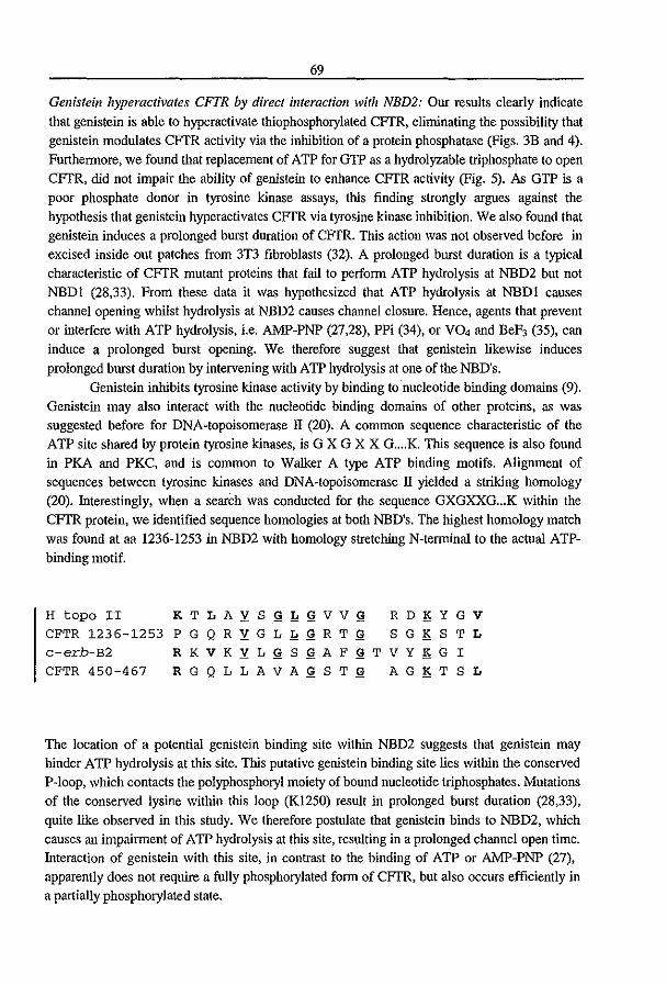
69
Gellisteill hyperactivates CFTR by direct interactioll with NBD2: Our results clearly indicate
that genistein is able to hyperactivate thiophosphorylated CFTR, eliminating the possibility that genistein modulates CFTR activity via the inhibition of a protein phosphatase (Figs. 3B and 4).
Furthennore, we found that replacement of ATP for GTP as a hydrolyzable triphosphate to open CFTR, did not impair the ability of genistein to enhance CFTR activity (Fig. 5). As GTP is a poor phosphate donor in tyrosine kinase assays, this finding strongly argues against the hypothesis that genistein hyperactivates CFTR via tyrosine kinase inhibition. We also found that genistein induces a prolonged burst duration of CFTR. This action was not observed before in excised inside out patches from 3T3 fibroblasts (32). A prolonged burst duration is a typical characteristic of CFTR mutant proteins that fail to perfonn ATP hydrolysis at NBD2 but not NBDI (28,33). From these data it was hypothesized that ATP hydrolysis at NBDI causes channel opening whilst hydrolysis at NBD2 causes channel closure. Hence, agents that prevent or interfere with ATP hydrolysis, i.e. AMP-PNP (27,28), PPi (34), or VO, and BeF3 (35), can induce a prolonged burst opening. We therefore suggest that genistein likewise induces
prolonged burst duration by intervening with ATP hydrolysis at one of the NBD's. Genistein inhibits tyrosine kinase activity by binding to nucleotide binding domains (9).
Genistein may also interact with the nucleotide binding domains of other proteins. as was suggested before for DNA-topoisomerase II (20). A common sequence characteristic of the ATP site shared by protein tyrosine kinases, is G X G X X G .... K. This sequence is also found in PKA and PKC, and is common to Walker A type ATP binding motifs. Aligument of sequences between tyrosine kinases and DNA-topoisomerase II yielded a striking homology (20). Interestingly, when a seareh was conducted for the sequence GXGXXG ... K within the CFTR protein, we identified sequence homologies at both NBD's. The highest homology match was found at aa 1236-1253 in NBD2 with homology stretching N-tenninal to the actual ATPbinding motif.
H topo II K T L A Y. S ~!!~VV~ R D 1> Y G V CFTR 1236-1253 P G Q R Y. G L!!~RT ~ S G 1> S T L c-erb-B2 R KV K Y. L ~ S ~ A F ~T VY 1> G I CFTR 450-467 R G Q L L AVA~ S T ~ A G 1> T S L
The location of a potential genistein binding site within NBD2 suggests that genistein may hinder ATP hydrolysis at this site. This putative genistein binding site lies within the conserved P-Ioop, which contacts the polyphosphoryl moiety of bound nucleotide triphosphates. Mutations of the conserved lysine within this loop (KI250) result in prolonged burst duration (28,33),
quite like observed in this study. We therefore postulate that genistein binds to NBD2, which causes an impainnent of A TP hydrolysis at this site, resulting in a prolonged channel open time. Interaction of genistein with this site, in contrast to the binding of ATP or AMP-PNP (27), apparently does not require a fully phosphorylated fonn of CFTR, but also occurs efficiently in a partially phosphorylated state.
70
CFTR is not the first ion transporter to be modulated by genistein. Effects of genistein on ion channels and transporters include i) inhibition of the Na+K+Cr cotransporter in brain capillary endothelial cells (36), ii) inhibition of a basal and calcium activated potassium channel in rat distal colon (15), iii) inhibition of a basolateral potassium current in HT-29fB6 (13), iv) inhibition of a voltage gated potasium current in vascular smooth muscle cells (19), v) inhibition of L-type calcium currents in rat ventricular cells (16), vi) inhibition of neutral NaCI absorption and Na+-W exchange in rabbit brush border membranes (37), and vii) inhibition of MRP-I, another member of the ATP-binding cassette (ABC) transporter superfamily (38). Although the mechanism underlying ion transport inhibition by genistein has not yet been established, it is tempting to speculate that genistein, analogous to its putative action on CFI'R, may interact directly with the transporters at an ATP-modulatory site.
Therapeutic potential of gellisteill: At present, genistein is under investigation as a possible anticancer therapeutic (39-41). Genistein is present in soy-food, and diet supplementation might be sufficient for beneficial effects (42). The results of the present and previous studies suggest that genistein might also be of benefit to some CF patients in which a residual CFTR activity is
present. Our preliminary attempts to activate &'508 CFTR-chloride channels in Nlli-3T3 fibroblasts revealed a strong synergistic activation of mutant CFTR by a combination of genistein and cAMP agonists (43). This property of genistein would render it an interesting therapeutic for CF, as genistein would only hyperactivate mutant CFTR channels in the additional presence of an endogenous secretagogue. The IC50 for CFTR activation is about
12.5-401tM (10,13,14), just above the presumed upper limit for the serum genistein
concentration under high soy diet (13.21tM;39). Currently, we are investigating the ill vitro
effects of genistein on chloride secretion in intestinal mucosa from CF patients. Also, we are exploring the possibility of diet supplementation as a CF therapeutic in a &'508 CFTR mouse model with residual CFTR activity (44,45). Finally, the distinction between the action of genistein as a CFTR-Cr channel opener and as a tyrosine kinase inhibitor revealed by the present study may open perspectives for the development of genistein-based CF therapeutics showing increased affinity for CFTR but reduced affinity for tyrosine kinases or other ion transporters.
We acknowledge Dr. Michael J. Welsh (Howard Hughes Medical Institute, University of Iowa, College
of Medicine, Iowa city) for donating the 3T3-CFTR fibroblasts and Dr. S.M. Lohmann (Medical
University Clinic, Wlirzburg, Gennany) for providing us with the purified catalytic subunit of cAK
REFERENCES
(1) Kerem, B.I Rommens, J. M'f Buchanan, J. A., Markiewicz, D., Cox, T. K, Chakravarti, A.,
Buchwald, M., and Tsui, L. C. (1989) Scimce 245,1073-1080
(2) Riordan, J. R (1993) A ..... Rev. Physiol. 55,609-630
(3) Anderson, M. P., Berger, H. A., Rich, D. P., Gregory, R J., Smith, A. E., and Welsh, M. J. (1991)
Cell 67,775-784
(4) Bear, C. E., ti, C. H, Kartner, N., Bridges, R J., Jensen, T. J., Ramjeesingh, M., and Riordan, J. R
71
(1992) Cell 68, 809-818
(5) Taocharani, J. A, Chang, X. B., Riordan, J. R, and Hanrahan, J. W. (1991) Nalflre 352,628-631
(6) French, P. J., Bijman, J., Edixhoven, M'I Vaandrager, A. B ... Scholte, B. ]" Lolunann, S. M" Nairn,
A. c., and de Jonge, H R (1995) J. Bioi. 0"'111. 270,26626-26631
(7) Anderson, M. P., Rich, D. P., Gregory, R J., Smith, A E., and Welsh, M. J. (1991) Science 251,
679-682
(8) Akiyama, T., and Ogawara, H. (1991) Methods En'Ylllol. 201,362-370
(9) Akiyama, T., Ishida, J./ Nakagawa,S., Ogawara, H'I Watanabe, S.,. Itoh, N., Slulmya" M., and
Fukami, Y. (1987) J. Bioi. Ch",n. 262,5592-5595
(10) Sears, C. L., Firoozmand, F., Mellander, A., Chambers, F. G ... Eromar, I. G., Bot, A. G., &holte, n., De Jonge, H. R, and Donowitz, M. (1995) Am. J. Physiol. 269, G874-G882
(11) illek, B., Fischer, H, Santos, G. F., Widdicombe, J. H., Machen, T. E., and Reenstra, W. W. (1995)
(12)
Am. J. Physiol. 268, C886-C893
Reenstra, W.W., Yurko-Mauro, K, Dam, A., Raman, S., and Shorten, S. (1996)
Physiol. 271, C650-C657
(13) illek, B., Fischer, H, and Machen, T. E. (1996) Am. J. Physiol. 39, C265-C275
(14) Lehrich, R W., and Forrest, J., Jr. (1995) Am. J. Physiol. 269, F594-.600
(15) Diener, M., and Hug, F. (1996) Enr. J. Plumnaco!. 299,161-170
(16) Yokoshiki, H, Sumii, K., and Sperelakis, N. (1996) J. Mol. Cell. Cardiol. 28,807-814
(17) Nakashima,S., Koike, T., and Nozawa, Y. (1991) Mol. Phamlilcol. 39,475-480
Am.
(18) Kniss, D. A., Zimmennan, P. D., Su, H. c., and Fertel, R H. (1996) Prostaglandins 51,87-105
(19) Smimov, S. V., and Aaronson, P.I. (1995) Circ. Res. 76,310-316
J.
(20) Markovits, J., Linassier, C, Fosse, P., Couprie, }'/ Pierre, J ... Jacquemin-Sablon, A., Saucier, J. M"
Le Pecq, J. B., and Larsen, A K. (1989) Cancer Res. 49,5111-5117
(21) Kaczmarek, L. K, Jennings, K. R, Stnunwasser, F., Nairn, A. c., Walter, V., Wilson, F. D., and
Greengard, P. (1980) Proc. Nail. Acad. Sci. USA 77,7487-7491
(22) Bijman, J." Dalemans, W., Kansen, M., Keulemans, L Verbeek, E., Hoogeveen, A., De Jonge, H.
R, Wilke, M., Dreyer, D., Lecocq, J. P., J.-P Pavirani, A., and SchoIte, B. J. (1993) Alii. J.
Physiol. 264, L229-L235
(23) Hamill, O. P., Marty, A, Neher, E., Sakmann, B., and Sigworth, F. J. (1981) Pflugers Arclt. 391,
85-100
(24) Kansen, M., Bajnath, R. B., Groot, J. A., de Jonge, H. R, Scholte" B., Hoogeveen, A. T., and
Bijman, J. (1993) Pfliigers Arch. 422,539-545
(25) Eckstein, F. (1985) Anll. Rev. Bioch",". 54, 367-402
(26) Hunter, T., and Cooper, J.A (1985) Alln. Rev. Bioch",". 54,897-930
(27) Hwang, T. c., Nagel, G., Nalm, A. c., and Gadsby, D. C. (1994) Proc. Nail. Acad. Sci. USA 91,
4698-4702
(28) Carson, M. R, Travis, S. M., and Welsh, M. J. (1995)J. Bioi. Chelll. 270,1711-1717
(29) Gribkoff, V. K., Champigny, G., Barbry, P., Dworetzky, S. I., Meanwell, N. A, and Lazdunski,
72
M. (1994) J. Bioi. Chelll. 269,10983-10986
(30) Hwang, T. c., Horie, M. , and Gadsby, D. C. (1993) J. Gen. Physiol. 101,629-650
(31) de longe, H. R, Bot, A. G. M., Scholte, B., Sears, C. L., and Donowitz, M. (1994) Pediatric
Pulmollol. Suppl.l0, 188
(32) Fisher, H., illek, B., and Machen, T. E. (1994) Mol. BioI. Cell. 5,319.
(33) Gunderson, K L., and Kopito, R R (1995) Cell 82,231-239
(34) Carson, M. R, Winter, M. c., Travis, S. M., and Welsh, M. I. (1995) J. Bioi. Chelll. 270,
20466-20472
(35) Baukrowitz, T., Hwang, T. c., Nairn, A. c., and Gadsby, D. C. (1994) Neuroll 12,473-482
(36) Vigne, P., Lopez Farre, A., and Frelin, C. (1994)J. Bioi. Chelll. 269,19925-19930
(37) Donowitz, M., Montgomery, I. L., Walker, M.S., and Cohen, M. E. (1994) Am. J. Physiol. 266,
G647-G656
(38) Versantvoort, C. H., Broxterman, H. I., Lankelma, I., Feller, N., and Pinedo, H. M. (1994) Biochelll.
PIUlmUlcol. 48, 1129-1136
(39) Barnes, S. (1995) J. Nllty. 125, m5-783S
(40) Peterson, G. (1995) J. Nuty. 125,7845-7895
(41) Fotsis, T., Pepper" M'I Adlercreutz, H" Hase, T., Montesano, R., and Schweigerer, L. (1995) J. Nlltr. 125,7905-7975
(42) Hendrich,S., Lee,K W.,Xu,x., Wang, H. I., and Murphy, P. A. (1994)1. Nlltr. 124,17895-17925
(43) de longe, H. R, Bot, A. G. M., French, P. I., Bijman, I., Sinaasappel, M., and Veeze, H. I. (1995)
Pediatric Pll/monol. Suppl12, 186
(44) van Doominck, J. H" French, P. J., Verbeek, E., Peters, R H, Morreau, H., Bijman. J., and Scholte,
B. I. (1995) EMBO J. 14,4403-4411
(45) French, P. J., Doorninck,]. H" Peters, R.,H. P. c., Verbeek, E., Ameen, N. A., Marino, C. R., de
longe, H. R, Bijman, I., and Scholte, B. I. (1996) J. Clill. [IlVest 98,1304-1312

73
Fluid Transport In The Trachea Of Normal And CF Mouse
P.I. French, R.H.P.C. Peters, I.R. van Doorninck, W.H. Colledge', R. Ratcliff', M.J.
Evans', B.I. Scholte and 1. Bijman
Departments of Cell Biology and Genetics, Pobox 3000 DR, Rotterdam, tile Netherlands and * We/(comelCRC,
Tennis court Rd Cambridge CB2,JQR. UK
We set out to examine the role of CFTR in active fluid transport in the murine
trachea. For this aim, normal .. and CF murine trachea's were mounted in a modified Ussing-chamber to measure net flux of water (JHzO net), transmural
Potential Difference- (PDtr) and Resistance (Rtr)' Compounds known to
interfere with NaCI-transport (amiloride, carbachol, IBMX, forskolin) across the tissue were evaluated for their effect on JHzO net and electrical parameters.
A basal state of net fluid resorption from the lumen was observed (0.4l±0.23
~lftracheaf2hrs) in the unstimnlated mouse trachea. With amiloride (0.1
mmollI) in the lumen Ieq decreased and net water movement was abolished
(0.080±06 ~lftracheaf2hrs). The addition of forskolin (to ~molll), or
forskolin and IBMX (0.2 mmolfl), resulted in a hyperpolarization of PDt r
indicating activation of a luminal chloride conductance. Although
forskolinlIBMX tended to decrease fluid resorption, it failed to reach
statistical significance. A combination of forskolinlIBMX and amiloride did
not result in a uet secretion of water, suggesting that the murine trachea is not
capable of secreting significant amounts of fluid. Electrophysiological
parameters, JHzO neh and the net changes in JHzO net to amiloride and
forskolinlIBMX, were similar in both normal (CFTR +f+ and +f-) and CF
(CFTR -f-)- mouse trachea, indicating that CFTR is not involved in the fluid
transport across mouse tracheal epithelium. Our results suggest that both in
normal- and CFTR(-f-)- mouse trachea amiloride sensitive net fluid uptake is
present which is attenuated similarly in the two tissues by forskolin/IBMX,
suggesting that CFTR is not involved in this process.
Cystic Fibrosis (CFl) is a common lethal genetic disease in the Caucasian population,
affecting 1:2500 new-barns (for review see (35)). The disease is caused by mutations in the
1 The abbreviations used are: CF, cystic fibrosis; CFTR cystic fibrosis transmembrane conductance regulator; DIDS. 4,4' .-diisothiocyanatostilbene-2.2' -disulfonic acid; DPC, diphenylamine 2-carboxylate; mMX, 3-isobutyl-
74
gene encoding the Cystic Fibrosis Transmembrane Conductance Regulator (CFTR) (22, 29,
30) which forms a cAMP and ATP regulated chloride channel (2a, 5, 7, 21, 34) and is
expressed in the apical membrane of various epithelial cells (11, 14). Although the gene
defective in CF has been identified, CF disease pathogenesis is still a poorly understood
process. It has been proposed that mucus plugging of small airways and ducts, a characteristic
CF disease symptom, is caused by a defective transepithelial fluid transport, resulting in an
insufficient hydration of mucus. The dehydrated mucus is viscid and difficult to clear, and
these changes in the rheological properties may explain the mucus plugging and subsequent
inflammation in CF.
In fluid transporting epithelia like gallbladder and renal proximal tubule, isotonic fluid
transport is dictated by the active transport of ions. In the absence of apical CFTR chloride
channels the transepithelial fluid transport therefore may be altered. Indeed, a reduced cAMP
induced fluid secretion was found in CF pancreatic (13, 23, 24) and pulmonary (19, 32)
epithelia. In addition, both CF mouse intestine (15 and PJF, unpublished observations) and
gallbladder (26) fail to secrete fluid when stimulated with cAMP agonists. A CFTR dependent
fluid secretion was also found in ADPKD (autosomal dominant polycystic kidney disease) cells
(12).
Although these observations clearly indicate an active role for CFTR in the process of
fluid secretion, measurements of fluid transport acrOss pulmonary epithelia have been restricted
to cultured cell systems. The presence of multiple cell types (e.g. ciliated and goblet cells) in
intact pulmonary tissue, suggests a more complex model system than cell culture, and
electrophysiological differences between cell culture and the intact system have been reported
(17). As a consequence, cultured pulmonary epithelia may not truly reflect the in vivo situation.
We therefore studied the role of CFTR in fluid transport across the intact murine trachea in a
CF mouse model (28). The measurement of CFTR dependent fluid transport across intact
pulmonary epithelium may help understand CF airway pathogenesis.
MATERIALANDME1HODS Tissue preparation: Mice were weighed and anaesthetised with hypnodil, 0.2 mg/g. Murine tracheas were carefully excised from either adult C57BU6 mice. or from age matched CF mice (28) (homozygous ./- CFrR knockout mice), and nonnal mice (helero·(+/-) and homozygous(+/+) litter mates). CF mice had a significant lower weight compared to their normalliuennates, 24.5 ± 5.5gr (n=22) vs. 19.4 ± 3.5gr (n=16), respectively (p<O.Ol). After excision, trachea's were mounted at 40C in a modified Ussing chamber (Fig. 1). The average length of excised trachea's was 4.7 mm, average diameter 0.85 mm. Fluid measurements were corrected for the number of cartilage rings. On average, an excised trachea will account for approx. 0.11 cm2 of epithelium. PDlr and Resistance (Rtr) measuremenr: In the perfused trachea (40 JllImin) PD,r was measured between agar bridges inside the lumen of the tracbea (± 1 nun distal from the pipette tip opening) and the bath. A small (0.2
l·methyl·xantine~ Ieq, equivalent short circuit current; Isc, short circuit current; JH20net, net fluid transport; PDu, transmural potential difference; Rtr• transmural resistance.
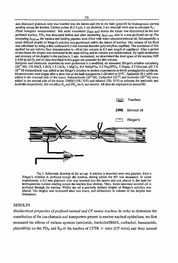
75
mm diameter) platinum wire was inserted into the lumen and one in the bath (ground) for homogenous current sending across the trachea Current pulses (0.5·1 J.LA. 1 sec duration,S sec interval) were sent to calculate Ru. Fluid transport measurement: Net water movement (JHlO nev across the tissue was detennined in the non perfused trachea. PDIl was measured before and acler measuring 1"20 net. also in a non perfused set-up. For measuring JH20 net the trachea and bolding pipettes were HIled with water-saturated mineral oil. Subsequently. a small defined droplet of Ringer's solution was positioned within the lumen of trachea. The volume of the fluid was calculated by using a thin calibrated 0.3 mm internal diameter poly-etbylene capillary. The resolution of this method for net volume flow determination is -40 01 (the volume in 0.5 mm length of capillary), After a period of two hours me droplet was recovered in the same tubing and its volume was redetermined. By rapid installation and recovery of lhedroplet in the trachea « 1 min. incubation), we determined the dead space of the trachea, 0.09 ± 0.04 flI (n=5), and all data descrlbed in Ibis paper are coJreCted for Ibis volume. Solutions and chemicals: experiments were performed in a modified, air saturated, Ringer's solution containing (10.3 M); 133 NaCI, 5 KCI, 1.5 caCI" I MgCi" 0.4 NalJ,PO" 0.3 Na,HPO" 5 Hepes, 5.5 Glucose, pH 7.4. 10.5 M indomethacin was added to the Ringer's solution in trachea experiments to block prostaglandin synthesis. Measurements were begun after a slow rise of the bath teffiP§f3.ture (-20 min) to 37OC. Amiloride (0.1 mM) was added to the mucosal side of the tissue. Indomethacin (10.5 M). Carbachol (10.5) and forskolin (10.5 M) were added to the serosal side of the tissue. DMSO (1%(1 VN) and ethanol (1%(1 VN) as solvents for amiloride and forskoUn respectively, did not affect Ru and PDt! (n=5; not shown). All data are expressed as mean±SD.
I~'I"I Trachea
1;;c~1 Mineral 011
Icce , I Ringer's
Fig L Schematic drawing of the set·up. A trachea is mounted onto two pipeues. First a Ringer's solution is perfused trough the trachea, during which the PD was measured. In some experiments, a 0.2 mm platinum wire was inserted into the lumen and one placed in the bath for homogeneous Current sending across the trachea (not shown). Then, water-saturated mineral oil is perfused through the trachea. Within the oil a precisely defined droplet of Ringer's solution was placed. The droplet was recovered after two hours, and differences in volume of the droplet was detennined.
RESULTS
Bioelectrical properties of perfused 1I0rmai and CF mouse trachea: In order to determine the
contribulion of Ihe ion-channels and Iransporters present in murine tracheal epiIhelium, we first
measured the effecls of various agonisls (amiloride, forskolinlIBMX, carbachol, bumetanide,
phloridzin) on the PDtr and Rtr of the trachea of CFfR -/- mice (CF mice) and their normal
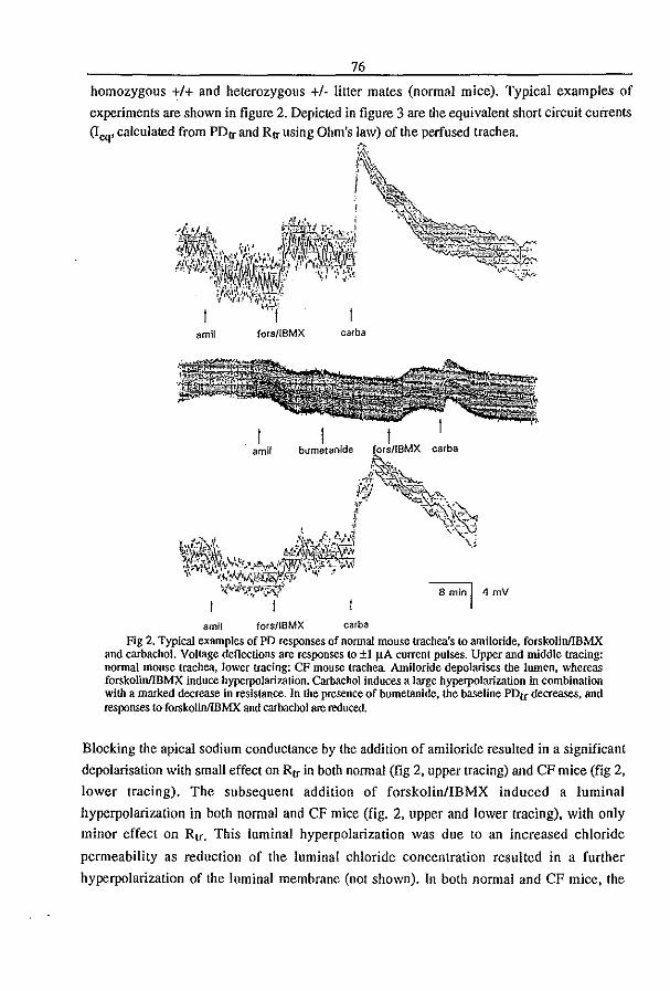
76
homozygous +1+ and heterozygous +1· litter mates (normal mice). Typical examples of
experiments are shown in figure 2. Depicted in figure 3 are the equivalent short circuit currents (Ieq' calculated from PD" and R" using Ohm's law) of the perfused trachea.
~\.
~. I ~-
~I~;:~ amil fors/lBMX carba
I amil blImet",nlde
8 min I 4 mV
amil fors/IBMX carba
Fig 2. Typical examples ofPD responses of normal mouse trachea's (0 amiloride, forskolinlIBMX and carbachol. VoHage deflections are responses to ±1 J.lA current pulses. Upper and middle tracing: normal mouse trachea, lower tracing: CF mouse trachea Amiloride depolarises the lumen, whereas forskolinlIBMX induce hyperpolarization. Carbachol induces a large hyperpolarization in combination with a marked decrease in resistance. In the presence of bumelanide. the baseline PDtr decreases, and responses to focskolin/lBMX and carbachol are reduced.
Blocking the apical sodium conductance by the addition of amiloride resulted in a significant
depolarisation with small effect on R" in both normal (fig 2, upper tracing) and CF mice (fig 2,
lower tracing). The subsequent addition of forskolin/IBMX induced a luminal
hyperpolarization in both normal and CF mice (fig. 2. upper and lower tracing), with only
minor effect on Rtf. This luminal hyperpolarization was due to an increased chloride
permeability as reduction of the luminal chloride concentration resulted in a further
hyperpolarization of the luminal membrane (not shown). In both normal and CF mice, the
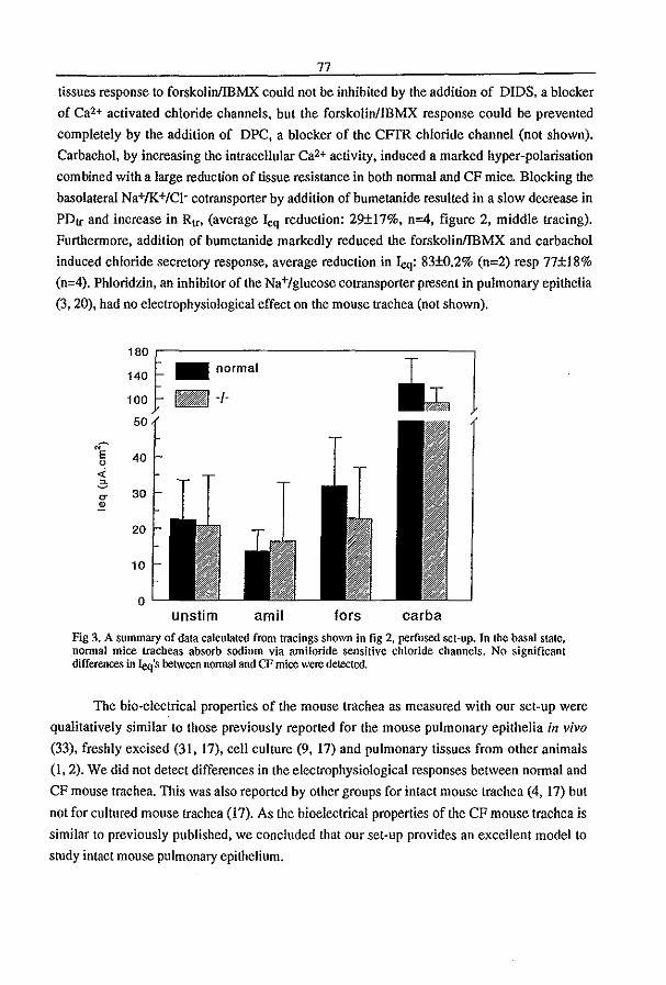
77
tissues response to forskolinlIBMX could not be inhibited by the addition of DlOS, a blocker
of Ca2+ activated chloride channels, but the forskolinlIBMX response could be prevented
completely by the addition of OPC, a blocker of the CFfR chloride channel (not shown).
Carbachol, by increasing the intracellular Ca2+ activity, induced a marked hyper-polarisation
com bined with a large reduction of tissue resistance in both normal and CF mice. Blocking the
basolateral Na+/K+/Cl- cotransporter by addition of bumetanide resulted in a slow decrease in
PO" and increase in R", (average Ieq reduction: 29±17%, n=4, figure 2, middle tracing).
Furthermore, addition of bumetanide markedly reduced the forskolinlIBMX and carbachol
induced chloride secretory response, average reduction in leq: 83±O.2% (n=2) resp 77±18%
(n=4). Phloridzin, an inhibitor of the Na+/glucose cotransporter present in pulmonary epithelia
(3,20), had no electrophysiological effect on the mouse trachea (not shown).
180 ~ 140
100
50
40
30
20
10
o
_ normal
~I
unslim amil tors carba
Fig 3. A summary of data calculated from tracings shown in fig 2, perfused set-up. In the basal slate, normal mice tracheas absorb sodium via amiloride sensitive chloride channels. No significant differences in Ieq's between normal and CF mice were detected.
The bio-electrical properties of the mouse trachea as measured with our set-up were
qualitatively similar to those previously reported for the mouse pulmonary epithelia in vivo (33), freshly excised (31, (7), cell culture (9, 17) and pulmonary tissues from other animals
(1,2). We did not detect differences in the electrophysiological responses between normal and
CF mouse trachea. This was also reported by other groups for intact mouse trachea (4, 17) but
not for cultured mouse trachea (17). As the bioelectrical properties of the CF mouse trachea is
similar to previously published, we concluded that our set-up provides an excellent model to
study intact mouse pulmonary epithelium.

78
Fluid transport in mouse trachea: Prior to the measurement of fluid transport, we detennined
tissue viability of the mouse trachea by measuring the PD in a non perfused set-up (a similar
condition as used to measure fluid transport). Under these conditions, baseline PD's were
-9.3±3.7 mY for nonnal mouse trachea. Interestingly, a significant hyperpolarisation was
observed in the CF mouse trachea (-12.0±4.6 mY, P<fW5). Responses to agonists (amiloride,
forskolinJIBMX or both) were qUalitatively similar to the perfused set-up in both nonnal and
CF mice (data not shown). Fig. 4 shows the net transport of fluid in the trachea of B57BlI6
mice. A net absorption was observed in the unstimulated trachea. When a combination of 10
11M forskolin and 0.1 mmolll amiloride was applied to the medium (bath and lumen), the net
fluid absorption was almost completely inhibited. The same method was used to investigate
fluid transport in the trachea of the CF and nonnal mouse. The results are shown in fig. 5 and
6. In nonnal mice we observed a net fluid absorption of 0.37±0.251lIltrachea/2hrs in the basal,
unstimulated state. Net fluid absorption in nonnal and CF mice tracheas were comparable (see
fig 6) Application of 0.1 mM amiloride to both sides of the trachea almost completely inhibited
net fluid absorption in the nonnal mouse trachea to 0.05±O.05 Illltrachea/2hrs. A combination
of 10 11M forskolin and 0.2 mM IBMX, did not significantly inhibit net fluid absorption,
neither in normal mice nor in CF mice (fig 6). A combination of both amiloride and
forskolinlIBMX completely inhibited net fluid absorption in the nonnal mouse trachea, but no
significant amounts of fluid secretion were observed.
Tissue viability after experimentation was checked by redetermining the PD in
(unstimulated) C57B1I6, normal, and CF mouse tracheas, and were -4.9±O.5 mY (n=3),·
2.5±1.4 mY (n=8), -2.5±1.4 mY (n=5) respectively. The response to carbachol after
experimentation was -13.6±3.3 mY (n=3) in nonnal mice and -15.7±6.7 mY (n=3) in CF
mice.
DISCUSSION
Water transport in normal mouse trachea: To our knowledge this report is the first in which
measurements of fluid transport in intact, excised (mouse) tracheas have been perfonned. Our
results indicate a basal state of fluid absorption of approximately 0.4 Ill/trachea/2hrs,
corresponding to -lllllcmz/h. The addition of amiloride almost completely inhibited the fluid
absorption, indicating that amiloride sensitive Na+ transport is responsible for the fluid
absorption in the basal state of the mouse trachea. The addition of forskolin and IBMX did not
have a significant effect on fluid absorption, and no significant net fluid secretion was observed
in the combined presence of amiloride and forskolin!IBMX.
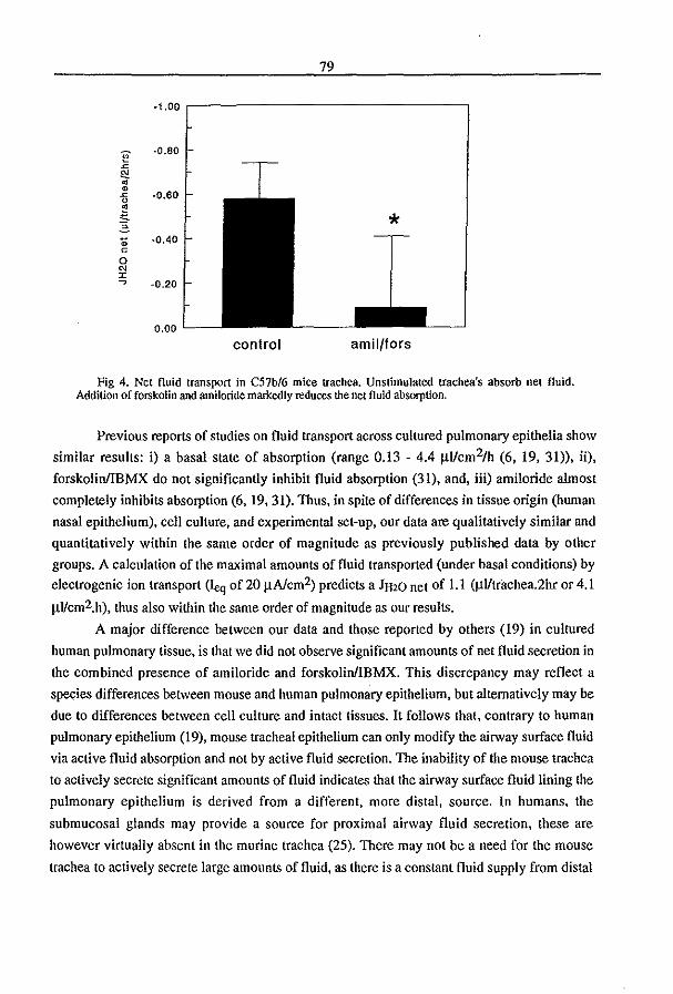
79
·1.00 ,-----------------,
·0.80
-0.60
-0.40
-0.20
0.00
control amil/fors
Fig 4. Net fluid transport in C57b/6 mice trachea. Unstimulated trachea's absorb net fluid. Addition of forskolin and arniloride markedly reduces the net fluid absorption.
Previous reports of studies on fluid transport across cultured pulmonary epithelia show
similar results: i) a basal state of absorption (range 0.13 - 4.4 ~Ucm2/h (6, 19, 31», ii),
forskolinllBMX do not significantly inhibit fluid absorption (31), and, iii) amiloride almost
completely inhibits absorption (6, 19,31). Thus, in spite of differences in tissue origin (human
nasal epithelium), cell culture, and experimental set-up, our data are qualitatively similar and
quantitatively within the same order of magnitude as previously published data by other
groups. A calculation of the maximal amounts of fluid transported (under basal conditions) by
electrogenic ion transport Ueq of 20 ~Alcm2) predicts a JH20 net of L I (~Utrachea.2hr or 4.1
~Ucm2.h), thus also within the same order of magnitude as our results.
A major difference between our data and those reported by others (19) in cultured
human pulmonary tissue, is that we did not observe significant amounts of net fluid secretion in
the combined presence of amiloride and forskolinllBMX. This discrepancy may reflect a
species differences between mouse and human pulmonary epithelium, but alternatively may be
due to differences between cell culture and intact tissues. It follows that, contrary to human
pulmonary epithelium (19), mouse tracheal epithelium can only modify the airway surface fluid
via active fluid absorption and not by active fluid secretion. The inability of the mouse trachea
to actively secrete significant amounts of fluid indicates that the airway surface fluid lining the
pulmonary epithelium is derived from a different, more distal, source. In humans, the
submucosal glands may provide a source for proximal airway fluid secretion, these are
however virtually absent in the murine trachea (25). There may not be a need for the mouse
trachea to actively secrete large amounts of fluid, as there is a constant fluid supply from distal
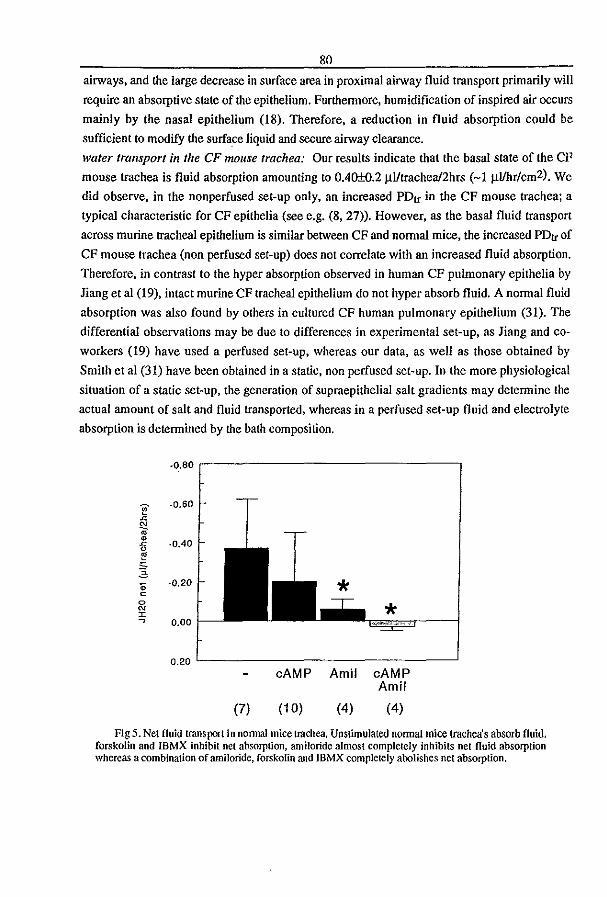
80
airways, and the large decrease in surface area in proximal airway fluid transport primarily will
require an absorptive state of the epithelium. Furthermore, humidification of inspired air occurs
mainly by the nasal epithelium (I8). Therefore, a reduction in fluid absorption could be
sufficient to modify the surface liquid and secure airway clearance.
water transport in the CF mollse trachea: Our results indicate that the basal state of the CF
mouse trachea is fluid absorption amounting to OAO±O.2 ~lItrachea!2hrs (-1 ~lIhr/cm2). We
did observe, in the non perfused set-up only, an increased PD" in the CF mouse trachea; a
typical characteristic for CF epithelia (see e.g. (8, 27». However, as the basal fluid transport
across murine tracheal epithelium is similar between CF and normal mice, the increased PD" of
CF mouse trachea (non perfused set-up) does not correlate with an increased fluid absorption.
Therefore, in contrast to the hyper absorption observed in human CF pulmonary epithelia by
Jiang et aI (19), intact murine CF tracheal epithelium do not hyper absorb fluid. A normal fluid
absorption was also found by others in cultured CF human pulmonary epithelium (31). The
differential observations may be due to differences in experimental set-up, as Jiang and co
workers (19) have used a perfused set-up, whereas our data, as well as those obtained by
Smith et al (31) have been obtained in a static, non perfused set-up. In the more physiological
situation of a static set-up, the generation of supraepithelial salt gradients may determine the
actual amount of salt and fluid transported, whereas in a perfused set-up fluid and electrolyte
absorption is determined by the bath composition.
F ~
~ ro ~ ~ 0 E 'S 3 .. c
0
'" r ~
·0.80 ,.------------------,
·0.60
·oAO
-0.20
* 0.00
0.20 '-------------------'
(7)
cAMP Amil cAMP Amil
(10) (4) (4)
Fig 5. Net fluid transport in nannal mice trachea, Unstimulated normal mice trachea's absorb fluid. forskolin and IBMX inhibit net absorption, amiloride almost completely inhibits net fluid absorption whereas a combination of arniloride, forskolin and IBMX completely abolishes net absorption.

81
-0.80
basal Fors/IBMX
F -0.60 ~
£! ~ ~
·0.40 ~ 0
~ ,;; 20 .. -0.20 c
0
'" I , 0,00 normal CF normal CF
.0.20
(7) (6) (10) (6)
Fig 6. Net fluid transport in normal and CF mice trachea. CF mice trachea absorb fluid at a rate no greater than nonnal mice trachea. Forskolin and IBMX inhibit net fluid absorption in the CF mice tracbea; (he magnitude of the inhibition is similar to the inhibition seen in Donnal mice trachea.
In CF mice. like in normal mice, we could observe a trend towards a decrease in
absorption when trachea's were stimulated by forskolinlIBMX. However, both normal and CF
mice respond to forskolinlIBMX to a similar extent, and therefore we conclude that CFTR <loes
not playa role in the active fluid transport across mouse tracheal epithelium. As isotonic fluid
transport is dictated by the active transport of ions, a similar conclusion can be deduced from
the almost identical bioelectrical properties between normal and CF mouse trachea. Our results
show a several times larger tissues response to the Ca2+ agonist carbachol than to cAMP
agonists, therefore the Ca2+ activated currents may be of more importance to the physiology of
the mouse trachea. It has been suggested that a Ca2+activated chloride channel can compensate
for CFTR in CF mouse epithelia (10, 16,36).
Summarising. the mouse trachea has a basal state of amiloride sensitive Na+
absorption. The role of CFTR in fluid transport is minor, and it is questioned whether CFTR
can be detected electrophysiologically in the mouse trachea. We conclude that CFTR does not
play an important role in the mouse trachea, thus providing an explanation for the absence of
pulmonary pathology in the CF mouse. As it is difficult to detect CFTR in the mouse trachea,
the suitability of this tissue for gene therapy protocols seems rather questionable.
[Supported by the Dutch organisation for scientific research (NWO)]
REFERENCES
82 1) Al-Bazzaz, FJ .. Tarka, C .. Farnh, M. (1991) All, J. Physiol. 260: L594-L602 20) Anderson, M.P, Rich, D.P .. Gregory, R.I., Smith, A.E .. Welsh, M.I. (1991) Science 251: 679-82 2) Ballard, S.T., Schepens, S.M .. Falcone, J.C., Meininger, G.A., Taylor, A.E. (1992) J. Appl. Physiol.
73: 2021-2027 3) Barker, PM .. Boyd, C.A.R .. Ramsden, C.A., Strang, L.B .. Walters, D.V. (1989) J. Physiol. 409: 15-27 4) Barker, P.M .. Brigman, K.K., Paradiso, A.M .. Boucher, R.C. Gatzy, J.T. (1995) Am. J. Respir. Cell.
Mol. Bioi. 13: 307-313 5) Bear, C.E., Li., C.H., Kartner, N., Bridges, R.I., Jensen, T.J., Rarnjeesingh, M .. Riordan, J.R. (1992)
Cell 68: 809-18 6) Benali, R., Pierrot, D., Zahm, J.M .. Debentzmann, S., Puchelle, E. (1994) Am. J. Resp. Cell. Mol.
Bioi. 10:363-368 7) Bijman, J. t DaJemans. W.o Kansen. M .. Keulemans. J., Verbeek, E .. de Jonge, H.R., Wilke. M .•
Dreyer, D .. Lecocq, J.P. et aI (1993) Am. J. Physiol 264: L229-35, 8) Boucher, R.C .. Jackson StultS, M., Knowles, M.R., Cantley, L. Gatzy, J.T (1986). J. Clin. Invest. 78:
1245-1252 9) Clarke, L.L., Bums, K.A .. Bayle, J.-Y., Boucher, R.C .. Van Scott, M.R. (1992) Am. J. Physiol.263:
L519-L525 10) Clarke, L.L., Gmbb, B.R .. Yankaskas, J.R .. COllon, C.U., McKenzie, A .. Boucher, R.C. (1994)
Proc. Natl. Acad. Sci. USA 91: 479483 11) Crawford, I, Maloney, P.C., Zeitlin, PL" Guggino, W.B., Hyde, S.C., Turley, H., Gauer, K.C ..
Harris, A .. Higgins, C.F. (1991) Proc. Natl. Acad. Sci. USA 88: 9262-9266 12) Davidow, C.J .. Maser, R.L.. Rome, L.A., Calvet, J.P .. Grantham, 1.1. (1996) Kidney International 50:
208-218 13) Davidson,G,P .• Kirubakaran, C.P" Ratcliffe. G., Cooper, D.M., Robb, T.A. (1986) Acta Pead. Scand
75:145-50 14) Engelhardt, J.F., Yankaskas, 1,R.. Ernst, S.A., Yang, Y .. Marino, C.R.. Boucher, R.C .. Cohn, I.A.,
Wilson, J.M. (1992) Nature Genel. 2: 240-248 15) Gabriel, S.E., Brigman, K.N., Koller, B.H" Boucher, R.C" Stutts, M.J. (1994) Science 266:107-109 16) Gmbb, B.R., Vick, R.N" Boucher, R.C. (1994) Am. J. Physiol. 266:CI478-CI483 17) Grubb, B.R" Paradiso, A.M" Boucher, R.C. (1994) Am; J. Physiol. 267: C293-C300 18) 1ngelstedt, S. (1956) Acta Otolaryngol. Suppl. 131: 1-80 19) Jiang, C.W., Finkbeiner, W.E., Widdicombe, J.H., McCray, P.B., Miller, S.S. (1993) Science 262:424-
427 20) loris, L and Quinton, P.M. (1989) Pjlligers Arch. 415:118-120 21) Kanner, N .. Hanrahan. I.W., Jensen, TJ., Naismith. AL., Sun, S.z .• Ackerly. C.A., Reyes, E.F .. Tsui,
L.-C., Rommens, J.M., Bear, CE., et ai, (1991) Cell 64: 681-91 22) Kerem, B. t RommeRs, I.M., Buchanan. 1.A., Markiewicz, D .• Cox, T.K., Chakravarti. A., Buchwald,
M" Tsui, L.C. (1989) Science 245: 1073-1080 23) Kopelman, H., Durie, P., Gaskin, K., Weizman, Z" Forstner, G. (1985) N. Engl. J. Med., 312:329-334
24) Kopelman, H., Corey, M., Gaskln. K., Durie. P .• Weizman, Z., Forstner, G. (1988) Gastroenterof . • 95: 349-355
25) Pack, R.J" AI-Ugaily, L.H" Monis, G., and Widdicombe, J.G (1980). Cell Tissue Res. 208: 65-84 26) Peters, R.H.P.C .. van Doominck. I.H., French. PJ .. Ratcliff. R.. Evans, M.l .. Colledge, W.H.. Bijman,
1. and Scholte, BJ. Hepatology. in press 27) Quinton, PM and Bijman, (1983) J.New Engl. J. Med. 308: 1185-1189 28) Ratcliff, R., Evans. MJ., Cuthbert, A.W., Macvinish, 1.L., FOSler, D .. Anderson, 1.R.. Colledge, W.H.
(1993) Nature. Genet. 4: 3541 29) Riordan, I.R .. Rommens. J.M., Kerem, B., Alon, N., Rozmahel, R, Grzelczak. Z.o Zielenski.l., Lolc.
S., Plavsic, N., Chou, J.L. et aI. (1989) Science 245: 1066-73 30) Rommens, I.M., Iannuzzi, M.C .• Kerem. B., Drumm. M.L., Melmer, G .• Dean, M., Rozmahel, R.,
Cole, J.L" Kennedy, D" Hidaka, N" et aI. (1989)Science 245: 1059-65 . 31) Smith, J.J" Welsh, MJ. (1993) J. Clin.lnvest,,91: 1590-1597
32) Smith, J.J, Karp, P.H., Welsh, (1994) MJ.J. Clin Invest. 93: 1307-1311 33) Takahashi, M., Freed, A.N., Croxton, T.L. (1995) Resp. Physiol. 99: 19-27, . 34) Tabcbarani, J.A., Chang, X.B., Riordan, J.R., Hanrahan, J.W. (1991) Nature 352: 628-31 35) Welsh, MJ., Tsui. L.-C, Boat, T.F., Beaudet. AL. Cystic Fibrosis.!n Sriver, C.R., Beaudet. AL..
Sly, W.S. and Valle, D (eds), the metabolic bases ojiniterited disease (1995) 3799-3876 36) Wilschanski, M.A" Rozmabel, R" Beharry, S., Kent, G., Li, C., Tsui, L.-C" Durie,. P., Bear, C.E.
(1996) Biochem. Biophys. res. comm 219: 753-759

83
CFTR expression and mucin secretion in cultured mouse gallbladder epithelial cells
RICHARD H. P. C. PETERS, PlM J. FRENCH, J. HIKKE VAN DOORNINCK, GENEVIEVE LAMBLIN, ROSEMARY RATCLIFF, MARTIN J. EVANS, WILLIAM H. COLLEDGE, JAN BIJMAN, AND BOB J. SCHOLTE Cell Biology, Erasmus University, 3000 DR Rotterdam, The Netherlands; Institut National de la Sante et de la Recherche Medicale UniM 377. 59045 Lille, France; and Welleome/Clinical Research Cenfur, Cambridge CB2 lQR, Unifud Kingdom
Peters, Richard H. P. C., Pim J. French, J. Hikke van Doorninck, Genevieve Lamblin, Rosemary Ratcliff, Martin J. Evans, William H. Colledge, Jan Bijman., and Bob J. Sehoite. CFI'R expression and mucin secretion in cultured mouse gallbladder epithelial cells. Am. J. Physioi. 271 (Gastrointest. Liuer PhysiQI. 34): GI074-GI083, 1996.Dysfunction of the cystic fibrosis transmembrane conduc· tance regulator (CFTR) in humans is frequently associated with progressive liver disease, which appears to result from obstruction of biliary ducts with mucous material. CFl'R in the liver is expressed in the biliary epithelium. With the use of a mouse model for cystic fibrosis (CF) we have studied the relationship between CFfR expression and glycoprotein secre~ tiOll in primary culture of mouse gallbladder epithelial cells (MOBC). MGBC in culture maintain a well~d.ifferentiated phenotype as shown by microscopy. The cells produce CFTR mRNA to levels comparable to the intact tissue. With patch. clamp analysis we could frequently observe a linear protein kinase A-regulated CI- channel that shows all the major characteristics of human CITR, although its conductance is lower (5 pS compared with 8 pS). MGBC in culture produce and secrete high molecular weight glycoproteins (HMG) in a time·dependent and temperature·sensitive manner. Secre· tion of HMG was not stimulated significantly by either adenosine S',5'~yclic monophosphate (cAMP), Ca2+, or pro· tein kinase C agonists in this system. High concentrations (S mM) of extracellular ATP stimulated secretion thrt'!efold, but low concentrations (O.S mM) had no effect. Approximately one-third of the HMG produced and secreted consisted of mucin. Cultured MGBC from CFTR·deficient mice produced and secreted mucin to a similar extent as normal cells. We conclude that cultured mouse gallbladder cells are a convenient model to study both CFTR function and mucin secre· tion. In this system, we found no evidence for a direct link between mucin secretion and CFI'R activity, as has been suggested for other cell types.
cystic fibrosis transmembrane conductance regulator; glyco. proteins
THE INTRAHEPATIC BILE DUCTS, as well as the cystic duct and gallbladder, are lined with epithelial cells with distinct morphology. This epithelial cell layer plays an important role in the regulation of bile volume and bile pH, using a combination ofion-transport systems situated in both apical and basolateral membranes. In addition to performing regulated ion transport, bile duct cells have a secretory function. Many subapical vesicles can be observed in electron micrographs of biliary epithelium. Mucins are prominent among the products secreted from this intracellular pool (36). The
physiolOgical function of mucin secretion is supposedly the formation of an extracellular gel protecting the tissue against the aggressive biliary environment. The regulation of mucin secretion in biliary epithelium has not been studied in detail.
Cystic fibrosis (CF) is a recessive inherited disease with a high incidence in the Caucasian population (6, 60). It is caused by mutations in an adenosine 3',5'cyclic monophosphate (cAMP)-regulated Cl- channel (cystic fibrosis transmembrane conductance regulator (CFTR)] situated in the apical membranes of a variety of epithelial cells (49). CFI'R is involved in the regula~ tion of isosmotic fluid transport. CFTR dysfunction results in a complex phenotype characterized by recurrent IWlg infections, pancreatic insufficiency. and ma1~ absorption. Morbidity and mortality in CF is mainly associated with lWlg disease. However, hepatobiliary abnormalities are quite common in CF patients. Obstruction of biliary ducts, focal, or multilobular biliary cirrhosis was reported in over 50% of adult CF patients at autopsy (5, 33, 59). Microgallbladder and high incidence of gallstones is also observed (41, 42, 52). CFTR in the liver is mainly expressed in bile duct cells, as shown for rats (21) and humans (13, 55). The pathogenesis of liver disease in CF remains to be elucidated. Defective isosmotic fluid transport may result in decreased solubility and subsequent precipita~ tion of bile components. In support ofthis, studies with a mouse model for CF in our laboratory showed that CFTR is required for cAMP-induced fluid secretion across the biliary epithelium (44a). On the other hand, there may be additional reasons for the abnormal properties of mucus secretions in CF epithelia. First, CFTR dysfunction affects membrane recycling (7, 45) and possibly secretion (20, 38, 50) in several cell types. Second, CFTR mutations can result in abnormal glyco~ sylation and sulfation of glycoproteins (11, 16, 34, 64). Biliary mucins are implicated as promoting factors in the formation of bile stones (1, 9). Therefore, both increased secretion of mucins and abnormal mucin structure could contribute to hepatic disease in CF.
We have studied the relationship between CFI'R and mucin secretion in cultured mouse biliary epithelial cells using a mouse model of CF. We show here that primary cultures of mouse gallbladder epithelium main· tain many characteristics of the original tissue, includ~ ing CFTR expression and mucin synthesis.
METHODS
Materials. Sepharose CL-4B was from Phannacia (Uppsaia, Sweden). and guanidinium 01- was from Merck (Darmstadt, Germany), Heparltinase I (Heparinase lID from Flavobacterium heparinum, keratanase (Pseudomonas species), and hyaluronidase from Streptomyces hyalurolyticus were purchased from Sigma (Saint Canteen FalJavier, France), and chondroitinase ABC from Proteus vulgaris was from Seik· agaku (lbkyo, Japan), The enhanced chemolwninescence (EeL) glycoprotein system. the EeL Western blotting detection system, and the nitrocellulose membrane Hybond C extract were from Amersham (Little Chalfont, UK). Scintillation cocktail was Aqualite from Baker (Deventer, Holland),
Animals. All animal experiments were performed in compliance with the guidelines issued by the Dutch government concerning an.i:i:D.al care. All experiments not involving CF(-/-) mice shown in this study were performed with C67BLS animals. Pilot studies showed that good explant cultures could also be obtained with BCBA or BALB/c mice. A colony of mice with a lesion in the cftr gene resulting in complete loss of function in C57BL6 genetic background (47) was bred under pathogen-free conditions in our transgenic unit. The genotype of each individual animal was tested by Southern blotting of tail DNA.
Mouse gallbladder epithelial cell culture. Gallbladders were removed from the animal and cut open lengthwise. After the bile was washed out with RPMI 1640 medium supplemented with 100 U/ml penicillin, 0.1 gil streptomycin, and 20 roM N-2-hydroxyethylpiperazine-N'-2-ethanesulfonic acid (HEPES), pH 7.2, the gallbladders were cut into small pieces and embedded in 1-mm collagen gels (Sigma, Calf skin type O. The explant cultures were incubated at 37°C in a humidified 10% CO2-air mixture in Dulbecco's modified Eagle's medium (DMEM) supplemented with 100 U/ml penicillin, 0.1 gil streptomycin, 2 mM: glutamine, and 10% fetal calf serum. Mouse gallbladder epithelial cells could be cultured for up to 3 wk, with mediwn changes every 2-3 days.
Light and electron microscopy. Cells cultured on collagen were either fixed in phosphaw..buffered saline containing 4% parafonnaldehyde for light microscopy, or in 0.15 M cacodylate buffer, pH 7.4, containing 2.5% glutaraldehyde for electron microscopy. The glutaraldehyde-fixed cells were postfixed with 1% osmium tetroxide and embedded in Epon 812. Wtrathin sections stained with uranyl citrate were examined and photographed in a Philips CMI00 electron microscope at 80 kYo The parafonnaldehyde-fixed cells were dehydrated and embedded in paraffin. Sections were made at 5-10 pm and stained with hematoxylin and eosin.
Quantitative polymerase chain reaction analysis. Two mouse-specific oligonucleotides, MCF1 (5' GCAGAAACAAGAGTAUAAG 3') and MCF4 (5' CTGCTGTAGTTGGCAAG 3'), were synthesized. The sequences are localized in exons 8 and 10, respectively and generate a mouse CFTR mRNA-specific 459·bp fragment in reverse transcri ptase (RT) polymerase chain reaction (PCR) analysis. Total RNA was isolated by extraction of various mouse tissues or cultured cells in guanidine isothiocyanate and centrifugation through a 5.2 M CsCl step gradient. The RNA was reverse transcribed. to cDNA by adding 10 U Avian myoblastoma virus (AMV)-RT in 20 pi eDNA buffer [7 pg heat-denatured RNA, 0,8 pM antisense oligonucleotide primer MCF4, 10-3 M dNTPs, 0.05 M KCl, 0.02 M tris(hydroxymethyl)aminomethane (Tris)HCl, pH 8.4, 0.0025 M MgC12, 0.1 gil bovine serum albumin, and 20 U RNasinl for 1 h at 37°0. The 50-pl PCR assay contained 0.3 ~\1 sense and antisense oligonucleotide primer (MCF1 and MCF41, 0.2 mM Tris-HCl, pH 8.4, 2.5 mM: MgCl2,
84
and 0.1 gil bovine serum albumin. After denaturing at 94°C, f U Thq polymerase were added at 72°C ("hot start"). ThE standard cycling program was 30 cycles of denaturation for !l min at 93°C, primer annealing for 2 min at 47~C. and elongation for 4 min at 72°C. Aldolase A oligonucleotide primers, which amplify a 442-bp cDNA fragment, were as in Bremer et aI. (8). The amplification of aldolase A sequences was done in parallel, under the same conditions as described above for CFTR. POR products were separated on a 1.5% agarose gel. For quantitative analysis of the PCR reaction, equal amounts of total RNA isolated from different tissues were subjected to an RT-PCR protocol in parallel incubations. Aliquots were withdrawn at regular intervals from the PCR and subjected to Southern blotting. Radioactivity was measured with a PhosphorImager (Molecular Dynamics).
Patch·clamp analysis. Patch-clamp experiments, data sampling, and analysis were perfonned as previously described (27). Patch pipettes of borosilicate glass (Clark GCl50-TF) were pulled to a resistance of 3--8 MO. Excised patches were transferred to a solution exchange compartment(27);this allowed us to add various substrates rATP, protein kinaseA(PKA), and glibenclamideJ as well as to change the buffer composition to a 1-, F-, NOi, Br-, or gluconate buffer. Pipette (external) and bath (internal) solutions contained (in mM) 140 NaCI, 5 KOI, 1.2 MgCl2, 0.15 CaCl2, 1 ethylene glycol-bis(~·aminoethyl ether)N,N,N',N' -tetraacetic acid, and 5 REPE8. The final Ca2+ concentration was 10-8 M, pH 7.4. Low (0.05 M) 01- buffer was identical except for NaCI (0.043 M); 0.155 M mannitol was added to adjust the osmolarity of the buffer. High (0.427 M) Cl- buffers contained 0.42 M NaCI. Other buffers replaced 0.14 M NaCl for either NaI, NaBr, NaN03, NaF, or sodium gluconate, all 140 mM.All experiments were pezformedat37°C.
Mucin secretion. Nonnal mouse gallbladder epithelial cells were incubated with 6 pOi (6-3HJglucosamine hydrochloride (25 X 103 CiJmol) in 2 ml culture medium for 20 h. After the radiolabeling, the cells were rinsed three times in serum-free medium (RPMI 1640 supplemented with 100,000 U/l penicil. lin, 0.1 gil streptomycin, and 0.02 M HEPES). The secretion experiment was started by adding 0.75 m1 mediwn to the cells. The incubation was performed at 37°0 or 4°C in 95% air plus 5% CO2 in a humidified atmosphere. After 1 h, a potential secretagogue was added to the cells. At different time points, the incubation medium was replaced, and the cells were rinsed to remove the adherent mucin. At the end of the incubation period, the cells were lysed with 0.1% Triton X-100. The labeled glycoproteins in the medium samples and the celllysates were washed with deionized water by centrifugation through a 10,000 molecular weight Millipore filter, until no unincorporated (3H]glucosamine was detectable in the eluate and lyophilized. Part of the samples was used for scintillation counting, and part was subjected to sodium dodecyl sulfate-polyacrylamide gel electrophoresis (SDS· PAGE) on 5.5% gels. The gels were fixed in 10% methanol-10% acetic acid, incubated inAmplify (Amersham), and dried. The radiolabeled glycoproteins in the gel were made visible by exposing the gels at -70°C to X-ray films (Kodak).
Analysis of muciris from CF( -/-) and normal cells. Gallbladder explant cultures (8 days) from 8 CF( -/-) and 7 control litter mates (+/- and +1+) were incubated for 16 h with 5 pCilml (3HJglucosamine (20-30. Cilmmol) in culture medium. The medium was collected, the cells were washed and incubated for 20 h at 37°C in serum free medium. After the incubation, the cells were lysed with 0.1% Triton X·100. The medium samples (overnight labeling medium and chase) and cell lysates were lyophilized in three aliquots, after extensive washing to remove the unincorporated label, and used for further investigation.
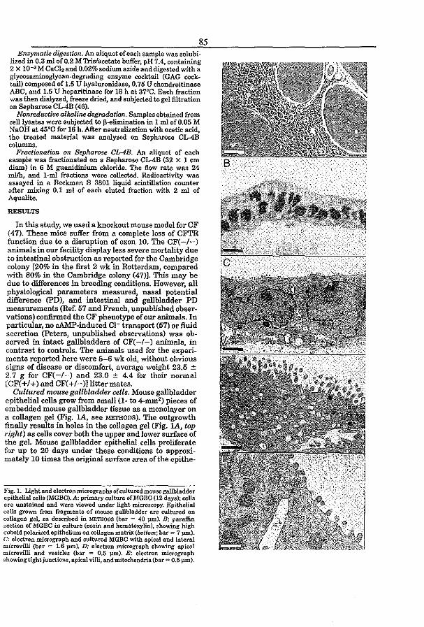
85 J!;nzymatic digestion. An aliquot of each sample was solubi
lized in 0.3 m1 of 0.2 M Tris/acetate buffer. pH 7.4, containing 2 X 10⁻³ M CaC12 and 0.02% sodium azide and digested with a glycosaminoglycan.degrading enzyme cocktail (GAG cock· tail) composed of 1.5 U hyaluronidase, 0.75 U chondroitinase ABC, and 1.5 U heparitinase for 18 h at 37°0. Each fraction was then dialyzed, freeze dried, and subjected to gel filtration on Sepharose CL4-B (46).
Nonreductiue alkaline degradation. Samples obtained from ceUlysates were subjected to f3,-elimination in 1 ml of 0.05 M NaOH at 45°0 for 16 h. After neutralization with acetic acid, the treated material was analyzed on Sepharose CL4B columns.
Fractronation on Sepharose CL4B. An aliquot of each sample was fractionated on a Sepharose CL-4B (32 X 1 em diarn) in 6 M guanidinium chloride. The flow rate was 24 mlIh, and l-ml fractions were collected. Radioactivity was assayed in a Beckman S 3801 liquid scintillation cOWlter after mixing 0.1 ml of each eluted fraction with 2 mI of Aqualite.
RESULTS
In this studY1 we used a knockout mouse model for CF (47). These mice suffer from a complete loss of CFI'R function due to a disruption of exon 10. The CF( -/-) animals in our facility display less severe mortality due to intestinal obstruction as reported for the Cambridge colony [20% in the first 2 wk in Rotterdam, compared with 80% in the Cambridge colony (47)]. This may be due to differences in breeding conditions. However, all physiological parameters measured, nasal potential difference (PD), and intestinal and gallbladder PD measurements (Ref. 57 and French, unpublished observations) confirmed the CF phenotype of our animals. In particular1 no cAMP-induced Cl- transport (57) or fluid secretion (peters, unpublished observations) was observed in intact gallbladders of CF( -/-) animals. in contrast to controls. The animals used for the experiments reported here were 5-6 wk old, without obvious signs of disease or discomfort, average weight 23.5 ± 2.7 g for CF(-/-) and 23.0 ± 4.4 for their normal [CF(+/+) and CF(+/-)] !ittermates.
Cultured mouse gallbladder cells. Mouse gallbladder epithelial cells grow from small (1- to 4-mm2) pieces of embedded mouse gallbladder tissue as a monolayer on a collagen gel (Fig. lA. see METHODS). The outgrowth finally results in holes in the collagen gel (Fig. lA, top right) as cells cover both the upper and lower surface of the gel. Mouse gallbladder epithelial cells proliferate for up to 20 days under these conditions to approximately 10 times the original surface area of the epithe-
Fig. 1. Lightand electron micrographs of cultured mouse gallbladder epithelial cells (MGBC}.A: primary culture ofltfGBC (12 days); cells are unstaIned and were viewed under light microscopy. Epithelial cells grown from fragments of mouse gallbladder are cultured on collagen gel, as described in METHODS (bar = 40 }lID). B: paraffin section of MGBC in culture (eosin and hematoxylin), showing high cuboid polarized epithelium on collagen matrix (bottom; bar = 7 vm). c: electron micrograph and cultured MGBC with apical and lateral microvilli (bar <= 1.6 }lID). D: electron micrograph sho .... -ing apical microvilli and vesicles (bar = 0.5 }lID). E: electron micrograph showing tight junctions, apical villi, and mitochondria (bar = 0.5 }lID).
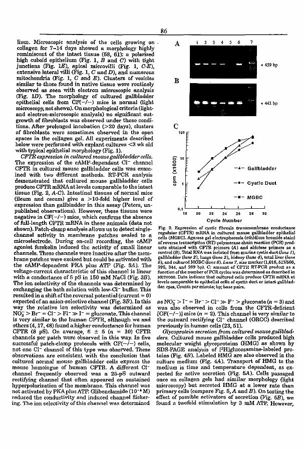
86
lium. Microscopic analysis of the cells growing on , collagen for 7-14 days showed a morphology highly reminiscent of the intact tissue (68, 61): a polarized high cuboid epithelium (Fig. I, B and 0) with tight junctions (Fig. lE), apical microvilli (Fig. I, C-E), extensive lateral villi (Fig. 1, C and D), and numerous mitochondria (Fig. 1, C and E), Clusters of vesicles similar to those found in native tissue were routinely observed as seen with electron microscopic analysis (Fig. W). The morphology of cultured gallbladder epithelial cells from CF(-/-) mice is normal (light microscopy, not shown), On morphological criteria (lightand electron-microscopic analysis) no significant outgrowth of fibroblasts was observed under these conditions. After prolonged incubation (>20 days), clusters of fibroblasts were sometimes observed in the open spaces in the collagen gel. All experiments described below were performed with explant cultures <3 wk old with typical epithelial morphology (Fig. 1).
A
B
c 100
l23456 7
_ .... ---
("" /./' ~.
.. 459 bp
... 442 bp
CFTR expression in cultured mouse gallbladder cells. The expression of the cAMP~dependent Cl- channel CFTR in cultured mouse gallbladder cells was exam~ ined with two different methods. RT·PCR analysis demonstrated that cultured mouse gallbladder cells produce CFrR mRNAat levels comparable to the intact tissue (Fig. 2, A·C). Intestinal tissues of normal mice (ileum and cecum) give a > 10·fold higher level of expression than gallbladder in this assay (Peters, un· published observations). However, these tissues were negative in CF( -/-) mice, which confirms the absence of full-length CFTR mRNA in these animals (data not shown). Patch·clamp analysis allows us to detect single-channel activity in membrane patches sealed to a microelectrode. During on-cell recording, the cAMP agonist forskolin induced the activity of small linear channels. These channels were inactive after the membrane patches were excised but could be activated with the cAMP-dependent PKA plus ATP (Fig. SA). The voltage--current characteristic of this channel is linear with a conductance of 5 pS in 150 roM NaCl (Fig. 3B). The ion selectivity of the channels was determined by exchanging the bath solution with low-CI- buffer. This resulted in a shift of the reversal potential (current = 0) expected of an anion-selective channel (Fig. 3B). In this way the relative ion selectivity was determined as NOi > Br- = CI- > FI-» 1- = gluconate. This channel is very similar to the human CFrR, although we and others (4, 17) 48) found a higher conductance for human CFTR (8 pS). On average, 6 ± 5 (n ~ 10) CFTR channels per patch were observed in this way. In five successful patch-clamp protocols with CF(-/-) cells, not one Cl- channel of this type was observed. These observations are consistent with the conclusion that cultured normal mOuse gallbladd~r cells express the mouse homologue of human CFTR. A different Clchannel frequently observed was a 25-pS outward rectifying channel that often appeared on sustained hyperpolarization of the membrane. This channel was not activated by PKA plus ATP. Glibenclamide (10-' M) reduced the conductivity and induced channel flickering. The ion selectivity of this channel was determined
S 10 o o ~
~ E fr 1
(/ -£- Gallbladder
l' -.- Cystic Duct
-.- MGBC
0.1 '---'--''---''---''---''---' 18 20 " 24 26 .. 30
Cycle Number
Fig. 2. Expression of cystic fibrosis transmembrane conductance regulator (CFl'R) mRNA in cultured mouse gallbladder epithelial cells (MGBO). Agarose gel electrophoresis (ethidium bromide stain) of reverse transcription (RT)'polymerase chain reaction (peR) prod· ucts obtained with CFl'R primers CA) and aldolase primers as a control (B). 'lbtal RNA was isolated from murine cystic duct (laM 1), gallbladder (lo.n.e 2), lungs (lane 3), kidney (lo.ne 4), total liver (io.ne 6), and cultured MGBC (lane 6). Lane 7, sixe marker (1,018, 517/506, 396, 344, and 289 bp). C: amount of CFrR RT·POR product as a function of the nUmber of peR cycles was determined as described in METHODS. Data indicate that cultured cells produce CFTR mRNA at levels comparable to epithelial cells of cystic duct or intact gallbladder. cpm, Counts per minute; bp; base pairs.
as NOi > 1- = Br- > Cl- » F- > gluconate (n = 3) and was also observed in cells from the CFTR-deficient [CF( -/-)] mice (n ~ 2). This channel is very similar to the outward rectifying Cl- channel (ORCC) described previously in human cells (23, 51).
Glycoprotein secretion from cultured mouse gallbladders. Cultured mouse gallbladder cells produced high molecular weight glycoproteins (HMG) as shown by SDS-PAGE analysis of [3H]glucosamine-Iabeled proteins (Fig. 48). Labeled HM:G are also observed in the culture medium (Fig. 4A). Transport of HMG tc the medium is time and temperature dependent, as expected for active secretion (Fig. 5A). Cells passaged once on collagen gels had similar morphology (light microscopy) but secreted HMG at a lower rate than primary cells (compare Fig. 5, A and B). On testing the effect of possible activators of secretion (Fig. 6B), we found a twofold stimulation by 3 mM ATP. However,
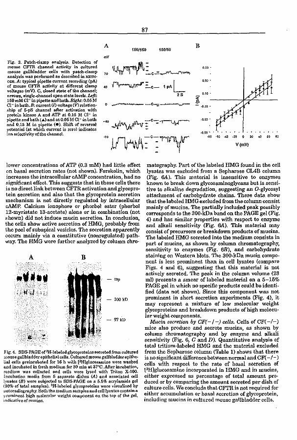
87
A B 1501150 150{50
mV
O.SO -
Fig. 3. Patch-clamp analysis. Detection of mouse CFI'R channel activity in cultured mouse gallbladder cells with patch-damp analysis was performed as described in METHODS. A: typica.l pipette currt'lnt recording CpA) of mouse CFTR activity at different clamp voltages (mV). C, closed state oftbe channel; arrows, single-channel open-state levels. Left: 150 mltt: 01- in pipette and bath. Right: 0.05 M 01- in bath.B: current(l)-voltage(V)relationship of S·pS channel after activation with protein kinase A and ATP at 0.15 M Cl- in pipette and bath (A) and at 0.05 M 01- in bath and 0.15 M in pipette (e). Shift of reversal potential (at which current is zero) indicates ion selectivity oftbe channel.
"~:c- 1 PAL 2S
o _-----
lower concentrations of ATP (0.3 roM) had little effect on basal secretion rates (not shown), Forskolin. which increases the intracellular cAMP concentration} had no significant effect. This suggests that in these cells there is no direct link between CFTR activation and glycopr<r tein secretion and also that the glycoprotein secretion mechanism is not directly regulated by intracellular cAMP. Calcium ionophore or phorbol ester (phorbol 12-myristate 13-acetate) alone or in combination (not shown) did not induce mucin secretion. In conclusion., the cells show active secretion of lIM:G, probably from the pool of subapical vesicles. The secretion apparently occurs mainly via a constitutive (nonregulated) pathway. The HMG were further analyzed by column chr<r
A B
- top
- 200kD
-97100
Fig.4. SDS·PAGE of3JI·labeled glycoproteins secreted from cultured mouse gallbladder epithelial cells. Cultured mouse gallbladder epithelial cells preincubated ror 16 h with f3ffiglucosamine were washed and incubated in fresh medium for 30 min at 37°0. After incubation, medium was collected and cells were lysed with Triton X-lOO. Incubation media from 5 separate dishes (A) and associated cell lysates (B) were subjected to SDS-PAGE on a 5.5% acrylamide gel (90% of total samples). 3H-labeled glYc:oproteins were visualized by autoradiography. Both the medium samples and celilysates contain a prominent high molecular weight component on the top of the gel, indicative ofmucins.
V(mVj
matography. Part of the labeled HMG found in the cell lysates was excluded from a Sepharose ClABo column (Fig. 6A). This material is insensitive to enzymes known to break down glycosaminoglycans but is sensitive to alkaline degradation, suggesting an O-glycosyl attachment of carbohydrate chains. These data show that the labeled HMG excluded from the column consist mainly ofmucins. The partially included peak possibly corresponds to the 300·kDa band on the PAGE gel (Fig. 4) and has similar properties with respect to enzyme and alkali sensitivity (Fig. SA). This material may consist of precursors or breakdown products ofmucins. The labeled HNIG secreted into the medium consists in part of mucins, as shown by column chromatography, sensitivity to enzymes (Fig. 6B), and carbohydrate staining on Western blots. The 300·kDa mucin component is less prominent than in cell lysates (compare Figs. 4 and 6), suggesting that this material is not actively secreted. The peak in the column volume (23 ml) presents a smear oflabeled material on a 5-15% PAGE gel in which no specific products could be identified (data not shown). Since this component was not prominent in short secretion experiments (Fig. 4), it may represent a mixture of low molecular weight glycoproteins and breakdown products of high molecular weight components.
Mucin secretion by CF(-/-) cells. Cells ofCF(-i-) mice also produce and secrete mucins, as shown by column chromatography and by enzyme and alkali sensitivity (Fig. 6, C and D). Quantitative analysis of total tritium-labeled HMG and the material excluded from the Sepharose column (Table 1) shows that there is no significant difference between normal and CF( -/-) cells with respect to the rate of basal secretion of (3H]glucosamine incorporated in HMG and in mucins, either expressed as percentage of total amount pro· duced or by comparing the amount secreted per dish of culture cells. We conclude that CFI'R is not required for either accumulation or basal secretion of glycoprotein, including mucins in cultured mouse gallbladder cells.
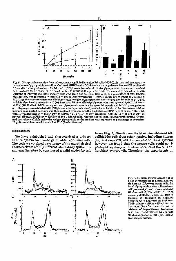
88
" A
I r ~
" 0
• 0
" N
-"
~~ .
~ 15
~
• 0 . ~ -" "
II
~I f .. -~-------
" " .. '" '" II. . • := :;; ~
TIme (min) ~ -Fig. 6. Glycoprotein secretion from cultured mouse gallbladder epithelial cells (MGBC). A: time and temperature dependence of glycoprotein secretion. Cultured MGBC and NIHf3T3 cells as a negative control (-30% confluenV 3.5-cm dish) were preincubated for 16 h with £3H]glucosamine to label cellular glycoprotcins. Dishes were washed and incubated for 3 h at 4"0 or 37°0 as described in METHODS. Samples were collected and analyzed as described in METIlODS at intervals indicated. Finally. cells were lysed and secretion from cells, as a percentage of total labeled glycoprotein, was calculated [%secretion = 100 X (medium/medium + lysate); values are averages of 7 dishes ± SDJ. Data show a steady secretion of high molecular weight glycoproteins from mouse gallbladder cells at 37"0 (A.), which is significantly reduced 8t4°0 (.). Less than 2% of total labeled glyooproteins were secreted by NIHI3T3 cells at 37"0 (.). B: effect of different agonists on glycoprotein secretion. In a parallel experiment, MGBC passaged once 011 collagen gels were labeled with pHJglucosamine (n, no. of dishes), washed, and incubated for 60 mw in label·free medium as indicated. Medium was then replaced by medium without additions at 3rc (n = 5) or at 4°C (n = 5). with 10-6 M forskolin(n = 4), 3 X 10⁻³ MATP(n = 3), 5 X 10⁻⁷ M Ca2+ ionophore (A-23187){n = 4). or 5 X 10⁻⁷ M phorbol dibutyrate (PDB) (n = 5) followed by a 2-hineubation. Medium was collected. cells were subsequently lysed. and the release of high molecular weight glycoprotein to the medium. was expressed as percentage of secretion. ·Significant difference with control at 37°0 (Student's t·test).
DISCUSSION
We have established and characterized a primary culture system for mouse gallbladder epithelial cells. The cells we obtained have many of the morphological characteristics offully differentiated biliary epithelium and can therefore be considered a valid model for this
A B - = ,~ - -- =
E -a.
tissue (Fig. 1). Similar results have been obtained wit gallbladder cells from other species, including human (62) and dogs (28, 43). In contrast to these system' however. we found that the mouse cells could not b passaged regularly without senescence of the cells an fibroblast overgrowth. Therefore. the experiments de
Fig. 6. Column chromatography of Is beled glyooproteins of normal and cys
" = - -:N-\ k " tie fibrosis (CF(-I-)] mouse cells_ La - /"""i,;}'l.1"" ... Nl ..... beled glyooproteins were collected frOll ,=
ft.!t • ~... .&:.(~ celllysates (A, C) and culture media (8 ,~
oil. D}ofnonnal (At B) and CF( -1-) (C,D
C 0 mouse gallbladder epithelial cells il
" ~ " ~ D " ro ro ~ culture. as described in METHODS
" " Samples were analyzed on Sepharo~ ClAB columns either without furthe:
" treatment (e), after incubation with j
" mixture of heparitinase, byaluroni
" dase, and chondroitinase (A), or wit} E~ alkaline degradation (O). dpm, Disinte a.~ ,,' grations per minute.
0 .c
.1 t:. ~ ,~ .'V'~ ...J ~
" ~ " " " N " " ml ml

Table 1. Secretion of HMG by cultures of normal and CF( -/-) mouse gallbladder epitludial cells
[3H)HMG medium (chase), dpm (3HJHMG medium (overnight), dpm rH)HMG cell lysate, dpm Total PH1HM:G, dpm [3l-I]IThfG secreted, % PHJmucin medium (total), dpm PHlmucin cell lysate, dpm 'IOta! (3ff)mucin, dpm [lff)mucin secreted, %
c"ntrol
92,000 100,000 9~000
282,000 68
4~000 36,000 78,000
53
96,000 96,000
175,000 367,000
52 60,000 69,000
129,000 48
Primary explant cultures of normal (n = 7) and cystic fibrosis (CF(-I-); n=8] gallbladders were labelled with (3HJglurosamine overnight. The medium was collected and replaced by unlabeled medium (chase). High molecular weight glyroproteins (HMG) of medium samples and oelilysates {expressed as disintegrations per minute (dpm) oPH] were determined by ultrafiltration as described. Combined medium samples and cell lysates were analyzed by gel filtration (Fig. 6) to determine the mucin componentofHM:G. Percent of mucin and HMO secreted is the percentage of material in the combined media of the total.
scribed in this study were performed with cells grown from primary explants or with cells passaged once to collagen gels.
Our data show that CFTR mRNA is produced in cultured mouse gallbladder cells with levels comparable to intact tissue (Fig. 2). Patch-clamp analysis revealed the presence of a small CI- channel with characteristics very similar to human CFTR (Fig. 3). We conclude that these cells express the mouse coWlterpart of human CFTR, providing the first de·scription of this Cl- channel. The numberofCI- channels per patch in these cells compares favorably to other primary epithelial cells in culture (24) or even colon carcinoma cell lines (HT-29, T84) that are often used as a model for CFI'R expression (2, 56). Cultured MGBC, therefore, are a convenient model for the study of CFTR activity in a well-differentiated epithelial cell in primary culture. This is particularly important in view of the availability of several mouse models of CF. These include loss of function mutants (44, 47, 54) and more recently the prevalent AF50B processing mutant (14, 57,63) and a G551D mutant (15). Other CFI'R muta· tions are likely to follow.
An additional interesting feature of the cultured MGBC is that they produce and secrete glycoproteins (Figs. 4 and 5), including mucins (Fig. 6). Mucin secretion has been shown for cultured human (62) and dog (29, 43) gallbladder epithelial cells. Mucin secretion in cultured dog epithelial cells was reported to be stimulated by prostaglandin E2• possibly by increasing the intracellular cAMP concentration (29). In contrast, our results suggest that in cultured MGBC glycoprotein secretion occurs via a constitutive pathway. Forsko· lin, Ca2+ ionophore, and phorbol ester had no significant effect on glycoprotein release in these cells. This suggests that an increase in intracellular cAM:.P, Ca2 +, or protein kinase C activity is not sufficient to induce secretion (Fig. 5). The effect of extracellular ATP is difficult to interpret. Since 0.3 mM ATP had no effect (not shown), the activity of a P2 purinergic receptor
89
seems excluded. Possibly, aspecific effects of ATP on membrane permeability (18) are involved.
As pointed out in the introduction, several observations in the literature suggest a relationship between CFTR activity and mucin secretion. This could contribute to the development of liver disease in CF. In bronchial segments, the f3-adrenergic secretion stimulus observed in normal tissue is reduced in CF patients (50). In this case, the coupling could be indirect. Since CFTR is mainly expressed in serous cells of submucosal glands (19), reduced output from serous acini could impair mucin transport. Mucus secretion in airway goblet cells is under purinergic rather than cA1vfi> control (32). In the intestinal crypt, mucin-secreting Goblet cells are stimulated by Ca2+ agonists but not by cAMP (25). In isolated submandibular salivary glands, a significant reduction of cAMP-induced secretion was observed in human CF patients (38) and in a CF mouse model (39). In these cells, mucin release is triggered by an increase of intracellular Ca2+ (40). Therefore, the exact relationship between CFI'R and secretion is not yet clearly established. Our current data show that MGBC from mice with a loss of function mutation of the cftr gene produce and secrete labeled glycoproteins, including mucin, to a similar extent as normal cells, as determined by [3H]glucosamine labeling (Fig. 6, Table 1). Therefore, neither activation of CFTR by forskolin nor absence of CFrR has a marked effect on glycoprotein secretion. This does not support the notion that, in gallbladder cells, CFI'R function and secretion are closely coupled, as suggested by experiments with Neomycin-selected dog gallbladder cell clones that express CFl'R from an expression vector (28). However, we cannot exclude that the mucins from CF-deficient cells differ from normal with respect to carbohydrate structure and protein/carbohydrate ratio. CFI'R deficiency in humans results in increased sialation and sulfation of glycoproteins, and abnormal carbohydrate structures were observed (10, 11, 34, 35). The exact cause of this phenomenon is not known, but it has been suggested that it relates to an effect of CFrR on acidification of intracellular vesicles (3). Therefore, it is possible that the mucins produced by CFrR-deficient gallbladder and biliary cells are abnormal due to increased sulfation or aberrant branching, as in human salivary and respiratory mucins (10, 34). Abnormal biliary mucin structure could contribute to the development of liver disease in CF patients. The amount of material that can be recovered from the present mouse gallbladder cell cultures does not allow a careful analysis of mucin carbohydrate molecular structure. We are presently attempting to increase the yield of cultured cells by crossing the AF50B CF1'R mouse model that we recently produced (57) with a mouse producing a temperature-sensitive version of Large 'l'-antigen. In Uris model, we can study the effect of CFTR dysfunction on mucin structure and membrane recycling.
Cells expressing ClITR show a marked cAMPdependent inhibition of endocytosis and CFTR internal-
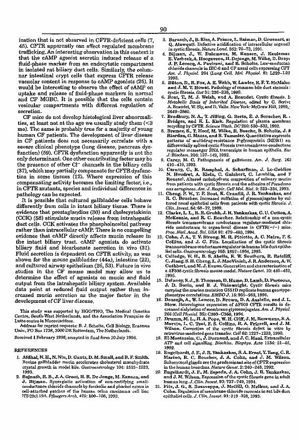
ization that is not observed in CFTR·deficient cells (7, 45). CFI'R apparently can affect regulated membrane trafficking. An interesting observation in this context is that the cAMP agonist secretin induced release of a fluid.phase marker from an endocytotic compartment in isolated rat biliary duct cells. Similarly, the colum· nar intestinal crypt cells that express CFTR release vacuolar content in response to cAMP agonists (25). It would be interesting to observe the effect of cAMP on uptake and release of fluid-phase markers in normal and OF MGBO. It is possible that the cells contain vesicular compartments with different regulation of secretion.
OF mice do not develop histological liver abnormalities, at least not at the age we usually study them (<3 mo). The same is probably true for a majority of young human OF patients. The development of liver disease in OF patients does not necessarily correlate with a severe clinical phenotype (lung disease, pancreas dys· function) (59). CFTR dysfunction apparently is not the only determinant. One other contributing factor may be the presence of other CI- channels in the biliary cells (37), which may partially compensate for OFTRdysfunction in some tissues (12). Where expression of this compensating activity becomes the limiting factor, i.e., in CFTR mutants, species and individual differences in pathology can be expected.
It is possible that cultured gallbladder cells behave differently from cells in intact biliary tissue. There is evidence that prostaglandins (30) and cholecystokinin (CCK) (58) stimulate mucin release from intrahepatic duct cells. CCK activates the phosphoinositol pathway rather than intracellular cAMP. There is no compelling evidence that cAMP directly affects mucin release in the intact biliary tract. cAMP agonists do activate biliary fluid and bicarbonate secretion in vivo (31). Fluid secretion is dependent on CFTR activity, as was shown for the mouse gallbladder (44a), intestine (22), and cultured airway epithelium (26, 53). Future in vivo studies in the CF mouse model may allow us to determine the effect of agonists on mucin and fluid output from the intrahepatic biliary system. Available data point at reduced fluid output rather than in· creased mucin secretion as the major factor in the development of CF Ii ver disease.
This study was supported by HGOtrNO, The Medical Genetics Centre, South·West Netherlands, and the Association FranfAise de Lutte contre la Mucoviscidose.
Address for reprint requests: B. J. Scholte, Cell Biology, Erasmus Univ., PO Box 1738, 3000 DR Rotterdam, The Netherlands.
Received 1 February 1996; aC1;epted in final form 10 July 1996.
REFERENCES
1. Mdhal, N. H .. N. Niu. D. Gantz,D. M. Small, and B. F. Smith. Bovine gallbladder mucin ae<:eterates cholesterol monohydrate crystal growth in model bile. Gastroenterology 104: 1515-1523, 1993.
2. Bajnatb. R. B., J.A. Groot, H. R. De Jonge,M. Ransen, and J. Bijman. Synergistic activation of non-rectifying small· conductance chloride channels by forskolin and phorbol esters in cell-attached patcl:!es of the human colon carcinoma cell line HT-29c1.19A.P/luegersArch. 425: 100-108,1993.
90 3. Barasch, J .. B. Kiss,A. Prince, L. Salman, D. Gruener!, al
Q. AlawqatL Defective acidification of intracellular organell in cystic fibrosis. NatureLond. 352: 70-73, 1991.
4. Bijman, J., W. Dalemans, l'tl. Ransen, J. Keulemat E. Verbeek,A. Hoogeveen, H. Dejonge, l'tL Wilke, D. DreYI J. P. Lecocq. A. Pa~ and B. Scholte. Low-<onductan chloride channels in IEc.s and CF nasal cells expressing CF!': Am. J. Physioi. 264 (Lung CelL Mol. Physiol. 8): L229-L23 1993.
5. Bilton, D .. R. Fox, A K. Webb, W. Lawler, R. F. T. Mc1'tiaho: and J. M. T. Howat. Pathology of common bile duct stenosis: cystic fibrosis. Gut 31: 236-238,1990.
6. Boat, T .. l'tL J. Welsh, and A. Beaudet. Cystic fibrosis. L Metabolic Basis of Inherited Disease, edited by C. Scrive A. Beaudet, W. Sly, andD. Valle. New York: McGraw Hill, 1989, I 2649-2860.
7. Bradbury, N. A., T. JiUing, G. Berta, E. J. Sorscher, R. .: Bridges, and K. L. Kirk. Regulation of plasma membran recycling by CITR. &ien.ce Wash. DC 256: 530-532, 1992.
8. Bremer, S., T. Hoof, M. WIlke, R. Busche, B. Scholle, J. E Riordan, G. Maass, and B. Tummler. Quantitative expressio patterns of multidrug·resistance P·glycoprotein (MDRl) an differentially spliced cystic fibrosis transmembraneo(X)nduct8nc regulator messenger RNA transcripts in human epithelia. BUI
J.·Bjochem.206: 137-149,1992. 9. Carey, M. C. Pathogenesis of gallstones. Am. J. Surg. 165
410-419,1993. 10. Carnoy, C., R. Ramphal, A. Scharfman, J. Lo-Guidice
N. Houdret, A IDein, C. Galabert, G. Lamblin, and F Roussel. Altered carbohydrate composition o( salivary mucinl from patients with cystic fibrosis and the adhesion of Pseudomo nas aeroginosa.Am. J. Respir. Cell Mol. Bioi. 9: 323-334, 1993.
11. Cheng, P. W., T. F. Boat, K. Cranfill, J. R. Yankaskas, and R. C. Boucher. Increased sul(ation of glyroconjugates by cul· tured nasal epithelial cells £rom patients with cystic fibrosis. J. CUn. Invest. 84: 68-72, 1989.
12. Clarke, 1.. L., B. R. Grubb, J. R. Yankaskas, C. U. Cotton,A. MoKenzie, and R. C. Boucher. Relationship of a non-<:ystic fibrosis transmembrane conduct.ance regulator.mediated chloride conductance to organ-level disease in CFI'R(-/-) mice. Proc.Natl.Acad. Sci. USA 91: 479-483,1994.
13. Cohn, J. A., T. V. Strong, M. R. Piciotto, A C. Nairn, F. S. Collins, and J. G. Fib. Localization of the cystic fibrosis transmembrane conductance regulator in human bile duct epithelial cells. Gastroenterology 105: 1857-1864,1993.
14. CoUedge, W. H., B. S. AbeUSt K. W. Southem, R. Ratcliff, C. Jiang, S. H. Cheng, L. J. MacVtnish. J. R.Anderson. A W. Cuthbert, and M. J. Evans. Generation and characterisation of a AF508 cystic fibrosis mouse model. Nature Genet. 10: 445-452, 1995.
15. Delaney, S. J., S. Thomson, D. Hume, D.Lamb, D. Porteous, J. D. Dorin, and B. J. Wainwright. Cystic fibrosis mice carrying the murine mutation 0551D replicate human genotype. phenotype correlations. EMBO J. 15: 955-963,1996.
16. Dosanjh, A., W. Lencer, D. Brown, D. A Awiello, and J. L. Stow. Heterologous expression of AF50S CFI'R results in de· creased sialylation of membrane glycoconjugates.Am. J. Physicl. 266 (Cell Physic/. 35): C360-C366, 1994.
17. Drumm, M.L., H. A Pope, W.H.Cliff,J.M.Rommens,S.A Marvin, L. C. Tsui, F. S. Collins, R. A. Frizzell, and J. ltL Wilson. Correction of the cystic fibrosis defect in vitro by retroviIus·mediatedgene transfer. Ce.ll62: 1227-1233, 1990.
18. EI·Moatassim, C., J. Dornand. and J. C. MauL Extracellular ATP and cell signalling. Biochim. Bwphys. Acta 1134: 31-45, 1992.
19. Engelhardt, J. F., J. R. Yankaskas, B.A Ernst, Y. Yang, C. R. Marino, R. C. Boucher, J. A Cohn, and J. M. Wilson. Submucosal glands are the predontinantsite ofCFI'R expression in the human bronchus. Nature Genet. 2: 240-248, 1992.
20. Engelhardt, J. F .. M. Zepeda, J. A Cohn, J. R. Yankaskas, and J.l'IL Wilson. Expression of the cystic fibrosis gene in adult human Iung.J. CUn.Invest. 93: 737-749,1994.
21. Fitz, J. G., S. Basavappa, J. McGill, O. Melhus, and J, A Cohn. Regulation of membrane chloride currents in rat bile duct epithelial cclls.J. CUn.Invest. 91: 319-328.1993.
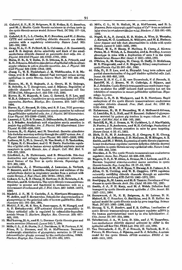
91
22. Gabriel, S. E., K. N. Brigman, B. lL Koller, R. C. Boucher, and M. J. Stutts. Cystic fibrosis resistance to cholera toxin in the cystic fibrosis mouse modeL Science Wash. DC 266: 107-109. 1994.
23. Gabriel, S. E., L. L. Clarke, R. C. Boucher, and M. J. Stutts. CFTR and outward rectifying chloride channels are distinct proteins with a regulatory relationship. Nature Lond. 363: 263-266,1993.
24. Gray, M. ~ C. E. Pollard, H. L. Coleman. J. R. Greenwell, and B. E. Argent. Anion selectivity and block of the smallconductance chloride channel on pancreatic duct cells. Am.. J. PhysioL 259 (eeUPhysioL 28): 0752-0761, 1990.
25. HaIm, D. ~ S. T. IIa1..mt D. R. Dibona., R. A. Frizzell, and R. D. Johnson. Selective stimulation of epithelial cells in colonic crypts: relation to active chloride secretion. Am. J. PhysiIJl. 269 (Cell Physwl. 38): C929-C942, 1995.
26. Jiang. C., W. E. Finkbeiner, J. H. Widdicombe, P. B. McCray, and S. S. Miller. Altered fluid transport across airway epithelium in cystic fibrosis. Science Wash. DC 262: 424-426, 1993.
27. Ka.nsellt M., R. B. Bajnath, J. A. Groot, R R. Dejonge, B. Scholte, A. T. Hoogeveellt and J. Bijman. Regulation of chloride channels in the human colon carcinoma cell line HT29.clI9A. PfluegersArch. 422: 539-545,1993.
28. Kuver, R., N. Rames~ S. Lau, C. Savard. S. P. Lee, and W. R.A. Osborne. Constitutive mucin secretion linked to CFrR expression. Biochem. Biophys. Res. Commun. 203: 1457-1462, 1994.
29. Kuver, R., C. Savardt D. Oda, and S. P. Lee. PGE generates intracellular cAMP and accelerates mucin seeretion by cultured dog gallbladder epithelial cells.Am. J. Physiol. 267 (Gastrointest. Lif)er Physiol. 30): G998-Gl003, 1994.
30. Lamont,J. T., B. S. Turner, D. DiBenedetto, R. HandJllt and A. L Schafer. Arachidonic acid stimulates mucin secretion in prairie dog gallbladder. Am. J. PhysilJl. 245 (Gastrointest. Liver Physiol. 8): G92-G98, 1983.
31. Lenzellt R., G. Alpini, and N. TavolonL Secretin stimulates bile ductular secretory activity through the cAMP system.Am. J. Physiol. 263 (Gastrointest. Liver PhYsiol. 26): G521-G532, 1992.
32. Lethem. M. L, M. L. Dowell, M. Van Scott, J. R. Yankaskas, T. Egan. R. C. Boucher, and C. W. Davis. Nucleotide regula. tion of goblet cells in human airway epithelial explants: normal exocyt.osis in cystic fibrosis. Am. J. &spir. Cell. Mol. Bicl. 9: 315-322,1993.
33. Lindblad. A., R. Hultcrantz, and B. Strandvik. Bile-duct destruction and collagen deposition-a prominent ultrastructural feature of the liver in cystic fibrosis. Hepato[lJgy 16: 372-381,1992.
34. Lo--Guidice, J., J. Wieruszes~ J. Lemoine, A. Verbert, P. Roussel, and G. Lamblin. Sialylation and sulfation of the carbohydrate chains in respiratory mucins from a patient with cystic fibrosis. J. BioI. Chem. 269: 18794-16813,1994.
35. Lukacs, G. L., X. B. Chang, N. Kartner, O. D. Rotste~ J. R. Rlordant and S. Grinstein. The cystic fibrosis transmembrane regulaklr is present and functional in endosomes-role as a determinant of endosomal pH. J. Bicl. Chern. 267: 14568-14572, 1992.
36. Madrid. J. F., M. T. Castells, J. A. ltlarttnez.Menarguez, M. Aviles, and F. Hernandez. Subcellular characterization of glycoproteins in the principal cells of human gallbladder. Histo· chemistry 101: 195-204, 1994.
37. McGill, J. M., M. S. Yellt S. Basavappa, A.. W. Mangel, and A. P. Kwiatkowski. ATP-activated chloride permeability in biliary epithelial oens is regulated by calmodulin-dependent protein kinase II. Biochem. Biophys. Res. Commun. 208: 457-462,1995.
38. McPhersoIl, M.A., and R. L. Dormer. Cystic fibrosis gene and mucin secretion (Abstract). Lancet 343: 7, 1994.
39. Mills,C.L.,J.R.Dorin,D.J.Davidson,E. W. Porteus, F. W. Altollt R. L. Dormer. and M. A. McPherson. Decreased fI.·adrenergic stimulation of glycoprotein secretion in CF mice submandibular glands: reversal by the methylxanthine. IBMX_ Biochem. Biophys.Res. Commun. 215: 674-681,1995.
40. Mills, C. L., M. B. Hallett, M. A. McPherson, and R. L. Dormer. Beta·adrenergic mobilization ofCa2 + from an intracel· lular store in rat submandibular acini. Biochem. J. 293: 691-695, 1993.
41. Nagel, R. A., A. Javaid. H. B. Meire, A. Wise, D. Westaby, J. Kavani, M. G. Lombardt R. Williams, and M. E. Hodson. Liver disease and bile duct abnormalities in adults with cystic fibrosis. Lancd 2: 1422-1425, 1989.
42. O'Neal, W. K., P. Hasty, P. McCray, B. Casey, J. Rivera' Perez, M. J. We18~ A. L. Beaudet, and A. Bradley. A severe phenotype in mice with a duplication of exon 3 in the cystic fibrosis locus. Human Mol. Genet. 2: 1561-1569, 1993.
43. O'Brien, S., M. Reogan, M. Casey, G. Duffy, D. McErlean, M. X. Fitzgerald, and J. E. Hegarty. Biliary complif.'Ations of cystic fibrosis. Gut 33: 387-391,1992.
44. Oda, D., s. P. Lee, and A. Hayashi. Long term culture and partial characterization of dog gall bladder epithelial cells. Lab. Invest. 64: 682-692, 1991.
44a.Peters R. H. P. C., J. H. van Doorninckt P. J. French, R. Ratcliff, M. J. Evans. W. H. Colledge, J. BijmaDt and B. J. Scholte. Cystic fibrosis transmembrane conductance regu· lator m:ldiates the cAMP induced fluid secretion but not the inhibition of resorption in mouse gallbladder epithelium. Hepatowgy. In press.
45. Prince, L. S., R. B. Workman, and R. B. Marcbase. Rapid endocytosis of the cystic fibrosis transmembrane conductance regulator chloride channel. Pnx;. Natl. Acad. Sci. USA 91: 5192-5196,1994.
46. Rahmoune, H., II. P. Rounding, W. J. McDonald-Gibsollt G. Lambllllt R. L. Hall, and P. Roussel. Sulfated O·glycopro. teins secreted by guinea pig trachea in organ culture. Am. J. Respir. Cell Mol. Biol. 4: 156-165,1991.
47. Ratcliff, R., 11. J. Evans, A. W. Cuthbert, L. J. MacVlllis~ D. Foster, J. R. Anderson, and W. H. Colledge. Production of a severe cystic fibrosis mutation in mice by gene targeting. Nature Genet. 4: 35-41, 1993.
48. Web, D. p .. M. P. Anderson, R. J. Gregory, S. H. Cheng, S. Paul, D. M. Jefferllollt J. D. McCann, K. W. Klinger, A. E. Smith. and M. J. Welsh. Expression of cystic fibrosis transmembrane conductance regulator corrects defective chloride channel regulation in cystic fibrosis airway epithelial cells. Nature Land. 347: 358-363, 1990.
49. Riorda..Dt J. R. The cystic fibrosis transmembrane conductance regulator.Annu. Rev. Physiol. 55: 609-630, 1993.
50. Rogerll, D. F., E. W. Alton, A. Dewar, M. L Lethem, and P. J. Barnes. Impaired stimulus-evoked mucus secretion in cystic fibrosis bronchi. E:cp. Lung &s. 19: 37-53, 1993.
51. Schwiebert, E. M.,ltL E. Egan, T. Hwang, S. B.Fulmer, S. S. Allen, G. R. Cutting, and W. B. Guggino. CFrR regulates outwardly rectifying chloride channels through an autocrlne mechanism involving ATP. Cell 81: 1063-1073, 1995.
52. Scottjupp, R., M. Lama. and M. S. Tanner. Prevalence of liver disease in cystic fibrosis. Arch. Dis. Child. 66: 698-701, 1991.
53. Smith, J. J., P. H. Karp, and M. J. Welsh. Defective fluid transport by cystic fibrosis airway epithelia. J. Clin. Invest. 93: 1307-1311,1994.
54. Snouwaert, J. N., K. K. Brlgmallt A. M. Latour, N. N. Malouf, R. C. Boucher, O. Smithies, and B. H. Koller. An animal model for cystic fibrosis made by gene targeting. Science Wash. DC 257: 1083-1088, 1992.
55. Strong, T. V., K. Boehm, and F. S. Collins. Localisation of cystic fibrosis transmembrane conductance regulator mRNA in the human gastrointestinal tract by in situ hybridisation. J. Clin.Invest. 93: 347-354, 1994.
56. Tabcharani. J. A., W. Low, D. Elie, and J. W. Hanrahan. Low--ronducl..anoe chloride channel activated by cAMP in the epithelial cell line TB4. FEBS utt. 270: 157-164-, 1990.
57. Van Doorninck, J. H., P. J. Frenc~ E. Verbeek, R. P. C. Peters, H. Morreau, J. Bijmallt and B. J. Scholte. A mouse model for the cystic fibrosis AF508 mutation. EMBO J. 14: 4403-4411,1995.

58. Wahlln, T., G. D. Bloom, andA. Danielsson. Effect of cholecystokinin (CCK-PZ) OD glyooprot.ein secretion from mouse gallblad. derepithelium. Cell 7iss/le &s.171: 425-435, 1976.
59. Waters, D. L., S, F. Dorney, M. A. Gruea, H. C. O. Marlin. R. Howman·GUes, A. E. Ran, M. De SUva, and K. J. Gaskin. Hepatobiliary disease in cystic fibrosis patients with pancreatic sufficiency. Heparology 21: 963-969, 1995.
GO. Welsh, M. J. The path of disrovery in understanding the biology of cystic fibrosis and approaches to therapy.Am. J. Gastroenterol. 89:897-8105,1994.
61. Yamada, E. The fine structure of the gall bladder epithelium of the mollOO. J. Biophys. Biochem. CytoI. 1: 445-458, 1955.
92 62. Yoshitomi, S., K. Miyazaki, and F. Nakayama. Demonstra
lion and maintenance of mucus secretion in cultured bum8.1 gallbladder epithelial cells. In Vitro Cell. De". Bioi. 23: 559-566 1987.
63. Zeiher, B. G., E. Eichwald, J. Zabner, J. J. Smith, A. P Puga. P. B. McCray, M. R. Capecc~ M. J. '\Ye1sh, and K. R Thomas. Amouse model for the dF508 allele of cystic fibrosis. J CUn.Invest. 96: 2051-2064,1995.
64. Zhang, Y., B. Doranz,J.R. Yankaskas,andJ. F.Engelhardl Genotypic analysis of respiratory muoous sulfation defects iJ:j cystic fibrosis. J. Clin.Invest. 96: 2997-3004, 1995.
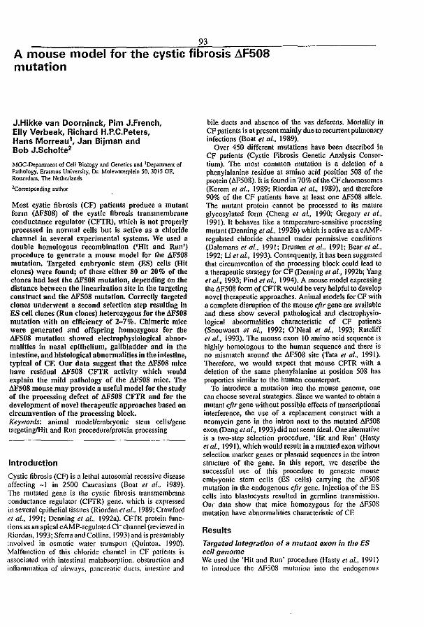
93
A mouse model for the cystic fibrosis AF508 mutation
JoHikke van Doorninck, Pim JoFrench, Elly Verbeek, Richard HoPoCoPeters, Hans Morreau1, Jan Bijman and Bob JoScholte'
MGC-Department of Cell Biology and Genetics and 'Department of Pathology. Erasmus Uni\'ersity, Dr. Molewaterplein 50. 3015 OE, Rotterdam. The Netherlands
2(:orresponding author
Most cystic fibrosis (CF) patients produce a mutant form (AFSOS) of the cystic fibrosis transmembrane conductance regulator (CFfR), which is not properly processed in normal cells but is active as a chloride channel in several experimental systems. We used a double homologous recombination ('Hit and Run') procedure to generate a mouse model for the llFS08 mutation. Targeted embryonic stem (ES) cells (Hit clones) were found; of these either 80 or 20% of the clones had lost the AF508 mutation, depending On the distance between the linearization site in the targeting construct and the AFS08 mutation. Correctly targeted clones underwent a second selection step resulting in ES cell clones (Run clones) heterozygous for the AFS08 mutation with an efficiency of 2-7%. Chimeric mice were generated and offspring homozygous for the dFS08 mutation showed electrophysiological abnormalities in nasal epithelium, gallbladder and in the intestine, and histological abnormalities in the intestine, typical of CF. Our data suggest that the AFS08 mice have residual AFS08 CFI'R activity which would explain the mild pathology of the AFSOS mice. The AFSOS mouse may provide a useful model for the study of the processing defect of dFS08 CITR and for the development of novellherapeutlc approaches based on circumvention of the processing block. Keywords: animal model/embryonic stem cells/gene targeting/Hit and Run procedure/protein processing
Introduction
Cystic fibrosis (CF) is a lethal autosomal recessive disease affecting -I in 2SOO Caucasians (Boat et al .. (989). The mutated gene is the cystic fibrosis transmembrane conductance regulator (CFfR) gene. which is expressed in several epithelial tissues (Riordan el al.. 1989; Crawford et al., 1991; Denning et al .. 1992a). CFfR protein functions as an apical cAMP-regulated CI- channel (reviewed in Riordan, 1993; Sferra and Collins. 1993) and is presumably involved in osmotic water transport (Quinton. 1990). Malfunction of this chloride channel in CF patients is associated with intestinal malabsorption. obstruction and innammation of airways. pancreatic ducts.. intestine and
bile duclS and absence of the vas deferens. Mortality in CF patienlS is at present mainly due to recurrent pulmonary infections (Boat el 01., 1989).
Over 450 different mutations have been described in CF patients (Cystic Fibrosis Genetic Analysis Consortium). The most common mutation is a deletion of a phenylalanine residue at amino acid position 508 of the protein (6F508). It is found in 70% of the CF chromosomes (Kerem et al., 1989; Riordan el 01., 1989), and therefore 90% of the CF patienlS have at least one 6FS08 allele. The mutant protein cannot be processed to ilS mature glycosylated fonn (Cheng et al., 1990; Gregory et al.. 1991). It behaves like a temperature-sensitive proceSSing mutant (Denning el al., 1992b) which is active as a cAMPregulated chloride channel under permissive conditions (Dalemans et al., 1991; Drumm et al., 1991; Bear el al.. 1992; Li el ai., 1993). Consequently, it has been suggested that circumvention of the processing block could lead to a therapeutic strategy for CF (Denning et al., 1992b; Yang el al., 1993; Piod et al., 1994). A mouse model expressing the .1,F508 fonn of CFfR would be very helpful to develop novel therapeutic approaches. Animal models for CF with a complete disruption of the mouse cftr gene are available and these show several pathological and electrophysio· logical abnonnalities characteristic of CF patients (Snouwaert et al., 1992; O'Neal et al., 1993; Ratcliff et af .. (993). The mouse exon 10 amino acid sequence is highly homologous to the human sequence and there is no mismatch around the .1.FS08 site (fata et a/ .. 1991). Therefore. we would expect that mouse CFTR with a deletion of the same phenylalanine at position S08 has properties similar to the human counterpart.
To introduce a mutation into the mouse genome, one can choose several strategies. Since we wanted to obtain a mutant cftr gene without possible effeclS of transcriptional interference. the use of a replacement construct with a neomycin gene in the intron next to the mutated .1.FS08 eXOn (Deng et af., 1993) did not seem ideal. One alternative is a two-step selection procedure, 'Hit and Run' (Hasty et al., 1991). which would result in a mutated exon without selection marker genes or plasmid sequences in the intron structure of the gene. In this report. we describe the successful use of this procedure to generate mouse embryonic stem cells (BS cells) carrying the &S08 mutation in the endogenous cftr gene. Injection of the ES cells into blastocysts resulted in gennline transmission. Our data show that mice homozygous for the .1,FS08 mutation have abnonnalities characteristic of CE
Results
Targeted integration of a mutant exon in the ES celJ genome We used the 'Hit and Run' proceJure (Hasty et al" 1991) to introduce the fiFS08 mulalkm into the endogenous.
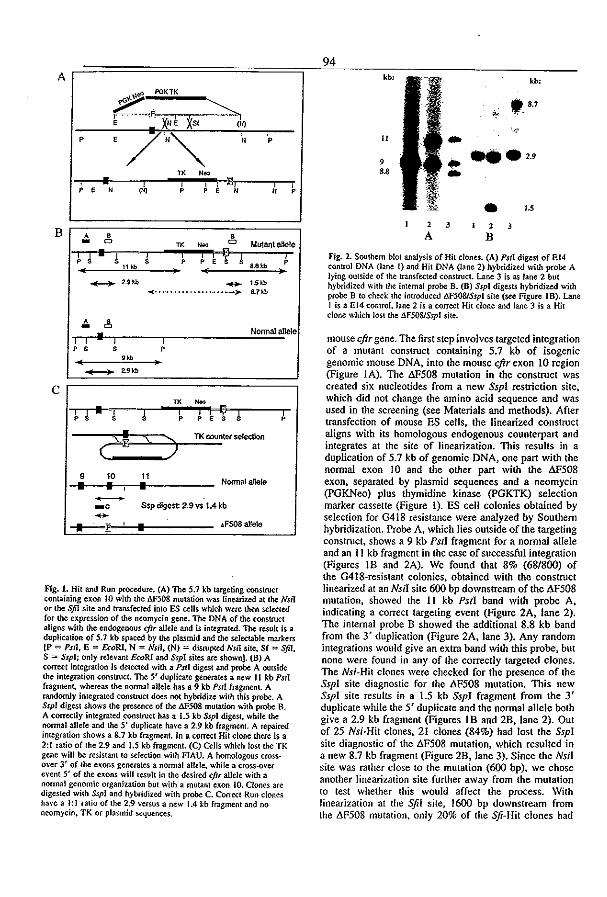
~======================~~9~4~ __ ~~=-________ =-____ ___ A ~
\\!Il: PG!(Tl(
? r-'~.q"~;~=-~.~~ , X"' XSf (N)
p ,
/""" " P
TX "00 ; • I i , E) , , ,
" (N) p p " " p
B A • • - 0 TX NOO 0 Mutant allele I 1 • I 1 i i i I;J 1 I p p p , ,
8.Ski> P ,,,,, • -- "'" - , .. " -< ---.----.-~--.,.-.--,. .m
"" • 0
• NOfTTlal allele I I I I p , , p , "" ---- "'''
C TX
I 1 • r- 1 i
p p
C! "\.. ) 9 10 11
• • • Noonalallele
Fig. 1. Hit and Run procedure. (A) The 5.7 kb targeting construCt containing exon 10 with the 61'508 mutation was linearized at the Nsll or the Sfil site and transfected into ES ulls which were lhen selected for the expression of the neomycin gene. The DNA of the construct aligns with the endogenous cftr allele and is integrated. 'The result is a duplication of 5.7 kb spaced by the plasmid and the selectable markers (P "" PSI!, E = froRI. N = NII1, (N) := disrup(ed Nsll sile, Sf = SfiJ. S = SspJ; only relevant &oRI and SspJ sites are shown}. (B) A correct integration is detected with a PSII digest and probe A outside the imegrn.tion construct. The 5' duplicate generates a new II kb PSli fragment, whereas the normal allele has a 9 kb PsI! fragment. A randomly integrated COflStruct does not hybridize with this probe. A Ssp! digest shows the presence of the &'508 mutation with probe B. A corre<:tly integrated ~nstnJCI has a 1.5 kb Sspl digest, while the nonnal allele and the 5' duplicate have a 2.9 kb fragment. A rep3ired integration shows a 8.7 kb fragment. Tn a correct Hit <:lone there is a 2:1 ratio of the 2.9 and 1.5 kb fragment. (C) Cells which lost the TK gene will be resistant to selection with FlAU. A homologous C~· over 3' of the exons generates a nonnal allele. while a cross-over e\'ent S' of the exons will result in the desired c/tr allele with a nonnal genomic organization but with a mutant exon 10. Clones are digested with Sspl and hybridized with probe C. Correct Run clones have a I: I ratio of the 2.9 \'ersus a new 1.4 kb fragment and no neomycin. TK or plasmid set.!uences.
II
, S.8
2
A
kb:
'_S.7 .. .. ".2., .. • IS
2 3
B
Fig. 2. Southern blot analysis of Hit dones. (A) Pstl digest of EI4 control DNA (lane I) and Hit DNA (lane 2) hybridized with probe A lying outside of the transfected construct. Lane 3 is as lane 2 but hybridized with the internal probe B. (B) Sspl digests hybridized with probe B to check the introduced 6F5081SspJ site (see Figure 18). Lane 1 is a El4 control. lane 2 is a correct Hit done and lane 3 is a Hit clone which lost the l1F508lSspI site.
mouse cftr gene. The first step involves targeted integration of a mUlant construct containing 5.7 kb of isogenic genomic mouse DNA, into the mouse cftr exon 10 region (Figure I A). The AP50B mutation in the construct was created six nucleotides from a new SspI restriction site. which did not change the amino acid sequence and was used in the screening (see Materials and methods). After transfection of mouse ES cells, the linearized construct aligns with its homologous endogenous counterpart and integrates at the site of linearization. This results in a duplication of 5.7 kb of genomic DNA, one part with the normal exon 10 and the other part with the M50B exon, separated by plasmid sequences and a neomycin (PGKNeo) plus thymidine kinase (pGKTK) selection marker cassette (Figure 1). ES cell colonies obtained by selection for G418 resistance were analyzed by Southern hybridization. Probe A, which lies outside of the targeting construct, shows a 9 kb PstI fragment for a normal allele and an I I kb fragment in the case of successful integration (Figures 1B and 2A). We found that 8% (68/Boo) of the G418-resistant colonies. obtained with the construct linearized al an Nsil site 600 bp downstream of the M50B mutation, showed the 11 kb Pst! band with probe A. indicating a correct targeting event (Figure 2A, fane 2). The internal probe B showed the additional B.8 kb band from the 3' duplication (Figure 2A, lane 3). Any random integrations would give an extra band with this probe, but none were found in any of the correctly targeted clones. The Nsi·Hit clones were checked for the presence of the Ssp! site diagnostic for the AP50B mutation. This new Ssp! site results in a 1.5 kb SspJ fragment from the 3' duplicate while the 5' duplicate and the normal allele both give a 2.9 kb fragment (Figures IB and 2B, lane 2). Out of 25 Nsi·Hit clones, 21 clones (84%) had lost the SspI site diagnostic of the M50B mutation, which resulled in a new B.7 kb fragment (Figure 2B, Jane 3). Since the Nsf I site was rather close 10 the mutation (600 bp), we chose another linearization site further away from the mutation to lest whether this would affect the process. With linearization at the Sfil site, 1600 bp downstream from the tlF508 mutation. only 20% of the Sfi-Hit clones had

95
Table I. Genotype and phenotype of FIAU-fC.~bt3.nt dones
Hit clone Hit clone cultured On 2 pM FIAU for 2 days Hit clone cultured for 3 p. on G418: 2 p. on 0418-; 0.4 pM HAU for 10 days
No. ofTK Genotype No.ofTK- G418-sensitive O<M<ypo clones analyzed
Wild Hit Scrambled clones analyzed
Wild
155 3 0 0 3 206 BJO 157 157 0 0 ND B47 39 39 0 0 ND B7J 33 2 " 3 198 B84 69 II 51 7 170
p. indicates passages: ND. not determined.
lost the SspI site (2/11 colonies), while the targeting efficiency was in the same range (5%, 13/279 colonies) as for Nsl1 linearization. These data suggest that a repair mechanism is active around the integration site with a preference for the endogenous chromosomal sequence.
ES cell clones heterozygous for the L1F508 mutation The next step of the procedure (Run) involves the removal of the duplication plus the intervening plasmid sequences via an intra-chromosomal homologous recombination (Figure I C). This leads to the loss of either the mutant or the wild-type exon (Figure Ie). The latter case gives us the desired clone with a mutant 6F508 exon 10 in an otherwise normal genomic organization of the cftr gene. In both cases, recombinants will be resistant to the toxic thymidine kinase substrate 1-(2-deoxy, 2-Huoro-pD-arabinofuranosyl)-5 (FIAU) and sensitive to 0418. The correct Run clone is identified by a new 1.4 kb SspI fragment, generated by an SspI site outside of the integrated construct and the SspI site in the mutated exon 10 (Figure I C). Different Hit clones with a correct karyotype were subjected to selection with 2 JiM FIAU for 2 days. Resistant clones were picked after 7-10 days and screened by Southern analysis. In initial experiments, we found either wild-type cells wilh two copies of the 2.9 kb SspI fragment, Or cells which had no genomic rearrangement compared with Hit clones (Table 1). The latter presumably represented TK mutants. A few clones showed a rearranged and scrambled genotype. To reduce the possibility of contaminating wild-type cells in the population, we cultured the Hit cells for three passages in the presence of 0418 before the FIAU selection. This decreased the number of PIAU-resistant clones 10- to loo-fold. In addition, we screened the resultant clones for G418 sensitivity to avoid Southern analysis of clones with a mutation in the TK gene. When we started the FIAU selection the day after ending the 0418 selection, we did not find any G418-sensitive cells (0/42 colonies). However, if we cultured the cells for two passages in non-selective medium prior to FIAU selection. 30--70% of the clones were G418 sensitive (Table 1). Of these. 2-7% had the 1.4 kb SspI fragment of the desired M508 Run clone. (Table I, Figure 3A). No rearrangements of the cftr gene were observed, as other digests show a wild-type restriction pattern (Figure 3B). Further, hybridization with the neomycin probe does not show a signal with any of the Run clones (Figure 3A. lanes 6-8). These results confirm the
61
113 115
A kb:
75
2.9
I.'
B kb:
133
II
9 ..
2
-• ..--
60
99 108
!·~tt t.,.~ ;}" !."
.'" ~ .. , 4 5
•
14 7
6
-.-,56
7 8
Fig. 3. Southern blot analysis of Run clones hybridized Io.,ith probe C. 5' of exOfi 10. (A) Sspl digests of Hit clone DNA (lane I). EI4 control DNA (lane 2) and two Run clones (lanes 3 and 4). Lanes 5--S: the same blot hybridiud with a neomycin probe demonstrating the absence of POKNeo in the Run clones. (8) PSII digests (lanes 1-3) and NsiI digests (lanes 4----6) of respectively EI4 DNA, Hit clone DNA and Run clone DNA hybridized with probe C were used to assay the correct chromosomal structure of the Run clones.
loss of the duplication and the normal genomic organization of the 8F508 allele in the Run clones.
Generation of a L1F50B CFTR mouse strain Cells of 11 Run clones derived from three different Hit clones. with a correct karyotype and heterozygous for the l'!.F508 mutation, were injected into blastocysls and gave rise to sex conversion and coat color chimerism ranging from 20 to 100%. Two male chimeras showed full gennline transmission in a cross with FVB mice. as indicated by coat color and Southern analysis (using the same procedure as for the Run clone analysis, results not shown). To check the COrrect transcription of the l'!.F508 allele, we isolated intestinal RNA from nonnal and mutant mice and performed 41 nested RT-PCR analysis wilh primers in
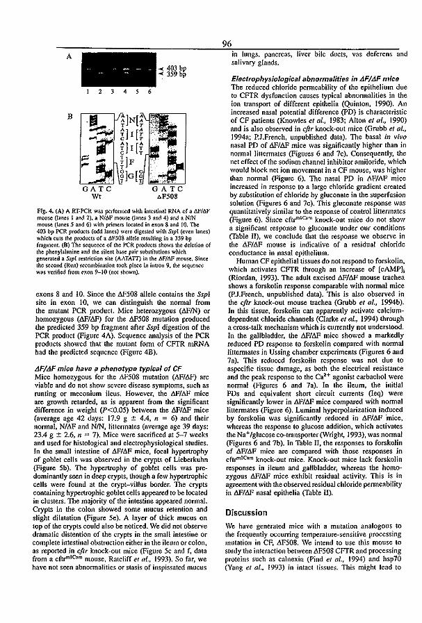
A
B
2 3 4 6
GATC WI
~ 403 bp ~ 359 bp
GATC "F508
Fig. 4. (A) A RT·PCR was perfonned with intestinal RNA of a M/I1F mouse (lanes I and 2), a NfM mouse (Janes 3 and 4) and a NIN mouse (lanes 5 and 6) with primers located in exon 8 and 10. The 403 bp PCR products (odd lanes) were digested with SspJ (even lanes) which cuts t~ products of a I1F50g allele resulting in a 359 bp fragment. (B) The sequence of the PCR products shows the deletion of the phenylalanine and the silent base pair substitutions which generated a SspI restriction site (AATATT) in the M/llF mouse. Since the ~ond (Run) recombination took place in intron 9, the sequence was verified from exon 9-10 (not shov.'1l).
exons 8 and 10. Since the AF508 allele contains the SspI site in exon to, we can distinguish the nonnal from the mutant peR product. Mice heterozygous (AFIN) or homozygous (AF/dF) for the AF508 mutation produced the predicted 359 bp fragment after SspI digestion of the PCR product (Figure 4A). Sequence analysis of the PCR products showed thar the mutant form of CFTR mRNA had the predicted sequence (Figure 4B).
LJF/LJF mice have iI phenotype typical of CF Mice homozygous for the LW508 mutation (LW!.6F) are viable and do not show severe disease symptoms, such as runting or meconium ileus. However, the 6F/6F mice are growth retarded, as is apparent from the significant difference in weight (P<O.05) between the I:!.FII:!.F mice (average age 42 days: 17.9 g ± 4.4, n = 6) and their normal, N/AF and NIN, liuermates (average age 39 days: 23.4 g ± 2.6, n = 7). Mice were sacrificed at 5-7 weeks and used for histological and elcctrophysiological studies. In the small intestine of tlF/M< mice, focal hypertrophy of goblet cells was observed in the crypts of Lieberkuhn (Figure 5b). The hypertrophy of goblet cells was pre· dominantly seen in deep crypts, though a few hypertrophic cells were found at the crypt-villus border. The crypts containing hypertrophic goblet cells appeared to be located in clusters. The majority of the intestine appeared normal. Crypts in the colon showed some mucus retention and slight dilatation (Figure 5e). A layer of thick mucus on top of the crypts could also be noticed. We did not observe dramatic distention of the crypts in the small intestine or complete intestinal obstruction either in the ileum or colon, as reported in cftr knock-out mice (Figure 5c and f, data from a cfo-m lC3m mouse, Ratcliff et at .. 1993). So far, we have not seen abnormalities or stasis of inspissated mucus
96 in lungs. pancreas, liver bile ducts, vas deferens and salivary glands.
Electrophysiologicai abnormalities in LJF/LJF mice The reduced chloride permeability of the epithelium due to CFTR dysfunction causes typical abnormalities in the ion transport of different epithelia (Quinton, 1990). An increased nasal potential difference (PD) is characteristic of CF patients (Knowles et 01., 1983; Alton et at., 1990) and is also observed in cftr knock·out mice (Grubb et 0/" 1994a; P.I.French, unpublished data). The basal in vivo nasal PD of tlF/AF mice was significantly higher than in normal Iittermates (Figures 6 and 7c). Consequently, the net effect of the sodium channel inhibitor amiloride, which would block net ion movement in a CF mouse, was higher than normal (Figure 6). The nasal PD in 6F/8F mice increased in response to a large chloride gradient created by substitution of chloride by gluconate in the superfusion solution (Figures 6 and 7c). This gluconate response was quantitatively similar to the response of controllittennates (Figure 6). Since cfum lCam knock·out mice do not show a significant response to gluconate under our conditions (Table 11), we conclude that the response we observe in the tlFlL\.F mouse is indicative of a residual chloride conductance in nasal epithelium,
Human CF epithelial tissues do not respond to forskoJin, which activates CFTR through an increase of {cAMP); (Riordan, 1993). The adult excised l!J.F/1iF mouse trachea shows a forskolin response comparable with nonnal mice (P.I.French. unpublished data), This is also observed in the cftr knock·out mouse trachea (Grubb et 0/" 1994b), In this tissue, forskolin can apparently activate calcium· dependent chloride channels (Clarke et al., 1994) through a cross-talk mechanism which is currently not understood. In the gallbladder, the tlF/tlF mice showed a markedly reduced PD response to forskolin compared with normal liuermates in Ussing chamber experiments (Figures 6 and 7a). This reduced forskolin response was not due to aspecific tissue damage, as both the electrical resistance and the peak response to the Ca2+ agonist carbachol were nonnal (Figures 6 and 7a). In the ileum, the initial PDs and equivalent short circuit currents (Jeq) were significantly lower in I:!.FltlF mice compared with normal liUennates (Figure 6). Luminal hyperpolarization induced by forskolin was significantly reduced in L\.F/tlF ·mice. whereas the response to glucose addition, which activates the Na+ Iglucose co-transporter (Wright, 1993), was nonnal (Figures 6 and 7b). In Table II, the responses to forskoHn of AF/tlF mice are compared with those responses in cfumlCam knock-out mice. Knock·out mice lack forskolin responses in ileum and gallbladder, whereas the homozygous I:!.FltlF mice exhibit residual activity. This is in agreement with the observed residual chloride penneability in llF/tlF nasal epithelia (Table ll).
Discussion
We have generated mice with a mutation analogous to the frequently occurring temperature-sensitive processing mutation in CP, tlF508. We intend to use this mouse to study the interaction between tlF508 CFfR and processing proteins such as calnexin (Pind et 0/" 1994) and hsp70 (Yang et of., 1993) in intact tissues. This might lead to
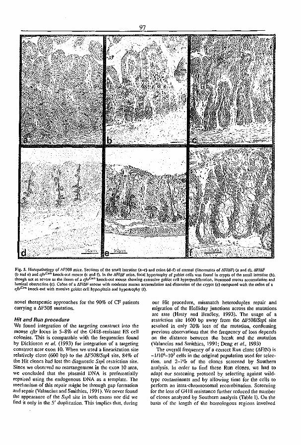
Fig. S. Hislop.1thOlogy of M508 mice. Sections of the small intestine (a-c) and colon (d-f) of normal (liltermates of MI.o.F) (a and d). 6.FlM (b and e) and cjt,c:..m knock-out mouse (c and f), In (he MIM mice. focal hyp¢rtropby of goblet cells was found in crypts of the small intestine (b). though not as severe as the ileum of a cjt,c= knock-:OUt mouse showing extensh'e goblet cell hyp¢rproliferation. increased mucus accumulatiOn and luminal obstruction (c). Colon of a .o.FI6F mouse with moderate mucus accumulation and distension of the crypts (e) compared with the colon of a cjt~ knock.-out with massive goblet cell hyperplasia and hypenrophy (f).
novel therapeutic approaches for the 90% of CF patients carrying a 6F508 mutation.
Hit and Run procedure We found integration of the targeting construct into the mouse cftr locus in 5-8% of the G418-resistant ES cell colonies. This is comparable with the frequencies found by Dickinson et al. (1993) for integration of a targeting construct near exon 10. When we used a linearization site relatively close (600 bp) to the AF508/Sspl site, 84% of the Hit clones had lost the diagnostic Ssp! restriction site. Since we observed no rearrangement in the exon 10 area. we concluded that the plasmid DNA is preferentially repaired using the endogenous DNA as a template. The mechanism of this repair might be through gap formation and repair (Valancius and Smithies. 1991). We never found the appearance of the Sspl site in bolh exom; nor did we find it only in the 5' duplication. This implies that, during
our Hit procedure, mismatch heteroduplex repair and migration of the Holliday junctions across the mutations are rare (Hasty and Bradley, 1993). The usage of a restriction site 1600 bp away from the 6F508lSspI site resuhed in only 20% loss of the mutation, confirming previous observations that the frequency of loss depends on the distance between the break and the mutation (Valancius and Smithies, 1991; Deng et at .. 1993)
The overall frequency of a correct Run clone (AFIN) is -11lQ6-107 cells in the original population used for selection. and 2~79'0 of the clones screened by Southern analysis. In order to find these Run clones. we had to adapt our screening protocol by selecting against wildtype contaminants and by allowing time for the cells to perform an intra-chromosomal recombination. Screening for the loss of G418 resistance further reduced the number of clones an31yzed by Southern analysis (Table I). On the basis of the length of the homologous regions involved

98 Gallbladder
"" '"
.• L-_-"-___ -----' Basal FotskOJiJ\ Cafbachor
Basal Forskolin
Ileum
Basal Forskolin Glutose Basal Forskolln Glu¢()se
Nose
- N/N lJ.F/N - t:.F/t:.F
Basal Glucona!e Amllorida
Fig. 6. Summary of the eJ~trophysiological data obtained from six 6F/~. fi,'c .6.FIN and three NIN mice. Left panels: PD. right panels: Ieq. Top: gallbladder. The basal PD and Jeq as well as the peak response 10 carbachol of 11F/6F mice and normal (NIN or t'!.FJN) littermates are comparable. Fo~kolin (10-$ M) responses, however, were markedly reduced in the lJ.F/6F gallbladder (P<O.OOI. nine li1Sue samples from six mice) compared with normal (eight tissue samples (r{'lm eight mice). Middk: ileum. MI!J.F ilea (12 tissue samples from six mice) had a significantly reduad basal PD and leq (P<O.02) compared with their norma] littermates (14 tissue samples from eight mice). Responses to forskolin were reduced in the AF/6F ileum (P<O.OI). Lower. nasal PD. In lJ.F/t1F mice the in ~'i\'o basal POs were significantly higher than their controllittermatcs (P<O.OI). Both normal (n == 6) and lJ.F/t1F (n = 6) nasal epithelia responded to a chloride gradient [153 mM chloride replaced gluconate (l33 mM), final CIconcentration 10 mM). Responses to the sodium channel inhibitor amiloride were Significantly higher in IlFllJP nasal epithelia (P<O.02), resulting in a similar residual PD. Values are averages:!: SO. Statistical analyses were perl"ooned with the siudent's (·Iest.
in the two possible Run recombinations, one would expect to find .... 25% AF508 Run clones (Figure IC). However, the actual ratio of llF508 to wild-type recombinants (± 1120) does not see.m to follow this simple rule. This may be due to cryptic sequence preferences in the recombination process.
The Hit and Run procedure has previously only been used to produce the mutant Hoxb4 mouse (Ramirez-Solis et 01 .• 1993). The possible drawback of the 'Hit and Run' procedure compared with a one-step replacement protocol is the number of passages under selective pressure that is involved. The starting passage of our El4 cells was 24; selections with both 0418 and PIAU plus .several expansion steps lead to D.F508 clones of around passage 57 (assuming about three divisions per passage). The blastocyst injections showed that gennline chimeras can still be
made with these cells. However, in Run clones derived from the B84 Hit clone, loss of totipotency was observed (20% chimerism, sex-reversal rate 1.4 and no gennline transmission).
.JF/.JF mice have a CF phenotype The Hit and Run procedure resulted in mice which produce 6.F508 CFfR mRNA with the predicted sequence (Figure 4B). The heterozygous 6.F/N mice do not show significant abnonnalities in any of our assays. We therefore consider them phenotypically nonnal. llF/6.F mice show a phenotype typical of CFTR dysfunction, including failure to grow to nonnal weight, histological abnonnalities in the intestine (Figure 5) and eleclrophysiological abnonnalities in nasal epithelium, gallbladder and intestine (Figures 6 and 7). We did not observe lethal obstruction of the

a "1 ~ .~ --rf-------------~------~
b
c
t~ ror,
, .~, 1 !
let IS 10 " time (min)
J-Jl-~ 1 1 1 1
~~~~ , ! t ! r , J ! !.!
os lOIS 20 2530 lS.j<).5
time (min)
::it~~_. ~ gk w •
!:~~~ Q.. ·15 ·10 ., 1 1 1 o ---~--.!'-----~----,~,~~-'-!,~~-'-!",......~-'-!".
rime (min)
"
Fig. 7. Reprcseotati\'t Ussing chamber tracings from normal mice (upper [facings) and 1:JF/6F mice (lower tracings) tissue. (a) Gallbladders of flF/lJP mice had a reduced response to forskolin (for) compared wilh normallittermates, whereas peak responses to carbachol (c) werc similar. (b) Ilea of I:JFlbF mice had a reduced response to forskolin (for) compared with normallitlermales. Responses to 30 mM glucose (glu) and subsequent 0.2 mM phloridzin (p), activating and inhibiting the Na+/glucose transporter (Wright, 1993) re5peclh'ely. were similar. Responses to carbachol (e) on average caused luminal depolarization and an i~rease in tissue resistance. (e) Nasal epithelia of bF/.o.F mice had a higher basal PD and an ilKreastd response 10 amiloride (a) but did respond to a chloride gradient (from 153 (0 10 mM CI-. chloride replaced gluconate (glc, 133 mM)]. w "" wash.
intestine in 6.F/.6.F mice. Meconium ileus, Le. Obstruction of the intestine, is diagnosed in only 10% of neonatal CF patients, and those patients show mucus obstruction in the crypts. Other CF patients do not show severe mucus accumulation but have distended glands and goblet cell hypertrophy similar to our .6.F/.6.F mice (Thomaidis and Arey. 1963). Another typical feature of CF, progressive lung inflammation accompanied by accumulation of viscous mucus (Boat el al .• 1989). is not observed in
99 either .6.F/.6.F or knock-out mice under normal conditions. However, it has been shown recently that CF mice, when challenged with lung pathogens, do show increased pathology compared with normal Iittennates (Davidson et at., 1995). It would therefore be interesting to subject the I:!F/I:!F mice to a similar protocol. As many abnOimalities in CF patients develop with age, further histological analysis will be done with mice older than those of 5-7 weeks which we used for this study.
The virtual absence of lethal intestinal obstruction in I:!FII:!F mice differs from the eftr knock-out models described earlier. The latter were reported to have a 50--90% mortality rate in the first 4 weeks, mainly due to intestinal obstructions (Snouwaert et al., 1992; O'Neal et aL, 1993; Ratcliff et al., 1993). The cji,mIHGU insertion mutant (Dorin et al., 1992) expresses low levels of normal CFI'R (Dorin et al., 1994) and has a low mortality rate. This suggests that the mild phenotype of the t>.F11:!F mice is related to the residual chloride penneability observed. However. mortality rates may prove a rather misleading parameter. The colony of eji,mICam mice in the Rotterdam animal facility has a considerably lower mortality rate than reported «40% in the first 4 weeks versus 80% in the first 5 days, Ratcliff et 01., 1993). Apparently, the breeding conditions have a major impact on mortality rates. The phenotypic effect of residual chloride penneability may be revealed by challenging knock-out and .6.F/1:!F mice with pathogens in parallel experiments.
Residual CFTR activity in AF/AF mice The small but significant forskolin response observed in ileum and gallbladder, and the response to a chloride gradient in the nasal epithelium (Figures 5 and 6) of the I:!FII:!F mouse might be due to residual 1:!F508 CFfR activity. An alternative chloride channel (Clarke et al., (994) seems a less plausible explanation since ejtrn lDm
knock-out mice completely lack a forskolin response in the gallbladder (Table II) and intestine (Table II, Clarke et al., 1992; Cuthbert et al., 1994) and show no significant gluconate response in the nasal PD measurements (fable II). ea2+ -activated chloride channels appear absent in the ileum, as the ileum does not respond to carbachol with a luminal hyperpolarization (Figure 7). Thus, the cAMPinduced activation of ea2+ -dependent chloride channels as described in mouse trachea (Grubb et al., 1994b) seems not to occur in the ileum or the gallbladder. It is conceivable that the mouse .6.F508 CFfR is partially processed into mature .6.F508 CFfR protein reaching the plasma membrane. At present, it is not known whether the same phenomenon occurs in CF patients. Residual intestinal chloride transport activity, observed in the minority of dF'/.6.F patients, is correlated with mild disease (Veeze et ai., 1994). Since this has not yet been analyzed at the level of single chloride channels, this activity cannot be attributed unequivocally to CFTR. Further experiments should detennine the channel characteristics of the mouse .6.F508 CFTR and the processing kinetics of aF508 CFTR in differentiated mouse epithelial cells. The mouse and human processing systems may differ with respect to the kinetics of their interactions with the AF508 CFfR protein, and the mouse .6.F508 CFTR may have different properties from the human AF508 form. The .6.F508 mouse model gives us an opportunity to study the CFTR processing
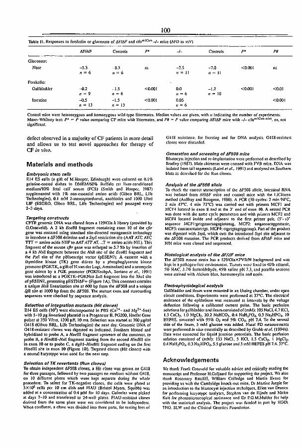
100
Thblt II. Responses to forskolin or gluconare of t.Fh\P and cftf"IC""'-I_ mice (APD in mY)
t;Flt'1F Controls p. -1- Controls p. PO
Gluconate:
Nose -5.3 ---8.3 "' -1.5 -7.0 <0.001 "' n = 6 n=6 II = II 1/'-; II
Forskolin:
Gallbladder -'>.2 -1.5 <0.001 0.0 -1.2 <0.001 <0.01 n '" 9 n=6 1/ = 6 n = iO
Intestine -'>5 -J.S <OJ)OI 0.05 <0.001 n = 13 n = 13 11=6
Control mice were heterozygous and homozygous wild-type liuennates. Median Y.;llues are given. with /I indicating the number of experiments. Mann--Whitney lesl: p~ = P value comparing CF mice with HUermales. and P# = P value comparing .6F/6F mice with _/_ cftr"'lC.", .ru.:~. ns, nO{ significant.
defect observed in a majority of CF patients in more delail and allows us to test novel approaches for therapy of CF in vivo.
Materials and methods
Embryonic stem cells EI4 ES cells (a gift of M.Hooper, Edinburgh) were cultured on 0.1% gelatine.ro<lted dishes in DMEMl6O% Buffalo rat Iiver-conditioned mediumlJO% fetal calf serum (FCS) (Smith and Hooper. 1987) supplemented with 1% non-e~ntial llmino acids (Oibco BRL. Life Technologies). 0.1 roM 2-mercaptoethanol. antibiotics and 1000 U/ml LlF (ESORO. Gibco BRL. Life Technologies) and passaged e\'try 2-3 days.
Targeting constructs CAR genomic DNA was cloned from a 129/0la A library (provided by G.Grosveld). A 2 kb £CoRl fragment containing exon JO of the cftr gene was mutated using standard site·directed mutagenesis technology to introduce a i1F508 deletion and aSspl restriction site (AAT ATC ATC TIT "" amino acids NIIFto AAT A1T AT ... T "" amino acids Nil.). This fragment of the mouse cftr gene was enlarged to 5.7 kb by insenion of a 4 kb NsH fragment between the Nsil site of the EcoRI fragment and the Ps/I site of the pBluescript vector (pESEN7). A casseue with a thymidine kinase (TK) gene driven by a phosphoglycerate kinase promoter (PGKTK. a giftofN.van der Lugt, Amsterdam) and a neomycin gene driven by a PGK promoter (PGKNeobpA, Soriano el al., 1991) was introduced as a PGKTK-PGKNeo San fragment into the XhoI site of pESEN7. generating pESTN6F+ (Figure 1 A). This constroctcontains a unique Nslliineariution site at 600 bp from the M508 and a unique SfiI sile al 1600 bp from the MS08. The mutant exon and surrounding sequences were checked by sequence analysis.
Selection of Integration mutants {Hit clones} E14 ES cells (107) were electroporated in PBS (CaH - and MgH -free) with 5-10 j.lg linearized plasmid in a Progenetor II. PG200, Hoefer Gene pulser at 350 Vlcm, 1200 j.lF. 10 rns. Selection was Slarted at 300 jlg/ml G418 (Gibco BRL. Life Technologies) the next day. Genomic DNA of G418·resistant clones was digested as indicated. Southern blotted and hybridized to probe A, a HaellI fragment upstream of the construct, to probe B, a Hindlll-Nsfl fragment starting from the second HindllI site in exon 10 or 10 probe C. a Bgm-Hil/dlll fragment ending on the first HindlIl site in exon 10 (Figure IB). Targeted clones (Hit clones) with a nonnal karyotype were used for the next step.
Selection of TK revertants {Run clones} To obtain indep.!ndent AF508 clones, a Hit clone was grown on 0418 for three passages. followed by two passages on medium without G418. on iO different plates which were kept separate during the whole procedure. To select for TK-negative clones, the cells were plated at 3X 1()6 cells per 10 cm dish and AAU (Bristol Myers, Squibb) was added al a concentration of 0.4 pM for 10 days. Colonies were picked at days 7-10 and tran.~ferred to 24-well plates. FIAU·resistant clones derived from the Sllme plate were not considered 10 be independent. When connuent. a done wa~ divided into three parts, for testing lo~ of
0418 resimnee. for freezing and for DNA analysis. O.tlg-resistant dones were discarded.
Generation and screening of tJF50B mice Blastocyst injection and re.implantation were perfonned as described by Bradley (1987). Male chimeras were crossed with FVB mice. DNA was isolated from tail segments (Laird el af .• 1991) and analyzed on Somhem blots as deS('ribed for the Run clones.
Analysis of the tJF508 allele To check Ihe correct tranS('riplion of the .&F508 allele. intestinal RNA was isolated from .&F/.&F mice and control mice whh the LiCl/urea method (Auffray and Rougeon. 1980). A PCR (30 cycles: 2 min 94·C. 2 min 47°C. -t min noq was carried out with primers MCFJ and MCF.J located in exon 8 and at the 3' end of exon 10. A nested PCR wa~ done wilh the same cycle parameters and with primers MCF2 and MCF4 located inside and adjacent to the first primer pair. (5'-t3' sequence MCFI:gcagaaacaagagtataaag. MCF2: aatgaccacaggca!aaIC, MCF3: caacactcuatatctgt, MCF4: ctgctgtagttggcaag). Part of the product was digested with SspT. which cuts the introduced Sspl site adjacent to the .&F508 mutation. The PCR products derived from 6.F/.&F mice and NIN mice were cloned and sequenced.
Histological analysis of the .tJF/.tJF mice The 6.F508 mouse strain has a 129/0JaxFVBIN background and was kept in a pathogen-free en\·ironmenl. TIs..~ues ..... ere fixed in 40% ethanol, 5'K HAC. 3.7% formaldehyde, 45% saline pH 7.3. and paraffin sections were stained with Alcium blue, haernatoxylin and eosin.
Electrophys;o!oglcal analysis Gallbladder and ileum were mounted in an Ussing chamber. under open circuit conditions. Experiments were pertormed at 37°C. 1be electrical resistance of the epithelium was measured at intervals by the \'oltage deflection following a calibrated current pulse. The basic perfusion solutions for gallbladderand ileum consistedof(mM}: 105 NaCI,4.7 KCI, 1.3 CaC11• 1.0 MgCh. 20.2 NaHCOJ, 0.4 NaH1P04, 0.3 Na2HP04, 10 HEPES, saturated with 95% O2 and 5% COz, pH 7.4. To the serosal side of the ifeum. 5 mM glucose was added. Nasal PD measurements were performed in \'i1'a essentially as described by Grubb et af. (1994b). Data were corrected for liquid junction potentials. TIle basic perfusi(lll solution consisted of (mM): 133 NaCl, 5 KCI, 1.5 CaCho I MgCI2, 0.4 NaH2P04. 0.3 Na2HP04. 5.5 glucose and 5 mM HEPES pH 7.4. 37·C.
Acknowledgements We thank Frank Gros\'e1d for vaillable advice and critically reading the manuscript and Professor H.Galjaard for supporting the project. We also thank Rosemary Ratcliff, William Colledge and Martin Evans for providing us with the Cambridge knock-out mice, Dr Martine Jaegle for an introduction to the blastocyst injection techniques. Ellen van Drunen for perfonning karyotype analyses. Steph:ln van de Eijnde :lnd Mirko Kuit for photomicroS('opicaJ assistance and Dr P.G.M.Mulder for help with the slati~lkal analysis. The project was funded in part by HGO\ TNO. SLW and the Clinic~1 Genetic.~ Foundation.

References Alton.B.W.P.W., Currie.D .• Logan.Sinclair.R., WamerJ.O .• Hodson.M.E.
nnd Geddes.D.M. (1990) Nasal potential difference: a clinical diagnostic lest for cystic fibrosis. Eur. Respir. J., 3. 922-926.
Auffray.C. and Rougeon.F. (19&0) Purification of mouse immunoglobulin heavy...ch.ain messenger RNAs from lotal myeloma tumor. Eur. J. BiIXhem., 101,303-314.
Bear,C.B., Jensen,TJ. and Riordan).R. (1992) Functional capacity of the major mutant fonn of the cystic fibrosis transmembrane conductan(e regulator. Bibphys. J., 61, A127.
Boo.t,T., Welsh,MJ. and Beaudet,A. (1989)Cystic Fibrosis. III Scrh·u,C .. Beaudet,A., Sly,W. and Valle,D. (cds), Metabolic Basis oj Inherited Disease. McGraw Hill, New Yorl:, PP. 2649-2860.
Bradley,A. (1987) ProdUCtiOIl and analysis of cbimaeric miet. In Robertson.BJ. (ed.), TeratIXordrwfTWS and EnWrYbnic Stem cells-A Practical Approach. IRL Press, Oxford, pp. 113-153.
Cheng,S.H., GregOl)'.RJ., Mmball.J., Paul.s., Souza.D.W .. White.GA. Riordan.J.R and Smith,A.E. (1990) Defective inuaetllular transport and processing of CFrR is the moie.::ular basis of most cystic fibrosis. Cell, 63, 827-834.
Clarkc.L.L., Grubb,B.R., GabrieI.S.B., Smithies.O.. Koller.B.H. and Boucber,RC. (1992) Defe.::tive epithelial chloride transport in a genetargeted mouse model of cystic fibrosis. Science. 257, 1125-1128-
Cfarke.L.L., Grubb,B.R., YankaskasJ.R., Cotton.C.U., McKenzie.A. and Bouchu,R.C. (1994) Relationship of a non.cystic fibrosis trans· membrane conductance regulator.mediated chloride conduclance to organ-level disease in Cftr(-I-) mice. Proc. NOll AcaJ. Sci. USA. 91, 479-483.
Crawford,I., Maloney,P.c., Zeitlin.P.I., Guggino,W.B., Hyde.S.c., Turley.H., Gatter,K.C., Harris. and Higgins,C.F. (1991) ImmunocytOChemical localization of the cystic fibrosis gene product CFTR. Proc. Natl Mad. Sci. USA, 88. 9262-9266.
Cuthbert.A.W., MavVinish.LJ .. Hickman.M.E., Ratc1iff.R.. Colledge. W.H. and Evans.MJ. (1994) lon-transporting activity in the murine colonic epilhelium of nonnal animals and animals with cystic fibrOSis. PjlDgers Arch. Em: J. Phys., 428, 508-515.
Dalemans,W. et al. (1991) Altered chloride ion channel kinetics associated with the l!.F508 cysic fibrosis mutation. Naw". 354, 526-528.
Davidson,DJ., Dorin,1.R., McLachlan.G., Ranaldi,V.. ~mb.D., Doherty,C., Govan.J. and PorteOUS,DJ. (1995) lung dis.:ase in the cystic fibrosis mouse exposed 10 bacterial pathogens. Natllre Genet .• 9,351-357.
Deng,C., Thomas.K.R. and CapecChi.M.R. (1993) Location of crOSSO\'ers during gene targeting with insertion and replacement vt-Ctors. Mol. Cell. BibL, 13,2134-2140.
Denning,G.M., Ostedgaard,L.S., Cheng,S.H., Smith.A.E. and Welsh.MJ. (I992a) Localization of cystic fibrosis tranSmembrane conductance regulator in chloride secretory epithelia. J. Clin. IIl1·ut .. 89. 339-349.
Denning,G.M .• Anderson,M.P., Amara,J.F.. MarshallJ., Smith.A.E. and Welsh,MJ. (I992b) ProceSSing of mutant cystic fibrosis lfaflsmembrane conductance regulator is temperature-sensitive. Nat/!re. 358,761-764.
Dickinson,P., Kimber,W.L., Kiianowski.F.M., Ste\·en$on.BJ .• Poneous. OJ. and Dorin.J.R. (993) High frequency gene largeting using insertional vectors. Hum. Mol. Gellet .• 2. 1299-1302
Dorin,J.R. etal.(1992)Cystic fibrosis in the mouse by targeted ill.<ertiona[ mutagenesis. Nature, 359, 211-215.
Dorin,J.R., Ste\'enson,BJ., Aeming.S., Alton,B.W .. Dickinson.P. and Portcous.DJ. (1994) Long-teem survival of the exon 10 insertional cystic fibrosis mutant mouse is a consequence of low le\'el re$iduai wild·type Cftr gene expression. Mawn. Genome. 5.465472.
Drumm,M.L., Wilkinson,D.S., Smit.LS .. Worrell.R.T.. Strong.T.V .. Frizzel,R.A., Dawson.D.C. and Collins.F.s. (1991) Chloride conductance expressed by l!.F508 and ot~r mutant CFfRs in XeMp/ls oocytes. Science, 254, 1797-1799.
Gregory,RJ., Rich,D.P., Cheng.S.H., Souza.D.W., Paul.S .. Mana\·abll.P.. Anderson.M.P., Welsh.MJ. and Smith,A.E. (1991) MalUrJlion and function of cy.~tic fibrosis transmembrane conductance regulator variants bearing mutations in putative nUcleotide~binding d(lmlin~ I and 2. Mol. Cell. Bioi .• 11,3886-3&93.
Grubb.B.R., Vick,R.N. and Boucher.R.C. 0994a) Hyperabsorplion of Na'" and raised Ca2+ ·mediated ct- sccrelion in nasal epilh~li;t of CF mice. Am. 1. Ph\·s. Cdl Pfll'.>jlll .• ;\5, CI47S-C148J.
Gruhb.B.R .• Par.ldiso.A.M. an'u B!luch<.'r.R.C. (199-1b) An!llllalks in i\'I1'
101 transport in CF moose tracheal epithelium. Am. J. Phys. Cell Phys;ol., 36. C293-C300.
Hasty.P. and Bradley,A. (1993) Gene targeting vectors for mammalian cells. In Ioyner.A.L. (ed.), GeM Targeting-A Practical Approach. lRL Press, Odord, pp. 1-31.
Hasty,P.. Ramfrez-SoIis,R., Krumlauf,R. and Bradley,A. (1991) Introduction of a subtle mutation into the Hox-2.6 locus in embryonic stem cells. Nature, 350, 243-246.
Kerem.B.-S .• Rommens.J.M., Buchanan).A., Marldewicz,D., Cox.T.K., Chakravarti.A., Buchwald,M. and Tsui,L.-C. (1989) Identification of the cystic fibrosis gene: genetic analYSis. SciellCe, 245, 1073-10&0.
Knowles,M., Gardzy.J. and Boucher,RC. (1983) lrt<:rease.i bioeieetric potential difference across respiratory epithelia in cystic fibrosis. N. Engl. J. Med.. 3()5, 1489-1495.
Laird.P.W •• Zijderveld,A .• Unders,K., Rudnicki,M.A., Iaenisch,R and Berns,A. (1991) Simplified mammalian DNA isolation procedure. Nucleic Acids Res., 19, 4293.
Li.C., Ramjeesingh,M., Reyes,E., Iensen,T., Chang,x., Ronunens.J.M. and Bear,C.E. (1993) The cystic fibrosis mutation (.iF508) does not influence the chloride channel activity of CFTR. Na/ure Genet., 3, 311-316.
O'Neal,W.K., Hasty,P., McCray,PJr, Casey,B., Rivera·Perez.J., Welsh, MJ., Beaudet.A.L. and Bradley,A. (1993) A se\'ere phenotype in mice with a duplication of exon 3 in the cystic fibrosis locus. Hum. Mol. Genet., 2. 156l-I569.
Pind,S., Riordan.J.R. and Williams,D.B. (1994) Participation of the endoplasmatic reticulum caperone caJnexin (pS8,1P90) in the biogenesis of the cystic fibrosis transmembrane conductance regulator. J. BioL Chem., 269. 12784-12788.
Quinton,P. (1990) Cysic fibrosis: a disease of eleetrolyte transport. FASEB J., 4, 2709-2717.
Ramirez·SoliS,R., Zheng,H., Whiting)., Krumlauf,R. and BradleY,A. (1993) Hoxb-4 (Hox-2.6) mutant mice show homeotic transformation of a cervical vertebra and defe<:ts in the closure of the sternal rudiments. Cell. 73, 279-294.
Ratcliff.R .• Evans.MJ., Cuthben,A.W., MacVinish.W.. Foster,D., ArMlClWn.J.R. and Coliedge,H. (1993) Production of a severe cystic fibrosis mutation in mice by gene targeting. Nature Genet.. 4. 3S-4L
Riordan.J.R. (1993) The cystic fibrosis transmembrane conductance regulator. AMu. Rev. PhysiiJl., SS, 609-630.
Riordan.J.R d ai. (1989) Identification of the cystic fibrosis gene: cloning and Characterization of the complementary DNA. Science. 245, 1066-1073.
Sferra.TJ. and Collins.F.S. (1993) The molecular biology of cystic fibroSis. Annu. Rev. Med., 44. 133-144.
Smith,A.G. and Hooper,M. (1987) Buffalo rat liver cells produce a diffuSible activity which inhibits the differentiation of murine embryonal carcinoma and embryonic stem cells. DueL Bioi., 121, 1-9.
Snouwaert.J.N., Brigman.K.K., Latour.A.M., Malouf,N.N., Boucher, R.C., Smithies,O. and Koller,B.H. (1992) An animal model for cystic fibrosis made by gene Wgeting. Science, 257, 1083-1088.
Soriano,P .. Montgomery,C .• Geske,R. and Bradley,A. (1991) Targeted disruption of the C-ST'C proto-oncogene leads to osteopetrosis in mice. Ceft. 64,693-702.
Tala.F. e/ af. (1991) Cloning the mouse homolog of the human cystic fibrosis transmembrane conductance regulator gene. GeMmics. 10. 301-307.
Tbomaidis.T.S. and Arey.J.B. (1963) The intestinal lesions in cystic fibrosis of the pancreas. 1. Pediatr., 63, 444-453.
Valancius,V. and Smithies.D. (1991) Double-strand gap repair in a mammalian gene targeting reaction. },fbI. Cell. BioI., 11. 4389-4397.
Veeze.HJ., Halley,DJ., Bijman.J., de Iongste.J.C., de Jonge.H.R and Sinaasappel.M. (1994) Detection of mild clinical symptoms in cystic fibrosis patients. Residual chloride secretion measured in rectal biopsies in relation to the genotype_ J. C/in. {m·ut .• 93. <ffi1-466.
Wrighl.E.M. (1993) The intestinal Na +{glucose cotransporter. Amlll. Rel~ Physio/., 55,575-589.
Yang.Y., Janich,S .. Cohn,lA. and Wilson,J.M. (i993) The common variant or cystic fibrosis transmembrane conductan<:e regulator is recognized by hsp70 and degraded in a pre-Goigi nonlysosomal compartment. Proc. NaIl Acad. Sci. USA. 90. 9480-9484.
Receil'ed on May 12. 1995; rerised on June 29. 1995

102
A ~F508 Mutation in Mouse Cystic Fibrosis Transmembrane Conductance Regulator Results in a Temperature-sensitive Processing Defect In Vivo Plm J. French," J. Hlkke van Doom'nck,.. RJchard H.P.C. Pele,.. .. Elly Ve~* Nadia A. Ameen,' Chriatopher R. Marino,' Hugo R. de Jonge.' Jan BIJman," and Bob J. Scholte" Depanmenu or-Cdt mology, 'CfiniaJl Genetics and fBtochmJi.rtry, Erasmus University, 3(}(XJ DR Rotterdam, The Ntlherlands; IDeportment of Pediatric Ga.stroenterology, University of MUun~ RorMa 33136,' and IDepal1menl of Medicine ond Physrology, Univel. of Tenne.ssee, Memphis Tetmessee38163
Abstract
The most prevalent mutation (AF508) in cystic fibrosis patients inhibits maturation and transfer to the plasma membrane of the mutant cystic fibrosis transmembrane conductance regulator (CFI'R). We have analyzed the properties of a AFS08 CFfR mouse model, which we described recently. We show that the mRNA levels of mutant CFTR ace normal In aU tlssues examined. Therefore the reduced mRNA levels reported In two similar models may be related to theIr intronic transcription units. Maturation of mutant CFTR was greatly reduced in freshly excised oviduct, compared with normal. Accumulation of mutant CFIR antigen in the apical region of jejunum crypt enterocytes was not observed, in contrast to normal mice. In cultured gaUbladder epithelial cells from AFS08 mice. CFIR chloride channel activity could be detected at only two percent of the nonnal frequency. However, in mutant cells that were grown at reduced temperature the channel frequency increased to over sixteen percent of the normal level at that temperature. The biophysical characteristics of the mutant channel were not significantly different from normal. In homozygous AFS08 mice. we did not observe a significant effect of genetic background on the level of residual chloride channcl activity, as detennlned by the size of tbe forskolin response in Ussing chamber experiments. Our data show that like its human homologue, mouse AFS08-CFI'R is a temperature sensitive processing mutant. The AFS08 mouse is therefore a valid in vivo model of human AFS08-CFJR. It may help us to elucidate the processing pathways of complex membrane proteins. Moreover, it may facilltate the discovery of new approaches towards therapy of cystic fibrosis. (J. Clla. Iovest. 1996. 98:1304-1312.) Key words: cystic fibrosis _ chloride channel_ antibody - animal models - patch clamp
Address correspondence to Dr. Bob J. Scholle. Cell Biology. Eras· mus University. P.O. Box 1738, 3(X)() DR Rotterdam, The Netherlands. Phone: 10 408 7205j FAX: 10 436 0225j E-mail: .sciJolfe@chl. fgg.eur.nl
Reaived for publication 4 March 1996 and accepted in revised fOT71l19ltliy 1996.
J. Ojn. Invest. © The American Society for QinicaJ Investigation, Inc. 0021·973&')6'(»/1301109 $2.00 Volume 98, Number 6, September 1996, 130-1-J312
IntroductIon
Cystic fibrosis is the most common lethal genetic disease in Caucasian population (1. 2). It is caused by mutations in gene encoding the cystic fibrosis transmembrane C()nducta regulator (CFTR)1 (3). CFTR is a cAMP-regulated chlor channel. which is expressed in the apical membrane of Il1J
epithelia. The most common mutation in cystic fibrosis « patients is a deletion of a phenylalanine at position ~ (AFS08) (4). This mutation affects correct processing of', protein to its mature. fully giycosyJated forot (5. 6). ThedFj fonn of CFfR is retained in the endoplasmic reticulum B
degraded, and does not reach the apical plasma membrane 8). However. AF508-CFfR can function as a cAMP regula1 chloride channel. both in the endoplasmic reticulum (9) a when expressed on the apical plasma membrane. The latter l curs when aF.5Q8..CFTR expressing ceUs are cultured at Ie temperatures (10) and when Vaccinia expression vectors c used (11). Cells CUltured at lower temperatures. Sf9 insect ce (12) and Xenopus oocytes (13). also express functionalAFSC CFTR on the plasma membrane when transduced with ~08·CFfR expression ve<:tor. The clinical significance this observation is that it may lead to a new approach towar the therapy of CF based on relieving the processing defect. an attempt to facilitate research in this field we have recenl reported the generation of a mouse model with the dFSO CFTR mutation using the "hit and run" mutagenesis proc dure (14). [n this model the intron structure is not disturbed oontrast to twosirnilar models presented (15.16). In our pre, OllS studies we demonstrated severely reduced chloride penn ability in several epithelial tissues, C()nfirming a CFphenotyp However, in Ussing chamber experiments with intactga11 blaj der and intestinal tissue rrom mutant mice we observed a smll residual cAMP regulated chloride secretion. possibly due 1 the presence of some functional AF500-CFfR (14). Ot present study shows that the mouse AF508·CFfR is not PC( cessed etficienlly to the fully glycosylated form in vivo. Hov ever, the mutant protein is expressed as functional chlorid channels in the plasma membrane of ceUs cultured at reduce temperature. Furthermore. we could show that the electrc physiological characteristics of the mouse AFS08-CFTR chaD nels were indistinguishable from nonna!.
I. AbbrevialiollS used in this paper: CF, cystic fibrosis; CFfR, cysticfi brosis transmembrane conductance regulator.
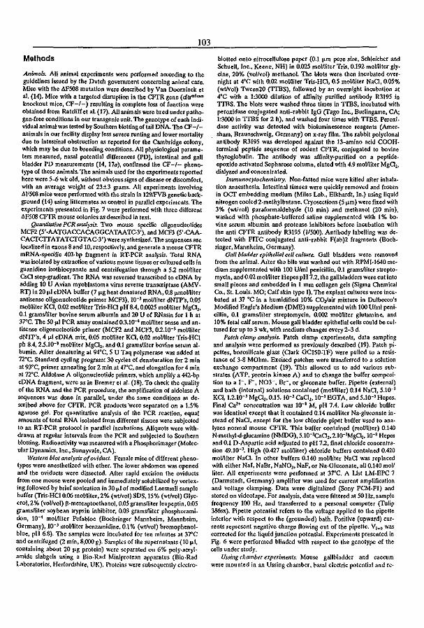
103
Methods
Animals. All animal experiments were performed 3ttOrding to the guidelines issued by the Dutch go\'emmenl concerning animal care. Mice with tbe AF508 mutation were described by Van Doorninck el aI. (14). Mice with a targeted disruption in the CFTR gene (dtr"'l ..... knockout mice, CF-I-) resulting in complete loss of {unction were obtained from Ratcliffet at (17). Allanimalswere bred underpatho· gen·free conditions in our transgenic unit. 'I'b¢ genotype of each individual animal was tested by Southern blottingo{ tail DNA. The cr-/animals in our facility display less severe ruoting and lower mortality due to Intestinal obstruceion as reported for the Cambridge colony, which may be due to breeding conditions. All physiologica1 parameters measured, nasa] potential differences (PD). intestinal and gaU bladder PD measurements (14, 170), confirmed the CF-I- phenotype of these animals. The animals used for the experiments reported here were 5-6 wk old. without obvious signs of disease or discomfort, with an average weight of 23±3 grams.. All experiments Involving AFS08 mice were performed with the strain In 129JFVB genetic background (14) using liltcnnates as control in parallel experiments. The experiments presented in Fig. 7 were performed with three different AF508 CFfR mouse colonies as described in text.
QuantiJat;ve peR onlllysis. 1\ro mouse SpecifIC oligonucleotides MCFl (5'·AATGACCACAGGCATAATC-3'). and MCF3 (5'·CAACAcrcrrATATCTGTAC·3') were synthesized. The sequenoes are localized in exons 8 and 10, respectively. and generate a mouse CFfR mRNA-specific 403·bp fragment in RT·PCR analysis. Total RNA was isolated by extraction of various mouse tissues or cultured cells in guanidine isotblocyanate and centrifugation through a 5.2 moliliter Csa step-gradient. The RNA was re\'ersed transcribed to cDNA by adding 10 U Avian myoblastoma virus reverse transaiptase (AMVRT) in 20 pol cDNA buffer (7 pog heat denatured RNA, 0.8 pomolJliter antisense oligonucleotide primer MCFJ). 10-' moUliter dNTP's, 0.05 mollliter Ka, 0.02 mollliterTris·HO pH 8.4, 0.0025 mol/liter Mg01,
0.1 gramslliter bovine serum albumin and 20 U of RNasin for 1 h at 37"C. The SO pol PCR assay contained 03.10-' mol!liter sense and an· tisense oligonucleotide primer (MCn and MCF3, 0.2.10-' mollliter dNfp's, 4 pol cDNA mix, 0.05 mollliter KO. 0.02 mollliter Tris·HCI ph SA, 2.5.10-3 moUliter Mg01• and OJ gramYliter bovine serum al_ bumin. Afler denaturing at 94"C, 5 U Taq polymerase was added at n°c. Standard cycling program: 30 cycles of denaturation for 2 min at 93"'C. primer annealing for 2 min at 47"C, and elongation for 4 min at 72°C. Aldolase A oligonucleotide primers, whkh amplify a 442-bp eDNA fragment, were as in Bremer et aJ. (18). To check the quality of the RNA and the PCR procedure, the amplification of aldolase A sequences was done in parallel, under the Same conditions as described above for CFfR. PCR products 'were separated on a 1.5% agarose gel. For quantitative analysis of the PCR reOKtion, equal amounts of total RNA isolated from different tissues were subjected to an RT·PCR prot()(Ol in parallel incubations. Aliquots were withdra ..... n at regular intervals from the PCR and subjected to Southern blolling. Radioactivity was measured with a Phosphorimager (Molecular Dynamics, Inc., Sunnyvale. CA).
Western blot (JIfaiysis of oviduct. Female mice of different pheno. types were anesthetized with ether. The lower abdomen was opened and the oviducts were dissected. After rapid excision tbe oviducts from one mouse were pooled and immediately solubilized by vortex. ing followed by brief sonication in 30 j.lJ of modified Laemmli sample buffer (Tris.HCI 0.06 molfliter,2% (wtlvoI}SDS, 15% (wtlvol) Glycerol,2% (voUvol) fl·mercaptoelhanol, 0.05 grams/liter leupeptin, 0.05 gramslliter soybean trypsin inhibitor, 0.00 gramslliter phosphorami. don, 10-4 mollliter Pefabloc (Boehringer Mannheim, Mannheim, Germany), 10-) mollliter benzamidine. 0.1% (wt/vol) bromopbenolblue, pH 6.8). The samples were incubated for ten minutes at 37"C and centrifuged (2 min. 8,000 g). Samples of the supernatants (10 !-LI, containing about 20 j.lg protein) were separated on 6% poly·aeryl. amide slabgels using a Bio·Rad Miniprotean apparatus (Bio.Rad Laboratories, Herfordshire, UK). Protein~ were subsequently electro·
blolled onto nitrocellulose paper (0.1 porn pore size, Schleicher and Schuell, Inc., Keene, NH) in 0.025 molfliter Tris, 0.192 mollliter g1y. dne,20% (vollvol) methanol. The blots were then incubated over· night at of'C with 0.02 molJliter Tris-HQ. 0.5 mollJiter NaO, 0.05% (wtlvol) Tween20 (TI'BS), followed by an overnight incubation at .rC with a 1:3000 dilution of affinity purified antibody R3195 in TrnS. The blots were washed three times in TrnS, incubated with peroxidase conjugated anti-rabbit JgG (rago Inc., Burlingame, CA; 1:3000 in TrnS for 2 h). and washed four times with TIBS. Peroxi· dase activity was detected with bioluminescence reagents (Amer. sham, Braunschweig. Germany) on x·ray film. The rabbit polyclonal antibody R3195 was developed against the 13-amino add COOH· terminal peptide sequence of rodent CFfR, conjugated to bovine thyroglobulin. The antibody was affinity-purified on a peptide. epoxide activated Sepharose column, eluted with 4.9 mollliler MgQ lo
dialyzed and concentrated. Immunocytochemistry. Non·fasted mice were killed after inhala.
tion anaesthesia. Intestinal tissues were quickly removed and frozen in ocr embedding medium (Miles Lab., Elkhardt, In.) using liquid nitrogen cooled 2-methylbutane. Cryosections (5 j.lm) were fIXed with 3% (wti'lol) parafonnaldehyde (10 min) and methanol (20 min). washed ..... ith phosphate·buffered saline supplemented with 1 % boo vine serum albumin and protease inhibitors before incubation ..... ith the anti CFfR antibody R3195 (1/500). Antibody labelling was detected ..... ith FITC conjugated anti-rabbit F(ab)2 fragments (Boeh. ringer, Mannheim, Germany).
Gaff bladdu epithelial cell culture. Gall bladders were removed from the animal. Aiter the bile was washed out with RPMJ·I640 me· dium supplemented with 100 U/ml penkillin. 0.1 gramslliter strepto. myeln, and 0.02 mollliter Hepes pH 7.2, the gallbladders were cut into small pieces and embedded in 1 mm collagen gets (Sigma OIemical Co., St. Louis. MO; Calf skin type I). The explant cultures were incu· bated at 37 °C in a humidified 10% CO/air mixture in Dulbecco's Modified Eagle's Medium (DME) supplemented ..... ith 100 Ulml penl. cillin, 0.1 gramslliler streptomycin, 0.002 mollliter glutamine. and 10% fetal calf serum. Mouse gall bladder epithelial cells could be cuI· tured for up to 3 wk, with medium changes every 2-3 d.
Patch clamp analysis. Patch clamp experiments, data sampling and analysis were performed as previously described (19). Patch pi· pettes, borosilicate glass (Cark GCISO-1F) were pulled to a resis· tance of 3·8 MOhm. Excised patches were transferred to a solution exchange compartment (19). This allowed us to add various sub· strates (ATP. protein kinase A) and to change the buffer composi. tion to a 1-. r. N03-. Br-, or gluconate buffer. Pipette (external) and bath (internal) solutions contained (mo1J1iter) 0.14 NaO, 5.10-3
KCI, 1.2.10-3 MgQ l,0.15. 10-3 Ca02,1Q-3 EGTA, and 5.10-3 Hepes. Final Ca2+ concentration was to-I M, pH 7,4. Low chloride buffer was identical except that it contained 0.14 moUliter Na·g1uronate in· stead of NaO, except for the low chloride pipet burrer used to ana· Iyzes normal mouse CfTR. This buffer contained (moUliter) 0.140 N·methyl..<J·g1ucamine (NMDG), 5.1O-l('.a02, 2.1O-JMg01, 10-2 Hepes and 0.1 D·Aspartic acid adjusted to pH 7.2, final chloride concentra· tion 49.10-). High (0,427 moUliter) chloride buffers contained 0.420 mollliter NaQ. In other buffers 0.140 mo1J1iter NaCl was replaced .....ith either NaI, NaBr, NaNO), NaF, or Na·Gluconate, all 0.140 mol! liter. All experiments were performed at 37"C. A Ust LM·EPC 7 (Darmstadt, Germany) amplifier was used for current amplification and voltage damping. Data were digitalized (Sony PCM·F1) and stored on videotape. For analysis. data were filtered at 50 Hz, sample frequency 100 Hz, and transferred to a personal computer (Tulip 386sx). Pipette potential refers to the ,'oltage applied to the pipette interior ..... ith respect to the (grounded) bath. PositiVe (upward) cur· rents represent negative charge flowing out of tbe pipette. V,_o was corrected for the liquid junction potential. Experiments presented in FJg. 6 were performed blinded ..... ilh respect to the genotype of the cells under study.
Ussillg chwnba experimmts. Mouse gallbladder and caecum were mounted in an Ussing chamber, basal electric potential and reo
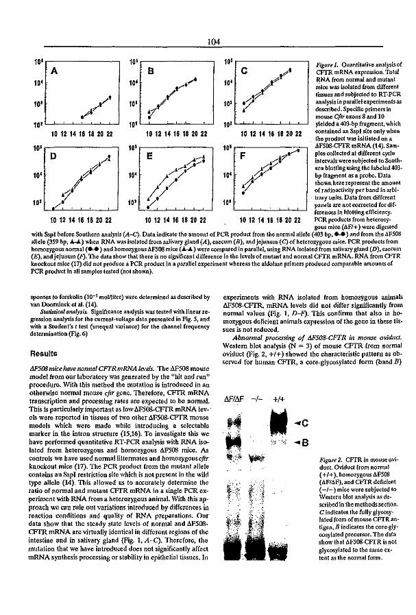
104
10' ,------, 10',-------, A B
10'
10' 10'
10 12 14 16 18 20 22 10 12 14 16 18 20 22
10' .---------, 10' ;---------, o E
10' 10'
10'
10 12 14 16 18 20 22 10 12 14 16 18 20 22
10' ,------,
10'
10 12 14 16 16 20 22
F
10'
10'
10 12 14 16 182022
Figure 1. Quantitath'c analysis of CFfR mRNA expression. Total RNA from normal and mutant mice was isolated from different tissues and subjected to RT·PCR analysis in parallel experimentsas described. Specific primers in mouse Cftrexons8 and 10 yielded a 403.bp fragment, which contained an SspJ site only when the product was initiated on a 4F508-CFIR mRNA (14). Samples collected at different cycle intervals weresubje<:ted loSoulh· ern blotting using the labeled 403· bp fragment as a probe. Data shown here represent tbe amount of radioactivity per band In arbi-
. l1ary units. Data from different panels are not corrected for dif· ferenoes In blotting efficiency. PCR products from heterozygous mice (AF/+) were digested
with Sspl before Southern analysis {A-C}. Data indicate the amount of peR product from the normal allele (403 bp, .... ) and from IheJ.FS08 allele (359 bp, .trr,..."l,) when RNA was isolated from salivary gland (A), caecum (B), and jejunum (C) of heterozygous mice. PCR products from homozygous nonnal ( ... ) and homozygous Af:SOS mice ( ...... ) were compared in parallel. using RNA isolated from salivaI)' gland (D). caecum (E), and jejunum (P). The data show thallhere is no significant difference in the levels of mutant and normal CFTR mRNA. RNA from CFfR knockout mice (17) did not produce a PCR product in a parallel experiment whereas the aldolase primers produced comparable amounts of PCR product in a1l samples tested (not sho ..... n).
sponses to forskolin (10-S molniler) were determined as described by van Doornlnck: et al. (14).
StatistiCil/ analysis. Significance analysis was tested with linear regression analysis for the current-voltage data presented in Fig. 5. and with a Student's t test (unequal variance) for the channel frequency determination (Fig. 6)
Results
tJF5(J!J mice have norma! CFTRmRNA le!,'eis. The dF508 mouse model from our laboratory was generated by the "hit and run" procedure. With this method the mutation is introduced in an otherwise nonnal mouse eftr gene. Therefore. CFfR mRNA transcription and processing rates are expected to be normal. This is particuJarly important as low L\F500-CFTR mRNA lev-' els were reported in tissues of two other dF508-CFfR mouse models which were made while introducing a sele<:table marker in the intron structure (15.16). To investigate this we have performed quantitative RT·PCR analysis with RNA isolated from heterozygous and homozygous AFSOO mice. As controls we have used normallittermates and homozygouseftr knockout mice (17). The peR product from the mutant allele contains an SspJ restriction site which is not present in the wild type allele (14). This allowed us to accurately detennine the ratio of nonnal and mutant CFfR mRNA in a single PCR experiment with RNA from a heterozygous animal. With this approach we can rule out variations introduced by differences in reaction conditions and quality of RNA preparations. Our data show that the steady state levels of normal and dF500-CFTR mRNA are virtually identical in different regions of the intestine and in salivary gland (Fig. 1. A-C). Therefore. the mutation that we have introduced does not significantly affect mRNA synthesis processing or stability in epithelial tissues. In
experiments with RNA isolated from homozygous animals AFSM·CFTR. mRNA levels did not differ significantly from normal values (Fig. 1, D-F). This confirms that also in homozygous deficient animals expression of the gene in these tissues is not reduced,
Abnormal processing of MS08-CFTR in mou.se ovidud. Western blot analysis (N = 3) of mouse CFTR from normal oviduct (Fig. 2, +1+) showed the characteristic pattern as observed for human CFfR, a core-glycosylated fonn (band B)
N'IN' -1-
H
t! ~
+1+
II ;!f' .... .;
... c
... B Figure 2. CFfR in mouse oviduct. Oviduct from normal (+1+), homozygous AF508 (AF/AF). and CfTR deficient (-/-) mice were subjected to Western blot analysis as described in the melhodssecfion. C indicates the fully glycosylated form of mouse CFTRanligen. B indicates the rore.gly· oosylated precursor. The data show that AFS08·CFfR is not glyoosylated to the same extent as the normal fonn.
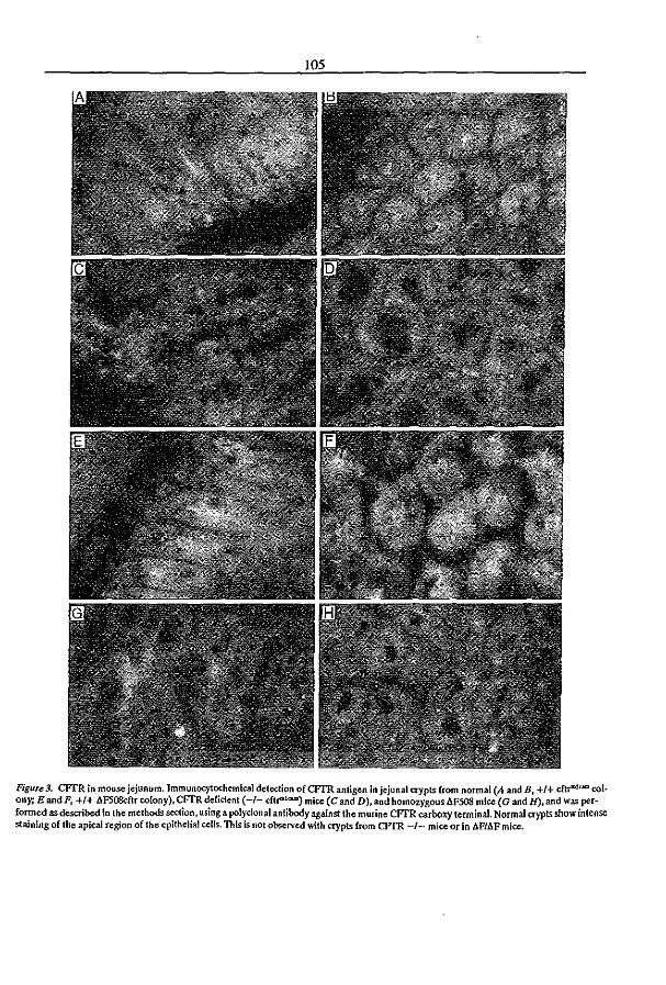
105
FiguTe3. CFfR in_mouse jejunum. ImmunocytCX:hemical deledion ofCFTR antigen In jejunal aypts from normal (A and B, +1+ cftr">\<.om colony; E and F, +1+ AFSOSdtr colony). CFfR deficient (-/- dlr<"I ..... ) mice (C and D). aDd homozygous AF508 mice (G and H). and was performed as described in the methods S«tion, using a polyclonal anlibody against the murine CFTR carboxy terminal Normal crypts show intense staining of the apica.l region of the epithelial cells. This is nOI observed with crypts from CFfR -/- mice or in AP/AF mice.
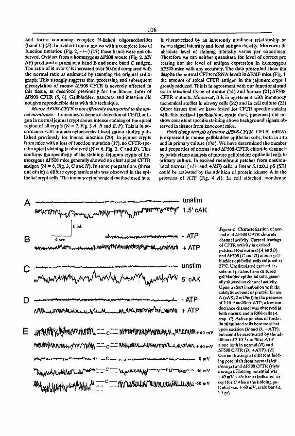
106
and forms containing complex N·linked oligosaccbarides (band C) (3). In oviduct from a mouse with a complete foss of function mutation (Fig. 2, -/-) (17) these bands were not observed. Oviduct from a homozygousaF508 mouse (Fig. 2. 11Ft AF) produced a prominent band B and some band C antigen. The ratio of B over C is increased over SO-fold compared with the nonnal ratio as estimated by scanning the original radiograph. This strongly suggests that processing and subsequent glycosyJation of mouse AFS08 CFfR is severely affected in this tissue. as described previously for the human form of ~08 CFTR (5, 6). Gallbladder. pancreas and intestine did not give reproducible data with this tecllnique.
Mouse M"SQ8·CFTR is not ejfidenrly tranrported to the apical membrane. Immunocytochemical detection of CFfR anti· gen in nonnal jejunal crypt shaM intense staining of the apical region of aU crypts (N = 7. Fig. 3 A, Band E, F). This is in accordance with inununocytochemicallocalization studies published previously for human intestine (20). In jejunal crypts from mice with a loss offunction mutation (17), no CFfR·spe. cific apical staining is observed (N = 6, Fig. 3, C and D). This confirms the specificity of the staining. Jejunum crypts of homozygous AF508 mice generally showed no dear apical CFfR antigen (N = 6,Fig. 3, G and H). In some preparations (three out of six) a diffuse cytoplasmic stain was observed in the epithelial crypt cells. The immunocytochemical method used here
is characterized by an inherently nonlinear relationship be tween signallntensity and local antigen density. Moreover th absolute level of staining intensity varies per experi.nrenl Therefore we can neither quantitate the level of correct pr(J cessiog oar the level of antigen expression in homozygou M5C8 mice with any accuracy. The data presented show tha despite the normal CFIR mRNA levels in4Fh1Fmice (Fig. 1: the amount of apical CFfR antigen in the jejunum crypt i greatly reduced. This is in agreement with our functional stud ies in intestinal tissue of mouse (14) and human (21) AFSQ8.
<FrR mutants. Moreover, it is in agreement with immunocytochemical studies in airway cells (22) and in cell culture (23) Other tissues that we have tested for CFI'R specific stainin~ with this method (gallbladder, cystic duct, pancreas) did nol show consistent specific staining above background signals ob· served in tissues from knockout mice.
Palm damp O1U1lysis o/mouse tWS08·CFTR. CFI'R mRNA is expressed in mouse gallbladder epithelial cells, both in situ and in primary culture (170). We have determined the number and properties of nonnal and AF508-CFTR chloride channels by patch clamp analysis of mouse gallbladder epithelial cells in primary culture. In excised membrane patches from unstimulated nonnal (+1+ and +/AF) cells, a linear 5.1±O.1 pS (SE) could be activated by the addition of protein kinase A in the presence of ATP (Fig. 4 A). In cell attached membrane
A --~-----,----- unstlm 1.5' cAK
B
c
~IPA 4 SfC
+ATP
unstim
5'cAK
o • __ --" .. · .. 'Ii ... ,~, .. ,-_ .... __ ... _" ....... , ... ,_~---......... _·~\I ... JJ., • ATP
+ATP
E ~:':'C=~~~6.mv ~=C=~+4.mv
~f ................ "",~--C ' ...... OmV
~=-C==~4.mV
~:= C=1Mt'Jjf~~.mv
Figure 4. Oaracterization of nor· mal and .6.FSOS CPfR chloride channel activity. Current tracings of CFIR activity in excised patches from normal (A andB) and .6.FS08 (C and D) mouse gall. bladder epithelial cells cultured al 2rc. Unstimulated excised, inside-out patches from cultured gallbladder epithelial eells generally showed no channel activity. Upon a short incubation with the catalytic subunit of protein kinase A (cAK,2 mU/ml) in the presence 0£2.10-3 moViiter ATP, a low ron· ductancechannel was observed in both normal and .6.FSOS eells (A resp. C). Acth'e patches of forskolin stimulated cells became silent upon excision (B and D, -ATP), but could be reactivated by the ad· dition 0£2.10-3 mollliter ATP alone both in normal (B) and "F508 CF'TR (D. +ATP). (E) Current tracings at different hold· ing potentials from normal (left tracings) and .6.FS08 CFTR (right tracings). Holding potential was +40 mY scale bar as indicated, excepl for C where the holding po. tential was + 60 mY, scale baI4 s, 1.5 pA.
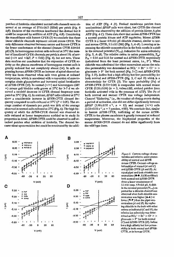
107
patches of forskolin.stimulated nonnal cells channels were observed at an average of 27.6±16.5 (SEM) per patch (Fig. 6 left). Excision of the membrane inactivated the channel but it could be reopened by addition of A TP (Fig. 4 B). Considering the resemblance with human CFrR (3) we conclude that these linear chloride channels represent normal mouse CFIR. The only difference observed between mouse and human CFTR is the lower conductance of the channel (human CFTR 8.QtO.6 pS) (3). In homozygous mutant cells cultured at 37C the num· ber of functional CFfR channels per patch is about 1% of nor· mal (+1+ and +/6F, P < 0.02) (Fig. 6). but not zero. These data confirm our conclusion that the expression of CFTR activity on the plasma membrane of homozygous mutant cells is greatly reduced but not completely absent (14). In cells expressing human AF508-CFfR an increase of apical channel activity has been Observed when ceUs were grown at reduced temperature, which is associated with a conversion of core-tocomplex chain glycosylation and increased apical localization of M'S08·CFTR (10). In normal (+1+) and heterozygote (llFl +) mouse gall bladder cells grown at 21'C for '>-7 d we observed a twofold decrease in CFfR channel frequency com· pared to 3-rC (Fig. 6). In contrast, 6F1b.Fcells cultured at zrc show a considerable increase in 6F5OS·CFfR channel frequency compared to cells cultured at 31'C (P < 0.05). The average number of channels per patch was 16% of the average observed in nonnal cells cultured at rrc (Fig. 6). The high fre· quency at which the AF508-CFfR channel was observed in cells cultured at lower temperatures enabled us to study its properties in detail. AF508-CFfR could be observed in cell attached patches after addition of forskolin. The channel be· came silent upon excision but could be reactivated by the addi·
A B O.SO - • 15O/1SOCf 0.50 -
., 15OI1lC[
0.30 - , U/1MCI 0.30 -
~ 0,10 -
~ 0.10 -
~ ~ ·0.10 - ·0.10 -
-0.30 - ·0.30 -
·0.50 -, , , , ,
·0.50 -... ·30 0 30 .. V(mV)
C D 0.50 - 0.60 -
0.30 - 0.30 -
~ 0.10 - ~
0.10 -
~
·0.10 - -0.10 -
·0.30 - -0.30 -
·0.60 -, ·0.50 -... .30 0 30 60
V (mV)
tion of ATP (Fig. 4 D). Bxcised membrane patches (rom unstimulated llF/6F cells were silent, but CFTR·like channel activity was observed by the addition of protein kinase A plus ATP (Fig. 4 C). These data show that mouseAF500·CFTR has a nonnal protein kinase and ATP regulation. Mouse b.F508-CFTR is a linear 4.9±0.1 pS chloride channel. similar to nor· mal mouse CFfR. 5.1±0.1 (SB) (Fig. S.A-B). Lowering or increasing the chloride concentration in the bath results in a shift in the reversal potential (V,,,o), indicative for anion selectivity (Fig. S. A-B). The relative cation to anion permeability. Pg,/ Po = 0.14 and 0.16 for normal and b.F5()8.CFTR respectively (calculated from the least permeant anion, i.e., F"). When chloride was substituted for other monovalent anions the rela· tive permeability was determined as NOJ - > Br- > CI- > > gluconate > F- (or both nonnal (Fig. 5 C) and AFS08·CFfR (Fig. 5 D). Iodine had a high arfinity but low permeability (or both nonnal and AF508-CFTR (Fig. 5, C and D) which is a characteristic for CFTR (3). The open probability (Po) of .A.FS08·CFTR (0.33±0.04) is comparable with nonnal mouse CFfR (035±0.04) (n = 5, values±SE, excised patches from forskolin activated cells in the presence of ATP). The Po of both nonnal and mutant CFfR was voltage independent. Channel "nickering," i.e., the number of closing events during a period of activation. also did Dot differ significantly between AF/AF (3.<}9±O.70 S-l, n :::: 12) and nonnal (+1+) cells (3.23±055 S-l, n = 7 patches, ±SE). We conclude that, similar to human M'S08·CFTR, trafficking of the mouse .A.F508-CFTR to the plasma membrane is greatly increased at reduced temperature. Moreover, the biophysical properties of the mouse .A.F508-CFfR channel do not differ significantly from the wild type (orm.
.'$OII50CI
l' lSO/12 a J. 15OIUOCl
Figure 5. Current·voltage marac-leristics and relative anion perroe-ability of nonnal and llFS08 mouse CFfR. Current·voltage ee-lationships of normal (A and C) and .6.F508·CFfR (8 and D). At
, , , equal pipet and bath chloride oon-... ·30 0 30 60 centrations ( .... O.ISO mollliter) both normal and AFSOS·CFTR
V (mV) show a linear conductance of S.l±O.l resp. 4.9±0.1 pS. A shift In the reversal potential (V,-o) ex-.e., pected for a chloride channel was .e. observed when bath chloride con-
ve ... centralion was higher ("'""""-) or o C~'tIO lower (T-'f) than the pipet con-centralion (A and B). By replac-ing chloride in the bath with other anions as indicated (C and D). the
dr' relative ion sele<:tivity was deter-mined as NO,- > Br- > CI- > > gluconate > F- for both normal , , , , , (C) and rn08·CFTR (D). Iodine ... ·30 0 30 60 has a high affinity bullow pelme-
V(mV) ability in both normal and AF508-CFTR. as in human CFTR.

37' '"
• i • 5 " ~ .. • .. z ,
" ... • ".
!J.F/lJ.F +/bF
, ,
•
•
, +/+
'" • ••• ,
"
•
.. ..
IJF/IlF +/llF
108
•
,
• •
+/+
Figure 6. CFTR chloride channels in gall bladder plasma membrane. CFTR channel activity on plasma membrane of cultured gallbladder epithelial cells was rerorded with patch damp analysis as described. Cells were (lIltured either at 3?C (left) or at 27°C (righl) Cor 3-7 d be· fore analysts.. Forskolin stimulated cells were recorded Cor Iwo minutes in the on cell patch clampoonfiguration, followed by a I-I0-min reoording in the excised patch configuration in the presence oC2 mM ATP. CFTR channel frequency wascakulated from theequalion n = 1/(I1tpo) where n Is the caladated number of IC\'els, I .. is the average current in the patch, Ills the current or one channel al holding potential. and Po is the open probability. When 4 or less levels of activity were observed in the palch tbe equation was not used, in these cases the number of cbannels correspond directly to the number of current levels observed. Patches whlcbshowed no CFTR activity were scored as zero. Cells were obtained from parallel cultures orlinennales. (e) Homozygous mutant, 6F/6F (oells from 3 mice al YrC and 8 mke at 2T'C); (A) heterozygous mutant +/!J.P(3 and 4 mice); (") homozy. gous normal +1+ mice (2 and 1 mice).
Inefficient processing of mouse tJF508 CFl'R is observed in differelll genetic backgrounds. The most sensitive avaUable measure of apical CFfR activity is probably the steady state response to torskolin in Vssing chamber experiments. Using this method, we have previously shown low but significant re· sidual AFSOO·CFfR activity in different tissues at homozygous AFSoo mice when compared to cftI"'l(:W knockout mice that showed no activity (14). Next, we studied whether genetic background influences the level of AFSoo·CFfR processing. The 129.FVB and 129·C57BU6 J1F strains were obtained by introduction of the original mutant clone (129/01a) in FVB and CS7BU6 blastocysts respectively. tiPl+ (129·FVB) mice were crossed with cftt"lo;am knockout mice (KO) that are 129·C57BU6. The forskolin response of gallbladder and cae· cum of these difterent tiP mouse strains was monitored in Ussing chamber experiments. For aU strains the forskolin reo sponse was equally reduced in homozygous4F/AF and in tiPlmice (Fig. 7). AU individual measurements from tiP mutant mice are in the same range with only one exception observed (Fig. 7). The data show no evidence for dominant alleles in ei· ther the 129/01a,.FVB or C57BU6 inbred genetic backgrounds that significantly increase or reduce the level of AF5Q8..CFfR
processing. However, one homozygous llF/AF mouse in a 129·FVB background did show a response to forskolln in the normal range using gallbladder (Fig. 7 A) and ileum (not shown). Therefore, more complex relationships involving several genetic loci can not yet be excluded and require further study.
.,
" ~ .. i ~ • ., :;; j .,
•
A
• , 'I' • ,,,
IN/FV8 129/8(6 129/FV8/B1$
.12 ~B~ _____ _
."
"
.,
•
• • • • • , ,
• t=='::'t::,,:::t,::±::J. ••• 129/FVB IN/BI& 12Q/FV8/S1E
Figure 7. Forskolin responses in homozygous!J.F/llFmice gallbladder and caecum in miw strains with different genetic backgrounds. Gall· bladder (A) and caecum (B) of mice from 6F508 CFTR strains with different genetic backgrounds were mounted in the Ussing chamber and monitored for their electricaJ response to forskolin (6PD, mY). 1291FVB and 1291B16 were obtained by blastocyst injection and cross· ing into fVB and C57BU6 type animals. 1291FVB1B16 was obtained by crossing 6FI+ (1291FVB) with +1- dtrm1 .... (129/C57BL6). (.A.) +1+, +/6 and +1- mice; (e) !J.F/6Fmice; (+) 6F/- mice.
Discussion
CFl'R mRNA expression is normal in mulatU mice. Using the "bit and run" procedure, we have generated a aF5Q8.CFIR mouse model without modifying the intron structure of the CFTR gene (14). Our quantitative PCR data show that in het· erozygotes the expression levels of nonnaI and mutant alleles are comparable in intestine and salivary gland. Also in ho· mozygote nonnal and mutant mice the expression levels are not significantly different. In two other AFS08-CFTR mouse models created by homologous recombination, low CFfR mRNA levels were reported in salivary glands and intestine (15,16). In these models an expression cassette encoding a se· lectable marker was introduced into the flanking intron, in ad· dition to the dFS08 mutation in exon 10. Comparison with our data suggests that the presence of this transcription unit affects the CFfR mRNA levels by interfering with RNA polymerase activity or RNA processing.
MatuFationofnwuseM'5OlJ.CFrRisahnonnaL InununocbemicaJ analysis of CFfR expression in intact mouse tissues is diffi· cult with the available antisera. This can be attributed in part to relatively low expression levels as in gallbladder tissue (17a) and to proteolytic activity. especially in intestinal tissue (H.R. de Jonge, unpublished data). This limits the scope of our inves· tigations to tissues that give consistent and reproducible data. We were able to make reproducible western blots of total ovi· ducts from normal and mutant mice with a purified antibody raised against the carboxy terminus of rodent CFI'R (Fig. 2). The data show that CFfR antigen is produced in both normal (+/+) and mutant (J1F/dF) mice. but not in CFTR deficient (-1-) mice. However, the proportion of mature,fully gIycosylated CFfR is severely reduced in homozygous mutant. We conclude that the moo mutation in the mouse CFIR protein results in a processing defect similar to that observed with hu· man AFSOO·CFfR. The available evidence suggests that transfer to the Golgi system and subsequent full glycosylation of the human mutant protein does not occur, because proper cbaper·
109 onin..dependent folding is arrested in the endoplasmic relicuIwn (7.8,10). With the same antibody as used for western blot analysis, we could show specific staining of the apical region of normal mouse jejunum crypt epithelial cells (Fig. 3, A, B, E, and F). Specificity of the staining was verified with serum preabsorbed with the antigenic peptide (not shown) and jejunum of CFfR..deficient mice (Fig. 3, C and D). Tbis is in agreement with localization studies in human intestine (20) and rat (24). In jejunum crypts of mice homozygous for the AFS08 mutation we did not observe a preferential staining of the apical region. Most crypts seemed devoid of specific staining. However. with tbis method we cannot rule out tbat cytoplasmic CFfR is present (note that also in control cells no specific cytoplasmic staining above background can be seen Fig. 3). In several preparationssome crypts did stain more intensely than background but the signal was not concentrated to the apical region as in normal crypts. These observations are in agreement with a processing defect of mouse AF5()8.CFI'R that prevents translocation of the protein to the apical region.
Temperature dependent apical targeting of AF508-CFTR Airway ceUs of AFS08 mutant mice in primary culture have increased cAMP-induced chloride conductance when cultured at reduced temperature (16). We "''ere unable to detect CFfR chloride channel activity in primary airway cells using patch damp single channel analysis (P J. French, unpublished data). In contrast, cultured mouse gall bladder cells provide a convenient system for the study of CFfR with this method. This allowed us to show that the actual frequency of active mutant CFTR molecules at the plasma membrane is increased by incubation of the gallbladder cells at low temperature. In homozygous mutant cells cultured at YrC few CFTR channels were observed, i.e" 1 % of the normal level at this temperature (Fig. 6), The expression of AF508-CFfR channels increased to 16% of normal levels when ceUs from homozygous mutants were grown at 2"fC (Fig. 6). A similar behavior was observed with human AFS08·CFfR (10).
a,anne! activity of AF508·CFrR. The channel conductance and anion selectivity of mouse AFS08-CFTR is in our analysis indistinguishable from nonnal mouse CFTR (Fig. 4). In addition, the open probability (Po) and number of closing events during a burst of activity of AFS()8.CFTR are nonnal. Studies of hUman AF5Q8·CFfR are inconsistent at this point as two groups reported normal conductance but reduced Po for human AFS08·CFfR (10, ll) whereas others claimed a normal conductance and Po (25). Since the activity of the CFIR chloride channel depends on a number of parameters in particular the state of phosphorylation of the protein, the reported difference in relative Po may result from differences in experimental conditions.
Residual adivity of L1F508·CFTR Both Ussing chamber experiments (14) (Fig. 7), and patch clamp analysis (Fig. 6) in our model indicated a low but significant level of apical activity of mouse AF508-CFfR at physiological temperature. This is confinned by the western blot analysis (Fig. 2), which shO'o"s a low level of fully glycosylated CFfR. Interestingly. a minority of human AF508 CFTR homozygotes possess a small residual intestinal chloride penneability, which correlates with a relatively mild clinical status (21). Unpublished data showed that in some. but not all, cases this activity is insensitive to the chan· nel blocker DIDS (H. Veeze, personal communications). This would suggest that in these cases the residual activity is due to AFS08-CFfR. It is possible therefore, that a significant level of
correct processing is also present in a subgroup of human AF508 CFfR homozygotes. Immunocytochemical analysis of airway cells from .6.FS08 CFTR patients seem to confirm this (26). It will be of great interest to analyze the genetic and envi. ronmental factors which contribute to this phenomenon. Our current data indicate that the level of mutant CFfR activity, as determined by Ussing chamber experiments, is quite constant in the different genetic backgrounds tested (Fig. 7). The one exception to this rule we found sofar is intriguing. but extensive further studies are required to establish a genetic basis for this effect. The level of .6.F508-CFIR processing could differ in various tissues and small variations in apical activity levels could have profound effects on pathology, Our electrophysio· logical data, supported by inununochemical data, show that in all epithelial tissues studied sofar the AFSOB mice have severely reduced apical activity. However. a careful analysis of .6.F508-CFfR processing kinetics may reveal subtle differences between cell types.
ApplicationsoftheAFS08mousemode! Our data show that the tissues of the AFS08 mouse model provide a valid model for the processing defect of the humanAF508-CFfR mutation. It gives us the opportunity to study several aspects of CFTR function in more detail in intact tissues. CFIR is active in intra· cellular compartments (9), possibly affecting the pH of vesicular compartments. This could explain why CFfR deficient cells produce mucins and surface glycoproteins with abnormal carbohydrate structures (27-31). These abnonnalities maycontribute to the fonnation of abnormal secretions and to tbe reduced clearance of airway pathogens characteristic of CP. CFfR is involved in regulated endocytosis and exocytosis in some ceU types (32, 33), In addition, recent evidence suggests that CFfR either directly or indirectly interacts with other ion transport systems (34, 35). AF508-CFIR expressed in epithelial cells may be partially active in these processes, Therefore, we may expect to observe differences between .6.FS08 mice and mice with complete loss of function mutations (knockout). It will be interesting to compare AF508 mice with knockout mice in an experiment in which the mice are challenged with lung pathogens (36). Furthermore, the.6.F508 mouse model will allow us to study conditions that may enhance the activity of AF508-CFfR at the apical epithelial membrane in vitro and in vivo. Processing of nonnal CFfR involves chaperonin mediated folding and core glycosylation at the endoplasmic reticulum. This is followed by full glycosylation in the Golgi system and transport to the apical membrane. It is a rather inefficient process since approximately 75% of nonnal CFfR is degraded in inunature form by cellular proteases (7, 8), Processing of AF508-CFfR to a fully glycosylated apical fonn is severely re· duced. The striking stimulating effect of reduced temperature on 4F508-CFfR expression, both with human and mouse mutants, suggests that the processing block can be relieved within the range of physiological conditions. We can consider several approaches towards frnding therapeutic substances. Compounds which interfere with AF50S-CFfR-chaperonin interaction may improve processing by relaxing the quality control mechanism. An example of this may be glycerol, which has an effect comparable to low temperature (37). Inhibition of CFfR degradation may improve processing of mutant CFfR, although this approach has not been successful to date (7). CFTR at the plasma membrane is activated by ATP binding and multi-site phosphorylation of the large cytoplasmic regula· tory domain (3, 38). Therefore, substances that stabilize the
110
open state of CFrR (39, 40) or inhibit protein phospbatases (41-43) are expected to improve the activity of both normal and AFS~·CFIR. These studies may lead to new developments, not only in our understanding of membrane protein processing, but also towards effective therapies for CP.
Acknowledgments
We thank Wendy Boomaars for expert technical assistance and Mirko Kuil fOf excellent photography. We thank Drs. W. Colledge, R. Ratcliff. and M. Evans (Cambridge. UK) for providing us with the dtf"'leam knockout mice.
This ,,""Ork WM funded in part by HGOfIND, SLW and the aIni· cal Genetics Foundation.
References
1. Boat, T., MJ, Welsh, and A. Beaudet 1989. Cystic F'ibrw!s.l" Metabolic Basisof Inherited Disease. C. Scriver, A. Beaudel, W. Sly, and D. VaIJe,«litors. M~r:aw HilI,NewYoIk. 2649-2860.
2. Welsh, ill. 1994. The path of discovery in undersLtnding the bioJogy of C)'51~fibros5 and approa~ to therapy. Am. J. Gastf'()tnltTol. 89 (8):S97-5105.
3. Riordan,J.R..I993.1he C)'$ticfibrosh lransm~braneoonduda~ regu· lator.Ann. RfY. PhrsioL 55;6()9-630.
4. Kerem, B.S.,J.M. Rommern,J.A BIKhanan, D. Martiev.kz, TK Cox, A O:.akravarti, M. Bucll~ld,and LC. Tsul 19S9.ldentiflCation of the c)''Sticfibrosis gene - ge~tk analysis. S&lIce (W4Ih. DC). 245:1073-1080.
5.0lenz,.S.H.,RJ.Grego/)"J.Manhan,S. Paul,D. W.Souza,G.A White. J. R. Riordan. and A.E. Smith. 1990. Defedi\.ll intrarelluJar lTansport and processing ofCFrR is !he basis ofmost()'stk fibrosis. CeIL 63:821-834.
6. Grego/)" RJ., D.P. Rich, S.H. Oleng, D.W. Souza, S. Paul. P. Manavalan, M. P. Anderson, MJ. Wel5h. and A.E. Smith. 1991. MaturatkJn and fundion of cystic fibrosis transmembrane ronductance regulator variants bearing mutations ... ~tativenudootide..bindng domao·1 and domaio·2 Mol. QIL BieL 11:3886-3893.
7. Jensen, TJ., MA Loo. S. POd, D.B. Williams. AL Goldberg, and J.R Riordan. 1995. Multiple proteolytic systems including the proteasome, rontribute to <F11t prO<eSSing. QIl. 83:1~135.
8. Ward, e.L,S. Omura, 3Ild RR Kopilo.1995. Degradation ofCFrR by !he ubiquitin-proteasome pathway. CdL 83:121-121.
9. P.uyk, A.E., 3Ild JK Foskett 1995. MUlanl (dFSOS) crm. a dunne! is functional: v.hen retained in endoplasmic retkulum of mammali3ll oells. I. BioI. Oion. 270:123.f7-12JSO.
10. Dennin&. G.M .• M.P. And¢rson, IF. Amara, J. ManhaU, AE. Smilh, and MJ. Welsh. 1992. Processing ofmulant cystic fibrosis tnnsmembrane con. ductan<e regulator b temperature-seruitr.·e. Notu~ (Lend.). 358:761-1&1.
11. Dalemans, W., P. Barbl)'. G. Ownpigny, S. Jallat, K. Dolt. D. Dreyer, RG. CI)'Sw, A Pa\iranl.J.P. Lecocq. and M. Lazdunsl;i. 1991. Altered chloride ion channel kinetics. associated with the 4FSOSCystic Fibrosis mutation. Nature (£elld.). 354:S26-528.
12 Li. C.H., M. RamjeesinPt. E. Reyes. T. Jensen. X.B. Ou.ng. J.M. Rom· mens, and c.E. Bear. 199"3. The C)'Stk (it)rosis mUlation (4FSOS) does nol inOo· eme!he dlloridemannel activity of(FI'R. HaJ. Gtlld. 3:311-316.
1.3. Drumm, M.L, DJ. Wilkinson, L5. Srn!!, RT. Worrell. T.V. Strong, RA. Frizzell. D.e. Dawson, and F.S. Collins. 1991. Cblorfde ronduetanre ex· pressed by I1F.S(6 and other mutant CFIRs in xenopus 00<:ytes. SciuJtt (Wruh. DC). 2S4 (5039):1797_1799.
14. Van Ooomind:. J.H., P J, French, E. Verbeek. RP.e. Peters, H. Mor· re.au, J. Bijman, and B.1. Scholle. 1995. A mouse model for the cystic fibrosis M<'SCe mulation, EMBO (£U,. MoL BloL OrgaJt.) I. 14:4403-44U.
15. Ze~r, B.G .• E. Ekh~Jd, J. Zabner, JJ. Smith, A.P. Fuga, P.B. MeO'a1, M.R. Cape«hl. ill. Welsh, and KR. Thomas. 1995. A mOl& model for the 4FSOSalleleof cystkfibrosis./. Gill. Jrlvul. 96:2051-2064.
16. Colledge. W.H., B.S. Abella. K. W. Southern, R Ratcliff, e. Jiang, S.H. O:.eng. U. MacVUlish, JoR An&!non, A.W. Cuthbert, and M.1. Evans. 1995. Generation and characterisation of a AFS0!3 cystic fibrosis mouse model NaJ. GOld. 1Ck44S-452.
17. Ratcliff, R. ~11. Evans, A W. Cuthbert. LJ, Macvinish, D. Fosler, J.R. Andel'$Oll,and W.H. Colledge. 1993. Production of a ~vereC)'Slic fibrosis muta· tion in mice by gene targeting. NaJ. Gtlltt.. :35-41.
17". Peters, R.P.C., J.H. van Doomkk. P J. French. R Ratcliff. MJ. Evans, W.n. Collins, J. Bijman, and BJ. Scholte. 1996. Cystic fibrosis transmembrane conductance regulator mediates the cAMP hdueed Iluid secretion oot not the inhibition ofre.solplion inmouse gallbladder epithelium.lltpaJology. In Press.
18. Bremer, S., T. Hoof, M. Wilke, R Busche. B. Scholle, J.R. Riordan, G. ~,and B. Tummler. 1992 Quantila!h'eexpression p.lllemsof rnultidrug.re$lStance p.glyroprolein (MDR1) and differentially $pIked cystk.fihrosis trans· membrane<Onduclanre regulator messenger RNA Ir~ripl.$ in human eplthe-
LiII. £U,./. BiccMm.. 206:137-149. 19. Kansen, M., RB. Bajnalh, J.A. Groot, H.R De Ionge. B. Scholle, A1
Hoogeveen, andJ. Bijm.m.l!m. Regubtionofchloride<hannelsin thtblmlan ('( Ion carcinoma reliline JIT2:9.cI19A. PjfugmArch. £U,. I. Phrlict. 412:539-545
20. Crawford, L, P.e. Maloney. P.L Zeitlin. W.B. Gut,gino, s.e. Hyde, II Turley, K.e. Gaiter, A Harris, and CF, Higgins. 1991. I.mmunocytod!emical 10 calizationo£theCysticFibrosis; geneproductCFIR. Pnx. NaJL Acad.Sci. USA 8&9262-9266.
2L Veeu, HJ., DJ. Halley, I. Bijman, I.e. DeJongste, H.R. DeJon&e, am M. S~ppell994. Det.eetioQ of mild clinical symptoms in C)'$tic fibrosis pa dents. ReMual mloride I«tetion measured in rectal biopsies in relationship Ie the genotype. I. O/n.lrlvu/.. 93:461-466.
22. PueheIle, E.,D. Gaillard, D. Pioton,J. Hinnnsky, e.FlKbey, M.e. Bout lerin, J. Jacquot, D. Dreyer, A. Pavitani, and W. D~mans. 1992. Diffuentia Ioc:aIiz.ttion of the cystic fibrosis transmembrane conductance regulator h norma and cystic fib~is aiJwayepithelium.Am. I. Rup. Q1l Moke. BwL 1:485-491.
23. Dalemans, W., J. Hinnr.uky, P. SIo.5, D. Dreyer. e. f'udJey, A Pavirani and E. PucheUe. 1992. lmmunocytocbemleal analysis fe\'uls diCferenees between the suboel1u1ar localization of normal and della·Pbe.5C6 r«Ombinant cystic fibrosis tnnsmembrane conductaneeregulator. up.Cdt Rt.I. 201 :2J5-'ZW.
24. Ameen, N.A, T. Ardito, M Kashgarian, and e.R. Marino. 1995. A uIDque suh$et of rat and human intestinal villus cells express the cystic fibrosis transmembrane ronductanee regulator. GlUtrotnluoL 108:1016-23.
25. Li, e., M. Ram;eesigh. E. Reyes, T. Jensen, X. OwIg, J.M. Rommens. and e.E. Bear. 1993. Thecysticfibrosis mutation (4FS08) doe$ not influence the dUoridechannel activity ofCFfR. NaJ. Gtlld.3:311-316.
26. Dupuit, E, N, Kalin, S. BlezUIon, J. Hinruasky, B. TUmmler, and E. Pu· melle. 1995. {FfR and differentiation marten expression in non·(F and dFS08 homozygous (FnasalepltheJium. I. 0111. Jllvul. 96:1601-1611.
n. Camoy, c., R. Ramph3l. A Sc:hatfinan, J. J..o..Guidice, N. Houdret, A. Klein, C. Galabert, G. Umb!in, and P. RousseL 199;1. Allered tafbohydrate rompositlon of $.alival)' mucins from patients with C)'$tic fibrosis and the adhesion of Pseudomorw aerugino$a.Am. I. Rupf,. Cdl Mol. BIoi. 9'.323--334.
28. l,o.Guidic.e, J., J. Wieruszesk~ J. lemoine, A Vetbert, P. ROUiSeJ, and G. LambJin.I994. Sialylation and $ulfation of the eatbohydrate cham in respira. tol)' mucins from a patient with cystic fibrosis. I. BloL Ckm. 269:18794-18813.
29. Dosanjh. A., W. Lencer. D. Broan, D.A AudeUo,and 11.. Sto."..I994. Heruologous expression of &FSOS CFI'R results in decre.1sed slalylation of membrane glyooeonjugates. Am. I. Physiof. 266:C360-366.
3O.Oleng. P.W., TF. Boot, K. Cranfill.J.R. Yank.ukas, and Re. Boucher. 1989. Increased sullalion of g1ycooonjugates by cultured rwal epithelial reUs from p.ltienl$ with Cystic Fibrosis. I. Qin.lllvest. 84:6'3-12.
31.7llang. Y., B. Ooranz,J.R. Yankasku,andJP. Engelhardt 1995. GenoIypic analysis of resplratol)' mucous sulfalion defects It cystic-fibrosis. I. Qin. III' \'tJt. 96:2991-3004.
32. Bradbul)', N.A, T. JUling. G. Berta, EJ. Sorseher, RJ. Bridge!;, and KL. Kid;. 1992 Regulation of plasma membrane recycling byCFfR. ScieMt (W4Ih. DC). 256:530-532.
33.l'rim.e, LS., RB. Woibnan, and R.B. Marduse. 1994. Rapid endocylosis of the <:ystk fibrosis transmembrane conductance- regulator dIloride channel Prcx. NaJL Acod. Sci. USA. 91:S192-5196.
34. Schwiebert, E.M., M.E. Egan, T. Hwang. S.B. Fulmer, S.S. AIJ.en, OR Cutting. and W.B. Guggino. 1995. CFfR regulates outwardly rectifying chloride dlannels through 3Il autocrine mechanism involving A TP. QIL 81:1063-1073.
35. Stull$, ~U., e. M Canessa, J.e. Olsen, M. Hamrid:, J.A Cohn, B.e. Rossier, and Re. Bouclwr. 1995. CF1R as a cAMP.dependent regulator of s0-dium channels. Scitnce (Wash. DC). 269-.8t7-850.
36. Mills, e.L, J.R. Dorin, DJ. Davidson, D.J. Portew. E. WF. W. Alton, Rt. Dormer, and M.A Mcpherson. 1995. Decreased beu·adrmergic stimulation of glycoprotein $tcretion in (Frnire submandibular gland$: re"enai by the meth)'ltanthine. JBMX'. Biodum. Brophr!. Rt.I. Comm. 215:m4-U1.
:Jl. Sato. S., Ward, e.L, Krouse, M.E., Wme, JJ., and RR. Kopito.l996. Gtyc.erol revents the misfolding phenotype of the most common cystic fibrosis mUIaIion.1. BieL Chen. 271:635-638.
38. Gadsby. D.C., and A.e. Nairn. 1994. Regulation of CFrR cha~1 pt· ing. TUllds Bioclu:m. Sci. 19".513-518.
39. H\\-ang. T.e., G. Nagel, Ae. Nairn, and D.e. Gadzby.I994. Regulation of !he gating of CFIR a channels by phosphol)'lation and ATP analysis. Proc. NaIL Ac4d. Sci. USA.9J:4698-4702-
40. GnOkoff, V.K, G. OIampigny, P. Barbl)', S.I. Dvrorellky. N.A Meanwen, and ~t Lazdunski.I994. Thesubstituted beruimlduole NS004 is anopener of the cystk fibrosis dunnell. Bwl. Chern. 269:10983-10986.
41. Hanrahan. J.W., F. B«q. J.H. Talxharani, TJ. Jemen, X.B. Oleng. and J.R. Riordan. 1995. Use of phosphatase inhibitors as therapeutic- a$tnl.$ to acri. vate mulant CFTR. Pd. PIllmenol. supp!12:152-1S3.
42. De Jonge. H.R, A.G.M. Bol, P J. Frendl,J. Bijman. M. Sinaasappel,and HJ. V~.l995. Synergiiticao:tivalionoi ~CFfRO<hameb bycyclie AMP agonists and the Iyrosine kimse ilhibitorgenistein. Pt<!. Pulmclllci. WWII2cl86.
4J.lkcq. P., TJ. Jensen, X.B. Cban8. A Sa\'oia, I.M Rommeni, LC. TSl.Ji, M. Buch\\-ald. 1.R RWrdan, and J.W. Hanrahan. 1994. Phosphatase inhibitors a,livate nonnal and defective CFfR chloride channeJs. Proc. NaIL AcoJ. Sd. USA. 91:9160-9164.
Curriculum Vitae
name: date of birth:
Peter James French march 28, 1968
111
place of birth: Leiden, the Netherlands nationality:
1981-1986
1986-1992
1992-1996
1997-2000
BritishIDutch
high school (VWO) at RijnJands Lyceum Oegstgeest
Medical Biology at the state University Utrecht specializations: Electrophysiology (10 months) 'Effects of Propressophysin Peptides on the synaptic transmission of ventral CAl hippocampal neurons, an intracellular study'.
Neurochemistry (10 months) 'Significance of the B-50/CaM complex in the mechanism of noradrenaline release' Molecular Biology (5 months) thesis 'Casein Kinase II'
PhD student at the Erasmus University Rotterdam, department of Cell Biology and Genetics. CFI'R: Function, Regulation and characterization
of a mouse model for the &'508 mutation
Postdoctoral position at the MRC-laboratory of Molecular Biology, Cambridge and the NIMR, London. Genes expression during LTP and LTD

112
List of publications
Spontaneous morphological changes by overexpression of the growth-associated protein B-50/GAP-43 in a PC12 cell line. (1993) H.B. Nielander, P. French, AB. Oestreicher, W.H.
Gispen and P. Schotman Neuroscience Lett. 162:46-50 isotype-specific Activation of Cystic Fibrosis Transmembrane Conductance Regulator-Chloride
Channels by cGMP-dependant protein kinase II. (1995) P.J. French, J. Bijman, M.
Edixhoven, AB. Vaandrager, B.J. SchoIte, S.M. Lohmann, AC. Nairn and H.R. de Jonge.
J. BioI. Chen!. 270:26626-26631
A Mouse Model for the cystic fibrosis &'508 mutation. (1995) J.H. van Doominck, P.J. French,
E. Verbeek, H.P.C. Peters, H. Morreau, J. Bijman and B.J. Scholte. EMBO J. 14:4403-
4411 CFTR expression and mucin secretion in cultured gallbladder epithelium. (1996) H.P.C. Peters,
P.J. French, J.H. van Doominck, R. Ratcliff, M.J. Evans, W.H. Colledge, J. Bijman and
B.J. Scholte.Am. J. Physio/. 271:Gl074-G1083
CFTR is required for active fluid secretion in biliary epithelium. (1996) H.P.C. Peters, J.H. van
Doomink, P.J. French, R. Ratcliff, M.J. Evans, W.H. Collins, J. Bijman and B.J. Scholte.
Hepatology Fluid transport in the trachea of normal and CF mouse. (1997) P.J. French, H.P.C. Peters, J.H.
van Doominck, W.H. Colledge, R. Ratcliff, M.J. Evans, B.J. Scholte and J. Bijman.
manuscript in preparation
A &'508 mutation in Mouse CFTR Results in a Temperature Sensitive Processing Defect ill
vivo. (1996) P.J. French, J.H. van Doominck, H.P.C. Peters, E. Verbeek, N.A. Ameen,
C.R. Marino, H.R. de Jonge, J. Bijman and B.J. SchoIte J. Clill. Invest. 98:1304-1312, 1996.
Genistein activates CFTR via a tyrosine kinase and phosphatase independent pathway. (1997)
P.J. French, J. Bijman, AG. Bot, W. Boomaars, B.J. SchoIte and H.R. de Jonge. submitted Antibodies directed to the Calmodulin-binding domain of B-50 (GAP-43) inhibit Ca2+-induced
dopamine release from permeated synaptosomes. (1996) J,J.H. Hens, G. Hoogland, P.J.
French, F. Boomsma and P.N.E. de Graan. Neurosc. Res. Comm. 19:9-17
dallkwoord dallkwoord dallkwoord 113 dallkwoord dallkwoord dallkwoord
Dankwoord
Ben dankwoord geschreven zoals ik me voorstel hoe een dankwoord geschreven hoort te
worden: Laat in de avond in een danker achteraf kamertje, de laatste hand leggend aan het
manuscript. Allereerst wil ik Jan bedanken voor het uitstekend begeleiden gedurende deze vier (en nogwa!) jaren. lk ben er achter gekomen dat er van jou naast de wetenschap heel veel meer te leren valt; van vaderlandse geschiederus tot het verspreiden van AIDS door duiven. Bob, oak van jall heb ik veel mogen leren; van gardenparties tot zeer interessante discussies. Hugo, je altijd parate kennis heef! me altijd verwonderd en is uiteraard veelvuldig van pas gekomen. Verder is het een plezierig en leerzaam geweest am met je samen te werken. Frank, jou wil ik oak graag bedanken door het laten zien hoe wetenschap eigenlijk zou moeten worden bedreven. Bedankt oak aan mijn (nou ja mijn) labgenoten: H-PCR alias HPLC Peters (alias Riis, alias
Ries, alias Rick, alias Ricky, alias Hendrikus P. de blazer), moede, Elly (he, lekker kOffie) V.,
calamity Hikke van D., Marion labhoofd Kamp, en de overige figuren op 753 die evenmin het
daglicht kunnen verdragen. Oak de rest vd CF groep: Anja, Screensaver Inez, Henk en Maarten, bedankt. Mebiklote. AJs laatste, en dan natuurlijk ruet de minst belangrijke (laatste auteurs bepalen tenslotte tot welke groep je behoort): Mijn ouders en ............... NANCY