Differential expression of E-cadherin gene in human neuroepithelial tumors

Cadherin Expression by EmbryonicDivisions and Derived Gray Matter
Structures in the Telencephalonof the Chicken
CHRISTOPH REDIES,1* LORETA MEDINA,2AND LUIS PUELLES2
1Institute of Anatomy, University of Essen, School of Medicine, D-45122 Essen, Germany2Department of Morphological Sciences, University of Murcia, Murcia 30100, Spain
ABSTRACTThe expression of three cadherins (cadherin-6B, cadherin-7, and R-cadherin) was studied
by immunohistochemistry in the telencephalon of chicken embryos at intermediate stages ofdevelopment (11 and 15 days of incubation). Expression patterns were related to cytoarchi-tecture and to previously published data on functional connections and on the expression ofgene regulatory proteins. Our results indicate that, like in other regions of the embryonicchicken brain, the expression of each cadherin is restricted to parts of embryonic divisions aswell as to particular nuclei, areas or their subdivisions. The expression patterns are largelycomplementary with partial overlap. The regional expression of the cadherins respects theboundary between the pallium and the subpallium as well as between various pallial andsubpallial subdivisions. Novel subdivisions were found in several telencephalic areas. Forexample, subjacent to the hyperstriatum, the neostriatum contains multiple islands of cellswith a profile of cadherin expression that differs from the surrounding matrix (“islandfields”). Moreover, the expression of each cadherin is apparently associated with parts ofintratelencephalic neural circuits and of thalamopallial and basal ganglia pathways. Theseresults support a role for cadherins in the aggregation and differentiation of gray matterstructures within embryonic brain divisions. The cadherin immunostaining patterns areinterpreted in the context of a recently proposed divisional scheme of the avian pallium thatpostulates medial, dorsal, lateral, and ventral divisions as complete radial histogenetic units(Puelles et al. [2000]). J. Comp. Neurol. 438:253–285, 2001. © 2001 Wiley-Liss, Inc.
Indexing terms: cell adhesion molecules; avian pallium; forebrain; basal ganglia; thalamopallial
circuits
Cadherins are a large family of calcium-dependent cellsurface glycoproteins (for review, see Nollet et al., 2000)that mediate cell–cell adhesion and, thereby, regulatetissue morphogenesis in the vertebrate brain and manyother organs (for review, see Takeichi, 1995; Gumbiner,1996). In general, cells expressing the same cadherin sub-type tend to aggregate, whereas cells expressing differentcadherin subtypes tend to segregate (for review, seeTakeichi, 1995; for exceptions, see Shimoyama et al.,2000). This preferentially homotypic binding of cadherin-expressing cell populations has been proposed to mediatethe selective association of neural cells as well as theirconnections (for review, see Redies, 1995, 2000). In partic-ular, cadherin-mediated adhesive specificity was postu-lated to be involved in the formation, maintenance, orboth, of divisional borders in the embryonic brain (Ganzler
and Redies, 1995; Espeseth et al., 1998), in the formationof gray matter structures, such as brain nuclei (Redies etal., 1993; Ganzler and Redies, 1995; Yoon et al., 2000); inthe outgrowth, pathfinding, and fasciculation of neurites(Matsunaga et al., 1988; Bixby and Zhang, 1990; Redies et
Grant sponsor: DFG; Grant number: Re 616/4-1; Grant sponsor: SenecaFoundation; Grant number: PB/25/FS/99; Grant sponsor: MCYT; Grantnumber: BFI2000-1359-C02-02; Grant sponsor: CICYT; Grant number:PB98-0397; Grant sponsor: Spanish Ministerio de Relaciones Exterioresand DAAD; Grant number: HA1996-0149; Grant sponsor: EC-BIOTECHProgram; Grant number: BIO4-CT96-0042.
*Correspondence to: Christoph Redies, Institute of Anatomy, UniversityHospital Essen, Hufelandstrasse 55, D-45122 Essen, Germany.E-mail: [email protected]
Received 11 May 2000; Revised 8 February 2001; Accepted 20 April 2001
THE JOURNAL OF COMPARATIVE NEUROLOGY 438:253–285 (2001)
© 2001 WILEY-LISS, INC.

al., 1992, 1993; Iwai et al., 1997; for review, see Redies,1997); in the formation of specific neural circuits (Redieset al., 1993; Arndt and Redies, 1996); and in synaptogen-esis (Fannon and Colman, 1996; Uchida et al., 1996; forreview, see Redies, 2000).
By mapping the expression of four cadherin subtypes,we have previously studied the developing diencephalon of
the chicken, delineated its embryonic divisions, and deter-mined their gray matter derivatives (Redies et al., 2000;Yoon et al., 2000). The cadherin mapping results wereinterpreted in relation to previous data on brain develop-ment, anatomic organization, and connectivity. Together,these studies indicated that the primary divisional pat-tern of the diencephalon is translated into a complex
Abbreviations
Aa anterior archistriatumAcc nucleus accumbensAi intermediate archistriatumAid intermediate archistriatum, dorsal partAii intermediate archistriatum, intermediate partAim intermediate archistriatum, medial portion of intermedi-
ate partAiv intermediate archistriatum, ventral partal ansa lenticularisAm medial archistriatumAp posterior archistriatumAPH parahippocampal areaAPHcl parahippocampal area, caudolateral partAPHi parahippocampal area, intermediate partAPHl parahippocampal area, lateral partAPHm parahippocampal area, medial partBas basal nucleus of neostriatumBSTa bed nucleus of the stria terminalis, anterior partBSTp bed nucleus of the stria terminalis, posterior partc caudalca anterior commissureCA nucleus of the anterior commissurecad6B cadherin-6Bcad7 cadherin-7CDL dorsolateral corticoid areacpa pallial commissureCPA nucleus of the pallial commissureCPi piriform cortexCPP prepiriform cortexd dorsalDLP dorsolateral posterior nucleus of the dorsal thalamusDP dorsal palliumdss dorsal suprapallial sulcusDT dorsal thalamusDVR dorsal ventricular ridgeE ectostriatumEc ectostriatal coreE11 embryonic day 11E15 embryonic day 15EIF ectostriatal island fieldEmT eminentia thalamif fimbria fibersHA accessory hyperstriatumHD dorsal hyperstriatumHDs dorsal hyperstriatum, superficial partHDp dorsal hyperstriatum, periventricular partHIS supreme intercalate hyperstriatumHISp supreme intercalate hyperstriatum, periventricular partHp hippocampusHV ventral hyperstriatumHVC ventral hyperstriatum, caudal portionHVCp ventral hyperstriatum, caudal portion, periventricular
partHVF ventral hyperstriatum, frontal portionHVFs ventral hyperstriatum, frontal portion, superficial partHVI ventral hyperstriatum, intermediate portionHVd ventral hyperstriatum, intermediate portion, dorsal areaHVds ventral hyperstriatum, intermediate portion, dorsal area,
superficial partHVv ventral hyperstriatum, intermediate portion, ventral areaHVvs ventral hyperstriatum, intermediate portion, ventral area,
superficial partHVn nucleus of the ventral hyperstriatumHVp ventral hyperstriatum, periventricular partINP intrapeduncular nucleusis neostriatal islandsl lateral
L field LL1 field L, area 1 (ventral)L2 field L, area 2 (intermediate)L3 field L, area 3 (dorsal)LA lateroanterior nucleus of the ventral thalamuslad dorsal archistriatal laminalfb lateral forebrain bundlelfs superior frontal laminalfsm supreme frontal laminalh hyperstriatal laminalmd dorsal medullary laminaLOT nucleus of the lateral olfactory tractLP lateral palliumlsa lateral striatal arterylv lateral ventriclem medialmE medial area of periectostriatal beltMes mesencephalonMP medial palliummz neostriatal marginal zoneN neostriatumNC caudal neostriatumNcad N-cadherinNCL caudal neostriatum, lateral partNCLp caudal neostriatum, lateral part, periventricular portionNCp caudal neostriatum, periventricular partNF frontal neostriatumNFp frontal neostriatum, periventricular partNI intermediate neostriatumNIF neostriatal island fieldNIp intermediate neostriatum, periventricular partNL lateral neostriatumOA anterior olfactory nucleusOB olfactory bulbolf olfactory tractom occipito-mesencephalic tractp5 prosomere 5PAd dorsal pallidumPAm medial pallidumPAv ventral pallidumpch choroid plexuspE periectostriatal beltPOM medial (main) portion of preoptic nucleusr rostralRcad R-cadherinrE periectostriatal belt, retroectostriatal portionrlis rostrolateral island of the ectostriatal island fieldS septumSbP subpalliumSI innominate substanceSL lateral septumSM medial septumsme stria medullarisst stria terminalisST striatumSTl striatum, lateral partSTm striatum, medial partTeO optic tectumthio thionine stainingTn nucleus taeniaeTO olfactory tubercleTPO temporo-parieto-occipital areatsm septo-mesencephalic tractv ventralva valleculaVP ventral palliumvss ventral suprapallial sulcus
254 C. REDIES ET AL.

framework of potentially adhesive cues. This framework isreflected, in part, by the regional and differential expres-sion of cadherins. Each cadherin-defined diencephalic di-vision is secondarily transformed to give rise to a fullydeveloped domain of gray matter in the mature dienceph-alon, which extends radially from the ventricular surfaceto the pial surface. Within each domain, additional region-alization in the cadherin expression patterns takes place,as individual brain nuclei or cell layers are formed (Redieset al., 2000; Yoon et al., 2000). In some cases, differentialcadherin expression reflects an evolving functional differ-entiation within gray matter structures. Examples are therestricted expression of cadherins in subdivisions of thethalamic nucleus rotundus in the chicken (Redies et al.,2000) and, in the cerebellar cortex, the parasagittalstripes of cadherin expression (Arndt et al., 1998).
In the current work, we mapped in the telencephalonthe expression of four cadherin subtypes (cadherin-6B,cadherin-7, R-cadherin, and N-cadherin), which were an-alyzed previously in the diencephalon of the chicken(Redies et al., 2000). The analysis was carried out at anintermediate stage of telencephalic development whenmost gray matter structures have already been formedand assume their final topologic positions. At this time,the cadherin expression profile is still relatively distinct inthe embryonic divisions (11 and 15 days of incubation;E11–E15). Preliminary results showed that cadherin ex-pression was very similar at the two stages examined. Thecadherin immunostaining patterns were compared withthe cytoarchitecture of the telencephalon, as well as topublished data on its development, neurochemistry, orconnections, and to other data available in the literatureon the avian brain. The present findings were interpretedwithin established schemes of telencephalic gray matterdivisions. Particular findings were also related to thenovel scheme of postulated avian pallial subdivisions thatwere recently proposed by Puelles et al. (1999, 2000).
As observed in the chicken diencephalon (Redies et al.,2000), the cadherin expression patterns can be related notonly to some of the major telencephalic divisions, but alsothey can be related to the differentiating gray matterstructures that derive from these divisions. Within somedivisions, several additional cell groups or areas wereidentified that were not noticed previously by using othercriteria. Some of these results suggest new aspects on theconnections and function of the avian telencephalon toconsider in future studies in a comparative context.
MATERIAL AND METHODS
For the current study, the same set of immunostainedsections was used that was prepared and analyzed for ourprevious study on the chicken diencephalon (Redies et al.,2000). The materials and methods used in the currentwork have been described in the publications by Yoon etal. (2000) and Redies et al. (2000).
Antibodies and immunohistochemistry
In brief, fertilized eggs from domestic chicken (Gallusdomesticus) were incubated at 38°C and 65% humidity ina forced-draft incubator (Ehret, Emmendingen, Germany).Before decapitation of the embryos, eggs were cooled on iceto induce deep anesthesia, in accordance with nationaland institutional guidelines on the use of animals in re-search (“Tierschutzgesetz”). Embryos were fixed at embry-
onic day 11 (E11, stage 37 according to Hamburger andHamilton, 1951) and at E15 (stage 41). For each embry-onic stage and for each cadherin subtype, several series ofconsecutive sections were obtained (E11: three transverseseries, one horizontal series, and one sagittal series; E15:one transverse series, one horizontal series, and onesagittal series). The series of sections were incubatedwith primary monoclonal antibodies against chickenN-cadherin (NCD-2; Hatta and Takeichi, 1986), againstchicken R-cadherin (RCD-2; Redies et al., 1992; Arndt andRedies, 1996), and against chicken cadherin-6B andcadherin-7 (CC6B-1 and CC7-1, respectively; Nakagawaand Takeichi, 1998), as described previously (Yoon et al.,2000). The antibodies were a kind gift of Dr. M. Takeichiand Dr. S. Nakagawa.
Data analysis and terminology
All sections stained were visualized with Axiophot orUltraphot microscopes (Zeiss, Oberkochen, Germany).Photographic images of the sections, which were selectedfor the figures, were scanned by using a computer-basedimage processing system, enhanced in contrast, if re-quired, and labelled with the Freehand software (Macro-media, San Francisco, CA) and the Photoshop software(Adobe Systems, Mountain View, CA). To display simul-taneously the immunostaining results for three cadherins(Fig. 16), photographic images were enhanced in contrast,color coded, and superimposed by using the Photoshopsoftware.
For identification of telencephalic gray matter struc-tures, the terminology used in the atlas of the chickenbrain by Kuenzel and Masson (1988) and in the atlas ofthe pigeon brain by Karten and Hodos (1967) was followedgenerally. However, based on recent suggestions by a com-mittee revising the terminology of the avian forebrain (inalphabetical order: A. Csillag, W. Kuenzel, L. Medina, L.Puelles, A. Reiner, G. Striedter, M. Wild, unpublisheddata), the following widely approved modern terms wereused for subpallial structures (traditional terms are givenin parenthesis): lateral striatum (paleostriatum augmen-tatum), medial striatum (rostral and caudodorsal part ofthe parolfactory lobe), dorsal pallidum (paleostriatumprimitivum), and ventral pallidum (caudoventral pallidalarea interstitial to the medial forebrain bundle). In theResults section, we keep mostly within the nomenclatureused in the above-mentioned atlases for the telencephalicpallium. The data are schematically represented and in-terpreted also in the context of a hypothetical divisionalscheme that emphasizes possible homologies with themammalian pallial divisions, as proposed on the basis ofgene expression data (see Discussion; Puelles et al., 1999,2000).
RESULTS
Cadherin-6B (cad6B), cadherin-7 (cad7), and R-cadherin(Rcad) each show a distinct regional immunoreactivitypattern in the telencephalon of the embryonic chicken.The patterns are largely complementary but partial over-lap occurs in some regions, especially between cad6B andcad7, as noted previously in other brain areas (Arndt etal., 1998; Wohrn et al., 1998, 1999; Redies et al., 2000).N-cadherin (Ncad) immunostaining is generally weakerand relatively uniform in the telencephalon. This unifor-mity may be partially due to the widespread expression of
255CADHERIN EXPRESSION IN CHICKEN TELENCEPHALON
Ncad by radial glia (Inuzuka et al., 1991; Redies et al.,1993). In contrast to the diencephalon, no regions of veryhigh or especially distinct Ncad immunostaining werefound in the telencephalon at the stages examined. Theexpression of the other cadherins more clearly relates tothe cytoarchitecture of the avian telencephalon, includingmany traditional neuroanatomic divisions and specific cellgroups (nuclei, laminae, bands, or areas).
Immunostaining results for cad6B, cad7, and Rcad areshown in Figures 1–6 for representative levels from aseries of transverse sections at E11. In addition, correla-tive results are shown in Figures 7–15 for selected levelsfrom a representative series of parasagittal sectionsthrough the telencephalon of an E15 chicken embryo. Ineach figure, adjacent sections immunostained for eithercad6B, cad7, or Rcad are shown in panels A, B, and C,respectively. An adjacent section stained for thionine isshown in panels D to demonstrate cytoarchitecture.
In the Discussion section, the immunostaining resultswill be related to a recently proposed model of telence-phalic pallial subdivisions (Puelles et al., 1999, 2000). Forreference, schematic diagrams of the major telencephalicdivisions of this model are shown in panels E and F,respectively, of Figures 1–6 and 15 (see Discussion sec-tion). Similar diagrams for the sections shown in Figures7, 9, 11, and 13 are displayed in Figures 8, 10, 12, and 14,respectively.
Results for Ncad immunostaining are described in thetext only. Table 1 lists the immunoreactivity of cad6B,cad7, Rcad, and Ncad for all anatomic structures thatwere defined in the current study. With the few exceptionsnoted in the text, cadherin immunoreactivity did notchange from E11 to E15 in these structures.
In addition to cadherin expression by cell groups, nu-merous axonal fibers expressing specific cadherin sub-types are observed (see Table 1). A detailed description ofthese fiber tracts and the gray matter areas, which theyconnect, will be the subject of a separate report (L. Me-dina, L. Puelles, C. Redies, unpublished data).
Subpallium
The subpallium is located ventral to the dorsal medul-lary lamina (lmd), a cell-poor glial palisade that separatesthe two major telencephalic divisions, i.e., the pallial andsubpallial territories (Kallen, 1962; Striedter and Beydler,1997; Puelles et al., 1999, 2000). The lmd coincides withan abrupt change in cad7 expression from relatively low inthe subpallium to high in the pallium (Figs. 2–5, 9–14). Incontrast, expression of cad6B is relatively low in the pal-lium and becomes moderate or high in some parts of thesubpallium. Rcad is also relatively low in the subpalliumand moderate to high in parts of the pallium (see samefigures as above).
The subpallium includes the basal ganglia (striatumand pallidum), the major part of the septum, and otherbasal forebrain areas like the bed nucleus of the striaterminalis, the intrapeduncular nucleus, the innominatesubstance, and the nucleus of the diagonal band (Kartenand Dubbeldam, 1973; Reiner et al., 1984, 1998; Medinaand Reiner, 1994, 1995; Striedter, 1997; Aste et al., 1998;Puelles et al., 2000). The immunoreactivity of cad6B, cad7,and Rcad is generally weak to moderate in the subpallialmantle. It is relatively diffuse and apparently related toboth neuropil and scattered cells, with the exception ofsome regions (for example, the dorsal pallidum) in which
stronger cadherin immunoreactivity is mostly related tothe presence of immunoreactive neurons (described below;Table 1).
Basal ganglia. The striatal mantle is characterized,in general, by low to moderate levels of cad6B immunore-activity (Figs. 2–4; but see below). Part of the nucleusaccumbens (Acc in Figs. 2, 7) also expresses moderatelevels of cad6B. The medial and lateral striatum (Figs.2–4, 11–15) express generally low levels of cad7 and Rcad,with the exception of the periventricular area of the me-dial striatum (STm) that is characterized by moderate tostrong immunoreactivity for cad6B, cad7, and Rcad (Figs.2–4, 9). Such immunoreactivity is less intense in theperiventricular region that is ventral to it (Fig. 3). Thischange in cadherin expression is gradual and does nothave a prominent cytoarchitectonic correlate. It is locatedat or close to the striatopallidal limit revealed by theexpression of the pallidum-related gene Nkx2.1 in thecaudoventral part of the parolfactory lobe at E10.5(Puelles et al., 2000). For this reason, we have called thisregion, which is classically considered as a caudoventralpart of the parolfactory lobe, the medial part of the palli-dum (or PAm).
The avian dorsal pallidum (PAd, classically called pa-leostriatum primitivum) shows generally weak cad7 im-munoreactivity (Figs. 3, 4, 11, 13) and contains groups ofdispersed, large neurons that are strongly immunoreac-tive for either cad6B or Rcad (Figs. 4, 11, 13). Theseimmunoreactive neurons are nonuniformly distributedthroughout the dorsal pallidum, showing opposed gradi-ents extending from medial to lateral. Neurons immuno-reactive for cad6B are located mainly in the medial (deeper)part of PAd, whereas neurons immunoreactive for Rcadare located mostly at intermediate and lateral (superficial)levels. However, the rostral pole of PAd appears free ofneurons immunoreactive for these cadherins (Fig. 3),thus, revealing a substantial heterogeneity of cadherinexpression in the PA. The ventral pallidum (PAv) and themedial, periventricular part of the pallidum (PAm) areweakly immunoreactive for cad6B, cad7, and Rcad (Figs.3, 4). Adjacent to the pial surface of the striatum and thepallidum, the olfactory tubercle (TO in Fig. 7D) does notshow significant expression levels for the cadherin sub-types studied here.
Other basal forebrain cell groups. The intrapedun-cular nucleus (INP) can be clearly distinguished from therostrally and dorsally adjacent dorsal pallidum by its neu-ropil and cells that are strongly and uniformly immuno-reactive for cad6B and Rcad (Figs. 4, 11, 13). The innom-inate substance (SI) is understood as a diffuse area that ispopulated, amongst other cells, by large dispersed cholin-ergic neurons around and within the lateral forebrainbundle, where it enters the subpallium (Medina andReiner, 1994). It is located caudal and ventromedial to theintrapeduncular nucleus and lateral to the ventral palli-dum. It strongly expresses cad6B but only in patches(Figs. 11, 13). The SI also contains some scattered cellsimmunoreactive for Rcad (Figs. 11, 13). Adjacent to the SI,the lateral forebrain bundle (lfb) contains numerous fibersimmunoreactive for cad6B, cad7, or Rcad (Figs. 4, 11, 13).
The anterior part of the bed nucleus of the stria termi-nalis (BSTa) contains moderate to strong immunoreactiv-ity for cad6B, cad7, Rcad, and Ncad (Figs. 4, 5, 9, 11). TheBSTa can be divided into subregions on the basis of cad-herin immunoreactivity, in agreement with the descrip-
256 C. REDIES ET AL.

tion of Aste et al. (1998). Close to the BSTa, the striaterminalis contains numerous fibers positive for Rcad, andalso some fibers positive for cad6B and cad7 (st; Figs. 6, 9,11). More caudally, the posterior part of the bed nucleus ofthe stria terminalis (BSTp) shows weak immunoreactivityfor Rcad but appears free of cad6B and cad7 immunore-activity (Fig. 5).
Septum. The septal region shows a heterogeneouscadherin expression pattern. In general, immunoreactiv-ity for cad6B is low laterally and immunoreactivity forcad7, Rcad, and Ncad is generally moderate to high (Figs.4, 5, 7, 9). Both medial and lateral septal areas (SM andSL, respectively) contain various subdivisions that differ-entially express cad6B, cad7, and Rcad. This heterogene-ity reflects uncharted complexities of this region. The fibertracts or commissures associated with the septal regioncontain fibers immunoreactive for different cadherins (Ta-ble 1). The septo-mesencephalic tract (tsm) contains nu-merous fibers positive for cad7 and a few fibers positive forcad6B (Figs. 4, 9). In addition, the anterior commissure(ca) contains numerous fibers strongly positive for cad7and also fibers positive for Rcad, whereas the pallial com-missure (cpa) contains only fibers positive for Rcad (Figs.5, 7, 9).
Pallium
The pallium lies dorsal to the dorsal medullary lamina.In general, it shows weak immunoreactivity for cad6B, butit contains numerous regions that are strongly immuno-reactive for cad7 and Rcad (Figs. 1–15). The main pallialdivisions described in the classic literature, i.e., the hip-pocampus (Hp), the parahippocampal area (APH), theWulst, the dorsal ventricular ridge (DVR, with its twoparts, the neostriatum and ventral hyperstriatum) andthe archistriatum can be readily distinguished by meansof their cadherin immunoreactivity patterns. Each pallialdivision contains distinct cadherin-related secondary sub-divisions (cell groups, nuclei, areas, strata, bands, or lam-inae), most of which represent known functional centers,whereas others are newly detected parts. As in the case ofthe subpallium, the immunoreactivity observed for thedifferent cadherins in the pallial cell groups is partiallycomplementary. It is relatively diffuse and apparentlyrelated to neuropil or cell aggregates and sometimes toscattered cells.
Hippocampus and parahippocampal area. The Hpand the APH appear rostrally in the medial wall of thehemisphere, and gradually, they expand dorsally and lat-erally into the caudolateral telencephalic pole. The APHestablishes contact laterally with the caudal piriform cor-tex (CPi; Figs. 6, 16). Guided by cytoarchitecture and thecadherin immunostaining profile, we distinguish four di-visions of the parahippocampal area (APH) that each ex-tend radially from the ventricular to the pial surface.These divisions are the medial APH (APHm), the inter-mediate APH (APHi), the lateral APH (APHl), and thecaudolateral APH (APHcl). We introduce the novel termAPHcl to denote a superficial part of the classic CDLregion (Karten and Hodos, 1967), based on the evidencepresented below.
The APHl is the part of the complex that extends mostrostrally, invading the medial wall of the hemisphere. Ittypically shows (1) a broad cell-poor periventricular layer;(2) a cortical cell plate that is divided into an inner, dense,and strongly basophilic sublayer and an outer sublayer
with less abundant and paler neurons; and (3) a thinmarginal layer with few cells (panels D and E in Figs.1–16). The outer cortical plate sublayer is strongly immu-noreactive for cad7, whereas the periventricular layerweakly expresses Rcad and cad6B (APHl; Figs. 1–6,16A,B).
The APHi consists of (1) a periventricular layer, whichis populated by small neurons and is narrower than itscounterpart in APHl; (2) a broader cortical cell plate sub-divided also into inner and outer sublayers; and (3) amarginal cell-poor layer (AHPi; panels D,E in Figs. 1–6).In APHi, both the inner and outer cortical plate sublayersshow prominent to very strong cad7 immunoreactivity(Figs. 2–16). The signal is distinctly stronger in the outersublayer. The periventricular layer of APHi is weaklycad6B immunoreactive and strongly expresses Rcad (Figs.2A,C, 3A,C).
The APHm is more compact but shows also a three-layered structure. It has (1) a thin periventricular layerwith small neurons; (2) a broad cortical plate that is againdivided into inner and outer sublayers, of which the innerone is more cell-dense; and (3) a characteristic superficiallayer of medium neurons that bulges out at the brainsurface, particularly at middle and caudal section levels(Figs. 4–15, 16C,D). At E11, the APHm shows weak cad7immunoreactivity in the superficial cell layer. Theependyma stains for cad6B and Rcad (Figs. 4–6, 16A). AtE15, the inner sublayer of the cortical plate of APHmshows moderate Rcad immunoreactivity, whereas the pro-truding superficial layer displays strong cad7 expression(Figs. 7–15, 16C).
The APHcl appears only in caudal sections through thetelencephalon, at levels where the ventricle starts to ex-pand lateralward. At its rostralmost appearance, it isdifficult to separate it from the caudal end of the Wulst(described below; Fig. 4). However, the APHcl shows athree-layered structure similar to that of other APH sub-divisions. It has a cell-poor periventricular layer similar tothat of the APHl, but its cortical plate is thinner anddenser. It is not divided into sublayers (Fig. 5D). Cad7expression in it is similar but weaker than that in theAPHl. At E15, the APHcl contains distinct patches of cad7expression (Fig. 13B). More caudally, it contacts laterallythe caudal piriform cortex (CPi in Figs. 5, 6) and can, thus,be conceived as a transition zone between the parahip-pocampal cortex and the CPi.
The hippocampus proper (Hp) shows (1) a periventricu-lar cell layer with small neurons, (2) a dense cortical platewith slightly larger neurons, and (3) a marginal layertraversed by fiber bundles converging caudally on thefimbria or rostrally on the septo-mesencephalic tract. TheHp expresses Rcad at the ependymal lining. Moderatecad7 immunostaining is observed at its ventralmost tip,close to the fimbria fornicis (f), and in the fibers of themarginal layer and fimbria (Hp in Figs. 3–16). The fimbriaexpands considerably at the back of the hemisphere andborders upon the telencephalic choroid plexus caudomedi-ally (Fig. 6D).
Wulst. The Wulst is surrounded on all sides by thehyperstriatum and the APH, which meet each other ros-tral and caudal to the Wulst (Kuhlenbeck, 1938). TheWulst appears rostrally just lateral to the APH at the topof the ventricle. A large medial part of it forms a superfi-cial protrusion that is limited laterally by the vallecula (vain Figs. 2D, 3D, 15D), a shallow longitudinal furrow at the
257CADHERIN EXPRESSION IN CHICKEN TELENCEPHALON

TABLE 1. Gray Matter Derivatives of Telencephalic Subdivisions, Fiber Tracts, and Their Cadherin Expression Profilein Chicken Embryos of 11 Days of Incubation1
Structure
Immunoreactivity
Abbr.2Shown inFigure(s)Cad6B Cad7 Rcad Ncad
Subpallium (SbP)
Pallidum(PA)Pallidum, dorsal portion s (1) s 2 PAd 3, 4, 11–14Pallidum, ventral part (1) (1) (1) 2 PAv 3, 4, 10, 16Pallidum, medial part (1) (1) (1) 1 PAm 3, 4
Striatum (ST)Striatum, medial portion 1 p p 1 STm 2–6, 9, 10, 16Striatum, lateral portion (1) (1) (1) 1 STl 2–5, 11–16
OtherIntrapeduncular nucleus 1 (1) 1 1 INP 4, 11, 13, 14Innominate substance p 2 s 2 SI 11–14Accumbens nucleus p 2 2 1 Acc 2, 7Olfactory tubercle 2 2 2 2 TO 7, 8Bed nucleus of the terminal stria, anterior part p p 1 1 BSTa 4, 5, 9–12, 16Septum p p p p S 2, 3
Lateral septum 2 p p p SL 4, 5, 7, 8Medial septum p p p p SM 3, 4, 7, 10, 16
Nucleus taeniae 2 2 1 2 Tn 5
Pallium (P)
Medial pallium (MP)
Cad6B Cad7 Rcad NcadParahippocampal area APH 1, 15, 17
Lateral part (p)3 p (p)3 (1) APHl 1–14Intermediate part (p)3 p p3 (1) APHi 2–16Medial part 2 p4 2 (1) APHm 3–16Caudolateral part (p) p (p) (1) APHcl 4–8, 13, 15, 16
Hippocampus 2 p 2 (1) Hp 3–16
Dorsal pallium (DP)
Cad6B Cad7 Rcad NcadAccessory hyperstriatum 2 1 2 2 HA 1–3, 7–10, 13–16Supreme intercalate hyperstriatum (1) 1 1 2 HIS 1–3, 7–11, 16
Periventricular portion (1) 1 1 1 HISp 2, 3Dorsal hyperstriatum5 2 1 2 (1) HD 1–3, 7–12, 16
Superficial portion 2 1 2 (1) HDs 3, 15Periventricular portion 2 1 2 (1) HDp 7, 8
Dorsolateral corticoid area 2 1 2 1 CDL 4, 15, 16
Lateral pallium (LP)
Cad6B Cad7 Rcad NcadVentral hyperstriatum, caudal portion 2 1 2 1 HVC 4–6, 13–15Ventral hyperstriatum, caudal portion,
periventricular part2 1 1 1 HVCp 4, 7–10
Ventral hyperstriatum, periventricular portion (1) 1 1 1 HVp 1, 2, 3, 7, 8Nucleus of ventral hyperstriatum (1) (s) 2 1 HVn 2, 11, 13–16Ventral hyperstriatum, intermediate portion 2 1 1 (1)
Dorsal part 2 1 1 (1) HVd 2, 3, 9–13, 16Superficial part 2 1 2 (1) HVds 2, 3, 13, 14
Ventral part 2 1 1 (1) HVv 2, 3, 9–13, 16Superficial part 2 s s (1) HVvs 2, 3, 15, 16
Ventral hyperstriatum, frontal portion 2 p p (1) HVF 1, 7–12, 14, 16Ventral hyperstriatum, frontal portion,
superficial stratum2 s s (1) HVFs 1
Temporo-parieto-occipital area 2 2 (1) 1 TPO 4Piriform cortex6 2 (p) 2 (1) CPi 4–6, 13–16Neostriatum, caudal portion, lateral subregion7 1 1 2 (1) NCL 4–6, 11–17
Periventricular part 2 18 2 2 NCLp 4, 17
Ventral pallium (VP)
Cad6B Cad7 Rcad NcadCaudal neostriatum 2 1 2 (1) NC 4, 5, 11–14, 16
Periventricular portion 2 1 2 1 NCp 4, 5, 7–10, 16Field L8 L 5–8, 10, 16
Portion 1 (ventral) 2 1 1 2 L1 9–11, 16Portion 2 (intermediate) 2 1 2 2 L2 9, 10, 16Portion 3 (dorsal) 1 1 2 2 L3 9, 10, 16
Neostriatal island field NIF 4–6, 11–16Matrix 2 (1) 1 2 —Islands 2 1 2 1 is 11–15
Intermediate neostriatum p 1 (p) (1) NI 3, 11–14Dorsal portion p 1 2 (1) NId 3, 9Ventral portion 2 1 (1) (1) NIv 3, 9Periventricular portion 2 2 (1) 1 NIp 3
Ectostriatum ECore (p) 2 2 p Ec 2, 3, 11–16Periectostriatal belt pE
Medial belt area (p) (p) (p) (1) mE 2, 3Retroectostriatal portion 2 2 1 (1) rE 4, 13–16Ectostriatal island field EIF 2, 3, 11–16
Matrix 2 (1) 1 2 —Islands (1) 1 2 1 is 3 (arrows), 11, 13, 15Rostrolateral island 1 (1) 2 (1) rlis 3, 15, 16
Lateral neostriatum 2 2 2 (1) NL 1–3, 15, 16Frontal neostriatum p 1 p 1 NF 1, 2, 11–14
258 C. REDIES ET AL.

brain surface. From lateral to medial, the Wulst consistsof three adjacent radial domains (for review, see Medinaand Reiner, 2000), also referred to as layers (Shimizu andKarten, 1990; Karten and Shimizu, 1991): the dorsal hy-perstriatum (HD), the superior intercalated hyperstria-tum (HIS), and the accessory hyperstriatum (HA). Thelatter includes the intercalated nucleus of the HA, a nar-row (radial) thalamorecipient band of HA adjacent to HIS(Karten et al., 1973; Shimizu and Karten, 1990; Medinaand Reiner, 2000).
At E11, the HD is distinguished cytoarchitectonically byits lower cell density (e.g., see Figs. 2D, 3D). Along itsradial dimension, a periventricular stratum of the HD(HDp) can be distinguished from a massive intermediatestratum (Figs. 2D, 3D). The HD shows moderate cad7immunostaining. Its marginal layer displays slightlyweaker cad7 immunoreactivity (HDs in Figs. 3, 13, 15).The HD is sharply limited from the strong cad7-expression in HIS. It has a less distinct border with the
adjacent ventral hyperstriatum (HV). The HIS can bedistinguished from both HD and HA by its relatively highlevel of Rcad and cad7 expression, weak cad6B expressionand a markedly higher cell density (Figs. 1–3, 9). At E11,the HA has a lower cell density than the HIS, shows onlya moderate immunostaining for cad7, and lacks detectablelevels of cad6B and Rcad expression (Figs. 1–3, 7, 9).Caudal to the section shown in Fig. 3, the entire Wulstrapidly diminishes in size, both mediolaterally and radi-ally, and its periventricular stratum finally disappears atthe level where the APH starts to expand lateralward.
At the caudal transition from the Wulst to the APH, adifferent corticoid domain becomes apparent more later-ally at the brain surface. This area is known in the liter-ature as the caudolateral corticoid area (CDL). It bordersthe caudal part of the HV and the APHl, and it overlies thelateral angle of the lateral ventricle, just before the lateralventricle expands caudolaterally (Fig. 4D). The thioninestain shows that the “classic” CDL consists of a rather
Structure
Immunoreactivity
Abbr.2Shown inFigure(s)Cad6B Cad7 Rcad Ncad
Dorsal portion p 1 2 1 NFd 2Ventral portion 2 1 (1) 1 NFv 2Periventricular portion (p) (1) p 1 NFp 2, 9, 10, 16
Basal nucleus 2 1 1 1 Bas 2, 11–16Nucleus of the lateral olfactory tract (1) 1 1 (1) LOT 2, 3, 15, 16Anterior olfactory nucleus 2 p 1 1 OA 1, 7–10, 16Olfactory bulb s p p p OB 1, 7–10, 16Prepiriform cortex 2 1 1 (1) CPP 1, 10, 13, 14Neostriatal marginal zone 2 1 2 (1) mz 1–3, 13, 15, 16
Archistriatal complex
Cad6B Cad7 Rcad Ncad
Subpallial partAnterior archistriatum 2 2 2 2 Aa 4, 13–16
Ventral pallial partIntermediate archistriatum, ventral part (p) p 1 2 Aiv 4–6, 13–16Medial archistriatum 2 2 1 2 Am 5, 6, 13–16
Lateral pallial partIntermediate archistriatum, dorsal part 2 (1) 1 (1) Aid 4–6, 15, 16Intermediate archistriatum, intermediate part 2 1 2 2 Aii 4–6, 15–17
Medial portion 1 1 2 2 *(Aim) 5, 6, 15–17Posterior archistriatum 2 (1) 1 2 Ap 5, 6, 13–16
Diencephalic/telencephalic transition zone
Cad6B Cad7 Rcad NcadNucleus taeniae 2 2 1 1 Tn 5, 16Nucleus of the anterior commissure 2 2 2 1 CA 5, 9Nucleus of the pallial commissure 2 2 2 1 CPA 5Eminentia thalami 2 2 2 2 EmT —
Fiber tracts
Cad6B Cad7 Rcad NcadAnsa lenticularis (1) 2 1 (1) al 4, 5, 11Anterior commissure (1) 1 (p) 2 ca 5, 7–11Fimbria fornicis 2 1 2 2 f 3–8, 12, 13Lateral forebrain bundle (p) p p p lfb 3–5, 11, 13Olfactory tract 2 2 2 (1) olf 2, 3, 11, 15, 16Occipito-mesencephalic tract p 1 p 2 om 5, 13Pallial commissure 2 2 1 2 cpa 5, 7, 9Septo-mesencephalic tract (p) p 2 2 tsm 4, 9Stria medullaris 2 2 p (1) sme 3–5Stria terminalis (1) 1 p 2 st 6, 9, 11
1Symbols are as follows: 2, structure is not immunoreactive; 1, structure is immunoreactive; n, only neuropil is immunoreactive; p, only parts of the structure are immunoreactive;and s, only scattered cells are immunoreactive. Parentheses denote weak immunoreactivity. For abbreviations, see list.2Abbreviation used in the present work.3Only periventricular layer positive.4Superficial layer positive at embryonic day 15.5Possibly derived from lateral pallium (see Discussion section).6Comprises also dorsopallial parts (see Discussion section and Figs. 5, 6).7Possibly ventral pallial and amygdaloid (see Discussion section).8Immunoreactivity at embryonic day 15 (see Results section).
TABLE 1. (continued)
259CADHERIN EXPRESSION IN CHICKEN TELENCEPHALON

thick and moderately dense periventricular layer, a cell-poor intermediate stratum, a thin cortical plate, and athin, fiber-rich marginal layer (Fig. 4D). This arrange-
ment probably can be best understood as the result ofsectioning through an oblique boundary between the APHand the caudolateral end of the Wulst complex. This obliq-
Fig. 1. Cadherin expression in adjacent transverse sectionsthrough the telencephalon of the embryonic day 11 chicken at thelevel of the frontal part of the ventral hyperstriatum (HVF). Sectionswere immunostained with antibodies against cadherin-6B (cad6B, A),cadherin-7 (cad7, B), and R-cadherin (Rcad, C). D: Thionine (thio)staining of an adjacent section. E: Schematic diagrams of the telen-
cephalic embryonic divisions (represented by different shadings;Puelles et al., 2000). F: Gray matter structures that are apparentlyderived from each division. In E and F, the solid lines representdivisional borders and the dashed lines represent borders of addi-tional subregions within telencephalic gray matter. For abbrevia-tions, see list. Scale bar 5 0.5 mm in E (applies to A–F).
260 C. REDIES ET AL.

Fig. 2. Cadherin expression in adjacent transverse sectionsthrough the telencephalon of the embryonic day 11 chicken at thelevel of the basal nucleus of the neostriatum (Bas). Sections wereimmunostained with antibodies against cadherin-6B (cad6B, A),cadherin-7 (cad7, B), and R-cadherin (Rcad, C). D: Thionine (thio)staining of an adjacent section. E: Schematic diagrams of the telen-cephalic embryonic divisions (represented by different shadings;Puelles et al., 2000). F: Gray matter structures that are apparently
derived from each division. In E and F, the solid lines representdivisional borders and the dashed lines represent borders of addi-tional subregions within telencephalic gray matter. The asterisk in Bindicates an artefact. The asterisk in F indicates a cell-dense laminaof the intermediate ventral hyperstriatum (HVv) that is supradjacentto the hyperstriatal lamina (lh). For abbreviations, see list. Scalebar 5 0.5 mm in E (applies to A–F).
261CADHERIN EXPRESSION IN CHICKEN TELENCEPHALON

Fig. 3. Cadherin expression in adjacent transverse sectionsthrough the telencephalon of the embryonic day 11 chicken at thelevel of the caudal part of the ectostriatum. Sections were immuno-stained with antibodies against cadherin-6B (cad6B, A), cadherin-7(cad7, B), and R-cadherin (Rcad, C). D: Thionine (thio) staining of anadjacent section. E: Schematic diagrams of the telencephalic embry-onic divisions (represented by different shadings; Puelles et al., 2000).
F: Gray matter structures that are apparently derived from eachdivision. In E and F, the solid lines represent divisional borders andthe dashed lines represent borders of additional subregions withintelencephalic gray matter. The arrows indicate ectostriatal islands.The asterisk in C indicates an artifact. For abbreviations, see list.Scale bar 5 0.5 mm in E (applies to A–F).

Fig. 4. Cadherin expression in adjacent transverse sectionsthrough the telencephalon of the embryonic day 11 chicken at thelevel of the anterior archistriatum (Aa). Sections were immunostainedwith antibodies against cadherin-6B (cad6B, A), cadherin-7 (cad7, B),and R-cadherin (Rcad, C). D: Thionine (thio) staining of an adjacentsection. E: Schematic diagrams of the telencephalic embryonic divi-
sions (represented by different shadings; Puelles et al., 2000). F: Graymatter structures that are apparently derived from each division. InE and F, the solid lines represent major divisional borders and thedashed lines represent borders of additional subregions within telen-cephalic gray matter. For abbreviations, see list. Scale bar 5 0.5 mmin E (applies to A–F).
263CADHERIN EXPRESSION IN CHICKEN TELENCEPHALON

Fig. 5. Cadherin expression in adjacent transverse sectionsthrough the telencephalon of the embryonic day 11 chicken at thelevel of the anterior commissure (ca). Sections were immunostainedwith antibodies against cadherin-6B (cad6B, A), cadherin-7 (cad7, B),and R-cadherin (Rcad, C). D: Thionine (thio) staining of an adjacentsection. E: Schematic diagrams of the telencephalic embryonic divi-sions (represented by different shadings; Puelles et al., 2000). F: Graymatter structures that are apparently derived from each division. A
color-coded superposition of the immunostaining results is shown inFigure 16A. In E and F, the solid lines represent major divisionalborders and the dashed lines represent borders of additional subre-gions within telencephalic gray matter. The asterisks in A–C and Findicate the position of a cad6B-positive medial part of the interme-diate archistriatum. For abbreviations, see list. Scale bar 5 0.5 mm inE (applies to A–F).

Fig. 6. Cadherin expression in adjacent transverse sectionsthrough the telencephalon of the embryonic day 11 chicken at thelevel of the posterior part of field L (L). Sections were immunostainedwith antibodies against cadherin-6B (cad6B, A), cadherin-7 (cad7, B),and R-cadherin (Rcad, C). D: Thionine (thio) staining of an adjacentsection. E: Schematic diagrams of the telencephalic embryonic divi-sions (represented by different shadings; Puelles et al., 2000). F: Graymatter structures that are apparently derived from each division. A
color-coded superposition of the immunostaining results is shown inFigure 16B. In E and F, the solid lines represent major divisionalborders and the dashed lines represent borders of additional subre-gions within telencephalic gray matter. The asterisks in A–C and Findicate the position of the cad6B-positive medial part of the inter-mediate archistriatum. For abbreviations, see list. Scale bar 5 0.5mm in E (applies to A–F).

uity may be a consequence of the change in position of theventricle and associated formations as they approachthe caudal telencephalic pole. In this interpretation, theperiventricular stratum represents the deep intermediateor periventricular strata of the caudalmost Wulst. Consis-tent with the rostral Wulst pattern, it shows weak to
moderate immunostaining for cad7. On the other hand,the dense cortical plate seems to represent the beginningof the APHcl, as judged by more caudal sections. Its cellplate is cad7 immunoreactive (Fig. 4B), as is in generaltypical of APH. Accordingly, the “classic” CDL would beheterogeneous and would consist of apparently super-
Fig. 7. Cadherin expression in adjacent parasagittal sectionsthrough the telencephalon of the embryonic day 15 chicken at thelevel of the olfactory tubercle (TO). Sections were immunostainedwith antibodies against cadherin-6B (cad6B, A), cadherin-7 (cad7, B),and R-cadherin (Rcad, C). D: Thionine (thio) staining of an adjacent
section. Corresponding schematic diagrams of the telencephalic em-bryonic divisions and their derivative gray matter are shown in Fig-ure 8. The asterisk in B indicates an artifact (missing part of thesection). For abbreviations, see list. Scale bar 5 1 mm in D (applies toA–D).
266 C. REDIES ET AL.

posed (but radially independent) Wulst and APH portions.The Wulst component disappears at more caudal levels ofthe telencephalon, where the HV and the piriform cortexare in direct contact with the APH (Fig. 5).
Ventral hyperstriatum. The ventral hyperstriatum(HV) extends practically all along the rostrocaudal extentof the telencephalon and represents the dorsal part of theavian DVR. Classic work on the avian telencephalon di-vides the HV into frontal, intermediate, and caudal por-tions (HVF, HVI, and HVC, respectively) as well as intoventral and dorsal parts (HVv and HVd; Huber andCrosby, 1929). The current data on cadherin immunore-activity support radial and dorsoventral divisions of HV,but do not clearly distinguish the rostrocaudal divisions.Use of the classic terms, nevertheless, is useful for topo-graphical reference, and it allows us to relate our findingsto previously published data.
Radial HV divisions appear as superposed strata thatcan be followed throughout the rostrocaudal axis, as oneproceeds from the ependymal to the pial surface: (1) adense periventricular stratum, (2) a massive intermediatestratum that can be divided into inner and outer parts,and (3) a superficial, less densely populated marginal stra-tum. The continuity of these strata is partly obscured bydrastic changes in the relative position of the ventricularzone and the corresponding pial surface as the sectioningplane advances from rostral to caudal levels.
The periventricular layer of the entire HV (HVp andHVCp) displays rather strong cad7 staining and someweaker immunoreactivity for cad6B and Rcad (Figs. 2–4,7–10, 16A–C). Many strongly immunoreactive neuronsstand out individually among less immunoreactive neu-rons. At the section level shown in Figure 3B, expression
of cad7 expands from the HVp layer into the adjacent deeppart of the intermediate stratum, particularly in the dor-sal part of HV (HVd, see below). This cad7-positive deepintermediate domain diminishes in thickness more cau-dally (HVC; Fig. 4B), but it can still be followed caudo-laterally as the ventricle expands in that direction.Finally, it seems to connect to a region called caudolateralneostriatum in the classic literature (NCL; Figs. 4–6).The NCL comes to overlie the archistriatal complex at thecaudolateral pole of the telencephalon and also expressescad6B (Fig. 17A) but no Rcad. As seen on sagittal sections(Fig. 15), it extends rostrally into a narrow region with asimilar profile of cadherin expression. This region extendsbelow the HD and continues below the lh. The periven-tricular lining of NCL is negative for the cadherins atE11 (NCLp in Fig. 4), but it expresses cad7 at E15 (Figs.13, 15, 17A).
The intermediate stratum of the HV has at least twodistinct dorsoventral subdivisions, which correspond tothe dorsal and ventral HV portions of Karten and Hodos(1967; HVd and HVv in Figs. 2, 3, 9–13, 16C). Thesesectors are intercalated between the hyperstriatal lamina(lh; limit between the neostriatum and the HV) and thesuperior frontal lamina (limit between the HV and theHD) that is barely visible on histologic grounds at E11 andE15 (lfs in Figs. 2D, 9). As discussed in detail below, thetwo sectors show some differential immunostaining forthe three cadherins, as well as cytoarchitectonic differ-ences. Due to the obliquity of the topologic radial dimen-sion relative to our cross-sections, it is not easy to identifythe two sectors in transverse sections cut at extreme fron-tal and caudal levels (HVF and the HVC, respectively).However, parasagittal sections (Figs. 7–14) show clearly
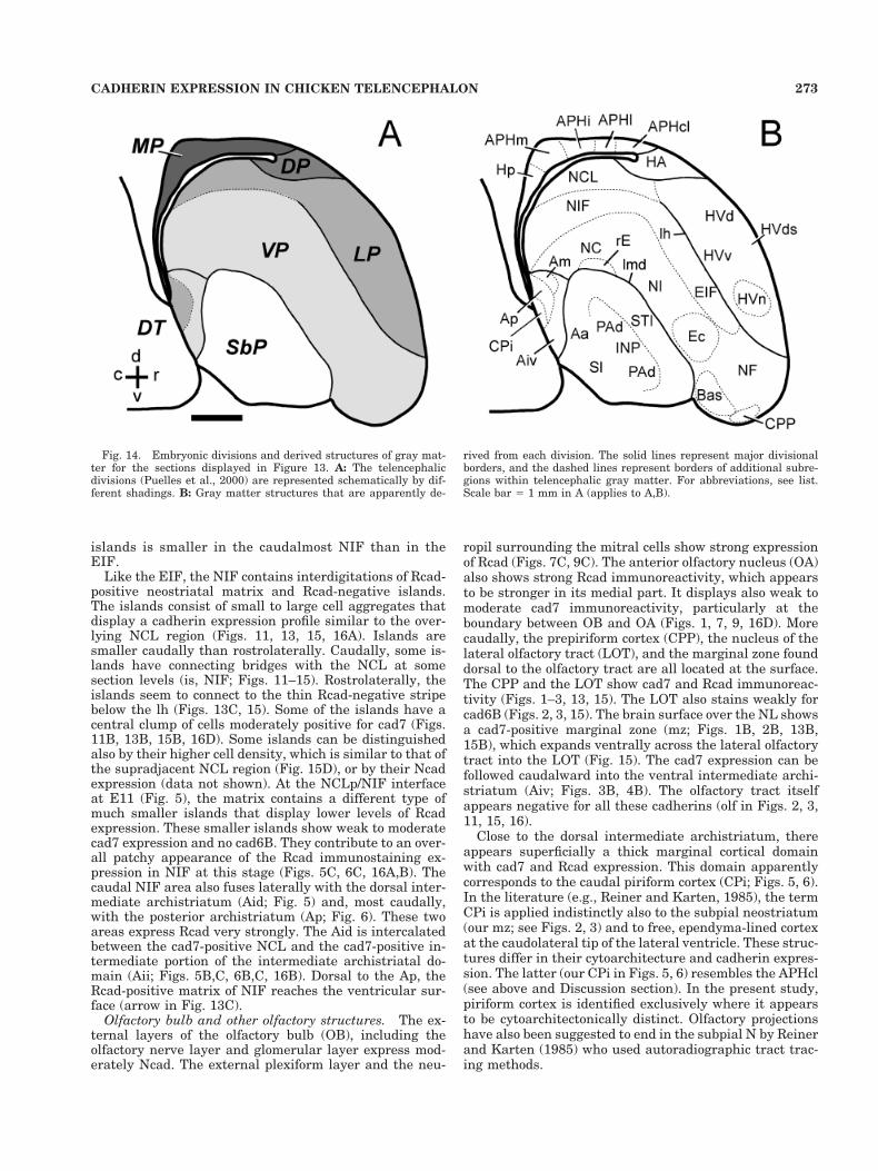
Fig. 8. Embryonic divisions and derived structures of gray matterfor the sections displayed in Figure 7. A: The telencephalic divisions(Puelles et al., 2000) are represented schematically by different shad-ings. B: Gray matter structures that are apparently derived from each
division. The solid lines represent divisional borders, and the dashedlines represent borders of additional subregions within telencephalicgray matter. For abbreviations, see list. Scale bar 5 1 mm in A(applies to A,B).
267CADHERIN EXPRESSION IN CHICKEN TELENCEPHALON

that the two sectors extend rostrally into the HVF andcaudally into the HVC.
The HVv is nonhomogeneous. Its neuronal cell densityincreases toward the hyperstriatal lamina (lh in Figs. 2, 9,
10, 13–16). On top of this boundary, there is a very denseband of cells that expresses moderate levels of cad7 andRcad (asterisk in Fig. 2F). Near the rostral brain surface,a well delimited, denser aggregate of HVv neurons is
Fig. 9. Cadherin expression in adjacent parasagittal sectionsthrough the telencephalon of the embryonic day 15 chicken at thelevel of the medial striatum (STm). Sections were immunostainedwith antibodies against cadherin-6B (cad6B, A), cadherin-7 (cad7, B),and R-cadherin (Rcad, C). D: Thionine (thio) staining of an adjacentsection. Corresponding schematic diagrams of the telencephalic em-
bryonic divisions and their derivative gray matter are shown in Fig-ure 10. A color-coded superposition of the immunostaining results isshown in Figure 16C. The asterisks indicate an artifact (cut at brainsurface). For abbreviations, see list. Scale bar 5 1 mm in D (applies toA–D).
268 C. REDIES ET AL.

negative for cad7 and Rcad. This nucleus (HVn in Figs.2A–E, 9A–D, 11A–D) contains only a few weakly cad7-positive scattered neurons and shows moderate Ncad andweak cad6B staining. The HVn overlies topographicallythe ectostriatum, a similar, though somewhat larger con-densation of cells in the neostriatum (Fig. 2). Apart fromthis lighter part, the rest of HVv displays medium tostrong cad7 expression and weak or moderate Rcad ex-pression (same figures). The boundary of the HVd with theHVv is best seen on cad7-immunostained parasagittalsections (Figs. 9B, 10B).
The superficial half of the intermediate stratum of theHV (as defined in the present work) shows only weakimmunoreactivity for Rcad (Fig. 4) and Ncad. Caudally, itlargely corresponds to the classic temporo-parieto-occipital area (TPO) that is found at section levels wherethe APHcl appears at the neighboring brain surface(Fig. 4F).
The marginal layer of the HVv and the HVF, found onlyrostrally, is rather cell poor and is populated by scatteredcells that express moderate levels of cad7 and Rcad (HVvsand HVFs in Figs. 1–3, 15, 16). The marginal layer of theHVd is more cell dense (HVds in Figs. 2, 3, 13, 14) anddisplays moderate cad7 expression.
Neostriatum and olfactory structures. The neostri-atum is a large ventral DVR territory that is separatedfrom the subpallium (the striatum, in particular) by thedorsal medullary lamina (lmd) and from the hyperstria-tum by the hyperstriatal lamina (lh). The lh limit is par-tially characterized by a dorsal-to-ventral drop in cad7expression (Figs. 2–4, 9). Rostrolaterally, the lh is alsounderlined by an Rcad-negative band (Figs. 2C, 3C, 11C,13C, 15C). The neostriatum extends rostrocaudallythroughout the telencephalon, and it includes several spe-
cialized separate sensory regions (basal nucleus, ectostria-tum, field L) and some superficial olfactory structures. Atthe rostral pial surface, these latter structures composethe nucleus of the lateral olfactory tract and the prepiri-form cortex (LOT, CPP, respectively; Figs. 1–3, 13–15). Atits rostral end, the neostriatum contacts the anterior ol-factory nucleus and the olfactory bulb (OA, OB; Figs. 9, 10;Puelles et al., 1999, 2000). Caudolaterally, the neostria-tum borders anterior parts of the archistriatal complex(Figs. 4–6, 15).
As a special added feature, the neostriatum containsmultiple islands of cells that differ in their cadherin ex-pression from the surrounding matrix. These islands varyin size and occur in regions that are either associated withthe ectostriatum (termed “ectostriatal island field”) orwith other parts of the neostriatum (termed “neostriatalisland field;” treated below).
Neostriatum. Standard terminology (Karten and Ho-dos, 1967) distinguishes frontal, intermediate, and caudaldivisions of the neostriatum (NF, NI, and NC, respectively).These terms are useful for topographical reference, butthe corresponding divisions show largely a common pat-tern of cytoarchitecture and cadherin immunoreactivity.For practical reasons, we will first describe a cross-sectionat the level of the ectostriatum (Fig. 3) and then follow thedistinctly stained areas rostrally and caudally.
A coronal section passing through the ectostriatumshows that the periventricular stratum of the intermedi-ate N (NIp) is characterized by lack of cad6B and cad7expression and weak to moderate Rcad expression at E11(Fig. 3A,B). Lateral to this stratum, there appears a do-main subdivided into dorsal and ventral sectors. The dor-sal sector is cad7 negative, whereas the ventral one is cad7positive (Figs. 3B, 4B). The ventral sector appears to cor-
Fig. 10. Embryonic divisions and derived structures of gray mat-ter for the sections displayed in Figure 9. A: The telencephalic divi-sions (Puelles et al., 2000) are represented schematically by differentshadings. B: Gray matter structures that are apparently derived from
each division. The solid lines represent divisional borders, and thedashed lines represent borders of additional subregions within telen-cephalic gray matter. For abbreviations, see list. Scale bar 5 1 mm inA (applies to A,B).
269CADHERIN EXPRESSION IN CHICKEN TELENCEPHALON

respond to the target in NI of the thalamic dorsolateralposterior nucleus (Wild, 1987b, 1989; Korzeniewska andGunturkun, 1990). Still more laterally, the neostriatum isenlarged and contains what may be called the “ectostriatalcomplex.” Here, the conventional ectostriatum, or ecto-striatal core (Ec), is surrounded by several other areas
that are related to it, at least topographically, but possiblyalso functionally. The Ec is a specialized visual thalamor-ecipient subregion of the NI (Karten and Hodos, 1970;Kroner and Gunturkun, 1999) that typically contacts thedorsal medullary lamina (lmd) and bulges inside the NIwithout reaching the hyperstriatal lamina (lh). It ex-
Fig. 11. Cadherin expression in adjacent parasagittal sectionsthrough the telencephalon of the embryonic day 15 chicken at thelevel of the innominate substance (SI). Sections were immunostainedwith antibodies against cadherin-6B (cad6B, A), cadherin-7 (cad7, B),and R-cadherin (Rcad, C). D: Thionine (thio) staining of an adjacent
section. Corresponding schematic diagrams of the telencephalic em-bryonic divisions and their derivative gray matter are shown in Fig-ure 12. The asterisk in A indicates an artifact. For abbreviations, seelist. Scale bar 5 1 mm in D (applies to A–D).
270 C. REDIES ET AL.

presses cad6B, cad7, and Rcad weakly or not at all (Figs.3, 11–15). This core area is surrounded medially by adistinct medial belt area with a similar cadherin expres-sion profile (mE; Figs. 2, 3). Dorsally and laterally, the Ecis covered by a cad7-negative periectostriatal belt area(pE) that strongly expresses Rcad. At some points, inter-digitations are observed between the Rcad-negative Ecand the Rcad-positive pE (Fig. 3C, arrows; Fig. 13C, is). AtE11, but not at E15, the pE shows also basophilic cellularclumps, presumably islands of aggregated neurons, thatare centered within the Rcad-negative digitations (arrowsin 3D). We have called this domain of pE the “ectostriatalisland field” (EIF; Figs. 2, 3, 11–15, 16C). The islands ofthe EIF express low to moderate levels of Ncad (only atE11), cad6B, and cad7. These markers are largely absentfrom the Ec and from the EIF matrix around the islands.In contrast, Rcad appears restricted to the matrix aroundthe islands. Rostrolateral to the Ec, a particularly largeand compact island is found, called here the “rostrolateralisland” (rlis; Figs. 3, 15, 16D).
Lateral to the EIF and close to the pial surface, thereappears a “lateral neostriatal region” that can be distin-guished by its very low levels of immunoreactivity for thefour cadherins studied. This lateral neostriatal region (NLin Figs. 1–3, 15) expands ventrally into a superficial do-main lying under the nucleus of the lateral olfactory tract(LOT; Figs. 2, 3; see below).
At more rostral section levels, the periventricular stra-tum of the N remains visible (NFp in Fig. 2), whereas thedeep intermediate domain gradually expands in front ofthe diminishing subpallium (NF in Fig. 2). Its ventralcad7-positive portion enlarges and acquires a more super-ficial position, encompassing the basal nucleus (Bas inFigs. 2, 11–14). The dorsal cad7-negative part also ex-pands slightly in front of the diminishing ectostriatal com-plex. The NL, together with the cad7-positive marginal
zone, caps rostrally the neostriatum, whereas the LOT issubstituted most rostrally by the prepiriform cortex (CPP)that expresses strongly both cad7 and Rcad (Figs. 1, 13–15, 16D).
Now tracing neostriatal regions caudalward from thelevel of the ectostriatum, we observe that the periventricu-lar stratum and associated cad7-positive intermediate do-main progressively increase in size, encompassing finallythe field L (L; Figs. 5–9, 16A–C). The different areas orzones of field L described in the literature (L1, L2, L3 ofBonke et al., 1979; Wild et al., 1993) can be discerned bytheir cadherin staining pattern at E15 but not at E11. AtE15, field L1 shows strong Rcad immunoreactivity butonly low to moderate levels of cad7 immunoreactivity (L1in Figs. 9, 16C). In contrast, field L2 shows strong cad7immunoreactivity and lacks the other cadherins (L2 inFigs. 9, 16C). Finally, field L3 expresses cad6B and cad7 atE15 (Figs. 9, 16C). The periventricular stratum of thecaudal neostriatum (NCp) expresses cad7 more stronglyat E15 than at E11 (Figs. 5B, 9B). Except for the ependy-mal layer, it is Rcad negative (Figs. 5C, 6C, 9C, 11C).
Laterally, the cad7-positive intermediate domain endsas a tail-like lateral extension behind the ectostriatum(Figs. 4–6). At this level, the outer intermediate domainoccupied before by the ectostriatal complex transformsinto two Rcad-positive areas. One of them is a flattenedcell aggregate that appears adjacent to the lmd. We pro-visionally named it the retro-ectostriatal nucleus (rE;Figs. 4, 13–15, 16D). Rcad staining in this region can befollowed back into the Rcad-positive medial part of thearchistriatum (Am in Figs. 5, 6, 15, 16A,B,D). The otherRcad-positive area is larger, and, like the EIF, it continuesshowing islands interspersed with the matrix. We havecalled it the “neostriatal island field” (NIF; Figs. 4–6,11–16; see below). The rE and NIF contact one anotherlaterally (Fig. 4C,F).
Fig. 12. Embryonic divisions and derived structures of gray mat-ter for the sections displayed in Figure 11. A: The telencephalicdivisions (Puelles et al., 2000) are represented schematically by dif-ferent shadings. B: Gray matter structures that are apparently de-
rived from each division. The solid lines represent major divisionalborders, and the dashed lines represent borders of additional subre-gions within telencephalic gray matter. For abbreviations, see list.Scale bar 5 1 mm in A (applies to A,B).
271CADHERIN EXPRESSION IN CHICKEN TELENCEPHALON

Neostriatal island field. This field represents a cau-dal continuation of the EIF, but it does not overlie anyobvious equivalent of the ectostriatal core. The neostria-tal island field (NIF) is not conspicuous in Nissl-stainedsections but can be clearly identified by its Rcad immu-
noreactivity, which labels its matrix cell populationeven more strongly than in the case of the EIF (Figs.4 – 6, 11–15, 16A,B,D). The NIF extends to caudal neo-striatal regions, where it reaches the caudal pole of theventricle (Figs. 11C, 13C). In general, the size of the
Fig. 13. Cadherin expression in adjacent parasagittal sectionsthrough the telencephalon of the embryonic day 15 chicken at thelevel of the medial archistriatum (Am). Sections were immunostainedwith antibodies against cadherin-6B (cad6B, A), cadherin-7 (cad7, B),and R-cadherin (Rcad, C). D: Thionine (thio) staining of an adjacentsection. Corresponding schematic diagrams of the telencephalic em-
bryonic divisions and their derivative gray matter are shown in Fig-ure 14. The arrow in C points to periventricular Rcad immunoreac-tivity that is continuous with the Rcad-immunoreactive matrix of theneostriatal island field (NIF). For abbreviations, see list. Scale bar 51 mm in D (applies to A–D).
272 C. REDIES ET AL.
islands is smaller in the caudalmost NIF than in theEIF.
Like the EIF, the NIF contains interdigitations of Rcad-positive neostriatal matrix and Rcad-negative islands.The islands consist of small to large cell aggregates thatdisplay a cadherin expression profile similar to the over-lying NCL region (Figs. 11, 13, 15, 16A). Islands aresmaller caudally than rostrolaterally. Caudally, some is-lands have connecting bridges with the NCL at somesection levels (is, NIF; Figs. 11–15). Rostrolaterally, theislands seem to connect to the thin Rcad-negative stripebelow the lh (Figs. 13C, 15). Some of the islands have acentral clump of cells moderately positive for cad7 (Figs.11B, 13B, 15B, 16D). Some islands can be distinguishedalso by their higher cell density, which is similar to that ofthe supradjacent NCL region (Fig. 15D), or by their Ncadexpression (data not shown). At the NCLp/NIF interfaceat E11 (Fig. 5), the matrix contains a different type ofmuch smaller islands that display lower levels of Rcadexpression. These smaller islands show weak to moderatecad7 expression and no cad6B. They contribute to an over-all patchy appearance of the Rcad immunostaining ex-pression in NIF at this stage (Figs. 5C, 6C, 16A,B). Thecaudal NIF area also fuses laterally with the dorsal inter-mediate archistriatum (Aid; Fig. 5) and, most caudally,with the posterior archistriatum (Ap; Fig. 6). These twoareas express Rcad very strongly. The Aid is intercalatedbetween the cad7-positive NCL and the cad7-positive in-termediate portion of the intermediate archistriatal do-main (Aii; Figs. 5B,C, 6B,C, 16B). Dorsal to the Ap, theRcad-positive matrix of NIF reaches the ventricular sur-face (arrow in Fig. 13C).
Olfactory bulb and other olfactory structures. The ex-ternal layers of the olfactory bulb (OB), including theolfactory nerve layer and glomerular layer express mod-erately Ncad. The external plexiform layer and the neu-
ropil surrounding the mitral cells show strong expressionof Rcad (Figs. 7C, 9C). The anterior olfactory nucleus (OA)also shows strong Rcad immunoreactivity, which appearsto be stronger in its medial part. It displays also weak tomoderate cad7 immunoreactivity, particularly at theboundary between OB and OA (Figs. 1, 7, 9, 16D). Morecaudally, the prepiriform cortex (CPP), the nucleus of thelateral olfactory tract (LOT), and the marginal zone founddorsal to the olfactory tract are all located at the surface.The CPP and the LOT show cad7 and Rcad immunoreac-tivity (Figs. 1–3, 13, 15). The LOT also stains weakly forcad6B (Figs. 2, 3, 15). The brain surface over the NL showsa cad7-positive marginal zone (mz; Figs. 1B, 2B, 13B,15B), which expands ventrally across the lateral olfactorytract into the LOT (Fig. 15). The cad7 expression can befollowed caudalward into the ventral intermediate archi-striatum (Aiv; Figs. 3B, 4B). The olfactory tract itselfappears negative for all these cadherins (olf in Figs. 2, 3,11, 15, 16).
Close to the dorsal intermediate archistriatum, thereappears superficially a thick marginal cortical domainwith cad7 and Rcad expression. This domain apparentlycorresponds to the caudal piriform cortex (CPi; Figs. 5, 6).In the literature (e.g., Reiner and Karten, 1985), the termCPi is applied indistinctly also to the subpial neostriatum(our mz; see Figs. 2, 3) and to free, ependyma-lined cortexat the caudolateral tip of the lateral ventricle. These struc-tures differ in their cytoarchitecture and cadherin expres-sion. The latter (our CPi in Figs. 5, 6) resembles the APHcl(see above and Discussion section). In the present study,piriform cortex is identified exclusively where it appearsto be cytoarchitectonically distinct. Olfactory projectionshave also been suggested to end in the subpial N by Reinerand Karten (1985) who used autoradiographic tract trac-ing methods.
Fig. 14. Embryonic divisions and derived structures of gray mat-ter for the sections displayed in Figure 13. A: The telencephalicdivisions (Puelles et al., 2000) are represented schematically by dif-ferent shadings. B: Gray matter structures that are apparently de-
rived from each division. The solid lines represent major divisionalborders, and the dashed lines represent borders of additional subre-gions within telencephalic gray matter. For abbreviations, see list.Scale bar 5 1 mm in A (applies to A,B).
273CADHERIN EXPRESSION IN CHICKEN TELENCEPHALON

Fig. 15. Cadherin expression in adjacent parasagittal sectionsthrough the telencephalon of the embryonic day 15 chicken at thelevel of the nucleus of the lateral olfactory tract (LOT). Sections wereimmunostained with antibodies against cadherin-6B (cad6B, A),cadherin-7 (cad7, B), and R-cadherin (Rcad, C). D: Thionine (thio)staining of an adjacent section. E: Schematic diagrams of the telen-cephalic embryonic divisions (represented by different shadings;Puelles et al., 2000). F: Gray matter structures that are apparently
derived from each division. A color-coded superposition of the immu-nostaining results is shown in Figure 16D. In E and F, the solid linesrepresent major divisional borders and the dashed lines representborders of additional subregions within telencephalic gray matter.The asterisks in A,C,F indicate the position of a cad6B-positive medialpart of the intermediate archistriatum. For abbreviations, see list.Scale bar 5 1 mm in E (applies to A–F).

Archistriatal complex. The archistriatal complex in-cludes anterior, intermediate, posterior, and medial divi-sions (Aa, Ai [divided into Aid, Aii, and Aiv], Ap, and Am,see Table 1). The identification of these divisions in thepresent study tentatively followed the adult schema pro-posed by Zeier and Karten (1971) but was adapted to the
slightly different topography of the archistriatum and itsparts observed at E11 and E15 (see also Puelles et al.,2000), as well as to the apparent limits of the differentdomains of cadherin expression. Some partial inconsisten-cies between our embryonic and the adult divisionalscheme may require further studies in the future.
Fig. 16. Color-coded overlays of immunostained sections forcadherin-6B (cad6B), cadherin-7 (cad7), and R-cadherin (Rcad). Theimages displayed in each panel are the result of the superposition ofadjacent sections. A: Results from adjacent transverse sectionsthrough the telencephalon of the embryonic day 11 chicken at thelevel of the anterior commissure. The data are the same as thoseshown in Figure 5A–C. B: Results from adjacent transverse sectionsthrough the telencephalon of the embryonic day 11 chicken at thelevel of the posterior part of field L (L). The data are the same as thoseshown in Figure 6A–C. C: Results from adjacent parasagittal sectionsthrough the telencephalon of the embryonic day 15 chicken at thelevel of the medial striatum (STm). The data are the same as thoseshown in Figure 9A–C. D: Results from adjacent parasagittal sections
through the telencephalon of the embryonic day 15 chicken at thelevel of the nucleus of the lateral olfactory tract (LOT). The data arethe same as those shown in Figure 15A–C. The different colors rep-resent the cadherin-immunostaining results, as indicated by theboxes in C. Note the partial overlap of cadherin expression indicatedby the mixed colors (pink for cad6B/cad7, turquoise for cad6B/Rcad,and yellow for cad7/Rcad). The lines represent the borders of embry-onic divisions and subregions of telencephalic gray matter (comparewith Figs. 5E,F, 6E,F, 10, and 15E,F). The asterisks in A,B,D indicatethe position of a cad6B-positive medial part of the intermediate arch-istriatum. For abbreviations, see list. Scale bars 5 0.5 mm in A,B, 1mm in C,D.
275CADHERIN EXPRESSION IN CHICKEN TELENCEPHALON

Fig. 17. Cadherin-6B expression in adjacent transverse sectionsthrough the telencephalon of the embryonic day 15 chicken at thelevel of the caudolateral neostriatum (NCL). A,C: A section that wasimmunostained with antibodies against cadherin-6B (cad6B). B: Thi-onine (thio) staining of an adjacent section. C represents an enlarge-
ment of the boxed area in A. The arrows in A and C point to cad6B-immunoreactive fibers that connect the NCL and the medial part ofthe intermediate archistriatum (Aim). For abbreviations, see list.Scale bars 5 0.5 mm in B (applies to A,B), 0.1 mm in C.
276 C. REDIES ET AL.

Both the Aid and Ap express Rcad very strongly andlack noticeable expression of cad7 and cad6B, whereas theAii lacks Rcad expression, but show moderate to strongcad7 immunoreactivity (Figs. 4–6, 15, 16A,B,D). TheRcad-positive Aid reaches the ventricular surface at thecaudolateral edge of the lateral ventricle (Fig. 6C). Withthe medially adjacent NCL, the Aid forms a border thatruns orthogonal to the ventricular surface.
The Aii contacts a medially protruding, rounded portion(Aim) that strongly expresses cad6B (asterisks in Figs. 5,6, 15, 16A,B,D, 17). This part of the Ai is connected to theoverlying cad6B-positive NCL by cad6B-positive nerve fi-bers (Fig. 17C). The medial nucleus (Am) and the ventralintermediate nucleus of the archistriatum (Aiv) are foundin topographic contiguity with the caudalmost NIF andNC domains. Similarly to the adjacent parts of the NIF,these nuclei of the archistriatum express Rcad strongly(Aiv) or even very strongly (Am). In these nuclei, cad7expression ranges from no expression to moderate expres-sion (Figs. 4–6, 13, 15).
The small Aa is found medially under the dorsal med-ullary lamina (Figs. 4, 13, 15). Like the adjacent subpallialregions, such as the striatum, the Aa is weakly immuno-reactive or negative for Rcad or cad7. The nucleus taeniae(Tn) is a superficial cell group positive for Rcad; it is foundmedial to the pallio-subpallial boundary at the caudal endof the archistriatum (Fig. 5). Puelles et al. (2000) recentlysuggested a possible extratelencephalic origin for this for-mation.
Solitary cad7-positive cells of the pallium. Apartfrom the regionalized expression of cad7 by cell aggre-gates, there are also solitary cad7-positive cells that arewidely dispersed at relatively regular intervals through-out most pallial regions. These cells have small to mediumcell nuclei, relatively little cytoplasm, and a few shortprocesses (data not shown). This type of cell is more widelydistributed in the pallium at E15 than at E11, and it israre in the subpallium. A similar type of cad7-positive cellwas not found in other parts of the brain (data not shown).
DISCUSSION
Regionalized and complementary expressionof cadherins in multiple telencephalic
(sub)divisions
Three of the four cadherins studied in the current work(cad6B, cad7, and Rcad) are expressed in a specific andcombinatorial manner in multiple areas widely distrib-uted over most of the major telencephalic (sub)divisions.Examples are the distinctive and sometimes complemen-tary expression patterns of cad7 and Rcad in the sensoryand/or thalamorecipient structures of the neostriatum(such as field L, ectostriatum, and basal nucleus, as wellas the specific target for the thalamic dorsolateral poste-rior nucleus in the intermediate neostriatum), in the mul-timodal association area called caudolateral neostriatum(NCL, Figs. 5, 6), or in the different areas of the avian(para)hippocampal complex (APH and Hp in Figs. 1–16).These data suggest an association of cadherin expressionto specific functional areas, aggregates, or nuclei of thetelencephalon. The current cadherin results also indicatethe existence of other gray matter structures not previ-ously described. Frequently, these structures are alsocharacterized by particular cytoarchitectonic features. A
typical example is the cell group in the HV, which we havecalled the nucleus of the ventral hyperstriatum (HVn; seebelow); it was first delimited by the expression of the threecadherins and corroborated cytoarchitectonically by thecompact aggregation of its cells (Fig. 2). Data reported forsongbirds and the parrot suggest the existence of an HNnucleus also in these adult brains (oval nucleus of HV:Striedter, 1994; Durand et al., 1997; Brauth et al., 1994;Hvo complex: Jarvis and Mello, 2000). More research isneeded to determine whether the HVn of the chickenembryo has an adult correlate and whether it is identicalto any of the HV elements in other birds. Other novelentities presented here are the NL, EIF, NIF, mE, NFv/d,HVs, rE, Aim, and some APH subdivisions (see below).Thus, cadherin expression is a useful tool for identifyingnovel areas or nuclei within the major divisions and sub-divisions of the telencephalon. It should be stressed thatwe did not assume cadherin expression alone to be suffi-cient to define any given gray matter region. Instead, acareful analysis of several features was carried out in eachcase by weighing results in light of additional data avail-able in the literature, especially when the cadherin ex-pression data suggested a novel divisional scheme or theexistence of a novel nucleus or area.
A similarly complex distribution of the same cadherintypes has been observed previously in the chicken dien-cephalon (Redies et al., 1993, 2000; Yoon et al., 2000), aswell as in the chicken mesencephalon and rhombenceph-alon (Arndt and Redies, 1996, 1998; Arndt et al., 1998;Wohrn et al., 1999; Redies et al., unpublished results).Other types of cadherins are also known to be expressed inspecific regions widely distributed throughout the brain inchicken (e.g., cadherin-10: Fushimi et al., 1997) andmouse (e.g., cadherin-8: Korematsu and Redies, 1997; cad-herin-6: Inoue et al., 1998; OL-cadherin: Hirano et al.,1999). Many of the gray matter structures expressing aspecific cadherin subtype are functionally connected(Redies et al., 1993; for review, see Redies, 2000). Thewidespread expression of Ncad by radial glia results in amore diffuse pattern of expression of this molecule, exceptfor a few areas displaying relatively high levels of Ncadexpression (Table 1).
The discrete expression of each cadherin subtype inmultiple brain divisions differs from the expression ofsome homeobox or regulatory genes (or the transcriptionfactors these genes codify), which frequently are restrictedto one or only a few of the major brain divisions or subdi-visions (Puelles and Rubenstein, 1993; Shimamura et al.,1995). For example, the neuronal expression of Emx-1 andTbr-1 is restricted to the whole pallium or to several of itsdivisions at early developmental stages in the mouse andin the chicken (Simeone et al., 1992; Smith-Fernandez etal., 1998; Puelles et al., 1999, 2000). Another example isthe expression of Dlx-2 and Nkx-2.1 which is restricted,early in development, to the entire subpallium (Dlx-2) orto one of its subdivisions (Nkx-2.1; Bulfone et al., 1993;Puelles and Rubenstein, 1993; Smith-Fernandez et al.,1998; Puelles et al., 1999, 2000). Such genes and tran-scription factors are good candidates for specifying gen-eral properties of major brain regions (e.g., general iden-tity and special differentiation features of cerebral cortex,basal ganglia, or thalamus).
277CADHERIN EXPRESSION IN CHICKEN TELENCEPHALON

Cadherin expression interpreted within amodel of topologically radial divisions in
chicken telencephalon
In the Results section, the immunoreactivity patternsfor the cadherins were described almost exclusively inconventional, atlas-derived terminology and subdivisions.Here, we interpret them also in the context of a recentlyproposed divisional scheme of the avian and mammaliantelencephalon (Puelles et al., 1999, 2000), as demon-strated in the schematic diagrams for each set of cadherinimmunostained sections (Figs. 1E,F–6E,F, 8, 10, 12, 14,15E,F) and supplementary diagrams (Fig. 18). This hypo-thetical divisional scheme was based on a comparativeanalysis of the expression of several gene transcriptionfactors in chicken and mouse embryos, as well as on dif-ferential hodology, in these and other vertebrate species(see also Smith-Fernandez et al., 1998). Our present anal-ysis neither proves nor disproves this scheme but ratherserves to point out possible alternative interpretations tomore conventional ones.
The scheme by Puelles et al. (1999, 2000) groups themajor pallial regions of classic and modern studies(Karten and Hodos, 1967; Reiner and Karten, 1983, 1985;Striedter, 1997; Dubbeldam, 1998) into the following fourmolecularly distinct histogenetic territories that are con-tinuous along the entire telencephalon (we indicate thecorresponding conventional avian telencephalic regions inparentheses): (1) the medial pallium (hippocampal andparahippocampal complexes), (2) the dorsal pallium (theWulst and other related corticoid regions), (3) the lateralpallium (the ventral hyperstriatum and associated corti-coid areas, including at least a posterior part of the piri-form olfactory cortex), and (4) the ventral pallium (theneostriatum, the olfactory bulb, the anterior olfactory nu-cleus, and the prepiriform olfactory cortical area). Thecaudal portions of the lateral and ventral pallium alsoinclude the dorsolateral and ventromedial parts of thearchistriatum, respectively, which are conceived as partsof the amygdaloid complex (Puelles et al., 1999, 2000).Note that, in classic terminology, the archistriatum formsthe posterior part of the dorsal ventricular ridge (DVR). Inthe proposed model, each of the pallial divisions repre-sents a histogenetic unit of the telencephalic wall thatradially extends from the ependymal to the pial surface.In the present work, we refer to such units as “radialunits.” By using this term, we do not wish to exclude thepossibility that minor cell populations migrate tangen-tially across the divisional boundaries, partially colonizingother radial units (e.g., see Anderson et al., 1997), and wedo not suggest that the radial glia, which help to define theunits, persist into adulthood (although they are visiblefrom the ventricle to the pia in proper sectioning planes atE11; Puelles unpublished observations). The same appliesto the striatal and pallidal subpallial portions, likewisedefined molecularly (Puelles et al., 1999, 2000).
As described in more detail below, some of the bound-aries between the postulated pallial and subpallial divi-sions coincide with abrupt changes in the cadherin expres-sion profile, even though this applies sometimes only toparts of their full extent, due to the regional expression ofthe cadherins (e.g., see cad7 expression along the lh).Other postulated boundaries do not coincide with changesin cadherin expression (see, e.g., the boundary betweenthe lateral and ventral pallium in the archistriatal com-
Fig. 18. Schematic representation of the embryonic divisions ofthe chicken telencephalon, as viewed from above (A, modified afterPuelles et al., 1999, 2000) and in a “flat-mount” projection of thetelencephalon (B). The telencephalon is composed of the subpallium(SbP) and the pallium. The two divisions are separated by the laminamedullaris dorsalis (lmd). The subpallium consists mainly of thestriatum (ST) and the pallidum (PA). The pallium can be furtherdivided into ventral pallium (VP), lateral pallium (LP), dorsal pallium(DP), and medial pallium (MP; see Discussion section). The divisionsare indicated by different shadings. For other abbreviations, see Table1 and list.
278 C. REDIES ET AL.

plex and in the area dorsally adjacent to it). The cadherinexpression profile varies characteristically within eachtelencephalic pallial or subpallial division, primarily ac-cording to strata superposed one upon another from theventricular to the pial surface (see Results section). Thecadherin expression profile secondarily relates to specificneuronal cell groups (layers or nuclei) within each stra-tum (see, e.g., sagittal sections in Fig. 9). Examples forthese types of staining patterns are discussed below foreach telencephalic division.
Pallium
Medial pallium. A comparison of the cadherin expres-sion profile to the cytoarchitecture of the medial palliumallowed us to identify four to five radial divisions, namely,the previously described Hp, plus at least three distinctradial divisions of APH, which we call APHm, APHi, andAPHl, and a possible novel one, APHcl. Within each divi-sion, both the cytoarchitecture and the cadherin expres-sion are layered in parallel to the ventricular and outersurfaces of the brain (for details, see Results section) andthe layers are oriented orthogonally to radial glial fibers inthe region (Striedter and Beydler, 1997). These findingsare consistent with the notion that such divisions repre-sent true radial subunits and, thus, cytoarchitectonicallydistinct areal subdivisions of the medial pallial cortex. TheAPHm, APHi, and APHl divisions can be followed fromrostromedial to caudolateral areas of the medial pallium,as reported in the classic literature (see below). Moreover,at caudal levels, a portion of the piriform cortex maycoincide partly with the APHcl (see Figs. 5, 6, 15, 18),which is distinguished here from the neighboring caudaldorsolateral corticoid area (Reiner and Karten, 1985).
Architectonic subdivisions of the avian medial palliumwere noted by several previous authors (Craigie, 1935,1939, 1940; Bures et al., 1960; Kuhlenbeck, 1938, 1973).With regard to Craigie’s classification, which seems at theroot of the other versions, it should be noted that heincluded our APHm as subfield H4 of his four hippocampalsubfields H1–H4, whereas our APHi possibly includes hisparts a, b, and c, and our APHl then corresponds to part dof his parahippocampal cortex. The additional chemoar-chitectonic and hodologic subdivisions proposed by Krebsand collaborators (Krebs et al., 1991; Montagnese et al.,1993, 1996; Erichsen et al., 1994; Szekely and Krebs,1996) did not aim to represent or construe histogeneticradial units, but can be reduced easily to the presentdivisional model, thus suggesting a fundamental consen-sus. The dorsomedial subdivision of these authors corre-sponds to the superficial layers of APHm. If the underly-ing strata that compose the area 4 and the dorsal part ofthe area 3 of Erichsen et al. (1991) are added to thisdorsomedial subdivision, the full radial unit of our APHmis constituted. Note that this correlates also with a radialdomain defined by a minimal density of neurotensin bind-ing sites (Brauth et al., 1986; their Figs. 5, 6). The rest ofthe dorsolateral subdivision of Krebs and collaboratorscorrelates with our APHi, whereas their ventral subdivi-sion (area 2 and ventral part of area 3 of Erichsen et al.,1991) coincides with our Hp. The area 7 of Erichsen et al.(1991) apparently coincides with our APHl. Interestingly,both the Hp and APHi areas stand out by the density ofzinc-containing neuropil, whereas APHm and APHl arelargely negative for this marker (Faber et al., 1989).
Dorsal pallium (Wulst). Based on its distinct cad-herin expression profile and cytoarchitecture, the dorsalpallium can be clearly distinguished from the mediallyand laterally adjacent divisions of the pallium (Figs. 2–4,9–15), which encroach upon each other not only caudo-laterally, but also at the rostral end of the brain (Fig. 18;see also Puelles et al., 2000). The HD, HIS, and HA sub-divisions of the dorsal pallium (or Wulst) have character-istic cadherin expression profiles and cytoarchitecture (seeResults section). Their boundaries can be followed fromthe periventricular stratum to the brain surface, consis-tent with the suggestion that these subdivisions representseparate radial units (Medina and Reiner, 2000) ratherthan cortical laminae (Shimizu and Karten, 1990; Kartenand Shimizu, 1991).
At the caudal transition from the Wulst to the APH, theCDL corticoid domain becomes apparent at the surface ofthe dorsal pallium. It is intercalated between the caudalsuperficial part of the HV and the APHl and APHcl areas,and it overlies the lateral angle of the lateral ventricle justas it starts to expand caudolaterally (Fig. 4D; comparewith the area labeled “HA?” in Fig. 10n of Puelles et al.,2000). In the Results section, we have argued that theclassic CDL seems heterogeneous and apparently consistsof obliquely superposed medial and dorsal pallial parts.We propose that the name CDL be reserved for the dorso-pallial derivative, which disappears at more caudal levelsof the telencephalon, where the lateral pallium (our CPi)comes to be in direct contact with the medial pallium(APHcl; Fig. 5).
Ventral and lateral pallium. The cadherin expres-sion profile allows a clear distinction between the lateraland ventral pallium from rostral to caudal levels of thetelencephalon, even at the level of the island fields (seebelow); thus, it can be used for delineating the boundarybetween the two pallial territories. The expression profileshows only minor differences at the frontal, intermediate,and caudal portions of the ventral and lateral pallial sub-divisions (N and HV, respectively). At all levels, cad7typically labels moderately to strongly the deep and inter-mediate portions of the ventral hyperstriatum (HV, in thelateral pallium) whereas the entire HV shows weak tomoderate Rcad immunostaining. In contrast, most of theventral pallium (neostriatum) represents a patchwork ofareas that express Rcad or cad7, in a complementarymanner. This patchiness is especially prominent in theisland fields (EIF and NIF) that extend continuously fromthe caudal periventricular area to the rostrolateral telen-cephalic surface (Figs. 11–15).
The quantitative architectonic studies of Rehkamper etal. (1984, 1985) are, in our view, largely coincident withthe present interpretation of the boundaries and the ex-tension of the lateral and ventral pallial territories (seebelow). Both pallial subdivisions have their maximalperiventricular and ventricular zone representations cau-domedially in the hemisphere, from where their radialdimension extends rostrolaterally to the brain surface, asbest seen on parasagittal sections (Figs. 7–15), and onradial glial preparations (Striedter and Beydler, 1997; L.Puelles, unpublished data).
Caudolateral neostriatum. The boundary between thelateral and ventral pallial divisions at the caudal telen-cephalon has been less well-defined in the literature. Thisis partly because the hyperstriatal lamina (lh), a relativelycell-free landmark that separates the ventral hyperstria-
279CADHERIN EXPRESSION IN CHICKEN TELENCEPHALON

tum from the neostriatum, is clearly visible only at rostraland intermediate levels of the telencephalon and cannotbe observed laterally at caudal levels (Karten and Hodos,1967). The absence of this histologic landmark, togetherwith the restricted distribution of some neuropeptide re-ceptors in the HV and medialmost caudal regions (Wacht-ler, 1985; Brauth et al., 1986; Reiner et al., 1989, 1994;Durstewitz et al., 1998, 1999) have led to the widespreadnotion that the caudal part of the HV is restricted only tothe medial periventricular region (for example, see Reineret al., 1989; Wild et al., 1993; Veenman et al., 1995;Metzger et al., 1996; Kroner and Gunturkun, 1999;Metzdorf et al., 1999). All the rest of the caudal pole of thedorsal ventricular ridge, bridging the space from the HVto the lateropallial part of the archistriatum and theneighboring CPi, has been interpreted, therefore, as “cau-dolateral neostriatum” (NCL).
However, there is developmental, architectonic, or hod-ologic evidence that the NCL also shares features withlateropallial structures. First, during embryogenesis, theNCL expresses the gene Emx1 that is typically absent inthe ventral pallium, or neostriatum (N) (Puelles et al.,1999, 2000), although this marker is partially down-regulated at more advanced stages (Fig. 10p of Puelles etal., 2000). Likewise, cad7 expression at E11 is continuousalong the entire periventricular HV and extends into theNCL area (Figs. 4B–6B). Second, data on cell densitymeasurements by Rehkamper et al. (1984) indicate thatthe caudal HV, if restricted to the medial part as definedin the literature, only correlates with characteristics of theperiventricular HV at more rostral levels, where interme-diate to superficial areas of HV are significantly less dense(their Figs. 1, 5, 7, 8). Moreover, the study by Rehkamperet al. (1985) of the pigeon neostriatum revealed a similar-ity in cell density and myeloarchitecture between the cau-dal HV and the caudal periventricular region of the neos-triatum (our NCLp in our Fig. 5; their area Ne15 in theirFigs. 7–9). Zeier and Karten (1973), in their experimentalanalysis of connections of the pigeon anterior commissure,illustrated dense terminals over the rostral superficialstratum of the HV, which they misinterpreted as the HIS(HIS is supposed to lie medial to HD, not lateral to it; seetheir Figs. 2, 3, 4; Medina and Reiner, 2000). This projec-tion domain is clearly continuous caudolaterally with anunidentified area that lies dorsal to the archistriatum andprecisely correlates with our NCL (their Figs. 2–4). Fi-nally, a mapping of zinc in the chicken brain (Faber et al.,1989) shows continuity of dense zinc-containing neuropilfrom the rostral and intermediate HV (lateral pallium)into our NCL domain (their Fig. 1D–K).
In several aspects, the avian NCL appears comparableto an amygdaloid nucleus in reptiles called the dorsolat-eral amygdala (DLA; Lanuza, 1997). First, both the avianNCL and reptilian DLA are ventrally adjacent to thecaudolateral edge of the telencephalic ventricle, and bothappear to be a caudal continuation of the dorsolateralDVR. In reptiles, this part of DVR has been suggested tobe part of the lateropallial domain and comparable to partof the avian HV (Guirado et al., 2000). Moreover, bothNCL and DLA show a dense dopaminergic and cholinergicinnervation and/or high acetylcholinesterase activity(Smeets et al., 1986; Medina et al., 1993; Metzger et al.,1996; Lanuza, 1997; Lanuza et al., 1998; Kroner and Gun-turkun, 1999; Riters et al., 1999; Puelles, unpublishedobservations in the chicken). Finally, both structures re-
ceive a great variety of information from various pallialregions (limbic, sensory, and motor input) and project tothe striatum and to major amygdaloid output centers(archistriatum in birds; Lanuza et al., 1998; Metzger etal., 1998; Kroner and Gunturkun, 1999). Interestingly, thereptilian DLA has been suggested to be comparable to themammalian basolateral amygdala, precisely according tothe similarities pointed out above in sauropsids. There-fore, the same could be true of NCL (Lanuza, 1997;Lanuza et al., 1998). The basolateral amygdala is a deriv-ative of the lateropallial domain of mammals, character-ized by its expression of the gene Emx1 during develop-ment (Puelles et al., 2000).
However, as noted above, many neurochemical featuresof the NCL indicate that this structure is in some waysdifferent from the periventricular HVC (e.g., see Brauth etal., 1986; Reiner et al., 1989). This may be explained bythe dispersion of cell clones along the caudorostral radialdimension within the DVR (Szele and Cepko, 1996). Analternative view that is consistent with the present resultsas well as with the previously published neurochemicaldata is that NCL may represent the caudal part of a radialdomain that is intercalated between the ventropallialneostriatum and the lateropallial HV. This domain, notcontemplated in Puelles et al. (2000), would be character-ized by a lack of Rcad expression and weak to moderatecad6B and cad7 expression. Rostrolaterally, it would thinout and underline the lh, as best seen on parasagittalsections (see Figs. 9C, 11C). Whether the NCL belongs tothe (ventropallial) neostriatum, or to a specialized caudalpart of the (lateropallial) HV, or forms a third independentdomain, which is intercalated between the neostriatumand the HV, remains unclear at present. To account forthe provisional status of this issue, we traced the bordersof the NCL by dotted lines in the schematic diagrams.Further studies are needed to evaluate the merits of eachof the three possibilities considered here.
One novel neostriatal domain observed in the Rcad-immunostained preparations lies at the caudal confluenceof the NIF (see below) with the medial archistriatum andthe pallio-subpallial boundary. This domain forms a spike-shaped area that diverts ventromedially from the NIF andadheres to the lmd at the back of the ectostriatum. Thisstrikingly distinct area, provisionally named here theretro-ectostriatal nucleus (rE), presently lacks any corre-lation with published connectivity data but possibly coin-cides with a domain highlighted in mappings of musca-rinic cholinergic receptors in the pigeon (Wachtler, 1985;his Fig. 1e; compare with our Fig. 4; Kohler et al., 1995,his Fig. 1f).
The anterior olfactory nucleus and the olfactory bulbhave a cadherin expression profile that supports inclusionamong ventral pallial structures, as was likewise con-cluded by mapping the expression of gene transcriptionfactors (Puelles et al., 1999, 2000). Both structures, there-fore, are considered here the rostralmost representation ofthe ventral pallium.
The tridimensional extent of the ventral and lateralpallia, as shown in the current study and by Puelles et al.(1999, 2000), corresponds well to the course of the radialglia in these subdivisions (Striedter and Beydler, 1997; L.Puelles, unpublished data). Moreover, assuming that ra-dial glia serve as a scaffold for neuronal migration pat-terns (Rakic, 1988; Nieuwenhuys et al., 1998), these re-sults are also in agreement with the predominantly
280 C. REDIES ET AL.

oblique, caudorostral dispersion of cell clones within theneostriatum and within the ventral hyperstriatum, re-spectively (Szele and Cepko, 1996; Striedter et al., 1998).Together, these studies suggest that the ventral and lat-eral pallia (similarly to the medial and dorsal pallia) rep-resent two adjacent but separate histogenetic fields, irre-spective of the potential ingression of tangentiallymigrated cellular cohorts (Cobos, 2000).
Island fields. At intermediate and caudal levels, a largelateral part of the ventral pallium is formed by what wehere call the neostriatal island field (NIF), supplementedrostrally by the ectostriatal island field (EIF). The islandfields are characterized by the presence of small island-like, sometimes dense, cellular aggregates located in thedorsal neostriatum and ectostriatal belt area. Rostrolat-erally, the islands become larger. The islands show acadherin expression profile and cytoarchitecture similar tothat found in the neighboring NCL domain (and alsopartly similar to the subjacent ectostriatal core), whereasthe surrounding matrix is characterized by Rcad expres-sion (for details, see Results section). A highly patchydistribution of cadherin expression in this area has beendescribed before for Rcad and cadherin-10 but was notcommented upon (see Fig. 4 in Arndt and Redies, 1996;and Fig. 11B in Fushimi et al., 1997; respectively). Asimilarly patchy expression pattern was found forcadherin-8 in the matrix of the striatum of the neonatalrat (Korematsu et al., 1998) and for cad7 in the caudo-lateral part of the E15 parahippocampal area (this study).
Like the ventral and lateral pallial divisions, the Rcad-positive matrix and the Rcad-negative islands extend ascomplete radial units from periventricular regions at cau-dal neostriatal levels (Fig. 11) to the rostrolateral pialsurface at intermediate neostriatal levels (Figs. 3, 15).One possible interpretation of the immunostaining resultsis that at least some islands represent interdigitated ra-dial domains that exhibit differential cadherin expression,as is suggested by the occasional cellular bridges thatconnect some islands to the overlying neostriatal or NCLareas. As an alternative or supplementary explanation,similar clumps of cells might be produced by differentialaggregation of cellular cohorts that originate in the sameembryonic subdivision, but whose cadherin expression dif-ferentially correlates with specific birthdates. Birthdate-related cell aggregates analogous in shape and position toour islands have been reported recently in the neostria-tum (and the HV) of the chicken (Striedter and Keefer,2000). These cell groups might express different adhesiveproperties related to their time origin and, thus, segregateinto cellular aggregates in the matrix environment. Suchhomotypic cell sorting and aggregation of cell populationsthat differentially express cadherin subtypes has beendemonstrated repeatedly in vitro and in vivo (for review,see Redies, 2000). The resulting patterns of cell mixing(e.g., whether layers, aggregates, or islands are formed)are likely to depend on the relative adhesive strengths andcell number ratios of the mixed cell populations (for re-view, see Steinberg, 1970; Redies, 2000), as well as on theextent of heterotypic interactions between cells expressingdifferent cadherin subtypes (Shimoyama et al., 2000).
Subpallium
The cadherin expression profile also may be useful fordelineating better the boundary between the major subdi-visions of the subpallium, i.e., between the striatum and
the pallidum, especially in the periventricular area. Here,a large region termed the parolfactory lobe (LPO) wasclassically thought to represent the medial part of theavian striatum. This assumption was based on numerousneurochemical features and connectivity data. For exam-ple, a similarity of the LPO to the mammalian striatumhas been suggested by its high acetylcholinesterase activ-ity, dense dopaminergic innervation and large number ofcells and fibers containing the neuropeptides substance Por enkephalin, as well as by reciprocal connections withthe avian ventral tegmental area and substantia nigra(Karten and Dubbeldam, 1973; Reiner et al., 1984, 1998;Medina and Reiner, 1995). However, the LPO is chemi-cally heterogeneous at caudal levels, where the avian dor-sal pallidum (paleostriatum primitivum) appears later-ally. At these caudal levels, the LPO typically shows adorsal part that is laterally continuous with the avianlateral striatum (paleostriatum augmentatum) and a ven-tral part that is located medial to the dorsal pallidum (forexample, see Fig. 1B in Karle et al., 1996). During embry-onic development, the caudoventral part of LPO expressesthe gene Nkx2.1, which is a pallidal marker in tetrapods(Puelles et al., 1999, 2000). This expression persists atleast until E14.5 (Puelles, unpublished observations) andmay well be present in the adult pallidum in Xenopus (O.Marın, personal communication; Marın et al., 1998). Con-sequently, the ventral part of LPO was termed here the“medial pallidum” (PAm in Figs. 3, 4), even though theliterature usually interprets this area as homogeneouslystriatal. The change of periventricular cadherin expres-sion in this region (Fig. 3A–C) possibly relates to the limitbetween the striatal and pallidal periventricular parts ofthe LPO. Note that, in general, many cells in the avianand mammalian pallidum have neurochemical character-istics typical of striatal cells (see, e.g., Sun and Reiner,2000). The striatal-like features found in the caudoventralLPO (PAm) in the adult may be due to the apparentpresence of a mixed population of both pallidal-type andstriatal-type cells in this region. Whether the striatal-typecells originate in striatal (Nkx2.1 negative) or pallidal(Nkx2.1 positive) neuroepithelium remains unclear atpresent.
Archistriatum
The expression of transcription factors (Puelles et al.,1999, 2000) indicate that the ventral and lateral pallialdivisions as well as the subpallium can be followed to thecaudal and ventrolateral pole of the telencephalon, invad-ing the archistriatum, where each of the correspondingsubdivisions consists of specific nuclei. Several of thesearchistriatal nuclei differentially express cadherins andshow a characteristic cytoarchitecture (Figs. 16, 18;Table 1).
Lateropallial subdivision. Similar to neighboringparts of the lateral pallium, this subdivision of the arch-istriatum (which includes Aid, Aii, and Ap) generallyshows mantle expression of the pallial genes Tbr1, Emx1,and ependymal expression of Pax6 (Puelles et al., 1999,2000). Some of the nuclei in this subdivision (with theexception of Aid) show also strong cad7 immunoreactivityand moderate or weak staining for Rcad (Figs. 4–6).
Ventropallial subdivison. Similar to the adjacentparts of the ventral pallium, this subdivision of the arch-istriatum (which includes Aiv and Am) generally containshigher levels of Rcad expression, whereas cad7 expression
281CADHERIN EXPRESSION IN CHICKEN TELENCEPHALON

is relatively low (Figs. 4–6). Like the ventral pallium, thisarchistriatal region expresses the pallial marker geneTbr1 but lacks expression of Emx1, except at the pialsurface (Puelles et al., 1999, 2000).
Subpallial subdivision. The anterior archistriatum(Aa) shows a cadherin expression profile that resemblesmore the adjacent striatum than the neighboring ventro-pallial archistriatum (Figs. 4–6). The subpallial nature ofAa, which was postulated on molecular marker evidence(notably the abundance of migrated Pax-6–positive cells)by Puelles et al. (2000), awaits corroboration with othercomparative methods.
It is appropriate to point out that some of the postulateddivisional boundaries in the archistriatum do not coincidewith abrupt changes in cadherin expression. For example,there is a prominent continuity of Rcad expression fromAid (postulated lateropallial subdivision) into NIF and Ap(postulated ventropallial subdivision). Moreover, the Rcadimmunoreactivity in Aid seems to extend to the ventricu-lar surface just medial to the lateral edge of the lateralventricle. This unique pattern coincides with a similarband of Emx-1 expression in E8 embryos, which seems toseparate the archistriatum from the lateral pallium (Figs.10i,j in Puelles et al., 2000). These inconsistencies of cad-herin staining with the postulated pallial divisional bor-ders will require further analysis. Given their provisionalstatus, we indicated these borders by dotted lines in theschematic diagrams.
Relation of cadherin expression to specificfunctional cell groups and neural circuits
The functional systems and neural circuits of the braintypically consist of several interconnected neuronal popu-lations or gray matter areas, which are distributed in aparticular way through the brain, following usually highlyspecific rules of connectivity. The cell populations partic-ipating in a given circuit are often derived from several ofthe major (sub)divisions of the vertebrate brain. The de-velopment of such complex neural circuitry requires con-nectional chemospecificity for axonal navigation, contactguidance and target recognition. Pioneering axons proba-bly use different cues than axons following their trail. Thedistribution of cadherins over the brain is widespread,although selective. This distribution includes the expres-sion by developing fiber tracts, apart from the expressionby specific cell groups (Table 1). The capacity of cadherinsto foster homotypic binding interactions between fascicu-lating axons, neural cells (aggregation and segregation),or both, have led to their being postulated as being one ofseveral possible regulators that build or stabilize specificneural circuits (for review, see Redies, 1995, 2000). This ispossibly achieved by way of specific matching of growingaxons, axonal pathways and target cells that express thesame combination of cadherins (Redies et al., 1993; Arndtand Redies, 1996; Fannon and Colman, 1996; Wohrn etal., 1998, 1999; Huntley and Benson, 1999; for review, seeRedies, 1995, 2000). Although many other types of mole-cules are thought to also participate in circuitry develop-ment at specific brain locations, many of them do not showcorrelative expression along given pathways (for review,see Tessier-Lavigne and Goodman, 1996; O’Leary andWilkinson, 1999; Raper, 2000).
Previous studies on the expression of cad6B, cad7, andRcad in chicken brain (Redies et al., 1993, 2000; Arndt andRedies, 1996; Wohrn et al., 1998; Arndt et al., 1998) dem-
onstrated a relation of each of these cadherins (or combi-nations of them, see Wohrn et al., 1999) to specific neuralcircuits. These studies, together with the current data inthe telencephalon, suggest the possibility that the samecadherins may be related also to specific thalamopallial,intratelencephalic and basal ganglia circuits of thechicken embryo, as outlined below. It should be stressedthat this hypothesis remains an extrapolation of findingsfrom our previous studies on the diencephalon and therhombencephalon (same references as above), to thepresent telencephalic data. To prove that telencephalicneural circuits differentially express specific cadherins,more detailed studies will be required. At the present levelof analysis, though, we scarcely found clear-cut examplesof mismatches in cadherin expression between specificprojections and their target areas. One such exception ispossibly found in the olfactory system, where neither theolfactory tract nor the mitral cells of the olfactory bulbexpressed any of the cadherins studied, although theprepiriform cortex strongly expresses cad7 and Rcad, asdoes the nucleus of the lateral olfactory tract and themarginal zone of the neostriatum. These are areas re-ported to receive olfactory projections (Reiner and Karten,1985).
Based on the cadherin expression profile in specific dor-sal thalamic nuclei (Redies et al., 2000) and in their tel-encephalic targets, the thalamopallial circuits appear todifferentially express the three cadherins. These circuitsinclude (1) the multimodal pathway from the thalamicdorsolateral posterior nucleus (DLP) to the thalamorecipi-ent area of the NI (Gamlin and Cohen, 1986; Wild, 1994;Kroner and Gunturkun, 1999), which are apparently as-sociated to cad7 or Rcad; (2) the auditory projections fromthe nucleus ovoidalis and some other periovoidal nuclei tofield L and its subdivisions (Wild et al., 1993; Kroner andGunturkun, 1999), which are apparently associated tocad7 or Rcad; (3) the visual pathway from the nucleusrotundus (part of the intermediate dorsal thalamic tier;Redies et al., 2000) to the ectostriatum and ectostriatalbelt, including to the EIF region demonstrated in thepresent study (Revzin and Karten, 1967; Karten and Ho-dos, 1970; Kroner and Gunturkun, 1999), which are ap-parently associated to cad6B, cad7, or Rcad; (4) the telen-cephalic projections from “dorsal tier” thalamic nuclei (inthe sense of Redies et al., 2000) to the Wulst, such as thosefrom the lateral part of the dorsolateral nucleus (Karten etal., 1973; Shimizu and Karten, 1990), the dorsal interme-diate ventral anterior nucleus (Wild, 1987a, 1989; Korze-niewska and Gunturkun, 1990; Kroner and Gunturkun,1999), and the ventrointermediate area (Medina andReiner, 1997; Medina et al., 1997), which are apparentlyassociated to cad7 or Rcad. A relation of cadherins tospecific thalamopallial pathways has also been suggestedin mammals (Huntley and Benson, 1999; Obst-Pernberget al., 2001).
The current results show that cadherins are expressedin a restricted and partially complementary manner alsoin many structures related to specific intratelencephaliccircuits. One particularly clear example of the associationof a cadherin subtype to an intratelencephalic pathway isthe projection of a specific region in the NCL to a restrictedmedial area of the intermediate archistriatum (Aim).These two structures as well as the fiber fascicles connect-ing them express cad6B (Fig. 17C). The intermediate arch-istriatum, in turn, projects to extratelencephalic targets
282 C. REDIES ET AL.

(Wild et al., 1993; Wild, 1994; Kroner and Gunturkun,1999). It is, thus, conceivable that these structures corre-spond to the high vocal center, the nucleus robustus arch-istriatalis and their connection, respectively, in songbirds(Nottebohm et al., 1976; Vicario, 1993; Wild, 1994). Thispossibility remains to be confirmed by a future, compara-tive study mapping the expression of cad6B and othermolecular markers.
Moreover, in the basal ganglia circuits, two cadherins(Rcad and cad6B) are related to particular groups of pal-lidal neurons. For example, the medial part of the dorsalpallidum contains cad6B-positive neurons, whereas theintermediate and lateral parts of the dorsal pallidum con-tain neurons expressing Rcad. The dorsal pallidum ofbirds is known to project to several targets in the subthal-amus, the ventral and dorsal thalamus, the pretectum,and the diencephalic and midbrain tegmentum (Kartenand Dubbeldam, 1973; Reiner et al., 1982, 1984; Medinaand Reiner, 1997; Medina et al., 1997). As shown previ-ously (Arndt and Redies, 1996; Redies et al., 2000), someof these targets express Rcad, such as the avian subtha-lamic nucleus (anterior nucleus of the ansa lenticularis),the ventrointermediate area (in the dorsal thalamus), aventral part of the dorsointermediate posterior nucleus (inthe dorsal thalamus), and a large part of the lateral spiri-form nucleus (in the pretectum). Other targets of thedorsal pallidum express cad6B, e.g., parts of the lateralspiriform nucleus (Redies et al., 2000).
ACKNOWLEDGMENTS
The authors thank Meike Ast for expert technical assis-tance, Shinichi Nakagawa and Masatoshi Takeichi forgenerous gifts of antibodies, Reimund Duchting for helpwith photography, Min-Suk Yoon and Min Jeong Ju forassistance in preparing the figures, and G. Striedter forsharing unpublished data. C.R. received support from theDeutsche Forschungsgemeinschaft; L.M. from the SenecaFoundation; L.M. and L.P. from the MCYT and CICYTGovernmental Grant Agency, Madrid; C.R. and L.P. fromthe Spanish Ministerio de Relaciones Exteriores and Ger-man Academic Exchange Service; and L.P. from the Eu-ropean Community BIOTECH Program.
LITERATURE CITED
Anderson SA, Eisenstat DD, Shi L, Rubenstein JLR. 1997. Interneuronmigration from basal forebrain to neocortex: dependence on Dlx genes.Science 278:474–476.
Arndt K, Redies C. 1998. Development of cadherin-defined parasagittalsubdivisions in the embryonic chicken cerebellum. J Comp Neurol401:367–381.
Arndt K, Redies C. 1996. Restricted expression of R-cadherin by brainnuclei and neural circuits of the developing chicken brain. J CompNeurol 373:373–399.
Arndt K, Nakagawa S, Takeichi M, Redies C. 1998. Cadherin-definedsegments and parasagittal cell ribbons in the developing chicken cer-ebellum. Mol Cell Neurosci 10:211–228.
Aste N, Balthazart J, Absil P, Grossmann R, Mulhbauer E, Viglietti-Panzica C, Panzica GC. 1998. Anatomical and neurochemical definitionof the nucleus of the stria terminalis in Japanese quail (Coturnixjaponica). J Comp Neurol 396:141–157.
Bixby JL, Zhang R. 1990. Purified N-cadherin is a potent substrate for therapid induction of neurite outgrowth. J Cell Biol 110:1253–1260.
Bonke BA, Bonke D, Scheich H. 1979. Connectivity of the auditory fore-brain nuclei in the guinea fowl (Numida meleagris). Cell Tissue Res200:101–121.
Brauth SE, Kitt CA, Reiner A, Quirion R. 1986. Neurotensin binding sitesin the forebrain and midbrain of the pigeon. J Comp Neurol 253:358–373.
Brauth SE, Heaton JT, Durand SE, Liang W, Hall WS. 1994. Functionalanatomy of forebrain auditory pathways in the budgerigar (Melopsit-tacus undulatus). Brain Behav Evol 44:210–233.
Bulfone A, Puelles L, Porteus MH, Frohmann MA, Martin GR, RubensteinJLR. 1993. Spatially restricted expression of Dlx-1, Dlx-2 (Tes-1),Gbx-2, and Wnt-3 in the embryonic day 12.5 mouse forebrain definespotential transverse and longitudinal segmental boundaries. J Neuro-sci 13:3155–3172.
Bures J, Fifkova E, Marsala J. 1960. Leao’s spreading depression in pi-geons. J Comp Neurol 114:1–10.
Cobos I. 2000. Morfogenesis e histogenesis del telencefalo: Estudio exper-imental en embriones de pollo. University of Murcia.
Craigie EH. 1935. The hippocampal and parahippocampal cortex of theemu (Dromiceus). J Comp Neurol 61:563–591.
Craigie EH. 1939. The cerebral cortex of Rhea americana. J Comp Neurol70:331–353.
Craigie EH. 1940. The cerebral cortex of palaeognathine and neognathinebirds. J Comp Neurol 73:179–234.
Dubbeldam JL. 1998. Birds. In: Nieuwenhuys R, ten Donkelaar HJ, Ni-cholson C, editors. The central nervous system of vertebrates. Berlin:Springer. p 1525–1636.
Durand SE, Heaton JT, Amateau SK, Brauth SE. 1997. Vocal controlpathways through the anterior forebrain of a parrot (Melopsittacusundulatus). J Comp Neurol 377:179–206.
Durstewitz D, Kroner S, Hemmings H Jr, Gunturkun O. 1998. The dopa-minergic innervation of the pigeon telencephalon: distribution ofDARPP-32 and co-occurrence with glutamate decarboxylase and ty-rosine hydroxylase. Neuroscience 83:763–779.
Durstewitz D, Kroner S, Gunturkun O. 1999. The dopaminergic innerva-tion of the avian telencephalon. Prog Neurobiol 59:161–195.
Erichsen JT, Bingman VP, Krebs JR. 1991. The distribution of neuropep-tides in the dorsomedial telencephalon of the pigeon (Columba livia): abasis for regional subdivisions. J Comp Neurol 314:478–492.
Erichsen JT, Ciocchetti A, Fontanesi G, Bagnoli P. 1994. Neuroactivesubstances in the developing dorsomedial telencephalon of the pigeon(Columba livia): differential distribution and time course of matura-tion. J Comp Neurol 345:537–561.
Espeseth A, Marnellos G, Kintner C. 1998. The role of F-cadherin inlocalizing cells during neural tube formation in Xenopus embryos.Development 125:301–312.
Faber H, Braun K, Zuschratter W, Scheich H. 1989. System-specific dis-tribution of zinc in the chick brain. A light- and electron-microscopicstudy using the Timm method. Cell Tissue Res 258:247–257.
Fannon AM, Colman DR. 1996. A model for central synaptic junctionalcomplex formation based on the differential adhesive specificities of thecadherins. Neuron 17:423–434.
Fushimi D, Arndt K, Takeichi M, Redies C. 1997. Cloning and expressionanalysis of cadherin-10 in the CNS of the chicken embryo. Dev Dyn209:269–285.
Gamlin PDR, Cohen DH. 1986. A second ascending visual pathway fromthe optic tectum to the telencephalon in the pigeon (Columba livia).J Comp Neurol 250:296–310.
Ganzler SII, Redies C. 1995. R-cadherin expression during nucleus forma-tion in chicken forebrain neuromeres. J Neurosci 15:4157–4172.
Guirado S, Davila JC, Real MA, Medina L. 2000. Light and electronmicroscopic evidence for projections from the thalamic nucleus rotun-dus to targets in the basal ganglia, the dorsal ventricular ridge, and theamygdaloid complex in a lizard. J Comp Neurol 424:216–232.
Gumbiner BM. 1996. Cell adhesion: the molecular basis of tissue architec-ture and morphogenesis. Cell 84:345–357.
Hamburger V, Hamilton H. 1951. A series of normal stages in the devel-opment of the chick embryo. J Morphol 88:49–92.
Hatta K, Takeichi M. 1986. Expression of N-cadherin adhesion moleculesassociated with early morphogenetic events in chick development. Na-ture 320:447–449.
Hirano S, Yan Q, Suzuki ST. 1999. Expression of a novel protocadherin,OL-protocadherin, in a subset of functional systems of the developingmouse brain. J Neurosci 19:995–1005.
Huber GC, Crosby EC. 1929. The nuclei and fiber paths of the aviandiencephalon, with consideration of telencephalic and certain mesen-cephalic centers and connections. J Comp Neurol 48:1–225.
283CADHERIN EXPRESSION IN CHICKEN TELENCEPHALON

Huntley GW, Benson DL. 1999. Neural (N)-cadherin at developingthalamocortical synapses provides an adhesion mechanism for theformation of somatotopically organized connections. J Comp Neurol407:453–471.
Inoue T, Tanaka T, Suzuki SC, Takeichi M. 1998. Cadherin-6 in thedeveloping mouse brain: expression along restricted connection sys-tems and synaptic localization suggest a potential role in neuronalcircuitry. Dev Dyn 211:338–351.
Inuzuka H, Redies C, Takeichi M. 1991. Differential expression of R- andN-cadherin in neural and mesodermal tissues during early chickendevelopment. Development 113:959–967.
Iwai Y, Usui T, Hirano S, Steward R, Takeichi M, Uemura T. 1997. Axonpatterning requires DN-cadherin, a novel neuronal adhesion receptor,in the Drosophila embryonic CNS. Neuron 19:77–89.
Jarvis ED, Mello CV. 2000. Molecular mapping of brain areas involved inparrot vocal communication. J Comp Neurol 419:1–31.
Kallen B. 1962. Embryogenesis of brain nuclei in the chick telencephalon.Ergeb Anat Entwicklungsgesch 36:62–82.
Karle EJ, Anderson KD, Medina L, Reiner A. 1996. Light and electronmicroscopic immunohistochemical study of dopaminergic terminals inthe striatal portion of the pigeon basal ganglia using antisera againsttyrosine hydoxylase and dopamine. J Comp Neurol 369:109–124.
Karten HJ, Dubbeldam JL. 1973. The organization and projection of thepaleostriatal complex in the pigeon (Columba livia). J Comp Neurol148:61–90.
Karten HJ, Hodos W. 1967. A stereotaxic atlas of the brain of the pigeon(Columba livia). Baltimore: Johns Hopkins University Press.
Karten HJ, Hodos W. 1970. Telencephalic projections of the nucleus rotun-dus in the pigeon (Columba livia). J Comp Neurol 140:35–52.
Karten HJ, Shimizu T. 1991. Are visual hierarchies in the brain of thebeholders? Constancy and variability in the visual system of birds andmammals. In: Bagnoli P, Hodos W, editors. The changing visual sys-tem. New York: Plenum Press. p 51–59.
Karten HJ, Hodos W, Nauta WJH, Revzin AM. 1973. Neural connections ofthe “visual Wulst” of the avian telencephalon. Experimental studies inthe pigeon (Columba livia) and owl (Speotyto cunicularia). J CompNeurol 150:253–278.
Kohler EC, Messer WS Jr, Bingman VP. 1995. Evidence for muscarinicacetylcholine receptor subtypes in the pigeon telencephalon. J CompNeurol 362:271–282.
Korematsu K, Redies C. 1997. Expression of cadherin-8 mRNA in thedeveloping mouse central nervous system. J Comp Neurol 387:291–306.
Korematsu K, Goto S, Okamura A, Ushio Y. 1998. Heterogeneity ofcadherin-8 expression in the neonatal rat striatum: comparison withstriatal compartments. Exp Neurol 154:531–536.
Korzeniewska E, Gunturkun O. 1990. Sensory properties and afferents ofthe N. dorsolateralis posterior thalami of the pigeon. J Comp Neurol292:457–479.
Krebs JR, Erichsen JT, Bingman VP. 1991. The distribution of neurotrans-mitters and neurotransmitter-related enzymes in the dorsomedial tel-encephalon of the pigeon (Columba livia). J Comp Neurol 314:467–477.
Kroner S, Gunturkun O. 1999. Afferent and efferent connections of thecaudolateral neostriatum in the pigeon (Columba livia): a retro- andanterograde pathway tracing study. J Comp Neurol 407:228–260.
Kuenzel WJ, Masson M. 1988. A stereotaxic atlas of the brain of the chick(Gallus domesticus). Baltimore: The Johns Hopkins University Press.
Kuhlenbeck H. 1938. The ontogenetic development and phylogenetic sig-nificance of the cortex telencephali in the chick. J Comp Neurol 69:273–301.
Kuhlenbeck H. 1973. The central nervous system of vertebrates. Berlin:Karger.
Lanuza E. 1997. Fiber connections of the telencephalic amygdala of squa-mate reptiles. Doctoral Thesis, University of Valencia.
Lanuza E, Belekhova M, Martinez-Marcos A, Font C, Martinez-Garcia F.1998. Identification of the reptilian basolateral amygdala: an anatom-ical investigation of the afferents to the posterior dorsal ventricularridge of the lizard Podarcis hispanica. Eur J Neurosci 10:3517–3534.
Marın O, Smeets WJ, Gonzales A. 1998. Evolution of the basal ganglia intetrapods: a new perspective based on recent studies in amphibians.Trends Neurosci 21:487–494.
Matsunaga M, Hatta K, Nagafuchi A, Takeichi M. 1988. Guidance of opticnerve fibers by N-cadherin adhesion molecules. Nature 334:62–64.
Medina L, Reiner A. 1994. Distribution of choline acetyltransferase immu-noreactivity in the pigeon brain. J Comp Neurol 342:497–537.
Medina L, Reiner A. 1995. Neurotransmitter organization and connectivityof the basal ganglia in vertebrates: implications for the evolution ofbasal ganglia. Brain Behav Evol 46:235–258.
Medina L, Reiner A. 1997. The efferent projections of the dorsal andventral pallidal parts of the pigeon basal ganglia, studied with biotin-ylated dextran amine. Neuroscience 81:773–802.
Medina L, Reiner A. 2000. Do birds possess homologues of mammalianprimary visual, somatosensory and motor cortices? Trends Neurosci23:1–12.
Medina L, Smeets WJAJ, Hoogland PV, Puelles L. 1993. Distribution ofcholine acetyltransferase immunoreactivity in the brain of the lizardGallotia galloti. J Comp Neurol 331:261–285.
Medina L, Veenman CL, Reiner A. 1997. Evidence for a possible aviandorsal thalamic region comparable to the mammalian ventral anterior,ventral lateral, and oral ventroposterolateral nuclei. J Comp Neurol384:86–108.
Metzdorf R, Gahr M, Fusani L. 1999. Distribution of aromatase, estrogenreceptor, and androgen receptor mRNA in the forebrain of songbirdsand nonsongbirds. J Comp Neurol 407:115–129.
Metzger M, Jiang S, Wang J, Braun K. 1996. Organization of the dopami-nergic innervation of forebrain areas relevant to learning: a combinedimmunohistochemical/retrograde tracing study in the domestic chick.J Comp Neurol 376:1–27.
Metzger M, Jiang S, Braun K. 1998. Organization of the dorsocaudalneostriatal complex: a retrograde and anterograde tracing study in thedomestic chick with special emphasis on pathways relevant to imprint-ing. J Comp Neurol 395:380–404.
Montagnese CM, Krebs JR, Szekely AD, Csillag A. 1993. A subpopulationof large calbindin-like immunopositive neurones is present in the hip-pocampal formation in food-storing but not in non-storing species ofbird. Brain Res 614:291–300.
Montagnese CM, Krebs JR, Meyer G. 1996. The dorsomedial and dorsolat-eral forebrain of the zebra finch, Taeniopygia guttata: a Golgi study.Cell Tissue Res 283:263–282.
Nakagawa S, Takeichi M. 1998. Neural crest emigration from the neuraltube depends on regulated cadherin expression. Development 125:2963–2971.
Nieuwenhuys R, ten Donkelaar HJ, Nicholson C. 1998. The central ner-vous system of vertebrates. Heidelberg: Springer.
Nollet F, Kools P, van Roy F. 2000. Phylogenetic analysis of the cadherinsuperfamily allows identification of six major subfamilies besides sev-eral solitary members. J Mol Biol 299:551–572.
Nottebohm F, Stokes TM, Leonard CM. 1976. Central control of song in thecanary, Serinus canarius. J Comp Neurol 165:457–486.
O’Leary DD, Wilkinson DG. 1999. Eph receptors and ephrins in neuraldevelopment. Curr Opin Neurobiol 9:65–73.
Obst-Pernberg K, Medina L, Redies C. 2001. Expression of R-cadherin andN-cadherin by cell groups and fiber tracts in the developing mouseforebrain: relation to the formation of functional circuits. Neuroscience(accepted for publication).
Puelles L, Rubenstein JLR. 1993. Expression patterns of homeobox andother putative regulatory genes in the embryonic mouse forebrainsuggest a neuromeric organization. Trends Neurosci 16:472–479.
Puelles L, Kuwana E, Puelles E, Rubenstein JL. 1999. Comparison of themammalian and avian telencephalon from the perspective of geneexpression data. Eur J Morphol 37:139–150.
Puelles L, Kuwana E, Puelles E, Bulfone A, Shimamura K, Keleher J,Smiga S, Rubenstein J. 2000. Pallial and subpallial derivatives in theembryonic chick and mouse telencephalon, traced by the expression ofthe genes Dlx-2, Emx-1, Nkx-2.1, Pax-6, and Tbr-1. J Comp Neurol424:409–438.
Rakic P. 1988. Specification of cerebral cortical areas. Science 241:170–176.
Raper JA. 2000. Semaphorins and their receptors in vertebrates and in-vertebrates. Curr Opin Neurobiol 10:88–94.
Redies C. 1995. Cadherin expression in the developing vertebrate brain:from neuromeres to brain nuclei and neural circuits. Exp Cell Res220:243–256.
Redies C. 1997. Cadherins and the formation of neural circuitry in thevertebrate CNS. Cell Tissue Res 290:405–413.
Redies C. 2000. Cadherins in the central nervous system. Prog Neurobiol61:611–648.
284 C. REDIES ET AL.

Redies C, Inuzuka H, Takeichi M. 1992. Restricted expression of N- andR-cadherin on neurites of the developing chicken CNS. J Neurosci12:3525–3534.
Redies C, Engelhart K, Takeichi M. 1993. Differential expression of N- andR-cadherin in functional neuronal systems and other structures of thedeveloping chicken brain. J Comp Neurol 333:398–416.
Redies C, Ast M, Nakagawa S, Takeichi M, Martınez-de-la-Torre M,Puelles L. 2000. Morphological fate of diencephalic neuromeres andtheir subdivisions revealed by mapping cadherin expression. J CompNeurol 421:481–514.
Rehkamper G, Zilles K, Schleicher A. 1984. A quantitative approach tocytoarchitectonics: IX. The areal pattern of the hyperstriatum ventralein the domestic pigeon, Columba livia f.d. Anat Embryol (Berl) 169:319–327.
Rehkamper G, Zilles K, Schleicher A. 1985. A quantitative approach tocytoarchitectonics: X. The areal pattern of the neostriatum in thedomestic pigeon, Columba livia f.d. A cyto- and myeloarchitectonicalstudy. Anat Embryol (Berl) 171:345–355.
Reiner A, Karten HJ. 1983. The laminar source of efferent projections fromthe avian Wulst. Brain Res 275:349–354.
Reiner A, Karten HJ. 1985. Comparison of olfactory bulb projections inpigeons and turtles. Brain Behav Evol 27:11–27.
Reiner A, Brecha NC, Karten HJ. 1982. Basal ganglia pathways to thetectum: the afferent and efferent connections of the lateral spiriformnucleus of pigeon. J Comp Neurol 208:16–36.
Reiner A, Brauth SE, Karten HJ. 1984. Evolution of the amniote basalganglia. Trends Neurosci 7:320–325.
Reiner A, Brauth SE, Kitt CA, Quirion R. 1989. Distribution of mu, delta,and kappa opiate receptor types in the forebrain and midbrain ofpigeons. J Comp Neurol 280:359–382.
Reiner A, Karle EJ, Anderson KD, Medina L. 1994. Catecholaminergicperikarya and fibers in the avian nervous system. In: Smeets WJAJ,Reiner A, editors. Phylogeny and development of catecholamine sys-tems in the cns of vertebrates. Cambridge: Cambridge UniversityPress. p 135–181.
Reiner A, Medina L, Veenman CL. 1998. Structural and functional evolu-tion of the basal ganglia in vertebrates. Brain Res Rev 28:235–285.
Revzin AM, Karten HJ. 1967. Rostral projections of the optic tectum andthe nucleus rotundus in the pigeon. Brain Res 5:264–276.
Riters LV, Erichsen JT, Krebs JR, Bingman VP. 1999. Neurochemicalevidence for at least two regional subdivisions within the homingpigeon (Columba livia) caudolateral neostriatum. J Comp Neurol 412:469–487.
Shimamura K, Hartigan DJ, Martınez S, Puelles L, Rubenstein JLR. 1995.Longitudinal organization of the anterior neural plate and neural tube.Development 121:3923–3933.
Shimizu T, Karten HJ. 1990. Immunohistochemical analysis of the visualWulst of the pigeon (Columba livia). J Comp Neurol 300:346–369.
Shimoyama Y, Tsujimoto G, Kitajima M, Natori M. 2000. Identification ofthree human type-II classic cadherins and frequent heterophilic inter-actions between different subclasses of type-II classic cadherins. Bio-chem J 349:159–167.
Simeone A, Acampora D, Gulisano M, Stornaiuolo A, Boncinelli E. 1992.Nested expression domains of four homeobox genes in developing ros-tral brain. Nature 358:687–690.
Smeets WJ, Hoogland PV, Voorn P. 1986. The distribution of dopamineimmunoreactivity in the forebrain and midbrain of the lizard Gekkogecko: an immunohistochemical study with antibodies against dopa-mine. J Comp Neurol 253:46–60.
Smith-Fernandez A, Pieau C, Reperant J, Boncinelli E, Wassef M. 1998.Expression of the Emx-1 and Dlx-1 homeobox genes define three mo-lecularly distinct domains in the telencephalon of mouse, chick, turtleand frog embryos: implications for the evolution of telencephalic sub-divisions in amniotes. Development 125:2099–2111.
Steinberg MS. 1970. Does differential adhesion govern self-assembly pro-cesses in histogenesis? Equilibrium configurations and the emergenceof a hierarchy among populations of embryonic cells. J Exp Zool 173:395–433.
Striedter GF. 1994. The vocal control pathways in budgerigars differ fromthose in songbirds. J Comp Neurol 343:35–56.
Striedter GF. 1997. The telencephalon of tetrapods in evolution. BrainBehav Evol 49:179–213.
Striedter GF, Beydler S. 1997. Distribution of radial glia in the developingtelencephalon of chicks. J Comp Neurol 387:399–420.
Striedter GF, Keefer BP. 2000. Cell migration and aggregation in thedeveloping telencephalon: pulse-labeling chick embryos with bromode-oxyuridine. J Neurosci 20:8021–8030.
Striedter GF, Marchant TA, Beydler S. 1998. The “neostriatum” developsas part of the lateral pallium in birds. J Neurosci 18:5839–5849.
Sun Z, Reiner A. 2000. Localization of dopamine D1A and D1B receptormRNAs in the forebrain and midbrain of the domestic chick. J ChemNeuroanat 19:211–224.
Szekely AD, Krebs JR. 1996. Efferent connectivity of the hippocampalformation of the zebra finch (Taenopygia guttata): an anterogradepathway tracing study using Phaseolus vulgaris leucoagglutinin.J Comp Neurol 368:198–214.
Szele FG, Cepko CL. 1996. A subset of clones in the chick telencephalonarranged in rostrocaudal arrays. Curr Biol 6:1685–1690.
Takeichi M. 1995. Morphogenetic roles of classic cadherins. Curr Opin CellBiol 7:619–627.
Tessier-Lavigne M, Goodman CS. 1996. The molecular biology of axonguidance. Science 274:1123–1133.
Uchida N, Honjo Y, Johnson KR, Wheelock MJ, Takeichi M. 1996. Thecatenin/cadherin adhesion system is localized in synaptic junctionsbordering transmitter release zones. J Cell Biol 135:767–779.
Veenman CL, Karle EJ, Anderson KD, Reiner A. 1995. Thalamostriatalprojection neurons in birds utilize LANT6 and neurotensin: a light andelectron microscopic double-labeling study. J Chem Neuroanat 9:1–16.
Vicario DS. 1993. A new brain stem pathway for vocal control in the zebrafinch song system. Neuroreport 4:983–986.
Wachtler K. 1985. Regional distribution of muscarinic acetylcholine recep-tors in the telencephalon of the pigeon (Columbia livia f. domestica). JHirnforsch 26:85–89.
Wild JM. 1987a. The avian somatosensory system: connections of regionsof body representation in the forebrain of the pigeon. Brain Res 412:205–223.
Wild JM. 1987b. Thalamic projections to the paleostriatum and neostria-tum in the pigeon (Columba livia). Neuroscience 20:305–327.
Wild JM. 1989. Avian somatosensory system: II. Ascending projections ofthe dorsal column and external cuneate nuclei in the pigeon. J CompNeurol 287:1–18.
Wild JM. 1994. Visual and somatosensory inputs to the avian song systemvia nucleus uvaeformis (Uva) and a comparison with the projections ofa similar thalamic nucleus in a nonsongbird, Columba livia. J CompNeurol 349:512–535.
Wild JM, Karten HJ, Frost BJ. 1993. Connections of the auditory forebrainin the pigeon (Columba livia). J Comp Neurol 337:32–62.
Wohrn J-CP, Puelles L, Nakagawa S, Takeichi M, Redies C. 1998. Cad-herin expression in the retina and retinofugal pathways of the chickenembryo. J Comp Neurol 396:20–38.
Wohrn J-CP, Nakagawa S, Ast M, Takeichi M, Redies C. 1999. Combina-torial expression of cadherins and the sorting of neurites in the tect-ofugal pathways of the chicken embryo. Neuroscience 90:985–1000.
Yoon M-S, Puelles L, Redies C. 2000. Formation of cadherin-expressingbrain nuclei in diencephalic alar plate subdivisions. J Comp Neurol421:461–480.
Zeier HJ, Karten HJ. 1973. Connections of the anterior commissure in thepigeon (Columba livia). J Comp Neurol 150:201–216.
285CADHERIN EXPRESSION IN CHICKEN TELENCEPHALON
Copyright © 2022 FDOKUMEN