Copulation activates Fos-like immunoreactivity in the male quail forebrain
Upload
independentCategory
view
2download
0
Virchows Archiv (1995) 426:149-154 © Springer-Verlag 1995
Giuseppe Gagliardi • Olcay Kandemir • Dan Liu Michele Guida • Serena Benvestito • Theo G. M. Ruers Irving S. Benjamin • John M. A. Northover Gordon W. H. Stamp - Ian C. Talbot • Massimo Pignatelli
Changes in E-cadherin immunoreactivity in the adenoma-carcinoma sequence of the large bowel
Received: 7 September 1994/Accepted: 31 October 1994
Abs t rac t We have used an avidin-biotin immunoperoxi- dase technique to localise epithelial cadherin (E-cad- herin), a calcium-dependent cell-cell adhesion molecule, in 107 paraffin-embedded sections from 93 patients con- sisting of 24 with colorectal adenoma, 55 with rectal car- cinoma and 14 with liver metastases. The corresponding primary colorectal tumours were also studied in these cases. E-cadherin was expressed by normal colorectal epithelial cells with typical membranous staining at the intercellular junctions. Loss of normal membranous E- cadherin expression and presence of cytoplasmic stain- ing were found frequently in adenomas larger than 1 cm (P<0.01), with high grade dysplasia and villous histolo- gy (P<0.01). In primary rectal cancers, loss of membra- nous expression correlated with high tumour grade. No correlation was seen with Dukes and Jass stage, local ex- tramural spread and 5-year recurrence rate. Complete loss of membranous E-cadherin immunoreactivity was seen in 7/14 (50%) liver metastases in which 6/7 (86%) showed intense membranous E-cadherin immunoreactiv- ity in the corresponding primary turnout. Our data indi- cate that changes in E-cadherin immunoreactivity and cellular localisation correlate with size, severe dysplasia in adenomas and turnout grade in carcinomas. However, there seems to be no correlation between loss of mem- branous E-cadherin immunoreactivity and the invasive and metastatic potential of the carcinomas.
Key words E-cadherin • Colon • Adenoma • Carcinoma • Turnout grade
G. Gagliardi • O. Kandemir - M. Guida • S. Benvestito G. W. M. Stamp. M. Pignatelli (~ ) Cell Adhesion Laboratory, Department of Histopathology, Royal Postgraduate Medical School, Du Cane Road, London, UK
T. G. M. Ruers • I. S. Benjamin Academic Department of Surgery, King's College School of Medicine and Dentistry, London, UK
J. M. A Northover • I. C. Talbot Imperial Cancer Research Fund Colorectal Unit, St Mark's Hospital, City Road, London, UK
Introduction
Colorectal tumourigenesis is a result of multiple genetic alteration causing uncontrolled proliferation, tissue inva- sion and metastasis [2, 6]. Most colorectal carcinomas seem to arise from adenomas which are classified ac- cording to the histological structure as tubular, tubulo- villous and villous [9]. Dysplasia, large size and villous histology are the major characteristics of the adenomas with malignant potential [16]
Alterations in the function and expression of adhesion receptors which mediate cell-cell and cell-substrate inter- actions have been shown to determine the phenotype and malignant behaviour of colorectal cancer [19]. E-cad- herin (also named L-CAM, uvomorulin, Arc-1 and cell- CAM 120/80), in particular, is the prime mediator of epi- thelial cell-cell adhesion via calcium dependent, homo- typic interactions, that is to say a molecule on one cell binds to cadherin molecules of the same type on another cell [29]. E-cadherin participates in the formation of ad- herens junctions and its function is in part regulated via the carboxy terminal intercellular domain by c~, ~3 and 3' catenins which interact with cytoskeletal elements of the cells which they bind [ 18]. E-cadherin is required for the induction and maintenance of normal epithelial integrity [29]. In vitro, loss of E-cadherin has been associated with an invasive and poorly differentiated phenotype of colon carcinoma cells [22]. Transfection of E-cadherin cDNA into a poorly differentiated human colon carcinoma cell line increases cell polarity and intercellular cohesion, and inhibits invasion in vitro [ 12] A number of clinico-patho- logical studies have demonstrated that loss of E-cadherin expression is commonly associated with high grade and advanced stage in a variety of malignancies including breast carcinoma, prostatic adenocarcinoma, bladder tu- mours and squamous carcinoma of the head and neck [20, 30]. In colorectal cancer, however, alterations in E- cadherin expression have not been found to correlate with its metastatic potential consistently [4, 11, 17, 31, 32]. However, in previous studies no paired samples of liver metastases and corresponding primary tumours
150
Fig. 1 Avidin-biotin indirect immunoperoxidase staining of nor- mal colorectal epithelial cells showing membranous E-cadherin immunoreactivity (avidin-biotin indirect immunoperoxidase stain- ing, bar=25 gm, magnification x200)
from the same patient were examined nor were correla- tions with prognosis and recurrence rate examined.
To this end, we have studied E-cadherin immunoreac- tivity and its cellular localisation in microwave-treated, paraffin-embedded sections from a series of 24 colorec- tal adenomas, 55 rectal carcinomas and 14 liver metas- tases with the corresponding primary tumours.
Materials and methods
One hundred and seven samples from 93 patients were included in this study. Twenty-four colorectal adenomas and 55 surgically re- sected primary rectal carcinomas were obtained from the archival material of St Mark's Hospital, London (UK). All patients with rectal cancer had a complete 5 year follow up and a known pattern of recurrence. In addition, 14 liver metastases were obtained from King's College Hospital and Hammersmith Hospital, London (UK). Paraffin blocks from the 14 corresponding primary tumours were obtained from a number of hospitals throughout United
Fig. 2 Preserved E-cadherin immunoreactivity in a tubular adeno- ma (avidin-biotin indirect immunoperoxidase staining, bar=25 gin, magnification x200)
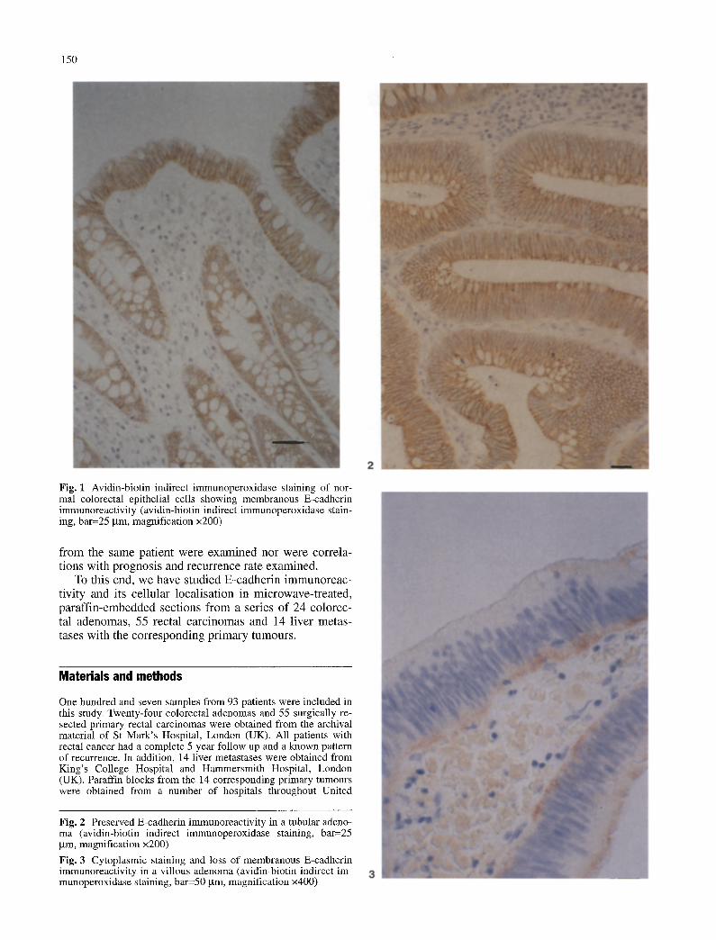
Fig. 3 Cytoplasmic staining and loss of membranous E-cadherin immunoreactivity in a villous adenoma (avidin-biotin indirect im- munoperoxidase staining, bar=50 gm, magnification x400)

151
Kingdom. Serial sections were stained with haematoxylin and eo- sin and used for histological classification of the lesions identified. Tumours were graded and classified according to Dukes [5] and Jass classifications [10] without knowledge of the staining results. Local extramural spread of tumour beyond the muscularis propria was described as absent, minimal, slight or extensive, based on a subjective assessment [14].
The avidin-biotin indirect immunoperoxidase method was em- ployed using the anti-E-cadherin HECD-1 (Human Epithelial Cad- herin-l) mouse monoclonal antibody as undiluted culture superna- tant. HECD-1 has been characterised previously and its specificity is described elsewhere [26]. To enhance E-cadherin immunohisto- chemistry in formalin-fixed, paraffin-embedded tissues, sections were treated with an antigen retrieval solution in a microwave ov- en, according to our previously described methods [8]. Briefly the slides were submerged in 0.01 M citrate buffer at pH 6.0 and are heated in a 700 W microwave on full power for 5x2 min cycles pausing to ensure there is no fluid loss due to evaporation. Slides were then rinsed in phosphate buffered saline (x3) after each stage. Fifty microliters of anti-E-cadherin HECD-1 primary anti- Total body was then added to the section and incubated overnight at 4 ° n C. An avidin-biotin complex immunoperoxidase technique was ut- (%) ilised to amplify epitope recognition (ABC kit, Dako, High Wy- combe, UK) and subsequent staining was achieved by 50 ~tl diami- Histology nobenzidine solution, at a concentration of 0.3 mg/ml (Dako). The Tubular slides were then washed and mounted for microscopic examina- n tion. Positive control tissue sections known to be of a homogene- (%) ous phenotype were used to ensure accurate and reproducible TV/V staining and included normal small and large intestinal mucosa, n Normal colorectal epithelial cells or normal hepatocytes present in (%) the primary or metastatic tumour sections respectively were also used as internal positive controls for E-cadherin staining which Dysplasia was normally seen at the cell-cell borders. Negative controls were Mild duplicate sections similarly stained in which the primary antibody n was omitted and replaced by normal mouse immunoglobulins. %
Slides were scored by assessing the proportion of stained cells, intensity of staining relative to the adjacent normal mucosa and Moderate/ site of staining (membrane or cytoplasmic) by three independent severe observers (GG, OK, MP) as previously described [8]. n
Fisher's exact test was used to compare E-cadherin immunore- % activity with the histopathological data. Spearman test was used to Size correlate loss of membranous expression with presence of cyto- >1 cm plasmic staining. Values of P less than 0.05 were considered sig- nificant, n %
<1 cm
Expres s ion o f E-cadher in in rectal ca rc inomas
E-cadher in immunoreac t iv i ty was present in 42/55 (76%) (Table 2). S ta in ing was focal (<30% of pos i t ive cells) in 11 (20%) tumours and diffuse in 31 (56%) (Fig. 4). Th i r ty -one percen t (9/24) o f tumours wi th negat ive or focal s taining were poor ly d i f ferent ia ted c o m p a r e d to 10% (3/31) o f tumours wi th a diffuse s ta ining (P<0.05). P rese rved m e m b r a n o u s E-cadher in immuno loc a l i s a t i on
Table 1 E-cadherin immunoreactivity in colorectal adenomas
Total Pattern Membrane Cytoplasm
<30% >30% + - + -
24 7 17 18 6 10 14 (100) (29) (71) (75) (25) (42) (58)
13 4 9 11 2 3 10 (54) (31) (69) (85) (15) (23) (77)
11 3 8 7 4 7 4 (46) (27) (73) (64) (36) (64) (36)
13 3 10 10 3 3 10 (54) (23) (77) (77) (23) (23) (77)
11 4 7 8 3 6 5 (46) (36) (64) (73) (27) (55) (45)
16 5 11 12 4 8 8 (67) (31) (69) (75) (25) (50) (50)
Results n 8 2 6 6 2 2 6 % (33) (25) (75) (75) (25) (25) (75)
Express ion o f E-cadher in in normal colorecta l ep i the l ium
Membranous E-cadher in staining was loca l i sed un i fo rmly at the in terce l lu lar borders o f h i s to logica l ly normal colo- rectal epi thel ia l cel ls present in the tumour specimens. No Total Pattern Membrane
cy top lasmic immunoreac t iv i ty was seen (Fig. 1). Neg <30% >30% + -
Table 2 Correlation of E-cadherin immunoreactivity with tumour grade in patients with rectal cancer
Cytoplasm
÷ - -
Expres s ion of E -cadhe r in in co lorec ta l adenomas
M e m b r a n e s taining o f in tens i ty equal to the ad jacent nor- mal m u c o s a was seen in 18/24 (75%) co lorec ta l adeno- mas (Fig. 2) whereas cy top la smic staining was obse rved in 10 cases (40%). The presence o f cy top la smic E-cad- herin immunoreac t iv i ty was s ta t i s t ica l ly assoc ia ted with loss o f m e m b r a n o u s express ion (Spea rman corre la t ion P<0.002) . This abnorma l pat tern was more f requent ly seen in large adenomas (more than 1 c m in d iameter ) (P<0.01) wi th high grade dysp la s i a (P<0.05) and vi l lous h i s to logy (P<0.05) (Table 1) (Fig. 3).
Total n 55 13 11 31 17 (%) (100) (24) (20) (56) (31)
Grade Well n 2 0 0 2 2 (%) (4) (0) (0) (100) (100)
Moderate n 41 8 7 26 15 (%) (74) (20) (17) (63) (37)
Poor n 12 5 4 3 0 (%) (22) (42) (33) (25) (0)
38 24 (69) (44)
0 0 (0) (0)
26 20 (63) (49)
12 4 (100) (33)
31 (56)
2 (100)
21 (51)
8 (67)

152
Fig. 4 Heterogenous E-cad- herin immunoreactivity in a moderately differentiated rectal adenocarcinoma (avidin-biotin indirect immunoperoxidase staining, bar=25 gm, magnifi- cation x200)
Fig. 5 Preserved E-cadherin immunoreactivity (arrowheads) in a liver metastasis from a moderately differentiated colo- rectal carcinoma (avidin-biotin indirect immunoperoxidase staining, bar=50 gin, magnifi- cation x400)
Table 3 Correlation of membranous E-cadherin immunoreactivity with stage in patients with rectal cancer
Total Positive Negative n (%) n (%) n (%)
Total 55 (100 17 (31) 38 (69) Dukes' A 8 (14) 3 (37) 5 (63) Dukes' B 20 (36) 6 (30) 14 (70) Dukes' C 27 (49) 8 (30) 19 (70) Jass 1 11 (20) 5 (45) 6 (55) Jass 2 14 (25) 4 (29) 10 (71) Jass 3 18 (33) 3 (17) 15 (83) Jass 4 12 (22) 5 (42) 7 (58)
was seen more frequently in well or moderately differen- tiated tumours than in poorly differentiated tumours (P<0.01). No correlation was seen between loss of mem- branous E-cadherin expression and advanced Dukes and Jass stages (Table 3), local extramural spread (Table 4) and 5-year recurrence rate (Table 5).
Expression of E-cadherin in liver metastases and the corresponding primary colorectal tumours
Fourteen liver metastases and the corresponding primary colorectal tumours were studied (Table 6). Seven out of 14 (50%) liver deposits exhibited preserved E-cadherin immunoreactivity on the cell membrane (Fig. 5). Com-

153
Table 4 Correlation of membranous E-cadherin immunoreactivity with extramural spread in patients with rectal cancer
Total Positive Negative n (%) n (%) n (%)
Total 55 (100) 17 (31) 38 (69) Absent 12 (22) 5 (42) 7 (58) Minimal 13 (24) 5 (38) 8 (61) Slight 17 (31) 3 (18) 14 (82) Extensive 13 (23) 4 (31) 9 (69)
Table 5 Correlation of membranous E-cadherin immunoreactivity with 5 year recurrence rate in patients with rectal cancer
Total Positive Negative n (%) n (%) n (%)
All patients 55 (100) 17 (31) 38 (69) Non-recurrence 24 (44) 7 (29) 17 (71) Recurrence 31 (56) 10 (32) 21 (68)
plete loss of membranous E-cadherin was seen in 7/14 (50%) (JH, DS, JF, AH, BS, WH, SB) liver metastases of which 6/7 showed intense membranous immunoreactivi- ty in the corresponding primary tumour.
Discussion
In this study we have investigated the expression of im- munoreactive E-cadherin in colorectal adenomas, carci- nomas and liver metastases using a microwave antigen retrieval method in formalin-fixed, paraffin-embedded tissue sections. The application of this method to archi- val material has several advantages as .retrospective stud- ies can be performed easily. In addition paraffin sections exhibit better morphological preservation when com- pared with frozen sections. Moll et al. [13] as well as our own group [8, 23] have demonstrated clearly that micro- wave antigen retrieval is a reliable technique to detect E- cadherin protein in formalin-fixed, paraffin-embedded tissues.
In this study normal colorectal epithelial cells showed membranous expression of E-cadherin protein at the cell-cell borders which reflects the normal localisation of an intercellular adhesion molecule. As the molecule has to be present at the cell surface to permit homotypic ad- hesion, cytoplasmic E-cadherin is by definition non- functional. Abnormal staining patterns (negative, hetero- geneous or cytoplasmic only) were seen frequently in ad- enomas as well as adenocarcinomas of the large bowel, as previously shown [4, 11, 17, 31, 32]. As in other hu- man tumours the abnormal E-cadherin staining pattern is likely to reflect reductions or loss of E-cadherin-mediat- ed adhesion. There is evidence to suggest that this abnor- mal pattern of E-cadherin expression is not an accessory finding in colorectal tumours as it has been demonstrated that a number of cell lines dedifferentiate in vitro when treated with anti-E-cadherin antibodies [1, 22] and be- come more polarised and differentiated when transfected with E-cadherin cDNA [7, 12]. Since E-cadherin is known to be the prime mediator of intercellular cohesion and epithelial tissue integrity, our data indicate that alter- ations in E-cadherin expression and function occur in pre-malignant lesions and, therefore, may play a role in the progression of adenomas with malignant potential.
E-cadherin may be detectable in the cytoplasm for a variety of theoretical reasons including an increased pro- duction rate or a failure to translocate or to anchor in the membrane. Alterations in o~-, 9- and q,-catenins which link the cadherin molecule to the actin cytoskeleton may underlie the abnormal cellular localisation of E-cadherin in adenomas. In particular, c~ and ~ catenins have been shown to bind the APC gene product [25, 28] which is frequently truncated in colorectal adenomas [24]. Re- cently, wild-type but not mutant APC has been shown to associate and promote the assembly of microtubules which influence the distribution of actin filaments and the cytoskeleton organisation in general [15, 27]. It is tempting to speculate that APC mutations may affect the stabilisation and/or functional state of the E-cad- herin/catenin complex and, perhaps, other signalling molecules linked to the cytoskeleton. Indeed, both APC mutations [3] and reduced E-cadherin and integrin ex-
Table 6 Membrane or cyto- plasmic E-cadherin immunore- activity in primary colorectal tumours and liver metastasis (M, membrane; C, cytoplasm; O, negative; NA, not avail- able
Primary tumours
Dukes' Grade E-cadherin
JF A mod M+C BVH B mod M+C JH C mod C DS B rood M GC C rood M+C JF C mod M AH B rood M+C KJ C mod C MK C well M+C CS C mod M BS C mod M+C JW C mod M+C WH B rood M+C SB C mod M+C
Liver metastasis
Grade E-cadherin
Overall survival
well M mod M mod 0 mod 0 mod M mod 0 mod 0 well M+C rood M mod M poor 0 mod M rood C mod C
2 months NA 23 months 2 months 10 years 18 months 1 week 2 years and 8 months 3 years and 7 months 10 years 2 years and 5 months NA 8 months 6 months

154
pression [21] seem to occur frequently in large adenomas with villous histology and high grade dysplasia.
Alterations in E-cadherin expression and cellular lo- calisation in the primary tumours were also found to cor- relate significantly with tumour grade but not with the ability to metastasise to the liver. In our series in which for the first time a direct comparison of E-cadherin ex- pression in paired samples of primary tumours and the corresponding liver metastases was made, membranous E-cadherin expression was seen in 50% of liver deposits. It is possible that a disregulation of intercellular adhe- sion may occur transiently at the time of invasion to al- low the cells to detach from the primary site. However, re-expression of E-cadherin on the cell membrane may be required to allow the turnout cells to localise and re- establish at the distant site.
In conclusion, we have demonstrated that changes in E-cadherin immunoreactivity and cellular localisation oc- cur in adenomas as well as carcinoma of the large bowel. Further studies are in progress to identify the mechanisms underlying the alterations in E-cadherin protein and its functional interaction with the catenin/APC complex.
Acknowledgements Support for this research was provided by the Medical Research Council (grant number G9204556CA). The authors are grateful to the histopathologists who kindly sent the archival paraffin blocks and in the particular the staff of the Histo- pathology Departments at Hammersmith Hospital and St Mark's Hospital for their invaluable assistance. We are grateful to M Takeichi and S Hirano for kindly donating the HECD-1 monoclo- hal antibody and to Bill Gullick, Chris Gilligan and Julia Verne for critical reading of the manuscript.
References
1. Behrens J, Mareel MM, Van Roy FM, Birchmeier W (1989) Dissecting turnout cell invasion: epithelial cells acquire inva- sire properties after the loss of uvomorulin-mediated cell-cell adhesion. J Cell Biol 108:2435-2447
2. Bodmer WF, Bishop T, Karran P (1994) Genetic steps in colo- rectal cancer. Nature Genet 6:217-219
3. DeBenedetti L, Sciallero S, Gismondi V, James R, Bafico A, Biticchi R, Masetti E, Bonelli L, Heouaine A, Picasso M, Gro- den J, Robertson M, Risio M, Caprilli R, Bruzzi P, White RL, Aste H, Santi L, Varesco L, Ferrara GB (1994) Association of APC gene mutations and histological characteristics of colo- rectal adenomas. Cancer Res 54:3553-3556
4. Dorudi S, Sheffield J, Poulsom R, Northover JMA, Hart IR (1993) E-cadherin expression in colorectal cancer. An immu- nohistochemical and in situ hybridisation study. Am J Pathol 142:981-986
5. Dukes CE, Bussey HJR (1958) The spread of rectal cancer and its effects on prognosis. Br J Cancer 12:309-320
6. Fearon ER, Vogelstein B (1990) A genetic model for colorec- tal tumorigenesis. Cell 61:759-767
7. Frixen UH, Behrens J, Sachs M, Eberle G, Voss B, Warda A, LiSchner D, Birchmeier W (1991) E-cadherin-mediated cell- cell adhesion prevents invasiveness of human carcinoma cells. J Cell Biol 113:173-185
8. Jankowsky JA, Newham PM, Kandemir O, Hirano S, Takeichi M, Pignatelli M (1994) Differential expression of E-eadherin in normal, metaplastic and dysplastic oesophageal mucosa: a putative biomarker. Int J Oncol 4:441-448
9. Jass JR (1989) Do all colorectal carcinomas arise in pre-exist- ing adenomas? World J Surg 13:45-51
10. Jass JR, Love SB, Northover JMA (1987) A new prognostic classification of rectal cancer. Lancet ii: 1303-1306
11. Kinsella AR, Green B, Lepts GC, Hill CI, Bowie G, Taylor BA (1993) The role of the cell-cell adhesion molecule E-cad- herin in large bowel turnout cell invasion and metastasis. Br J Cancer 67:904-909
12. Liu D, Nigam AK, Lalani E-N, Stamp GWH, Pignatelli M (1993) Transfection of E-cadherin into a human colon carcino- ma cell line induces differentiation and inhibits growth in vit- ro. Gut 34:27
13. Moll R, Mitze M, Frixen UH, Birchmeier W (1993) Differen- tial loss of E-cadherin expression in infiltrating ductal and lob- ular breast carcinomas. Am J Pathol 143:1731-1742
14. Morson BC, Bussey HJR (1963) Pelvic recurrence after exci- sion of rectum for carcinoma. B M J 2:13-18
15. Munemitsu S, Souza B, M~ller O, Albert I, Rubinfeld B, Po- lakis P (1994) The APC gene product associates with microtu- bules in vivo and promotes their assembly in vitro. Cancer Res 54:3676-3681
16. Muto T, Bussey HJR, Morson BC (1975) The evolution of cancer of the colon and rectum. Cancer 36:2251-2270
17. Nigam AK, Savage FJ, Boulos PB, Stamp GWH, Liu D, Pig- natelli M (1993) Loss of cell-cell and cell-matrix adhesion molecules in colorectal cancer. Br J Cancer 68:507-514
18. Ozawa M, Kemler R (1992) Molecular organization of the uv- omorulin-catenin complex. J Cell Biol 116:989-996
19. Pignatelli M (1993) Models of colorectal tumour differentia- tion. In: Lemoine NR, Wright NA (eds) The molecular pathol- ogy of cancer. Cancer Surveys: Cold Spring Harbor Laborato- ry Press, New York, pp 3-13
20. Pignatelli M (1993) E-cadherin: a biological marker of turnout differentiation. J Pathol 171:81-82
21. Pignatelli M, Stamp GWH (1994) Integrins in tumour devel- opment and spread. In: Hart I, Hogg N (eds) Tumour cell ad- hesion. Cancer Surveys: Cold Spring Harbor Laboratory Press, New York (in press)
22. Pignatelli M, Liu D, Nasim MM, Stamp GWH, Hirano S, Takeichi M (1992) Morphoregulatory activities of E-cadherin and beta-1 integrins in colorectal tumour cells. Br J Cancer 66:629-634
23. Pignatelli M, Ansari TQ, Gunter E Liu D, Hirano S, Takeichi M, K1/Sppel G, Lemoine NR (1994) Loss of membranous E- cadherin expression in pancreatic cancer: correlation with lymph node metastasis, high grade and advanced stage. J Pa- thol (in press)
24. Powell SM, Zilz N, Beazer-Barclay Y, Bryan TM, Hamilton SR, Thibodeau SN, Vogelstein B, Kinzler KW (1992) APC mutations occur early during colorectal tumorigenesis. Nature 359:235-237
25. Rubinfeld B, Souza B, Albert I, Mfiller O, Chamberlain SH, Masiarz FR, Munemitsu S, Polakis P (1993) Association of the APC gene product with ~-catenin. Science 262:1731-1734
26. Shimoyama Y, Hirohashi S, Hirano S, Noguchi M, Shimosato Y, Takeichi M, Abe O (1989) Cadherin cell adhesion mole- cules in human epithelial tissues and carcinomas. Cancer Res 49:2128-2133
27. Smith KJ, Levy DB, Maupin P, Pollard TD, Vogelstein B, Kin- zler KW (1994) Wild-type but not mutant APC associates with the microtubule cytoskeleton. Cancer Res 54:3672-3675
28. Su LK, Vogelstein B, Kinzler KW (1993) Association of the APC tumor suppressor protein with catenins. Science 262:1734-1737
29. Takeichi M (1991) Cadherin cell adhesion receptors as a mor- phogenetic regulator. Science 251:1451-1455
30. Takeichi M (1993) Cadherins in cancer: implications for inva- sion and metastasis. Curr Opin Cell Biol 5:806-811
31. Van der Wurff AAM, Kate JT, Van der Linden EPM, Dinjens WNM, Arends J-W, Bosman FT (1992) L-CAM expression in normal, premalignant, and malignant colon mucosa. J Pathol 168:287-291
32. Van der Wurff AAM, Arends J-W, Van der Linden EPM, Kate JT, Bosman FT (1994) L-CAM expression in lymph node and liver metastases of colorectal carcinomas. J Pathol 172:177-182
Copyright © 2022 FDOKUMEN