A Prosthetic Hand Body Area Controller Based on Efficient ...
Upload
independentCategory
view
0download
0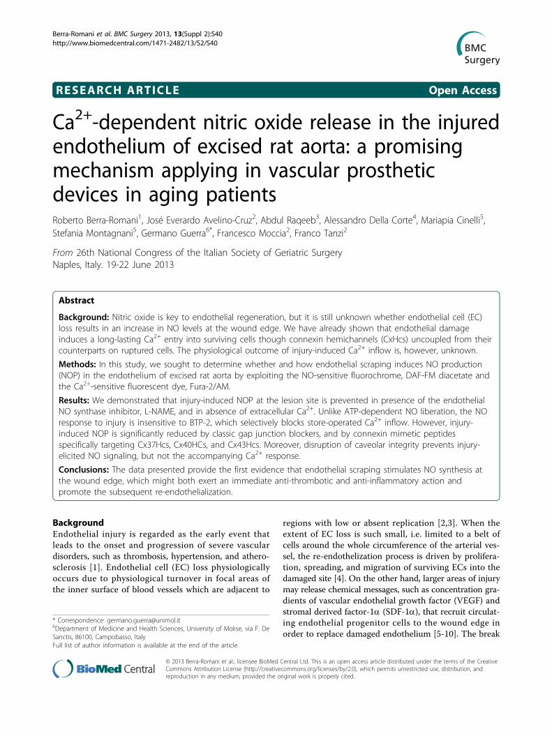
RESEARCH ARTICLE Open Access
Ca2+-dependent nitric oxide release in the injuredendothelium of excised rat aorta: a promisingmechanism applying in vascular prostheticdevices in aging patientsRoberto Berra-Romani1, José Everardo Avelino-Cruz2, Abdul Raqeeb3, Alessandro Della Corte4, Mariapia Cinelli5,Stefania Montagnani5, Germano Guerra6*, Francesco Moccia2, Franco Tanzi2
From 26th National Congress of the Italian Society of Geriatric SurgeryNaples, Italy. 19-22 June 2013
Abstract
Background: Nitric oxide is key to endothelial regeneration, but it is still unknown whether endothelial cell (EC)loss results in an increase in NO levels at the wound edge. We have already shown that endothelial damageinduces a long-lasting Ca2+ entry into surviving cells though connexin hemichannels (CxHcs) uncoupled from theircounterparts on ruptured cells. The physiological outcome of injury-induced Ca2+ inflow is, however, unknown.
Methods: In this study, we sought to determine whether and how endothelial scraping induces NO production(NOP) in the endothelium of excised rat aorta by exploiting the NO-sensitive fluorochrome, DAF-FM diacetate andthe Ca2+-sensitive fluorescent dye, Fura-2/AM.
Results: We demonstrated that injury-induced NOP at the lesion site is prevented in presence of the endothelialNO synthase inhibitor, L-NAME, and in absence of extracellular Ca2+. Unlike ATP-dependent NO liberation, the NOresponse to injury is insensitive to BTP-2, which selectively blocks store-operated Ca2+ inflow. However, injury-induced NOP is significantly reduced by classic gap junction blockers, and by connexin mimetic peptidesspecifically targeting Cx37Hcs, Cx40HCs, and Cx43Hcs. Moreover, disruption of caveolar integrity prevents injury-elicited NO signaling, but not the accompanying Ca2+ response.
Conclusions: The data presented provide the first evidence that endothelial scraping stimulates NO synthesis atthe wound edge, which might both exert an immediate anti-thrombotic and anti-inflammatory action andpromote the subsequent re-endothelialization.
BackgroundEndothelial injury is regarded as the early event thatleads to the onset and progression of severe vasculardisorders, such as thrombosis, hypertension, and athero-sclerosis [1]. Endothelial cell (EC) loss physiologicallyoccurs due to physiological turnover in focal areas ofthe inner surface of blood vessels which are adjacent to
regions with low or absent replication [2,3]. When theextent of EC loss is such small, i.e. limited to a belt ofcells around the whole circumference of the arterial ves-sel, the re-endothelization process is driven by prolifera-tion, spreading, and migration of surviving ECs into thedamaged site [4]. On the other hand, larger areas of injurymay release chemical messages, such as concentration gra-dients of vascular endothelial growth factor (VEGF) andstromal derived factor-1a (SDF-1a), that recruit circulat-ing endothelial progenitor cells to the wound edge inorder to replace damaged endothelium [5-10]. The break
* Correspondence: [email protected] of Medicine and Health Sciences, University of Molise, via F. DeSanctis, 86100, Campobasso, ItalyFull list of author information is available at the end of the article
Berra-Romani et al. BMC Surgery 2013, 13(Suppl 2):S40http://www.biomedcentral.com/1471-2482/13/S2/S40
© 2013 Berra-Romani et al.; licensee BioMed Central Ltd. This is an open access article distributed under the terms of the CreativeCommons Attribution License (http://creativecommons.org/licenses/by/2.0), which permits unrestricted use, distribution, andreproduction in any medium, provided the original work is properly cited.
in the anatomic integrity of vascular endothelium is signif-icantly larger in subjects undergoing medical interven-tions, such as deployment of endovascular devices andpercutaneous transluminal coronary angioplasty [2,11,12].Such a heavy loss of ECs from the vascular wall dampensthe beneficial effects of reconstructive surgery andprompts the quest for pharmacological treatments aimingat restoring the continuity of endothelial monolayer[2,11-13]. Accordingly, the de-endothelialisation of arterialvessels may cause thrombi formation and neointimalhyperplasia, giving raise to a process termed as “in-stentrestenosis” (ISR) which renarrows the arterial lumen[2,11,12]. Drug-eluting stents (DES) may be implantedduring or after angioplasty to inhibit neointimal hyperpla-sia and prevent ISR [2,12,14]. Unfortunately, the mostcommonly employed stents deliver drugs, such as siroli-mus and paclixatel, that cause a long-term inhibition ofendothelial proliferation and migration. These untowardoff-target effects significantly delay endothelial regrowth,thereby leaving uncovered the surface of the stent andincreasing the risk of late in-stent thrombosis [14,15].Aging is accompanied by a decline in the healthy func-
tion of multiple organ systems, leading to increased inci-dence of mortality from many diseases. Oxidative stressand elevated ROS (Reactive oxygen species) has beenimplicated in the mechanism of senescence and aging;they are also involved in cancer, diabetes, neurodegenera-tive, cardiovascular and other diseases [16,17] Overpro-duction of oxidant molecules is due to several stressagents such chemicals, drugs, pollutants, high-caloricdiets and exercise [18].Nitric oxide (NO), a gasotransmitter that may be
synthesized and released by ECs [1,19], might play a keyrole in healing injured endothelium. Accordingly, NOinhibits apoptosis and enhances EC proliferation, migra-tion, and tubulogenesis [19]. Moreover, NO might serveas anti-inflammatory signal at the injured site by pre-venting local platelet activation and thrombus formation,by causing vasorelaxation, and by inhibiting the pheno-typic switch of vascular smooth muscle cell (VSMC)[1,19]. Restoration of NO production at the site of vas-cular injury may attenuate neointimal hyperplasia andadverse the onset of the atherosclerotic process [20]. Invascular endothelium, NO may be synthesized by two dif-ferent isoforms of NO synthase (NOS), namely inducibleNOS (iNOS/NOS2), which mediates NO liberation duringinflammatory reactions, and endothelial NOS (eNOS/NOS3), which is preferentially recruited by stimuli [19,21].The catalytic reaction consists in the conversion of L-argi-nine to L-citrulline and requires several cofactors, such asNAPDH, tetrahydrobiopterin and O2 [19]. Unlike iNOS,which is inducible and Ca2+-independent, both iNOS andeNOS are constitutive and bear a calmodulin-binding site
whose binding to cytosolic Ca2+ is essential to stimulateNO synthesis [19]. In several types of ECs, eNOS is prefer-entially recruited by Ca2+ store-operated Ca2+ entry(SOCE), which is gated by depletion of the inositol-1,4,5-trisphosphate (InsP3)-sensitive stores within the endoplas-mic reticulum (ER) [8,22]. This feature is due to the closeproximity between eNOS and SOC channels at the caveo-lae, cholesterol-enriched surface microdomains thatcompartmentalize signal transduction molecules [23].Cholesterol-binding drugs, such as methyl-b-cyclodextrin(MbCD), have been used to disrupt caveolae and impairNO signalling in vascular endothelium [24]. That NO isinvolved in EC response to injury has been suggested bythe elevation in both eNOS protein and enzyme activity inthe regenerating endothelium of rat aorta [25]. Moreover,a recent study carried out on this preparation reported anincrease in [Ca2+]i in ECs nearby the lesion site [2,9,26,27].The Ca2+ response to injury comprises an initial peak,mainly due to Ca2+ release from InsP3-sensitive receptors(InsP3Rs), which is followed by a long-lasting decay phasemediated by Ca2+ entry across the plasma membrane [27].InsP3 synthesis requires phospholipase C (PLC) activationby ATP (or ATP-derived nucleotides) released from rup-tured cells [27]. Ca2+ inflow is supported by connexin (Cx)hemichannels (CxHcs), which have recently beendescribed as alternative Ca2+ entry routes in vascular ECsas well as other non-excitable cell types [26-31]. Cxs maybe classified on the basis of the molecular weight withthree main isoforms (Cx37, Cx40, and Cx43) being foundin ECs from several vascular beds, including rat aorta[32,33]. Despite the available evidences show an increaseboth in eNOS activity and in [Ca2+]i within lesionedendothelium, there is no report of injury-induce NOrelease in blood vessels. Understanding the signal trans-duction machinery leading to endothelial activation andNO synthesis might be clinically relevant to design alter-native strategies to restore endothelial integrity andprevent ISR.In the present investigation, we sought to determine
whether NO synthesis occurs in ECs facing the injurysite of excised rat aorta. We further aimed at elucidatingthe signal transduction pathway responsible for NOrelease by lesioned endothelium. These goals wereaccomplished by loading ECs with Fura-2/AM, a Ca2+-sensitive fluorochrome, and DAF-FM diacetate, a NO-sensitive fluorescent dye. We provided, for the firsttime, the evidence that mechanical injury induces a longlasting NO production (NOP) in cells nearby the lesionsite which requires Ca2+ entry through uncoupledCx37Hcs, Cx40Hcs, and Cx43Hc. These results mightaid in developing novel pharmacological treatmentsdevoted to restore endothelial integrity upon invasivemedical interventions.
Berra-Romani et al. BMC Surgery 2013, 13(Suppl 2):S40http://www.biomedcentral.com/1471-2482/13/S2/S40
Page 2 of 17

MethodsDissection of the aortaWistar rats aged 2-3 months were sacrificed with anoverdose of diethyl ether. The thoracic and abdominalaorta were dissected out and perfused with physiologicalsalt solution (PSS). The vessel was cleaned of the sur-rounding connective tissue, cut in ~5 mm long rings,stored in PSS at room temperature (22-24 °C) and usedwithin 5 hours. All the animal protocols were approvedby the East Tennessee State University’s Animal Careand Use Committee. All experiments conform to theGuide for the Care and Use of Laboratory Animals pub-lished by the US National Institutes of Health (NIHPublication No. 85-23, revised 1996). The animal hand-ling was under the continuous control of the VeterinarySurgeon of the University of Pavia.SolutionsPSS had the following composition (in mM): 150 NaCl,6 KCl, 1.5 CaCl2, 1 MgCl2, 10 Glucose, 10 Hepes. InCa2+-free solution (0 Ca2+), Ca2+ was substituted with2 mM NaCl, and 0.5 mM EGTA was added. Solutionswere titrated to pH 7.4 with NaOH. Aortic rings werebathed in 0 Ca2+ for no longer than 90 sec before indu-cing the injury. Control experiments have demonstratedthat such a short pre-incubation period is not abledeplete intracellular Ca2+ stores [27].[Ca2+]i and NO measurements.The technique used to evaluate changes in [Ca2+]i in
intact endothelium has been previously described[26,27,34-37]. The aortic ring was opened and loadedwith 16 μmol Fura-2/AM for 60 min at room tempera-ture, washed and fixed by small pins with the luminalface up. In situ ECs were visualized by an upright epi-fluorescence Axiolab microscope (Carl Zeiss, Oberko-chen, Germany), equipped with a Zeiss ×63 Achroplanobjective (water-immersion, 2.0 mm working distance,0.9 numerical aperture). ECs were excited alternately at340 and 380 nm, and the emitted light was detected at510 nm. The exciting filters were mounted on a filterwheel (Lambda 10, Sutter Instrument, Novato, CA,USA). Custom software, working in the LINUX environ-ment, was used to drive the camera (Extended-ISISCamera, Photonic Science, Millham, UK) and the filterwheel, and to measure and plot on-line the fluorescencefrom 10-15 rectangular “regions of interest” (ROI)enclosing one single cell. [Ca2+]i was monitored by mea-suring, for each ROI, the ratio of the mean fluorescenceemitted at 510 nm when exciting alternatively at 340and 380 nm (shortly termed “ratio”). An increase in[Ca2+]i causes an increase in the ratio. Ratio measure-ments were performed and plotted on-line every 5 s.The experiments were performed at room temperature(21-23 °C).
NO production in ECs in intact rat aorta was moni-tored with the membrane permeant 4-Amino-5-methy-lamino-2’,7’-difluorofluorescein (DAF-FM) diacetate.Once open, the aortic ring was fixed by small pins withthe luminal side facing up, loaded with 10 μM DAF-FMfor 60 min at room temperature and washed in PSS forone hour. DAF-FM fluorescence was measured by usingthe same equipment described for Ca2+ recordings butwith a different filter set, i.e. excitation at 480 nm andemission at 535 nm wavelength (emission intensity wasshortly termed “NOi”). NO measurements were per-formed and plotted on-line every 5 s. Again, off-line ana-lysis was performed by using custom-made macrosdeveloped by Microsoft Office Excel software. Theexperiments were performed at room temperature. DAF-FM is essentially non-fluorescent until it irreversiblyreacts with the nitrosonium cation produced by sponta-neous oxidation of newly synthesized NO. The resultingfluorescent compound is trapped in the cytoplasm, sothat DAF-FM fluorescence summates with continual NOproduction. As also found in bovine aortic ECs [38],DAF-FM fluorescence in rat aortic ECs underwent a per-sistent and linear increase likely due to the basal NOsynthesis [19]. Accordingly, the change in baseline fluor-escence was dramatically reduced when aortic rings werepre-incubated with L-NAME (5 mM), which inhibitseNOS by competing with its natural substrate, L-arginine(data not shown), or in 0Ca (data not shown). This obser-vation was somehow expected since aortic endotheliumhas long been known to regulate vascular tone by basalrelease of NO [19]. Therefore, for each experiment, thebasal DAF-FM signal was recorded for at least 20 min-utes and the resulting curve fitted off-line by a linearregression equation in order to calculate the slope (i.e.the rate) of basal fluorescence increase. The slope wassubtracted from the recorded trace, thus enabling tomeasure S-Nitroso-N-acetylpenicillamine (SNAP)-, ATP-and injury-induced NOP independently on basal eNOSactivity [39] (see below). NO synthesis was evaluated bysubtracting the baseline level (1 min before the increasein DAF-FM fluorescence) to the average value of the pla-teau phase (1 min before the end of the recording)recorded upon either ATP administration or endothelialscraping.In order to assess the ability of the fluorimetric set up
to detect evoked changes in intracellular NO levels, aor-tic rings were exposed to the NO donor, SNAP, andATP, which has been shown to elicit NO production ina variety of ECs from different vascular districts [38,40].As shown in Figure 1A and Figure 1B, respectively,SNAP (500 μM) and ATP (300 μM) caused an increasein the intensity of DAF-FM fluorescence in 19 out of19 cells and in 170 out of 170 cells, respectively.
Berra-Romani et al. BMC Surgery 2013, 13(Suppl 2):S40http://www.biomedcentral.com/1471-2482/13/S2/S40
Page 3 of 17

On average, L-NAME (5 mM) reduced by 86% ATP-induced NO production (0.025 ± 0.001, n = 170, vs.0.0041 ± 0.0009, n = 192, p < 0.001) (Figure 1B).Mechanical disruption of ECsAs shown in [26,27], aortic endothelium was injuredunder microscopic control by means of a glass microelec-trode with a broken tip of about 30 μm diameter, drivenby an XYZ hydraulic micromanipulator (Narishige,Japan). Images of either Fura-2 or DAF-FM/Fura-2loaded ECs, together with numbered ROIs, were takenbefore the lesion, in order to identify the cells facing theinjury site. As aforementioned, the dissection procedurecould itself damage the intimal layer and cause SMCs tobe loaded with Fura-2. These rings were, therefore, dis-carded. The microelectrode was first positioned almostparallel and very near to the endothelium surface. It wasthen moved downward, along the Z-axis, until the elec-trode tip gently touched the endothelium, and moved
horizontally across the visual field to scrape 1-3 consecu-tive rows of ECs along the whole diameter of the aorticring. The following lesion was 170 μm long and 20 μmwide. This procedure, which allowed monitoring of NOsignals in ECs adjacent to the lesion, mimics the ablationof the endothelial lining achieved in pre-clinical studiesaddressing the cellular mechanisms of intimal regrowthupon stent deployment [41-43]. The scraping of theendothelial monolayer imposed a physical stretching onthe underlying SMCs which resembles the conditionoccurring during clinical interventions. Ethidium bro-mide (EB), a fluorescent molecule unable to cross anintact plasma membrane and therefore indicative ofdamaged/dead cells, was used to check the viability ofboth ECs nearby the injury and underlying SMCs [27].When smooth muscle fibers were lesioned by mechanicalscratching and stained by EB, the experiments werediscarded.
Figure 1 NO production in rat aortic endothelium. A, exposition to SNAP (500 μM) caused an increase in DAF-FM fluorescence in native rataortic endothelium in situ. The trace is the mean±SE of 11 cells from the same visual field. B, ATP (300 μM) induced an elevation in intracellularNO (closed circles) that was dramatically hampered when the rings were pre-treated with L-NAME (5 mM) for 40 min (white circles). The tracesare the mean±SE, respectively, of 10 (PSS) and 8 (L-NAME) cells from two aortic rings harvested from the same animal on the same day.
Berra-Romani et al. BMC Surgery 2013, 13(Suppl 2):S40http://www.biomedcentral.com/1471-2482/13/S2/S40
Page 4 of 17
Data analysisFor each protocol, data were collected from at leastthree rats. The amplitude of the peak Ca2+ response wasmeasured as the difference between the ratio at the peakand the mean ratio of 1 min baseline before the peak.The details of NO measurements have been reportedabove. Only cells residing in the first row nearby thewound were used to elaborate the average value. ForCa2+ and NO measurements, statistical comparisonswere made by Student’s t-test for unpaired observations.ChemicalsFura-2/AM and DAF-FM was obtained from MolecularProbes (Molecular Probes Europe BV, Leiden, TheNetherlands). N-(4-[3,5-bis(trifluoromethyl)-1H-pyrazol-1-yl]phenyl)-4-methyl-1,2,3-thiadiazole-5-carboxamide(BTP-2) was purchased from Calbiochem (La Jolla, CA,USA). All other chemicals were purchased from Sigma.Cx-mimetic peptides, including 37,43Gap27 and 40Gap27,were synthesized by Severn Biotech (Kidderminster,UK); purity was >95%. In more detail, 37,43Gap27 and40Gap27 mimic a highly conserved SRPTEK sequencepresent in the second extracellular loop of Cxs 37 and43 and of Cx 40, respectively [15,19]. Their scrambledversions were also synthesized by Severn Biotech. Allother chemicals were of analytical grade and obtainedfrom Sigma.
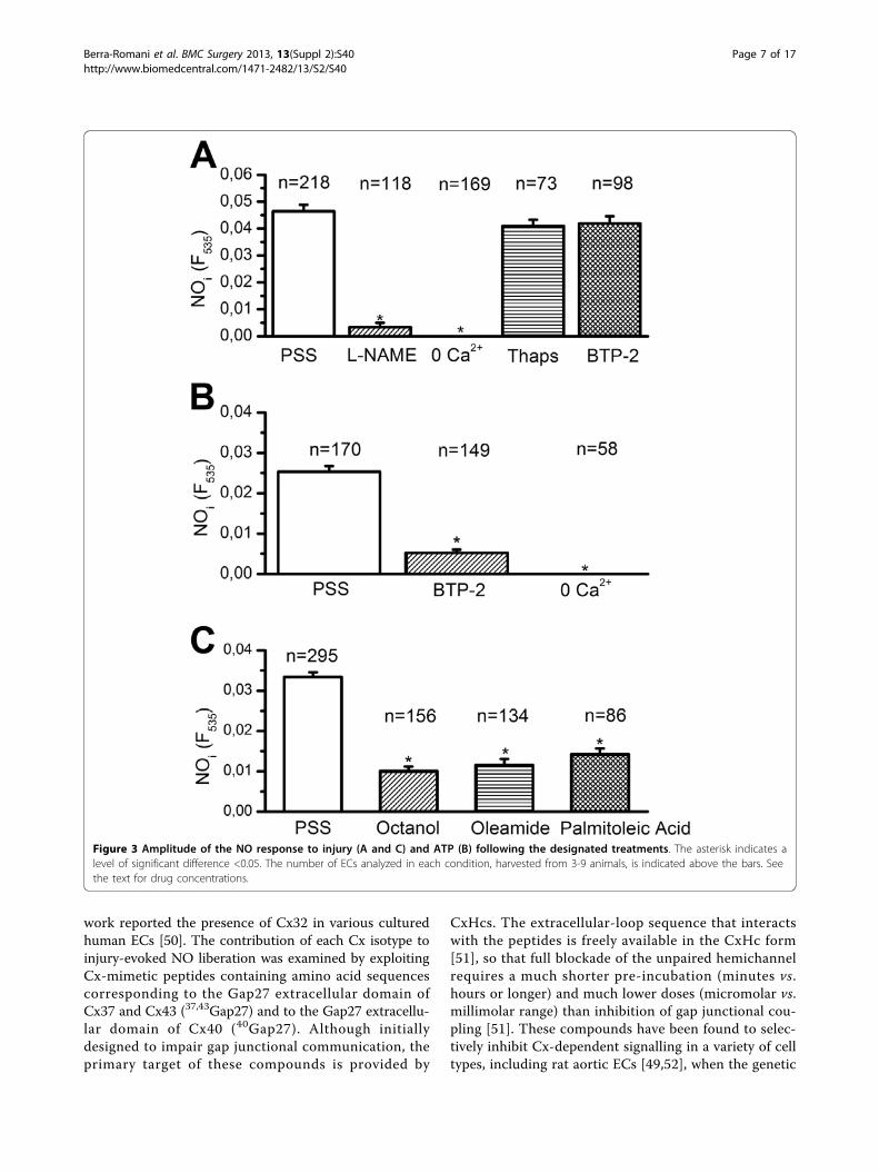
ResultsInjury augments intracellular NO levels in the intactendothelium of rat aortaFigure 2A depicts the injury-induced increase in [Ca2+]ioccurring within the ECs adjacent to the lesion site andwhich have lost their contact with the scraped cells. TheCa2+ signal consisted in an initial transient peak, mainlydue to InsP3-dependent Ca
2+ release, followed by a pro-longed decay phase caused by Ca2+ entry from theextracellular milieu [27]. When the damage was per-formed on an aortic ring loaded with DAF-FM, itinduced a gradual increase in fluorescence that reacheda plateau after about 20 min (Figure 2B). This feature isconsistent with the concomitant long-lasting Ca2+ inflow[27], which might sustain NO synthesis [19,38]. In thepresence of the eNOS inhibitor, L-NAME (5 mM),mechanical damage failed to augment intracellular NO(Figure 2B). The statistical analysis is reported in Figure3A. Similar to the intercellular Ca2+ wave, which did notspread farther than the 4th raw of cells, the amplitude ofthe NO signal significantly (p < 0.05) decayed at thislocation (Figure 2C). This feature hints at a strong cor-relation between the increase in [Ca2+]i triggered by theinjury and the accompanying NO synthesis.Injury-induced NOP depends on extracellular Ca2+ entryIt has long been known that, in vascular endothelium,eNOS is selectively activated by Ca2+ inflow rather than
intracellular Ca2+ release [8,22,40]. When mechanicaldamage was carried out in absence of extracellular Ca2+,a maneuver which abolished the prolonged decay-phaseof the Ca2+ signal, the injury did not result in a detect-able NOP (Figure 3A). The involvement of intracellularCa2+ mobilization was investigated with the aid of thap-sigargin (2 μM), an inhibitor of the endoplasmic reticu-lum Ca2+-ATPase, which prevents Ca2+ re-uptake intothe stores and leads to their depletion. Preliminaryexperiments showed that pre-incubating the rings for 30min with 2 μM thapsigargin abolished the intracellularCa2+ mobilization triggered by supramaximal concentra-tions of ATP (300 μM) (n = 79; data not shown). There-fore, such a treatment causes the depletion of the Ca2+
reservoir underlying the initial Ca2+ response to injury[27]. The NO signal induced by endothelial scraping,however, was not significantly affected by thapsigargin(Figure 3A). Overall, these results strongly suggest thatinjury-promoted NOP in surviving ECs is sustained byCa2+ entry, while is insensitive to Ca2+ release from ER.SOCE represents the preferential route for extracellularCa2+ to engage eNOS in ECs [8,22,40]. However, BTP-2(20 μM), a widely employed blocker of store-dependentCa2+ inflow [6,8], does not significantly affect injury-induced NO synthesis (Figure 3A). We subsequentlyfocused on the response to ATP, which elicits a BTP-2-sensitive SOCE in rat aortic endothelium [27]. ATP-induced NO synthesis was strongly inhibited in 0 Ca2+
(Figure 3B) and in the presence BTP-2 (Figure 3B).Collectively, these data concur with our previous find-ings on injury-induced Ca2+ inflow and rule out adetectable role for SOCE in NO release by lesionedendothelium.Gap junction inhibitors reduce NO synthesis induced by Ca2+
entry in injured endotheliumThe long-lasting decay phase of injury-induced Ca2+ ele-vation in rat aortic rings is dramatically diminished bygap junction blockers, such as octanol, palmitoleic acidand oleamide [26,27,44,45]. In agreement with the Ca2+
measurements [27], octanol (4 mM), oleamide (200 μM),and palmitoleic acid (50 μM) dramatically affected NOsynthesis elicited by endothelial scraping (Figure 3C). Inthe light of our previous observations [27], these findingshint at a role for Ca2+ inflow through CxHcs in mediatingNOP in injured endothelium. We assessed the specificityof gap junction blockers by measuring the Ca2+ responseto ATP (20 μM) in the presence of octanol. At this dose,ATP evokes an InsP3-dependent Ca
2+ release, which issubsequently buffered by the combined action of SERCAand Na+/Ca2+ exchanger [27,34,46]. As shown in Figure4, pre-incubating rat aortic rings with 4 mM octanol for20 min did not significantly affect either the magnitudeof the [Ca2+]i elevation or the rate of its decay phase tothe baseline. This result concurs with previous evidence
Berra-Romani et al. BMC Surgery 2013, 13(Suppl 2):S40http://www.biomedcentral.com/1471-2482/13/S2/S40
Page 5 of 17

about the efficacy of gap junction blockers as selectiveuncoupling drugs in rat aortic rings [47,48], and furthersupport the role of CxHcs in mediating NO synthesis ininjured endothelium.
Effect of Cx-mimetic peptides on injury-induced Ca2+
elevation and NO productionRat aortic endothelium may express three Cx isoforms,namely Cx37, Cx40, and Cx43 [32,49], albeit a recent
Figure 2 NO synthesis induced by mechanical injury rat aortic endothelium. A, Ca2+ signal elicited by lesioning rat aorta in PSS. The trace,recorded from a single cell, is representative of 10 cells from the same visual field. B, injury induced an elevation in intracellular NO levels thatwas abolished when the cells were pre-treated with the eNOS inhibitor, L-NAME (5 mM), for 40 min. The traces are the mean±SE, respectively, of10 (PSS, closed circles) and 5 (L-NAME, open circles) cells from two aortic rings harvested from the same animal on the same day. C, mean±SEof the NO signal recorded at increasing distance from the lesion site. In this and the following figures, the arrow indicates when injury isperformed. Asterisk indicates a level of significant difference <0.05.
Berra-Romani et al. BMC Surgery 2013, 13(Suppl 2):S40http://www.biomedcentral.com/1471-2482/13/S2/S40
Page 6 of 17
work reported the presence of Cx32 in various culturedhuman ECs [50]. The contribution of each Cx isotype toinjury-evoked NO liberation was examined by exploitingCx-mimetic peptides containing amino acid sequencescorresponding to the Gap27 extracellular domain ofCx37 and Cx43 (37,43Gap27) and to the Gap27 extracellu-lar domain of Cx40 (40Gap27). Although initiallydesigned to impair gap junctional communication, theprimary target of these compounds is provided by
CxHcs. The extracellular-loop sequence that interactswith the peptides is freely available in the CxHc form[51], so that full blockade of the unpaired hemichannelrequires a much shorter pre-incubation (minutes vs.hours or longer) and much lower doses (micromolar vs.millimolar range) than inhibition of gap junctional cou-pling [51]. These compounds have been found to selec-tively inhibit Cx-dependent signalling in a variety of celltypes, including rat aortic ECs [49,52], when the genetic
Figure 3 Amplitude of the NO response to injury (A and C) and ATP (B) following the designated treatments. The asterisk indicates alevel of significant difference <0.05. The number of ECs analyzed in each condition, harvested from 3-9 animals, is indicated above the bars. Seethe text for drug concentrations.
Berra-Romani et al. BMC Surgery 2013, 13(Suppl 2):S40http://www.biomedcentral.com/1471-2482/13/S2/S40
Page 7 of 17

suppression of specific Cx isoforms is not feasible, suchas in rat endothelium [33,45]. In addition, Cx-mimeticpeptides do not present the drawbacks reported in Cx-deficient mice [53]. For instance, Cx40-/- mice manifesteither higher [54] or lower [55] levels of Cx37 as well as a
significant decrease in Cx43 expression [56]. This featuremakes the interpretation of the physiological data col-lected from these animals difficult. Incubating the aorticrings for 1 hour with either 37,43Gap27 (300 μM) or40Gap27 (300 μM) significantly (p < 0.05) reduced injury-
Figure 4 Ethanol does not significantly affect the Ca2+ response to ATP. A, 20 min pre-incubation with octanol (4 mM) does notremarkably affect either the amplitude or the kinetics of the Ca2+ response to ATP (20 μM). The trace is the average of 16 cells from the samemicroscopic field of view. B, mean±SE of the peak of ATP-induced elevation in [Ca2+]i. The asterisk indicates a level of significant difference<0.05. The number of ECs analyzed in each condition is indicated above the bars. C, B, mean±SE of the rate of decay of ATP-induced Ca2+
signal. The decay was measured by fitting the Ca2+ tracings with a first order exponential equation (OriginPro 7, OriginLab Corp, NorthamptonMA). The asterisk indicates a level of significant difference <0.05. The number of ECs analyzed in each condition is indicated above the bars.
Berra-Romani et al. BMC Surgery 2013, 13(Suppl 2):S40http://www.biomedcentral.com/1471-2482/13/S2/S40
Page 8 of 17

evoked NOP (Figure 5A and Figure 5B). The statisticalanalysis of these experiments has been reported inFigure 5C and Figure 5D. NO signalling was unaffectedby peptides corresponding to scrambled sequences inGap27, indicating that the inhibition resulted from speci-fic sequence recognition on the selected Cx isotypes(Figure 5C and Figure 5D). Therefore, it is reasonable toconclude that Ca2+ entry driving eNOS activity and NOliberation at the wound edge is supported by Cx37Hcs,Cx40Hcs, and Cx43Hcs. As discussed elsewhere [57],eNOS activation requires a sub-plasmalemmal Ca2+
increase rather a bulk increase in cytosolic Ca2+ levels.Such a localized Ca2+ event may not even be detected byconventional epifluorescence microscopy [58]. As a con-sequence, we went on in assessing whether Cx-mimeticpeptides targeting Cx37, Cx40, and Cx43 were also ableto reduce the long-lasting decay phase of the Ca2+
response to mechanical damage. Figures 6A to 6D depictthat neither 37,43Gap27 (300 μM) nor 40Gap27 (300 μM)significantly reduce either the initial Ca2+ peak or thesubsequent decay phase. Rather, both Cx-mimetic pep-tides increased the amplitude of the initial Ca2+ response(Figure 6A-6C). Consistently, acute application of bothpeptides during recovery of the Ca2+ signal towards thebaseline did not decreased the [Ca2+]i (n = 31, 40Gap27;n = 52, 37,43Gap27) (not shown). The same results werefound by extending the pre-incubation period with both37,43Gap27 and 40Gap27 up to 2 hours (Figure 6B-6D)[49]. Taken as a whole, these results suggest thatCx37Hcs, Cx40Hcs, and Cx43Hcs mediate the sub-membranal Ca2+ elevation responsible for eNOS activa-tion, but not the bulk increase in [Ca2+]i caused bymechanical damage in surviving ECs. As the pharmacolo-gical profile of the decay phase is consistent with a CxHc
Figure 5 Connexin-mimetic peptides affect injury-induced NO synthesis. 1 hour pre-incubation with either 37,43Gap27 (300 μM) (A) or40Gap27 (300 μM) (B) dramatically attenuated the increase in intracellular NO levels provoked by endothelial lesion. For sake of clarity, thecontrol curve has been depicted in each Panel. All the experiments depicted in this figure have been carried out on the same day on differentrings isolated from the same animal. The control (PSS, closed circles) trace is the mean±SE of 9 cells, while the 37,43Gap27 (open circles in PanelA) and 40Gap27 traces (open circles in Panel B) are, respectively, the mean±SE of 5 and 7 cells. C and D, mean±SE of NOP elicited by injuryfollowing the designated treatments. Each drug was applied was 20 min before carrying out the lesion. The asterisk indicates a level ofsignificant difference <0.05. The number of ECs analyzed in each condition is indicated above the bars. See the text for drug concentrations
Berra-Romani et al. BMC Surgery 2013, 13(Suppl 2):S40http://www.biomedcentral.com/1471-2482/13/S2/S40
Page 9 of 17

pathway [2,26,27], it is conceivable that one or moreadditional Cx isoforms are expressed in rat aorticendothelium.NO synthesis is abolished by caveolar disruption withMbCDNO production by a highly localized Ca2+ signal sneak-ing beneath the plasma membrane requires the spatialproximity between the Ca2+ source and eNOS [57,58].Caveolae represent a signalling platform responsible fortranslating Ca2+ entry into specific signal transductionpathways [24,57]. MbCD is a cholesterol-binding agentwhich has been widely employed to disrupt caveolarintegrity in a variety of cell types, including ECs [24].We first probed MbCD (10 mM) efficacy in impairingcaveolae-dependent signalling by testing its effect onSOCE. As shown elsewhere [27,35], SOCE sustains the
Ca2+ response to high concentrations of ATP (300 μM)in the endothelium of excised rat aorta. As shown inFigure 7A, MbCD significantly reduced the amplitudesof both the initial Ca2+ peak and the subsequent plateau,which are both supported by SOCE [27]. Consistently,MbCD inhibited ATP-dependent NOP in 85 out of 85cells, whereas 93 out of 93 untreated cells liberated NOwhen exposed to ATP (Figure 7B). Then, to assess therole of caveolae in coupling CxHcs-mediated Ca2+ inflowwith eNOS activation, rat aortic rings were pre-incubated(30 min) in the presence of MbCD (10 mM). Expositionto MbCD did not significantly affect the Ca2+ response toinjury (Figure 8A and Figure 8B), but prevented thedownstream NO synthesis (Figure 8C and Figure 8D).Collectively, these data indicate that the proximitybetween CxHcs and eNOS is maintained by the caveolar
Figure 6 Connexin mimetic peptides do not impair injury-induced elevation in [Ca2+]i in rat aortic endothelium. A, 1 hour pre-incubation with either 37,43Gap27 (300 μM) (gray trace) or 40Gap27 (300 μM) (dark grey trace) did not reduce injury-elicited intracellular Ca2+
signals in native endothelium of rat aorta. B, 2 hour pre-incubation with both 37,43Gap27 (300 μM) and 40Gap27 (300 μM) (grey trace) did notimpair the intracellular Ca2+ response to injury at wound edge of rat aortic endothelium. C and D, mean±SE of NOP elicited by injury followingthe designated treatments. The asterisk indicates a level of significant difference <0.05. The number of ECs analyzed in each condition isindicated above the bars.
Berra-Romani et al. BMC Surgery 2013, 13(Suppl 2):S40http://www.biomedcentral.com/1471-2482/13/S2/S40
Page 10 of 17

signalling platform within the plasma membrane. Thisfeature is consistent with the highly localized nature ofthe Ca2+ signal driving NO synthesis.
DiscussionEndothelial-derived NO drives a number of processeswhich protects the vessel wall by pro-atheroscleroticevents and maintain vascular homeostasis. NO exertsboth an anti-aggregating and anti-inflammatory action,promotes endothelial survival, proliferation and migra-tion, and prevents the phenotypic remodeling of theunderlying layer of SMCs [2,19,59]. The present investi-gation provides the first evidence that endothelial abla-tion similar to that induced in vivo by stent deploymentcauses a local increase in NO levels by activating theECs immediately adjacent to the lesion site. We furtherelucidated the molecular machinery responsible for
eNOS recruitment and showed that injury-induced NOPis triggered by Ca2+ entry though Cx37Hcs, Cx40Hcs,and Cx43Hcs. The coupling between Ca2+ inflow andeNOS activation might be provided by cholesterolenriched membrane microdomains, such as caveolae.These data shed novel light on the endothelial reactionto mechanical scraping and hint at the Ca2+ machineryas a novel target to restore endothelial integrity afterreconstructive surgery and to treat vascular diseases[2,12].NO synthesis in the injured endothelium of rat aorta issupported by CxHcs-mediated Ca2+ entryThe procedure that we adopted for mechanical scrapingmimics the partial denudation of the endothelial mono-layer that has been described upon stent deployment in anumber of pre-clinical models [41-43]. The extent of thelesion we inferred is smaller as compared to the degree
Figure 7 Caveolar impairment impairs both the Ca2+ response to ATP and the accompanying NO production in rat aorticendothelium. A, pre-treatment with 10 mM MbCD to disrupt caveolae significantly hinders both the magnitude and the plateau of the Ca2+
response to ATP (300 μM). Both phases depend on store-dependent Ca2+ inflow when ATP is administered at this dose. The traces are the mean±SE of 220 (PSS, black trace) and 186 cells (MbCD, red trace), respectively, from no less than three different animals. B, MbCD (10 mM)suppresses ATP-induced NO synthesis. The traces are the mean±SE, respectively, of 150 (PSS, closed circles) and 4 (MbCD, open circles) cells fromno less than three different animals.
Berra-Romani et al. BMC Surgery 2013, 13(Suppl 2):S40http://www.biomedcentral.com/1471-2482/13/S2/S40
Page 11 of 17

of endothelial ablation observed after coronary stentingin humans in patients [60], albeit it is in the same rangeas that described in previous studies addressing the roleof Ca2+ signals in the regeneration of wounded mono-layers [2,12]. However, a focal region devoid of the ECcovering is rapidly repaired by neighbouring ECs, whichspread into the lesioned area and undergo mitosis [61].Accordingly, the daily rate of EC replication is increasedby 50% at these sites in the rat aorta [62]. Therefore, theapproach we utilized to perform the endothelial injury isuseful to: 1) get insights into the acute response to thearterial damage in terms of Ca2+ response and accompa-nying NO synthesis and 2) elucidate the molecularmachinery driving the subsequent process of endothelialregeneration Three pieces of evidences suggest thatinjury-induced NO synthesis is mainly driven by Ca2+
influx. First, there is no detectable NOP when theendothelium is scraped in absence of extracellular Ca2+.Second, injury-elicited NOP is not significantly affectedby depletion of the InsP3-sensitive Ca
2+ stores with thap-sigargin, although we cannot rule out the possibility thatInsP3-sensitive NOP falls below the resolution of ourfluorimetric system. Third, blockade of Ca2+ entry byclassic gap junction blockers, but not by BTP-2, dampenNO synthesis. These observations are consistent with theFura-2 data and strongly suggest that CxHcs, rather thanSOCE, provide the Ca2+ source driving NO liberation in
damaged endothelium. Rat aortic endothelium has beenfound to express three main Cx isoforms, namely Cx37,Cx40, and Cx43 [32,49]. In order to elucidate which ofthem underpins NO synthesis, we exploited the reportedselectivity of Cx-mimetic peptides [33,45]. Both 37,43Gap27and 40Gap27 significantly attenuated NOP triggered byCa2+ entry at the lesion site. The following lines of evi-dence confirm the specificity of 37,43Gap27 and 40Gap27:1) pre-treatment with scrambled peptides did not detecta-bly alter injury-induced NO synthesis (present study); and2) 37,43Gap27 and 40Gap27 do not alter endogenousexpression of Cx40 and Cx43 in the smooth muscle cellline A7r5, as well as connexin trafficking and de novo for-mation of gap plaques in A7r5 cells expressing Cx43-GFP[49]. These data strongly suggest that injury-elicited NOPat the wound edge of the aortic surface is sustained by Ca2+ influx through Cx37Hcs, Cx40Hcs, and Cx43Hcs. Theseresults are further supported by previous studies, whichshowed that Cx43-containing gap junctions mediate thespreading of pro-inflammatory Ca2+ waves in lung capil-laries [36] and sustain ATP-promoted Ca2+ bursts in uter-ine artery ECs [63]. Consistent with the role served byCxHc as Ca2+ entry pathways in vascular endothelium,Cx37Hc and Cx43Hc were recently shown to maintainbradykinin-induced intracellular Ca2+ oscillations in ratbrain endothelial cells [29], while Cx32Hc- and Cx43Hc-mediated Ca2+ inflow supports the spiking response to
Figure 8 Effect of caveolar impairment on injury-induced signalling in the luminal face of rat aorta. Pre-treatment with 10 mM MbCD todisrupt caveolae does not significantly affect the Ca2+ response to endothelial damage (A), but fully abolishes the accompanying NOP (C). B,MbCD effect on both the peak and the plateau of the Ca2+ elevation. The traces are the mean±SE, respectively, of 5 (PSS, closed circles) and 4(MbCD, open circles) cells from two aortic rings harvested from the same animal on the same day. D, MbCD effect on NOP amplitude measuredat 20 min after the injury. For both B and D, the number of ECs analyzed in each condition is indicated above the bars.
Berra-Romani et al. BMC Surgery 2013, 13(Suppl 2):S40http://www.biomedcentral.com/1471-2482/13/S2/S40
Page 12 of 17

bradykinin in Madin-Darby canine kidney cells [30]. Simi-larly, Cx43Hcs has already been shown to mediate Ca2+
influx and activate Ca2+-dependent downstream targets ina variety of cell types other than endothelial cells [31,64].Caveolar integrity is required to support injury-dependentNO synthesis in rat aortic endotheliumThe subcellular analysis of the Ca2+ signal driving eNOSactivation has shown that Ca2+ entry results in a highlylocalized Ca2+ microdomain which is restricted to theinner leaflet of the plasma membrane [57,58]. Therefore,CxHcs and eNOS are likely to tightly interact in orderfor Ca2+ inflow to stimulate NOP in injured endothe-lium. This hypothesis is supported by a number ofrecent studies. First, eNOS physically interacts with bothCx37 and Cx40 in in situ ECs of mouse aorta [65]. Sec-ond, Cx43 colocalizes with eNOS in native ECs coveringthe lumen of mice thoracodorsal arteries [66]. Third,Cx37 coimmunoprecipitates with eNOS in a number ofhuman primary ECs [67]. The physical coupling betweenCxHcs and eNOS might be provided by cholesterolenriched membrane domains, such as caveolae. Indeed,MbCD abrogated injury-triggered NOP without affectingthe underpinning Ca2+ signal. This feature suggests thatdisruption of caveolae does not modify CxHs gating, butphysically displaces eNOS from the local source of Ca2+
responsible for its engagement. Consistently, caveolin-1regulates the correct trafficking and localization to theplasma membrane of several Cx isoforms, including Cx37, 40, and 43 [66,68,69]. Moreover, the expression ofCx37, Cx40 and Cx43 is decreased in caveolin-1 knockout arteries as compared to wild-type mice [70]. On theother hand, eNOS is targeted to caveolae by cotransla-tional N-myristoylation and posttranslational palmitoyla-tion and interacts with the scaffolding domain ofcaveolin-1 [23]. It this, therefore, likely that caveolaemaintain CxHcs in close proximity to eNOS and, per-haps, to other relevant signalling proteins, as recentlysuggested in [70]. Interestingly, while ineffective in regu-lating the Ca2+-mobilizing properties of CxHcs, caveolarintegrity is key to SOCE recruitment in vascular ECs[57]. In line with these evidences, cholesterol depletionwith MbCD prevents store-operated Ca2+ inflow andNOP triggered by ATP in native rat aortic endothelium.The putative role of NO in lesioned endotheliumThe increase in NO levels at the wound edge representsan immediate, localized self-protecting response of thevessel wall to EC loss. Indeed, the NO signal is confinedwithin the area experiencing the elevation in [Ca2+]iinduced by the injury and does not spread farther away(see Figure 2C). EC detachment due to physiologicalturnover or pathological events causes the exposition ofpatches of sub-endothelial matrix which may activateplatelets, a process known to promote an inflammatoryreaction which favours the onset of atherosclerosis [71].
However, increased local NO synthesis inhibits both pla-telet aggregation and VSMC proliferation and migration,thus adversing both neointima formation and inflamma-tion [1,19]. NO might play a key role also in subsequenthealing [19,59]. Accordingly, NO inhibits apoptosis andstimulates proliferation and migration, which are anessential pre-requisite for ECs to spread and cover thede-endothelized area [19,59]. In injured rat aortic rings,both eNOS protein and enzyme activity have beenshown to augment at the proliferating wound edge, butnot in ECs distant from the lesion [25]. Moreover, aorticsegments obtained from eNOS knock-out (KO) miceand cultured in Matrigel displayed a dramatic reductionin EC sprouting and proliferation as compared to wild-type animals [72]. Consistent with these in vitro obser-vations, intravenous infusion of a NO donor acceleratedin vivo regeneration of the endothelium lining thelumen of injured rat carotid artery [73]. In addition,neo-vascularization of ischemic hindlimbs, whichdepends on EC proliferation and migration, is signifi-cantly reduced in eNOS KO mice [74] and enhanced ineNOS overexpressing mice [75]. Finally, NO liberated bycells at the wound edge might recruit circulatingendothelial progenitor cells and speed up the healingprocess [76].The changes in DAF-FM fluorescence measured from
the endothelial layer could also reflect some NOPoccurring within the underlying SMCs, which generate aCa2+ wave in response to mechanical injury [48]. ThisCa2+ signal might, in turn, activate SMC eNOS and con-tribute to the increase in DAF-FM fluorescence werecorded from ECs at the wound edge [77]. However,injury-induced Ca2+ waves in SMCs entirely depend onintracellular Ca2+ release, rather than Ca2+ influx [48].As a consequence, the fact that all the protocols impair-ing injury-elicited Ca2+ inflow into ECs nearby thelesion site (i.e. removal of external Ca2+ and expositionto gap junction blockers) affected the accompanyingincrease in DAF-FM fluorescence strongly hint at theendothelial origin of the NO signal. Nevertheless, thepossibility that NO synthesis also takes place within thetunica media of damaged rat aortic rings cannot beruled out.Evidence for the expression of additional Cx isoforms in theendothelium of rat aortaThe results presented in the current investigation sup-port a role for Cx37Hcs, Cx40Hcs, and Cx43Hcs indriving the Ca2+-dependent eNOS activation at thewound edge of rat aortic endothelium. However, thedecay phase of the Ca2+ response to injury is not signifi-cantly affected by 1 hour pre-treatment with Cx-mimetic peptides. This feature concurs with the highlylocalized, sub-membranal nature of the Ca2+ signalengaging eNOS in vascular ECs [57,58], which are
Berra-Romani et al. BMC Surgery 2013, 13(Suppl 2):S40http://www.biomedcentral.com/1471-2482/13/S2/S40
Page 13 of 17

detected by genetically-encoded Ca2+ fluorophores selec-tively targeting the caveolae, but not the bulk cytoplasm[58]. This finding raises us to wonder about the molecu-lar nature of the membrane pathway(s) responsible forthe massive Ca2+ influx observed during the decayphase. As widely discussed elsewhere [2,12], the phar-macological profile of such a route is typical of a CxHc.The lack of effect of Cx-mimetic peptides on the bulkincrease in [Ca2+]i that features the decay phase of theCa2+ response to injury, therefore, suggests that Cx iso-forms other than Cx 37, 40, and 43 are expressed in thenative endothelium of rat aorta. This hypothesis isstrongly supported by the recent discovery of Cx32mRNA and protein in both cultured and blood vesselECs [50]. Cx32Hcs, in particular, has recently beenshown to mediate Ca2+ influx when transfected in HeLacells [78] and in Madin-Darby canine kidney cells [30].Additional Cx isoforms that have been implicated inCa2+ entry across the plasmalemma are Cx26 [79] andCx45 [80]. That Cx isoforms other than Cx37, Cx40,and Cx43 are expressed in the vessel wall has also beenproposed by Tang and Vanhoutte [52] and by Sorensenet al. [81]. Both groups reached the same conclusionupon the finding that connexin mimetic peptides failedto reproduce the effects of classic gap junction blockersin the vasculature. Recent evidence hinted at pannexins(Pxs), which are hexameric channels structurally unre-lated to Cxs, as the molecular substrates for the hemi-channels (the so-called pannexons) present in the non-junctional area of cell membrane [82]. However, to thebest of our knowledge, Px expression has hitherto beenreported only in microvascular beds rather than inmacrovessels, such as aorta [82]. Moreover, as recentlypointed out [2,12], Px hemichannels are unaffected byLa3+ and heptanol, which block CxHcs and impairinjury-induced Ca2+ entry in the intact endothelium ofrat aorta [26,27]. Accordingly, our preliminary data indi-cate that probenecid, a widely employed Px blocker,does not significantly affect the Ca2+ response tomechanical injury in rat aortic endothelium. It is worthof noting that Cx-mimetic peptides, either separately orin combination, caused an increase in the InsP3-depen-dent initial Ca2+ peak. A recent study reported thatCxHcs may mediate InsP3 efflux into the extracellularspace [83]. It is, therefore, conceivable that the pro-longed pre-treatment with Cx-mimetic peptides pre-vented InsP3 produced at the wound edge from movingout of the cytosol, thus increasing the extent of Ca2+
release in the first raw of cells.
ConclusionIn conclusion, this study provides the first clear-cut evi-dence that arterial damage activates surviving ECs toproduce NO by inducing Ca2+ entry via Cx37Hcs,
Cx40Hcs, and Cx43Hcs. Our data indicate that themembranal compartmentalization of CxHcs and eNOSdepends on is enabled by caveolae. Experiments areunder way in our laboratory to assess the role played byCxHcs-induced NO synthesis in the regeneration of rataortic endothelium. These results outline the impor-tance of designing novel prosthetic devices able torelease NO after vascular interventions, such as arterialbypass grafting and intravascular stent deployment [84].Indeed, the currently employed drug-eluting stents mayresult in late arterial occlusion and, eventually, in patientdeath due to their reported capability to inhibitendothelial regeneration [11]. Alternatively, the signal-ling cascade leading to NO production that we havedescribed in the present work might be the alternativetarget of novel drug-eluting stents. The cardiologicalpractice has unveiled a variety of compounds, such asZP123 or rotigaptide and (2S,4R)-1-(2-aminoacetyl)-4-benzamidopyrrolidine-2-carboxylic acid hydrochloride(GAP-134), which increase CxHc conductance and,therefore, might be exploited to increase Ca2+ influx atthe lesion site [2,12]. These substances have successfullybeen probed as orally-active and anti-arrhythmic drugsin Phase I and II clinical trials conducted on patientssuffering either from atrial fibrillation or from unstableangina or myocardial infarction [85,86]. In perspective,these molecules might be locally released by drug-elut-ing stents to improve CxHc-mediated signalling andboost NO synthesis after surgical interventions on dis-eased vessels [2,12].
Competing interestsThe authors declare that they have no competing interests.
Authors’ contributionsRBR: conceived the study, carried out the experiments, analyzed andinterpreted the data, drafted the manuscript. JEAC: conceived the study,carried out the experiments, analyzed and interpreted the data. AR:conceived the study, carried out the experiments, analyzed and interpretedthe data. ADC: critically revised the manuscript. MPC: critically revised themanuscript. SM: critically revised the manuscript. GG: conceived the studyand critically revised the manuscript. FM: conceived the study, analyzed andinterpreted the data and drafted the manuscript. FT: conceived the study,analyzed and interpreted the data, critically revised the drafted manuscript.All authors read and approved the final manuscript.
Authors’ informationRBR: Professor of Physiology at Benemérita Universidad Autónoma dePuebla. JEAC: Researcher at University of Pavia. AR: Postdoctoral Fellow atUniversity of Calgary. ADC: Assistant Professor of Cardiac Surgery at SecondUniversity of Naples. MPC: Assistant Professor of Anatomy at University ofNaples “Federico II”. SM: Full Professor of Anatomy at University of Naples“Federico II”. GG: Assistant Professor of Anatomy at University of Molise. FM:Assistant Professor of Physiology at University of Pavia. FT: AssociateProfessor of Physiology at University of Pavia.
AcknowledgementsThe work was supported by FAR (Fondo d’Ateneo per la Ricerca, Universityof Pavia, Italy) and CONACYT (Consejo Nacional de Ciencia y Tecnolog´ıa,Mexico). Grant No. 115230 to RBR.
Berra-Romani et al. BMC Surgery 2013, 13(Suppl 2):S40http://www.biomedcentral.com/1471-2482/13/S2/S40
Page 14 of 17

DeclarationsPublication charges for this article were covered by research funds of theproject Bando Faro 2012 - Finanziamenti per l’Avvio di Ricerche Originali,cofounded by the Compagnia di San Paolo and by the Polo per le Scienzee le Tecnologie per la Vita of the University Federico II in Naples.This article has been published as part of BMC Surgery Volume 13Supplement 2, 2013: Proceedings from the 26th National Congress of theItalian Society of Geriatric Surgery. The full contents of the supplement areavailable online at http://www.biomedcentral.com/bmcsurg/supplements/13/S2
Authors’ details1School of Medicine, Department of Biomedicine, Benemérita UniversidadAutónoma de Puebla, 13 Sur 2702, Colonia Volcanes, 72000 Puebla, Mexico.2Department of Biology and Biotechnology “Lazzaro Spallanzani”, Laboratoryof Physiology, University of Pavia, via Forlanini 6, 27100 Pavia, Italy.3Department of Pharmacology and Therapeutics, Faculty of Medicine,University of Calgary, Calgary, Alberta, Canada. 4Department ofCardiothoracic Sciences, Second University of Naples, Naples, Italy.5Department of Public Health, University of Naples “Federico II”, via Pansini 5,80131, Naples, Italy. 6Department of Medicine and Health Sciences,University of Molise, via F. De Sanctis, 86100, Campobasso, Italy.
Published: 8 October 2013
References1. Tesfamariam B, DeFelice AF: Endothelial injury in the initiation and
progression of vascular disorders. Vascular Pharmacology 2007,46(4):229-237.
2. Moccia F, Billington RA, Santella L: Pharmacological characterization ofNAADP-induced Ca2+ signals in starfish oocytes. Biochemical andBiophysical Research Communications 2006, 348:329-336.
3. Vanhoutte PM: Endothelial Dysfunction - The First Step Toward CoronaryArteriosclerosis. Circulation Journal 2009, 73(4).
4. Yoder M, Rounds S: Bad blood, bad endothelium: ill fate? Blood 2011,117(13):3479-3480.
5. Dragoni S, Laforenza U, Bonetti E, Lodola F, Bottino C, Berra-Romani R,Bongio GC, Cinelli MP, Guerra G, Pedrazzoli P, et al: Vascular EndothelialGrowth Factor Stimulates Endothelial Colony Forming Cells Proliferationand Tubulogenesis by Inducing Oscillations in Intracellular Ca2+Concentration. Stem Cells 2011, 29(11):1898-1907.
6. Sanchez-Hernandez Y, Laforenza U, Bonetti E, Fontana J, Dragoni S,Russo M, Avelino-Cruz JE, Schinelli S, Testa D, Guerra G, et al: Store-Operated Ca2+ Entry Is Expressed in Human Endothelial ProgenitorCells. Stem Cells and Development 2010, 19(12):1967-1981.
7. Lodola F, Laforenza U, Bonetti E, Lim D, Dragoni S, Bottino C, Ong HL,Guerra G, Ganini C, Massa M, et al: Store-operated ca(2+) entry isremodelled and controls in vitro angiogenesis in endothelial progenitorcells isolated from tumoral patients. PloS one 2012, 7(9):e4254.1.
8. Moccia F, Dragoni S, Lodola F, Bonetti E, Bottino C, Guerra G, Laforenza U,Rosti V, Tanzi F: Store-Dependent Ca2+ Entry in Endothelial ProgenitorCells As a Perspective Tool to Enhance Cell-Based Therapy and AdverseTumour Vascularization. Current Medicinal Chemistry 2012,19(34):2561-2580.
9. Piscopo S, Moccia F, Di Cristo C, Caputi L, Di Cosmo A, Brown ER: Pre- andpostsynaptic excitation and inhibition at octopus optic lobephotoreceptor terminals; implications for the function of the‘presynaptic bags’. European Journal of Neuroscience 2007, 26:2196-2203.
10. Moccia F, Bonetti E, Dragoni S, Fontana J, Lodola F, Berra Romani R,Laforenza U, Rosti V, Tanzi F: Hematopoietic Progenitor and Stem CellsCirculate by Surfing on Intracellular Ca2+ Waves: A Novel Target forCell-based Therapy and Anti-cancer Treatment? Current SignalTransduction Therapy 2012, 7(2).
11. Zargham R: Preventing restenosis after angioplasty: a multistageapproach. Clinical Science 2008, 114(3-4):257-264.
12. Moccia F, Di Cristo C, Winlow W, Di Cosmo A: GABA(A)- and AMPA-likereceptors modulate the activity of an identified neuron within thecentral pattern generator of the pond snail Lymnaea stagnalis.Invertebrate Neuroscience 2009, 9(1):29-41.
13. Siddique A, Shantsila E, Lip GYH, Varma C: Endothelial progenitor cells:what use for the cardiologist? Journal of angiogenesis research 2010, 2:6-6.
14. Finn AV, Nakazawa G, Joner M, Kolodgie FD, Mont EK, Gold HK, Virmani R:Vascular responses to drug eluting stents - Importance of delayedhealing. Arteriosclerosis Thrombosis and Vascular Biology 2007, 27(7).
15. Otsuka F, Finn AV, Yazdani SK, Nakano M, Kolodgie FD, Virmani R: Theimportance of the endothelium in atherothrombosis and coronarystenting. Nature Reviews Cardiology 2012, 9(8).
16. Testa D, Guerra G, Marcuccio G, Landolfo PG, Motta G: Oxidative stress inchronic otitis media with effusion. Acta oto-laryngologica 2012,132(8):834-837.
17. Cattaneo F, Iaccio A, Guerra G, Montagnani S, Ammendola R: NADPH-oxidase-dependent reactive oxygen species mediate EGFRtransactivation by FPRL1 in WKYMVm-stimulated human lung cancercells. Free radical biology and medicine 2011, 51(6):1126-1136.
18. Conti V, Russomanno G, Corbi G, Guerra G, Grasso C, Filippelli W,Paribello V, Ferrara N, Filippelli A: Aerobic training workload affectshuman endothelial cells redox homeostasis. Medicine and science in sportsand exercise 2013, 45(4):644-653.
19. Mancardi D, Pla AF, Moccia F, Tanzi F, Munaron L: Old and NewGasotransmitters in the Cardiovascular System: Focus on the Role ofNitric Oxide and Hydrogen Sulfide in Endothelial Cells andCardiomyocytes. Current Pharmaceutical Biotechnology 2011,12(9):1406-1415.
20. Al-Sa’doni HH, Ferro A: S-nitrosothiols as nitric oxide-donors: Chemistry,biology and possible future therapeutic applications. Current MedicinalChemistry 2004, 11(20):2679-2690.
21. Yao XQ, Huang Y: From nitric oxide to endothelial cytosolic Ca2+: anegative feedback control. Trends in Pharmacological Sciences 2003,24(6):263-266.
22. Moccia F, Berra-Romani R, Tanzi F: Update on vascular endothelial Ca(2+)signalling: A tale of ion channels, pumps and transporters. World journalof biological chemistry 2012, 3(7).
23. Sbaa E, Frerart F, Feron O: The double regulation of endothelial nitricoxide synthase by caveolae and caveolin: A paradox solved through thestudy of angiogenesis. Trends in Cardiovascular Medicine 2005,15(5):157-162.
24. Xu Y, Bulikema H, van Gilst WH, Henning RH: Caveolae and endothelialdysfunction: Filling the caves in cardiovascular disease. European Journalof Pharmacology 2008, 585(2-3):256-260.
25. Poppa V, Miyashiro JK, Corson MA, Berk BC: Endothelial NO synthase isincreased in regenerating endothelium after denuding injury of the rataorta. Arteriosclerosis Thrombosis and Vascular Biology 1998, 18(8):1312-1321.
26. Berra-Romani R, Raqeeb A, Torres-Jacome J, Guzman-Silva A, Guerra G,Tanzi F, Moccia F: The Mechanism of Injury-Induced Intracellular CalciumConcentration Oscillations in the Endothelium of Excised Rat Aorta.Journal of Vascular Research 2012, 49(1):65-76.
27. Berra-Romani R, Raqeeb A, Avelino-Cruz JE, Moccia F, Oldani A, Speroni F,Taglietti V, Tanzi F: Ca2+ signaling in injured in situ endothelium of rataorta. Cell Calcium 2008, 44(3):298-309.
28. Verma V, Hallett MB, Leybaert L, Martin PE, Evans WH: Perturbing plasmamembrane hemichannels attenuates calcium signalling in cardiac cellsand HeLa cells expressing connexins. European Journal of Cell Biology2009, 88(2).
29. De Bock M, Culot M, Wang N, Bol M, Decrock E, De Vuyst E, da Costa A,Dauwe I, Vinken M, Simon AM, et al: Connexin channels provide a targetto manipulate brain endothelial calcium dynamics and blood-brainbarrier permeability. Journal of Cerebral Blood Flow and Metabolism 2011,31(9).
30. De Bock M, Wang N, Bol M, Decrock E, Ponsaerts R, Bultynck G, Dupont G,Leybaert L: Connexin 43 Hemichannels Contribute to Cytoplasmic Ca2+Oscillations by Providing a Bimodal Ca2+-dependent Ca2+ EntryPathway. Journal of Biological Chemistry 2012, 287(15).
31. Park JH, Lee MY, Heo JS, Han HJ: A potential role of connexin 43 inepidermal growth factor-induced proliferation of mouse embryonicstem cells: Involvement of Ca2+/PKC, p44/42 and p38 MAPKs pathways.Cell Proliferation 2008, 41(5):786-802.
32. Gabriels JE, Paul DL: Connexin43 is highly localized to sites of disturbedflow in rat aortic endothelium but connexin37 and connexin40 aremore uniformly distributed. Circulation Research 1998, 83(6):636-643.
33. Griffith TM: Endothelium-dependent smooth muscle hyperpolarization:do gap junctions provide a unifying hypothesis? British Journal ofPharmacology 2004, 141(6):881-903.
Berra-Romani et al. BMC Surgery 2013, 13(Suppl 2):S40http://www.biomedcentral.com/1471-2482/13/S2/S40
Page 15 of 17

34. Berra-Romani R, Rinaldi C, Raqeeb A, Castelli L, Magistretti J, Taglietti V,Tanzi F: The duration and amplitude of the plateau phase of ATP- andADP-evoked Ca2+ signals are modulated by ectonucleotidases in in situendothelial cells of rat aorta. Journal of Vascular Research 2004,41(2):166-173.
35. Berra-Romani R, Raqeeb A, Guzman-Silva A, Torres-Jacome J, Tanzi F,Moccia F: Na+-Ca2+ exchanger contributes to Ca2+ extrusion in ATP-stimulated endothelium of intact rat aorta. Biochemical and BiophysicalResearch Communications 2010, 395(1):126-130.
36. Moccia F, Bertoni G, Pla AF, Dragoni S, Pupo E, Merlino A, Mancardi D,Munaron L, Tanzi F: Hydrogen Sulfide Regulates Intracellular Ca(2+)Concentration in Endothelial Cells From Excised Rat Aorta. CurrentPharmaceutical Biotechnology 2011, 12(9):1416-1426.
37. Pupo E, Pla AF, Avanzato D, Moccia F, Cruz J-EA, Tanzi F, Merlino A,Mancardi D, Munaron L: Hydrogen sulfide promotes calcium signals andmigration in tumor-derived endothelial cells. Free Radical Biology andMedicine 2011, 51(9):1765-1773.
38. Koyama T, Kimura C, Park SJ, Oike M, Ito Y: Functional implications of Ca2+ mobilizing properties for nitric oxide production in aorticendothelium. Life Sciences 2002, 72(4-5):511-520.
39. Hartell NA, Archer HE, Bailey CJ: Insulin-stimulated endothelial nitric oxiderelease is calcium independent and mediated via protein kinase B.Biochemical Pharmacology 2005, 69(5):781-790.
40. Dedkova EN, Blatter LA: Nitric oxide inhibits capacitative Ca2+ entry andenhances endoplasmic reticulum Ca2+ uptake in bovine vascularendothelial cells. Journal of Physiology-London 2002, 539(1):77-91.
41. Rogers C, Parikh S, Seifert P, Edelman ER: Endogenous cell seeding -Remnant endothelium after stenting enhances vascular repair. Circulation1996, 94(11):2909-2914.
42. Orford JL, Selwyn AP, Ganz P, Popma JJ, Rogers C: The comparativepathobiology of atherosclerosis and restenosis. American Journal ofCardiology 2000, 86(4B).
43. Rogers C, Tseng DY, Squire JC, Edelman ER: Balloon-artery interactionsduring stent placement - A finite element analysis approach to pressure,compliance, and stent design as contributors to vascular injury.Circulation Research 1999, 84(4):378-383.
44. Schalper KA, Palacios-Prado N, Orellana JA, Saez JC: Currently usedmethods for identification and characterization of hemichannels. CellCommunication and Adhesion 2008, 15(1-2):207-218.
45. Evans WH, De Vuyst E, Leybaert L: The gap junction cellular internet:connexin hemichannels enter the signalling limelight. Biochemical Journal2006, 397:1-14.
46. Moccia F, Berra-Romani R, Baruffi S, Spaggiari S, Signorelli S, Castelli L,Magistretti J, Taglietti V, Tanzi F: Ca2+ uptake by the endoplasmicreticulum Ca2+-ATPase in rat microvascular endothelial cells. BiochemicalJournal 2002, 364:235-244.
47. Christ GJ, Spektor M, Brink PR, Barr L: Further evidence for the selectivedisruption of intercellular communication by heptanol. American Journalof Physiology-Heart and Circulatory Physiology 1999, 276(6):H1911-H1917.
48. Moses S, Dreja K, Lindqvist A, Lovdahl C, Hellstrand P, Hultgardh-Nilsson A:Smooth muscle cell response to mechanical injury involves intracellularcalcium release and ERK1/ERK2 phosphorylation. Experimental CellResearch 2001, 269(1):88-96.
49. Martin PEM, Wall C, Griffith TM: Effects of connexin-mimetic peptides ongap junction functionality and connexin expression in cultured vascularcells. British Journal of Pharmacology 2005, 144(5):617-627.
50. Okamoto T, Akiyama M, Takeda M, Gabazza EC, Hayashi T, Suzuki K:Connexin32 is expressed in vascular endothelial cells and participates ingap-junction intercellular communication. Biochemical and BiophysicalResearch Communications 2009, 382(2):264-268.
51. Evans WH, Boitano S: Connexin mimetic peptides: specific inhibitors ofgap-junctional intercellular communication. Biochemical SocietyTransactions 2001, 29.
52. Tang EHC, Vanhoutte PM: Gap junction inhibitors reduce endothelium-dependent contractions in the aorta of spontaneously hypertensive rats.Journal of Pharmacology and Experimental Therapeutics 2008,327(1):148-153.
53. Brisset AC, Isakson BE, Kwak BR: Connexins in Vascular Physiology andPathology. Antioxidants & Redox Signaling 2009, 11(2):267-282.
54. Kruger O, Beny JL, Chabaud F, Traub O, Theis M, Brix K, Kirchhoff S,Willecke K: Altered dye diffusion and upregulation of connexin37 in
mouse aortic endothelium deficient in connexin40. Journal of VascularResearch 2002, 39(2):160-172.
55. Simon AM, McWhorter AR: Decreased intercellular dye-transfer anddownregulation of non-ablated connexins in aortic endotheliumdeficient in connexin37 or connexin40. Journal of Cell Science 2003,116(11):2223-2236.
56. Isakson BE, Damon DN, Day KH, Liao YB, Duling BR: Connexin40 andconnexin43 in mouse aortic endothelium: evidence for coordinatedregulation. American Journal of Physiology-Heart and Circulatory Physiology2006, 290(3):H1199-H1205.
57. Isshiki M, Anderson RGW: Function of caveolae in Ca2+ entry and Ca2+-dependent signal transduction. Traffic 2003, 4(11):717-723.
58. Isshiki M, Ying YS, Fujita T, Anderson RGW: A molecular sensor detectssignal transduction from caveolae in living cells. Journal of BiologicalChemistry 2002, 277(45):43389-43398.
59. Munaron L: Intracellular calcium, endothelial cells and angiogenesis.Recent Patents on Anti-Cancer Drug Discovery 2006, 1(1):105-119.
60. Fischell TA, Derby G, Tse TM, Stadius ML: Coronary-artery vasoconstrictionroutinely occurs after percutaneous trans-luminal coronary angioplasty -a quantitative arteriographic analysis. Circulation 1988, 78(6):1323-1334.
61. Yoder MC: Human endothelial progenitor cells. Cold Spring Harborperspectives in medicine 2012, 2(7).
62. Schwartz SM, Benditt EP: Clustering of replicating cells in aorticendothelium. Proceedings of the National Academy of Sciences of the UnitedStates of America 1976, 73(2):651-653.
63. Parthasarathi K, Ichimura H, Monma E, Lindert J, Quadri S, Issekutz A,Bhattacharya J: Connexin 43 mediates spread of Ca2+-dependentproinflammatory responses in lung capillaries. Journal of ClinicalInvestigation 2006, 116(8):2193-2200.
64. Schalper KA, Sanchez HA, Lee SC, Altenberg GA, Nathanson MH, Saez JC:Connexin 43 hemichannels mediate the Ca(2+) influx induced byextracellular alkalinization. American Journal of Physiology-Cell Physiology2010, 299(6):C1504-C1515.
65. Alonso F, Boittin F-X, Beny J-L, Haefliger J-A: Loss of connexin40 isassociated with decreased endothelium-dependent relaxations andeNOS levels in the mouse aorta. American Journal of Physiology-Heart andCirculatory Physiology 2010, 299(5):H1365-H1373.
66. Straub AC, Billaud M, Johnstone SR, Best AK, Yemen S, Dwyer ST, Looft-Wilson R, Lysiak JJ, Gaston B, Palmer L, et al: Compartmentalized Connexin43 S-Nitrosylation/Denitrosylation Regulates HeterocellularCommunication in the Vessel Wall. Arteriosclerosis Thrombosis and VascularBiology 2011, 31(2):399-U353.
67. Pfenniger A, Derouette J-P, Verma V, Lin X, Foglia B, Coombs W, Roth I,Satta N, Dunoyer-Geindre S, Sorgen P, et al: Gap Junction Protein Cx37Interacts With Endothelial Nitric Oxide Synthase in Endothelial Cells.Arteriosclerosis Thrombosis and Vascular Biology 2010, 30(4):827-U445.
68. Rath G, Dessy C, Feron O: Caveolae, caveolin and control of vasculartone: nitric oxide (no) and endothelium derived hyperpolarizing factor(edhf) regulation. Journal of Physiology and Pharmacology 2009, 60:105-109.
69. Schubert AL, Schubert W, Spray DC, Lisanti MP: Connexin family memberstarget to lipid raft domains and interact with caveolin-1. Biochemistry2002, 41(18):5754-5764.
70. Saliez J, Bouzin C, Rath G, Ghisdal P, Desjardins F, Rezzani R, Rodella LF,Vriens J, Nilius B, Feron O, et al: Role of caveolar compartmentation inendothelium-derived hyperpolarizing factor-mediated relaxation - Ca2+signals and gap junction function are regulated by caveolin inendothelial cells. Circulation 2008, 117(8):1065-1074.
71. Weber C: Platelets and chemokines in atherosclerosis - Partners in crime.Circulation Research 2005, 96(6):612-616.
72. Lee PC, Salyapongse AN, Bragdon GA, Shears LL, Watkins SC, Edington HDJ,Billiar TR: Impaired wound healing and angiogenesis in eNOS-deficientmice. American Journal of Physiology-Heart and Circulatory Physiology 1999,277(4):H1600-H1608.
73. Guo JP, Panday MM, Consigny PC, Lefer AM: Mechanisms of vascularpreservation by a novel no donor following rat carotid-artery intimalinjury. American Journal of Physiology-Heart and Circulatory Physiology 1995,269(3):H1122-H1131.
74. Murohara T, Asahara T, Silver M, Bauters C, Masuda H, Kalka C, Kearney M,Chen DH, Chen DF, Symes JF, et al: Nitric oxide synthase modulatesangiogenesis in response to tissue ischemia. Journal of ClinicalInvestigation 1998, 101(11):2567-2578.
Berra-Romani et al. BMC Surgery 2013, 13(Suppl 2):S40http://www.biomedcentral.com/1471-2482/13/S2/S40
Page 16 of 17

75. Amano K, Matsubara H, Iba O, Okigaki M, Fujiyama S, Imada T, Kojima H,Nozawa Y, Kawashima S, Yokoyama M, et al: Enhancement of ischemia-induced angiogenesis by eNOS overexpression. Hypertension 2003,41(1):156-162.
76. Dimmeler S: Regulation of Bone Marrow-Derived Vascular Progenitor CellMobilization and Maintenance. Arteriosclerosis Thrombosis and VascularBiology 2010, 30(6).
77. Isakson BE, Ramos SI, Duling BR: Ca2+ and inositol 1,4,5-trisphosphate-mediated signaling across the myoendothelial junction. CirculationResearch 2007, 100(2):246-254.
78. Sanchez HA, Orellana JA, Verselis VK, Saez JC: Metabolic inhibitionincreases activity of connexin-32 hemichannels permeable to Ca2+ intransfected HeLa cells. American Journal of Physiology-Cell Physiology 2009,297(3):C665-C678.
79. Sanchez HA, Mese G, Srinivas M, White TW, Verselis VK: Differentiallyaltered Ca(2+) regulation and Ca(2+) permeability in Cx26 hemichannelsformed by the A40V and G45E mutations that cause keratitis ichthyosisdeafness syndrome. Journal of General Physiology 2010, 136(1):47-62.
80. Schalper KA, Palacios-Prado N, Retamal MA, Shoji KF, Martinez AD, Saez JC:Connexin hemichannel composition determines the FGF-1-inducedmembrane permeability and free Ca2+ (i) responses. Molecular Biology ofthe Cell 2008, 19(8):3501-3513.
81. Sorensen CM, Salomonsson M, Braunstein TH, Nielsen MS, Holstein-Rathlou N-H: Connexin mimetic peptides fail to inhibit vascularconducted calcium responses in renal arterioles. American Journal ofPhysiology-Regulatory Integrative and Comparative Physiology 2008, 295(3):R840-R847.
82. Shestopalov VI, Panchin Y: Pannexins and gap junction protein diversity.Cellular and Molecular Life Sciences 2008, 65(3):376-394.
83. Gossman DG, Zhao H-B: Hemichannel-Mediated Inositol 1,4,5-Trisphosphate (IP3) Release in the Cochlea: A Novel Mechanism of IP3Intercellular Signaling. Cell Communication and Adhesion 2008,15(4):305-315.
84. Varu VN, Tsihlis ND, Kibbe MR: Nitric Oxide-Releasing Prosthetic Materials.Vascular and Endovascular Surgery 2009, 43(2):121-131.
85. De Vuyst E, Boengler K, Antoons G, Sipido KR, Schulz R, Leybaert L:Pharmacological modulation of connexin-formed channels in cardiacpathophysiology. British Journal of Pharmacology 2011, 163(3).
86. Salameh A, Dhein S: Pharmacology of Gap junctions. Newpharmacological targets for treatment of arrhythmia, seizure andcancer? Biochimica Et Biophysica Acta-Biomembranes 2005, 1719(1-2).
doi:10.1186/1471-2482-13-S2-S40Cite this article as: Berra-Romani et al.: Ca2+-dependent nitric oxiderelease in the injured endothelium of excised rat aorta: a promisingmechanism applying in vascular prosthetic devices in aging patients.BMC Surgery 2013 13(Suppl 2):S40.
Submit your next manuscript to BioMed Centraland take full advantage of:
• Convenient online submission
• Thorough peer review
• No space constraints or color figure charges
• Immediate publication on acceptance
• Inclusion in PubMed, CAS, Scopus and Google Scholar
• Research which is freely available for redistribution
Submit your manuscript at www.biomedcentral.com/submit
Berra-Romani et al. BMC Surgery 2013, 13(Suppl 2):S40http://www.biomedcentral.com/1471-2482/13/S2/S40
Page 17 of 17
Copyright © 2022 FDOKUMEN