Brain Repair After Stroke - Repository Poltekkes Kaltim
307
-
Upload
khangminh22 -
Category
Documents
-
view
1 -
download
0
Transcript of Brain Repair After Stroke - Repository Poltekkes Kaltim

Brain Repair After Stroke

Brain Repair After Stroke
Edited by
Steven C. CramerUniversity of California, Irvine
Randolph J. NudoKansas University Medical Center
cambridge university pressCambridge, New York, Melbourne, Madrid, Cape Town, Singapore,São Paulo, Delhi, Dubai, Tokyo, Mexico City
Cambridge University PressThe Edinburgh Building, Cambridge CB2 8RU, UK
Published in the United States of America by Cambridge University Press, New York
www.cambridge.orgInformation on this title: www.cambridge.org/9780521515337
© Cambridge University Press 2010
This publication is in copyright. Subject to statutory exceptionand to the provisions of relevant collective licensing agreements,no reproduction of any part may take place without the writtenpermission of Cambridge University Press.
First published 2010
Printed in the United Kingdom at the University Press, Cambridge
A catalog record for this publication is available from the British Library
ISBN 978-0-521-51533-7 Hardback
Cambridge University Press has no responsibility for the persistence oraccuracy of URLs for external or third-party internet websites referred to inthis publication, and does not guarantee that any content on suchwebsites is, or will remain, accurate or appropriate.
Every effort has been made in preparing this book to provide accurate and up-to-date information which is in accord withaccepted standards and practice at the time of publication. Although case histories are drawn from actual cases, every effort hasbeen made to disguise the identities of the individuals involved. Nevertheless, the authors, editors and publishers can make nowarranties that the information contained herein is totally free from error, not least because clinical standards are constantlychanging through research and regulation. The authors, editors and publishers therefore disclaim all liability for direct orconsequential damages resulting from the use of material contained in this book. Readers are strongly advised to pay carefulattention to information provided by the manufacturer of any drugs or equipment that they plan to use.

Contents
Preface page viiList of contributors viii
Section I. Basic Science and AnimalStudies
1. Motor map plasticity: a neural substratefor improving motor functionafter stroke 1Jeffrey A. Kleim & Susan Schwerin
2. Molecular mechanisms of neural repairafter stroke 11S. Thomas Carmichael
3. Behavioral influences on neuronal eventsafter stroke 23Theresa A. Jones & DeAnna L. Adkins
4. Post-stroke recovery therapiesin animals 35G. Campbell Teskey & Bryan Kolb
5. Environmental effects on functional outcomeafter stroke 47Barbro B. Johansson
6. Functional and structural MRimaging of brain reorganizationafter stroke 57Maurits P. A. van Meer & RickM. Dijkhuizen
7. Stroke recovery in non-humanprimates: a comparative perspective 67Randolph J. Nudo
8. Issues in translating stroke recoveryresearch from animalsto humans 77J. Leigh Leasure, Andreas Luft & TimothySchallert
Section II. Spontaneous StrokeRecovery in Humans
9. Brain events in the acute period of strokein relation to subsequent repair 87Rüdiger J. Seitz
10. Changes in cortical excitabilityand interhemispheric interactionsafter stroke 103P. Talelli, O. Swayne & J. C. Rothwell
11. Human brain mapping of the motor systemafter stroke 113Nick S. Ward
12. Recovery from aphasia: lessons from imagingstudies 125Cornelius Weiller & Dorothee Saur
13. Brain mapping of attention and neglectafter stroke 133Alex R. Carter, Gordon L. Shulman & MaurizioCorbetta
14. Depression and its effects after stroke 145Thomas Platz
15. Epidemiology of stroke recovery 163Samir Belagaje & Brett Kissela
Section III. Treatment Strategies16. Issues in clinical trial methodology for brain
repair after stroke 173Steven C. Cramer
17. Neuropharmacology in stroke recovery 183Isabelle Loubinoux & François Chollet
v
18. Robotic approaches to stroke recovery 195David J. Reinkensmeyer
19. Electromagnetic approaches to strokerecovery 207Gottfried Schlaug & Leonardo G. Cohen
20. Intensive physical therapeutic approachesto stroke recovery 219Steven L. Wolf & Carolee J. Winstein
21. Cognitive approaches to strokerecovery 233Valerie M. Pomeroy, Stephen J. Page & MeganFarrell
22. Electrical stimulation approaches to strokerecovery 247John Chae & Leigh R. Hochberg
23. Growth factors as treatments for stroke 259Seth P. Finklestein & JingMei Ren
24. Cellular approaches to strokerecovery 267Yi Li & Michael Chopp
Index 275
The color plates will be found between pages86 and 87.
Contents
vi
Preface
For years, stroke was a disease with few treatmentoptions. This changed in the mid 1990s with theapproval of thrombolytic therapy. Despite this revolu-tionary change in acute stroke management, only alimited number of patients reach the hospital in timeto benefit from such interventions; many who are sotreated none the less have significant long-term dis-ability. A need exists for therapies that are accessibleand efficacious for a majority of patients beyond thecurrent narrow treatment window.
Recent years have seen the dawning of a new fieldof clinical therapeutics based on the neuroscience ofbrain repair. With this approach, the aim is not torescue threatened tissue, but to rewire, restore, repair,and rehabilitate. The current volume examines brainrepair after stroke, from the latest basic science experi-ments performed in animal models of stroke recovery(Section I) to the process of spontaneous recoveryin human stroke survivors, including results of mod-ern neuroimaging studies (Section II) to treatmentstrategies in humans largely based on brain repairprinciples (Section III).
In the first section (Chapters 1–8), preclinical stud-ies pave the way for evidence-based hypothesis testingin humans. Molecular data, derived from species rang-ing from rodents to primates, provide a mechanisticfoundation. An important chapter focuses on MRimaging of stroke recovery in animals, with resultsrelating directly to the human findings that are pre-sented in the second section. Effects of environment,therapy, and behavior are also considered, topics par-ticularly relevant to translational efforts.
In the second section (Chapters 9–15), the scienceof spontaneous stroke recovery in humans is reviewed.
The relationship to core aspects of the field of stroke,such as acute stroke therapy and epidemiology,is examined. Several brain systems are considered,including motor, language, attention, and affect, withmany areas of overlap among the findings. These dataprovide a baseline against which interventional thera-pies will be compared, and also suggest key brainevents whose measurement might help optimize pre-scription of repair-based therapies after stroke.
In the third section (Chapters 16–24), a range ofemerging therapies is examined. Approaches includedrugs, robotics, stimulation, physical therapies, cogni-tive approaches, growth factors, and cells. The pro-gress and potential for each approach is considered. Aseparate chapter considers issues of clinical trial meth-odology that might be of particular importance tobrain repair approaches.
The field of brain repair after stroke is young.However, already, animal and human sciences areconverging on core principles. The literature is wit-nessing a blossoming of reports focused on this area ofresearch. The current volume brings together interna-tional experts to review the current state of brainrepair after stroke. We expect that the future will seeincreasingly successful efforts to reduce disability afterstroke based on this approach.
This book will serve as a valuable reference forclinicians wanting to gain a better understanding ofemerging brain repair therapies, for scientists andstudents wanting to gain increased knowledge ofhuman stroke recovery and its underlying principles,and for basic scientists working with animal modelsto provide a comprehensive volume that covers thespectrum of stroke research from laboratory to clinic.
vii
Contributors
DeAnna L. AdkinsDepartment of Psychology, University of Texas atAustin, Texas, USA
Samir BelagajeUniversity of Cincinnati College of Medicine,Cincinnati, Ohio, USA
S. Thomas CarmichaelDepartment of Neurology, University of CaliforniaLos Angeles Geffen School of Medicine, Los Angeles,California, USA
Alex R. CarterDepartment of Neurology, Washington UniversitySchool of Medicine, St. Louis, Missouri, USA
John ChaeDepartment of Physical Medicine and Rehabilitation,Department of Biomedical Engineering, ClevelandFunctional Electrical Stimulation Center, CaseWestern Reserve University, StrokeRehabilitation MetroHealth Medical Center,Cleveland, Ohio, USA
François CholletINSERM, Institut des Sciences du Cerveau deToulouse, and Department of Neurology, CHUHospital, Toulouse, France
Michael ChoppDepartment of Neurology, Henry Ford HealthSystem, Detroit, Michigan and Departmentof Physics, Oakland University, Rochester,Michigan, USA
Leonardo G. CohenHuman Cortical Physiology and StrokeNeurorehabilitation Section, National Institute ofNeurological Disorders and Stroke, NIH, Bethesda,Maryland, USA
Maurizio CorbettaDepartment of Neurology, Department of Radiology,and Department of Anatomy and Neurobiology,Washington University School of Medicine, St. Louis,Missouri, USA
Steven C. CramerDepartment of Neurology, and Department ofAnatomy and Neurobiology, University of California,Irvine, California, USA
Rick M. DijkhuizenDepartment of Medical Imaging, Image SciencesInstitute, University Medical Center Utrecht, Utrecht,The Netherlands
Megan FarrellDepartment of Rehabilitation Sciences, Universityof Cincinnati, Academic Medical Center, Cincinnati,Ohio, USA
Seth P. FinklesteinBiotrofix Inc., Needham, Maryland, USA
Leigh R. HochbergCenter for Restorative and Regenerative Medicine,Rehabilitation Research & Development Service,Department of Veterans Affairs, Providence RhodeIsland; and Stroke and Neurocritical Care Services,Department of Neurology, Massachusetts GeneralHospital, Brigham & Women’s Hospital, andSpaulding Rehabilitation Hospital; and HarvardMedical School, Boston, Massachusetts, USA
Barbro B. JohanssonDepartment of Clinical Neuroscience, WallenbergNeuroscience Center, Lund, Sweden
Theresa A. JonesDepartment of Psychology, University of Texas atAustin, Texas, USA
viii
Brett KisselaDepartment of Neurology, University of CincinnatiCollege of Medicine, Cincinnati, Ohio, USA
Jeffrey A. KleimMcKnight Brain Institute, Department ofNeuroscience, University of Florida, and BrainResearch Rehabilitation Center, Malcom Randall VAHospital, Gainesville, Florida, USA
Bryan KolbCanadian Centre for Behavioural Neuroscience,University of Lethbridge, Lethbridge, Alberta,Canada
J. Leigh LeasureDepartment of Psychology, University of Houston,Houston, Texas, USA
Yi LiDepartment of Neurology, Henry Ford Health System,Detroit, Michigan, USA
Isabelle LoubinouxINSERM and Institut des Sciences du Cerveau deToulouse, Toulouse, France
Andreas LuftDepartment of Neurology, Johns Hopkins University,Baltimore, Mayland, USA
Randolph J. NudoDepartment of Molecular and IntegrativePhysiology and London Center on Aging, KansasUniversity Medical Center, Kansas City,Kansas, USA
Stephen J. PageDepartment of Rehabilitation Sciences, Universityof Cincinnati Academic Medical Center, Cincinnati,Ohio, USA
Thomas PlatzNeurological Rehabilitation Centre, Ernst-Moritz-Arndt University, Greifswald, Germany
Valerie M. PomeroyNeurorehabilitation, University of East Anglia, UK
David J. ReinkensmeyerDepartment of Mechanical and AerospaceEngineering and Department of BiomedicalEngineering, University of California at Irvine, Irvine,California, USA
JingMei RenBiotrofix Inc., Needham, Maryland, USA
J. C. RothwellSobell Department, University College London,Institute of Neurology, London, UK
Dorothee SaurNeurologische Universitatsklinik Freiburg, Freiburg,Germany
Timothy SchallertDepartment of Psychology, University of Texasat Austin, Austin, Texas, USA
Gottfried SchlaugDepartment of Neurology, Neuroimaging and StrokeRecovery Laboratories, Beth Israel Deaconess MedicalCenter and Harvard Medical School, Boston,Massachusetts, USA
Susan SchwerinMcKnight Brain Institute, Department ofNeuroscience, University of Florida, Gainesville,Florida, USA
Rüdiger J. SeitzDepartment of Neurology, University HospitalDüsseldorf, Heinrich-Heine-University, Düsseldorf,Germany
Gordon L. ShulmanDepartment of Neurology, Washington UniversitySchool of Medicine, St. Louis,Missouri, USA
O. SwayneSobell Department, University College London,Institute of Neurology, London, UK
P. TalelliSobell Department, University College London,Institute of Neurology, London, UK
G. Campbell TeskeyDepartment of Psychology, University of Calgary,Calgery, Alberta, Canada
Maurits P. A. van MeerDepartment of Medical Imaging, Image SciencesInstitute, and Department of Neurosurgery,Rudolf Magnus Institute of Neuroscience;University Medical Center Utrecht, Utrecht,The Netherlands
Contributors
ix
Nick S. WardSobell Department of Motor Neuroscience, UniversityCollege London Institute of Neurology, London, UK
Cornelius WeillerNeurologische Universitatsklinik Freiburg, Freiburg,Germany
Carolee J. WinsteinDivision of Biokinesiology and Physical Therapy,School of Dentistry, Department of Neurology, Keck
School of Medicine, University of Southern California,Los Angeles, California, USA
Steven L. WolfDepartments of Rehabilitation Medicine andMedicine, Department of Cell Biology, EmoryUniversity School of Medicine, Center forRehabilitation Medicine; Health and Elder Care, NellHodgson Woodruff School of Nursing at EmoryUniversity; Atlanta VA Rehabilitation R&D Center,Atlanta, Georgia, USA
Contributors
x

Section I
1Basic Science and Animal Studies
Motor map plasticity: a neural substratefor improving motor function after strokeJeffrey A. Kleim & Susan Schwerin
Motor map plasticity as a model forstudying functional improvementsafter strokeThe loss of neural tissue associated with stroke inducesprofound neurophysiological changes throughout thebrain that incite a wide range of behavioral impair-ments. Such impairments are not solely a manifesta-tion of the damaged brain region, but are also anexpression of the ability of the rest of the brain tomaintain normal function. Indeed, the capacity tomaintain function is often hindered by a cascade ofneuronal events within residual neural tissue afterstroke including inflammation, edema and deafferen-tation that can occur both proximal and distal to theinfarction. In some instances, behavioral improve-ments can be attributed to the progressive resolutionof these factors that allow for the compromised brainareas to regain control of lost function. However, func-tional gains can be brought about that are independentof simply resolving neural dysfunction resultingfrom edema or inflammation. These changes can bedriven by rehabilitation and are supported by struc-tural and functional adaptation of residual neuralcircuits. Identifying the specific neural mechanismsunderlying rehabilitation-dependent neural plasticityfor any given functional impairment after stroke is nottrivial. It is difficult to obtain neurobiological meas-ures that can be directly related to specific changes inbehavior and thereby targeted for therapy. For exam-ple, even in healthy subjects we do not yet have aneural measure that directly reflects linguistic abilityor capacity for memory, so it is difficult to identifyspecific adaptations in function related to recoveryfrom aphasia or amnesia in brain-injured patients.Animal models provide a partial solution to the prob-lem as they afford us the luxury of obtaining more
specific neurobiological measures that may be moredirectly related to changes in behavior. The limitationsof animal models of behavior, however, make studyingthe neural basis for improvements in complex behav-iors such as language or memory after injury arduous.
Studies of motor behavior in animal models haveseveral advantages for understanding the neural mech-anisms of functional recovery after stroke for severalreasons. First, it has long been known that the primarymotor cortex is a critical brain area for the execution ofskilled movement andmost laboratory animals used tostudy stroke have highly evolved corticospinal systems(see Chapter 7). Second, primary motor cortex con-tains a well-characterized somatotopic map of move-ment representations that can be derived from surfaceor intracortical stimulation. Third, the topography ofthese representations is highly adaptable and reflectsmotor capacity both in the intact and injured nervoussystem. Fourth, changes in motor map organizationcan be related to adaptations in motor performancethat occur both in response to injury and subsequentrehabilitation interventions. Finally, motor maps canbe readily derived from most laboratory animals usedto study stroke. The present chapter reviews the evi-dence for motor map plasticity after stroke and pro-vides examples of how understanding the neuralmechanisms underlying such plasticity can guide thedevelopment of adjuvant therapies. We propose thatthere is a fundamental set of neuroplastic mechanismsthat operate throughout the nervous system and existin order for new behaviors to be acquired in the intactCNS (learning) and for behavioral improvements inthe damaged CNS (relearning or recovery). Thus,studies of motor map plasticity and improvements inmotor performance may reveal potential treatmentstrategies that could be used for treating a wide rangeof both motor and non-motor impairments that occurafter stroke.
Brain Repair After Stroke, ed. S. C. Cramer and R. J. Nudo. Published by Cambridge University Press.© Cambridge University Press 2010.
1

Measuring corticospinal function: themotor mapJohn Hughlings Jackson was the first to suggest thatmovement control was organized somatotopicallywithin the brain [1]. His inference was based in parton observations in epileptic patients that seizuresoften began in one area of the body and passed sys-tematically to adjacent body parts and from the earlystudies by Fritsch and Hitzig who demonstrated thatelectrical current delivered through the surface of theprecentral gyrus evoked movement in dogs [2]. Hishypothesis was confirmed when more detailed cort-ical stimulation studies revealed that systematicstimulation across the precentral gyrus produced asomatotopically organized motor map. Modernmotor mapping techniques involve either stimula-tion of the cortical surface such as with transcranialmagnetic stimulation (TMS), or stimulation withinlayer V as with intracortical microstimulation(ICMS). In both cases, corticospinal neurons aretrans-synaptically activated. With TMS, a single mag-netic field is pulsed directly over the head, via aspecialized coil, inducing a downward electrical cur-rent across the skull and into the cortex. Becauseneuronal axons have the highest density of ionchannels, they become preferentially activated duringa weak magnetic pulse and drive synaptic inputs ontolarge populations of neurons throughout the cortex,including layer V corticospinal neurons. TMSresponses are measured ultimately as motor
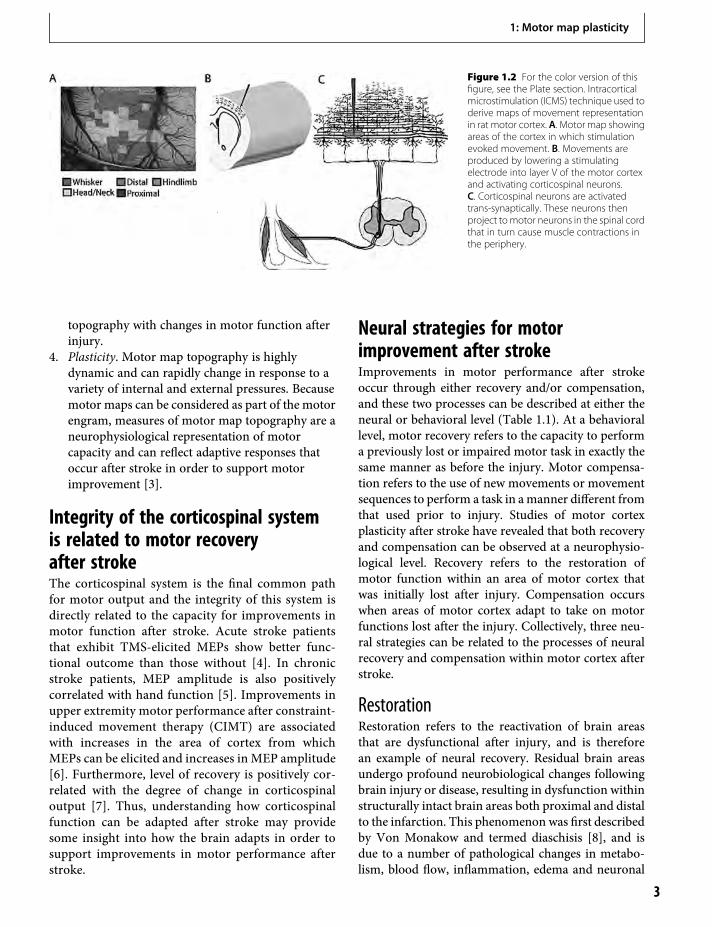
evoked potentials (MEPs) reflecting changes in elec-tromyographic (EMG) activity within discretemuscles (Figure 1.1). ICMS techniques are typicallyused in animal studies, where a microelectrode islowered into layer V of the exposed motor cortexstimulating more restricted patches of axons drivingcorticospinal neurons. In these studies the responseis measured by visual confirmation of movement(Figure 1.2) and/or EMG activity.
Decades of cortical stimulation experiments haverevealed four general principles of motor maporganization.
1. Fractured somatotopy. Individual movementsare represented multiple times and are highlyinterspersed with adjacent movementrepresentations across discrete cortical regions. Thisfunctional redundancy can contribute to the capacityfor the motor cortex to adapt to injury.
2. Interconnectivity. Corticospinal neurons fromadjacent cortical areas are densely interconnectedvia reciprocal intracortical connections. Thisprovides a platform for functional compensationafter injury and facilitates the capacity for motormap reorganization.
3. Area equals dexterity. Movements requiring agreater degree of dexterity are more easily evokedin response to stimulation and occupy a largerproportion of the map. This provides a neuralmeasure that can be compared to changes in motorbehavior and used to relate changes in motor map
Figure 1.1 Transcranial magnetic stimulation (TMS) technique used to derive maps of movement representation in human motor cortex.A. Electrodes are placed over a target muscle to measure small changes in electrical potential associated with muscle contraction in responseto magnetic stimulation of the motor cortex. In this example two electrodes are placed over the first dorsal interosseus (FDI) muscle and aground electrode on the wrist. B. Electromyograph (EMG) showing a motor-evoked potential (MEP) within the FDI after TMS (at time 0). Theamplitude and latency of the MEP can be measured and used to assess the strength of corticospinal output. C. The location of stimulation overthe cortex can be integrated with a three-dimensional MRI of the subject’s brain. This allows for the experimenter to determine the area andlocation in the cortex from which MEPs can be elicited by TMS.
1: Motor map plasticity
2
topography with changes in motor function afterinjury.
4. Plasticity. Motor map topography is highlydynamic and can rapidly change in response to avariety of internal and external pressures. Becausemotor maps can be considered as part of the motorengram, measures of motor map topography are aneurophysiological representation of motorcapacity and can reflect adaptive responses thatoccur after stroke in order to support motorimprovement [3].
Integrity of the corticospinal systemis related to motor recoveryafter strokeThe corticospinal system is the final common pathfor motor output and the integrity of this system isdirectly related to the capacity for improvements inmotor function after stroke. Acute stroke patientsthat exhibit TMS-elicited MEPs show better func-tional outcome than those without [4]. In chronicstroke patients, MEP amplitude is also positivelycorrelated with hand function [5]. Improvements inupper extremity motor performance after constraint-induced movement therapy (CIMT) are associatedwith increases in the area of cortex from whichMEPs can be elicited and increases in MEP amplitude[6]. Furthermore, level of recovery is positively cor-related with the degree of change in corticospinaloutput [7]. Thus, understanding how corticospinalfunction can be adapted after stroke may providesome insight into how the brain adapts in order tosupport improvements in motor performance afterstroke.
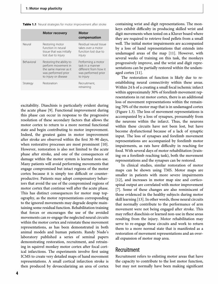
Neural strategies for motorimprovement after strokeImprovements in motor performance after strokeoccur through either recovery and/or compensation,and these two processes can be described at either theneural or behavioral level (Table 1.1). At a behaviorallevel, motor recovery refers to the capacity to performa previously lost or impaired motor task in exactly thesame manner as before the injury. Motor compensa-tion refers to the use of new movements or movementsequences to perform a task in amanner different fromthat used prior to injury. Studies of motor cortexplasticity after stroke have revealed that both recoveryand compensation can be observed at a neurophysio-logical level. Recovery refers to the restoration ofmotor function within an area of motor cortex thatwas initially lost after injury. Compensation occurswhen areas of motor cortex adapt to take on motorfunctions lost after the injury. Collectively, three neu-ral strategies can be related to the processes of neuralrecovery and compensation within motor cortex afterstroke.
RestorationRestoration refers to the reactivation of brain areasthat are dysfunctional after injury, and is thereforean example of neural recovery. Residual brain areasundergo profound neurobiological changes followingbrain injury or disease, resulting in dysfunction withinstructurally intact brain areas both proximal and distalto the infarction. This phenomenon was first describedby Von Monakow and termed diaschisis [8], and isdue to a number of pathological changes in metabo-lism, blood flow, inflammation, edema and neuronal
Figure 1.2 For the color version of thisfigure, see the Plate section. Intracorticalmicrostimulation (ICMS) technique used toderive maps of movement representationin rat motor cortex. A. Motor map showingareas of the cortex in which stimulationevoked movement. B. Movements areproduced by lowering a stimulatingelectrode into layer V of the motor cortexand activating corticospinal neurons.C. Corticospinal neurons are activatedtrans-synaptically. These neurons thenproject tomotor neurons in the spinal cordthat in turn cause muscle contractions inthe periphery.
1: Motor map plasticity
3
excitability. Diaschisis is particularly evident duringthe acute phase [9]. Functional improvement duringthis phase can occur in response to the progressiveresolution of these secondary factors that allows themotor cortex to return to a more normal functionalstate and begin contributing to motor improvement.Indeed, the greatest gains in motor improvementafter stroke are observed within the first 30–90 days,when restorative processes are most prominent [10].However, restoration is also not limited to the acutephase after stroke, and one of the consequences ofdamage within the motor system is learned non-use.Many patients will avoid performing movements thatengage compromised but intact regions of the motorcortex because it is simply too difficult or counter-productive. Patients may adopt compensatory behav-iors that avoid the use of the compromised regions ofmotor cortex that continue well after the acute phase.This has distinct consequences for motor map top-ography, as the motor representations correspondingto the ignored movements may degrade despite main-taining some residual function. Rehabilitation trainingthat forces or encourages the use of the avoidedmovements can re-engage the neglected neural circuitswithin the motor cortex and reinstate these movementrepresentations, as has been demonstrated in bothanimal models and human patients. Randy Nudo’slaboratory published a series of seminal papersdemonstrating restoration, recruitment, and retrain-ing in squirrel monkey motor cortex after focal cort-ical infarctions. The experiments involve first usingICMS to create very detailed maps of hand movementrepresentations. A small cortical infarction stroke isthen produced by devascularizing an area of cortex
containing wrist and digit representations. The mon-keys exhibit difficulty in producing skilled wrist anddigit movements when tested on a Kluver board wherethey are required to retrieve food pellets from a smallwell. The initial motor impairments are accompaniedby a loss of hand representations that extends intoundamaged areas of the map [11]. However, withseveral weeks of training on this task, the monkeysprogressively improve, and the wrist and digit repre-sentations can be partially restored within the undam-aged cortex [11].
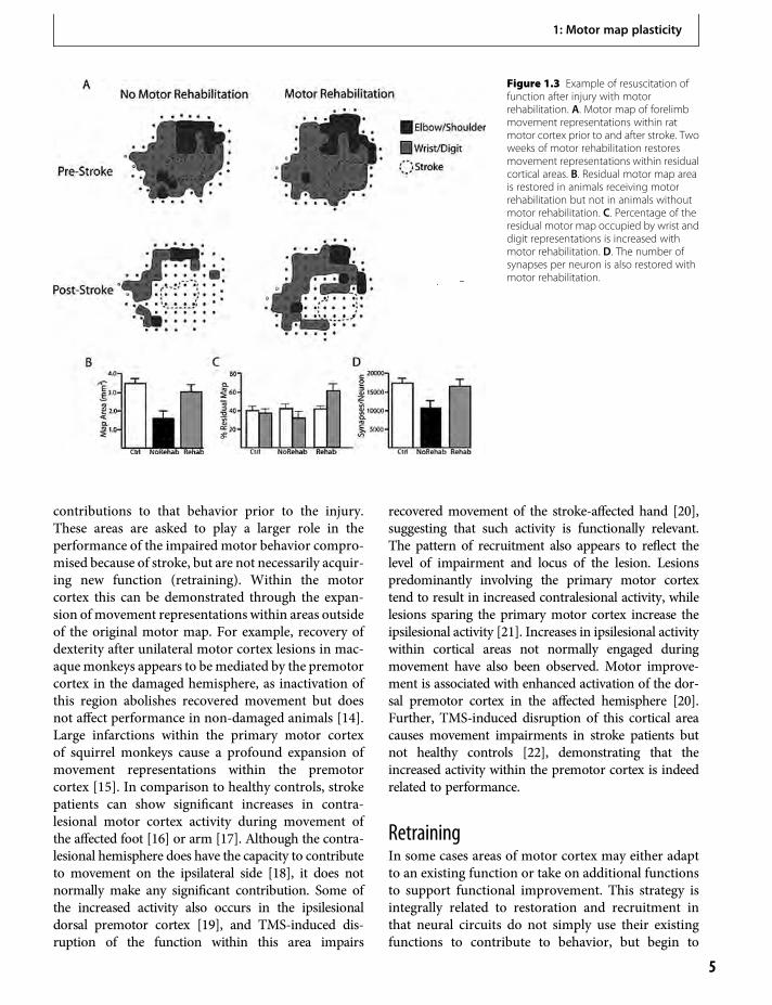
The restoration of function is likely due to re-establishing neural connectivity within these areas.Within 24 h of a creating a small focal ischemic infarctwithin approximately 30% of forelimb movement rep-resentations in rat motor cortex, there is an additionalloss of movement representations within the remain-ing 70% of the motor map that is in undamaged cortex(Figure 1.3). The loss of movement representations isaccompanied by a loss of synapses, presumably fromthe neurons within the infarct. Thus, the neuronswithin these circuits have not been lost, but havebecome dysfunctional because of a lack of synapticinput. The loss of synapses and forelimb movementrepresentations are accompanied by forelimb motorimpairments, as rats have difficulty in reaching forfood. With several days of motor rehabilitation (train-ing on a forelimb reaching task), both the movementrepresentations and the synapses can be restored.
In clinical studies, similar restoration of motormaps can be shown using TMS. Motor maps aresmaller in patients with more severe impairments[12], and increases in motor map size and cortico-spinal output are correlated with motor improvement[7]. Some of these changes are also reminiscent ofthose evidenced in the healthy subjects during motorskill learning [13]. In other words, those neural circuitsthat normally contribute to the performance of armmovement were not being engaged after stroke. Thismay reflect diaschisis or learned non-use in these areasresulting from the injury. Motor rehabilitation mayserve to re-engage these circuits and work to returnthem to a more normal state that is manifested as arestoration of movement representations and an over-all expansion of motor map area.
RecruitmentRecruitment refers to enlisting motor areas that havethe capacity to contribute to the lost motor function,but may not normally have been making significant
Table 1.1 Neural strategies for motor improvement after stroke
Motor recovery Motorcompensation
Neural Restoring motorfunction in neuraltissue that was initiallylost due to injury
Residual neural tissuetakes over a motorfunction lost due toinjury
Behavioral Restoring the ability toperform movement inthe same manner as itwas performed priorto injury or disease
Performing a motortask in a mannerdifferent from how itwas performed priorto injury
Strategy Restoration Recruitment,retraining
1: Motor map plasticity
4
contributions to that behavior prior to the injury.These areas are asked to play a larger role in theperformance of the impaired motor behavior compro-mised because of stroke, but are not necessarily acquir-ing new function (retraining). Within the motorcortex this can be demonstrated through the expan-sion of movement representations within areas outsideof the original motor map. For example, recovery ofdexterity after unilateral motor cortex lesions in mac-aque monkeys appears to be mediated by the premotorcortex in the damaged hemisphere, as inactivation ofthis region abolishes recovered movement but doesnot affect performance in non-damaged animals [14].Large infarctions within the primary motor cortexof squirrel monkeys cause a profound expansion ofmovement representations within the premotorcortex [15]. In comparison to healthy controls, strokepatients can show significant increases in contra-lesional motor cortex activity during movement ofthe affected foot [16] or arm [17]. Although the contra-lesional hemisphere does have the capacity to contributeto movement on the ipsilateral side [18], it does notnormally make any significant contribution. Some ofthe increased activity also occurs in the ipsilesionaldorsal premotor cortex [19], and TMS-induced dis-ruption of the function within this area impairs
recovered movement of the stroke-affected hand [20],suggesting that such activity is functionally relevant.The pattern of recruitment also appears to reflect thelevel of impairment and locus of the lesion. Lesionspredominantly involving the primary motor cortextend to result in increased contralesional activity, whilelesions sparing the primary motor cortex increase theipsilesional activity [21]. Increases in ipsilesional activitywithin cortical areas not normally engaged duringmovement have also been observed. Motor improve-ment is associated with enhanced activation of the dor-sal premotor cortex in the affected hemisphere [20].Further, TMS-induced disruption of this cortical areacauses movement impairments in stroke patients butnot healthy controls [22], demonstrating that theincreased activity within the premotor cortex is indeedrelated to performance.
RetrainingIn some cases areas of motor cortex may either adaptto an existing function or take on additional functionsto support functional improvement. This strategy isintegrally related to restoration and recruitment inthat neural circuits do not simply use their existingfunctions to contribute to behavior, but begin to
Figure 1.3 Example of resuscitation offunction after injury with motorrehabilitation. A. Motor map of forelimbmovement representations within ratmotor cortex prior to and after stroke. Twoweeks of motor rehabilitation restoresmovement representations within residualcortical areas. B. Residual motor map areais restored in animals receiving motorrehabilitation but not in animals withoutmotor rehabilitation. C. Percentage of theresidual motor map occupied by wrist anddigit representations is increased withmotor rehabilitation. D. The number ofsynapses per neuron is also restored withmotor rehabilitation.
1: Motor map plasticity
5

perform novel or additional functions. As describedabove, focal lesions within the motor cortex cause aloss of movement representations within residual cort-ical areas that can be restored with motor rehabilita-tion. These same studies also provide clear examples ofretraining. For example, when an area of motor cortexcontaining digit representations is removed, digit rep-resentations can be observed to re-emerge in areas ofthe remaining cortex that used to contain elbow orshoulder [23]. It is also important to point out that theemergence of a movement representation within anarea previously occupied by a different representationdoes not mean that the original representation hasbeen completely replaced. This is largely the result ofthe method used to derive motor maps. Movementrepresentations are determined by lowering theamount of current delivered until only one movementcan be observed. However, in many cases, if the stim-ulating current is increased, the previous movementappears. In other words, this particular cortical regionhas not lost the elbow representation, but has becomebiased towards wrist. In effect, the area has becomecapable of contributing to both movements. In sup-port of this notion, there is an increase in the numberof stimulation sites where such responses occur[11,24]. Operationally, dual responses are stimulationsites where two movements occur with the same stim-ulation current and the two movements cannot beclearly dissociated by reducing stimulation current.The number of these sites also positively correlateswith the degree of functional improvement.
There is now evidence that motor map reorganiza-tion is not simply an epiphenomenon of rehabilitationbut is indeed involved in supporting motor improve-ment. Conner et al. demonstrated that depleting cort-ical acetylcholinergic input by lesioning the nucleusbasalis of Meynert reduced motor recovery and motormap reorganization [25]. They further showed thatlesioning the reorganized areas of the motor map rein-stated the motor deficits. Together these results providestrong evidence that motor map plasticity is a neuralsubstrate for rehabilitation-dependent motor recovery.
Neural basis of motor cortexreorganization: synaptic plasticityThe above studies demonstrate that restoration,recruitment and retraining of motor maps withinresidual cortical tissue accompany motor recoveryafter stroke. Understanding the cellular basis and
key neural signaling pathways that drive such reor-ganization would aid in the development of adjuvanttherapies that will enhance recovery. This firstrequires understanding the neurophysiological andanatomical properties of motor cortex that underliemotor maps.
The functional organization of primary motor cor-tex has long been defined by cortical stimulationexperiments [26]. These studies have revealed twoimportant characteristics of motor cortex organiza-tion. Neurons within the motor cortex are aggregatedsuch that small groups of cells appear to encode ele-mentary movement representations in that neighbor-ing cells have similar output properties [27]. Further,these pools of neurons are interconnected by densehorizontal connections that can extend several milli-meters [28]. ICMS evokes movement via direct [25]and indirect [30] activation of pyramidal tract neu-rons. In fact, the majority of pyramidal tract neuronsthat drive movement in response to stimulation aretrans-synaptically activated [31], presumably throughactivation of horizontal afferents. Therefore, the spa-tial characteristics of motor maps, as defined by stim-ulation, are dependent upon the synaptic activation oflocalized groups of pyramidal tract neurons. Any alter-ation in motor map topography or loss of map areaafter damage must therefore involve changes in thepattern of intracortical connectivity through modifi-cations in synaptic efficacy [26].
There are several lines of evidence that support theidea that motor map reorganization is dependentupon changes in synaptic efficacy within intracorticalmicrocircuitry. First, manipulations that inducechanges in synaptic strength also induce map reorgan-ization. Long-term potentiation (LTP) causes mapexpansion and synaptogenesis [32], while long-termdepression (LTD) induces map retraction and synapticloss. Second, cortical kindling that drives increases incortical excitatory postsynaptic potentials (EPSPs) alsoincreases motor map area [33]. Third, synaptic poten-tiation that occurs in response to motor skill learning[32,34] is colocalized within regions of cortex thatexhibit motor map reorganization [35]. Finally,learning-dependent motor map plasticity is also colo-calized with synaptogenesis [36]. Thus, reorganizationof motor maps after brain injury and in response tomotor rehabilitation must be supported by changes insynaptic efficacy within motor cortex. Indeed, motormap reorganization is accompanied by synaptogenesiswithin residual motor cortex (Figure 1.2).
1: Motor map plasticity
6
Adjuvant therapies that promotemotor map plasticity enhance motorrecovery after strokeThe reorganization of cortical movement representa-tions is likely mediated by synaptic plasticity withinintracortical microcircuitry. In normal motor learning,regions of motor cortex that undergo redistributionof movement representations during skill learningalso show increases in synapse number [37]. Further,these same regions also demonstrate enhanced synapticresponses after skill learning [32]. In addition, manip-ulations that alter synaptic strength or number alsochange motor map topography. Finally, restoration ofmovement representations within residual corticalareas is accompanied by increases in synapse number(Figure 1.3). Given that such synaptic plasticity is akey neural mechanism mediating motor map plasticityand motor improvement after stroke, manipulationsthat enhance synaptic plasticity may serve to augmentmotor recovery. Molecular, genetic, pharmacologicaland electrophysiological studies in a variety of organ-isms have revealed several key neural signaling systemscritical for orchestrating synaptic plasticity. These stud-ies have inspired the development of several adjuvant,plasticity-promoting therapies for enhancing motormap plasticity and concomitant motor improvementafter stroke.
Pharmacological stimulationOne approach to developing adjuvant therapies isto develop pharmacological manipulations that upre-gulate endogenous intracellular signaling pathwaysthat drive synaptic plasticity. Although numeroussignaling pathways have been identified, the mostwell characterized is the cAMP/CREB pathway. Avariety of experimental models and systems haveestablished the cAMP/CREB signaling pathway to bea key regulatory pathway in experience-dependentsynaptic plasticity. Administration of the type IV-specific phosphodiesterase inhibitors (PDE 4) thatenhance cAMP/CREB signaling facilitate memory innormal and aged rodents [3]. PDE treatment in com-bination with motor rehabilitation following a focalstroke significantly enhanced motor recovery [38].Further, the drug increased motor map area in resid-ual cortex (restoration), increased the proportion ofthe maps occupied by distal forelimb representations(retraining) and expanded movement representationsinto new cortical areas (recruitment).
In addition to drugs that enhance plasticity, severalneurochemicals have been identified that may blockplasticity inhibiting processes such as axonal sprout-ing. For example, myelin-associated inhibitory factors(such as Nogo-A or MAG) are present in neural tissuethat block neurite outgrowth after damage. Whenantibodies for Nogo-A (IN-1) are applied to motorcortex following ischemic lesion, an increase in apicaland basilar dendritic arborization and spine density isobserved [39]. In addition, new projections to the de-afferented striatum [40] and red nucleus [41] havebeen observed. ICMS mapping of the intact hemi-sphere following treatment with monoclonal antibodyIN-1 results in a substantial increase in ipsilateralmovements [42]. Such manipulations could facilitatecompensation through recruitment of distal brainareas.
Cortical stimulationThere is a growing body of evidence that electricallystimulating the motor cortex facilitates recovery ofmotor function after CNS injury. In humans, trans-cranial direct cortical stimulation (tDCS) improvesmotor function in patients with chronic motor impair-ments when anodal current is delivered over lesionedmotor cortex or cathodal current was delivered overthe contralesional motor cortex [43]. Clinical reportssuggest that epidural motor cortex stimulation, usedto reduce chronic pain after subcortical strokes,reduces hemiparetic impairments [44], motor weak-ness [45], motor spasticity [46], action tremor [47] anddystonia [14].
The efficacy of CS-RT (cortical stimulation reha-bilitation therapy) at enhancing motor recovery afterstroke has been demonstrated in rats [48,49] and inmonkeys [50]. Furthermore, the enhanced motorrecovery is associated with increased cortical dendritichypertrophy [49] in comparison to animals in stand-ard rehabilitation. The increased postsynaptic spaceis also accompanied by an enlargement of the poly-synaptic component of motor cortical-evoked poten-tials [51]. Finally, CS-RT also induces a greaterexpansion of movement representations in rats [48]and monkeys [50]. All of these data demonstrate thatCS-RT drives significantly greater motor recoveryafter stroke and that the functional gains are accom-panied by an upregulation of the neuroplastic changesobserved with standard rehabilitation. Although themeans by which CS-RT enhances recovery and pro-motes cortical plasticity are unknown, upregulation
1: Motor map plasticity
7
of neurotrophins is one viable mechanism. Indeed,electrical stimulation of cortical tissue increasesbrain-derived neurotrophic factor (BDNF) levels [52].
Genetic factorsThe neural signals that drive neural plasticity ofteninvolve altering the expression of specific genesthat coordinate the synthesis of specific proteinsrequired for synaptic plasticity and motor map reor-ganization. Naturally occurring genetic alterations inthose plasticity-related genes may then influence thecapacity for motor map plasticity. Indeed, such geneticpolymorphisms have been identified in the humanpopulation that may influence both the capacityfor motor map plasticity and functional improvementafter stroke. For example, a polymorphism hasbeen identified in the human BDNF gene (BDNFval66met). BDNF polymorphic individuals showreduced training-dependent motor map expansionin comparison to non-polymorphic subjects [53].Furthermore, val66met subjects show reductions inmotor cortex plasticity after paired association stim-ulation and intermittent theta burst stimulation [54].This reduced capacity for cortical plasticity is pro-posed to be due to a reduction in the capacityfor synaptic plasticity within motor cortical neurons.Because motor map plasticity supports motorimprovement after stroke, it is possible that theseindividuals may not respond to motor rehabilitationthe same way the non-polymorphic subjects do, limit-ing their capacity for recovery. Indeed, after subarach-noid hemorrhage patients with the polymorphism arethree times more likely to have poor recovery thannon-polymorphic individuals [55]. Thus, understand-ing the relationship between genotype and thecapacity for motor cortex plasticity may be a criticalstep towards developing effective motor rehabilitationinterventions. Such polymorphic individuals mightrequire more or different forms of rehabilitation inorder to drive motor cortex plasticity. Further, theefficacy of adjuvant therapies such as those describedabove may be affected by genotype.
ConclusionsThere is now significant evidence that motor mapplasticity is one of the key neural substrates supportingmotor improvement after stroke. This plasticityappears to fall into one of three general strategies:restoration, retraining, and recruitment that are notmutually exclusive. All these three strategies also
appear to be mediated by synaptic plasticity withincortical circuits to allow for behavioral recovery andcompensation. Understanding the neural signalingpathways that influence such synaptic plasticity mayguide the development of novel, adjuvant therapiesthat can upregulate endogenous plasticity mechanismsand enhance motor improvement after stroke whenadministered in conjunction with motor rehabilita-tion. Finally, the fundamental behavioral and neuralsignals that drive motor map plasticity and improve-ments in motor function may also mediate enhance-ment of non-motor functions impaired after strokesuch as memory or language processing. Thus, studiesof motor map plasticity and motor improvement afterstroke may provide important insights into potentialtherapies for overcoming various non-motor func-tional impairments such as cognitive deficits observedafter stroke.
References1. Hughlings Jackson J. Remarks on the relations of
different divisions of the central nervous system toone another and to parts of the body. Br MedJ. 1898;1:65–9.
2. Fritz G, Hitzig, E. Über die elektrische Erregbarkeit desGrosshirns. Arch Anat Physiol wiss Med.1870;37:300–32.
3. Monti B, Berteotti C, Contestabile A. Subchronicrolipram delivery activates hippocampal CREB and arc,enhances retention and slows down extinction ofconditioned fear. Neuropsychopharmacology.2006;31:278–86.
4. Escudero JV, Sancho J, Bautista D, et al. Prognosticvalue of motor evoked potential obtained bytranscranial magnetic brain stimulation in motorfunction recovery in patients with acute ischemicstroke. Stroke. 1998;29:1854–9.
5. Brouwer BJ, Schryburt-Brown K. Hand function andmotor cortical output poststroke: are they related? ArchPhys Med Rehabil. 2006;87:627–34.
6. Wittenberg GF, Chen R, Ishii K, et al. Constraint-induced therapy in stroke: Magnetic-stimulation motormaps and cerebral activation. Neurorehabil NeuralRepair. 2003;17:48–57.
7. Koski L, Mernar TJ, Dobkin BH. Immediate and long-term changes in corticomotor output in response torehabilitation: Correlation with functionalimprovements in chronic stroke. Neurorehabil NeuralRepair. 2004;18:230–49.
8. vonMonakow C.Die Lokalisation im Grosshirn und derAbbau der Funktion durch kortikale Herde. Weisbaden:Bergmann; 1914.
1: Motor map plasticity
8
9. Cramer SC. Repairing the human brain after stroke:I. Mechanisms of spontaneous recovery. Ann Neurol.2008;63:272–87.
10. Duncan PW, Goldstein LB, Matchar D, et al.Measurement of motor recovery after stroke. Outcomeassessment and sample size requirements. Stroke.1992;23:1084–9.
11. Nudo RJ, Milliken GW. Reorganization of movementrepresentations in primarymotor cortex following focalischemic infarcts in adult squirrel monkeys.J Neurophysiol. 1996;75:2144–9.
12. Talelli P, Rothwell J. Does brain stimulation after strokehave a future? Curr Opin Neurol. 2006;19:543–50.
13. Karni A, Meyer G, Jezzard P, et al. Functional MRIevidence for adult motor cortex plasticity during motorskill learning. Nature. 1995;377:155–8.
14. Franzini A, Ferroli P, Dones I, et al. Chronic motorcortex stimulation for movement disorders: Apromising perspective. Neurol Res. 2003;25:123–6.
15. Frost SB, Barbay S, Friel KM, et al. Reorganization ofremote cortical regions after ischemic brain injury: Apotential substrate for stroke recovery. J Neurophysiol.2003;89:3205–14.
16. Enzinger C, Johansen-Berg H, Dawes H, et al.Functional MRI correlates of lower limb function instroke victims with gait impairment. Stroke.2008;39:1507–13.
17. Zemke A, Heagerty P, Lee C, et al. Motor cortexorganization after stroke is related to side of stroke andlevel of recovery. Stroke. 2003;34:E23–8.
18. Jankowska E, Edgley SA. How can corticospinal tractneurons contribute to ipsilateral movements? Aquestion with implications for recovery of motorfunctions. Neuroscientist. 2006;12:67–79.
19. Gerloff C, Braun C, Staudt M, et al. Coherentcorticomuscular oscillations originate from primarymotor cortex: Evidence from patients with early brainlesions. Hum Brain Mapp. 2006;27:789–98.
20. Feydy A, Carlier R, Roby-Brami A, et al. Longitudinalstudy of motor recovery after stroke: recruitment andfocusing of brain activation. Stroke. 2002;33:1610–7.
21. Lotze M, Cohen LG. Volition and imagery inneurorehabilitation. Cogn Behav Neurol.2006;19:135–40.
22. Weiller C, Chollet F, Friston KJ, et al. Functionalreorganization of the brain in recovery fromstriatocapsular infarction in man. Ann Neurol.1992;31:463–72.
23. Fridman EA, Hanakawa T, Chung M, et al.Reorganization of the human ipsilesional premotorcortex after stroke. Brain. 2004;127:747–58.
24. Barbay S, Plautz EJ, Friel KM, et al. Behavioral andneurophysiological effects of delayed training
following a small ischemic infarct in primary motorcortex of squirrel monkeys. Exp Brain Res.2006;169:106–16.
25. Conner JM, Chiba AA, Tuszynski MH. The basalforebrain cholinergic system is essential for corticalplasticity and functional recovery following braininjury. Neuron. 2005;46:173–9.
26. Monfils MH, Plautz EJ, Kleim JA. In search of themotor engram: Motor map plasticity as a mechanismfor encoding motor experience. Neuroscientist.2005;11:471–83.
27. Mountcastle VB. The columnar organization of theneocortex. Brain. 1997;120:701–22.
28. Ghosh S, Porter R. Morphology of pyramidal neuronesin monkey motor cortex and the synaptic actions oftheir intracortical axon collaterals. J Physiol.1988;400:593–615.
29. Jankowska E, Padel Y, Tanaka R. The mode ofactivation of pyramidal tract cells by intracorticalstimuli. J Physiol. 1975;249:617–36.
30. Stoney SD, Jr., ThompsonWD, Asanuma H. Excitationof pyramidal tract cells by intracorticalmicrostimulation: Effective extent of stimulatingcurrent. J Neurophysiol. 1968;31:659–69.
31. Cheney PD, Fetz EE, Palmer SS. Patterns of facilitationand suppression of antagonist forelimb muscles frommotor cortex sites in the awake monkey. J Neurophysiol.1985;53:805–20.
32. Monfils MH, Teskey GC. Skilled-learning-inducedpotentiation in rat sensorimotor cortex: A transientform of behavioural long-term potentiation.Neuroscience. 2004;125:329–36.
33. Teskey GC, Monfils MH, VandenBerg PM, et al. Motormap expansion following repeated cortical and limbicseizures is related to synaptic potentiation. CerebCortex. 2002;12:98–105.
34. Rioult-Pedotti MS, Friedman D, Hess G, et al.Strengthening of horizontal cortical connectionsfollowing skill learning. Nat Neurosci. 1998;1:230–4.
35. Kleim JA, Barbay S, Nudo RJ. Functionalreorganization of the rat motor cortex following motorskill learning. J Neurophysiol. 1998;80:3321–5.
36. Kleim JA, Barbay S, Cooper NR, et al. Motor learning-dependent synaptogenesis is localized to functionallyreorganized motor cortex. Neurobiol Learn Mem.2002;77:63–77.
37. Kleim JA, Hogg TM, VandenBerg PM, et al. Corticalsynaptogenesis and motor map reorganization occurduring late, but not early, phase of motor skill learning.J Neurosci. 2004;24:628–33.
38. MacDonald E, Van der Lee H, Pocock D, et al. A novelphosphodiesterase type 4 inhibitor, HT-0712, enhancesrehabilitation-dependent motor recovery and cortical
1: Motor map plasticity
9
reorganization after focal cortical ischemia.Neurorehabil Neural Repair. 2007;21:486–96.
39. Papadopoulos CM, Tsai SY, Cheatwood JL, et al.Dendritic plasticity in the adult rat following middlecerebral artery occlusion and Nogo-a neutralization.Cereb Cortex. 2006;16:529–36.
40. Kartje GL, Schulz MK, Lopez-Yunez A, et al.Corticostriatal plasticity is restricted by myelin-associated neurite growth inhibitors in the adult rat.Ann Neurol. 1999;45:778–86.
41. Wenk CA, Thallmair M, Kartje GL, et al. Increasedcorticofugal plasticity after unilateral cortical lesionscombined with neutralization of the IN-1 antigen inadult rats. J Comp Neurol. 1999;410:143–57.
42. Emerick AJ, Neafsey EJ, Schwab ME, et al. Functionalreorganization of the motor cortex in adult rats aftercortical lesion and treatment with monoclonal antibodyIN-1. J Neurosci. 2003;23:4826–30.
43. Fregni F, Pascual-Leone A. Hand motor recoveryafter stroke: Tuning the orchestra to improvehand motor function. Cogn Behav Neurol.2006;19:21–33.
44. Tsubokawa T, Katayama Y, Yamamoto T, et al. Chronicmotor cortex stimulation in patients with thalamicpain. J Neurosurg. 1993;78:393–401.
45. Katayama Y, Oshima H, Fukaya C, et al. Control ofpost-stroke movement disorders using chronic motorcortex stimulation. Acta Neurochir Suppl.2002;79:89–92.
46. Garcia-Larrea L, Peyron R, Mertens P, et al. Electricalstimulation of motor cortex for pain control: Acombined PET-scan and electrophysiological study.Pain. 1999;83:259–73.
47. Nguyen JP, Pollin B, Feve A, et al. Improvement ofaction tremor by chronic cortical stimulation. MovDisord. 1998;13:84–8.
48. Kleim JA, Bruneau R, VandenBerg P, et al. Motorcortex stimulation enhances motor recovery andreduces peri-infarct dysfunction following ischemicinsult. Neurol Res. 2003;25:789–93.
49. Adkins-Muir DL, Jones TA. Cortical electricalstimulation combined with rehabilitative training:enhanced functional recovery and dendritic plasticityfollowing focal cortical ischemia in rats. Neurol Res.2003;25:780–8.
50. Plautz EJ, Barbay S, Frost SB, et al. Post-infarct corticalplasticity and behavioral recovery using concurrentcortical stimulation and rehabilitative training: Afeasibility study in primates. Neurol Res.2003;25:801–10.
51. Teskey GC, Flynn C, Goertzen CD, et al. Corticalstimulation improves skilled forelimb use following afocal ischemic infarct in the rat. Neurol Res.2003;25:794–800.
52. Yukimasa T, Yoshimura R, Tamagawa A, et al. High-frequency repetitive transcranial magnetic stimulationimproves refractory depression by influencingcatecholamine and brain-derived neurotrophic factors.Pharmacopsychiatry. 2006;39:52–9.
53. Kleim JA, Chan S, Pringle E, et al. BDNFval66metpolymorphism is associated with modified experience-dependent plasticity of human motor cortex. NatNeurosci. 2006;9:735–7.
54. Cheeran B, Talelli P, Mori F, et al. A commonpolymorphism in the brain-derived neurotrophic factorgene (BDNF) modulates human cortical plasticity andthe response to rTMS. J Physiol. 2008;586(Pt 23):5717–25.
55. Siironen J, Juvela S, Kanarek K, et al. The Met allele ofthe BDNF Val66Met polymorphism predicts pooroutcome among survivors of aneurysmal subarachnoidhemorrhage. Stroke. 2007;38:2858–60.
1: Motor map plasticity
10

2 Molecular mechanisms of neuralrepair after strokeS. Thomas Carmichael
Stroke induces a limited process of neuronal reorgan-ization, repair and recovery. This reorganizationincludes axonal and dendritic sprouting in cortexipsi- and contralateral to the stroke, formation ofnew patterns of short- and long-distance connectionswithin sensorimotor cortical, striatal, brainstem andspinal circuits, and migration of newly born immatureneurons into damaged tissue. From the formerly staidperspective of the adult brain, these processes areremarkable examples of structural plasticity. Withinthe past several years, some of the molecular systemsthat underlie these processes have been determined.With a better understanding of the molecular controlof neural repair after stroke it may be possible to har-ness these processes to promote functional recovery.This chapter will describe axonal and dendritic sprout-ing and neurogenesis after stroke, and detail themolecular systems that may operate within definedcellular contexts to promote structural reorganizationin the adult brain after stroke.
Cellular concepts of neural repairafter strokeAxonal and dendritic sprouting occur in cortex ipsi-lateral and contralateral to the infarct. Axonal sprout-ing in peri-infarct cortex in rodent models of focalstroke establishes new patterns of connections withinsensorimotor maps [1]. As detected directly with high-resolution mapping of cortical connections, axonalsprouting occurs in a subset of the total cortical con-nections in sensory and motor areas. This sprouting issubstantial enough that it re-maps the predominantpattern of cortical connections in, for example, thefacial somatosensory map of the rat or mouse [1,2].In non-human primates, small strokes in the motorcortex induce a remarkable long-distance sprouting incortical connections from parietal to frontal cortex
within a system of motor to somatosensory connec-tions [3]. These findings of axonal sprouting and reor-ganization of cortical connections after stroke aresupported by similar reports of axonal sprouting incortex after peripheral de-afferentation, such as retinal[4] and peripheral nerve lesions [5,6]. Axonal sprout-ing in the peri-infarct cortex occurs at a time of rapiddendritic plasticity and spine turnover in the peri-infarct cortex [7,8]. Post-stroke axonal and dendriticsprouting may account for the changes in receptivefield maps in reorganizing sensorimotor representa-tions in peri-infarct cortex [9]. However, the directphysiological significance of axonal and dendriticsprouting in peri-infarct cortex remains to be deter-mined. Its timing is closely linked to the most dramaticperiods of behavioral recovery and occurs in a regionin which cortical re-mapping is closely associated withsuccessful recovery of function [10].
Stroke also induces axonal sprouting from neuronsin cortex contralateral to the infarct. After ischemiclesions, neurons in contralateral cortex sprout in theirprojections to the contralateral striatum, midbrain,and cervical spinal cord, and into the region of theperi-infarct cortex itself [11–14]. These regions thatreceive projections have been de-afferented as a resultof the stroke and, from an anatomical perspective, thesprouting projections appear to elaborate a distalbranch into the zone that was de-afferented. For exam-ple, axonal sprouting from contralateral (to the lesion)cortex into ipsilateral dorsal striatum establishes aprojection into that region of the striatum that lostits projections as a result of the stroke [13,14]. Inaxonal sprouting into the red nucleus or cervical spinalcord, sprouting axons appear to take off from existing,normally present, projections to the red nucleus orcervical spinal cord ipsilateral to the stroke. Thesecollateral sprouts then grow the short distances intothe region of red nucleus and cervical spinal cord that
Brain Repair After Stroke, ed. S. C. Cramer and R. J. Nudo. Published by Cambridge University Press.© Cambridge University Press 2010.
11

previously received a projection from the now-infarcted cortex. This sprouting from contralateralcortex can be increased by treatments that promoteneuronal growth, such as inosine [1], or block myelin-associated growth inhibitors, such as NogoA [12].The degree of axonal sprouting from contralateralcortex after stroke correlates with functional recovery.However, as with peri-infarct cortical sprouting afterstroke, there have been no studies that have causallylinked contralateral cortical sprouting with functionalrecovery.
Axonal sprouting in peri-infarct cortex and axonalsprouting from contralateral cortex after strokediffer in several important anatomical characteristics.Axonal sprouting from contralateral cortex afterstroke into red nucleus and spinal cord, and possiblydorsolateral striatum, arises from normal, undamagedcorticofugal axons into a brain region that is distantfrom the stroke site, but de-afferented or disconnectedfrom it. There is no direct ischemic damage from thestroke at the site of the axonal sprouting, or at the siteof origin of the cell bodies of the sprouting neurons.These areas that are de-afferented from a cortical orcorticostriatal infarct do havemicroglial and astrocyticactivation from the degenerating axons [15]. The acti-vation of these two cell types as a result of Walleriandegeneration in distant projection zones of an infarctmay play a role in axonal sprouting from contralateralcortex after stroke. However, this localized microglialand astrocytic reaction is a very different anatomicalsituation from the tissue reaction that occurs in thesetting of peri-infarct axonal sprouting. In peri-infarctcortex neurons sprout new connections within corticalfields in which there is adjacent direct damage fromthe stroke, partial ischemic neuronal dropout, wide-spread activation of inflammatory cytokines and leu-kocyte infiltration, local angiogenesis, and distributedinduction of glial growth-inhibitory proteins [16,17].As will be discussed below, in developmental studiesthe molecular control of distal axonal branch forma-tion can differ from the overall control of neuronalgrowth cone function [18]. By analogy, it may be thataxonal sprouting from neurons within the peri-infarctcortex is under a different molecular control thandistal axonal branch formation into midbrain andcervical spinal cord.
Stroke induces a process of neurogenesis andmigra-tion of immature neurons to areas of damage. Inmiddlecerebral artery infarct models this post-stroke neuro-genesis sends immature neurons into the striatum
adjacent to the main neurogenic zone, the subventricu-lar (or subependymal) zone (SVZ). In smaller corticalinfarcts, immature neurons migrate long distances intoperi-infarct cortex [19,20]. Immature neurons migratewith [21,22], or localize to [19] angiogenic blood vesselsin peri-infarct cortex and striatum. Angiogenesis iscausally linked to neurogenesis after stroke [19]. Thesefindings suggest that there is a close molecular relation-ship between endothelial cells and immature neurons ina post-stroke neurovascular niche. However, astrocytesform a third cellular component to this niche [23], andare also activated during post-stroke neurogenesis [24].Molecular signaling systems that might communicateamong immature neurons, blood vessels and astrocytesin post-stroke neurogenesis will be discussed below.
Gene expression profilingand biological meaningGene expression profiling in a high throughput andmassively parallel manner developed in the mid to late1990s. Using microarray analysis or polymerase chainreaction (PCR)-based approaches it became possible toassay large gene pools and later the expression patternof the entire genome in various time points or tissuelocations after stroke. The biological interpretation ofthese data sets is limited because of the many variablesthat occur in the sampled time points or brainregions: angiogenesis, neurogenesis, astrocytosis, axo-nal sprouting, apoptosis, axonal degeneration and sev-eral distinct processes of inflammation. It is difficult tosort through the large tables of differentially regulatedgenes and ascribe these genes to specific biologicalevents in repair and recovery. None the less, severalimportant findings come out of these gene expressionscreens in terms of neural repair. Genes that have aprominent role in neuronal plasticity are upregulatedin peri-infarct cortex at very early time points, such as c-jun, SPRR1, NARP, and CPG21 activation 1 day afterstroke in peri-infarct cortex [25,26]. There are substan-tial changes in gene expression in peri-infarct and con-tralateral cortex during late periods in reorganizationand repair after stroke [27,28]. The pattern of geneexpression in the brain in the first and second weekafter stroke differs with respect to age, and implicatesseveral molecular pathways or cellular systems in theinterplay of stroke repair with aging: several Notch-related genes, TGFb1, activin and the growth factor-related genes FGF22, NGFb, IGF1 receptor, and IGF2are upregulated in the young adult and not aged
2: Molecular mechanisms of neural repair after stroke
12
brain after stroke [28,29]. Notch and TGFb pathwaysplay a role in axonal sprouting and neurogenesis [30].Insulin-like growth factors regulate axonal sprouting ofcortical neurons during development [31] and neuro-genesis both in the normal and post-stroke state [29].This age effect in induction of these genes systems mayrelate to differences in recovery in the aged vs. youngadult brain. In this chapter molecular events, includingtranscriptional changes, will be described not as lists ofregulated genes but within the cellular context of neuralrepair and recovery.
Post-stroke neuronal sprouting:growth promoting molecularprogramsNeuronal sprouting after stroke or other forms of CNSinjury involves the interaction of two molecular pro-grams that are both set in play within a broad regionof peri-infarct tissue. Stroke activates a neuronalgrowth program in sprouting neurons and induces aset of glial inhibitory molecules. Cortical neurons areinduced to sprout new connections over a broad zone,as these novel connections can be detected in the rangeof millimeters in the rodent to centimeters in theprimate in peri-infarct cortex [1,3]. In counterpoint,axonal growth inhibitory molecules, produced in largepart by activated astrocytes, are present not just in thelocal vicinity of the immediate stroke scar, but areupregulated throughout a range of peri-infarct tissue.The chondroitin sulfate proteoglycan neurocan isinduced in a very broad region of peri-infarct cortexand striatum after stroke, well beyond the local glialscar [17,32]. NogoA is induced in neurons throughoutthe contralateral and ipsilateral cortex after stroke[33,34] and the developmentally regulated growthinhibitory molecules neuropilin 1 (semaphorin 3areceptor) and ephrin A5 are induced in cells extendingwell away from the glial scar in peri-infarct cortex[17,35]. Thus, in at least one area of post-stroke axonalsprouting, competing cellular programs of growthpromotion and growth inhibition physically overlapwithin a region of active re-mapping and recovery offunction. This overlap suggests that therapies that tipthe balance toward axonal sprouting might improverecovery in peri-infarct cortex.
Axonal sprouting occurs across several temporalepochs of a neuronal growth program that is inducedby stroke. The initial damage of the ischemic stimulusinitiates an axonal growth response in adjacent or
connected brain regions within the first week afterstroke. Dendritic spine turnover in peri-infarct cortexaccelerates 5–8-fold [8] and ultrastructural evidence ofsynapse number and axon terminals indicates axonalsprouting [7]. The timing of the ischemic stimulus foraxonal sprouting is early after stroke and unique tothe pathophysiology of acute, focal ischemia. Non-ischemic brain lesions induce a more limited axonalsprouting response than ischemic lesions [13,36,37].Waves of highly correlated neuronal discharges withinthe first three days of the stroke induce axonal sprout-ing in at least one model of ischemic cortical damage[13]. During this early phase of axonal sproutingspecific genes are activated that may initiate post-stroke axonal sprouting. These include the transcrip-tion factor c-jun, the growth cone phosphoproteinsGAP43 and CAP23, and the cytoskeletal modifyingprotein SPRR1 [17,26,38,39]. These proteins have allbeen linked to the initiation of axonal outgrowth andregenerative axonal sprouting, through direct effectson growth cone signaling, transcriptional induction ofa growth program or modification of the actin cytos-keleton [40–42]. During this early initiation period ofaxonal sprouting after stroke, neurons in peri-infarctcortex either express or become associated withVEGF1 and 2 in a region of cortex in which ischemiapotentiates Hif1 signaling [43,44]. Hif1 induces VEGFand other genes that are linked to angiogenesis, suchas erythropoietin, but also genes that play a potent rolein neurite outgrowth (EPO and VEGF) and neuro-genesis responses (EPO, VEGF, and SDF-1, reviewedin [45]).
At later phases in the axonal sprouting responsecytoskeletal and cell adhesion molecules are induced.Tα1 tubulin, an embryonic tubulin isoform that isinduced in regenerating neurons [46], is activated atday 14 after stroke. p21/waf1, a protein originallyidentified as a cyclin-dependent kinase inhibitor, isalso induced at this time point. p21 inhibits Rho kinaseto promote axonal outgrowth [47] and is induced byHif1 [48], suggesting both a mechanistic role in peri-infarct neuronal sprouting and a link to the earlyinduction of Hif1 after ischemia in this region. Theaxonal guidance molecule L1 and the growth conephosphoprotein MARCKS are also induced in thisintermediate period in post-stroke axonal sprouting.MARCKS, CAP23 and GAP43 mediate PI3 kinasesignal transduction in the growth cone and promoteactin-associated motility [49]. Over-expression ofCAP23 and GAP43 act together to promote axonal
2: Molecular mechanisms of neural repair after stroke
13

sprouting into the spinal cord [40]. At one month afterstroke new patterns of cortical connections can bedetected neuroanatomically [1,2,13]. In axonal sprout-ing in the hippocampal system or after peripheralnerve lesion, this time period demarcates a termina-tion phase of axonal sprouting. The exact period inwhich synaptogenesis occurs within post-stroke axo-nal sprouting is not established, but long-term changesin synaptic markers can be detected after this time [50]and so the one-month period after stroke has beenidentified as a termination or, possibly more accu-rately, a maturation phase of post-stroke axonalsprouting [10]. During this late phase of post-strokeaxonal sprouting the stathmin family genes SCG10and SCLIP are upregulated [17,27]. Stathmin familyproteins interact with and destabilize microtubules toallow for the change and structural turnover necessaryfor axonal growth [51]. In contrast to their late induc-tion in stroke, these genes are induced at early timepoints in peripheral nerve regeneration [52,53]. Thelate induction of MARCKS, SCG10 and SCLIP inpost-stroke vs. peripheral nerve sprouting highlightsimportant differences in the molecular underpinningsof central vs. peripheral axonal sprouting.
The induction of a neuronal growth state during anaxonal sprouting response has been more completelyanalyzed in peripheral nerve and optic nerve injurymodels. The anatomy of these systems allows moreselective (dorsal root ganglion, DRG) or nearly com-plete (retinal ganglion cell, RGC) isolation of theregenerating neurons. In comparison, the neuronsthat sprout new connections in the brain after strokerepresent a relatively small subset of all neurons withina given region [1,3]. The tissue level of RNA isolationthat has been reported for gene expression analysis inthe post-stroke brain mixes the mRNA signals fromthis small population of cells that sprout after strokewith thousands of non-sprouting neurons (withdiverse phenotypes), endothelial cells, inflammatorycells, and glia.
The gene expression studies from isolated regen-erating DRGs and RGCs develop several importantprinciples. Regeneration in peripheral nerve can bedriven by transcription factors that control a cassetteof expressed genes which mediate axonal outgrowth,possible “master switches” for nerve regeneration[52,54]. Stat3 and ATF3 are such transcription factorsthat are induced in regenerating DRG, and mediateperipheral nerve regeneration over inhibitory sub-strates [54,55]. An intrinsic growth program is likely
to involve an orchestrated response of adhesion,membrane-associated, and intracellular signalingmol-ecules. Over-expressing a subset of growth-associatedproteins, such as α7 integrin and GAP43, does notinduce regeneration in poorly regenerating adult neu-rons [56]. Regenerating neurons activate a transcrip-tional profile that is overlapping with, but distinctfrom, developing neurons. Both RGCs and DRGs acti-vate genes specific to regeneration, such as SPRR1a,galanin, GADD45, and moesin [42,57,58]. Further,in vitro studies suggest that axonal inhibitory mole-cules, such as the chondroitin sulfate proteoglycanaggrecan, activate regenerating DRG neurons in away that is distinct from the predominant signalingform for axonal growth in development: Akt and ERKsignaling are important second messenger signals indevelopment whereas integrin-based systems are moreimportant for regeneration [59]. In a phrase, regener-ation does not fully recapitulate development. A fullcharacterization of the molecular program that under-lies post-stroke axonal sprouting will require selectiveisolation of the sprouting neurons after stroke, in asimilar fashion to what has been done with DRG andRGC neurons.
Post-stroke neuronal sprouting:growth inhibitory molecularprogramsThe nature and role of proteins that inhibit axonalsprouting after CNS injury have been extensivelyreviewed [60,61]. These belong to three broad classes.Myelin-associated inhibitory proteins include myelinassociated glycoprotein (MAG), oligodendrocyte mye-lin glycoprotein (OMgp), and NogoA. Extracellularmatrix inhibitory proteins include chondroitin sulfateproteoglycans (CSPGs), heparin sulfate proteoglycans,NG2, and tenascin. Developmentally associated inhib-itory proteins are termed this because their mostprominent role is in regulation of axonal pathfindingin the developing CNS. These include ephrins, sema-phorins, and slits. The role of many of these moleculesin axonal growth inhibition is increasingly defined inspinal cord and optic nerve injury models [60,61].Surprisingly, there has not been much study of theseinhibitory systems in their normal, endogenousresponse to stroke. Instead, these have often beenstudied only in the context of a treatment, such asdelivery of anti-Nogo, cytokine, or growth factor treat-ments, or stem/progenitor cell delivery. To determine
2: Molecular mechanisms of neural repair after stroke
14
the biology of growth inhibitory proteins after strokeand their relationship to the glial scar, and to moredistant peri-infarct brain regions, recent studies havemapped candidate growth inhibitory proteins afterstrokes in specific relationship to the phases of axonalsprouting. The question in these studies is: who is in aposition to block axonal sprouting after stroke whenthis process is just getting ramped up? The datasuggest that, of the potentially broad number of can-didate inhibitory proteins in CNS injury, only a smallsubset are active in the right place and the righttime after stroke to be in a position to block axonalsprouting.
EphrinA5, EphB1, neurocan, MAG, and neuropi-lin 1 are induced early after stroke and persist for twoweeks. These genes are induced near the infarctcore, and extending away from the infarct core intoperi-infarct striatum and cortex [17,32,35,62].Semaphorin 3a, the ligand for neuropilin 1, andNG2, a proteoglycan, are induced at two weeks afterstroke [17]. These data indicate that three very differ-ent molecular systems might mediate inhibition ofpost-stroke axonal sprouting: ephrinA5, neurocan,and semaphorin3a/neuropilin 1. Ephrin A5 andMAG are of interest because they are further inducedin the aged brain after stroke, and may relate to thediminished axonal sprouting and functional recoveryseen in some aged lesion models. CSPGs are secretedby reactive astrocytes, and some neurons, and play aprominent role in glial scar formation and axonalgrowth inhibition in spinal cord injury and penetrat-ing brain trauma (stab wounds). The response ofCSPGs after stroke differs with age. In young adultrats the expression of genes for phosphocan, brevican,versican, and aggrecan increases only late in thesprouting response, at day 28 [17]. In aged rats, phos-phocan and brevican are induced early in the sprout-ing response – at days 3–14 post-stroke [62]. This geneexpression profile suggests that aged animals may havea significant induction of CSPGs during the period ofaxonal sprouting, whereas young adult animals do not.This suggestion is supported by the fact that agedanimals have an accelerated astrocytic reaction afterstroke [63].
Axonal sprouting after stoke:a turf warOne important difference between axonal regenera-tion and developmental axonal outgrowth is the
local environments that growing axons experience.Developmental axonal growth progresses throughintermediate targeting stages in which growth conesare sensitized to attractive and repulsive molecules in asynchronized progression through space. The growthof commissural neurons across the ventral spinal corduses what is now a paradigmatic sequence of responsesto Slit and Robo proteins to both grow toward andthen become repelled from the ventral midline [64]. Inthe optic nerve there is a sequential progression of anL1-expressing growth cone into a laminin substratethat controls growth cone collapse, attraction, or paus-ing when exposed to the inhibitory protein EphB2,through intracellular SCG10 microtubule control[65]. However, in axonal sprouting after stroke it islikely that there is no ordered presentation of guidancemolecules to the regenerating growth cone. Neurocan,ephrinA5, semaphorin 3a, and MAG are all inducedthroughout peri-infarct cortex in the region of post-stroke axonal sprouting [17,35]. In axonal sproutingfrom the contralateral cortex to midbrain and spinalcord, activated microglia and astrocytes are also likelyaltering the zone of axonal sprouting in an inhomoge-neous distribution. This suggests that rather than theorderly series of growth and re-direction points ofneurodevelopment, the regenerative growth cone isfighting through a field of conflicting inhibitory sig-nals, and responding to as yet uncharacterized attrac-tive or positive signals. One such positive signal forpost-stroke axonal and dendritic sprouting may be thereorganizing vasculature. In spinal cord injury axonalsprouting appears to directionally relate to reorganizingblood vessels near the lesion site [66]. In peri-infarctcortex after stroke, dendrites exhibit an orientationaround radially projecting blood vessels that reorganizeafter the infarct [8]. Blood vessels provide both cellguidance molecules and cell adhesion substrates foraxonal sprouting in the peripheral nervous system [67].The reorganizing vasculature after stroke may play asimilar role. Intermixed positive and negative cues forneuronal growth, in the setting of rapidly changingCNS tissue morphology after stroke, suggest a turfwar in peri-infarct and other post-stroke axonal sprout-ing zones, in which regenerating axons confront hostileand friendly forces in the same field. The concept ofsuch an axonal sprouting turf war also comes from thestudies of DRG growth cones and spinal cord regener-ation, in which axonal growth cones stall at inhibitorymolecular boundaries, and attempt to digest their waythrough these zones [68].
2: Molecular mechanisms of neural repair after stroke
15

Neuronal sprouting after stroke:the ischemic critical periodDuring neural development, the critical period (some-times called the sensitive period) is a time zone in whichchanges in environmental stimulation can induce mas-sive alterations in cortical organization and corticalsensory maps. The critical period is characterized byrapid shifts in cortical responses to visual, somatosen-sory, auditory, or other inputs and is accompanied bychanges in dendritic and axonal structure within theseregions. A classic example of critical period plasticity isthe rapid shift in the response elements within theprimary visual cortex to deprivation of one eye, termedocular dominance plasticity [69]. In most cortical sys-tems the critical period closes after the juvenile period.The critical period is a state of cortex likely set in placeby a combination of several factors: protease activitythat mediates breaking and re-forming of synapses [70],NMDA responses [71], extracellular matrix composi-tion including a lack of perineuronal nets [72], lack ofmyelin-based axonal growth inhibitors [73], and aheightened expression of several key growth-relatedproteins, such as IGF-1 and CPG-15/neuritin [74,75].The time course and action of tissue plasminogen acti-vator (tPA) is essential for critical period plasticity, as itis in long-term potentiation (LTP), and appears toprovide the mechanism for synaptic remodelingthrough proteolytic action [69,70]. Perineuronal netsare chondroitin sulfate proteoglycan and hyaluronanstructures found in the adult cortex, prominentlyaround interneurons but also more weakly stainedaround pyramidal neurons [76]. Perineuronal netsform at the end of the critical period, can be delayedby conditions that prolong the critical period, anddigestion of perineuronal nets can re-open the criticalperiod and provide substantial cortical plasticity [72].The onset of Nogo expression appears to close thecritical period, at least in mouse visual cortex [73].
These characteristics of the critical period in juve-nile cortex are remarkably similar to features of thepost-stroke peri-infarct cortex. Like the juvenile crit-ical period, stroke or similar cortical lesions in theadult induce changes in cortical maps, dendritic andaxonal plasticity, and a heightened response to alteredperipheral stimulation [77,78]. Protease activity isupregulated in peri-infarct tissue after stroke throughexpression of matrix metalloproteinases 2 and 9 andADAMTS 1 and 4 [79,80]. Perineuronal nets arereduced for at least one month in peri-infarct cortex
after stroke and have a long lag in re-synthesis afterstroke, at least in the young adult [17,62,81]. NMDAresponses are heightened in peri-infarct cortex, with apotentiation in LTP [82]. IGF-1 and CPG15/neuritinare induced in peri-infarct cortex [29,83]. These stud-ies suggest a similarity in reorganization of corticalmaps and structure between the juvenile critical periodand a period of post-stroke brain plasticity, in terms ofphysiology, cellular structure, molecular expressionprofile, and extracellular matrix composition. It maybe that stroke induces an “ischemic critical period” inperi-infarct and connected cortical areas for a briefinterval, which in the rodent may be one month.There are limitations to this analogy. The juvenilecritical period involves maturation changes inGABAergic signaling that do not appear to be presentin the adult brain after stroke [84]. Similarly, thenormal adult brain operates with different plasticityrules to the same inputs that induce juvenile criticalperiod changes [85], and these “adult” plasticity ruleslikely influence the properties of post-stroke neuronalplasticity. And of course the cortical plasticity manifestin the post-stroke brain occurs in an environment ofischemic damage, myelin and neuronal debris, inflam-mation, angiogenesis, astrocytosis, and neurogenesis.These tissue responses will contribute directly to ele-ments of cortical plasticity, but also limit neuronalresponses and introduce an element of tissue chaosthat is not present in the orderly progression of cort-ical plasticity seen in the juvenile animal after periph-eral inputs are altered. Future experiments that explorethe concept of an ischemic critical period may uncovermolecular and cellular plasticity principles in the zonesof synaptic connections affected by stroke, in an anal-ogous manner to the way in which molecular andcellular principles have emerged through systematicstudy of the juvenile critical period.
Post-stroke neurogenesisStroke induces proliferation in the SVZ, migration ofimmature neurons to areas of ischemic damage, andsurvival of a very small number of newly born neuronsin peri-infarct tissue. The endogenous molecularmechanisms that underlie post-stroke neurogenesishave not been studied extensively. Instead, many stud-ies have delivered growth factors, cytokines, or stem/progenitor cells and assayed their effects on post-stroke neurogenesis. These studies have been reviewedrecently [45,86]. Delivery of a cell therapy or a growth
2: Molecular mechanisms of neural repair after stroke
16
factor in pharmacological doses may indeed stimulateneurogenesis; however, it does not provide any infor-mation on the underlying pathophysiological eventswithin the tissue that produce a neurogenic responseafter stroke. As one past mentor put it: “just becausepenicillin cures pneumonia does not mean thatpneumonia is caused by a lack of penicillin” [Landau,personal communication, 1995].
The ischemic events in stroke initiate cell prolifer-ation within the SVZ in the first detectable change inneurogenesis. Gene expression profiling of the SVZ ofthe mouse after middle cerebral artery occlusion indi-cates induction in specific gene sets within the TGFβ/bone morphogenic protein family, fibroblast growthfactor family, integrin and inflammatory/chemokinesignaling systems, Notch and Wnt/catenin systems[87]. These data identify functional systems that mayparticipate in post-stroke neurogenesis.
Inflammatory cytokine/chemokine signaling appearsto play a critical role in post-stroke neurogenesis. Thechemokine CCL2 is induced in the ischemic SVZ [87,88]and promotes migration of neural progenitor cells inculture. The chemokineMCP-1 is induced in peri-infarctcortex and striatum after stroke, where it is expressed inactivated microglia and astrocytes, and induces migra-tion of neural progenitor cells into peri-infarct tissues[89]. The chemokine SDF-1 is induced in peri-infarctblood vessels and is tropic for migrating neuroblasts intoperi-infarct cortex [19]. Chemokines are specialized sig-naling molecules, which function as tropic factors withinthe immune system [90], and for neural progenitorcells during development [91]. It is likely that they areplaying a similar role in post-stroke neurogenesis,and mediate a link between stroke-induced tissuereorganization (inflammation and angiogenesis) andneurogenesis [45]. Inflammation and inflammatory sig-naling can also inhibit neurogenesis, both in the normalbrain [92] and after stroke [93], particularly via TNFα[94]. This potential “dual role” for inflammation in post-neurogenesis is incompletely understood and clearlywarrants further study. However, it may be incorrect toclassify chemokine upregulation and tropic function inpost-stroke neurogenesis as “inflammatory”. These pro-teins were first described in their role as tropic factors inthe immune system, but in post-stroke neurogenesis theyare functioning as astrocyte-to-neuroblast or bloodvessel-to-neuroblast signaling and may be classified assimply “tropic” rather than “inflammatory”.
Post-stroke neurogenesis occurs within a neuro-vascular niche. In models with small cortical strokes,
migrating neuroblasts localize in peri-infarct cortexaround angiogenic blood vessels. Blocking angiogene-sis nearly completely eliminates neurogenesis [19]. Instroke models with large hemispheric or striatal dam-age, neuroblasts migrate along blood vessels into theperi-infarct striatum [21,22], a pattern reminiscent oftheir normal migration in the olfactory bulb [23]. Thesignaling molecules that mediate this migration arenot known, but vascular angiopoietin 1 and SDF-1are tropic for migrating neuroblasts after stroke[19]. Activated endothelial cells can secrete matrixmetalloproteinases that induce neuroblast migrationin vitro [95]. Protease activity is critical to migratingneuroblasts after stroke in vivo [96], and these cellsmay have to digest their way to the source of the tropicfactor. This process is similar to growth cone functionin an inhibitory environment (see above) and themigrating neuroblast and growth cone share similarcellular profiles and functions [45]. Overall, the con-cept of vascular–neuroblast associations in post-strokeneurogenesis provides a cellular framework for thefuture identification of key molecular signaling sys-tems that communicate between these cells. For exam-ple, the cytokine erythropoietin has an endogenousrole that both affects angiogenesis and promotes neu-roblast migration after stroke [20]. The degree ofangiogenesis after stroke has been linked to functionalrecovery in humans [97]. Because angiogenesis occurswell after any tissue might be salvaged from the initialischemic event, it is likely that any role angiogenesishas in functional recovery is in its interaction withaxonal sprouting (see above), neurogenesis or otheraspects of tissue repair.
ConclusionsThe molecular control of neural repair after strokeinvolves gene systems that support axonal and den-dritic sprouting, cortical plasticity, neurogenesis andangiogenesis. Many of these molecular systems arealso activated during development, but regenerationin the nervous system of the adult incorporates somemolecular pathways that are distinct from those activein development. The brain after stroke shares molec-ular, cellular, and physiological similarities with thejuvenile brain during the critical period. These find-ings suggest the concept of an ischemic critical periodfor neuroplasticity and neural repair after stroke. Animportant aspect of this ischemic critical period is thatit occurs within the unique environment of the injuredand reorganizing brain. Neural repair involves a
2: Molecular mechanisms of neural repair after stroke
17
communication among sprouting neurons, activatedastrocytes, inflammatory cells, migrating neuroblastsand angiogenic blood vessels. The tissue environmentsthat these cells form in the reorganizing brain hold thekey to an integrated molecular view of neural repair.As research tools allow study of the adhesive, soluble,and cell membrane proteins that signal between thesecell types, therapeutic targets can be developed thatmanipulate repair to promote recovery.
References1. Carmichael ST, Wei L, Rovainen CM, et al. New
patterns of intra-cortical connections after focal stroke.Neurobiol Dis. 2001;8:910–22.
2. Overman JJ, Kwok A, Willis D, et al. Post-strokeblockade of ephrinA5 increases axonal sprouting inlayer IV of the mouse somatosensory cortex. SocNeurosci Abst. 2007;898.22.
3. Dancause N, Barbay S, Frost SB, et al. Extensive corticalrewiring after brain injury. J Neurosci.2005;25:10 167–79.
4. Darian-Smith C, Gilbert CD. Axonal sproutingaccompanies functional reorganization in adult catstriate cortex. Nature. 1994;368:737–40.
5. Pons TP, Garraghty PE, Ommaya AK, et al. Massivecortical reorganization after sensory deafferentation inadult macaques. Science. 1991;252:1857–60.
6. Florence SL, Taub HB, Kaas JH. Large-scale sproutingof cortical connections after peripheral injury in adultmacaque monkeys. Science. 1998;282:1117–21.
7. Ito U, Kuroiwa T, Nagasao J, et al. Temporal profiles ofaxon terminals, synapses and spines in the ischemicpenumbra of the cerebral cortex: Ultrastructure ofneuronal remodeling. Stroke. 2006;37:2134–9.
8. Brown CE, Li P, Boyd JD, et al. Extensive turnover ofdendritic spines and vascular remodeling in corticaltissues recovering from stroke. J Neurosci.2007;27:4101–09.
9. Winship IR, Murphy TH. In vivo calcium imagingreveals functional rewiring of single somatosensoryneurons after stroke. J Neurosci. 2008;28:6592–606.
10. Carmichael ST. Cellular and molecular mechanisms ofneural repair after stroke: Making waves. Ann Neurol.2006;59:735–72.
11. Chen P, Goldberg DE, Kolb B, et al. Inosine inducesaxonal rewiring and improves behavioral outcome afterstroke. Proc Natl Acad Sci USA. 2002;99:9031–6.
12. Papadopoulos CM, Tsai SY, Alsbiei T, et al.Functional recovery and neuroanatomical plasticityfollowing middle cerebral artery occlusion and IN-1antibody treatment in the adult rat. Ann Neurol.2002;51:433–41.
13. Carmichael ST, Chesselet M-F. Synchronous neuronalactivity is a signal for axonal sprouting after corticallesions in the adult. J Neurosci. 2002;22:6062–70.
14. Riban V, Chesselet MF. Region-specific sproutingof crossed corticofugal fibers after unilateralcortical lesions in adult mice. Exp Neurol.2006;197:451–7.
15. Aldskogius H, Liu L, Svensson M. Glial responses tosynaptic damage and plasticity. J Neurosci Res.1999;58:33–41.
16. Wei L, Erinjeri JP, Rovainen CM, et al. Collateralgrowth and angiogenesis around cortical stroke. Stroke.2000;32:2179–84.
17. Carmichael ST, Archibeque I, Luke L, et al. Growth-associated gene expression after stroke: Evidence for agrowth-promoting region in peri-infarct cortex. ExpNeurol. 2005;193:291–311.
18. Dent EW, Barnes AM, Tang F, et al. Netrin-1 andsemaphorin 3A promote or inhibit cortical axonbranching, respectively, by reorganization of thecytoskeleton. J Neurosci. 2004;24:3002–12.
19. Ohab JJ, Fleming S, Blesch A, et al. A neurovascularniche for neurogenesis after stroke. J Neurosci.2006;26:13 007–16.
20. Tsai PT, Ohab JJ, Kertesz N, et al. A critical role oferythropoietin receptor in neurogenesis and post-stroke recovery. J Neurosci. 2006;26:1269–74.
21. Yamashita T, Ninomiya M, Hernandez Acosta P, et al.Subventricular zone-derived neuroblasts migrate anddifferentiate into mature neurons in the post-strokeadult striatum. J Neurosci. 2006;26:6627–36.
22. Thored P, Wood J, Arvidsson A, et al. Long-termneuroblast migration along blood vessels in an areawith transient angiogenesis and increasedvascularization after stroke. Stroke. 2007;38:3032–9.
23. Bovetti S, Hsieh YC, Bovolin P, et al. Blood vessels forma scaffold for neuroblast migration in the adultolfactory bulb. J Neurosci. 2007;27:5976–80.
24. Teramoto T, Qiu J, Plumier JC, et al. EGF amplifies thereplacement of parvalbumin-expressing striatalinterneurons after ischemia. J Clin Invest.2004;111:1125–32.
25. Rao VL, Bowen KL, Dhodda VK, et al. Gene expressionanalysis of spontaneously hypertensive rat cerebralcortex following transient focal cerebral ischemia.J Neurochem. 2002;83:1072–86.
26. Lu A, Tang Y, Ran R, et al. Genomics of theperiinfarction cortex after focal cerebral ischemia.J Cereb Blood Flow Metab. 2003;23:786–810.
27. Krüger C, Cira D, Sommer C, et al. Long-term geneexpression changes in the cortex following corticalischemia revealed by transcriptional profiling. ExpNeurol. 2006;200:135–52.
2: Molecular mechanisms of neural repair after stroke
18
28. Buga AM, Dunoiu C, Bălşeanu A, et al. Cellular andmolecular mechanisms underlying neurorehabilitationafter stroke in aged subjects. Rom J Morphol Embryol.2008;49:279–302.
29. Yan YP, Sailor KA, Vemuganti R, et al. Insulin-likegrowth factor-1 is an endogenous mediator of focalischemia-induced neural progenitor proliferation. EurJ Neurosci. 2006;24:45–54.
30. Schölzke MN, Schwaninger M. Transcriptionalregulation of neurogenesis: Potential mechanisms incerebral ischemia. J Mol Med. 2007;85:577–88.
31. Arlotta P, Molyneaux BJ, Chen J, et al. Neuronalsubtype-specific genes that control corticospinalmotor neuron development in vivo. Neuron.2005;45:207–21.
32. Deguchi K, Takaishi M, Hayashi T, et al. Expression ofneurocan after transient middle cerebral arteryocclusion in adult rat brain. Brain Res.2005;1037:194–9.
33. Eslamboli A, Grundy RI, Irving EA. Time-dependentincrease in Nogo-A expression after focal cerebralischemia in marmoset monkeys. Neurosci Lett.2006;408:89–93.
34. Cheatwood JL, Emerick AJ, Schwab ME, et al. Nogo-Aexpression after focal ischemic stroke in the adult rat.Stroke. 2008;39:2091–8.
35. Fujita H, Zhang B, Sato K, et al. Expressions ofneuropilin-1, neuropilin-2 and semaphorin 3A mRNAin the rat brain after middle cerebral artery occlusion.Brain Res. 2001;914:1–14.
36. Kartje GL, Schulz MK, Lopez-Yunez A, et al.Corticostriatal plasticity is restricted by myelin-associated neurite growth inhibitors in the adult rat.Ann Neurol. 1999;45:778–86.
37. Napieralski JA, Butler AK, Chesselet MF. Anatomicaland functional evidence for lesion-specific sprouting ofcorticostriatal input in the adult rat. J Comp Neurol.1996;373:484–97.
38. Gregersen R, Christensen T, Lehrmann E, et al. Focalcerebral ischemia induces increased myelin basicprotein and growth-associated protein-43 genetranscription in peri-infarct areas in the rat brain. ExpBrain Res. 2001;138:384–92.
39. Miyake K, Yamamoto W, Tadokoro M, et al.Alterations in hippocampal GAP-43, BDNF, and L1following sustained cerebral ischemia. Brain Res.2002;935:24–31.
40. Bomze HM, Bulsara KR, Iskandar BJ, et al. Spinal axonregeneration evoked by replacing two growth coneproteins in adult neurons. Nat Neurosci. 2001;4:38–43.
41. Bonilla IE, Tanabe K, Strittmatter SM. Small proline-rich repeat protein 1A is expressed by axotomizedneurons and promotes axonal outgrowth. J Neurosci.2002;22:1303–15.
42. Raivich G, Bohatschek M, Da Costa C, et al. The AP-1transcription factor c-Jun is required for efficientaxonal regeneration. Neuron. 2004;43:57–67.
43. Stowe AM, Plautz EJ, Eisner-Janowicz I, et al. VEGFprotein associates to neurons in remote regionsfollowing cortical infarct. J Cereb Blood Flow Metab.2007;27:76–85.
44. Stowe AM, Plautz EJ, Nguyen P, et al. Neuronal HIF-1alpha protein and VEGFR-2 immunoreactivity infunctionally related motor areas following a focal M1infarct. J Cereb Blood Flow Metab. 2008;28:612–20.
45. Ohab JJ, Carmichael ST. Poststroke neurogenesis:Emerging principles of migration and localization ofimmature neurons. Neuroscientist. 2008;14:369–80.
46. Miller FD, Geddes JW. Increased expression of themajor embryonic alpha-tubulin mRNA, T alpha 1,during neuronal regeneration, sprouting, and inAlzheimer’s disease. Prog Brain Res. 1990;86:321–30.
47. Tanaka H, Yamashita T, Asada M, et al. Cytoplasmicp21(Cip1/WAF1) regulates neurite remodeling byinhibiting Rho-kinase activity. J Cell Biol.2002;58:321–9.
48. Greijer AE, van der Groep P, Kemming D, et al.Up-regulation of gene expression by hypoxia ismediated predominantly by hypoxia-inducible factor 1(HIF-1). J Pathol. 2005;206:291–304.
49. Laux T, Fukami K, Thelen M, et al. GAP43, MARCKS,and CAP23 modulate PI(4,5)P(2) at plasmalemmalrafts, and regulate cell cortex actin dynamics through acommon mechanism. J Cell Biol. 2000;149:1455–72.
50. Stroemer RP, Kent TA, Hulsebosch CE. Neocorticalneural sprouting, synaptogenesis, and behavioralrecovery after neocortical infarction in rats. Stroke.1995;26:2135–44.
51. Mori N, Morii H. SCG10-related neuronal growth-associated proteins in neural development, plasticity,degeneration, and aging. J Neurosci Res.2002;70:264–73.
52. Costigan M, Befort K, Karchewski L, et al. Replicatehigh-density rat genome oligonucleotide microarraysreveal hundreds of regulated genes in the dorsal rootganglion after peripheral nerve injury. BMC Neurosci.2002;3:16.
53. Mason MR, Lieberman AR, Grenningloh G, et al.Transcriptional upregulation of SCG10 and CAP-23 iscorrelated with regeneration of the axons of peripheraland central neurons in vivo. Mol Cell Neurosci.2002;20:595–615.
54. Seijffers R, Mills CD, Woolf CJ. ATF3 increases theintrinsic growth state of DRG neurons to enhanceperipheral nerve regeneration. J Neurosci.2007;27:7911–20.
55. Qiu J, Cafferty WB, McMahon SB, et al. Conditioninginjury-induced spinal axon regeneration requires signal
2: Molecular mechanisms of neural repair after stroke
19
transducer and activator of transcription 3 activation.J Neurosci. 2005;25:1645–53.
56. Leclere PG, Norman E, Groutsi F, et al. Impairedaxonal regeneration by isolectin B4-binding dorsalroot ganglion neurons in vitro. J Neurosci.2007;27:1190–9.
57. Fischer D, Petkova V, Thanos S, et al. Switching matureretinal ganglion cells to a robust growth state in vivo:Gene expression and synergy with RhoA inactivation.J Neurosci. 2004;24:8726–40.
58. Wang JT, Kunzevitzky NJ, Dugas JC, et al. Disease genecandidates revealed by expression profiling of retinalganglion cell development. J Neurosci.2007;27:8593–603.
59. Zhou FQ, Walzer M, Wu YH, et al. Neurotrophinssupport regenerative axon assembly over CSPGs by anECM-integrin-independent mechanism. J Cell Sci.2006;119:2787–96.
60. Liu BP, Cafferty WB, Budel SO, et al. Extracellularregulators of axonal growth in the adult central nervoussystem. Phil Trans R Soc Lond B Biol Sci.2006;361:1593–610.
61. Busch SA, Silver J. The role of extracellular matrix inCNS regeneration. Curr Opin Neurobiol.2007;17:120–7.
62. Li S, Carmichael ST. Growth-associated gene andprotein expression in the region of axonal sprouting inthe aged brain after stroke. Neurobiol Dis.2006;23:362–73.
63. Popa-Wagner A, Carmichael ST, Kokaia Z, et al.The response of the aged brain to stroke: Toomuch, too soon? Curr Neurovasc Res.2007;4:216–27.
64. Long H, Sabatier C, Ma L, et al. Conserved roles for Slitand Robo proteins in midline commissural axonguidance. Neuron. 2004;42:213–23.
65. Suh LH, Oster SF, Soehrman SS, et al. L1/Lamininmodulation of growth cone response to EphB triggersgrowth pauses and regulates the microtubuledestabilizing protein SCG10. J Neurosci.2004;24:1976–86.
66. Richter MW, Fletcher PA, Liu J, et al. Lamina propriaand olfactory bulb ensheathing cells exhibit differentialintegration and migration and promote differentialaxon sprouting in the lesioned spinal cord. J Neurosci.2005;25:10 700–11.
67. Carmeliet P, Tessier-Lavigne M. Commonmechanismsof nerve and blood vessel wiring. Nature.2005;436:193–200.
68. Tom VJ, Steinmetz MP, Miller JH, et al. Studies on thedevelopment and behavior of the dystrophic growthcone, the hallmark of regeneration failure, in an in vitromodel of the glial scar and after spinal cord injury.J Neurosci. 2004;24:6531–9.
69. Hooks BM, Chen C. Critical periods in the visualsystem: Changing views for a model of experience-dependent plasticity. Neuron. 2007;56:312–26.
70. Mataga N, Mizuguchi Y, Hensch TK. Experience-dependent pruning of dendritic spines in visual cortexby tissue plasminogen activator. Neuron.2004;44:1031–41.
71. Kleinschmidt A, Bear MF, Singer W. Blockade of“NMDA” receptors disrupts experience-dependentplasticity of kitten striate cortex. Science.1987;238:355–8.
72. Pizzorusso T, Medini P, Berardi N, et al. Reactivation ofocular dominance plasticity in the adult visual cortex.Science. 2002;298:1248–51.
73. McGee AW, Yang Y, Fischer QS, et al. Experience-driven plasticity of visual cortex limited by myelin andNogo receptor. Science. 2005;309:2222–6.
74. Lee WC, Nedivi E. Extended plasticity of visual cortexin dark-reared animals may result from prolongedexpression of cpg15-like genes. J Neurosci.2002;22:1807–15.
75. Tropea D, Kreiman G, Lyckman A, et al. Geneexpression changes and molecular pathways mediatingactivity-dependent plasticity in visual cortex. NatNeurosci. 2006;9:660–8.
76. Alpár A, Gärtner U, Härtig W, et al. Distribution ofpyramidal cells associated with perineuronal nets in theneocortex of rat. Brain Res. 2006;1120:13–22.
77. Biernaskie J, Chernenko G, Corbett D. Efficacy ofrehabilitative experience declines with time after focalischemic brain injury. J Neurosci. 2004;24:1245–54.
78. Ramanathan D, Conner JM, Tuszynski MH. A form ofmotor cortical plasticity that correlates with recovery offunction after brain injury. Proc Natl Acad Sci USA.2006;103:11 370–5.
79. Planas AM, Sole S, Justicia C. Expression and activationof matrix metalloproteinase-2 and -9 in rat brain aftertransient focal cerebral ischemia. Neurobiol Dis.2001;8:834–46.
80. Cross AK, Haddock G, Stock CJ, et al. ADAMTS-1and -4 are up-regulated following transient middlecerebral artery occlusion in the rat and their expressionis modulated by TNF in cultured astrocytes. Brain Res.2006;1088:19–30.
81. Hobohm C, Günther A, Grosche J, et al.Decomposition and long-lasting downregulation ofextracellular matrix in perineuronal nets induced byfocal cerebral ischemia in rats. J Neurosci Res.2005;80:539–48.
82. Carmichael ST. Plasticity of cortical projections afterstroke. Neuroscientist. 2003;9:64–75.
83. RickhagM, TeilumM,Wieloch T. Rapid and long-terminduction of effector immediate early genes (BDNF,
2: Molecular mechanisms of neural repair after stroke
20
Neuritin and Arc) in peri-infarct cortex and dentategyrus after ischemic injury in rat brain. Brain Res.2007;1151:203–10.
84. Hensch TK. Critical period plasticity in local corticalcircuits. Nat Rev Neurosci. 2005;6:877–88.
85. Sato M, Stryker MP. Distinctive features of adultocular dominance plasticity. J Neurosci.2008;28:10 278–86.
86. Greenberg DA, Jin K. Growth factors and stroke.NeuroRx. 2006;3:458–65.
87. Liu XS, Zhang ZG, Zhang RL, et al. Comparison ofin vivo and in vitro gene expression profiles insubventricular zone neural progenitor cells from theadult mouse after middle cerebral artery occlusion.Neuroscience. 2007;146:1053–61.
88. Liu XS, Zhang ZG, Zhang RL, et al. Chemokine ligand 2(CCL2) induces migration and differentiation ofsubventricular zone cells after stroke. J Neurosci Res.2007;85:2120–5.
89. Yan YP, Sailor KA, Lang BT, et al. Monocytechemoattractant protein-1 plays a critical role inneuroblast migration after focal cerebral ischemia.J Cereb Blood Flow Metab. 2007;27:1213–24.
90. Wells TN, Proudfoot AE, Power CA. Chemokinereceptors and their role in leukocyte activation.Immunol Lett. 1999;65:35–40.
91. López-Bendito G, Sánchez-Alcañiz JA, Pla R, et al.Chemokine signaling controls intracortical migrationand final distribution of GABAergic interneurons.J Neurosci. 2008;28:1613–24.
92. Monje ML, Toda H, Palmer TD. Inflammatoryblockade restores adult hippocampal neurogenesis.Science. 2003;302:1760–65.
93. Hoehn BD, Palmer TD, Steinberg GK. Neurogenesis inrats after focal cerebral ischemia is enhanced byindomethacin. Stroke. 2005;36:2718–24.
94. Iosif RE, Ahlenius H, Ekdahl CT, et al. Suppression ofstroke-induced progenitor proliferation in adultsubventricular zone by tumor necrosis factor receptor1. J Cereb Blood Flow Metab. 2008;28:1574–87.
95. Wang L, Zhang ZG, Zhang RL, et al. Matrixmetalloproteinase 2 (MMP2) and MMP9 secreted byerythropoietin-activated endothelial cells promoteneural progenitor cell migration. J Neurosci.2006;26:5996–6003.
96. Lee SR, Kim HY, Rogowska J, et al. Involvement ofmatrix metalloproteinase in neuroblast cell migrationfrom the subventricular zone after stroke. J Neurosci.2006;26:3491–5.
97. Krupinski J, Kaluza J, Kumar P, et al. Role ofangiogenesis in patients with cerebral ischemic stroke.Stroke. 1994;25:1794–8.
2: Molecular mechanisms of neural repair after stroke
21

3 Behavioral influences on neuronalevents after strokeTheresa A. Jones & DeAnna L. Adkins
IntroductionBrain plasticity occurs in response to an ever-changingset of environmental and experiential demands, allow-ing animals, including humans, to adapt and learn.Experiences can modulate synaptic strengths, inducesynapse formation and loss, alter glial–neuronal inter-actions, stimulate new brain vasculature and, some-times, create new neurons [1,2]. These processescontinue throughout the life span, even in stroke sur-vivors. However, after stroke, the brain is also healingand reorganizing, and there is growing evidence thatexperience interacts with post-stroke degenerative andregenerative processes in a manner that significantlyimpacts functional outcome.
With a sufficient understanding of how experiencealters the post-stroke brain, it should be possible to usebehavioral manipulations to optimize brain reorgan-ization. Why target experience for this purpose?Experience is a powerful means for driving function-ally appropriate neural plasticity. While many prom-ising avenues now exist for saving cells, facilitatingneural plasticity, and modulating neural activity,there may be no more efficient way than to rely, atleast in part, on this evolutionarily ancient means forplacing new and stronger synapses (and possibly neu-rons) in just the right places within neural networks toimprove function. Another consideration is thatstroke itself results in diverse changes in behavioralexperience. For example, stroke leads to disuse ofimpaired modalities (movements, language, memory,etc.) and compensatory strategies for coping with theimpairments. Unless impeded by behavioral interven-tion, some of these post-stroke experiences may drivebrain reorganization in detrimental directions.
Environmental enrichment and rehabilitativetraining have been found to induce robust restructur-ing in the brain and improve behavioral function in
animal models of brain injury. However, not all behav-ioral manipulations improve function, and not allbrain plasticity is adaptive. Here we review findingsfrom animal models of experience–injury interactionsand discuss how the manipulation of behavioral expe-rience, either alone or in combination with other ther-apeutic approaches, can improve post-stroke functionin adults.
Time-dependent post-strokeprocesses that are sensitiveto experienceAfter stroke, there is a widespread and temporallyevolving sequence of degenerative and regenerativeresponses. Stroke results in neurotoxic effects, includ-ing excitotoxicity, oxidative stress, mitochondrialinjury, inflammation, and apoptosis that, in additionto fatally impacting the ischemic core, cause wide-spread neural and synaptic loss in the adjacent penum-bra and connected areas [3] (Figure 3.1A–C). Thesedegenerative events also act as signals to induce regen-erative responses.
Growth and survival-promoting factors increase,glia react, neuronal cytoskeleton restructures, den-drites grow, axons sprout, synapses form, vascula-ture remodels, and newly generated cells migrate tothe ischemic region [10–13]. Molecules that nor-mally keep wayward axons at bay (presumablyto reduce the formation of inappropriate neuralconnections) may be transiently inhibited, whichcontributes to a neuronal growth-permissive envi-ronment [10] (and see Chapter 2). Partially dener-vated neurons increase the expression of genesand molecules that help them survive the damage,and promote cytoskeletal restructuring, permittingdendrites and spines to grow or alter shape and
Brain Repair After Stroke, ed. S. C. Cramer and R. J. Nudo. Published by Cambridge University Press.© Cambridge University Press 2010.
23
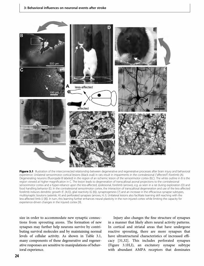
size in order to accommodate new synaptic connec-tions from sprouting axons. The formation of newsynapses may further help neurons survive by contri-buting survival molecules and by maintaining normallevels of cellular activity. As shown in Table 3.1,many components of these degenerative and regener-ative responses are sensitive to manipulations of behav-ioral experience.
Injury also changes the fine structure of synapsesin a manner that likely alters neural activity patterns.In cortical and striatal areas that have undergonereactive sprouting, there are more synapses thathave ultrastructural characteristics of increased effi-cacy [31,32]. This includes perforated synapses(Figure 3.1H,I), an excitatory synapse subtypewith abundant AMPA receptors that dominates
Figure 3.1 Illustration of the interconnected relationship between degenerative and regenerative processes after brain injury and behavioralexperience. Unilateral sensorimotor cortical lesions (black oval) in rats result in impairments in the contralesional (“affected”) forelimb (A).Degenerating neurons (fluorojade-B labeled) in the region of an ischemic lesion of the sensorimotor cortex (B,C). The white outline in B is theregion viewed at higher magnification in C. The lesion leads to degeneration of transcallosal axonal projections to the contralesionalsensorimotor cortex and a hyper-reliance upon the less-affected, ipsilesional, forelimb (arrows), e.g. as seen in a rat during exploration (D) andfood handling behavior (E). In the contralesional sensorimotor cortex, the interaction of transcallosal degeneration and use of the less-affectedforelimb induces dendritic growth (F, [4,5]), glial reactivity (G [6]), synaptogenesis [7] and an increase in the efficacious synapse subtypes,multisynaptic boutons (asterisk, H) and perforated synapses (arrows, H, I). Unilateral lesions also facilitate learning skill reaching with theless-affected limb (J [8]). In turn, this learning further enhances neural plasticity in the non-injured cortex while limiting the capacity forexperience-driven changes in the injured cortex [9].
3: Behavioral influences on neuronal events after stroke
24

its simpler, typically smaller synaptic neighbors in itsability to depolarize the postsynaptic neuron [33].Furthermore, multiple synapse complexes (Figure3.1H), where a single bouton contacts more thanone postsynaptic dendritic process, are moreabundant than normal [31,34], suggesting that there-utilization of boutons may be one means of re-innervating a brain region. At least in the cortex, theprevalence of these synapse subtypes is also sensitiveto experience [7]. These synaptic changes may con-tribute to changes in neural activity and excitabilitythat can be long lasting after the injury.
The degenerative–regenerative cascade occurs byvarying degrees and on different time scales dependingon whether one examines the perilesion or remotebrain areas [10]. Sprouting patterns vary dependingon injury locus and size, among other factors. Newsynapses formed in denervated regions tend to arisefrom the most proximal and prominent of the remain-ing pre-existing inputs [35]. Relative to the pre-injuredstate, the ultimate patterns of synaptic connectivitywithin and between brain regions may be dramaticallyaltered after reorganization. The time course andextent of these processes can vary with age (older
Table 3.1 Examples of behavioral influences on injury-induced brain changes
Injury-inducedevent
Direction Finding
Tissue loss + Early forced use of the affected (contralesional) forelimb increased cortical damage after corticallesions [14,15]. Restraint stress after motor cortical infarcts impeded recovery of forelimb functionand exaggerated lesion size [16].
Tissue loss − Constraint-induced movement-like therapy decreased tissue loss after striatal hemorrhage [17].
Cell death + Early EC exaggerated post-ischemic hippocampal neuron death [18].
Cell death − Forced use of the affected forelimb decreased loss of dopamine neurons in a unilateral Parkinson’smodel [19]. EC after lesion of the ventral subiculum attenuated degeneration in hippocampus [20].
Growth factors + Delayed exercise increased brain derived neurotrophic factor (BDNF) after traumatic brain injury[21,22].
Growth factors − Too early exercise decreased BDNF and other plasticity-related proteins after traumatic brain injury[21,22].
Reactive astrocytes + Forced forelimb use increased astrocytic reactions to callosal denervation [6]. EC after cerebralischemia increased astrocytic reactivity and fibroblast growth factor in hippocampus [23].
Axonal sprouting + EC increased axonal sprouting into deafferented superior colliculus after retinal lesion [24].
Axonal sprouting − Axonal sprouting was blocked by preventing behavioral asymmetries after unilateral nigrostriataldamage [25].
Dendritic growth + EC following cerebral ischemia increased dendritic density in hippocampus [26].
Dendritic growth − Dendritic growth contralateral to sensorimotor cortex lesions was blocked by constraint of theipsilesional forelimb [4].
Synaptogenesis + Motor skills training increased cortical lesion-induced synaptogenesis in motor cortex [7]. EC aftercerebral infarct increased synaptic density in hippocampus [26].
Neurogenesis + EC increased subventricular zone neurogenesis after cortical infarcts [27] (see Chapter 5). Skilledforelimb training increased hippocampal neurogenesis after sensorimotor cortical damage [28].
Neurogenesis − Exercise decreased subventricular zone neurogenesis after cortical infarct [27].
Cortical functionalactivity
+ Skilled reach training with the affected limb increased movement representations near corticalinfarcts [29,30].
This table illustrates two points: (1) behavioral experiences can influence the degenerative and regenerative processes that follow brain injury;(2) the nature of the effect varies with dose, type, and timing of the behavioral experiences and with severity and type of injury. +, increased,–, decreased compared to control, EC, complex environment housing.
3: Behavioral influences on neuronal events after stroke
25
brains take longer, reviewed in [36]), and type ofdamage [37]. Although the most dramatic activityoccurs earlier after the injury, degenerative and regen-erative reactions continue for at least several monthsin rodent models and perhaps much longer [10,11].
Reorganization patterns can vary dramaticallywith injury extent. Dancause et al. [38] found that, insquirrel monkeys, ischemic lesions that destroy morethan 50% of the forelimb representation region ofthe primary motor cortex (M1) induce extensiveexpansion of forelimb movement representations inthe ipsilesional ventral premotor cortex (PMv).However, smaller lesions within M1 actually decreasethe forelimb motor map in PMv. The expansion ofPMv is associated with greater axonal sproutingfrom PMv into the penumbra of the M1 lesions andwith improved rehabilitation-inducedmotor perform-ance [39].
Sprouting is activity dependent. Brus-Ramer et al.[40] found that, after unilateral pyramidal tract lesionsin adult rats, the sprouting of ipsilateral corticospinalprojections into denervated regions of the spinal cordcould be increased by electrical stimulation of thispathway. This indicates that there is a considerablecapacity to refine sprouting patterns after injury bymanipulating activity-dependent plasticity. It also sup-ports that the developmental process of activity-dependent synaptic competition that is involved inestablishing neural networks is also at play in reorgan-izing the CNS after damage.
Ischemia and other types of brain injury canincrease the generation and migration of new gliaand neurons (see Figure 3.1F) [41]. Molecules releasedby remodeling tissue appear to draw neural precursorsto injured tissue [42]. However, in rats, few of the newneurons in the striatum and cortex survive for verylong. Although new neurons are found near the cort-ical lesion core and penumbra, the overwhelmingmajority of newly generated cells develop into glia[43]. This injury-induced proliferation of glia maywell play a critical role in neuronal plasticity afterstroke. However, the sensitivity of neurogenesis tobehavioral experience is also well established (seeChapter 5). Targeting post-stroke neurogenesis withbehavioral manipulations provides another opportu-nity to manipulate brain reorganization with behavior.
Do the regenerative responses to stroke supportbetter functional outcome? Functional recovery isgreater following cortical ischemia, which inducesmajor axonal sprouting in the striatum, compared to
injuries of the same size and location that result in littlesprouting [37]. Treatments that reduce growth inhib-itory substances, such as Nogo, and thus enhanceneurite outgrowth, can also promote greater func-tional recovery [13]. Administration of substances,like neurotrophins, that promote neurogenesis andmigration of immature neurons [42] also results ingreater functional outcome after cortical infarcts.However, it should be expected that some examplesof post-stroke plasticity are non-optimal or even mal-adaptive given that there are many examples of patho-logical neuroplasticity, such as that proposed tounderlie the development of epilepsy, focal hand dys-tonia, and chronic pain. Experience-dependent neuralplasticity can also have detrimental or mixed effects onfunctional outcome after stroke, as reviewed below.
Although many aspects of the degenerative–regen-erative cascade have been found to be sensitive tobehavioral experience, we currently know too littleabout how specifically to use this information toimprove function after stroke. The problem is com-plex, considering that a single locus of damage insti-gates neural (and non-neural) reorganization innumerous brain areas. If examined in sufficient detail,the pattern of reorganization must be unique for everyindividual brain injury. Furthermore, it is clear thatexperience effects vary with timing and types of injury,among other factors (Table 3.1). Nevertheless, it ishoped that we will learn enough about the generalprinciples of how experience influences these pro-cesses to be able to devise rehabilitative techniquesthat optimize brain remodeling and can be tailoredfor injuries that vary in type, locus, and extent.
Effects of “self-taught” behavioralchange on brain reorganizationWe have found that neural plasticity after unilateralcortical lesions is driven by the convergence of injury-and behaviorally induced pressures. After unilaterallesions of the sensorimotor cortex in rats, there isdegeneration of transcallosal projections to the con-tralateral homotopic region of the motor cortex. Thisis followed by reactive astrocytic changes, growth ofpyramidal neuron dendrites and synaptogenesis in thecontralesional cortex [31,44,45]. Over time, the num-bers and complexity of dendrites and synapses areincreased relative to the same brain region of intactanimals. Dendrites are overproduced and then parti-ally eliminated, which is reminiscent of the dendritic
3: Behavioral influences on neuronal events after stroke
26

overproduction and pruning found in studies of braindevelopment.
The degeneration of transcallosal projections is oneinstigator of the contralesional glial and neural changes[5,6]. Another driver of the cellular changes is theanimal’s behavior. After the lesions, rats show impair-ments in the use of the contralesional forelimb. Thesechanges are evident in forelimb movements used dur-ing exploration (Figure 3.1D) and in the dexterous waythe animals handle food or other objects (Figure 3.1E).Much like humans [46], rats with these impairmentsbegin to disuse the affected limb and rely on the ipsile-sional forelimb more and in different ways to compen-sate [4,47]. When rats are prevented from using theipsilesional forelimb (with limb-restricting vests), thedendritic growth does not occur [4]. Denervation andforelimb asymmetries, together, are sufficient variablesto cause the dendritic outgrowth and astrocyticchanges. They can be reproduced in animals by trans-ecting the callosal fibers to themotor cortex and forcinguse of one limb [4–6]. Together, these studies suggestthat degeneration enhances the propensity of neuronsin the contralateral cortex to grow new synapses anddendrites, and that behavioral pressures act on thissensitive neuronal environment to dramatically alterneuronal structure and connectivity.
This lesion–behavior interaction has functionalconsequences. After lesions of the sensorimotor cor-tex, rats have some impairments in the “less-affected”(ipsilesional) limb, but they are also able to better learna new skilled motor task of manual dexterity (skilledreaching) with this limb than intact animals learningthe same task with one limb (Figure 3.1J) [8,45]. Theenhanced skill learning co-exists with impairments inipsilesional forelimb skills that were established beforethe injury [48]. Thus, the lesions appear to impairexisting motor engrams of the ipsilesional forelimbwhile enhancing some capacity to learn new skillswith this limb.
This interaction between denervation-induced andexperience-dependent neural plasticity may be consid-ered adaptive if it permits the injured animal to rapidlylearn compensatory behaviors. However, there aremaladaptive consequences for the function of theaffected limb. Training the ipsilesional forelimb inskilled reaching worsens motor performance of theaffected limb and reduces the benefits of later rehabil-itative training focused on the affected limb [45,49]. Italso reduces experience-dependent activation of neu-rons in the peri-infarct motor cortex resulting from
affected limb training [9]. Bilateral training does notproduce similar effects, suggesting that lateralized skilllearning may be the culprit [9]. The peri-infarct motorcortex is important for functional recovery of theaffected limb and for rehabilitative training efficacy(see the section on effects of training focused onimpairments). As it already tends to have impairedexperience-dependent plasticity [50] and undergoes amajor loss of dendrites and spines [12], limiting itsactivity and plasticity even further may be particularlydetrimental. The neural basis and behavioral parame-ters of this effect require much more investigation.
These findings are consistent with the idea thatunilateral brain injury unbalances competitive interac-tions between the hemispheres [51], and indicate thatbehavioral experiences can contribute to this imbalancein a manner that limits functionally beneficial restruc-turing in the damaged hemisphere. They also suggestthat the phenomenon of learned non-use, long studiedby Taub and others [46], may be exacerbated byexperience-dependent plasticity. Because maintenanceof normal neuronal function is use-dependent, disuse ofthe affected body side may contribute to its dysfunctionand limit opportunities for adaptive neural reorganiza-tion in the damaged hemisphere (reviewed in [2]).Behavioral interventions, such as extensive focusedtraining of affected extremities (see the section onthe effects of training focused on impairments) or com-bined therapeutic treatments (see the section oncombining behavioral manipulation with other inter-ventions), may be needed to counter the effect of learn-ing with the less-affected, ipsilesional body side.
Another general implication of these findings isthat post-injury regenerative processes sometimescause brain regions to become more sensitive toexperience-dependent plasticity. This could facilitatefunctionally beneficial neural plasticity, but could alsobe permissive of excessive and maladaptive plasticity.This leads to the question of how to capitalize on thisprocess and steer it in an optimal direction through theuse of specific behavioral manipulations. There is nowa wealth of research indicating that manipulations ofbehavioral experience can influence post-injury neuralplasticity and behavioral outcome.
Manipulating environmentalcomplexityA classic approach in animal models for studying theinfluence of behavioral experience is to compare rats
3: Behavioral influences on neuronal events after stroke
27
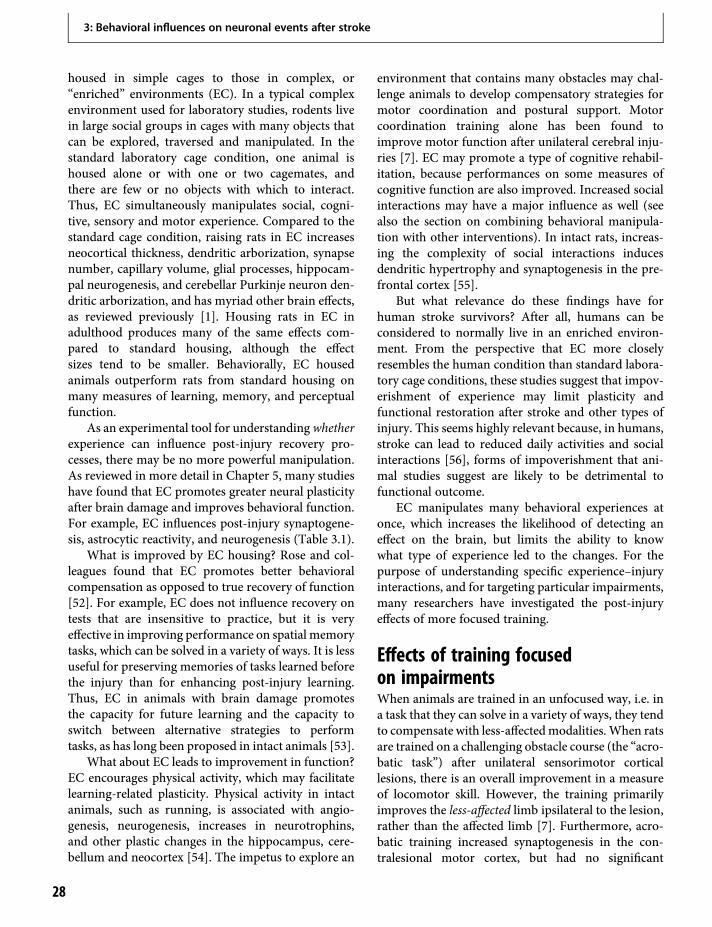
housed in simple cages to those in complex, or“enriched” environments (EC). In a typical complexenvironment used for laboratory studies, rodents livein large social groups in cages with many objects thatcan be explored, traversed and manipulated. In thestandard laboratory cage condition, one animal ishoused alone or with one or two cagemates, andthere are few or no objects with which to interact.Thus, EC simultaneously manipulates social, cogni-tive, sensory and motor experience. Compared to thestandard cage condition, raising rats in EC increasesneocortical thickness, dendritic arborization, synapsenumber, capillary volume, glial processes, hippocam-pal neurogenesis, and cerebellar Purkinje neuron den-dritic arborization, and has myriad other brain effects,as reviewed previously [1]. Housing rats in EC inadulthood produces many of the same effects com-pared to standard housing, although the effectsizes tend to be smaller. Behaviorally, EC housedanimals outperform rats from standard housing onmany measures of learning, memory, and perceptualfunction.
As an experimental tool for understanding whetherexperience can influence post-injury recovery pro-cesses, there may be no more powerful manipulation.As reviewed in more detail in Chapter 5, many studieshave found that EC promotes greater neural plasticityafter brain damage and improves behavioral function.For example, EC influences post-injury synaptogene-sis, astrocytic reactivity, and neurogenesis (Table 3.1).
What is improved by EC housing? Rose and col-leagues found that EC promotes better behavioralcompensation as opposed to true recovery of function[52]. For example, EC does not influence recovery ontests that are insensitive to practice, but it is veryeffective in improving performance on spatial memorytasks, which can be solved in a variety of ways. It is lessuseful for preserving memories of tasks learned beforethe injury than for enhancing post-injury learning.Thus, EC in animals with brain damage promotesthe capacity for future learning and the capacity toswitch between alternative strategies to performtasks, as has long been proposed in intact animals [53].
What about EC leads to improvement in function?EC encourages physical activity, which may facilitatelearning-related plasticity. Physical activity in intactanimals, such as running, is associated with angio-genesis, neurogenesis, increases in neurotrophins,and other plastic changes in the hippocampus, cere-bellum and neocortex [54]. The impetus to explore an
environment that contains many obstacles may chal-lenge animals to develop compensatory strategies formotor coordination and postural support. Motorcoordination training alone has been found toimprove motor function after unilateral cerebral inju-ries [7]. EC may promote a type of cognitive rehabil-itation, because performances on some measures ofcognitive function are also improved. Increased socialinteractions may have a major influence as well (seealso the section on combining behavioral manipula-tion with other interventions). In intact rats, increas-ing the complexity of social interactions inducesdendritic hypertrophy and synaptogenesis in the pre-frontal cortex [55].
But what relevance do these findings have forhuman stroke survivors? After all, humans can beconsidered to normally live in an enriched environ-ment. From the perspective that EC more closelyresembles the human condition than standard labora-tory cage conditions, these studies suggest that impov-erishment of experience may limit plasticity andfunctional restoration after stroke and other types ofinjury. This seems highly relevant because, in humans,stroke can lead to reduced daily activities and socialinteractions [56], forms of impoverishment that ani-mal studies suggest are likely to be detrimental tofunctional outcome.
EC manipulates many behavioral experiences atonce, which increases the likelihood of detecting aneffect on the brain, but limits the ability to knowwhat type of experience led to the changes. For thepurpose of understanding specific experience–injuryinteractions, and for targeting particular impairments,many researchers have investigated the post-injuryeffects of more focused training.
Effects of training focusedon impairmentsWhen animals are trained in an unfocused way, i.e. ina task that they can solve in a variety of ways, they tendto compensate with less-affectedmodalities.When ratsare trained on a challenging obstacle course (the “acro-batic task”) after unilateral sensorimotor corticallesions, there is an overall improvement in a measureof locomotor skill. However, the training primarilyimproves the less-affected limb ipsilateral to the lesion,rather than the affected limb [7]. Furthermore, acro-batic training increased synaptogenesis in the con-tralesional motor cortex, but had no significant
3: Behavioral influences on neuronal events after stroke
28
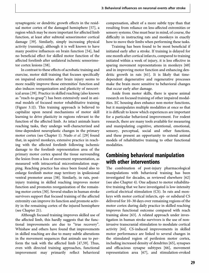
synaptogenic or dendritic growth effects in the resid-ual motor cortex of the damaged hemisphere [57], aregion which may be more important for affected limbfunction, at least after subtotal sensorimotor corticaldamage [39]. Similarly, simply increasing physicalactivity (running), although it is well known to havemany positive influences on brain function [54], hadno beneficial effect for skilled motor function of theaffected forelimb after unilateral ischemic sensorimo-tor cortex lesions [58].
In contrast to these effects of acrobatic training andexercise, motor skill training that focuses specificallyon impaired extremities after brain injury seems tomore readily improve those extremities’ function andalso induces reorganization and plasticity of neocort-ical areas [39]. Practice in skilled reaching (also knownas “reach-to-grasp”) has been extensively used in ani-mal models of focused motor rehabilitative training(Figure 3.1J). This training approach is believed tocapitalize upon neural mechanisms of motor skilllearning to drive plasticity in regions relevant to thefunction of the affected limb. As intact animals learnreaching tasks, they undergo well-characterized andtime-dependent neuroplastic changes in the primarymotor cortex (see Chapter 1). Nudo et al. [29] foundthat, in squirrel monkeys, extensive practice in reach-ing with the affected forelimb following ischemicdamage to the forelimb representation area of theprimary motor cortex spared the tissue surroundingthe lesion from a loss of movement representation, asmeasured with intracortical microstimulation map-ping. Reaching practice has since been found also toenlarge forelimb motor map territory in ipsilesionalventral premotor areas [38]. Similarly, in rats, post-injury training in skilled reaching improves motorfunction and promotes reorganization of the remain-ing motor cortex [30]. Several studies in human strokesurvivors support that focused training of the affectedextremity can improve its function and promote activ-ity in the remaining cortex of the injured hemisphere(see Chapter 21).
Although focused training improves skilled use ofthe affected limb, this hardly suggests that the func-tional improvements are due to “true recovery”.Whishaw and others have found that improvementsin skilled reaching are due to many subtle alterationsin the movement sequences that animals use to per-form the task with the affected limb [47,59]. Thus,even with directed training approaches, functionalimprovement may primarily reflect behavioral
compensation, albeit of a more subtle type than thatresulting from reliance on less-affected extremities orsensory systems. Onemust bear inmind, of course, thedifficulty in instructing rats and monkeys in exactlyhow to move their limbs when performing these tasks.
Training has been found to be most beneficial ifinitiated early after a stroke. If training is delayed forone month after cortical infarcts, compared to traininginitiated within a week of injury, it is less effective insparing movement representations in monkeys [60]and in improving motor function and enhancing den-dritic growth in rats [61]. It is likely that time-dependent degenerative and regenerative processesmake the brain more sensitive to behavioral changesthat occur early after damage.
Aside from motor skills, there is sparse animalresearch on focused training of other impaired modal-ities. EC housing does enhance non-motor functions,but it manipulates multiple modalities at once so thatit is difficult to know which experiences are importantfor a particular behavioral improvement. For rodentresearch, there are many tools available for measuringand manipulating cognitive, attentional, emotional,sensory, perceptual, social and other functions,and these present an opportunity to extend animalmodels of rehabilitative training to other functionalmodalities.
Combining behavioral manipulationwith other interventionsThe combination of post-injury pharmacologicalmanipulations with behavioral training has beeninvestigated for decades, as reviewed elsewhere [62](see also Chapter 4). One adjunct to motor rehabilita-tive training that we have investigated is low-intensitycortical electrical stimulation (CS). In rats and mon-keys with motor cortical infarcts, epi- or subdural CSdelivered for 10–30 days over remaining regions of themotor cortex during daily practice in skilled reachingimproves functional outcome compared with reachtraining alone [63]. A related approach under inves-tigation in human stroke survivors is the use of non-invasive transcranial stimulation to modulate corticalactivity [64]. CS-induced improvements in skilledmotor performance are linked to several changes inthe stimulated region of perilesion motor cortex,including increased density of dendrites [65], synapsesand efficacious synapse subtypes [66], movementrepresentation area [67], and stimulation-evoked
3: Behavioral influences on neuronal events after stroke
29

potentials [68]. We found that rats with more severepost-lesion motor impairment benefit less from CScompared with rats with more moderate impairments[66]. However, the severely impaired rats were justbeginning to attain levels of reaching performanceequivalent to moderately impaired rats by the end ofthe treatment period and therefore might have bene-fited from a longer interval of CS and training [66].
As reviewed by Plow et al. [63], these animal stud-ies were used to support the initiation of clinical trialsin human stroke patients. Early phase studies werepromising, indicating that CS was a safe and effica-cious way to improve upper-extremity motor functionin stroke survivors. However, the phase III, Everest,study failed tomeet primary endpoints when the entiregroup of subjects was included in the analysis. Sincethis initial report, follow-up analysis has revealed thata subset of subjects showed major improvements.These subjects were ones in which the CS was deliv-ered at intensity levels that were 50% of those neededto evoke movement, consistent with the parametersfound to be effective in the smaller clinical trials and inthe animal studies. In the other subjects, no move-ments could be evoked and therefore the CS was deliv-ered at a predefined and probably insufficient intensitylevel. This may indicate that CS can be effective ifdescending motor pathways are sufficiently activated,which is likely to vary with different injuries and withstimulation location. This supports the need to betterunderstand how to tailor treatment strategies for par-ticular types of stroke.
Maladaptive behavioral experienceIn an earlier section, we described how learning skillswith the ipsilesional “less-affected” upper extremitymay be detrimental to the outcome of the affectedbody side. Too much or too intense an experiencewith affected extremities early after a brain injury canalso hinder recovery. If rats are forced to rely on theiraffected limb (by placing them in vests that restrict theless-affected limb) during the first week after sensori-motor cortical damage, motor impairments are wors-ened and there is greater degeneration of the perilesioncortex [14,69]. Tissue loss is further exaggerated in ratsthat are moved from group housing to social isolationat the time of the injury [70]. Furthermore, earlyforced disuse of the affected limb extends the timewindow of vulnerability to later overuse [69]. Earlyvoluntary exercise after traumatic brain damagereduces the expression of plasticity-related molecules
in the hippocampus [21]. However, if voluntary exer-cise is delayed, animals show improved function andthere is an enhanced expression of these molecules[21]. These effects vary with injury severity and type.Griesbach et al. [22] found that a longer delay isneeded after more severe injuries for exercise to pro-mote plasticity related proteins in the hippocampus.Tillerson et al. [19] found that early forced use of theaffected forelimb reduces cell death and motor impair-ment in a rat model of unilateral Parkinson’s degen-eration, and this occurs only if it is initiated soonenough after the onset of neurodegeneration.
Intensely varied social experience may also impairrecovery. Silasi, Hamilton, and Kolb [55] used a para-digm of increased social complexity in which male ratswere exposed to a rotating set of cagemates of the samesex, all of which were familiar to them, but which werechanged every 48 h. In intact animals, this experiencepromotes dendritic growth and synaptogenesis in theprefrontal cortex. However, when this regimen ofsocial change is begun the day following ischemicmotor cortex damage, it blocks spontaneous recoveryof motor function. This, together with previous find-ings of the benefit of social housing after brain injury[70], suggests that, at least among adult male rats,stable social interaction may be optimal for outcomeafter brain injury. This study also provides anotherexample of how experience-dependent plasticity inter-acts with injury in ways that can be maladaptive forfunctional outcome.
ConclusionIn some respects we may think of the adult post-strokebrain as similar to the developing brain in that it needsto be shaped by appropriate experiences. We are nowfaced with the challenge of understanding how bestto do this. In this chapter, we overviewed findingsfrom animal research on the significant interactionsbetween injury and experience as the brain reorganizesfollowing a stroke. Many aspects of the degenerative–regenerative cascade can be changed by behavioralmanipulations, influencing post-stroke plasticity andimproving behavioral outcome. However, experiencehas the capacity to worsen function, increase degener-ation, and limit functionally beneficial plasticity. Notattempting to implement rehabilitative behavioraltechniques is also risky, because non-directed experi-ences of the animal may drive neural reorganization infunctionally maladaptive directions. This accentuatesthe urgency that we obtain more detailed and more
3: Behavioral influences on neuronal events after stroke
30
clinically relevant knowledge about these processes.Because experience effects vary with injury severity,location and type, it seems important to investigate themany varieties of stroke that occur (see Chapter 8).There remain many other under-investigated stroke-related issues, including how age, gender, and modalityof impairment interact with experience and degenera-tive processes. All of these issues are highly pertinentand suitable for investigation with animal models.
AcknowledgmentsWe thank Cole Husbands for editing, Soo Y. Kim forthe fluorojade image and Dr. Rachel Allred for graphicdesign. Grant support: NS056839, NS06332 andNS064423.
References1. Grossman AW, Churchill JD, Bates KE, et al. A brain
adaptation view of plasticity: Is synaptic plasticity anoverly limited concept? Prog Brain Res.2002;138:91–108.
2. Kleim JA, Jones TA. Principles of experience-dependent neural plasticity: Implications forrehabilitation after brain damage. J Speech Lang HearRes. 2008;51:S225–39.
3. Doyle KP, Simon RP, Stenzel-Poore MP. Mechanismsof ischemic brain damage. Neuropharmacology.2008;55:310–8.
4. Jones TA, Schallert T. Use-dependent growth ofpyramidal neurons after neocortical damage.J Neurosci. 1994;14:2140–52.
5. Adkins DL, Bury SD, Jones TA. Laminar-dependentdendritic spine alterations in the motor cortex ofadult rats following callosal transection andforced forelimb use. Neurobiol Learn Mem.2002;78:35–52.
6. Bury SD, Eichhorn AC, Kotzer CM, et al. Reactiveastrocytic responses to denervation in the motor cortexof adult rats are sensitive to manipulations ofbehavioral experience. Neuropharmacology.2000;39:743–55.
7. Jones TA, Chu CJ, Grande LA, et al. Motor skillstraining enhances lesion-induced structural plasticity inthe motor cortex of adult rats. J Neurosci.1999;19:10 153–63.
8. Bury SD, Jones TA. Unilateral sensorimotor cortexlesions in adult rats facilitate motor skill learning withthe “unaffected” forelimb and training-induceddendritic structural plasticity in the motor cortex.J Neurosci. 2002;22:8597–606.
9. Allred RP, Jones TA. Maladaptive effects of learningwith the less-affected forelimb after focal corticalinfarcts in rats. Exp Neurol. 2008;210:172–81.
10. Carmichael ST. Cellular and molecular mechanisms ofneural repair after stroke: Making waves. Ann Neurol.2006;59:735–42.
11. Wieloch T, Nikolich K. Mechanisms of neural plasticityfollowing brain injury. Curr Opin Neurobiol.2006;16:258–64.
12. Brown CE, Murphy TH. Livin’ on the edge: Imagingdendritic spine turnover in the peri-infarct zone duringischemic stroke and recovery. Neuroscientist.2008;14:139–46.
13. Cheatwood JL, Emerick AJ, Kartje GL. Neuronalplasticity and functional recovery after ischemic stroke.Top Stroke Rehabil. 2008;15:42–50.
14. Kozlowski DA, James DC, Schallert T. Use-dependentexaggeration of neuronal injury after unilateralsensorimotor cortex lesions. J Neurosci.1996;16:4776–86.
15. DeBow SB, McKenna JE, Kolb B, et al. Immediateconstraint-induced movement therapy causes localhyperthermia that exacerbates cerebral cortical injuryin rats. Can J Physiol Pharmacol. 2004;82:231–7.
16. Kirkland SW, Coma AK, Colwell KL, et al. Delayedrecovery and exaggerated infarct size by post-lesionstress in a rat model of focal cerebral stroke. Brain Res.2008;1201:151–60.
17. DeBow SB, Davies ML, Clarke HL, et al. Constraint-induced movement therapy and rehabilitation exerciseslessen motor deficits and volume of brain injury afterstriatal hemorrhagic stroke in rats. Stroke.2003;34:1021–6.
18. Farrell R, Evans S, Corbett D. Environmentalenrichment enhances recovery of function butexacerbates ischemic cell death. Neuroscience.2001;107:585–92.
19. Tillerson JL, Cohen AD, Philhower J, et al. Forced limb-use effects on the behavioral and neurochemical effectsof 6-hydroxydopamine. J Neurosci. 2001;21:4427–35.
20. Dhanushkodi A, Bindu B, Raju TR, et al. Exposure toenriched environment improves spatial learningperformances and enhances cell density but not cholineacetyltransferase activity in the hippocampus of ventralsubicular-lesioned rats. Behav Neurosci.2007;121:491–500.
21. Griesbach GS, Gomez-Pinilla F, Hovda DA. Theupregulation of plasticity-related proteins followingTBI is disrupted with acute voluntary exercise. BrainRes. 2004;1016:154–62.
22. Griesbach GS, Gomez-Pinilla F, Hovda DA. Timewindow for voluntary exercise-induced increases inhippocampal neuroplasticity molecules after traumaticbrain injury is severity dependent. J Neurotrauma.2007;24:1161–71.
23. Briones TL, Woods J, Wadowska M, et al. Astrocyticchanges in the hippocampus and functional recovery
3: Behavioral influences on neuronal events after stroke
31
after cerebral ischemia are facilitated by rehabilitationtraining. Behav Brain Res. 2006;171:17–25.
24. Caleo M, Tropea D, Rossi C, et al. Environmentalenrichment promotes fiber sprouting afterdeafferentation of the superior colliculus in the adult ratbrain. Exp Neurol. 2009;21:515–9.
25. Morgan S, Huston JP, Pritzel M. Effects of reducingsensory-motor feedback on the appearance of crossednigro-thalamic projections and recovery from turninginduced by unilateral substantia nigra lesions. Brain ResBull. 1983;11:721–7.
26. Briones TL, Suh E, Jozsa L, et al. Behaviorallyinduced synaptogenesis and dendritic growth inthe hippocampal region following transientglobal cerebral ischemia are accompanied byimprovement in spatial learning. Exp Neurol.2006;198:530–8.
27. Komitova M, Zhao LR, Gido G, et al. Postischemicexercise attenuates whereas enriched environment hascertain enhancing effects on lesion-inducedsubventricular zone activation in the adult rat.Eur J Neurosci. 2005;21:2397–405.
28. Wurm F, Keiner S, Kunze A, et al. Effects of skilledforelimb training on hippocampal neurogenesis andspatial learning after focal cortical infarcts in the adultrat brain. Stroke. 2007;38:2833–40.
29. Nudo RJ, Wise BM, SiFuentes F, et al. Neural substratesfor the effects of rehabilitative training on motorrecovery after ischemic infarct. Science.1996;272:1791–4.
30. Conner JM, Chiba AA, Tuszynski MH. The basalforebrain cholinergic system is essential for corticalplasticity and functional recovery following braininjury. Neuron. 2005;46:173–9.
31. Jones TA. Multiple synapse formation in the motorcortex opposite unilateral sensorimotor cortex lesionsin adult rats. J Comp Neurol. 1999;414:57–66.
32. Meshul CK, Cogen JP, Cheng H-W, et al. Alterations inrat striatal glutamate synapses following a lesion of thecortico- and/or nigrostriatal pathway. Exp Neurol.2000;165:191–206.
33. Ganeshina O, Berry RW, Petralia RS, et al. Synapseswith a segmented, completely partitioned postsynapticdensity express more AMPA receptors than otheraxospinous synaptic junctions. Neuroscience.2004;125:615–23.
34. McNeill TH, Brown SA, Hogg E, et al. Synapsereplacement in the striatum of the adult rat followingunilateral cortex ablation. J Comp Neurol.2003;467:32–43.
35. Raisman G, Field PM. Synapse formation in the adultbrain after lesions and after transplantation ofembryonic tissue. J Exp Biol. 1990;153:277–87.
36. Petcu EB, Sfredel V, Platt D, et al. Cellular andmolecular events underlying the dysregulated responseof the aged brain to stroke: A mini-review. Gerontology.2008;54:6–17.
37. Carmichael ST, Chesselet MF. Synchronousneuronal activity is a signal for axonal sproutingafter cortical lesions in the adult. J Neurosci.2002;22:6062–70.
38. Dancause N, Barbay S, Frost SB, et al. Effects ofsmall ischemic lesions in the primary motorcortex on neurophysiological organization inventral premotor cortex. J Neurophysiol.2006;96:3506–11.
39. Nudo RJ. Postinfarct cortical plasticity and behavioralrecovery. Stroke. 2007;38:840–5.
40. Brus-Ramer M, Carmel JB, Chakrabarty S, et al.Electrical stimulation of spared corticospinalaxons augments connections with ipsilateral spinalmotor circuits after injury. J Neurosci.2007;27:13 793–801.
41. Lichtenwalner RJ, Parent JM. Adult neurogenesis andthe ischemic forebrain. J Cereb Blood Flow Metab.2006;26:1–20.
42. Ohab JJ, Fleming S, Blesch A, et al. A neurovascularniche for neurogenesis after stroke. J Neurosci.2006;26:13 007–16.
43. Gotts JE, Chesselet MF. Migration and fate of newlyborn cells after focal cortical ischemia in adult rats.J Neurosci Res. 2005;80:160–71.
44. Adkins DL, Voorhies AC, Jones TA. Behavioral andneuroplastic effects of focal endothelin-1 inducedsensorimotor cortex lesions. Neuroscience.2004;128:473–86.
45. Allred RP, Jones, TA. Experience – a double edgedsword for restorative neural plasticity after braindamage. Future Neurol. 2008;3:189–98.
46. Taub E, Uswatte G,Mark VW, et al. The learned nonusephenomenon: Implications for rehabilitation. EuraMedicophys. 2006;42:241–56.
47. Whishaw IQ. Loss of the innate cortical engram foraction patterns used in skilled reaching and thedevelopment of behavioral compensation followingmotor cortex lesions in the rat. Neuropharmacology.2000;39:788–805.
48. Hsu JE, Jones TA. Contralesional neural plasticity andfunctional changes in the less-affected forelimb afterlarge and small cortical infarcts in rats. Exp Neurol.2006;201:479–94.
49. Allred RP, Maldonado MA, Hsu And JE, et al. Trainingthe “less-affected” forelimb after unilateral corticalinfarcts interferes with functional recovery of theimpaired forelimb in rats. Restor Neurol Neurosci.2005;23:297–302.
3: Behavioral influences on neuronal events after stroke
32
50. Jablonka JA, Witte OW, Kossut M. Photothromboticinfarct impairs experience-dependent plasticity inneighboring cortex. Neuroreport. 2007;18:165–9.
51. Ward NS, Cohen LG. Mechanisms underlying recoveryof motor function after stroke. Arch Neurol.2004;61:1844–8.
52. Rose FD, Davey MJ, Attree EA. How doesenvironmental enrichment aid performancefollowing cortical injury in the rat? Neuroreport.1993;4:163–6.
53. Greenough WT, Fass B, DeVoogd TJ. The influence ofexperience on recovery following brain damage inrodents: Hypotheses based on development research.In: Walsh RN, Greenough WT, editors. Environmentsas therapy for brain dysfunction. New York, NY:Plenum Press; 1976: pp. 10–50.
54. Vaynman S, Gomez-Pinilla F. License to run: Exerciseimpacts functional plasticity in the intact and injuredcentral nervous system by using neurotrophins.Neurorehabil Neural Repair. 2005;19:283–95.
55. Silasi G, Hamilton DA, Kolb B. Social instability blocksfunctional restitution following motor cortex stroke inrats. Behav Brain Res. 2008;188:219–26.
56. Salter K, Hellings C, Foley N, et al. The experience ofliving with stroke: A qualitative meta-synthesis.J Rehabil Med. 2008;40:595–602.
57. Chu CJ, Jones TA. Experience-dependent structuralplasticity in cortex heterotopic to focal sensorimotorcortical damage. Exp Neurol. 2000;166:403–14.
58. Maldonado MA, Allred RP, Felthauser EL, et al. Motorskill training, but not voluntary exercise, improvesskilled reaching after unilateral ischemic lesions of thesensorimotor cortex in rats. Neurorehabil NeuralRepair. 2008;22:250–61.
59. Alaverdashvili M, Foroud A, Lim DH, et al. “Learnedbaduse” limits recovery of skilled reaching for foodafter forelimb motor cortex stroke in rats: A newanalysis of the effect of gestures on success. Behav BrainRes. 2008;188:281–90.
60. Barbay S, Plautz EJ, Friel KM, et al. Behavioral andneurophysiological effects of delayed training following
a small ischemic infarct in primary motor cortex ofsquirrel monkeys. Exp Brain Res. 2005;5:1–11.
61. Biernaskie J, Chernenko G, Corbett D. Efficacy ofrehabilitative experience declines with time after focalischemic brain injury. J Neurosci. 2004;24:1245–54.
62. Goldstein LB. Neurotransmitters and motor activity:Effects on functional recovery after brain injury.NeuroRx. 2006;3:451–7.
63. Plow EB, Carey JR, Nudo RJ, et al. Cortical stimulationto promote recovery of function after stroke: A criticalappraisal. Stroke. 2009;40:1926–31.
64. Edwards D, Fregni F. Modulating the healthy andaffected motor cortex with repetitive transcranialmagnetic stimulation in stroke: Development of newstrategies for neurorehabilitation. NeuroRehabilitation.2008;23:3–14.
65. Adkins-Muir DL, Jones TA. Cortical electricalstimulation combined with rehabilitative training:Enhanced functional recovery and dendritic plasticityfollowing focal cortical ischemia in rats. Neurol Res.2003;25:780–8.
66. Adkins DL, Hsu JE, Jones TA. Motor corticalstimulation promotes synaptic plasticity and behavioralimprovements following sensorimotor cortex lesions.Exp Neurol. 2008;212:14–28.
67. Kleim JA, Bruneau R, VandenBerg P, et al. Motorcortex stimulation enhances motor recovery andreduces peri-infarct dysfunction following ischemicinsult. Neurol Res. 2003;25:789–93.
68. Teskey GC, Flynn C, Goertzen CD, et al. Corticalstimulation improves skilled forelimb use following afocal ischemic infarct in the rat. Neurol Res.2003;25:794–800.
69. Schallert T, Leasure JL, Kolb B. Experience-associatedstructural events, subependymal cellular proliferativeactivity, and functional recovery after injury to thecentral nervous system. J Cereb Blood Flow Metab.2000;20:1513–28.
70. Woodlee MT, Schallert T. The impact of motor activityand inactivity on the brain. Curr Directions Psychol Sci.2006;15:203–06.
3: Behavioral influences on neuronal events after stroke
33

4 Post-stroke recovery therapies in animalsG. Campbell Teskey & Bryan Kolb
IntroductionOne of the primary reasons scientists perform post-stroke recovery research on rodents is to determine theusefulness or efficacy of certain treatments or therapies.The foundation for the validity of animal research is theextended common history that other animals sharewith our species and the relatively conservative processby which evolutionary change takes place (Chapter 7).Thus there should be constancy in cerebral organiza-tion and function, both between and within mamma-lian species (Chapter 1). That said, the failure of clinicaltrials for certain treatments that were effective in ani-mal models has raised an opposing view, expressed inits most extreme form as “animal research tells usnothing about whether a treatment will be effective inpeople.” Whereas some postulated reasons for the fail-ure of the clinical trials have been summarized else-where [1], here we offer a series of considerations thatshould be well contemplated when adopting an animalmodel to investigate whether a certain treatment ortherapy will be effective and potentially translate topeople that have sustained a stroke. The considerationsfall into four broad categories: intervention issues,organismal factors, modeling stroke, and measure-ment. We also review four categories of treatmentsand therapies that are utilized in animal models tofacilitate recovery: enrichment/experience, pharmaco-therapy, cell-based therapies, and electrical stimulation.
Considerations (Table 4.1)
Timing and intensity of interventionWhen a cerebral attack or stroke occurs there is a non-trivial interruption of the blood supply to one of thecerebral arteries. This sets off a domino-like cascadeof neurodegenerative changes that develop overseveral timescales (Figure 4.1). The drop in oxygen
concentration causes changes in the ionic balance ofthe affected regions over the first seconds to minutes,including changes in pH and properties of the cellmembrane. These ionic changes result in a variety ofpathological events, including the release of excessiveamounts of glutamate and the prolonged opening ofcalcium channels. The open calcium channels allowhigh levels of calcium to enter the cell, which in turnleads to a number of toxic effects that result in celldeath. The development of considerable edema (swel-ling) is a major complication over the first day follow-ing stroke. The swelling itself can cause neuronalinjury, dysfunction, and death. These post-strokechanges in neuronal functioning lead to a drop inmetabolic rate and/or glucose utilization in the injuredhemisphere that may persist for days. Moreover, areasthat receive synaptic input from the primarily dam-aged area suffer a sudden withdrawal of excitation orinhibition. Such sudden changes in input lead to agreater loss of function and secondary cell death thatmay be even more pronounced than the cell deathresulting directly from the injury itself. Following celldeath, inflammatory processes begin almost immedi-ately. Microglia invade the damaged region via thevascular system and clear away degenerative debris, aprocess that may take months to complete. Astrocytesadjacent to the lesioned area enlarge and extendfibrous processes that serve to isolate the surfaces ofthe injury from the surrounding tissue. Stem cells mayalso be stimulated to increase division in the subven-tricular zone and to migrate to the injury, although wedo not yet know what function these cells might have.Thus, when considering a treatment, the timing of theintervention in relation to the post-stroke neurodege-nerative cascade must be evaluated.
Treatments for cerebral injury can be targeted atdifferent aspects of the neurodegenerative cascade.Agents that are designed to protect neurons from the
Brain Repair After Stroke, ed. S. C. Cramer and R. J. Nudo. Published by Cambridge University Press.© Cambridge University Press 2010.
35
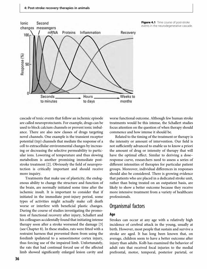
cascade of toxic events that follow an ischemic episodeare called neuroprotectants. For example, drugs can beused to block calcium channels or prevent ionic imbal-ance. There are also new classes of drugs targetingnovel channels. One example is the transient receptorpotential (trp) channels that mediate the response of acell to extracellular environmental changes by increas-ing or decreasing the selective permeability to partic-ular ions. Lowering of temperature and thus slowingmetabolism is another promising immediate post-stroke treatment [2]. Obviously the field of neuropro-tection is critically important and should receivemore inquiry.
Treatments that make use of plasticity, the endog-enous ability to change the structure and function ofthe brain, are normally initiated some time after theischemic insult. It is important to consider that ifinitiated in the immediate post-injury period, sometypes of activities might actually make cell deathworse or interfere with beneficial plastic changes.During the course of studies investigating the promo-tion of functional recovery after injury, Schallert andhis colleagues accidentally found that initiating intensetherapy soon after a stroke worsened the damage [3](see Chapter 8). In these studies, rats were fitted with arestraint harness that prevented them from using theforelimb ipsilateral to a sensorimotor cortex injury,thus forcing use of the impaired limb. Unfortunately,the rats that had continual forced use of the affectedlimb showed significantly enlarged lesion cavity and
worse functional outcome. Although few human stroketreatments would be this intense, the Schallert studiesfocus attention on the question of when therapy shouldcommence and how intense it should be.
Related to the timing of the treatment or therapy isthe intensity or amount of intervention. Our field isnot sufficiently advanced to enable us to know a priorithe amount of drug or intensity of therapy that willhave the optimal effect. Similar to deriving a dose–response curve, researchers need to assess a series ofdifferent intensities of therapies for particular patientgroups. Moreover, individual differences in responsesshould also be considered. There is growing evidencethat patients who are placed in a dedicated stroke unit,rather than being treated on an outpatient basis, arelikely to show a better outcome because they receivemore intensive treatment from a variety of healthcareprofessionals.
Organismal factorsAgeStrokes can occur at any age with a relatively highincidence of cerebral attack in the young, usually atbirth. However, most people that sustain and survive astroke are aged. It has long been known that, onaverage, children seem to have a better outcome afterinjury than adults. Kolb has examined the behavior ofadult rats that received focal injuries to the medialprefrontal, motor, temporal, posterior parietal, or
Figure 4.1 Time course of post-strokeevents in the neurodegenerative cascade.
4: Post-stroke recovery therapies in animals
36
posterior cingulate cortex on postnatal days 1, 4, 7, 10,or 90 (i.e., adult) [4]. The overall result was thatregardless of the location of injury, the functional out-come was always best after injury sustained during thesecond week of life, which in the rat is a time of intensecerebral synaptogenesis and glial formation. The take-home message is that it is the stage of neural develop-ment, and not age per se, that is the important variablein recovery.
Injured young brains have been shown to compen-sate for lost tissue in three ways: (1) reorganization ofexisting neuronal networks; (2) development of novelnetworks; and (3) regeneration of the lost tissue(which is dealt with later in the chapter). Existingnormal circuits are found to reorganize following uni-lateral damage to motor systems. When damageoccurs to the cortex that normally gives rise to thecorticobulbar and corticospinal pathways, the intactpathway on the opposite side sprouts both an enlargedipsilateral corticospinal pathway as well as new con-nections to subcortical motor regions of the damagedhemisphere [5]. Similar findings are seen in sensorysystems as novel pathways develop after damage [6].Neuronal network remodeling in the form of changesto dendritic morphology have also been observed afterearly injuries. The overall result of these studies isthat when functional outcome is positive, there is anincrease in dendritic arborization and spine density inpyramidal neurons in the remaining cortex. Whenfunctional outcome is poor, however, there is an atro-phy of dendritic arbor and spine density.
It is generally assumed that as animals age theybecome less plastic. Teuber reported that brain-injured soldiers also showed a benefit of younger age:18-year-olds fared better than 25-year-olds, who inturn fared better than older soldiers [7]. Although ithas been shown that older animals demonstrate lesssynaptic potentiation in response to high-frequencystimulation [8], even senescent animals can show con-siderable cortical plasticity [9]. Systematic studies ofcerebral plasticity and behavior throughout the lifespan in both normal and brain-injured animals arestill required. Moreover, the health status and life-history of experience rather than the age of the animalmay be the predominant factor.
Comorbid conditionsMost people that survive a stroke often have a varietyof other pre-existing medical conditions such as dia-betes, arthritis, atherosclerosis, and hypertension that
need to be modeled by stroke researchers. Forinstance, a clinically relevant model of human strokeis the stroke-prone spontaneously hypertensive rat.This strain of rat has been shown to have a geneticpredisposition to cerebral ischemia and exhibitshypertension and an increased sensitivity to experi-mentally induced stroke. Following stroke, these ratsexhibit greater impaired functional recovery com-pared to a normotensive strain from which the hyper-tensive strain was derived [10]. The usage of animalmodels that replicate at least some of the more preva-lent comorbid conditions in people that have hadstrokes will need to be more fully embraced byresearchers.
Most stroke survivors take numerous medicationsfor a variety of ailments and conditions. For instance,because clinical depression is found in the majority ofstroke survivors (see Chapter 14), the prescription ofspecific serotonin reuptake inhibitors (SSRIs) has beenalmost universal where available. SSRIs have beenshown to alter brain activity and modulate motorperformance in stroke patients in a use-dependentfashion. Moreover, several antidepressants, includingfluoxetine, increase growth factors and other proteinsassociated with plasticity, such as brain-derived neuro-trophic factor (BDNF). In one study, however, theaddition of fluoxetine treatment to rehabilitation ther-apy did not alter the degree or rate of recovery offunction compared to non-treated animals [11]. Inanother study, fluoxetine did not affect sensorimotoror water-maze performance in aged rats after exper-imental stroke [12]. The ability of fluoxetine to alterbrain activity and increase growth factors does notappear to be an effective pharmacological adjunct torehabilitative therapy after ischemia in rats.
Seizures are observed following strokes in peopleand are also commonly observed following the crea-tion of a stroke in animal models. Many investigatorsignore the seizures and treat them as a nuisance.However, the duration and severity of seizures candramatically affect stroke size and behavioral outcome,with long seizures associated with a negative outcome.Seizures can dramatically change the balance betweenexcitation and inhibition, resulting in reorganizedmovement representations [13] and sensory function[14]. Moreover, seizures are known to reorganize cir-cuits throughHebbian-like algorithms due to the coin-cident firing of pre- and postsynaptic neurons thatoccurs during an ictal event. Thus, seizures may “useup” plastic capacity, leaving the brain less responsive
4: Post-stroke recovery therapies in animals
37
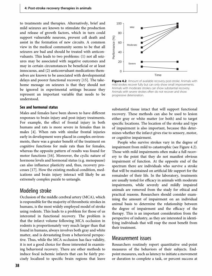
to treatments and therapies. Alternatively, brief andmild seizures are known to stimulate the productionand release of growth factors, which in turn couldsupport vulnerable neurons, prevent cell death andassist in the formation of new circuits. A commonview in the medical community seems to be that allseizures are bad and should be treated with anticon-vulsants. This leads to two problems: (1) not all seiz-ures may be associated with negative outcomes andmay in certain circumstances be beneficial or at leastinnocuous, and (2) anticonvulsant medications them-selves are known to be associated with developmentaldelays and poorer functional recovery [15]. The take-home message on seizures is that they should notbe ignored in experimental settings because theyrepresent an important variable that needs to beunderstood.
Sex and hormonal statusMales and females have been shown to have differentresponses to brain injury and post-injury treatments.For example, the effect of frontal injury in bothhumans and rats is more severe in females than inmales [4]. When rats with similar frontal injuriesearly in development were placed in complex environ-ments, there was a greater benefit of the treatment oncognitive functions for male rats than for females,whereas the opposite pattern of results was found formotor functions [16]. Moreover, the cyclic nature ofhormone levels and hormonal status (e.g. menopause)can also influence plasticity and, thus, recovery pro-cesses [17]. How the existing medical condition, med-ications and brain injury interact will likely be anextremely complex puzzle to untangle.
Modeling strokeOcclusion of the middle cerebral artery (MCA), whichis responsible for the majority of thrombotic strokes inhumans, is the most widely employed model of strokeusing rodents. This leads to a problem for those of usinterested in functional recovery. The problem isthat the infarct volume following MCA occlusion inrodents is proportionately very much larger than thatfound in humans, always involves both gray and whitematter, and is devastating from a behavioral perspec-tive. Thus, while the MCA occlusion has face validity,it is not a good choice for those interested in examin-ing behavioral recovery. There are other methods toinduce focal ischemic infarcts that can be fairly pre-cisely localized to specific brain regions that leave
substantial tissue intact that will support functionalrecovery. These methods can also be used to lesioneither gray or white matter (or both) and to targetspecific locations. The location of the stroke and typeof impairment is also important, because this deter-mines whether the infarct gives rise to sensory, motor,or cognitive impairment.
People who survive strokes vary in the degree ofimpairment from mild to catastrophic (see Figure 4.2).Those with mild impairments often make a full recov-ery to the point that they do not manifest obviousimpairment of function. At the opposite end of thespectrum there are individuals who survive a strokethat will be maintained on artificial life support for theremainder of their life. In the laboratory, treatmentsare usually tested for efficacy in animals with moderateimpairments, while severely and mildly impairedanimals are removed from the study for ethical andpractical reasons. Researchers should consider exam-ining the amount of impairment on an individualanimal basis to determine the relationship betweenthe degree of impairment and the efficacy of thetherapy. This is an important consideration from theperspective of industry, as they are interested in identi-fying individuals that will reap the most benefit fromtheir treatment.
Measurement issuesResearchers routinely report quantitative end-pointmeasures of the behaviors of their subjects. End-point measures, such as latency to initiate a movementor duration to complete a task, or percent success at
Mild
Moderate
Severe
Time
Per
form
ance
(%
)
100
80
60
40
20
0
Figure 4.2 Amount of available recovery post-stroke. Animals withmild strokes recover fully but can only show small improvements.Animals with moderate strokes can show substantial recovery.Animals with severe strokes often do not recover and showprogressive deterioration.
4: Post-stroke recovery therapies in animals
38
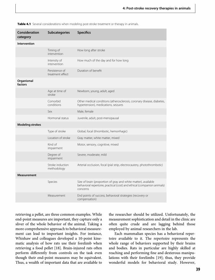
retrieving a pellet, are three common examples. Whileend-point measures are important, they capture only asliver of the whole behavior of the animal. Taking amore comprehensive approach to behavioral measure-ment can lead to important insights. For instance,Whishaw and colleagues developed a 10-point kine-matic analysis of how rats use their forelimb whenretrieving a food pellet [18]. Brain-injured rats oftenperform differently from controls on the task eventhough their end-point measures may be equivalent.Thus, a wealth of important data that are available to
the researcher should be utilized. Unfortunately, themeasurement sophistication and detail in the clinic areoften quite crude and are lagging behind thoseemployed by animal researchers in the lab.
Each mammalian species has a behavioral reper-toire available to it. The repertoire represents thewhole range of behaviors supported by their brainsand bodies. Rats in particular are highly skilled atreaching and performing fine and dexterous manipu-lations with their forelimbs [19]; thus, they providewonderful models for behavioral study. However,
Table 4.1 Several considerations when modeling post-stroke treatment or therapy in animals.
Considerationcategory
Subcategories Specifics
Intervention
Timing ofintervention
How long after stroke
Intensity ofintervention
How much of the day and for how long
Persistence oftreatment effect
Duration of benefit
Organismalfactors
Age at time ofstroke
Newborn, young, adult, aged
Comorbidconditions
Other medical conditions (atherosclerosis, coronary disease, diabetes,hypertension), medications, seizures
Sex Male, female
Hormonal status Juvenile, adult, post-menopausal
Modeling strokes
Type of stroke Global, focal (thrombotic, hemorrhagic)
Location of stroke Gray matter, white matter, mixed
Kind ofimpairment
Motor, sensory, cognitive, mixed
Degree ofimpairment
Severe, moderate, mild
Stroke inductionmethodology
Arterial occlusion, focal (pial strip, electrocautery, photothrombotic)
Measurement
Species Size of brain (proportion of gray and white matter), availablebehavioral repertoire, practical (cost) and ethical (companion animals)concerns
Measurement End points of success, behavioral strategies (recovery orcompensation)
4: Post-stroke recovery therapies in animals
39
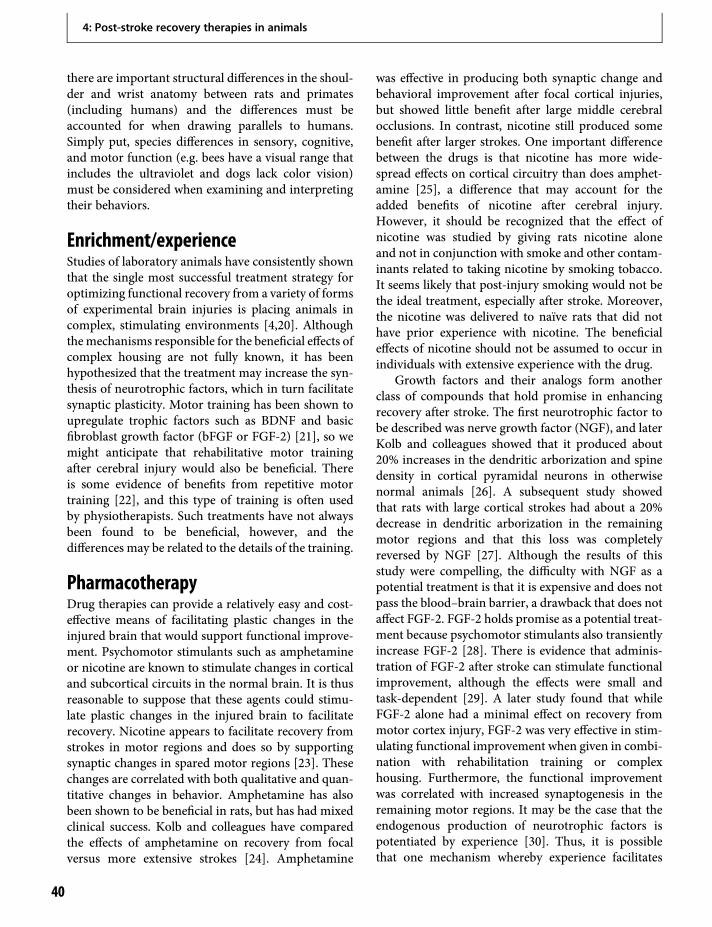
there are important structural differences in the shoul-der and wrist anatomy between rats and primates(including humans) and the differences must beaccounted for when drawing parallels to humans.Simply put, species differences in sensory, cognitive,and motor function (e.g. bees have a visual range thatincludes the ultraviolet and dogs lack color vision)must be considered when examining and interpretingtheir behaviors.
Enrichment/experienceStudies of laboratory animals have consistently shownthat the single most successful treatment strategy foroptimizing functional recovery from a variety of formsof experimental brain injuries is placing animals incomplex, stimulating environments [4,20]. Althoughthemechanisms responsible for the beneficial effects ofcomplex housing are not fully known, it has beenhypothesized that the treatment may increase the syn-thesis of neurotrophic factors, which in turn facilitatesynaptic plasticity. Motor training has been shown toupregulate trophic factors such as BDNF and basicfibroblast growth factor (bFGF or FGF-2) [21], so wemight anticipate that rehabilitative motor trainingafter cerebral injury would also be beneficial. Thereis some evidence of benefits from repetitive motortraining [22], and this type of training is often usedby physiotherapists. Such treatments have not alwaysbeen found to be beneficial, however, and thedifferences may be related to the details of the training.
PharmacotherapyDrug therapies can provide a relatively easy and cost-effective means of facilitating plastic changes in theinjured brain that would support functional improve-ment. Psychomotor stimulants such as amphetamineor nicotine are known to stimulate changes in corticaland subcortical circuits in the normal brain. It is thusreasonable to suppose that these agents could stimu-late plastic changes in the injured brain to facilitaterecovery. Nicotine appears to facilitate recovery fromstrokes in motor regions and does so by supportingsynaptic changes in spared motor regions [23]. Thesechanges are correlated with both qualitative and quan-titative changes in behavior. Amphetamine has alsobeen shown to be beneficial in rats, but has had mixedclinical success. Kolb and colleagues have comparedthe effects of amphetamine on recovery from focalversus more extensive strokes [24]. Amphetamine
was effective in producing both synaptic change andbehavioral improvement after focal cortical injuries,but showed little benefit after large middle cerebralocclusions. In contrast, nicotine still produced somebenefit after larger strokes. One important differencebetween the drugs is that nicotine has more wide-spread effects on cortical circuitry than does amphet-amine [25], a difference that may account for theadded benefits of nicotine after cerebral injury.However, it should be recognized that the effect ofnicotine was studied by giving rats nicotine aloneand not in conjunction with smoke and other contam-inants related to taking nicotine by smoking tobacco.It seems likely that post-injury smoking would not bethe ideal treatment, especially after stroke. Moreover,the nicotine was delivered to naïve rats that did nothave prior experience with nicotine. The beneficialeffects of nicotine should not be assumed to occur inindividuals with extensive experience with the drug.
Growth factors and their analogs form anotherclass of compounds that hold promise in enhancingrecovery after stroke. The first neurotrophic factor tobe described was nerve growth factor (NGF), and laterKolb and colleagues showed that it produced about20% increases in the dendritic arborization and spinedensity in cortical pyramidal neurons in otherwisenormal animals [26]. A subsequent study showedthat rats with large cortical strokes had about a 20%decrease in dendritic arborization in the remainingmotor regions and that this loss was completelyreversed by NGF [27]. Although the results of thisstudy were compelling, the difficulty with NGF as apotential treatment is that it is expensive and does notpass the blood–brain barrier, a drawback that does notaffect FGF-2. FGF-2 holds promise as a potential treat-ment because psychomotor stimulants also transientlyincrease FGF-2 [28]. There is evidence that adminis-tration of FGF-2 after stroke can stimulate functionalimprovement, although the effects were small andtask-dependent [29]. A later study found that whileFGF-2 alone had a minimal effect on recovery frommotor cortex injury, FGF-2 was very effective in stim-ulating functional improvement when given in combi-nation with rehabilitation training or complexhousing. Furthermore, the functional improvementwas correlated with increased synaptogenesis in theremaining motor regions. It may be the case that theendogenous production of neurotrophic factors ispotentiated by experience [30]. Thus, it is possiblethat one mechanism whereby experience facilitates
4: Post-stroke recovery therapies in animals
40

functional recovery is by increasing the endogenousproduction of neurotrophic factors, which in turnstimulate synaptic changes. For example, allowing ani-mals to spontaneously run in running wheels orexplore complex environments can increase levels ofgrowth factors, stimulate neurogenesis, increase resist-ance to brain injury, and improve learning and mentalperformance [31]. Little is known, however, about theoptimal timing or intensity of exercise needed to max-imally enhance behavioral outcome or neuronalplasticity.
Cell-based therapiesThe rapid progress in stem cell biology in recent yearshas led to an increasing interest in regeneration-basedtreatment strategies for damaged adult brain tissue.These novel tactics are based on two generalapproaches, namely replenishment of lost cells bytransplantation, and enhanced activation of endoge-nous stem cells. Both approaches have their strengthsand weaknesses with respect to practical and ethicalconsiderations. A perfect scenario for recovery involv-ing endogenous stem cells would at least involve thefollowing general steps. Precursor cells are upregu-lated to sufficient levels to replace the cells lost due tothe stroke. The cells then migrate to the site of injuryand differentiate into the lost cell types (neurons,astrocytes, oligodendroglia, and others) and theuncounted myriad of subtypes (e.g. excitatory projec-tion pyramidal cells and local inhibitory interneur-ons), in the correct, brain area-specific proportions.The neurons then form a working neural network withappropriate internal circuitry and correct synapticconnections with the spared tissue. In parallel, glialcells would provide their supportive and myelinationroles. Finally, on the systems level the new tissue mustoperate correctly to restore function for the ultimategoal of regenerative medicine to be achieved. While atfirst this scenario may seem highly improbable, itshould be remembered that during developmentmost of this process occurs. The trick is to get theadult brain to recapitulate the normal developmentalsequences within the lesioned area and then makeproper contact with existing tissue. Thus, from abasic science perspective we need to fully understandthe principles and specifics of brain development if weare to achieve brain tissue replacement.
A fortuitous observation that the medial prefrontalcortex can regenerate in postnatal rats provides a
model of brain tissue replacement. Pioneering studiesby Bryan Kolb demonstrated that neonatal rats thatreceived medial frontal cortex lesions at 10 days of ageexhibited spontaneous filling-in of the lesion cavity[32] and, when assessed as adults, these animalsshowed restitution of some behavioral abilities thatare dependent on an intact medial frontal cortex[33]. Although the spontaneous regeneration thatcan occur after medial prefrontal cortex (mPFC)lesions is uncommon in other structures, it can beinduced by infusion of FGF-2 after cortical injury onday 10 [34,35]. Richard Dyck confirmed the observa-tions of spontaneous regeneration in mice lesioned atpostnatal day 7 and extended this model tomake use oftransgenic technologies and thus identify molecularand genetic substrates that are necessary for regener-ation. This understudied phenomenon could lead toenormous breakthroughs in the development of brainrepair technologies.
The perfect scenario for recovery may be out ofreach given our current limitations, but it is quitereasonable to propose a beneficial role of neuronalstem cells (see also Chapter 24). The exploitation ofendogenous neural stem cells for therapeutic purposeshas been investigated within a number of diseasemodels including stroke. Experimental models ofstroke induce proliferation of stem cells residentin the subventricular zone and/or dentate gyrus(Figure 4.3). Neuronal migration is rerouted fromthe rostral migratory stream to damaged cortex andstriatum. Likewise, cells resident in the dentate gyruswill migrate to the lesion site within the hippocampus.Once at the lesion site, these new cells incorporate intothe ischemic penumbra. Although most of these cellsdie within the first few weeks [36], those cells that dosurvive appear to differentiate into the predominantneuronal or glial phenotype and integrate into theexisting circuitry. Unfortunately, this small numberof surviving cells is not enough to support full func-tional recovery. As such, methods aimed at furtherincreasing proliferation, differentiation, and survivalare required to make optimal use of intrinsic stemcells. Singly and in combination, epidermal growthfactor (EGF), FGF-2, and erythropoietin (EPO) havebeen shown to assist in the differentiation and survivalof new neurons. Kolb and colleagues gave adult ratsan ischemic lesion to the primary motor cortex andtreated them three days later with EGF followed byEPO [27]. This treatment resulted in tissue regenera-tion concomitant with motor recovery. Importantly,
4: Post-stroke recovery therapies in animals
41
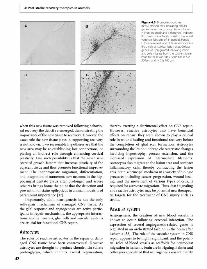
when this new tissue was removed following behavio-ral recovery the deficit re-emerged, demonstrating theimportance of the new tissue to recovery. However, theexact role the new tissue plays in supporting recoveryis not known. Two reasonable hypotheses are that thenew area may be re-establishing lost connections, orplaying an indirect role through enhancing corticalplasticity. One such possibility is that the new tissuesecreted growth factors that increase plasticity of theadjacent tissue and thus promote functional improve-ment. The inappropriate migration, differentiation,and integration of numerous new neurons in the hip-pocampal dentate gyrus after prolonged and severeseizures brings home the point that the detection andprevention of status epilepticus in animal models is ofparamount importance [37].
Importantly, adult neurogenesis is not the onlyself-repair mechanism of damaged CNS tissue. Asthe glial response and angiogenesis are active partic-ipants in repair mechanisms, the appropriate interac-tions among neurons, glial cells and vascular systemsare crucial for functional CNS repair.
AstrocytesThe roles of reactive astrocytes in the repair of dam-aged CNS tissue have been controversial. Reactiveastrocytes are thought to produce chondroitin sulfateproteoglycan, which inhibits axonal regeneration,
thereby exerting a detrimental effect on CNS repair.However, reactive astrocytes also have beneficialeffects on repair: they were shown to play a crucialrole in wound healing and functional recovery beforethe completion of glial scar formation. Astrocytessurrounding the lesion undergo characteristic changesinvolving hypertrophy, process extension, and theincreased expression of intermediate filaments.Astrocytes also migrate to the lesion area and compactinflammatory cells, thereby contracting the lesionarea. Stat3, a principal mediator in a variety of biologicprocesses including cancer progression, wound heal-ing, and the movement of various types of cells, isrequired for astrocyte migration. Thus, Stat3 signalingand reactive astrocytes may be potential new therapeu-tic targets for the treatment of CNS injury such asstroke.
Vascular systemAngiogenesis, the creation of new blood vessels, isknown to occur following cerebral infarction. Theexpression of several angiogenesis-related genes isregulated in an orchestrated fashion in the brain afterischemia [38]. The role of the vascular system in CNSrepair appears to be highly significant, and the poten-tial roles of blood vessels as scaffolds for neuroblastmigration in ischemic brain are intriguing. Palmer andcolleagues speculated that neurogenesis was intimately
Figure 4.3 Bromodeoxyuridine(BrdU)-stained cells indicating cellulargenesis after motor cortex lesion. PanelsA (non-lesioned) and B (lesioned) indicateBrdU cells immediately dorsal to the lateralventricle (bottom left in panels). PanelsC (non-lesioned) and D (lesioned) indicateBrdU cells at cortical lesion sites. Cellulargenesis is upregulated following lesionand cells migrate from the subventricularzone to the lesion sites. Scale bar in A is300 µm and in C is 100 µm.
4: Post-stroke recovery therapies in animals
42
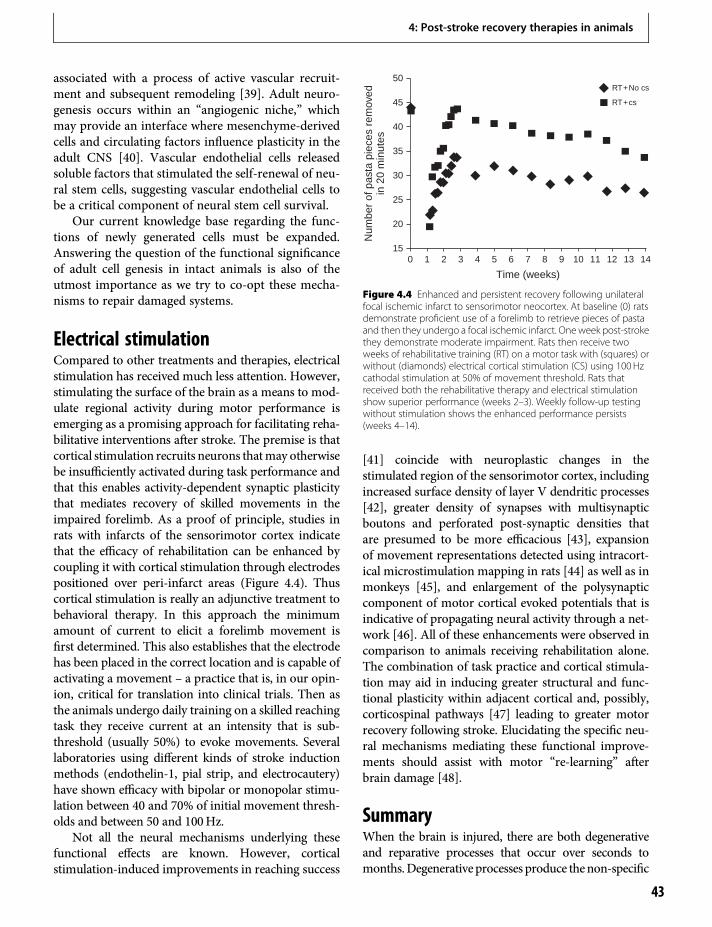
associated with a process of active vascular recruit-ment and subsequent remodeling [39]. Adult neuro-genesis occurs within an “angiogenic niche,” whichmay provide an interface where mesenchyme-derivedcells and circulating factors influence plasticity in theadult CNS [40]. Vascular endothelial cells releasedsoluble factors that stimulated the self-renewal of neu-ral stem cells, suggesting vascular endothelial cells tobe a critical component of neural stem cell survival.
Our current knowledge base regarding the func-tions of newly generated cells must be expanded.Answering the question of the functional significanceof adult cell genesis in intact animals is also of theutmost importance as we try to co-opt these mecha-nisms to repair damaged systems.
Electrical stimulationCompared to other treatments and therapies, electricalstimulation has received much less attention. However,stimulating the surface of the brain as a means to mod-ulate regional activity during motor performance isemerging as a promising approach for facilitating reha-bilitative interventions after stroke. The premise is thatcortical stimulation recruits neurons thatmay otherwisebe insufficiently activated during task performance andthat this enables activity-dependent synaptic plasticitythat mediates recovery of skilled movements in theimpaired forelimb. As a proof of principle, studies inrats with infarcts of the sensorimotor cortex indicatethat the efficacy of rehabilitation can be enhanced bycoupling it with cortical stimulation through electrodespositioned over peri-infarct areas (Figure 4.4). Thuscortical stimulation is really an adjunctive treatment tobehavioral therapy. In this approach the minimumamount of current to elicit a forelimb movement isfirst determined. This also establishes that the electrodehas been placed in the correct location and is capable ofactivating a movement – a practice that is, in our opin-ion, critical for translation into clinical trials. Then asthe animals undergo daily training on a skilled reachingtask they receive current at an intensity that is sub-threshold (usually 50%) to evoke movements. Severallaboratories using different kinds of stroke inductionmethods (endothelin-1, pial strip, and electrocautery)have shown efficacy with bipolar or monopolar stimu-lation between 40 and 70% of initial movement thresh-olds and between 50 and 100Hz.
Not all the neural mechanisms underlying thesefunctional effects are known. However, corticalstimulation-induced improvements in reaching success
[41] coincide with neuroplastic changes in thestimulated region of the sensorimotor cortex, includingincreased surface density of layer V dendritic processes[42], greater density of synapses with multisynapticboutons and perforated post-synaptic densities thatare presumed to be more efficacious [43], expansionof movement representations detected using intracort-ical microstimulation mapping in rats [44] as well as inmonkeys [45], and enlargement of the polysynapticcomponent of motor cortical evoked potentials that isindicative of propagating neural activity through a net-work [46]. All of these enhancements were observed incomparison to animals receiving rehabilitation alone.The combination of task practice and cortical stimula-tion may aid in inducing greater structural and func-tional plasticity within adjacent cortical and, possibly,corticospinal pathways [47] leading to greater motorrecovery following stroke. Elucidating the specific neu-ral mechanisms mediating these functional improve-ments should assist with motor “re-learning” afterbrain damage [48].
SummaryWhen the brain is injured, there are both degenerativeand reparative processes that occur over seconds tomonths.Degenerative processes produce thenon-specific
Time (weeks)
0
50
45
40
35
30
25
20
151 2 3 4 5 6 7 8 9 10 11 12 1413
RT+No cs
RT+cs
Num
ber
of p
asta
pie
ces
rem
oved
in 2
0 m
inut
es
Figure 4.4 Enhanced and persistent recovery following unilateralfocal ischemic infarct to sensorimotor neocortex. At baseline (0) ratsdemonstrate proficient use of a forelimb to retrieve pieces of pastaand then they undergo a focal ischemic infarct. Oneweek post-strokethey demonstrate moderate impairment. Rats then receive twoweeks of rehabilitative training (RT) on a motor task with (squares) orwithout (diamonds) electrical cortical stimulation (CS) using 100Hzcathodal stimulation at 50% of movement threshold. Rats thatreceived both the rehabilitative therapy and electrical stimulationshow superior performance (weeks 2–3). Weekly follow-up testingwithout stimulation shows the enhanced performance persists(weeks 4–14).
4: Post-stroke recovery therapies in animals
43
effects of the brain injury, such as the loss of cellsand synapses in widespread brain regions. The reparativechanges that follow use the brain’s endogenous capacityto restructure, allowing compensatory behaviors toemerge and improve performance. Several treatmentsand therapies appear to facilitate the rate and extentof behavioral recovery, many of which work bestwhen used in combination. There are many factors andissues that should be considered when selecting andimplementing an animal model of stroke. Learningmore about the brain’s ability to relearn should help usimprove recovery and quality of life for people who havehad a stroke.
References1. Gladstone DJ, Black SE, Hakim AM. Toward wisdom
from failure: Lessons from neuroprotective stroke trialsand new therapeutic directions. Stroke.2002;33:2123–36.
2. Colbourne F, Sutherland G, Corbett D. Postischemichypothermia. A critical appraisal with implicationsfor clinical treatment.Mol Neurobiol. 1997;14:171–201.
3. Kozlowski DA, James DC, Schallert T. Use-dependentexaggeration of neuronal injury after unilateralsensorimotor cortex lesions. J Neurosci.1996;16:4776–86.
4. Kolb B. Brain plasticity and behavior. Philadelphia, PA:Lawrence Erlbaum Associates; 1995.
5. Whishaw IQ, Kolb B. Sparing of skilled forelimbreaching and corticospinal projections after neonatalmotor cortex removal or hemidecortication in the rat:Support for the Kennard doctrine. Brain Res.1988;451:97–114.
6. Lomber S, Eggermont JJ. Reprogramming the cerebralcortex. London: Oxford University Press; 2006.
7. Teuber HL. Recovery of function after brain injury inman. Ciba Found Symp. 1975;34:159–90.
8. Barnes CA. Long-term potentiation and the ageingbrain. Phil Trans R Soc Lond B Biol Sci.2003;358:765–72.
9. Kolb B, Whishaw IQ. Fundamentals of humanneuropsychology. 5th ed. New York: Worth; 2003.
10. McGill JK, Gallagher L, Carswell HV, et al. Impairedfunctional recovery after stroke in the stroke-pronespontaneously hypertensive rat. Stroke 2005;36:135–41.
11. Windle V, Corbett D. Fluoxetine and recovery of motorfunction after focal ischemia in rats. Brain Res.2005;1044:25–32.
12. Zhao C, Puurunen K, Schallert T, et al. Behavioral andhistological effects of chronic antipsychotic andantidepressant drug treatment in aged rats with focal
ischemic brain injury. Behav Brain Res.2005;158:211–20.
13. Teskey GC, Monfils M-H, VandenBerg P, et al. Motormap expansion following repeated cortical and limbicseizures is related to synaptic potentiation. CerebCortex. 2002;12:98–105.
14. Valentine PA, Teskey GC, Eggermont JJ. Kindlinglimits the inter-ictal neuronal temporal responseproperties in cat primary auditory cortex. Epilepsia.2005;46:171–8.
15. Hernandez TD. Preventing post-traumatic epilepsyafter brain injury: Weighing the costs and benefits ofanticonvulsant prophylaxis. Trends Pharmacol Sci.1997;18:59–62.
16. Kolb B, Gibb R, Gorny G. Experience-dependentchanges in dendritic arbor and spine density inneocortex vary qualitatively with age and sex. NeurobiolLearn Mem. 2003;79:1–10.
17. Kolb B, Stewart J. Sex-related differences in dendriticbranching of cells in the prefrontal cortex of rats.J Neuroendocrinol. 1991;3:95–9.
18. Whishaw IQ, Pellis SM, Gorny BP, et al. Theimpairments in reaching and the movements ofcompensation in rats with motor cortex lesions: anendpoint, videorecording, and movement notationanalysis. Behav Brain Res. 1991;42:77–91.
19. Whishaw IQ, Kolb B. The behaviour of the laboratoryrat. London: Oxford University Press; 2004.
20. Biernaskie J, Corbett D. Enriched rehabilitative trainingpromotes improved forelimb motor function andenhanced dendritic growth after focal ischemic injury.J Neurosci. 2001;21:5272–80.
21. Kleim JA, Jones TA, Schallert T. Motor enrichment andthe induction of plasticity before or after brain injury.Neurochem Res. 2003;28:1757–69.
22. Nudo RJ, Wise BM, SiFuentes F, et al. Neural substratesfor the effects of rehabilitative training on motorrecovery after ischemic infarct. Science.1996;272:1791–4.
23. Gonzalez CLR, Gharbawie OA, Kolb B. Chronic low-dose administration of nicotine facilitates recovery andsynaptic change after focal ischemia in rats.Neuropharmacology. 2006;50:777–87.
24. Kolb B, Gorny G, Li Y, et al. Amphetamine or cocainelimits the ability of later experience to promotestructural plasticity in the neocortex and nucleusaccumbens. Proc Natl Acad Sci USA.2003;100:10 523–8.
25. Robinson TE, Kolb B. Structural plasticity associatedwith drugs of abuse. Neuropharmacology. 2004;47Suppl 1: 33–46.
26. Kolb B, Cote S, Ribeiro-da-Silva A, et al. Nerve growthfactor treatment prevents dendritic atrophy and
4: Post-stroke recovery therapies in animals
44
promotes recovery of function after cortical injury.Neuroscience. 1997;76:1139–51.
27. Kolb B, Morshead C, Gonzalez C, et al. Growth factor-stimulated generation of new cortical tissue andfunctional recovery after stroke damage to the motorcortex of rats. J Cereb Blood Flow Metab.2007;27:983–97.
28. Flores C, Stewart J. Changes in astrocytic basicfibroblast growth factor expression duringand after prolonged exposure to escalatingdoses of amphetamine. Neuroscience.2000;98:287–93.
29. Kawamata T, Speliotes EK, Finklestein SP. The role ofpolypeptide growth factors in recovery from stroke.Adv Neurol. 1997;73:377–82.
30. Kolb B, ForgieM, Gibb R, et al. Age, experience, and thechanging brain. Neurosci Biobehav Revs.1998;22:143–59.
31. Cotman CW, Berchtold NC. Exercise: A behavioralintervention to enhance brain health and plasticity.Trends Neurosci. 2002;25:295–301.
32. Kolb B. Animal models for human PFC-relateddisorders. Prog Brain Res. 1990;85:501–19.
33. Kolb B, Petrie B, Cioe J. Recovery from early corticaldamage in rats: VII. Comparison of the behavioural andanatomical effects of medial prefrontal lesions atdifferent ages of neural maturation. Behav Brain Res.1996;79:1–14.
34. Monfils MH, Driscoll I, Vandenberg PM, et al. Basicfibroblast growth factor stimulates functional recoveryafter neonatal lesions of motor cortex in rats.Neuroscience 2005;134:1–8.
35. Monfils MH, Driscoll I, Kamitakahara H, et al. FGF-2-induced cell proliferation stimulates anatomical,neurophysiological and functional recovery fromneonatal motor cortex injury. Eur J Neurosci.2006;24:739–49.
36. Arvidsson A, Collin T, Kirik D, et al. Neuronalreplacement from endogenous precursors in the adultbrain after stroke. Nat Med. 2002;8:963–70.
37. Scharfman HE, Hen R. Is more neurogenesis alwaysbetter? Science. 2007;315:336–8.
38. Hayashi T, Noshita N, Sugawara T, et al. Temporalprofile of angiogenesis and expression of related genesin the brain after ischemia. J Cereb Blood Flow Metab.2003;23:166–80.
39. Palmer TD, Willhoite AR, Gage FH. Vascular niche foradult hippocampal neurogenesis. J Comp Neurol.2000;425:479–94.
40. Shen Q, Goderie SK, Jin L, et al. Endothelial cellsstimulate self-renewal and expand neurogenesis ofneural stem cells. Science. 2004;304:1338–40.
41. Adkins DL, Campos P, Quach D, et al. Epidural corticalstimulation enhances motor function aftersensorimotor cortical infarcts in rats. Exp Neurol.2006;200:356–70.
42. Adkins-Muir DL, Jones TA. Cortical electricalstimulation combined with rehabilitative training:Enhanced functional recovery and dendritic plasticityfollowing focal cortical ischemia in rats. Neurol Res.2003;25:780–8.
43. Adkins DL, Hsu JE, Jones TA. Motor corticalstimulation promotes synaptic plasticity and behavioralimprovements following sensorimotor cortex lesions.Exp Neurol. 2008;212:14–28.
44. Kleim JA, Bruneau R, VandenBerg P, et al. Motorcortex stimulation enhances motor recovery andreduces peri-infarct dysfunction following ischemicinsult. Neurol Res. 2003;25:789–93.
45. Plautz EJ, Barbay S, Frost SB, et al. Post-infarct corticalplasticity and behavioral recovery using concurrentcortical stimulation and rehabilitative training: Afeasibility study in primates. Neurol Res.2003;25:801–10.
46. Teskey GC, Flynn C, Goertzen CD, et al. Corticalstimulation improves skilled forelimb use following afocal ischemic infarct in the rat. Neurol Res.2003;25:794–800.
47. Brown JA, Lutsep H, Cramer SC, et al. Motor cortexstimulation for enhancement of recovery after stroke:Case report. Neurol Res. 2003;25:815–8.
48. Cramer SC, Bastings EP. Mapping clinically relevantplasticity after stroke. Neuropharmacology.2000;39:842–51.
4: Post-stroke recovery therapies in animals
45


5 Environmental effects on functionaloutcome after strokeBarbro B. Johansson
IntroductionInteraction between genes and environment is neces-sary for all life and has reached the highest level in thehuman cerebral cortex. The property of the brain tointeract and adapt to environmental requirements andmultisensory stimulation as well as to our activities,skill acquisitions, and thoughts is referred to as brainplasticity, and it occurs at many levels from moleculesand cells to cortical networks [1,2]. Although the con-cept of plasticity had been proposed earlier, the firstknown experimental evidence that environment caninfluence behavior was provided by Donald Hebb in1947, who reported that rats that were allowed to runaround in his house had better memory and highercapacity to solve problems than rats housed in thelaboratory [3]. In the 1960s, Rosenzweig and associatesdemonstrated that functional improvement inducedby environmental enrichment was associated withmorphological and biochemical changes [4]. Furtherexperimental studies have verified that brain plasticityplays a role in the intact brain and after acute braindisorders [5–8], and there is accumulating evidencethat environmental stimulation may have a beneficialinfluence in delaying the progress in degenerative dis-orders [9].We usually talk about the positive aspects ofplasticity, but it is important to realize that reducedsensory input and motor activity have the opposite,negative effects. The question dealt with here is whatexperimental and clinical evidence we have to supportthat environmental issues are important in strokerehabilitation [10].
Environment, dendrites anddendritic spinesAfter a ligation of the middle cerebral artery (MCA),rats housed in an enriched environment during both
the pre- and post-ischemic period improve earlier andslightly more than when housed in the enriched envi-ronment only during the post-ischemic period. Bothgroups perform significantly better in a variety of teststhan rats housed in single cages, a condition that, in thisreview, is called deprived housing [11]. Environmentalenrichment can also reduce the spatial memory deficitthat is caused by cortical infarcts in the rat [12].
A consistent effect of environmental enrichment inintact animals has been on the morphology of den-drites and dendritic spines. It was shown 35 years agothat enriched housing increased the number of den-dritic branches and dendritic spines in the intact ratbrain [13,14] and many later studies have confirmedthese observations [6]. Dendritic spines are the pri-mary postsynaptic targets for excitatory glutaminergicsynapses in the mature brain. The dendritic tree iscovered with a variety of excitable synaptic channelsoperating on different timescales and with activity-dependent sensitivity enabling a sophisticated neuro-nal plasticity.
With a method that allows a three-dimensionalview of dendritic spines and optimal quantificationof dendrites, three weeks of enriched housing signifi-cantly increased the number of dendritic spines perunit length in pyramidal neurons in layers 2/3 andlayer 5 compared with rats from the same litter housedin cages with 3–4 rats per cage. After a cortical infarct,the same effect was observed in layer 2/3 neurons inthe homotopic contralateral somatosensory cortex(Figure 5.1) [15]. In contrast, the number of layer 5pyramidal neurons was reduced in both groups withinfarcts, most likely due to extensive reduction ofconnections from the corpus callosum in this model.The three-week postoperative time was chosenbecause at that time significant differences in func-tional outcome between enriched, standard (3–4 ratshoused together), or deprived rats are well established.
Brain Repair After Stroke, ed. S. C. Cramer and R. J. Nudo. Published by Cambridge University Press.© Cambridge University Press 2010.
47
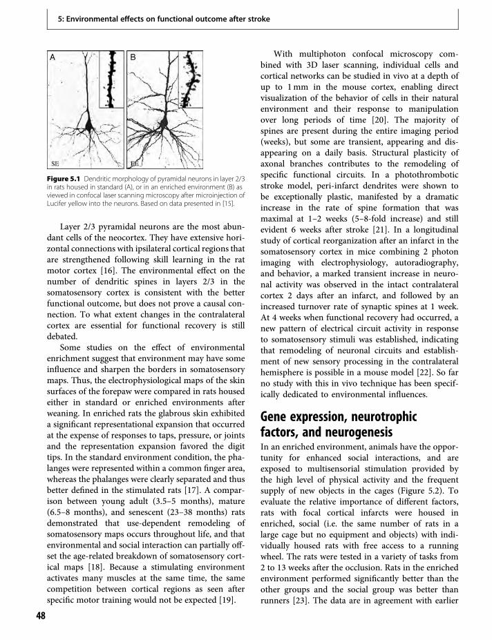
Layer 2/3 pyramidal neurons are the most abun-dant cells of the neocortex. They have extensive hori-zontal connections with ipsilateral cortical regions thatare strengthened following skill learning in the ratmotor cortex [16]. The environmental effect on thenumber of dendritic spines in layers 2/3 in thesomatosensory cortex is consistent with the betterfunctional outcome, but does not prove a causal con-nection. To what extent changes in the contralateralcortex are essential for functional recovery is stilldebated.
Some studies on the effect of environmentalenrichment suggest that environment may have someinfluence and sharpen the borders in somatosensorymaps. Thus, the electrophysiological maps of the skinsurfaces of the forepaw were compared in rats housedeither in standard or enriched environments afterweaning. In enriched rats the glabrous skin exhibiteda significant representational expansion that occurredat the expense of responses to taps, pressure, or jointsand the representation expansion favored the digittips. In the standard environment condition, the pha-langes were represented within a common finger area,whereas the phalanges were clearly separated and thusbetter defined in the stimulated rats [17]. A compar-ison between young adult (3.5–5 months), mature(6.5–8 months), and senescent (23–38 months) ratsdemonstrated that use-dependent remodeling ofsomatosensory maps occurs throughout life, and thatenvironmental and social interaction can partially off-set the age-related breakdown of somatosensory cort-ical maps [18]. Because a stimulating environmentactivates many muscles at the same time, the samecompetition between cortical regions as seen afterspecific motor training would not be expected [19].
With multiphoton confocal microscopy com-bined with 3D laser scanning, individual cells andcortical networks can be studied in vivo at a depth ofup to 1mm in the mouse cortex, enabling directvisualization of the behavior of cells in their naturalenvironment and their response to manipulationover long periods of time [20]. The majority ofspines are present during the entire imaging period(weeks), but some are transient, appearing and dis-appearing on a daily basis. Structural plasticity ofaxonal branches contributes to the remodeling ofspecific functional circuits. In a photothromboticstroke model, peri-infarct dendrites were shown tobe exceptionally plastic, manifested by a dramaticincrease in the rate of spine formation that wasmaximal at 1–2 weeks (5–8-fold increase) and stillevident 6 weeks after stroke [21]. In a longitudinalstudy of cortical reorganization after an infarct in thesomatosensory cortex in mice combining 2 photonimaging with electrophysiology, autoradiography,and behavior, a marked transient increase in neuro-nal activity was observed in the intact contralateralcortex 2 days after an infarct, and followed by anincreased turnover rate of synaptic spines at 1 week.At 4 weeks when functional recovery had occurred, anew pattern of electrical circuit activity in responseto somatosensory stimuli was established, indicatingthat remodeling of neuronal circuits and establish-ment of new sensory processing in the contralateralhemisphere is possible in a mouse model [22]. So farno study with this in vivo technique has been specif-ically dedicated to environmental influences.
Gene expression, neurotrophicfactors, and neurogenesisIn an enriched environment, animals have the oppor-tunity for enhanced social interactions, and areexposed to multisensorial stimulation provided bythe high level of physical activity and the frequentsupply of new objects in the cages (Figure 5.2). Toevaluate the relative importance of different factors,rats with focal cortical infarcts were housed inenriched, social (i.e. the same number of rats in alarge cage but no equipment and objects) with indi-vidually housed rats with free access to a runningwheel. The rats were tested in a variety of tasks from2 to 13 weeks after the occlusion. Rats in the enrichedenvironment performed significantly better than theother groups and the social group was better thanrunners [23]. The data are in agreement with earlier
Figure 5.1 Dendritic morphology of pyramidal neurons in layer 2/3in rats housed in standard (A), or in an enriched environment (B) asviewed in confocal laser scanning microscopy after microinjection ofLucifer yellow into the neurons. Based on data presented in [15].
5: Environmental effects on functional outcome after stroke
48
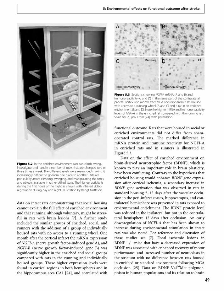
data on intact rats demonstrating that social housingcannot explain the full effect of enriched environmentand that running, although voluntary, might be stress-ful in rats with brain lesions [7]. A further studyincluded the similar groups of enriched, social, andrunners with the addition of a group of individuallyhoused rats with no access to a running wheel. Onemonth after the cortical infarct the mRNA expressionof NGFI-A (nerve growth factor-induced gene A), andNGFI-B (nerve growth factor-induced gene B) wassignificantly higher in the enriched and social groupscompared with rats in the running and individuallyhoused groups. These higher expression levels werefound in cortical regions in both hemispheres and inthe hippocampus area CA1 [24], and correlated with
functional outcome. Rats that were housed in social orenriched environments did not differ from sham-operated control rats. The marked difference inmRNA protein and immune reactivity for NGFI-Ain enriched rats and in runners is illustrated inFigure 5.3.
Data on the effect of enriched environment onbrain-derived neurotrophic factor (BDNF), which isknown to play an important role in brain plasticity,have been conflicting. Contrary to the hypothesis thatenriched housing would enhance BDNF gene expres-sion after cortical ischemia, a secondary increase inBDNF gene activation that was observed in rats instandard housing 2–12 days after the vascular occlu-sion in the peri-infarct cortex, hippocampus, and con-tralateral hemisphere was prevented in rats exposed toenvironmental enrichment. The BDNF protein levelwas reduced in the ipsilateral but not in the contrala-teral hemisphere 12 days after occlusion. An earlydownregulation of NGFI-A that has been shown toincrease during environmental stimulation in intactrats was also noted. For reference and discussion ofthese studies see [7]. Focal ischemic lesions inBDNF +/– mice that have a decreased expression ofBDNF was associated with enhanced recovery of motorperformance and increased number of neuroblasts inthe striatum with no difference between rats housedin enriched or standard environment following MCAocclusion [25]. Data on BDNF Val66Met polymor-phism in human populations and its relation to brain
Figure 5.3 Sections showing NGFI-A mRNA (A and B) andimmunoreactivity (C and D) in the same part of the contralateralparietal cortex one month after MCA occlusion from a rat housedwith access to a running wheel (A and C) and a rat in an enrichedenvironment (B and D). Note the higher mRNA and immunoreactivitylevels of NGFI-A in the enriched rat compared with the running rat.Scale bar 20 μm. From [24], with permission.
Figure 5.2 In the enriched environment rats can climb, swing,investigate, and handle a number of tools that are changed two orthree times a week. The different levels were rearranged making itincreasingly difficult to go from one place to another. Rats areparticularly active climbing, swinging, and manipulating the toolsand objects available in rather skilled ways. The highest activity isduring the first hours of the night as shown with infrared video-registration during day and night. Illustration by Bengt Mattsson.
5: Environmental effects on functional outcome after stroke
49
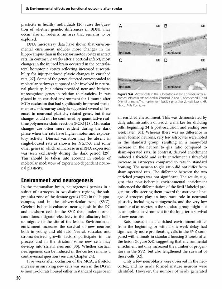
plasticity in healthy individuals [26] raise the ques-tion of whether genetic differences in BDNF mayoccur also in rodents, an area that remains to beexplored.
DNA microarray data have shown that environ-mental enrichment induces more changes in thehippocampus than in the sensorimotor cortex in intactrats. In contrast, 2 weeks after a cortical infarct, mostchanges in the injured brain occurred in the contrala-teral homotopic cortex reflecting increased suscepti-bility for injury-induced plastic changes in enrichedrats [27]. Some of the genes detected corresponded tomolecular pathways supposed to be involved in neuro-nal plasticity, but others provided new and hithertounrecognized genes in relation to plasticity. In ratsplaced in an enriched environment for 1 month afterMCA occlusion that had significantly improved spatialmemory, microarray analysis suggested several differ-ences in neuronal plasticity-related genes, but thesechanges could not be confirmed by quantitative real-time polymerase chain reaction (PCR) [28]. Molecularchanges are often more evident during the darkphase when the rats have higher motor and explora-tory activity. Diurnal variation may be absent insingle-housed rats as shown for NGFI-A and someother genes in which an increase in mRNA expressionwas seen exclusively during the dark period [29].This should be taken into account in studies ofmolecular mediators of experience-dependent neuro-nal plasticity.
Environment and neurogenesisIn the mammalian brain, neurogenesis persists in asubset of astrocytes in two distinct regions, the sub-granular zone of the dentate gyrus (DG) in the hippo-campus, and in the subventricular zone (SVZ).Cerebral ischemia enhances neurogenesis in the DGand newborn cells in the SVZ that, under normalconditions, migrate selectively to the olfactory bulb,or migrate to the site of the lesion. Environmentalenrichment increases the survival of new neuronsboth in young and old rats. Neural, vascular, andstroma-derived growth factors participate in theprocess and in the striatum some new cells maydevelop into striatal neurons [30]. Whether corticalneurogenesis can be induced in the cortex remains acontroversial question (see also Chapter 24).
Five weeks after occlusion of the MCA, a fivefoldincrease in surviving new cells was seen in the DG in6-month-old rats housed either in standard cages or in
an enriched environment. This was demonstrated bydaily administration of BrdU, a marker for dividingcells, beginning 24 h post-occlusion and ending oneweek later [31]. Whereas there was no difference innewly formed neurons, very few astrocytes were notedin the standard group, resulting in a many-foldincrease in the neuron to glia ratio compared tosham-operated rats. In contrast, delayed enrichmentinduced a fivefold and early enrichment a threefoldincrease in astrocytes compared to rats in standardhousing. The neuron to glia ratio did not differ fromsham-operated rats. The difference between the twoenriched groups was not significant. The results sug-gest that post-ischemic environmental enrichmentinfluenced the differentiation of the BrdU-labeled pro-genitor cells, steering them toward the astrocytic line-age. Astrocytes play an important role in neuronalplasticity including synaptogenesis, and the very lownumber of astrocytes in the standard group might notbe an optimal environment for the long-term survivalof new neurons.
Rats housed in an enriched environment eitherfrom the beginning or with a one-week delay hadsignificantly more proliferating cells in the SVZ com-pared with animals in standard housing 5 weeks afterthe lesion (Figure 5.4), suggesting that environmentalenrichment not only increased the number of progen-itors in the SVZ, but also lengthened the survival ofthose cells [32].
Only a few neuroblasts were observed in the neo-cortex, and no newly formed mature neurons wereidentified. However, the number of newly generated
Figure 5.4 Mitotic cells in the subventricular zone 5 weeks after acortical infarct in rats housed in standard (A and B) or enriched (C andD) environment. Themarker for mitosis is phosphorylated histone H3.Photo: Mila Komitova.
5: Environmental effects on functional outcome after stroke
50
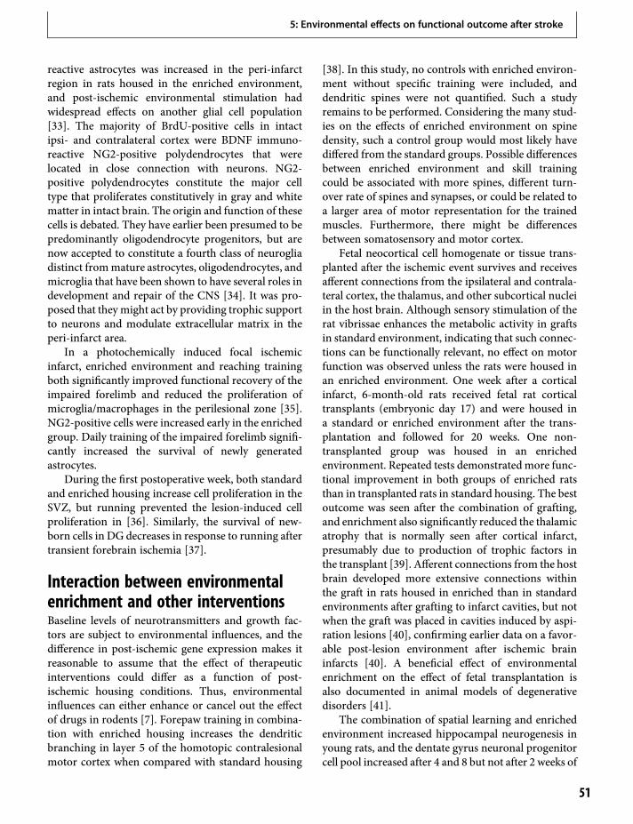
reactive astrocytes was increased in the peri-infarctregion in rats housed in the enriched environment,and post-ischemic environmental stimulation hadwidespread effects on another glial cell population[33]. The majority of BrdU-positive cells in intactipsi- and contralateral cortex were BDNF immuno-reactive NG2-positive polydendrocytes that werelocated in close connection with neurons. NG2-positive polydendrocytes constitute the major celltype that proliferates constitutively in gray and whitematter in intact brain. The origin and function of thesecells is debated. They have earlier been presumed to bepredominantly oligodendrocyte progenitors, but arenow accepted to constitute a fourth class of neurogliadistinct frommature astrocytes, oligodendrocytes, andmicroglia that have been shown to have several roles indevelopment and repair of the CNS [34]. It was pro-posed that theymight act by providing trophic supportto neurons and modulate extracellular matrix in theperi-infarct area.
In a photochemically induced focal ischemicinfarct, enriched environment and reaching trainingboth significantly improved functional recovery of theimpaired forelimb and reduced the proliferation ofmicroglia/macrophages in the perilesional zone [35].NG2-positive cells were increased early in the enrichedgroup. Daily training of the impaired forelimb signifi-cantly increased the survival of newly generatedastrocytes.
During the first postoperative week, both standardand enriched housing increase cell proliferation in theSVZ, but running prevented the lesion-induced cellproliferation in [36]. Similarly, the survival of new-born cells in DG decreases in response to running aftertransient forebrain ischemia [37].
Interaction between environmentalenrichment and other interventionsBaseline levels of neurotransmitters and growth fac-tors are subject to environmental influences, and thedifference in post-ischemic gene expression makes itreasonable to assume that the effect of therapeuticinterventions could differ as a function of post-ischemic housing conditions. Thus, environmentalinfluences can either enhance or cancel out the effectof drugs in rodents [7]. Forepaw training in combina-tion with enriched housing increases the dendriticbranching in layer 5 of the homotopic contralesionalmotor cortex when compared with standard housing
[38]. In this study, no controls with enriched environ-ment without specific training were included, anddendritic spines were not quantified. Such a studyremains to be performed. Considering the many stud-ies on the effects of enriched environment on spinedensity, such a control group would most likely havediffered from the standard groups. Possible differencesbetween enriched environment and skill trainingcould be associated with more spines, different turn-over rate of spines and synapses, or could be related toa larger area of motor representation for the trainedmuscles. Furthermore, there might be differencesbetween somatosensory and motor cortex.
Fetal neocortical cell homogenate or tissue trans-planted after the ischemic event survives and receivesafferent connections from the ipsilateral and contrala-teral cortex, the thalamus, and other subcortical nucleiin the host brain. Although sensory stimulation of therat vibrissae enhances the metabolic activity in graftsin standard environment, indicating that such connec-tions can be functionally relevant, no effect on motorfunction was observed unless the rats were housed inan enriched environment. One week after a corticalinfarct, 6-month-old rats received fetal rat corticaltransplants (embryonic day 17) and were housed ina standard or enriched environment after the trans-plantation and followed for 20 weeks. One non-transplanted group was housed in an enrichedenvironment. Repeated tests demonstratedmore func-tional improvement in both groups of enriched ratsthan in transplanted rats in standard housing. The bestoutcome was seen after the combination of grafting,and enrichment also significantly reduced the thalamicatrophy that is normally seen after cortical infarct,presumably due to production of trophic factors inthe transplant [39]. Afferent connections from the hostbrain developed more extensive connections withinthe graft in rats housed in enriched than in standardenvironments after grafting to infarct cavities, but notwhen the graft was placed in cavities induced by aspi-ration lesions [40], confirming earlier data on a favor-able post-lesion environment after ischemic braininfarcts [40]. A beneficial effect of environmentalenrichment on the effect of fetal transplantation isalso documented in animal models of degenerativedisorders [41].
The combination of spatial learning and enrichedenvironment increased hippocampal neurogenesis inyoung rats, and the dentate gyrus neuronal progenitorcell pool increased after 4 and 8 but not after 2 weeks of
5: Environmental effects on functional outcome after stroke
51
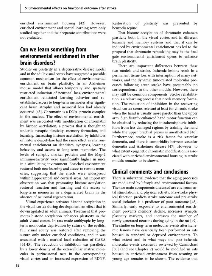
enriched environment housing [42]. However,enriched environment and spatial learning were onlystudied together and their separate contributions werenot evaluated.
Can we learn something fromenvironmental enrichment in otherbrain disorders?Studies on plasticity in a degenerative disease modeland in the adult visual cortex have suggested a possiblecommon mechanism for the effect of environmentalenrichment on brain plasticity. In an Alzheimermouse model that allows temporally and spatiallyrestricted induction of neuronal loss, environmentalenrichment reinstated learning behavior and re-established access to long-term memories after signifi-cant brain atrophy and neuronal loss had alreadyoccurred [43]. Chromatin is a DNA–protein complexin the nucleus. The effect of environmental enrich-ment was associated with modification of chromatinby histone acetylation, a process that is thought tounderlie synaptic plasticity, memory formation, andlearning. Increasing histone acetylation by inhibitorsof histone deacetylase had the same effect as environ-mental enrichment on dendrites, synapses, learningbehavior, and access to long-term memories. Thelevels of synaptic marker proteins and the Map-2immunoreactivity were significantly higher in micein a stimulating environment. Enriched environmentrestored both new learning and access to remote mem-ories, suggesting that the effects were widespreadwithin hippocampal and cortical areas. An importantobservation was that promoting histone acetylationrestored function and learning and the access tolong-term memories in a degenerated brain in theabsence of neuronal regeneration.
Visual experience activates histone acetylation inthe visual cortex during development, an effect that isdownregulated in adult animals. Treatment that pro-motes histone acetylation enhances plasticity in theadult visual cortex. In rats made amblyopic by long-term monocular deprivation by suture of the eyelids,full visual acuity was restored after removing thesuture only under enriched conditions, and it wasassociated with a marked local reduction of GABA[44,45]. The reduction of inhibition was paralleledby a lower density of the extracellular matrix mole-cules in perineuronal nets in the correspondingvisual cortex and an increased expression of BDNF.
Restoration of plasticity was prevented bybenzodiazepine.
That histone acetylation of chromatin enhancesplasticity both in the visual cortex and in differentlearning and memory systems and that it can beinduced by environmental enrichment has led to theproposal that chromatin remodeling may be the finalgate environmental enrichment opens to enhancebrain plasticity.
There are important differences between thesetwo models and stroke. Ischemic lesions result in apermanent tissue loss with interruption of many net-works, and the dynamic time-related molecular pro-cesses following acute stroke have presumably nocorrespondence in the other models. However, theremay still be common components. Stroke rehabilita-tion is a relearning process that needs memory forma-tion. The reduction of inhibition in the recoveringvisual cortex seems relevant at least for chronic strokewhen the hand is usually more paretic than the upperarm. Significantly enhanced hand motor function canbe obtained by reducing the intrahemispheric inhib-ition from less damaged regions by training the handwhile the upper brachial plexus is anesthetized [46].Furthermore, stroke is a risk factor for vasculardementia, and there is comorbidity between vasculardementia and Alzheimer disease [47]. However, towhat extent epigenetic chromatin modification is asso-ciated with enriched environmental housing in strokemodels remains to be shown.
Clinical comments and conclusionsThere is substantial evidence that the aging processesare modulated by lifestyle and environmental factors.The two main components discussed are environmen-tal stimulation and physical activity. Pre-stroke phys-ical function predicts stroke outcome and pre-strokesocial isolation is a predictor of poor outcome [48].Similarly, early exposure to environmental enrich-ment prevents memory decline, increases synapticplasticity markers, and increases the number ofnewly generated neurons during aging in the rat [49].The studies on long-term molecular events after ische-mic lesions have essentially been performed in ratshoused in standard or deprived environments. Towhat extent and in what ways the post-ischemicmolecular events excellently reviewed by Carmichael[50] (and see Chapter 2) would be modified in ratshoused in enriched environment from weaning oryoung age remains to be shown. The evidence that
5: Environmental effects on functional outcome after stroke
52
environmental enrichment induces epigenetic changesthat facilitate synaptogenesis and memory in othermodels of brain plasticity should stimulate moreresearch on basic mechanisms underlying environ-mental influence on the recovery after stroke.
Stroke units may be the closest correspondence toan enriched environment for stroke patients. A strokeis a dramatic change for the patients and anything thatcan be done to optimize the hospital and rehabilitationenvironment should be beneficial. No study has shownto what extent the beneficial effect is due to specificrehabilitation strategies, to the time spent in physio-therapy and occupational therapy, and a non-specificeffect of a more stimulating environment with com-petent staff that can encourage and support thepatients and family members, but all these factors arelikely to be important. The benefit of stroke unitscompared to general wards is most likely a combina-tion of optimal medical and nursing care, task-oriented activities, and for the individual, meaningfultraining in an environment that gives them confi-dence, stimulation, and motivation. Mere admittanceto a stroke unit with specially trained staff encouragingactive participation in the rehabilitation process cre-ates a positive and stimulating atmosphere. Also, cog-nitively impaired stroke patients do benefit fromadmission to an acute rehabilitation unit [51].
Patients with partial cortical infarcts and with cap-sular infarcts together constitute the major partici-pants in chronic stroke trials, but have so far notbeen studied extensively in rodents, mainly due tothe lack of good rodent models. The human brainhas proportionally more white matter than all otherspecies and the difference compared with rodents isenormous. The white matter tracts facilitate the spreadof brain edema and make the consequences of com-plete occlusion of the MCA much more severe inhuman patients than in mice and rats. The models ofstriatal infarction after short-term MCA occlusion inmice and rats have told us much about post-ischemicneurogenesis. However, it has little correspondence tosubcortical stroke in humans where the main func-tional deficits are related to the damage of the cortico-spinal tracts, and myelin inhibitor factors andmodification of the extracellular matrix are likely tobe more important.
Quality of life is not exclusively determined by thedisability, but also by the patient’s attitude and how theresulting handicap can be compensated for to enablethe patient to take part in social and cultural activities.
Complex cognitive–emotional behavior emerges fromdynamic interactions between brain networks.Memory problems, anxiety, mood disturbances, andpersistent fatigue can remain despite minimal motorimpairment. Patients with mild stroke may have subtledisabilities and difficulty with complex tasks that affectmeaningful activity and life satisfaction [52]. Post-stroke physical and cognitive activities are essentialto improve the quality of life and reduce the incidenceof new vascular events, depression and dementia.Patient associations that arrange various physical andcultural activities for both patients and family care-givers can play a large role and should be activelysupported bymedical staff andmembers of the society.A stimulating environment is likely to be the optimalbase from which to start specific training and otherinterventions as well as for continuous social andcognitive rehabilitation.
References1. Pascual-Leone A, Amedi A, Fregni F, et al. The plastic
human brain cortex. Ann Rev Neurosci.2005;28:377–401.
2. Johansson BB. Brain plasticity in health and disease.Keio J Med. 2004;53:231–46.
3. Hebb DO. The effect of early experience on problemsolving at maturity. Am Psychol. 1947;2:737–45.
4. Bennet EL, Diamond MC, Krech D, et al. Chemical andanatomical plasticity of brain. Science.1964:146:610–19.
5. Mohammed AH, Zhu SW, Darmopil S, et al.Environmental enrichment and the brain. Prog BrainRes. 2002;138:109–33.
6. Will B, Galani R, Kelche C, et al. Recovery from braininjury in animals: Relative efficacy of environmentalenrichment, physical exercise or formal training(1990–2002). Prog Neurobiol. 2004;72:167–82.
7. Johansson BB. Functional and cellular effects ofenvironmental enrichment after experimentalbrain infarcts. Restor Neurol Neurosci.2004;22:163–74.
8. Nilsson M, Pekny M. Enriched environment andastrocytes in central nervous system regeneration.J Rehabil Med. 2007;39:345–52.
9. Nithianantharajah J, Hannan AJ. Enrichedenvironment, experience-dependent plasticity anddisorders of the nervous system. Nat Rev Neurosci.2006;7:697–709.
10. Johansson BB. Brain plasticity and strokerehabilitation. The Willis lecture. Stroke.2000;31:223–30.
5: Environmental effects on functional outcome after stroke
53

11. Ohlsson A-L, Johansson BB. Environment influencesfunctional outcome of cerebral infarction in rats.Stroke. 1995;26:644–9.
12. Dahlqvist P, Rönnbäck A, Bergström S-A, et al.Environmental enrichment reverses learningimpairment in the Morris water maze after focalcerebral ischemia in rats. Eur J Neurosci.2004;19:2288–98.
13. Globus A, Rosenzweig MR, Bennett EL, et al. Effect ofdifferential experience on dendritic spine counts in ratcerebral cortex. J Comp Physiol Psychol.1973;82:175–81.
14. Greenough WT, Volkmar FR, Juraska JM. Effects ofrearing complexity on dendritic branching infrontolateral and temporal cortex of the rat. Exp Neurol.1973;41:371–8.
15. Johansson BB, Belichenko PV. Neuronal plasticity anddendritic spines: Effect of environmental enrichmenton intact and postischemic rat brain. J Cereb Blood FlowMetab. 2002;22:89–96.
16. Rioult-Pedotti MS, Friedman D, Hess G, et al.Strengthening of horizontal corticalconnections following skill learning. Nat Neurosci.1998;1:230–4.
17. Coq JO, Xerri C. Environmental enrichment altersorganizational features of the forepaw representation inthe primary somatocortex of adult rats. Exp Brain Res.1998;121:191–204.
18. Coq JO, Xerri C. Sensorimotor experience modulatesage-dependent alterations of the forepawrepresentation in the rat primary somatosensory cortex.Neuroscience. 2001;104:705–15.
19. Nudo RJ, Wise BM, SiFuentes F, et al. Neural substratesfor the effects of rehabilitative training on motorrecovery after ischemic infarct. Science.1996;272:1791–4.
20. Holtmaat A, Svoboda K. Experience-dependentstructural synaptic plasticity in the mammalian brain.Nat Rev Neurosci. 2009;10:647–58.
21. Brown CE, Li P, Boyd JD, et al. Extensive turnover ofdendritic spines and vascular remodeling in corticaltissues recovering from stroke. J Neurosci.2007;27:4101–09.
22. Takatsuru Y, Fukumoto D, Yoshitomo M, et al.Neuronal circuit remodeling in the contralateralcortical hemisphere during functional recovery fromcerebral infarction. J Neurosci. 2009;29;10 061–6.
23. Johansson BB, Ohlsson A-L. Environment, socialinteraction, and physical activity as determinants offunctional outcome after cerebral infarction in the rat.Exp Neurol. 2006;139:322–7.
24. Dahlqvist P, Rönnbäck A, Risedal A, et al. Effects ofpostischemic environment on transcription factor andserotonin receptor expression after permanent focal
cortical ischemia in the rat. Neuroscience.2003;119:643–52.
25. Nygren J, Kokaia M, Wieloch T. Decreased expressionof brain-derived neurotrophic factor in BDNF(+/–)mice is associated with enhanced recovery of motorperformance and increased neuroblast numberfollowing experimental stroke. J Neurosci Res.2006;84:626–31.
26. Kleim JA, Chan S, Pringle E, et al. BDNF val66metpolymorphism is associated with modified experience-dependent plasticity in human motor cortex. NatNeurosci. 2006;9:735–7.
27. Keyvani K, Sachser N,Witte OW, et al. Gene expressionprofiling in the intact and injured brain followingenvironmental enrichment. J Neuropathol Exp Neurol.2004;63:598–609.
28. Rönnbäck A, Dahlqvist P, Svensson P-A, et al. Geneexpression profiling of the rat hippocampus one monthafter focal ischemia followed by enriched environment.Neurosci Lett. 2005;385:173–8.
29. Rönnbäck A, Dahlqvist P, Bergström SA, et al. Diurnaleffects of enriched environment on immediately earlygene expression in the rat brain. Brain Res.2005;1046:127–44.
30. Johansson BB. Regeneration and plasticity in the brainand spinal cord. J Cerebr Blood Flow Metab.2007;27:1417–30.
31. Komitova M, Perfilieva E, Mattsson B, et al. Effectsof cortical ischemia and postischemic environmentalenrichment on hippocampal cell genesis anddifferentiation in the adult rat. J Cereb Blood FlowMetab. 2002;22:852–60.
32. KomitovaM,Mattsson B, Johansson BB, et al. Enrichedenvironment increases neural stem/progenitor cellproliferation and neurogenesis in the subventricularzone of stroke-lesioned adult rats. Stroke.2005;36:1278–82.
33. Komitova M, Perfilieva E, Mattsson B, et al. Enrichedenvironmental after focal cortical ischemia enhancesthe generation of astroglia and NG2 positivepolydendrocytes in adult rat neocortex. Exp Neurol.2006;199:113–21.
34. Nishiyama A. Polydendrocytes: NG2 cells with manyroles in development and repair of the CNS.Neuroscientist. 2007;13:62–76.
35. Keiner S, Wurm F, Kunze A, et al. Rehabilitativetherapies differentially alter proliferation and survivalof glial cell populations in the perilesional zone ofcortical infarcts. Glia. 2008;56:516–27.
36. Komitova M, Zhao LR, Gido G, et al. Postischemicexercise attenuates whereas enriched environment hascertain enhancing effects on lesion-inducedsubventricular zone activation in the adult rat. EurJ Neurol. 2005;21:2397–405.
5: Environmental effects on functional outcome after stroke
54

37. Yagita Y, Kitagawa K, Sasaki T, et al. Postischemicexercise decreases neurogenesis in the adult rat dentategyrus. Neurosci Lett. 2006;409:24–9.
38. Biernaskie, Corbett D. Enriched rehabilitative trainingpromotes improved forelimb motor function andenhanced dendritic growth after focal ischemic injury.J Neurosci. 2001;21:5272–80.
39. Mattsson B, Sorensen JC, Zimmer J, et al. Neuralgrafting to experimental neocortical infarcts improvesbehavioral outcome and reduces thalamic atrophy inrats housed in an enriched but not in standardenvironments. Stroke. 1997;28:1225–32.
40. Zeng J, Mattsson B, Schultz MK. Expression of zinc-positive cells and terminals in fetal neocorticalhomografts to adult rat depends on lesion type andrearing conditions. Exp Neurol. 2000;164:176–83.
41. Döbrössy MD, Dunnett SB. Optimizing plasticity:Environmental and training-associated brain repair.Rev Neurosci. 2005;16:1–21.
42. Matsumori Y, Hong SM, Fan Y, et al. Enrichedenvironment and spatial learning enhancehippocampal neurogenesis and salvages ischemicpenumbra after focal cerebral ischemia. Neurobiol Dis.2006;22:187–98.
43. Fischer A, Sananbenesi F, Wang X, et al. Recovery oflearning and memory is associated with chromatinremodeling. Nature. 2007;447:178–82.
44. Spolidoro M, Sale A, Berardi M, et al. Plasticity of theadult brain: Lesions from the visual system. Exp BrainRes. 2009;192:335–41.
45. Sale A, Maya Vetencourt JF, Medini P, et al.Environmental enrichment in adulthoodpromotes amblyopia recovery through a reductionof intracortical inhibition. Nat Neurosci.2007;10:679–81.
46. Muellbacher W, Richards U, Ziemann U, et al.Improving hand function in chronic stroke. ArchNeurol. 2002;59:1278–82.
47. Jin YP, De Legge S, Ostbye T, et al. The reciprocal riskof stroke and cognitive impairment in an elderlypopulation. Alzheim Dementia 2006;2:171–8.
48. Boden-Albala B, Litwak E, Elkind MS, et al. Socialisolation and outcomes post stroke. Neurology.2005;64:1888–92.
49. Leal-Galicia P, Castaneda-Bueno M, Quiroz-Baez R,et al. Long-term exposure to environmental enrichmentsince youth prevents recognition memory decline andincreases synaptic plasticity markers in aging.NeurobiolLearn Mem. 2008;90:511–8.
50. Carmichael ST. Themes and strategies for studying thebiology of stroke recovery in the poststroke epoch.Stroke. 2008;39:1380–5.
51. Rabadi MH, Rabadi FM, Edelstein L, et al. Cognitivelyimpaired stroke patients do benefit from admission toan acute rehabilitation unit. Arch Phys Med Rehabil.2008;89:441–8.
52. Edwards DF, Hahn M, Baum C, et al. The impactof mild stroke on meaningful activity and lifesatisfaction. J Stroke Cerebrovasc Dis.2006;14:151–7.
5: Environmental effects on functional outcome after stroke
55

6 Functional and structural MR imagingof brain reorganization after strokeMaurits P. A. van Meer & Rick M. Dijkhuizen
IntroductionOver the years, imaging methodologies have beenapplied increasingly to assess stroke pathophysiologyfor clinical diagnosis as well as for scientific research.The major neuroimaging modalities are computedtomography (CT), positron emission tomography(PET), and magnetic resonance imaging (MRI). Inthe clinic CT is the most commonly used techniqueand remains the standard for initial assessment onsuspicion of stroke in most hospitals. It may rule outhemorrhage, visualize occluding thrombi, and identifyearly tissue changes and swelling. PET is less widelyavailable than CT, but this modality enables uniquemeasurements of the metabolic status of ischemictissue. For example, PET-based calculation of the cer-ebral metabolic rate of oxygen (CMRO2) after strokemay allow differentiation between partially viable andirreversibly damaged tissue [1]. In addition, PET hasbeen applied in pioneering functional imaging studiesto measure changes in brain activation patterns instroke subjects [2]. However, themost versatile modal-ity for stroke imaging isMRI, which relies on detectionof MR signals from the highly abundant water protonsin tissues. Its versatility stems from the ability to sen-sitize MRI acquisitions to distinct parameters, such asproton density, MR relaxation times (T1, T2), suscept-ibility contrast, diffusion, perfusion and flow. Thisenables multiparametric imaging studies on differentpathological events as well as remodeling processesthat are involved in the evolution of stroke-affectedtissue. In particular, T2-, diffusion-, and perfusion-weighted MRI are effective methods to detect earlychanges in brain tissue and perfusion status afterstroke [3,4], and prospective MRI trials have shownthat identification of a mismatch between the areas ofdiminished perfusion and reduced tissue water diffu-sion might provide a criterion for selection of patients
who will benefit from recanalization therapies [5]. Inrecent years, MRI has also been applied to assess func-tional and structural reorganization in the brain inrelation to recovery after stroke at later stages. A betterunderstanding of long-term repair mechanisms canoffer potential leads for therapeutic interventions.Earlier histological and electrophysiological experi-ments in animal stroke models have already providedconsiderable insights into post-stroke plasticity ofneuronal circuitry in the brain [6]. MRI can add val-uable information to this important research area as itallows repetitive in-vivo whole-brain measurements ofboth functional and anatomical changes in post-strokebrain. This chapter discusses different functional andstructural MRI methods that may be used for studieson brain plasticity, with a focus on application inexperimental stroke models.
Functional MRIFunctional MRI (fMRI) is based on detection of MRsignals that are associated with neuronal activation.Typically, fMRI measures hemodynamic changes inresponse to increases in neuronal activity upon cogni-tive, visual, sensory, or motor stimulation. MRI meth-ods that have been employed are blood oxygenationlevel-dependent (BOLD) MRI, which is susceptible tothe magnetic properties of blood; arterial spin labeling(ASL) techniques, which are based on detection of thesignal from magnetically labeled endogenous arterialwater; and dynamic susceptibility contrast-enhancedand steady-state susceptibility contrast-enhancedMRI, which measure signal changes after intravascularinjection of an exogenous paramagnetic contrastagent. These different MRI approaches can be utilizedfor detection of neuronal activation-induced changesin blood oxygenation (BOLD fMRI), cerebral bloodflow (CBF) (ASL-based fMRI), or cerebral blood
Brain Repair After Stroke, ed. S. C. Cramer and R. J. Nudo. Published by Cambridge University Press.© Cambridge University Press 2010.
57
volume (CBV) (contrast-enhanced fMRI) [7,8]. Adetailed review of the principles of these distinctfMRI techniques can be found in a book chapter byMandeville and Rosen [7].
The first reports on the use of fMRI to non-invasively investigate changes in activation patternsin stroke patients appeared in the mid to late 1990s(see Cramer and Bastings for a review [9]). Crameret al. [10] and Cao et al. [11] conducted pioneeringfMRI studies on stroke subjects who had recoveredfrom hemiparesis. BOLD MRI acquisition duringfinger tapping revealed activation-induced signalincreases in peri-infarct regions, secondary sensori-motor areas and the contralesional hemisphere,which were more extensive than in controls. Fromthen on, numerous BOLD fMRI studies havedemonstrated post-stroke rearrangement of ipsi- andcontralesional functional brain fields in patients recov-ering from hemiparesis, aphasia, or hemianopia[9,12,13]. These patient fMRI studies have providedimportant details on changes in functional activationpatterns. Yet variations in lesion size and location,comorbidities, differences in the post-stroke time offMRI acquisition, head motion, and incomplete datasets complicate systematic examination of functionalbrain reorganization after stroke.
Contrary to most patient fMRI studies, fMRIexperiments in animal stroke models can be per-formed under highly controllable and reproducibleconditions. Furthermore, results from animal fMRIstudies may be directly correlated with more invasivemeasurements, such as electrophysiological record-ings and (immuno)histochemistry, which may aid inelucidation of the basis of normal and altered fMRIresponses. A detailed overview of advantages andissues of fMRI in laboratory animals can be read in areview by Van der Linden et al. [14]. A number ofstudies have applied fMRI to analyze changes infunctional brain activation in animal stroke models[15–22]. In general, the experimental protocol con-sisted of an electrical forelimb stimulation paradigmin rats, which under normal conditions gives rise toa robust response in the sensorimotor cortex (seeFigure 6.1). Animal fMRI studies are often conductedat high magnetic field strengths (≥ 7 T) [20–22], and/or with contrast-enhanced CBV-weighted fMRI[15–21], which significantly improve sensitivityand specificity to detect activation responses [7,8].Despite the common use of BOLD fMRI in humanstudies, BOLD signal intensity changes are typically on
the order of only a few percent. Increased fMRI sensi-tivity can be achieved by acquisition at higher mag-netic field strengths (horizontal bore MR systems foranimal imaging up to field strengths of 14.1 T areavailable), or by enhancement with an exogenous con-trast agent, as reviewed by Mandeville and Rosen [7]and Harel et al. [8]. Steady-state contrast-enhancedMRI requires the injection of a (super)paramagneticblood pool contrast agent. Although this decreases thesignal-to-noise ratio in brain MR images, the contrast-to-noise ratio for fMRI can be significantly increasedas compared to BOLD techniques. The technique islargely CBV-dependent, since the relatively high con-centration of the paramagnetic blood pool agent min-imizes contribution of a BOLD effect to the CBVmeasurement, which also eliminates unwanted signalfrom large, remote vessels.
Consistent with patient fMRI papers, fMRI studiesin experimental stroke models have reported dimin-ished activation in the ipsilesional sensorimotor cortex[15–17,19,22], enhanced contralesional activation[16,17,19], and reinstatement of perilesional activityat later stages [17,19,22]. Figure 6.1 shows examples ofshifts in cerebral responses to somatosensory stimula-tion in a rat stroke model detected with contrast-enhanced CBV-weighted fMRI.
Functional MRI studies in rats after permanent ortransient unilateral middle cerebral artery (MCA)occlusion have shown that the fMRI-detected activa-tion response to forelimb stimulation could be absentin the somatosensory cortex forelimb representationzone despite normal appearance on diffusion- andT2-weighted MR images [15,17,19]. This indicatesthat intact structural integrity around a stroke lesiondoes not necessarily imply preservation of neuronalfunction, and demonstrates the significance of fMRIto detect tissue dysfunction in lesion border zones.Notably, it is possible that activation responses inperilesional cortical tissue recur. Dijkhuizen et al.[19] and Weber et al. [22] have shown that preserva-tion or reinstatement of activation within theipsilesional sensorimotor cortex is associated withfunctional recovery after transient MCA occlusion inrats, which agrees with results from fMRI studies inrecovering hemiparetic stroke patients [13].
A rise in contralesional fMRI activity after unilat-eral cerebral ischemia has been demonstrated insome rat studies [16,17,19], and the relative degree ofactivation in the contralesional hemispheres corre-sponded with the extent of tissue injury in the
6: Functional and structural MR imaging of brain reorganization after stroke
58

ipsilesional sensorimotor cortex [19]. Recruitment ofcontralateral counterparts of damaged functionalfields may hypothetically contribute to functionalrecovery. On the other hand, contralesional activitymay be a direct pathophysiological consequence(e.g. due to broad disinhibition). Thus the actualfunctional significance of enhanced activation in theunaffected hemisphere, which may be functionallynonspecific [17,19], remains unclear and requires fur-ther research (see also Chapter 3).
Although fMRI has become a well-establishedtool in neuroscience research, it is important to real-ize that the fMRI-based measurement of hemody-namic responses to neuronal activation relies onintact neurovascular coupling and cerebrovascularreactivity, which may be affected after cerebral ische-mic injury. Moreover, fMRI studies in animalsrequire the use of anesthetics that may influenceneurovascular coupling. Nevertheless, fMRI is
certainly feasible with anesthesia protocols that pre-serve functional–metabolic coupling [14], and withthe addition of tests to evaluate electrical activity andvascular responsiveness, the interpretation of fMRIdata can be significantly improved. For example,Dijkhuizen et al. measured the hemodynamicresponse to a CO2 inhalation challenge with CBV-weighted MRI and detected a significant vasodilatoryeffect in perilesional sensorimotor cortex wherestimulus-induced fMRI responses were absent. Thisimplies that the loss of fMRI-detected activation wasnot simply the result of local deficient vasoreactivity[17,19]. A tight coupling between the degree of fMRIresponses and evoked potentials was observed byWeber et al. in a longitudinal fMRI study on ratsrecovering from stroke [22]. These authors demon-strated that re-emergence of fMRI activation in theintact ipsilesional somatosensory cortex was accom-panied by restoration of electrical activity.
0 50 100 150 (s)
–5
0
5
10
15
–5
0
5
10
15
–5
0
5
10
15
Δ C
BV
(%
of
bas
elin
e)
fMRI during forelimb stimulation in rats
14 days post-stroke
3 days post-stroke
–log P 135
control
Figure 6.1 For the color version of thisfigure, see the Plate section. Left: T2-weightedMR images of a rat brain slice overlaid bystatistical activation maps calculated fromcontrast-enhanced CBV-weighted fMRI incombination with an electrical forelimbstimulation paradigm as described byDijkhuizen et al. [17]. Right: Time-course ofCBV changes in the right sensorimotor cortexin response to left forelimb stimulation (thestimulation period is indicated by the blackbar) (mean ± SD, n = 6). Left forelimbstimulation induced significant activationresponses in the right sensorimotor cortex incontrol rats. At 3 days after right-sided stroke,activation responses in the right, ipsilesionalsensorimotor cortex were largely absent;however, responses were found in the left,contralesional hemisphere. After 14 days,partial restoration of activation was detectedin the right, ipsilesional sensorimotor cortex.The unilateral stroke lesion is characterized byincreased T2-weighted signal intensity in theright somatosensory cortex and lateralstriatum.
6: Functional and structural MR imaging of brain reorganization after stroke
59
Besides elucidation of neural correlates of sponta-neous functional recovery, fMRI may also provide ameans to evaluate effects of treatment strategies onrestoration of brain tissue function after stroke.Sauter et al. have demonstrated that prevention ofischemic damage to the primary somatosensory cortexby administration of a Ca2+ antagonist after perma-nent MCA occlusion in rats was accompanied byrecovery of neuronal responses to electrical forelimbstimulation [18]. In studies on the therapeutic efficacyof late treatment with neuroprotective agents in a ratstroke model, Kim and colleagues reported enhancedfMRI activity in the perilesional somatosensory cortexin albumin- and lithium-treated animals, even thoughlesion size was not different from control animals[20,21]. Correspondingly, hyperactivity in the ipsi-lesional hemisphere, coinciding with improvedmotor performance, has been observed in a patientfMRI study after administration of fluoxetine [23].
Alternative functional MRI approachesTraditional fMRI studies, as described above, focuson the brain’s activation response to somatosensorystimuli or execution of a task. However, there arealternative fMRI approaches to study neuronal activityand excitability in the post-stroke brain.
Pharmacological MRI (phMRI), which measureshemodynamic changes associated with cerebral activ-ity in response to centrally acting pharmacologicalagents [24], has been applied to detect functionallycompromised brain tissue after stroke [25]. It wasshown that the cerebral activation response to systemicadministration of bicuculline, a GABAA antagonist, wasstrongly diminished after unilateral MCA occlusion inrats, as compared to controls. In theory, phMRI mayallow evaluation of upregulation of specific neurotrans-mitter receptor systems associated with brain plasticityafter stroke. However, to our knowledge this has notyet been demonstrated experimentally.
A method that computes changes in spatial func-tional correlations within various neural networks,without the need of stimulating a specific functionalsystem, has been introduced as resting-state fMRI [26].This technique aims to detect baseline brain activityrelated to ongoing neuronal signaling at ‘rest’ andis carried out by low-pass filtering of spontaneousBOLD MRI signals. Resting-state fMRI can identifytemporal correlations of low-frequency BOLD signalfluctuations (< 0.1Hz) between different brain areas,indicative of functional connectivity. In a pioneering
resting-state fMRI study in neglect patients, He et al.illustrated dynamic changes in connectivity betweenattentional networks from acute to chronic stages afterstroke [27]. We have recently conducted a pilotresting-state fMRI study in rats recovering from tran-sient unilateral stroke. Functional connectivity mapswere computed by voxel-wise calculation of correla-tions of spontaneous low-frequency BOLD signalsbetween sensorimotor network regions. Figure 6.2shows maps of mean functional connectivity betweenthe contralesional forelimb region of the primarysomatosensory cortex and the rest of the brain.Before stroke, there was a strong BOLD signal corre-lation particularly with the ipsi- and contralateral pri-mary and secondary sensorimotor cortices. At 3 daysafter unilateral subcortical stroke, functional connec-tivity with the ipsilesional sensorimotor cortex wassignificantly reduced. After 10 weeks, however, inter-hemispheric functional connectivity was largelyrestored, which coincided with significant behavioralrecovery.
The above-described phMRI and resting-state fMRIas well as other unconventional fMRI techniques,such as magnetic-source MRI [28] and activity-inducedmanganese-dependent MRI [29], are emergent func-tional imaging methods that, alongside traditionalfMRI, might provide additional insights into reorgan-ization of functional fields after cerebrovascular injury.
Structural MRIThe previous section describes MRI methods formeasurement of brain activity, which can be appliedto identify altered patterns of brain activation afterstroke. Changes in functional brain responses areoften closely linked to modification of neuronal,glial and endothelial structures. As a multiparametricimaging tool, MRI allows complementary assessmentof structural changes that are associated with braininjury and repair after stroke, as will be discussedbelow.
Ischemic damage to brain tissue can be identifiedwith T2- and diffusion-weighted MRI, which are estab-lished imaging methods for detection of stroke lesionsin the clinic and laboratory. Acute ischemia-inducedcellular swelling, or cytotoxic edema, leads to signifi-cant reduction of the apparent diffusion coefficient(ADC) of tissue water, while vasogenic edema andchronic tissue degeneration combined with fluid accu-mulation result in T2 prolongation and ADC increase[3,4]. The potential of MRI to assess structural
6: Functional and structural MR imaging of brain reorganization after stroke
60
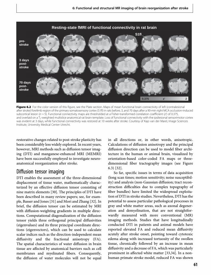
restorative changes related to post-stroke plasticity hasbeen considerably less widely explored. In recent years,however, MRI methods such as diffusion tensor imag-ing (DTI) and manganese-enhanced MRI (MEMRI)have been successfully employed to investigate neuro-anatomical reorganization after stroke.
Diffusion tensor imagingDTI enables the assessment of the three-dimensionaldisplacement of tissue water, mathematically charac-terized by an effective diffusion tensor consisting ofnine matrix elements [30]. The principles of DTI havebeen described in many review papers; see, for exam-ple, Basser and Jones [31] andMori and Zhang [32]. Inbrief, the diffusion tensor can be estimated by MRIwith diffusion-weighting gradients in multiple direc-tions. Computational diagonalization of the diffusiontensor yields three orthogonal principal diffusivities(eigenvalues) and its three principal coordinate direc-tions (eigenvectors), which can be used to calculatescalar indices such as the direction-independent meandiffusivity and the fractional anisotropy (FA).The spatial characteristics of water diffusion in braintissue are affected by anatomical barriers such as cellmembranes and myelinated fibers. Consequently,the diffusion of water molecules will not be equal
in all directions or, in other words, anisotropic.Calculations of diffusion anisotropy and the principaldiffusion direction can be used to model fiber archi-tecture in the human or animal brain, visualized byorientation-based color-coded FA maps or three-dimensional fiber tractography images (see Figure6.3) [32].
So far, specific issues in terms of data acquisition(long scan times; motion sensitivity; noise susceptibil-ity) and analysis (non-Gaussian diffusion; tract recon-struction difficulties due to complex topography offiber bundles) have limited the widespread exploita-tion of DTI in stroke studies. Nevertheless, DTI has thepotential to assess particular pathological processes ingray and white matter areas, such as axonal degener-ation and demyelination, that are not straightfor-wardly measured with more conventional (MR)imaging methods. Studies that have longitudinallyconducted DTI in patients and animal models havereported elevated FA and reduced mean diffusivityacutely after stroke onset, pointing toward cytotoxicedema along with increased tortuosity of the swollentissue, chronically followed by an increase in meandiffusivity and a decrease of FA, which was particularlyprominent in affected white matter [33,34]. In a non-human primate stroke model, reduced FA was shown
pre-stroke
3 dayspost-stroke
70 dayspost-stroke
S1fl
z’0.375 2
Resting-state fMRI of functional connectivity in rat brain
Figure 6.2 For the color version of this figure, see the Plate section. Maps of mean functional brain connectivity of left (contralesionalafter stroke) forelimb region of the primary somatosensory cortex (S1fl) in rats before, 3, and 70 days after a 90-min right MCA occlusion-inducedsubcortical lesion (n = 5). Functional connectivity maps are thresholded at a Fisher-transformed correlation coefficient (z’) of 0.375,and overlaid on a T2-weighted multislice anatomical rat brain template. Loss of functional connectivity with the ipsilesional sensorimotor cortexwas evident at 3 days, while functional connectivity was restored at 10 weeks after stroke. Courtesy of Kajo van der Marel, Image SciencesInstitute, University Medical Center Utrecht.
6: Functional and structural MR imaging of brain reorganization after stroke
61

to be correlated with the amount of myelin damage atchronic stages after transient or permanent MCAocclusion in non-human primates [34].
A growing amount of data suggest that DTI mayalso inform on alterations in brain connections asso-ciated with neuroplasticity. For instance, structuralrepair or remodeling of neuronal projections result-ing in increased density of (myelinated) fibers wouldtheoretically give rise to higher FA. In fact a progres-sive increase of FA in non-lesioned white matter hasbeen found in patients during the first two years afterstroke [35]. This was hypothesized to representongoing improvement of the integrity of normal-appearing white matter as a result of remyelinationprocesses. Recent studies on neuronal repair afterexperimental cerebral ischemia have also shown anincrease in FA in perilesional white matter at chronictime-points [36–38]. Jiang et al. [36] and Ding et al.[38] observed that FA rises progressively along theischemic lesion boundary, which was significantlyenhanced in rats treated with neural progenitor cells[36] (Figure 6.3) or sildenafil [38]. Histologicalexamination in these studies revealed a high densityof axons and myelin in areas with elevated FA. Vander Zijden et al. described a temporal profile of FAincrease over a period of 9 weeks that followed aninitial decline at 3 days after transient MCA occlu-sion in rats [37]. This particular pattern was specifi-cally observed in areas consisting of white matter,
i.e. the internal capsule and corpus callosum. Thesedata suggest that white matter integrity may improveafter initial loss. However, other processes such asgliosis, i.e. proliferation of glial cells, may also occurin ischemic lesion border zones and could affectdiffusion anisotropy. In the near future, additionalexperimental studies are required to establish to whatextent diverse structural modifications in post-strokebrain, which include neuronal restructuring andglial scar formation, contribute to changes in DTIparameters.
Manganese-enhanced MRIBesides DTI, MRI provides another interesting tooltermed manganese-enhanced MRI (MEMRI) forassessment of neuronal networks. This methodmakes use of paramagnetic Mn2+ as a Ca2+ analogthat can enter neurons through Ca2+ channels. Intissue, manganese shortens the local T1, giving rise toan increase in signal intensity on T1-weighted MRimages. Under normal conditions endogenous man-ganese is present in only trace amounts, henceMEMRItypically involves application of exogenous manga-nese. The manganese-induced increase in longitudinalrelaxation rate, R1 (1/T1), is proportional to the localmanganese concentration [39], hence the amount ofmanganese accumulation after injection can be esti-mated from the difference between pre- and post-contrast R1 (ΔR1).
DTI of white matter architecturein post-stroke rat and human brain
Figure 6.3 For the color version of this figure,see the Plate section. Top row: T2-weighted MRimage and fiber-tracking image of a rat braintreated with neuroprogenitor cells after unilateralstroke, showing tracking of axonal projections inthe lesion border zone. The marker c representsthe ischemic core. Bottom row: FA, fiber direction,and fiber-tracking image of a horizontal brain slicefrom a stroke patient, illustrating orientationchanges in the lesion boundary. Both fiberdirection and fiber-tracking images show thattracking of axonal projections circumscribes thelesion boundary (white arrows). Red, green, andblue colors represent x, y, and z directions,respectively, on the fiber direction image. Detailsof the experimental protocols can be found inJiang et al. [36]. Courtesy of Dr. Quan Jiang, HenryFord Health Sciences Center.
6: Functional and structural MR imaging of brain reorganization after stroke
62
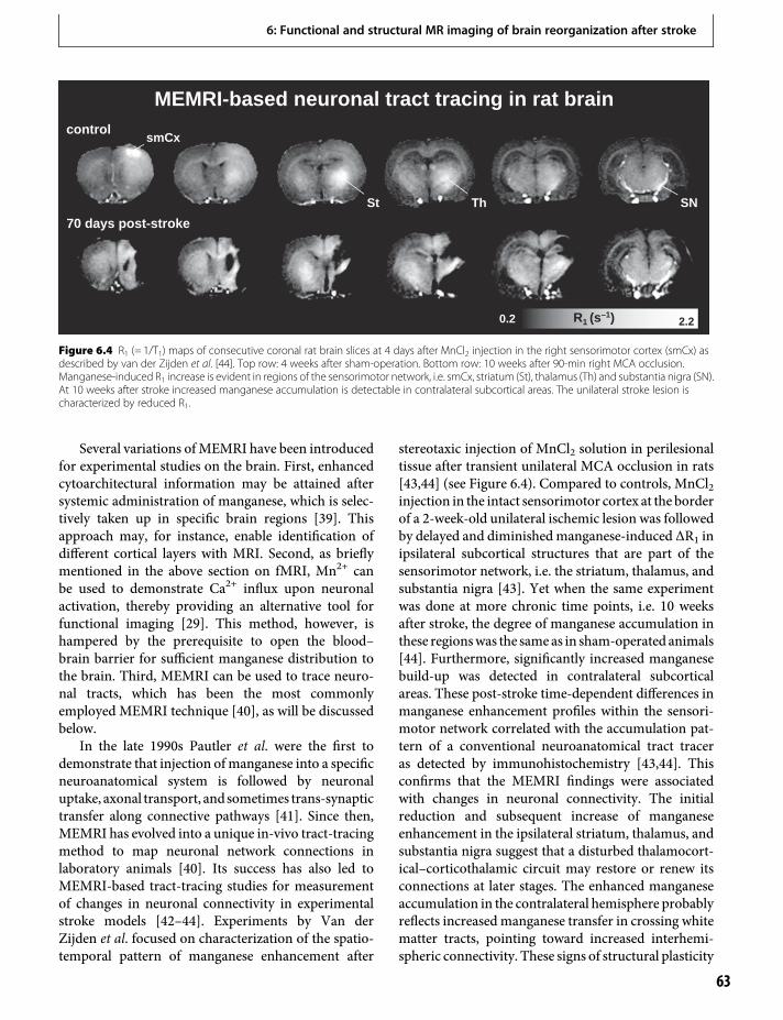
Several variations ofMEMRI have been introducedfor experimental studies on the brain. First, enhancedcytoarchitectural information may be attained aftersystemic administration of manganese, which is selec-tively taken up in specific brain regions [39]. Thisapproach may, for instance, enable identification ofdifferent cortical layers with MRI. Second, as brieflymentioned in the above section on fMRI, Mn2+ canbe used to demonstrate Ca2+ influx upon neuronalactivation, thereby providing an alternative tool forfunctional imaging [29]. This method, however, ishampered by the prerequisite to open the blood–brain barrier for sufficient manganese distribution tothe brain. Third, MEMRI can be used to trace neuro-nal tracts, which has been the most commonlyemployed MEMRI technique [40], as will be discussedbelow.
In the late 1990s Pautler et al. were the first todemonstrate that injection ofmanganese into a specificneuroanatomical system is followed by neuronaluptake, axonal transport, andsometimes trans-synaptictransfer along connective pathways [41]. Since then,MEMRI has evolved into a unique in-vivo tract-tracingmethod to map neuronal network connections inlaboratory animals [40]. Its success has also led toMEMRI-based tract-tracing studies for measurementof changes in neuronal connectivity in experimentalstroke models [42–44]. Experiments by Van derZijden et al. focused on characterization of the spatio-temporal pattern of manganese enhancement after
stereotaxic injection of MnCl2 solution in perilesionaltissue after transient unilateral MCA occlusion in rats[43,44] (see Figure 6.4). Compared to controls, MnCl2injection in the intact sensorimotor cortex at the borderof a 2-week-old unilateral ischemic lesion was followedby delayed and diminished manganese-induced ΔR1 inipsilateral subcortical structures that are part of thesensorimotor network, i.e. the striatum, thalamus, andsubstantia nigra [43]. Yet when the same experimentwas done at more chronic time points, i.e. 10 weeksafter stroke, the degree of manganese accumulation inthese regionswas the same as in sham-operated animals[44]. Furthermore, significantly increased manganesebuild-up was detected in contralateral subcorticalareas. These post-stroke time-dependent differences inmanganese enhancement profiles within the sensori-motor network correlated with the accumulation pat-tern of a conventional neuroanatomical tract traceras detected by immunohistochemistry [43,44]. Thisconfirms that the MEMRI findings were associatedwith changes in neuronal connectivity. The initialreduction and subsequent increase of manganeseenhancement in the ipsilateral striatum, thalamus, andsubstantia nigra suggest that a disturbed thalamocort-ical–corticothalamic circuit may restore or renew itsconnections at later stages. The enhanced manganeseaccumulation in the contralateral hemisphere probablyreflects increased manganese transfer in crossing whitematter tracts, pointing toward increased interhemi-spheric connectivity. These signs of structural plasticity
MEMRI-based neuronal tract tracing in rat brain
70 days post-stroke
controlsmCx
Th SNSt
R1 (s–1)0.2 2.2
Figure 6.4 R1 (= 1/T1) maps of consecutive coronal rat brain slices at 4 days after MnCl2 injection in the right sensorimotor cortex (smCx) asdescribed by van der Zijden et al. [44]. Top row: 4 weeks after sham-operation. Bottom row: 10 weeks after 90-min right MCA occlusion.Manganese-induced R1 increase is evident in regions of the sensorimotor network, i.e. smCx, striatum (St), thalamus (Th) and substantia nigra (SN).At 10 weeks after stroke increased manganese accumulation is detectable in contralateral subcortical areas. The unilateral stroke lesion ischaracterized by reduced R1.
6: Functional and structural MR imaging of brain reorganization after stroke
63

as detected with in-vivo MEMRI-based neuroana-tomical tract tracing correspond with histological evi-dence of unmasking or new formation of connectionswith perilesional areas in experimental stroke models[45].
Despite the exclusive opportunities for serial,three-dimensional, in-vivo neuronal tracing withMEMRI, there are some noteworthy limitations.First, spatial resolution of MEMRI is currently limitedto about 100 μm, while much more detailed anatomi-cal information can be achieved with microscopicanalysis of conventional tracers. Second, althoughneuronal uptake and subsequent axonal transportlikely represent the major mechanisms responsiblefor manganese enhancement in the brain, other pro-cesses such as passive diffusion in interstitial fluid,reabsorption in blood and cerebrospinal fluid, as wellas glial uptake, may complicate the use of manganeseas a pure neuronal tract tracer [46]. Third, the majordrawback of manganese for brain studies is its neuro-toxicity at high concentrations or after chronic expo-sure [39], which is the main reason why MEMRI-based neuronal tracing has not been applied inhuman studies. None the less, Canals et al. haverecently shown that intracortical injection of a lowdose of MnCl2 at a very slow rate did not lead toneuronal and glial injury and improved the efficiencyof tracing neuronal connections [47]. Without adoubt, such improvements in experimental protocolswill further expand the prospects of in-vivo MEMRI-based neuronal tract-tracing experiments in a widerange of neuroscience research areas, including assess-ments of structural plasticity after stroke.
In conclusion, MRI offers a versatile tool to eval-uate the spatiotemporal pattern of functional andstructural changes after stroke in both clinical andexperimental settings. The techniques discussed inthis chapter demonstrate the ability of MRI to detect(changes in) brain activity and neuroanatomical con-nectivity, but its potential stretches even further.Recent experimental studies have shown that MRmethods can also inform on angiogenesis (with T2*-weighted MRI [48] or steady-state contrast-enhancedMRI [49]) and metabolic alterations (with 1H/13C MRspectroscopic imaging [50]) in recovering post-stroketissue. Thus in the coming years it is to be expectedthat variousMR techniques will play an increasing rolein the elucidation of structural and functional re-organization in the brain. The availability of MRI inexperimental and clinical settings will further promote
translational research on brain plasticity and may aidin monitoring of existing and novel therapeutic strat-egies to improve brain repair.
AcknowledgmentsPart of the presented work was funded by the AlexandreSuerman program of the University Medical CenterUtrecht and Utrecht University’s High Potentialprogram.
References1. Baron JC. Mapping the ischaemic penumbra with PET:
Implications for acute stroke treatment. CerebrovascDis. 1999;9:193–201.
2. Chollet F, Weiller C. Imaging recovery of functionfollowing brain injury. Curr Opin Neurobiol.1994;4:226–30.
3. Baird AE, Warach S. Magnetic resonance imaging ofacute stroke. J Cereb Blood Flow Metab.1998;18:583–609.
4. Dijkhuizen RM, Nicolay K. Magnetic resonanceimaging in experimental models of brain disorders.J Cereb Blood Flow Metab. 2003;23:1383–402.
5. Neumann-Haefelin T, Steinmetz H. Time is brain: IsMRI the clock? Curr Opin Neurol. 2007;20:410–6.
6. Nudo RJ. Recovery after damage to motor corticalareas. Curr Opin Neurobiol. 1999;9:740–7.
7. Mandeville JB, Rosen BR. Functional MRI. In: TogaAW,Mazziotta JC, editors. Brain mapping: the methods.2nd ed. New York, NY: Academic Press; 2002:315–49.
8. Harel N, Ugurbil K, Uludag K, et al. Frontiers of brainmapping using MRI. J Magn Reson Imaging.2006;23:945–57.
9. Cramer SC, Bastings EP. Mapping clinically relevantplasticity after stroke. Neuropharmacology.2000;39:842–51.
10. Cramer SC, Nelles G, Benson RR, et al. A functionalMRI study of subjects recovered from hemipareticstroke. Stroke. 1997;28:2518–27.
11. Cao Y, D’Olhaberriague L, Vikingstad EM, et al. Pilotstudy of functional MRI to assess cerebral activation ofmotor function after poststroke hemiparesis. Stroke.1998;29:112–22.
12. Rijntjes M, Weiller C. Recovery of motor and languageabilities after stroke: The contribution of functionalimaging. Prog Neurobiol. 2002;66:109–22.
13. Calautti C, Baron JC. Functional neuroimaging studiesof motor recovery after stroke in adults: A review.Stroke. 2003;34:1553–66.
14. Van der Linden A, Van CampN, Ramos-Cabrer P, et al.Current status of functional MRI on small animals:
6: Functional and structural MR imaging of brain reorganization after stroke
64
Application to physiology, pathophysiology andcognition. NMR Biomed. 2007;20:522–45.
15. Reese T, Porszasz R, Baumann D, et al. Cytoprotectiondoes not preserve brain functionality in rats duringthe acute post-stroke phase despite evidence ofnon-infarction provided by MRI. NMR Biomed.2000;13:361–70.
16. Abo M, Chen Z, Lai LJ, et al. Functional recovery afterbrain lesion–contralateral neuromodulation: An fMRIstudy. Neuroreport. 2001;12:1543–7.
17. Dijkhuizen RM, Ren J, Mandeville JB, et al. Functionalmagnetic resonance imaging of reorganization in ratbrain after stroke. Proc Natl Acad Sci USA.2001;98:12 766–71.
18. Sauter A, Reese T, Porszasz R, et al. Recovery offunction in cytoprotected cerebral cortex in rat strokemodel assessed by functional MRI. Magn Reson Med.2002;47:759–65.
19. Dijkhuizen RM, Singhal AB, Mandeville JB, et al.Correlation between brain reorganization,ischemic damage, and neurologic status aftertransient focal cerebral ischemia in rats: A functionalmagnetic resonance imaging study. J Neurosci.2003;23:510–7.
20. Kim YR, Van Meer MP, Mandeville JB, et al. fMRI ofdelayed albumin treatment during stroke recovery inrats: Implication for fast neuronal habituation inrecovering brains. J Cereb Blood Flow Metab.2007;27:142–53.
21. KimYR, VanMeerMP, Tejima E, et al. Functional MRIof delayed chronic lithium treatment in rat focalcerebral ischemia. Stroke. 2008;39:439–47.
22. Weber R, Ramos-Cabrer P, Justicia C, et al. Earlyprediction of functional recovery after experimentalstroke: Functional magnetic resonance imaging,electrophysiology, and behavioral testing in rats.J Neurosci. 2008;28:1022–9.
23. Pariente J, Loubinoux I, Carel C, et al. Fluoxetinemodulates motor performance and cerebral activationof patients recovering from stroke. Ann Neurol.2001;50:718–29.
24. Chen YC, GalpernWR, Brownell AL, et al. Detection ofdopaminergic neurotransmitter activity usingpharmacologic MRI: Correlation with PET,microdialysis, and behavioral data. Magn Reson Med.1997;38:389–98.
25. Reese T, Bochelen D, Baumann D, et al. Impairedfunctionality of reperfused brain tissue following shorttransient focal ischemia in rats. Magn Reson Imaging.2002;20:447–54.
26. Biswal B, Yetkin FZ, Haughton VM, et al. Functionalconnectivity in themotor cortex of resting human brainusing echo-planar MRI. Magn Reson Med.1995;34:537–41.
27. He BJ, Snyder AZ, Vincent JL, et al. Breakdown offunctional connectivity in frontoparietal networksunderlies behavioral deficits in spatial neglect. Neuron.2007;53:905–18.
28. Xiong J, Fox PT, Gao JH. Directly mapping magneticfield effects of neuronal activity by magnetic resonanceimaging. Hum Brain Mapp. 2003;20:41–9.
29. Lin YJ, Koretsky AP. Manganese ion enhancesT1-weightedMRI during brain activation: An approachto direct imaging of brain function. Magn Reson Med.1997;38:378–88.
30. Basser PJ, Mattiello J, LeBihan D. MR diffusion tensorspectroscopy and imaging. Biophys J. 1994;66:259–67.
31. Basser PJ, Jones DK. Diffusion-tensor MRI: Theory,experimental design and data analysis – A technicalreview. NMR Biomed. 2002;15:456–67.
32. Mori S, Zhang J. Principles of diffusion tensor imagingand its applications to basic neuroscience research.Neuron. 2006;51:527–39.
33. Sotak CH. The role of diffusion tensor imaging in theevaluation of ischemic brain injury – A review. NMRBiomed. 2002;15:561–9.
34. Liu Y, D’Arceuil HE, Westmoreland S, et al. Serialdiffusion tensor MRI after transient and permanentcerebral ischemia in nonhuman primates. Stroke.2007;38:138–45.
35. Wang C, Stebbins GT, Nyenhuis DL, et al. Longitudinalchanges in white matter following ischemic stroke:A three-year follow-up study. Neurobiol Aging.2006;27:1827–33.
36. Jiang Q, Zhang ZG, Ding GL, et al. MRI detects whitematter reorganization after neural progenitor celltreatment of stroke. Neuroimage. 2006;32:1080–9.
37. Van der Zijden JP, Van der Toorn A, Van der Marel K,et al. Longitudinal in vivo MRI of alterations inperilesional tissue after transient ischemic stroke in rats.Exp Neurol. 2008;212:207–12.
38. Ding G, Jiang Q, Li L, et al. Magnetic resonanceimaging investigation of axonal remodeling andangiogenesis after embolic stroke in sildenafil-treatedrats. J Cereb Blood Flow Metab. 2008;28:1440–8.
39. Silva AC, Lee JH, Aoki I, et al. Manganese-enhancedmagnetic resonance imaging (MEMRI):Methodological and practical considerations. NMRBiomed. 2004;17:532–43.
40. Pautler RG. In vivo, trans-synaptic tract-tracingutilizing manganese-enhanced magnetic resonanceimaging (MEMRI). NMR Biomed. 2004;17:595–601.
41. Pautler RG, Silva AC, Koretsky AP. In vivo neuronaltract tracing using manganese-enhanced magneticresonance imaging. Magn Reson Med. 1998;40:740–8.
42. Allegrini PR, Wiessner C. Three-dimensional MRI ofcerebral projections in rat brain in vivo after
6: Functional and structural MR imaging of brain reorganization after stroke
65
intracortical injection of MnCl2. NMR Biomed.2003;16:252–6.
43. Van der Zijden JP, Wu O, Van der Toorn A, et al.Changes in neuronal connectivity after stroke in rats asstudied by serial manganese-enhanced MRI.Neuroimage. 2007;34:1650–7.
44. Van der Zijden JP, Bouts MJ, Wu O, et al. Manganese-enhanced MRI of brain plasticity in relation tofunctional recovery after experimental stroke. J CerebBlood Flow Metab. 2008;28:832–40.
45. Carmichael ST. Plasticity of cortical projections afterstroke. Neuroscientist. 2003;9:64–75.
46. Watanabe T, Frahm J,Michaelis T. Functional mappingof neural pathways in rodent brain in vivo usingmanganese-enhanced three-dimensional magneticresonance imaging. NMR Biomed. 2004;17:554–68.
47. Canals S, Beyerlein M, Keller AL, et al. Magneticresonance imaging of cortical connectivity in vivo.Neuroimage. 2008;40:458–72.
48. Ding G, Jiang Q, Li L, et al. Angiogenesis detected afterembolic stroke in rat brain using magnetic resonanceT2*WI. Stroke. 2008;39:1563–8.
49. Lin CY, Chang C, CheungWM, et al. Dynamic changesin vascular permeability, cerebral blood volume,vascular density, and size after transient focal cerebralischemia in rats: Evaluation with contrast-enhancedmagnetic resonance imaging. J Cereb Blood FlowMetab.2008;28:1491–501.
50. Van der Zijden JP, Van Eijsden P, De Graaf RA, et al.1H/13C MR spectroscopic imaging of regionallyspecific metabolic alterations after experimental stroke.Brain. 2008;131:2209–19.
6: Functional and structural MR imaging of brain reorganization after stroke
66
7 Stroke recovery in non-humanprimates: a comparative perspectiveRandolph J. Nudo
Functional recovery following injuryto motor cortexMotor cortex lesions in humans result in weakness orparalysis in contralateral musculature and disruptionof skilled limb use. However, gradual recovery of somemotor abilities is commonly observed during the ensu-ing weeks. For example, within a few days followinginjury, tendon reflexes may again become active, andresistance to passive movement may increase. Limitedrecovery of voluntary movement often occurs. A grad-ual increase in muscle strength and coordination oftenfollows. However, at least in humans, complete recov-ery of function in distal musculature, including inde-pendent control of digits, is uncommon.
Mechanistic bases to account for functional motorrecovery following cortical injury have long beensought. Results as early as the 1950s by Glees andCole using surface electrical stimulation techniquessuggested that lost cortical functions are assumed bythe adjacent cortical tissue [1]. Others have suggestedthat other cortical motor areas in the same or oppositehemisphere may play a role in recovery. Still othershave suggested that subcortical structures are respon-sible. Over the past two decades, detailed examinationsof neuroplasticity mechanisms in animal modelsof focal ischemia, as well as an explosion of neuro-imaging data in human stroke survivors (see alsoChapters 11–13), has provided a wealth of informationthat is beginning to shed light on critical questions.While we are still far from a complete understandingof the recovery process and the structures involved, itis now feasible to propose general principles regardingpost-injury plasticity.
The use of non-human primates has contributedsubstantial information to this field. These specieshave great importance for translating new therapiesto human stroke populations. The present review will
focus principally on the unique role of non-humanprimates in stroke recovery research, primarily froman evolutionary perspective.
Non-human primates in studiesof stroke recovery: practicaland ethical issues
Evolution of primatesTo understand the value of a particular model speciesfor translation of neurological research, it is useful toconsider phyletic relationships. It is reasonable toassume that the likelihood that a particular extant spe-cies will share common neuroanatomical and neuro-physiological traits with humans is directly proportionalto the recency of their common ancestors. Conversely,species that have been divergent in their phyletic histor-ies will share relatively fewer features in common. In thepast, evolutionary trees (or cladograms) were basedentirely on morphological features of extant speciesand fossil records of presumed common ancestors. Inthe past decade, a revolution in evolutionary biology hastaken place, as cladistic relationships are now derived onthe basis of molecular genetic sequence analyses [2].While there is still some discrepancy between moleculardates and the fossil record, and multiple hypotheses stillexist regarding the relationships of a few higher-levellineages (e.g. colugos, tree shrews), there is general con-sensus thatmolecular data sets are rapidly becoming thegold standard for deciphering phyletic relationshipsamong living mammals [3].
Four Superorders are now recognized withinthe mammalian Class. The Superorders Xenarthra(sloths, anteaters, armadillos, etc.) and Afrotheria(aardvarks, manatees, elephants, etc.) have theirorigins in the Southern Hemisphere (South America
Brain Repair After Stroke, ed. S. C. Cramer and R. J. Nudo. Published by Cambridge University Press.© Cambridge University Press 2010.
67
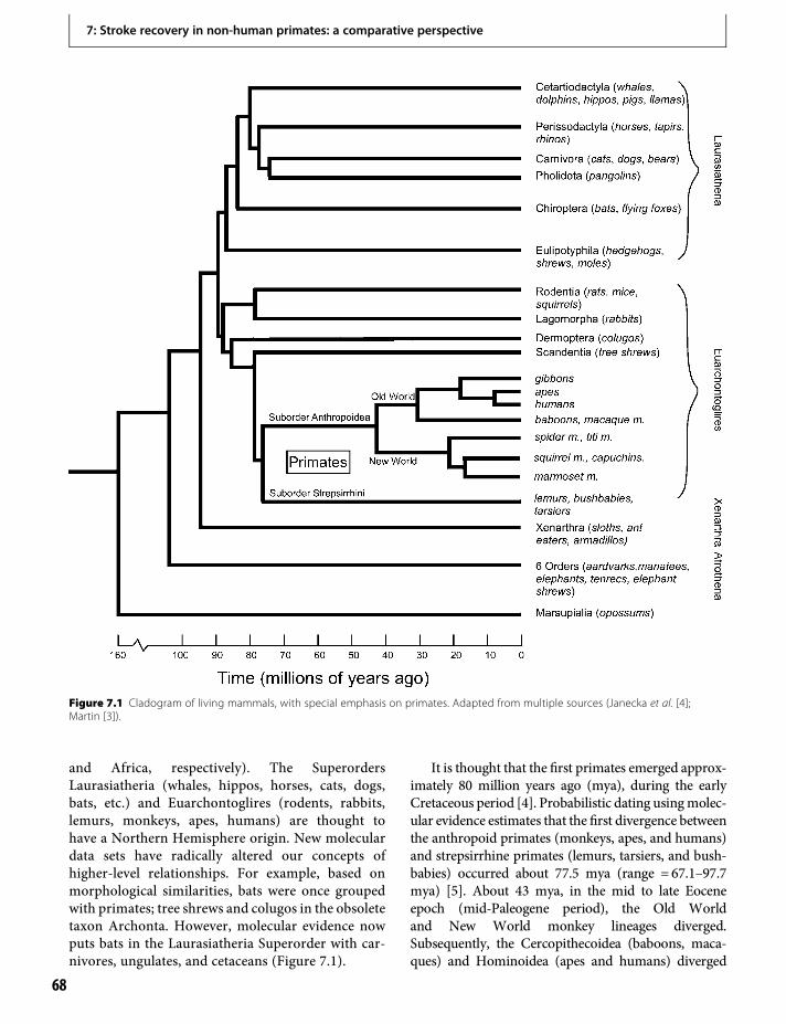
and Africa, respectively). The SuperordersLaurasiatheria (whales, hippos, horses, cats, dogs,bats, etc.) and Euarchontoglires (rodents, rabbits,lemurs, monkeys, apes, humans) are thought tohave a Northern Hemisphere origin. New moleculardata sets have radically altered our concepts ofhigher-level relationships. For example, based onmorphological similarities, bats were once groupedwith primates; tree shrews and colugos in the obsoletetaxon Archonta. However, molecular evidence nowputs bats in the Laurasiatheria Superorder with car-nivores, ungulates, and cetaceans (Figure 7.1).
It is thought that the first primates emerged approx-imately 80 million years ago (mya), during the earlyCretaceous period [4]. Probabilistic dating usingmolec-ular evidence estimates that the first divergence betweenthe anthropoid primates (monkeys, apes, and humans)and strepsirrhine primates (lemurs, tarsiers, and bush-babies) occurred about 77.5 mya (range = 67.1–97.7mya) [5]. About 43 mya, in the mid to late Eoceneepoch (mid-Paleogene period), the Old Worldand New World monkey lineages diverged.Subsequently, the Cercopithecoidea (baboons, maca-ques) and Hominoidea (apes and humans) diverged
Figure 7.1 Cladogram of living mammals, with special emphasis on primates. Adapted from multiple sources (Janecka et al. [4];Martin [3]).
7: Stroke recovery in non-human primates: a comparative perspective
68

about 30 mya, in the mid-Oligocene epoch (latePaleogene period). The last common ancestor of humansand gorillas was about 8.6 mya, while the last commonancestor of humans and chimps was about 6.6 mya, orduring the late Miocene epoch (early Neogene period).
Returning to the hypothesis that morphologicalfeatures become increasingly different between divergenttaxa with increasing geological time, one can utilize thedates of last commonancestors as a quantitative gauge fortheir utility in translational studies. Thus, even for speciescurrently used in stroke research that have divergedfrom the human lineage most recently (baboons, maca-ques), over 30 million years of brain evolution hasoccurred with different selective pressures in divergenttaxa. If we search for the common ancestor of humanswith New World monkeys (marmosets and squirrelmonkeys), we need to trace back even further – over40 million years. Carrying this logic back in geologicaltime still further, one must retreat nearly 90 millionyears to find the last common ancestor of humans androdents. Thus, even though primate and rodent speciesaremembers of the same Superorder (Euarchontoglires),they have not shared a common ancestor since the lateCretaceous period. Common ancestry for humans andother species used in neurological research (dogs, pigs,etc.) is a bit earlier than for rodents (88.8 versus87.9mya), although by only a small amount in geologicaltime, since rapid divergence of mammalian lineagesoccurred during the late Cretaceous. Thus, from thestandpoint of our hypothesis relating neurological diver-sity to phyletic relationships, rodents, carnivores, andungulates are equally valid species.
It is primarily this phyletic rationale which makesnon-human primates so valuable for understandinghuman brain function, and serving as models forstroke research. Undoubtedly there are other factorsto consider when establishing an animal model. Thebrains of some mammalian species have becomehighly specialized, and probably do not closely resem-ble the brains of the common ancestors. Somebrain properties of interest may be quite similar inwidely divergent lineages due to convergent evolution.However, on a probabilistic basis, relative kinship isthe principal determining factor in predicting geneticvariance, and thus morphological variance, betweenthe brains of different mammalian species.
Ethical and practical concernsIt is this very advantage of shared common ancestrythat makes invasive research on non-human primates
so controversial. For example, those opposed to inva-sive animal research generally point to the notion thatsome non-human organisms are sentient; that is, in itsmost literal sense, are capable of sensation and percep-tion. With regard to invasive research, sentience ismost controversial in the perception of pain and suf-fering. The fuzzy area is the affective component ofany sensation or perception. The extreme view is toassume that any animal would perceive unpleasantevents the same as a human would. But of course,this is a very anthropomorphic view, and is rejectedby most scientists. None the less, if we accept thatrecency of common ancestry is a gauge of the degreeof similarity in brain function among mammalianspecies, then one might conclude that non-humanprimates share more similarity in their awareness ofpain and suffering than other mammalian species.
Additional factors that limit the use of certainprimate species include the interrelated factors ofavailability and cost. Rapidly escalating costs even forprimate species that currently exist in abundance inthe wild or can be bred readily in captivity increasinglyhave limited the scope of non-human primateresearch.
Special characteristics of non-humanprimate brains for stroke recovery researchSeveral factors warrant the consideration of non-human primate species as models in stroke recoveryresearch. First, from a theoretical standpoint, becauseof more recent shared common ancestry, non-humanprimate brains should respond to injury more simi-larly than rodents, especially with respect to behaviorsthat rely on specialized regions of cerebral cortex.Second, brain size may be an important factor in thepenetration of pharmaceutical agents, although severallarger-brained mammalian species, other than pri-mates, can be used in stroke research including cats,dogs, pigs, and sheep. Third, white matter to graymatter ratio is significantly greater in primate brainsthan animals from other Orders, especially rodents.This fact may be critical in certain stroke models thattarget injury to descending pathways [6].
Finally, it is likely that several other structuraland functional brain traits evolved in the primatelineage, especially with respect to the corticospinal(CS) tract. Primates uniquely have a spatially distinctconcentration of CS neurons in a portion of thefrontal cortex, corresponding to the ventral premo-tor cortex (PMv). This cortical field does not appear
7: Stroke recovery in non-human primates: a comparative perspective
69
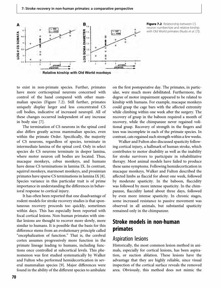
to exist in non-primate species. Further, primateshave more corticospinal neurons concerned withcontrol of the hand compared with other mam-malian species (Figure 7.2). Still further, primatesuniquely display larger and less concentrated CScell bodies, indicative of increased neuropil. All ofthese changes occurred independent of any increasein body size [7].
The termination of CS neurons in the spinal cordalso differs greatly across mammalian species, evenwithin the primate Order. Specifically, the majorityof CS neurons, regardless of species, terminate inintermediate lamina of the spinal cord. Only in selectspecies do CS neurons terminate in deeper lamina,where motor neuron cell bodies are located. Thus,macaque monkeys, cebus monkeys, and humanshave dense CS terminations in lamina IX. In contrast,squirrel monkeys, marmoset monkeys, and prosimianprimates have sparse CS terminations in lamina IX [8].Species variance in this particular trait is of primeimportance in understanding the differences in behav-ioral response to cortical injury.
It has often been reported that one disadvantage ofrodent models for stroke recovery studies is that spon-taneous recovery proceeds too quickly, sometimeswithin days. This has especially been reported withfocal cortical lesions. Non-human primates with sim-ilar lesions are thought to recover more slowly, moresimilar to humans. It is possible that the basis for thisdifference stems from an evolutionary principle called“encephalization of function.” That is, the cerebralcortex assumes progressively more function in theprimate lineage leading to humans, including func-tions once controlled at subcortical levels. This phe-nomenon was first studied systematically by Walkerand Fulton who performed hemidecortication in sev-eral mammalian species [9]. Major differences werefound in the ability of the different species to ambulate
on the first postoperative day. The primates, in partic-ular, were much more debilitated. Furthermore, thedegree of motor impairment appeared to be related tokinship with humans. For example, macaque monkeyscould grasp the cage bars with the affected extremitywhile climbing within one week after the surgery. Therecovery of grasp in the baboon required a month ofrecovery, while the chimpanzee never regained voli-tional grasp. Recovery of strength in the fingers andtoes was incomplete in each of the primate species. Incontrast, cats regained such strength within a fewweeks.
Walker and Fulton also discussed spasticity follow-ing cortical injury, a hallmark of human stroke, whichcontributes to motor disability as well as the inabilityfor stroke survivors to participate in rehabilitativetherapy. Most animal models have failed to producethese same symptoms. Following hemidecortication inmacaque monkeys, Walker and Fulton described theaffected limbs as flaccid for about one week, followedby moderate spasticity. In the baboon, flacciditywas followed by more intense spasticity. In the chim-panzee, flaccidity lasted about three days, followedby even more intense spasticity. In chronic stages,some increased resistance to passive movement wasobserved in all animals, but substantial spasticityremained only in the chimpanzee.
Stroke models in non-humanprimates
Aspiration lesionsHistorically, the most common lesion method in ani-mals, especially for cortical lesions, has been aspira-tion, or suction ablation. These lesions have theadvantage that they are highly reliable, since visualinspection of the cortical surface reveals the removedarea. Obviously, this method does not mimic the
Figure 7.2 Relationship between CSneuron number/size and relative kinshipwith Old World primates (Nudo et al. [7]).
7: Stroke recovery in non-human primates: a comparative perspective
70
clinical condition of stroke, except that a sizeable por-tion of cortical tissue is destroyed. They more closelymimic resection procedures that might be performedfor removal of tumors or epileptic foci. There aredisadvantages to this technique, since, unless the sur-geon is highly skilled, inadvertent damage can be doneto underlying white matter and adjacent gray matter.Further, fibers of passage are necessarily destroyed.Also, it is not clear what role necrotic tissue may playin subsequent inflammatory responses after injury.However, aspiration lesions have been very valuablein understanding the role of specific cortical areas inmotor and sensory behavior.
Middle cerebral artery occlusionOne of the most common methods for creating aclinically relevant stroke in rodent models has beenmiddle cerebral artery occlusion, or MCAo. Thisapproach is attractive, since a large number ofhuman strokes occur in the MCA, and this vesselsupplies a wide swath of frontal and parietalcortex, affecting motor and somatosensory function.However, subtle vascular differences in the morphol-ogy of cerebral vessels exist across species, and suchdifferences may result in wide variance in the resultinginfarcted territory. For example, in the macaque, theanterior cerebral arteries join to form a single perical-losal artery in the midline, differing distinctly from thehuman cerebral vasculature pattern [10].
While surgical access to the MCA is relativelystraightforward in rats, a much more invasive, trans-orbital surgical approach requiring enucleation is typ-ically employed in non-human primate species.Usually, vessels are then occluded by microaneurysmclips [11], although photocoagulation using RoseBengal has also been used [12]. Endovascular occlu-sion approaches would seem to be preferable, andsome success has recently been reported using thismethod in macaque monkeys [13]. In this approach,a microcatheter is inserted into the femoral artery andguided fluoroscopically to occlude a branch of theMCA. However, substantial variability has beenfound in the outcomes. In the study by D’Arceuilet al. [13], in five macaque monkeys with MCAo for3 h, stroke volume ranged from 0.1 to 4.5ml. In fourmonkeys with permanent MCAo, stroke volumeranged from 2.9 to 20.5ml. Animals with large lesionsrequired extensive postoperative care, and survival wasvariable. Thus, while relatively non-invasive occlusionof the MCA is possible in non-human primates, the
large variability in the outcome precludes its use incontrolled therapeutic studies, since large numbers arerequired for adequate statistical power.
Another approach to modeling stroke is to injectemboli into the internal carotid artery (ICA). In oneearly study, emboli were formed within polyethylenetubing using silicone rubber compound, which pri-marily lodged in the M1 segment of the MCA [14].In about 14% of the experiments, the embolus frac-tured, and lodged in multiple segmental locationsaffecting the distribution of the infarctions. In a varia-tion on this technique, multiple silicone spheres wereinjected into the ICA of macaque monkeys [15]. Inover 80% of the monkeys, the spheres were lodged atthe junction of the ICA and theMCA. However, some-times the distal embolus was located in the proximalanterior cerebral artery (ACA). In these early studies,only gross qualitative changes in cerebral morphologywere described. In a more recent study, autologousblood clots were injected into the ICA in macaquemonkeys [16]. This method resulted in extensiveinfarcts throughout the MCA territory in the cerebralcortex as well as extensive subcortical damage in thestriatum and thalamus. Mean infarct volume as apercentage of the hemisphere was 28%, ranging fromabout 12 to 55% (based on scatterplot). This methodhas been subsequently used by this group to examinethe effects of thrombolytic agents after stroke. Whilethe infarct size range is somewhat broad, the variabil-ity is not unexpected given individual variation in thedistribution of the MCA. Further development of cer-ebral vessel occlusion methods that do not requireintracranial surgery will be extremely valuable to rep-licate the conditions of human stroke in non-humanprimate models.
Focal electrocoagulationSeveral studies have employed more focal ischemiatechniques, resulting in more restricted cortical injurythan what is experienced in clinical stroke. Thisapproach is usedmost often in conjunction with neuro-physiological mapping techniques to determine detailsin the functional reorganization of the spared regionsafter injury. Since these techniques result in occlusion ofthe arterioles and venules of the entire vascularbed within a targeted region, there is little if anypenumbra. Thus, they are not useful for neuroprotec-tion studies, which generally have as a primary aim tosalvage threatened penumbra. Also, focal coagulationtechniques are most often used in lissencephalic brains
7: Stroke recovery in non-human primates: a comparative perspective
71
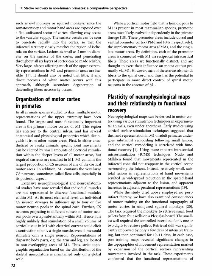
such as owl monkeys or squirrel monkeys, since thesomatosensory and motor hand areas are exposed overa flat, unfissured sector of cortex, allowing easy accessto the vascular supply. The surface vessels can be seento penetrate radially into the cortex, so that theinfarcted territory closely matches the region of ische-mia on the surface. Lesions as small as 2mm in diam-eter on the surface of the cortex and penetratingthroughout all six layers of cortex can be made reliably.Very large infarcts affecting much of the upper extrem-ity representations in M1 and premotor areas are fea-sible [17]. It should also be noted that little, if any,direct necrosis of white matter occurs with thisapproach, although secondary degeneration ofdescending fibers necessarily occurs.
Organization of motor cortexin primatesIn all primate species studied to date, multiple motorrepresentations of the upper extremity have beenfound. The largest and most functionally importantarea is the primary motor cortex, or M1. This regionlies anterior to the central sulcus, and has severalanatomical and physiological properties which distin-guish it from other motor areas. First, in either anes-thetized or awake animals, specific joint movementscan be elicited by small amounts of electrical stimula-tion within the deeper layers of all motor areas, butrequired currents are smallest in M1. M1 contains thelargest proportion of CS neurons of any of the corticalmotor areas. In addition, M1 contains the very largeCS neurons, sometimes called Betz cells, especially inits posterior aspect.
Extensive neurophysiological and neuroanatomi-cal studies have now revealed that individual musclesare not represented in discrete functional moduleswithin M1. At its most elemental level, an individualCS neuron diverges to influence up to four or fivemotor neuron pools in the spinal cord. Further, CSneurons projecting to different subsets of motor neu-ron pools overlap substantially within M1. Hence, it ishighly unlikely that stimulation of a small volume ofcortical tissue in M1 with electrical current could elicita contraction of only a single muscle, even if one couldstimulate only a single neuron. Representations ofdisparate body parts, e.g. the arm and leg, are locatedin non-overlapping areas of M1. Thus, strict topo-graphic organization based on the distribution of theskeletal musculature is maintained only on a globalscale.
While a cortical motor field that is homologous toM1 is present in most mammalian species, premotorareas most likely evolved independently in the primatelineage [18]. These premotor areas include dorsal andventral premotor cortex (PMd and PMv, respectively),the supplementary motor area (SMA), and the cingu-late motor areas. By definition, each of the premotorareas is connected with M1 via reciprocal intracorticalfibers. These areas are functionally distinct, and arethought to exert their influence on motor output pri-marily via M1. However, each of these areas also sendsfibers to the spinal cord, and thus has the potential toparticipate in more direct control of spinal motorneurons in the absence of M1.
Plasticity of neurophysiological mapsand their relationship to functionalrecoveryNeurophysiological maps can be derived in motor cor-tex using various stimulation techniques in experimen-tal animals, even under anesthesia. Early studies usingcortical surface stimulation techniques suggested thatthe hand representation inM1 of adult primates under-goes substantial remodeling following small lesions,and the cortical remodeling is correlated with func-tional recovery [1]. Using more modern intracorticalmicrostimulation (ICMS) techniques, Nudo andMilliken found that movements represented in theinfarcted zone did not reappear in the cortical sectorsurrounding the infarct. Instead, relatively small, sub-total lesions in representations of hand movementsresulted in widespread reduction in the spared handrepresentations adjacent to the lesion, and apparentincreases in adjacent proximal representations [19].
While the study cited above employed no post-infarct therapy, we have also examined consequencesof motor training on the functional topography ofmotor cortex in uninjured squirrel monkeys [20].The task required the monkeys to retrieve small foodpellets from four wells on a Plexiglas board. The small-est well required the controlled insertion of only one ortwo digits to retrieve pellets. Retrieval skill was signifi-cantly improved by only a few days of intensive train-ing, but then continued for 10–11 days. In each case,post-training maps revealed significant changes inthe topographies of movement representation markedby expansion of the cortical sectors representingmovements involved in the task. These experimentsconfirmed that the functional representations of
7: Stroke recovery in non-human primates: a comparative perspective
72
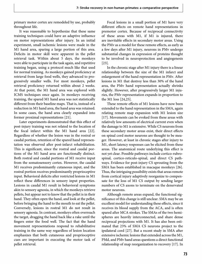
primary motor cortex are remodeled by use, probablythroughout life.
It was reasonable to hypothesize that these sametraining techniques could have an adaptive influenceon motor representations after injury. In an initialexperiment, small ischemic lesions were made in theM1 hand area, sparing a large portion of this area.Deficits in motor skill were apparent in the pelletretrieval task. Within about 5 days, the monkeyswere able to participate in the task again, and repetitivetraining began, using a protocol much like that usedfor normal training. As monkeys gained proficiency atretrieval from large food wells, they advanced to pro-gressively smaller wells. For most monkeys, pelletretrieval proficiency returned within about 2 weeks.At that point, the M1 hand area was explored withICMS techniques once again. In monkeys receivingtraining, the spared M1 hand area was not statisticallydifferent from their baseline maps. That is, instead of areduction inM1 hand area, the hand area was retained.In some cases, the hand area clearly expanded intoformer proximal representations [21].
Later experiments demonstrated that this effect ofpost-injury training was not related to the location ofthe focal infarct within the M1 hand area [22].Regardless of whether the lesion was in the rostral orcaudal portion, retention of the spared hand represen-tation was observed after post-infarct rehabilitation.This is significant, since the rostral and caudal por-tions of the M1 hand area are functionally distinct.Both rostral and caudal portions of M1 receive inputfrom the somatosensory cortex. However, the caudalM1 receives predominantly cutaneous input, and therostral portion receives predominantly proprioceptiveinput. Behavioral deficits after restricted lesions in M1reflect these differences in sensory input properties.Lesions in caudal M1 result in behavioral symptomsakin to sensory agnosia, in which the monkeys retrievepellets, but appear not to know that the pellet is in theirhand. They often open the hand, and look at the pellet,before bringing the hand to the mouth to eat the pellet.Conversely, lesions in rostral M1 do not result insensory agnosia. In contrast, monkeys often overreachthe target, dragging the hand back like a rake until thefingers enter the food well. The fact that the handmovement representations respond to rehabilitativetraining in the same way regardless of lesion locationemphasizes that both cutaneous and proprioceptivecues are important in executing the motor task ofpellet retrieval.
Focal lesions in a small portion of M1 have verydifferent effects on remote hand representations inpremotor cortex. Because of reciprocal connectivityof these areas with M1, if M1 is injured, thereare inevitable effects in secondary motor areas. Usingthe PMv as a model for these remote effects, as early asa few days after M1 injury, neurons in PMv undergosubstantial changes in expression of proteins thoughtto be involved in neuroprotection and angiogenesis[23].
In the chronic stage after M1 injury there is a linearrelationship between the size of the M1 infarct andenlargement of the hand representation in PMv. Afterlesions in M1 that destroy less than 30% of the handarea, the PMv hand representation actually shrinksslightly. However, after progressively larger M1 inju-ries, the PMv representation expands in proportion tothe M1 loss [24,25].
These remote effects of M1 lesions have now beenextended to the hand representation in the SMA, againrelating remote map expansion with M1 lesion size[17]. Movements can be evoked from these areas withrelatively low amounts of electrical current even whenthe damage toM1 is extensive.While CS neurons fromthese secondary motor areas exist, their direct effectson spinal cord motor neurons are thought to be mea-ger. However, at least in chronic stages after injury toM1, short latency responses can be elicited from theseareas. The anatomical route underlying this effect isnot yet clear. Possible pathways include cortico-rubro-spinal, cortico-reticulo-spinal, and direct CS path-ways. Evidence for post-injury CS sprouting from theSMA has been established in macaque monkeys [26].Thus, the intriguing possibility exists that areas remotefrom cortical injury adaptively reorganize to compen-sate for the loss of M1 CS output by sending largernumbers of CS axons to terminate on the denervatedmotor neurons.
Although remote areas expand, the functional sig-nificance of this change is still unclear. SMAmay be anexcellent model for understanding these effects, since itreceives its blood supply from the ACA, and is oftenspared after MCA strokes. The SMAs of the two hemi-spheres are heavily interconnected, and share densereciprocal projections with M1. It has also been esti-mated that 23% of SMA CS neurons project to theipsilateral cord [27]. But a recent study in SMA afterextensive ischemic lesions that extended across theM1,PMd, and PMv hand areas questions a direct functionalrelationship of map reorganization to recovery [17]. In
7: Stroke recovery in non-human primates: a comparative perspective
73

this study, behavioral recovery was limited to the firstthree weeks post-injury. Behavioral performanceremained relatively constant and suboptimal through-out the next 10 weeks. However, maps of the handarea in SMA actually contracted in the first threepost-infarct weeks. They subsequently expanded overthe next 10 weeks. This temporal mismatch is noteasily explained by a simple relationship of remotereorganization in a single area, such as SMA, to behav-ioral recovery.
Another interesting aspect of SMA is that it hasgreater influence on motor neuron pools controllingproximal, rather than distal muscles [28]. In theEisner-Janowicz study [17], the changes that wereseen in the SMA hand representation after injurywere attributed to wrist and forearm movement rep-resentations, not more distal, finger and thumb repre-sentations. In these chronic stages, monkeys were ableto reach out and touch the pellet board, but were notable to retrieve pellets from the wells, or even inserttheir fingers into the wells. This is not unlike humanstroke survivors who can use proximal musculature topropel the limb forward, but do not have distal controlover hand movements. Thus, it is possible that SMAmay contribute to the development of compensatorymovement patterns that rely on more proximalmusculature.
Neuroanatomical plasticityafter injuryDue to a rich network of reciprocal intracortical con-nections, after focal injury to M1, remote areas aretriggered to reorganize their axonal projection path-ways. It is well known that in rats, after cortical injury,the cerebral cortex in the intact hemisphere sproutscrossed axonal projections to the striatum of theinjured side of the brain. However, little is knownregarding plasticity of intrinsic intracortical pathwaysinterconnecting various cortical areas. Recently, usinga squirrel monkey model of M1 infarct, Dancause et al.discovered that several months after an M1 infarct,PMv intracortical axons developed an aberrant trajec-tory [29]. Axons projecting toward the site of thelesion made sharp turns, and avoided the lesion zone.A substantial number of axons then turned morecaudally, heading more lateral to circumvent the cen-tral sulcus, and finally terminated in a parietal areawithin the somatosensory cortex, area 1 (possibly bothareas 1 and 2) [29]. This de novo cortical connectionrepresented axonal growth of more than 1 cm, a very
long distance in the small squirrel monkey brain.Understanding the functional significance of injury-induced sprouting is an important topic for futureresearch.
Studies of post-stroke interventionsin non-human primatesNon-human primates have been used sporadically inpreclinical investigations of new therapeutics forstroke recovery, including both pharmaceutical anddevice-oriented approaches. Impetus for non-humanprimate studies gained momentum after a large num-ber of failed neuroprotection trials that did not trans-late from preclinical rodent studies to primates. As oneof a large list of recommendations for preclinical drugdevelopment, the STAIR consensus conference rec-ommended the increased use of non-human primatesin stroke research [30]. It was thought that substantialspecies differences in drug metabolism, penetration ofthe drug into the brain, brain organization, and behav-ioral response to injury require that clinical trials bepreceded by some level of assurance that the drugworked in a non-human primate model.
One study that has received considerable attentionin this regard examined the effects of the free radicaltrapping agent, NXY-059, administered at 4 h afterMCA branch occlusion in a marmoset monkeymodel [31]. NXY-059 reduced infarct volume by28%, and resulted in attenuated motor and spatialneglect. However, clinical trials have had mixed results[32]. Discussions in the literature have questionedboth the quality of preclinical study designs as well asadherence to preclinical protocols in the randomizedclinical studies.
ConclusionsA full survey of the critical issues in successful trans-lational research is well beyond the scope of this chap-ter. However, from the standpoint of comparativeneurobiology, it should be kept in mind that humansand non-human primates do, indeed, have manyneurological traits in common. Based on phyletic evi-dence, non-human primate models should approxi-mate the response to ischemic injury better than othermammalian species. Likewise, a larger proportion ofthe variance in the effects of neuroprotective andrestorative agents after stroke in humans can be pre-dicted by examining their effects in non-human pri-mate models in comparison with other mammalian
7: Stroke recovery in non-human primates: a comparative perspective
74
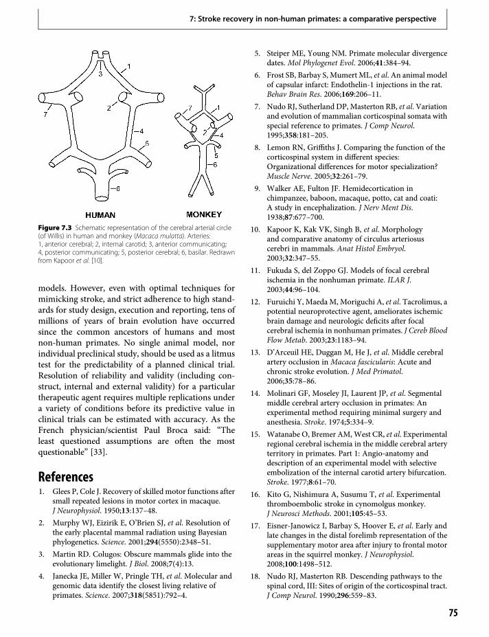
models. However, even with optimal techniques formimicking stroke, and strict adherence to high stand-ards for study design, execution and reporting, tens ofmillions of years of brain evolution have occurredsince the common ancestors of humans and mostnon-human primates. No single animal model, norindividual preclinical study, should be used as a litmustest for the predictability of a planned clinical trial.Resolution of reliability and validity (including con-struct, internal and external validity) for a particulartherapeutic agent requires multiple replications undera variety of conditions before its predictive value inclinical trials can be estimated with accuracy. As theFrench physician/scientist Paul Broca said: “Theleast questioned assumptions are often the mostquestionable” [33].
References1. Glees P, Cole J. Recovery of skilled motor functions after
small repeated lesions in motor cortex in macaque.J Neurophysiol. 1950;13:137–48.
2. Murphy WJ, Eizirik E, O’Brien SJ, et al. Resolution ofthe early placental mammal radiation using Bayesianphylogenetics. Science. 2001;294(5550):2348–51.
3. Martin RD. Colugos: Obscure mammals glide into theevolutionary limelight. J Biol. 2008;7(4):13.
4. Janecka JE, Miller W, Pringle TH, et al. Molecular andgenomic data identify the closest living relative ofprimates. Science. 2007;318(5851):792–4.
5. Steiper ME, Young NM. Primate molecular divergencedates. Mol Phylogenet Evol. 2006;41:384–94.
6. Frost SB, Barbay S, Mumert ML, et al. An animal modelof capsular infarct: Endothelin-1 injections in the rat.Behav Brain Res. 2006;169:206–11.
7. Nudo RJ, Sutherland DP,Masterton RB, et al. Variationand evolution of mammalian corticospinal somata withspecial reference to primates. J Comp Neurol.1995;358:181–205.
8. Lemon RN, Griffiths J. Comparing the function of thecorticospinal system in different species:Organizational differences for motor specialization?Muscle Nerve. 2005;32:261–79.
9. Walker AE, Fulton JF. Hemidecortication inchimpanzee, baboon, macaque, potto, cat and coati:A study in encephalization. J Nerv Ment Dis.1938;87:677–700.
10. Kapoor K, Kak VK, Singh B, et al. Morphologyand comparative anatomy of circulus arteriosuscerebri in mammals. Anat Histol Embryol.2003;32:347–55.
11. Fukuda S, del Zoppo GJ. Models of focal cerebralischemia in the nonhuman primate. ILAR J.2003;44:96–104.
12. Furuichi Y, MaedaM, Moriguchi A, et al. Tacrolimus, apotential neuroprotective agent, ameliorates ischemicbrain damage and neurologic deficits after focalcerebral ischemia in nonhuman primates. J Cereb BloodFlow Metab. 2003;23:1183–94.
13. D’Arceuil HE, Duggan M, He J, et al. Middle cerebralartery occlusion in Macaca fascicularis: Acute andchronic stroke evolution. J Med Primatol.2006;35:78–86.
14. Molinari GF, Moseley JI, Laurent JP, et al. Segmentalmiddle cerebral artery occlusion in primates: Anexperimental method requiring minimal surgery andanesthesia. Stroke. 1974;5:334–9.
15. Watanabe O, Bremer AM,West CR, et al. Experimentalregional cerebral ischemia in the middle cerebral arteryterritory in primates. Part 1: Angio-anatomy anddescription of an experimental model with selectiveembolization of the internal carotid artery bifurcation.Stroke. 1977;8:61–70.
16. Kito G, Nishimura A, Susumu T, et al. Experimentalthromboembolic stroke in cynomolgus monkey.J Neurosci Methods. 2001;105:45–53.
17. Eisner-Janowicz I, Barbay S, Hoover E, et al. Early andlate changes in the distal forelimb representation of thesupplementary motor area after injury to frontal motorareas in the squirrel monkey. J Neurophysiol.2008;100:1498–512.
18. Nudo RJ, Masterton RB. Descending pathways to thespinal cord, III: Sites of origin of the corticospinal tract.J Comp Neurol. 1990;296:559–83.
Figure 7.3 Schematic representation of the cerebral arterial circle(of Willis) in human and monkey (Macaca mulatta). Arteries:1, anterior cerebral; 2, internal carotid; 3, anterior communicating;4, posterior communicating; 5, posterior cerebral; 6, basilar. Redrawnfrom Kapoor et al. [10].
7: Stroke recovery in non-human primates: a comparative perspective
75
19. Nudo RJ, Milliken GW. Reorganization of movementrepresentations in primarymotor cortex following focalischemic infarcts in adult squirrel monkeys.J Neurophysiol. 1996;75:2144–9.
20. Nudo RJ, Wise BM, SiFuentes F, et al. Neural substratesfor the effects of rehabilitative training on motorrecovery after ischemic infarct. Science. 1996;272(5269):1791–4.
21. Nudo RJ, Milliken GW, Jenkins WM, et al. Use-dependent alterations of movement representations inprimary motor cortex of adult squirrel monkeys.J Neurosci. 1996;16:785–807.
22. Friel KM, Barbay S, Frost SB, et al. Effects of a rostralmotor cortex lesion on primary motor cortex handrepresentation topography in primates. NeurorehabilNeural Rep. 2007;21:51–61.
23. Stowe AM, Plautz EJ, Nguyen P, et al. Neuronal HIF-1alpha protein and VEGFR-2 immunoreactivity infunctionally related motor areas following a focal M1infarct. J Cereb Blood Flow Metab. 2008;28:612–20.
24. Frost SB, Barbay S, Friel KM, et al. Reorganization ofremote cortical regions after ischemic brain injury: Apotential substrate for stroke recovery. J Neurophysiol.2003;89:3205–14.
25. Dancause N, Barbay S, Frost SB, et al. Effects of smallischemic lesions in the primary motor cortex onneurophysiological organization in ventral premotorcortex. J Neurophysiol. 2006; 96: 3506–11.
26. McNeal DW, Darling WG, Pizzimenti M, et al. Long-term reorganization of the corticospinal projection
from the supplementary motor cortex following braininjury is accompanied by motor recovery andprolonged microgliosis in the rhesus monkey.Program No. 77.4. 2007 Neuroscience MeetingPlanner. San Diego, CA: Society for Neuroscience,2007. Online.
27. Dum RP, Strick PL. Spinal cord terminations of themedial wall motor areas in macaque monkeys.J Neurosci. 1996;16:13–25.
28. Boudrias MH, Belhaj-Saif A, Park MC, et al.Contrasting properties of motor output from thesupplementary motor area and primarymotor cortex inrhesus macaques. Cereb Cortex. 2006;16:632–8.
29. Dancause N, Barbay S, Frost SB, et al. Extensive corticalrewiring after brain injury. J Neurosci.2005;25:10 167–79.
30. Stroke Therapy Academic Industry Roundtable(STAIR). Recommendations for standards regardingpreclinical neuroprotective and restorative drugdevelopment. Stroke. 1999;30: 2752–8.
31. Marshall JW, Cummings RM, Bowes LJ, et al.Functional and histological evidence for theprotective effect of NXY-059 in a primate model ofstroke when given 4 hours after occlusion. Stroke.2003;34:2228–33.
32. Diener HC, Lees KR, Lydon P, et al. NXY-059 for thetreatment of acute stroke: Pooled analysis of the SAINTI and II Trials. Stroke. 2008;39:1751–8.
33. Schiller F. Paul Broca, explorer of the brain. New York,NY: Oxford University Press; 1992.
7: Stroke recovery in non-human primates: a comparative perspective
76
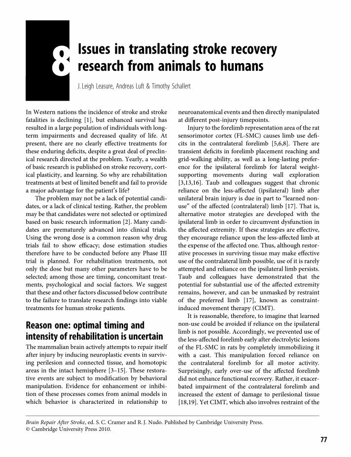
8 Issues in translating stroke recoveryresearch from animals to humansJ. Leigh Leasure, Andreas Luft & Timothy Schallert
In Western nations the incidence of stroke and strokefatalities is declining [1], but enhanced survival hasresulted in a large population of individuals with long-term impairments and decreased quality of life. Atpresent, there are no clearly effective treatments forthese enduring deficits, despite a great deal of preclin-ical research directed at the problem. Yearly, a wealthof basic research is published on stroke recovery, cort-ical plasticity, and learning. So why are rehabilitationtreatments at best of limited benefit and fail to providea major advantage for the patient’s life?
The problem may not be a lack of potential candi-dates, or a lack of clinical testing. Rather, the problemmay be that candidates were not selected or optimizedbased on basic research information [2]. Many candi-dates are prematurely advanced into clinical trials.Using the wrong dose is a common reason why drugtrials fail to show efficacy; dose estimation studiestherefore have to be conducted before any Phase IIItrial is planned. For rehabilitation treatments, notonly the dose but many other parameters have to beselected; among those are timing, concomitant treat-ments, psychological and social factors. We suggestthat these and other factors discussed below contributeto the failure to translate research findings into viabletreatments for human stroke patients.
Reason one: optimal timing andintensity of rehabilitation is uncertainThe mammalian brain actively attempts to repair itselfafter injury by inducing neuroplastic events in surviv-ing perilesion and connected tissue, and homotopicareas in the intact hemisphere [3–15]. These restora-tive events are subject to modification by behavioralmanipulation. Evidence for enhancement or inhibi-tion of these processes comes from animal models inwhich behavior is characterized in relationship to
neuroanatomical events and then directly manipulatedat different post-injury timepoints.
Injury to the forelimb representation area of the ratsensorimotor cortex (FL-SMC) causes limb use defi-cits in the contralateral forelimb [5,6,8]. There aretransient deficits in forelimb placement reaching andgrid-walking ability, as well as a long-lasting prefer-ence for the ipsilateral forelimb for lateral weight-supporting movements during wall exploration[3,13,16]. Taub and colleagues suggest that chronicreliance on the less-affected (ipsilateral) limb afterunilateral brain injury is due in part to “learned non-use” of the affected (contralateral) limb [17]. That is,alternative motor strategies are developed with theipsilateral limb in order to circumvent dysfunction inthe affected extremity. If these strategies are effective,they encourage reliance upon the less-affected limb atthe expense of the affected one. Thus, although restor-ative processes in surviving tissue may make effectiveuse of the contralateral limb possible, use of it is rarelyattempted and reliance on the ipsilateral limb persists.Taub and colleagues have demonstrated that thepotential for substantial use of the affected extremityremains, however, and can be unmasked by restraintof the preferred limb [17], known as constraint-induced movement therapy (CIMT).
It is reasonable, therefore, to imagine that learnednon-use could be avoided if reliance on the ipsilaterallimb is not possible. Accordingly, we prevented use ofthe less-affected forelimb early after electrolytic lesionsof the FL-SMC in rats by completely immobilizing itwith a cast. This manipulation forced reliance onthe contralateral forelimb for all motor activity.Surprisingly, early over-use of the affected forelimbdid not enhance functional recovery. Rather, it exacer-bated impairment of the contralateral forelimb andincreased the extent of damage to perilesional tissue[18,19]. Yet CIMT, which also involves restraint of the
Brain Repair After Stroke, ed. S. C. Cramer and R. J. Nudo. Published by Cambridge University Press.© Cambridge University Press 2010.
77
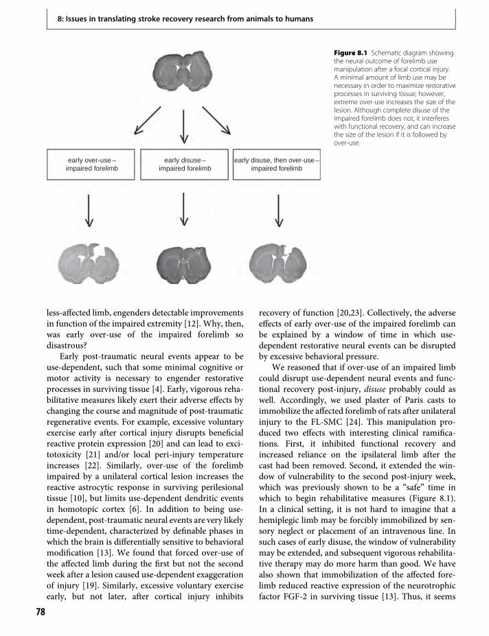
less-affected limb, engenders detectable improvementsin function of the impaired extremity [12]. Why, then,was early over-use of the impaired forelimb sodisastrous?
Early post-traumatic neural events appear to beuse-dependent, such that some minimal cognitive ormotor activity is necessary to engender restorativeprocesses in surviving tissue [4]. Early, vigorous reha-bilitative measures likely exert their adverse effects bychanging the course and magnitude of post-traumaticregenerative events. For example, excessive voluntaryexercise early after cortical injury disrupts beneficialreactive protein expression [20] and can lead to exci-totoxicity [21] and/or local peri-injury temperatureincreases [22]. Similarly, over-use of the forelimbimpaired by a unilateral cortical lesion increases thereactive astrocytic response in surviving perilesionaltissue [10], but limits use-dependent dendritic eventsin homotopic cortex [6]. In addition to being use-dependent, post-traumatic neural events are very likelytime-dependent, characterized by definable phases inwhich the brain is differentially sensitive to behavioralmodification [13]. We found that forced over-use ofthe affected limb during the first but not the secondweek after a lesion caused use-dependent exaggerationof injury [19]. Similarly, excessive voluntary exerciseearly, but not later, after cortical injury inhibits
recovery of function [20,23]. Collectively, the adverseeffects of early over-use of the impaired forelimb canbe explained by a window of time in which use-dependent restorative neural events can be disruptedby excessive behavioral pressure.
We reasoned that if over-use of an impaired limbcould disrupt use-dependent neural events and func-tional recovery post-injury, disuse probably could aswell. Accordingly, we used plaster of Paris casts toimmobilize the affected forelimb of rats after unilateralinjury to the FL-SMC [24]. This manipulation pro-duced two effects with interesting clinical ramifica-tions. First, it inhibited functional recovery andincreased reliance on the ipsilateral limb after thecast had been removed. Second, it extended the win-dow of vulnerability to the second post-injury week,which was previously shown to be a “safe” time inwhich to begin rehabilitative measures (Figure 8.1).In a clinical setting, it is not hard to imagine that ahemiplegic limb may be forcibly immobilized by sen-sory neglect or placement of an intravenous line. Insuch cases of early disuse, the window of vulnerabilitymay be extended, and subsequent vigorous rehabilita-tive therapy may do more harm than good. We havealso shown that immobilization of the affected fore-limb reduced reactive expression of the neurotrophicfactor FGF-2 in surviving tissue [13]. Thus, it seems
early over-use –impaired forelimb
early disuse –impaired forelimb
early disuse, then over-use –impaired forelimb
Figure 8.1 Schematic diagram showingthe neural outcome of forelimb usemanipulation after a focal cortical injury.A minimal amount of limb use may benecessary in order to maximize restorativeprocesses in surviving tissue; however,extreme over-use increases the size of thelesion. Although complete disuse of theimpaired forelimb does not, it interfereswith functional recovery, and can increasethe size of the lesion if it is followed byover-use.
8: Issues in translating stroke recovery research from animals to humans
78
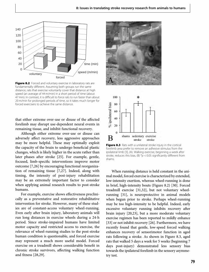
that either extreme over-use or disuse of the affectedforelimb may disrupt use-dependent neural events inremaining tissue, and inhibit functional recovery.
Although either extreme over-use or disuse canadversely affect recovery, less aggressive approachesmay be more helpful. These may optimally exploitthe capacity of the brain to undergo beneficial plasticchanges, which is likely higher in the acute rather thanlater phases after stroke [25]. For example, gentle,focused, limb-specific interventions improve motoroutcome [7,26] by encouraging functional reorganiza-tion of remaining tissue [7,27]. Indeed, along withtiming, the intensity of post-injury rehabilitationmay be an extremely important factor to considerwhen applying animal research results to post-strokehumans.
For example, exercise shows effectiveness preclini-cally as a preventative and restorative rehabilitativeintervention for stroke. However, many of these stud-ies are of constant-access voluntary wheel-running.Even early after brain injury, laboratory animals willrun long distances in exercise wheels during a 24-hperiod. Since stroke-impaired humans have limitedmotor capacity and restricted access to exercise, therelevance of wheel-running studies to the post-strokehuman condition is questionable, and forced exercisemay represent a much more useful model. Forcedexercise on a treadmill shows considerable benefit inchronic stroke survivors, affecting walking functionand fitness [28,29].
When running distance is held constant in the ani-malmodel, forced exercise is characterized by extended,low-intensity exertion, whereas wheel-running is donein brief, high-intensity bouts (Figure 8.2) [30]. Forcedtreadmill exercise [31,32], but not voluntary wheel-running [31], is neuroprotective in animal modelswhen begun prior to stroke. Perhaps wheel-runningmay be too high-intensity to be helpful. Indeed, earlyexcessive voluntary running inhibits recovery afterbrain injury [20,23], but a more moderate voluntaryexercise regimen has been reported to mildly enhance[33] or not inhibit recovery [26]. Furthermore, we haverecently found that gentle, low-speed forced walkingenhances recovery of sensorimotor function in agedrats following a stroke. As shown in Figure 8.3, agedrats that walked 5 days a week for 5 weeks (beginning 7days post-injury) demonstrated less sensory biastowards the ipsilateral forelimb in the sensory asymme-try test.
voluntaryforced
speed (m/min)
time (min)
0
20
40
60
80
100
120
140
Figure 8.2 Forced and voluntary exercise in laboratory rats arefundamentally different. Assuming both groups run the samedistance, rats that exercise voluntarily cover that distance at highspeed (an average of 44m/min) in a short period of time (about47min). In contrast, it is difficult to force rats to run faster than about20m/min for prolonged periods of time, so it takes much longer forforced exercisers to achieve the same distance.
Figure 8.3 Rats with a unilateral stroke injury in the corticalforelimb area prefer to remove an adhesive stimulus from theipsilateral limb [3], (A). Walking exercise, beginning a week afterstroke, reduces this bias, (B) *p < 0.05 significantly different fromshams.
8: Issues in translating stroke recovery research from animals to humans
79
Preclinical animal research studies indicate thatearly, vigorous physical rehabilitative efforts do notenhance functional recovery and can actually inhibitit. Yet determining when and how vigorously to insti-tute rehabilitation in human stroke patients is verydifficult. While CIMT can mitigate chronic stroke-induced impairments [12], CIMT was no better thantraditional therapy when applied early in the course ofstroke recovery [34]. It is possible that adverse effectsof early training cancel or even counteract the benefi-cial effects. Recently, a study of human stroke patientsfound that forced rehabilitation immediately follow-ing stroke led to a worsening of outcome compared tountreated patients [35]. Furthermore, the effectivenessof rehabilitative measures may be “dose-dependent.”A recent study of delayed therapy showed that patientson a 4 visits/week program improved more than thoseon a 1 or 3 visits/week regimen [36]. Animal studiesthat take into account the post-stroke human condi-tion and clinical trials that are guided by the impor-tance of timing and intensity of rehabilitation arenecessary to pinpoint an effective treatment forchronic stroke-induced impairments.
Reason two: strokesare heterogeneousHuman strokes vary greatly in size, location andtype. Most animal models are of ischemic injuries,produced by vessel occlusion, photothrombosis,thermocoagulation, pial vessel stripping, or vasocon-striction. Because the neural response to injury islesion-dependent, each stroke model may have only asubset of neural responses and a unique behavioralimpairment profile, and therefore be relevant only toa specific subset of human strokes.
Increasingly, it is being realized that recovery frombrain injury is lesion-specific. For example, dendriticarborization occurs in the homotopic region followingunilateral electrolytic [5,6] but not aspiration lesions[8] of the rat sensorimotor cortex. Axonal sproutingfrom the contralesional homotopic area is seen in thedenervated striatum following unilateral cortical ther-mocoagulation of blood vessels but not aspirationlesions [9]. Furthermore, aspiration lesions decreaseexpression of GAP-43 in the striatum, but thermocoa-gulation lesions do not [37]. Finally, the neuralresponse to hemorrhagic stroke is different fromthat to ischemia [38–43]. Therefore, it follows thatfunctional impairment and recovery are also lesion-
dependent, and indeed, the severity and stability ofpost-injury motor impairments depend on whetherthey result from thermocoagulation or aspirationlesions [44].
Neural response and functional impairment arealso dependent on the size of the lesion. This phenom-enon is particularly obvious from study of middlecerebral artery occlusion (MCAo). MCAo for 2 h gen-erated significantly more neurogenesis in the ipsilat-eral striatum compared to 30min of MCAo [45],likely because 2 h of MCAo resulted in greater striataldamage. Furthermore, over-use of the forelimbimpaired by MCAo confined to the cortex resulted inan increase in infarct volume, although when over-usefollowed a more severe MCAo that included the stria-tum, infarct size was not increased. Presumably,this was due to the fact that the more severe injuryproduced maximal damage that could not be exacer-bated by post-stroke behavioral modification [13].Behavioral recovery, too, is dependent on the typeand size of the injury, and the degree of striatalinvolvement [3,46,47]. When there is minimal striataldamage, rats with a unilateral FL-SMC injury gradu-ally begin to use the affected forelimb for some weight-supporting movements, such as landing from a rear.However, if striatal damage is significant, as it iswhen there is use-dependent exaggeration of injury,recovery of contralateral forelimb use is minimal orabsent [18].
Finally, location of the lesion may also influencethe neural response. Brain injury increases metabolicactivity [48] and the proliferation of progenitor cells inneurogenic regions of the adult mammalian brain (forreview see [14] and see Chapter 24). It has been sug-gested that in order to induce maximal proliferativeresponse, an injury must be proximal to one of the twoneurogenic regions, either the subependymal zone[49,50] or the dentate gyrus of the hippocampus [50].To illustrate, lesions that are proximal to the dentategyrus induce a more robust reactive response fromprogenitor cells than do more distal injuries [50].However, introducing growth factors in the injuredarea may encourage tissue repair, functional recoveryand generation of new tissue at the site of injury or inmore remote areas [51].
Animal models offer the benefit of homogeneity ofsubject characteristics, lesion placement, and post-injury treatment, but information collected fromthemmust be applied judiciously in order to maximizepredictive validity. Findings from a specific animal
8: Issues in translating stroke recovery research from animals to humans
80
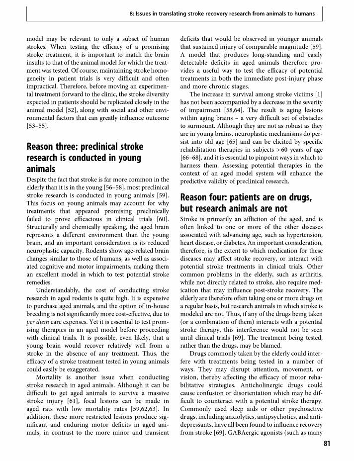
model may be relevant to only a subset of humanstrokes. When testing the efficacy of a promisingstroke treatment, it is important to match the braininsults to that of the animal model for which the treat-ment was tested. Of course, maintaining stroke homo-geneity in patient trials is very difficult and oftenimpractical. Therefore, before moving an experimen-tal treatment forward to the clinic, the stroke diversityexpected in patients should be replicated closely in theanimal model [52], along with social and other envi-ronmental factors that can greatly influence outcome[53–55].
Reason three: preclinical strokeresearch is conducted in younganimalsDespite the fact that stroke is far more common in theelderly than it is in the young [56–58], most preclinicalstroke research is conducted in young animals [59].This focus on young animals may account for whytreatments that appeared promising preclinicallyfailed to prove efficacious in clinical trials [60].Structurally and chemically speaking, the aged brainrepresents a different environment than the youngbrain, and an important consideration is its reducedneuroplastic capacity. Rodents show age-related brainchanges similar to those of humans, as well as associ-ated cognitive and motor impairments, making theman excellent model in which to test potential strokeremedies.
Understandably, the cost of conducting strokeresearch in aged rodents is quite high. It is expensiveto purchase aged animals, and the option of in-housebreeding is not significantly more cost-effective, due toper diem care expenses. Yet it is essential to test prom-ising therapies in an aged model before proceedingwith clinical trials. It is possible, even likely, that ayoung brain would recover relatively well from astroke in the absence of any treatment. Thus, theefficacy of a stroke treatment tested in young animalscould easily be exaggerated.
Mortality is another issue when conductingstroke research in aged animals. Although it can bedifficult to get aged animals to survive a massivestroke injury [61], focal lesions can be made inaged rats with low mortality rates [59,62,63]. Inaddition, these more restricted lesions produce sig-nificant and enduring motor deficits in aged ani-mals, in contrast to the more minor and transient
deficits that would be observed in younger animalsthat sustained injury of comparable magnitude [59].A model that produces long-standing and easilydetectable deficits in aged animals therefore pro-vides a useful way to test the efficacy of potentialtreatments in both the immediate post-injury phaseand more chronic stages.
The increase in survival among stroke victims [1]has not been accompanied by a decrease in the severityof impairment [58,64]. The result is aging lesionswithin aging brains – a very difficult set of obstaclesto surmount. Although they are not as robust as theyare in young brains, neuroplastic mechanisms do per-sist into old age [65] and can be elicited by specificrehabilitation therapies in subjects > 60 years of age[66–68], and it is essential to pinpoint ways in which toharness them. Assessing potential therapies in thecontext of an aged model system will enhance thepredictive validity of preclinical research.
Reason four: patients are on drugs,but research animals are notStroke is primarily an affliction of the aged, and isoften linked to one or more of the other diseasesassociated with advancing age, such as hypertension,heart disease, or diabetes. An important consideration,therefore, is the extent to which medication for thesediseases may affect stroke recovery, or interact withpotential stroke treatments in clinical trials. Othercommon problems in the elderly, such as arthritis,while not directly related to stroke, also require med-ication that may influence post-stroke recovery. Theelderly are therefore often taking one ormore drugs ona regular basis, but research animals in which stroke ismodeled are not. Thus, if any of the drugs being taken(or a combination of them) interacts with a potentialstroke therapy, this interference would not be seenuntil clinical trials [69]. The treatment being tested,rather than the drugs, may be blamed.
Drugs commonly taken by the elderly could inter-fere with treatments being tested in a number ofways. They may disrupt attention, movement, orvision, thereby affecting the efficacy of motor reha-bilitative strategies. Anticholinergic drugs couldcause confusion or disorientation which may be dif-ficult to counteract with a potential stroke therapy.Commonly used sleep aids or other psychoactivedrugs, including anxiolytics, antipsychotics, and anti-depressants, have all been found to influence recoveryfrom stroke [69]. GABAergic agonists (such as many
8: Issues in translating stroke recovery research from animals to humans
81
commonly used anxiolytics) disrupt recovery fromhead injury in rats if used repeatedly for a week ormore after the first few post-injury days [70].Polypharmacy is increasing in the elderly at risk ofstroke, but based on their mechanisms of action,many of the drugs may either cancel efficacy or leadto adverse effects [69].
Taking patients off drugs is not realistic. Althoughstatins have been shown to be beneficial early afterbrain injury [71–73], discontinuation of anticholes-terol or antihypertension medications could increasethe chances of future stroke. Taking patients off anti-depressants may also be unwise, since depressioncontributes to poor post-stroke prognosis [74],while a positive attitude towards recovery is a pre-dictor of improvement [75]; however, antidepres-sants increase the risk of future strokes [76], andone must carefully counterweigh risks and benefitsbefore prescribing these medications. Taking strokepatients off their drugs may not be always possible,but putting research animals on them is. In particu-lar, the new generation antipsychotics need to beexamined with respect to stroke, especially since useof them is increasing in the elderly for the treatmentof emotional and dementia-related behavioral issues,such as delusions and aggression [69]. Ideally,research animals should be put on the drugs priorto stroke injury, and maintained on them throughoutthe study.
Concluding remarksIt is becoming increasingly recognized that any singlebiological or behavioral therapeutic intervention isunlikely to provide all necessary components of aneffective treatment for stroke. Furthermore, the char-acteristics required of a treatment change with time.In the case of ischemia, for example, an acute treat-ment should improve blood flow to the affected areaand protect surviving tissue, whereas for chronicstages, restorative treatments should replace cells,re-myelinate, stimulate formation of appropriate newconnectivity, enhance blood flow and metaboliccapacity of the injured tissue, reduce barriers to axonaloutgrowth, and reduce the extent of delayed degener-ation. Pinpointing an effective stroke therapy thereforerepresents a major challenge. Addressing the problemsoutlined here, as well as other translational issues, mayhelp overcome this challenge and unclog the basicresearch pipeline to optimal treatments for stroke[77–80].
References1. Rautio A, Eliasson M, Stegmayr B. Favorable trends in
the incidence and outcome in stroke in nondiabetic anddiabetic subjects. Findings from the Northern SwedenMONICA stroke registry in 1985 to 2003. Stroke.2008;39:3137–44.
2. Lindner MD. Clinical attrition due to biased preclinicalassessments of potential efficacy. Pharmacol Ther.2007;115:148–75.
3. Schallert T, Fleming SM, Leasure JL, et al. CNSplasticity and assessment of forelimb sensorimotoroutcome in unilateral rat models of stroke, corticalablation, parkinsonism and spinal cord injury.Neuropharmacology. 2000;39:777–87.
4. Schallert T, Jones TA. “Exuberant” neuronal growthafter brain damage in adult rats: The essential role ofbehavioral experience. J Neural Transplant Plast.1993;4:193–8.
5. Jones TA, Schallert T. Overgrowth and pruning ofdendrites in adult rats recovering from neocorticaldamage. Brain Res. 1992;581:156–60.
6. Jones TA, Schallert T. Use-dependent growth ofpyramidal neurons after neocortical damage.J Neurosci. 1994;14:2140–52.
7. Nudo RJ, Wise BM, SiFuentes F, et al. Neural substratesfor the effects of rehabilitative training on motorrecovery after ischemic infarct. Science. 1996;272(5269):1791–4.
8. Prusky G, Whishaw IQ. Morphology of identifiedcorticospinal cells in the rat following motor cortexinjury: Absence of use-dependent change. Brain Res.1996;714:1–8.
9. Napieralski JA, Butler AK, Chesselet MF. Anatomicaland functional evidence for lesion-specific sprouting ofcorticostriatal input in the adult rat. J Comp Neurol.1996;373:484–97.
10. Bury SD, Eichhorn AC, Kotzer CM, et al. Reactiveastrocytic responses to denervation in the motor cortexof adult rats are sensitive to manipulations ofbehavioral experience. Neuropharmacology.2000;39:743–55.
11. Hsu JE, Jones TA. Time-sensitive enhancement ofmotor learning with the less-affected forelimb afterunilateral sensorimotor cortex lesions in rats. EurJ Neurosci. 2005;22:2069–80.
12. Taub E, Uswatt G. Constraint-Induced MovementTherapy: Answers and questions after two decades ofresearch. NeuroRehabilitation. 2006;21:93–5.
13. Schallert T, Humm JL, Bland ST, et al. Activity-associated growth factor expression and related neuralevents in recovery of function after brain injury. In:Choi D, Dacey RG, Hsu CY, Powers WJ, editors.Cerebrovascular disease: Momentum at the end of the
8: Issues in translating stroke recovery research from animals to humans
82
second millennium. Armonk, NY: Futura PublishingComany, Inc.; 2002:401–26.
14. Schallert T, Leasure JL, Kolb B. Experience-associatedstructural events, subependymal cellular proliferativeactivity, and functional recovery after injury to thecentral nervous system. J Cereb Blood Flow Metab.2000;20:1513–28.
15. Kleim, JA, Jones TA, Schallert T. Motor enrichmentand the induction of plasticity before and after braininjury. Neurochem Res. 2003;28:1757–69.
16. Schubring-Giese M, Molina-Luna K, Hertler B, et al.Speed of motor re-learning after experimental strokedepends on prior skill. Exp Brain Res. 2007;181:359–65.
17. Taub E, Crago JE, Burgio LD, et al. An operantapproach to rehabilitation medicine: Overcominglearned nonuse by shaping. J Exp Anal Behav.1994;61:281–93.
18. Kozlowski DA, James DC, Schallert T. Use-dependentexaggeration of neuronal injury after unilateralsensorimotor cortex lesions. J Neurosci.1996;16:4776–86.
19. Humm JL, Kozlowski DA, James DC, et al. Use-dependent exacerbation of brain damage occurs duringan early post-lesion vulnerable period. Brain Res.1998;783:286–92.
20. Griesbach GS, Hovda DA, Molteni R, et al. Voluntaryexercise following traumatic brain injury: Brain-derived neurotrophic factor upregulation and recoveryof function. Neuroscience. 2004;125:129–39.
21. Humm JL, Kozlowski DA, Bland ST, et al. Use-dependentexaggeration of brain injury: Is glutamate involved? ExpNeurol. 1999;157:349–58.
22. DeBow SB, McKenna JE, Kolb B, et al. Immediateconstraint-induced movement therapy causes localhyperthermia and exacerbates cerebral corticalinjury in rats. Can J Physiol Pharmacol. 2004;82:231–7.
23. Griesbach GS, Gomez-Pinilla F, Hovda DA. Theupregulation of plasticity-related proteins followingTBI is disrupted with acute voluntary exercise. BrainRes. 2004;1016:154–62.
24. Leasure JL, Schallert T. Consequences of forced disuseof the impaired forelimb after unilateral cortical injury.Behav Brain Res. 2004;150:83–91.
25. Carmichael ST. Cellular and molecular mechanisms ofneural repair after stroke: Making waves. Ann Neurol.2006;59:735–42.
26. Maldonado MA, Allred RP, Felthauser EL, et al. Motorskill training, but not voluntary exercise, improvesskilled reaching after unilateral ischemic lesions of thesensorimotor cortex in rats. Neurorehabil NeuralRepair. 2008;22:250–61.
27. Ro T, Noser E, Boake C, et al. Functional reorganizationand recovery after constraint-induced movement
therapy in subacute stroke: Case reports. Neurocase.2006;12:50–60.
28. Luft AR, Macko RF, Forrester LW, et al. Treadmillexercise activates subcortical neural networks andimproves walking after stroke: A randomizedcontrolled trial. Stroke. 2008;39:3341–50.
29. Macko RF, Ivey FM, Forrester LW, et al. Treadmillexercise rehabilitation improves ambulatory functionand cardiovascular fitness in patients with chronicstroke: A randomized, controlled trial. Stroke.2005;36:2206–11.
30. Leasure JL, Jones M. Forced and voluntary exercisedifferentially affect brain and behavior. Neuroscience.2008;156:456–65.
31. Hayes K, Sprague S, Guo M, et al. Forced, notvoluntary, exercise effectively induces neuroprotectionin stroke. Acta Neuropathol. 2008;115:289–96.
32. Stummer W, Weber K, Tranmer B, et al. Reducedmortality and brain damage after locomotor activity ingerbil forebrain ischemia. Stroke 1994;25:1862–9.
33. Zhao X, Aronowski J, Liu SJ, et al. Wheel-runningmodestly promotes functional recovery after aunilateral cortical lesion in rats. Behav Neurol.2005;16:41–9.
34. Boake C, Noser EA, Ro T, et al. Constraint-inducedmovement therapy during early stroke rehabilitation.Neurorehabil Neural Repair. 2007;21:14–24.
35. Dromerick AW, Lang CE, Powers WJ, et al. Very earlyconstraint-induced movement therapy (VECTORS):Phase II trial results. Stroke. 2007;38:465.
36. Byl NN, Pitsch EA, Abrams GM. Functional outcomescan vary by dose: Learning-based sensorimotor trainingfor patients stable poststroke. Neurorehabil NeuralRepair. 2008;22:494–504.
37. Szele FG, Alexander C, Chesselet MF. Expression ofmolecules associated with neuronal plasticity in thestriatum after aspiration and thermocoagulatorylesions of the cerebral cortex in adult rats. J Neurosci.1995;15:4429–48.
38. Auriat AM, Grams JD, Yan RH, et al. Forced exercisedoes not improve recovery after hemorrhagic stroke inrats. Brain Res. 2006;1109:183–91.
39. Wu J, Hua Y, Keep RF, et al. Oxidative brain injuryfrom extravasated erythrocytes after intracerebralhemorrhage. Brain Res. 2002;953:45–52.
40. Nakamura T, Keep RF, Hua Y, et al. Deferoxamine-induced attenuation of brain edema and neurologicaldeficits in a rat model of intracerebral hemorrhage.J Neurosurg. 2004;100:672–8.
41. Nakamura T, Xi G, Hua Y, et al. Intracerebralhemorrhage in mice: Model characterization andapplication for genetically modified mice. J Cereb BloodFlow Metab. 2004;24:487–94.
8: Issues in translating stroke recovery research from animals to humans
83

42. Hua Y, Nakamura T, Keep RF, et al. Long-term effectsof experimental intracerebral hemorrhage: The role ofiron. J Neurosurg. 2006;104:305–12.
43. Shao J, Xi G, Hua Y, et al. Intracerebral hemorrhage inthe iron deficient rat. Stroke. 2005;36:660–4.
44. Napieralski JA, Banks RJ, Chesselet MF. Motor andsomatosensory deficits following uni- and bilaterallesions of the cortex induced by aspiration orthermocoagulation in the adult rat. Exp Neurol.1998;154:80–8.
45. Thored P, Arvidsson A, Cacci E, et al. Persistentproduction of neurons from adult brain stem cellsduring recovery after stroke. Stem Cells.2006;24:739–47.
46. Schallert T. Behavioral tests for preclinical interventionassessment. NeuroRx. 2006;3:497–504.
47. Hua Y, Schallert T, Keep RF, et al. Behavioral tests afterintracerebral hemorrhage in the rat. Stroke.2002;33:2478–84.
48. Valla J, Humm JL, Schallert T, et al. Metabolic activityincreases in the subependymal zone following corticalinjury. NeuroReport. 1999;10:2731–4.
49. Gotts JE, Chesselet MF. Migration and fate of newlyborn cells after focal cortical ischemia in adult rats.J Neurosci Res. 2005;80:160–71.
50. Kluska MM, Witte OW, Bolz J, et al. Neurogenesis inthe adult dentate gyrus after cortical infarcts: effects ofinfarct location, N-methyl-d-aspartate receptorblockade and anti-inflammatory treatment.Neuroscience. 2005;135:723–35.
51. Kolb B, Morshead C, Gonzalez C, et al. Growth factor-stimulated generation of new cortical tissue andfunctional recovery after stroke damage to the motorcortex of rats. J Cereb Blood FlowMetab. 2007;27:983–97.
52. Adkins DL, Schallert T, Goldstein LB. Issues in pre-clinical and clinical research: Lost in translation. Stroke.2008;40:8–9.
53. Silasi G, Hamilton DA, Kolb B. Social instability blocksfunctional restitution following motor cortex stroke inrats. Behav Brain Res. 2008;188:219–26.
54. Woodlee MT, Schallert T. The impact of motoractivity and inactivity on the brain: implicationsfor the prevention and treatment of nervoussystem disorders. Curr Dir Psychol Sci. 2006;15:203–06.
55. Craft TKS, Glasper ER, McCullough L, et al. Socialinteraction improves experimental stroke outcome.Stroke. 2006;36:2006–11.
56. Marini C, Triggiani L, Cimini N, et al. Proportion ofolder people in the community as a predictor ofincreasing stroke incidence. Neuroepidemiology.2001;20:91–5.
57. Ramirez-Lassepas M. Stroke and the aging of the brainand the arteries. Geriatrics. 1998;53(Suppl 1):44–8.
58. Wyller TB. Prevalence of stroke and stroke-relateddisability. Stroke. 1998;29:866–7.
59. Lindner MD, Gribkoff VK, Donlan NA, et al. Long-lasting functional disabilities in middle-aged rats withsmall cerebral infarcts. J Neurosci. 2003;23:10 913–22.
60. Gladstone DJ, Black SE, Hakim AM. Toward wisdomfrom failure: Lessons from neuroprotective stroke trialsand new therapeutic directions. Stroke.2002;33:2123–36.
61. Wang RY, Wang PS, Yang YR. Effect of age in ratsfollowing middle cerebral artery occlusion.Gerontology. 2003;49:27–32.
62. Jin K, Minami M, Xie L, et al. Ischemia-inducedneurogenesis is preserved but reduced in the agedrodent brain. Aging Cell. 2004;3:373–7.
63. Futrell N, Garcia JH, Peterson E, et al. Embolic stroke inaged rats. Stroke. 1991;22:1582–91.
64. Westling B, Norrving B, ThorngrenM. Survival followingstroke. A prospective population-based study of 438hospitalized cases with prediction according to subtype,severity and age. Acta Neurol Scand. 1990;81:457–63.
65. Kramer AF, Bherer L, Colcombe SJ, et al.Environmental influences on cognitive and brainplasticity during aging. J Gerontol A Biol Sci Med Sci.2004;59:M940–57.
66. Luft AR, McCombe-Waller S, Whitall J, et al. Repetitivebilateral arm training and motor cortex activation inchronic stroke: A randomized controlled trial. JAMA.2004;292:1853–61.
67. Liepert J, Bauder H, Miltner WH, et al. Treatment-induced cortical reorganization after stroke in humans.Stroke. 2000;31:1210–6.
68. Luft AR, Macko RF, Forrester LW, et al. Treadmillexercise activates subcortical neural networks andimproves walking after stroke: A randomizedcontrolled trial. Stroke. 2008;39:3341–50.
69. Zhao CS, Hartikainen S, Schallert T, et al. CNS-activedrugs in aging population at high risk ofcerebrovascular events: Evidence from preclinical andclinical studies. Neurosci Biobehav Rev. 2008;32:56–71.
70. Schallert T, Hernandez TD, Barth TM. Recovery offunction after brain damage: Severe and chronicdisruption by diazepam. Brain Res. 1986;379:104–11.
71. Lu D, Mahmood A, Goussev A, et al. Atorvastatinreduces intravascular thrombosis, increases cerebralmicrovascular patency and integrity, and enhancesspatial learning in rats subjected to traumatic braininjury. J Neurosurg. 2004;101:813–21.
72. Lu D, Qu C, Goussev A, et al. Statins increaseneurogenesis in the dentate gyrus, reduce delayedneuronal death in the hippocampal CA3 region, andimprove spatial learning in rat after traumatic braininjury. J Neurotrauma. 2007;24:1132–46.
8: Issues in translating stroke recovery research from animals to humans
84
73. Qu CS, Lu D, Goussev A, et al. Effect of atorvastatin onspatial memory, neuronal survival, and vascular densityin female rats after traumatic brain injury. J Neurosurg.2005;103:695–701.
74. Cole MG, Elie LM, McCusker J, et al. Feasibility andeffectiveness of treatments for post-stroke depression inelderly inpatients: Systematic review. J GeriatrPsychiatry Neurol. 2001;14:37–41.
75. Hama S, Yamashita H, Kato T, et al. “Insistence onrecovery” as a positive prognostic factor in Japanesestroke patients. Psychiatry Clin Neurosci.2008;62:386–95.
76. Chen Y, Guo JJ, Li H, et al. Risk of cerebrovascularevents associated with antidepressant use in patientswith depression: A population-based, nested case-control study. Ann Pharmacother. 2008;42:177–84.
77. Cumberland Consensus Working Group: Cheeran B,Cohen L, Dobkin B, Ford G, Greenwood R, Howard D,Husain M, Macleod M, Nudo R, Rothwell J, Rudd A,Teo J, Ward N, Wolf S. The future of restorativeneurosciences in stroke: Driving the translationalresearch pipeline from basic science to rehabilitation ofpeople after stroke. Neurorehab Neural Repair.2009;23:97–107.
78. GinsbergMD.Neuroprotection for ischemic stroke: Past,present and future.Neuropharmacology. 2008;55:363–89.
79. Keyvani K, Schallert T. Plasticity associated molecularand structural events in postlesional brain.J Neuropathol Exper Neurol. 2002;61:831–40.
80. Hicks A, Schallert T, Jolkkonen J. Cell-based therapiesand functional outcome in experimental stroke. CellStem Cell. 2009;7:139–40.
8: Issues in translating stroke recovery research from animals to humans
85

Section II
9Spontaneous Stroke Recovery in Humans
Brain events in the acute period of strokein relation to subsequent repairRüdiger J. Seitz
IntroductionBrain ischemia results from a depression of the cere-bral blood supply leading to disturbances of neuralfunction and, in severe cases, ultimately manifestingas structural brain damage, e.g. infarction. Typically,stroke symptoms start abruptly, related to a cerebralartery occlusion [1]. Interruption of circulation due tocerebral artery occlusion induces immediate suppres-sion of cerebral electrical activity causing peri-infarctdepolarization with repeated episodes of metabolicstress and growth of the infarct lesion up to 24 hpost-occlusion [2–6]. During ischemia, the thresholdsfor selective neuronal and tissue necrosis are a func-tion of regional cerebral blood flow (rCBF) reduction[7]. Thus, critical determinants of acute stroke are theocclusion of a cerebral artery and the thereby inducedlocal depression of cerebral blood flow and subsequentelectrical, metabolic, and ionic changes [8]. Recentevidence in humans points to the occurrence ofspreading depression in severe infarctions compli-cated by progressive expansion [9].
Stroke treatment should be initiated as early aspossible in the acute period. However, it is not wellestablished how long the acute period of stroke lasts.Does it last for 24 h of infarct manifestation when themajority of cerebral artery occlusion has been recan-alized spontaneously [10,11]? Does the acute periodlast for 48 h, when emergency carotid surgery forcarotid endarterectomy can be performed safely inacute stroke [12]? Does it last up to some 7 days, aslong as stroke patients are treated on the stroke careunits? Or does the acute period of stroke extend evenfurther? For the purpose of this chapter we define theacute period of stroke operationally as the time thepatients spend in the stroke care unit, which on aver-age is 7 days after symptom onset. In this period, thepatients receive acute stroke treatment such as
thrombolysis, initiation of secondary preventionincluding anticoagulation, antiplatelet therapy aswell as antihypertensive and antidiabetic medication,and dedicated physiotherapy or speech therapy.In fact, many patients enjoy a substantial recoveryin this period, particularly when treated withthrombolysis [13].
Recovery from a brain lesion such as stroke com-prises aspects of neural repair and functional reorgan-ization. Since brain infarcts result in damage of grayand white matter, tissue repair resulting in replace-ment of the ischemic tissue debris by functional braintissue would be optimal.Work in experimental modelsin laboratory animals suggests that stem cells in thesubventricular germinal zone as well as neural progen-itor cells proliferate in response to focal ischemia[14,15]. They seem to be regulated by a number ofdifferent factors, such as neurotrophic substances andinflammatory mediators [16,17]. However, it seemsthat neurons have only a limited capacity of regenera-tion and nerve fibers do not grow out across longdistances, as they get stuck at the scars of central nerv-ous tissue lesions [18]. Nevertheless, there is goodevidence from animal experiments that nerve fibers ofintact nerve cells in the perilesional cortex grow outeven across considerable distances as well as that newsynapses are formed from the outgrowing nerve fibers[19–21]. In fact, these reorganizational changes affordclinical recovery, which has been shown to be enhancedby dedicated rehabilitative training [22]. Such reorgan-izational processes are, however, slow and need manymonths to take place. Also, following stroke there islimited repair but mainly functional reorganizationrelated to dedicated exercise and training. Notably,recovery commences early after the ischemic event. Aswill be described here, one of the major aspects of thisspontaneous recovery is the rapid cerebral reperfusion.It determines the salvage of brain tissue threatened by
Brain Repair After Stroke, ed. S. C. Cramer and R. J. Nudo. Published by Cambridge University Press.© Cambridge University Press 2010.
87

ischemia and limits the expansion of the ischemicbrain lesion.
A major step in acute stroke treatment has been theadvent of thrombolysis. It is targeted towards the res-cue of brain tissue at the risk of ischemic damage byearly recanalization. It has been shown to be effectivewhen initiated within 4.5 h of stroke onset, with max-imal efficacy within the first 90min after symptomonset [23,24]. The beneficial role of early recanalizationwas shown by functional imaging [25,26] andmonitor-ing with transcranial Doppler sonography [27,28]. Ifthrombolysis is not feasible, patients may developsevere infarctions with a disabling neurological deficitand little recovery, particularly in the advanced agegroup [29]. While the neurological deficit induced byischemia resolves completely within 15min after reper-fusion as shown by artificial balloon occlusion of thehuman carotid artery [1], the evolution of a manifestbrain infarct is more complex. The most importantdeterminants of a brain infarct are the causes of ische-mia (including its severity), the dimension and com-position of the causal arterial emboli, the anatomy andthe vascular changes of the cerebral arteries, and hyper-glycemia in associated diabetes [30–32]. Beyond theacute time window of up to 24 h, secondary changescontribute to infarct manifestation, including vaso-genic edema and inflammatory infiltration. Thesechanges, however, have a smaller impact and regressspontaneously within 2 weeks after stroke. Althoughthere is a large heterogeneity of spontaneous recoveryover the first 3 months after stroke [33], the neuro-logical state by day 4 is a powerful predictor of long-term neurological outcome [34,35]. Therefore, thebrain events in the acute period of stroke are of greatinterest and will be discussed in this chapter. It will beshown that brain infarcts differ with respect to post-stroke recovery in terms of pathophysiology andimportant aspects of cerebral artery anatomy.
Perfusion–diffusion mismatch:relevance to prognosis and to repairAn occlusion of a cerebral artery results in a depres-sion of cerebral perfusion, which if sustained willeventually result in the subsequent development ofan infarct lesion. The depression of cerebral perfu-sion can be assessed non-invasively in the acutestroke patient with perfusion imaging using magneticresonance imaging (MRI) and computed tomogra-phy (CT). The developing infarct lesion can beidentified early by diffusion-weighted imaging(DWI) using MRI (Figure 9.1). DWI is sensitive tothe development of cytotoxic brain edema whichresults in a depression of water perfusion throughthe affected brain tissue [36]. Since the changes ofdiffusion develop in a more protracted manner thanthe instantaneous depression of brain perfusion afteran acute occlusion of a cerebral artery, perfusionabnormalities are supposed to exceed DWI changesresulting in a perfusion–diffusion mismatch [37–39].In fact, a perfusion–diffusion mismatch is typicalfor the acute situation in an occlusion of a cerebralartery stem, while in an occlusion of a distal branchof a cerebral artery, the perfusion deficit areatypically matches the DWI abnormality. In addition,blood oxygen level-dependent (BOLD) imaging canindicate the oxygen deprivation [40]. Nevertheless,these measures are affected by the dynamic growth ofthe DWI lesion volume and the regression of the perfu-sion deficit in relation to spontaneous or pharmaco-logically induced thrombus dissolution. Typically,the dynamic changes result in a spatial match of theperfusion deficit and the DWI lesion after 24 h [41].
Importantly, there is a close relationship betweenthe volume of critically reduced cerebral perfusion andthe neurological deficit [37,42]. It was found that in thefirst 3 h after symptom onset the most important
Figure 9.1 Perfusion–diffusion mismatchin acute MCA stroke before treatmentonset. In a voxel-by-voxel analysis the areawith a PWI delay (a) exceeded the area ofDWI abnormality (b). The DWI changesinvolved the insula cortex and the adjacenthemispheric white matter [63]. (c)Volumetric data showed that the patientswho showed recanalization (“Recanal”)upon thrombolysis had smaller PWI (blackcolumns) and DWI (hatched columns)lesion volumes than patients in whomthrombolysis failed (“No Recanal”).
9: Brain events in the acute period of stroke in relation to subsequent repair
88
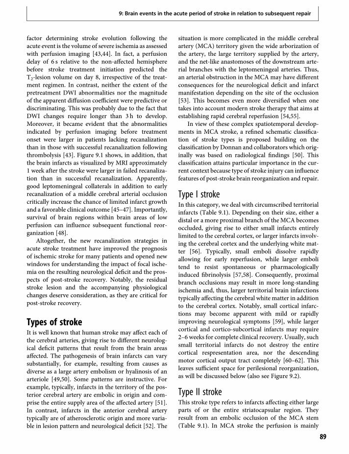
factor determining stroke evolution following theacute event is the volume of severe ischemia as assessedwith perfusion imaging [43,44]. In fact, a perfusiondelay of 6 s relative to the non-affected hemispherebefore stroke treatment initiation predicted theT2-lesion volume on day 8, irrespective of the treat-ment regimen. In contrast, neither the extent of thepretreatment DWI abnormalities nor the magnitudeof the apparent diffusion coefficient were predictive ordiscriminating. This was probably due to the fact thatDWI changes require longer than 3 h to develop.Moreover, it became evident that the abnormalitiesindicated by perfusion imaging before treatmentonset were larger in patients lacking recanalizationthan in those with successful recanalization followingthrombolysis [43]. Figure 9.1 shows, in addition, thatthe brain infarcts as visualized by MRI approximately1 week after the stroke were larger in failed recanaliza-tion than in successful recanalization. Apparently,good leptomeningeal collaterals in addition to earlyrecanalization of a middle cerebral arterial occlusioncritically increase the chance of limited infarct growthand a favorable clinical outcome [45–47]. Importantly,survival of brain regions within brain areas of lowperfusion can influence subsequent functional reor-ganization [48].
Altogether, the new recanalization strategies inacute stroke treatment have improved the prognosisof ischemic stroke for many patients and opened newwindows for understanding the impact of focal ische-mia on the resulting neurological deficit and the pros-pects of post-stroke recovery. Notably, the residualstroke lesion and the accompanying physiologicalchanges deserve consideration, as they are critical forpost-stroke recovery.
Types of strokeIt is well known that human stroke may affect each ofthe cerebral arteries, giving rise to different neurolog-ical deficit patterns that result from the brain areasaffected. The pathogenesis of brain infarcts can varysubstantially, for example, resulting from causes asdiverse as a large artery embolism or hyalinosis of anarteriole [49,50]. Some patterns are instructive. Forexample, typically, infarcts in the territory of the pos-terior cerebral artery are embolic in origin and com-prise the entire supply area of the affected artery [51].In contrast, infarcts in the anterior cerebral arterytypically are of atherosclerotic origin and more varia-ble in lesion pattern and neurological deficit [52]. The
situation is more complicated in the middle cerebralartery (MCA) territory given the wide arborization ofthe artery, the large territory supplied by the artery,and the net-like anastomoses of the downstream arte-rial branches with the leptomeningeal arteries. Thus,an arterial obstruction in the MCA may have differentconsequences for the neurological deficit and infarctmanifestation depending on the site of the occlusion[53]. This becomes even more diversified when onetakes into account modern stroke therapy that aims atestablishing rapid cerebral reperfusion [54,55].
In view of these complex spatiotemporal develop-ments in MCA stroke, a refined schematic classifica-tion of stroke types is proposed building on theclassification by Donnan and collaborators which orig-inally was based on radiological findings [50]. Thisclassification attains particular importance in the cur-rent context because type of stroke injury can influencefeatures of post-stroke brain reorganization and repair.
Type I strokeIn this category, we deal with circumscribed territorialinfarcts (Table 9.1). Depending on their size, either adistal or a more proximal branch of theMCA becomesoccluded, giving rise to either small infarcts entirelylimited to the cerebral cortex, or larger infarcts involv-ing the cerebral cortex and the underlying white mat-ter [56]. Typically, small emboli dissolve rapidlyallowing for early reperfusion, while larger embolitend to resist spontaneous or pharmacologicallyinduced fibrinolysis [57,58]. Consequently, proximalbranch occlusions may result in more long-standingischemia and, thus, larger territorial brain infarctionstypically affecting the cerebral white matter in additionto the cerebral cortex. Notably, small cortical infarc-tions may become apparent with mild or rapidlyimproving neurological symptoms [59], while largercortical and cortico-subcortical infarcts may require2–6 weeks for complete clinical recovery. Usually, suchsmall territorial infarcts do not destroy the entirecortical respresentation area, nor the descendingmotor cortical output tract completely [60–62]. Thisleaves sufficient space for perilesional reorganization,as will be discussed below (also see Figure 9.2).
Type II strokeThis stroke type refers to infarcts affecting either largeparts of or the entire striatocapsular region. Theyresult from an embolic occlusion of the MCA stem(Table 9.1). In MCA stroke the perfusion is mainly
9: Brain events in the acute period of stroke in relation to subsequent repair
89
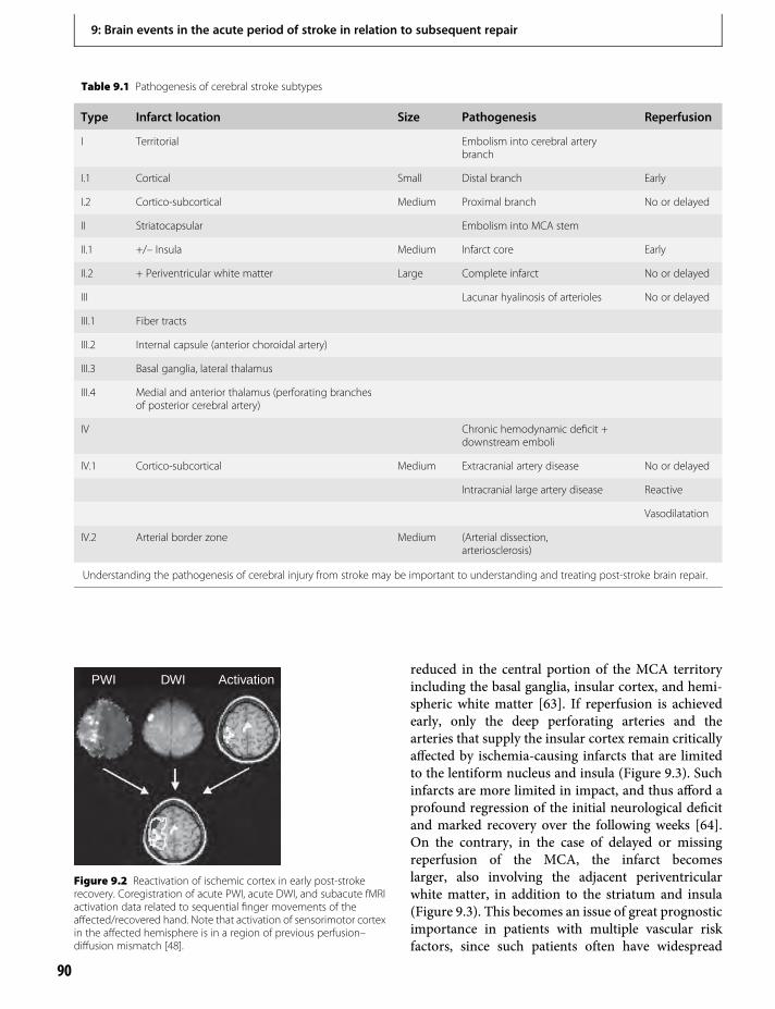
reduced in the central portion of the MCA territoryincluding the basal ganglia, insular cortex, and hemi-spheric white matter [63]. If reperfusion is achievedearly, only the deep perforating arteries and thearteries that supply the insular cortex remain criticallyaffected by ischemia-causing infarcts that are limitedto the lentiform nucleus and insula (Figure 9.3). Suchinfarcts are more limited in impact, and thus afford aprofound regression of the initial neurological deficitand marked recovery over the following weeks [64].On the contrary, in the case of delayed or missingreperfusion of the MCA, the infarct becomeslarger, also involving the adjacent periventricularwhite matter, in addition to the striatum and insula(Figure 9.3). This becomes an issue of great prognosticimportance in patients with multiple vascular riskfactors, since such patients often have widespread
Table 9.1 Pathogenesis of cerebral stroke subtypes
Type Infarct location Size Pathogenesis Reperfusion
I Territorial Embolism into cerebral arterybranch
I.1 Cortical Small Distal branch Early
I.2 Cortico-subcortical Medium Proximal branch No or delayed
II Striatocapsular Embolism into MCA stem
II.1 +/– Insula Medium Infarct core Early
II.2 + Periventricular white matter Large Complete infarct No or delayed
III Lacunar hyalinosis of arterioles No or delayed
III.1 Fiber tracts
III.2 Internal capsule (anterior choroidal artery)
III.3 Basal ganglia, lateral thalamus
III.4 Medial and anterior thalamus (perforating branchesof posterior cerebral artery)
IV Chronic hemodynamic deficit +downstream emboli
IV.1 Cortico-subcortical Medium Extracranial artery disease No or delayed
Intracranial large artery disease Reactive
Vasodilatation
IV.2 Arterial border zone Medium (Arterial dissection,arteriosclerosis)
Understanding the pathogenesis of cerebral injury from stroke may be important to understanding and treating post-stroke brain repair.
PWI DWI Activation
Figure 9.2 Reactivation of ischemic cortex in early post-strokerecovery. Coregistration of acute PWI, acute DWI, and subacute fMRIactivation data related to sequential finger movements of theaffected/recovered hand. Note that activation of sensorimotor cortexin the affected hemisphere is in a region of previous perfusion–diffusion mismatch [48].
9: Brain events in the acute period of stroke in relation to subsequent repair
90
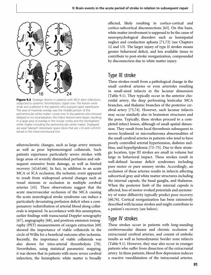
atherosclerotic changes, such as large artery stenosisas well as poor leptomeningeal collaterals. Suchpatients experience particularly severe strokes withlarge areas of severely diminished perfusion and sub-sequent extensive brain damage, as well as limitedrecovery [43,65,66]. In fact, in addition to an acuteMCA or ICA occlusion, the ischemic event appearedto result from widespread arterial changes such asvessel stenosis or occlusion in multiple cerebralarteries [43]. These observations suggest that theacute macrovascular occlusion of the MCA causingthe acute neurological stroke syndrome can induce aparticularly devastating perfusion deficit when a com-pensatory redistribution of arterial blood along collat-erals is impaired. In accordance with this assumption,earlier findings with transcranial Doppler sonography[67], angiography [68], and positron emission tomog-raphy (PET) measurements of oxygen extraction [69]showed the importance of viable collaterals in thecircle of Willis for a beneficial outcome after ischemia.Recently, the importance of viable collaterals wasalso shown for intra-arterial thrombolysis [70].Nevertheless, using statistical parametric mapping,it was shown that in patients with more severe cerebralinfarction, the hemispheric white matter is broadly
affected, likely resulting in cortico-cortical andcortico-subcortical disconnections [63]. On this basis,white matter involvement is supposed to be the cause ofneuropsychological disorders such as hemispatialneglect and conduction aphasia [71,72] (see Chapters12 and 13). The larger injury of type II strokes meansgreater behavioral deficit, and less available tissue tocontribute to post-stroke reorganization, compoundedby disconnection due to white matter injury.
Type III strokeThese strokes result from a pathological change in thesmall cerebral arteries or even arterioles resultingin small-sized infarcts in the lacunar dimension(Table 9.1). They typically occur in the anterior cho-roidal artery, the deep perforating lenticular MCAbranches, and thalamic branches of the posterior cer-ebral artery [73,74]. However, such lacunar infarctsmay occur similarly also in brainstem structures andthe pons. Typically, these strokes proceed to a com-pleted infarct lesion, although of small spatial dimen-sion. They result from local thrombosis subsequent tosevere hyalinoid or microatheroma abnormalities ofthe small cerebral arteries in patients who tend to havepoorly controlled arterial hypertension, diabetes mel-litus, and hyperlipidemia [73–75]. Due to their strate-gic location, type III strokes are small in volume butlarge in behavioral impact. These strokes result inwell-defined lacunar deficit syndromes includingpure motor or pure sensory stroke. Accordingly, anocclusion of these arteries results in infarcts affectingsubcortical gray and white matter structures includingthe internal capsule, the basal ganglia, and thalamus.When the posterior limb of the internal capsule isaffected, loss of motor-evoked potentials and asymme-try of water diffusivity typically predict poor recovery[60,76]. Cortical reorganization has been extensivelydescribed with lacunar strokes andmight contribute toa patient’s recovery (see below).
Type IV strokesThese strokes occur in patients with long-standingcerebrovascular disease and chronic occlusion ofextracranial cerebral arteries, and consist of embolicinsults as well as hemodynamic border zone infarcts(Table 9.1). However, they may also occur in youngerpatients who suffer from dissection of the extracranialartery. In these patients, blood flow depression inducesa reactive vasodilatation of the intracranial arteries.
Figure 9.3 Strategic lesions in patients with MCA stem infarctionssubjected to systemic thrombolysis. Upper row: The lesions weresmall and scattered in the patients who enjoyed rapid reperfusion.The area of maximal overlap was the middle portion of theperiventricular white matter. Lower row: In the patients who showeddelayed or no recanalization, the infarct lesions were larger, resultingin a large area of overlap in the insular cortex and the hemisphericwhite matter including the periventricular white matter. Displayedare axial Talairach stereotaxic space slices that are +16 and +24mmdorsal to the intercommissural line.
9: Brain events in the acute period of stroke in relation to subsequent repair
91
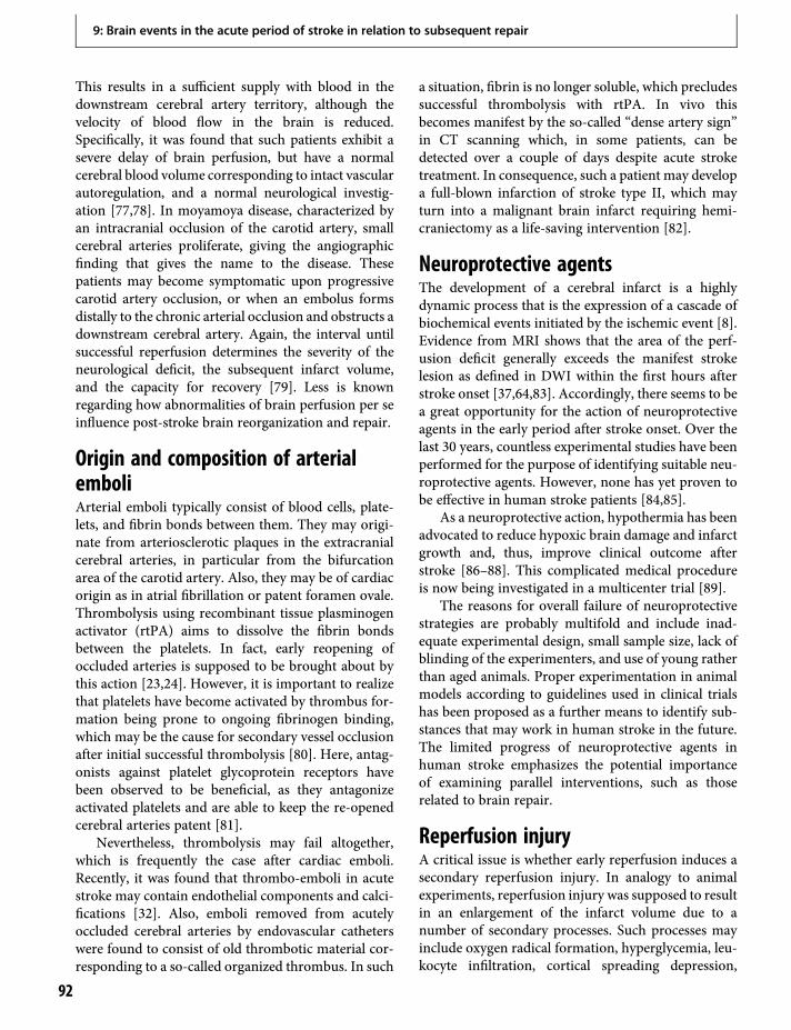
This results in a sufficient supply with blood in thedownstream cerebral artery territory, although thevelocity of blood flow in the brain is reduced.Specifically, it was found that such patients exhibit asevere delay of brain perfusion, but have a normalcerebral blood volume corresponding to intact vascularautoregulation, and a normal neurological investig-ation [77,78]. In moyamoya disease, characterized byan intracranial occlusion of the carotid artery, smallcerebral arteries proliferate, giving the angiographicfinding that gives the name to the disease. Thesepatients may become symptomatic upon progressivecarotid artery occlusion, or when an embolus formsdistally to the chronic arterial occlusion and obstructs adownstream cerebral artery. Again, the interval untilsuccessful reperfusion determines the severity of theneurological deficit, the subsequent infarct volume,and the capacity for recovery [79]. Less is knownregarding how abnormalities of brain perfusion per seinfluence post-stroke brain reorganization and repair.
Origin and composition of arterialemboliArterial emboli typically consist of blood cells, plate-lets, and fibrin bonds between them. They may origi-nate from arteriosclerotic plaques in the extracranialcerebral arteries, in particular from the bifurcationarea of the carotid artery. Also, they may be of cardiacorigin as in atrial fibrillation or patent foramen ovale.Thrombolysis using recombinant tissue plasminogenactivator (rtPA) aims to dissolve the fibrin bondsbetween the platelets. In fact, early reopening ofoccluded arteries is supposed to be brought about bythis action [23,24]. However, it is important to realizethat platelets have become activated by thrombus for-mation being prone to ongoing fibrinogen binding,which may be the cause for secondary vessel occlusionafter initial successful thrombolysis [80]. Here, antag-onists against platelet glycoprotein receptors havebeen observed to be beneficial, as they antagonizeactivated platelets and are able to keep the re-openedcerebral arteries patent [81].
Nevertheless, thrombolysis may fail altogether,which is frequently the case after cardiac emboli.Recently, it was found that thrombo-emboli in acutestroke may contain endothelial components and calci-fications [32]. Also, emboli removed from acutelyoccluded cerebral arteries by endovascular catheterswere found to consist of old thrombotic material cor-responding to a so-called organized thrombus. In such
a situation, fibrin is no longer soluble, which precludessuccessful thrombolysis with rtPA. In vivo thisbecomes manifest by the so-called “dense artery sign”in CT scanning which, in some patients, can bedetected over a couple of days despite acute stroketreatment. In consequence, such a patient may developa full-blown infarction of stroke type II, which mayturn into a malignant brain infarct requiring hemi-craniectomy as a life-saving intervention [82].
Neuroprotective agentsThe development of a cerebral infarct is a highlydynamic process that is the expression of a cascade ofbiochemical events initiated by the ischemic event [8].Evidence from MRI shows that the area of the perf-usion deficit generally exceeds the manifest strokelesion as defined in DWI within the first hours afterstroke onset [37,64,83]. Accordingly, there seems to bea great opportunity for the action of neuroprotectiveagents in the early period after stroke onset. Over thelast 30 years, countless experimental studies have beenperformed for the purpose of identifying suitable neu-roprotective agents. However, none has yet proven tobe effective in human stroke patients [84,85].
As a neuroprotective action, hypothermia has beenadvocated to reduce hypoxic brain damage and infarctgrowth and, thus, improve clinical outcome afterstroke [86–88]. This complicated medical procedureis now being investigated in a multicenter trial [89].
The reasons for overall failure of neuroprotectivestrategies are probably multifold and include inad-equate experimental design, small sample size, lack ofblinding of the experimenters, and use of young ratherthan aged animals. Proper experimentation in animalmodels according to guidelines used in clinical trialshas been proposed as a further means to identify sub-stances that may work in human stroke in the future.The limited progress of neuroprotective agents inhuman stroke emphasizes the potential importanceof examining parallel interventions, such as thoserelated to brain repair.
Reperfusion injuryA critical issue is whether early reperfusion induces asecondary reperfusion injury. In analogy to animalexperiments, reperfusion injury was supposed to resultin an enlargement of the infarct volume due to anumber of secondary processes. Such processes mayinclude oxygen radical formation, hyperglycemia, leu-kocyte infiltration, cortical spreading depression,
9: Brain events in the acute period of stroke in relation to subsequent repair
92
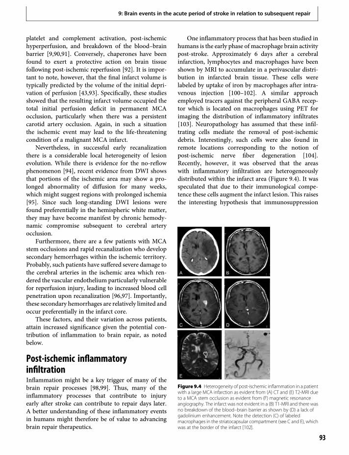
platelet and complement activation, post-ischemichyperperfusion, and breakdown of the blood–brainbarrier [9,90,91]. Conversely, chaperones have beenfound to exert a protective action on brain tissuefollowing post-ischemic reperfusion [92]. It is impor-tant to note, however, that the final infarct volume istypically predicted by the volume of the initial depri-vation of perfusion [43,93]. Specifically, these studiesshowed that the resulting infarct volume occupied thetotal initial perfusion deficit in permanent MCAocclusion, particularly when there was a persistentcarotid artery occlusion. Again, in such a situationthe ischemic event may lead to the life-threateningcondition of a malignant MCA infarct.
Nevertheless, in successful early recanalizationthere is a considerable local heterogeneity of lesionevolution. While there is evidence for the no-reflowphenomenon [94], recent evidence from DWI showsthat portions of the ischemic area may show a pro-longed abnormality of diffusion for many weeks,which might suggest regions with prolonged ischemia[95]. Since such long-standing DWI lesions werefound preferentially in the hemispheric white matter,they may have become manifest by chronic hemody-namic compromise subsequent to cerebral arteryocclusion.
Furthermore, there are a few patients with MCAstem occlusions and rapid recanalization who developsecondary hemorrhages within the ischemic territory.Probably, such patients have suffered severe damage tothe cerebral arteries in the ischemic area which ren-dered the vascular endothelium particularly vulnerablefor reperfusion injury, leading to increased blood cellpenetration upon recanalization [96,97]. Importantly,these secondary hemorrhages are relatively limited andoccur preferentially in the infarct core.
These factors, and their variation across patients,attain increased significance given the potential con-tribution of inflammation to brain repair, as notedbelow.
Post-ischemic inflammatoryinfiltrationInflammation might be a key trigger of many of thebrain repair processes [98,99]. Thus, many of theinflammatory processes that contribute to injuryearly after stroke can contribute to repair days later.A better understanding of these inflammatory eventsin humans might therefore be of value to advancingbrain repair therapeutics.
One inflammatory process that has been studied inhumans is the early phase of macrophage brain activitypost-stroke. Approximately 6 days after a cerebralinfarction, lymphocytes and macrophages have beenshown by MRI to accumulate in a perivascular distri-bution in infarcted brain tissue. These cells werelabeled by uptake of iron by macrophages after intra-venous injection [100–102]. A similar approachemployed tracers against the peripheral GABA recep-tor which is located on macrophages using PET forimaging the distribution of inflammatory infiltrates[103]. Neuropathology has assumed that these infil-trating cells mediate the removal of post-ischemicdebris. Interestingly, such cells were also found inremote locations corresponding to the notion ofpost-ischemic nerve fiber degeneration [104].Recently, however, it was observed that the areaswith inflammatory infiltration are heterogeneouslydistributed within the infarct area (Figure 9.4). It wasspeculated that due to their immunological compe-tence these cells augment the infarct lesion. This raisesthe interesting hypothesis that immunosuppression
Figure 9.4 Heterogeneity of post-ischemic inflammation in a patientwith a large MCA infarction as evident from (A) CT and (E) T2-MRI dueto a MCA stem occlusion as evident from (F) magnetic resonanceangiography. The infarct was not evident in a (B) T1-MRI and there wasno breakdown of the blood–brain barrier as shown by (D) a lack ofgadolinium enhancement. Note the detection (C) of labeledmacrophages in the striatocapsular compartment (see C and E), whichwas at the border of the infarct [102].
9: Brain events in the acute period of stroke in relation to subsequent repair
93
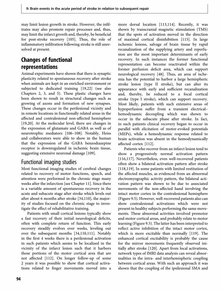
may limit lesion growth in stroke. However, the infil-trates may also promote repair processes and, thus,may limit the infarct growth and, thereby, be beneficialfor post-stroke recovery [105]. Thus, the role ofinflammatory infiltration following stroke is still unre-solved at present.
Changes of functionalrepresentationsAnimal experiments have shown that there is synapticplasticity related to spontaneous recovery after strokewhen animals are kept in an enriched environment orsubjected to dedicated training [19,22] (see alsoChapters 2, 3, and 5). These plastic changes havebeen shown to result in structural changes such asgrowing of axons and formation of new synapses.These changes occur in the perilesional vicinity andin remote locations in functionally related areas in theaffected and contralesional non-affected hemisphere[19,20]. At the molecular level, there are changes inthe expression of glutamate and GABA as well as ofneurotrophic mediators [106–108]. Notably, Heissand collaborators were able to show in the humanthat the expression of the GABA benzodiazepinereceptor is downregulated in ischemic brain tissue,suggesting extensive neuronal damage [109].
Functional imaging studiesMost functional imaging studies of cerebral changesrelated to recovery of motor functions, speech, andattention were performed in the chronic stage manyweeks after the infarction [see Chapter 11]. Since thereis a variable amount of spontaneous recovery in theacute and subacute stage after stroke which levels outafter about 6 months after stroke [34,110], the major-ity of studies focused on the chronic stage to inves-tigate the effect of rehabilitative training.
Patients with small cortical lesions typically showa fast recovery of their initial neurological deficits,often with complete return of function. This earlyrecovery steadily evolves over weeks, leveling outover the subsequent months [34,110,111]. Notablyin the first 4 weeks there is a perilesional activationin such patients which seems to be localized in thevicinity of the infarct lesion such that it harborsthose portions of the motor cortical area that arenot affected [112]. On longer follow-up of some2 years it was possible to show that cortical activa-tions related to finger movements moved into a
more dorsal location [113,114]. Recently, it wasshown by transcranial magnetic stimulation (TMS)that the spots of activation moved in the directionof maximal cortical disinhibition [115]. In largeischemic lesions, salvage of brain tissue by rapidrecanalization of the supplying artery and reperfu-sion are the most important determinants of earlyrecovery. In such instances the former functionalrepresentation can become reactivated within theformer perfusion deficit area, which can supportneurological recovery [48]. Thus, an area of ische-mia has the potential to harbor a large hemisphericstroke lesion (type II stroke), but can alter itsappearance with early and sufficient recanalizationand, thereby, be reduced to a focal corticalstroke (type I stroke), which can support recovery.Most likely, patients with such extensive areas ofhypoperfusion suffer from a transient electrical–hemodynamic decoupling which was shown tooccur in the subacute phase after stroke. In fact,in such patients clinical recovery began to occur inparallel with elicitation of motor-evoked potentials(MEPs), while a hemodynamic response related tobrain activation was absent in the hemodynamicallyaffected cortex [112].
Patients who recover from an infarct lesion tend toshow a progressively normal activation pattern[116,117]. Nevertheless, even well-recovered patientsoften show a bilateral activation pattern after stroke[118,119]. In some patients with limited restitution ofthe affected muscles, as evidenced from an abnormalelectromyographic activity pattern, the bilateral acti-vation pattern was shown to be due to associatedmovements of the non-affected hand involving theintact motor cortex in the contralesional hemisphere(Figure 9.5). However, well-recovered patients also canshow contralesional activations which were notpresent in healthy subjects performing the samemove-ments. These abnormal activities involved premotorand motor cortical areas, and probably relate to motorlearning (Figure 9.5). The latter has been interpreted toreflect active inhibition of the intact motor cortex,which is more excitable than normally [119]. Theenhanced cortical excitability is probably the causefor the mirror movements frequently observed ini-tially after stroke [120]. Apart from local activations,network types of fMRI data analysis can reveal abnor-malities in the intra- and interhemispheric couplingbetween cortical areas. With such an approach it wasshown that the coupling of the ipsilesional SMA and
9: Brain events in the acute period of stroke in relation to subsequent repair
94
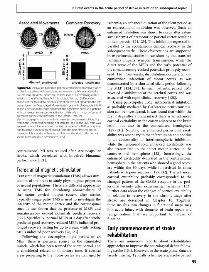
contralesional MI was reduced after striatocapsularstroke, which correlated with impaired bimanualperformance [121].
Transcranial magnetic stimulationTranscranial magnetic stimulation (TMS) allows stim-ulation of the brain to study physiological propertiesof neural populations. There are different approachesto using TMS for elucidating abnormalities ofthe motor cortical output system after stroke.Typically single-pulse TMS is used to investigate theintegrity of the motor cortex and the corticospinaltract. It was shown that the presence of MEPs andsomatosensory evoked potentials predicts recovery[122]. Specifically, normal MEPs at 1 day after strokepredicted good recovery, reducedMEPs indicated pro-longed recovery lasting for up to a year, while lackingMEPs indicated poor recovery [76,123].
Following the electrophysiologic period of anMEP, there is electrical silence in the stimulatedmuscle, which has been termed the silent period, andis considered related to inhibitory tone. If corticalareas projecting to the motor cortex are damaged by
ischemia, an enhanced duration of the silent period asan expression of inhibition was observed. Such anenhanced inhibition was shown to occur after exten-sive ischemia of premotor or parietal cortex resultingin hemiparesis [124,125]. This inhibition regressed inparallel to the spontaneous clinical recovery in thesubsequent weeks. These observations are supportedby experimental studies in rats showing that transientischemia impairs synaptic transmission, while thedirect wave of the MEPs and the early potential ofthe somatosensory-evoked potentials promptly recov-ered [126]. Conversely, disinhibition occurs after cir-cumscribed infarction of motor cortex as wasdemonstrated by a shortened silent period followingthe MEP [124,127]. In such patients, paired TMSrevealed disinhibition of the cerebral cortex and wasassociated with rapid clinical recovery [128].
Using paired-pulse TMS, intracortical inhibitionas probably mediated by GABAergic neurotransmis-sion can be investigated. It was found that within thefirst 7 days after a brain infarct there is an enhancedcortical excitability in the cortex adjacent to the brainlesion but also in the contralateral hemisphere[129–131]. Notably, the enhanced perilesional excit-ability was secondary to the infarct lesion and not dueto an abnormality of interhemispheric inhibition,while the lesion-induced enhanced excitability wasalso transmitted to the intact motor cortex in thecontralesional hemisphere [132]. Interestingly, theenhanced excitability decreased in the contralesionalhemisphere in the patients who showed a good recov-ery within the 90 days, while it persisted in thosepatients with poor recovery [129,132]. The enhancedcortical excitability probably corresponded to thechanged pattern of the GABA receptor in the peri-lesional vicinity after experimental ischemia [133].Further data about the changes of cortical excitabilityin relation to recovery in the chronic stage afterstroke are described in Chapter 10. Together,these insights into changes in functional maps maylink acute injury with elements of brain repair andreorganization that are important to return offunction.
Early commencement of strokerehabilitationThere are numerous reports about rehabilitativeapproaches to improve the neurological deficit follow-ing stroke [134]. However, in the acute phase, data arelargely missing. Typically, a hemiparetic stroke patient
Figure 9.5 Activation pattern in patients with excellent recovery afterstroke. In a patient with associated movements, a bilateral activationpattern was apparent. Note too the low level of electromyographicactivity in the affected hand of this subject. Without EMG-guidedanalysis of the fMRI data, cerebral activation was not apparent (the leftbrain slice under “Associated Movements”), but with EMG-guided fMRIanalysis, activation become apparent (the right brain slice). In a patientwith complete recovery, note activation bilaterally in motor cortex andpremotor cortex contralesional to the infarct. Here, theelectromyographic activity (ratio is presented, movement divided byrest) in the unaffected hand did not increase, and so the EMG ratio wasapproximately 1. It was argued that the contralesional fMRI activity wasdue to active suppression of output from the non-affected motorcortex, which is under enhanced excitatory drive due to the corticallesion in the opposite hemisphere [119].
9: Brain events in the acute period of stroke in relation to subsequent repair
95
stays in bed, since he/she cannot move by him/herself.In addition, the early medical situation after stroke isoften so grave that the patients are bedridden. There is,however, the notion that early mobilization will facil-itate cerebral reorganization and will enhance thepatient’s insight that he/she can move, counteractingpsychological regression and secondary immobiliza-tion [135]. There is a large trial ongoing to address thisquestion and to investigate if very early onset of mobi-lization and stroke rehabilitation will be beneficial.Notably, some data suggest that the intensity of thetraining in the early weeks following a stroke, ratherthan the type of training, appears to be the greatestdeterminant of long-term motor improvement [136].Also, constraint-induced therapy has been shown tobe effective in enhancing recovery after stroke.Interestingly, these therapeutic approaches result in awidespread recruitment of motor representations inthe affected hemisphere during the learning period asobserved with fMRI and TMS [137,138]. Based on theevidence from fMRI and TMS studies, a number ofstrategies that capitalize on pathophysiological mech-anisms have been proposed to promote motor recov-ery after stroke [122]. These include modulations thattarget the altered excitability in the affected and con-tralesional hemisphere and are applied as corticalstimulation directly onto the brain or as anaesthesiaof the peripheral nerves [139–142]. Future work needsto evaluate how the combination of dedicated trainingand these external stimulations result in sustainedneurological improvements. It should be noted, how-ever, that infarct lesions do not only induce sensori-motor deficits, but are known to induceneuropsychological disturbances such as aphasia,apraxia, and neglect depending on the infarct locationswithin the brain. In addition, cortical and cortico-subcortical infarcts can induce dementia [143].Particularly with respect to these post-stroke cognitiveimpairments, it is unclear whether repetitive or cogni-tive training approaches based on motor imagery areuseful in the acute phase after stroke.
References1. Reivich M. (1992) Crossed cerebellar diaschisis. Am
J Neuroradiol. 1992;13:62–4.
2. Heiss WD, Huber M, Fink GR, et al. Progressivederangement of periinfarct viable tissue in ischemicstroke. J Cereb Blood Flow Metab. 1992;12:193–203.
3. Hossmann KA, Fischer M, Bockhorst K, et al. NMRimaging of the apparent diffusion coefficient (ADC) for
the evaluation of metabolic suppression and recoveryafter prolonged cerebral ischemia. J Cereb Blood FlowMetab. 1994;14:723–31.
4. Lee LJ, Kidwell CS, Alger J, et al. Impact on strokesubtype diagnosis of early diffusion-weighted magneticresonance imaging and magnetic resonanceangiography. Stroke. 2000;31:1081–9.
5. Li F, Liu KF, Silva MD, et al. Transient and permanentresolution of ischemic lesions on diffusion-weightedimaging after brief periods of focal ischemia in rats:Correlation with histopathology. Stroke.2000;31:946–54.
6. Mohr JP, Foulkes MA, Polis AT, et al. Infarcttopography and hemiparesis profiles with cerebralconvexity infarction: The Stroke Data Bank. J NeurolNeurosurg Psychiatry. 1993;56:344–51.
7. Hossmann KA. Viability thresholds and the penumbraof focal ischemia. Ann Neurol. 1994;36:557–65.
8. Dirnagl U, Iadecola C, Moskowitz MA. Pathobiology ofischaemic stroke: An integrated view. Trends Neurosci.1999;22:391–7.
9. Dohmen C, Sakowitz OW, Fabricius M, et al. Spreadingdepolarizations occur in human ischemic stroke withhigh incidence. Ann Neurol. 2008;63:720–8.
10. Ringelstein EB, Biniek R, Weiller C, et al. Type andextent of hemispheric brain infarctions and clinicaloutcome in early and delayed middle cerebral arteryrecanalization. Neurology. 1992;42:289–98.
11. Wunderlich MT, Goertler M, Postert T, et al.Recanalization after intravenous thrombolysis: Does arecanalization time window exist? Neurology.2007;68:1364–8.
12. Huber R, Muller BT, Seitz RJ, et al. Carotid surgery inacute symptomatic patients. Eur J Vasc Endovasc Surg.2003;25:60–7.
13. Veronel D, Ringelstein A, Cohnen M, et al. Systemicthrombolysis based on CT or MRI stroke imaging.J Neuroimag. 2008;18:381–7.
14. Kokaia Z, Thored P, Arvidsson A, et al. Regulation ofstroke-induced neurogenesis in adult brain – Recentscientific progress. Cereb Cortex. 2006;16:162–7.
15. Ohab JJ, Fleming S, Blesch A, et al. A vascular niche forneurogenesis after stroke. J Neurosci.2006;26:13 007–16.
16. Schäbitz WR, Steigleder T, Cooper-Kuhn CM, et al.Intravenous brain-derived neurotrophic factorenhances poststroke sensorimotor recovery andstimulates neurogenesis. Stroke. 2007;38:2165–72.
17. Pluchino S, Muzio L, Imitola J, et al. Persistentinflammation alters the function of the endogenousbrain stem cell compartment. Brain. 2008;131:2564–78.
18. Hermanns S, Klapka N, Müller HW. The collagenouslesion scar – An obstacle for axonal regeneration in
9: Brain events in the acute period of stroke in relation to subsequent repair
96
brain and spinal cord injury. Restor Neurol Neurosci.2001;19:139–48.
19. Biernaskie, Corbett D. Enriched rehabilitative trainingpromotes improved forelimb motor function andenhanced dendritic growth after focal ischemic injury.J Neurosci. 2001;21:5272–80.
20. Frost SB, Barbay S, Friel KM, et al. Reorganization ofremote cortical regions after ischemic brain injury: Apotential substrate for stroke recovery. J Neurophysiol.2003;89:3205–14.
21. Dancause N, Barbay S, Frost SB, et al. Extensive corticalrewiring after brain injury. J Neurosci.2005;25:10 167–79.
22. Nudo R,Wise B, SiFuentes F, et al. Neural substrates forthe effects of rehabilitative training on motor recoveryafter ischemic infarct. Science. 1996;272:1791–4.
23. Hacke W, Donnan G, Fieschi C, et al. Association ofoutcome with early stroke treatment: Pooled analysis ofATLANTIS, ECASS, and NINDS rt-PA stroke trials.Lancet. 2004;363:768–74.
24. HackeW, Kaste M, Bluhmki E, et al. Thrombolysis withalteplase 3 to 4.5 hours after acute ischemic stroke.N Engl J Med. 2008;359:1317–29.
25. Heiss WD, Grond M, Thiel A, et al. Tissue at risk ofinfarction rescued by early reperfusion: A positronemission tomography study in systemic recombinanttissue plasminogen activator thrombolysis of acutestroke. J Cereb Blood Flow Metab. 1998;18:1298–307.
26. Kidwell CS, Saver JL, Starkman S, et al. Late secondaryischemic injury in patients receiving intraarterialthrombolysis. Ann Neurol. 2002;52:698–703.
27. Alexandrov AV, Demchuk AM, Felberg RA, et al. Highrate of complete recanalization and dramatic clinicalrecovery during tPA infusion when continuouslymonitored with 2-MHz transcranial Dopplermonitoring. Stroke. 2000;31:610–4.
28. Alexandrov AV, BurginWS, Demchuk AM, et al. Speedof intracranial clot lysis with intravenous tissueplasminogen activator therapy: Sonographicclassification and short-term improvement.Circulation. 2001;103:2897–902.
29. Jorgensen HS, Reith J, Nakayama H, et al. Whatdetermines good recovery in patients with the mostsevere strokes? The Copenhagen Stroke Study. Stroke.1999;30:2008–12.
30. Els T, Klisch J, Orszagh M, et al. Hyperglycemia inpatients with focal cerebral ischemia after intravenousthrombolysis: Influence on clinical outcome and infarctsize. Cerebrovasc Dis. 2002;13:89–94.
31. Parsons MW, Barber PA, Desmond PM, et al. Acutehyperglycemia adversely affects stroke outcome: Amagnetic resonance imaging and spectroscopy study.Ann Neurol. 2002;52:20–8.
32. AlekhlafiMA, HuWY, Hill MD, et al. Calcification andendothelialisation of thrombi in acute stroke. AnnNeurol. 2008;64:344–52.
33. Cramer SC. Repairing the human brain after stroke:I. Mechanisms of spontaneous recovery. Ann Neurol.2008;63:272–87.
34. Kwakkel G, Kollen BJ, van der Grond J, et al.Probability of regaining dexterity in the flaccid upperlimb: Impact of severity of paresis and time since onsetin acute stroke. Stroke. 2003;34:2181–6.
35. Sprigg N, Gray LJ, Bath PM, et al. Early recovery andfunctional outcome are related with causal strokesubtype: Data from the tinzaparin in acute ischemicstroke trial. J Stroke Cerebrovasc Dis. 2007;16:180–4.
36. KimD, Liebeskind DS. Neuroimaging advances and thetransformation of acute stroke care. Semin Neurol.2005;25:345–61.
37. Neumann-Haefelin T, Wittsack H-J, Wenserski F, et al.Diffusion- and perfusion-weighted MRI. The DWI/PWI mismatch region in acute stroke. Stroke.1999;30:1591–7.
38. Rohl L, Ostergaard L, Simonsen CZ, et al. Viabilitythresholds of ischemic penumbra of hyperacute strokedefined by perfusion-weighted MRI and apparentdiffusion coefficient. Stroke. 2001;32:1140–6.
39. Sobesky J, Weber OZ, Lehnhardt F-G, et al. Whichtime-to-peak threshold best identifies penumbral flow?A comparison of perfusion-weighted magneticresonance imaging and positron emission tomographyin acute ischemic stroke. Stroke. 2004;35:2843–7.
40. Geisler BS, Brandhoff F, Fiehler J, et al. Blood-oxygen-level-dependent MRI allows metabolic description oftissue at risk in acute stroke patients. Stroke.2006;37:1778–84.
41. Wittsack HJ, Ritzl A, Fink GR, et al. MR imaging inacute stroke: diffusion-weighted and perfusion imagingparameters for predicting infarct size. Radiology.2002;222: 397–403.
42. Beaulieu C, de Crespigny A, Tong DC, et al.Longitudinal magnetic resonance imaging study ofperfusion and diffusion in stroke: Evolution of lesionvolume and correlation with clinical outcome. AnnNeurol. 1999;46:568–78.
43. Seitz RJ, Meisel S, Weller P, et al. The initial ischemicevent: PWI and ADC for stroke evolution. Radiology.2005;237:1020–8.
44. Weller P, Wittsack H-J, Siebler M, et al. Motor recoveryas assessed with isometric finger movements andperfusion MRI after acute ischemic stroke. NeurorehabNeural Rep. 2006;20:390–7.
45. Toni D, Fiorelli M, Bastianello S, et al. Acute ischemicstrokes improving during the first 48 hours of onset:Predictability, outcome, and possible mechanisms.
9: Brain events in the acute period of stroke in relation to subsequent repair
97
A comparison with early deteriorating strokes. Stroke.1997;28:10–4.
46. Toni D, Fiorelli M, Zanette EM, et al. Early spontaneousimprovement and deterioration of ischemic strokepatients. A serial study with transcranial Dopplerultrasonography. Stroke. 1998;29:1144–8.
47. Wildermuth S, Knauth M, Brandt T, et al. Role of CTangiography in patient selection for thrombolytictherapy in acute hemispheric stroke. Stroke.1998;29:935–8.
48. Kleiser R, Wittsack H-J, Bütefisch CM, et al. Functionalactivation within the PI-DWI mismatch region inrecovery from hemiparetic stroke: Preliminaryobservations. Neuroimage. 2005;24:515–23.
49. Thrift AG, Dewey HM, MacDonnell RA, et al.Incidence of the major stroke subtypes: Initial findingsfrom the North East Melbourne stroke incidence study(NEMESIS). Stroke. 2001;32:1732–8.
50. Dewey HM, Sturm J, Donnan GA, et al. Incidence andoutcome of subtypes of ischaemic stroke: Initial resultsfrom the North East Melbourne stroke incidence study(NEMESIS). Cerebrovasc Dis. 2003;15:133–9.
51. Finelli PF. Neuroimaging in acute posterior cerebralartery infarction. Neurologist. 2008;14:170–80.
52. Kang SY, Kim JS. Anterior cerebral artery infarction.Stroke mechanism and clinical-imaging study in 100patients. Neurology. 2008;70:2386–93.
53. Bang OY, Lee PH, Heo KG, et al. Stroke specific DWIlesion patterns predict prognosis after acute ischaemicstroke within the MCA territory. J Neurol NeurosurgPsychiatry. 2005;76:1222–8.
54. Delgado-Mederos R, Rovira A, Alvarez-Sabín J, et al.Speed of tPA-induced clot lysis predicts DWI lesionevolution in acute stroke. Stroke. 2007;38:955–60.
55. von Kummer R, Meyding-Lamadé U, Forsting M, et al.Sensitivity and prognostic value of early CT inocclusion of the middle cerebral artery trunk. AmJ Neuroradiol. 1994;15:9–15.
56. Wang X, Lam WW, Fan YH, et al. Topographicpatterns of small subcortical infarcts associated withMCA stenosis: A diffusion-weighted MRI study.J Neuroimaging. 2006;16:266–71.
57. Derex L, Hermier M, Adeleine P, et al. Influence of thesite of arterial occlusion on multiple baselinehemodynamic MRI parameters and post-thrombolyticrecanalization in acute stroke. Neuroradiology.2004;46:883–7.
58. Gadda D, Vannucchi L, Niccolai F, et al. Multidetectorcomputed tomography of the head in acute stroke:Predictive value of different patterns of dense arterysign revealed by maximum intensity projectionreformations for location and extent of the infarctedarea. Eur Radiol. 2005;15:2387–95.
59. Coutts SB, Simon Je, Eliasiw M, et al. Triaging transientischemic attack and minor stroke patients using acutemagnetic resonance imaging. Ann Neurol.2005;57:848–54.
60. Binkofski F, Seitz RJ, Arnold S, et al. Thalamicmetabolism and integrity of the pyramidal tractdetermine motor recovery in stroke. Ann Neurol.1996;39:460–70.
61. Crafton KR, Mark AN, Cramer SC. Improvedunderstanding of cortical injury by incorporatingmeasures of functional anatomy. Brain.2003;126:1650–9.
62. Rey B, Frischknecht R, Maeder P, et al. Patterns ofrecovery following focal hemispheric lesions:Relationship between lasting deficit and damage tospecialized networks. Restor Neurol Neurosci.2007;25:285–94.
63. Stoeckel MC, Meisel S, Wittsack HJ, et al. Pattern ofcortex and white matter involvement in severemiddle cerebral artery ischemia. J Neuroimag.2007;17:131–40.
64. Neumann-Haefelin T, du Mesnil de Rochemont R,Fiebach JB, et al. Effect of incomplete (spontaneous andpostthrombolytic) recanalization after middle cerebralartery occlusion: A magnetic resonance imaging study.Stroke. 2004;35:109–14.
65. Bozzao A, Floris R, Gaudiello F, et al. Hemodynamicmodifications in patients with symptomatic unilateralstenosis of the internal carotid artery: Evaluation withMR imaging perfusion sequences. Am J Neuroradiol.2002;23:1342–5.
66. Bang OY, Saver JL, Buck BH, et al. Impact of collateralflow on tissue fate in acute ischaemic stroke. J NeurolNeurosurg Psychiatry. 2008;79:625–9.
67. van Everdingen KJ, Visser GH, Klijn CJ, et al. Role ofcollateral flow on cerebral hemodynamics in patientswith unilateral internal carotid artery occlusion. AnnNeurol. 1998;44:167–76.
68. Mull M, Schwarz M, Thron A. Cerebral hemisphericlow-flow infarcts in arterial occlusive disease. Lesionpatterns and angiomorphological conditions. Stroke.1997;28:118–23.
69. Furlan M, Marchal G, Viader F, et al. Spontaneousneurological recovery after stroke and the fate of theischemic penumbra. Ann Neurol. 1996;40:216–26.
70. Arnold M, Nedeltchev K, Mattle HP, et al. Intra-arterialthrombolysis in 24 consecutive patients with internalcarotid artery T occlusions. J Neurol NeurosurgPsychiatry. 2003;74:739–42.
71. Karnath HO, Fruhmann Berger M, Kuker W, et al. Theanatomy of spatial neglect based on voxelwise statisticalanalysis: A study of 140 patients. Cereb Cortex.2004;14:1164–72.
9: Brain events in the acute period of stroke in relation to subsequent repair
98
72. Saur D, Lange R, Baumgaertner A, et al. Dynamics oflanguage reorganization after stroke. Brain.2006;129:1371–84.
73. Fisher CM. Lacunar strokes and infarcts: A review.Neurology. 1982;32:871–6.
74. Boiten J, Lodder J. Lacunar infarcts. Pathogenesis andvalidity of the clinical syndromes. Stroke.1991;22:1374–8.
75. Chen X, Wen W, Anstey KJ, et al. Prevalence,incidence, and risk factors of lacunar infarcts in acommunity sample. Neurology. 2009;73:266–72.
76. Stinear CM, Barber PA, Smale PR, et al. Functionalpotential in chronic stroke patients depends oncorticospinal tract integrity. Brain. 2007;130:170–80.
77. Surikova I, Meisel S, Siebler M, et al. Significance of theperfusion–diffusion mismatch area in chronic cerebralischemia. J Magn Reson Imaging. 2006;24:771–8.
78. Blondin D, Seitz RJ, Rusch O, et al. Clinical impact ofMRI perfusion disturbances and normal diffusion inacute stroke patients. Eur J Radiol. 2009;71:1–10.
79. Kurada S, Houkin K. Moyamoya disease: Currentconcepts and future perspectives. Lancet Neurol.2008;7:1056–66.
80. Alexandrov AV, Felberg RA, Demchuk AM, et al.Deterioration following spontaneous improvement:Sonographic findings in patients with acutelyresolving symptoms of cerebral ischemia. Stroke.2000;31:915–9.
81. Seitz RJ, Siebler M. Platelet GPIIb/IIIa receptorantagonists in human ischemic brain disease. CurrVasc Pharmacol. 2008;6:29–36.
82. Jüttler E, Schwab S, Schmiedek P, et al. Decompressivesurgery for the treatment of malignant infarction of themiddle cerebral artery (DESTINY): Randomized,controlled trial. Stroke. 2007;38:2518–25.
83. Kidwell CS, Saver JL, Mattiello J, et al. Thrombolyticreversal of acute human cerebral ischemic injuryshown by diffusion/perfusion magnetic resonanceimaging. Ann Neurol. 2000;47:462–9.
84. Sena E, Bart van derWorpH, Howells D, et al. How canwe improve the pre-clinical development of drugs forstroke? TINS. 2007;30:433–9.
85. Dirnagl U. Bench to bedside: the quest for quality inexperimental stroke research. J Cereb Blood FlowMetab. 2006;26:1465–78.
86. Keller E, Steiner T, Fandino J, et al. Changes in cerebralblood flow and oxygen metabolism during moderatehypothermia in patients with severe middle cerebralartery infarction. Neurosurg Focus. 2000;8:e4.
87. DeGeorgia MA, Krieger DW, Abou-Chebi A, et al.Cooling for acute ischemic brain damage (COOLAID): A feasibility trial of endovascular cooling.Neurology. 2004;63:312–7.
88. Ramani R. Hypothermia for brain protection andresuscitation. Curr Opin Anaesthesiol. 2006;19:487–91.
89. Kollmer R, Schwab S. Ischaemic stroke: Acutemanagement, intensive care, and future perspectives. BrJ Anaesth. 2007;99:95–101.
90. Kent TA, Soukup VM, Fabian RH. Heterogeneityaffecting outcome from acute stroke therapy: Makingreperfusion worse. Stroke. 2001;32:2318–27.
91. Pan J, Konstas AA, Bateman B, et al. Reperfusion injuryfollowing cerebral ischemia: Pathophysiology, MRimaging, and potential therapies. Neuroradiology.2007;49:93–102.
92. Giffard RG, Xu L, CarricoW, et al. Chaperones, proteinaggregation, and brain protection from hypoxic/ischemic injury. J Exp Biol. 2004;207:3213–20.
93. Murphy BD, Fox AJ, Lee DH, et al. Identification ofpenumbra and infarct in acute ischemic stroke usingcomputed tomography perfusion-derived blood flowand blood volume measurements. Stroke.2006;37:1771–7.
94. Nadasy GL, Greenberg JH, Reivich M, et al. Localcerebral blood flow during and after bilateral carotidartery occlusion in unanesthetized gerbils. Stroke.1990;21:901–17.
95. Rivers CS, Wardlaw JM, Armitage PA, et al. Persistentinfarct hyperintensity on diffusion-weighted imaginglate after stroke indicates heterogeneous, delayed,infarct evolution. Stroke. 2006;37:1418–23.
96. Bang OY, Buck BH, Saver JL, et al. Prediction ofhemorrhagic transformation after recanalizationtherapy using T2*-permeability magnetic resonanceimaging. Ann Neurol. 2007;62:170–6.
97. Kastrup A, Gröschel K, Ringer TM, et al. Earlydisruption of the blood–brain barrier afterthrombolytic therapy predicts hemorrhage in patientswith acute stroke. Stroke. 2008;39:2385–7.
98. Feuerstein G, Wang X, Barone F. Inflammatory geneexpression in cerebral ischemia and trauma. Potentialnew therapeutic targets. Ann NY Acad Sci.1997;825:179–93.
99. Lucas SM, Rothwell NJ, Gibson RM. The role ofinflammation in CNS injury and disease.Br J Pharmacol. 2006;147(Suppl 1):S232–40.
100. Schroeter M, Saleh A, Wiedermann D, et al.Histochemical detection of ultrasmallsuperparamagnetic iron oxide (USPIO) contrastmedium uptake in experimental brain ischemia. MagnReson Med. 2004;52:403–06.
101. Saleh A, Schroeter M, Jonkmanns C, et al. In vivo MRIof brain inflammation in human ischaemic stroke.Brain. 2004;127:1670–7.
102. Saleh A, Schroeter M, Ringelstein A, et al. Iron oxideparticle-enhanced MRI suggests variability of brain
9: Brain events in the acute period of stroke in relation to subsequent repair
99

inflammation at early stages after ischemic stroke.Stroke. 2007;38:2733–7.
103. Price CJ, Wang D, Menon DK, et al. Intrinsic activatedmicroglia map to the peri-infarct zone in the subacutephase of ischemic stroke. Stroke 2006;37:1749–53.
104. Gerhard A, Schwarz J, Myers R, et al. Evolution ofmicroglial activation in patients after ischemic stroke:A [11C](R)-PK11195 PET study. Neuroimage.2005;24:591–5.
105. McCombe PA, Read SJ. Immune and inflammatoryresponses to stroke: Good or bad? Int J Stroke.2008;3:254–65.
106. Witte OW, Bidmon H-J, Schiene K, et al. Functionaldifferentiation of multiple perilesional zones after focalcerebral ischemia. J Cereb Blood Flow Metab.2000;20:1149–65.
107. Carmichael ST, Wei L, Rovainen CM, et al. Growth-associated gene expression after stroke: Evidence for agrowth-promoting region in the peri-infarct cortex.Exp Neurol. 2005;193:291–311.
108. Centonze D, Rossi S, Tortiglione A, et al. Synapticplasticity during recovery from permanent occlusion ofthe middle cerebral artery. Neurobiol Dis.2007;27:44–53.
109. Heiss WD, Sobeski J, Smekal U, et al. Probability ofcortical infarction predicted by flumazenil binding anddiffusion-weighted imaging signal intensity:A comparative positron emission tomography/magnetic resonance imaging study in early ischemicstroke. Stroke. 2004;35:1892–8.
110. Duncan PW, Lai SM, Keighley J. Defining post-strokerecovery: Implications for design and interpretation ofdrug trials. Neuropharmacology. 2000;39:835–41.
111. Binkofski F, Seitz RJ, Hackländer T, et al. The recoveryof motor functions following hemiparetic stroke:A clinical and MR-morphometric study. CerebrovascDis. 2001;11:273–81.
112. Binkofski F, Seitz RJ. Modulation of the BOLD-response in early recovery from sensorimotor stroke.Neurology. 2004;63:1223–9.
113. Hamzei F, Knab R, Weiller C, et al. The influence ofextra- and intracranial artery disease on the BOLDsignal in fMRI. Neuroimage. 2003;20:1393–9.
114. Jaillard A, Martin CD, Garambois K, et al. Vicariousfunction within the human primary motor cortex?A longitudinal fMRI stroke study. Brain.2005;128:1122–38.
115. Liepert J, Haevernick K, Weiller C, et al. The surroundinhibition determines therapy-induced corticalreorganization. Neuroimage. 2006;32:1216–20.
116. Marshall RS, Perera GM, Lazar RM, et al. Evolution ofcortical activation during recovery from corticospinaltract infarction. Stroke. 2000;31:656–61.
117. Nhan H, Barquist K, Bell K, et al. Brain function earlyafter stroke in relation to subsequent recovery. J CerebBlood Flow Metab. 2004;24:756–63.
118. Foltys H, Krings T, Meister IG, et al. Motorrepresentation in patients rapidly recovering afterstroke: A functional magnetic resonance imaging andtranscranial magnetic stimulation study. ClinNeurophysiol. 2003;114:2404–15.
119. Bütefisch CM, Kleiser R, Körber B, et al. Recruitmentof contralesional motor cortex in stroke patients withrecovery of hand function. Neurology.2005;64:1067–9.
120. Nelles G, Cramer S, Schaechter J, et al. Quantitativeassessment of mirror movements after stroke. Stroke.1998;29:1182–7.
121. Grefkes C, Nowak AD, Eickhoff SB, et al. Corticalconnectivity after subcortical stroke assessed withfunctional magnetic resonance imaging. Ann Neurol.2008;63:236–46.
122. Seitz RJ, Buetefisch CM. Recovery from ischemicstroke: A translational research perspective forneurology. Future Neurol. 2006;1:571–86.
123. Delvaux V, Alagona G, Gérard P, et al. Post-strokereorganization of hand motor area: A 1-yearprospective follow-up with focal transcranial magneticstimulation. Clin Neurophysiol. 2003;114:1217–25.
124. von Giesen HJ, Roick H, Benecke R. Inhibitory actionsof motor cortex following unilateral brain lesions asstudied by magnetic brain stimulation. Exp Brain Res.1994;99:84–96.
125. Classen J, Schnitzler A, Binkofski F, et al. The motorsyndrome associated with exaggerated inhibitionwithin the primary motor cortex of patients withhemiparetic stroke. Brain. 1997;120:605–19.
126. Bolay H, Dalkara T. Mechanisms of motor dysfunctionafter transient MCA occlusion: Persistent transmissionfailure in cortical synapses is a major determinant.Stroke. 1998;29:1988–93.
127. Schnitzler A, Benecke R. The silent period aftertranscranial magnetic stimulation is of exclusivecortical origin: Evidence from isolated cortical ischemiclesions in man. Neurosci Lett. 1994;180:41–5.
128. Liepert J, Hamzei F, Weiller C. Motor cortexdisinhibition of the unaffected hemisphere after acutestroke. Muscle Nerve. 2000;23:1761–3.
129. Bütefisch CM, Netz J, Wessling M, et al. Remotechanges in cortical excitability after stroke. Brain.2003;126:470–81.
130. Cincenelli P, Pascualetti P, Zaccagnini M, et al.Interhemispheric asymmetries of motor cortexexcitability in the postacute stroke stage: A paired-pulsetranscranial magnetic stimulation study. Stroke.2003;34:2653–8.
9: Brain events in the acute period of stroke in relation to subsequent repair
100
131. Manganotti P, Acler M, Zanette GP, et al. Motorcortical disinhibition during early and late recoveryafter stroke. Neurorehab Neural Repair.2008;22:396–403.
132. Bütefisch CM,WesslingM, Netz J, et al. Excitability andof ipsi- and contralesional motor cortices and theirrelationship in stroke patients. Neurorehab NeuralRepair. 2008;22:4–21.
133. Redecker C, Luhmann HJ, Hagemann G, et al.Differential downregulation of GABA-A receptorsubunits in widespread brain regions in the freeze-lesionmodel of focal cortical malformations. J Neurosci.2000;20:5045–53.
134. Cramer SC. Repairing the human brain after stroke:II. Restorative therapies. Ann Neurol. 2008;63:549–60.
135. Bernhardt J, Dewey H, Thrift A, et al. A very earlyrehabilitation trial for stroke (AVERT): Phase II safetyand feasibility. Stroke. 2008;39:390–6.
136. Kwakkel G, Wagenaar RC, Twisk JW, et al. Intensity ofleg and arm training after primary middle-cerebral-artery stroke: A randomised trial. Lancet.1999;354:191–6.
137. Boake C, Noser EA, Baraniuk S, et al. Constraint-induced movement therapy during early strokerehabilitation. Neurorehabil Neural Repair.2008;21:14–24.
138. Wittenberg GF, Chen R, Ishii K, et al. Constraint-induced therapy in stroke: Magnetic-stimulation motormaps and cerebral activation. Neurorehabil NeuralRepair. 2003;16:1–10.
139. Muehlbacher W, Richards C, Ziemann U, et al.Improving hand function in chronic stroke. ArchNeurol. 2002;59:1278–82.
140. Floel A, Nagorsen U, Werhahn KJ, et al. Influence ofsomatosensory input on motor function in patientswith chronic stroke. Ann Neurol. 2004;56:206–12.
141. Fregni F, Boggio PS, Mansur CG, et al. Transcranialdirect current stimulation of the unaffected hemispherein stroke patients. Neuroreport. 2005;16:1551–5.
142. Hummel F, Celnik P, Giraux P, et al. Effects ofnon-invasive cortical stimulation on skilled motorfunction in chronic stroke. Brain. 2005;128:490–9.
143. Troncoso JC, Zonderman AB, Resnick SM, et al. Effectof infarcts on dementia in the Baltimore longitudinalstudy of aging. Ann Neurol. 2008;64:168–76.
9: Brain events in the acute period of stroke in relation to subsequent repair
101


10 Changes in cortical excitability andinterhemispheric interactionsafter strokeP. Talelli, O. Swayne & J. C. Rothwell
Animal models of stroke have provided essentialinsights into the neural mechanisms that contributeto recovery of function. These involve not only synap-tic plasticity but also growth of new connectionswithin and between cortical areas. Recent work inhumans has been directed to developing non-invasivemethods of testing whether similar changes occur inpatients after stroke. If successful, they would give usmethods of following changes in neural organizationduring recovery and of using this knowledge to guidetreatment. Tools presently available for this includefunctional brain imaging, electroencephalography,magnetoencephalography, and transcranial magneticstimulation (TMS). This chapter will focus on knowl-edge obtained from TMS measures of cortical excit-ability in the hemisphere damaged by stroke as well asin the non-stroke hemisphere. We concentrate onmotor function, since this forms the basis of themajority of studies in this area, and since this isamong the most commonly affected domains ofbehavior affected by stroke.
Using TMS to quantify the damageresulting from a strokeSingle pulses of TMS over the motor cortex evoketwitches in contralateral muscles of the body that canbe recorded electrophysiologically by placing surfaceelectrodes over the muscle of interest (motor-evokedpotential, MEP) [1]. A series of basic physiologicalstudies, including recordings of descending cortico-spinal activity from electrodes implanted into the spi-nal epidural space for treatment of chronic pain, haveshown the following sequence of events [2,3]. Eachstimulus activates the axons of interneurons in thecortex that in turn have excitatory synapses onto
corticospinal pyramidal cells. When these discharge,the action potential travels in rapidly conductingcorticospinal projections to the spinal cord wherethere are monosynaptic connections onto spinalmotoneurons. Discharge of the latter leads to theMEP. In resting subjects, a single descending volleyin the corticospinal tract fails to discharge any spinalmotoneurons because the synaptic depolarization isinsufficient to bring the neural membrane to thresh-old. However, a single TMS pulse to the cortex actuallyresults in repetitive discharge of corticospinal neuronsat high frequency (600Hz: I-waves) due to reverber-ation of activity in intracortical circuits. Receipt of twoor more of these descending volleys brings restingspinal motoneurons to threshold.
Given this sequence of events, it is evident thatMEPs ought to be able to provide some estimate ofthe functional integrity of the corticospinal tract afterstroke [4]. However, it should also be clear that sinceMEPs rely on a rather complex sequence of events,involving not only corticospinal conduction but alsosynaptic transmission at cortex and cord, the interpre-tation of changes can sometimes be complex.
Two main measures have been used to quantifycorticospinal function after stroke: (a) the motorthreshold (MT) for generating an MEP response, and(b) the relationship between the intensity of the TMSat suprathreshold levels and the amplitude of theevoked MEP. When the TMS intensity is increasedgradually, in steps commonly expressed as percentageof the MT, an input–output (I/O) curve can be gen-erated. In practice, many researchers have measuredthe MEP amplitude at a single point of the I/O curve.As detailed below, threshold depends on the excitabil-ity of cortical axons and synapses [5], whereas theslope of the I/O curve depends on the distribution of
Brain Repair After Stroke, ed. S. C. Cramer and R. J. Nudo. Published by Cambridge University Press.© Cambridge University Press 2010.
103
excitability in the corticospinal projection as well asthe total number of available fibers in that connection[6]. A more detailed description of the principles ofTMS appears in this chapter’s Appendix.
Three main factors can change the threshold, forstimulation, and the contribution of each will dependon the lesion location and load. (1) Changes in the ioniccomposition of extracellular fluid: at a cortical level,these can increase the threshold for activating axons; incapsular strokes they may reduce or block conductionin corticospinal axons. These effects should resolverelatively quickly after the stroke [7]. (2) Altered excit-ability of synaptic connections at both cortex andspinal cord: in the cortex, there may be disconnectionfrom peripheral afferent inputs in the case of asubcortical lesion, and also from cortico-cortical inputsin the case of pure cortical strokes. In both cases, thiswill affect the excitability of postsynaptic neurons andincrease thresholds. At the spinal cord, loss of any tonicdescending facilitation will also reduce spinal moto-neuron excitability and again increase thresholds.(3) Related to both these considerations is the factthat multiple descending volleys are necessary to acti-vate spinal motoneurons, especially with the targetmuscle at rest. The system may fail to produce thesebecause of changes in the excitability of intracorticalcircuits or in the membrane properties of corticospinalneurons. In addition, any compromise of axonal con-duction in the internal capsule may cause conductionblock of repetitive transmission to the cord.
Similarly, the amplitude of the MEP may bereduced for a number of reasons. One possibility isthat there are not enough working connections avail-able to generate the full response to a standard supra-threshold TMS pulse. Indeed, failure to producerepetitive firing and dysynchronization of thedescending impulses at the spinal level could alsoresult in smaller multiphasic MEPs commonly seenin stroke patients. In theory, however, the same couldbe seen if the remaining connections were adequate innumbers but the distribution of excitability wasskewed towards higher values. In this situation,threshold measurements could even remain relativelynormal, but typical increments in stimulation inten-sity might not be enough to recruit additional fibers. Inthis case the slope of the I/O curve would be reduced. Ifthe stimulator’s output was enough to activate all theavailable connections, the plateau of the curve, i.e. themaximum available output, should not be affected. Onthe contrary, a critical reduction in the number of
fibers would additionally affect the plateau level.Obviously, the plateau level is a major determinantof the gradient of the curve, thus such interpretationsare really informative when a plateau has beenreached. Finally, as with threshold assessments, I/Ocurves are subject to excitability changes in the spinalcord. In most instances, mainly in terms of thresholdand MEP amplitude, both active and resting measuresshow similar trends, which suggests that the cause ofthe abnormalities cannot be placed solely at spinallevel [8–12]. Additional support comes from studyingspinal reflex arcs, such as H-reflexes and F-waves,which do not appear to be changed, at least withinthe first few months after the stroke [13,14].
Given the complexity of the events following aTMS pulse, it is not surprising that the results reportedin the literature have been relatively variable (seeTalelli et al. for review [4]). As a general rule, TMSoften fails to elicit responses in the affected handmuscles [8,11,13,15–20]. When responses are present,increased threshold and reduced MEP amplitudes canbe expected. The slope of the I/O curves has been alsoshown to be reduced [21]. In most instances, measuresimprove with time after stroke, tending to reach aplateau after about 3–6 months, paralleling the usualtime course of improvement of motor symptoms[9,12,14,21,22]. Often this improvement is incompleteand abnormal TMS values persist in the chronic stageeven when clinical recovery is good [23–25].
There are, however, interesting details that may beexplicable given the complex mechanisms of the MEPoutlined above. For example, some researchers havefound that patients with cortical lesions may haverelatively normal thresholds as early as the firstpost-stroke day [20], although the amplitude of theMEP was significantly reduced. This could suggest apatchy ischemia pattern sparing some of thelow-threshold connections that are preferentially acti-vated by TMS. In fact increased extracellular K+ con-centration could even tend to depolarize neuralmembranes and reduce thresholds. In one study,repeated measures of active thresholds (AMT) in theacute post-stroke period revealed an initial tendencyfor AMTs to increase before starting to steadily decline[21]. Whether these variations are relevant to thelong-term evolution in electrophysiology and/or recov-ery remains unclear. They should, however, be kept inmind, since threshold calculations are used to delivermost TMS protocols, both observational, such as I/Ocurves, and interventional, such as repetitive TMS.
10: Changes in cortical excitability and interhemispheric interactions after stroke
104
Another interesting point is the pattern in whichTMS measures improve over time. Most reports tendto agree that most of the change is seen within the first90 days. Thresholds appear to be the first measure toreach a plateau [14], which can sometimes lie withinthe normal range [21]. The amplitude of the MEP andthe gradient of the I/O curve may continue to changein the next 2–3 months [21]. It also appears morecommon for these values to remain abnormal in thelong term [14,20,23–25]. It is possible that the I/Orelationship, depending both on the distribution ofexcitability and on the availability of corticospinalconnections, is more difficult to normalize.
An important question that is usually posed byTMS measures is their relationship to an individualpatient’s clinical status. Early recovery is most prob-ably related to reperfusion of the ischemic penumbraand resolution of edema resulting in reinstitution ofconnections that have been malfunctioning but notcritically damaged [7] (see also Chapter 9). Theseevents could also be the cause of the electrophysiolo-gical amelioration seen within the first few weeks, atwhich point the lesion load should be final. Indeed,many authors suggest that during this stage boththresholds [19,22] and amplitude of the MEP [14]show some association with severity of symptoms. Inone report, 56% of the variability in the function of theaffected hand could be explained by either MTs or thegradient of the I/O curve [21]. Interestingly, the cor-relation between the slope of I/O curve and clinicalmeasures became weaker over time. Similarly, corre-lations between TMS measures and clinical scores incross-sectional studies in chronic patients have beenless consistent. Several reasons may contribute to that.Some of these must relate to the way we measureclinical recovery. For example, when functional testsare used, it is possible that what we measure early afterthe stroke is “closer” to the core deficits. With time,patients may perform tasks using compensatory strat-egies and score higher in clinical tests without neces-sarily improving their core deficits. Whether thesecompensatory phenomena would be reflected in meas-ures of corticospinal excitability/connectivity is ques-tionable. On the other hand, clinical tests may beinadequate to record improvements in the quality ofmotor control. This is particularly problematic whendealing with a group of patients of variable severity.However, other issues are also worthy of considera-tion. For example, a gradually weakening correlationbetween I/O curves and clinical scores could suggest
that these measures are not adequate to quantify long-term improvements in corticospinal connectivity, orthat other mechanisms or pathways may graduallybecome more relevant to the recovery process. Thesepossibilities are discussed below.
Using TMS to explore mechanismsof recoveryAs detailed in other chapters, animal models of strokehave providedmany insights into the mechanisms thatmay underlie the improvement in motor functioncommonly seen after stroke. While it is not possibleto obtain the equivalent information in humanpatients, non-invasive techniques have recentlybegun to make a positive contribution by allowingaspects of cortical physiology to be measured in realtime during the recovery phase. As described above,single-pulse TMS can be used to assess the excitabilityof the corticospinal projection. Using pairs of pulsesseparated by a few milliseconds, information can alsobe gained about the activity in regulatory intracorticalcircuits within the motor cortex (see Table 10.1 fordetails) – the use of a second coil can even allow thefunctional connections between two separate regionsto be tested. If such parameters can be related toclinical measures in groups of patients, then conclu-sions can be drawn about the relevance of physiolog-ical changes for recovery.
The affected hemisphereThere is evidence from animal models that changesoccur in cortical motormaps – the areas of cortex fromwhich movements may be evoked – in the regionssurrounding but not directly involved in an area ofcortical infarction. It seems reasonable that such aprocess may prove helpful to recovery, either byrecruiting adjacent intact cortex (in the case of a cort-ical lesion) or by providing access to an intact cortico-spinal outflow tract (in the case of a subcortical lesion).In support of this, cross-sectional and longitudinalTMS mapping studies in conscious stroke patientscomparing the motor hand representations of theaffected and unaffected hemispheres have shown thatthe “center of gravity” of such representations mayshift in the ipsilesional side often by several centi-meters [20,26]. These shifts are not usually presentearly after the stroke, suggesting that they may occurby means of a gradual cortical process similar to thatseen in non-human primates [20,23,25–27]. Some
10: Changes in cortical excitability and interhemispheric interactions after stroke
105

have suggested that the greatest shifts tend to be seenafter dense subcortical strokes, which often disconnecta significant area of cortex [9]. Although results havebeen variable, a positive correlation has been reportedbetween the magnitude of map shift and motor recov-ery in a group of patients with intact corticospinalexcitability [28], suggesting that such a phenomenonmay represent a constructive adaptive response toinjury.
How do such changes in motor representationsoccur? Animal studies have pointed to the importanceof horizontal cortical connecting fibers as potentialcandidates. There is evidence that cortical reorganiza-tion depends on the removal of GABAergic intracort-ical inhibition, being enhanced or blocked by GABAantagonists or agonists, respectively. A form of intra-cortical inhibition (short-interval intracortical inhib-ition, SICI) which is GABA-dependent can readily bemeasured using paired pulse TMS, and is known to bereduced in the context of normal motor learning,which is widely used as an analogy for recovery follow-ing stroke [29]. A number of studies have therefore
investigated whether such a release from GABAergicinhibition may play a role in allowing the reorganiza-tion of motor representations after stroke.
Reduced SICI in the affected hemisphere has beenwidely reported in the acute period after stroke[10,13,30] and in some investigations in the chronicstage [31]. The spatial distribution of SICI mightinfluence patterns of shift in the centre of gravity thatarise with treatment-induced plasticity [32]. A reduc-tion in a second form of GABAergic inhibition, termedlong-interval intracortical inhibition (LICI), has alsobeen described after stroke [21], while intracorticalfacilitation (a possible glutamatergic phenomenon)has been consistently reported as being normal. Thepresence of clear disinhibition would be consistentwith reduced GABAergic activity and would favor reor-ganization according to animal models. However, with-out more invasive tests, it is not possible to be certainwhether this represents a constructive “response” toinjury or an epiphenomenon. Finally, when interpret-ing the results of paired-pulse TMS experiments, it isworth bearing in mind that while certain parameters
Table 10.1 Commonly used TMS techniques: what are they and what do they measure?
Measure Definition and application
Resting and active motorthreshold (RMT/AMT)
The minimum stimulation intensity that can produce a predefined recordable level of EMG response(motor evoked potential, MEP) in the target muscle at rest (RMT) or during a low-strength activemuscle contraction (AMT).
This is the most basic measure of cortical excitability. It is also used to set the stimulation intensityfor other measures.
Input/output curve (I/O curve) Plots the increase in the amplitude of theMEP (“output”) with increasing stimulation intensity (“input”).Typically S-shaped, the curve reaches a plateau at 140–160% MT.
Used for detailed assessment of corticospinal output. The plateau level gives information about themaximum output available; the gradient of the middle, linear part of the curve, on the distribution ofexcitability in the corticospinal projections, i.e. how many of the projections available are closer tothreshold.
Short intracortical inhibition (SICI) Paired-pulse stimulation protocol. Measures the reduction of the MEP elicited by a standardsuprathreshold pulse when this is preceded by a subthreshold pulse by 1–4ms.
Assesses the function of mainly GABAA (inhibitory) interneurons that synapse onto pyramidal cells.
Intracortical facilitation (ICF) As with SICI, but using a time between pulses (interstimulus interval) that is longer, i.e. 7–15ms.Assesses facilitatory interneurons that are thought to be primarily glutamatergic.
Long intracortical inhibition (LICI) As above, but both pulses are suprathreshold and separated by 100–200ms.Assesses probably GABAB (inhibitory) interneurons that synapse onto pyramidal cells.
Interhemispheric inhibition (IHI) Paired-pulse stimulation protocol. Measures the reduction of the MEP elicited by a standardsuprathreshold pulse when a pulse of similar intensity is given to the contralateral motor cortex7–40ms earlier.
Assesses the function of callosal projections, but other pathways involving subcortical structurescould also be involved. Both GABAA and GABAB circuits may be implicated.
GABA, gamma amino butyric acid.
10: Changes in cortical excitability and interhemispheric interactions after stroke
106
(SICI, LICI, etc.) are commonly ascribed to specificintracortical populations, the reality may be more com-plex. The investigator can simply measure the effect ofthe conditioning pulse on the test pulse, which mayreflect the overlapping influences of several neuronalpopulations. Recent studies in healthy subjects havebegun to tease out these contributions by using a varietyof intensities and orientations of conditioning and testpulses, but these have yet to be applied to patientgroups.
The contralesional hemisphereThere has been considerable interest in studying phys-iological changes in cortical motor areas in the“intact”, or contralesional, hemisphere. This hasstemmed from the finding of altered contralesionalcortical excitability in animal models of stroke, andalso from the demonstration of increased activity incontralesional cortical regions during use of theaffected hand using functional imaging in humans.
The unaffected hemisphere is in many ways moreamenable to study by TMS than the affected hemi-sphere, with motor thresholds that are normal andstable. Although one group reported abnormallyincreased MEP amplitudes early on after stroke [20],it is now generally accepted that corticospinal excit-ability probably remains within normal limits. Somegroups [33,34] reported a higher than normal proba-bility of evoking an ipsilateral (uncrossed) MEP fromthe unaffected hemisphere in the affected limb, but thisphenomenon was only seen in severely affectedpatients, and it is not thought that uncrossed projec-tions play any significant role in recovery of handfunction. However, the situation may be different inmore proximal muscles; for example, recovery of swal-lowing in dysphagic patients after hemispheric strokeappears to rely mostly on expansion of control fromthe unaffected hemisphere.
As with other TMS measures, investigations ofGABAergic inhibition in the unaffected hemispherehave yielded a variety of results with the majority ofreports finding normal or reduced inhibition.Interestingly, the situation may change over time. Inone longitudinal study, unaffected hemisphere SICIwas measured at two early time points; only patientswho recovered well showed reduced inhibition sug-gesting that it may have positive role in promotingchange after damage [13]. A second longitudinal studyshowed that clinical correlations with a number ofpaired pulse TMS parameters, including SICI in the
unaffected hemisphere, only became strong aroundthe 3 month time point and weakened again withtime after that [21]. Such a relationship was notobserved when SICI was tested within the first fewweeks [35], or across a wide range of later time points[31], and it may well be that the clinical relevance ofactivity in intracortical circuits changes dynamicallyduring recovery.
The rise in the correlation of paired-pulse TMSmeasures with clinical state seems to occur at thesame time as there is a gradual decline in the correla-tions with measures of corticospinal excitability in theaffected hemisphere. One interpretation of this is thatfunction immediately after stroke is limited by theextent of damage to the original corticospinal projec-tion. However, over time, disinhibition allows corticalareas to reorganize in order to maximize function inthe remaining pathways and improve movement. Thiswould be consistent with functional imaging studies ofmotor activation patterns after stroke that show sec-ondary and contralesional motor cortical regions arerecruited in a “step-wise” manner in the face ofincreasing severe disruption of the corticospinaltract [36].
Interhemispheric interactionsTMS can be used to measure interhemispheric inter-actions, most commonly between the two motor cor-tices, by using a paired-pulse interaction delivered viatwo coils. At rest, the predominant effect is inhibition,but prior to movement there is a reduction in inhib-ition from the inactive to the active hemisphere, whichthen becomes facilitatory just before EMG onset. Afterstroke, interhemispheric inhibition (IHI) from theunaffected to the affected hemisphere (UH–AH) isnormal at rest, but that in the otherdirection (AH–UH) is thought to be deficient[37,38]. This is not necessarily due to direct damageto the transcallosal projections [39], since it is also seenwhen the stroke is more caudal. A relationship hasbeen demonstrated in such patients between reducedIHI (AH–UH) and loss of SICI within the unaffectedhemisphere, suggesting that the latter may be a resultof transcallosal disinhibition.
The switch to facilitation normally observed justprior to movement onset does not occur when patientsmove their paretic hand (i.e. measuring IHI from UHto AH) [37]. This is consistent with the hypothesis thatthe unaffected hemisphere suppresses excitability ofthe affected hemisphere, thus doubly disabling its
10: Changes in cortical excitability and interhemispheric interactions after stroke
107
residual motor function. This has led to the idea thatreducing the excitability of the unaffected motor cor-tex may improve recovery, and has provided therationale for a number of therapeutic studies. It hasnot been clearly established which hemisphere isresponsible for the failure to “switch off” IHI; itcould reside within the unaffected hemisphere, or itcould equally well result from an abnormality in cir-cuits mediating IHI within the affected hemisphere. Infavor of the first interpretation, a study of experimen-tal anesthesia of the healthy arm produced improvedmotor function in the paretic arm while also reducingpremovement IHI [40]. However, not all evidencesupports the idea that the non-stroke hemisphereinterferes with function of the damaged side. Theperformance of complex movements with the paretichand is degraded by a disruptive train of TMS pulsesgiven to the contralesional primary motor cortex [41],suggesting a positive contribution from this region. Insummary, the significance of IHI targeting thelesioned hemisphere after stroke is yet to be resolved.This question may turn out to be important sinceindividual differences in the presence of excessiveIHI may determine whether or not interventions thatare designed to reduce contralesional excitability aresuccessful.
TMS has been used to characterize a number ofinter-regional interactions from non-primary motorregions targeting the motor cortex in healthy humans,and their roles in relation to different aspects of move-ment (see Reis et al. for review [42]). Although anumber of the cortical regions involved in these inter-actions show greater than normal hemodynamic activ-ity during hand movement after stroke [43], almostnothing is known about what role, if any, such activitymay play in recovered motor function. One exceptionis the dorsal premotor cortex (PMd), which in healthyhumans is involved in movement selection. Imagingstudies have suggested that, depending on the extent ofdamage, either the ipsilesional or contralesional PMdmay be an important contributor to recovered armmovement following stroke [44]. Two “virtual lesion”TMS studies have shown that disruption of activity inthis area increases reaction times of the paretic hand instroke patients, whereas no effect is seen in healthysubjects [41,45]. Unpublished data from this grouphave also demonstrated a loss or reversal of the normalinhibitory interhemispheric influence of the contra-lesional PMd on the primary motor cortex of thelesioned hemisphere, and have suggested that this
region may help to support recovered function viasuch an interaction.
ConclusionsTMS has made it possible for the physiology of themotor system to be examined in some detail afterstroke, providing information in two broad categories.First, single-pulse TMS measures have been used toquantify the damage to the corticospinal tract asreflected in raised motor thresholds and impairedMEP. Second, changes in cortical motor maps andpaired-pulse measures of intracortical activity haveprovided insights into the mechanisms by which reor-ganization at the cortical level may assist motor recov-ery. There has been considerable variability betweenstudies with respect to the pathophysiological changesreported. Some of this may relate to methodologicaldifferences, patient heterogeneity, or to the range oftime points after stroke at which such measures havebeen made. It is also likely that some of this variabilitymay reflect physiological differences between individ-ual patients which are not yet understood. Suchbetween-patient differences may relate to the degreeof corticospinal disruption, to the location of thelesion, to genetic factors, or even to other factorswhich are not yet clear. A better understanding ofwhat determines the pattern of physiological changesis likely to be important if such measurements are toinform rehabilitation strategies.
Current approaches to rehabilitation usually aimeither to increase the excitability of the primary motorcortex in the stroke hemisphere or to reduce the excit-ability of the intact hemisphere, based largely on evi-dence suggesting an “imbalance” between therespective contributions of the two hemispheres [46].The methods by which this has been achieved haveincluded repetitive TMS, direct current stimulation,increasing afferent input from the affected limb, orreducing afferent input from the contralateral limb[47–50]. These approaches have often proved success-ful in small patient groups, but it has frequently beenfound that patients respond to such interventions to avariable degree: at present there is no way to predictwho will or will not respond. Given the between-subject variability of physiological changes seen afterstroke, it seems important to explore methods thatmight predict the response to a given therapeuticintervention and guide the choice of treatment.
The idea of physiological measures guiding treat-ment strategies in individual patients is not a new one,
10: Changes in cortical excitability and interhemispheric interactions after stroke
108
and one might foresee two different approaches. First,virtual lesion TMS could be used to identify corticalregions that contribute to movement of the paretichand and these regions could be “primed” by techni-ques such as repetitive TMS prior to physical therapy.Second, it may be that certain physiological abnormal-ities are associated with a good response to a giventherapeutic strategy. A number of investigators haveidentified physiological changes that occurred inresponse to a successful treatment approach, such asimprovements in motor cortical representations [51],an increase in SICI [51], or a reduction in IHI target-ing the stroke hemisphere [40]. However, there is sofar little information as to which baseline physiologicalabnormalities may predict a good response to treat-ment [52], and such an approach may only provefruitful once the pathophysiological changes are betterunderstood. To use such information to create rationalrehabilitation strategies in individual patients is theultimate aim of this work.
Appendix
Understanding the basic principlesof TMS functionTMS is delivered via a coil held tangentially on thescalp over the targeted brain area. The coil consists ofan insulated copper wire and is connected to thestimulator, a capacitor that can generate electricalcurrents up to 8000mA in 50 μs. TMS is based onthe principle of electromagnetic induction. The cur-rent running through the coil induces a perpendicularmagnetic field of 1.5–2.5 Tesla lasting for less than1ms. The magnetic field is attenuated to a minimaldegree, depending mainly on the thickness of thescalp and the distance to the brain surface [53]. Therapidly changing magnetic field in turn induces aperpendicular electrical field at the brain surfacewhich runs parallel to the current in the coil, i.e.tangentially to the brain surface. This current acti-vates the neuronal axons. Maximal activation occurswhen the axon turns away from the current atapproximately 90°. It is thought that the first elementsto be activated are interneurons [54,55], which in turnactivate the pyramidal cells trans-synaptically.However, direct activation of pyramidal cells is pos-sible with higher stimulation intensities [3]. Once thepyramidal cells fire, the resulting impulses traveldown the corticospinal tract (CST), depolarize the αspinal motoneurons and a motor-evoked potential
(MEP) can be recorded from the target muscle.With increasing stimulation intensity more motorunits are activated and the size of the MEP increases,reaching a plateau at 140–160% of the threshold. Itshould be noted that the depolarization of the αmotoneurons depends on the temporal synchroniza-tion of the descending impulses, which partiallyexplains why MEPs are so variable, and on the levelof activation of the a motoneurons themselves, whichexplains why MEPs are facilitated by pre-activatingthe target muscle.
The area and the depth of cortical activationdepend on several parameters whose details arebeyond the scope of this chapter. In short, the areaof activation is smaller – i.e. TMS is more focal –with figure-of-eight coils compared to circular coilsand with lower stimulation intensities. With this typeof “flat” coil the depth of activation is generallylimited to 2–4 cm below the surface, such that TMSis thought not to activate subcortical structuresdirectly. However, with higher intensities and special“bent” coils, the shape of the induced field canchange and the depth of the activation increase, forexample enough to reach the bottom of the motorhomunculus and elicit responses from the small footmuscles.
TMS measures of corticospinal excitabilitySeveral TMS measures can be used to assess cortico-spinal excitability. The motor threshold (MT), definedas the minimal intensity required to elicit a responsefrom the target muscle, is thought mainly to reflect theexcitability of the axonal membrane and is markedlyincreased by membrane stabilizing drugs, e.g. carba-mazepine. At suprathreshold intensities the MEPlatency can be recorded; subsequent TMS over thespinal roots allows the calculation of the centralmotor conduction time, simply by deducting theperipheral latency from the central latency. The MEPamplitude can also be measured at a standard supra-threshold intensity, typically defined relative to theMT. Perhaps a more informative approach is to pro-duce a recruitment curve (RC) plotting the MEPamplitude against the stimulating intensity, graduallyincreasing from subthreshold to “plateau” intensities.The maximal MEP recorded can be used as a measureof the maximal available output, while the gradient ofthe RC additionally reflects the relative excitability ofthe whole neuronal population: the more neurons arecloser to firing threshold the steeper the curve will be.
10: Changes in cortical excitability and interhemispheric interactions after stroke
109
TMS maps may be derived by recording all scalp areasfrom which an MEP can be elicited at a standardsuprathreshold intensity; the total map area and/orvolume can be computed, but perhaps the mostreliable measure is the map’s “center of gravity”(TMS map CoG), which is often close to the the spotconsistently producing the largest MEPs. TMS motormaps show good spatial accuracy, as evaluated by PET,fMRI, and direct stimulation of the exposed cortex[56,57]. Apart from quantifying the available cortico-spinal output, this method also allows the identifica-tion of significant shifts in the cortical representationof the target muscles that may reflect acute or chronicplastic reorganization.
Most of these TMS parameters are measured withthe target muscle at rest, but for some the target muscleis slightly pre-activated (e.g. active MTs). When astandard amount of tonic activation is generated,some of the αmotoneurons are expected to be at firinglevel when the TMS pulse arrives, making such meas-ures less susceptible to changes in spinal excitability.Thus the “active” TMS measures are thought to bebetter markers of supraspinal excitability; however,their reliance on voluntary activation makes themprone to greater variability.
TMS measures of intracortical excitabilityand inter-regional interactionsShort-interval intracortical inhibition (SICI) is meas-ured using a paired-pulse protocol: when a suprathres-hold (test) pulse is preceded by a subthreshold(conditioning) pulse given 1–4ms earlier, the resultingMEP is reduced and the extent of this reduction can bequantified as SICI. Conversely, intracortical facilita-tion (ICF) can be measured as the increase seen in thetest MEP when the conditioning pulse is given7–20ms beforehand. Long-interval intracorticalinhibition (LICI) is seen when the conditioning pulseis suprathreshold and given 100–200ms beforehand.These paired-pulse protocols are usually performed atrest. There is evidence to suggest that each measurereflects the function of a particular intracorticalcircuit. SICI has been strongly associated withGABAA circuits, cortical silent period (CSP) andLICI with GABAB, while ICF appears at least partlyto reflect glutamatergic activity.
The use of two TMS coils allows the conditioningand test pulses to be delivered to separate corticalregions. Such an approach can be used to test specificphysiological interactions, both cortico-cortical,
between remote cortical regions and the primarymotor cortex, and interhemispheric [42]. The interac-tion between the two primary motor cortices, asassessed by TMS, is mainly inhibitory, and it is com-monly referred to as interhemispheric inhibition(IHI). This is mostly done with the subject at rest,but by employing a reaction time paradigm, dynamicchanges in IHI can be demonstrated in relation to theonset of a voluntary movement. In both intracorticaland inter-regional paired-pulse protocols, the interac-tions measured depend critically on the intensity ofboth the conditioning and the test pulse.
References1. Rothwell JC, Thompson PD, Day BL, et al. Motor
cortex stimulation in intact man. 1. Generalcharacteristics of EMG responses in different muscles.Brain. 1987;110:1173–90.
2. Day BL, Rothwell JC, Thompson PD, et al. Delay in theexecution of voluntary movement by electrical ormagnetic brain stimulation in intact man. Evidence forthe storage of motor programs in the brain. Brain.1989;112:649–63.
3. Di Lazzaro, V, Oliviero A, Pilato F, et al. Corticospinalvolleys evoked by transcranial stimulation of the brainin conscious humans. Neurol Res. 2003;25:143–50.
4. Talelli P, Greenwood RJ, Rothwell JC. Arm functionafter stroke: Neurophysiological correlates andrecovery mechanisms assessed by transcranial magneticstimulation. Clin Neurophysiol. 2006;117:1641–59.
5. Ziemann U, Lonnecker S, Steinhoff BJ, et al. Effects ofantiepileptic drugs on motor cortex excitability inhumans: A transcranial magnetic stimulation study.Ann Neurol. 1996;40:367–78.
6. Devanne H, Lavoie BA, Capaday C. Input–outputproperties and gain changes in the human corticospinalpathway. Exp Brain Res. 1997;114:329–38.
7. Furlan M, Marchal G, Viader F, et al. Spontaneousneurological recovery after stroke and the fate of theischemic penumbra. Ann Neurol. 1996;40:216–26.
8. Catano A, Houa M, Caroyer JM, et al. Magnetictranscranial stimulation in non-haemorrhagic sylvianstrokes: Interest of facilitation for early functionalprognosis. Electroencephalogr Clin Neurophysiol.1995;97:349–54.
9. Cicinelli P, Traversa R, Rossini PM. Post-strokereorganization of brain motor output to the hand: A2–4 month follow-up with focal magnetic transcranialstimulation. Electroencephalogr Clin Neurophysiol.1997;105:438–50.
10. Cicinelli P, Pasqualetti P, Zaccagnini M, et al.Interhemispheric asymmetries of motor cortexexcitability in the postacute stroke stage: A paired-pulse
10: Changes in cortical excitability and interhemispheric interactions after stroke
110

transcranial magnetic stimulation study. Stroke.2003;34:2653–8.
11. Cruz-Martinez A, Tejada J, Diez TE. Motor handrecovery after stroke. Prognostic yield of earlytranscranial magnetic stimulation. Electromyogr ClinNeurophysiol. 1999;39:405–10.
12. Traversa R, Cicinelli P, Pasqualetti P, et al. Follow-up ofinterhemispheric differences of motor evokedpotentials from the ‘affected’ and ‘unaffected’hemispheres in human stroke. Brain Res. 1998;803:1–8.
13. Manganotti P, Patuzzo S, Cortese F, et al. Motordisinhibition in affected and unaffected hemisphere inthe early period of recovery after stroke. ClinNeurophysiol. 2002;113:936–43.
14. Traversa R, Cicinelli P, Oliveri M, et al.Neurophysiological follow-up of motor cortical outputin stroke patients. Clin Neurophysiol.2000;111:1695–703.
15. Berardelli A, Inghilleri M, Manfredi M, et al. Corticaland cervical stimulation after hemispheric infarction.J Neurol Neurosurg Psychiatry. 1987;50:861–5.
16. Pennisi G, Rapisarda G, Bella R, et al. Absence ofresponse to early transcranial magnetic stimulation inischemic stroke patients: Prognostic value for handmotor recovery. Stroke. 1999;30:2666–70.
17. Trompetto C, Assini A, Buccolieri A, et al. Motorrecovery following stroke: A transcranial magneticstimulation study. Clin Neurophysiol. 2000;111:1860–7.
18. Alagona G, Delvaux V, Gerard P, et al. Ipsilateral motorresponses to focal transcranial magnetic stimulation inhealthy subjects and acute-stroke patients. Stroke.2001;32:1304–09.
19. Nardone R, Tezzon F. Inhibitory and excitatory circuitsof cerebral cortex after ischaemic stroke: Prognosticvalue of the transcranial magnetic stimulation.Electromyogr Clin Neurophysiol. 2002;42:131–6.
20. Delvaux V, Alagona G, Gerard P, et al. Post-strokereorganization of hand motor area: A 1-yearprospective follow-up with focal transcranialmagnetic stimulation. Clin Neurophysiol.2003;114:1217–25.
21. Swayne OB, Rothwell JC, Ward NS, et al. Stages ofmotor output reorganization after hemispheric strokesuggested by longitudinal studies of cortical physiology.Cereb Cortex. 2008;18:1909–22.
22. Catano A, Houa M, Caroyer JM, et al. Magnetictranscranial stimulation in acute stroke: Earlyexcitation threshold and functional prognosis.Electroencephalogr Clin Neurophysiol. 1996;101:233–9.
23. Byrnes ML, Thickbroom GW, Phillips BA, et al.Long-term changes in motor cortical organisation afterrecovery from subcortical stroke. Brain Res.2001;889:278–87.
24. Pennisi G, Alagona G, Rapisarda G, et al. Transcranialmagnetic stimulation after pure motor stroke. ClinNeurophysiol. 2002;113:1536–43.
25. Thickbroom GW, Byrnes ML, Archer SA, et al. Motoroutcome after subcortical stroke: MEPs correlate withhand strength but not dexterity. Clin Neurophysiol.2002;113:2025–9.
26. Bastings EP, Greenberg JP, Good DC. Hand motorrecovery after stroke: A transcranial magneticstimulation mapping study of motor output areas andtheir relation to functional status. Neurorehabil NeuralRepair. 2002;16:275–82.
27. Liepert J, Bauder H, Wolfgang HR, et al. Treatment-induced cortical reorganization after stroke in humans.Stroke. 2000;31:1210–6.
28. Thickbroom GW, Byrnes ML, Archer SA, et al. Motoroutcome after subcortical stroke correlates with thedegree of cortical reorganization. Clin Neurophysiol.2004;115:2144–50.
29. Liepert J, Classen J, Cohen LG, et al. Task-dependentchanges of intracortical inhibition. Exp Brain Res.1998;118:421–6.
30. Liepert J, Storch P, Fritsch A, et al. Motor cortexdisinhibition in acute stroke. Clin Neurophysiol.2000;111:671–6.
31. Shimizu T, Hosaki A, Hino T, et al. Motor corticaldisinhibition in the unaffected hemisphere afterunilateral cortical stroke. Brain, 2002;125:1896–907.
32. Liepert J, Haevernick K, Weiller C, et al. The surroundinhibition determines therapy-induced corticalreorganization. Neuroimage. 2006;32:1216–20.
33. Turton A, Wroe S, Trepte N, et al. Contralateral andipsilateral EMG responses to transcranial magneticstimulation during recovery of arm and hand functionafter stroke. Electroenceph Clin Neurophys.1996;101:316–28.
34. Netz J, Lammers T, Homberg V. Reorganization ofmotor output in the non-affected hemisphere afterstroke. Brain. 1997;120:1579–86.
35. Butefisch CM, Netz J, Wessling M, et al. Remotechanges in cortical excitability after stroke. Brain.2003;126:470–81.
36. Ward NS, Newton JM, Swayne OB, et al. Motorsystem activation after subcortical stroke depends oncorticospinal system integrity. Brain.2006;129:809–19.
37. Murase N, Duque J, Mazzocchio R, et al. Influence ofinterhemispheric interactions on motor function inchronic stroke. Ann Neurol. 2004;55:400–09.
38. Butefisch CM, Wessling M, Netz J, et al. Relationshipbetween interhemispheric inhibition and motor cortexexcitability in subacute stroke patients. NeurorehabilNeural Repair. 2008;22:4–21.
10: Changes in cortical excitability and interhemispheric interactions after stroke
111
39. Boroojerdi B, Diefenbach K, Ferbert A. Transcallosalinhibition in cortical and subcortical cerebral vascularlesions. J Neurol Sci. 1996;144:160–70.
40. Floel A, Hummel F, Duque J, et al. Influence ofsomatosensory input on interhemispheric interactionsin patients with chronic stroke. Neurorehabil NeuralRepair. 2008;22:477–85.
41. Lotze M, Markert J, Sauseng P, et al. The role ofmultiple contralesional motor areas for complex handmovements after internal capsular lesion. J Neurosci.2006;26:6096–102.
42. Reis J, Swayne OB, Vandermeeren Y, et al.Contribution of transcranial magnetic stimulation tothe understanding of cortical mechanisms involved inmotor control. J Physiol. 2008;586:325–51.
43. Ward NS, Brown MM, Thompson AJ, et al. Neuralcorrelates of motor recovery after stroke: A longitudinalfMRI study. Brain. 2003;126:2476–96.
44. Ward NS, Newton JM, Swayne OB, et al. Therelationship between brain activity and peak grip forceis modulated by corticospinal system integrity aftersubcortical stroke. Eur J Neurosci. 2007;25:1865–73.
45. Johansen-Berg H, Rushworth MF, Bogdanovic MD,et al. The role of ipsilateral premotor cortex in handmovement after stroke. Proc Natl Acad Sci USA.2002;99:14 518–23.
46. Talelli P, Rothwell J. Does brain stimulation after strokehave a future? Curr Opin Neurol. 2006;19:543–50.
47. Celnik P, Hummel F, Harris-Love M, et al.Somatosensory stimulation enhances the effects oftraining functional hand tasks in patients with chronicstroke. Arch Phys Med Rehabil. 2007;88:1369–76.
48. Floel A, Nagorsen U, Werhahn KJ, et al. Influence ofsomatosensory input on motor function in patientswith chronic stroke. Ann Neurol. 2004;56:206–12.
49. Fregni F, Boggio PS, Valle AC, et al. A sham-controlledtrial of a 5-day course of repetitive transcranialmagnetic stimulation of the unaffected hemisphere instroke patients. Stroke. 2006;37:2115–22.
50. Talelli P, Greenwood RJ, Rothwell JC. Exploring thetaburst stimulation as an intervention to improve motorrecovery in chronic stroke. Clin Neurophysiol.2007;118:333–42.
51. Stinear CM, Barber PA, Coxon JP, et al. Priming themotor system enhances the effects of upper limbtherapy in chronic stroke. Brain.2008;131:1381–90.
52. Stinear CM, Barber PA, Smale PR, et al. Functionalpotential in chronic stroke patients depends oncorticospinal tract integrity. Brain. 2007;130:170–80.
53. Stokes MG, Chambers CD, Gould IC, et al. Simplemetric for scaling motor threshold based on scalp-cortex distance: Application to studies usingtranscranial magnetic stimulation. J Neurophysiol.2005;94:4520–7.
54. Day BL, Thompson PD, Dick JP, et al. Different sites ofaction of electrical and magnetic stimulation of thehuman brain. Neurosci Lett. 1987;75:101–06.
55. Day BL, Dressler D, Maertens de Noordhout A, et al.Electric and magnetic stimulation of human motorcortex: Surface EMG and single motor unit responses.J Physiol. 1989;412:449–73.
56. Classen J, Knorr U, Werhahn KJ, et al. Multimodaloutput mapping of human central motorrepresentation on different spatial scales. J Physiol.1998;512:163–79.
57. Krings T, Buchbinder BR, Butler WE, et al. Stereotactictranscranial magnetic stimulation: Correlation withdirect electrical cortical stimulation. Neurosurgery.1997;4:1319–25.
10: Changes in cortical excitability and interhemispheric interactions after stroke
112
11Human brain mapping of the motorsystem after strokeNick S. Ward
IntroductionAfter stroke, recovery of useful upper-limb functionoccurs in only 50% of those with significant earlyparesis, leading to dramatically impaired quality oflife and sense of well-being [1,2]. For most patients,the term “rehabilitation” refers to approachesdesigned to improve societal participation and qualityof life. In this sense, rehabilitation is often successful,but on its own may not take full advantage of theenormous potential for plastic change in the adulthuman brain, even after focal injury [3]. We havelearned much from studying animal models of focalbrain injury, but the tools available for studying theworking human brain are different to those used inanimal models. In human subjects, experiments areperformed at the level of neural systems rather thansingle cells or molecules. Both approaches have some-thing to learn from the other, and it is likely that for acomplete understanding of the way the brain respondsto injury, both will be helpful. This chapter will con-centrate on the ways that functional brain imaging hascontributed to our understanding of how the brainresponds to injury, and how it might be used in thefuture to help improve therapeutic approaches tostroke patients with persistent impairment.
BOLD signal in cerebrovasculardiseaseMost functional imaging studies performed in strokepatients have used functional magnetic resonanceimaging (fMRI), which relies on the blood oxygenlevel-dependent (BOLD) signal. The BOLD signalrelies on the close coupling between blood flow andmetabolism. During an increase in neuronal activa-tion, the most significant change is an increase in localcerebral blood flow. Only a small proportion of the
greater amount of oxygen delivered locally to the tis-sue is used, and so the net result is an increase in thetissue ratio of oxyhemoglobin and deoxyhemoglobinin the local capillary bed. The magnetic properties ofhemoglobin depend on its level of oxygenation so thatthis change in balance results in an change in localmagnetic field, increasing tissue-derived signal inten-sity on T2*-weighted MR images [4].
The mechanism of neurovascular coupling is stillunclear, although it is likely to involve metabolic [5]and neurochemical [6] mechanisms. The generation ofthe BOLD signal is influenced by other parameterssuch as venous blood volume, blood flow, bloodoxygenation, and oxygen consumption, and anydisease state that changes these parameters may mod-ify the BOLD signal. It is therefore reasonable to con-sider whether the BOLD signal is reliable in patientswho have suffered stroke and in subjects with evidenceof both large- and small-vessel atherosclerosis.
In most forms of analysis, the BOLD signal isassumed to have the same shape in all subjects and inall brain regions [7], but this might not always be thecase after stroke. Newton et al. demonstrated a greatertime to peak BOLD response in primary motor cortex(M1) contralateral to the moving hand compared toipsilateral M1 in healthy controls [8]. However, inthree chronic stroke patients the time to peak BOLDresponse in contralesional M1 was equivalent or lessthan that for ipsilesional M1, representing a findingopposite to that seen in healthy controls. Pineiro et al.[9] also described a slower time to peak BOLDresponse in sensorimotor cortex bilaterally in 12chronic stroke patients with lacunar infarcts; thereforemodeling the BOLD response with a canonical hemo-dynamic response function (HRF) might be less effi-cient in stroke patients. It is worth considering how theresults of a standard functional imaging analysis usingthe general linear model approach would be affected. If
Brain Repair After Stroke, ed. S. C. Cramer and R. J. Nudo. Published by Cambridge University Press.© Cambridge University Press 2010.
113
the canonical HRF was a poor fit for the true response,the residual error of the analytical model wouldbe greater (than if the fit was good), thus loweringt-and Z-scores and reducing sensitivity to detectionof differences. In general, most studies of strokepatients have found increased activation in a numberof brain regions over and above healthy controls, aswill be described below. It is unlikely that alteredhemodynamics can account for these results and infact it is possible that these overactivations have beenunderestimated.
In addition to temporal changes, the BOLD signalmay be reduced, or even become negative, in patientswith impaired cerebrovascular reserve or advancednarrowing of the cerebral arteries [10–12]. Rötheret al. [13] describe a single patient who was found tohave bilateral occluded internal carotid arteries and anoccluded vertebral artery. The cerebrovascular reactiv-ity, as determined by reduced change in T2*-signalduring hypercapnia, was severely impaired in the lefthemisphere and the local BOLD response was negativefor the duration of the task. This suggests thatthe initial dip in BOLD signal, due to a relativedecrease in oxyhemoglobin, was not followed by thenormal vascular response. This subject had previouslysuffered from a transient ischemic attack involving theright arm. It is likely that these symptoms were relatedto hemodynamic insufficiency and it is interesting tospeculate that the presence of a prolonged negative dipin BOLD signal represents a marker for those at riskfrom such symptoms. Several studies have now sug-gested that impairment of normal vasodilatation inresponse to hypercapnia is associated with diminishedmagnitude of BOLD signal [11,12,14,15]. Thus inpatients with severely impaired cerebrovascular reac-tivity, neuronal activation may not translate into aBOLD response in the conventional sense, andstandard models using the canonical HRF may notbe sufficient.
Although these results suggest that the BOLD sig-nal may have shortcomings as an investigative tool forstudying selected stroke patients, the scale of the prob-lem is not yet clear. For example, the cerebrovascularreactivity in the right hemisphere of the patientstudied by Röther et al. [13] was moderately impairedand the BOLD response during a motor task with theleft hand was entirely normal. Patients with hemody-namic symptoms are relatively uncommon, and arelikely to be excluded from many fMRI studies. Inaddition, patients with severe stenosis of ipsilesional
internal carotid arteries are usually also excluded,although it is not clear that this is necessary. It mayalso be the case that small-vessel disease may also makea significant contribution to impaired cerebrovascularreactivity.
There is no evidence that the BOLD signal is erro-neously detected in patients with impaired cerebrovas-cular reactivity, i.e. this is largely a problem of falsenegative results, which is unlikely to explain the com-mon finding of overactivity in patients compared tocontrols. Several studies have begun to use a correla-tion approach rather than categorical comparison ofcontrol subjects and stroke patients. This is based onthe idea that stroke patients are different by virtue ofseverity and lesion location in particular, and so aver-aging across a group of stroke patients who are nota-priori selected as homogeneous is not useful.Correlation approaches attempt to explain variabilityin the task-related BOLD signal with some otherparameter such as a measure of impairment [16] orcorticospinal tract integrity [17,18], for example. It isunlikely that changes in cerebrovascular responsive-ness will correlate with these measures and so differ-ences in cerebrovascular reactivity cannot account forthe results from these studies. Ultimately, to addressspecific hypotheses about BOLD signal alterations willrequire a multimodal approach using different imag-ing techniques (BOLD, perfusion, hypercapnic chal-lenge) and concurrent neurophysiological methods(EEG, MEG, TMS).
The effect of increasing age on cerebrovascularhemodynamics has also been examined. This is rele-vant to the discussion given that stroke is more com-mon with advancing age, and that often age-matchedcontrols are used in studies of stroke patients.D’Esposito et al. examined the effect of age on theBOLD signal generated during a button press taskusing a sparse event-related design [19]. Four timesthe number of suprathreshold voxels were present inthe younger compared to the older subjects. However,there was no difference in the magnitude oftask-related signal change in primary motor cortexbetween the older and younger subjects. The maindifference accounting for the reduced number ofsuprathreshold voxels is a reduced signal to noiseratio (SNR) in elderly subjects. Results from singlesubject or group fixed effects analyses of functionalimaging data are generally presented as t-statistics foreach voxel (volume element) of the brain. The result istherefore dependent on both the magnitude of the
11: Human brain mapping of the motor system after stroke
114
signal change and the residual variance. Thus anincreased SNR will lead to a lower t-statistic, andtherefore fewer suprathreshold voxels.
The problem of reduced SNR can be effectively dealtwith by employing a random-effects analysis rather thana fixed-effects analysis. Random-effects analysis of func-tional imaging data treats each subject as a randomvariable. The experimental variance is dominated bybetween-subjects variability (as opposed to within-subject variability in the case of fixed-effects models).The data for each subject comprise the voxel-wiseparameter estimate for the task under consideration,which reflects the magnitude of the signal change ineach voxel. Appropriate statistics can be performed onthese data, which are less likely to be influenced bydifferences in SNR [19]. Using a random effects analysis,and employing both temporal and dispersion derivativesof the HRF, Ward et al. [20] demonstrated no change inthe shape of the hemodynamic response during a handgrip task with advancing age, in keeping with the find-ings described above.
Patterns of reorganization inthe motor system after strokeThe first studies of stroke patients using functionalimaging were exploratory and designed simply toestablish whether the cortical motor system wasorganized differently after subcortical stroke.Investigators chose to study chronic stroke patientswith good recovery so that all subjects would be ableto perform the same task. Differences in activationbetween the patients and healthy volunteers wouldthen provide some clue about how these patientswere able to move previously paretic limbs despitethe persistence of anatomical damage. In general,group studies of stroke patients with subcorticallesions found greater activation within a number ofmotor-related cortical regions compared to controlsduring a finger tapping task, first using positron emis-sion tomography (PET) [21–25] and subsequentlywith fMRI [26,27]. Specifically, task-related increasesin brain activity were seen in cortical regions such aspremotor cortex (both ventral and dorsal), supple-mentary motor area, cingulate motor areas, as wellas prefrontal and parietal cortices, and was oftenbilateral.
Furthermore, there was evidence to suggest thatthe site of representations of body parts, in particularthe hand, had shifted in these patients. Cramer et al.reported shifts in cortical maps in two patients with
good recovery following mild cortical strokes involv-ing either pre- or post-central gyrus [28]. Weiller et al.[25] described a ventral shift in peak sensorimotorcortex activation and others have reported an overallcaudal shift in a group of recovered stroke patients [9].Rossini et al. [29] also reported this shift of corticalhand representation using fMRI, magnetoencephalog-raphy, and TMS, in a patient with recovered handfunction following cortical stroke. There appears tobe no consistent direction of this shift, and the obser-vation has not been clearly linked to recovery,although it may be influenced by the pattern of injury,and furthermore might reflect the finding that thehand, for example, has several spatially distinct repre-sentations in primary motor cortex [30].
There are also data to support the notion thatsurviving peri-infarct cortical tissue may be helpfulto the recovering patient. Cao et al. [31] studied teen-age patients who had suffered a perinatal infarct, eachwith only moderate recovery. Sequential finger move-ments of the affected hand were associated with bilat-eral activations, as well as peri-infarct cortical rimactivations. Peri-infarct cortical rim activations inrecovered stroke patients were seen by Cramer et al.during similar tasks [27]. Taken together, these studiesindicated that there had been reorganization bothwithin and between regions in a distributedmotor-related network, and highlighted that function-ally relevant reorganization was possible after stroke.
Anatomical substrate of recoveredmotor functionThese results must be viewed in the context of theknown anatomical structures and pathways of themotor system. The cortical motor system is made upof four main regions: primary motor cortex (M1),premotor cortex, supplementary motor area, and cin-gulate motor area [32]. Premotor cortex has dorsal(PMd) and ventral (PMv) regions, each with differentanatomical connectivity profiles [33]. These corticalmotor regions can be further subdivided based ontopographic organization and the demands of thetask. For example, primary motor cortex is dividedinto anterior (Brodmann area 4a) and posterior(Brodmann area 4p) regions, with activity in BA 4pmodulated by attention to the task [34]. Many of theseareas contribute fibers to the descending corticofugalmotor pathways, some of which project to the ventralhorn of the spinal cord (corticospinal pathway)[35–37] and others which project to brainstem nuclei.
11: Human brain mapping of the motor system after stroke
115
Cortical regions including the primary sensory cortex(S1), posterior parietal cortex and insula also contrib-ute to these pathways. Although descending motorpathways from M1 are of critical importance, it isclear that several structures contribute to motor con-trol and may potentially be useful in supporting recov-ery of movement after stroke [38]. The pathwaysthrough which secondary cortical motor regionsmight generate this motor output are not clear. Inprimates, projections from secondary motor areas tospinal cord motor neurons are less numerous and lessefficient at exciting spinal cord motoneurons thanthose from M1 [39,40]. Moreover, unlike M1, facilita-tion of distal muscles from SMA, PMd, and PMv is notsignificantly stronger than facilitation of proximalmuscles. Another possibility is that signals descendvia the reticulospinal projections to cervical proprio-spinal premotoneurons [41,42]. Propriospinal projec-tions have divergent projections to muscle groupsoperating at multiple joints [43,44]. This solutionmight account for the multijoint “associated” move-ments such as the synergistic flexion seen whenpatients with only poor and moderate recoveryattempt isolated hand movements. The fact thatdescending pathways from secondary motor areasare thought not to be able to efficiently generate distallimb movements suggests that cortico-cortical inter-actions, presumably with surviving primary motorcortex output, play an important role. Indeed, severalsecondary motor areas have bilateral projections tocortical motor regions, in particular M1 [45,46].Overall, it is feasible that a number of motor networksacting in parallel could generate an output to the spinalcord necessary for movement, and that damage in oneof these networks could be at least partially compen-sated for by activity in another [47,48].
Relationship to recoveryMany of the first functional imaging studies afterstroke were performed in well-recovered patients.The conclusion drawn from the studies describedabove was that there were a number of mechanismsresponsible for recovery, including recruitment of par-allel descending motor pathways originating from sec-ondary cortical motor areas. However, this conclusionwould be less compelling if the same results were alsofound in patients with greater impairment. Addressingthis issue required that a cross-sectional cohort ofpatients with a wide variety of outcomes be studied.Alternatively, longitudinal studies in single patients
could be performed. In the first such cross-sectionalstudy, a group of chronic stroke patients with infarctssparing primary motor cortex were scanned during ahand grip with visual feedback task using fMRI [16].Using the hand grip allowed patients without return offractionated finger movements to be studied. The tar-get forces used were always a proportion of each sub-ject’s own maximum grip force, so that any differenceswere unlikely to be due to differences in effort. Therewere no differences between the activation maps ofcontrol subjects and patients without residual impair-ment. However, patients with more marked impair-ment showed relative overactivations in a number ofsecondary motor areas bilaterally [16]. Thus, a nega-tive correlation was found between the magnitude ofbrain activation and outcome in these brain regions,almost all of them part of the normal motor network.Patients with greater motor impairment had increasedtask-related activity in secondary motor regions inboth affected and unaffected hemispheres, whereaspatients with little residual impairment had activationpatterns that were no different from those in healthyage-matched volunteers. A similar result was observedin a group of patients studied at earlier time pointsafter stroke, i.e. approximately 10 days post-stroke[49]. It was hypothesized that secondary motor areasare recruited in response to damage to corticospinaloutput and are available to participate in the genera-tion of a simple movement (if required) as early as 10days post-stroke. A subsequent study demonstrated astrong positive correlation between secondary motorarea recruitment in both hemispheres and corticospi-nal system damage as assessed with transcranial mag-netic stimulation (TMS) [17]. A more injuredcorticospinal system was associated with greatertask-related activity in contralesional M1 (hand area)compared to those with less corticospinal system dam-age. Together these results suggest a progressive shiftaway from primary to secondary motor areaswith increasing disruption to corticospinal system(Figure 11.1), presumably because ipsilesional M1 isless able to influence motor output in these patients.
Another way to explore the relationship betweenreorganization and recovery is to follow individualpatients over time. Longitudinal fMRI studies haveshed further light on the process [21,50–53], althoughonly a few have studied patients on more than twooccasions. One study scanned subcortical stroke patientson average eight times over the first 6 months afterstroke [53], and demonstrated an early overactivation
11: Human brain mapping of the motor system after stroke
116
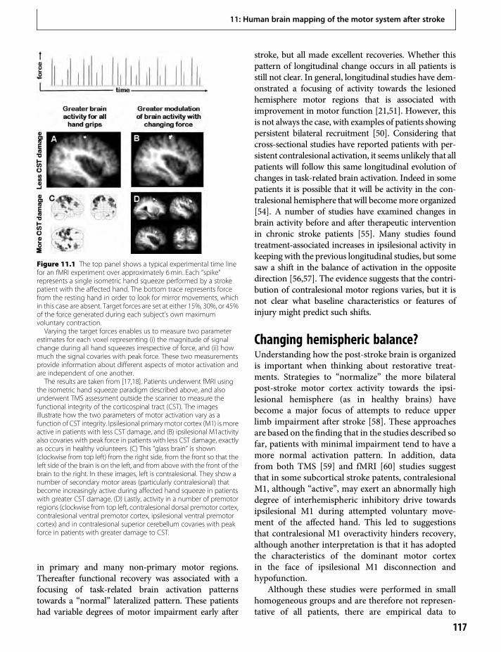
in primary and many non-primary motor regions.Thereafter functional recovery was associated with afocusing of task-related brain activation patternstowards a “normal” lateralized pattern. These patientshad variable degrees of motor impairment early after
stroke, but all made excellent recoveries. Whether thispattern of longitudinal change occurs in all patients isstill not clear. In general, longitudinal studies have dem-onstrated a focusing of activity towards the lesionedhemisphere motor regions that is associated withimprovement in motor function [21,51]. However, thisis not always the case, with examples of patients showingpersistent bilateral recruitment [50]. Considering thatcross-sectional studies have reported patients with per-sistent contralesional activation, it seems unlikely that allpatients will follow this same longitudinal evolution ofchanges in task-related brain activation. Indeed in somepatients it is possible that it will be activity in the con-tralesional hemisphere that will becomemore organized[54]. A number of studies have examined changes inbrain activity before and after therapeutic interventionin chronic stroke patients [55]. Many studies foundtreatment-associated increases in ipsilesional activity inkeeping with the previous longitudinal studies, but somesaw a shift in the balance of activation in the oppositedirection [56,57]. The evidence suggests that the contri-bution of contralesional motor regions varies, but it isnot clear what baseline characteristics or features ofinjury might predict such shifts.
Changing hemispheric balance?Understanding how the post-stroke brain is organizedis important when thinking about restorative treat-ments. Strategies to “normalize” the more bilateralpost-stroke motor cortex activity towards the ipsi-lesional hemisphere (as in healthy brains) havebecome a major focus of attempts to reduce upperlimb impairment after stroke [58]. These approachesare based on the finding that in the studies described sofar, patients with minimal impairment tend to have amore normal activation pattern. In addition, datafrom both TMS [59] and fMRI [60] studies suggestthat in some subcortical stroke patents, contralesionalM1, although “active”, may exert an abnormally highdegree of interhemispheric inhibitory drive towardsipsilesional M1 during attempted voluntary move-ment of the affected hand. This led to suggestionsthat contralesional M1 overactivity hinders recovery,although another interpretation is that it has adoptedthe characteristics of the dominant motor cortexin the face of ipsilesional M1 disconnection andhypofunction.
Although these studies were performed in smallhomogeneous groups and are therefore not represen-tative of all patients, there are empirical data to
Figure 11.1 The top panel shows a typical experimental time linefor an fMRI experiment over approximately 6min. Each “spike”represents a single isometric hand squeeze performed by a strokepatient with the affected hand. The bottom trace represents forcefrom the resting hand in order to look for mirror movements, whichin this case are absent. Target forces are set at either 15%, 30%, or 45%of the force generated during each subject’s own maximumvoluntary contraction.
Varying the target forces enables us to measure two parameterestimates for each voxel representing (i) the magnitude of signalchange during all hand squeezes irrespective of force, and (ii) howmuch the signal covaries with peak force. These two measurementsprovide information about different aspects of motor activation andare independent of one another.
The results are taken from [17,18]. Patients underwent fMRI usingthe isometric hand squeeze paradigm described above, and alsounderwent TMS assessment outside the scanner to measure thefunctional integrity of the corticospinal tract (CST). The imagesillustrate how the two parameters of motor activation vary as afunction of CST integrity. Ipsilesional primarymotor cortex (M1) ismoreactive in patients with less CST damage, and (B) ipsilesional M1activityalso covaries with peak force in patients with less CST damage, exactlyas occurs in healthy volunteers. (C) This “glass brain” is shown(clockwise from top left) from the right side, from the front so that theleft side of the brain is on the left, and from above with the front of thebrain to the right. In these images, left is contralesional. They show anumber of secondary motor areas (particularly contralesional) thatbecome increasingly active during affected hand squeeze in patientswith greater CST damage. (D) Lastly, activity in a number of premotorregions (clockwise from top left, contralesional dorsal premotor cortex,contralesional ventral premotor cortex, ipsilesional ventral premotorcortex) and in contralesional superior cerebellum covaries with peakforce in patients with greater damage to CST.
11: Human brain mapping of the motor system after stroke
117
support the idea that increased contralesional M1activity is often maladaptive. Several studies haveused this concept to transiently improve motor func-tion after stroke by suppressing excitability in con-tralesional M1 [61–66]. Proof-of-principal studies inchronic mild impairment subcortical stroke patientsare encouraging [67], but a critical question remainswhether this normalization is appropriate for allpatients. However, the question of whether contra-lesional M1 is a help or a hindrance in supportingrecovered function is not yet answered. Many func-tional imaging studies have observed motortask-related brain activity in contralesional M1 instroke patients as described earlier [8,16,17,25–27],probably more so in patients with residual impairment[16,49]. Contralesional hemisphere activity has alsobeen demonstrated in stroke patients using EEG [68].The fine temporal resolution of EEG was able to detectthat this activity occurred after themotor response hadbeen made, suggesting that it was not related to move-ment initiation in these patients. However, becauseEEG lacks fine spatial resolution, it is not certain thatthis result related to contralesional M1 and not topremotor cortex, for example. A similar approachusing an event-related fMRI design has demonstratedthat contralesional M1 activity peaked seconds beforeipsilesional M1 in stroke patients, in comparison tocontrols in whom the opposite relationship wasobserved [8]. This change in the characteristics ofmotor system activation might point to a change ofrole in different patients, but does not prove a func-tional role during movement nor exclude the possibil-ity that contralesional M1 activity is hinderingrecovery.
In studies involving adult patients with small sub-cortical infarcts, the effect of disruption of contrale-sional M1 function by TMS depends on whether themotor task is a simple react time task (no disruption)[69,70], or involves pressing sequences of buttons (dis-ruption of timing) [71], supporting a role for contra-lesional M1 in some patients and in some tasks.Serrien and colleagues used directed EEG coherenceto investigate whether there is increased informationalflow from the contralesional motor cortex followingmotor stroke [72]. They found that in less well-recovered patients, most task-related flow of informa-tion between the sensorimotor cortices in the low betaband of the EEG came from the contralesional hemi-sphere (i.e. ipsilateral to movement) during grip withthe affected hand. This was not the case in recovered
patients nor in controls, among whom cortical activitywas driven from the contralateral sensorimotor cortex.These findings again suggest a functional role for thecontralesional hemisphere in organizing movement ofthe impaired, ipsilateral limb following stroke, butonly in those patients that do not make a good func-tional recovery. Patients making a fuller recovery,however, continue to organize movement-relatedcortical activity from the hemisphere contralateral tomovement.
In addition, there is evidence that non-M1 secon-dary motor areas in the contralesional hemispherecontribute to recovered function. First, experimentsin which TMS transiently disrupts activity in eitheripsilesional or contralesional PMd, or contralesionalM1, demonstrate worsening of recovered motorbehaviors in some chronic subcortical stroke patientswith no effect on healthy volunteers [69,71,73].Furthermore, TMS to contralesional PMd is moredisruptive in patients with greater impairment [69],whereas TMS to ipsilesional PMd is more disruptive inless impaired patients [73], implying a contralesionalshift in the balance of functionally relevant activitywith greater impairment.
Another approach is to measure how task-relatedactivity covaries with modulation of task parameters.In healthy humans, for example, increasing forceproduction is associated with linear increases inBOLD signal in contralateral M1 and medial motorregions, implying that they have a functional role inforce production [74–76]. A recent study examinedspecifically for regional changes in the control offorce modulation after stroke [18]. In patients withgreater corticospinal system damage, force-related sig-nal changes were seen mainly in contralesional dorso-lateral premotor cortex, bilateral ventrolateralpremotor cortices, and contralesional cerebellum, butnot ipsilesional primary motor cortex (Figure 11.1).Interestingly, a qualitatively similar result was foundin healthy volunteers with increasing age, suggestingthat this “reorganization” might be a generic propertyof the cortical motor system in response to a variety ofinsults [20]. Thus not only do premotor corticesbecome increasingly active as corticospinal systemintegrity diminishes [17], but they can take on a new“M1-like” role during modulation of force output,which implies a new and functionally relevant role inmotor control.
These results are important because they tell us thatthe response to focal injury does not involve simple
11: Human brain mapping of the motor system after stroke
118

up- or downregulation of the motor network as awhole. It is clear that nodes within remaining motornetworks can take on new functional roles and that thepost-stroke motor system is organized differently tothat in the normal brain. However, there are nowseveral lines of evidence to suggest that the mecha-nisms of reorganization are lesion-specific. If restora-tion of function is dependent on the interaction of thetreatment and the residual motor network, it thereforefollows that treatments will have different effects indifferent patients. Furthermore, individual patientsmay require different rehabilitative strategies in orderto “interact” with motor systems which are organizeddifferently. An interesting line of research to emergefrom this line of thinking is whether it is possible topredict the likelihood of improving with a particulartreatment, based on a careful study of the residualpost-stroke structural and functional anatomy.
Predicting recoveryHow does this help us to understand how best to treatthe impairment suffered by patients after stroke?Clearly, the anatomy of the damage will introduceconstraints. For example, Stinear and colleagues [77]set out to determine whether characterizing the state ofthe motor system of a series of chronic stroke patientswould help in predicting the functional gains made ina subsequent motor practice programme. A variety oftools were used, including TMS, structural MRI, andfunctional MRI. The presence or absence of MEPs toTMS in the affected upper limb, and fractional aniso-tropy values were both used to assess the structuralintegrity of the descending white matter pathways inthe posterior limb of the internal capsules. In addition,fMRI was used to assess the degree of lateralization ofactivity during a motor task. Not surprisingly, inpatients with MEPs, meaningful gains with motorpractice were still possible three years after stroke.The situation in patients without MEPs has neverbeen clear-cut, as this is often taken as a poor prog-nostic sign [78]. In this study, the functional potentialof patients without MEPs declined with increasingcorticospinal pathway disruption. Disappointingly,the degree of lateralization of brain activity as meas-ured with fMRI could not predict future functionalgains through motor practice. The task used duringscanning was a self paced opening and closing of thehand, and so it is possible that variations in perform-ance of the task (to which the magnitude of activationis sensitive) introduced additional variability into the
fMRI data. A task that probed for a measure of motorsystem plasticity in the context of motor practicemight have provided different results. Nevertheless,this study illustrates how multimodal imaging andneurophysiological data can be used to assess thestate of the motor system which might in turn predictresponse to therapeutic interventions and lead to treat-ment algorithms for clinical practice.
In a similar approach, Cramer et al. [79] assessedwhich of 13 baseline clinical/radiological measures wereable to predict subsequent gainsmade during 6 weeks ofrehabilitation therapy. Of all these measures onlygreater clinical status and lower motor cortex activationduring fMRI at baseline remained significant and inde-pendent predictors of clinical improvement. This is aninteresting finding, because in general, patients withgreater impairment are more likely to have less task-related ipsilesional M1 activity, although this is aninconsistent finding. This result tells us that there issomething in the imaging data which is independentof baseline clinical impairment which predicts improve-ments. Lower baseline motor cortex activation was alsoassociated with larger increases in motor cortex activa-tion after treatment, so perhaps in some patients, lowbaseline cortical activity represents underuse of surviv-ing cortical resources. A measure of brain functionmight be important for optimal clinical decision-making in the context of a restorative intervention.
A separate study attempted to use fMRI dataacquired in the first few days after stroke to determinea subsequent change in motor performance over andabove initial stroke severity and lesion volume [80].They found a pattern of brain activation which washighly predictive of clinical change. Although the mul-tivariate analysis used did not allow anatomical infer-ence to be made, it is clear that there is somethingabout the way the function of the brain responds toinjury, over and above the anatomy of the damage,that holds clues about future clinical progression. Thepattern was distributed and certainly not confined tothe motor system, even though clinical improvementwas measured in the motor domain. The idea thatmotor improvement may not be solely related to theintegrity of the corticospinal system, but also withother characteristics of the post-stroke brain is sup-ported by the finding that motor performance at 3months correlated only weakly with a measure ofcorticospinal tract integrity (using TMS) but stronglywith a measure of intracortical excitability [81]. Thesefindings suggest that the anatomy of the damage may
11: Human brain mapping of the motor system after stroke
119
set a limit on the extent of recovery, but that at least insome patients, other parameters, perhaps preservedcortico-cortical connectivity, might be importantwhen considering whether a patient has the capacityor potential to improve.
SummaryAfter focal brain damage, there is a reconfiguration ofthe cerebral motor system. This does not appear to be asimple up- or downregulation of motor networks intheir entirety, but the residual functional networksseem to operate in a different way, with some brainregions adopting the characteristics of damaged ordisconnected regions. This reorganization variesacross chronic stroke patients, but does so in a waythat appears to be predictable, which gives us hope thata deeper understanding of these processes will allow usto treat patients with impairments more effectively. Itis important to stress that this reorganization is oftennot successful in returning motor function to normal.It is less effective than that in the intact brain, butwill nevertheless support what recovered functionthere is. The exact configuration of this new motorsystem will be determined most obviously by theextent of the anatomical damage. This includes theextent to which the damage affects cortical motorregions, white matter pathways [82], and even whichhemisphere is affected [83]. In patients with damageto primary sensorimotor cortex, for example, testsof fractionated finger movement correlated morestrongly with the proportion of surviving “normal”sensorimotor cortex (as defined by functional activa-tion maps in normal controls) than with total infarctvolume [84]. The potential for functionally relevantchange to occur will depend on a number of otherfactors, not least the biologic age of the subject and thepremorbid state of their brain [20], but also currentdrug treatments [85]. Furthermore, levels of neuro-transmitters and growth factors which are able toinfluence the ability of the brain to respond to afferentinput (i.e. how plastic it is) might be determined bytheir genetic status [86]. The basis of impairment-based treatment is likely to be the promotion ofactivity-dependent change within these surviving net-works, and so understanding the factors that shape it –possibly at the level of the individual patient – will becritical [87]. Functional brain imaging will play acentral role in achieving this goal.
References1. Nichols-Larsen DS, Clark PC, Zeringue A, et al. Factors
influencing stroke survivors’ quality of life duringsubacute recovery. Stroke. 2005;36:1480–4.
2. Wyller TB, Sveen U, Sodring KM, et al. Subjective well-being one year after stroke. Clin Rehabil.1997;11:139–45.
3. Cramer SC, Chopp M. Recovery recapitulatesontogeny. Trends Neurosci. 2000;23:265–71.
4. Buxton RB. An introduction to functional magneticresonance imaging: principles and techniques.Cambridge: Cambridge University Press; 2002.
5. Magistretti PJ, Pellerin L. Cellular mechanisms of brainenergy metabolism and their relevance to functionalbrain imaging. Phil Trans R Soc Lond B Biol Sci.1999;354:1155–63.
6. Attwell D, Iadecola C. The neural basis of functionalbrain imaging signals. Trends Neurosci. 2002;25:621–5.
7. Friston KJ, Josephs O, Rees G, et al. Nonlinear event-related responses in fMRI. Magn Reson Med.1998;39:41–52.
8. Newton J, Sunderland A, Butterworth SE, et al. A pilotstudy of event-related functional magnetic resonanceimaging ofmonitored wrist movements in patients withpartial recovery. Stroke. 2002;33:2881–7.
9. Pineiro R, Pendlebury S, Johansen-Berg H, et al.Functional MRI detects posterior shifts in primarysensorimotor cortex activation after stroke: Evidence oflocal adaptive reorganization? Stroke. 2001;32:1134–9.
10. Carusone LM, Srinivasan J, Gitelman DR, et al.Hemodynamic response changes in cerebrovasculardisease: Implications for functional MR imaging. AmJ Neuroradiol. 2002;23:1222–8.
11. Hamzei F, Knab R, Weiller C, et al. The influence ofextra- and intracranial artery disease on the BOLDsignal in fMRI. Neuroimage. 2003;20:1393–9.
12. Rossini PM, Altamura C, Ferretti A, et al. Doescerebrovascular disease affect the coupling betweenneuronal activity and local haemodynamics? Brain.2004;127:99–110.
13. Röther J, Knab R, Hamzei F, et al. Negative dip inBOLD fMRI is caused by blood flow–oxygenconsumption uncoupling in humans. Neuroimage.2002;15:98–102.
14. Krainik A, Hund-Georgiadis M, Zysset S, et al. Regionalimpairment of cerebrovascular reactivity and BOLDsignal in adults after stroke. Stroke. 2005;36:1146–52.
15. Murata Y, Sakatani K, Hoshino T, et al. Effects ofcerebral ischemia on evoked cerebral bloodoxygenation responses and BOLD contrast functionalMRI in stroke patients. Stroke. 2006;37:2514–20.
11: Human brain mapping of the motor system after stroke
120
16. Ward NS, Brown MM, Thompson AJ, et al. Neuralcorrelates of outcome after stroke: A cross-sectionalfMRI study. Brain. 2003;126:1430–48.
17. Ward NS, Newton JM, Swayne OB, et al. Motor systemactivation after subcortical stroke depends oncorticospinal system integrity. Brain. 2006;129:809–19.
18. Ward NS, Newton JM, Swayne OB, et al. Therelationship between brain activity and peak grip forceis modulated by corticospinal system integrity aftersubcortical stroke. Eur J Neurosci. 2007;25:1865–73.
19. D’Esposito M, Zarahn E, Aguirre GK, et al. The effect ofnormal aging on the coupling of neural activity to thebold hemodynamic response. Neuroimage.1999;10:6–14.
20. Ward NS, Swayne OB, Newton JM. Age-dependentchanges in the neural correlates of force modulation:An fMRI study. Neurobiol Aging. 2008;29:1434–46.
21. Calautti C, Leroy F, Guincestre JY, et al. Dynamics ofmotor network overactivation after striatocapsularstroke: A longitudinal PET study using a fixed-performance paradigm. Stroke. 2001;32:2534–42.
22. Chollet F, DiPiero V, Wise RJ, et al. The functionalanatomy of motor recovery after stroke in humans:A study with positron emission tomography. AnnNeurol. 1991;29:63–71.
23. Seitz RJ, Hoflich P, Binkofski F, et al. Role of thepremotor cortex in recovery from middle cerebralartery infarction. Arch Neurol. 1998;55:1081–8.
24. Weiller C, Chollet F, Friston KJ, et al. Functionalreorganization of the brain in recovery fromstriatocapsular infarction in man. Ann Neurol.1992;31:463–72.
25. Weiller C, Ramsay SC, Wise RJ, et al. Individualpatterns of functional reorganization in the humancerebral cortex after capsular infarction. Ann Neurol.1993;33:181–9.
26. Cao Y, D’Olhaberriague L, Vikingstad EM, et al. Pilotstudy of functional MRI to assess cerebral activation ofmotor function after poststroke hemiparesis. Stroke.1998;29:112–22.
27. Cramer SC, Nelles G, Benson RR, et al. A functionalMRI study of subjects recovered from hemipareticstroke. Stroke. 1997;28:2518–27.
28. Cramer SC, Moore CI, Finklestein SP, et al. A pilotstudy of somatotopic mapping after cortical infarct.Stroke. 2000;31:668–71.
29. Rossini PM, Caltagirone C, Castriota-Scanderbeg A,et al. Handmotor cortical area reorganization in stroke:A study with fMRI, MEG and TCS maps. Neuroreport.1998;9:2141–6.
30. Sanes JN, Donoghue JP. Plasticity and primary motorcortex. Ann Rev Neurosci. 2000;23:393–415.
31. Cao Y, Vikingstad EM, Huttenlocher PR, et al.Functional magnetic resonance studies of thereorganization of the human hand sensorimotor areaafter unilateral brain injury in the perinatal period.Proc Natl Acad Sci USA 1994;91:9612–6.
32. Porter R, Lemon RN. Corticospinal function andvoluntary movement. Oxford: Oxford University Press;1993.
33. Tomassini V, Jbabdi S, Klein JC, et al. Diffusion-weighted imaging tractography-based parcellation ofthe human lateral premotor cortex identifies dorsal andventral subregions with anatomical and functionalspecializations. J Neurosci. 2007;27:10 259–69.
34. Johansen-Berg H, Matthews PM. Attention tomovement modulates activity in sensori-motor areas,including primary motor cortex. Exp Brain Res.2002;142:13–24.
35. Dum RP, Strick PL. The origin of corticospinalprojections from the premotor areas in the frontal lobe.J Neurosci. 1991;11:667–89.
36. He SQ, Dum RP, Strick PL. Topographic organizationof corticospinal projections from the frontal lobe:Motor areas on the lateral surface of the hemisphere.J Neurosci. 1993;13:952–80.
37. He SQ, Dum RP, Strick PL. Topographic organizationof corticospinal projections from the frontal lobe:Motor areas on the medial surface of the hemisphere.J Neurosci. 1995;15:3284–306.
38. Strick PL. Anatomical organization of multiple motorareas in the frontal lobe: Implications for recovery offunction. Adv Neurol. 1988;47:293–312.
39. Boudrias MH, Belhaj-Saif A, Park MC, et al.Contrasting properties of motor output from thesupplementary motor area and primarymotor cortex inrhesus macaques. Cereb Cortex. 2006;16:632–8.
40. Maier MA, Armand J, Kirkwood PA, et al. Differencesin the corticospinal projection from primary motorcortex and supplementary motor area to macaqueupper limb motoneurons: An anatomical andelectrophysiological study. Cereb Cortex.2002;12:281–96.
41. Mazevet D, Meunier S, Pradat-Diehl P, et al. Changes inpropriospinally mediated excitation of upper limbmotoneurons in stroke patients. Brain.2003;126:988–1000.
42. Stinear JW, Byblow WD. The contribution of cervicalpropriospinal premotoneurons in recoveringhemiparetic stroke patients. J Clin Neurophysiol.2004;21:426–34.
43. Mazevet D, Pierrot-Deseilligny E. Pattern of descendingexcitation of presumed propriospinal neurones at theonset of voluntary movement in humans. Acta PhysiolScand. 1994;150:27–38.
11: Human brain mapping of the motor system after stroke
121

44. Pierrot-Deseilligny E. Transmission of the corticalcommand for human voluntary movement throughcervical propriospinal premotoneurons. ProgNeurobiol. 1996;48:489–517.
45. Dancause N, Barbay S, Frost SB, et al. Ipsilateralconnections of the ventral premotor cortex in a newworld primate. J Comp Neurol. 2006;495:374–90.
46. Dancause N, Barbay S, Frost SB, et al. Interhemisphericconnections of the ventral premotor cortex in a newworld primate. J Comp Neurol. 2007;505:701–15.
47. Dum RP, Strick PL. Spinal cord terminations of themedial wall motor areas in macaque monkeys.J Neurosci. 1996;16:6513–25.
48. Rouiller EM,Moret V, Tanne J, et al. Evidence for directconnections between the hand region of thesupplementary motor area and cervical motoneurons inthe macaque monkey. Eur J Neurosci. 1996;8:1055–9.
49. Ward NS, Brown MM, Thompson AJ, et al. Theinfluence of time after stroke on brain activationsduring a motor task. Ann Neurol. 2004;55:829–34.
50. Feydy A, Carlier R, Roby-Brami A, et al. Longitudinalstudy of motor recovery after stroke: Recruitment andfocusing of brain activation. Stroke. 2002;33:1610–7.
51. Marshall RS, Perera GM, Lazar RM, et al. Evolution ofcortical activation during recovery from corticospinaltract infarction. Stroke. 2000;31:656–61.
52. Small SL, Hlustik P, Noll DC, et al. Cerebellarhemispheric activation ipsilateral to the paretic handcorrelates with functional recovery after stroke. Brain.2002;125:1544–57.
53. Ward NS, Brown MM, Thompson AJ, et al. Neuralcorrelates of motor recovery after stroke: A longitudinalfMRI study. Brain. 2003;126:2476–96.
54. Luft AR, Waller S, Forrester L, et al. Lesion locationalters brain activation in chronically impaired strokesurvivors. Neuroimage. 2004;21:924–35.
55. Hodics T, Cohen LG, Cramer SC. Functional imagingof intervention effects in stroke motor rehabilitation.Arch Phys Med Rehabil. 2006;87(12 Suppl 2):S36–42.
56. Luft AR, McCombe-Waller S, Whitall J, et al. Repetitivebilateral arm training and motor cortex activation inchronic stroke: A randomized controlled trial. JAMA2004;292:1853–61.
57. Schaechter JD, Kraft E, Hilliard TS, et al. Motorrecovery and cortical reorganization after constraint-induced movement therapy in stroke patients: Apreliminary study. Neurorehabil Neural Repair.2002;16:326–38.
58. Ward NS, Cohen LG. Mechanisms underlying recoveryof motor function after stroke. Arch Neurol.2004;61:1844–8.
59. Murase N, Duque J, Mazzocchio R, et al. Influence ofinterhemispheric interactions on motor function inchronic stroke. Ann Neurol. 2004;55:400–09.
60. Grefkes C, Nowak DA, Eickhoff SB, et al. Corticalconnectivity after subcortical stroke assessed withfunctional magnetic resonance imaging. Ann Neurol.2008;63:236–46.
61. Mansur CG, Fregni F, Boggio PS, et al. A shamstimulation-controlled trial of rTMS of the unaffectedhemisphere in stroke patients. Neurology.2005;64:1802–04.
62. Liepert J, Zittel S, Weiller C. Improvement of dexterityby single session low-frequency repetitive transcranialmagnetic stimulation over the contralesional motorcortex in acute stroke: A double-blind placebo-controlled crossover trial. Restor Neurol Neurosci.2007;25:461–5.
63. Takeuchi N, Chuma T, Matsuo Y, et al. Repetitivetranscranial magnetic stimulation of contralesionalprimary motor cortex improves hand function afterstroke. Stroke. 2005;36:2681–6.
64. Nowak DA, Grefkes C, Dafotakis M, et al. Effects oflow-frequency repetitive transcranial magneticstimulation of the contralesional primary motor cortexon movement kinematics and neural activity insubcortical stroke. Arch Neurol. 2008;65:741–7.
65. Fregni F, Boggio PS, Mansur CG, et al. Transcranialdirect current stimulation of the unaffected hemispherein stroke patients. Neuroreport. 2005;16:1551–5.
66. Fregni F, Boggio PS, Valle AC, et al. A sham-controlledtrial of a 5-day course of repetitive transcranialmagnetic stimulation of the unaffected hemisphere instroke patients. Stroke. 2006;37:2115–22.
67. Talelli P, Rothwell J. Does brain stimulation after strokehave a future? Curr Opin Neurol. 2006;19:543–50.
68. Verleger R, Adam S, Rose M, et al. Control of handmovements after striatocapsular stroke: High-resolution temporal analysis of the function ofipsilateral activation. Clin Neurophysiol.2003;114:1468–76.
69. Johansen-Berg H, Rushworth MF, Bogdanovic MD,et al. The role of ipsilateral premotor cortex in handmovement after stroke. Proc Natl Acad Sci USA.2002;99:14 518–23.
70. Werhahn KJ, Conforto AB, Kadom N, et al.Contribution of the ipsilateral motor cortex to recoveryafter chronic stroke. Ann Neurol. 2003;54:464–72.
71. Lotze M, Markert J, Sauseng P, et al. The role ofmultiple contralesional motor areas for complex handmovements after internal capsular lesion. J Neurosci.2006;26:6096–102.
72. Serrien DJ, Strens LH, Cassidy MJ, et al. Functionalsignificance of the ipsilateral hemisphere during
11: Human brain mapping of the motor system after stroke
122
movement of the affected hand after stroke. Exp Neurol.2004;190:425–32.
73. Fridman EA, Hanakawa T, Chung M, et al.Reorganization of the human ipsilesional premotorcortex after stroke. Brain. 2004;127:747–58.
74. Dettmers C, Fink GR, Lemon RN, et al. Relationbetween cerebral activity and force in the motor areas ofthe human brain. J Neurophysiol. 1995;74:802–15.
75. Thickbroom GW, Phillips BA, Morris I, et al.Differences in functional magnetic resonance imagingof sensorimotor cortex during static and dynamicfinger flexion. Exp Brain Res. 1999;126:431–8.
76. Ward NS, Frackowiak RS. Age-related changes in theneural correlates of motor performance. Brain.2003;126:873–88.
77. Stinear CM, Barber PA, Smale PR, et al. Functionalpotential in chronic stroke patients depends oncorticospinal tract integrity. Brain. 2007;130:170–80.
78. Heald A, Bates D, Cartlidge NE, et al. Longitudinalstudy of central motor conduction time followingstroke. 2. Central motor conduction measured within72 h after stroke as a predictor of functional outcome at12 months. Brain. 1993;116:1371–85.
79. Cramer SC, Parrish TB, Levy RM, et al. Predictingfunctional gains in a stroke trial. Stroke.2007;38:2108–14.
80. Marshall RS, Zarahn E, Alon L, et al. Early imagingcorrelates of subsequent motor recovery after stroke.Ann Neurol. 2009;65:596–602.
81. Swayne OB, Rothwell JC, Ward NS, et al. Stages ofmotor output reorganization after hemispheric strokesuggested by longitudinal studies of cortical physiology.Cereb Cortex. 2008;18:1909–22.
82. Newton JM, Ward NS, Parker GJ, et al. Non-invasivemapping of corticofugal fibres from multiple motorareas – Relevance to stroke recovery. Brain.2006;129:1844–58.
83. Zemke AC, Heagerty PJ, Lee C, et al. Motor cortexorganization after stroke is related to side of stroke andlevel of recovery. Stroke. 2003;34:e23–8.
84. Crafton KR, Mark AN, Cramer SC. Improvedunderstanding of cortical injury by incorporatingmeasures of functional anatomy. Brain.2003;126:1650–9.
85. Goldstein LB. Pharmacology of recovery after stroke.Stroke. 1990;21(Suppl):III139–42.
86. Kleim JA, Chan S, Pringle E, et al. BDNF val66metpolymorphism is associated with modifiedexperience-dependent plasticity in human motorcortex. Nat Neurosci. 2006;9:735–7.
87. Ward NS. Getting lost in translation. Curr Opin Neurol.2008;21:625–7.
11: Human brain mapping of the motor system after stroke
123

12 Recovery from aphasia: lessons fromimaging studiesCornelius Weiller & Dorothee Saur
During the last 20 years, application of functional brainimaging to stroke patients has brought a new momen-tum into rehabilitation [1]. We understand better whathappens in the brain of patients: we “see an active ipsi-lateral motor cortex”when we noticemirrormovementsof the healthy hand during the ward rounds (see alsoChapter 11), or assume a resolution of diaschisis whenlanguage performance improves abruptly from one dayto the other within the first week after a stroke (see alsoChapter 9). This chapter highlights recent findings fromneuroimaging studies of aphasia, focusing on thedynamic process of reorganization from acute to chronicstroke and points to recent developments, which enablethe investigation of language reorganization within aframework of interconnected brain regions.
Imaging the acute phaseImaging the acute phase of recovery from aphasia, i.e.the first days after stroke onset, offers the opportunityfor unique insights into the function and dysfunctionof the language network.
As displayed in Figure 12.1a, loss of function canbe explained in terms of direct or indirect consequen-ces of ischemia. On the one hand, the ischemic lesionmay directly affect language-relevant gray (A) andwhite matter (B) structures due to either critical hypo-perfusion with incomplete infarction, or to parenchy-mal damage (complete infarction) [2]. Ischemia mayalso cause a dysfunction in remote, non-infarctedareas (C, D). The idea of “diaschisis” was introducedby von Monakow [3]. Diaschisis is explained by afunctional disconnection. The observed languageimpairment reflects the result of both direct local andindirect remote effects. Taken together, the lesion of acritical network component may result in an acuteglobal network breakdown. In this situation, we typi-cally observe a severe aphasia.
In this hyperacute phase the lesion may be unsta-ble, resulting in sometimes rapid fluctuations in lan-guage performance. This mainly includes changes incerebral perfusion and extension of the peri-infarctedema. Reperfusion of left posterior middle temporaland frontal areas may be associated with acuteimprovement in picture naming [4]. Von Monakow[3] introduced the term “diaschisis” to explain acutelanguage improvements after stroke. Figure 12.1b–ddisplays an example of early functional MRI (fMRI)activation in a patient with acute global aphasia due toa left temporal middle cerebral artery (MCA) infarc-tion. In an auditory language comprehension task, thepatient listened to three sentences of intelligible speech(SP), and also listened to sentences of reversed speech(REV). Extraction of condition-wise effect sizes(Figure 12.1c and d) showed that in the hyperacutephase about 10 h after onset, remote L and R inferiorfrontal gyrus (IFG) were dysfunctional. Although astrong effect for both the SP and REV conditions wasobserved, both areas did not distinguish between intel-ligible SP and unintelligible REV – activation was thesame for both tasks. However, three and seven dayslater, in parallel with improvement of language behav-ior, a clear differentiation between both conditionsreturned to both brain areas, indicating functionalrecovery of these remote areas, which is most likelyexplained by a resolution of diaschisis. That is, injuryto left temporal language areas produced diaschisis inbilateral IFG which produced dysfunction and inabil-ity to distinguish SP from REV; with resolution of thisdiaschisis, bilateral IFG function returned, and theselanguage network areas were able to restart functionand thereby contribute to effective language behaviorand thus compensation for the deficit [5].
Another lesson that may be learned from imagingthe acute phase of aphasia relates to the understandingof subcortical aphasia. Combining the findings from
Brain Repair After Stroke, ed. S. C. Cramer and R. J. Nudo. Published by Cambridge University Press.© Cambridge University Press 2010.
125
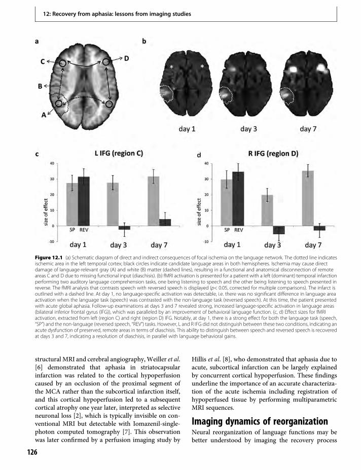
structural MRI and cerebral angiography,Weiller et al.[6] demonstrated that aphasia in striatocapsularinfarction was related to the cortical hypoperfusioncaused by an occlusion of the proximal segment ofthe MCA rather than the subcortical infarction itself,and this cortical hypoperfusion led to a subsequentcortical atrophy one year later, interpreted as selectiveneuronal loss [2], which is typically invisible on con-ventional MRI but detectable with Iomazenil-single-photon computed tomography [7]. This observationwas later confirmed by a perfusion imaging study by
Hillis et al. [8], who demonstrated that aphasia due toacute, subcortical infarction can be largely explainedby concurrent cortical hypoperfusion. These findingsunderline the importance of an accurate characteriza-tion of the acute ischemia including registration ofhypoperfused tissue by performing multiparametricMRI sequences.
Imaging dynamics of reorganizationNeural reorganization of language functions may bebetter understood by imaging the recovery process
Figure 12.1 (a) Schematic diagram of direct and indirect consequences of focal ischemia on the language network. The dotted line indicatesischemic area in the left temporal cortex; black circles indicate candidate language areas in both hemispheres. Ischemia may cause directdamage of language-relevant gray (A) and white (B) matter (dashed lines), resulting in a functional and anatomical disconnection of remoteareas C and D due to missing functional input (diaschisis). (b) fMRI activation is presented for a patient with a left (dominant) temporal infarctionperforming two auditory language comprehension tasks, one being listening to speech and the other being listening to speech presented inreverse. The fMRI analysis that contrasts speech with reversed speech is displayed (p< 0.05, corrected for multiple comparisons). The infarct isoutlined with a dashed line. At day 1, no language-specific activation was detectable, i.e. there was no significant difference in language areaactivation when the language task (speech) was contrasted with the non-language task (reversed speech). At this time, the patient presentedwith acute global aphasia. Follow-up examinations at days 3 and 7 revealed strong, increased language-specific activation in language areas(bilateral inferior frontal gyrus (IFG)), which was paralleled by an improvement of behavioral language function. (c, d) Effect sizes for fMRIactivation, extracted from left (region C) and right (region D) IFG. Notably, at day 1, there is a strong effect for both the language task (speech,“SP”) and the non-language (reversed speech, “REV”) tasks. However, L and R IFG did not distinguish between these two conditions, indicating anacute dysfunction of preserved, remote areas in terms of diaschisis. This ability to distinguish between speech and reversed speech is recoveredat days 3 and 7, indicating a resolution of diaschisis, in parallel with language behavioral gains.
12: Recovery from aphasia: lessons from imaging studies
126
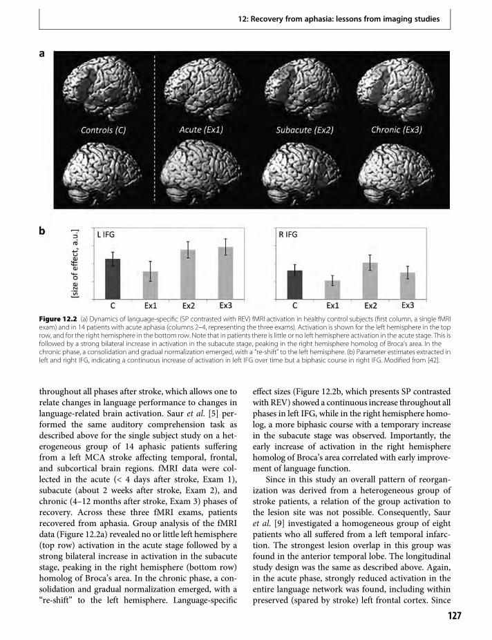
throughout all phases after stroke, which allows one torelate changes in language performance to changes inlanguage-related brain activation. Saur et al. [5] per-formed the same auditory comprehension task asdescribed above for the single subject study on a het-erogeneous group of 14 aphasic patients sufferingfrom a left MCA stroke affecting temporal, frontal,and subcortical brain regions. fMRI data were col-lected in the acute (< 4 days after stroke, Exam 1),subacute (about 2 weeks after stroke, Exam 2), andchronic (4–12 months after stroke, Exam 3) phases ofrecovery. Across these three fMRI exams, patientsrecovered from aphasia. Group analysis of the fMRIdata (Figure 12.2a) revealed no or little left hemisphere(top row) activation in the acute stage followed by astrong bilateral increase in activation in the subacutestage, peaking in the right hemisphere (bottom row)homolog of Broca’s area. In the chronic phase, a con-solidation and gradual normalization emerged, with a“re-shift” to the left hemisphere. Language-specific
effect sizes (Figure 12.2b, which presents SP contrastedwith REV) showed a continuous increase throughout allphases in left IFG, while in the right hemisphere homo-log, a more biphasic course with a temporary increasein the subacute stage was observed. Importantly, theearly increase of activation in the right hemispherehomolog of Broca’s area correlated with early improve-ment of language function.
Since in this study an overall pattern of reorgan-ization was derived from a heterogeneous group ofstroke patients, a relation of the group activation tothe lesion site was not possible. Consequently, Sauret al. [9] investigated a homogeneous group of eightpatients who all suffered from a left temporal infarc-tion. The strongest lesion overlap in this group wasfound in the anterior temporal lobe. The longitudinalstudy design was the same as described above. Again,in the acute phase, strongly reduced activation in theentire language network was found, including withinpreserved (spared by stroke) left frontal cortex. Since
Figure 12.2 (a) Dynamics of language-specific (SP contrasted with REV) fMRI activation in healthy control subjects (first column, a single fMRIexam) and in 14 patients with acute aphasia (columns 2–4, representing the three exams). Activation is shown for the left hemisphere in the toprow, and for the right hemisphere in the bottom row. Note that in patients there is little or no left hemisphere activation in the acute stage. This isfollowed by a strong bilateral increase in activation in the subacute stage, peaking in the right hemisphere homolog of Broca’s area. In thechronic phase, a consolidation and gradual normalization emerged, with a “re-shift” to the left hemisphere. (b) Parameter estimates extracted inleft and right IFG, indicating a continuous increase of activation in left IFG over time but a biphasic course in right IFG. Modified from [42].
12: Recovery from aphasia: lessons from imaging studies
127

in all patients left IFG was intact, reduced activation inthis area can be best interpreted in terms of diaschisis.That is, in the acute phase, the left IFG lacks functionalinput from left temporal areas. In the subacute phase, astrong increase in bilateral IFG was observed; how-ever, in contrast to the mixed group, the highest acti-vation remained in the left hemisphere. Notably,activation in left and right IFG emerged simultane-ously. This is of particular interest since righthemisphere activation in recovery from aphasia isinterpreted to reflect disinhibition caused by dysfunc-tional left language areas, which are unable to inhibittheir homologs in the right hemisphere. If this is thecase, we would expect that right hemisphere activationreveals a reciprocal rather than a parallel course ofactivation. However, a reciprocal pattern is not alwaysobserved. In fact, simultaneous increase of activationin bilateral IFG could also be interpreted in terms of afunctional dependency of right homolog languageareas on their left hemisphere counterparts. Finally,in the chronic phase, perilesional activation in poste-rior temporal areas emerged. This perilesional activa-tion was also found in areas activated in healthysubjects, indicating a reactivation of temporarily dys-functional tissue rather than a true recruitment ofnovel brain areas. Importantly, in this perilesionaltissue, high correlation between language performanceand language activation over time was found. Thisfurther supports the high functional relevance of lefthemisphere areas for long-term language recovery.
Taken together, we postulated a model of languagerecovery, which proceeds in three phases [1,5]. In afirst period during which near-complete abolishmentof language function is seen, there is little if any acti-vation in brain regions which later can be activated bythe language task. In this acute phase a global networkbreakdown, in which “diaschisis”, or ischemic stun-ning [10], is the key factor. A second, “hyperactive”stage of brain activation follows, in which the alteredfunction recovers at a rapid pace. It is characterized bya sudden return of activation in left and often a hyper-activation of homolog right hemisphere areas. In thethird stage, a consolidation of activation resemblingthe patterns in healthy controls is observed.
These patterns of reorganization are derived froma highly selected group of patients. In fact, Saur et al.[5] screened a total of 198 consecutively admittedaphasic patients to find 14 to match the inclusioncriteria. Most patients were excluded since they werenot able to perform early fMRI due to poor general
medical condition. Thus, the results are somewhatbiased by a selection of the best patients, and furtherstudies might clarify how these findings extend to thebroader spectrum of stroke patients.
Imaging the chronic phaseThe majority of imaging studies on language recoverywere performed in the chronic phase after stroke. Inthese studies, patients who recovered from aphasia tovarious degrees were examined once or sequentiallywith PET or fMRI to identify the pattern of the lan-guage activation in the lesioned brain. In sum, as in themotor system (see Chapter 11), these studies with veryheterogeneous designs, methods, and patients haveshown that post-stroke language system reorganiza-tion of function involves undamaged areas in the lefthemisphere, perilesional tissue, and homologous areasin the right hemisphere [11–18]. Depending on siteand size of the lesion and the residual language impair-ment, portions of these three areas are more activatedas compared to healthy control subjects [17]. Morespecific conclusions can be drawn by correlating theproficiency of a particular language function with task-related activation in a cross-sectional design. Forinstance, Crinion et al. [19] demonstrated in a groupof 17 patients with left temporal stroke that perform-ance in auditory sentence comprehension was posi-tively correlated with activation in the right lateralsuperior temporal gyrus (STG). These results under-line that in the chronic phase, too, right hemisphereactivation might be beneficial. More sustained evi-dence comes from a dynamic challenge in which repet-itive behavioral studies are performed. Musso et al.showed that after repeated 8-min sessions of languagecomprehension training, subsequent behavioralimprovement correlated with an increase of activationin the right temporal cortex [12], supporting a role forright hemisphere language homolog areas in languagefunction after stroke.
When discussing the functional role of right hemi-sphere activation in chronic aphasia, we have to thinkabout the potential factors influencing the amount ofright hemisphere involvement. First, the premorbiddegree of language lateralization seems to be animportant factor. Although impossible to control, itis likely that patients with a more bilateral premorbidlanguage representation are more likely to utilize thehomolog right areas after left hemisphere damage.Second, the more complete the damage of left corelanguage areas and the more severe the associated
12: Recovery from aphasia: lessons from imaging studies
128

residual language deficit, the more likely a permanentinvolvement of the right hemisphere homolog areaswill be observed (see also below regarding the conceptof critical lesions). This might lead to the conclusionthat right hemisphere activation is disadvantageousor maladaptive rather than functionally beneficial,assuming a negative influence of right hemisphere(in particular right frontal) activation which preventsrecovery. In support of this logic, note that studiesusing repetitive transcranial magnetic stimulation(rTMS) to inhibit function of right hemispherelanguage regions in chronic stroke patients havedescribed improvement of language functions [20].However, a deterioration of language functions afterrTMS application to right frontal areas has also beenobserved [21]. A final judgment of this issue is difficult,as neither transcallosal interaction nor the mecha-nisms of rTMS are anywhere near to completelyunderstood.
Impact of therapy on reorganizationSeveral studies have shown task-related activationchanges from pre- to post-treatment scans. Even stud-ies providing short-term treatment influence the neuralbasis of language processing, such as the one by Mussoet al. [12]. Blasi et al. [22] demonstrated increasedactivity in the right frontal cortex associated with learn-ing a word retrieval task. Studies performing long-termtraining found either left [23–26], or right [27,28], orbilateral activation to be associated with treatment-induced improvement [29–31]. In contrast, Richteret al. [32] found no significant changes of fMRI activa-tion at all after 2 weeks of “constraint-induced aphasiatherapy”. This heterogeneity of results is best explainedby differences in treatment strategies, language impair-ments, as well as differences in lesion sites and sizes. Inpost-stroke brain reorganization, as with many aspectsof acute stroke, many factors can contribute to differ-ences between patients. However, these studies doclearly demonstrate that remodeling of cortical func-tions is possible even years after the stroke, and typi-cally occurs in both left and right as well as perilesionallanguage zones, similar to what has been described inthe motor system [33] (see also Chapter 11).
Summary and outlook: the networkapproachMost brain functions are organized in distributed,segregated and interconnected networks, and brainreorganization due to lesions or interventions mainly
takes place within the framework of these networks[1,34]. Language is a brain function, which is especiallysuited to assess a network approach on recovery, as it isrepresented in a widespread, bilateral brain network.Recent combinations of fMRI with probabilistic fibertracking, integrating pairwise point-to-point anatom-ical connections along with measurements of func-tional connectivity, resulted in a relatively completenetwork description of language processing aroundthe Sylvian fissure [35,36]. In contrast to classical dia-grams, which suppose speech comprehension to belocalized in the temporal lobe and speech productionin the inferior frontal lobe and both regions connectedthrough the arcuate fascicle, the modern anatomicalview sees the left temporal lobe connected to most ofthe frontal lobe along two segregated and integratinglarge association tracts [35,37–39] (Figure 12.3): adorsal route along the arcuate fasciculus/superior lon-gitudinal fasciculus mainly projects to premotor cor-tices and renders automated sensory-motor mapping,while several aspects of language comprehension uti-lize a ventral route via the extreme capsule mainlyprojecting to the prefrontal cortex. These new findingsnot only require a new description of anatomical cor-relation of language functions and dysfunctions, butalso imply various routes of compensation after stroke.
The network view addresses another frequent mis-conception: the assumption of functional independenceof brain “centers”. As an example, conduction aphasia,which is often defined as an isolated repetition failurewith preserved comprehension and spontaneousspeech, is supposed to result from a destruction of thepathway (i.e. the arcuate fascicle) connecting the sen-sory and themotor speech center, which are both intact.Such a conclusion assumes that the temporal lobe suf-fices for comprehension and the inferior frontal cortex
Figure 12.3 Schematic diagram of ventral and dorsal languagepathways projecting to the prefrontal (PFC) and premotor (PM)cortex. MdlF=middle longitudinal fascicle, EmC=extreme capsule,AF=arcuate fasciculus, SLF=superior longitudinal fasciculus.
12: Recovery from aphasia: lessons from imaging studies
129
for language production and that a disruption of theconnecting pathway does not affect the function ofeither area. We know from imaging experiments thatthis is not true: semantic tasks activate Broca’s area andpropositional speech also the temporal lobe [40]. Thevarious constituents of the language circuitry are notindependent from each other; interruption of the net-work has an impact on the remaining parts of the net-work. In other words, the functions of Broca’s orWernicke’s area with an intact arcuate fascicle maynot be the same as after that fascicle’s destruction. Thenetwork view takes this into account when interpretingsymptoms and activations in patients with aphasia.
If the individual nodes of the network influence eachother through their connections, the importance of anetwork node might be determined by the number ofits connections used for a certain task. This leads directlyto the concept of critical lesions. The more important anetwork node is to a behavior, the more behavioraleffects its lesioning will have. Using a combination offunctional and anatomical connectivity to identify net-works subserving auditory language comprehensionwithin a temporofrontal network, the posterior part ofthe middle temporal gyrus and the inferior frontal gyrusin the left hemisphere were identified to have the mostfunctional relevant connections [36]. Therefore bothregions are especially important in comprehension.One question posed was, how much of the left hemi-sphere network must be spared to prevent right hemi-sphere recruitment [1]? The network approach abovewould focus this question to critical lesions of the lefthemisphere, i.e. those that affect the regions with mostconnections. For example, in patients with temporallesions sparing the posterior temporal region, it is exactlythis posterior region which shows the best correlationwith language performance over time [9]. In contrast, ifthis posterior region is involved, the contralateral hemi-sphere comes into play [15]. However, there is also aclear hierarchy between the nodes of a network, such asBroca’s area, which is indispensible for grammar [41].
Combining functional and structural networkidentification procedures is a promising approach toimprove our understanding of loss and recovery ofbrain functions. However, beyond this more theoret-ical interest, we suggest that a precise description ofthe lesioned network might be useful to guide theapplication of focal brain stimulation techniquessuch as transcranial direct current stimulation orrTMS (see also Chapter 19). In future settings, patientswill be investigated with fMRI and diffusion tensor
imaging prior to brain stimulation. Processing of theimpaired language function will be analyzed with theabove-mentioned network identification procedures.Subsequent brain stimulation and adjuvant therapiesmight then be applied to the identified central process-ing nodes to support behavioral training.
The study of brain adaptation after stroke and dur-ing recovery still poses one of the most intriguingaspects of neurological research. While an ever moredetailed description of such complex issues as brain andlanguage is needed (e.g. various functions for Broca’sarea, BOLD versus electrical signals, functional defini-tion of tracts, cytochemical mapping down to synapses,proteins, and DNA), a complete explanation remainsevasive. However, the better our understanding of brainreorganization will be, including with respect to normalconnections and patterns of network activity, the betterthat restorative treatments can effectively be used.
References1. Rijntjes M, Weiller C. Recovery of motor and language
abilities after stroke: The contribution of functionalimaging. Prog Neurobiol. 2002;66:109–22.
2. Weiller C, Willmes K, Reiche W, et al. The case ofaphasia of neglect after striatocapsular infarction.Brain. 1993;116:1509–25.
3. Monakow C von. Aphasie und Diaschisis.Neurologisches Centralblatt. 1906;25:1026–38.
4. Hillis AE, Kleinman JT, Newhart M, et al. Restoringcerebral blood flow reveals neural regions critical fornaming. J Neurosci. 2006;26:8069–73.
5. Saur D, Lange R, Baumgaetner A, et al. Dynamics oflanguage reorganization after stroke. Brain.2006;129:1371–84.
6. Weiller C, Ringelstein EB, Reiche W, et al. Thelarge striatocapsular infarct. Arch Neurol.1990;47:1085–91.
7. Saur D, Buchert R, Knab R, et al. Iomazenil-single-photon emission computed tomography revealsselective neuronal loss in magnetic resonance-definedmismatch areas. Stroke. 2006;37:2713–9.
8. Hillis AE, Wityk RJ, Barker PB, et al. Subcorticalaphasia and neglect in acute stroke: The role of corticalhypoperfusion. Brain. 2002;125:1094–104.
9. Saur D, Kummerer D, Kellmeyer P, et al., editors.Dynamics of language reorganization in left temporalstroke. 15th Annual Meeting of the Organization forHuman Brain Mapping; 2009; San Francisco, CA:NeuroImage: in press.
10. Alexandrov AV, Hall CE, Labiche LA, et al. Ischemicstunning of the brain: Early recanalization without
12: Recovery from aphasia: lessons from imaging studies
130

immediate clinical improvement in acute ischemicstroke. Stroke. 2004;35:449–52.
11. Heiss WD, Kessler J, Thiel A, et al. Differential capacityof left and right hemispheric areas for compensation ofpoststroke aphasia. Ann Neurol. 1999;45:430–8.
12. Musso M, Weiller C, Kiebel S, et al. Training-inducedbrain plasticity in aphasia. Brain. 1999;122:1781–90.
13. Rosen HJ, Petersen SE, Linenweber MR, et al. Neuralcorrelates of recovery from aphasia after damage toleft inferior frontal cortex. Neurology.2000;55:1883–94.
14. Warburton E, Price CJ, Swinburn K, et al. Mechanismsof recovery from aphasia: Evidence from positronemission tomography studies. J Neurol NeurosurgPsychiatry. 1999;66:155–61.
15. Weiller C, Isensee C, Rijntjes M, et al. Recovery fromWernicke’s aphasia: A positron emission tomographicstudy. Ann Neurol. 1995;37:723–32.
16. Zahn R, Schwarz M, Huber W. Functional activationstudies of word processing in the recovery fromaphasia. J Physiol Paris. 2006;99:370–85.
17. Leff A, Crinion J, Scott S, et al. A physiological changein the homotopic cortex following left posteriortemporal lobe infarction. Ann Neurol. 2002;51:553–8.
18. Sharp DJ, Scott SK, Wise RJ. Retrieving meaning aftertemporal lobe infarction: The role of the basal languagearea. Ann Neurol. 2004;56:836–46.
19. Crinion J, Price CJ. Right anterior superior temporalactivation predicts auditory sentence comprehensionfollowing aphasic stroke. Brain. 2005;128:2858–71.
20. Naeser MA, Martin PI, Nicholas M, et al. Improvedpicture naming in chronic aphasia after TMS to part ofright Broca’s area: An open-protocol study. Brain Lang.2005;93:95–105.
21. Winhuisen L, Thiel A, Schumacher B, et al. Role of thecontralateral inferior frontal gyrus in recovery oflanguage function in poststroke aphasia: A combinedrepetitive transcranial magnetic stimulation andpositron emission tomography study. Stroke.2005;36:1759–63.
22. Blasi V, Young AC, Tansy AP, et al. Word retrievallearning modulates right frontal cortex in patients withleft frontal damage. Neuron. 2002;36:159–70.
23. Cornelissen K, LaineM, Tarkiainen A, et al. Adult brainplasticity elicited by anomia treatment. J Cogn Neurosci.2003;15:444–61.
24. Leger A, Demonet JF, Ruff S, et al. Neural substrates ofspoken language rehabilitation in an aphasic patient:An fMRI study. Neuroimage. 2002;17:174–83.
25. Meinzer M, Flaisch T, Breitenstein C, et al. Functionalrerecruitment of dysfunctional brain areas predictslanguage recovery in chronic aphasia. Neuroimage.2008;39:2038–46.
26. Vitali P, Abutalebi J, Tettamanti M, et al. Training-induced brain remapping in chronic aphasia: A pilotstudy. Neurorehabil Neural Repair. 2007;21:152–60.
27. Crosson B, Moore AB, Gopinath K, et al. Role of theright and left hemispheres in recovery of functionduring treatment of intention in aphasia. J CognNeurosci. 2005;17:392–406.
28. Raboyeau G, De Boissezon X, Marie N, et al. Righthemisphere activation in recovery from aphasia: Lesioneffect or function recruitment?Neurology. 2008;70:290–8.
29. Fridriksson J, Morrow-Odom L, Moser D, et al. Neuralrecruitment associated with anomia treatment inaphasia. Neuroimage. 2006;32:1403–12.
30. Fridriksson J, Moser D, Bonilha L, et al. Neuralcorrelates of phonological and semantic-based anomiatreatment in aphasia. Neuropsychologia.2007;45:1812–22.
31. Meinzer M, Obleser J, Flaisch T, et al. Recovery fromaphasia as a function of language therapy in an earlybilingual patient demonstrated by fMRI.Neuropsychologia. 2007;45:1247–56.
32. Richter M, Miltner WH, Straube T. Association betweentherapy outcome and right hemispheric activation inchronic aphasia. Brain. 2008;131:1391–401.
33. Liepert L, Bauder H, Miltner WHR, et al. Treatment-induced cortical reorganisation after stroke in humans.Stroke. 2000;31:1210–6.
34. Weiller C, Rijntjes M. Learning, plasticity, and recoveryin the central nervous system. Exp Brain Res.1999;128:134–8.
35. Saur D, Kreher BW, Schnell S, et al. Ventral and dorsalpathways for language. Proc Natl Acad Sci USA.2008;105:18 035–40.
36. Saur D,MaderW, Schnell S, et al. Combining functionaland anatomical connectivity reveals brain networks forauditory comprehension. NeuroImage. 2010. In press.
37. Anwander A, Tittgemeyer M, von Cramon DY, et al.Connectivity-based parcellation of Broca’s area. CerebCortex. 2007;17:816–25.
38. Frey S,Campbell JS, PikeGB, et al. Dissociating thehumanlanguage pathways with high angular resolution diffusionfiber tractography. J Neurosci. 2008;28:11 435–44.
39. Makris N, Pandya DN. The extreme capsule in humansand rethinking of the language circuitry. Brain StructFunct. 2009;213:343–58.
40. Vigneau M, Beaucousin V, Herve PY, et al. Meta-analyzing left hemisphere language areas: Phonology,semantics, and sentence processing. Neuroimage.2006;30:1414–32.
41. Musso M, Moro A, Glauche V, et al. Broca’s area andthe language instinct. Nat Neurosci. 2003;6:774–81.
42. Saur D, Lange R, Baumgaertner A, et al. Dynamics oflanguage reorganization after stroke. Brain.2006;129:1371–84.
12: Recovery from aphasia: lessons from imaging studies
131

13 Brain mapping of attention and neglectafter strokeAlex R. Carter, Gordon L. Shulman & Maurizio Corbetta
The psychology of attention“Selective attention” broadly refers to a set of mecha-nisms that allow people to perceive and respond toevents that are behaviorally relevant. Selection isimportant to maintain behavioral coherence in theface of multiple stimuli and thoughts that may callfor contradictory actions, and to address the limitedprocessing capacity of the brain.
Spatial attention, the most commonly investigatedform of attention, is defined as the ability to selectinformation from one or multiple locations in theenvironment. Selection is based on internal knowledge(top-down or goal-driven) as well as salient sensoryinformation (bottom-up or stimulus-driven) [1],which may be integrated in a common “saliency”map [2]. Selection of environmental stimuli amplifiesthe neural responses to those stimuli and suppressesneural responses to stimuli in unattended regions [3].Hence lesions in the brain can disrupt selective pro-cessing of stimuli by affecting associative regions thatdirect spatial attention and/or by affecting sensoryregions in which selection modulates neural activity.
Once sensory stimuli are selected, they must belinked with the appropriate motor action (e.g. lookingor reaching). As multiple motor responses are poten-tially available, the process of “stimulus–responsemapping” is also selective. Visual stimuli are codedin visual cortex based on their retinal location, whilemovements are coded in motor cortex in directionalcoordinates, such as centered on the arm. This is thewell-known problem of frames of reference in sensory-to-motor transformations. The brain’s solution is tofirst integrate retinal information with eye position,and then integrate this eye-centered representationwith other inputs (such as vestibular, proprioceptive,or auditory) to create a viewer-centered map of theenvironment that is used by the motor system to plan
actions. Different spatial maps may be used to guidedifferent kinds of actions (looking, reaching, grasp-ing). For example, information about “far” spacemay be used to optimize goal-directed behavior fordirecting the eyes or turning the head toward a salientfeature in the environment, while information about“near” space may be used for reaching with a handtoward a desired object [4].
Spatial neglect: a model of attentiondeficits after brain injuryHemispatial neglect is the prototypical spatial atten-tion deficit after focal brain injury. Patients withneglect present with deficits in perceiving, respondingto, moving toward, visualizing, remembering, andeven conceiving of stimuli in the contralesional field[5,6]. Patients with neglect may have difficulty visual-izing the left side of a well-known mental image,although the memory for both sides of that image ispreserved. These deficits point to a bias in sensory andmotor processing that favors stimuli on the right sideof the environment or of mental images (Figure 13.1).
Several subtypes of neglect have been proposed basedon the modality or frame of reference that is impaired.We recently found that only 9 of 43 patients with acuteneglect showed clear signs of directional hypokinesia, adifficulty in reaching toward contralesional stimuli, thehallmark deficit of motor neglect [7]. In a longitudinalanalysis of neglect symptoms (n=61), deficits of lateral-ized attention were far more severe than motor deficitsand tended to improve more in time [8]. Therefore,lateralized deficits of attention/perception are the mostimportant for tracking recovery of neglect.
Neglect occurs in multiple reference frames. Thereis strong evidence for deficits of attending/respondingto stimuli in both allocentric, i.e. with respect to theenvironment, and egocentric, i.e. with respect to the
Brain Repair After Stroke, ed. S. C. Cramer and R. J. Nudo. Published by Cambridge University Press.© Cambridge University Press 2010.
133

observer, frames of reference [9]. Interestingly, deficitsare not mapped in retinal coordinates, the most com-mon reference frame found in visual sensory areas,consistent with the notion that core deficits of neglectreflect intermediate representations, in which viewer-centered coding is important, rather than sensory rep-resentations [10].
A second cluster of deficits in neglect that areclinically relevant have been termed “non-lateralized”deficits of attention [11], including deficits of vigi-lance/arousal and spatial/temporal capacity that gen-eralize across the entire visual field. Many authors havesuggested that the alerting/arousal component ofattention is right hemisphere dominant, both when
an alert state is endogenously maintained and whenit is transiently increased by a warning signal [12].Clinically, non-lateralized deficits are seen especiallyin right hemisphere injured patients, who have lowerarousal and wider fluctuations in arousal than lefthemisphere patients. Heilman and colleagues sug-gested the lateralization of arousal is related to thelateralization of neglect, i.e. the observation that con-tralesional neglect is more sustained and severe afterright than left hemisphere lesions [13]. In subjects withspatial neglect after right hemispheric strokes, audi-tory cues known to produce a change in phasic arousalcause a significant, albeit transient, improvement ofspatial neglect [14].
Figure 13.1 The left upper panel shows performance of a patient with a right hemispheric lesion on a clock drawing task, where the patientwith left neglect draws more numbers on the right side of the clock than the left. The right panel illustrates performance on the Mesulam targetcancellation task, where the patient with left neglect cancels manymore symbols on the right than left side of the page. The bottompanel showsthat not all tests are equally sensitive to the presence of spatial neglect. Here, clock drawing and the Mesulam test are compared with acomputerized Posner task. In the Posner task, the patient fixates on a central cross that is flanked by two boxes. At the beginning of every trial anarrow pointing left or right transiently replaces the fixation cross to indicate where the target will appear. After a variable interval, the targetappears either to the right or the left of fixation at which time the patient must make a button press. In persons with left spatial neglect, reactiontimes are increased when the target appears in the left visual field. An analysis of the receiver operating characteristics of these tests reveals thatwhile all the tests were reasonably good at detecting the non-lateralized component of impaired attention, the computerized Posner task wasbest at detecting the lateralized component of neglect, especially at the chronic stage [8].
13: Brain mapping of attention and neglect after stroke
134
Anatomical basis of neglectand related modelsNeglect can arise from lesions in many cortical andsubcortical regions. An interesting study that com-pared superior parietal and inferior parietal (ortemporo-parietal junction, TPJ) cortex damage ontasks designed to measure shifts of spatial attentionto sensory stimuli reported problems only for TPJdamage [15]. Other studies have reported neglect fol-lowing lesions of inferior frontal cortex [16], middletemporal gyrus and/or the temporo-parietal periven-tricular white matter [17], parietal and anterior cingu-late cortex and posterior white matter [18], superiortemporal gyrus [19], as well as subcortical regions suchas basal ganglia and thalamus [20] (Figure 13.2).Overall, anatomical studies indicate that the most fre-quently damaged cortical regions in unilateral spatialneglect correspond to the ventral portion of the pari-etal and frontal lobes and the superior temporal gyrus.
Several groups have attempted to identify the anat-omy of different neglect subtypes [8,21,22]. Thedistinction between perceptual/attention vs. motor/intentional neglect, for example, has been mappedonto parietal and frontal cortex, respectively [23–25],although this distinction is variable at the behaviorallevel and has not been tested using modern voxel-based mapping methods.
Damage to white matter tracts connecting temporal,parietal, and frontal cortex may be critical to the genesisof neglect (Figure 13.2B). A CT/SPECT study byLeibovitch [18] implicated lesions in the white matterconnecting the parietal and temporal lobes and theparietal and frontal lobes, while Doricchi [26] reportedthat lesions in 21 neglect patients showed a region ofmaximal overlap in the superior longitudinal fasciculus(SLF) beneath the supramarginal gyrus (SMG). He et al.[27] reported that the SLF was damaged in patients withmore severe neglect, and electrical stimulation of the SLFshifted the subjective midline during line bisection [28].It has also been proposed that previous studies such asthose of Committeri et al. [21] and Karnath et al. [18]may have overlooked a significant contribution fromSLF damage [29]. This evidence has led some authorsto re-cast spatial neglect as a disconnection syndrome[29–31], in which a focal lesion results in the functionalisolation of cortical modules and dysfunctional activityin regions that are structurally intact. Damage to severaldistinct long-range fronto-parietal paths, including thearcuate fasciculus (AF), SLF, inferior longitudinal
fasciculus (ILF), and fronto-occipital fasciculus (FOF)has been proposed to underlie different types of neglect.
Several influential models based on anatomicalinformation have been proposed to account for thebehavioral deficits of hemispatial neglect. BothHeilman and Mesulam proposed that neglect is medi-ated by a distributed cortical/subcortical network forspatial behavior in which different parts of the networkare specialized for different forms of neglect. The dis-tinction between perceptual/attention andmotor/inten-tional aspects of neglect was mapped onto the parietalor prefrontal nodes of a fronto-parietal network forspatial behavior and awareness, modulated in theirtone by the ascending reticular activating system(ARAS). Mesulam also argued for the presence ofa motivational component in neglect localized tothe anterior cingulate [32] that affected parietal regionsthrough connections with posterior cingulate. WhileHeilman emphasized right hemisphere dominanceof arousal/vigilance functions to partly explain the lat-eralization of neglect [6], both authors also proposedthat the predominance of left neglect reflected an asym-metric representation of space in posterior parietal andfrontal cortex (Figure 13.3, left panels).
Functional mapping studies of attentionand spatial neglectFunctional neuroimaging methods have shown thatregions involved in directing attention to spatial loca-tions are localized in dorsal frontal and posterior pari-etal cortex [33,34] and overlap regions involved in eyemovement planning/execution (frontal and parietaleye regions) [35,36]. Dorsal fronto-parietal regionscontain topographic maps of contralateral space[37–39], their activity is modulated by manipulationof egocentric, but not object-centered, referenceframes [40], and they generate top-down signals thatbias sensory processing in visual regions [41,42].These regions form a “dorsal attention network” thatcontrols stimulus–response selection both under goal-driven and stimulus-driven conditions [43].
In the healthy brain, orienting to novel or behav-iorally relevant stimuli presented outside the focus ofattention involves a second system whose coreregions include TPJ cortex and ventral frontal cortex(VFC). This ventral network is “non-spatial” in thesense that it responds equally well to stimuli pre-sented on both sides of space and signals the pres-ence of novel salient stimuli even when they do not
13: Brain mapping of attention and neglect after stroke
135

require a shift of attention. The physiologicalresponse of the ventral network may be related tothe activity of locus coeruleus (LC) neurons, with thehemispheric asymmetry of the ventral networkreflecting a right>left projections from the LC to
the cortex [12,44]. During normal perception, thedorsal and ventral attention networks interact, withthe ventral network acting as a “sentinel” to detectimportant events, which are selected by the dorsalnetwork [43,44].
Figure 13.2 Numerous sites of right hemisphere injury have been associated with spatial neglect. (A) Summarizes cortical and subcortical graymatter lesions associated with spatial neglect from studies where Talairach coordinates could be obtained. Coordinates from Committeri et al. [21]were converted from MNI to Talairach coordinates using the Nonlinear Yale MNI to Talairach Conversion Algorithm available at http://www.bioimagesuite.org/Mni2Tal/index.html. (B) Recent lesion studies implicating white matter tracts involved in hemispatial neglect. Some resultsstem from the re-examination of previous studies of cortical damage that revealed concurrent damage to the underlying white matter as well, andhence the contribution of this mechanism to the genesis of spatial neglect could not be completely ruled out. (B) is adapted from Bartolomeo et al.[30] and reprinted here with permission from the authors and Oxford Journals.
13: Brain mapping of attention and neglect after stroke
136

The physiological properties of dorsal and ventralnetworks map onto the two major behavioral clustersin neglect, namely rightward spatial bias and non-lateralized deficits of vigilance/arousal and capacity(Figure 13.3). The locus of attention seems to dependon a competitive interhemispheric mechanism thatcalculates the position of attention in space from therelative gradient of activity between contralateral andipsilateral maps in dorsal fronto-parietal regions.These physiological properties make the dorsal net-work a prime candidate for mediating lateralized spa-tial biases in neglect. Similarly, the properties of theventral network, i.e. absence of spatial topography,modulation by behaviorally relevant and unexpectedtargets, and by arousal/vigilance, map onto the non-lateralized deficits of spatial neglect.
However, lesions causing neglect tend to occur ven-trally in temporo-parietal and frontal cortex (see pre-vious section), and typically spare dorsal fronto-parietalregions involved in spatial attention and visuomotorbehavior. This sparing is problematic for the Heilman/
Mesulam model, in which structural damage to dorsalspatial representations causes neglect. Furthermore,lesions in dorsal parietal cortex cause visuomotor prob-lems but do not cause neglect. We proposed the follow-ing solution to this puzzle: structural lesions causingneglect damage predominantly the ventral network, butin addition produce physiological abnormalities in thedorsal network due to faulty interactions withthe damaged ventral network (Figure 13.3B).
This hypothesis was tested in a longitudinal studyof neglect patients (n=11) with a first right hemispherestroke, tested at 2–4 weeks and again at 39 weeks post-stroke [45]. Patients received fMRI scans while per-forming the Posner spatial orienting task, which yieldsseparate behaviormeasures of rightward bias and non-lateralized attention. As expected, structural lesionsoccurred in perisylvian regions, with maximal damagein the superior temporal gyrus, frontal operculum,insula, and putamen. The TPJ, supramarginal gyrus,and underlying white matter were also often involved.This area of damage closely overlapped the ventral
Figure 13.3 Mesulam (left panels) and Corbetta and Shulman (right panels) models that propose differentmechanisms to account for the non-lateralized and lateralized aspects of neglect. In this figure the degree of overall vigilance is represented by the density of visual field coverage inthe Mesulam model, and by the size of the crosshairs in the Corbetta Shulman model. Tonic bias is represented by a displacement of thecrosshairs to the right as well as a gradient of spatial representation favoring one side. (A) In the Mesulam model, the ascending reticularactivating system (ARAS) is crucial to maintaining overall attentional tone. The left parietal cortex houses a representation of the right side ofspace (black triangles), but the right parietal cortex houses a representation for both left and right space (open circles). Damage to the rightparietal cortex (shaded region) results in impaired left representation of space, while left parietal damage can be compensated for by the rightparietal representation of both sides of space. (B) In the Corbetta and Shulmanmodel, there is a balanced interaction between the two halves of adorsal attention network (FEF, IPS, MT+ in black). It is modulated by a right dominant ventral attention network (TPJ, VFC in white) whichresponds to novel unexpected stimuli and provides a circuit breaker signal (solid white arrow) that redirects the resources of the dorsal attentionnetwork. The ventral attention network also appears to promote overall vigilance, possibly via asymmetric input from the locus coeruleus/norepinephrine system. After a right hemisphere neglect-inducing stroke (shaded region), the right ventral network is often damaged anddeactivated resulting in decreased vigilance (smaller crosshairs) as well as decreased response to novel stimuli. In addition, this damage hasremote effects on the right hemisphere dorsal attention network, which is structurally intact but none the less relatively hypoactivated comparedto the left (larger arrow from left hemisphere). This relative hypoactivation alongwith unopposed orienting from the left hemisphere leads to thecommonly observed tonic rightward bias. In left hemispheric lesions there is little or no ventral attention network to disrupt, a weak tonicdeviation and only a mild decrease in vigilance attributable to non-specific effects of a brain lesion. (FEF: frontal eye fields; IPS: intraparietal sulcus;MT+: middle temporal complex; TPJ: temporo-parietal junction; VFC: ventro-frontal cortex; LC/NE: locus coeruleus–norepinephrine system.)
13: Brain mapping of attention and neglect after stroke
137

attention network but spared the dorsal network(Figure 13.4, column A, note that the areas of righthemisphere activation present in young adult controlsoverlap with the right hemisphere lesion distributionapparent in both the acute and the chronic strokeneglect patients). Even though the dorsal networkwas spared, there was a physiological imbalance oftask-evoked activity in dorsal parietal cortex acutely,with stronger responses in left than right parietal cor-tex (Figure 13.4, column B). Finally, the degree of leftdorsal parietal activation correlated with the severityof spatial neglect as assessed by the number of missedtargets and response speed to targets in the left visualfield. These results suggest that acutely, left-fieldneglect was caused by an imbalance in task-drivenresponses in posterior parietal cortex that resulted ina decreased and less specific response to stimuli inright visual cortex, presumably because of decreasedtop-down drive from parietal to occipital areas.
The demonstration that physiological abnormalitiesin task-evoked activity in the structurally undamageddorsal attention network are partly responsible formediating lateralized biases of attention was furthersupported by the observation that recovery of spatialneglect at 39 weeks paralleled the rebalancing of left andright parietal responses (Figure 13.4, Column B).Consistent with the prediction that ventral networkdamage correlates with non-lateralized deficits ofneglect, activation of the ventral network was correlatedwith the speed of detection irrespective of visual field.
In summary, this experiment supports the hypoth-esis that acutely, spatial neglect is caused by structuraldamage to the ventral network and the parallel physio-logical dysfunction of the structurally intact dorsalattention network, and that the mechanism responsiblefor the lateralized spatial bias is an interhemisphericimbalance of activity in cortical regions involved inspatial orienting.
Figure 13.4 For the color version of this figure, see the Plate section. Acutely after stroke there is (A) a reduction of evoked BOLD activity in bothhemispheres as compared to young adult controls, (B) a marked imbalance of evoked BOLD activity between the two hemispheres, (C) a loss ofinterhemispheric coherence of spontaneous BOLD activity between core regions of the attention network, and (D) impaired spatialperformance. In the chronic state, there is a reactivation of evoked BOLD activity, rebalancing of relative activity between left and right posteriorparietal cortex, return of spontaneous BOLD coherence, and behavioral improvement. A striking findingwas that activity of the left dorso-parietalcortex was relatively stronger at the acute stage, but this imbalance decreased at the chronic stage when the right dorso-parietal cortex hadreactivated. This “push–pull” pattern was detected in the IPS-SPL and visual cortex, but not in the FEF or TPJ. The relative contributions ofinterhemispheric imbalance versus loss of spontaneous BOLD coherence to behavioral deficits remain to be determined. Whether a similarpattern of recovery might apply to other bihemispherically represented systems is still unknown. (SMCX: somatomotor cortex; DLFPC:dorsolateral prefrontal cortex; IPS/SPL: intraparietal sulcus/superior parietal lobule; TPJ: temporo-parietal junction; SMG: supramarginal gyrus;STG: superior temporal gyrus; FEF: frontal eye fields; IFG: inferior frontal gyrus.)
13: Brain mapping of attention and neglect after stroke
138
Biases in spatial orienting are also tonically presentat rest, as evidenced by tonic attentional and oculomo-tor biases to the right visual field [46,47]. Interestingly,tonic biases are also observed in the resting-state corre-lation of the BOLD signal between brain regions.Studies in normal subjects have shown that spontane-ous fluctuations of the BOLD signal, measured at rest,are more highly correlated between brain regions thatbelong to the same functional network [48]. Resting-state interregional correlations may indicate the func-tional “health” of different anatomical networks withoutthe confounds introduced by differences in task per-formance between individuals. He et al. [27] studied thesame neglect subjects as Corbetta et al. [45], butremoved the mean task-evoked response, and analyzedthe residuals containing the intrinsic (not task-driven)signals from different regions of the brain. At 2 weeks,the normally high correlation between left and rightdorsal parietal cortex was disrupted and the degree ofincoherence correlated with the severity of left spatialneglect (Figure 13.4C). In parallel, incoherence betweentemporal and prefrontal regions in the ventral networkcorrelated with poor performance in both visual fields.As subjects improved, at 39 weeks, the coherence in thedorsal network but not the damaged ventral networkincreased toward normal levels. Importantly, the degreeof incoherence between ventral prefrontal and dorsalparietal network regions correlated with the degree ofneglect, and this physiological imbalance in turndepended on the presence of white matter damage inthe superior longitudinal and arcuate fascicles connect-ing anterior to posterior brain regions. Recently, theseresults have been confirmed and generalized underresting-state conditions for the entire attention network,and a similar resting-state relationship has beenobserved between interhemispheric coherence of amotor network and motor impairments [49].
These findings show that focal lesions can result indysfunction at remote sites through interference withthe normal physiological baseline of the brain.Neurological syndromes observed at the bedside donot just reflect the functions of local, structurally dam-aged regions, but also the effects of this damage on thephysiology of distributed brain networks.
Recovery from neglect after strokeLeft hemispatial neglect is a common result of righthemispheric stroke, although right neglect from lefthemispheric stroke has also been reported.Spontaneous recovery from neglect after stroke does
occur, but time courses for recovery vary by patientage, affected modality and diagnostic test [8,50–52].Many studies have clearly shown that hemispatialneglect is a poor prognostic factor for overall improve-ment during acute rehabilitation, maximum level ofrecovery, and return to independence [53].
Only a handful of experiments have examined theneural correlates of neglect recovery, probably due tothe challenges inherent to longitudinal studies. Deuel[54] induced neglect inmonkeys with frontal or parietallesions and found remote hypometabolism in structur-ally undamaged areas. Reduction of hypometabolismwas correlated with behavioral improvement. The ear-liest human brain mapping studies of spontaneousrecovery from neglect confirmed the importance ofreactivation of previously damaged areas in the righthemisphere [55]. For example, in a SPECT study ofcerebral blood flow in two neglect patients studiedwithin one month and then again after one month,Vallar et al. [56] found that recovery from neglectafter a subcortical lesion was associated with a reductionin cortical hypoperfusion in the damaged hemisphere.Hillis [57] recently showed that in an individual subject,fluctuations in performance on a task of visuospatialattention are correlated with levels of cerebral perfusionin acute stroke, but large-scale longitudinal studies ofcortical perfusion and neglect at the acute and chronicstages have not been performed.
Corbetta et al. studied neglect subjects (N=11) withfMRI scanned at 2–4 weeks and then again at 39 weekswith a Posner spatial orienting task [45] and reportedbilateral depression at the acute stage in both hemi-spheres that improved over time. Similar observationswere reported in fewer patients by Pizzamiglio et al.[58] and Thimm et al. [59]. The bilateral depressionobserved in neglect may reflect an overall loss of cort-ical responsiveness possibly associated with decre-ments in arousal/vigilance, and lack of corticalmodulation by noradrenaline/dopamine. So far thisbilateral reactivation has not been correlated with spe-cific improvement in behavioral measures. One pre-diction is that this bilateral reactivation may correlatewith measures of non-lateralized attention.
Corbetta and colleagues have also reported thatinterhemispheric normalization of task-drivenresponses and resting-state coherence in parietalregions of the dorsal attention network occurs fromacute to chronic stage. In patients with anatomicalsparing, task-driven response can also recover in theTPJ, the core region of the ventral attention network,
13: Brain mapping of attention and neglect after stroke
139
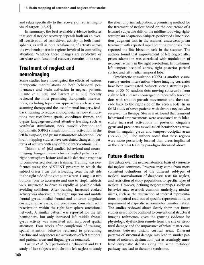
and relate specifically to the recovery of reorienting tovisual targets [45,27].
In summary, the best available evidence indicatesthat spatial neglect recovery depends both on an over-all reactivation of task-driven activity in both hemi-spheres, as well as on a rebalancing of activity acrossthe two hemispheres in regions involved in controllingattention. Whether these changes are predictive orcorrelate with functional recovery remains to be seen.
Treatment of neglect andneuroimagingSome studies have investigated the effects of varioustherapeutic manipulations on both behavioral per-formance and brain activation in neglect patients.Luaute et al. [60] and Barrett et al. [61] recentlyreviewed the most promising therapeutic interven-tions, including top-down approaches such as visualscanning therapy and the use of mental imagery, feed-back training to reduce anosognosia, sensory stimula-tions that recalibrate spatial coordinate frames, andbypass language-mediated attentive learning such asvestibular stimulation, neck muscle vibration andoptokinetic (OPK) stimulation, limb activation in theleft hemispace, and prism visuomotor adaptation. Fewbrain mapping studies have correlated changes in pat-terns of activity with any of these interventions [52].
Thimm et al. [62] studied behavioral and neuro-imaging changes in seven chronic neglect patients withright hemisphere lesions and stable deficits in responseto computerized alertness training. Training was per-formed using the AIXTENT program in which thesubject drives a car that is heading from the left sideto the right side of the computer screen. Using just twobuttons (one to accelerate and one to stop), subjectswere instructed to drive as rapidly as possible whileavoiding collisions. After training, increased evokedactivity was observed in the right superior and middlefrontal gyrus, medial frontal and anterior cingulatecortex, angular gyrus, and precuneus, consistent withreactivation within the right-hemisphere attentionalnetwork. A similar pattern was reported for the lefthemisphere, but only increased left middle frontalgyrus activity was associated with improved spatialattention. Four weeks after completion of training,spatial attention behavior returned to pretrainingbaseline and only increased activations of left temporaland parietal areas and lingual gyrus remained.
Luaute et al. [63] performed a behavioral and PETstudy of five subjects with chronic left neglect to study
the effect of prism adaptation, a promising method forthe treatment of neglect based on the occurrence of aleftward subjective shift of the midline following right-ward prism adaptation. Subjects performed a line bisec-tion judgment task in the scanner, underwent prismtreatment with repeated rapid pointing responses, thenrepeated the line bisection task in the scanner. Theauthors found that improvement of left neglect afterprism adaptation was correlated with modulation ofneuronal activity in the right cerebellum, left thalamus,left temporo-occipital cortex, right posterior parietalcortex, and left medial temporal lobe.
Optokinetic stimulation (OKS) is another visuo-sensory-motor intervention whose imaging correlateshave been investigated. Subjects view a stimulus pat-tern of 30–70 random dots moving coherently fromright to left and are encouraged to repeatedly track thedots with smooth pursuit movements and then sac-cade back to the right side of the screen [64]. In anfMRI study of seven patients with chronic neglect whoreceived this therapy, Sturm et al. found that transientbehavioral improvements were associated with bilat-erally increased activations in posterior cingulategyrus and precuneus as well as left hemisphere activa-tions in angular gyrus and temporo-occipital areas(BA 22) [65]. The authors noted that these regionswere more posteriorly located than areas implicatedin the alertness training paradigm discussed above.
Future directionsThe debate over the neuroanatomical basis of visuospa-tial neglect continues. Progress may come from moreconsistent definitions of the different subtypes ofneglect, normalization of diagnostic tests for neglect,and restriction of study populations to specific types ofneglect. However, defining neglect subtypes solely onbehavior may overlook common underlying mecha-nisms, such as the destruction of internal representa-tions, impaired read-out of specific representations, orimpairment of a specific sensorimotor transformation.The studies reviewed above clearly show that futurestudies must not be confined to conventional structuralimaging techniques, given the growing evidence forphysiologic dysfunction remote from the site of struc-tural damage and the importance of white matter con-nections between distant cortical areas. Differentpatterns of structural damage may lead to similar pat-terns of network dysfunction, just as seemingly unre-lated enzymatic deficits along the same metabolicpathway can lead to the same syndrome.
13: Brain mapping of attention and neglect after stroke
140
To fully chart the course of brain reorganization ofattention systems after injury, well-controlled longitu-dinal studies are needed, as it is likely that differentprocesses take place at the acute, subacute, and chronicstages. However, because neuroimaging is a correla-tional technique, imaging alone cannot answer suchquestions about whether newly recruited regions con-tribute to recovery or are maladaptive and contributeto further deficits. Interventional techniques such astranscranial magnetic stimulation (TMS) may signifi-cantly constrain conclusions drawn from brain map-ping studies, allowing us to distinguish between thesetwo possibilities for specific brain regions.
Clinically, research that maps the effects of differ-ent interventions on the recovery from neglect ishighly important. Unfortunately, rehabilitation sci-ence historically has been plagued with difficulties indeveloping hypothesis-driven interventions, as well asshowing that interventions have effects beyond thoseattributable to spontaneous recovery. Moreover, themajority of the behavioral improvements reported todate have been transient. Neuroimaging of the overlapbetween areas implicated in attention processes andother sensorimotor networks, such as those responsi-ble for visuomotor adaptation or vestibular function,may reveal networks that can be used as a “back door”mechanism for modulating the damaged attentionnetwork (i.e. via prism adaptation, optokinetic stim-ulation, or vestibular stimulation). The interpretationof neuroimaging changes during recovery is compli-cated by significant performance confounds, althoughthis problem can be mitigated using approaches suchas resting-state functional connectivity MRI. It seemsunlikely that any one brain mapping technique used inisolation will provide a conclusive picture of attentionnetwork reorganization after stroke. Rather, the con-vergence of multiple approaches including structuralMRI, fMRI, PET, and TMS is necessary for a morecomplete understanding of the determinants of overallnetwork health and integrity after brain damage.
AcknowledgmentsWe thank Oxford University Press and Dr Bartolomeofor permission to adapt a figure from their studypublished in Cerebral Cortex 2007 17:2479–90.
References1. Posner MI, Cohen Y. Components of attention. In:
Bouman H, Bowhuis D, editors. Attention andperformance X. Hillsdale, NJ: Erlbaum; 1984: pp. 55–66.
2. Koch C, Ullman S. Shifts in visual attention: Towardsthe underlying circuitry. Human Neurobiol.1985;4:219–27.
3. Desimone R, Duncan J. Neural mechanisms of selectivevisual attention. Ann Rev Neurosci. 1995;18:193–222.
4. Jeannerod M, Arbib MA, Rizzolatti G, et al. Graspingobjects: The cortical mechanisms of visuomotortransformation. Trends Neurosci. 1995;18:314–20.
5. Mesulam M-M. A cortical network for directedattention and unilateral neglect. Ann Neurol.1981;10:309–25.
6. Heilman KM, Bowers D, Valenstein E, et al. Hemispaceand hemispatial neglect. In: Jeannerod M, editor.Neurophysiological and neuropsychological aspects ofspatial neglect. Amsterdam: Elsevier; 1987: pp. 115–50.
7. Sapir A, Kaplan JB, He BJ, et al. Anatomical correlatesof directional hypokinesia in patients with hemispatialneglect. J Neurosci. 2007;27:4045–51.
8. Rengachary J, d’Avossa G, Sapir A, et al. Is the Posnerreaction time test more accurate than clinical tests indetecting left neglect in acute and chronic stroke? ArchPhys Med Rehabil. 2009;90:2081–8.
9. Hillis AE, Newhart M, Heidler J, et al. Anatomy ofspatial attention: Insights from perfusion imaging andhemispatial neglect in acute stroke. J Neurosci.2005;25:3161–7.
10. Hillis AE. Neurobiology of unilateral spatial neglect.Neuroscientist. 2006;12:153–63.
11. Robertson IH. Do we need the “lateral” in unilateralneglect? Spatially nonselective attention deficits inunilateral neglect and their implications forrehabilitation. Neuroimage. 2001;14:S85–90.
12. Posner MI, Petersen SE. The attention system of thehuman brain. Ann Rev Neurosci. 1990;13:25–42.
13. Heilman KM, Van Den Abell T. Right hemispheredominance for attention: The mechanism underlyinghemispheric asymmetries of inattention (neglect).Neurology. 1980;30:327–30.
14. Robertson IH, Mattingley JB, Rorden C, et al. Phasicalerting of neglect patients overcomes their spatialdeficit in visual awareness. Nature. 1998;395:169–72.
15. Friedrich FJ, Egly R, Rafal RD, et al. Spatial attentiondeficits in humans: A comparison of superior parietaland temporal–parietal junction lesions.Neuropsychology. 1998;12:193–207.
16. Husain M, Kennard C. Visual neglect associated withfrontal lobe infarction. J Neurol. 1996;243:652–7.
17. Samuelsson H, Jensen C, Ekholm S, et al. Anatomicaland neurological correlates of acute and chronicvisuospatial neglect following right hemisphere stroke.Cortex. 1997;33:271–85.
18. Leibovitch FS, Black SE, Caldwell CB, et al. Brain–behavior correlations in hemispatial neglect using CT
13: Brain mapping of attention and neglect after stroke
141
and SPECT: The Sunnybrook Stroke Study. Neurology.1998;50:901–08.
19. Karnath HO, Fruhmann Berger M, Kuker W, et al. Theanatomy of spatial neglect based on voxelwise statisticalanalysis: A study of 140 patients. Cereb Cortex.2004;14:1164–72.
20. Karnath HO, Himmelbach M, Rorden C. Thesubcortical anatomy of human spatial neglect: Putamen,caudate nucleus and pulvinar. Brain. 2002;125:350–60.
21. Committeri G, Pitzalis S, Galati G, et al. Neural bases ofpersonal and extrapersonal neglect in humans. Brain.2007;130:431–41.
22. Grimsen C, Hildebrandt H, Fahle M. Dissociation ofegocentric and allocentric coding of space in visualsearch after right middle cerebral artery stroke.Neuropsychologia. 2008;46:902–14.
23. Coslett HB, Bowers D, Fitzpatrick E, et al. Directionalhypokinesia and hemispatial inattention in neglect.Brain. 1990;113:475–86.
24. Bisiach E, Geminiani G, Berti A, et al. Perceptual andpremotor factors of unilateral neglect. Neurology.1990;40:1278–81.
25. Tegner R, Levander M. Through a looking glass. A newtechnique to demonstrate directional hypokinesia inunilateral neglect. Brain. 1991;114:1943–51.
26. Doricchi F, Tomaiuolo F. The anatomy of neglectwithout hemianopia: A key role for parietal–frontaldisconnection? Neuroreport. 2003;14:2239–43.
27. He BJ, Snyder AZ, Vincent JL, et al. Breakdown offunctional connectivity in frontoparietal networksunderlies behavioral deficits in spatial neglect. Neuron.2007;53:905–18.
28. Thiebaut de Schotten M, Urbanski M, Duffau H, et al.Direct evidence for a parietal–frontal pathwaysubserving spatial awareness in humans. Science.2005;309:2226–8.
29. Doricchi F, Thiebaut de SchottenM, Tomaiuolo F, et al.White matter (dis)connections and gray matter (dys)functions in visual neglect: Gaining insights into thebrain networks of spatial awareness. Cortex.2008;44:983–95.
30. Bartolomeo P, Thiebaut de SchottenM, Doricchi F. Leftunilateral neglect as a disconnection syndrome. CerebCortex. 2007;17:2479–90.
31. Urbanski M, Thiebaut de Schotten M, Rodrigo S, et al.Brain networks of spatial awareness: Evidence fromdiffusion tensor imaging tractography. J NeurolNeurosurg Psychiatry. 2008;79:598–601.
32. Mesulam MM. Spatial attention and neglect: Parietal,frontal and cingulate contributions to the mentalrepresentation and attentional targeting of salientextrapersonal events. Phil Trans R Soc Lond B Biol Sci.1999;354:1325–46.
33. Corbetta M, Miezin FM, Shulman GL, et al. A PETstudy of visuospatial attention. J Neurosci.1993;13:1202–26.
34. Nobre AC, Sebestyen GN, Gitelman DR, et al.Functional localization of the system for visuospatialattention using positron emission tomography. Brain.1997;120:515–33.
35. Corbetta M, Akbudak E, Conturo TE, et al. A commonnetwork of functional areas for attention and eyemovements. Neuron. 1998;21:761–73.
36. Luna B, Thulborn KR, Strojwas MH, et al. Dorsalcortical regions subserving visually-guided saccades inhumans: An fMRI study. Cerebral Cortex. 1998;8:40–7.
37. Sereno MI, Pitzalis S, Martinez A. Mapping ofcontralateral space in retinotopic coordinates by aparietal cortical area in humans. Science.2001;294:1350–4.
38. Silver MA, Ress D, Heeger DJ. Topographic maps ofvisual spatial attention in human parietal cortex.J Neurophysiol. 2005;94:1358–71.
39. Jack AI, Patel GH, Astafiev SV, et al. Changing humanvisual field organization from early visual to extra-occipital cortex. PLoS ONE. 2007;2:e452.
40. Galati G, Lobel E, Vallar G, et al. The neural basis ofegocentric and allocentric coding of space in humans:A functional magnetic resonance study. Exp Brain Res.2000;133:156–64.
41. Ruff CC, Blankenburg F, Bjoertomt O, et al.Concurrent TMS-fMRI and psychophysics revealfrontal influences on human retinotopic visual cortex.Curr Biol. 2006;16:1479–88.
42. Bressler SL, Tang W, Sylvester CM, et al. Top-downcontrol of human visual cortex by frontal and parietalcortex in anticipatory visual spatial attention.J Neurosci. 2008;28:10 056–61.
43. Corbetta M, Shulman GL. Control of goal-directed andstimulus-driven attention in the brain. Nat RevNeurosci. 2002;3:201–15.
44. Corbetta M, Patel G, Shulman GL. The reorientingsystem of the human brain: From environment totheory of mind. Neuron. 2008;58:306–24.
45. Corbetta M, Kincade MJ, Lewis C, et al. Neural basisand recovery of spatial attention deficits in spatialneglect. Nat Neurosci. 2005;8:1603–10.
46. Hornak J. Ocular exploration in the dark by patientswith visual neglect. Neuropsychologia. 1992;30:547–52.
47. Karnath HO. Optokinetic stimulation influences thedisturbed perception of body orientation in spatialneglect. J Neurol Neurosurg Psychiatry. 1996;60:217–20.
48. Biswal B, Yetkin F, Haughton V, et al. Functionalconnectivity in themotor cortex of resting human brainusing echo-planar MRI. Magn Res Med.1995;34:537–41.
13: Brain mapping of attention and neglect after stroke
142

49. Carter AR, Astafiev SV, Lang CE, et al. Restinginterhemispheric functional magnetic resonanceimaging connectivity predicts performance after stroke.Ann Neurol. 2010;67:365–75.
50. Hier DB, Mondlock J, Caplan LR. Recovery ofbehavioral abnormalities after right hemisphere stroke.Neurology. 1983;33:345–50.
51. Stone SP, Patel P, Greenwood RJ, et al. Measuring visualneglect in acute stroke and predicting its recovery: Thevisual neglect recovery index. J Neurol NeurosurgPsychiatry. 1992;55:431–6.
52. Arene NU, Hillis AE. Rehabilitation of unilateral spatialneglect and neuroimaging. Eura Medicophys.2007;43:255–69.
53. Buxbaum LJ, Ferraro MK, Veramonti T, et al.Hemispatial neglect: Subtypes, neuroanatomy, anddisability. Neurology. 2004;62:749–56.
54. Deuel RK. Neural dysfunction during hemineglect aftercortical damage in two monkey models. In: JeannerodM, editor. Neurophysiological and neuropsychologicalaspects of spatial neglect. Amsterdam: Elsevier; 1987:pp. 315–34.
55. Perani D, Vallar G, Paulesu E, et al. Left and righthemisphere contribution to recovery from neglect afterright hemisphere damage – An [18F]FDG study of twocases. Neuropsychologia. 1993;31:115–25.
56. Vallar G, Perani D, Cappa SF, et al. Recovery fromaphasia and neglect after subcortical stroke:Neuropsychological and cerebral perfusion study.J Neurol Neurosurg Psychiatry. 1988;51:1269–76.
57. Hillis AE. Rehabilitation of unilateral spatial neglect:New insights from magnetic resonance perfusion
imaging. Arch Phys Med Rehabil. 2006;87(12 Suppl 2):S43–9.
58. Pizzamiglio L, Perani D, Cappa SF, et al. Recovery ofneglect after right hemispheric damage: H2(15)Opositron emission tomographic activation study. ArchNeurol. 1998;55:561–8.
59. Thimm M, Fink GR, Kust J, et al. Recovery fromhemineglect: Differential neurobiological effects ofoptokinetic stimulation and alertness training. Cortex.2009;45:850–62.
60. Luaute J, Halligan P, Rode G, et al. Visuo-spatialneglect: A systematic review of current interventionsand their effectiveness. Neurosci Biobehav Rev.2006;30:961–82.
61. Barrett AM, Buxbaum LJ, Coslett HB, et al.Cognitive rehabilitation interventions for neglectand related disorders: Moving from bench tobedside in stroke patients. J Cogn Neurosci.2006;18:1223–36.
62. Thimm M, Fink GR, Kust J, et al. Impact of alertnesstraining on spatial neglect: A behavioural and fMRIstudy. Neuropsychologia. 2006;44:1230–46.
63. Luaute J, Michel C, Rode G, et al. Functional anatomyof the therapeutic effects of prism adaptation on leftneglect. Neurology. 2006;66:1859–67.
64. Kerkhoff G, Keller I, Ritter V, et al. Repetitiveoptokinetic stimulation induces lasting recoveryfrom visual neglect. Restor Neurol Neurosci.2006;24:357–69.
65. SturmW, ThimmM, Kust J, et al. Alertness-training inneglect: Behavioral and imaging results. Restor NeurolNeurosci. 2006;24:371–84.
13: Brain mapping of attention and neglect after stroke
143

14 Depression and its effects after strokeThomas Platz
DescriptionEmotional symptoms are probably among the mostignored impairments in stroke patients. Depressivesymptoms, anxiety, anger and inadequate anger con-trol, fatigue, alexithymia or unawareness of emotions,emotionalism, and pathological crying or laughter aresigns and symptoms that can frequently be encounteredin stroke victims. Reported changes in personality afterstroke include reduced patience and increased frustra-tion, reduced confidence, more dissatisfaction, and aless easy-going nature. The descriptive picture wouldnot be complete if one did not consider the impact ofthe disease, stroke, on family caregivers of strokepatients. Anxiety, depression, and poor physical healthare common sequelae among family caregivers ofstroke survivors. Emotional symptoms impact manyaspects of stroke recovery in humans, as describedbelow, and so represent a dimension of post-stroke lifethat is incompletely captured in animal models.
While this chapter will primarily focus on depres-sion and its effects after stroke, the broader perspectiveof post-stroke emotional disorders is kept in mind;comments on other emotional disorders will amendand contribute to the picture of cause and effects ofdepression after stroke.
DiagnosisOne important challenge in neuropsychiatry is how todiagnose depression in patients with acute brain lesions,since there may be an overlap between symptoms ofdepression and signs associated with the neurologicaldisease. The diagnosis of a depressive syndrome shouldbe made using standardized diagnostic criteria formood disorders due to neurological disease such as inthe DSM-IV or the ICD-10. The best approach is toassess the presence of depressive symptoms usingsemi-structured or structured psychiatric interviews
such as the Present State Exam, the Structured ClinicalInterview for DSM-IV (SCID), or the Schedules forClinical Assessment in Neuropsychiatry.
Most studies in acute and chronic neurologicaldisorders demonstrated the specificity of both auto-nomic and psychological symptoms for the syndromeof depression.
Symptom profiles and thereby the validity of clin-ically rated DSM-IV depressive symptoms were inves-tigated in stroke patients suffering from (1) majordepressive disorder, (2) minor depressive disorder,and (3) those free of any neuropsychiatric disorders[1]. First-ever stroke patients (N=200) were approachedwithin 3 months of the acute stroke and wereinterviewed with the SCID-P and administered theHamilton Depression Rating Scale (HDRS) andthe Beck Depression Inventory (BDI) among others.The only symptom that did not differ among patientgroups (1–3) was Feelings of Guilt; all the other eightDSM-IV symptoms were significantly different. In par-ticular, the frequency of Depressed Mood, DiminishedInterest or Pleasure, Fatigue or Loss of Energy,Insomnia, and Psychomotor Agitation/Retardationwas higher in minor depressive disorder patients thanin those free of any neuropsychiatric disorders.
Depression rating scales may be used to rate theseverity of depression and to monitor its changes overtime, e.g. with antidepressant treatment. Several diag-nostic instruments are in use to identify emotionalsymptoms in patients who suffer from stroke. Of these,clinician-administered are the Hamilton DepressionRating Scale (HDRS) and the Montgomery–AsbergDepression Rating Scale (MADRS); self-rating scalesare the Beck Depression Inventory (BDI), theCenter for Epidemiological Studies – Depression Scale(CES-D), the Zung Self-Rating Depression Scale, theGeriatric Depression Scale (GDS), and the HospitalAnxiety and Depression Scale (HADS).
Brain Repair After Stroke, ed. S. C. Cramer and R. J. Nudo. Published by Cambridge University Press.© Cambridge University Press 2010.
145
Aphasia and other cognitive impairments maysometimes make these instruments difficult to use.Nevertheless, depression diagnosis (DSM-IV) andseverity rating (MADRS) can reliably be made in theacute phase in at least two-thirds of aphasic patients, andfeasibility increases over time [2]. Otherwise, the StrokeAphasic Depression Questionnaire (SADQ) (completedby a spouse or a caregiver; also validated for aphasicpatients in the hospital, completed by the nurse) [3] orthe Aphasic Depression Rating Scale (ADRS) (validatedfor the evaluation of depression by the rehabilitationteam) [4] can be recommended for screening purposes.
Frequency and evolution
Incidence and prevalence of post-strokedepressionDepression occurs in 20–40% of stroke patients. Otheremotional symptoms commonly described includeanxiety (20–30%), emotional instability (10–25%), cri-sis reaction (20%), and reduced initiative and fatigue(50–70%) [5].
An Australian cohort study assessed the frequencyand correlates of depression at 3 and 15 months afterstroke. A total of 164 consecutive eligible strokepatients and 100 comparison subjects received exten-sive medical, psychiatric, and neuropsychologicalassessments. Comprehensive assessments includedratings for DSM-IV major or minor depression at3–6 months (index assessment) and 15 months(follow-up assessment) after stroke. Major or minordepression was present in 12.0% of stroke patients atindex assessment and in 20.7% at follow-up, whichincluded 18 new cases (13.4%) [6].
The Italian multicenter observational DESTROstudy included from a total of 53 centers 1064 consec-utively admitted patients with ischemic or hemorrha-gic stroke who were then periodically assessed in thefirst 9 months after the event. Patients with depressionwere followed for two years [7]. Post-stroke depressionwas detected in 383 patients (36%), most of whom hadminor depression (80.17%), with dysthymia, ratherthan major depression and adaptation disorder.About 80% of these developed their depression withinthree months of the stroke. Cases with later onsettended to have less severe symptoms.
A Swedish study investigated the risk of depressionin elderly patients one and a half years after stroke, andcompared it to the risk in a population-based controlsample [8]. One hundred and forty-nine elderly stroke
survivors and 745 age- and sex-matched controls fromthe general population were examined with semi-structured psychiatric examinations and cognitiveassessments. Diagnoses were made according toDSM-III-R. The frequency of depression was 34% instroke patients and 13% in population controls (oddsratio, 3.4; 95% CI, 2.3–5.0). The risk of depression wasincreased in both men and women and in all agegroups, but was not related to the predominant sideof stroke symptoms.
When patients with post-stroke depression wereassessed 1 month and 6 months after stroke, commu-nication impairment was found to be a strong predic-tor of depression severity and prognosis [9].
In a cohort study with 145 patients, self-ratedhealth was rated as very good or rather good by 62%at 3months after stroke, and by 78% at 12months afterstroke. Nevertheless, more than half of the patientssuffered from symptoms of depression, with no sig-nificant improvement at 12 months. The most com-mon general symptoms at 3 months after stroke werefatigue, sadness, pain in the legs, dizziness, and irrita-bility. Fatigue and sadness were still common at12 months [10].
Frequency of other post-stroke emotionalsymptomsAnxiety symptoms had been found to be more fre-quent than depressive symptoms in the acute stage ofischemic stroke using the Hospital Anxiety andDepression Scale (HADS) between days 3 and 7 afteradmission to the stroke unit: 26.4% of 178 patientssuffered from anxiety symptoms, 14.0% from depres-sive symptoms, and 7.9% from both during this earlystage [11]. Similarly, in another study both patientsand caregivers were found to be more anxious thandepressed at one year post-stroke [12]. Anxiety symp-toms were associated with single marital state and alow Mini Mental State Examination (MMSE) score,whereas depressive symptoms were related to a lowBarthel Activities of Daily Living index (BI). In afurther cohort study, anxiety remained stable over 3years post-stroke and was best predicted by prior,early, anxiety, and female gender [13].
The incidence of anger had prospectively beenscreened in 202 consecutive acute stroke patients(< or = 4 days) using 8 items from 3 psychiatric scales(Catastrophic Reaction Scale, Mania Rating Scale, andComprehensive Psychopathological Rating Scale)[14]. Anger was considered present if the patient
14: Depression and its effects after stroke
146
scored in at least one item. Anger was detected in 71(35%) patients, and 26 of these were severely angry(> or = 4 points). There was no association betweenanger and the considered variables. Analysis of theitems extracted two factors: (i) the emotional-cognitive and (ii) the behavioural components ofanger. Using the 10-item Spielberger Trait AngerScale, another study interviewed 145 patients withstroke regarding inability to control anger or aggres-sion (ICAA) [15]. ICAA was present in 47 patients(32%) and was closely related to motor dysfunction,dysarthria, emotional incontinence, and lesions affect-ing frontal–lenticulocapsular–pontine base areas.
Post-stroke fatigue is distinct from depression inmany instances. It is especially disabling and frustrat-ing in that it typically involves patients with total ornear-total neurological recovery, who should havebeen able to go back to their previous activities butwho become severely disabled because of early andpersisting exhaustion [16]. A related symptom com-plex revolves around unawareness. Unawareness mayhave different forms, such as anosognosia, neglect, andalexithymia or unawareness of emotions. Despite thestrong comorbidity rate among the different forms ofunawareness, there are patients who suffer from pureforms of these types of lack of awareness [17].
Post-stroke personality changes as reported by thepatients’ main carer at 9 months after the stroke werereduced patience and increased frustration, reducedconfidence, more dissatisfaction, and a less easy-going nature [18].
Cause and effectThe interaction between depression and stroke is com-plex and the pathophysiological mechanisms have notas yet been fully elucidated, although an interactionbetween genetic, biochemical, anatomical, and espe-cially psychosocial factors may be important in post-stroke depression development.
Epidemiological reasoningDepression as a risk factor for strokePresence of depressive symptoms has been shown tobe a strong risk factor for stroke in men [19,20];greater depressive symptoms are associated with anincrease in the risk of all-cause and, more specifically,cerebrovascular disease mortality in men [21].
A prospective cohort study was conducted on 4120Framingham Heart Study participants aged 29–100
years with up to 8 years of follow-up and sought toexamine whether depressive symptoms are associatedwith an increased risk of cerebrovascular events in acommunity-based sample [22]. In participants < 65years, the risk of developing stroke/transient ischemicattack (TIA) was 4.21 times greater in those withsymptoms of depression. After adjusting for compo-nents of the Framingham Stroke Risk Profile (hazardratio = 3.43, 95% CI = 1.60–7.36) and education (haz-ard ratio = 4.89, 95% CI = 2.19–10.95), similar resultswere obtained. In subjects aged 65 and older, depres-sive symptoms were not associated with an increasedrisk of stroke/TIA. Taking antidepressant medicationsdid not alter the risk associated with depressive symp-toms. Depressive symptoms were thus an independentrisk factor for incident stroke/TIA in individuals < 65years.
However, in very elderly persons residing in acontinuing-care retirement community (N = 181),controlling for demographic factors, both depressionand the number of cardiovascular risk factors(CVRFs) at baseline were strongly predictive of stroke[23]. Depression accounted for 12% of the variance instroke incidence, beyond the contribution of CVRFs.
Thus, pre-existing depression increases the risk forstroke.
Causes of post-stroke depression (and fatigue)As mentioned above, in the Italian multicenter obser-vational DESTRO study, a total of 53 centers consec-utively admitted 1064 patients with ischemic orhemorrhagic stroke and assessed them periodically inthe first 9 months after the event [7]. Risk factors forpost-stroke depression were a history of depression,severe disability, previous stroke, and female gender,but not the type or site of the vascular lesion.
The relevance of established risk factors for depres-sion in the community (such as female gender, priorpersonal history of depression, positive family historyof depression, and somatic comorbidity other thanstroke) and five potential disease-related risk factors(disability, cognitive deterioration, inter- and intra-hemispheric lesion location, and generalized vasculardamage on computed tomography (CT) scan) wereassessed in a multivariate prediction model for depres-sion in the first year after stroke. The four general riskfactors for depression in the community constituted avalid model to predict depression in stroke patients. Ofthe disease-specific factors, only incorporation of “dis-ability” in this model improved its significance [24]. In
14: Depression and its effects after stroke
147
the subacute to chronic phase, the causal contributionof stroke-specific factors may therefore be less thanmight be assumed.
Significantly higher correlations between self-esteem and mood ratings were noted in stroke patientsas compared to healthy controls. Thus, lower self-esteem ratings do not appear to be a byproduct ofdepressive mood. Self-esteem is negatively impactedby stroke and is strongly, but independently, associ-ated with depressive mood [25].
Genetic aspects seem to play a role in modifyingthe association between incident stroke and depres-sion, as has been shown for the brain-derived neuro-trophic factor (BDNF) gene val66met polymorphism.Five hundred community residents aged > 65 yearswithout stroke or depression at baseline were re-evaluated after 2 years [26]. The association betweenincident stroke and depression was strengthened pro-gressively with increasing numbers of met alleles, andwas only significant in subjects with the met/met geno-type after adjustment for disability and cognitivefunction.
In conclusion, genetic aspects, post-stroke disabil-ity, self-esteem, and certain established risk factors fordepression in the community such as female genderand prior personal history of depression each increasethe risk for post-stroke depression.
What increases the risk for post-stroke fatigue? Across-sectional study with 220 consecutive outpatientsat an average of 15 months after the onset of strokeassessed the presence of pre- and post-stroke fatigue,post-stroke depression, as well as various other factors[27]. One hundred and twenty-five patients (57%) hadpost-stroke fatigue, 83 (38%) had pre-stroke fatigue,and 53 (24%) had post-stroke depression. The impactof post-stroke fatigue on patients’ daily activities wasmore severe in the physical domain as compared withthe psychological or cognitive domains. Multivariateanalyses showed that the presence of pre-stroke fatigue(OR 33.5), a high modified Rankin scale (OR 3.3), andpost-stroke depression (OR 2.7) were independentlyassociated with post-stroke fatigue. Thus, pre-strokefatigue was the most important factor related to post-stroke fatigue in this study.
Associations with post-stroke depressionEarly after stroke (within 3 weeks after a first-eversymptomatic stroke), moderate or severe symptomsof depression had been shown to be associated with aspecific pattern of cognitive impairment (memory,
visual perception, and language), lesion size (but notlesion location), and functional status (Barthel Index,Rankin scale) [28].
A cohort study examined prevalence of depressionand anxiety as well as the relationships of age, gender,hemisphere of lesion, functional independence, andcognitive functioning (i.e. memory, attention/impul-sivity, cognitive speed) to depression and anxiety at 3months post-stroke in 73 individuals [29]. Prevalenceof moderate to severe depression and anxiety in thesample were high (22.8 and 21.1%, respectively), withcomorbidity in 12.3% of cases. In the regression analy-sis, 74.6% of the variance in depression was explained,with significant relationships between increaseddepression and younger age, reduced cognitive speed,poorer verbal memory, left hemisphere lesion, andincreased impact of interference (Stroop ratio).Having a left-sided hemispheric lesion also contrib-uted to the statistical prediction of anxiety, as didcognitive speed, explaining 50.7% of the variance.While age and side of hemispheric lesion contributed,cognitive performance explained the greatest propor-tion of variance in both depression and anxiety (51.3and 38.5%, respectively).
These findings suggest that in the subacute phaseof stroke cognition and mood are linked over andabove physical independence.
Causes for the evolution of depression after strokeIn the Swedish national quality assessment register(Riks-Stroke), 15 747 stroke survivors were recorded.They were asked about depressivemood and antidepres-sant treatment 3 months after stroke. At 3 months afterstroke, 12.4% of male and 16.4% of female stroke survi-vors reported that they always or often felt depressed. Ina multiple logistic regression model, female gender, ageyounger than 65 years, living alone, having had a recur-rent stroke, being dependent on others, and institutionalliving 3 months after stroke were independent predic-tors of self-reported depression [30].
The strength of religious beliefs has also beenshown to influence the ability to cope after a strokeevent, with stronger religious beliefs acting as a possi-ble protective factor against emotional distress [31].
The aim of another cohort study of stroke patientsadmitted for rehabilitation was to identify factors thatare significantly related to depression in chronic strokepatients, years after stroke onset [32]. A total of 165first-ever stroke patients over 18 years of age wereassessed at 1 and 3 years post-stroke. At 3 years
14: Depression and its effects after stroke
148
post-stroke, 19% of the patients were depressed.Multivariate logistic regression analysis showed thatdepression at 3 years post-stroke was predicted by1-year instrumental activities of daily living (ADL)and fatigue. Sensitivity of the model was 63%, whilespecificity was 85%.
A further cohort study assessed the correlates ofdepression at 3 and 15months after stroke with a focuson dementia and vascular brain pathology. In a total of164 consecutive eligible stroke patients, depressionwas not associated with age, intellectual decline priorto stroke, or side or severity of stroke [6]. Patients whoexperienced a TIA or stroke during the follow-up, whohad developed dementia by 3 months, or who were notliving with a relative or partner were more likely to bedepressed at follow-up. Dementia at 3 months pre-dicted depression, but the reverse did not hold.Depression therefore seemed related to cumulativevascular brain pathology rather than side and severityof single strokes. Fatigue, development of dementia,being dependent on others, living alone, institutionalliving, as well as recurrent stroke (or TIA) had anegative influence on the evolution of depressionafter stroke.
Effects of post-stroke depressionIn a 6-month prospective cohort study of 141 post-acute stroke patients, demographic and clinical dataon admission, and neurological, cognitive, depressivesymptoms, and functional variables on admission andat 6 months after stroke were measured. Multivariateanalysis indicated that greater magnitudes of func-tional recovery (Barthel Index change score) wereachieved by patients with better baseline depressivesymptoms and improvement over time. Functionalrecovery was not related to baseline cognitive statusor its improvement, however [33]. These data sug-gested that depressive symptoms early after strokeare inversely related to functional recovery that againis improved when depressive symptoms improve, sug-gesting an independent causal role for depression inlimiting functional recovery.
Previous mood disorder, an independent predictorof post-stroke depression [34], also reduces the odds offavorable outcome: among stroke patients who sur-vived hospitalization, those with pre-existing depres-sion had significantly higher odds of being dischargedto an institution instead of their home thandid patients without any pre-existing mental healthcondition [35].
Neuroscience reasoningNeuroimaging evidenceTo investigate age-related white matter changes onmagnetic resonance imaging (MRI) as an independentpredictor of depressive symptoms at 1 year, after con-trolling for known confounders, a pan-European mul-ticenter study followed 639 older adults withoutsignificant disability and assessed MRI white matterchanges as well as demographic and clinical variablesthat included cognitive scores, quality of life, disability,and depressive symptoms. Clinical assessments wererepeated at 1 year. Using logistic regression analysis,severity of white matter changes at baseline was shownto independently and significantly predict depressivesymptoms at 1 year after controlling for baselinedepressive symptoms, quality of life, and worseningdisability [36]. Thus, white matter changes (independ-ent of stroke) pre-dated and were related to the devel-opment of depressive symptoms, suggesting thepossibility of an organic cause.
Some studies have suggested that post-stroke depres-sion and anxiety are specifically related to organiclesions in the anterior parts of the left hemisphere.
To test the independent effects of lesion location(left hemisphere, anterior region) and of co-occurringgeneralized vascular damage on the development ofdepression in the first year after ischemic stroke, whileother risk factors for depression were controlled for,190 patients with a first-ever, supratentorial infarctwere followed up for 1 year [37]. CT was performedin the acute phase of stroke, while in 75 patients anadditional MRI scan was also available. Negativeresults appeared from one overall, multivariate analy-sis including variables of both focal and generalizedvascular brain damage, as well as other non-cerebralrisk factors. In addition, level of handicap and neuroti-cism were independent predictors of depression in thiscohort. A biopsychosocial model including both pre-morbid (prior to stroke) vulnerability factors, such asneuroticism and (family) history of depression, as wellas post-stroke stressors, such as level of handicap,could be substantiated as risk factors in this predictivemodel while lesion location (left hemisphere, anteriorregion) or co-occurring generalized vascular damagecould not.
However, other studies support a possible contribu-tion of lesion site to depression onset. When 70 patientswith a single brain infarct on MRI were studied3 months after ischemic stroke by a standardized
14: Depression and its effects after stroke
149
protocol that detailed side, site, type, and extent of thebrain infarct, as well as severity of white matter lesionsand brain atrophy, it could be shown that affectedstructures of the frontal–subcortical circuits, i.e. thepallidum and caudate, especially on the left side, pre-disposed stroke patients to depression (diagnosed byDSM-III-R and DSM-IV criteria) [38]. Note too thatthe size of the infarcts at these sites was larger amongdepressed patients. Using a logistic regression analysis,the authors found that a brain infarct that affected thepallidum was a strong independent MRI predictor forpost-stroke depression (odds ratio = 7.2).
Further, a meta-analysis of the correlation betweenseverity of depression following stroke and proximityof the lesion to the frontal pole showed that there was asignificant inverse correlation between severity ofdepression and distance of the lesion from the frontalpole among 163 patients with left hemisphere stroke,but not among 106 patients with right hemispherestroke [39].
Any role of specific sites of brain lesion in thedevelopment of post-stroke depression might be fur-ther modified by the specific constellation of the indi-vidual neuropsychiatric post-stroke disorder.
In a study with 243 stroke patients the severity ofaffective depression (Zung Self-rating Depression Scalescores) was associated with left frontal lobe (but notbasal ganglia) damage, while that of apathetic depres-sion (Apathy Scale scores) was related to damage to thebilateral basal ganglia (but not to the frontal lobe) [40].
In a separate study, stroke patients who had bothpost-stroke depression and an executive dysfunction,i.e. a depression–dysexecutive syndrome, were studied.Examination included neurological, psychiatric, andneuropsychological examination carried out 3 monthsafter ischemic stroke, and included an MRI to evaluatebrain infarcts, white-matter changes, and brain atrophy.These 21 patients with depression–dysexecutive syn-drome had significantly more brain infarcts affectingtheir frontal–subcortical circuit structures than the 137patients without depression–dysexecutive syndrome, orthe 41 patients with depression but without executivedysfunction [41]. Patients with depression–dysexecutivesyndrome also had more severe depressive symptomsand worse psychosocial functioning, and they coped lesswell in complex activities of daily living.
In addition, right hemisphere damage can influencethe presentation of depressive disorders ensuing afterstroke by disrupting emotion processing mechanisms.Compared to dysphoric depression, nondysphoric
depression (i.e. depressive ideation without endorse-ment of sad emotions) in acute stroke patients showedmore frequent right anterior hemisphere lesions. Thelocation of damage suggested that nondysphoricdepression may be a special presentation of depressivedisorder following stroke in which right hemispheredamage limits the apprehension of personal emotionalchanges [42].
The frontal midline structures have been demon-strated by functional neuroimaging to be involved inthe affective control of human behavior. A case studydocumented a patient with a right anterior cingulateinfarct who presented with an alexithymia-likedisorder [43].
Inability to control anger or aggression (ICAA)had been shown to be associated with lesions affectingfrontal–lenticulocapsular–pontine base areas [15].
Thus, there might be reason to assume that studiesthat failed to demonstrate an association betweenlesion side and site and post-stroke depression mighthave reached their conclusions in some instancesbecause the anatomical correlates of post-strokedepression differ depending on the specific presenta-tion of post-stroke depression (e.g. affective, nondys-phoric, or apathetic). As a notion of caution it must,however, be stated that the above-mentioned evidenceassessed only a subset of aspects and that this hypoth-esis requires validation in a pooled multivariateanalysis.
The serotonergic systemThere is some evidence that points to a role of theserotonergic system in post-stroke depression.
Variations of serotonin transporter-linked pro-moter region (5-HTTLPR) functional polymorphismwere assessed in 26 stroke patients with major depres-sion and in 25 unrelated nondepressed stroke subjects.The findings indicated a significant associationbetween 5-HTTLPR short variant genotype and post-stroke major depression [44].
The intensity dependence of the auditory-evokedpotentials (IDAP) is inversely related to serotonergictone. The linear amplitude/stimulus intensity function(ASF) slope, by measuring the peak-to-peak amplitudeof Nl-P2, was found to be markedly increased in strokepatients compared with controls; while stroke patientswith depressive symptoms had a significantly steeperASF slope than controls, there was no statistical differ-ence in ASF slope between stroke patients withoutdepressive symptoms and controls [45]. Together,
14: Depression and its effects after stroke
150
these findings suggest that the serotonergic systemmight be involved in post-stroke depression.
For both post-stroke depression and pathologicalcrying, an imbalance of serotonergic neurotransmissionhas been postulated. To test this hypothesis, patientswith acute stroke and pathological crying underwentPET scanning, with results compared to age-matchedhealthy control subjects [46]. Maps of 5-HT(1A) recep-tor availability were generated from the images of eightcortical regions and raphe nuclei. The maps showedhighest binding in limbic areas and raphe nuclei,while binding in basal ganglia and cerebellum was neg-ligible. Baseline binding potentials of patients werelower than those of control subjects. Treatment withselective serotonin reuptake inhibitor (SSRI) markedlyreduced extent of free receptor sites, whereas placeboadministration led to a global increase. The studyresults thus described changes in serotonergic neuro-transmission in the early phase of stroke and theirmodulation with SSRI treatment, and suggested apossible relationship to pathological crying.
Metabolic changesMetabolic changes might also be associated with post-stroke depression and might be modified by time, i.e.might differ in depression early and late after stroke asa hint to different involved mechanisms. In groups offirst-ever stroke patients, the relationship betweenpost-stroke depression and the metabolic/biochemicalmilieu, as assessed by proton magnetic resonancespectroscopy (1)H-MRS measurements in unaffectedfrontal lobes, was assessed. Single-voxel proton mag-netic resonance spectroscopy ((1)H-MRS) was per-formed in these lobes to assess N-acetylaspartate/creatine (NAA)/Cr, glutamate+glutamine (Glx)/Cr,choline (Cho)/Cr and myo-inositol (mI)/Cr ratios.Twenty-six patients with a first ischemic stroke locatedoutside the frontal lobes were studied [47]. Patientswere assessed within the first 10 days after stroke andagain 4 months later. In the group of 26 patients, 8(31%) met criteria for depression at the first assess-ment, and 9 (35%) met criteria for depression atfollow-up. Patients with depression in the immediatepost-stroke phase had significantly higher Glx/Crratios in the contralesional hemisphere than non-depressive patients. No biochemical differences werefound between the groups at 4-month follow-up.These findings suggested that post-stroke depressionis accompanied by changes in frontal lobe glutamate/glutamine levels, perhaps reflecting abnormalities in
glutamatergic transmission in the immediate post-stroke period. Further, 13 out of a sample of 31 strokepatients (42%) demonstrated apathy. Of these subjects,significantly lowered NAA/Cr ratios were found in theright hemispheres of apathetic patients in the sub-group with left-sided brain lesions. These findingspoint to the association between apathy and frontallobe integrity, suggest different reactions of the hemi-spheres, and indicate that changes in the NAA/Cr ratiocould be related to the apathy [48]. Whether thesemetabolic changes are a contributor to, or a merereflection of, these emotional states requires furtherstudy.
Implications for recoveryPost-stroke depression has been shown to impede therehabilitation progress following stroke and to be asso-ciated with impaired functional outcome, cognitivedecline, and increased mortality [49]. Similarly,depression has been linked to increased risk of strokeoccurrence.
Post-stroke depression and its impact onactivities of daily living functionsA cohort study examined the relationship betweendepressive symptoms and time courses in achievingindependence in basic activities of daily living (bADL)and instrumental activities of daily living (iADL) [50].At baseline, 1, 3, and 6 months after stroke, 459 strokepatients were prospectively assessed with the GeriatricDepression Scale (GDS) to determine depressive sta-tus, outcomes were achieved independence in bADL(Barthel Index scores > 95) and independence in atleast three iADL during follow-up. Depressed patientswere 0.3 times less likely than nondepressed patients toachieve bADL score of > 95 and 0.4-times less likely tobe independent in three or more iADL. The cumula-tive percentages to achieve a bADL score of > 95 areshown in Figure 14.1. Similarly, the cumulative per-centages for nondepressed patients to achieve com-plete independence in three or more iADL at 1,3, and 6 months after stroke were 56%, 72%, and85%, and for the depressed patients, they were 32%,47%, and 72%, respectively. Thus, depressed patientshad significantly poorer functional recovery patternsand took longer to achieve independence with activ-ities of daily living.
Another cohort study investigated the associationbetween stroke and depression, the co-occurrence of
14: Depression and its effects after stroke
151

stroke and depression, and functional health outcomesamong adults in the US population using a nationallyrepresentative sample of adults aged 25–74. Multiplelogistic regression analyses were used to determine theassociation between stroke (past 12-month preva-lence), depression (past 12-month prevalence), andfunctional health outcomes (past 12 months), and todetermine whether there is an interaction betweendepression and stroke in predicting impairment infunctioning. Almost one-third (29.2%) of adults withstroke in the past year also had depression in the pastyear. The co-occurrence of stroke and depression wasassociated with significantly greater limitations inwalking and climbing stairs and poorer general phys-ical functioning as compared to either stroke ordepression alone [51].
Similarly, a further cohort study assessed the fre-quency and correlates of depression at 3 and 15months after stroke in a total of 164 consecutive eligi-ble stroke patients [52]. Major or minor depressionwas present in 20.7% at follow-up. By follow-up,stroke patients with depression had significantlygreater impairment of functional ability and globalcognition than nondepressed stroke patients or com-parison subjects.
While post-stroke depression seems to have a neg-ative impact on ADL activities and quality of life afterstroke, there is some evidence to suggest that motorrecovery (sensu stricto), i.e. regarding selective inner-vation and active movement capacity as assessed withthe Fugl-Meyer scale, is not affected by post-strokedepression [53].
The picture that emerges from these studies is thatpost-stroke depression might not impede motorrecovery per se in the more restricted sense of regain-ing selective innervation when paresis followed stroke.However, it seems to affect the ability to use elemen-tary motor behaviors to perform ADL negatively, andto have the effect of retarding the re-acquisition ofthese functional skills. These effects are not contra-dictory, since the ability to perform activities of dailyliving is more complex, i.e. determined by both per-ceptive and motor recovery, as well as the ability tocompensate for any remaining neuro-impairments.
Post-stroke depression and its impact onparticipation and quality of lifeThe determinants of “restricted participation”, i.e. thedegree of inability to perform social roles, had alsobeen investigated among long-term stroke survivors[54]. Self-competence with basic activities of dailyliving (Functional Independence Measure, FIM) andemotional status (HADS) are the independent deter-minants of restricted participation for the LondonHandicap Scale (LHS) domains most related to bodyfunction (mobility, physical independence, occupa-tion). Depression was the determinant factor for ori-entation and social integration. Thus, both functionaldisability andmood disorders may independently con-tribute to the restricted participation of post-strokepatients.
In another population-based investigation of 266patients with incident stroke, among those who werealive at 2 years (N = 226; 85%), handicap was alsoassessed with the LHS [55]. Disability, physical impair-ment, depression, anxiety, living arrangements, andrecurrent stroke at 2 years were also documented; ifnecessary, proxy assessments were obtained, except formood. The independent determinants of handicap(LHS) were age and 2-year physical impairment anddisability. In the analysis restricted to nonproxy data,depression and anxiety were also independently asso-ciated with handicap.
Further, fewer depressive symptoms after stroke –together with walking and acceptance of the stroke –have been documented as the best predictors forparticipation at 3 months in older adults who had astroke [56]. Similarly, depression was also most sig-nificantly and independently associated with post-stroke handicap at 12 months post-stroke amongelderly stroke victims [57].
19
34
5247
63
72
0.0
10.0
20.0
30.0
40.0
50.0
60.0
70.0
80.0
90.0
100.0
1 month 3 months 6 months
Ind
epen
den
ce w
ith
bA
DL
(cu
mu
lati
ve p
erce
nta
ge)
Depressed
Non-depressed
Figure 14.1 Independence with basic ADLs.The cumulative percentage of non-depressed and depressed
stroke patients who are independent with basic activities of dailyliving (bADL) indicates that stroke patients with depressivesymptoms progressed significantly more slowly in achievingindependence during the first 6 months after stroke [50].
14: Depression and its effects after stroke
152

In a group of stroke patients with a first unilateral,middle cerebral artery (MCA) territory stroke, no sig-nificant neurological improvement could be docu-mented between 3 and 12 months after the stroke[58]. The patients as a group were depressed andremained so over the period of the study. The resultingquality of life scores were abnormal at 3 months andimproved only slightly. Stepwise regression analysisrevealed that depression and degree of paresis werethe most important variables for the quality of lifeoutcome (Sickness Impact Profile, SIP).
In another study, post-stroke depression seemedto exert a generalized negative influence on health-related quality of life (at 1 year post-stroke), whiledependence on caregivers for ADL affected onlysome dimensions of health-related quality of life [59].
In summary, post-stroke depression seems to exerta profound negative effect on health-related quality oflife and participation including social integration.These detrimental effects seem to be caused by post-stroke depression beyond and above what is attribut-able to physical impairment and disability.
Can treatment of post-stroke depressionameliorate its negative effects on ADLfunctions?Given these suggested profound effects of post-strokedepression, it would be important to know whetherintervention for post-stroke depression, to bedescribed in a later section, can effectively treat thedisorder and thereby influence functional recovery,participation, and quality of life. This type of knowl-edge would be important for clinical therapeutic pur-poses as well as in relationship to above negativeassociations.
One study examined the differences in functionalrecovery among post-stroke depressed patients com-pared to post-stroke nondepressed patients over thecourse of 6 months after stroke. Patients had sufferedfrom first-time stroke, and did not have a history ofpremorbid depression. During follow-up, treatmentwith 20mg/day citalopram was initiated whenever adiagnosis of depression was established. Functionalrecovery was assessed using the Scandinavian StrokeScale, the modified Rankin Scale, and the BarthelIndex during acute hospitalization, at the time ofdepression diagnosis and at the third and sixth monthfollow-up visits. All stroke patients with post-strokedepression whose mood improved after administration
of citalopram showed also improved ADL functionsduring the follow-up, suggesting that remission ofpost-stroke depression was associated with improve-ment in functional recovery [60].
Another study examined the effect of early versuslate treatment with antidepressants on recovery inADL [61]. Among 62 patients after stroke, the thera-peutic effect of a 3-month course of antidepressantsbegun during the first month after stroke was com-pared with the effect of treatment begun after 1 month.ADL functions were assessed with the FunctionalIndependence Measure (FIM); post-treatment out-come was assessed over the following 21 months.Although both the early and late treatment groupsshowed improvements in FIM scores during the3 months of treatment, the early treatment groupimproved significantly more than did the late treat-ment group, consistent with the time course ofrecovery-related cellular events that take place in thebrain after stroke (see also Chapters 1–3 and 6–7).After the treatment, the early treatment group main-tained this improvement over 2 years while the latetreatment group deteriorated over time. There were nosignificant differences in the two groups that wouldexplain the findings. Therefore, recovery in ADL func-tions after stroke appeared to be enhanced by the useof antidepressant medication if treatment was startedwithin the first month after stroke. One might spec-ulate that this earlier antidepressant intervention ena-bles maximum expression of post-stroke cellularrepair processes.
These observations strengthen the view that post-stroke depression exerts a negative effect on the recov-ery in ADL functions and that its negative effects canbe ameliorated by antidepressant medication. Earlyinitiation of antidepressant treatment might meet aspecific therapeutic window.
It had further been assessed whether antidepressanttreatment would even reduce post-stroke mortality(over 9 years of follow-up) [62]. A total of 104 patientshad randomly been assigned to receive a 12-weekdouble-blind course of nortriptyline, fluoxetine, or pla-cebo early in the recovery period after a stroke.Mortality data were obtained for all 104 patients9 years after initiation of the study. Of the 104 patients,50 (48.1%) had died by the time of the 9-year follow-up.Of 53 patients whowere given full-dose antidepressants,36 (67.9%) were alive at follow-up, compared with only10 (35.7%) of 28 placebo-treated patients, a significantdifference. Logistic regression analysis showed that the
14: Depression and its effects after stroke
153
beneficial effect of antidepressants remained significantboth in patients who were depressed and in those whowere nondepressed at enrollment, after the effects ofother factors associated with mortality (i.e. age,co-existing diabetes mellitus, and chronic relapsingdepression) were controlled. There were no intergroupdifferences in severity of stroke, impairment in cogni-tive functioning and ADL, and other medicationsreceived. Treatment with fluoxetine or nortriptylinefor 12 weeks during the first 6 months post-stroketherefore seemed to have increased the survival ofboth depressed and nondepressed patients.
It is also of interest to note that veterans whoreceived antidepressant prescription after acute strokeshowed a lower 1 year all-cause mortality rate (4.8%)as compared to those who did not (8.0%) [63].
Further implications
Family lifePerception of deficits is a prerequisite for post-strokemanagement. Goal setting, adjustments, and actionplans might best be facilitated, and thereby stressreduced, when they can be agreed on by the patient,his or her family, and healthcare professionals.Anosognosia, the reduced ability to perceive deficitssuch as neuro-impairments including cognitive andemotional post-stroke disorders, is more frequentlyand typically seen after right brain damage and posesproblems in that respect.
The presence and severity of changes in emotionand cognition as experienced by left- and right-sidedstroke patients at 3 months post-stroke has been com-pared with those observed by their partners [64]. Itappeared that while left hemisphere stroke patientsagreed with their partners on the number and severityof most changes, partners of right hemisphere patientsreported more frequent and more severe changes thandid the patients themselves.
Aside from differences in perceiving deficits, fam-ily members can be profoundly affected by disabilitycaused by stroke: Both stroke patients’ degree of dis-ability and post-stroke depression affect emotionalstress and marital dissatisfaction of caregivers andspouses. A consistent association between patients’levels of disability and emotional state and the emo-tional distress of their caregivers has been documented[65]. Patient depression in particular constitutes a riskfactor for marital dissatisfaction in the first fewmonths following stroke [66].
Given that spousal partners provide a large portionof informal support to stroke patients, successful treat-ment of patient depressionmay therefore have benefitsat the level of the individual and the family.
In a study of sexual dysfunction among 100patients (75 men and 25 women) following strokeand its relationship to neuropsychiatric impairmentsor stroke characteristics, 44 men (58.6%) and 11women (44.0%) reported dissatisfaction with theirsexual functioning after stroke, as compared withonly 16 men (21.3%) and 5 women (20.0%) beforestroke [67]. Based on logistic regression, post-strokedepression (OR, 8.09; 95% CI, 1.28–51.38) was astrong independent predictor of sexual dysfunction.
Healthcare utilizationPost-stroke depressionmight be one cause of increasedhealthcare utilization. After adjusting for patient dem-ographic and clinical factors, a study among veteransfound that patients with stroke and post-stroke depres-sion had significantly more hospitalizations, outpa-tient visits, and longer length of stays 12 monthspost-stroke compared with stroke patients withoutpost-stroke depression [68]. Thus, patients with post-stroke depression had greater 12-month post-strokehealthcare use even when controlling for other demo-graphic and clinical variables.
Therefore, appropriate diagnosis and treatment ofpost-stroke depression might have the potential notonly to benefit the patient, but also to influence health-care utilization.
Therapeutic options
Treatment of post-stroke depressionwith antidepressantsAt present, pharmacological treatment of post-strokedepression consists mainly of selective serotonin reup-take inhibitors and serotonin noradrenaline reuptakeinhibitors [69]. Tricyclic antidepressive therapy is alsoeffective, but often unsuitable due to side effects.
A meta-analysis of randomized placebo-controlledtrials (RCTs) of antidepressants in post-stroke patientswith depression was conducted using published stud-ies from 1984 to 2006 [70]. Outcome measures ofantidepressant treatment included response rate,depression rating scale scores, recovery of neurologicimpairments, and improvements in ADLs after stroke.The effect size was presented as rate difference (RD)
14: Depression and its effects after stroke
154
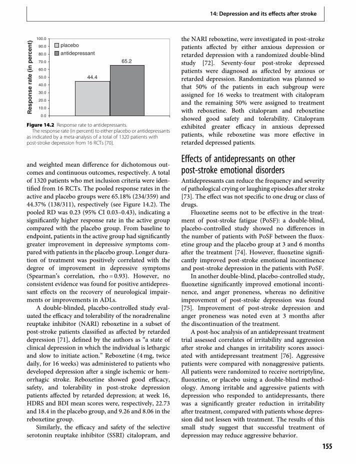
and weighted mean difference for dichotomous out-comes and continuous outcomes, respectively. A totalof 1320 patients who met inclusion criteria were iden-tified from 16 RCTs. The pooled response rates in theactive and placebo groups were 65.18% (234/359) and44.37% (138/311), respectively (see Figure 14.2). Thepooled RD was 0.23 (95% CI 0.03–0.43), indicating asignificantly higher response rate in the active groupcompared with the placebo group. From baseline toendpoint, patients in the active group had significantlygreater improvement in depressive symptoms com-pared with patients in the placebo group. Longer dura-tion of treatment was positively correlated with thedegree of improvement in depressive symptoms(Spearman’s correlation, rho = 0.93). However, noconsistent evidence was found for positive antidepres-sant effects on the recovery of neurological impair-ments or improvements in ADLs.
A double-blinded, placebo-controlled study eval-uated the efficacy and tolerability of the noradrenalinereuptake inhibitor (NARI) reboxetine in a subset ofpost-stroke patients classified as affected by retardeddepression [71], defined by the authors as “a state ofclinical depression in which the individual is lethargicand slow to initiate action.” Reboxetine (4mg, twicedaily, for 16 weeks) was administered to patients whodeveloped depression after a single ischemic or hem-orrhagic stroke. Reboxetine showed good efficacy,safety, and tolerability in post-stroke depressionpatients affected by retarded depression; at week 16,HDRS and BDI mean scores were, respectively, 22.73and 18.4 in the placebo group, and 9.26 and 8.06 in thereboxetine group.
Similarly, the efficacy and safety of the selectiveserotonin reuptake inhibitor (SSRI) citalopram, and
the NARI reboxetine, were investigated in post-strokepatients affected by either anxious depression orretarded depression with a randomized double-blindstudy [72]. Seventy-four post-stroke depressedpatients were diagnosed as affected by anxious orretarded depression. Randomization was planned sothat 50% of the patients in each subgroup wereassigned for 16 weeks to treatment with citalopramand the remaining 50% were assigned to treatmentwith reboxetine. Both citalopram and reboxetineshowed good safety and tolerability. Citalopramexhibited greater efficacy in anxious depressedpatients, while reboxetine was more effective inretarded depressed patients.
Effects of antidepressants on otherpost-stroke emotional disordersAntidepressants can reduce the frequency and severityof pathological crying or laughing episodes after stroke[73]. The effect was not specific to one drug or class ofdrugs.
Fluoxetine seems not to be effective in the treat-ment of post-stroke fatigue (PoSF): a double-blind,placebo-controlled study showed no differences inthe number of patients with PoSF between the fluox-etine group and the placebo group at 3 and 6 monthsafter the treatment [74]. However, fluoxetine signifi-cantly improved post-stroke emotional incontinenceand post-stroke depression in the patients with PoSF.
In another double-blind, placebo-controlled study,fluoxetine significantly improved emotional inconti-nence, and anger proneness, whereas no definitiveimprovement of post-stroke depression was found[75]. Improvement of post-stroke depression andanger proneness was noted even at 3 months afterthe discontinuation of the treatment.
A post-hoc analysis of an antidepressant treatmenttrial assessed correlates of irritability and aggressionafter stroke and changes in irritability scores associ-ated with antidepressant treatment [76]. Aggressivepatients were compared with nonaggressive patients.All patients were randomized to receive nortriptyline,fluoxetine, or placebo using a double-blind method-ology. Among irritable and aggressive patients withdepression who responded to antidepressants, therewas a significantly greater reduction in irritabilityafter treatment, compared with patients whose depres-sion did not lessen with treatment. The results of thissmall study suggest that successful treatment ofdepression may reduce aggressive behavior.
44.4
65.2
0.0
10.0
20.0
30.0
40.0
50.0
60.0
70.0
80.0
90.0
100.0
Res
po
nse
rat
e (i
n p
erce
nt) placebo
antidepressant
Figure 14.2 Response rate to antidepressants.The response rate (in percent) to either placebo or antidepressants
as indicated by a meta-analysis of a total of 1320 patients withpost-stroke depression from 16 RCTs [70].
14: Depression and its effects after stroke
155
Non-pharmacological treatmentof post-stroke depressionNonmedication treatment options might includerepetitive transcranial magnetic stimulation (rTMS).A randomized, parallel, double-blind study of activeversus sham rTMS assessed the effects of left prefrontalrTMS in patients with medication-refractory post-stroke depression. After discontinuing antidepres-sants, patients were randomly assigned to receive 10sessions of active (10Hz, 110% of the motor threshold,20 trains of 5 s duration) or sham left prefrontal rTMS[77]. When compared with sham stimulation, 10 ses-sions of active rTMS of the left dorsolateral prefrontalcortex were associated with a significant reduction ofdepressive symptoms. This reduction was not influ-enced by patient’s age, type or location of stroke,volume of left frontal leukoaraiosis, or by the distanceof the stimulating coil to the prefrontal cortex. Inaddition, there were few and mild adverse effects thatwere equally distributed among groups. Takentogether, these preliminary findings suggest thatrTMS may be an effective and safe treatment alterna-tive for patients with refractory depression afterstroke.
The effect of an adjunct light therapy in strokevictims with major depression receiving citalopramwas examined by use of two different intensities oflight therapy under double-blind conditions.Altogether, 63 patients were included in the study.After 4 weeks of therapy, the 6-item subscale of theHADRS showed a larger improvement in patientsreceiving high-intensity light treatment compared tothose treated with medium-intensity light [78].
Healthcare management with post-strokedepressionWhile efficacy of drug therapy is one issue, anotherissue in treatment of relatively neglected medical con-ditions such as post-stroke depression is whether andhow management of care improves outcome.
When 5-year survivors from a prospectivecommunity-based stroke incidence study wereassessed for depression, 17% of those assessed weredepressed [79]. However, only 22% of these patientswith depression were taking an antidepressant medi-cation. Of those taking an antidepressant, 72% werenot depressed. Thus, although nearly one-fifth of sur-vivors were depressed, few were taking antidepressantsalthough the treatment is likely to be effective.
A randomized, outcome-blinded multisite trial in188 ischemic stroke survivors with post-stroke depres-sion investigated the efficacy of a care managementprogram; it performed depression screening andenrollment between 1 and 2 months post-stroke [80].The Activate–Initiate–Monitor intervention was a caremanagement program that included activation of thepatient to recognize depression symptoms and accepttreatment, initiation of an antidepressant medication,and monitoring and adjusting treatment. Usual caresubjects received nondepression-related educationand were prescribed antidepressants at the discretionof their provider. The primary outcome measurewas depression response, defined as a HamiltonDepression Inventory score < 8 (remission) or adecrease from baseline of at least 50% at 12 weeks.Both depression response (51% versus 30%) andremission (39% versus 23%) were more likely in theActivate–Initiate–Monitor intervention than in theusual care group. This difference in depression scoreswas present by 6 weeks and persisted through the12-week assessment. The Activate–Initiate–Monitorcare management model was thus more efficaciousthan usual care in improving depression outcomes inpatients with post-stroke depression.
Are there reasons to promote preventionof post-stroke depression?Information provision, physical exercise programs,and listening to musicA recent meta-analysis assessed the effectiveness ofinformation provision strategies in improving the out-come for stroke patients and/or their identified care-givers; there is evidence that information improvespatient and caregiver knowledge of stroke, aspects ofpatient satisfaction, and reduces patient depressionscores [81].
Physical exercise programs might also help to pre-vent post-stroke depression and improve physical andemotional outcome. When 100 stroke survivors whohad completed acute rehabilitation were randomlyassigned to either a progressive, structured, 3-monthphysical exercise program, or usual care only, signifi-cant differences were found: 6 (14%) of the exercisegroup and 16 (35.6%) of the usual-care group haddepressive symptoms at 3 months (93 assessed) (seeFigure 14.3) [82]. At 9 months (80 assessed), 3 (7.5%)of the exercisers had significant depressive symptomscompared with 10 (25%) who received usual care.
14: Depression and its effects after stroke
156

Participants with and without baseline depressivesymptoms had equivalent treatment-related gains inimpairments and functional limitations, but only par-ticipants with depressive symptoms had improvedquality of life. It might be concluded that exercisemay help to reduce post-stroke depressive symptoms.In this study, depressive symptoms did not limit gainsin physical function due to exercise. Further, exercisemay contribute to improved quality of life in thosewith post-stroke depressive symptoms.
Similarly, for bedridden stroke survivors in resi-dential care, it could be demonstrated by a randomizedcontrolled trial that among other effects a simplenurse-led range-of-motion exercise program can gen-erate positive effects on depressive symptoms amongbedridden older people with stroke [83].
Further, listening to music could be a preventivemeasure for negative mood after stroke. In an RCT,MCA stroke patients who listened daily to self-selectedmusic for 2 months showed lower depression scoresafter 3 months post-stroke as compared to two controlgroups who either listened to audio books or receivedusual treatment only [84].
Prophylactic antidepressant treatmentOther studies have examined whether prophylacticantidepressant treatment can prevent depression andimprove functional outcome (ADL functions, partic-ipation, quality of life), with good tolerability andsafety in nondepressed patients with recent stroke.
To test whether prophylactic treatment with theantidepressant mirtazapine (30mg/day) in patientswith acute stroke starting one day after stroke onsetprevents post-stroke depression, 70 patients wereenrolled in an open randomized controlled study andwere re-examined up to day 380 post-stroke [85].Those post-stroke patients who developed depression(DSM-IV criteria) but had been randomly assigned tothe non-treatment group were given the antidepres-sant mirtazapine after the diagnosis of depression hadbeen established. Forty percent (14/35) of the non-treated patients and only 5.7% (2/35) of the patientswho were treated with mirtazapine developed post-stroke depression. Altogether, 16 patients developedpost-stroke depression, 15 of whom remitted afterinitiation of treatment with mirtazapine.
Sertraline (50mg/day), however, was not effectiveto prevent post-stroke depression in a double-blind,placebo-controlled 24-week clinical trial [86].
A multisite RCT for prevention of depressionamong 176 nondepressed patients was conductedwithin 3 months following acute stroke with a double-blind placebo-controlled comparison of escitalopramwith placebo, and a nonblinded problem-solving ther-apy group [87]. The use of either escitalopram orproblem-solving therapy resulted in a significantlylower incidence of depression over 12 months of treat-ment as compared with placebo. However, only esci-talopram, but not the problem-solving therapy,achieved significant results over placebo using anintention-to-treat conservative method of analysis.
A meta-analysis of randomized placebo-controlledtrials also provided support for a prophylactic effect ofantidepressants in nondepressed patients with stroke[88]. The search included trials from 1950 to August2006. From 10 RCTs, a total of 703 non-depressedpatients after stroke were identified. The pooledoccurrence rate of newly developed post-strokedepression cases in the intervention and controlgroups differed significantly, being 12.54% (41/327)and 29.17% (91/312), respectively (pooled rate differ-ence =−0.17, 95% confidence interval =−0.26 to −0.08).Prophylactic effects of antidepressants were not relatedto duration of use.
In conclusion, antidepressant prophylaxis is associ-ated with a significant reduction in the occurrence rateof newly developed post-stroke depression, suggestingantidepressants may be considered along with otherpreventive strategies in the management of strokepatients.
36
25
148
0.0
10.0
20.0
30.0
40.0
50.0
60.0
70.0
80.0
90.0
100.0
3 months 9 months
Dep
ress
ive
sym
pto
mes
( in
per
cen
t)
usual carestructured home exercise
Figure 14.3 Prevalence of depressive symptoms among strokesurvivors following a structured, 3-month physical exercise programat home or usual care only.
The prevalence of depressive symptoms (in percent) among strokesurvivors was significantly lower among stroke victims who receiveda structured, 3-month physical exercise program at home ascompared to those who received usual care only. Data from arandomized controlled trial [82].
14: Depression and its effects after stroke
157
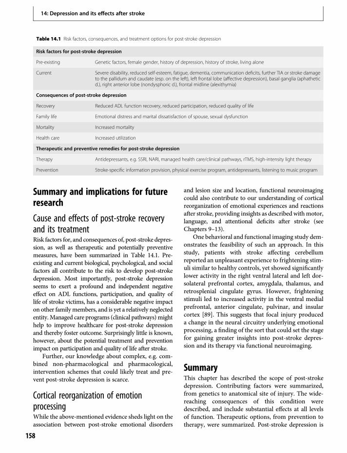
Summary and implications for futureresearch
Cause and effects of post-stroke recoveryand its treatmentRisk factors for, and consequences of, post-stroke depres-sion, as well as therapeutic and potentially preventivemeasures, have been summarized in Table 14.1. Pre-existing and current biological, psychological, and socialfactors all contribute to the risk to develop post-strokedepression. Most importantly, post-stroke depressionseems to exert a profound and independent negativeeffect on ADL functions, participation, and quality oflife of stroke victims, has a considerable negative impacton other familymembers, and is yet a relatively neglectedentity.Managed care programs (clinical pathways)mighthelp to improve healthcare for post-stroke depressionand thereby foster outcome. Surprisingly little is known,however, about the potential treatment and preventionimpact on participation and quality of life after stroke.
Further, our knowledge about complex, e.g. com-bined non-pharmacological and pharmacological,intervention schemes that could likely treat and pre-vent post-stroke depression is scarce.
Cortical reorganization of emotionprocessingWhile the above-mentioned evidence sheds light on theassociation between post-stroke emotional disorders
and lesion size and location, functional neuroimagingcould also contribute to our understanding of corticalreorganization of emotional experiences and reactionsafter stroke, providing insights as described withmotor,language, and attentional deficits after stroke (seeChapters 9–13).
One behavioral and functional imaging study dem-onstrates the feasibility of such an approach. In thisstudy, patients with stroke affecting cerebellumreported an unpleasant experience to frightening stim-uli similar to healthy controls, yet showed significantlylower activity in the right ventral lateral and left dor-solateral prefrontal cortex, amygdala, thalamus, andretrosplenial cingulate gyrus. However, frighteningstimuli led to increased activity in the ventral medialprefrontal, anterior cingulate, pulvinar, and insularcortex [89]. This suggests that focal injury produceda change in the neural circuitry underlying emotionalprocessing, a finding of the sort that could set the stagefor gaining greater insights into post-stroke depres-sion and its therapy via functional neuroimaging.
SummaryThis chapter has described the scope of post-strokedepression. Contributing factors were summarized,from genetics to anatomical site of injury. The wide-reaching consequences of this condition weredescribed, and include substantial effects at all levelsof function. Therapeutic options, from prevention totherapy, were summarized. Post-stroke depression is
Table 14.1 Risk factors, consequences, and treatment options for post-stroke depression
Risk factors for post-stroke depression
Pre-existing Genetic factors, female gender, history of depression, history of stroke, living alone
Current Severe disability, reduced self-esteem, fatigue, dementia, communication deficits, further TIA or stroke damageto the pallidum and caudate (esp. on the left), left frontal lobe (affective depression), basal ganglia (aphatheticd.), right anterior lobe (nondysphoric d.), frontal midline (alexithymia)
Consequences of post-stroke depression
Recovery Reduced ADL function recovery, reduced participation, reduced quality of life
Family life Emotional distress and marital dissatisfaction of spouse, sexual dysfunction
Mortality Increased mortality
Health care Increased utilization
Therapeutic and preventive remedies for post-stroke depression
Therapy Antidepressants, e.g. SSRI, NARI, managed health care/clinical pathways, rTMS, high-intensity light therapy
Prevention Stroke-specific information provision, physical exercise program, antidepressants, listening to music program
14: Depression and its effects after stroke
158

common and has major effects. Further research canaim to better understand this disorder, individualizeits therapy, and increase the fraction of patients forwhom post-stroke depression is successfully treated.
References1. Spalletta G, Ripa A, Caltagirone C. Symptom profile of
DSM-IV major and minor depressive disorders in first-ever stroke patients. Am J Geriatr Psychiatry.2005;13:108–15.
2. Laska AC, Martensson B, Kahan T, et al. Recognition ofdepression in aphasic stroke patients. Cerebrovasc Dis.2007;24:74–9.
3. Bennett HE, Thomas SA, Austen R, et al. Validation ofscreening measures for assessing mood in strokepatients. Br J Clin Psychol. 2006;45:367–76.
4. Benaim C, Cailly B, Perennou D, et al. Validation ofthe aphasic depression rating scale. Stroke.2004;35:1692–6.
5. Fure B. [Depression, anxiety and other emotionalsymptoms after cerebral stroke.] Tidsskr NorLaegeforen. 2007;127:1387–9.
6. Brodaty H, Withall A, Altendorf A, et al. Rates ofdepression at 3 and 15 months post-stroke and theirrelationship with cognitive decline: The Sydney StrokeStudy. Am J Geriatr Psychiatry. 2007;15:477–86.
7. Paolucci S, Gandolfo C, Provinciali L, et al. The Italianmulticenter observational study on post-strokedepression (DESTRO). J Neurol. 2006;253:556–62.
8. Linden T, Blomstrand C, Skoog I. Depressive disordersafter 20 months in elderly stroke patients: A case-control study. Stroke. 2007;38:1860–3.
9. Thomas SA, Lincoln NB. Factors relating to depressionafter stroke. Br J Clin Psychol. 2006;45:49–61.
10. Skaner Y, Nilsson GH, Sundquist K, et al. Self-ratedhealth, symptoms of depression and general symptomsat 3 and 12 months after a first-ever stroke: Amunicipality-based study in Sweden. BMC Fam Pract.2007;8:61.
11. Fure B, Wyller TB, Engedal K, et al. Emotionalsymptoms in acute ischemic stroke. Int J GeriatrPsychiatry. 2006;21:382–7.
12. Smith LN, Norrie J, Kerr SM, et al. Impact andinfluences on caregiver outcomes at one year post-stroke. Cerebrovasc Dis. 2004;18:145–53.
13. Morrison V, Pollard B, Johnston M, et al. Anxiety anddepression 3 years following stroke: Demographic,clinical, and psychological predictors. J Psychosom Res.2005;59:209–13.
14. Santos CO, Caeiro L, Ferro JM, et al. Anger, hostilityand aggression in the first days of acute stroke. Eur JNeurol. 2006;13:351–8.
15. Kim JS, Choi S, Kwon SU, et al. Inability to controlanger or aggression after stroke. Neurology.2002;58:1106–08.
16. Bogousslavsky J. William Feinberg lecture 2002:Emotions, mood, and behavior after stroke. Stroke.2003;34:1046–50.
17. Spalletta G, Serra L, Fadda L, et al. Unawareness ofmotor impairment and emotions in right hemisphericstroke: A preliminary investigation. Int J GeriatrPsychiatry. 2007;22:1241–6.
18. Stone J, Townend E, Kwan J, et al. Personality changeafter stroke: Some preliminary observations. J NeurolNeurosurg Psychiatry. 2004;75:1708–13.
19. Bos MJ, Linden T, Koudstaal PJ, et al. Depressivesymptoms and risk of stroke: The RotterdamStudy. J Neurol Neurosurg Psychiatry.2008;79:997–1001.
20. Gromova HA, Gafarov VV, Gagulin IV. Depressionand risk of cardiovascular diseases among males aged25–64 (WHO MONICA–psychosocial). Alaska Med.2007;49:255–8.
21. Gump BB, Matthews KA, Eberly LE, et al. Depressivesymptoms and mortality in men: Results from theMultiple Risk Factor Intervention Trial. Stroke.2005;36:98–102.
22. Salaycik KJ, Kelly-Hayes M, Beiser A, et al. Depressivesymptoms and risk of stroke: The Framingham Study.Stroke. 2007;38:16–21.
23. Krishnan M, Mast BT, Ficker LJ, et al. The effects ofpreexisting depression on cerebrovascular healthoutcomes in geriatric continuing care. J Gerontol A BiolSci Med Sci. 2005;60:915–9.
24. Leentjens AF, Aben I, Lodder J, et al. General anddisease-specific risk factors for depression afterischemic stroke: A two-step Cox regression analysis.Int Psychogeriatr. 2006;18:739–48.
25. Vickery CD, Sepehri A, Evans CC. Self-esteem in anacute stroke rehabilitation sample: A control groupcomparison. Clin Rehabil. 2008;22:179–87.
26. Kim JM, Stewart R, Kim SW, et al. BDNF genotypepotentially modifying the association between incidentstroke and depression. Neurobiol Aging.2008;29:789–92.
27. Choi-Kwon S, Han SW, Kwon SU, et al. Post-strokefatigue: Characteristics and related factors. CerebrovascDis. 2005;19:84–90.
28. Nys GM, van Zandvoort MJ, van der Worp HB, et al.Early depressive symptoms after stroke:Neuropsychological correlates and lesioncharacteristics. J Neurol Sci. 2005;228:27–33.
29. Barker-Collo SL. Depression and anxiety 3 monthspost-stroke: Prevalence and correlates. Arch ClinNeuropsychol. 2007;22:519–31.
14: Depression and its effects after stroke
159
30. Eriksson M, Asplund K, Glader EL, et al. Self-reporteddepression and use of antidepressants after stroke: Anational survey. Stroke. 2004;35:936–41.
31. Giaquinto S, Spiridigliozzi C, Caracciolo B. Can faithprotect from emotional distress after stroke? Stroke.2007;38:993–7.
32. van de Port, I, Kwakkel G, Bruin M, et al. Determinantsof depression in chronic stroke: A prospective cohortstudy. Disabil Rehabil. 2007;29:353–8.
33. Saxena SK, Ng TP, Koh G, et al. Is improvement inimpaired cognition and depressive symptoms in post-stroke patients associated with recovery in activities ofdaily living? Acta Neurol Scand. 2007;115:339–46.
34. Caeiro L, Ferro JM, Santos CO, et al. Depression inacute stroke. J Psychiatry Neurosci. 2006;31:377–83.
35. Nuyen J, Spreeuwenberg PM, Groenewegen PP, et al.Impact of preexisting depression on length of stay anddischarge destination among patients hospitalized for acutestroke: Linked register-based study. Stroke. 2008;39:132–8.
36. Teodorczuk A, O’Brien JT, Firbank MJ, et al. Whitematter changes and late-life depressive symptoms:Longitudinal study. Br J Psychiatry. 2007;191:212–7.
37. Aben I, Lodder J, Honig A, et al. Focal or generalizedvascular brain damage and vulnerability to depressionafter stroke: A 1-year prospective follow-up study. IntPsychogeriatr. 2006;18:19–35.
38. Vataja R, Leppavuori A, Pohjasvaara T, et al. Post-stroke depression and lesion location revisited. JNeuropsychiatry Clin Neurosci. 2004;16:156–62.
39. Narushima K, Kosier JT, Robinson RG. A reappraisal ofpost-stroke depression, intra- and inter-hemisphericlesion location using meta-analysis. J NeuropsychiatryClin Neurosci. 2003;15:422–30.
40. Hama S, Yamashita H, Shigenobu M, et al. Post-strokeaffective or apathetic depression and lesion location:Left frontal lobe and bilateral basal ganglia. Eur ArchPsychiatry Clin Neurosci. 2007;257:149–52.
41. Vataja R, Pohjasvaara T, Mantyla R, et al. Depression–executive dysfunction syndrome in stroke patients. AmJ Geriatr Psychiatry. 2005;13:99–107.
42. Paradiso S, Vaidya J, Tranel D, et al. Nondysphoricdepression following stroke. J Neuropsychiatry ClinNeurosci. 2008;20:52–61.
43. Schafer R, Popp K, Jorgens S, et al. Alexithymia-likedisorder in right anterior cingulate infarction.Neurocase. 2007;13:201–08.
44. Ramasubbu R, Tobias R, Buchan AM, et al. Serotonintransporter gene promoter region polymorphismassociated with post-stroke major depression.J Neuropsychiatry Clin Neurosci. 2006;18:96–9.
45. Rocco A, Afra J, Toscano M, et al. Acute subcorticalstroke and early serotonergic modification: An IDAPstudy. Eur J Neurol. 2007;14:1378–82.
46. Moller M, Andersen G, Gjedde A. Serotonin 5HT1Areceptor availability and pathological crying afterstroke. Acta Neurol Scand. 2007;116:83–90.
47. Glodzik-Sobanska L, Slowik A, McHugh P, et al.Single voxel proton magnetic resonance spectroscopyin post-stroke depression. Psychiatry Res.2006;148:111–20.
48. Glodzik-Sobanska L, Slowik A, Kieltyka A, et al.Reduced prefrontal N-acetylaspartate in stroke patientswith apathy. J Neurol Sci. 2005;238:19–24.
49. Dafer RM, Rao M, Shareef A, et al. Post-strokedepression. Top Stroke Rehabil. 2008;15:13–21.
50. Lai SM, Duncan PW, Keighley J, et al. Depressivesymptoms and independence in BADL and IADL.J Rehabil Res Dev. 2002;39:589–96.
51. Goodwin RD, Devanand DP. Stroke, depression, andfunctional health outcomes among adults in thecommunity. J Geriatr Psychiatry Neurol. 2008;21:41–6.
52. He J, Shen PF. [Clinical study on the therapeutic effectof acupuncture in the treatment of post-strokedepression.] Zhen Ci Yan Jiu. 2007;32:58–61.
53. Nannetti L, Paci M, Pasquini J, et al. Motor andfunctional recovery in patients with post-strokedepression. Disabil Rehabil. 2005;27:170–5.
54. D’Alisa S, Baudo S, Mauro A, et al. How does strokerestrict participation in long-term post-strokesurvivors? Acta Neurol Scand. 2005;112:157–62.
55. Sturm JW, Donnan GA, Dewey HM, et al.Determinants of handicap after stroke: The North EastMelbourne Stroke Incidence Study (NEMESIS). Stroke.2004;35:715–20.
56. Desrosiers J, Demers L, Robichaud L, et al. Short-termchanges in and predictors of participation of olderadults after stroke following acute care orrehabilitation. Neurorehabil Neural Repair.2008;22:288–97.
57. Lo RS, Cheng JO, Wong EM, et al. Handicap and itsdeterminants of change in stroke survivors: One-yearfollow-up study. Stroke. 2008;39:148–53.
58. Jonkman EJ, de Weerd AW, Vrijens NL. Quality of lifeafter a first ischemic stroke. Long-term developmentsand correlations with changes in neurological deficit,mood and cognitive impairment. Acta Neurol Scand.1998;98:169–75.
59. Kwok T, Lo RS, Wong E, et al. Quality of life of strokesurvivors: A 1-year follow-up study. Arch Phys MedRehabil. 2006;87:1177–82.
60. Bilge C, Kocer E, Kocer A, et al. Depression andfunctional outcome after stroke: The effect ofantidepressant therapy on functional recovery. EurJ Phys Rehabil Med. 2008;44:13–8.
61. Narushima K, Robinson RG. The effect of early versuslate antidepressant treatment on physical impairment
14: Depression and its effects after stroke
160
associated with post-stroke depression: Is there a time-related therapeutic window? J Nerv Ment Dis.2003;191:645–52.
62. Jorge RE, Robinson RG, Arndt S, et al. Mortalityand post-stroke depression: A placebo-controlledtrial of antidepressants. Am J Psychiatry.2003;160:1823–9.
63. Ried LD, Tueth MJ, Jia H. A pilot study to describeantidepressant prescriptions dispensed to veterans afterstroke. Res Social Adm Pharm. 2006;2:96–109.
64. Visser-Keizer AC, Meyboom-de JB, Deelman BG, et al.Subjective changes in emotion, cognition andbehaviour after stroke: Factors affecting the perceptionof patients and partners. J Clin Exp Neuropsychol.2002;24:1032–45.
65. Chow SK, Wong FK, Poon CY. Coping and caring:Support for family caregivers of stroke survivors. J ClinNurs. 2007;16:133–43.
66. Blonder LX, Langer SL, Pettigrew LC, et al. The effectsof stroke disability on spousal caregivers.NeuroRehabilitation. 2007;22:85–92.
67. Kimura M, Murata Y, Shimoda K, et al. Sexualdysfunction following stroke. Compr Psychiatry.2001;42:217–22.
68. Jia H, Damush TM, Qin H, et al. The impact of post-stroke depression on healthcare use by veterans withacute stroke. Stroke. 2006;37:2796–801.
69. Starkstein SE, Mizrahi R, Power BD. Antidepressanttherapy in post-stroke depression. Expert OpinPharmacother. 2008;9:1291–8.
70. Chen Y, Guo JJ, Zhan S, et al. Treatment effects ofantidepressants in patients with post-strokedepression: A meta-analysis. Ann Pharmacother.2006;40:2115–22.
71. Rampello L, Alvano A, Chiechio S, et al. An evaluationof efficacy and safety of reboxetine in elderly patientsaffected by “retarded” post-stroke depression. Arandom, placebo-controlled study. Arch GerontolGeriatr. 2005;40:275–85.
72. Rampello L, Chiechio S, Nicoletti G, et al. Prediction ofthe response to citalopram and reboxetine in post-stroke depressed patients. Psychopharmacology (Berl).2004;173:73–8.
73. House AO, Hackett ML, Anderson CS, et al.Pharmaceutical interventions for emotionalismafter stroke. Cochrane Database Syst Rev. 2004;CD003690.
74. Choi-Kwon S, Choi J, Kwon SU, et al. Fluoxetine is noteffective in the treatment of post-stroke fatigue: Adouble-blind, placebo-controlled study. CerebrovascDis. 2007;23:103–08.
75. Choi-Kwon S, Han SW, Kwon SU, et al. Fluoxetinetreatment in post-stroke depression, emotional
incontinence, and anger proneness: A double-blind,placebo-controlled study. Stroke. 2006;37:156–61.
76. Chan KL, Campayo A, Moser DJ, et al. Aggressivebehavior in patients with stroke: Association withpsychopathology and results of antidepressanttreatment on aggression. Arch Phys Med Rehabil.2006;87:793–8.
77. Jorge RE, Robinson RG, Tateno A, et al. Repetitivetranscranial magnetic stimulation as treatment of post-stroke depression: A preliminary study. Biol Psychiatry.2004;55:398–405.
78. Sondergaard MP, Jarden JO, Martiny K, et al. Doseresponse to adjunctive light therapy in citalopram-treated patients with post-stroke depression. Arandomised, double-blind pilot study. PsychotherPsychosom. 2006;75:244–8.
79. Paul SL, Dewey HM, Sturm JW, et al. Prevalence ofdepression and use of antidepressant medication at5 years post-stroke in the North East Melbourne StrokeIncidence Study. Stroke. 2006;37:2854–5.
80. Williams LS, Kroenke K, Bakas T, et al. Caremanagement of post-stroke depression: A randomized,controlled trial. Stroke. 2007;38:998–1003.
81. Smith J, Forster A, House A, et al. Informationprovision for stroke patients and their caregivers.Cochrane Database Syst Rev. 2008;CD001919.
82. Lai SM, Studenski S, Richards L, et al. Therapeuticexercise and depressive symptoms after stroke. J AmGeriatr Soc. 2006;54:240–7.
83. Tseng CN, Chen CC,Wu SC, et al. Effects of a range-of-motion exercise programme. J Adv Nurs.2007;57:181–91.
84. Sarkamo T, Tervaniemi M, Laitinen S, et al. Musiclistening enhances cognitive recovery and mood aftermiddle cerebral artery stroke. Brain. 2008;131:866–76.
85. Niedermaier N, Bohrer E, Schulte K, et al. Preventionand treatment of post-stroke depression withmirtazapine in patients with acute stroke. J ClinPsychiatry. 2004;65:1619–23.
86. Almeida OP, Waterreus A, Hankey GJ. Preventingdepression after stroke: Results from a randomizedplacebo-controlled trial. J Clin Psychiatry. 2006;67:1104–09.
87. Robinson RG, Jorge RE, Moser DJ, et al. Escitalopramand problem-solving therapy for prevention of post-stroke depression: A randomized controlled trial.JAMA. 2008;299:2391–400.
88. Chen Y, Patel NC, Guo JJ, et al. Antidepressantprophylaxis for post-stroke depression: A meta-analysis. Int Clin Psychopharmacol 2007;22:159–66.
89. Turner BM, Paradiso S, Marvel CL, et al.The cerebellum and emotional experience.Neuropsychologia. 2007;45:1331–41.
14: Depression and its effects after stroke
161


15 Epidemiology of stroke recoverySamir Belagaje & Brett Kissela
Epidemiology is defined as the study of factors influenc-ing health and disease in populations. When applied tostroke recovery, it refers to the natural history of strokerecovery and defining factors that affect recoveryderived from post-stroke populations. This is a pillarof stroke recovery knowledge that is important andpractical when caring for stroke patients, and that alsorequires further research attention. One of the mostcommon questions a stroke victim or their familymembers will ask the physician is to what degree theirloved one will recover from their stroke, and over whattime frame that recovery can be expected. This questioncurrently poses a huge challenge because of the complexand multifactorial processes involved in recovery, so atpresent one can only provide best estimates based onepidemiological, natural history knowledge combinedwith other aspects of the patient’s condition that mayinfluence the predicted outcome.
This chapter consists of four sections. The first partof this chapter will provide an overview of the differenttypes of epidemiological studies, highlighting theirrespective strengths and weaknesses. The second willfocus on the current state of knowledge regardinggeneral epidemiological patterns seen in stroke recov-ery, as well as the gaps in our knowledge. Thenext portion will discuss the epidemiological datasurrounding factors that can affect an individual’srecovery from a stroke as well as how those factorsinteract with each other. In the final section, the appli-cation of epidemiology will be discussed with regard toclinical care and planning of future clinical research.
Overview of types of epidemiologicalstudies in strokeEpidemiological studies primarily are observational innature but experimental designs also contribute epi-demiological information.
RegistryStroke registries are databases of information collectedabout patients with stroke. The databases typicallycollect all details about the patient such as age, sex,comorbidities, and treatments and are primarilydescriptive in nature. Analysis of the data then allowsone to make inferences about incidence and preva-lence. Depending on what data are collected aboutthe patients, these types of studies may be helpful tofirst generate relationships between diseases and asso-ciated factors. By nature of their design, however, thesestudies by themselves cannot be used to prove thoseexact relationships, and require further studies. Arecent example of a stroke registry is the PaulCoverdell National Acute Stroke Registry [1].
Case controlCase control studies are another type of epidemiolog-ical study that attempts to determine factors that influ-ence a disease or condition. In such a study for stroke,a population with stroke (“cases”) is compared to asimilar population without stroke (“controls”). In thecomparison, the investigator looks for differences inthe characteristics or factors in cases vs. controls thatmight have caused the cases to develop stroke. Thesetypes of studies are relatively inexpensive compared toother types and are useful in investigating uncommonconditions (such as subarachnoid hemorrhage orintracerebral hemorrhage). Their disadvantagesinclude the inability to make a direct cause and effectrelationship because of potentially confounding vari-ables. Furthermore, because of their retrospectivenature, the information gained may be subjected torecall bias and investigator bias. A recent example ofa case control study in stroke is the Geneticsand Environmental Risk Factors for HemorrhagicStroke [2,3].
Brain Repair After Stroke, ed. S. C. Cramer and R. J. Nudo. Published by Cambridge University Press.© Cambridge University Press 2010.
163
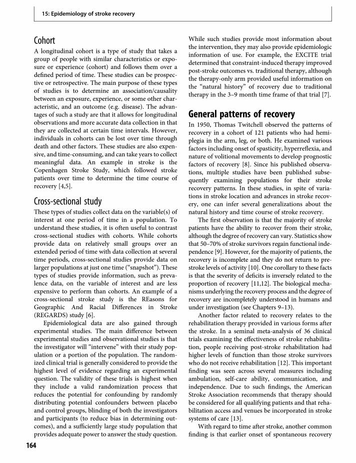
CohortA longitudinal cohort is a type of study that takes agroup of people with similar characteristics or expo-sure or experience (cohort) and follows them over adefined period of time. These studies can be prospec-tive or retrospective. The main purpose of these typesof studies is to determine an association/causalitybetween an exposure, experience, or some other char-acteristic, and an outcome (e.g. disease). The advan-tages of such a study are that it allows for longitudinalobservations and more accurate data collection in thatthey are collected at certain time intervals. However,individuals in cohorts can be lost over time throughdeath and other factors. These studies are also expen-sive, and time-consuming, and can take years to collectmeaningful data. An example in stroke is theCopenhagen Stroke Study, which followed strokepatients over time to determine the time course ofrecovery [4,5].
Cross-sectional studyThese types of studies collect data on the variable(s) ofinterest at one period of time in a population. Tounderstand these studies, it is often useful to contrastcross-sectional studies with cohorts. While cohortsprovide data on relatively small groups over anextended period of time with data collection at severaltime periods, cross-sectional studies provide data onlarger populations at just one time (“snapshot”). Thesetypes of studies provide information, such as preva-lence data, on the variable of interest and are lessexpensive to perform than cohorts. An example of across-sectional stroke study is the REasons forGeographic And Racial Differences in Stroke(REGARDS) study [6].
Epidemiological data are also gained throughexperimental studies. The main difference betweenexperimental studies and observational studies is thatthe investigator will “intervene” with their study pop-ulation or a portion of the population. The random-ized clinical trial is generally considered to provide thehighest level of evidence regarding an experimentalquestion. The validity of these trials is highest whenthey include a valid randomization process thatreduces the potential for confounding by randomlydistributing potential confounders between placeboand control groups, blinding of both the investigatorsand participants (to reduce bias in determining out-comes), and a sufficiently large study population thatprovides adequate power to answer the study question.
While such studies provide most information aboutthe intervention, they may also provide epidemiologicinformation of use. For example, the EXCITE trialdetermined that constraint-induced therapy improvedpost-stroke outcomes vs. traditional therapy, althoughthe therapy-only arm provided useful information onthe “natural history” of recovery due to traditionaltherapy in the 3–9 month time frame of that trial [7].
General patterns of recoveryIn 1950, Thomas Twitchell observed the patterns ofrecovery in a cohort of 121 patients who had hemi-plegia in the arm, leg, or both. He examined variousfactors including onset of spasticity, hyperreflexia, andnature of volitional movements to develop prognosticfactors of recovery [8]. Since his published observa-tions, multiple studies have been published subse-quently examining populations for their strokerecovery patterns. In these studies, in spite of varia-tions in stroke location and advances in stroke recov-ery, one can infer several generalizations about thenatural history and time course of stroke recovery.
The first observation is that the majority of strokepatients have the ability to recover from their stroke,although the degree of recovery can vary. Statistics showthat 50–70% of stroke survivors regain functional inde-pendence [9]. However, for the majority of patients, therecovery is incomplete and they do not return to pre-stroke levels of activity [10]. One corollary to these factsis that the severity of deficits is inversely related to theproportion of recovery [11,12]. The biological mecha-nisms underlying the recovery process and the degree ofrecovery are incompletely understood in humans andunder investigation (see Chapters 9–13).
Another factor related to recovery relates to therehabilitation therapy provided in various forms afterthe stroke. In a seminal meta-analysis of 36 clinicaltrials examining the effectiveness of stroke rehabilita-tion, people receiving post-stroke rehabilitation hadhigher levels of function than those stroke survivorswho do not receive rehabilitation [12]. This importantfinding was seen across several measures includingambulation, self-care ability, communication, andindependence. Due to such findings, the AmericanStroke Association recommends that therapy shouldbe considered for all qualifying patients and that reha-bilitation access and venues be incorporated in strokesystems of care [13].
With regard to time after stroke, another commonfinding is that earlier onset of spontaneous recovery
15: Epidemiology of stroke recovery
164
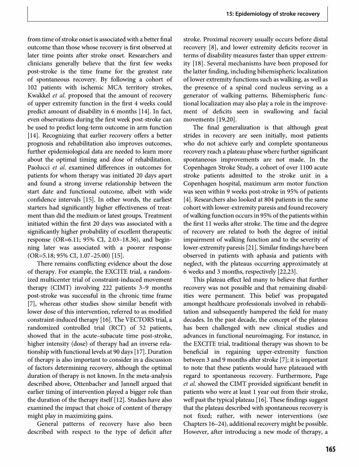
from time of stroke onset is associated with a better finaloutcome than those whose recovery is first observed atlater time points after stroke onset. Researchers andclinicians generally believe that the first few weekspost-stroke is the time frame for the greatest rateof spontaneous recovery. By following a cohort of102 patients with ischemic MCA territory strokes,Kwakkel et al. proposed that the amount of recoveryof upper extremity function in the first 4 weeks couldpredict amount of disability in 6 months [14]. In fact,even observations during the first week post-stroke canbe used to predict long-term outcome in arm function[14]. Recognizing that earlier recovery offers a betterprognosis and rehabilitation also improves outcomes,further epidemiological data are needed to learn moreabout the optimal timing and dose of rehabilitation.Paolucci et al. examined differences in outcomes forpatients for whom therapy was initiated 20 days apartand found a strong inverse relationship between thestart date and functional outcome, albeit with wideconfidence intervals [15]. In other words, the earlieststarters had significantly higher effectiveness of treat-ment than did the medium or latest groups. Treatmentinitiated within the first 20 days was associated with asignificantly higher probability of excellent therapeuticresponse (OR=6.11; 95% CI, 2.03–18.36), and begin-ning later was associated with a poorer response(OR=5.18; 95% CI, 1.07–25.00) [15].
There remains conflicting evidence about the doseof therapy. For example, the EXCITE trial, a random-ized multicenter trial of constraint-induced movementtherapy (CIMT) involving 222 patients 3–9 monthspost-stroke was successful in the chronic time frame[7], whereas other studies show similar benefit withlower dose of this intervention, referred to as modifiedconstraint-induced therapy [16]. The VECTORS trial, arandomized controlled trial (RCT) of 52 patients,showed that in the acute–subacute time post-stroke,higher intensity (dose) of therapy had an inverse rela-tionship with functional levels at 90 days [17]. Durationof therapy is also important to consider in a discussionof factors determining recovery, although the optimalduration of therapy is not known. In the meta-analysisdescribed above, Ottenbacher and Jannell argued thatearlier timing of intervention played a bigger role thanthe duration of the therapy itself [12]. Studies have alsoexamined the impact that choice of content of therapymight play in maximizing gains.
General patterns of recovery have also beendescribed with respect to the type of deficit after
stroke. Proximal recovery usually occurs before distalrecovery [8], and lower extremity deficits recover interms of disability measures faster than upper extrem-ity [18]. Several mechanisms have been proposed forthe latter finding, including bihemispheric localizationof lower extremity functions such as walking, as well asthe presence of a spinal cord nucleus serving as agenerator of walking patterns. Bihemispheric func-tional localization may also play a role in the improve-ment of deficits seen in swallowing and facialmovements [19,20].
The final generalization is that although greatstrides in recovery are seen initially, most patientswho do not achieve early and complete spontaneousrecovery reach a plateau phase where further significantspontaneous improvements are not made. In theCopenhagen Stroke Study, a cohort of over 1100 acutestroke patients admitted to the stroke unit in aCopenhagen hospital, maximum arm motor functionwas seen within 9 weeks post-stroke in 95% of patients[4]. Researchers also looked at 804 patients in the samecohort with lower-extremity paresis and found recoveryof walking function occurs in 95% of the patients withinthe first 11 weeks after stroke. The time and the degreeof recovery are related to both the degree of initialimpairment of walking function and to the severity oflower-extremity paresis [21]. Similar findings have beenobserved in patients with aphasia and patients withneglect, with the plateaus occurring approximately at6 weeks and 3 months, respectively [22,23].
This plateau effect led many to believe that furtherrecovery was not possible and that remaining disabil-ities were permanent. This belief was propagatedamongst healthcare professionals involved in rehabili-tation and subsequently hampered the field for manydecades. In the past decade, the concept of the plateauhas been challenged with new clinical studies andadvances in functional neuroimaging. For instance, inthe EXCITE trial, traditional therapy was shown to bebeneficial in regaining upper-extremity functionbetween 3 and 9 months after stroke [7]; it is importantto note that these patients would have plateaued withregard to spontaneous recovery. Furthermore, Pageet al. showed the CIMT provided significant benefit inpatients who were at least 1 year out from their stroke,well past the typical plateau [16]. These findings suggestthat the plateau described with spontaneous recovery isnot fixed; rather, with newer interventions (seeChapters 16–24), additional recoverymight be possible.However, after introducing a new mode of therapy, a
15: Epidemiology of stroke recovery
165

typical pattern is to see some improvement initially,after which a new plateau is reached, as reported instudies such as the EXCITE trial [7].
Specific factors in stroke recoveryWhen examining stroke recovery via epidemiologicalstudies, factors associated with an individual’s clinicalimprovement can be determined. The next section willdiscuss those factors, both modifiable and non-modifiable, that are generally accepted as importantfactors in stroke recovery.
AgeSeveral studies examining stroke outcomes continu-ously show that age is a factor in determining out-comes. The relationship between age and recovery isan inverse correlation as seen in the CopenhagenStroke Study cohort and in the EXCITE trial popula-tion [5,7]. That is, the older someone is, the less like-lihood of recovery they have. Said another way,younger stroke victims have a better prognosis forrecovery. Older age is associated with less recovery invirtually all epidemiology studies of stroke outcome.One may argue that older age is associated with otherfactors that have been associated with lesser recovery,including a larger number of medical comorbidities,higher amount of periventricular white matter disease,a lesser reserve of neurons to assume lost functions,and a greater risk for cognitive dysfunction. And yet,age remains an independent risk factor for less recov-ery after controlling for all of these other factors inmost studies [5,24], suggesting that factors directlyrelated to aging effects influence outcome.
Race/geneticsCertain groups may recover better from their strokesthan others. Several studies show that African-American patients have more severe strokes thanCaucasians, and that furthermore they do not recoveras well. In the Greater Cincinnati Northern KentuckyStroke Study, a population-based cohort of people liv-ing in the Cincinnati area, it was shown that African-American patients have a higher incidence of strokethan Caucasian populations [25]. In general, non-Caucasian races score worse on quality-of-life measuresduring the recovery process than Caucasian races [24].There may be some controversy as to whether raceactually is a factor, or whether it is mainly a surrogatefor other factors such as socio-economic status and
access to care. One study examined 1073 African-American and Caucasian stroke patients and showedthat rates of utilization and time to referral for inpatientrehabilitation were equal, but that low-income African-American patients had worse functional recovery in12 months [26]. While these non-biologic factorsneed further investigation, the disparities in recoverybetween race/ethnic groups naturally lead to an interestin underlying genetic differences that may be related tothe biological processes underlying recovery. For exam-ple, it has been shown that the presence of a polymor-phism in the gene for brain-derived neurotrophic factor(BDNF) impairs motor cortex plasticity in human sub-jects [27]. This leads to the suggestion that patients whopossess this polymorphism might not recover as well asthe general population, and this (or other genotypesassociated with poor recovery) may be more prevalentin some race/ethnic groups than others. Such hypoth-eses require direct study, and if supported, might pro-vide new insights to maximize recovery across thespectrum of human genetics.
GenderThe role of gender in influencing recovery generatessome controversy. Earlier studies did not show a dif-ference in recovery by gender [28]. However, otherstudies from both Europe and North America haveconsistently shown that women have less favorableoutcomes after stroke. In terms of physical outcomes,data from the Riks-Stroke Registry, a national strokequality registry in Sweden consisting of 9666 womenand 9881 men, 54% of women compared to 67%of men were independent in primary ADL within3 months of stroke onset [29]. Similarly, in theCanadian Stroke Registry covering 21 tertiary hospi-tals, outcomes of 1527 females were compared against1796 males as measured by the SIS-16 scale. Womendid worse than men, although quality of life (QOL)measures at 6 months were not different [30]. In otherstudies using stroke-specific QOL measures, such asthe Michigan Stroke Registry, women scored signifi-cantly lower after stroke [1].
In trying to explain the conflicting data regardinggender and outcomes, some researchers have generallyattributed differences to poorer pre-stroke functioningand higher prevalence of depression in the femalegroup. Compared to men, women live slightly longerand might have strokes at older ages. However, evenwhen these factors are accounted for, women still hadpoorer outcomes [1,31,32]. Post-stroke, women are
15: Epidemiology of stroke recovery
166

less likely to be discharged home and tend to beplaced in nursing homes and long-term care facilities[29,30,32]. Even though several studies have shownthat women have equal access to rehabilitation serv-ices, they do not experience the same level of recovery[30,32]. In the only study comparing gender respon-siveness to rehabilitation, men were about 3 timesmore likely than women to be independent as definedby a Barthel index greater than or equal to 95; in thisstudy, the genders were matched in terms of strokeseverity, age, and time since stroke onset [32]. As it hasbeen the only study of its kind to date, some of thedifferences have been argued that the men had moremuscular strength at baseline compared to theirfemale counterparts. Further research is needed toclarify whether it is gender that may influence out-come, or whether it serves as a surrogate marker forother factors such as social isolation, baseline premor-bid functioning levels, depression, or others.
Nature of stroke and acute treatmentIt is intuitive that the size of stroke is correlated withoutcomes. Larger strokes in terms of brain volume andclinical severity (measured by scales such as the NIHStroke Scale) are associated with poorer outcomes.Hemorrhagic strokes typically present with larger def-icits than ischemic strokes, and are typically associatedwith worse outcome. Location of the stroke in terms ofinvolvement of specific tracts and tissue is also impor-tant (see below). For instance, a stroke that injures theinferior parietal lobule or causes hemineglect will gen-erally lead to worse outcomes compared to strokes ofsimilar size that do not injure this region or causehemineglect. The NINDS tPA study group found intheir post-hoc subanalysis of their original RCT pop-ulation that a person receiving tPA for acute stroketreatment, regardless of age or stroke severity, willhave better chances for recovery [33].
Medical comorbiditiesIt is not surprising to imagine that one’s comorbiditieswould affect the ability to recover from a stroke. Inaddition to limiting the body’s ability to repair thebrain, medical comorbidities could also affect some-one’s ability to participate fully in therapy, and influ-ence many other psychosocial functions.
There have been specific comorbidities which havebeen implicated in the recovery process. For instance,high blood glucose levels have been independently asso-ciated with worse outcomes as seen in the populations
of the TOAST study, which was a randomized clinicaltrial of 1259 stroke patients whose admission bloodglucose level was compared to 3-month outcomes,and in the Greater Cincinnati/Northern Kentucky epi-demiological cohort [34,35]. Along similar lines,depression has been associated with poorer outcomes(see also Chapter 14). This was seen in the SunnybrookStroke Study involving approximately 150 patients whocompleted the study where depressive symptoms werecorrelated with functional outcome (r= –.31) andhandicap (r= .41) at 3 months and 1 year [36].
Stroke patients are at a high risk for developinginfections such as aspiration pneumonia or urinarytract infections. Such complications may affect theiroutcomes through multiple mechanisms. For exam-ple, as these stroke-related infections occur, they canproduce fevers. In a meta-analysis involving 3790patients, Hajat et al. showed that fever after strokeonset is associated with a marked increase in morbid-ity and mortality (odds ratio for mortality was 1.19)[37]. In addition to causing pyrexia, one can hypothe-size that post-stroke infections prolong hospitaliza-tions and increase the time until rehabilitationreferral. Yet, the relationship is not quite clear. In thesingle randomized, double-blinded clinical trial to dateexamining prophylactic antibiotic use in strokepatients, patients who received prophylactic levoflox-acin did not have significantly better outcomes thanpatients who received standard preventive care [38]. Inanother cohort of 229 patients, Vargas et al. showedthat stroke-related infections were a marker of strokeseverity, and when treated appropriately were notassociated with poor outcomes [39]. However, othercohorts such as the Greater Cincinnati NorthernKentucky Epidemiological Cohort have shown thatinfections such as urinary tract infections are associ-ated with worse outcomes [40].
Not only do comorbidities have the potential toaffect stroke outcomes, but the medications used totreat and manage them also have the potential to affectoutcomes. For example, Naidech et al. showed in astudy of 527 patients that phenytoin adversely affectsoutcomes in strokes and subarachnoid hemorrhages[41]. Indeed, a range of other drugs has also beensuggested as important in this regard [42].
However, some drugs may actually play a role inenhancing recovery. For instance, bromocriptine isused as an adjunct to speech therapy in the subacutephase to help aphasic patients recover their speech[43]. Other dopaminergic medications have also been
15: Epidemiology of stroke recovery
167
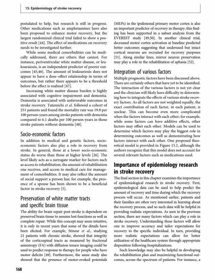
postulated to help, but research is still in progress.Other medications such as amphetamines have alsobeen proposed to enhance motor recovery, but thelargest randomized clinical trial failed to show a pos-itive result [44]. The effect of medications on recoveryneeds to be investigated further.
While some medical comorbidities can be medi-cally addressed, there are others that cannot. Forinstance, periventricular white matter disease, or leu-koaraiosis, is an independent predictor of poorer out-comes [45,46]. The amount of leukoariosis does notappear to have a dose–effect relationship in terms ofoutcomes, but rather there appears to be a thresholdbefore the effect is realized [47].
Increasing white matter disease burden is highlyassociated with cognitive impairment and dementia.Dementia is associated with unfavorable outcomes instroke recovery. Tatemichi et al. followed a cohort of251 patients and found the mortality rate was 19.8 per100 person-years among stroke patients with dementiacompared to 6.1 deaths per 100 person-years in thosestroke patients without dementia [48].
Socio-economic factorsIn addition to medical and genetic factors, socio-economic factors also play a role in recovery fromstroke. In general, those at a lower socio-economicstatus do worse than those at higher levels [26]. Thelevel likely acts as a surrogate marker for factors suchas access to rehabilitation, the amount of rehabilitationone receives, and access to medical care for manage-ment of comorbidities. It may also reflect the amountof social support a person has; for example, the pres-ence of a spouse has been shown to be a beneficialfactor in stroke recovery [5].
Preservation of white matter tractsand specific brain tissueThe ability for brain repair post-stroke is dependent onpreserved brain tissue to assume lost functions as well ascomplete repair. While this concept may seem intuitive,it is only in recent years that some of the details havebeen elicited. For example, Stinear et al., studying21 patients with chronic stroke, showed that integrityof the corticospinal tracts as measured by fractionalanisotropy (FA) with diffusion tensor imaging could beused to predict response to therapy for people who havemotor deficits [49]. Furthermore, the same study alsoshowed that the presence of motor-evoked potentials
(MEPs) in the ipsilesional primary motor cortex is alsoan important predictor of recovery in therapy; this find-ing has been supported in a subset analysis from theEVEREST study [49,50]. In another clinical trial,decreased motor cortex activation at baseline predictedbetter outcomes suggesting that underused but intactcortical neurons are recruited for recovery purposes[51]. Along similar lines, mirror neuron preservationmay play a role in the rehabilitation of aphasia [52].
Integration of various factorsMultiple prognostic factors have been discussed above.There are certainly others that have yet to be identified.The interaction of the various factors is not yet clearand the clinician will likely have difficulty in determin-ing how to integrate the effects of various stroke recov-ery factors. As all factors are not weighted equally, theexact contribution of each factor, in each patient, isunclear. This can become even more complicatedwhen the factors interact with each other; for example,while some factors can have additive effects, otherfactors may offset each other. Models can be used todetermine which factors may play the biggest role indetermining outcomes as well as demonstrating howfactors interact with each other. One proposed theo-retical model is provided in Figure 15.1, although theauthors recognize that this model does not account forseveral relevant factors such as medications used.
Importance of epidemiology researchin stroke recoveryThe final section in this chapter examines the importanceof epidemiological research in stroke recovery. First,epidemiological data can be used to help predict theamount of recovery and time during which the recoveryprocess will occur. As mentioned earlier, patients andtheir families are often very interested in learning aboutthe recovery process, and so such data will be helpful inproviding realistic expectations. As seen in the previoussection, there are many factors which can play a role instroke recovery. Understanding these factors will allowone to improve accuracy and tailor expectations forrecovery to the specific individual. In turn, providingmore realistic expectations can allow for betterutilization of the healthcare system through appropriatedisposition following hospitalizations.
Such knowledge may also be helpful in developingthe rehabilitation plan and maximizing functional out-comes, across the spectrum of patients. For instance, a
15: Epidemiology of stroke recovery
168
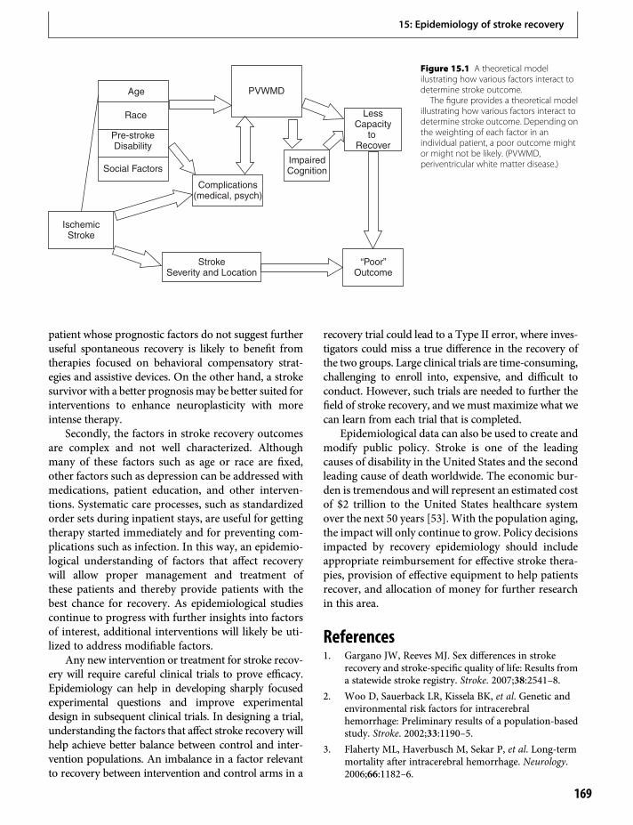
patient whose prognostic factors do not suggest furtheruseful spontaneous recovery is likely to benefit fromtherapies focused on behavioral compensatory strat-egies and assistive devices. On the other hand, a strokesurvivor with a better prognosismay be better suited forinterventions to enhance neuroplasticity with moreintense therapy.
Secondly, the factors in stroke recovery outcomesare complex and not well characterized. Althoughmany of these factors such as age or race are fixed,other factors such as depression can be addressed withmedications, patient education, and other interven-tions. Systematic care processes, such as standardizedorder sets during inpatient stays, are useful for gettingtherapy started immediately and for preventing com-plications such as infection. In this way, an epidemio-logical understanding of factors that affect recoverywill allow proper management and treatment ofthese patients and thereby provide patients with thebest chance for recovery. As epidemiological studiescontinue to progress with further insights into factorsof interest, additional interventions will likely be uti-lized to address modifiable factors.
Any new intervention or treatment for stroke recov-ery will require careful clinical trials to prove efficacy.Epidemiology can help in developing sharply focusedexperimental questions and improve experimentaldesign in subsequent clinical trials. In designing a trial,understanding the factors that affect stroke recovery willhelp achieve better balance between control and inter-vention populations. An imbalance in a factor relevantto recovery between intervention and control arms in a
recovery trial could lead to a Type II error, where inves-tigators could miss a true difference in the recovery ofthe two groups. Large clinical trials are time-consuming,challenging to enroll into, expensive, and difficult toconduct. However, such trials are needed to further thefield of stroke recovery, and wemust maximize what wecan learn from each trial that is completed.
Epidemiological data can also be used to create andmodify public policy. Stroke is one of the leadingcauses of disability in the United States and the secondleading cause of death worldwide. The economic bur-den is tremendous and will represent an estimated costof $2 trillion to the United States healthcare systemover the next 50 years [53]. With the population aging,the impact will only continue to grow. Policy decisionsimpacted by recovery epidemiology should includeappropriate reimbursement for effective stroke thera-pies, provision of effective equipment to help patientsrecover, and allocation of money for further researchin this area.
References1. Gargano JW, Reeves MJ. Sex differences in stroke
recovery and stroke-specific quality of life: Results froma statewide stroke registry. Stroke. 2007;38:2541–8.
2. Woo D, Sauerback LR, Kissela BK, et al. Genetic andenvironmental risk factors for intracerebralhemorrhage: Preliminary results of a population-basedstudy. Stroke. 2002;33:1190–5.
3. Flaherty ML, Haverbusch M, Sekar P, et al. Long-termmortality after intracerebral hemorrhage. Neurology.2006;66:1182–6.
PVWMD
LessCapacity
to Recover
“Poor”Outcome
StrokeSeverity and Location
IschemicStroke
ImpairedCognition
Complications(medical, psych)
Age
Race
Pre-strokeDisability
Social Factors
Figure 15.1 A theoretical modelilustrating how various factors interact todetermine stroke outcome.
The figure provides a theoretical modelillustrating how various factors interact todetermine stroke outcome. Depending onthe weighting of each factor in anindividual patient, a poor outcome mightor might not be likely. (PVWMD,periventricular white matter disease.)
15: Epidemiology of stroke recovery
169
4. Nakayama H, Jorgensen H, Raaschou H, et al. Recoveryof upper extremity function in stroke patients: TheCopenhagen Stroke Study. Arch Phys Med Rehab.1994;75:394–8.
5. Jørgensen HS, Reith J, Nakayama H, et al. Whatdetermines good recovery in patients with the mostsevere strokes? The Copenhagen Stroke Study. Stroke.1999;30:2008–12.
6. Howard VJ, CushmanM, Pulley L, et al. The reasons forgeographic and racial differences in stroke study:Objectives and design. Neuroepidemiology.2005;25:135–43.
7. Wolf S, Winstein CJ, Miller JP, et al. Effect ofconstraint-induced movement therapy on upperextremity function 3 to 9 months after stroke: TheEXCITE randomized clinical trial. JAMA.2006;296:2095–3015.
8. Twitchell TE. The restoration of motor functionfollowing hemiplegia in man. Brain. 1951;74:443–80.
9. American Heart Association. Heart Disease and StrokeStatistics – 2004 Update. Dallas, TX: American HeartAssociation; 2003.
10. Dobkin B. Principles and practices of neurologicalrehabilitation. In: Bradley WG, Daroff RB, FenichelGM, et al., editors. Neurology in clinical practice. 4th ed.Philadelphia, PA: Butterworth Heineman; 2004:pp. 1027–70.
11. Shelton FD, Volpe BT, RedingM.Motor impairment asa predictor of functional recovery and guide torehabilitation treatment after stroke. NeurorehabNeural Repair. 2001;15:229–37.
12. Ottenbacher KJ, Jannell S. The results of clinical trials instroke rehabilitation research. Arch Neurol.1993;50:37–44.
13. Task Force Members. Recommendations for theestablishment of stroke systems of care:Recommendations from the American StrokeAssociation’s task force on the development of strokesystems. Stroke. 2005;36:690–703.
14. Kwakkel G, Kollen BJ, van der Grond J, et al.Probability of regaining dexterity in the flaccid upperlimb: The impact of severity of paresis and time sinceonset in acute stroke. Stroke. 2003;34:2181–6.
15. Paolucci S, Antonucci G, Grasso MG, et al. Early versusdelayed inpatient stroke rehabilitation: A matchedcomparison conducted in Italy. Arch Phys Med Rehab.2000;81:695–700.
16. Page SJ, Levine P, Leonard A, et al. Modified constraint-induced therapy in chronic stroke: Results of a single-blinded randomized controlled trial. Phys Ther.2008;88:333–40.
17. Dromerick AW, Lang CE, Birkenmeier RL, et al. VeryEarly Constraint-Induced Movement (VECTORS).A single-center RCT. Neurology. 2009;73:195–201.
18. Desrosiers J, Malouin F, Richards C, et al. Comparisonof changes in upper and lower extremity impairmentsand disabilities after stroke. Int J Rehab Res.2003;26:109–16.
19. Cramer SC, Crafton KR. Somatotopy and movementrepresentation sites following cortical stroke. Exp BrainRes. 2006;168:25–32.
20. Hamdy S, Aziz Q, Rothwell JC, et al. Recovery ofswallowing after dysphagic stroke relates to functionalreorganization in the intact motor cortex. Gastro.1998;115:1104–12.
21. Jongbloed L. Prediction of function after stroke:A critical review. Stroke. 1986;17:765–76.
22. Pedersen P, Jorgensen H, NakayamaH, et al. Aphasia inacute stroke: Incidence, determinants, and recovery.Ann Neurol. 1995;38:659–66.
23. Hier D, Mondlock J, Caplan L. Recovery of behavioralabnormalities after right hemisphere stroke. Neuro.1983;33:345–50.
24. Nichols-Larsen DS, Clark PC, Zeringue A, GreenspanA, Blanton S. Factors influencing stroke survivors’quality of life during subacute recovery. Stroke.2005;36:1480–4.
25. Kissela B, Schneider A, Kleindorfer D, et al. Stroke in abiracial population: The excess burden of stroke amongblacks. Stroke. 2004;35:426–31.
26. Horner RD, Swanson JW, Bosworth HB, et al. Effects ofrace and poverty on the process and outcome ofinpatient rehabilitation services among stroke patients.Stroke. 2003;34:1027–31.
27. Kleim JA, Chan S, Pringle E, et al. BDNF val66metpolymorphism is associated with modified experience-dependent plasticity in human motor cortex. NatNeurosci. 2006;9:735–7.
28. Wade DT, Hewer RL, Wood VA. Stroke: Influence ofpatient’s sex and side of weakness on outcome. ArchPhys Med Rehab. 1984;65:513–6.
29. Glader EL, Stegmayr B, Norrving B, et al. Sexdifferences in management and outcome after stroke: ASwedish national perspective. Stroke. 2003;34:1970–5.
30. Kapral MK, Fang J, Hill MD, et al. Sex differences instroke care and outcomes: Results from the registry ofthe Canadian Stroke Network. Stroke. 2005;36:809–14.
31. Lai SM, Duncan PW, Dew P, et al. Sex differences instroke recovery. Prev Chronic Dis. 2005;3:A13.
32. Fusco R, Morelli D, Venturiero V, et al. Is sex aprognostic factor in stroke rehabilitation? A matchedcomparison. Stroke. 2006;37:2989–94.
33. The NINDS t-PA Stroke Study Group. Generalizedefficacy of t-PA for acute stroke: Subgroup analysis ofthe NINDS t-PA stroke trial. Stroke. 1997;28:2119–25.
34. Bruno A, Biller J, Adams HP, et al. Acute blood glucoselevel and outcome from ischemic stroke: Trial of ORG
15: Epidemiology of stroke recovery
170
10172 in Acute Stroke Treatment (TOAST)Investigators. Neuro. 1999;52:280–4.
35. Kissela BM, Khoury J, Alwell K, et al. Long termmortality after ischemic stroke: The effect of diabetes inthe greater Cincinnati/Northern Kentucky stroke study[abstract from 2004 International Stroke Conference].Stroke. 2004;35:315.
36. Herrmann N, Black SE, Lawrence J, et al. TheSunnybrook Stroke Study: A prospective study ofdepressive symptoms and functional outcome. Stroke.1998;29:618–24.
37. Hajat C, Hajat S, Sharma P. Effects of poststrokepyrexia on stroke outcome: A meta-analysis of studiesin patients. Stroke. 2000;31:410–4.
38. Chamorro A, Horcajada JP, Obach V, et al. The earlysystemic prophylaxis of infection after stroke study:A randomized clinical trial. Stroke. 2005;36:1495–500.
39. Vargas M, Horcajada JP, Obach V, et al. Clinicalconsequences of infection in patients with acute stroke:Is it prime time for further antibiotic trials? Stroke.2006;37:461–5.
40. Kissela B, Lindsell CJ, Kleindorfer D, et al. Clinicalprediction of functional outcome after ischemic stroke:The surprising importance of periventricular whitematter disease and race. Stroke. 2009;41:530–6.
41. Naidech AM, Kreiter KT, Janjua N, et al. Phenytoinexposure is associated with functional and cognitivedisability after subarachnoid hemorrhage. Stroke.2005;36:583–7.
42. Goldstein L, Sygenin Acute Stroke Study Investigators.Common drugs may influence motor recovery afterstroke. Neurology. 1995;45:865–71.
43. Bragoni M, Altieri M, Di Piero V, et al. Bromocriptineand speech therapy in non-fluent chronic aphasia afterstroke. Neurol Sci. 2000;21:19–22.
44. Martinsson L, Hårdemark HG, Wahlgren NG.Amphetamines for improving recovery after stroke
(Cochrane systematic review). In: The CochraneLibrary, 2003. Oxford: Update Software; 2003.
45. Miyao S, Takano A, Teramoto J, et al. Leukoaraiosis inrelation to prognosis for patients with lacunarinfarction. Stroke. 1992;23:1434–8.
46. Samuelsson M, Soderfeldt B, Olsson GB. Functionaloutcome in patients with lacunar infarction. Stroke.1996;27:842–86.
47. Belagaje SR, Lindsell C, Alwell K, et al. The effect ofwhite matter disease (WMD) on post-strokeoutcomes: Dose–effect vs. threshold? [Abstract].Accepted for presentation at the 34th InternationalConference on Stroke and Cerebral Circulation(February 2009).
48. Tatemichi TK, Desmond DW, Mayeux R, et al.Dementia after stroke: Baseline frequency, risks, andclinical features in a hospitalized cohort. Neuro.1992;42:1185–93.
49. Stinear CM, Barber PA, Smale PR, et al. Functionalpotential in chronic stroke patients depends oncorticospinal tract integrity. Brain. 2007;130:170–80.
50. Levy RM, Benson RR, Winstein CJ for the EverestStudy Investigators. Cortical stimulation for upper-extremity hemiparesis from ischemic stroke:Everest Study primary endpoint results [Abstract].In: International Stroke Conference: 2008,New Orleans.
51. Cramer SC, Parrish TB, Levy RM, et al. Predictingfunctional gains in a stroke trial. Stroke.2007;38:2108–14.
52. Buccino G, Solodkin A, Small SL. Functions of themirror neuron system: Implications forneurorehabilitation. Cognit Behav Neurol.2006;19:55–63.
53. Brown DL, Boden-Albala B, Langa KM, et al. Projectedcosts of ischemic stroke in the United States. Neuro.2006;67:1390–5.
15: Epidemiology of stroke recovery
171

Section III
16Treatment Strategies
Issues in clinical trial methodologyfor brain repair after strokeSteven C. Cramer
Brain repair after stroke andpotential clinical applicationsStroke remains a leading cause of adult disability, thethird greatest cause of death in the US (the secondgreatest cause in the world), and the most commonneurologic reason for hospital admission. The annualcost exceeds 68 billion dollars in the US. Approximately795 000 new symptomatic strokes are diagnosed eachyear in the US. Note that asymptomatic strokes, whichare likely a further source of disability [1], occur with aprevalence that is several-fold higher, for example,being present in 22% of adults 65–69 years of age [2].
Currently approved stroke therapies are limited tothe early hours after stroke. As a result, only 2–3%of patients with stroke receive such an intervention[3,4], although this is improving with time, andrates are higher in selected centers. Nonetheless, manypatients who do receive thrombolytic therapy in theacute stage of stroke demonstrate significant disabilitymonths later [5,6]. Currently approved acute stroketherapies target the vasculature rather than the neuralsubstance of the brain. New therapies are needed thattarget the brain itself, reduce disability, and can beaccessed by a majority of patients with stroke. Repair-based therapies have many candidates that might meetthese criteria.
Brain repair after stroke can be defined as a therapeu-tic strategywhose goal is not to salvage acutely threatenedtissue, but instead is to promote the repair and restorationof function in surviving tissue. Repair-based strategiescan thus be pursued across a wide range of post-stroketime epochs, from the hours following infarct stabiliza-tion to years after stroke onset. Divergent biologicaltargets and therapeutic modalities have been advancedin this regard (see Chapters 17–24). Some focus on spe-cific cellular and molecular events, such as catecholami-nergic drugs, while others such as brain stimulation aim
to improve brain function through more global means.Clinical trials are underway to evaluate therapiesacross several post-stroke temporal epochs, rangingfrom days to weeks to years after stroke onset.
Clinical application of brain repair after stroke is,in general, at an early phase: in most cases, clinicalinvestigations are either preclinical or at phase I or II,although positive phase III data have been publishedfor methods such as intensive physiotherapy [7,8]. Asa result, fundamental questions remain to optimizeclinical trial methodology for brain repair after stroke,some of which are considered below.
Issues from acute stroke trials informrepair-based stroke trialsTo date, the vast majority of acute stroke trials have notshown benefit over placebo. A number of lessons havebeen gleaned from this acute stroke experience thatmightbe useful to design of clinical trials in the emerging field ofbrain repair. Issues range fromclinical development [9] toentry criteria [10] to outcomemeasures [11] and beyond.
Methods to identify, thenmeasure, the target tissue ortarget brain system, are important, and need to be vali-dated [12,13]. Incomplete addressing of this issue mighthave contributed to the outcome of some previouslynegative stroke trials [14]. In the setting of brain repair,thismight consist ofmeasuring the activity or integrity ofa target cortical area [15,16], neurochemical system [17],or white matter tract [18] that is a core component ofthe therapeutic target. Suchmethodsmight be importantin relation to improved patient selection, which is likelyto be just as important for repair as it is in acute stroke,and in relation to biomarkers (see below). Examplesmight include measures of brain function via functionalMRI, measures of tissue physiology such as transcranialmagnetic stimulation, or measures of regional braininjury such as diffusion tensor imaging [16,18].
Brain Repair After Stroke, ed. S. C. Cramer and R. J. Nudo. Published by Cambridge University Press.© Cambridge University Press 2010.
173

Dose–response relationships, important in somany other areas of clinical medicine, are likely to bejust as important with brain repair [14]. The results ofthe VECTORS trial, where a higher dose of physio-therapy was associated with poorer behavioraloutcome, underscores this point [19]. These relation-ships might change during the different temporalepochs that follow a stroke [20]. For example, effectsof a GABA agonist or NMDA receptor blocker varydepending on whether they are given early [21,22] orlate [23–25] after stroke.
When translating the results of animal studies intohuman clinical trial protocols, many of the lessons fromacute stroke trials [26–30] might prove useful forrepair-based trials. Issues include matching variablessuch as treatment time window, route of administra-tion, or dosing schedule. Review of methodologicalquality of animal studies is an increasing consideration[30]. Use in animal studies of human clinical trial levelrandomization, concealment of treatment allocation,and blinded outcome assessment might be a key deter-minant of how well animal studies translate to humantrials [31]. Animalmodels can be of limited relevance tohuman patients, with animals often being relativelyyounger [9] and without medical complications orstroke risk factors – for example, in most human butfew animal stroke studies, the majority of subjectshave hypertension or diabetes mellitus or both (seeChapters 8 and 15). Measures related to executive func-tion, affect, hand dominance, and bipedal gait aregenerally not considered in animal studies, but candominate the recovery of a human patient. The organ-ization of rodent brains shows vast differences as com-pared to humans; for example, white matter constitutes14% of the rodent brain volume vs. 50% in humans[32,33]. Reorganization after stroke might show differ-ences of a similar magnitude. The experimental infarctin preclinical models tends to be homogeneous, insharp contrast with humans [34]. Complicating this isthat the choice of animal infarct model influences sub-sequent repair-related molecular events [35–37], andthus might also influence the likelihood of successfultranslation to humans in the restorative therapy setting.
Repair-based therapies interact withexperience, training, andenvironmentOne major difference between acute and repair-basedstroke clinical trials is that acute stroke therapies exert
their effect rapidly, around the time of therapy,whereas for a repair-based trial, many of the biologicalevents that improve final behavioral outcome willoccur days to weeks after therapy initiation. A subject’sexperiences, concomitant training, and environmentcan each interact with a repair-based therapy in defin-ing the final therapy effects. Evidence suggests thatboth the quantity and the quality of influences canaffect brain repair and behavioral outcome [38–45].
One implication of these influences is that for arepair-based trial, many variables that would be con-sidered of limited significance to an acute stroke trialattain increased importance as potential covariates ofinterest. The details of human experience are complexbut become relevant. Furthermore, some of thesedetails are unique to humans, challenging translationalefforts from preclinical studies to human trials. Thus,influences that arise between the time that a repair-based therapy is initiated (e.g. time of discharge fromthe hospital) and the time of final clinical trial outcomeassessment (e.g. 90 days after stroke onset) requiresubstantial consideration because they can interactwith repair-based therapy and modify its effects.
There are multiple ways that study design might, atleast in part, attempt to control these issues. In addi-tion in some cases, these influences can be preciselycontrolled. In some studies, entry criteria exclude sub-jects whose post-stroke experience is likely to be highlyunusual – for example, subjects who will not be able toreceive standard of care physiotherapy. Another wayfor study design to address this issue relates to thetiming of certain study components. For example, instudies of amphetamine that aimed to improve armmotor function early after stroke [46,47], a predefinedphysiotherapy protocol was initiated at a set numberof minutes after medication ingestion, timed to coin-cide with peak drug levels. In many cases, however,such control would be more difficult. For example,controlling and even monitoring patient experiencewould be challenging for a growth factor that isthought to exert its effects over days to weeks followingits ingestion. Even in the amphetamine examples,enrollees might have had varied experiences and phys-iotherapy regimens outside of the study – for example,between days of drug provision and the day of finaloutcome measurements.
When external influences cannot be controlled,study design can insure that they are at least measured.Thus, prospective plans to measure those componentsof human experience that are most relevant to drug
16: Issues in clinical trial methodology for brain repair after stroke
174

effects or outcome measures can be incorporated into astudy’s data collection plan. Such an approach provideduseful insights in one recent repair-based stroke clinicaltrial, where the amount of “outside” physiotherapy (i.e.physiotherapy occurring in parallel with trial participa-tion, but prescribed by private physicians, outside oftrial jurisdiction), was found to differ significantlybetween active and placebo treatment arms [20]. Suchmeasures can then be treated as planned covariates ofinterest in statistical analyses. Note too that such influ-ences might be relatively domain-specific. For example,a study that aims to study how a repair-based therapyaffects gait velocity would likely prioritize measurementof non-study therapy focused on gait training overmeasurement of non-study therapy focused on swal-lowing. Measurement tools might include question-naires or selected devices such as pedometers [48] oractigraphy [49].
Other covariates relevant torepair-based trialsOther factors might also influence the likelihood that arepair-based therapy will be found to be effective inhuman subjects (see Chapters 14 and 15). A numberof such factors that influence brain repair, and are thusof potential interest to clinical trials of repair-basedtherapies, have been described [50]. These includesocio-economic factors [51], caregiver status [52,53],and depression/affective state [54–56]. In addition,many of the covariates relevant to acute stroke trialsare likely to remain important when trials enroll thesepatients in repair-based trials. For example, in a study ofcognitive recovery after a first stroke, diabetes mellituswas found to be associated with poorer outcome [57].
Genetic factors might also be important to recov-ery after stroke, and thus therapeutic attempts toimprove it [58–61].
An important point is that certain medications canadversely influence brain repair, recovery, and out-come after stroke [62–67]. Thus, medications notonly can promote brain repair, evidence suggeststhey can also impede it (see Chapter 17).
For many of these covariates, a great need exists tostandardize methods of measurement. Improved meth-ods and tools are needed to consistently and accuratelymeasure experience-related covariates of interest.Medical comorbidities and complications are commonin the early days to weeks after a stroke [68–70], and caninfluence final outcome [71]. Harmonization is neededfor defining, recording, and assigning significance to
such events in the context of a clinical trial. The overalleffectiveness of a therapy active during the weeks fol-lowing a stroke might hinge as much on the accurateassessment of these details as on therapeutic benefits.
Issues of study design forrepair-based trialsA number of issues of study design arise when target-ing brain repair after stroke. Some are not unique tobrain repair; for example, a cross-over study designmight not be feasible in many cases [72], as in somecases, e.g. during the first month post-stroke, one canonly try one intervention without changing the targetneural system forever.
One approach worthy of consideration for repair-based therapies is to focus on within-subject change asthe primary means of assessing the endpoint. Manyacute stroke trials have been limited in this regard, asmeasurement of global outcome scales or disabilityscales can be difficult in the early hours after stroke,when baseline measurements are recorded. However,the trial design of most repair-based trials likely willallow for recording of most endpoints at baseline, priorto initiation of therapy, due to the relatively wide treat-ment time window typically suggested for this categoryof intervention. Assessing therapy effects using within-subject change over time, as compared to using cross-sectional subject data acquired at a single late timepoint, can reduce variance and increase statistical power.
As an extension of this issue, study design forrepair-based trials initiated in the acute or early sub-acute period after stroke must carefully define whenthe baseline measures are acquired. In many forms ofstroke, substantial fluctuation of symptoms is notuncommon during the first few days [73]. Baselinemeasures should be acquired as close as possible tothe time of therapy initiation.
Blinding might be more difficult with repair-basedstudies as compared to acute stroke studies, for multi-ple possible reasons. For example, this might arise dueto use of multiple therapy exposures in many repair-based paradigms, such as the many days of therapyemployed in constraint-induced or robot-based ther-apy, in contrast to briefer therapy exposures for manyacute stroke interventions. Also, patients are oftenmore alert during the period of repair-based interven-tions as versus the early hours [26] post-stroke.Patients in a repair-based trial might be more likelyto notice therapy side effects, which could lead tounblinding and thus, to expectation bias [20].
16: Issues in clinical trial methodology for brain repair after stroke
175
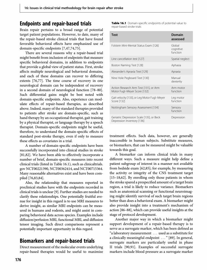
Endpoints and repair-based trialsBrain repair pertains to a broad range of potentialtarget patient populations. However, to date, many ofthe repair-based stroke clinical trials that have foundfavorable behavioral effects have emphasized use ofdomain-specific endpoints [7,47,74,75].
There are several reasons why a repair-based trialmight benefit from inclusion of endpoints that measurespecific behavioral domains, in addition to endpointsthat provide a global view of patient status. First, strokeaffects multiple neurological and behavioral domains,and each of these domains can recover to differingextents [76,77]. The time course of recovery in oneneurological domain can be independent of recoveryin a second domain of neurological function [78–80].Such differential gains might be best noted withdomain-specific endpoints. Also, experience can mod-ulate effects of repair-based therapies, as describedabove. Indeed, many of the standard therapies providedto patients after stroke are domain-specific, such ashand therapy by an occupational therapist, gait trainingby a physical therapist, or language therapy by a speechtherapist. Domain-specific endpoints might be useful,therefore, to understand the domain-specific effects ofstandard post-stroke therapy, even if only to measurethese effects as covariates in a trial.
A number of domain-specific endpoints have beensuccessfully incorporated into clinical studies in stroke[81,82]. We have been able to effectively incorporate anumber of brief, domain-specific measures into recentclinical trials (listed in Table 16.1), such as clinicaltrials.gov NCT00221390, NCT00362414, and NCT00715364.Many reasonable alternatives exist and have been com-piled [76,83,84].
Also, the relationship that measures reported inpreclinical studies have with the endpoints recorded inclinical trials is unclear [9]. Further studies are needed toclarify these relationships. One potentially fruitful ave-nue for insight in this regard is to use MRI measures toderive insight, as similar MRI endpoints can be meas-ured in humans and rodents, and might assist in com-paring behavioral data across species. Examples includediffusion/perfusion MRI, functional MRI, and diffusiontensor imaging. Such direct comparisons represent apotentially important opportunity in this regard.
Biomarkers and repair-based trialsDirectmeasurement of themolecular events underlyingrepair-based therapies would be useful to maximize
treatment effects. Such data, however, are generallyinaccessible in human subjects. Substitute measures,or biomarkers, that can be measured might be valuabletowards this goal.
A biomarker can inform clinical trials in manydifferent ways. Such a measure might help define apatient subgroup of interest in a manner not availablefrom bedside exam [62,85]; for example, by measuringthe activity or integrity of the CNS treatment target[15–18,62]. By enrolling only those patients in whomthe stroke spared a prespecified amount of a target brainregion, a trial is likely to reduce variance. Biomarkerssuch as anatomical scanning or functional neuroimag-ing might identify survival of such target brain regionsbetter than does a behavioral exam. A biomarker mightalso provide insight into a treatment’s mechanism ofaction [86–88], which can provide useful insights at thestage of protocol development.
Another major way in which a biomarker mightsupport development of a repair-based therapy is toserve as a surrogate marker, which has been defined as“a laboratorymeasurement . . . used as a substitute fora clinically meaningful endpoint . . . ” [89]. In general,surrogate markers are particularly useful in phaseII trials [90,91]. Examples of successful surrogatemarkers include blood pressure as a surrogate marker
Table 16.1 Domain-specific endpoints of potential value torepair-based stroke trials
Test Domainassessed
Folstein Mini-Mental Status Exam [126] Globalcognitivefunction
Line cancellation test [127] Spatial neglect
Boston Naming Test [128] Aphasia
Alexander’s Apraxia Test [129] Apraxia
Nine Hole Pegboard Test [130] Manualdexterity
Action Research Arm Test [131], or ArmMotor Fugl–Meyer Score [132]
Arm motorfunction
Gait velocity [133], or Leg Motor Fugl–MeyerScore [132]
Leg motorfunction
Nottingham Sensory Assessment [134] Sensoryfunction
Geriatric Depression Scale [135], or BeckDepression Inventory [136]
Depression
16: Issues in clinical trial methodology for brain repair after stroke
176
for vascular death, tumor size as a surrogate marker ofsurvival, and HIV RNA level as a surrogate marker ofprogression to AIDS. Biomarker data can at times be aunique source of information for guiding therapeuticdecision-making. This approach has been suggested inthe acute stroke setting, where diffusion–perfusionmismatch has been proposed as a surrogate markerof salvable ischemic penumbra [92–94].
A number of caveats exist when considering usinga surrogate marker [95]. A good surrogate marker isgenerally easier to measure than are behavioral end-points, easier to standardize, and saves time andmoney [90,96]. However, for many of the measuressuggested as surrogate markers for brain repair, suchas TMS, fMRI, or PET, these features are not clearlyestablished. Nevertheless, a proof of concept study in acarefully defined patient subgroup can be informativeand of high guidance, and so investigations into theutility of such measures in this context continue.
Another caveat is that a surrogatemarker serves bestwhen its relationships with the disease process and withthe therapy are well understood. For example, a surro-gate measure has reduced utility when it is not in thecausal pathway of the disease process, when the therapyselectively affects physiology of the surrogate, or whenthe surrogate measure does not fully capture the neteffect of therapy on the clinical outcome [90,97]. Theusual clinometric properties by which other outcomemeasures are evaluated, such as validity, reliability, sen-sitivity, and specificity, remain important to surrogatemarkers, but limited study of such measures has beenperformed in the context of brain repair [98–101]. Themany covariates influencing brain repair (above) likelyalso color the utility of surrogatemarkers in this context.
A number of measures of potential interest areavailable to serve as biomarkers in this context, someof which are relatively simple to measure. For example,certain blood tests might provide a measure of repair-related biological events that influence subsequentclinical outcome [102,103]. Direct anatomical meas-ures, such as infarct volume [104–106] or regionalcortical thickness [107], might have predictive valuefor response to a repair-based therapy, and so havepotential as biomarkers. Measures of injury to a prede-fined functional brain region, such as the extent ofinsult to the hand region of primary motor cortex[108], white matter cholinergic projections [109], orleft temporal language areas [110], might provide sub-stantial insight into the likelihood that a particulartherapy will be able to promote repair in a given patient.
More complexmeasures are also of potential value asbiomarkers of repair-based therapy effects. A number ofMRI-based measures have been examined. Diffusiontensor imaging (DTI) provides insight into integrity ofwhite matter tracts and can provide insight into post-stroke brain repair [18,111]. One potential biomarkerusing this method is the mean value for fractionalanisometry within a prespecified region of interest,such as the posterior limb of the internal capsule for amotor-related study [18]. Blood oxygenation level-dependent (BOLD) fMRI provides insights into brainrepair [112,113] and can predict treatment response ina clinical stroke trial [16,114]. The impact of cerebro-vascular disease on the use of this method, which relieson neuronovascular coupling, remains uncertain, butthe convergence of fMRI data findings with resultsfrom numerous other human and animal investigationssupports its validity [50]. Potential biomarkers usingfMRI include the laterality index, a measure of thebalance of interhemispheric control [115,116]; task-related signal change, which reflects the magnitude ofneuronal activity in relation to task performance [16];and regional activation volume [20], which describesthe spatial extent of task-related activation within aregion of interest. Connectivity-based [117], spectro-scopic [118,119], and multimodal approaches have alsobeen advanced [120,121].
Othermethodsmight also provide useful biomarkersfor trials of repair-based therapies. Physiological meas-ures such as TMS can provide insight into tissue functionand its changes with therapy [88,122], especially in themotor system (see Chapter 10). Potential biomarkerswith this method include the motor threshold, whichdescribes the amount of energy required to elicit a crite-rion motor system response; motor-evoked potential(MEP), which describes the skeletal muscle response tomotor cortex stimulation; and the latency, which reflectsthe time between cortical stimulation and the MEP.Brain function can also be measured with PET, as wellas with electroencephalography or magnetoencephalog-raphy. PET is also able to measure many other brainprocesses, such as receptor occupation, oxygen extrac-tion, protein synthesis, and metabolism [123–125].
The choice of biomarker might also be driven bypragmatic issues. Many patients with stroke are oftenfrail or unwell, with limited capacity to undergoextended testing. Many stroke patients have disabilityin a number of neurological domains [77] that mightlimit testing. For example, a patient with aphasia,hemi-inattention, depression, or dementia might not
16: Issues in clinical trial methodology for brain repair after stroke
177
be able to properly undergo surrogate marker testingwhen testing requires a high level of cooperation orparticipation.
Much work remains to bring biomarkers of strokerecovery to the point of utility. The degree of variance inbehavioral outcome after therapy explained by bio-markers remains limited. For example, a measure ofCNS injury based on DTI accounted for only 38% ofthe variance in clinical gains in one study [18]; an fMRI-based measure of CNS function, only 20% [16]. Muchstudy is needed for all repair-based candidates asbiomarkers to understand their value to clinical trials.
ConclusionsA number of classes of therapy are under evaluation topromote brain repair after stroke. Clinical trials of theseagents are emerging. In some regards, the design of thesetrials is directly informed by experience with acute stroketrials. In other regards, there are unique features torepair-based trials. Issues related to covariates of interest,endpoints, and biomarkers in this context have beenconsidered above. Improvements in the design of repair-based trials will help translate preclinical findings, andmaximize the application of this class of therapy to thelarge population of patients living with stroke.
Measures of global outcome such as the NIH StrokeScale, the Barthel Index, and the modified Rankin Scaleare of potential value to measuring effects of repair-based therapies. In addition, measuring therapy effectsin specific domains might have value. Table 16.1 listsselected domain-specific endpoints of potential interest.
References1. Yue NC, Longstreth WT Jr., Elster AD, et al. Clinically
serious abnormalities found incidentally at MRimaging of the brain: Data from the CardiovascularHealth Study. Radiology. 1997;202:41–6.
2. Lloyd-Jones D, Adams R, Carnethon M, et al. Heartdisease and stroke statistics – 2009 update: A reportfrom the American Heart Association StatisticsCommittee and Stroke Statistics Subcommittee.Circulation. 2009; 119:480–6.
3. Reed S, Cramer S, Blough D, et al. Treatment withtissue plasminogen activator and inpatient mortalityrates for patients with ischemic stroke treated incommunity hospitals. Stroke. 2001;32:1832–40.
4. Katzan I, Furlan A, Lloyd L, et al. Use of tissue-typeplasminogen activator for acute ischemic stroke: TheCleveland area experience. JAMA. 2000;283:1151–8.
5. The National Institute of Neurological Disorders andStroke rt-PA Stroke Study Group. Tissue plasminogen
activator for acute ischemic stroke. N Engl J Med.1995;333:1581–7.
6. Furlan A, Higashida R, Wechsler L, et al. Intra-arterialprourokinase for acute ischemic stroke. The PROACTII study: A randomized controlled trial. Prolyse inAcute Cerebral Thromboembolism. JAMA.1999;282:2003–11.
7. Wolf SL,Winstein CJ,Miller JP, et al. Effect of constraint-induced movement therapy on upper extremity function3 to 9 months after stroke: The EXCITE randomizedclinical trial. JAMA. 2006;296:2095–104.
8. Wolf SL,Winstein CJ, Miller JP, et al. Retention of upperlimb function in stroke survivors who have receivedconstraint-induced movement therapy: The EXCITErandomised trial. Lancet Neurol. 2008;7:33–40.
9. Savitz SI, Fisher M. Future of neuroprotection for acutestroke: In the aftermath of the SAINT trials. AnnNeurol. 2007;61:396–402.
10. Uchino K, Billheimer D, Cramer S. Entry criteria andbaseline characteristics predict outcome in acute stroketrials. Stroke. 2001;32:909–16.
11. Duncan P, Jorgensen H,Wade D. Outcomemeasures inacute stroke trials: A systematic review and somerecommendations to improve practice. Stroke.2000;31:1429–38.
12. Toth G, Albers GW. Use of MRI to estimate thetherapeutic window in acute stroke: Is perfusion-weighted imaging/diffusion-weighted imagingmismatch an EPITHET for salvageable ischemic braintissue? Stroke. 2009;40:333–5.
13. Donnan GA, Baron JC, Ma H, et al. Penumbralselection of patients for trials of acute stroke therapy.Lancet Neurol. 2009;8:261–9.
14. Feuerstein GZ, Zaleska MM, Krams M, et al. Missingsteps in the STAIR case: A Translational Medicineperspective on the development of NXY-059 fortreatment of acute ischemic stroke. J Cereb Blood FlowMetab. 2008;28:217–9.
15. Cramer S, Benson R, Burra V, et al. Mapping individualbrains to guide restorative therapy after stroke:Rationale and pilot studies. Neurol Res. 2003;25:811–4.
16. Cramer SC, Parrish TB, Levy RM, et al. Predictingfunctional gains in a stroke trial. Stroke.2007;38:2108–14.
17. Tardy J, Pariente J, Leger A, et al. Methylphenidatemodulates cerebral post-stroke reorganization.NeuroImage. 2006;33:913–22.
18. Stinear CM, Barber PA, Smale PR, et al. Functionalpotential in chronic stroke patients depends oncorticospinal tract integrity. Brain. 2007;130:170–80.
19. Dromerick A, Lang C, Powers W, et al. Very EarlyConstraint-Induced Movement Therapy (VECTORS):Phase II trial results. Stroke. 2007;38:465.
16: Issues in clinical trial methodology for brain repair after stroke
178
20. Cramer S, Dobkin B, Noser E, et al. A randomized,placebo-controlled, double-blind study of ropinirole inchronic stroke. Stroke. 2009;40:3034–8.
21. Green AR, Hainsworth AH, Jackson DM. GABApotentiation: A logical pharmacological approach forthe treatment of acute ischaemic stroke.Neuropharmacology. 2000;39:1483–94.
22. Ovbiagele B, Kidwell CS, Starkman S, et al.Neuroprotective agents for the treatment of acuteischemic stroke. Curr Neurol Neurosci Rep.2003;3:9–20.
23. Kozlowski D, Jones T, Schallert T. Pruning of dendritesand restoration of function after brain damage: Role ofthe NMDA receptor. Restor Neurol Neurosci.1994;7:119–26.
24. Wahlgren N, Martinsson L. New concepts for drugtherapy after stroke. Can we enhance recovery?Cerebrovasc Dis. 1998;8(Suppl 5): 33–8.
25. Barth T, Hoane M, Barbay S, et al. Effects of glutamateantagonists on the recovery and maintenance ofbehavioral function after brain injury. In: Goldstein L,editor. Restorative neurology: Advances inpharmacotherapy for recovery after stroke. Armonk, NY:Futura Publishing Co., Inc.; 1998.
26. Grotta J, Bratina P. Subjective experiences of 24 patientsdramatically recovering from stroke. Stroke.1995;26:1285–8.
27. Fisher M, Ratan R. New perspectives on developingacute stroke therapy. Ann Neurol. 2003;53:10–20.
28. Gladstone D, Black S, Hakim A. Toward wisdom fromfailure: lessons from neuroprotective stroke trials andnew therapeutic directions. Stroke. 2002;33:2123–36.
29. Fisher M, Feuerstein G, Howells DW, et al. Update ofthe Stroke Therapy Academic Industry RoundtablePreclinical Recommendations. Stroke.2009;40:2244–50.
30. Philip M, Benatar M, Fisher M, et al. Methodologicalquality of animal studies of neuroprotective agentscurrently in phase II/III acute ischemic stroke trials.Stroke. 2009;40:577–81.
31. Macleod MR, van der Worp HB, Sena ES, et al.Evidence for the efficacy of NXY-059 in experimentalfocal cerebral ischaemia is confounded by study quality.Stroke. 2008;39:2824–9.
32. Goldberg MP, Ransom BR. New light on white matter.Stroke. 2003;34:330–2.
33. Cramer S. Clinical issues in animal models of strokeand rehabilitation. ILAR J. 2003;44:83–4.
34. Endres M, Engelhardt B, Koistinaho J, et al. Improvingoutcome after stroke: Overcoming the translationalroadblock. Cerebrovasc Dis. 2008;25:268–78.
35. Napieralski JA, Butler AK, Chesselet MF. Anatomicaland functional evidence for lesion-specific sprouting of
corticostriatal input in the adult rat. J Comp Neurol.1996;373:484–97.
36. Voorhies A, Jones T. The behavioral and dendriticgrowth effects of focal sensorimotor cortical damagedepend on the method of lesion induction. Behav BrainRes. 2002;133:237–46.
37. Windle V, Szymanska A, Granter-Button S, et al. Ananalysis of four different methods of producing focalcerebral ischemia with endothelin-1 in the rat. ExpNeurol. 2006;201:324–34.
38. Kwakkel G. Impact of intensity of practice after stroke:Issues for consideration. Disabil Rehabil.2006;28:823–30.
39. Kwakkel G, Wagenaar R, Twisk J, et al. Intensity of legand arm training after primary middle-cerebral-arterystroke: A randomised trial. Lancet. 1999;354:191–6.
40. Dobkin B. The clinical science of neurologicrehabilitation. New York, NY: Oxford University Press;2003.
41. Van Peppen RP, Kwakkel G, Wood-Dauphinee S, et al.The impact of physical therapy on functional outcomesafter stroke: What’s the evidence? Clin Rehabil.2004;18:833–62.
42. Cicerone KD, Dahlberg C, Malec JF, et al. Evidence-based cognitive rehabilitation: Updated review of theliterature from 1998 through 2002. Arch Phys MedRehabil. 2005;86:1681–92.
43. Bhogal S, Teasell R, Speechley M. Intensity of aphasiatherapy, impact on recovery. Stroke. 2003;34:987–93.
44. Jones T, Chu C, Grande L, et al. Motor skills trainingenhances lesion-induced structural plasticity in themotor cortex of adult rats. J Neurosci.1999;19:10 153–63.
45. Johansson B. Brain plasticity and stroke rehabilitation.The Willis lecture. Stroke. 2000;31:223–30.
46. Gladstone DJ, Danells CJ, Armesto A, et al.Physiotherapy coupled with dextroamphetamine forrehabilitation after hemiparetic stroke: A randomized,double-blind, placebo-controlled trial. Stroke.2006;37:179–85.
47. Walker-Batson D, Smith P, Curtis S, et al.Amphetamine paired with physical therapy acceleratesmotor recovery after stroke. Further evidence. Stroke.1995;26:2254–9.
48. Macko RF, Haeuber E, Shaughnessy M, et al.Microprocessor-based ambulatory activity monitoringin stroke patients.Med Sci Sports Exerc. 2002;34:394–9.
49. Reiterer V, Sauter C, Klosch G, et al. Actigraphy – Auseful tool for motor activity monitoring in strokepatients. Eur Neurol. 2008;60:285–91.
50. Cramer SC. Repairing the human brain after stroke:I. Mechanisms of spontaneous recovery. Ann Neurol.2008;63:272–87.
16: Issues in clinical trial methodology for brain repair after stroke
179
51. McFadden E, Luben R, Wareham N, et al. Social class,risk factors, and stroke incidence in men and women: Aprospective study in the European prospectiveinvestigation into cancer in Norfolk cohort. Stroke.2009;40:1070–7.
52. Smith J, Forster A, Young J. Cochrane review:Information provision for stroke patients and theircaregivers. Clin Rehabil. 2009;23:195–206.
53. Glass TA, Matchar DB, Belyea M, et al. Impact of socialsupport on outcome in first stroke. Stroke.1993;24:64–70.
54. Lai SM, Duncan PW, Keighley J, et al. Depressivesymptoms and independence in BADL and IADL.J Rehabil Res Dev. 2002;39:589–96.
55. Jonsson AC, Lindgren I, Hallstrom B, et al.Determinants of quality of life in stroke survivors andtheir informal caregivers. Stroke. 2005;36:803–8.
56. Mukherjee D, Levin RL, Heller W. The cognitive,emotional, and social sequelae of stroke: Psychologicaland ethical concerns in post-stroke adaptation. TopicsStroke Rehabil. 2006;13:26–35.
57. Nys GM, Van Zandvoort MJ, De Kort PL, et al.Domain-specific cognitive recovery after first-everstroke: A follow-up study of 111 cases. J IntNeuropsychol Soc. 2005;11:795–806.
58. Kleim JA, Chan S, Pringle E, et al. BDNF val66metpolymorphism is associated with modified experience-dependent plasticity in human motor cortex. NatNeurosci. 2006;9:735–7.
59. Siironen J, Juvela S, Kanarek K, et al. The Met allele ofthe BDNF Val66Met polymorphism predicts pooroutcome among survivors of aneurysmal subarachnoidhemorrhage. Stroke. 2007;38:2858–60.
60. Alberts MJ, Graffagnino C, McClenny C, et al. ApoEgenotype and survival from intracerebral haemorrhage.Lancet. 1995;346:575.
61. Pearson-Fuhrhop KM, Kleim JA, Cramer SC. Brainplasticity and genetic factors. Top Stroke Rehabil.2009;16:282–99.
62. Butefisch C, Davis B, Wise S, et al. Mechanisms of use-dependent plasticity in the human motor cortex. ProcNatl Acad Sci USA. 2000;97:3661–5.
63. Feeney D, Gonzalez A, Law W. Amphetamine,haloperidol, and experience interact to affect the rate ofrecovery after motor cortex injury. Science.1982;217:855–7.
64. Goldstein L, Sygen in Acute Stroke Study Investigators.Common drugs may influence motor recovery afterstroke. Neurology. 1995;45:865–71.
65. Troisi E, Paolucci S, Silvestrini M, et al. Prognosticfactors in stroke rehabilitation: The possible role ofpharmacological treatment. Acta Neurol Scand.2002;105:100–06.
66. Conroy B, Zorowitz R, Horn SD, et al. An explorationof central nervous systemmedication use and outcomesin stroke rehabilitation. Arch Phys Med Rehabil.2005;86(12 Suppl 2):S73–S81.
67. Lazar R, Fitzsimmons B, Marshall R, et al. Reemergenceof stroke deficits with midazolam challenge. Stroke.2002;33:283–5.
68. Dromerick AW, Khader SA. Medical complicationsduring stroke rehabilitation. Adv Neurol.2003;92:409–13.
69. Kalra L, Yu G, Wilson K, et al. Medical complicationsduring stroke rehabilitation. Stroke. 1995;26:990–4.
70. McLean D. Medical complications experienced by acohort of stroke survivors during inpatient, tertiary-level stroke rehabilitation. Arch Phys Med Rehabil.2004;85:466–9.
71. Berlowitz DR, Hoenig H, Cowper DC, et al. Impact ofcomorbidities on stroke rehabilitation outcomes: Doesthe method matter? Arch Phys Med Rehabil.2008;89:1903–06.
72. Piantadosi S. Clinical trials. A methodologic perspective.New York, NY: John Wiley & Sons, Inc.; 1997.
73. Ozdemir O, Beletsky V, Chan R, et al. Thrombolysis inpatients with marked clinical fluctuations in neurologicstatus due to cerebral ischemia. Arch Neurol.2008;65:1041–3.
74. Volpe BT, FerraroM, Lynch D, et al. Robotics and otherdevices in the treatment of patients recovering fromstroke. Curr Neurol Neurosci Rep. 2005;5:465–70.
75. Scheidtmann K, Fries W, Muller F, et al. Effect oflevodopa in combination with physiotherapy onfunctional motor recovery after stroke: A prospective,randomised, double-blind study. Lancet.2001;358:787–90.
76. Gresham G, Duncan P, Stason W, et al. Post-strokerehabilitation. Rockville, MD: US Department ofHealth and Human Services. Public Health Service,Agency for Health Care Policy and Research; 1995.
77. Rathore S, Hinn A, Cooper L, et al. Characterization ofincident stroke signs and symptoms: Findings from theAtherosclerosis Risk in Communities study. Stroke.2002;33:2718–21.
78. Hier D, Mondlock J, Caplan L. Recovery of behavioralabnormalities after right hemisphere stroke. Neurology.1983;33:345–50.
79. Marshall R, Perera G, Lazar R, et al. Evolution ofcortical activation during recovery from corticospinaltract infarction. Stroke. 2000;31:656–61.
80. Markgraf C, Green E, Hurwitz B, et al. Sensorimotorand cognitive consequences of middle cerebral arteryocclusion in rats. Brain Res. 1992;575:238–46.
81. Hillis A, Newhart M, Heidler J, et al. Anatomy of spatialattention: Insights from perfusion imaging and
16: Issues in clinical trial methodology for brain repair after stroke
180
hemispatial neglect in acute stroke. J Neurosci.2005;25:3161–7.
82. Hillis A, Wityk R, Barker P, et al. Subcortical aphasiaand neglect in acute stroke: The role of corticalhypoperfusion. Brain. 2002;125:1094–104.
83. Duncan PW, Zorowitz R, Bates B, et al. Management ofadult stroke rehabilitation care: A clinical practiceguideline. Stroke. 2005;36:e100–43.
84. Salter K, Jutai J, Zettler L, et al. Outcome measures instroke rehabilitation. http://wwwebrsrcom/uploads/Outcome_Measures_SREBR11_finalpdf. 2008.
85. Collins JM. Functional imaging in phase I studies:Decorations or decision making? J Clin Oncol.2003;21:2807–09.
86. Carey J, Kimberley T, Lewis S, et al. Analysis of fMRIand finger tracking training in subjects with chronicstroke. Brain. 2002;125:773–88.
87. Johansen-Berg H, Dawes H, Guy C, et al. Correlationbetween motor improvements and altered fMRIactivity after rehabilitative therapy. Brain.2002;125:2731–42.
88. Koski L, Mernar T, Dobkin B. Immediate and long-term changes in corticomotor output in response torehabilitation: Correlation with functionalimprovements in chronic stroke. Neurorehabil NeuralRepair. 2004;18:230–49.
89. Temple R. A regulatory authority’s opinion aboutsurrogate endpoints. In: Nimmo W, Tucker G, editors.Clinical measurement in drug evaluation. New York,NY: John Wiley and Sons; 1995.
90. Fleming T, DeMets D. Surrogate end points in clinicaltrials: Are we being misled? Ann Intern Med.1996;125:605–13.
91. Stephen RM, Gillies RJ. Promise and progress forfunctional and molecular imaging of response totargeted therapies. Pharm Res. 2007;24:1172–85.
92. Lansberg MG, Thijs VN, Hamilton S, et al. Evaluationof the clinical-diffusion and perfusion–diffusionmismatch models in DEFUSE. Stroke.2007;38:1826–30.
93. Furlan AJ, Eyding D, Albers GW, et al. Dose Escalationof Desmoteplase for Acute Ischemic Stroke (DEDAS):Evidence of safety and efficacy 3 to 9 hours after strokeonset. Stroke. 2006;37:1227–31.
94. Hacke W, Albers G, Al-Rawi Y, et al. The Desmoteplasein Acute Ischemic Stroke trial (DIAS): A phase IIMRI-based 9-hour window acute stroke thrombolysistrial with intravenous desmoteplase. Stroke.2005;36:66–73.
95. Hsia A, Kidwell C. Magnetic resonance imaging forsurrogate outcomes and patient selection. In:Woodbury-Harris K, Coull B, editors. Clinical trials inthe neurosciences. Basel: Karger; 2009: pp. 89–92.
96. Boissel J, Collet J, Moleur P, et al. Surrogate endpoints:A basis for a rational approach. Eur J Clin Pharmacol.1992;43:235–44.
97. Bucher H, Guyatt G, Cook D, et al. Users’ guides tothe medical literature: XIX. Applying clinical trialresults. A. How to use an article measuring the effectof an intervention on surrogate end points. Evidence-Based Medicine Working Group. JAMA.1999;282:771–8.
98. Takahashi CD, Der-Yeghiaian L, Le V, et al. Robot-based hand motor therapy after stroke. Brain.2008;131:425–37.
99. Bosnell R, Wegner C, Kincses ZT, et al. Reproducibilityof fMRI in the clinical setting: Implications for trialdesigns. NeuroImage. 2008;42:603–10.
100. Eaton KP, Szaflarski JP, Altaye M, et al. Reliability offMRI for studies of language in post-stroke aphasiasubjects. NeuroImage. 2008;41:311–22.
101. Kimberley TJ, Khandekar G, Borich M. fMRI reliabilityin subjects with stroke. Exp Brain Res/ExperimentelleHirnforschung. 2008;186:183–90.
102. Geiger S, Holdenrieder S, Stieber P, et al. Nucleosomesas a new prognostic marker in early cerebral stroke.J Neurol. 2007;254:617–23.
103. Yip HK, Chang LT, ChangWN, et al. Level and value ofcirculating endothelial progenitor cells in patients afteracute ischemic stroke. Stroke. 2008;39:69–74.
104. Brott T, Marler J, Olinger C, et al. Measurements ofacute cerebral infarction: Lesion size by computedtomography. Stroke. 1989;20:871–5.
105. Saver J, Johnston K, Homer D, et al. Infarct volume as asurrogate or auxiliary outcome measure in ischemicstroke clinical trials. The RANTTAS Investigators.Stroke. 1999;30:293–8.
106. Schiemanck SK, Kwakkel G, Post MW, et al. Predictivevalue of ischemic lesion volume assessed with magneticresonance imaging for neurological deficits andfunctional outcome poststroke: A critical review of theliterature. Neurorehabil Neural Repair.2006;20:492–502.
107. Schaechter JD, Moore CI, Connell BD, et al. Structuraland functional plasticity in the somatosensory cortex ofchronic stroke patients. Brain. 2006;129:2722–33.
108. Crafton K,Mark A, Cramer S. Improved understandingof cortical injury by incorporating measures offunctional anatomy. Brain. 2003;126:1650–9.
109. Bocti C, Swartz RH, Gao FQ, et al. A new visual ratingscale to assess strategic white matter hyperintensitieswithin cholinergic pathways in dementia. Stroke.2005;36:2126–31.
110. Hillis AE, Gold L, Kannan V, et al. Site of the ischemicpenumbra as a predictor of potential for recovery offunctions. Neurology. 2008;71:184–9.
16: Issues in clinical trial methodology for brain repair after stroke
181
111. Ding G, Jiang Q, Li L, et al. Magnetic resonanceimaging investigation of axonal remodeling andangiogenesis after embolic stroke in sildenafil-treatedrats. J Cereb Blood Flow Metab. 2008;28:1440–8.
112. Hodics T, Cohen LG, Cramer SC. Functional imagingof intervention effects in stroke motor rehabilitation.Arch Phys Med Rehabil. 2006;87(12 Suppl):36–42.
113. Richards LG, Stewart KC, Woodbury ML, et al.Movement-dependent stroke recovery: A systematicreview and meta-analysis of TMS and fMRI evidence.Neuropsychologia. 2008;46:3–11.
114. Dong Y, Dobkin BH, Cen SY, et al. Motor cortexactivation during treatment may predict therapeuticgains in paretic hand function after stroke. Stroke.2006;37:1552–5.
115. Cramer S, Nelles G, Benson R, et al. A functional MRIstudy of subjects recovered from hemiparetic stroke.Stroke. 1997;28:2518–27.
116. Naccarato M, Calautti C, Jones PS, et al. Does healthyaging affect the hemispheric activation balance duringpaced index-to-thumb opposition task? An fMRI study.NeuroImage. 2006;32:1250–6.
117. Grefkes C, Nowak DA, Eickhoff SB, et al. Corticalconnectivity after subcortical stroke assessed withfunctional magnetic resonance imaging. Ann Neurol.2008;63:236–46.
118. Parsons M, Li T, Barber P, et al. Combined (1)H MRspectroscopy and diffusion-weightedMRI improves theprediction of stroke outcome. Neurology.2000;55:498–505.
119. Pendlebury S, Blamire A, Lee M, et al. Axonal injury inthe internal capsule correlates with motor impairmentafter stroke. Stroke. 1999;30:956–62.
120. Nair DG, Hutchinson S, Fregni F, et al. Imagingcorrelates of motor recovery from cerebral infarctionand their physiological significance in well-recoveredpatients. NeuroImage. 2007;34:253–63.
121. Ward NS, Newton JM, Swayne OB, et al. Motor systemactivation after subcortical stroke depends oncorticospinal system integrity. Brain. 2006;129:809–19.
122. Talelli P, Greenwood RJ, Rothwell JC. Arm functionafter stroke: Neurophysiological correlates andrecovery mechanisms assessed by transcranial magneticstimulation. Clin Neurophysiol. 2006;117:1641–59.
123. Powers W, Raichle M. Positron emission tomographyand its application to the study of cerebrovasculardisease in man. Stroke. 1985;16:361–76.
124. Heiss WD. Imaging the ischemic penumbra andtreatment effects by PET. Keio J Med. 2001;50:249–56.
125. Ward NS. Neural plasticity and recovery of function.Prog Brain Res. 2005;150:527–35.
126. Folstein MF, Folstein SE, McHugh PR. “Mini-mentalstate”. A practical method for grading the cognitivestate of patients for the clinician. J Psychiatr Res.1975;12:189–98.
127. Albert ML. A simple test of visual neglect. Neurology.1973;23:658–64.
128. Kent PS, Luszcz MA. A review of the Boston NamingTest and multiple-occasion normative data for olderadults on 15-item versions. Clin Neuropsychol.2002;16:555–74.
129. Alexander MP, Baker E, Naeser MA, et al.Neuropsychological and neuroanatomical dimensionsof ideomotor apraxia. Brain. 1992;115: 87–107.
130. Wade DT. Measuring arm impairment and disabilityafter stroke. Int Disabil Stud. 1989;11:89–92.
131. Yozbatiran N, Der-Yeghiaian L, Cramer SC. Astandardized approach to performing the actionresearch arm test. Neurorehabil Neural Repair.2008;22:78–90.
132. Fugl-Meyer A, Jaasko L, Leyman I, et al. The post-stroke hemiplegic patient: a method for evaluation ofphysical performance. Scand J Rehabil Med.1975;7:13–31.
133. Richards C, Malouin F, Dumas F, et al. Gait velocity asan outcome measure of locomotor recovery afterstroke. In: Craik R, Oates C, editors. Gait analysis:Theory and application. St. Louis: Mosby; 1995:pp. 355–64.
134. Lincoln N, Jackson J, Adams S. Reliability and revisionof the Nottingham Sensory Assessment for strokepatients. Physiotherapy. 1998;84:358–65.
135. Agrell B, Dehlin O. Comparison of six depressionrating scales in geriatric stroke patients. Stroke.1989;20:1190–4.
136. Beck AT, Beck RW. Screening depressed patients infamily practice. A rapid technique. Postgrad Med.1972;52:81–5.
16: Issues in clinical trial methodology for brain repair after stroke
182
17 Neuropharmacology in stroke recoveryIsabelle Loubinoux & François Chollet
IntroductionClinicians have long recognized that most strokesurvivors recover over time, albeit to varying degrees.Until now, rTPA thrombolysis within the first hoursof the stroke is recognized as the only validatedtreatment able to improve the spontaneous – andmost of the time incomplete – recovery of neurologicalfunctions after stroke. However, we have learnt fromresearch over the last decade, in part based on theconsiderable improvement of neuroimaging techni-ques, that spontaneous recovery of neurological func-tions was associated with a large intracerebralreorganization of the damaged human brain (seeChapters 9–13). Recruitment of remote functionalareas, overactivation of primary cortices, and changesin corticalmaps are now considered as the physiologicalsubstratum of clinical recovery. Moreover, it has nowbeen demonstrated that brain post-stroke plasticitycan be modulated in order to reduce the residualneurological deficit and the subsequent disability.Rehabilitation, rTMS, peripheral stimulations, localanesthetic blocks, drugs, and other interventions havedemonstrated in certain conditions a capacity to inducean intracerebral reorganization after focal lesion (seealso Chapters 18–24). All are potential therapeuticagents for recovery. They rely on induced functionalchanges in a damaged intracerebral network. Each ofthese therapeutic interventions requires extensive vali-dation. We review in this chapter the data concerningdrug and other neuropharmacological aspects of theissue. We also address the clinical questions concerningthe repair of damaged neuronal networks and potentialfuture therapy using external cellular material.
Animal modelsStudies in laboratory animals clearly show that the rateand extent of functional recovery after focal brain
injury can be modulated by drugs affecting certainneurotransmitters in the central nervous system.Those laboratory experiments indicate that the rateand degree of recovery can be affected by changes inselected neurotransmitters in the central nervoussystem (CNS).
Norepinephrine and motor recoverySeveral lines of evidence suggest that motor recoveryafter injury to the cerebral cortex can be modulatedthrough the effects of norepinephrine on the CNS.Detailed reviews have been published [1–3]. For exam-ple, in rats, central infusion of norepinephrine hastenslocomotor recovery after a unilateral sensorimotorcortex lesion. In addition, bilateral or unilateral selec-tive lesions of the locus ceruleus, the major source ofnoradrenergic projection fibers to the cerebral cortexand cerebellum, also impair motor recovery after asubsequent unilateral cortical lesion. In a provocativeinitial experiment, Feeney and Sutton [1] found that,when combined with task-relevant experience, a singledose of dextroamphetamine given the day after a uni-lateral sensorimotor cortex ablation in the rat resultedin an enduring enhancement of motor recovery. Theamphetamine effect found by Feeney and Sutton hassubsequently been confirmed in relation to functionaldeficits arising from focal lesions produced through avariety of mechanisms, to lesions affecting other areasof the cortex, and to a range of other behaviors.
Norepinephrine agonists and antagonistsGiven the hypothesis that the effect of amphetamineon recovery is exerted through its effect on nor-epinephrine, other drugs that enhance norepinephrinetone would be expected to have similarly favorableeffects on behavioral recovery after stroke. In fact,yohimbine and idazoxan (α2-adrenergic receptor
Brain Repair After Stroke, ed. S. C. Cramer and R. J. Nudo. Published by Cambridge University Press.© Cambridge University Press 2010.
183
antagonists that increase the release of norepinephrinein the CNS) facilitate motor recovery when given as asingle dose after unilateral sensorimotor cortex injury.Similarly, phentermine, an amphetamine analog withweaker cardiovascular effects, phenylpropanolamine,and methylphenidate hydrochloride also acceleratemotor recovery after experimental focal brain injury.
If drugs that enhance norepinephrine release arebeneficial, then drugs that decrease norepinephrinerelease, increase its catatabolism, or block its postsy-naptic effects would be hypothesized to be harmful torecovery. In experiments designed to test this hypoth-esis, a single dose of the α2-adrenergic receptor agonistclonidine hydrochloride, given the day after cortexinjury, was found to have a prolonged detrimentaleffect on motor recovery in rats and to reinstate thedeficit in recovered animals. Prazosin and phenoxy-benzamine, α1-adrenergic receptor antagonists thatact on the CNS, also interfere with recovery.
Other drugsAntidepressantsDepression is common after stroke (see Chapter 14)and often prompts the use of antidepressant medica-tions. The administration of a single dose of trazo-done, a 5HT reuptake blocker, transiently slowsmotor recovery in rats with sensorimotor cortex injuryand reinstates the hemiparesis in recovered animals. Asingle dose of desipramine, a NE reuptake blocker,facilitates motor recovery.
GABAIntracortical infusion of gamma-aminobutyric acid(GABA) was found to increase the hemiparesis pro-duced by a small motor cortex lesion in rats. Theshort-term administration of the benzodiazepinediazepam, an indirect GABA agonist, permanentlyimpedes recovery from the sensory asymmetry causedby damage to the anteromedial neocortex in the rat[4]. The deleterious effect of GABA on motor recoveryafter motor cortex injury is increased by the peripheraladministration of phenytoin. Phenobarbital alsodelays behavioral recovery after injury to the cerebralcortex in laboratory studies. In contrast, neither car-bamazepine nor vigabatrin has detrimental effects.
Dopamine and dopaminergic drugsDopaminergic agents may influence recovery fromneglect caused by prefrontal cortical injury. Apomor-phine, a dopamine agonist, reduces the severity of
experimentally induced neglect, and spiroperidol,a dopamine receptor antagonist, reinstates neglectin recovered animals. Concurrent administration ofdopamine-blocking drugs such as haloperidol alsoblocks amphetamine-promoted recovery, and haloper-idol, as well as other butyrophenones (fluanisone, dro-peridol), transiently reinstates the deficits in recoveredanimals.
General trends from experimental studiesFrom published experimental studies, several mainconclusions can be emphasized.
– First, one can be convinced from these studies thatthere is obviously a large interaction betweencertain drugs and the recovery process in animalmodels. Norepinephrine and its agonists andantagonists have probably been the most studieddrugs, but others with potentially fewer side effectsmight also be beneficial. This is undoubtedlysufficient data from animal studies to support theneed for further human studies [5].
– Second, it appears that the cellular mechanismsunderlying these significant effects of drugs actingon the CNS are still not well understood. Additionalbasic research is needed to better understand suchpharmacological actions in the setting of rewiringand cellular growth in the damaged brain.
– Third, drugs can have varying effects based on thedosage and also the dose regimen. For example,animal studies have found that with increasingdose, amphetamine has increasing then decreasingbenefit. We have demonstrated kinetics in humansubjects; for example, with increasing dose ofparoxetine, primary motor cortex activationincreases then decreases.
– Motor deficits have been the most frequentlystudied domain of recovery after a focal lesion.Other neurological domains require further study.
– Moreover, the timing of drug administration maybe crucial. A therapeutic time window probablyexists. Some drugs, such as benzodiazepines, thatmay be neuroprotective when given soon after thestroke, are harmful when given later [6].Amphetamine may no longer be effective after atherapeutic window of opportunity has passed [7].
– Last, the effects of many drugs, particularly theeffects of noradrenergic agents on motor recoveryafter injury to the cerebral cortex, are highlydependent on experimental details. For example,drug infusion paired with behavioral training does
17: Neuropharmacology in stroke recovery
184
not have the same behavioral effect as compared todrug infusion without training.
Clinical trialsTrials reported here were considered reliable sincethey were all randomized, double-blind, and placebo-controlled. In this part of the chapter we will onlymention trials where the main evaluation criterionwas clinical outcome. Ideally, the optimal pharmaco-logical treatment of an individual patient should bepredictable and hypothesis-driven. Hypotheses can insome instances be derived from findings in animalexperiments. Other approaches are possible. Forexample, functional imaging tools such as fMRI,PET, electroencephalography, or TMS allow evalua-tion of the effects of a drug on brain activity in greaterdetail. In some cases, they can be considered as surro-gate markers for drug activity on the human damagedbrain (see Chapter 16). They might represent the ini-tial step of clinical evaluation of the drug, but the finaljudgment must be clinical. The interest of neuroimag-ing will be detailed in the next part of the chapter. Thebelow review considers selected trials. More compre-hensive reviews regarding clinical trials have beenpublished [8,9].
AmphetamineDextroamphetamine enhances the release of noradre-naline and dopamine in the synaptic cleft. Such amechanism seemed promising because animal experi-ments have indicated that the two neurotransmittersmodulated by this drug support activity and motorfunctions.
Recovery of motor functionSeveral studies have tested the effects of d-amphetamineon motor recovery after stroke, including a total of atleast 280 patients.
The first study of the effects of amphetamine onrecovery after stroke in humans [10] was carefullydesigned to simulate the paradigm used in the labo-ratory. Eight patients with stable motor deficits wererandomized to receive a single dose of amphetamineor a placebo within 10 days of ischemic stroke, withdrug administration tightly coupled with physicaltherapy. The following day, the amphetamine-treatedgroup had a significant improvement in motor per-formance (P < .05), whereas there was little change inthe placebo-treated group. A second double-blind,placebo-controlled trial involving 12 patients [57]
found no treatment effect, but it differed in severalways from the previous study. A different dosing regi-men was used, interventions beganmore than 1monthafter the stroke, and the administration of the drug orplacebo was not tightly linked with physical therapy.In a third double-blind, placebo controlled trial [11],a short course of treatment began between 15 and30 days after the stroke, with each dose of amphet-amine or placebo given in tight conjunction withphysical therapy. Patients treated with amphetaminehad significantly greater improvements in motorscores compared with placebo-treated patients(P<.05), and that benefit persisted for as long as 10months after the intervention ceased. In combinationwith the principles learned from the laboratory, thesethree clinical studies suggest that drug dosage, timing,and the tight coupling of drug therapy with physicaltherapy may be critical determinants of whether thetreatment is efficacious.
Despite these results, subsequent studies failedto show a superiority of d-amphetamine comparedwith placebo [12–17], even though some adhered tokey principles such as tight coupling of peak drugblood levels with physical therapy. A recent reviewsummarized that it is currently impossible to drawany definite conclusions about the potential role ofd-amphetamine in motor rehabilitation [18].
Recovery of aphasiaTwo placebo-controlled studies have evaluatedchanges of aphasic symptoms during treatment withd-amphetamine. Walker-Batson et al. [19] studied 21stroke patients in the subacute phase. Ten milligramsof d-amphetamine were administered every third orfourth day, combined with a 1-h session of speechtherapy. Altogether, 10 sessions were performed.The Porch Index of Communicative Ability wasused as a measure of changes. Patients receivingd-amphetamine improved significantly, and 6 monthslater there was still a trend for greater improvement inthe d-amphetamine group.
Whiting et al. [20] published a study, althoughonly two patients were enrolled, making it difficult todraw large conclusions. In this report, one patientshowed greater improvement during d-amphetaminetreatment, whereas the other did not.
MethylphenidateMethylphenidate produces an increase in dopamineand noradrenaline signaling through multiple actions.
17: Neuropharmacology in stroke recovery
185
A prospective, randomized, double-blind, placebo-controlled trial with 21 patients early after stroke indi-cated that the combination of methylphenidate withphysical therapy over a period of 3 weeks improvedmotor functions (as measured with the Fugl–MeyerMotor Scale and with a modified version of theFunctional Independence Measure) and decreaseddepression [21]. A subsequent neuroimaging studyby Tardy et al. confirmed these findings [22].
Levodopa: chronic dose versus single doseChronic dose: conflicting resultsA randomized study with stroke patients (n=53)6 weeks after stroke onset demonstrated that 100mglevodopa given once a day over a period of 3 weeks incombination with carbidopa was significantly betterthan placebo in reducing motor deficits as measuredwith the Rivermead Motor Assessment. The improve-ment persisted over the subsequent 3 weeks [23].However, the study results have not been replicatedby others up to now. Rather, on the contrary, a recentstudy with subacute stroke patients who received100mg levodopa per day for 2 weeks did not find astronger improvement of motor functions than inthe group treated with placebo [24]. Sample sizeswere smaller in the latter study, and carbidopa wasnot concomitantly administered, complicating directcomparison of the two studies.
Single dose: conflicting results againThree placebo-controlled studies have tested the effectof a single dose of 100mg levodopa in chronic strokepatients. Floel et al. [25] used a paradigm that isthought to reflect short-term plasticity in motor neu-ronal circuits. Thumb movements evoked by trans-cranial magnetic stimulation (TMS) are trained inthe opposite direction for 30min. After the trainingperiod, this paradigm generally finds that TMStypically evokes thumb movements in the traineddirection. After another 15–20min, TMS-inducedmovements return to the original (pretraining) direc-tion. Using this paradigm, Floel et al. demonstratedthat, compared with placebo, ingestion of levodopawas associated with more frequent TMS-evokedmovements in the trained direction. The authors inter-preted their results as an indicator that levodopa is ableto enhance the ability to encode a motor memory withtraining. The design of this study is elegant, but thefindings do not necessarily imply that levodopa is alsoeffective in improving activities of daily living.
Restemeyer et al. [26] used other parameters to testthe effects of levodopa in 10 chronic stroke patientswho participated in a double-blind, placebo-controlledcrossover trial. The authors employed the nine-hole pegtest as a measure of dexterity, a dynamometer to meas-ure grip strength, and the Action Research Arm Test,which allows evaluation of proximal and distal armfunctions. In addition, motor excitability was measuredby a variety of TMS techniques. There was no differencebetween levodopa and placebo – neither in the clinicaltests nor in the TMS results. A third study [27] foundthat levodopa was superior to placebo for improvingreaction time in 18 patients with chronic stroke under-going procedural motor learning. Each study appliedthe same dose of levodopa, suggesting that other factorsmight be important to effectiveness of this intervention.
Selective serotonin reuptake inhibitorsThe effectiveness of the selective serotonin reuptakeblocker fluoxetine on recovery of motor functions hasbeen tested in two main studies.
Single-dose studyIn a double-blind, placebo-controlled crossover trial,a recent study with eight chronic stroke patientsinvestigated the effects of a single dose of 40mg cit-alopram [28]. Grip strength was measured with amechanical dynamometer, and dexterity was evaluatedwith the nine-hole peg test. After citalopram intake,dexterity (but not grip strength) was significantly moreimproved than after placebo. The drug effect waspresent only in the affected, and not the unaffected,hand.
Chronic-dose studyIn an early study, Dam et al. [29] applied 20mg fluox-etine per day in severely disabled stroke patients andcompared the effects with those in the control groupsreceiving either 150mg maprotiline per day or placebo.They found the greatest improvements regardingwalking and activities of daily living in the fluoxetine-treated patient group. Other effects of selective serotoninreuptake inhibitors on activities of daily living, mood,and cognitive status are reviewed in Chapter 14.
Pending studyThe FLAME trial is a double-blinded randomized trialthat is comparing placebo with 3 months of 20mgfluoxetine in 110 patients presenting with an ischemicstroke with motor deficit. The results should be
17: Neuropharmacology in stroke recovery
186

available within the following year. Effects of selectiveserotonin reuptake inhibitor therapy on depressionfollowing stroke are considered in Chapter 14.
PiracetamVery little is known about the mechanism of action, butthere is some evidence that piracetam enhances glucoseutilization and cellular metabolism in the brain.Placebo-controlled trials in subacute stroke patients(n=203) indicated that application of 4.800mg pirace-tamdaily reduced aphasic symptoms as evaluated by theAachener Aphasie Test [30,31]. The drug was given foreither 12 weeks [30] or 6 weeks [31]. In the study byHuber et al. [31], piracetam-associated improvementswere detected in the patients’ “written language” and“profile level.” Kessler et al. [32] performed a positiveprospective, double-blind, placebo-controlled PETstudy in 24 stroke patients with aphasia, included inthe study within 14 days after stroke. A CochraneReview concluded that “treatment with piracetam maybe effective in the treatment of aphasia after stroke” [33].
OthersReboxetineReboxetine inhibits the reuptake of noradrenaline.Ten chronic stroke patients receiving a single dose of6mg reboxetine were studied in a double-blind,placebo-controlled crossover design [34] by Zittelet al. Grip strength, dexterity and hand tapping speedwere evaluated before drug ingestion, 3 h after drugintake, and after a single session of physiotherapyaimed at improvement of hand function. In addition,motor excitability changes were investigated withTMS. Reboxetine induced a significant improvementof tapping speed and grip strength. Dexterity and TMSresults remained unchanged.
MoclobemideMoclobemide is an inhibitor of monoamine oxidaseA. Compared to placebo, treatment withmoclobemide(600mg per day) for 6 months did not enhance theregression of aphasia following an acute stroke [35].
DonepezilDonepezil inhibits the acetylcholine esterase.Donepezil has been tested in 26 patients with post-stroke aphasia [36]. The patients received donepezil(10mg per day) or placebo for 16 weeks. The AphasiaQuotient of the Aphasia Test Battery showed a drug-
associated improvement, indicating less severity.However, the Communicative Ability Log, a scalethat assesses the patient’s communicative behavior ineveryday life, remained unchanged.
Summary of clinical trialsCurrently, there is only limited evidence for supportingor refuting the use of centrally acting drugs to enhanceeffects of neurorehabilitation. Many reasons can beunderlined to explain the difficulties encountered bythe investigators: recruitment of patients (25–40screened for 1 enrolled), heterogeneity in stroke types,size, location of lesion, concomitant neurological symp-toms (within-subject variability in recovery), standard-ization of rehabilitation programs, dose of the drug,specific chemical formulation of the drug under study(d or dl amphetamines), time of prescription, durationof the treatment, and more. Most studies were per-formed in well-selected small patient groups and ratherserve as a proof-of-principle investigation. The inter-pretation is further complicated by conflicting results,for example, with d-amphetamine and levodopa. Onecould interpret the current limited data as suggestingthat the most promising post-stroke pharmacologicalstrategies might be piracetam for aphasia, and levodopafor improvement of motor functions. These drugs haveonly minor side effects and might prove to be reason-ably well tolerated. No regulatory agency will grantapproval for use of such drugs until evidence is alsoprovided by properly powered, formal, phase 3 clinicaltrials. Such trials would likely have to evaluate effects inthe long term, and consider effects on function anddisability.
Neuroimaging and pharmacotherapyNeuroimaging can provide useful insights for optimaluse of pharmacotherapy towards stroke recovery. Anumber of such applications are considered below, aswell as in Chapter 16.
Neuroimaging trials in aphasiaFor aphasia, as mentioned above, there may be mildpositive effect of piracetam on behavioral status [9,33].The only neuroimaging trial in aphasia was performedby Kessler et al. [32] in a study combining seriallanguage assessment with serial positron emissiontomography (PET) measurements using O15-labeledwater in an activation experiment (word repetitionagainst resting state). Twenty-four patients were
17: Neuropharmacology in stroke recovery
187

included after 2 weeks post-stroke, and randomized toeither a placebo or a piracetam group (2.4 g/twice day)for a 6-week session. No significant difference existedbetween the two groups for language deficits at enroll-ment. Improvements of language functions wereassessed using subscores from the Aachen aphasiatest. Over the course of treatment, the placebo groupshowed improvements in three subscores, while thepiracetam group improved on seven of them.More lefthemisphere areas showed an increase in activationacross the period of therapy in the piracetam groupas compared to the placebo group. rCBF was increasedin the left inferior precentral gyrus (BA 6) for theplacebo group and in preserved left areas (Broca,Wernicke’s areas, Helsch’s gyrus) for the piracetamgroup. A reduction of rCBF was observed in theright hemisphere. This study supports the idea thatrecovery is better when occurring in the hemisphere towhich language is normally localized. However,although activation correlated with performance inthe first PET scan, no such correlation was present atthe post-therapy PET scan, raising questions as towhether the observed PET changes were indeedresponsible for the observed drug-related behavioralrecovery.
Neuroimaging trials in hemiparesisDepletion in norepinephrine (NE) and dopamine(DA) has been demonstrated after the acute phasefollowing cortical infarction [37]. In light of this,pharmacological strategies have been based on mono-aminergic supplementation, although other excitatoryneurotransmitters such as acetylcholine might alsoprove to be good candidates. Below, neuroimagingfindings relevant to pharmacological studies of strokerecovery are considered.
Functional MRIOur group in Toulouse have investigated monoami-nergic drugs (noradrenergic-, dopamine-, and seroto-nine enhancers) on motor deficits. Only single-dosestudies have been conducted in patients, whereassingle- and multiple-dose treatments have been testedin healthy subjects [38–42]. In the scanner, task param-eters were controlled so that placebo or drug intakewas the only parameter that changed between bothevaluations. Briefly, first, it was demonstrated thata single dose of monoaminergic enhancer increasessensorimotor activity and motor performance, whichcannot be attributed to an antidepressant effect that
requires weeks to occur, that was not a placebo effect,a dose effect could be evidenced, and cortical motorexcitability was modified. Second, chronic treatmentsdisclosed an increase inmotor performance, decrease insensorimotor activation, and an increased facilitationexplained by excitatory interneurons projecting onmotorneurons [38]. Moreover, as a general arousingsystem, the serotoninergic system may modulate andfacilitate other functions such as language [43].
In patients, the serotoninergic pathway was firstexplored by Pariente et al. [44] with a crossover designin eight stroke patients with subcortical lesion on thepyramidal tract and pure motor deficit. They demon-strated that a single dose of fluoxetine, a selectiveserotoninergic reuptake inhibitor, was able to enhanceBOLD signal in the ipsilesional sensorimotor cortexcompared to placebo (parameters of the task beingsimilar), and that this enhancement was correlatedwith better performances on grip force and tappingspeed. Significant improvements were observed onlyon the paretic side, and furthermore, no imaging effectwas seen when they moved the “healthy” side. Thisstudy demonstrated that drug can target a preservedsensorimotor network. Slight displacements in thez-direction of the ipsilesional sensorimotor area wereevidenced [45], demonstrating a very localized reor-ganization. Those displacements have been demon-strated functionally relevant [46].
Similar results were found by Tardy et al. [22] withmethylphenidate (a noradrenergic and dopaminergicenhancer). A passive training (electrical stimulation)was applied twice during 10min. The finger tappingspeed was improved by 9% on average 2 h afterintake at peak plasma concentration. The main differ-ence relied on the brain region whose activation wasrecruited by drug, which was demonstrated to betowards the ipsilesional face sensorimotor area.Activity in this region demonstrated a correlationwith motor performance (finger tapping speed), dem-onstrating that a drug is able to target efficiently areasthat are recruited. The anterior cingulum was hypo-activated after drug intake, suggesting an effect onattention expected with this type of drug. This secondstudy highlighted the pharmacological targeting of acompensatory reorganized network.
Transcranial magnetic stimulationA number of studies have also examined pharmaco-logical effects using TMS. The preserved primarymotor cortex was explored on the ipsilesional side inthe study conducted by Zittel et al. [47]. A single dose
17: Neuropharmacology in stroke recovery
188
(6mg) of reboxetine, a noradrenaline reuptake inhib-itor, was tested at peak plasma concentration before andafter a 1-h physiotherapy session. TMS investigationdisclosed no effect on motor-evoked potential (MEP)amplitude. Compared with placebo, reboxetine inges-tion was followed by an increase of tapping speed andgrip strength in the paretic but not in the unaffectedhand. No further improvement was noticed after phys-iotherapy. Restemeyer et al. [26] also found no TMSchanges in their study of levodopa, although this neg-ative TMS result was in association with absence ofbehavioral effects of the drug. Although these two trialsdo not provide support for TMS as a means to examinepharmacological effects in stroke recovery, TMS hasperformed well in many non-pharmacological studies,such as constraint-induced therapy [48].
Lessons regarding pharmacotherapy mechanismsThe effects of a single dose can be explained by short-term plasticity. Mechanisms might include alleviationof metabolic depression, stimulation of sensorimotorpathways, increase in cortical excitability [38,22], andpartial re-establishment of interhemispheric inhibi-tion between both M1. In analogy with amphetamine,monoaminergic drugs can increase the signal/noiseratio, i.e. the ratio between task-dependent activityand tonic activity [22]. Applying this to post-strokerecovery, drugs could increase adaptive brain activityin a compensatory network.
More specifically, DA through attention and rewardmay reinforce associative learning, and the effects ofdopamine drugs might be mediated through dopamineprojections to cerebral areas related to these functions.Noradrenaline (NA) modulates saliency of sensoryinputs, attention, and memory. One additional hypoth-esis is that a primary function of the brain serotoniner-gic system would be to facilitate motor output [49],which underlines the fact that drug intake wouldbe more efficient when paired with training. Further,5HT enhances arousal and vigilance [40] and enhancesenergetic supply by stimulating glycogenolysis. 5HT isalso involved in spatial memory acquisition, learning,consolidation, short-term facilitation (enhancement oftransmitter release), storage of long-term memory inaplysia sensorimotor synapses, and long-term facilita-tion (growth of new synaptic contacts between sensoryandmotor neuron and facilitation of synaptic strength),growth factor gene expression, declarativememory, andthe associated hippocampal neurogenesis in animalsand humans [41].
All monoamines (NE, DA, 5HT), and Ach, maydrive long-term potentiation (LTP), optimize activity-dependent learning in humans and possibly relearningafter stroke. Finally, amphetamine takes part in post-lesional remodeling of motor areas through NA mod-ulation in ref. [22].
ConclusionIn studies of pharmacotherapy for stroke recovery,consideration must be given to a broad range of issuesincluding choice of drug, the dose, the treatment win-dow, and the treatment duration. In many cases,single-dose studies will be of less value than chronic,recurrent strategies. Attention must be given to cort-ical vs. white matter injury, as biological targets mightvary across such groups. Individual levels of mono-amines, genetic profile, dose, and location of strokeinjury might each contribute to variability in subjectresponse, and so might be important to measure ascovariates.
In conclusion, neuroimaging and single-dose stud-ies may identify therapeutic targets, and lead to largerclinical trials. Studies will likely eventually involvemultiple treatment modalities, such as pharmacother-apy combined with direct cortical stimulation, repeti-tive TMS, transcranial direct cortical stimulation, orpaired stimulation (coupled cortical and sensory stim-ulation) to improve post-lesional plasticity.
Stem cells in relationto pharmacotherapyThe biology of stem cells and their effects in animalmodels of stroke are reviewed in Chapter 24. Therehave been very few human clinical trials of stem celltherapies published to date. Stem cell applications inhumans with other conditions, such as acute myocar-dial infarction, suggest safety and possible clinicalbenefit.
Four stem cell trials have been published to date inhuman subjects. One study [50] examined intravenousinjection of 1 × 10⁸ autologous mesenchymal stemcells 1–2 months after a severe hemispheric stroke(n= 5 transplanted, n= 25 controls), and reportedthat, up to 1 year post-therapy, cell therapy was safeand improved outcome on measures such as theBarthel Index. A second study examined neural stemcells derived from embryonic porcine striatum,implanted into the striatum of five patients who were1.5–10 years after a basal ganglia strokes (no control
17: Neuropharmacology in stroke recovery
189

group). The trial was terminated by the US FDA afterthe occurrence of two adverse events, including onepatient with cortical vein occlusion possibly related tosurgery [51].
Two stem cell trials examined intracerebralimplantation of differentiated hNT cells, also knownas NT2N cells or LBS neurons (Layton Bioscience,Inc.), which are postmitotic human neuronal cellsgenerated from an immortalized NT2 (ntera-2) cellline derived from a human teratocarcinoma uponexposure to retinoic acid. In contrast to the abovetwo studies, administration of these cells requiredconcomitant immunosuppression. The first study[52] examined a dose of 2–6 million cells 7–55 monthspost-stroke. No safety concerns were raised after oneyear of follow-up. At 24 weeks, 6 patients showedneurological improvement; 3, decline. Positron emis-sion tomography scanning 6 months after implanta-tion showed increased glucose consumption at theimplant site, possibly reflecting cell survival or effectson host cells. Autopsy examination of one patient whodid not show clinical improvement and died of myo-cardial infarction found no signs of inflammation,neoplasia, or infectious disease 27 months afterimplantation [53]. Fluorescent in-situ hybridizationdetected NT2N DNA in this patient, suggesting long-term stem cell survival. A second trial [54] in 14 sub-jects examined 5–10 million cells or control (n= 4, nosurgery), with implantation 1–6 years post-stroke. Celltherapy was safe. However, no statistically significantgains were seen in any of several stroke scales.
Debate continues as to the extent to which somestem cell therapies might replace, or regenerate,actual neural circuitry after brain injury such asstroke. Evidence suggests, however, that some stemcells are preferentially attracted to sites of braininjury, where they are transformed by local factorsand begin releasing a range of neurotrophic factors[55]. In this regard, some stem cells, such as mesen-chymal stromal cells, represent a mobile, induciblesource of local pharmacological therapies (seeChapter 24). Favorable effects of such cells com-bined with measurement of the local factors releasedby such cells when introduced after stroke mightprovide insights useful for planning pharmacologi-cal interventions.
The optimal conditions for cell transplant therapyafter stroke are not known [56,57]. Future stem cell trialsmust be designed in accordance with the ConsolidatedStandards of Reporting Trials (CONSORT) guidelines.
Also, guidelines for stem cells clinical trials were devel-oped by a group of basic scientists and research clini-cians actively involved with CNS transplantation andpublished as the recommendation of the AmericanAssociation of Neural Transplantation and Repair [53].
An exciting future direction for research pertainsto monitoring stem cells introduced therapeutically.Tagging cells with nanoparticles such as superparamag-netic iron oxide particles (SPIO) or gadonanotubes(carbon nanotubes complexed with a paramagneticcontrast agent such as gadolinium) allows them to bemonitored with the use of MRI [56]. SPIO labelingallows in-vivo cell tracking over several weeks. Data todate suggest that this method of tagging does not haveobvious effects on cell migration, integration, or differ-entiation. The complete safety profile of this approachin humans remains under investigation. If feasible, in-vivo MRI of labeled transplanted stem cells might behelpful in detecting whether sufficient cells enter andremain viable in the injured brain area. However, thistechnique will not differentiate whether labeled cellshave been trapped by macrophages or not.
To bring such innovative cell-based therapies tothe clinic, many questions have to be solved. Do thecells survive in a sufficient number; how do they act onneural tissue; how do they impact impairment anddisabilities? The robustness of their effects on neuro-modulation, reorganization, regeneration, and behav-ioral recovery is a work in progress [57,58–60].
SummaryNeuropharmacological intervention, directly via phar-macological therapies and perhaps indirectly via factorselaborated by stem cells, has the potential to improverecovery from stroke. Many studies have focused on thechronic phase of stroke, but likely biological targets ofinterest exist in early phases too. Many possible ther-apeutic strategies have been suggested, and requirefurther study. Functional neuroimaging has the poten-tial to be of guiding value to many of these studies.
References1. Feeney DM, Sutton RL. Pharmacotherapy for recovery
of function after brain injury. Crit Rev Neurobiol.1987;3:135–97.
2. Feeney DM, Weisend MP, Kline AE. Noradrenergicpharmacotherapy, intracerebral infusion and adrenaltransplantation promote functional recovery aftercortical damage. J Neural Transplant Plast.1993;4:199–213.
17: Neuropharmacology in stroke recovery
190
3. Goldstein LB. Basic and clinical studies ofpharmacologic effects on recovery from brain injury.J Neural Transplant Plast. 1993;4:175–92.
4. Schallert T, Jones TA, Weaver MS, et al. Pharmacologicand anatomic considerations in recovery of function.Phys Med Rehabil. 1992;6:375–93.
5. Goldstein LB. Influence of common drugs and relatedfactors on stroke outcome. Curr Opin Neurol.1997;10:52–7.
6. Jones TA, Schallert T. Subcortical deterioration aftercortical damage: effects of diazepam and relation torecovery of function. Behav Brain Res. 1992; 51: 1–13.
7. Reding M, Solomon B, Borucki S. The effect ofdextroamphetamine on motor recovery after stroke.Neurology. 1995;45(Suppl 4):A222.
8. Liepert J. Pharmacotherapy in restorative neurology.Current Opin Neurol. 2008; 21: 639–43.
9. de Boissezon X, Peran P, de Boysson C, et al.Pharmacotherapy of aphasia: Myth or reality? BrainLang. 2007;102:114–25.
10. Crisostomo EA, Duncan PW, Propst M, et al. Evidencethat amphetamine with physical therapy promotesrecovery of motor function in stroke patients. AnnNeurol. 1988;23:94–7.
11. Walker-Batson D, Smith P, Curtis S, et al.Amphetamine paired with physical therapy acceleratesmotor recovery after stroke. Further evidence. Stroke.1995;26:2254–9.
12. Sonde L, Lökk J. Effects of amphetamine and/or l-dopaand physiotherapy after stroke: A blinded randomizedstudy. Acta Neurol Scand. 2007;115:55–9.
13. Sonde L, Nordstrom M, Nilsson CG, et al. A double-blind placebo-controlled study of the effects ofamphetamine and physiotherapy after stroke.Cerebrovasc Dis. 2001;12:253–7.
14. Martinsson L, Eksborg S, Wahlgren NG. Intensive earlyphysiotherapy combined with dexamphetaminetreatment in severe stroke: A randomized, controlledpilot study. Cerebrovasc Dis. 2003;16:338–45.
15. Treig T, Werner C, Sachse M, et al. No benefit fromd-amphetamine when added to physiotherapy afterstroke: A randomized, placebo-controlled study. ClinRehabil. 2003;17:590–9.
16. Platz T, Kim IH, Engel U, et al. Amphetamine fails tofacilitate motor performance and to enhance motorrecovery among stroke patients with mild arm paresis:Interim analysis and termination of a double blind,randomised, placebo-controlled trial. Restor NeurolNeurosci. 2005;23:271–80.
17. Gladstone DJ, Danells CJ, Armesto A, et al. Subacutetherapy with amphetamine and rehabilitation for strokestudy investigators. Physiotherapy coupled withdextroamphetamine for rehabilitation after
hemiparetic stroke: A randomized, double-blind,placebo-controlled trial. Stroke. 2006;37:179–85.
18. Martinsson L, Hårdemark H, Eksborg S.Amphetamines for improving recovery after stroke.Cochrane Database Syst Rev 2007:CD002090. Acomprehensive review of published and unpublishedstudies.
19. Walker-Batson D, Curtis S, Natarajan R, et al. Adouble-blind, placebo-controlled study of the use ofamphetamine in the treatment of aphasia. Stroke.2001;32:2093–8.
20. Whiting E, Chenery HJ, Chalk J, et al.Dexamphetamine boosts naming treatment effectsin chronic aphasia. J Int Neuropsychol Soc.2007;13:972–9.
21. Grade C, Redford B, Chrostowski J, et al.Methylphenidate in early poststroke recovery: a double-blind, placebo-controlled study.Arch PhysMed Rehabil.1998;79:1047–50.
22. Tardy J, Pariente J, Leger A, et al. Methylphenidatemodulates cerebral post-stroke reorganization.Neuroimage. 2006;33:913–22.
23. Scheidtmann K, Fries W, Muller F, et al. Effect oflevodopa in combination with physiotherapy onfunctional motor recovery after stroke: A prospective,randomized, double-blind study. Lancet.2001;358:787–90.
24. Sonde L, Lökk J. Effects of amphetamine and/or l-dopaand physiotherapy after stroke: a blinded randomizedstudy. Acta Neurol Scand. 2007;115:55–9.
25. Floel A, Hummel F, Breitenstein C, et al. Dopaminergiceffects on encoding of a motor memory in chronicstroke. Neurology. 2005;65:472–4.
26. Restemeyer C, Weiller C, Liepert J. No effect of alevodopa single dose on motor performance and motorexcitability in chronic stroke. A double-blind placebo-controlled cross-over pilot study. Restor NeurolNeurosci. 2007;25:143–50.
27. Rosser N, Heuschmann P, Wersching H, et al.Levodopa improves procedural motor learning inchronic stroke patients. Arch Phys Med Rehabil.2008;89:1633–41.
28. Zittel S, Weiller C, Liepert J. Citalopram improvesdexterity in chronic stroke patients. NeurorehabilNeural Repair. 2008;22:311–4.
29. Dam M, Tonin P, De Boni A, et al. Effects of fluoxetineand maprotiline on functional recovery in poststrokehemiplegic patients undergoing rehabilitation therapy.Stroke. 1996;27:1211–4.
30. Enderby P, Broeckx J, Hospers W, et al. Effect ofpiracetam on recovery and rehabilitation after stroke:A double-blind, placebo-controlled study. ClinNeuropharmacol. 1994;17:320–31.
17: Neuropharmacology in stroke recovery
191
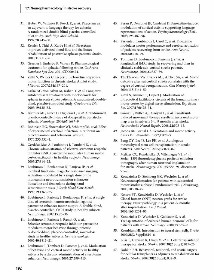
31. Huber W, Willmes K, Poeck K, et al. Piracetam asan adjuvant to language therapy for aphasia:A randomized double-blind placebo-controlledpilot study. Arch Phys Med Rehabil.1997;78:245–50.
32. Kessler J, Thiel A, Karbe H, et al. Piracetamimproves activated blood flow and facilitatesrehabilitation of poststroke aphasic patients. Stroke.2000;31:2112–6.
33. Greener J, Enderby P, Whurr R. Pharmacologicaltreatment for aphasia following stroke. CochraneDatabase Syst Rev. 2001:CD000424.
34. Zittel S, Weiller C, Liepert J. Reboxetine improvesmotor function in chronic stroke: A pilot study.J Neurol. 2007;254:197–201.
35. Laska AC, von Arbin M, Kahan T, et al. Long-termantidepressant treatment with moclobemide foraphasia in acute stroke patients: A randomised, double-blind, placebo-controlled study. Cerebrovasc Dis.2005;19:125–32.
36. Berthier ML, Green C, Higueras C, et al. A randomized,placebo-controlled study of donepezil in poststrokeaphasia. Neurology. 2006;67:1687–9.
37. Robinson RG, Shoemaker WJ, Schlumpf M, et al. Effectof experimental cerebral infarction in rat brain oncatecholamines and behaviour. Nature.1975;255:332–4.
38. Gerdelat-Mas A, Loubinoux I, Tombari D, et al.Chronic administration of selective serotonin reuptakeinhibitor (SSRI) paroxetine modulates human motorcortex excitability in healthy subjects. Neuroimage.2005;27:314–22.
39. Loubinoux I, Boulanouar K, Ranjeva JP, et al.Cerebral functional magnetic resonance imagingactivation modulated by a single dose of themonoamine neurotransmission enhancersfluoxetine and fenozolone during handsensorimotor tasks. J Cereb Blood Flow Metab.1999;19:1365–75.
40. Loubinoux I, Pariente J, Boulanouar K, et al. A singledose of serotonin neurotransmission agonistparoxetine enhances motor output. A double-blind,placebo-controlled, fMRI study in healthy subjects.NeuroImage. 2002;15:26–36.
41. Loubinoux I, Pariente J, Rascol O, et al.Selective serotonin reuptake inhibitor paroxetinemodulates motor behavior through practice.A double-blind, placebo-controlled, multi-dosestudy in healthy subjects. Neuropsychologia.2002;40:1815–21.
42. Loubinoux I, Tombari D, Pariente J, et al. Modulationof behavior and cortical motor activity in healthysubjects by a chronic administration of a serotoninenhancer. Neuroimage. 2005;27:299–313.
43. Peran P, Demonet JF, Cardebat D. Paroxetine-inducedmodulation of cortical activity supporting languagerepresentations of action. Psychopharmacology (Berl).2008;195:487–96.
44. Pariente J, Loubinoux I, Carel C, et al. Fluoxetinemodulates motor performance and cerebral activationof patients recovering from stroke. Ann Neurol.2001;50:718–29.
45. Tombari D, Loubinoux I, Pariente J, et al. Alongitudinal fMRI study: in recovering and then inclinically stable sub-cortical stroke patients.Neuroimage. 2004;23:827–39.
46. Thickbroom GW, Byrnes ML, Archer SA, et al. Motoroutcome after subcortical stroke correlates with thedegree of cortical reorganization. Clin Neurophysiol.2004;115:2144–50.
47. Zittel S, Baumer T, Liepert J. Modulation ofintracortical facilitatory circuits of the human primarymotor cortex by digital nerve stimulation. Exp BrainRes. 2007;176:425–31.
48. Sawaki L, Butler AJ, Xiaoyan L, et al. Constraint-induced movement therapy results in increased motormap area in subjects 3 to 9 months after stroke.Neurorehabil Neural Repair. 2008;22:505–13.
49. Jacobs BL, Fornal CA. Serotonin and motor activity.Curr Opin Neurobiol. 1997;7:820–5.
50. Bang OY, Lee JS, Lee PH, et al. Autologousmesenchymal stem cell transplantation in strokepatients. Ann Neurol. 2005;57:874–82.
51. Meltzer CC, Kondziolka D, Villemagne VL, et al.Serial [18F] fluorodeoxyglucose positron emissiontomography after human neuronal implantationfor stroke. Neurosurgery. 2001;49:586–91; discussion91–2.
52. Kondziolka D, Steinberg GK, Wechsler L, et al.Neurotransplantation for patients with subcorticalmotor stroke: a phase 2 randomized trial. J Neurosurg.2005;103:38–45.
53. Nelson PT, Kondziolka D, Wechsler L, et al.Clonal human (hNT) neuron grafts for stroketherapy: Neuropathology in a patient 27 monthsafter implantation. Am J Pathol.2002;160:1201–06.
54. Kondziolka D, Wechsler L, Goldstein S, et al.Transplantation of cultured human neuronal cells forpatients with stroke. Neurology. 2000;55:565–9.
55. KornblumHI. Introduction to neural stem cells. Stroke.2007;38(2 Suppl):810–6.
56. Bliss T, Guzman R, Daadi M, et al. Cell transplantationtherapy for stroke. Stroke. 2007;38(2 Suppl):817–26.
57. Dobkin BH. Behavioral, temporal, and spatial targetsfor cellular transplants as adjuncts to rehabilitation forstroke. Stroke. 2007;38(2 Suppl):832–9.
17: Neuropharmacology in stroke recovery
192

58. Savitz SI, Dinsmore J, Wu J, et al. Neurotransplantationof fetal porcine cells in patients with basal gangliainfarcts: A preliminary safety and feasibility study.Cerebrovasc Dis. 2005;20:101–07.
59. Bakay RA. Neural transplantation. J Neurosurg.2005;103:6–8; discussion.
60. Redmond DE, Jr, Freeman T. The American Societyfor Neural Transplantation and RepairConsiderations and guidelines for studies ofhuman subjects. The practice committee of thesociety. Approved by council. Cell Transplant.2001;10:661–4.
17: Neuropharmacology in stroke recovery
193


18 Robotic approaches to stroke recoveryDavid J. Reinkensmeyer
IntroductionResearch on robot-assisted movement therapy isincreasing exponentially, as measured by estimates ofthe number of publications in the field (Figure 18.1).Most of this work has focused on rehabilitation ofmovement after stroke, because stroke is a leadingcause of adult disability in industrialized nations, andtherefore survivors of stroke are a large target popula-tion, although there is also some work on roboticmovement training after spinal cord injury, cerebralpalsy, and multiple sclerosis.
The most commonly explored paradigm is to use arobotic device to physically assist in completing desiredmotions of the arms, hands, or legs as the patient playscomputer games presented on a screen (Figure 18.2). Awide variety of assistive control strategies have beendesigned (see [1] for review) , ranging from robots thatrigidly move limbs along fixed paths, to robots thatassist only if patient performance fails to stay withinsome spatial or temporal bound, to soft robots thatform a model of the patient’s weakness. There is alsosome preliminary work on devices that perturb patientmovements (“error-amplification strategies” [2]), orthat act as non-contacting coaches [3]. Most robotictherapy devices are currently used as stationary exer-cise machines in clinics, although there is substantialinterest in making devices that are portable, wearable,and usable at home (e.g. [4–7]).
There is now substantial clinical evidence thatexercise with robotic therapy devices can benefit strokepatients (see [13–15] for reviews). As reviewed below,improvements in movement ability following robot-assisted training in acute, subacute, or chronic strokeare statistically significant, but modest. Less clear is thespecific role that robotic actuation itself plays in thesefindings [16]. A robot is commonly defined as a devicethat can apply forces (and therefore move itself and
other things) in response to computerized commands.Adopting this focused definition of robotic therapy –i.e. that robot therapy is the application of computer-controlled forces to limbs – clearly distinguishesrobotic therapy and non-robotic approaches, such assensor-based computer games and passive exercisemachines, which also show promise for stroke reha-bilitation [17–22]. Under what conditions should oneexpect that applying forces to a limb will enhancemotor recovery?
The purpose of this chapter is to critically reviewexisting clinical evidence for efficacy of robotic move-ment training, with a view toward identifying themechanisms of neuroplasticity that are stimulated byrobot-assisted movement training. This chapter focu-ses on upper extremity training because of space lim-itations, although results are similar for robotic gaittraining after stroke. Evidence for sufficiency ofrobotic assistance in providing therapeutic benefitduring movement training is extensive, but evidencefor necessity of robotic actuation in providing thera-peutic benefit is limited. From a mechanistic perspec-tive, the evidence is consistent with the followingworking hypothesis: movement practice, consistingof the repetitive execution of descending commandsto try to achieve a presented motor goal, is thekey stimulus for motor recovery, independently ofwhether this movement practice is accompanied byrobotic assistance. The benefits of robotic therapydevices may therefore be more related to providingmotivating, quantifiable, and economic delivery oftraining, rather than a specific enhancement of plasti-city attributable to robotic forces, although it is still apossibility that some motor benefits might be derivedfrom the somatosensory signals generated whenrobotic therapy moves a limb. A practical implicationthen is that non-robotic technology that improvesthe motivating, quantifying, or economic qualities
Brain Repair After Stroke, ed. S. C. Cramer and R. J. Nudo. Published by Cambridge University Press.© Cambridge University Press 2010.
195
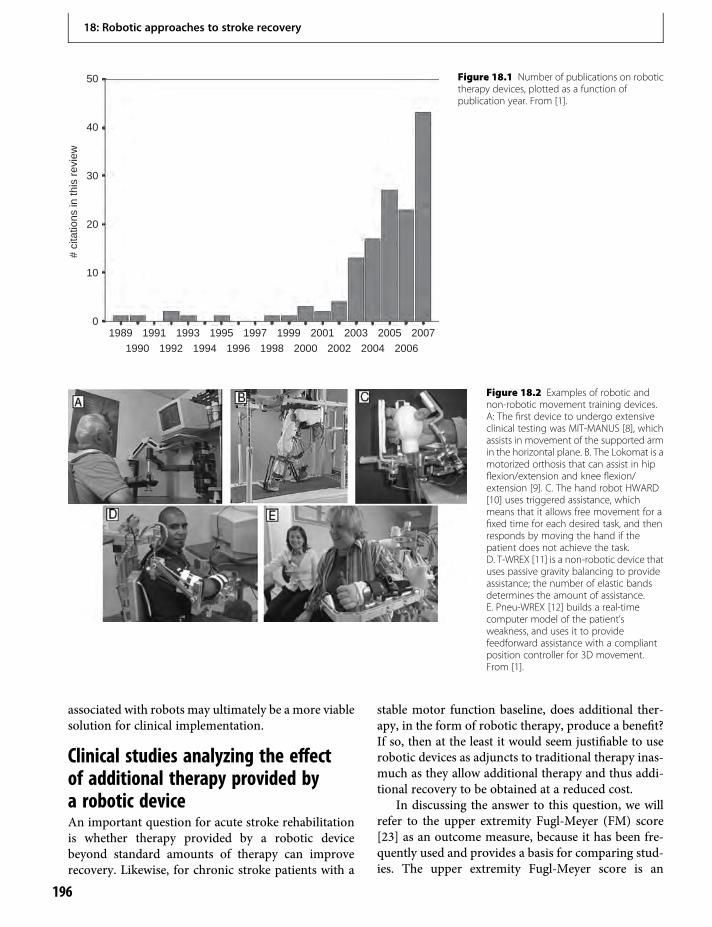
associated with robots may ultimately be a more viablesolution for clinical implementation.
Clinical studies analyzing the effectof additional therapy provided bya robotic deviceAn important question for acute stroke rehabilitationis whether therapy provided by a robotic devicebeyond standard amounts of therapy can improverecovery. Likewise, for chronic stroke patients with a
stable motor function baseline, does additional ther-apy, in the form of robotic therapy, produce a benefit?If so, then at the least it would seem justifiable to userobotic devices as adjuncts to traditional therapy inas-much as they allow additional therapy and thus addi-tional recovery to be obtained at a reduced cost.
In discussing the answer to this question, we willrefer to the upper extremity Fugl-Meyer (FM) score[23] as an outcome measure, because it has been fre-quently used and provides a basis for comparing stud-ies. The upper extremity Fugl-Meyer score is an
50
40
30
20
10
01989 1991
1990 1992 1994 1996 1998 2000 2002 2004 2006
1993 1995 1997 1999 2001 2003 2005 2007
# ci
tatio
ns in
this
rev
iew
Figure 18.1 Number of publications on robotictherapy devices, plotted as a function ofpublication year. From [1].
Figure 18.2 Examples of robotic andnon-robotic movement training devices.A: The first device to undergo extensiveclinical testing was MIT-MANUS [8], whichassists in movement of the supported armin the horizontal plane. B. The Lokomat is amotorized orthosis that can assist in hipflexion/extension and knee flexion/extension [9]. C. The hand robot HWARD[10] uses triggered assistance, whichmeans that it allows free movement for afixed time for each desired task, and thenresponds by moving the hand if thepatient does not achieve the task.D. T-WREX [11] is a non-robotic device thatuses passive gravity balancing to provideassistance; the number of elastic bandsdetermines the amount of assistance.E. Pneu-WREX [12] builds a real-timecomputer model of the patient’sweakness, and uses it to providefeedforward assistance with a compliantposition controller for 3D movement.From [1].
18: Robotic approaches to stroke recovery
196
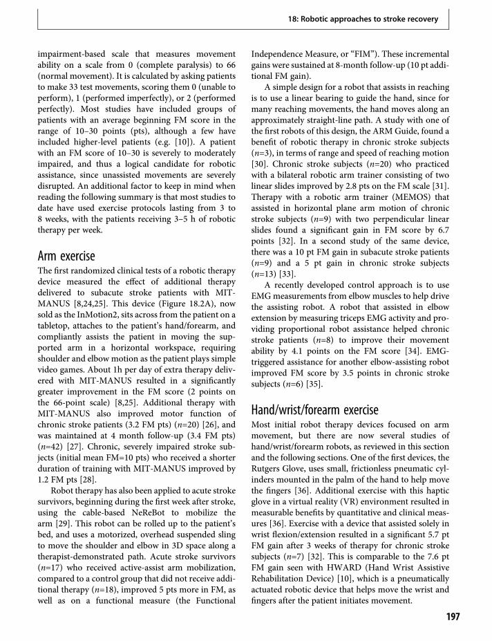
impairment-based scale that measures movementability on a scale from 0 (complete paralysis) to 66(normal movement). It is calculated by asking patientsto make 33 test movements, scoring them 0 (unable toperform), 1 (performed imperfectly), or 2 (performedperfectly). Most studies have included groups ofpatients with an average beginning FM score in therange of 10–30 points (pts), although a few haveincluded higher-level patients (e.g. [10]). A patientwith an FM score of 10–30 is severely to moderatelyimpaired, and thus a logical candidate for roboticassistance, since unassisted movements are severelydisrupted. An additional factor to keep in mind whenreading the following summary is that most studies todate have used exercise protocols lasting from 3 to8 weeks, with the patients receiving 3–5 h of robotictherapy per week.
Arm exerciseThe first randomized clinical tests of a robotic therapydevice measured the effect of additional therapydelivered to subacute stroke patients with MIT-MANUS [8,24,25]. This device (Figure 18.2A), nowsold as the InMotion2, sits across from the patient on atabletop, attaches to the patient’s hand/forearm, andcompliantly assists the patient in moving the sup-ported arm in a horizontal workspace, requiringshoulder and elbow motion as the patient plays simplevideo games. About 1h per day of extra therapy deliv-ered with MIT-MANUS resulted in a significantlygreater improvement in the FM score (2 points onthe 66-point scale) [8,25]. Additional therapy withMIT-MANUS also improved motor function ofchronic stroke patients (3.2 FM pts) (n=20) [26], andwas maintained at 4 month follow-up (3.4 FM pts)(n=42) [27]. Chronic, severely impaired stroke sub-jects (initial mean FM=10 pts) who received a shorterduration of training with MIT-MANUS improved by1.2 FM pts [28].
Robot therapy has also been applied to acute strokesurvivors, beginning during the first week after stroke,using the cable-based NeReBot to mobilize thearm [29]. This robot can be rolled up to the patient’sbed, and uses a motorized, overhead suspended slingto move the shoulder and elbow in 3D space along atherapist-demonstrated path. Acute stroke survivors(n=17) who received active-assist arm mobilization,compared to a control group that did not receive addi-tional therapy (n=18), improved 5 pts more in FM, aswell as on a functional measure (the Functional
Independence Measure, or “FIM”). These incrementalgains were sustained at 8-month follow-up (10 pt addi-tional FM gain).
A simple design for a robot that assists in reachingis to use a linear bearing to guide the hand, since formany reaching movements, the hand moves along anapproximately straight-line path. A study with one ofthe first robots of this design, the ARM Guide, found abenefit of robotic therapy in chronic stroke subjects(n=3), in terms of range and speed of reaching motion[30]. Chronic stroke subjects (n=20) who practicedwith a bilateral robotic arm trainer consisting of twolinear slides improved by 2.8 pts on the FM scale [31].Therapy with a robotic arm trainer (MEMOS) thatassisted in horizontal plane arm motion of chronicstroke subjects (n=9) with two perpendicular linearslides found a significant gain in FM score by 6.7points [32]. In a second study of the same device,there was a 10 pt FM gain in subacute stroke patients(n=9) and a 5 pt gain in chronic stroke subjects(n=13) [33].
A recently developed control approach is to useEMGmeasurements from elbow muscles to help drivethe assisting robot. A robot that assisted in elbowextension by measuring triceps EMG activity and pro-viding proportional robot assistance helped chronicstroke patients (n=8) to improve their movementability by 4.1 points on the FM score [34]. EMG-triggered assistance for another elbow-assisting robotimproved FM score by 3.5 points in chronic strokesubjects (n=6) [35].
Hand/wrist/forearm exerciseMost initial robot therapy devices focused on armmovement, but there are now several studies ofhand/wrist/forearm robots, as reviewed in this sectionand the following sections. One of the first devices, theRutgers Glove, uses small, frictionless pneumatic cyl-inders mounted in the palm of the hand to help movethe fingers [36]. Additional exercise with this hapticglove in a virtual reality (VR) environment resulted inmeasurable benefits by quantitative and clinical meas-ures [36]. Exercise with a device that assisted solely inwrist flexion/extension resulted in a significant 5.7 ptFM gain after 3 weeks of therapy for chronic strokesubjects (n=7) [32]. This is comparable to the 7.6 ptFM gain seen with HWARD (Hand Wrist AssistiveRehabilitation Device) [10], which is a pneumaticallyactuated robotic device that helps move the wrist andfingers after the patient initiates movement.
18: Robotic approaches to stroke recovery
197

A recent study examined the effect of a combina-tion of robot assistance of the wrist and fingersthrough a cyclic motion, while vibrating the tendonsof the lengthening muscles for chronic stroke subjects(n=11) [4]. The goal of this “AMES” (AssistedMovement with Enhanced Sensation) technique wasto enhance the sensory input associated with move-ment, and thus to strengthen functional connectionsbetween somatosensory neurons and motor outputneurons in the cortex. Notably, subjects treated them-selves at home with this technique, 30 minutes per dayfor 6 months, complying to complete an average of70% of the recommended treatments. Subjects signifi-cantly improved their strength and range of motion, aswell as a functional outcome score.
Another recent study combined training with theBiManuTrac device with a brief period of transcranialdirect current stimulation (tDCS, see Chapter 19) overthe motor cortex at the beginning of each trainingsession [37]. The BiManuTrac device allows bilateralforearm supination/pronation or wrist flexion/exten-sion movements, after a quick configuration change.For subacute stroke patients, the combined robotic/brain stimulation approach resulted in large improve-ments in FM score (+33 pts) in 3 patients, 2 of whichhad subcortical lesions, and smaller improvements(5 pts) in 7 patients, all with cortical lesions.
SummaryAdditional motor training provided with a roboticdevice can significantly improve movement ability
following acute, subacute, or chronic stroke, but thereduction is modest – typically on the order of 2–6additional FM points, leaving patients well short of thepeak possible score of 66 (Figure 18.3). A plausibleuse-dependent plasticity mechanism that robotictraining stimulates to achieve these gains is simplythe mechanism (or mechanisms) by which all formsof repetitive practice improve movement ability.
Clinical studies comparing a matchedamount of two therapy types, at leastone of them being robotic, andfinding benefits, but no differencebetween groupsGiven that additional therapy delivered with a roboticdevice is beneficial, it is logical to ask whether robot-assisted therapy is more effective than other types oftherapy a patient could undertake. Several studies thathave tried to address this question have producedinconclusive results. We review these studies in thissection.
Arm exerciseThe first study to compare robot therapy with anotherdose-matched therapy compared a matched amountof robot-assisted therapy using the MIME device withtherapist-provided neurodevelopmental therapy withchronic stroke subjects [38]. The MIME device usesan industrial robot to assist the arm in moving in
Figure 18.3 Summary of gains in upper extremity Fugl-Meyer (FM) score, as a function of robotic or non-robotic therapy, chronic versus acutetraining, or hand/wrist versus arm therapy. The FM score varies from 0 (complete paralysis) to 66 (normal movement). Most of the studiesreported here included subjects with an initial FM score between 10 and 30. The size of the sphere is the size of the FM gain for a study. A spherewith a diameter corresponding to 7 FM points is labeled. Red spheres denote hand/wrist studies; blue, arm studies. A: FM Gains reported fromstudies reviewed here that examined the effect of an additional dose of movement therapy delivered with a robotic or non-robotic device. Forstudies on acute and subacute patients, only studies that controlled for intrinsic recovery by including a control group that did not receiveadditional therapy are included, and only the change on top of that due to intrinsic recovery is plotted. FM gains are modest but consistentlypositive. B: Differential FM gains due to robotic therapy, when the control therapy was duration matched. Only one study [46] with a distal robotand subacute stroke patients showed a substantial differential benefit. For both A and B, there is no obvious advantage to the distal versusproximal, chronic versus acute, or arm versus hand/wrist parameters.
18: Robotic approaches to stroke recovery
198
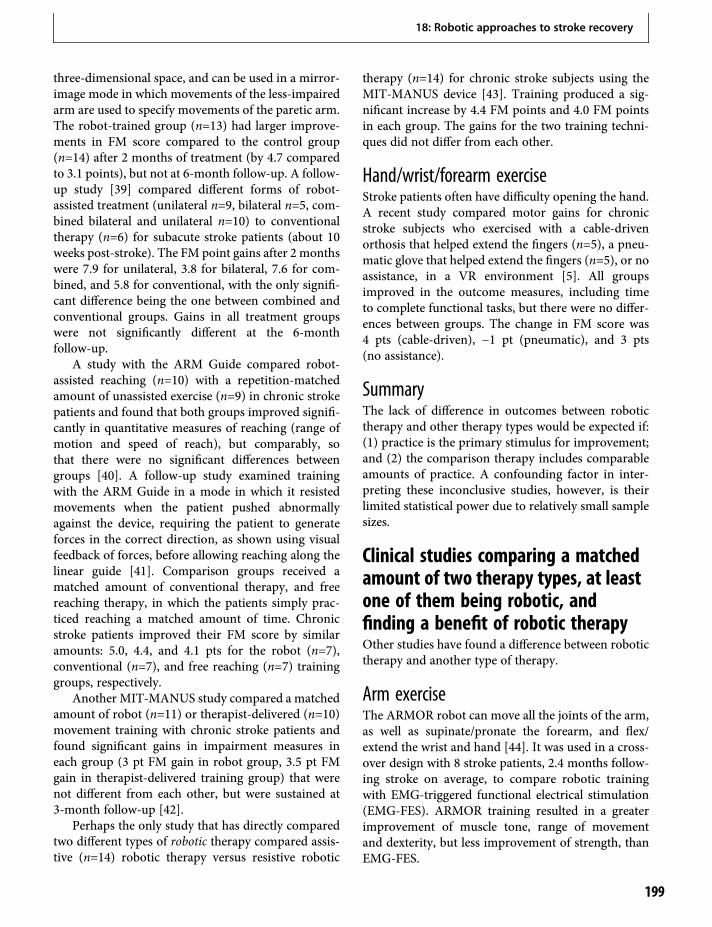
three-dimensional space, and can be used in a mirror-image mode in which movements of the less-impairedarm are used to specify movements of the paretic arm.The robot-trained group (n=13) had larger improve-ments in FM score compared to the control group(n=14) after 2 months of treatment (by 4.7 comparedto 3.1 points), but not at 6-month follow-up. A follow-up study [39] compared different forms of robot-assisted treatment (unilateral n=9, bilateral n=5, com-bined bilateral and unilateral n=10) to conventionaltherapy (n=6) for subacute stroke patients (about 10weeks post-stroke). The FM point gains after 2 monthswere 7.9 for unilateral, 3.8 for bilateral, 7.6 for com-bined, and 5.8 for conventional, with the only signifi-cant difference being the one between combined andconventional groups. Gains in all treatment groupswere not significantly different at the 6-monthfollow-up.
A study with the ARM Guide compared robot-assisted reaching (n=10) with a repetition-matchedamount of unassisted exercise (n=9) in chronic strokepatients and found that both groups improved signifi-cantly in quantitative measures of reaching (range ofmotion and speed of reach), but comparably, sothat there were no significant differences betweengroups [40]. A follow-up study examined trainingwith the ARM Guide in a mode in which it resistedmovements when the patient pushed abnormallyagainst the device, requiring the patient to generateforces in the correct direction, as shown using visualfeedback of forces, before allowing reaching along thelinear guide [41]. Comparison groups received amatched amount of conventional therapy, and freereaching therapy, in which the patients simply prac-ticed reaching a matched amount of time. Chronicstroke patients improved their FM score by similaramounts: 5.0, 4.4, and 4.1 pts for the robot (n=7),conventional (n=7), and free reaching (n=7) traininggroups, respectively.
Another MIT-MANUS study compared a matchedamount of robot (n=11) or therapist-delivered (n=10)movement training with chronic stroke patients andfound significant gains in impairment measures ineach group (3 pt FM gain in robot group, 3.5 pt FMgain in therapist-delivered training group) that werenot different from each other, but were sustained at3-month follow-up [42].
Perhaps the only study that has directly comparedtwo different types of robotic therapy compared assis-tive (n=14) robotic therapy versus resistive robotic
therapy (n=14) for chronic stroke subjects using theMIT-MANUS device [43]. Training produced a sig-nificant increase by 4.4 FM points and 4.0 FM pointsin each group. The gains for the two training techni-ques did not differ from each other.
Hand/wrist/forearm exerciseStroke patients often have difficulty opening the hand.A recent study compared motor gains for chronicstroke subjects who exercised with a cable-drivenorthosis that helped extend the fingers (n=5), a pneu-matic glove that helped extend the fingers (n=5), or noassistance, in a VR environment [5]. All groupsimproved in the outcome measures, including timeto complete functional tasks, but there were no differ-ences between groups. The change in FM score was4 pts (cable-driven), −1 pt (pneumatic), and 3 pts(no assistance).
SummaryThe lack of difference in outcomes between robotictherapy and other therapy types would be expected if:(1) practice is the primary stimulus for improvement;and (2) the comparison therapy includes comparableamounts of practice. A confounding factor in inter-preting these inconclusive studies, however, is theirlimited statistical power due to relatively small samplesizes.
Clinical studies comparing a matchedamount of two therapy types, at leastone of them being robotic, andfinding a benefit of robotic therapyOther studies have found a difference between robotictherapy and another type of therapy.
Arm exerciseThe ARMOR robot can move all the joints of the arm,as well as supinate/pronate the forearm, and flex/extend the wrist and hand [44]. It was used in a cross-over design with 8 stroke patients, 2.4 months follow-ing stroke on average, to compare robotic trainingwith EMG-triggered functional electrical stimulation(EMG-FES). ARMOR training resulted in a greaterimprovement of muscle tone, range of movementand dexterity, but less improvement of strength, thanEMG-FES.
18: Robotic approaches to stroke recovery
199
The Gentle-S robot uses a force-feedback con-trolled 3D commercial robot to assist in moving thearm [45]. A study of Gentle-S compared robot-assisted3D movements to sling-suspension based exercises ina crossover design, and found a nearly significant(p=0.06) greater rate of improvement during therobot therapy.
Hand/wrist/forearm exerciseRobotic training of the forearm and wrist using theBiManuTrac device produced greater improvementsthan EMG-triggered FES of the wrist in subacutestroke patients (n=44) [46]. The FM score was 15points higher at study end and 13 points higher at3-month follow-up than the FES group. It is importantto note that the intensity of the exercise achieved withthe two modalities was quantified and found to bedifferent: the robot group performed about 800 repe-titions per session, whereas the FES group performedabout 80 repetitions per session.
Perhaps the only study to show a direct benefit ofrobotic forces per se during robotic training examinedhand training [10]. Chronic stroke patients whoreceived robot assistance using the HWARD device(Figure 18.2C), which assists in hand opening andclosing and wrist flexion/extension, for all of theirtraining movements (n=7) recovered significantlymore hand function than patients who receivedrobot assistance for only half of their training move-ments (i.e. for only the last 7.5 sessions of a 15 sessionprotocol, n=6). The increase in FM scorewas 9.1 versus5.8 points, for the two groups. Caveats include a smallsample size that was slightly mismatched in impair-ment level at baseline, and uncertainty in whether thegroup that received half the amount of assistance com-pleted as many training movements when assistancewas absent. This study is also different from mostothers in that the patients had a higher beginningFM score (mean score of 45 out of 66).
A recent study with MIT-MANUS compared train-ing with three forms of assistance for combined armand hand movements for chronic stroke subjects(n=47) [13]. All groups received active-assistance forreaching to targets. One group did not use the handduring training; another group interacted with objectsat the target; and another group grasped, transported,and released virtual objects between targets bycontrolling isometric grip force against a sensor. Theno-hand group improved the most in FM score (3 FMpts), followed by the group that gripped actual objects
(2.7 FM pts), followed by the group gripping virtualobjects (0.8 FM pts). Note that the virtual grip grouptrained at a different hospital and was evaluated bya different therapist. The improvement in the groupthat trained without using the hand was significantlygreater than the groups that incorporated handtraining.
It is possible that robot therapy could be less effec-tive than other therapy types. We mention one recentgait training study in this regard. This study compareda matched amount of gait training with the Lokomatrobot (Figure 18.2B) in chronic stroke subjects (n=48)to gait training with a physical therapist [9]. Thesubjects randomized to receive gait training with aphysical therapist exhibited greater improvements inself-selected and maximum gait speed (~2× greatergain). In a separate study, this same group quantifiedpatient energy expenditure in the Lokomat comparedto during therapist-assisted gait training, and foundit to be about 50% less, as gauged by oxygen uptake,unless the patient is instructed to work against themachine [54]. These studies suggest that some formsof assistance provided by robotic therapy devicesmight encourage slacking by patients, reducing therecovery possible with a fixed amount of training.For the upper extremity, a tendency for patients toreduce effort in response to robotic assistance providedwith a compliant device Pneu-WREX (Figure 18.2E)has also been observed [12], but no relative reductionin training effects with robotic therapy has beenreported to our knowledge. A partial exception is thattraining with the ARMOR robot resulted in lessimprovement of strength than EMG-FES, although italso resulted in greater improvements in muscle tone,range of movement, and dexterity [44].
SummaryA few studies have demonstrated that robot-assistedtherapy is modestly more effective than another typeof therapy, with the largest difference coming fora distal robot (BiManuTrac) compared to EMG-FES (13 FM pt advantage at study follow-up) [46](Figure 18.3). However, a confounding factor for thisstudy, as well as several others showing a difference, isthat patients received more repetitions in, and thus ahigher dose of, the robot-assisted therapy compared tothe comparison therapy. Thus, the working hypothesisthat practice is the key mechanistic stimulus to prac-tice rather than robotic assistance could still explainthese studies.
18: Robotic approaches to stroke recovery
200
Clinical studies with non-robotictechnology showing a comparablebenefit to robotic technologySeveral non-robotic devices are being developed toassist patients in practicing upper extremity move-ments and have produced comparable results torobotic devices. We briefly review several of thesestudies.
Arm exerciseThe NudelHoltz device provides mechanically assistedexercise using a rolling-pin like mechanism thatrequires patients to use two hands to slide up anddown a ramp [19]. This non-robotic exercise modalitywas compared with EMG-triggered FES of the wristextensors for 54 subacute patients with severe impair-ment (FM score ~ 8 out of 66), 20–30 min per day,5 days per week, for 6 weeks [19]. Both groupsimproved significantly (10 and 5 pts on FM score),but there were no significant differences between thetwo groups. More NudelHolz patients became able tomove blocks (5 responders versus 0 at end of 6 weeks).Note that the number of movements performed by themechanically assisted group was larger (18 000 move-ments versus ~2000 movements), included moredegrees of freedom (DOF) of the upper extremity,and incorporated a bilateral approach.
The BATRAC device incorporates two low-frictionlinear tracks on which the patient slides handles, andprovides auditory cueing for movement [47]. Trainingbilateral push–pull movements with BATRACimproved arm function in 14 chronic stroke subjectsby 3 pts after 6 weeks of training, and by 10 pts at2-month follow-up [47]. When BATRAC training wascompared to standard, dose-matched exercises inchronic stroke patients (n=21), patients in theBATRAC group increased brain activation during adistal motor task, but there was no difference in func-tional outcome between groups, unless only consider-ing patients with fMRI response (n=6), in which caseBATRAC improved arm function more than thestandard exercises did [18]. However, a subsequentstudy found no benefit of training with a modifiedform of BATRAC in chronic stroke (n=14), as evi-denced by no significant change in FM or Wolf MotorFunction Test scores [17].
The Sensorimotor Active Rehabilitation Training(SMART) Arm is comprised of a sensorized, low-friction linear track [21]. Training with the SMART
Arm alone (n=13) was compared with SMART ARMtraining plus EMG-triggered FES (n=10), and nointervention (n=13) in chronic stroke patients [21].Both SMART Arm groups demonstrated significantimprovements in all impairment and activitymeasuresafter training and at follow-up. There was no signifi-cant difference between these two SMART Armgroups. There was no change in the control group.
The T-WREX (Figure 18.2D) is an elastic-banddriven, passive, arm-supporting orthosis that is con-nected to simple VR games [11]. Training withT-WREX produced greater benefits than conventionaltable-top exercises for chronic stroke subjects (n=28),resulting in a 3.8 FM increase at 6-month follow-upcompared to a 1.5 pt FM increase [11]. T-WREX isnow sold as ARMEO by Hocoma, A. G., the maker ofthe Lokomat.
A very simple non-robotic technology is the rock-ing chair. When acute stroke patients (n=100) rockedin a chair by pushing with their air-splinted pareticarm, they improved by 7 more FM points at 1-yearfollow-up compared to a group that did not receive theextrasensory motor stimulation [48]. The FM differ-ence was 17 pts at 5-year follow-up (n=64) [49].
Hand/wrist/forearm exerciseFor the hand, an example of a non-robotic therapeuticapproach is to measure finger movements and providecomputer feedback in the form of movement trackinggames [50]. Practice with the system at homeimproved scores of hand movement ability in chronicstroke subjects (n=20) [50]. Another device, the Auto-Cite, consists of a motorized table that automaticallypresents trays of sensor-based hand exercise activities[22]. In three groups of chronic stroke patients (n=9 ineach group) with varying levels of therapist supervi-sion (25, 50, and 100%), there were significant motorgains after training with Auto-Cite and no differencesbetween groups.
SummaryNon-robotic training devices that facilitate autono-mous practice also produce significant reductions inimpairment that are comparable with results fromrobotic devices (Figure 18.3).
DiscussionIn its current state, robot-assisted therapy producesmodest but consistent benefits that appear attributable
18: Robotic approaches to stroke recovery
201
to the practice undertaken, rather than a specific driveof plasticity attributable to the robotic forces applied.Non-robotic technology shows comparable therapeu-tic promise at this stage, as well, and may thus be amore clinically viable approach as it is potentially lessexpensive and safer.
What can be done to optimize robotic therapy? Animportant direction is to challenge the assumptions onwhich robotic therapy is based. For example, severaldesign goals that are often stated for robotic therapydevices need to be rigorously evaluated, including thegoals of:
(1) Physically assisting the patient in completingdesired motions normally. Rationales given for thisgoal include providing enhanced somatosensorystimulation, stretching limbs to maintain their softtissue compliance and reduce spasticity, andencouraging patient practice by allowing patientsto accomplish meaningful tasks. However,providing assistance may reduce motor learning bychanging the nature of the task to be learned or byencouraging slacking. Assistance has been found toenhance learning by unimpaired subjects onlyslightly in specific tasks [51–53]. In contrast,increasing performance errors may better provokeplasticity from some motor tasks, sinceperformance errors drive adaptation [2,52].The relationship between patient effortduring training and therapeutic outcomeis actually not established – is it a strongrelationship?
(2) Allowing motions to be as naturalistic as possible.Although many devices assist in single degree offreedom (DOF) or two DOF movements becausesuch movements require less complex devices, anoften-stated goal is to allow more naturalisticmovement. The rationale is that motor learning istask-specific, and therefore it would be desirablefor patients to train the movements that they seekto perform in activities of daily living. Thisrationale needs to be rigorously tested. There is apossibility that the greater amount of repetitionspossible in a fixed amount of time with simplermovements may ultimately be more effective. Inaddition, even simple movements require complexmultiple muscle coordination as well, to stabilizethe non-moving joints. The robotic therapy devicethat produced the largest differential therapeuticgain compared to a time-matched therapy focusedon single joint distal movement [46].
(3) Providing bilateral training. Some devices use abilateral training approach [19,38,46,47], usingneural science on bilateral movements to inferthat bilateral movements may produce brainactivation patterns beneficial for plasticity. Onestudy that addressed this hypothesis in acontrolled therapeutic context failed to verifyit [39]. Again, this approach requires rigorousevaluation.
(4) Providing intense but relatively brief therapy. Mostrobotic therapy studies have used therapeuticprotocols lasting from 3 to 8 weeks, with thepatients receiving 3–5 h of robotic therapy perweek. Is residual capacity exhausted by this dosage?Constraint-induced therapy protocols oftenprovide therapy for 6 or more hours per day. Thesetrials tend to be in more mildly affected patients,and so the answer to the question of therapyintensity and duration might vary in relation toindividual patient features. The issue of therapyintensity is also considered in Chapter 20.
Another direction for optimizing robotic therapy is tocouple it with other techniques to stimulate plasticity,with the hopes of synergy – the combination of theparts greater that their simple sum. As reviewed above,there are at least two studies along these lines so far,one using tendon vibration to enhance somatosensoryinput during robotic therapy [4], and the otherusing tDCS to prime the motor cortex during robotictherapy [37]. Neither study was controlled with amatched-dose comparison group, so conclusions arenot possible at this point.
Ultimately, optimizing robotic therapy will require amuch more detailed understanding of how practice pro-vokes plasticity. We have suggested the working hypoth-esis that movement practice, consisting of the repetitiveexecution of descending commands to try to achieve apresented motor goal, is the key stimulus for motorrecovery, independently of whether this movement prac-tice is accompaniedby robotic assistance.This is a generalstatement that provides little insight into how practiceactually improves performance. Measures of stroke-induced injury and brain function in individual patientsmight provide insights useful for optimizing an indi-vidual patient’s robotic therapy protocol (see alsoChapter 16). Another key direction for future researchis to identify the behavioral/biomechanical signals thattrigger the neurochemical signals that result in imple-mentation of neuroplasticity mechanisms during move-ment practice. Defining these signals would allow
18: Robotic approaches to stroke recovery
202
exercise protocols and devices to then be designed thatoptimize them.
AcknowledgmentsThis work was supported in part by NIH N01-HD-3–3352 from NCMRR and NIBIB, and by the NIDRRRERC on Robotics and Telerehabilitation
References1. Marchal-Crespo L, Reinkensmeyer DJ. Review of
control strategies for robotic movement training afterneurologic injury. J Neur Eng Rehab. 2009;6:20.
2. Patton J, Kovic M, Mussa-Ivaldi F. Custom-designedhaptic training for restoring reaching ability toindividuals with poststroke hemiparesis. J Rehabil ResDev. 2006;43:643–56.
3. Eriksson J, Mataric M, Winstein C. Hands-off assistiverobotics for post-stroke arm rehabilitation. Proceedingsof the 2005 IEEE International Conference onRehabilitation Robotics, 28 June –1 July, Chicago,Illinois 2005: 21–4.
4. Cordo P, Lutsep H, Cordo L, et al. Assisted MovementWith Enhanced Sensation (AMES): Coupling motorand sensory to remediate motor deficits in chronicstroke patients. Neurorehabil Neural Repair.2010;23:67–77.
5. Fischer HC, Stubblefield K, Kline T, et al. Handrehabilitation following stroke: A pilot study of assistedfinger extension training in a virtual environment. TopStroke Rehabil. 2007;14:1–12.
6. Sugar TG, He J, Koeneman EJ, et al. Design and controlof RUPERT: A device for robotic upper extremityrepetitive therapy. IEEE Trans Neural Syst RehabilEngng. 2007;15:336–46.
7. Ferris D. Powered lower limb orthoses for gaitrehabilitation. Top Spinal Cord Injury Rehab.2005;11(2):34–49.
8. Aisen ML, Krebs HI, Hogan N, et al. The effect ofrobot-assisted therapy and rehabilitative training onmotor recovery following stroke. Arch Neurol.1997;54:443–6.
9. Hornby TG, Campbell DD, Kahn JH, et al. Enhancedgait-related improvements after therapist- versusrobotic-assisted locomotor training in subjects withchronic stroke: A randomized controlled study. Stroke.2008;39:1786–92.
10. Takahashi CD, Der-Yeghiaian L, Le V, et al. Robot-based hand motor therapy after stroke. Brain.2008;131:425–37.
11. HousmanSJ, ScottKM,ReinkensmeyerDJ.A randomizedcontrolled trial of gravity-supported, computer-enhancedarm exercise for individuals with severe hemiparesis.Neurorehab Neural Repair. 2009;23:505–14.
12. Wolbrecht ET, Reinkensmeyer DJ, Bobrow JE.Optimizing compliant, model-based robotic assistanceto promote neurorehabilitation. IEEE Trans Neural SystRehab Engng. 2008;16:286–97.
13. Krebs HI, Mernoff S, Fasoli SE, et al. A comparison offunctional and impairment-based robotic training insevere to moderate chronic stroke: A pilot study.NeuroRehabilitation. 2008;23:81–7.
14. Brewer BR, McDowell SK, Worthen-Chaudhari LC.Poststroke upper extremity rehabilitation: A review ofrobotic systems and clinical results. Top Stroke Rehabil.2007;14(6):22–44.
15. Mehrholz J, Platz T, Kugler J, et al. Electromechanicaland robot-assisted arm training for improving armfunction and activities of daily living after stroke.Cochrane Database Syst Rev. 2008;4(4):CD006876.
16. Kahn L, Lum P, Rymer W, et al. Robot-assistedmovement training for the stroke-impaired arm: Doesit matter what the robot does? J Rehab Res Dev.2006;43:619–30.
17. Richards LG, Senesac CR, Davis SB, et al. Bilateral armtraining with rhythmic auditory cueing in chronicstroke: Not always efficacious. Neurorehabil NeuralRepair. 2008;22:180–4.
18. Luft AR, McCombe-Waller S, Whitall J, et al. Repetitivebilateral arm training and motor cortex activation inchronic stroke: A randomized controlled trial. JAMA.2004;292:1853–61.
19. Hesse S, Werner C, Pohl M, et al. Mechanical armtrainer for the treatment of the severely affected armafter a stroke: A single-blinded randomized trial in twocenters. Am J Phys Med Rehabil. 2008;87:779–88.
20. Sanchez R, Liu J, Rao S, et al. Automating armmovement training following severe stroke: Functionalexercises with quantitative feedback in a gravity-reduced environment. IEEE Trans Neural RehabEngng.2006;14:378–89.
21. Barker RN, Brauer SG, Carson RG. Training ofreaching in stroke survivors with severe and chronicupper limb paresis using a novel nonrobotic device:A randomized clinical trial. Stroke. 2008;39:1800–07.
22. Taub E, Lum PS, Hardin P, et al. AutoCITE: Automateddelivery of CI therapy with reduced effort by therapists.Stroke. 2005;36:1301–04.
23. Fugl-Meyer AR, Jaasko L, Leyman I, et al. The post-stroke hemiplegic patient. 1. Amethod for evaluation ofphysical performance. Scand J Rehabil Med.1975;7:13–31.
24. Krebs HI, Hogan N, Aisen ML, et al. Robot-aidedneurorehabilitation. IEEE Trans Rehabil Engng.1998;6:75–87.
25. Volpe B, Krebs H, Hogan N, et al. Robot trainingenhanced motor outcome in patients with strokemaintained over 3 years. Neurology. 1999;53:1874–6.
18: Robotic approaches to stroke recovery
203
26. Fasoli S, Krebs H, Stein J, et al. Effects of robotic therapyon motor impairment and recovery in chronic stroke.Arch Phys Med Rehabil. 2003;84:477–82.
27. Fasoli SE, Krebs HI, Stein J, et al. Robotic therapyfor chronic motor impairments after stroke:Follow-up results. Arch Phys Med Rehabil.2004;85:1106–11.
28. Finley MA, Fasoli SE, Dipietro L, et al. Short-durationrobotic therapy in stroke patients with severe upper-limb motor impairment. J Rehabil Res Dev.2005;42:683–92.
29. Masiero S, Celia A, Rosati G, et al. Robotic-assistedrehabilitation of the upper limb after acute stroke. ArchPhys Med Rehabil. 2007;88:142–9.
30. Reinkensmeyer DJ, Kahn LE, Averbuch M, et al.Understanding and treating arm movementimpairment after chronic brain injury: Progresswith the ARM Guide. J Rehab Res Dev. 2000;37:653–62.
31. Chang JJ, Tung WL, Wu WL, et al. Effects of robot-aided bilateral force-induced isokinetic arm trainingcombined with conventional rehabilitation on armmotor function in patients with chronic stroke. ArchPhys Med Rehabil. 2007;88:1332–8.
32. Colombo R, Pisano F, Micera S, et al. Robotictechniques for upper limb evaluation and rehabilitationof stroke patients. IEEE Trans Neural Syst RehabilEngng. 2005;13:311–24.
33. Colombo R, Pisano F, Micera S, et al. Assessingmechanisms of recovery during robot-aidedneurorehabilitation of the upper limb. NeurorehabilNeural Repair. 2008;22:50–63.
34. Song R, Tong KY, Hu X, et al. Assistive control systemusing continuous myoelectric signal in robot-aided armtraining for patients after stroke. IEEE Trans NeuralSyst Rehabil Engng. 2008;16:371–9.
35. Stein J, Narendran K, McBean J, et al.Electromyography-controlled exoskeletal upper-limb-powered orthosis for exercise training after stroke.Am J Phys Med Rehabil. 2007;86:255–61.
36. Adamovich SV, Merians AS, Boian R, et al. A virtualreality based exercise system for hand rehabilitationpost-stroke: Transfer to function. Conf Proc IEEE EngngMed Biol Soc. 2004;7:4936–9.
37. Hesse S, Werner C, Schonhardt EM, et al. Combinedtranscranial direct current stimulation and robot-assisted arm training in subacute stroke patients:A pilot study. Restor Neurol Neurosci. 2007;25:9–15.
38. Lum PS, Burgar CG, Majmundar M, et al. Robot-assisted movement training compared withconventional therapy techniques for the rehabilitationof upper limb motor function following stroke. ArchPhys Med Rehabil. 2002;83:952–9.
39. Lum P, Burgar C, Van der Loos M, et al. MIME roboticdevice for upper-limb neurorehabilitation in subacute
stroke subjects: A follow-up study. J Rehabil Res Dev.2006;43:631–42.
40. Kahn LE, ZygmanML, RymerWZ, et al. Robot-assistedreaching exercise promotes arm movement recovery inchronic hemiparetic stroke: A randomized controlledpilot study. J Neuroengng Neurorehabil. 2006;3:12.
41. Fischer H, Kahn L, Pelosin E, et al. Can robot-assistedtherapy promote generalization of motor learningfollowing stroke? Preliminary results. Proceedingsof the First IEEE/RAS-EMBS International Conferenceon Biomedical Robotics and Biomechatronics 2006(BioRob 2006), 20–22 February 2006, Pisa, Italy. IEEEPress: 865–8.
42. Volpe BT, Lynch D, Rykman-Berland A, et al.Intensive sensorimotor arm training mediated bytherapist or robot improves hemiparesis in patientswith chronic stroke. Neurorehabil Neural Repair.2008;22:305–10.
43. Stein J, Krebs HI, Frontera WR, et al. Comparison oftwo techniques of robot-aided upper limb exercisetraining after stroke. Am J Phys Med Rehabil.2004;83:720–8.
44. Mayr A, Kofler M, Saltuari L. ARMOR: Anelectromechanical robot for upper limb trainingfollowing stroke. A prospective randomised controlledpilot study. Handchir Mikrochir Plast Chir.2008;40:66–73.
45. Coote S, Murphy B, Harwin W, et al. The effect of theGENTLE/s robot-mediated therapy system on armfunction after stroke. Clin Rehabil. 2008;22:395–405.
46. Hesse S, Werner C, Pohl M, et al. Computerized armtraining improves the motor control of the severelyaffected arm after stroke: A single-blinded randomizedtrial in two centers. Stroke. 2005;36:1960–6. Epub 2005Aug 18.
47. Whitall J, McCombe Waller S, Silver KH, et al.Repetitive bilateral arm training with rhythmicauditory cueing improves motor function inchronic hemiparetic stroke. Stroke. 2000;31:2390–5.
48. Feys H, De Weerdt W, Selz B, et al. Effect of atherapeutic intervention for the hemiplegic upper limbin the acute phase after stroke: A single-blind,randomized, controlled multicenter trial. Stroke.1998;29:785–92.
49. Feys H, De Weerdt W, Verbeke G, et al. Early andrepetitive stimulation of the arm can substantiallyimprove the long-term outcome after stroke: A 5-yearfollow-up study of a randomized trial. Stroke.2004;35:924–9.
50. Carey JR, Durfee WK, Bhatt E, et al. Comparison offinger tracking versus simple movement training viatelerehabilitation to alter hand function and corticalreorganization after stroke. Neurorehabil NeuralRepair. 2007;21:216–32.
18: Robotic approaches to stroke recovery
204
51. Marchal-Crespo L, McHughen S, Cramer SC, et al. Theeffect of haptic guidance, aging, and initial skill level onmotor learning of a steering task. Exp Brain Res,2009;201:209–20.
52. Reinkensmeyer DJ, Patton JL. Can robots help thelearning of skilled actions? Exp Sports Sci Rev.2009;37:43–51.
53. Milot MH, Marchal-Crespo L, Green CS,et al. Comparison of error amplification
and haptic guidance training techniques forlearning of a timing-based motor task by healthyindividuals. Exp Brain Res. 2009 Sep 29[Epub ahead of print].
54. Israel JF, Campbell DD, Kahn JH, et al. Metabolic costsand muscle activity patterns during robotic- andtherapist-assisted treadmill walking in individuals withincomplete spinal cord injury. Phys Ther.2006;86:1466–78.
18: Robotic approaches to stroke recovery
205

19 Electromagnetic approaches to strokerecoveryGottfried Schlaug & Leonardo G. Cohen
Neurophysiological studies in chronic stroke patientshave demonstrated that disinhibition of contra-lesionalmotor regions co-exists with increased inhib-ition of ipsilesional motor regions that results in animbalance of interhemispheric interactions [1–3].The indirect effect of this imbalance on the lesionedhemisphere combined with the stroke’s direct effecton the unimpaired parts of the lesioned motor cortexand its efferent motor system appears to interferewith the recovery process under some circumstances.Similarly, imaging studies in well-recovered patientshave shown that brain reorganization during therecovery phase is associated with reactivation oroveractivation of unimpaired sensorimotor and pre-motor networks in the lesional hemisphere [4–7],while the significance of activation in the contra-lesional motor regions when the affected arm/handperforms a motor task remains under study [8,9].One explanation is that this activation is a sign ofdisinhibition (the lesional hemisphere’s lack of inhib-itory effect on the contralesional hemisphere’s motorregion) that could potentially impede recovery.Although this model of interhemispheric imbalancemay appear to be a simplified representation ofthe many underlying pathophysiological processesinvolved in recovery from stroke, it provides a frame-work for hypotheses focused on two facets: (1) down-regulating activity in the contralesional motor regionto check its unbalanced influence on the lesionalmotor region, and/or (2) facilitating activity in theipsilesional motor region [10–15]. Support for theseapproaches can be found in pilot and proof-of-principle studies that have shown temporary benefi-cial effects in motoric measures, primarily in singlesession experiments using either transcranial mag-netic stimulation (TMS), or, more recently, transcra-nial direct current stimulation (tDCS) [11,12,15–17].TMS used as a diagnostic tool (see Chapter 10) has
also revealed that the re-emergence of motor-evokedpotentials (MEPs) recorded from muscles in theparetic extremity after stimulation of the ipsilesionalM1 in some cases indicates good motor recovery afterstroke [18,19], although some patients with undetect-able MEPs have shown good motor recovery [18].TMS studies also have suggested that increased cort-ical excitability in contralesional M1 is not associatedwith good recovery [20,21].
The concept of therapeutic electricity on excitabletissues such as the brain is not new considering theattempts to cure epileptic disorders using electriccatfish as early as the eleventh century (for a historicperspective, see [22]). The initial experiments ofEduard Hitzig (1870; cited in [23]) on dog cortexsubsequent to a serendipitous discovery of abnormalinvoluntary movements in patients treated with high-voltage transcranial electric currents led to an interestin using electric currents to identify the cortical repre-sentations of limb movements (for more historicdetails see [23]). Electro-sleep therapy, which latercame to be known as cranial electrical stimulation(CES), was used to treat sleep disorders and depressionsince 1902.
In the 1960s, the experiments of Bindman [24],suggesting long-lasting polarization effects followingelectric stimulation of the exposed motor cortex ofanimals, led to a resurgence of studies focusing on itsclinical applications, including the use of brain polar-ization in depressed patients. Although these studiesshowed some benefits, replicating these beneficialeffects in controlled settings yielded mixed resultswhich subsequently led to a diminished interest intranscranial electrical treatments [25].
Several forms of electromagnetic stimulation areunder evaluation to improve behavioral status afterstroke. This stimulation is targeted towards a partic-ular brain region, such as the motor cortex. This
Brain Repair After Stroke, ed. S. C. Cramer and R. J. Nudo. Published by Cambridge University Press.© Cambridge University Press 2010.
207
chapter focuses on three of these: repetitive transcra-nial magnetic stimulation (rTMS), epidural corticalstimulation, and transcranial direct current stimula-tion (tDCS), which all lead to changes in polarity, butdiffer among others with regard to how focal andspecific the stimulation is being applied and whetheror not the intervention leads to neuronal discharges.
Several means for defining the stimulation targethave been used. In some studies, landmarks on thescalp are used to direct stimulation to the underlyingbrain structures. Other studies use neurophysiologicalmethods such as TMS to identify a specific neuraltarget. Yet others have used functional magnetic reso-nance imaging (fMRI) activation sites to focus thestimulation. The relative strengths of each approachis a topic that requires further study.
Repetitive transcranial magneticstimulationOne stimulation approach that has been examined inpatients with stroke is rTMS. Depending on the stim-ulation frequency, rTMS has inhibitory or excitatoryeffects on cortical excitability [26]. As such, goals caninclude increasing excitation in ipsilesional corticalregions [27–30], or decreasing excitation in contra-lesional cortical regions that are uninhibitied andshow increased cortical excitability [31–33].
Several studies have examined high frequency(≥ 3–20Hz) rTMS in patients with stroke applied tothe ipsilesional hemisphere. Khedr et al. [27] foundthat 10 sessions of 3Hz rTMS to the motor corteximproved disability and overall neurological status toa greater extent than sham rTMS did in patientswith subacute stroke, all of whom received standardof care physiotherapy. No effect was seen in thepatients with the largest stroke. Also, no correlationwas found between behavioral gains and changes incortical excitability, a finding that suggests that elec-trostimulation effects on behavior are not mediated bysimply changing motor cortex excitability. Kim et al.[28], in a cohort of patients with chronic hemipareticstroke, found that a single session of 10Hz rTMSapplied to the ipsilesional motor cortex as subjectspracticed a complex finger-sequencing motor taskimproved motor learning more than did sham rTMS.Application of rTMS induced a significantly largerincrease in the MEP amplitude than did sham rTMS.In this case, a change in motor cortex excitability wasassociated with greater motor behavioral gains. In asmall sample, Talelli et al. [34] found that a single
session of excitatory theta burst stimulation, consist-ing of 3 pulses at 50Hz repeated 5 times/s, increasedMEP amplitude from the affected hemisphere and alsoimproved reaction time. Yozbatiran et al. [30], in astudy of patients with chronic hemiparetic stroke,found small increases in blood pressure when applyinga single session of 20Hz rTMS.
Application of low-frequency rTMS to the con-tralesional hemisphere is intended to reduce its excit-ability and activity, and thereby reduce its unbalancedinhibitory effects on the ipsilesional hemisphere,resulting in improvements in motor control of theipsilesional motor cortex upon the affected hand[2,35–37]. Even a single session of such stimulationcan improve motor function in the affected handafter stroke, at least transiently [38]. This suppressiveapproach was effective in modifying cortical silentperiods [39], and increasing excitability of the ipsi-lesional motor cortex [31]. A single session of low-frequency (1Hz) rTMS to the affected hemisphere insubacute stroke patients, followed by motor training,tended to increase cortical excitability, but without aneffect on motor behavior [40].
Many questions remain with using TMS for a non-invasive brain stimulation. Furthermore, the mecha-nisms underlying spontaneous functional recoveryafter stroke are likely to differ depending on a varietyof factors that may include magnitude of impairment,lesion site and size, affected hemisphere, and hemi-spheric dominance among others. Such factors likelyalso have an influence regarding the extent of effectsfrom electromagnetic stimulation. Similarly, the opti-mal approach to pairing with behavioral experience[41], the role that adjuvant cellular or pharmacologicaltherapy might play [42,43], the proper dose of rTMS[44], the preferred stimulation site(s), safety, and otherissues require clarification, possibly with prescriptionbeing individualized according to features of individ-ual stroke.
Epidural cortical stimulationAn alternative approach to non-invasive cortical stim-ulation involves surgical placement of electrodes inthe epidural space above targeted brain regions. Thisapproach was supported by a series of studies inrodents [45–47] and primates [48], where animalsreceiving epidural stimulation during rehab trainingshowed greater stroke recovery than did animalsreceiving sham stimulation during rehab training. Asmall phase II study [49] evaluated 3 weeks of therapy
19: Electromagnetic approaches to stroke recovery
208
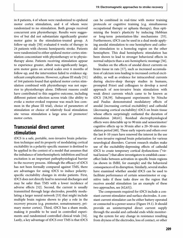
in 8 patients, 4 of whom were randomized to epiduralmotor cortex stimulation, and 4 of whom wererandomized to no stimulation. Both groups receivedconcurrent arm physiotherapy. Results were sugges-tive of but did not substantiate significantly greatermotor gains in the stimulation-treated group. Afollow-up study [50] evaluated 6 weeks of therapy in24 patients with chronic hemiparetic stroke. Patientswere randomized to either epidural motor cortex stim-ulation concomitant with physiotherapy or to physio-therapy alone. Patients receiving stimulation appearto experience greater, albeit non-significantly larger,arm motor gains on several scales over 6 months offollow-up, and the intervention failed to evidence sig-nificant complications. However, a phase III study [51]of 164 patients found that epidural motor cortex stim-ulation combined with physiotherapy was not supe-rior to physiotherapy alone. Different reasons couldhave contributed to this negative outcome, includingdifferent patient selection across trials (the ability toevoke a motor-evoked response was much less com-mon in the phase III trial), choice of parameters ofstimulation or choice of relatively focal stimulationsite versus stimulation a large area of premotor/motor cortex.
Transcranial direct currentstimulationtDCS is a safe, portable, non-invasive brain polariza-tion technique and its property of modulating corticalexcitability in a polarity-specific manner is destined tobe applied in the context of a model that assumes thatthe imbalance of interhemispheric inhibition and localexcitation is an important pathophysiological barrierin the recovery process. Although the efficacy of tDCShas not been formally compared against TMS, thereare advantages for using tDCS to induce polarity-specific excitability changes in stroke patients. First,tDCS does not directly lead to neuronal discharges andmay be safer than TMS with a lower incidence ofadverse effects [52]. Second, the current is usuallytransmitted through large electrodes, possibly modu-lating a larger neural network [53] that might includemultiple brain regions shown to play a role in therecovery process (e.g. premotor, somatosensory, pri-mary motor cortex). Third, tDCS has a sham mode,making it possible to be used in controlled experi-ments and randomized controlled clinical trials [54].Lastly, a key advantage of tDCS over TMS is that tDCS
can be combined in real-time with motor trainingprotocols or cognitive training (e.g. simultaneousoccupational therapy or aphasia therapy), thus opti-mizing the brain’s plasticity by inducing Hebbianor long-term potentiation-like mechanisms [55].Furthermore, tDCS can be used in a dual mode apply-ing anodal stimulation to one hemisphere and catho-dal stimulation to a homolog region on the otherhemisphere. This dual hemispheric stimulation hasbeen shown to lead to stronger behavioral effects innormal subjects than a uni-hemispheric montage [56].
Studies on the effects of anodal direct currents onbrain tissue in rats [57], such as increased accumula-tion of calcium ions leading to increased cortical excit-ability, as well as evidence for intracerebral currentsduring electro-sleep therapy studies in humans,prompted Priori and colleagues to develop a novelapproach of non-invasive brain stimulation withweak direct currents which came to be known astDCS [58,59]. Subsequent experiments by Nitscheand Paulus demonstrated modulatory effects ofanodal (increasing cortical excitability) and cathodal(decreasing cortical excitability) tDCS on brain tissuewhose effects surprisingly outlasted the duration ofstimulation [60,61]. Residual electrophysiologicaleffects were detectable up to 90min and sensorimotor/cognitive effects up to 30min after a 20–30-min stim-ulation period [60]. These early reports and others overthe last 8–10 years have renewed the interest in the useof non-invasive regional brain polarization for variousneurological disorders. Current research studies makeuse of the excitability-depressing effects of cathodaltDCS to create temporary cortical dysfunctions (“vir-tual lesions”) that allow investigators to establish cause–effect links between activation in specific brain regions(as shown in fMRI, for example) and the behavioralconsequences of its disruption. Similarly, several studieshave examined whether anodal tDCS can be used tofacilitate performance of certain sensorimotor or cog-nitive tasks if these tasks draw on the region thatreceives anodal stimulation (as an example of thesetwo approaches, see [62,63]).
The components required for tDCS include a con-stant current stimulator and surface electrodes. A con-stant current stimulator can be either battery operatedor connected to a power source (Figure 19.1). It shouldprovide an uninterrupted direct current supplythrough the anodal and cathodal ends while monitor-ing the system for any change in resistance resultingfrom dryness of the electrodes, loss of contact, or other
19: Electromagnetic approaches to stroke recovery
209
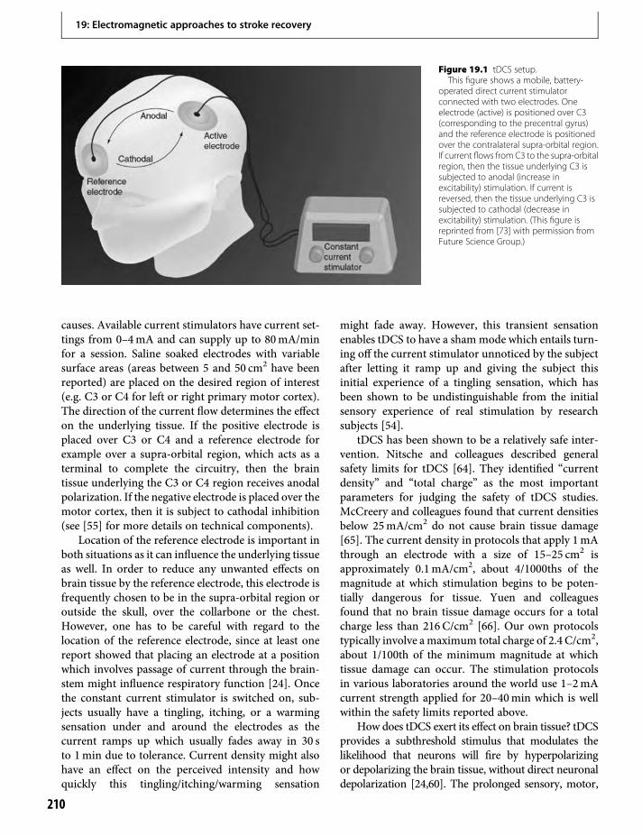
causes. Available current stimulators have current set-tings from 0–4mA and can supply up to 80mA/minfor a session. Saline soaked electrodes with variablesurface areas (areas between 5 and 50 cm2 have beenreported) are placed on the desired region of interest(e.g. C3 or C4 for left or right primary motor cortex).The direction of the current flow determines the effecton the underlying tissue. If the positive electrode isplaced over C3 or C4 and a reference electrode forexample over a supra-orbital region, which acts as aterminal to complete the circuitry, then the braintissue underlying the C3 or C4 region receives anodalpolarization. If the negative electrode is placed over themotor cortex, then it is subject to cathodal inhibition(see [55] for more details on technical components).
Location of the reference electrode is important inboth situations as it can influence the underlying tissueas well. In order to reduce any unwanted effects onbrain tissue by the reference electrode, this electrode isfrequently chosen to be in the supra-orbital region oroutside the skull, over the collarbone or the chest.However, one has to be careful with regard to thelocation of the reference electrode, since at least onereport showed that placing an electrode at a positionwhich involves passage of current through the brain-stem might influence respiratory function [24]. Oncethe constant current stimulator is switched on, sub-jects usually have a tingling, itching, or a warmingsensation under and around the electrodes as thecurrent ramps up which usually fades away in 30 sto 1min due to tolerance. Current density might alsohave an effect on the perceived intensity and howquickly this tingling/itching/warming sensation
might fade away. However, this transient sensationenables tDCS to have a shammode which entails turn-ing off the current stimulator unnoticed by the subjectafter letting it ramp up and giving the subject thisinitial experience of a tingling sensation, which hasbeen shown to be undistinguishable from the initialsensory experience of real stimulation by researchsubjects [54].
tDCS has been shown to be a relatively safe inter-vention. Nitsche and colleagues described generalsafety limits for tDCS [64]. They identified “currentdensity” and “total charge” as the most importantparameters for judging the safety of tDCS studies.McCreery and colleagues found that current densitiesbelow 25mA/cm2 do not cause brain tissue damage[65]. The current density in protocols that apply 1mAthrough an electrode with a size of 15–25 cm2 isapproximately 0.1mA/cm2, about 4/1000ths of themagnitude at which stimulation begins to be poten-tially dangerous for tissue. Yuen and colleaguesfound that no brain tissue damage occurs for a totalcharge less than 216C/cm2 [66]. Our own protocolstypically involve amaximum total charge of 2.4 C/cm2,about 1/100th of the minimum magnitude at whichtissue damage can occur. The stimulation protocolsin various laboratories around the world use 1–2mAcurrent strength applied for 20–40min which is wellwithin the safety limits reported above.
How does tDCS exert its effect on brain tissue? tDCSprovides a subthreshold stimulus that modulates thelikelihood that neurons will fire by hyperpolarizingor depolarizing the brain tissue, without direct neuronaldepolarization [24,60]. The prolonged sensory, motor,
Figure 19.1 tDCS setup.This figure shows a mobile, battery-
operated direct current stimulatorconnected with two electrodes. Oneelectrode (active) is positioned over C3(corresponding to the precentral gyrus)and the reference electrode is positionedover the contralateral supra-orbital region.If current flows fromC3 to the supra-orbitalregion, then the tissue underlying C3 issubjected to anodal (increase inexcitability) stimulation. If current isreversed, then the tissue underlying C3 issubjected to cathodal (decrease inexcitability) stimulation. (This figure isreprinted from [73] with permission fromFuture Science Group.)
19: Electromagnetic approaches to stroke recovery
210

and cognitive effects of tDCS have been attributed to apersistent bidirectional modification of post-synapticconnections similar to long-term potentiation (LTP)and long-term depression (LTD) effects [57,67,68].Dextromethorphan, an N-methyl-d-aspartate (NMDA)antagonist, suppressed both anodal and cathodal tDCSeffects, strongly suggesting the involvement of NMDAreceptors in both types of DC-induced neuroplasticity.In contrast, carbamazepine selectively eliminated anodaleffects. Since carbamazepine stabilizes the membranepotential through voltage-gated sodium channels (sta-bilizing the inactivated state of sodium channels), theresults were interpreted as indicative that aftereffects ofanodal tDCS require a depolarization of membranepotentials [69]. Ardolino and colleagues also proposeda non-synaptic mechanism involving changes in mem-brane excitability and ionic shifts [70]. Nevertheless,more studies are needed, in humans as well as in animalmodels, to verify the effects of tDCS on brain tissue, tobetter understand the underlying mechanisms of action,its sustained effects, and to determine relations betweencurrent strength/duration and neural tissue/behavioraleffects interactions. Recent studies on brain modelingand current density distribution have suggested that inspite of a large fraction of the direct current beingshunted through the scalp, tDCS carries adequate cur-rents to the underlying cortex to be able to modulateneuronal excitability [71,72] and correspondingregional blood flow changes have been seen usingnon-invasive arterial spin labeling techniques [55].
Targeting the affected, unaffected,or both hemispheresThe model of an imbalance in the interhemisphericinhibitory interactions after a stroke led to the formu-lation of hypotheses focused on downregulating activ-ity in the contralesional M1, facilitating it in theipsilesional M1 or applying both at the same timeresulting in three modes (Figure 19.2) of interventions[10,11,13,55,56,73–75]. The first set of studies thatreported a significant improvement in motor functionafter stroke [12,14] applied anodal tDCS to the stroke-affected hemisphere. Later the same year, Fregni et al.found similar effects applying cathodal tDCS tothe contralesional hemisphere [11]. However, thesestudies only used a single session of tDCS (1mAfor 20min) in a pre–post assessment design andshowed short-lasting effects. In a double-blind,sham-controlled, crossover study, Hummel et al.
reported an improvement in reaction time and pinchforce in chronic stroke patients following anodal tDCSof the lesional hemisphere [76]. Amore recent study inhealthy subjects indicated that daily applications ofanodal tDCS over M1 for 5 days in association withmotor training led to a substantial improvement inskill acquisition through facilitation of off-line learn-ing that remained present 3months after the end of thetraining-stimulation period relative to controls [77],an effect possibly present also in elderly healthy humansubjects [78]. Ongoing studies in chronic strokepatients are assessing longer-lasting effects using mul-tiple daily sessions, often in combination with periph-eral sensorimotor activities such as occupationaltherapies [16].
Anodal or cathodal stimulation have both led torelatively consistent effects across various laborato-ries around the world, suggesting that some aspects ofthe model of an imbalance in interhemispheric inhib-ition may be correct. Further support for this modelcomes also from the facilitatory influence of somato-sensory stimulation of the paretic hand or anesthesiaor disuse of the healthy hand in patients with stroke[79–82]. It is quite obvious that a technique thatwould allow the stimulation of ipsilesional M1, inva-sive or non-invasive, in addition to modulating inter-hemispheric interactions, might take advantage offacilitating perilesional cortical activity in the caseof small cortical strokes [83]. There have also beenattempts to apply cathodal and anodal stimulation tothe motor regions of both hemispheres simultane-ously in normal subjects in order to increase motorskill acquisition in the hand that is controlled by thehemisphere receiving anodal polarization. The inter-hemispheric inhibitory effect on this hemisphere wasdampened by applying cathodal stimulation to theopposite hemisphere. Results showed significant dif-ferences in a motor skill acquisition between this“dual” stimulation approach compared to the uni-hemispheric stimulation [56].
Nevertheless, despite similarities with other cogni-tive models of interhemispheric inhibitory interac-tions [84], it remains to be investigated whether thismechanism operates in regions other than M1 afterstroke. It is also important to keep in mind that cort-ical reorganization is likely to operate at a networklevel and approaches that focus on only one brainregion at a time, while a reasonable start, are likely torepresent only a partial aspect of a more complex fullpicture of recovery of function.
19: Electromagnetic approaches to stroke recovery
211

Combining non-invasive brainstimulation with peripheralsensorimotor activitiesand neuromodulatory agentsSeveral recent studies [7,15] combined tDCS withrehabilitative therapy to further enhance the facilitat-ing effect of non-invasive brain stimulation that hadsome effect already by itself. The idea behind thissimultaneous approach is that combined peripheralsensorimotor activities (which also provide increasedsensory feedback) and central brain stimulation(which has the ability to increase or decrease regionalexcitability) can enhance synaptic plasticity and motorskill acquisition/consolidation by increasing or
modulating afferent inputs to the cortex at a timewhen it is receiving central stimulation. Cortical stim-ulation studies in experimental stroke models haveshown stronger effects when peripheral sensorimotoractivities were combined with central stimulation [85].It has also been shown that paired associative brainstimulation and repetitive median nerve stimulationraised motor cortical excitability to a level higher thanthat produced by cortical stimulation alone [86]. Thisincrease was not seen when the same procedure wasperformed under the influence of dextromethorphan,which is known to block LTP [87]. Motor skill learninghas shown to produce LTP and LTD changes in theprimary motor cortex in animal studies [88]. It seemspossible that combined repetitive peripheral stimula-tion or rehabilitative therapy with non-invasive brain
Figure 19.2 Brain model of alteredinterhemispheric inhibition in patientswith a unihemispheric stroke and thetherapeutic options to ameliorate thisimbalance.The balance of interhemispheric
inhibition becomes disrupted after a stroke(A). This leaves the healthy hemisphere in aposition that it could exert too much of anunopposed influence onto the lesionalhemisphere and possibly interfere in therecovery process. There are three possibleways to ameliorate this process: either theexcitability in the affected (lesional)hemisphere (B) is upregulated, theexcitability in the unaffected (normal)hemisphere (C) is downregulated, or acombination of both approaches (D).
19: Electromagnetic approaches to stroke recovery
212
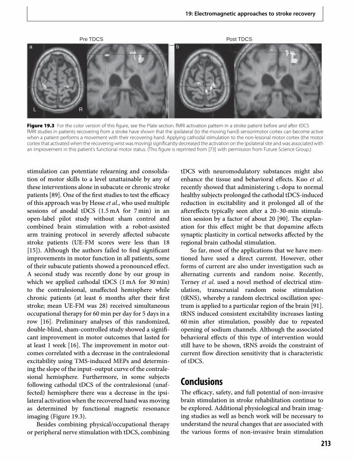
stimulation can potentiate relearning and consolida-tion of motor skills to a level unattainable by any ofthese interventions alone in subacute or chronic strokepatients [89]. One of the first studies to test the efficacyof this approach was by Hesse et al., who used multiplesessions of anodal tDCS (1.5mA for 7min) in anopen-label pilot study without sham control andcombined brain stimulation with a robot-assistedarm training protocol in severely affected subacutestroke patients (UE-FM scores were less than 18[15]). Although the authors failed to find significantimprovements in motor function in all patients, someof their subacute patients showed a pronounced effect.A second study was recently done by our group inwhich we applied cathodal tDCS (1mA for 30min)to the contralesional, unaffected hemisphere whilechronic patients (at least 6 months after their firststroke; mean UE-FM was 28) received simultaneousoccupational therapy for 60min per day for 5 days in arow [16]. Preliminary analyses of this randomized,double-blind, sham-controlled study showed a signifi-cant improvement in motor outcomes that lasted forat least 1 week [16]. The improvement in motor out-comes correlated with a decrease in the contralesionalexcitability using TMS-induced MEPs and determin-ing the slope of the input–output curve of the contrale-sional hemisphere. Furthermore, in some subjectsfollowing cathodal tDCS of the contralesional (unaf-fected) hemisphere there was a decrease in the ipsi-lateral activation when the recovered hand wasmovingas determined by functional magnetic resonanceimaging (Figure 19.3).
Besides combining physical/occupational therapyor peripheral nerve stimulation with tDCS, combining
tDCS with neuromodulatory substances might alsoenhance the tissue and behavioral effects. Kuo et al.recently showed that administering l-dopa to normalhealthy subjects prolonged the cathodal tDCS-inducedreduction in excitability and it prolonged all of theaftereffects typically seen after a 20–30-min stimula-tion session by a factor of about 20 [90]. The explan-ation for this effect might be that dopamine affectssynaptic plasticity in cortical networks affected by theregional brain cathodal stimulation.
So far, most of the applications that we have men-tioned have used a direct current. However, otherforms of current are also under investigation such asalternating currents and random noise. Recently,Terney et al. used a novel method of electrical stim-ulation, transcranial random noise stimulation(tRNS), whereby a random electrical oscillation spec-trum is applied to a particular region of the brain [91].tRNS induced consistent excitability increases lasting60min after stimulation, possibly due to repeatedopening of sodium channels. Although the associatedbehavioral effects of this type of intervention wouldstill have to be shown, tRNS avoids the constraint ofcurrent flow direction sensitivity that is characteristicof tDCS.
ConclusionsThe efficacy, safety, and full potential of non-invasivebrain stimulation in stroke rehabilitation continue tobe explored. Additional physiological and brain imag-ing studies as well as bench work will be necessary tounderstand the neural changes that are associated withthe various forms of non-invasive brain stimulation
Pre TDCSa
L R
bPost TDCS
Figure 19.3 For the color version of this figure, see the Plate section. fMRI activation pattern in a stroke patient before and after tDCSfMRI studies in patients recovering from a stroke have shown that the ipsilateral (to the moving hand) sensorimotor cortex can become activewhen a patient performs a movement with their recovering hand. Applying cathodal stimulation to the non-lesional motor cortex (the motorcortex that activated when the recovering wrist was moving) significantly decreased the activation on the ipsilateral site and was associated withan improvement in this patient’s functional motor status. (This figure is reprinted from [73] with permission from Future Science Group.)
19: Electromagnetic approaches to stroke recovery
213

and to elucidate the neural correlates of the therapeuticeffects that such interventions may induce. There ispreliminary evidence that different forms of corticalstimulation lead to clear modulation of measures ofcortical excitability and interhemispheric interactionswhen functional motor activation is compared betweenpre- and post-stimulation (Figure 19.3). Second, patientselection may have to be improved to better determinewhich patients might benefit from this approach con-sidering the variability in post-stroke recovery andwhich physiological, structural, and/or functional imag-ing parameters can be used to determine which form ofbrain stimulation will maximize gains. For example,different patients might show differential benefitsfrom anodal, cathodal, or dual hemispheric tDCS.Factors that might contribute to the variance in out-come data include the anatomical and functional integ-rity of the descending pyramidal tract as well as otherdescending motor tracts [17], lesion load affectingdescending motor fibers, hemisphere affected (domi-nant versus nondominant), age, gender, handedness,and presence or absence of electrophysiologicalmarkers of excitability or inhibition. Third, it willneed to be explored whether there is an optimal ther-apeutic window during which the application ofelectromagnetic brain stimulation leads to beneficialeffects andwhat stimulation parameters and applicationmodes are optimal to make the effects last as long aspossible. Last but not least, the combination of brainstimulation with peripheral stimulation techniques andpharmacological therapies, in order to optimally mod-ulate neural plasticity, similar to paired associative stim-ulation protocols, needs to be further explored [86,92].
References1. Shimizu T, Hosaki A, Hino T, et al. Motor cortical
disinhibition in the unaffected hemisphere afterunilateral cortical stroke. Brain. 2002;125:1896–907.
2. Murase N, Duque J, Mazzocchio R et al. Influence ofinterhemispheric interactions on motor function inchronic stroke. Ann Neurol. 2004;55:400–09.
3. Duque J, Mazzocchio R, Dambrosia J, et al.Kinematically specific interhemispheric inhibitionoperating in the process of generation of a voluntarymovement. Cereb Cortex. 2005;15:588–93.
4. Cramer SC, Mark A, Barquist K, et al. Motor cortexactivation is preserved in patients with chronichemiplegic stroke. Ann Neurol. 2002; 52:607–16.
5. Calautti C, Baron JC. Functional neuroimaging studiesof motor recovery after stroke in adults: A review.Stroke. 2003;34:1553–66.
6. Loubinoux I, Carel C, Pariente J, et al. Correlationbetween cerebral reorganization and motor recoveryafter subcortical infarcts. Neuroimage.2003;20:2166–80.
7. Nair DG, Hutchinson S, Fregni F, et al. Imagingcorrelates of motor recovery from cerebral infarctionand their physiological significance in well-recoveredpatients. Neuroimage. 2007;34:253–63.
8. Johansen-Berg H, Rushworth MF, Bogdanovic MD,et al. The role of ipsilateral premotor cortex in handmovement after stroke. Proc Natl Acad Sci USA.2002;99:14 518–23.
9. Lotze M, Markert J, Sauseng P, et al. The role ofmultiple contralesional motor areas for complex handmovements after internal capsular lesion. J Neurosci.2006;26:6096–102.
10. Mansur CG, Fregni F, Boggio PS, et al. A shamstimulation-controlled trial of rTMS of the unaffectedhemisphere in stroke patients. Neurology.2005;64:1802–04.
11. Fregni F, Boggio PS, Mansur CG, et al. Transcranialdirect current stimulation of the unaffectedhemisphere in stroke patients. Neuroreport.2005;16:1551–5.
12. Hummel F, Celnik P, Giraux P, et al. Effects of non-invasive cortical stimulation on skilled motor functionin chronic stroke. Brain. 2005;128:490–9.
13. Ward NS, Cohen LG. Mechanisms underlying recoveryof motor function after stroke. Arch Neurol. 2004;61:1844–8.
14. Hummel F, Cohen LG. Improvement of motorfunction with noninvasive cortical stimulation in apatient with chronic stroke. Neurorehabil NeuralRepair. 2005;19:14–9.
15. Hesse S, Werner C, Schonhardt EM, et al. Combinedtranscranial direct current stimulation and robot-assisted arm training in subacute stroke patients:A pilot study. Restor Neurol Neurosci.2007;25:9–15.
16. Nair D, Renga V, Hamelin S, et al. Improving motorfunction in chronic stroke patients using simultaneousoccupational therapy and tDCS. Restor Neurol Neurosci.2010; in press.
17. Lindenberg R, Renga V, Zhu LL, et al. Bihemisphericbrain stimulation facilitates motor recovery in chronicstroke. Neurology. 2010; in press.
18. Escudero JV, Sancho J, Bautista D, et al. Prognosticvalue of motor evoked potential obtained bytranscranial magnetic brain stimulation in motorfunction recovery in patients with acute ischemicstroke. Stroke. 1998;29:1854–9.
19. Heald A, Bates D, Cartlidge NE, et al. Longitudinalstudy of central motor conduction time followingstroke. 2. Central motor conduction measured within
19: Electromagnetic approaches to stroke recovery
214
72 h after stroke as a predictor of functional outcome at12 months. Brain. 1993;116:1371–85.
20. Caramia MD, Iani C, Bernardi, G. Cerebral plasticityafter stroke as revealed by ipsilateral responses tomagnetic stimulation. Neuroreport. 1996;7:1756–60.
21. Turton A, Wroe S, Trepte N, et al. Contralateral andipsilateral EMG responses to transcranial magneticstimulation during recovery of arm and hand functionafter stroke. Electroencephalogr Clin Neurophysiol.1996;101:316–28.
22. Priori A. Brain polarization in humans: A reappraisal ofan old tool for prolonged non-invasive modulation ofbrain excitability. Clin Neurophysiol. 2003;114:589–95.
23. Gross CG. The discovery of motor cortex and itsbackground. J Hist Neurosci. 2007;16:320–31.
24. Bindman LJ, Lippold OC, Redfearn JW. The action ofbrief polarizing currents on the cerebral cortex of therat (1) during current flow and (2) in the production oflong-lasting after-effects. J Physiol. 1964;172:369–82.
25. Arfai E, Theano G, Montagu JD, et al. A controlledstudy of polarization in depression. Br J Psychiatry.1970;116:433–4.
26. Valero-Cabre A, Payne BR, Pascual-Leone A. Oppositeimpact on (14)C-2-deoxyglucose brain metabolismfollowing patterns of high and low frequency repetitivetranscranial magnetic stimulation in the posteriorparietal cortex. Exp Brain Res. 2007:176:603–15.
27. Khedr EM, Ahmed MA, Fathy N, et al., Therapeutictrial of repetitive transcranial magnetic stimulationafter acute ischemic stroke. Neurology. 2005;65:466–8.
28. Kim YH, You SH, Ko MH, et al. Repetitive transcranialmagnetic stimulation-induced corticomotorexcitability and associated motor skill acquisition inchronic stroke. Stroke. 2006;37:1471–6.
29. Malcolm MP, Triggs WJ, Light KE, et al. Repetitivetranscranial magnetic stimulation as an adjunct toconstraint-induced therapy: An exploratory randomizedcontrolled trial.Am J PhysMed Rehabil. 2007;86:707–15.
30. Yozbatiran N, Alonso-Alonso M, See J, et al. Safety andbehavioral effects of high-frequency repetitivetranscranial magnetic stimulation in stroke. Stroke.2009;40:309–12.
31. Fregni F, Boggio PS, Valle AC, et al. A sham-controlledtrial of a 5-day course of repetitive transcranialmagnetic stimulation of the unaffected hemisphere instroke patients. Stroke. 2006;37:2115–22.
32. Martin P, Naeser M, Theoret H, et al. Transcranialmagnetic stimulation as a complementary treatment foraphasia. Semin Speech Lang. 2004;25:181–91.
33. Takeuchi N, Tada T, Toshima M, et al. Inhibition of theunaffected motor cortex by 1 Hz repetitive transcranicalmagnetic stimulation enhances motor performance andtraining effect of the paretic hand in patients withchronic stroke. J Rehabil Med. 2008;40:298–303.
34. Talelli P, Greenwood RJ, Rothwell JC. Exploring thetaburst stimulation as an intervention to improve motorrecovery in chronic stroke. Clin Neurophysiol.2007;118:333–42.
35. Webster BR, Celnik PA, Cohen LG. Noninvasive brainstimulation in stroke rehabilitation. NeuroRx.2006;3:474–81.
36. Butefisch CM, Wessling M, Netz J, et al. Relationshipbetween interhemispheric inhibition and motor cortexexcitability in subacute stroke patients. NeurorehabilNeural Repair. 2008;22:4–21.
37. Nowak DA, Grefkes C, Dafotakis M, et al. Effects oflow-frequency repetitive transcranial magneticstimulation of the contralesional primary motor cortexon movement kinematics and neural activity insubcortical stroke. Arch Neurol. 2008;65:741–7.
38. Liepert J, Zittel S, Weiller C. Improvement of dexterityby single session low-frequency repetitive transcranialmagnetic stimulation over the contralesional motorcortex in acute stroke: A double-blind placebo-controlled crossover trial. Restor Neurol Neurosci.2007;25:461–5.
39. Takeuchi N, Chuma T, Matsuo Y, et al. Repetitivetranscranial magnetic stimulation of contralesionalprimary motor cortex improves hand function afterstroke. Stroke. 2005;36:2681–6.
40. Pomeroy VM, Cloud G, Tallis RC, et al. Transcranialmagnetic stimulation and muscle contraction toenhance stroke recovery: A randomized proof-of-principle and feasibility investigation. NeurorehabilNeural Repair. 2007;21:509–17.
41. Stefan K, Classen J, Celnik P, et al. Concurrentaction observation modulates practice-inducedmotor memory formation. Eur J Neurosci.2008;27:730–8.
42. Kirschner J, Moll GH, Fietzek UM, et al.Methylphenidate enhances both intracortical inhibitionand facilitation in healthy adults. Pharmacopsychiatry.2003;36:79–82.
43. Lang N, Speck S, Harms J, et al. Dopaminergicpotentiation of rTMS-induced motor cortex inhibition.Biol Psychiatry. 2008;63:231–3.
44. Hiscock A, Miller S, Rothwell J, et al. Informing dose-finding studies of repetitive transcranial magneticstimulation to enhance motor function: A qualitativesystematic review. Neurorehabil Neural Repair.2007;22:228–49.
45. Adkins-Muir D, Jones T. Cortical electrical stimulationcombined with rehabilitative training: Enhancedfunctional recovery and dendritic plasticity followingfocal cortical ischemia in rats. Neurol Res.2003;25:780–8.
46. Kleim J, Bruneau R, VandenBerg P, et al. Motorcortex stimulation enhances motor recovery and
19: Electromagnetic approaches to stroke recovery
215
reduces peri-infarct dysfunction following ischemicinsult. Neurol Res. 2003;25:789–93.
47. Teskey G, Flynn C, Goertzen C, et al. Corticalstimulation improves skilled forelimb use following afocal ischemic infarct in the rat. Neurol Res.2003;25:794–800.
48. Plautz E, Barbay S, Frost S, et al. Post-infarct corticalplasticity and behavioral recovery using concurrentcortical stimulation and rehabilitative training: Afeasibility study in primates. Neurol Res.2003;25:801–10.
49. Brown JA, Lutsep HL, Weinand M, et al. Motor cortexstimulation for the enhancement of recovery fromstroke: A prospective, multicenter safety study.Neurosurgery. 2006;58:464–73.
50. Huang M, Harvey RL, Stoykov ME, et al. Corticalstimulation for upper limb recovery following ischemicstroke: A small phase II pilot study of a fully implantedstimulator. Top Stroke Rehabil. 2008;15:160–72.
51. Levy R, Benson R, Winstein C, for the Everest StudyInvestigators. Cortical stimulation for upper-extremityhemiparesis from ischemic stroke: Everest studyprimary endpoint results. In International StrokeConference. 2008. New Orleans, LA.
52. Poreisz C, Boros K, Antal A, et al. Safety aspects oftranscranial direct current stimulation concerninghealthy subjects and patients. Brain Res Bull.2007;72:208–14.
53. Lang N, Siebner HR, Ward NS, et al. How doestranscranial DC stimulation of the primary motorcortex alter regional neuronal activity in the humanbrain? Eur J Neurosci. 2005;22:495–504.
54. Gandiga PC, Hummel FC, Cohen LG. Transcranial DCstimulation (tDCS): A tool for double-blind sham-controlled clinical studies in brain stimulation. ClinNeurophysiol. 2006;117:845–50.
55. Schlaug G, Renga V, Nair D. Transcranial directcurrent stimulation in stroke recovery. Arch Neurol.2008;65:1571–6.
56. Vines BW, Cerruti C, Schlaug G. Dual-hemispheretDCS facilitates greater improvements forhealthy subjects’ non-dominant hand compared touni-hemisphere stimulation. BMC Neurosci.2008;9:103.
57. Islam N, Aftabuddin M, Moriwaki A, et al. Increase inthe calcium level following anodal polarization in therat brain. Brain Res. 1995;684:206–08.
58. Priori A, Berardelli A, Rona S, et al. Polarization of thehuman motor cortex through the scalp. Neuroreport.1998;9:2257–60.
59. Priori A, Egidi M, Pesenti A, et al. Do intraoperativemicrorecordings improve subthalamic nucleustargeting in stereotactic neurosurgery for Parkinson’sdisease? J Neurosurg Sci. 2003;47:56–60.
60. Nitsche MA, Paulus W. Excitability changes induced inthe human motor cortex by weak transcranial directcurrent stimulation. J Physiol. 2000;527:633–9.
61. NitscheMA, PaulusW. Sustained excitability elevationsinduced by transcranial DCmotor cortex stimulation inhumans. Neurology. 2001;57:1899–901.
62. Vines BW, Nair DG, Schlaug G. Contralateral andipsilateral motor effects after transcranial direct currentstimulation. Neuroreport. 2006;17:671–4.
63. Vines BW, Schnider NM, Schlaug G. Testing forcausality with transcranial direct current stimulation:Pitch memory and the left supramarginal gyrus.Neuroreport. 2006;17:1047–50.
64. Nitsche MA, Liebetanz D, Lang N, et al. Safety criteriafor transcranial direct current stimulation (tDCS) inhumans. Clin Neurophysiol. 2003; 114:2220–2; authorreply 2222–3.
65. McCreery DB, Agnew WF, Yuen TG, et al. Chargedensity and charge per phase as cofactors in neuralinjury induced by electrical stimulation. IEEE TransBiomed Engng. 1990;37:996–1001.
66. Yuen TG, Agnew WF, Bullara LA, et al. Histologicalevaluation of neural damage from electricalstimulation: Considerations for the selection ofparameters for clinical application. Neurosurgery.1981;9:292–9.
67. Hattori Y, Moriwaki A, Hori Y. Biphasic effects ofpolarizing current on adenosine-sensitive generation ofcyclic AMP in rat cerebral cortex. Neurosci Lett.1990;116:320–4.
68. Moriwaki A. Polarizing currents increasenoradrenaline-elicited accumulation of cyclic AMP inrat cerebral cortex. Brain Res. 1991;544:248–52.
69. Liebetanz D, Nitsche MA, Paulus W. Pharmacology oftranscranial direct current stimulation: Missing effect ofriluzole. Suppl Clin Neurophysiol. 2003;56:282–7.
70. Ardolino G, Bossi B, Barbieri S, et al. Non-synapticmechanisms underlie the after-effects of cathodaltranscutaneous direct current stimulation of the humanbrain. J Physiol. 2005;568:653–63.
71. Miranda PC, Lomarev M, Hallett M. Modeling thecurrent distribution during transcranial direct currentstimulation. Clin Neurophysiol. 2006;117:1623–9.
72. Wagner T, Fregni F, Fecteau S, et al. Transcranial directcurrent stimulation: A computer-based human modelstudy. Neuroimage. 2007;35:1113–24.
73. Schlaug G, Renga V. Transcranial direct currentstimulation: A noninvasive tool to facilitate strokerecovery. Expert Rev Med Devices. 2008;5:759–68.
74. Hummel FC, Cohen LG. Non-invasive brainstimulation: A new strategy to improveneurorehabilitation after stroke? Lancet Neurol.2006;5:708–12.
19: Electromagnetic approaches to stroke recovery
216
75. Vines BW, Nair D, Schlaug G. Modulating activity inthe motor cortex affects performance for the twohands differently depending upon whichhemisphere is stimulated. Eur J Neurosci.2008;28:1667–73.
76. Hummel FC, Voller B, Celnik P, et al. Effects of brainpolarization on reaction times and pinch force inchronic stroke. BMC Neurosci. 2006;7:73.
77. Reis J, Schambra HM, Cohen LG, et al. Noninvasivecortical stimulation enhances motor skill acquisitionover multiple days through an effect on consolidation.Proc Natl Acad Sci USA. 2009;106:1590–5.
78. Hummel FC, Heise K, Celnik P, et al. Facilitating skilledright hand motor function in older subjects by anodalpolarization over the left primary motor cortex.Neurobiol Aging. 2009.
79. Taub E, Uswatte G, Pidikiti R. Constraint-inducedmovement therapy: A new family of techniques withbroad application to physical rehabilitation – a clinicalreview. J Rehabil Res Dev. 1999;36:237–51.
80. Voller B, Floel A,Werhahn KJ, et al. Contralateral handanesthesia transiently improves poststroke sensorydeficits. Ann Neurol. 2006;59:385–8.
81. Floel A, Hummel F, Duque J, et al. Influence ofsomatosensory input on interhemispheric interactionsin patients with chronic stroke. Neurorehabil NeuralRepair. 2008;22:477–85.
82. Floel A, Nagorsen U, Werhahn KJ, et al. Influence ofsomatosensory input on motor function in patientswith chronic stroke. Ann Neurol. 2004;56:206–12.
83. Nudo RJ, Wise BM, SiFuentes F, et al. Neural substratesfor the effects of rehabilitative training on motorrecovery after ischemic infarct. Science.1996;272:1791–4.
84. Kinsbourne M, Swanson JM, Ledlow A. Measuringinterhemispheric transfer time in man. Trans AmNeurol Assoc. 1977;102:163–7.
85. Adkins-Muir DL, Jones TA. Cortical electricalstimulation combined with rehabilitative training:Enhanced functional recovery and dendritic plasticityfollowing focal cortical ischemia in rats. Neurol Res.2003;25:780–8.
86. Stefan K, Kunesch E, Cohen LG, et al. Induction ofplasticity in the human motor cortex by pairedassociative stimulation. Brain. 2000;123:572–84.
87. Stefan K, Kunesch E, Benecke R, et al. Mechanismsof enhancement of human motor cortex excitabilityinduced by interventional paired associativestimulation. J Physiol. 2002;543:699–708.
88. Rioult-Pedotti MS, Friedman D, Donoghue JP.Learning-induced LTP in neocortex. Science.2000;290:533–6.
89. Celnik P, Paik NJ, Vandermeeren Y, et al. Effects ofcombined peripheral nerve stimulation and brainpolarization on performance of a motor sequence taskafter chronic stroke. Stroke. 2009;40:1764–71.
90. Kuo MF, Paulus W, Nitsche MA. Boosting focally-induced brain plasticity by dopamine. Cereb Cortex.2008;18:648–51.
91. Terney D, Bergmann I, Poreisz C, et al. Pergolideincreases the efficacy of cathodal direct currentstimulation to reduce the amplitude of laser-evokedpotentials in humans. J Pain Symptom Manage.2008;36:79–91.
92. Celnik P, Hummel F, Harris-Love M, et al.Somatosensory stimulation enhances the effects oftraining functional hand tasks in patients with chronicstroke. Arch Phys Med Rehabil. 2007;88:1369–76.
19: Electromagnetic approaches to stroke recovery
217

20 Intensive physical therapeuticapproaches to stroke recoverySteven L. Wolf & Carolee J. Winstein
The notion of “intensity”The Merriam-Webster Dictionary defines “intensity”as the quality or state of being intense; especially:extreme degree of strength, force, energy or feeling(www.merrriam-webster.com/dictionary/intnsity),and the Free Dictionary defines it as exceptionallygood concentration, power or force (www.thefreedic-tionary.com/intensity). While both these definitionsshare the notion of “force”, reality dictates that “inten-sity” is a concept defined within the unique constructfor which it is intended. In physics, for example, inten-sity means the amount or degree of strength of elec-tricity, light, heat, or sound per unit area or volume,while in the context of sports competition, intensitymeans showing a great energy of emotion or a com-ponent of motivation that relates to the amount ofeffort an athlete makes in a particular situation. Inthe context of a jubilant youth, it means excessiveand mindless enthusiasm for an activity that mightbe considered trivial or pointless (www.urbandiction-ary.com/define.php?term=intensity). Inevitably, whatthis myriad of definitions shares is a biologicalor physical sense of concentrated if not confinedenergy.
Intensity applied to strokerehabilitationIf one now shifts this definition toward rehabilitationin general and stroke rehabilitation in particular, with-out wanting to lose a general perspective, indeed, wecan readily become lost. As Kwakkel [1] has noted,intensity of practice has defied a clearly accepted def-inition. Are we referring to the number of repetitionsof one or multiple movements (frequency) per unit oftime (duration) within one treatment session or overmultiple sessions, or the totality of effort exerted
during practice, including both mental and physicalaspects? Do we measure intensity in terms of energyexpenditure or some combination of frequency andduration of effort? Or should any definition of treat-ment intensity in stroke rehabilitation be referencedprimarily to what is being attempted as a function ofchronicity, thus unveiling modifiers including age,concordance, or changes in fatigue, pain, and cognitivestates as critical factors for consideration?What can besaid, at this time, is that the prevailing data suggestearlier is better in terms of potential exploitation ofcortical plasticity and that more may be better [2],although with some limits [3]. The extensiveness ofsuch plasticity and how one operationalizes the mean-ing of “more” remain profoundly open-ended ques-tions. Additionally, our collective failure to control forintensity of practice in many stroke studies, includingrandomized Phase III clinical trials, may have led toinconsistent results and multiple interpretations, bothwithin and outside the stroke rehabilitation commun-ities [4,5]. In fact, arguments have been made that inthe absence of precisely specifying at least frequencyand duration of treatment, replication of treatmentapproaches can be compromised [6].
Perhaps our free-spirited approach to stroke sur-vivor treatment finds its historical roots in the advo-cacy of our rehabilitation stalwarts, such as Karl andBerta Bobath [7,8], Signe Brunnstrom [9], MaggieKnott or Dorothy Voss [10]. These dedicated indi-viduals were keen on extracting fundamental princi-ples of reflex organization, hierarchies of posturalcontrol, including the expression of synergies, andthe impact of sensory stimuli on normal and abnor-mal motor responses. The genesis of their treatmentapproaches and the emergence of derivations, such asneurodevelopmental treatment (NDT) [11], arosefrom extricating findings in fundamental scientificstudies from such notables as Sherrington, Magnus,
Brain Repair After Stroke, ed. S. C. Cramer and R. J. Nudo. Published by Cambridge University Press.© Cambridge University Press 2010.
219
Denny-Brown, and Hughlings Jackson. While dis-semination of approaches for the treatment ofpatients with stroke excited practitioners because,for the first time, efforts had been set in motion toapply sound scientific observations from animalmodels of movement pathology to the human con-dition, those advocates were blinded by the collectivefailure to quantitatively validate the observationsfrom these fundamental studies. In fact, the literaturederived from the enthusiasm generated by these clin-ical approaches was deplete of quantitative data tosupport the observations, lacked long-term follow-up, and failed to demonstrate relationships betweenmagnitude of impairment, reacquisition of ability,and treatment specificity. Observation without rigor-ous quantitative analysis is insufficient.
What was generated from such empiricalapproaches, however, was a sense that prolonged andrigorous treatment, using Bobath or Brunnstromtreatment techniques, for example, could lead to lessimpairment. The time course over which such treat-ment would be delivered was not an essential consid-eration; yet, the intimacy and longevity of exposure topatients quite satisfactorily met the altruistic desire ofmany physical and occupational therapists to validatetheir commitment to improve movement capabilityin catastrophically injured patients. Hence, for over40 years, health-related professionals within the strokecommunity were trained with the belief that prolongedtreatment, often in the absence of data to support suchextended treatment, was the appropriate form ofinteraction.
When one undertakes a retrospective “look at thedata,” it becomes apparent that few studies advocatingspecific “neuromuscular treatment techniques,” espe-cially those espoused by many of our neurorehabilita-tion founders and their disciples, have any substantivedata to support the benefits derived from their imple-mentation. In fact, there is uncertainty surroundingthe justification of the amount of time in treatmentrelative to the outcome, and this reality now confrontsall of us treating stroke survivors.
Accordingly, this chapter focuses on upper extrem-ity rehabilitation by exploring the fundamental questionregarding the relationship between the amount of timespent in treatment (to designate “intensity’) and out-come. In the process, a review of some approaches willexemplify the paucity of support for unequivocal blan-ket treatments, and several limitations that haveimpeded our comprehension of intensity will be
identified. The suggestion will be made that the stron-gest data to demonstrate enhancedmeasures of functionhave been derived from patients whomight be classifiedas having sustained a mild to moderate stroke. Last, aconfluence of circumstance and fact will be used tosuggest that future applications of upper-extremitystroke rehabilitation must consider elements of patientself-efficacy as a potential cornerstone to promote bothtreatment effectiveness and retention of functionalimprovements.
Performing literature searchesThese searches were not intended to be all inclusive,but, rather, to provide for a grasp of informationrelevant to the notion of intensity of practice andoutcomes as they relate to reasonably well-recognizedforms of treatment. Thus, intensive reviews thatcharacterize the Cochrane Collection or PEDro havebeen forsaken in favor of simple PubMed searchesthat cross-reference key terms. This exploratoryform also represents what most clinicians wouldundertake to gain information to address the primaryquestion.
The relationship between treatmentand outcome: BrunnstromIn an effort to appreciate the relationship betweentreatment and outcome, the current search cross-referenced the terms “Brunnstrom,” “stroke,” and“outcome.” The search yielded 19 eligible studies, 5of which addressed upper-extremity rehabilitationapproaches. They are summarized in Table 20.1. Thedecision to include them was based solely on referenceto the use of Brunnstrom treatment techniques or theBrunnstrom stage of stroke recovery as an outcomemeasure. While each had appropriately matchedcontrol groups (not shown), the approaches variedconsiderably as did the measure of intensity andrelative chronicity of the participants. Moreover,“Brunnstrom” as a term varied between use of thesix-level synergy scale and combining Brunnstromwith Fugl-Meyer into what is more contemporarilyreferred to as the Fugl-Meyer Assessment Scale [12].Only one study actually made reference to theBrunnstrom approach to treatment. Perhaps most dis-concerting is the fact that while several studiesreported significant outcomes, only the Feys et al.study [13] showed a statistical change that mighthave implications for clinical importance.
20: Intensive physical therapeutic approaches to stroke recovery
220
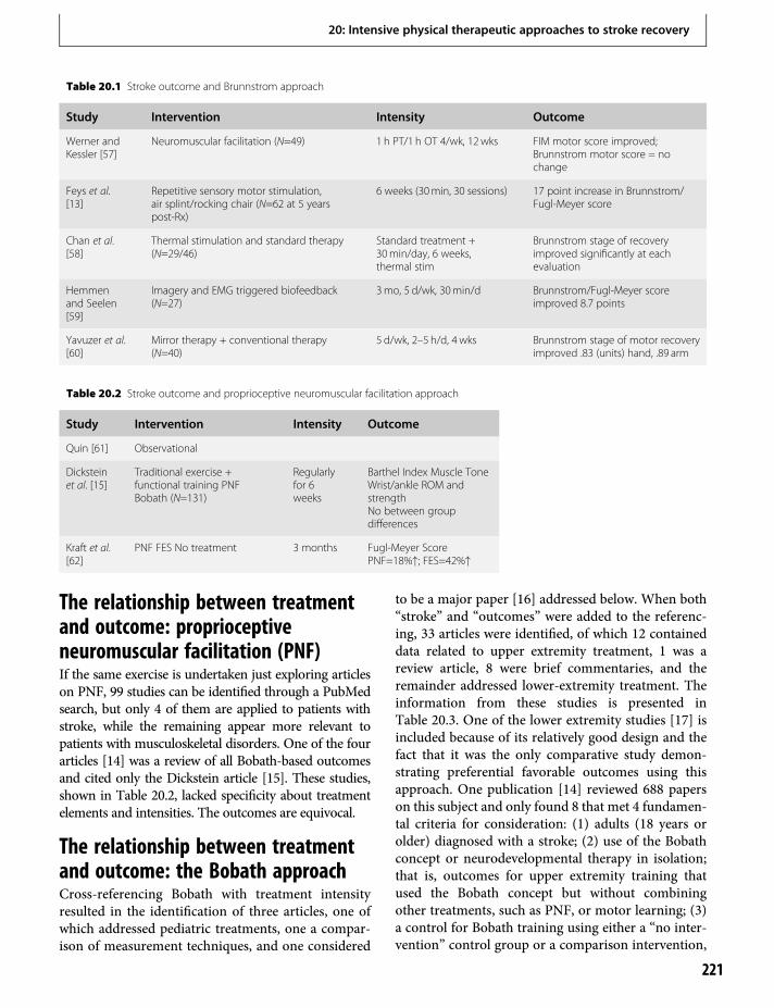
The relationship between treatmentand outcome: proprioceptiveneuromuscular facilitation (PNF)If the same exercise is undertaken just exploring articleson PNF, 99 studies can be identified through a PubMedsearch, but only 4 of them are applied to patients withstroke, while the remaining appear more relevant topatients with musculoskeletal disorders. One of the fourarticles [14] was a review of all Bobath-based outcomesand cited only the Dickstein article [15]. These studies,shown in Table 20.2, lacked specificity about treatmentelements and intensities. The outcomes are equivocal.
The relationship between treatmentand outcome: the Bobath approachCross-referencing Bobath with treatment intensityresulted in the identification of three articles, one ofwhich addressed pediatric treatments, one a compar-ison of measurement techniques, and one considered
to be a major paper [16] addressed below. When both“stroke” and “outcomes” were added to the referenc-ing, 33 articles were identified, of which 12 containeddata related to upper extremity treatment, 1 was areview article, 8 were brief commentaries, and theremainder addressed lower-extremity treatment. Theinformation from these studies is presented inTable 20.3. One of the lower extremity studies [17] isincluded because of its relatively good design and thefact that it was the only comparative study demon-strating preferential favorable outcomes using thisapproach. One publication [14] reviewed 688 paperson this subject and only found 8 that met 4 fundamen-tal criteria for consideration: (1) adults (18 years orolder) diagnosed with a stroke; (2) use of the Bobathconcept or neurodevelopmental therapy in isolation;that is, outcomes for upper extremity training thatused the Bobath concept but without combiningother treatments, such as PNF, or motor learning; (3)a control for Bobath training using either a “no inter-vention” control group or a comparison intervention,
Table 20.1 Stroke outcome and Brunnstrom approach
Study Intervention Intensity Outcome
Werner andKessler [57]
Neuromuscular facilitation (N=49) 1 h PT/1 h OT 4/wk, 12wks FIM motor score improved;Brunnstrom motor score = nochange
Feys et al.[13]
Repetitive sensory motor stimulation,air splint/rocking chair (N=62 at 5 yearspost-Rx)
6 weeks (30min, 30 sessions) 17 point increase in Brunnstrom/Fugl-Meyer score
Chan et al.[58]
Thermal stimulation and standard therapy(N=29/46)
Standard treatment +30min/day, 6 weeks,thermal stim
Brunnstrom stage of recoveryimproved significantly at eachevaluation
Hemmenand Seelen[59]
Imagery and EMG triggered biofeedback(N=27)
3mo, 5 d/wk, 30min/d Brunnstrom/Fugl-Meyer scoreimproved 8.7 points
Yavuzer et al.[60]
Mirror therapy + conventional therapy(N=40)
5 d/wk, 2–5 h/d, 4 wks Brunnstrom stage of motor recoveryimproved .83 (units) hand, .89 arm
Table 20.2 Stroke outcome and proprioceptive neuromuscular facilitation approach
Study Intervention Intensity Outcome
Quin [61] Observational
Dicksteinet al. [15]
Traditional exercise +functional training PNFBobath (N=131)
Regularlyfor 6weeks
Barthel Index Muscle ToneWrist/ankle ROM andstrengthNo between groupdifferences
Kraft et al.[62]
PNF FES No treatment 3 months Fugl-Meyer ScorePNF=18%↑; FES=42%↑
20: Intensive physical therapeutic approaches to stroke recovery
221

or a baseline phase; and (4) an outcome that reflectedchange in upper limb impairment, activity limitationor participation restriction. Many of these studies canbe categorized by provision of treatments that spanned4–5 weeks, but the actual dosage varied considerably(Table 20.3). Most outcomes described in that publi-cation [14] were either not favorable or equivocal.
A closer examination of intensity:contemporary approachesA PubMed search that cross-referenced the terms“stroke,” “upper extremity,” and “intensity of treat-ment,” without specifying the nature of the interven-tion, yielded 79 articles, 14 of which specifically
Table 20.3 Stroke outcome and the Bobath approach
Study Intervention Intensity Outcome
Basmajian et al.[63]
Bobath or EMG biofeedback (N=29) 5 weeks Upper extremity function test; Nodifferences
Partridge et al.[64]
Bobath or cryotherapy (N=65) 4 weeks Shoulder pain rating; Less pain withNDT
Nakayama et al.[65]
“Bobath” technique with outcomesassessed by Scandinavian StrokeSeverity Scale (N=636 acute)
69–74 hospital days with nodefined treatment duration orfrequency
Discharged: 64/115=no use andsubsequently: 26/64=compensateXlat UE 10/64=full function 28/64=partial
Lincoln et al.[16]
Typical British approach (N=282) Routine PT (standard Bobathtreatment, 5 d/wk, 30–45min)Qualified PT (standard PT + 2 h/wksenior research PT) Assistant PT(standard treatment given by PTA+ 2 h/wk Bobath); each treatmentoption given for 5 weeks
Barthel Index, Rivermead MotorAssessment; No differencesbetween groups
Van der Leeet al. [66]
Bobath or forced use 2 weeks 5 d/wk More improved in Action ResearchArm Test (forced use); less pain(Bobath)
Langhammerand Stanghelle[67]
Motor relearning program or Bobath(N=61) stratified into equal groupsby gender and lesion site
At least 40min, 5 d/wk throughouthospitalization (21–34 days)
Motor Assessment Scale BarthelIndex; Both groups improved butmotor relearning group significantlymore
Luke [14] 8/688 studies reviewed 30–45min, 5 d/wk in 7/8 studies;amount of therapist experiencevaried across studies
Reduce shoulder pain but nodifference compared to functionaltraining
Platz et al. [68] No augmented treatment time;Augmented with Bobath;Augmented with Arm BASIS,systematic repetition training (N=62severely impaired)
20 additional sessions per group;45min over 4 weeks
No overall between-groupdifferences in Fugl-MeyerAssessment scores but BASIStraining superior to Bobath inFugl-Meyer change score
Wang et al. [17] Bobath program or orthopedictreatment (N=21 stroke withspasticity; N=23 relative recoverystage)
20 treatments, 40min, 5 d/wk Motor Assessment Scale and StrokeImpact Scale improved more inboth groups with Bobath treatment
Hofsteinsdottir[69]
Bobath versus task-oriented training(N=324), 1 year post-stroke
Not specified Bobath treatment had no effectupon shoulder pain or health-related quality of life
Van Vliet et al.[70]
Bobath-based vs. movementscience-based (N=120) 2wks poststroke
As long as needed but matchedbetween groups
Motor Assessment Scale; 10-holepeg test; muscle tone; sensation; Nodifference between groups through6-month follow-up
20: Intensive physical therapeutic approaches to stroke recovery
222

addressed intensity of training and 10 of which speci-fied constraint-induced movement therapy (CIMT) asthe intervention. Inevitably, if more specific or alter-native terms had been chosen, undoubtedly manymore articles would appear. Of the 14, 2 [5,18] providecritical reviews of published studies to date, andone [19] was a critique of the Kwakkel et al. 1999study [20]. Two points of view by Dobkin [4] andWolf et al. [5] discussed recent clinical trials on ambu-lation in the Spinal Cord Injury Locomotor Trial(SCILT) and in the Extremity Constraint InducedTherapy Evaluation (EXCITE Trial). While notincluded in Table 20.4, both papers noted a pervasiveshortcoming in the rehabilitation literature includingclinical trial studies, and poorly defined characteriza-tion of intensity of training.
The studies depicted in Table 20.4 are more con-temporary than many of the neuromuscular re-education studies that have preceded them, and arecharacteristically defined by novel treatment techni-ques, several of which engage new technologies. Whilethe issue of delineating and condensing intensity canstill be seen as elusive, several interesting patterns haveemerged. The outstanding studies and analyses by theKwakkel group [5,20,21] have exposed issues related tovariability in stroke patient attributes and poorlydefined control over blinding. Langhorne [19] notedthat differences in outcomes and a failure to observepersistence of improvements in the long term may beattributable to heterogeneity of study groups that arenot appropriately counterbalanced, thus permittingimprovement in controls over time. Efforts to com-pare treatments comprised of Bobath techniques withor without additional therapy [22] or by differentiatingunimanual from bilateral training [23] still produceequivocal results and, for the first time, suggest thatintensity per se may not be a determining factor inoutcome, although this might be less true in the veryearly weeks after a stroke [3]. In fact, the emergingdiscussions now suggest a need to examine dosingmore closely and, indeed, Morris et al. [23] quantifiedfrequency of movements across tasks during training.In this context, the review by Van Peppen et al. [18]draws attention to the fact that focused task trainingstudies yield greater effect sizes than those that aremore oriented towards application of a technique.
Yet, beyond the review of traditional “hands on”interventions, introducing novel technologies, suchas robotics and virtual environments (see alsoChapter 18), offers hope for further improving
upper-extremity outcomes in stroke rehabilitation.The numbers of comprehensive studies in these twoareas are limited, but they share in common the veryunique opportunity for participants to work withoutdetailed instruction and to potentially better imposetheir own thought processes toward rendering solu-tions toward further movement control with prospectsfor functional restitution. Thus, while only represen-tative of the emerging work in mental imagery as arehabilitation technique, Liu et al. [24] found that theirpatients showed greater improvements in retentionand transfer to relevant tasks than those patientsundergoing functional training. They noted that men-tal imagery subjects were left to their own thoughtswith minimal clinician intrusion in efforts to optimizetheir imaging. This same strategy has been employedby Steve Page and his colleagues in their use of mentalimagery as a therapeutic technique [25,26] (see alsoChapter 21).
Opportunities to more effectively exploit advancesin robotics and virtual environments are plentiful.However, the articles represented herein point to theneed for investigators to better stratify patients andmore effectively link these interventions to changes inability as well as impairment. Thus far, few studieshave made much headway in this direction.Moreover, the novelty of these approaches creates apotential to thwart systematic examination of dosingin favor of the excitement ensconced in enthusiasticendorsement and engagement without thinking aboutpatient fatigue or motoric capabilities, let alone cogni-tive acumen. In addition and perhaps more impor-tantly, the field must move past the “feasibility” stageto one where these innovative therapies are shown tobring about better outcomes than those achievedthrough dose-matched standard methods.
Intensity revisitedIn summary, it would appear that newer approachesoffer chances for patients to become more directlyinvolved in their upper extremity therapy. Commentswithin review articles [5,18] indicate that earlier andmore intense, goal-directed interventions among lessseverely impaired stroke survivors produce betterchanges in impairment and ability. Interestingly, in2000, the Heart and Stroke Foundation of Ontariosupported the work of the Consensus Panel onHemiplegic Arm andHand, who undertook the difficulttask of assembling and classifying all available literature
20: Intensive physical therapeutic approaches to stroke recovery
223
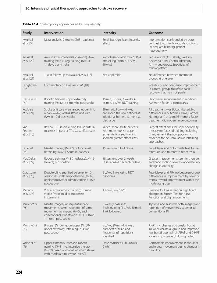
Table 20.4 Contemporary approaches addressing intensity
Study Intervention Intensity Outcome
Kwakkelet al. [5]
Meta-analysis, 9 studies (1051 patients) Small but significant intensityeffect
Interpretation confounded by poorcontrast to control group descriptions,inadequate blinding, patientheterogeneity
Kwakkelet al. [20]
Arm splint immobilization (N=37); Armtraining (N=33); Leg training (N=31);14 days post-stroke
Immobilization (30min, 5 d/wkarm or leg (30min, 5 d/wk,20wks)
Leg>Control (ADL ability, walking,dexterity) Arm>Control (dexterityArm = Leg group; Specificity oftraining effect
Kwakkelet al. [21]
1 year follow-up to Kwakkel et al. [18] Not applicable No difference between treatmentgroups at one year
Langhorne[19]
Commentary on Kwakkel et al. [18] Possibly due to continued improvementin control group; therefore earlierrecovery that may not persist
Hesse et al.[71]
Robotic bilateral upper extremitytraining (N=12) > 6 months post-stroke
15min, 5 d/wk, 3 weeks +45min, 5 d/wk NDT training
Short-term improvement in modifiedAshworth for 8/12 participants
Rodgerset al. [21]
Stroke unit care + enhanced upper limbrehab (N=62) versus stroke unit care(N=61), 10 d post-stroke
30min/d, 5 d/wk, 6wks;enhanced therapy defined asadditional home treatment atdischarge
All treatment was Bobath-based. Nodifferences in outcomes ARAT, Barthel,Nottingham) at 3 and 6 months. Moretreatment did not enhance outcomes
VanPeppenet al. [18]
Review 151 studies using PEDro criteriato assess impact of PT; assess effect sizes
Varied, more acute patientswith more intense upper-extremity focused trainingshowed greater effect sizes
Largest effect sizes for upper-extremitytherapy for focused training includingCI movement therapy; poor or noevidence for neuromuscular retrainingapproaches
Liu et al.[24]
Mental imagery (N=27) or functionalretraining (N=22) Acute in-patients
15 sessions; 1 h/d, 3 wks Fugl-Meyer and Color Trails Test; betterretention and transfer to other tasks
MacClellanet al. [72]
Robotic training N=8 (moderate), N=19(severe). No controls
18 sessions over 3 weeks(2 sessions/d, 1 h each, 3 d/wk)
Greater improvements seen in shoulderand hand motion severe>moderate; nochange in disability
Gladstoneet al. [73]
Double-blind stratified by severity 10sessions PT with amphetamine (N=34)or placebo (N=37) administration 5–10 dpost-stroke
2 d/wk, 5wks using NDTprinciples
Fugl-Meyer and FIM no between-groupdifferences in improvement by severity;trends toward improvement within themoderate group
Merianset al. [74]
Virtual environment training; Chronicstroke (N=8); mild to moderateimpairment
13 days, 2–2.5 h/d Baseline to 1wk retention; significantchanges in Jepsen Test for HandFunction and digit movements
Muller et al.[75]
Mental imagery of sequential handmovements (N=6), repetition of samemovement as imaged (N=6), andconventional (Bobath and PNF) PT (N=5)1 month post-stroke
3 weekly baselines;4 wks training (5 d/wk, 30min),1 wk follow-up
Jepsen Hand Test with both imagery andrepetition of movements superior toconventional PT
Morris et al.[23]
Bilateral (N=56) vs. unilateral (N=50)upper extremity retraining, 2–4wkspost-stroke
5 d/wk, 20min/d, 6wks ;numbers of tasks andfrequency of repetitionsspecified
ARAT=no change at 6 weeks; but at18 weeks bilateral group had improvedless based upon pinch ARAT and 9 HPTscores; importance of dosing noted
Volpe et al.[76]
Upper extremity intensive robotictraining (N=11) vs. intensive therapy(N=10) based on Bobath chronic strokewith moderate to severe (NIHSS)
Dose matched (1 h, 3 d/wk,6 wks)
Comparable improvement in shoulderand elbowmovement but no changes indisability
224
20: Intensive physical therapeutic approaches to stroke recovery

on upper-extremity rehabilitation of stroke survivorswith the intent of developing predictive equations forthe expected magnitude of functional restoration [27].The assembled panel cleverly categorized all the data byintervention type and then applied the evidence to ninecase scenarios of decreasing complexity (less impair-ment). As a result, the panel could produce a quantita-tive value, based upon the Chedoke–McMaster StrokeAssessment Scale, for expected return of shoulder,elbow, and wrist/hand function.
The results validated the same conclusions reachedindependently by Kwakkel [20] and later by vanPeppen [18]. Fundamentally, the evidence suggestedthat the best functional restoration would be generatedfor patients with Chedoke–McMaster Assessmentscores of ≥ 4, indicative of isolated movement out offlexion synergy. Moreover for patients who wereseverely impaired, provision of passive motion, main-taining joint alignment, and preventing shoulder sub-luxation would be the best use of available time andresources. A summary of these observations was pub-lished subsequently [27], and highlighted the factthat one important element, intensity of training,had not been studied sufficiently during the review ofstudies comprising the consensus report. Recently,The Canadian Stroke Network [28], one of the world’soutstanding clinical and research stroke organizations,has provided a summary statement about stroke studypriorities, based upon their 2003 consensus confer-ence. Indeed, defining the ideal timing and intensityof rehabilitation after mild to moderate stroke is nowone of the Network’s top five priorities. Inevitably, theemphasis on mild to moderate stroke may well havebeen determined by the compilation of data, suggest-ing that these patients have the greatest potential toovercome impairments and to maximize function.
Yet, the elements that should constitute “intensityof practice” remain elusive. Previously, Wolf [29] dis-cussed factors that can influence outcomes usingCIMT and which, to date, had not been investigated.These factors included patient fatigue, family compli-ance, and socio-economic factors that can increasinglyimpede cooperation and optimization of treatmentresults. When one adds to these important consider-ations the fact that reimbursement for servicescontinues to constrict and the factors contributing todecisions to pay for services are dictated by private andgovernmental agencies whose policies and proceduresvary throughout the world, there is little wonderwhy defining intensity can be difficult. If, in fact,
non-clinical factors control rehabilitation treatmentexposure, reaching any form of consensus on definingintensity becomes a most onerous task. These realitiesoften make the inclusion of more clinically soundcontributions to defining intensity, such as locationof lesion, severity of impairment, and implementationof supportive evidence, more difficult. Last, cliniciantreatment biases, often driven by an educational envi-ronment that might have been provided by teachersresistant to processing supportive or contrary data,obstruct a desire to achieve rational boundaries todelineate intensity.
Providing a perspectiveNomatter the preferences for treatment approaches toreduce impairments and improve upper-extremityfunction among stroke survivors, several clinicianbehaviors have withstood the test of time. Inevitably,most health professionals, given the remarkableresponsibility for improving the status of catastroph-ically injured individuals, have been motivated to doso because of their remarkable sense of compassionand their desire to work regularly and directly withpatients, often independently from other healthcareproviders. These individuals are usually physical andoccupational therapists, who have enjoyed a traditionof “hands on” approaches to form the core of theirtreatment repertoires, often perceived as remarkably“unique” and self-defining. Historically, many of theneuromuscular re-education approaches (see above)do indeed require manual guidance with some formof repetition, often dictated and directed by the clini-cian. The number of trials and their consistencyvaries considerably across these various approaches,as does the form of feedback provided to patients.Many approaches have lacked true goal directionthat includes strategies and decision-making withinthe context of a function. Rather, treatments haveaddressed single or multi-joint motions directedtoward reducing tone or enhancing volitional move-ment, but often at the expense of incorporating themovement within a context that has limited functionor relevance to the patient. In addition, the constructof the repetition varies, as does the nature of the feed-back. Are movements repeated the same way and, if so,how often? Are they repeated within a session or overtreatment sessions? When are they modified, and onwhat basis? Answers to these questions have oftenbeen intuitive to clinicians, but made without respect
20: Intensive physical therapeutic approaches to stroke recovery
225
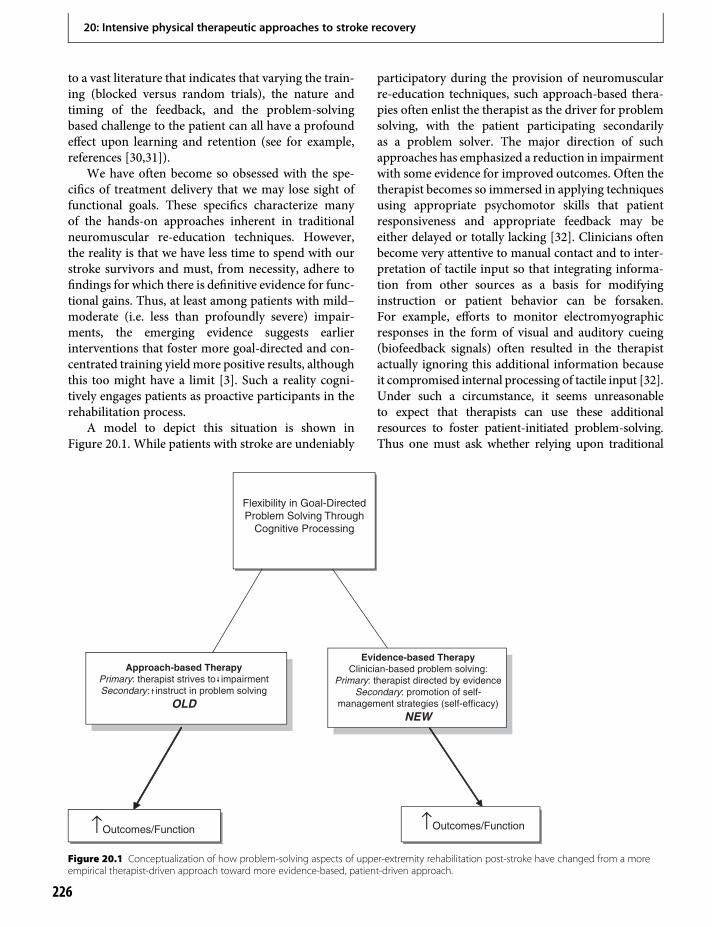
to a vast literature that indicates that varying the train-ing (blocked versus random trials), the nature andtiming of the feedback, and the problem-solvingbased challenge to the patient can all have a profoundeffect upon learning and retention (see for example,references [30,31]).
We have often become so obsessed with the spe-cifics of treatment delivery that we may lose sight offunctional goals. These specifics characterize manyof the hands-on approaches inherent in traditionalneuromuscular re-education techniques. However,the reality is that we have less time to spend with ourstroke survivors and must, from necessity, adhere tofindings for which there is definitive evidence for func-tional gains. Thus, at least among patients with mild–moderate (i.e. less than profoundly severe) impair-ments, the emerging evidence suggests earlierinterventions that foster more goal-directed and con-centrated training yieldmore positive results, althoughthis too might have a limit [3]. Such a reality cogni-tively engages patients as proactive participants in therehabilitation process.
A model to depict this situation is shown inFigure 20.1. While patients with stroke are undeniably
participatory during the provision of neuromuscularre-education techniques, such approach-based thera-pies often enlist the therapist as the driver for problemsolving, with the patient participating secondarilyas a problem solver. The major direction of suchapproaches has emphasized a reduction in impairmentwith some evidence for improved outcomes. Often thetherapist becomes so immersed in applying techniquesusing appropriate psychomotor skills that patientresponsiveness and appropriate feedback may beeither delayed or totally lacking [32]. Clinicians oftenbecome very attentive to manual contact and to inter-pretation of tactile input so that integrating informa-tion from other sources as a basis for modifyinginstruction or patient behavior can be forsaken.For example, efforts to monitor electromyographicresponses in the form of visual and auditory cueing(biofeedback signals) often resulted in the therapistactually ignoring this additional information becauseit compromised internal processing of tactile input [32].Under such a circumstance, it seems unreasonableto expect that therapists can use these additionalresources to foster patient-initiated problem-solving.Thus one must ask whether relying upon traditional
Flexibility in Goal-DirectedProblem Solving Through
Cognitive Processing
Evidence-based TherapyClinician-based problem solving:
Primary: therapist directed by evidenceSecondary: promotion of self-
management strategies (self-efficacy)
NEW
Approach-based TherapyPrimary: therapist strives to impairmentSecondary: instruct in problem solving
OLD
↑Outcomes/Function ↑Outcomes/Function
Figure 20.1 Conceptualization of how problem-solving aspects of upper-extremity rehabilitation post-stroke have changed from a moreempirical therapist-driven approach toward more evidence-based, patient-driven approach.
20: Intensive physical therapeutic approaches to stroke recovery
226
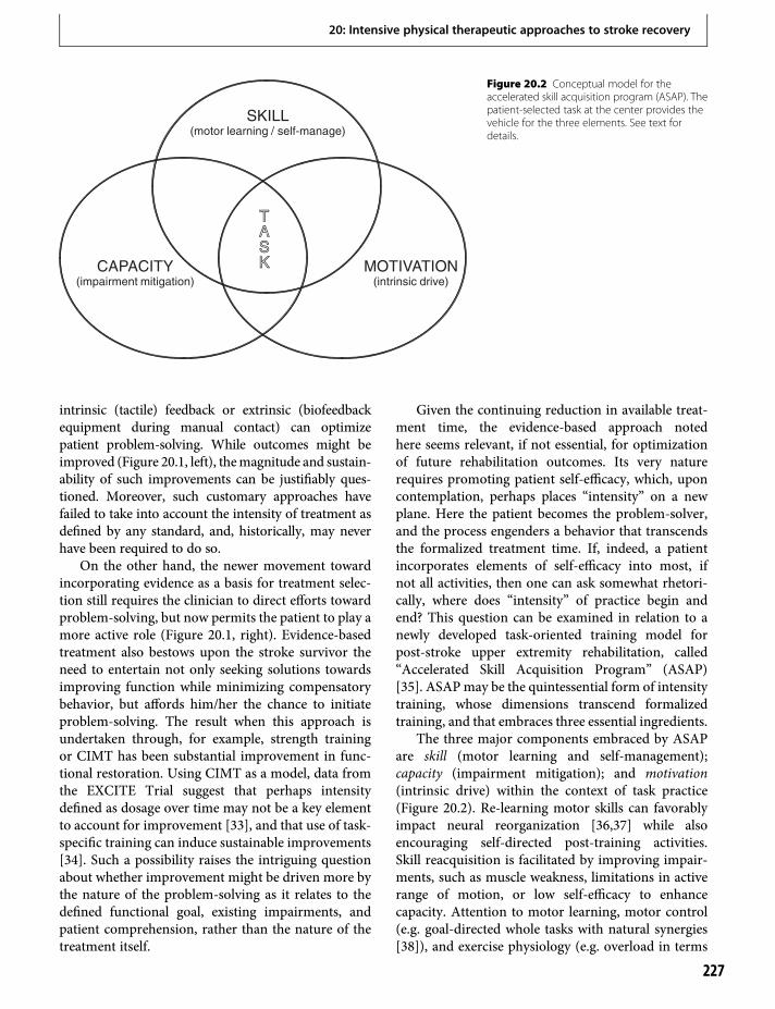
intrinsic (tactile) feedback or extrinsic (biofeedbackequipment during manual contact) can optimizepatient problem-solving. While outcomes might beimproved (Figure 20.1, left), themagnitude and sustain-ability of such improvements can be justifiably ques-tioned. Moreover, such customary approaches havefailed to take into account the intensity of treatment asdefined by any standard, and, historically, may neverhave been required to do so.
On the other hand, the newer movement towardincorporating evidence as a basis for treatment selec-tion still requires the clinician to direct efforts towardproblem-solving, but now permits the patient to play amore active role (Figure 20.1, right). Evidence-basedtreatment also bestows upon the stroke survivor theneed to entertain not only seeking solutions towardsimproving function while minimizing compensatorybehavior, but affords him/her the chance to initiateproblem-solving. The result when this approach isundertaken through, for example, strength trainingor CIMT has been substantial improvement in func-tional restoration. Using CIMT as a model, data fromthe EXCITE Trial suggest that perhaps intensitydefined as dosage over time may not be a key elementto account for improvement [33], and that use of task-specific training can induce sustainable improvements[34]. Such a possibility raises the intriguing questionabout whether improvement might be driven more bythe nature of the problem-solving as it relates to thedefined functional goal, existing impairments, andpatient comprehension, rather than the nature of thetreatment itself.
Given the continuing reduction in available treat-ment time, the evidence-based approach notedhere seems relevant, if not essential, for optimizationof future rehabilitation outcomes. Its very naturerequires promoting patient self-efficacy, which, uponcontemplation, perhaps places “intensity” on a newplane. Here the patient becomes the problem-solver,and the process engenders a behavior that transcendsthe formalized treatment time. If, indeed, a patientincorporates elements of self-efficacy into most, ifnot all activities, then one can ask somewhat rhetori-cally, where does “intensity” of practice begin andend? This question can be examined in relation to anewly developed task-oriented training model forpost-stroke upper extremity rehabilitation, called“Accelerated Skill Acquisition Program” (ASAP)[35]. ASAPmay be the quintessential form of intensitytraining, whose dimensions transcend formalizedtraining, and that embraces three essential ingredients.
The three major components embraced by ASAPare skill (motor learning and self-management);capacity (impairment mitigation); and motivation(intrinsic drive) within the context of task practice(Figure 20.2). Re-learning motor skills can favorablyimpact neural reorganization [36,37] while alsoencouraging self-directed post-training activities.Skill reacquisition is facilitated by improving impair-ments, such as muscle weakness, limitations in activerange of motion, or low self-efficacy to enhancecapacity. Attention to motor learning, motor control(e.g. goal-directed whole tasks with natural synergies[38]), and exercise physiology (e.g. overload in terms
SKILL(motor learning / self-manage)
CAPACITY(impairment mitigation)
MOTIVATION(intrinsic drive)
TASK
TASK
Figure 20.2 Conceptual model for theaccelerated skill acquisition program (ASAP). Thepatient-selected task at the center provides thevehicle for the three elements. See text fordetails.
20: Intensive physical therapeutic approaches to stroke recovery
227
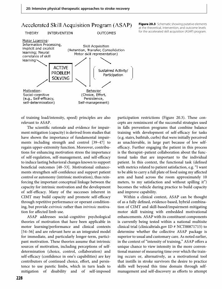
of training load/intensity, speed) principles are alsorelevant to ASAP.
The scientific rationale and evidence for impair-ment mitigation (capacity) is derived from studies thathave shown the importance of fundamental impair-ments including strength and control [39–47] toregain upper-extremity function. Moreover, contribu-tions for enhancing motivation stress the importanceof self-regulation, self-management, and self-efficacyto induce lasting behavioral changes known to supportbeneficial outcomes [48–53]. Motivational enhance-ments strengthen self-confidence and support patientcontrol or autonomy (intrinsic motivation), thus rein-forcing the important conceptual linkage between thecapacity for intrinsic motivation and the developmentof self-efficacy. Many of the successes inherent inCIMT may build capacity and promote self-efficacythrough repetitive performance or operant condition-ing, but provide extrinsic rather than intrinsicmotiva-tion for affected limb use.
ASAP addresses social–cognitive psychologicaltheories of motivation that have been applicable inmotor learning/performance and clinical contexts[54–56] and are relevant here as an integrated modelfor immediate, and particularly longer-term, partici-pant motivation. These theories assume that intrinsicsources of motivation, including perceptions of self-determination (choice, control, collaboration) andself-efficacy (confidence in one’s capabilities) are keycontributors of continued choice, effort, and persis-tence to use paretic limbs, which in turn leads tomitigation of disability and of self-imposed
participation restrictions (Figure 20.3). These con-cepts are reminiscent of the successful strategies usedin falls prevention programs that combine balancetraining with development of self-efficacy for tasks(e.g. stairs, bathtub, curbs) that were initially perceivedas unachievable, in large part because of low self-efficacy. Further engaging the patient in this processis the therapist–patient collaboration about the func-tional tasks that are important to the individualpatient. In this context, the functional task (definedwith metrics related to patient satisfaction, e.g. “I wantto be able to carry a full plate of food using my affectedarm and hand across the room approximately 10meters, to my satisfaction and without spilling it”)becomes the vehicle during practice to build capacityand improve capability.
Within a clinical context, ASAP can be thoughtof as a fully defined, evidence-based, hybrid combina-tion of CIMT and skill-based/impairment-mitigatingmotor skill training with embedded motivationalenhancements. ASAP with its constituent componentsis currently being tested within a national multisiteclinical trial (clinicaltrials.gov ID # NCT00871715) todetermine whether the collective ASAP package issuperior to usual and customary care. As noted earlier,in the context of “intensity of training,” ASAP offers aunique chance to view intensity in the more conven-tional manner of measuring time over which the train-ing occurs or, alternatively, as a motivational toolthat instills in stroke survivors the desire to practiceskills well beyond this time domain through self-management and self-discovery as efforts to attempt
Figure 20.3 Schematic showing putative elementsat the theoretical, intervention, and outcome levelsfor the accelerated skill acquisition (ASAP) program.
20: Intensive physical therapeutic approaches to stroke recovery
228
task execution and solve new motor problems as theypresent in the absence of an awareness of time. Inessence, this new model is designed to sustain theimproved skills and capacity and thereby enable par-ticipation in meaningful activities (Figure 20.3).
SummaryThere is little question that the concept of “intensity”of upper extremity re-training during recovery fromstroke has assumed different perspectives among clini-cians. Foremost is the fact that historically the rela-tionship between dosing and outcomes has beenpoorly elucidated in the stroke rehabilitation litera-ture, and often there has been a dependence uponpersistent and prolonged delivery of several neuro-muscular re-education techniques for which a paucityof evidence exists to support efficacy. As a result, asubstantial effort is extended toward teaching strokesurvivors compensatory behaviors that forsake theintrinsic possibilities to maximize functional restitu-tion for the more impaired limb.
This situation has now been influenced substantiallyby cuts in length of hospital stay, outpatient services,and availability of fiscal resources. Accordingly, a morepractical approach has emerged that builds upon thespecific aspects of task-specific training that can includeelements of strengthening, skill reacquisition, and self-efficacy. In fact, evidence is accruing to indicate thatsuch a constellation of elements within the treatment“package” may be efficacious. Inevitably, allocation oftime and resources will determine the extent to which amore contemporary approach must be embraced byclinicians and family members.
References1. Kwakkel G. Impact of intensity of practice after stroke:
Issues for consideration. Disabil Rehabil. 2006;28:823–30.
2. Richards L, Senesac C, McGuirk T, et al. Response tointensive upper extremity therapy by individuals withataxia from stroke. Top Stroke Rehabil. 2008;15:262–71.
3. Dromerick AW, Lang CE, Birkenmeier RL, et al. VeryEarly Constraint-Induced Movement during StrokeRehabilitation (VECTORS): A single-center RCT.Neurology. 2009;73:195–201.
4. Dobkin BH. Confounders in rehabilitation trials oftask-oriented training: Lessons from the designs of theEXCITE and SCILT multicenter trials. NeurorehabilNeural Repair. 2007;21:3–13.
5. Wolf SL, Winstein CJ, Miller JP, et al. Looking in therear view mirror when conversing with back seat
drivers: The EXCITE trial revisited. NeurorehabilNeural Repair. 2007;21:379–87.
6. Kwakkel G, Wagenaar RC, Koelman TW, et al. Effectsof intensity of rehabilitation after stroke. A researchsynthesis. Stroke. 1997;28:1550–6.
7. Bobath B. Adult hemiplegia: Evaluation and treatment.3rd ed. Oxford: Heinemann; 1990.
8. Bobath B. Abnormal postural reflex activity caused bybrain lesions. 2nd ed. London: Heinemann; 1971.
9. Brunnstrom S.Movement therapy in hemiplegia. 1st ed.New York, NY: Harper & Row; 1970.
10. Voss DE, Ionta MK, Myers B. Proprioceptiveneuromuscular facilitation: Patterns and techniques.3rd ed. Philadelphia: Harper & Row; 1985.
11. Howle JM. Neuro-developmental treatment approach:Theoretical foundations and principles of clinicalpractice. 1st ed. Laguna Beach: The North AmericanNeurodevelopmental Tretment Association; 2003.
12. Fugl-Meyer AR, Jaasko L, Leyman I, et al. The post-stroke hemiplegic patient. 1. Amethod for evaluation ofphysical performance. Scand J Rehab Med.1975;7:13–31.
13. Feys H, De Weerdt W, Verbeke G, et al. Early andrepetitive stimulation of the arm can substantiallyimprove the long-term outcome after stroke: A 5-yearfollow-up study of a randomized trial. Stroke.2004;35:924–9.
14. Luke C, Dodd KJ, Brock K. Outcomes of the Bobathconcept on upper limb recovery following stroke. ClinRehabil. 2004;18:888–98.
15. Dickstein R, Hocherman S, Pillar T, et al. Strokerehabilitation. Three exercise therapy approaches. PhysTher. 1986;66:1233–8.
16. Lincoln NB, Parry RH, Vass CD. Randomized,controlled trial to evaluate increased intensity ofphysiotherapy treatment of arm function after stroke.Stroke. 1999;30:573–9.
17. Wang RY, Chen HI, Chen CY, et al. Efficacy of Bobathversus orthopaedic approach on impairment andfunction at different motor recovery stages after stroke:A randomized controlled study. Clin Rehabil.2005;19:155–64.
18. Van Peppen RP, Kwakkel G, Wood-Dauphinee S, et al.The impact of physical therapy on functional outcomesafter stroke: What’s the evidence? Clin Rehabil.2004;18:833–62.
19. Langhorne P. Intensity of rehabilitation: Some answersand more questions? J Neurol Neurosurg Psychiatry.2002;72:430–1.
20. Kwakkel G, Wagenaar RC, Twisk JW, et al. Intensity ofleg and arm training after primary middle-cerebral-artery stroke: A randomised trial. Lancet.1999;354:191–6.
20: Intensive physical therapeutic approaches to stroke recovery
229

21. Kwakkel G, Kollen BJ, Wagenaar RC. Long term effectsof intensity of upper and lower limb training afterstroke: A randomised trial. J Neurol NeurosurgPsychiatry. 2002;72:473–9.
22. Rodgers H, Mackintosh J, Price C, et al. Does an earlyincreased-intensity interdisciplinary upper limbtherapy programme following acute stroke improveoutcome? Clin Rehabil. 2003;17:579–89.
23. Morris JH, van Wijck F, Joice S, et al. A comparison ofbilateral and unilateral upper-limb task training in earlypoststroke rehabilitation: A randomized controlledtrial. Arch Phys Med Rehabil. 2008;89:1237–45.
24. Liu KP, Chan CC, Lee TM, et al. Mental imagery forpromoting relearning for people after stroke: Arandomized controlled trial. Arch Phys Med Rehabil.2004;85:1403–08.
25. Page SJ, Levine P, Leonard AC. Effects of mentalpractice on affected limb use and function in chronicstroke. Arch Phys Med Rehabil. 2005;86:399–402.
26. Page SJ, Levine P, Leonard A. Mental practice inchronic stroke: Results of a randomized, placebo-controlled trial. Stroke. 2007;38:1293–7.
27. Barreca S, Wolf SL, Fasoli S, et al. Treatmentinterventions for the paretic upper limb of strokesurvivors: A critical review. Neurorehabil NeuralRepair. 2003;17:220–6.
28. Bayley MT, Hurdowar A, Teasell R, et al. Priorities forstroke rehabilitation and research: Results of a 2003Canadian Stroke Network consensus conference. ArchPhys Med Rehabil. 2007;88:526–8.
29. Wolf SL. Revisiting constraint-induced movementtherapy: Are we too smitten with the mitten? Is allnonuse “learned”? and other quandaries. Phys Ther.2007;87:1212–23.
30. Lee TD, Swanson LR, Hall AL. What is repeated in arepetition? Effects of practice conditions on motor skillacquisition. Phys Ther. 1991;71:150–6.
31. Schmidt RA, Lee TD. Motor control and learning: Abehavioral emphasis. 4th ed. Champaign, IL: HumanKinetics; 2005.
32. Wolf SL, Edwards DI, Shutter LA. Concurrentassessment of muscle activity (CAMA). A proceduralapproach to assess treatment goals. Phys Ther.1986;66:218–24.
33. Wolf SL, Newton H, Maddy D, et al. The Excite Trial:Relationship of intensity of constraint inducedmovement therapy to improvement in the Wolfmotor function test. Restor Neurol Neurosci.2007;25:549–62.
34. Wolf SL, Winstein CJ, Miller JP, et al. Retention ofupper limb function in stroke survivors who havereceived constraint-induced movement therapy: TheEXCITE randomised trial. Lancet Neurol.2008;7:33–40.
35. Winstein C, Wolf SL. Task-oriented training topromote extremity recovery. In: Stein RLH, Macko RF,Winstein CJ, Zorowitz RD, editors. Stroke recovery &rehabilitation. New York, NY: Demos Medical; 2008:pp. 267–390.
36. Dobkin BH. Strategies for stroke rehabilitation. LancetNeurol. 2004;3:528–36.
37. Nudo RJ, Plautz EJ, Frost SB. Role of adaptive plasticityin recovery of function after damage to motor cortex.Muscle Nerve. 2001;24:1000–19.
38. Wu C, Trombly CA, Lin K, et al. A kinematic study ofcontextual effects on reaching performance in personswith and without stroke: Influences of objectavailability. Arch Phys Med Rehabil. 2000;81:95–101.
39. Duncan P, Studenski S, Richards L, et al. Randomizedclinical trial of therapeutic exercise in subacute stroke.Stroke. 2003;34:2173–80.
40. Platz T. Impairment-oriented training (IOT) –Scientific concept and evidence-based treatmentstrategies. Restor Neurol Neurosci. 2004;22:301–15.
41. Platz T, van Kaick S, Moller L, et al. Impairment-oriented training and adaptive motor cortexreorganisation after stroke: A fTMS study. J Neurol.2005;252:1363–71.
42. Platz T, Winter T, Muller N, et al. Arm ability trainingfor stroke and traumatic brain injury patients with mildarm paresis: A single-blind, randomized, controlledtrial. Arch Phys Med Rehabil. 2001;82:961–8.
43. Winstein CJ, Rose DK, Tan SM, et al. A randomizedcontrolled comparison of upper-extremityrehabilitation strategies in acute stroke: A pilot study ofimmediate and long-term outcomes. Arch Phys MedRehabil. 2004;85:620–8.
44. Sunderland A, Tuke A. Neuroplasticity, learningand recovery after stroke: A critical evaluation ofconstraint-induced therapy. Neuropsychol Rehabil.2005;15:81–96.
45. Winstein CJ, Wing AM, Whitall J. Motor control andlearning principles for rehabilitation of upper limbmovements after brain injury. In: Boller F, Grafman J,editors. Handbook of neurospychology. 2nd ed.Amsterdam: Elsevier Science; 2003: pp. 77–137.
46. Patten C, Dozono J, Schmidt SG, et al. Combinedfunctional task practice and dynamic high intensityresistance training promotes recovery of upper-extremity motor function in post-stroke hemiparesis:A case study. J Neurol Phys Ther. 2006;30:99–115.
47. Zackowski KM, Dromerick AW, Sahrmann SA, et al.How do strength, sensation, spasticity and jointindividuation relate to the reaching deficits ofpeople with chronic hemiparesis? Brain.2004;127:1035–46.
48. Bandura A. Self-efficacy: Toward a unifying theory ofbehavioral change. Psychol Rev. 1977;84:191–215.
20: Intensive physical therapeutic approaches to stroke recovery
230
49. Bandura A. Self-efficacy: The exercise of control. NewYork, NY: Freeman; 1997.
50. Jones F. Strategies to enhance chronic disease self-management: How can we apply this to stroke? DisabilRehabil. 2006;28:841–7.
51. Kendall E, Catalano T, Kuipers P, et al. Recoveryfollowing stroke: The role of self-managementeducation. Soc Sci Med. 2007;64:735–46.
52. Williams GC, McGregor HA, Sharp D, et al. Testing aself-determination theory intervention for motivatingtobacco cessation: Supporting autonomyand competencein a clinical trial. Health Psychol. 2006;25:91–101.
53. Williams GC, McGregor HA, Zeldman A, et al. Testinga self-determination theory process model forpromoting glycemic control through diabetes self-management. Health Psychol. 2004;23:58–66.
54. Ewart CK, Stewart KJ, Gillilan RE, et al. Self-efficacymediates strength gains during circuit weight trainingin men with coronary artery disease. Med Sci SportsExerc. 1986;18:531–40.
55. Taylor CB, Bandura A, Ewart CK, et al. Exercise testingto enhance wives’ confidence in their husbands’ cardiaccapability soon after clinically uncomplicated acutemyocardial infarction. Am J Cardiol. 1985;55:635–8.
56. Sniehotta FF, Scholz U, Schwarzer R, et al. Long-termeffects of two psychological interventions on physicalexercise and self-regulation following coronaryrehabilitation. Int J Behav Med. 2005;12:244–55.
57. Werner RA, Kessler S. Effectiveness of an intensiveoutpatient rehabilitation program for postacute strokepatients. Am J Phys Med Rehabil. 1996;75:114–20.
58. Chen G, Patten C, Kothari DH, et al. Gait differencesbetween individuals with post-stroke hemiparesis andnon-disabled controls at matched speeds. Gait Posture.2005;22:51–6.
59. Hemmen B, Seelen HA. Effects of movement imageryand electromyography-triggered feedback on arm handfunction in stroke patients in the subacute phase. ClinRehabil. 2007;21:587–94.
60. Yavuzer G, Selles R, Sezer N, et al. Mirror therapyimproves hand function in subacute stroke: Arandomized controlled trial. Arch Phys Med Rehabil.2008;89:393–8.
61. Quin CE. Observations on the effects of proprioceptiveneuromuscular facilitation techniques in the treatmentof hemiplegia. Rheumatol Phys Med. 1971;11:186–92.
62. Kraft GH, Fitts SS, Hammond MC. Techniquesto improve function of the arm and hand inchronic hemiplegia. Arch Phys Med Rehabil.1992;73:220–7.
63. Basmajian JV, Gowland CA, Finlayson MA, et al.Stroke treatment: Comparison of integratedbehavioral–physical therapy vs. traditional physical
therapy programs. Arch Phys Med Rehabil.1987;68:267–72.
64. Partridge C, Edwards S, Mee R, et al. Hemiplegicshoulder pain: A study of two methods ofphysiotherapy treatment. Clin Rehab. 1990;4:43–9.
65. Nakayama H, Jorgensen HS, Raaschou HO, et al.Recovery of upper extremity function in stroke patients:The Copenhagen Stroke Study. Arch Phys Med Rehabil.1994;75:394–8.
66. van der Lee JH, Wagenaar RC, Lankhorst GJ, et al.Forced use of the upper extremity in chronic strokepatients: Results from a single-blind randomizedclinical trial. Stroke. 1999;30:2369–75.
67. Langhammer B, Stanghelle JK. Bobath or motorrelearning programme? A comparison of two differentapproaches of physiotherapy in stroke rehabilitation: Arandomized controlled study.ClinRehabil. 2000;14:361–9.
68. Platz T, Eickhof C, van Kaick S, et al. Impairment-oriented training or Bobath therapy for severe armparesis after stroke: A single-blind, multicentrerandomized controlled trial. Clin Rehabil.2005;19:714–24.
69. Hafsteinsdottir TB, Kappelle J, Grypdonck MH, et al.Effects of Bobath-based therapy on depression,shoulder pain and health-related quality of life inpatients after stroke. J Rehabil Med. 2007;39:627–32.
70. van Vliet PM, Lincoln NB, Foxall A. Comparison ofBobath based and movement science based treatmentfor stroke: A randomised controlled trial. J NeurolNeurosurg Psychiatry. 2005;76:503–08.
71. Hesse S, Schmidt H, Werner C, et al. Upper and lowerextremity robotic devices for rehabilitation and forstudying motor control. Curr Opin Neurol.2003;16:705–10.
72. Macclellan LR, Bradham DD, Whitall J, et al. Roboticupper-limb neurorehabilitation in chronic strokepatients. J Rehabil Res Dev. 2005;42:717–22.
73. Gladstone DJ, Danells CJ, Armesto A, et al.Physiotherapy coupled with dextroamphetamine forrehabilitation after hemiparetic stroke: A randomized,double-blind, placebo-controlled trial. Stroke.2006;37:179–85.
74. Merians AS, Poizner H, Boian R, et al. Sensorimotortraining in a virtual reality environment: Does itimprove functional recovery poststroke? NeurorehabilNeural Repair. 2006;20:252–67.
75. Muller K, Butefisch CM, Seitz RJ, et al. Mental practiceimproves hand function after hemiparetic stroke. RestorNeurol Neurosci. 2007;25:501–11.
76. Volpe BT, Lynch D, Rykman-Berland A, et al. Intensivesensorimotor arm training mediated by therapist orrobot improves hemiparesis in patients with chronicstroke. Neurorehabil Neural Repair. 2008;22:305–10.
20: Intensive physical therapeutic approaches to stroke recovery
231

21 Cognitive approaches to stroke recoveryValerie M. Pomeroy, Stephen J. Page & Megan Farrell
The international incidence of acquired brain injury(ABI) (e.g. stroke; traumatic brain injury) is severalmillion new cases annually, and is not changing overtime [1]. The efficacy and speed with which acutelybased ABI care is delivered are also each increasing.Given these factors, and an increasingly aged andobese population, the prevalence of ABI survivors isexpected to grow exponentially in the next decade.
Most ABI survivors exhibit psychological, social,motor, and/or cognitive sequelae [2,3]. Among thesedeficits, hemiparesis may be the most disabling,because it diminishes ability to perform valued activ-ities of daily living (ADLs) (e.g. writing, feeding one-self, dressing). For example, following stroke, armhemiparesis is the primary impairment underlyingdisability [4], is the most frequent impairment treatedby therapists [5], and is a primary reason why stroke isthe leading cause of disability. Upper limb hemiparesisin traumatic brain injury (TBI) can be similarlyproblematic [6].
Despite the increasing prevalence and devastatingimpact of ABI, conventional rehabilitative techniquestargeting hemiparesis are incompletely effective [7](see also Chapter 20), with several factors believed tobe responsible. Most notably, skill is improved as adirect result of practice [8]; yet, most patients arereceiving diminished focused practice time with thera-pists, less task-specific practice, and often spend sub-stantial time doing little to nothing when they could bepracticing [9–12]. With significant reduction in reha-bilitation length of stay and outpatient services, thera-pists are also often forced to spend valuable therapytime focusing on compensatory strategies rather thanrestitution of function [13].
Besides the frequency of motor practice, the natureof motor practice provided during rehabilitation mayalso be suboptimal. Indeed, it is now believed thatrepeated, task-specific practice with the more affected
limb induces cortical reorganization and correlativefunctional improvement. Due to the above factors,30–50% of community-dwelling stroke survivors donot regain the ability to live independently; 10–30%are permanently disabled, and 20% require institu-tional care at 3 months post-onset [14]. Thus, a fun-damental gap exists, centering on growing strokeincidence and prevalence, and the paucity of effica-cious, easily implemented, non-invasive rehabilitativeapproaches.
A new age for ABI rehabilitativetherapy?Over the past 50 years, neurorehabilitative therapieshave often focused on compensation using the unaf-fected arm (e.g. one-handed shoe tying; eating withthe unaffected arm), rather than restitution of theaffected arm. Studies have also reported learned non-use in humans and reliance on contralesional motorsystems in primates [15]. Corresponding changes inbrain structure or function resulting from thesedisuse patterns can sometimes be found. For example,following neurologic insult, individuals with certainsymptomatologies (e.g. phantom limb pain, focalhand dystonia in musicians, hemiparesis) exhibit mas-sive cortical reorganizations that largely explain theirsymptoms [16].
The good news is that the size of cortical represen-tations of the affected limbs depends on the use ofthose limbs [17]. Consequently, the representationsof anatomical regions can be modified throughrepeated, task-specific practice [18]. Jenkins and col-leagues [19] similarly showed that repeated tappingtraining with the second, third, and fourth fingerscaused expansion in the cortical representations cor-responding to these areas, and correlative motorimprovements. Karni and colleagues [20] similarly
Brain Repair After Stroke, ed. S. C. Cramer and R. J. Nudo. Published by Cambridge University Press.© Cambridge University Press 2010.
233
reported enlargement in human primary motor corti-ces engaging in daily practice of several motor tasks,while Classen and colleagues [21] showed that evensimple thumb movements repeated over a shortperiod of time induce lasting cortical representationalchanges. Others have repeated this finding, using anumber of practice scenarios [22–24]. Further sum-maries of the brain events underlying stroke recoverycan be found in many of the other chapters in thisbook.
Recently, this knowledge has been harnessed toimprove rehabilitative care after ABI. Specifically,when repetitive, task-specific, affected arm practice(RTP) is provided to hemiparetic patients, the size ofthe cortical areas representing that limb increase, andcorrelative functional changes can be seen [25–27]. Anumber of programs have taken advantage of thisinformation to produce motor changes after stroke,with the most notable example being constraint-induced movement therapy (CIMT) [28–30]. CIMTemphasizes affected arm RTP by: (1) restricting par-ticipants’ less-affected upper limbs during 90% ofwaking hours of a 2-week period; and (2) requiringsubjects to engage in 6-h activity sessions using theirmore affected limbs on the 10 weekdays of the same2-week period. Shaping (see [31] for a description) isalso applied during the 6-h therapy sessions, in whichthe subject is verbally encouraged to perform progres-sively more difficult components of the movement.
Although CIMT efficacy was shown in a recentlycompleted multicenter trial [32], the broader impactand feasibility have been questioned by some [33], asfor some subjects its intensive parameters maybe difficult to clinically implement and/or mayencounter resistance or low adherence. Shorter CIMTversions have been developed with comparable efficacy[34–36], yet issues of intensive home and/or clinicalpractice still linger. To provide RTP, yet assuageCIMT contact time concerns, a variety of researchershave developed robotic (see Volpe and colleagues [37]for a review; also see Chapter 18) or other mechanizedapproaches to administering affected arm RTP [38–40].Still others are attempting to facilitate RTP withimplanted devices [41–43], or other machinery (seealso Chapters 19 and 22).
Despite this explosion in the number and diversityof RTP approaches, some of the above regimens oftenrequire particular equipment to administer, can beintensive and/or taxing for the patient and therapist,and/or can be invasive. Many of the strategies and
devices inherent in the above approaches are alsocostly for individual clinics or hospitals, and are notreimbursed by insurance plans, making them alsoimplausible for individual patient purchase. Thus,there remains a need for easily implemented, inexpen-sive, non-invasive RTP approaches for hemiparesis,which indeed in some cases complement and enhancemore invasive approaches.
Towards this goal, and given the difficulties asso-ciated with implementing current RTP therapies, thischapter examines the application of cognitively basedapproaches in ABI. This discussion has been purpose-fully limited to stroke, primarily because the greatestamount of literature and, thus, evidence, is available inthis population. None the less, most of the findingsreported herein are also applicable to other types ofABI, such as TBI and cerebral palsy.
Cognitive interventions – techniquesand evidence
Active participation in stroke rehabilitationfor motor recoveryChanges in connectivity of the central nervous system(CNS) after stroke disrupt the ability to perform motortasks such as walking or writing. Consequently, manystroke survivors have to learn to perform previouslylearnt motor tasks or to learn new motor skills such asusing an assistive device to enable walking [44]. In thischapter, motor learning is defined as “a change in thecapability of a person to perform a skill that must beinferred from a relatively permanent improvement inperformance as a result of practice or experience” [45].The aim of motor learning after stroke therefore is thatan improvement in ability to perform a motor task isretained after a period of training of that task [46]. Theprocess ofmotor learning is generally described in termsof stages of learning. Several models exist, but all have incommon the need for cognitive problem-solving abilityin the early stages and for the ability to detect error andproduce appropriate corrective adjustments in a varietyof environment conditions in the later stages [45].
Rehabilitation of motor function is thus an activeparticipatory process that is most effective when strokesurvivors are engaged in activities that are functional,challenging, and meaningful. Hence, interventionsdesigned to enhance motor recovery are not, and can-not, be delivered in the absence of consideration of
21: Cognitive approaches to stroke recovery
234

stroke survivors’ attention capacity, communicationability, problem-solving ability, and priorities forrecovery. Two examples of important cognitive pro-cesses are working memory and attention. Workingmemory within the domain of executive functions isimportant for modifying a motor plan in response tothe specific requirements of subsequent performanceof the task in different environmental contexts [47].Attention is also important, and if this is disruptedthen stroke survivors could have difficulty in havingconscious awareness of stimuli, selecting relevantstimuli in the environment to direct desired activity,being able to undertake more than one activity at thesame time, and/or concentrating on an activity over aperiod of time [48]. Indeed, in a group of people 3–24months after stroke, improvements in motor functionin response to repetitive upper limb pointing trainingwere associated with cognitive function [49].
Interventions to enhance motor function should not,therefore, ignore cognitive impairments. A therapist is“in essence a designer of the learning situations” [50]. Thelearning situationwhich forms the focus of this chapter isthat concerned with the restoration of motor control andfunction after stroke. Hence we will focus on motorlearning and outline some cognitive interventions toprovide alongside physical interventions for the purposeof enhancing motor learning after stroke.
Motor learning after strokeLearning a motor task after stroke is influenced by acombination of ability to produce voluntary contrac-tion of paretic muscle; ability to adapt force appropri-ately in groups of muscles to achieve the intendedaction; and by cognitive functions such as attention,memory and communication [48]. The process ofmotor learning after stroke can therefore be disruptedby lesions in several different brain areas and not justin those specifically associated with planning or exe-cution of a motor task.
Many brain regions are often implicated in motorlearning, including the prefrontal cortex, premotorcortex, primary motor cortex, parietal regions, occipi-tal regions, basal ganglia, and cerebellum (reviewed by[47,51,52]). Not all of these are activated to the samelevel, with different neural substrates supporting dif-ferent forms and stages of learning.
For implicit learning, the medial temporal lobe maybe important [53] and discrete lesions in the basalganglia may not be detrimental [54]. However, thebasal ganglia may be involved in explicit learning of a
stimulus–response task [53]. Stages of motor learningmay also be associated with different neural substrates.Investigation of healthy adults learning a novelsequence of pressing buttons with digits of the righthand found that during the early stages of learning,activation was mainly in superior parietal lobes bilat-erally, ipsilateral middle frontal gyrus, and the left cau-date nucleus [51]. In the later stages of learning,activation moved to the contralateral occipito-temporalcortex, contralateral superior frontal cortex, and bilat-eral parahippocampal region [51]. Within the basalganglia activation moved from rostrodorsal (associa-tive) regions to caudoventral (sensorimotor) regionsof the putamen as healthy adults increased in motorskill [52]. These anatomical findings have implicationsfor stroke, as specific lesion locations might disruptparticular aspects of motor learning.
Some specific experiments have investigated motorlearning ability in well-characterized groups of strokesurvivors. Findings from these investigations indicatethat:
a. People at least 6 months after unilateral stroke withupper-limb hemiparesis ranging from severe tomild (Fugl-Meyer scores 19–66) who were able tounderstand the instructions and perform the taskwere able to use correction strategies similar tothose used by healthy adults after one trial ofperturbation by adding a resistance to movement iftheir cognitive and sensorimotor impairment wasmild. Those with more severe impairmentspersisted in showing errors after three trials[47,55]. These findings support the clinical practiceof considering sensorimotor function togetherwith cognitive processes involved in motormemory and problem-solving ability.
b. Stroke survivors may be able to learn motor tasksimplicitly [56], but this ability may not bepresent in all stroke survivors. After unilateralstroke, individuals with lesions in thesensorimotor area or sensorimotor output areawere unable to demonstrate implicit learningof a key pressing sequence even after a periodof extra practice [57]. In contrast, strokesurvivors with unilateral cerebellar lesions diddemonstrate implicit learning of a tracking taskalthough this was only evident for spatial accuracy.Temporal accuracy was not learned [60]. Insummary, aspects of implicit learning may beachievable depending on the site of the strokelesion.
21: Cognitive approaches to stroke recovery
235

c. Individuals with unilateral stroke lesions insensorimotor areas or sensorimotor outputdemonstrated learning of a motor task as assessedby a decrease in reaction time to the go signal whenthey were informed that there was a sequence tolearn and given time to study it visually. Thisfinding suggests that stroke survivors might needto be provided with explicit knowledge about themotor task to be learnt before beginning physicalpractice [57]. However, provision of explicitknowledge about the task to be learnt might not bebeneficial in all situations. For example: explicitinformation adversely affected learning of a motorsequence task in stroke survivors with lesions inthe MCA territory [59] and stroke survivors withbasal ganglia lesions were able to decrease trackingerror but did not benefit from explicit information[58]. Decisions about whether or not to provideexplicit information about the motor task to belearnt therefore need to consider the site of thestroke lesion.
d. Stroke survivors with minimal residual paresis ofthe paretic upper limb, resulting in the majority ofcases from basal ganglia lesions, were able to learna 3D motor task and two precision tasks, butdemonstrated a longer time to complete the tasks,more time to correct errors and higher variabilityin performance [61]. None of the stroke survivorshad “rigidity, bradykinesia, dyskinesia,somatosensory impairment, perceptual, apraxic orother cognitive deficits,” and therefore poorerperformance than healthy adults probably resultedfrommotor paresis and/or reduced motor learningability.
Implications for using motor learning strategiesin stroke rehabilitationThe therapist needs to understand the cognitive abilitiesof an individual stroke survivor and also stage of learn-ing. At the basic level there is a need to differentiatebetween motor deficits resulting from motor executiondeficits and from motor learning deficits. By so doing,the intervention session can be structured aroundan individual’s motor needs and cognitive abilities[48]. For example, a therapist may have to usedifferent communication strategies including chunkinginformation, physical demonstration/guidance, visualfeedback and checking for understanding to enablestroke survivors to problem-solve for themselves(reviewed by [62]). Opportunity to practice is also
important; specifically, the opportunity to practice var-iants of the task in different environmental contexts.There are several distinct strategies to enhance motorlearning and thus recovery after stroke. The remainderof this chapter describes some of these strategies. Itneeds to be appreciated that it is likely that there is nota one-size-fits-all approach and that assessment of whatstrategies to use will need to be undertaken, whichmight include site of brain lesion and specific testingof physical and cognitive abilities.
Cognitive strategies to enhance motorlearning after strokeTherapists use physical interventions to enhancemotor recovery after stroke, such as muscle strengthtraining and weight transfer training. Delivery of suchphysical interventions needs to consider not justthe ability of individuals to produce appropriatetemporal–spatial activation of voluntary muscle con-traction for a motor task, but also whether or notcognitive impairments are present that may have adetrimental effect onmotor learning. Therapists there-fore need to be able to adapt the mode of delivery ofphysical interventions to maximize stroke survivors’ability for motor learning. There are a variety of cog-nitive strategies available for use, and for ease ofdescription these can be loosely grouped into thosedesigned to enhance instruction, practice, and feed-back. In practice, of course, the strategies outlinedbelow are not used exclusively within these groups.
Providing information and/or instructionProviding information and or instruction is importantespecially during the early stages of learning [48].Information/instruction can be provided verbally, viaauditory stimulus (rhythm), and by action observation(demonstration).
Verbal
Verbal information and instructions need to be tail-ored to stroke survivors’ ability to attend to pertinentinformation, to retain information, and to communi-cate. This could mean:
* curtailing the number of instructions if attentionspan and/or working are affected adversely;
* directing verbal instructions towards aspects of thetask pertinent for improved performance [45]. Forexample, attention might be directed to theintended effect of a task such as grasping a cup or
21: Cognitive approaches to stroke recovery
236
to the performance of the task such as movingsmoothly;
* directing verbal instruction towards a feature ofthe environment with the aim of distractingconscious attention from motor performance. Anexample of this strategy is asking an individual tobounce a ball whilst standing up when the aim is toenhance balance ability;
* using verbal instruction to focus an individualonto specific aspects of the environment and thusenhance selective attention.
Whatever the focus of verbal instruction, in thepresence of cognitive impairment, enhanced commu-nication strategies need to be used. Enhanced commu-nication strategies could include: chunking informationinto small bits, repeating information, checking forunderstanding, writing/drawing, facial expression, ges-ture, and demonstration (see below). Such enhancedcommunication strategies have been found to be bene-ficial especially for people with aphasia after stroke. SeeCherney et al. [63] for a fuller discussion.
Auditory stimulus
When the motor task to be learnt has an inherentrhythmic component, then auditory cues to emphasisthe rhythm are beneficial [45]. Several studies havefound benefits for using rhythmic auditory stimuli toaugment retraining of lower limb motor function afterstroke. Benefits have been found for temporal–spatialgait parameters [64,65], walking speed [64,65], andbalance during movement.
Benefits have also been found on spatial–temporalcontrol of sequential reaching movements of theparetic upper limb after stroke. Improvements werefound for variability of timing, variability of reachingtrajectories, increases in elbow range of motion, andincreases in smoothness of movement [66]. Lessonsfrom contextual interference apply in this regard, withrandom practice being more effective than blockedpractice for motor learning after stroke [67].
Action observation
A systematic review has concluded that observation ofanother person’s movement produces activation of theobserver’s neural network responsible for the planningand execution of the observed movement [68]. Evenmore important clinically is that this activation mayalso found in the muscles that would be used to pro-duce the observed movement [68]. Essentially,
observation of motor sequences may be encoded inan effector-specific fashion, i.e. motor learning occursand primes effector muscles for physical action [69].This so-called mirror neuron system is not exclusive tomotor function: “The same neural structures that areinvolved in processing and controlling executedactions, felt sensations and emotions are also activewhen the same actions, sensations and emotions are tobe detected in others. It therefore appears that a wholerange of different ‘mirror matching mechanisms’maybe present in our brains” [70].
In healthy adults observation enhances learningof what movements to make (reviewed by Mattarand Gribble [71]) and how to make them [71,72].Furthermore, action observation has been found toencode a motor memory in the primary motor cortexof healthy adults [73] and in some cases actual physicalpractice may not always be necessary for implicitmotor learning [74]. Demonstration of the motortask to be learnt may therefore be a beneficial strategyto include in the treatment of stroke survivors.Perhaps not unexpectedly, in older adults a combina-tion of action observation and motor practice mightproduce the best effect [75]. Details by which therapybased on action observation is delivered must be care-fully considered [76].
The position of the demonstrator may also be animportant variable. First-person perspective appearsto be superior. This was illustrated by a study whichfound that the best effect on learning a sequence ofpostures resulted from a video film showing the modelfrom the rear, rather than from either (a) the frontwith ipsilateral posture, or (b) the front with contra-lateral posture (mirror effect) [77]. Other studies sup-port this finding of better motor learning whenobservation occurs from a first-person perspective[78,79]. Of interest is that a first-person perspectivewas associated with a higher level of activity in brainsensorimotor areas than viewing from a third-personperspective [79].
Most studies investigating the effects of actionobservation have used video film or still pictures.However, this type of presentation of the posture ormovement to be learnt may be less effective than view-ing a live human hand, which has been found toproduce higher activity in the primary motor cortexof healthy adults than watching the same action fromthe same perspective via a video film [78]. Indeed,receiving the stimulus via a human hand producesbetter effect than when the stimulus is a robotic hand
21: Cognitive approaches to stroke recovery
237
[80]. This is probably because observation of a move-ment performed by a robotic form does not activateneural networks that are involved in motor executionto the same extent as observation of a movementperformed by another human [81].
There is as yet only a limited evidence base forthe use of action observation with stroke survivors.Published preliminary studies provide proof-of-conceptfor using action observation of daily upper limb actionstogether with actual physical practice in people at least6 months after stroke in terms of motor function [82]and encoding a motor memory in the primary motorcortex [83]. Current findings provide importantinformation for the design of subsequent exploratory(Phase II) and definitive (Phase III) clinical trials.Therefore, at present there is insufficient evidence onwhich to change current clinical practice regardingtherapist demonstration of the activity to be learned.
Motor imagery
Motor imagery (MI) has been defined as “a dynamicstate during which a subject mentally simulates a givenaction” [84]. Imaging of a specific movement or func-tional task is undertaken in the first person, i.e. theindividual imagines themselves actually doing the taskin terms of how it feels rather than as an observer ofthemselves (third person or visual imagery). Mostimportantly, there is no command to perform anactual movement during MI.
There is evidence for common neural mechanismsin MI and planning the execution of the same motoraction [84–86]. Brain areas involved include: prefron-tal cortex, inferior frontal gyrus, supplementary motorarea, premotor area, anterior cingulate cortex, inferiorparietal lobule, basal ganglia, caudate nucleus, and thecerebellum. Primary motor cortex is activated but to alesser extent than during executed action [84,87,88]with the level of activation possibly influenced bycharacteristics of the motor task being imagined [86].Lateralization of MI ability after stroke may be impor-tant in this context [89], and lesions in the left parietallobe could affect ability to perform mental imagery[90]. Decety highlights that MI processing is closelyrelated to the network activation during directedattention but does not overlap completely [84].Moreover, MI has been found to increase EMG activ-ity in target muscles in the absence of actual movement(reviewed by Jeannerod [91]) and increase cardiac andrespiratory activity (reviewed by Decety [84] andJeannerod [91]). The central effects of MI thus preparefor the energy needs to execute the imagined action.
Because the same neural and muscular structuresare activated during MI as physical practice, MI maybe a useful means of accessing the motor network inthe presence of paresis, and improving outcome afterstroke [92]. Consistently, there is evidence that MI isbeneficial for learning a motor skill and for increasingforce production in target muscles [84,91]. Below, wereview selected studies showing MI efficacy after ABI.Specifically, we highlight MI efficacy in stroke, as thelargest number of studies with MI have been per-formed in this group. However, MI is also likely toimprove performance in other ABI diagnoses.
Motor imagery efficacy for the affected arm in stroke
Motor imagery reduces affected arm impairment:a pilot study [93]
We initially provided 8 chronic stroke patients withright-arm hemiparesis a 4-week program combiningMI and RTP (Group 1), while 8 controls receivedexposure to stroke information + RTP (Group 2).At the pretesting period (PRE), mean scores ofGroup 1 and Group 2 on the upper extremity sectionof the Fugl-Meyer Assessment (FM), a stroke-specificimpairment measure, were nearly identical.However, after treatment (POST), scores indicatedthat MI + RTP patients exhibited significantly greaterreductions in impairment than patients in Group 2(F[1,14]=14.71; p<.05). A case report was concordantwith these findings [94].
Motor imagery improves reaching kinematics [95]
We also showed that MI + RTP use improves thekinematics with which the affected arm performs areaching task. In other words, not only did MIimprove function, but the quality and efficiency withwhich patients performed a functional activityimproved.
Before and after MI + RTP, five chronic strokepatients (3 males; mean age= 52.6±15.4 years, agerange 38–76 years; mean time since stroke=51.2months, range 13–126 months) were instrumentedwith 15 retro reflective markers placed bilaterally.Subjects were seated at an adjustable chair and feetpositioned flat. They then randomly performed twodifferent reaching tasks: reach out and reach up. Bothtasks consisted of reaching a plastic cylinder (5 cmdiameter; 17 cm high) positioned in line with theolecranon and at a height of either the olecranon(reach out) or the acromioclavicular joint (reach up).Before MI + RTP, mean horizontal reaching distance
21: Cognitive approaches to stroke recovery
238
was 8.3±1.7 cm and 10.9±2.2 cm for the reach up andreach out tasks, respectively. After intervention, abilityto reach up significantly improved to 9.9±1.6 cm(P< 0.001); horizontal reach distance improved non-significantly during the reach out task (11.7±2.2 cm,P=0.366). Subjects exhibited marked, nonsignificantincreases in linear hand velocity (reach out: pre20.5±3.4 cm/s, post 27.3±4.8 cm/s, P=0.068; reach up:pre 19.3±3.9 cm/s, post 26.1±3.8 cm/s, P=0.072) by7 cm/s (approximately 35%).
A randomized, placebo-controlled study of MI in chronicstroke [96]
The above studies culminated in a recently completedclinical trial. Using a multiple baseline design, all sub-jects (n=32) were administered the Fugl-Meyer (FM)and the Action Research Arm Test (ARAT) on twooccasions. Patients were then randomly assigned toeither (a) MI + RTP or (b) relaxation tape + RTP.No baseline group differences were found on anydemographic variable or movement scale. Subjectsreceiving MI + RTP showed significantly greaterreductions in affected arm impairment and functionallimitation as compared to subjects randomized torelaxation tape + RTP (both at the P< 0.0001 level).Functionally, patients receiving MI + RTP were able toperform valued activities that they had not performedin years, such as writing, and using a computer key-board with the affected hand.
Other research groups have corroborated the find-ing that MI reduces affected arm impairment andincreases movement in stroke patients [97,98].
Motor imagery use for affected leg retraining after stroke
As mentioned above, MI is especially usefulin situations where physical practice may be difficultor unsafe. For example, walking is, perhaps, the pri-mary goal after stroke. Indeed, Lord et al. [99] foundthat the ability to “get out and about” in the commun-ity was considered to be either essential or very impor-tant by 75% of stroke patients. Yet, practicing walkingin one’s hospital room or one’s home is unwise formost patients, especially if they lack substantive carepartner support. In such situations, MI may be aviable, safe method of practicing walking, since skillsare cognitively rehearsed without actual, physicalpractice.
Spurred by the above findings, MI has been testedin small pilot studies targeting the stroke-affected leg.These studies included: (a) a case series [100] in which
four chronic stroke subjects were administered MI incombination with exercises addressing gait impair-ments, and some gait training, all occurring 3 days/week for 6 weeks. Subjects exhibited increased gaitvelocity, stride length, and cadence, and a decrease indouble support time; (b) a single case [101] who alsoreceived the above intervention. He also showed a 23%increase in gait velocity, and reduced double supporttime; (c) finally, a group [85] has suggested that M1and the orbitofrontal cortex are implicated in motorskill relearning of the affected leg following mentalpractice. Note that, of relevance, one other study [94]has shown that MI participation increases serialresponse time to a task performed with the affectedleg, while another [102] showed that subjects whoincorporated MI with visual feedback exhibited betterresponses to a symmetrical weight-bearing trainingprogram compared to those who used visual feedbackonly.
Mechanisms during motor imagery
Although individuals do not physically move duringMI, decades of research show that widespread muscu-lar activations occur during MI as if the activity isbeing physically performed. Indeed, in one group’swords, MI produces “an identical, minute innervationin localized muscles activated during the same overtmovement” [103].
Evidence for this finding first came from Jacobsen[104], who reported activations in the biceps brachii ofparticipants who were asked to visualize bending theirright arms. More recent studies have confirmedJacobsen’s findings using electromyography (EMG)[105,106]. Moreover, the EMG force characteristicsobserved during MI are proportional to the force char-acteristics of the task imaged, and vegetative responses(e.g. heart rate, oxygen consumption) duringMI covarywith degrees of imagined effort on a particular task[107]. In other words, an imagined task that isphysically more strenuous elicits greater physiologicresponses than one that is less strenuous. A number ofstudies have also reported correlative neural activityduring MI. Indeed, motor-evoked potentials, EEGactivity, and increased cerebral blood flow to corticalareas that are used during physical performance of aparticular task are all observed during MI [108,109].
Not all stroke survivors, or indeed healthy adults,are able to perform MI. It has been postulated thatincreasing age might be a factor, although the correla-tion between age and ability to performMI as assessedby the Vividness of Movement Imagery Questionnaire
21: Cognitive approaches to stroke recovery
239
is rather weak (r= 0.31, p< 0.001) [110]. Damage to theleft parietal lobemight also affect ability to performMI[90], and so this might constitute an additional factorlimiting capacity to derive gains from MI. Whateverthe mechanism, after stroke it is possible that a sub-stantial proportion of stroke survivors may be unableto perform MI [86,111,112]. It is important thereforefor both clinical practice and research to assess indi-viduals for MI ability.
Time to perform a MI task is considered to be auseful means of assessing MI ability [86]. In healthyadults, MI and execution of the same movement takesessentially the same time [84,113,114]. In people withhemiplegia, this time relationship between imaginedand executed movement is probably also present.Interestingly MI of action of the paretic limb hasbeen found to take more time than for the non-pareticlimb and to be consistent with Fitts Law of speed/accuracy trade off (reviewed by [84,91]). A specificform of assessment is to measure how long an indi-vidual takes to identify whether pictures of handspresented in different orientations are of a left orright hand. Such tasks have been found to be reliable[114] and are now being used in evaluative studies ofthe effects of MI in stroke survivors [111].
In clinical practice, MI is rarely provided on itsown but is used as an adjunct to physical practice. Thisis reflected in evaluative studies to date, for example,(a) a combination of MI and physical practice andvisual feedback of performance [102] and (b) MI,physical practice, a virtual reality system, and audio-visual feedback of performance [116]. Whatever com-bination of MI and physical practice is used, it isimportant to know what duration and intensity ofMI to provide, and of what imagined motor tasks/movements. Meta-analysis of published reports ofevaluative studies of MI is of limited value as thereis heterogeneity in interventions [92]. Recently,Simmons and colleagues [111] identified, in consulta-tion with stroke survivors, that the most appropriatedaily dose was two 20-min periods separated by a10-min rest. A standardized list of tasks (treatmentschedule) was used for MI training and some benefitswere found, although this was only an exploratorytrial. This treatment schedule and daily dose requiredfurther evaluation.
FeedbackAn important role for therapists is to provide feedbackto enhance learning [45–62]. Two general types offeedback can be augmented. The first is intrinsic
feedback, which consists of the sensorimotor informa-tion resulting from performing the motor task. Thesecond is extrinsic feedback, which consists of knowl-edge of the results of the motor task and knowledge ofperformance of the motor task [45].
Intrinsic feedback
The theoretical rationale for providing intrinsic feed-back is that enhancing the learner’s conscious knowl-edge of physiological processes will facilitate ability tomanipulate these and thus improve motor function[117]. The most common means to provide intrinsicfeedback of physical performance is by using EMGbiofeedback [117].
There has been much research interest in the effectsof EMG biofeedback to improve motor function afterstroke, but the number of high-quality clinical trials islimited. A systematic review of augmented feedback toenhance upper limb motor recovery of rehabilitationpatients identified 11 trials of EMG biofeedback instroke [118]. Only 3 of the 11 primary trials reported apositive result. Findings are limited, however, by poormethodological quality of some of the primary trials; forexample, only two used both concealment of the orderof random allocation and blinded assessment [118]. Asubsequent systematic review, performed within theCochrane Collaboration, found 13 randomized andquasi-randomized trials. Meta-analysis was limited,however, by the small sample sizes, lack of high meth-odological quality, and by the differences across theprimary trials in outcome measures used [119]. Thisreview concluded that there may be some benefits for:
* muscle strength (one trial, WMD 1.09, 95% CI0.48–1.70);
* range of movement at the shoulder (one trial, SMD0.88, 95% CI 0.07–1.70), but not at ankle, knee orwrist; and
* functional ability (two trials, SMD 0.69, 95% CI0.15–1.23), but not for stride length or walkingspeed [119].
Overall, the current evidence base neither supportsnor rejects the use of EMG biofeedback after stroke,although it is unclear whether published studies con-sidered the important issue as to how “learning isspecific to the sources of sensory feedback availableduring practice” [45]. Thus, future trials need to con-sider the potential for disruption to intrinsic feedbacknetworks arising from specific stroke lesions whichmight inhibit response to augmented biofeedback[120].
21: Cognitive approaches to stroke recovery
240
A further consideration is that the trials includedin these systematic reviews were focused on specificimpairments. It has been argued that a focus on per-formance of functional tasks in their entirety would bea more appropriate focus for biofeedback therapy[121]. Such a focus involves mostly the use of extrinsicfeedback.
Extrinsic feedback
Extrinsic feedback takes two forms of knowledge, oneof results and one of performance. Feedback focusedon enhancing knowledge of results concentrates onprovision of information about how successful amotor action has been in achieving the desired goal.In contrast, feedback focused on enhancing knowledgeof performance concentrates on provision of informa-tion about the quality of the movement used to achievethe goal, such as smoothness of the trajectory, jointrange of movement, and whether compensatorymovements were present in adjacent body segments.Comparisons of the two types of extrinsic feedbackhave found that:
* in a study of subjects with stroke, verbal feedbackof knowledge of results of upper limb pointingmovements to a target in front of the contralateralworkspace of subjects improved precision ofpointing at movement end, whereas verbalfeedback of knowledge of performance improvedmovement speed and variability [49]; and
* improvement in the kinematics of a pointingmovement occurred only in those stroke survivorsprovided with feedback focused on knowledge ofperformance [122].
These findings suggest that if the aim of therapy is toenhance recovery of normal movement patterns, thenfeedback of knowledge of performance may be thepreferred strategy. However, if the aim of therapy isto enable stroke survivors to perform a functional taskirrespective of the presence of compensatory move-ment patterns, then feedback to enhance knowledge ofresults may be the preferred approach. These sugges-tions have not yet been tested robustly [120].
The evidence for the clinical use of feedback ofknowledge of results with stroke survivors is sparse.Indeed, a systematic review of the effects of augmentedfeedback on motor function of the paretic upper limbin rehabilitation patients found no primary trials[118]. More research is needed to investigate whetherstroke survivors benefit from receiving feedbackfocused on knowledge of results [120].
A larger number of clinical trials have investigatedthe use of knowledge of performance modalities toenhance motor function after stroke. These havebeen included in systematic reviews [118,123,124].However, these systematic reviews have been unableto make unequivocal recommendations as to whetheror not providing knowledge of performance has abeneficial effect on motor recovery after stroke. Someprimary trials report beneficial effects [118], but a keylimitation to interpretation of the collated findings insystematic reviews has been limited methodologicalquality of these primary clinical trials, for exampledue to lack of use of concealment of group allocationand/or blinded assessment [118]. Although the resultsof meta-analyses are mostly equivocal [123,124], thereare some small indications of probable benefit inlaboratory environments for visual feedback (twotrials, SMD −0.68, 95% CI −1.31 to −0.04) and con-current visual and auditory feedback (two trials,WMD −4.02, 95% CI −5.99 to −2.04) [123]. No bene-fits on balance were found, however, when movingor walking [123,124]. Furthermore, differencesbetween the experimental and control groups foundat outcome were not present at follow-up of onemonth or more after the intervention [123].
Many questions remain unanswered about theeffects of extrinsic feedback on motor recovery afterstroke including the following [120].
* Is motor recovery enhanced more by feedback on(a) errors made (see also Chapter 18) or(b) elements of correct performance exhibited?
* Do stroke survivors gain more benefit fromfeedback after every trial or from a lower frequencyof feedback?
* Does most benefit arise from providing feedbackimmediately after performance of the motor taskor from providing feedback after a delay?
* Is there an interaction between type and frequencyof feedback and stage of learning a motor task afterstroke?
ConclusionIn this chapter, we have discussed evidence and prom-ise of several promising cognitive techniques, includ-ing various forms of feedback, and the use of MI.Current evidence suggests that these techniques willone day hold a place in the rehabilitation milieu,given their easy administration, and high efficacy.Yet, there is still much work to do. Studies examiningthe window during which these techniques are most
21: Cognitive approaches to stroke recovery
241
efficaciously administered, their optimal duration,and their optimal frequency and intensity are but afew of the questions that must be answered to assuretheir appropriate, cost-effective administration.Future authors must consider these questions, aswell as the ways in which cognitive techniques canbe co-administered with promising physical techni-ques, to continue to optimize rehabilitation after ABI.
References1. Kleindorfer D, Broderick J, Khoury J, et al. The
unchanging incidence and case-fatality of stroke in the1990s: A population-based study. Stroke.2006;37:2473–8.
2. Centers for Disease Control and Prevention, NationalCenter for Injury Prevention and Control. Traumaticbrain injury in the United States: A report to Congress.Atlanta: CDC; 1999.
3. American Heart Association. Stroke Statistics.[On-line]. Available from: http://www.americanheart.org/Heart_and_Stroke_A_Z_Guide/strokes.html;2000.
4. Carr J, Shepherd R. Neurological rehabilitation:Optimizing motor performance. Oxford: Butterworth-Heineman; 1998.
5. Ottenbacher K. Cerebral vascular accident: Somecharacteristics of occupational therapy evaluationforms. Am J Occ Ther. 1980;34:268–71.
6. Wallen MA, Mackay S, Duff SM, et al. Upper-limbfunction in Australian children with traumatic braininjury: A controlled, prospective study. Arch Phys MedRehabil. 2001;82:642–9.
7. Duncan PW. Synthesis of intervention trails to improvemotor recovery following stroke. Top Stroke Rehabil.1997;3:1–20.
8. Newell A., Rosenbloom PS. Mechanisms of skillacquisition and the law of practice. In Anderson JR,editor. Cognitive skills and their acquisition. Hilisdale,NJ: Erlbaum; 1981:1–55.
9. Keith RA, Cowell KS. Time use of stroke patients inthree rehabilitation hospitals. Soc Sci Med.1987;24:524–33.
10. Lincoln NB, Willis D, Phillips SA, et al. Comparison ofrehabilitation practice on hospital wards for strokepatients. Stroke 1996;27:18–23.
11. Mackay F, Ada L, Heard R, et al. Stroke rehabilitation:Are highly structured units more conducive to physicalactivity than less structured units? Arch Phys MedRehabil. 1996;77:1066–70.
12. Bernhardt J, Dewey H, Thrift A, et al. Inactive andalone: Physical activity within the first 14 days of acutestroke unit care. Stroke. 2004;35:1005–09.
13. Wolf SL. Approaches to facilitating movement control.Top Stroke Rehabil. 1997;3:v–vi.
14. Broderick JP. William M. Feinberg lecture: Stroketherapy in the year 2025: Burden, breakthroughs, andbarriers to progress. Stroke. 2004;35:205–11.
15. Nudo R, Milliken GW. Reorganization of movementrepresentations in primary cortex following focalischemic infarcts in adult squirrel monkeys.J Neurophysiol. 1996;75:2144–9.
16. Elbert T, Flor H, Birbaumer N, et al. Extensivereorganization of the somatosensory cortex in adulthumans after nervous system injury. Neuroreport.1994;5:2593–7.
17. Nudo RJ, Wise BM, SiFuentes F, et al. Neural substratesfor the effects of rehabilitative training on motorrecovery following ischemic infarct. Science.1996;272:1791–4.
18. Butefisch C, Hummeisheim H, Denzler P, et al.Repetitive training of isolated movements improves theoutcome of motor rehabilitation of the centrally paretichand. J Neurol Sci. 1995;130:59–68.
19. Jenkins WM, Merzenich MM, Ochs MT, et al.Functional reorganization of primary somatosensorycortex in adult owl squirrel monkeys after behaviorallycontrolled tactile stimulation. J Physiol.1990;63:82–104.
20. Karni A, Meyer G, Jezzard P, et al. Functional MRIevidence for adult motor cortex plasticity during motorskill learning. Nature. 1995;377:155–8.
21. Classen J, Liepert J, Wise SP, et al. Rapid plasticity ofhuman cortical movement representation induced bypractice. J Neurophysiol. 1998;79:1117–23.
22. Liepert J, Terborg C, Weiller C. Motor plasticityinduced by synchronized thumb and foot movements.Exp Brain Res. 1999;125:435–9.
23. Elbert T, Pantev C, Wienbruch C, et al. Increasedcortical representation of the fingers of the left hand instring players. Science. 1995;270:305–07.
24. Sterr A, Müller MM, Elbert T, et al. Changedperceptions in Braille readers. Nature. 1998;391:134–5.
25. Dean CM, Shepherd RB. Task-related trainingimproves performance of seated reaching tasks afterstroke. A randomized controlled trial. Stroke.1997;28:722–8.
26. Galea MP, Miller KJ, Kilbreath SL. Early task-relatedtraining enhances upper limb function followingstroke. Poster presented at the annual meeting of theSociety for Neural Control of Movement, Sevilla, Spain;2001.
27. Smith GV, Silver KH, Goldberg AP, et al. “Task-oriented” exercise improves hamstring strength andspastic reflexes in chronic stroke patients. Stroke.1999;30:2112–8.
21: Cognitive approaches to stroke recovery
242
28. Taub E, Miller NE, Novack TA, et al. Technique toimprove chronic motor deficit after stroke. Arch PhysMed Rehabil. 1993;74:347–54.
29. Miltner W, Bauder H, Sommer M, et al. Effects ofconstraint-induced movement therapy on patients withchronic motor deficits after stroke: A replication.Stroke. 1999;30:586–92.
30. van der Lee JH, Wagenaar RC, Lankhorst GJ, et al.Forced use of the upper extremity in chronic strokepatients: Results from a single-blind randomizedclinical trial. Stroke. 1999; 30:2369–75.
31. Taub E. Movement in nonhuman primates deprived ofsomatosensory feedback. In Exercise and sports sciencereviews. Santa Barbara, CA: Journal PublishingAffiliates; 1977: pp. 335–74.
32. Wolf SL, Winstein CJ, Miller JP, et al. Effect ofconstraint-induced movement therapy on upperextremity function 3 to 9 months after stroke: theEXCITE randomized clinical trial. JAMA.2006;296:2095–104.
33. Siegert RJ, Lord S, Porter K. Constraint-inducedmovement therapy: Time for a little restraint? ClinRehabil. 2004;18:110–4.
34. Sterr A, Elbert T, Berthold I, et al. Longer versus shorterdaily constraint-induced movement therapy of chronichemiparesis: An exploratory study. Arch Phys MedRehabil. 2002;83:1374–7.
35. Pierce SR, Gallagher KG, Schaumburg SW, et al. Homeforced use in an outpatient rehabilitation program foradults with hemiplegia: A pilot study. NeurorehabilNeural Repair. 2003;17:214–9.
36. Page SJ, Sisto S, Levine P, et al. Efficacy of modifiedconstraint-induced therapy in chronic stroke: A singleblinded randomized controlled trial. Arch Phys MedRehabil. 2004;85:14–8.
37. Volpe BT, Ferraro M, Lynch D, et al. Robotics andother devices in the treatment of patientsrecovering from stroke. Curr Neurol Neurosci Rep.2005;5:465–70.
38. Whitall J, McCombe Waller S, Silver KH, et al.Repetitive bilateral arm training with rhythmicauditory cueing improves motor function inchronic hemiparetic stroke. Stroke.2000;31:2390–5.
39. Merians AS, Poizner H, Boian R, et al. Sensorimotortraining in a virtual reality environment: Does itimprove functional recovery poststroke? NeurorehabilNeural Repair. 2006;20:252–67.
40. Taub E, Lum PS, Hardin P, et al. AutoCITE: Automateddelivery of CI therapy with reduced effort by therapists.Stroke. 2005;36:1301–04. Epub 2005 May 5.
41. Northstar Neuroscience, Inc. Safety and effectiveness ofcortical stimulation in hemiparetic stroke patients.Northstar study code “Everest”; study # VO267.
42. Baker LL, Eberly V, Rakoski D, et al. CSM 2007Poster Presentations: Preliminary experience withimplanted microstimulators for management ofpost-stroke impairments. J Neurol Phys Ther.2006;30:209–22.
43. Chae J, Hart R. Intramuscular hand neuroprosthesis forchronic stroke survivors. Neurorehabil Neural Repair.2003;17:109–17.
44. Bakkes ES, Groenewald SJ, Hughes JR. The use offunctional activities in therapy. SA J Physiotherapy1996;52(2):33–36.
45. Magill RA. Motor learning and control. Concept andapplications. 8th ed. New York, NY: McGraw-Hill; 2007.
46. Dobkin BH. Strategies for stroke rehabilitation. LaucetNeurol. 2004;3:528–36.
47. Dancause N, Ptito A, Levin MF. Error correctionstrategies for motor behaviour after unilateral braindamage: Short-term motor learning processes.Neuropsychologia. 2002;40:1313–23.
48. Hochstenbach J, Mulder T. Neuropsychology and therelearning of motor skills following stroke. Int J RehabRes. 1999;22:11–9.
49. Cirstea VM, Ptito A, LevinMF. Feedback and cognitionin arm motor skill reacquisition after stroke. Stroke.2006;37:1237–42.
50. Mulder T. A process-orientated model of humanbehaviour: Toward a theory-based rehabilitationapproach. Phys Ther. 1991:71:157–64.
51. Muller R-A, Kleinhans N, Pierce K, et al. FunctionalMRI of motor sequence acquisition: Effects of learningstage and performance. Cogn Brain Res.2002;14:277–93.
52. Lehericy S, Benali H, Van de Moortele P-F, et al.Distinct basal ganglia territories are engaged in earlyand advanced motor sequence learning. Proc Natl AcadSci USA. 2005;102:12 566–71.
53. Rose M, Haider H, Weiller C, et al. The role ofmedial temporal lobe structures in implicit learning:An event-related fMRI study. Neuron. 2002;36:1221–31.
54. Vakil E, Kahan S, Huberman M, et al. Motor and non-motor sequence learning in patients with basal ganglialesions: The case of serial reaction time (SRT).Neuropsychologia. 2000;38:1–10.
55. Weeks DL, Aubin MP, Feldman AG, et al. One-trialadaptation of movement to changes in load.J Neurophysiol. 1996;75:60–74.
56. Pohl PS,McDowd JM, Filion DL, et al. Implicit learningof a perceptual-motor skill after stroke. Phys Ther.2001;81:1780–90.
57. Boyd LA, Winstein CJ. Implicit motor-sequencelearning in humans following unilateral stroke: Theimpact of practice and explicit knowledge. NeurosciLett. 2001;298:65–9.
21: Cognitive approaches to stroke recovery
243

58. Boyd LA, Winstein CJ. Providing explicit informationdisrupts implicit motor learning after basal gangliastroke. Learning Memory. 2004;11:388–96.
59. Boyd LA, Winstein CJ. Impact of explicit informationon implicit motor-sequence learning followingmiddle cerebral artery stroke. Phys Ther.2003;83:976–89.
60. Boyd LA, Winstein CJ. Cerebellar stroke impairstemporal but not spatial accuracy during implicit motorlearning. Neurorehabil Neural Repair. 2004;18:134–43.
61. Platz T, Denzler P, Kaden B, et al. Motor learning afterrecovery from hemipareis. Neuro Neuropsychologia.1994;32:1209–23.
62. Gilmore PE, Spaulding SJ. Motor control and motorlearning: Implications for treatment of individuals poststroke. Phys Occup Ther Geriatrics. 2001;20:1–15.
63. Cherney LR, Patterson JP, Raymer A, et al. Evidence-based systematic review: Effects of intensity oftreatment and constraint-induced language therapy forindividuals with stroke-induced aphasia. J Speech LangHear Res. 2008;51:1282–99.
64. Schauer M, Mauritz K-H. Musical motor feedback(MMF) in walking hemiparetic stroke patients:Randomised trials of gait improvement. Clin Rehabil.2003;17:713–22.
65. Thaut MH, Leins AK, Rice RR, et al. Rhythmic auditorystimulation improves gait more than NDT/Bobathtraining in near ambulatory patients early poststroke: Asingle-blind, randomized trial. Neurorehabil NeuralRepair. 2007;21:455–9.
66. Thaut MH, Kenyon GP, Hurt CP, et al. Kinematicoptimization of spatiotemporal patterns in paretic armtraining with stroke patients. Neuropsychologia.2002;40:1073–81.
67. Hanlon RE. Motor learning following unilateral stroke.Arch Phys Med Rehabil. 1996;77:811–5.
68. Pomeroy VM, Clark CA,Miller JSG, et al. The potentialfor utilising the “mirror neurone system” to enhancerecovery of the severely affected upper limb early afterstroke. A review and hypothesis. Neurorehabil NeuralRepair. 2005;19;4–13.
69. Heyes CM, Foster CL. Motor learning by observation:Evidence from a serial reaction task. Q J Exp Psychol.2002;55A:593–607.
70. Gallese V. The roots of empathy: The shared manifoldhypothesis and the neural basis of intersubjectivity.Psychopathology. 2003;36:171–80.
71. Mattar AAG, Gribble PL. Motor learning by observing.Neuron. 2005;46:153–60.
72. Osman M, Bird G, Heyes C. Action observationsupports effector-dependent learning of fingermovement sequences. Exp Brain Res.2005;165:19–27.
73. Stefan K, Cohen LG, Duque J, et al. Formation of amotor memory by action observation. J Neurosci.2005;25:9339–46.
74. Vinter A, Perruchet P. Implicit motor learning throughobservational training in adults and children. MemoryCognition. 2002;30:256–61.
75. Celnik P, Stefan K, Hummel F, et al. Encoding a motormemory in the older adult by action observation.NeuroImage. 2006;29:677–84.
76. Weeks DL, Anderson LP. The interaction ofobservational learning with overt practice:Effects on motor skill learning, Acta Psychol.2000;104:259–71.
77. Ishikura T, Inomata K. Effects of angle of model-demonstration on learning of motor skill. PerceptMotor Skills. 1995;80:651–8.
78. Jarvelainen J, Schurmann M, Avikainen S, et al.Stronger reactivity of the human primary motor cortexduring observation of live rather than video motor acts.NeuroReport. 2001;12:3493–5.
79. Jackson PL, Meltzoff AN, Decety J. Neural circuitsinvolved in imitation and perspective-taking.NeuroImage. 2006;31:429–39.
80. Press C, Bird G, Flach R, et al. Robotic movementelicits automatic imitation. Cogn Brain Res.2005;25:632–40.
81. Kilner JM, Paulignan Y, Blakemore SJ. An interferenceeffect of observed biological movement on action. Curr.Biol. 2003;13:522–5.
82. Ertelt D, Small S, Solodkin A, et al. Action observationhas a positive impact on rehabilitation of motor deficitsafter stroke. Neuroimage. 2007;36:T164–73.
83. Celnik P, Webster B, Glasser DM, et al. Effects of actionobservation on physical training after stroke. Stroke.2008;39:1814–20.
84. Decety J. The neurophysiological basis of motorimagery. Behav Brain Res. 1996;77:45–52.
85. Jackson PL, Lafleur MF, Malouin F, et al. Functionalcerebral reorganization following motor sequencelearning through mental practice with motor imagery.NeuroImage. 2003;20:1171–80.
86. De Vries S, Mulder T. Motor imagery and strokerehabilitation: A critical discussion. J Rehabil Med.2007;39:5–13.
87. Sharma N, Jones PS, Carpenter TA, et al. Mapping theinvolvement of BA 4a and 4p during motor imagery.NeuroImage. 2008;41:92–9.
88. Huda S, Rodriguez R, Lastra L, et al. Cortical activationduring foot movements: II effect of movement rate andside. NeuroReport. 2008;19:1573–7.
89. Stinear CM, Fleming MK, Barber PA, et al.Lateralization of motor imagery following stroke. ClinNeurophysiol. 2007;118:1794–801.
21: Cognitive approaches to stroke recovery
244
90. Sirigu A, Duhamel JR, Cohen L, et al. The mentalrepresentation of hand movements after parietal cortexdamage. Science. 1996;273:1564–8.
91. Jeannerod M. Mental imagery in the motor context.Neuropsychologia. 1995;33:1419–32.
92. Sharma N, Pomeroy VM, Baron J-C. Motor imagery. Abackdoor to the motor system after stroke? Stroke.2006;37:1941–52.
93. Page SJ. Imagery improves motor function in chronicstroke patients with hemiplegia: A pilot study. OccupTher J Res. 2000;20:200–15.
94. Jackson PL, Doyon J, Richards CL, et al. The efficacy ofcombined physical and mental practice in the learningof a foot-sequence task after stroke: A case report.Neurorehabil Neural Rep. 2004;18:106–11.
95. Hewitt TE, Ford K, Levine P, et al. Reaching kinematicsto measure motor changes after motor imagery instroke. Top Stroke Rehabil. 2007;14:23–9.
96. Page SJ, Levine P, Leonard A.Motor imagery in chronicstroke: Results of a randomized, placebo controlledtrial. Stroke. 2007;38:1293–7.
97. Crosbie JH, McDonough SM, Gilmore DH, et al. Theadjunctive role of mental practice in the rehabilitationof the upper limb after hemiplegic stroke: A pilot study.Clin Rehabil. 2004;18:60–8.
98. Dijkerman HC, Letswaart M, Johnston M, et al. Doesmotor imagery training improve hand function inchronic stroke patients? A pilot study. Clin Rehabil.2004;18:538–49.
99. Lord SE, McPherson K, McNaughton HK, et al.Community ambulation after stroke: How importantand obtainable is it and what measures appearpredictive? Arch Phys Med Rehabil. 2004;85:234–9.
100. Dunsky A, Dickstein R, Ariav C, et al. Motor imagerypractice in gait rehabilitation of chronic post-strokehemiparesis: Four case studies. Int J Rehabil Res.2006;29:351–6.
101. Dickstein R, Dunsky A,Marcovitz E.Motor imagery forgait rehabilitation in post-stroke hemiparesis. PhysTher. 2004;84:1167–77.
102. Yoo EY, Chung BI. The effect of visual feedback plusmental practice on symmetrical weight-bearingtraining in people with hemiparesis. Clin Rehabil.2006;20:388–97.
103. Hale BD. The effects of internal and external imageryon muscular and ocular concomitants. J Sport Psychol.1982;4:379–87.
104. Jacobsen E. Electrical measurement of neuromuscularstates during mental activities: A note on mentalactivities concerning an amputated limb. Am J Physiol.1931;43:122–5.
105. Bakker FC, Boscher M, Chung J. Changes in muscularactivity while imagining weight lifting using stimulus
response propositions. J Sport Exerc Psychol.1996;18:313–24.
106. Livesay JR, Samras MR. Covert neuromuscular activityof the dominant forearm during visualization of amotor task. Percept Motor Skills. 1998;86:371–4.
107. Decety J, Jeannerod M, Germain M, et al.Vegetative response during imagined movement isproportional to mental effort. Behav Brain Res.1991;42:1–5.
108. Izumi S, Findley T, Ikai T, et al. Facilitatory effect ofthinking about movement on motor evoked potentialsto transcranial magnetic stimulation of the brain. Am JPhys Med Rehabil. 1995;74:207–13.
109. Salford E, Ryding E, Rosen I, et al. Motor performanceand motor ideation of arm movements after stroke: ASPECT rCBF study. In: Proceedings of the WorldConfederation of Physical Therapy Congress:Washington, DC; 1995: 793.
110. Mulder TH, Hochstenbach JBH, van Heuvelen MJG,et al. Motor imagery: The relation between age andimagery capacity. Human Movt Sci. 2007;26:203–11.
111. Simmons L, Sharma N, Baron J-C, et al. Motor imageryto enhance recovery after subcortical stroke: Whomight benefit, daily dose and potential effects.Neurorehabil Neural Repair. 2008;22:458–67.
112. Malouin F, Richards CL, Durand A, et al.Clinical assessment of motor imagery abilityafter stroke. Neurorehabil Neural Repair.2008;22:330–40.
113. Papaxanthis C, Schieppati M, Gentili R, et al.Imagined and actual arm movements have similardurations when performed under differentconditions of direction and mass. Exp Brain Res.2002;143:447–52.
114. Gentili R, Cahouet V, Ballay Y. Inertial properties of thearm are accurately predicted during motor imagery.Behav Brain Res. 2004;155:231–9.
115. Malouin F, Richards CL, Durand A, et al. Reliability ofmental chronometry for assessing motor imageryability after stroke. Arch Phys Med Rehabil.2008;89:311–9.
116. Gaggioli A, Meneghini A, Morganti F, et al. A strategyfor computer-assisted mental practice in strokerehabilitation. Neurorehabil Neural Repair.2006;20:503–07.
117. Glanz M, Klawansky S, Chalmers T. Biofeedbacktherapy in stroke rehabilitation: A review. J R Soc Med.1997;90:33–9.
118. Van Dijk H, Jannink MJA, Hermens HJ. Effect ofaugmented feedback on motor function of the affectedupper extremity in rehabilitation patients: A systematicreview of randomized controlled trials. J Rehabil Med.2005;37:202–11.
21: Cognitive approaches to stroke recovery
245
119. WoodfordH, Price C. EMGbiofeedback for the recoveryof motor function after stroke. Cochrane Database ofSystematic Reviews 2007. Issue 2. Art. No.: CD004585.DOI: 10.1002/14651858.CD004585.pub2.
120. van Vliet PM, Wulf G. Extrinsic feedback for motorlearning after stroke: What is the evidence? DisabilRehabil. 2006;28:831–40.
121. Huang H, Wolf SL, He J. Recent developments inbiofeedback for neuromotor rehabilitation. JNeuroEngng Rehabil. 2006:3:11.
122. CirsteaMC, LevinMF. Improvement of armmovementpatterns and endpoint control depends on type of
feedback during practice in stroke survivors.Neurorehabil Neural Repair. 2007;21:398–411.
123. Barclay-Goddard R, Stevenson T, PoluhaW, et al. Forceplatform feedback for standing balance training afterstroke. Cochrane Database of Systematic Reviews 2004,Issue 4. Art. No.: CD004129. DOI: 10.1002/14651858.CD004129.pub2.
124. Van Peppen RPS, Kortsmit M, Lindeman E, et al.Effects of visual feedback therapy on posturalcontrol in bilateral standing after stroke: asystematic review. J Rehabil Med.2006;38:3–9.
21: Cognitive approaches to stroke recovery
246
22 Electrical stimulation approachesto stroke recoveryJohn Chae & Leigh R. Hochberg
Electrical stimulation can modify the function ofperipheral or central nervous system elements.Neuromuscular electrical stimulation (NMES) instroke rehabilitation provides both therapeutic andfunctional benefits. NMES produces muscle contrac-tions by directly exciting peripheral motor nerves ormotor points. Therapeutic applications may lead to aspecific effect that enhances, but does not directlyprovide, function. Specific NMES therapeutic applica-tions reviewed in this chapter include post-strokemotor relearning and reduction of hemiplegic shoul-der pain. The term functional electrical stimulation orFES refers to the use of NMES to directly accomplishfunctional tasks. Devices that provide FES are alsoreferred to as neuroprostheses, and are also reviewedin this chapter. Specific “functional” or neuropros-thetic applications reviewed in this chapter includeupper and lower limb motor movement for activitiesof daily living (ADL) and mobility, respectively.Finally, the fourth section of this chapter considers arange of emerging electrical stimulation techniques forpromoting recovery and repair after stroke, some ofwhich specifically target the central nervous system.
Motor relearning
Basic science and theoretical considerationsMotor relearning is defined as “the recovery of previ-ously learned motor skills that have been lost followinglocalized damage to the central nervous system” [1]. Innon-humanprimatemodels, goal-oriented, active repet-itive movement training of the paretic limb after localdamage to the motor cortex shapes subsequent func-tional reorganization in the adjacent intact cortex, andthe undamagedmotor cortex plays an important role inmotor relearning. Specific types of tasks that appear toinduce long-term plasticity entail the development of
motor skills [2] (task features and effects on restoringmovement are also considered in Chapters 18, 20,and 21). If goal-oriented repetitive movement therapyfacilitates motor relearning via cortical mechanisms,NMES-mediated goal-oriented repetitive movementtherapy may also facilitate motor relearning.
Spinal mechanisms may also have a role in motorrelearning. Rushton theorized that the corticospinal–anterior horn cell synapse is a Hebb-type, modifiablesynapse and that the synapse can be modified byNMES [3]. Under normal circumstances, neural activ-ity in the pyramidal tract easily discharges the anteriorhorn cells and the strength of the presumed Hebb-typepyramidal tract/anterior horn cell synapse is main-tained. However, following brain injury, the failureto restore this traffic leads to “decorrelation” of pre-synaptic and postsynaptic activity. Rushton suggestedthat NMES-mediated antidromic impulses provide anartificial means to resynchronize presynaptic and post-synaptic activity. Accordingly, he predicted that com-bining NMES with simultaneous voluntary effort is aneffective means of facilitating motor relearning.
As many stroke survivors lack sufficient motorability to take part in volitional, active repetitive move-ment therapy, NMES-mediated motor activation mayhave an important role in motor restoration followingstroke. Regardless of cortical or spinal mechanisms,the experimental and theoretical considerations sug-gest that the necessary prerequisites for NMES-mediated motor relearning include high repetition,novelty of activity, concurrent volitional effort, andhigh functional content [2].
Three forms of NMES are available for motorrelearning: cyclic NMES, EMG-mediated NMES, andneuroprostheses. Cyclic NMES-mediated activity isnovel in that the stroke survivor has difficulty perform-ing the task without the NMES; however, there is novolitional input and the task is not functionally relevant.
Brain Repair After Stroke, ed. S. C. Cramer and R. J. Nudo. Published by Cambridge University Press.© Cambridge University Press 2010.
247
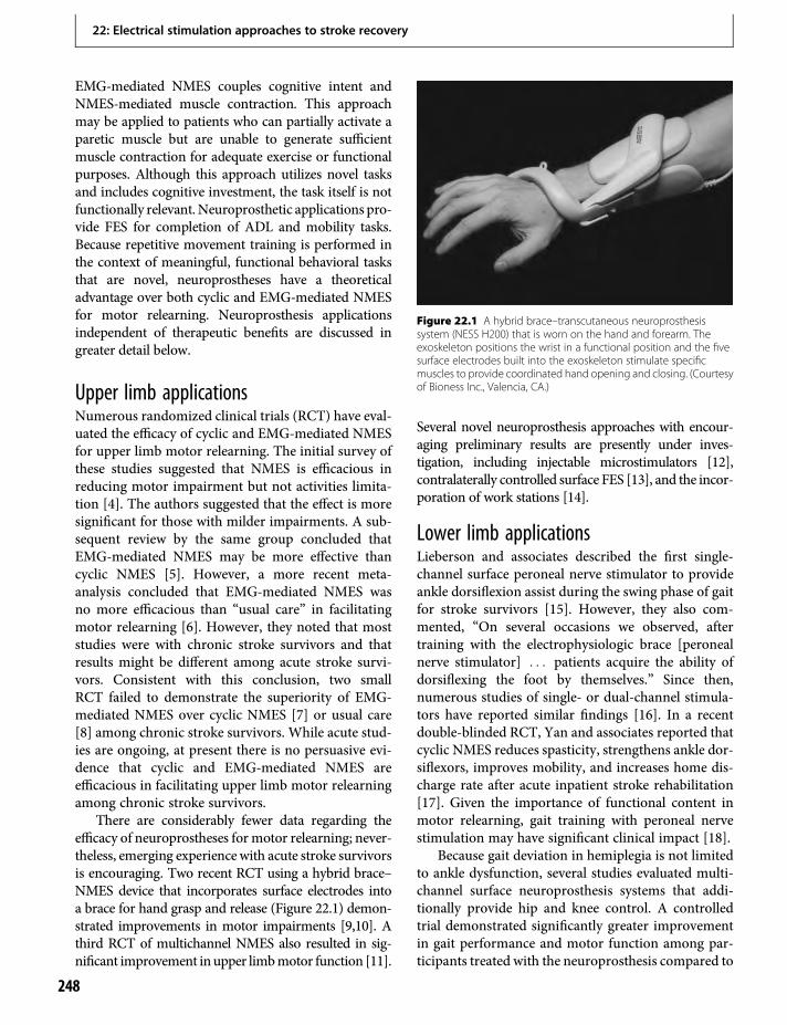
EMG-mediated NMES couples cognitive intent andNMES-mediated muscle contraction. This approachmay be applied to patients who can partially activate aparetic muscle but are unable to generate sufficientmuscle contraction for adequate exercise or functionalpurposes. Although this approach utilizes novel tasksand includes cognitive investment, the task itself is notfunctionally relevant. Neuroprosthetic applications pro-vide FES for completion of ADL and mobility tasks.Because repetitive movement training is performed inthe context of meaningful, functional behavioral tasksthat are novel, neuroprostheses have a theoreticaladvantage over both cyclic and EMG-mediated NMESfor motor relearning. Neuroprosthesis applicationsindependent of therapeutic benefits are discussed ingreater detail below.
Upper limb applicationsNumerous randomized clinical trials (RCT) have eval-uated the efficacy of cyclic and EMG-mediated NMESfor upper limb motor relearning. The initial survey ofthese studies suggested that NMES is efficacious inreducing motor impairment but not activities limita-tion [4]. The authors suggested that the effect is moresignificant for those with milder impairments. A sub-sequent review by the same group concluded thatEMG-mediated NMES may be more effective thancyclic NMES [5]. However, a more recent meta-analysis concluded that EMG-mediated NMES wasno more efficacious than “usual care” in facilitatingmotor relearning [6]. However, they noted that moststudies were with chronic stroke survivors and thatresults might be different among acute stroke survi-vors. Consistent with this conclusion, two smallRCT failed to demonstrate the superiority of EMG-mediated NMES over cyclic NMES [7] or usual care[8] among chronic stroke survivors. While acute stud-ies are ongoing, at present there is no persuasive evi-dence that cyclic and EMG-mediated NMES areefficacious in facilitating upper limb motor relearningamong chronic stroke survivors.
There are considerably fewer data regarding theefficacy of neuroprostheses formotor relearning; never-theless, emerging experience with acute stroke survivorsis encouraging. Two recent RCT using a hybrid brace–NMES device that incorporates surface electrodes intoa brace for hand grasp and release (Figure 22.1) demon-strated improvements in motor impairments [9,10]. Athird RCT of multichannel NMES also resulted in sig-nificant improvement in upper limbmotor function [11].
Several novel neuroprosthesis approaches with encour-aging preliminary results are presently under inves-tigation, including injectable microstimulators [12],contralaterally controlled surface FES [13], and the incor-poration of work stations [14].
Lower limb applicationsLieberson and associates described the first single-channel surface peroneal nerve stimulator to provideankle dorsiflexion assist during the swing phase of gaitfor stroke survivors [15]. However, they also com-mented, “On several occasions we observed, aftertraining with the electrophysiologic brace [peronealnerve stimulator] . . . patients acquire the ability ofdorsiflexing the foot by themselves.” Since then,numerous studies of single- or dual-channel stimula-tors have reported similar findings [16]. In a recentdouble-blinded RCT, Yan and associates reported thatcyclic NMES reduces spasticity, strengthens ankle dor-siflexors, improves mobility, and increases home dis-charge rate after acute inpatient stroke rehabilitation[17]. Given the importance of functional content inmotor relearning, gait training with peroneal nervestimulation may have significant clinical impact [18].
Because gait deviation in hemiplegia is not limitedto ankle dysfunction, several studies evaluated multi-channel surface neuroprosthesis systems that addi-tionally provide hip and knee control. A controlledtrial demonstrated significantly greater improvementin gait performance and motor function among par-ticipants treated with the neuroprosthesis compared to
Figure 22.1 A hybrid brace–transcutaneous neuroprosthesissystem (NESS H200) that is worn on the hand and forearm. Theexoskeleton positions the wrist in a functional position and the fivesurface electrodes built into the exoskeleton stimulate specificmuscles to provide coordinated hand opening and closing. (Courtesyof Bioness Inc., Valencia, CA.)
22: Electrical stimulation approaches to stroke recovery
248

those treated with conventional therapy [19].Similarly, a single-blinded RCT demonstrated that amultichannel percutaneous NMES-mediated ambula-tion training improves gait components and knee flex-ion coordination relative to controls [20].
Motor relearning: summary and futuredirectionsAlthough earlier studies suggested that cyclic and EMG-mediated NMES reduce motor impairment, morerecent data, especially among chronic stroke survivors,raise considerable doubts regarding their therapeuticbenefits, particularly for the upper extremity. Theymay be efficacious among acute stroke survivors, butthere are insufficient data to confirm this. While theefficacy of cyclic and EMG-mediated NMES in facilitat-ingmotor relearning remains uncertain, the implemen-tation of neuroprostheses will likely have significantclinical impact due to the higher functional content.
Future investigations on NMES for motor relearn-ing should demonstrate impact on clinical outcomes atthe level of activity limitation and quality of life. Thesestudies should be large, multicenter RCT, whichshould be at least single-blinded. Studies should care-fully define the subject populations, identify potentialconfounders, and evaluate long-term outcomes usingvalid and reliable measures of motor impairment,activity limitations, and quality of life. These trialsshould directly compare the various types of NMESto at least the standard of care to identify the mosteffective clinical paradigm and the populations thatwill likely benefit from each approach. Future studiesshould determine the optimal dose and prescriptiveparameters. Systems that harness the natural com-mand signals for limb movement, such as those gen-erated from the primary motor cortex, should also bedeveloped. Neuroprostheses that provide clear func-tional, cost-effective benefit to a broad range of strokesurvivors should be developed to provide goal-oriented, repetitive movement therapy in the contextof functional and meaningful tasks. Finally, studiesshould further investigate mechanisms in order tooptimize the treatment paradigm.
Post-stroke shoulder pain
Theoretical considerationsShoulder pain is a common complication followingstroke [21]. The exact cause of shoulder pain remains
uncertain. However, spasticity and weakness followingstroke, which can lead to mechanical instability andimmobility of the glenohumeral joint, may be impor-tant contributing factors [22]. These conditions maycause pain directly or place the capsule and extracap-sular soft tissue at risk for micro and macro trauma,subsequently leading to inflammation or degenerativechanges, immobility, and pain. Numerous treatmentapproaches have been reported, but with limited suc-cess [23]. However, the use of NMES of the musclessurrounding the shoulder to improve biomechanicalintegrity of the shoulder complex may be an effectivestrategy for reducing post-stroke shoulder pain.
Surface systemsA number of RCT of surface NMES for the treatmentof post-stroke shoulder pain have been reported.Most studies stimulated the posterior deltoid andthe supraspinatus to reduce glenohumeral subluxa-tion. Participants were treated for 4–6 h daily for 4–6weeks. Outcomes included extent of glenohumeralsubluxation, pain-free lateral range of motion(ROM), and motor impairment. In general, stimula-tion parameters were adjusted iteratively by skilledpersonnel to help participants accommodate to thediscomfort of surface stimulation and minimize therisk for muscle fatigue. Skilled personnel also placedthe electrodes on motor points to maximize reliabil-ity of stimulation. The Cochrane Review concludedthat NMES improves pain-free passive external rota-tion ROM and reduces subluxation, but does notimprove shoulder pain or motor impairment [24].A meta-analysis by Ada and Foongchomecheay con-cluded that surface NMES reduces or prevents sub-luxation and improves motor impairment in thesubacute phase but not in the chronic phase [25].Recent evidence-based reviews and practice guide-lines now all recommend surface NMES for the pre-vention and treatment of post-stroke shoulder pain[26–28].
Intramuscular systemsDespite the evidence for therapeutic benefit, the clin-ical implementation of surface NMES for shoulderpain in hemiplegia has been difficult. The primaryreason for this is the difficulty of use, which requiresskilled personnel to ensure reliable and tolerable stim-ulation. This was predicted by Baker and Parker, theauthors of the first RCT of surface NMES, when theyconcluded, “until implanted electrode systems become
22: Electrical stimulation approaches to stroke recovery
249
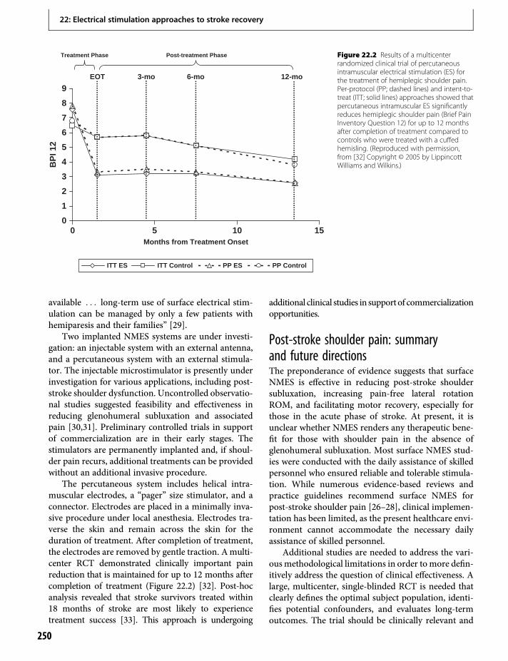
available . . . long-term use of surface electrical stim-ulation can be managed by only a few patients withhemiparesis and their families” [29].
Two implanted NMES systems are under investi-gation: an injectable system with an external antenna,and a percutaneous system with an external stimula-tor. The injectable microstimulator is presently underinvestigation for various applications, including post-stroke shoulder dysfunction. Uncontrolled observatio-nal studies suggested feasibility and effectiveness inreducing glenohumeral subluxation and associatedpain [30,31]. Preliminary controlled trials in supportof commercialization are in their early stages. Thestimulators are permanently implanted and, if shoul-der pain recurs, additional treatments can be providedwithout an additional invasive procedure.
The percutaneous system includes helical intra-muscular electrodes, a “pager” size stimulator, and aconnector. Electrodes are placed in a minimally inva-sive procedure under local anesthesia. Electrodes tra-verse the skin and remain across the skin for theduration of treatment. After completion of treatment,the electrodes are removed by gentle traction. Amulti-center RCT demonstrated clinically important painreduction that is maintained for up to 12 months aftercompletion of treatment (Figure 22.2) [32]. Post-hocanalysis revealed that stroke survivors treated within18 months of stroke are most likely to experiencetreatment success [33]. This approach is undergoing
additional clinical studies in supportof commercializationopportunities.
Post-stroke shoulder pain: summaryand future directionsThe preponderance of evidence suggests that surfaceNMES is effective in reducing post-stroke shouldersubluxation, increasing pain-free lateral rotationROM, and facilitating motor recovery, especially forthose in the acute phase of stroke. At present, it isunclear whether NMES renders any therapeutic bene-fit for those with shoulder pain in the absence ofglenohumeral subluxation. Most surface NMES stud-ies were conducted with the daily assistance of skilledpersonnel who ensured reliable and tolerable stimula-tion. While numerous evidence-based reviews andpractice guidelines recommend surface NMES forpost-stroke shoulder pain [26–28], clinical implemen-tation has been limited, as the present healthcare envi-ronment cannot accommodate the necessary dailyassistance of skilled personnel.
Additional studies are needed to address the vari-ous methodological limitations in order to more defin-itively address the question of clinical effectiveness. Alarge, multicenter, single-blinded RCT is needed thatclearly defines the optimal subject population, identi-fies potential confounders, and evaluates long-termoutcomes. The trial should be clinically relevant and
EOT 3-mo 6-mo 12-mo
Months from Treatment Onset
Treatment Phase Post-treatment Phase
0
1
2
3
4
5
6
7
8
9
0 5 10 15
BP
I 12
ITT ES ITT Control PP ES PP Control
Figure 22.2 Results of a multicenterrandomized clinical trial of percutaneousintramuscular electrical stimulation (ES) forthe treatment of hemiplegic shoulder pain.Per-protocol (PP; dashed lines) and intent-to-treat (ITT; solid lines) approaches showed thatpercutaneous intramuscular ES significantlyreduces hemiplegic shoulder pain (Brief PainInventory Question 12) for up to 12 monthsafter completion of treatment compared tocontrols who were treated with a cuffedhemisling. (Reproduced with permission,from [32] Copyright © 2005 by LippincottWilliams and Wilkins.)
22: Electrical stimulation approaches to stroke recovery
250
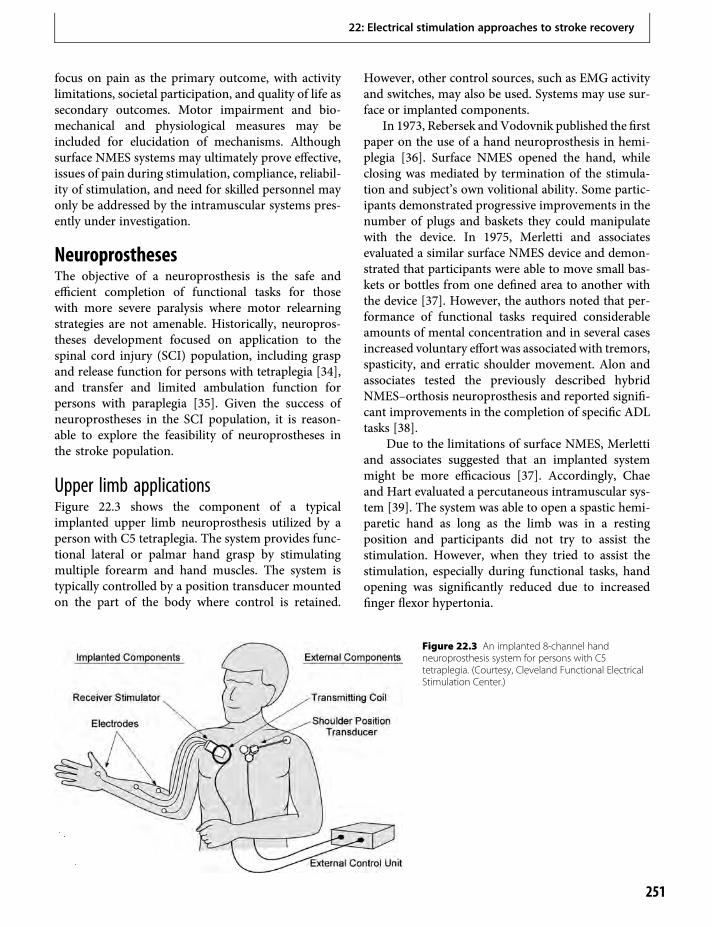
focus on pain as the primary outcome, with activitylimitations, societal participation, and quality of life assecondary outcomes. Motor impairment and bio-mechanical and physiological measures may beincluded for elucidation of mechanisms. Althoughsurface NMES systems may ultimately prove effective,issues of pain during stimulation, compliance, reliabil-ity of stimulation, and need for skilled personnel mayonly be addressed by the intramuscular systems pres-ently under investigation.
NeuroprosthesesThe objective of a neuroprosthesis is the safe andefficient completion of functional tasks for thosewith more severe paralysis where motor relearningstrategies are not amenable. Historically, neuropros-theses development focused on application to thespinal cord injury (SCI) population, including graspand release function for persons with tetraplegia [34],and transfer and limited ambulation function forpersons with paraplegia [35]. Given the success ofneuroprostheses in the SCI population, it is reason-able to explore the feasibility of neuroprostheses inthe stroke population.
Upper limb applicationsFigure 22.3 shows the component of a typicalimplanted upper limb neuroprosthesis utilized by aperson with C5 tetraplegia. The system provides func-tional lateral or palmar hand grasp by stimulatingmultiple forearm and hand muscles. The system istypically controlled by a position transducer mountedon the part of the body where control is retained.
However, other control sources, such as EMG activityand switches, may also be used. Systems may use sur-face or implanted components.
In 1973, Rebersek and Vodovnik published the firstpaper on the use of a hand neuroprosthesis in hemi-plegia [36]. Surface NMES opened the hand, whileclosing was mediated by termination of the stimula-tion and subject’s own volitional ability. Some partic-ipants demonstrated progressive improvements in thenumber of plugs and baskets they could manipulatewith the device. In 1975, Merletti and associatesevaluated a similar surface NMES device and demon-strated that participants were able to move small bas-kets or bottles from one defined area to another withthe device [37]. However, the authors noted that per-formance of functional tasks required considerableamounts of mental concentration and in several casesincreased voluntary effort was associated with tremors,spasticity, and erratic shoulder movement. Alon andassociates tested the previously described hybridNMES–orthosis neuroprosthesis and reported signifi-cant improvements in the completion of specific ADLtasks [38].
Due to the limitations of surface NMES, Merlettiand associates suggested that an implanted systemmight be more efficacious [37]. Accordingly, Chaeand Hart evaluated a percutaneous intramuscular sys-tem [39]. The system was able to open a spastic hemi-paretic hand as long as the limb was in a restingposition and participants did not try to assist thestimulation. However, when they tried to assist thestimulation, especially during functional tasks, handopening was significantly reduced due to increasedfinger flexor hypertonia.
Figure 22.3 An implanted 8-channel handneuroprosthesis system for persons with C5tetraplegia. (Courtesy, Cleveland Functional ElectricalStimulation Center.)
22: Electrical stimulation approaches to stroke recovery
251
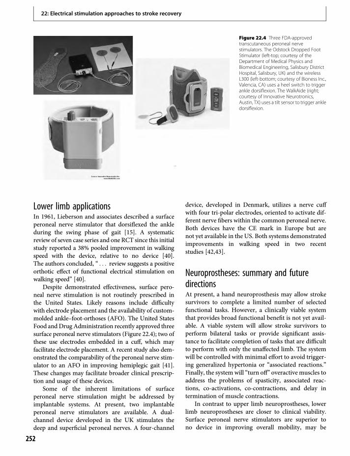
Lower limb applicationsIn 1961, Lieberson and associates described a surfaceperoneal nerve stimulator that dorsiflexed the ankleduring the swing phase of gait [15]. A systematicreview of seven case series and one RCT since this initialstudy reported a 38% pooled improvement in walkingspeed with the device, relative to no device [40].The authors concluded, “ . . . review suggests a positiveorthotic effect of functional electrical stimulation onwalking speed” [40].
Despite demonstrated effectiveness, surface pero-neal nerve stimulation is not routinely prescribed inthe United States. Likely reasons include difficultywith electrode placement and the availability of custom-molded ankle–foot-orthoses (AFO). The United StatesFood andDrug Administration recently approved threesurface peroneal nerve stimulators (Figure 22.4); two ofthese use electrodes embedded in a cuff, which mayfacilitate electrode placement. A recent study also dem-onstrated the comparability of the peroneal nerve stim-ulator to an AFO in improving hemiplegic gait [41].These changes may facilitate broader clinical prescrip-tion and usage of these devices.
Some of the inherent limitations of surfaceperoneal nerve stimulation might be addressed byimplantable systems. At present, two implantableperoneal nerve stimulators are available. A dual-channel device developed in the UK stimulates thedeep and superficial peroneal nerves. A four-channel
device, developed in Denmark, utilizes a nerve cuffwith four tri-polar electrodes, oriented to activate dif-ferent nerve fibers within the common peroneal nerve.Both devices have the CE mark in Europe but arenot yet available in the US. Both systems demonstratedimprovements in walking speed in two recentstudies [42,43].
Neuroprostheses: summary and futuredirectionsAt present, a hand neuroprosthesis may allow strokesurvivors to complete a limited number of selectedfunctional tasks. However, a clinically viable systemthat provides broad functional benefit is not yet avail-able. A viable system will allow stroke survivors toperform bilateral tasks or provide significant assis-tance to facilitate completion of tasks that are difficultto perform with only the unaffected limb. The systemwill be controlled with minimal effort to avoid trigger-ing generalized hypertonia or “associated reactions.”Finally, the system will “turn off” overactive muscles toaddress the problems of spasticity, associated reac-tions, co-activations, co-contractions, and delay intermination of muscle contractions.
In contrast to upper limb neuroprostheses, lowerlimb neuroprostheses are closer to clinical viability.Surface peroneal nerve stimulators are superior tono device in improving overall mobility, may be
Figure 22.4 Three FDA-approvedtranscutaneous peroneal nervestimulators. The Odstock Dropped FootStimulator (left-top; courtesy of theDepartment of Medical Physics andBiomedical Engineering, Salisbury DistrictHospital, Salisbury, UK) and the wirelessL300 (left-bottom; courtesy of Bioness Inc.,Valencia, CA) uses a heel switch to triggerankle dorsiflexion. The WalkAide (right;courtesy of Innovative Neurotronics,Austin, TX) uses a tilt sensor to trigger ankledorsiflexion.
22: Electrical stimulation approaches to stroke recovery
252
equivalent to an ankle–foot-orthosis, and are ready forclinical implementation. Implanted peroneal nervestimulation devices are available in Europe and arelikely to be equivalent to the surface devices withrespect to function. They may be appropriate forthose who experience significant improvement inmobility with the surface system but have difficultywith electrode placement, skin irritation, painful sen-sation of stimulation, or donning and doffing of thedevice.
Although the development of lower limb neuro-prostheses for hemiplegia is further along than upperlimb systems, several issues remain to be further elu-cidated. Studies must document clinical effectivenesswith respect to mobility and quality of life and notjust walking speed. Large RCTs that compare stimula-tion of peroneal nerves with standard of care, such asankle–foot-orthosis, are needed to further demon-strate clinical relevance. For implanted systems, poten-tial benefits over surface systems must bedemonstrated given the added risks and costs associ-ated with an invasive procedure. Finally, ambulation isa multijointed task. Future devices should exploremultichannel devices that also control the knee andhip and not just the ankle.
Emerging techniquesAs noted above, future development of neuroprosthe-ses will need to incorporate control strategies thatare near effortless and more intuitive. In addition,while neuroprostheses are effective in activating weakmuscles, they do little in reducing unwanted motoractivity such as spasticity, co-contractions, and asso-ciated reactions. Emergence of advanced techniquessuch as brain-controlled interfaces and high-frequencyblock may usher in a new era of promising opportu-nities for neuroprostheses.
Brain-controlled interfacesfor neuroprosthesesThe emerging field of brain–computer interfaces(BCI) is focused on developing technologies that willallow people with paralysis (from brainstem stroke,spinal cord injury, and neuromuscular disease) to use“brain signals” for control of external devices. Thereare a number of methods for recording cortical activ-ity, including scalp-recorded electroencephalography,brain-surface recorded electrocorticography, andintracortically recorded neuronal ensemble activity
including local field potentials and the action poten-tials from single neurons [44]. Voluntary upperextremity movement is largely governed by the outputfrom primary motor cortex and premotor cortex (seealso Chapters 10 and 11), which are important gener-ators of the movement signals that have been discon-nected from their targets due to paralyzing disease orillness. Toward the development of a cortically con-trolled neuroprosthesis, there have been extensivestudies in animals documenting the ability to notonly “decode” the activity of ensembles of individualneurons in these and other cortical areas, but to allowmonkeys to direct the movement of a computer cursor[45] or a robot arm [46] simply from the real-timedecoded output of its motor cortex.
Following the demonstration of a unidirectionalmovement of a computer cursor controlled by themotor cortical activity of a gentleman with brainstemstroke [47], and the proofs-of-concept monkey stud-ies, early clinical trials of an intracortically based neu-roprosthesis, “BrainGate”*, were launched. Thesestudies (see www.braingate2.org) have thus far dem-onstrated that a person with tetraplegia from SCI cancontrol a computer cursor, open and close a prosthetichand, and move a robotic arm, all from the neuralactivity recorded by a tiny microelectrode arrayimplanted chronically into the motor cortex [48].More recently, a person with brainstem stroke hasused the same technology to gain point-and-click con-trol over a cursor [49], and to use that neurallyrecorded activity for functionally useful tasks. Thecontrol of these external devices was achieved whilethe trial participants simply imagined the movementof their paralyzed limb.
As described above, it is hoped that the corticalactivity generated during the intent or attempt tomove a paralyzed limb can be converted into a power-ful controller for implanted limb neuroprostheses.Recently there has been an initial preclinical demon-stration of a motor cortex-controlled FES device. Aftermonkeys were trained to play a video game with theirhand (requiring only isometric flexion and extension),recording electrodes were placed in the motor cortexand stimulating electrodes were placed in the wristmuscles. Wrist movements were then temporarily par-alyzed with a peripheral nerve block. A brain-to-FESsystem was then turned on, and the animals quicklylearned to modulate motor cortical neurons for thedirect activation of the wrist muscles, and they againsuccessfully performed the video game [50].
22: Electrical stimulation approaches to stroke recovery
253
Anticipated next steps in this research include theintracortical control of a “virtual arm” based on arealistic model of FES-enabled arm dynamics [51].Work toward a fully implanted brain-to-FES systemalso progresses; such a system may serve both thera-peutic and functional purposes in the future.
High-frequency blockMany stroke survivors exhibit significant spasticity,co-activation of synergistic muscles, co-contractionof antagonists, and delay in termination of motoractivation after volitional activation. These manifesta-tions limit the clinical utility of available neuropros-theses. While standard clinical approaches such as oralmedications and chemoneurolysis may provide somesymptom relief, side effects are common, the effectmay be too global, or the nerve may be damaged;more importantly, these approaches do not providerapid initiation and termination of tone reduction.
One innovative approach is high-frequency alter-nating current (HFAC) block of the peripheral nerve[52]. The mechanism of HFAC block remains uncer-tain, but there are several postulated mechanisms.Using a model of the unmyelinated axon based onthe Hodgkin–Huxley equations, Zhang and associates[53] concluded that HFAC block was due to activationof potassium channels. Bhadra and associates [54]used a mammalian myelinated axon model andreported that during HFAC block, the axons showeda dynamic steady-state depolarization of multiplenodes, strongly suggesting a depolarization mecha-nism with opening of Na+ channels. However, placingthe nerve in a stronger depolarizing field actuallyresulted in conduction failure, because it forces theinactivation Na+ gate to remain closed.
The block typically has three phases: an onsetresponse; a period of asynchronous firing; and a steadystate of complete or partial block [52]. Using frequen-cies between 10 and 30 kHz at amplitudes between2 and 10V, complete and reversible blocks can beobtained. The block is gradable and can be quicklyreversed. Thus the approach can provide both a partialblock, similar to that produced by botulinum toxinand phenol neurolysis, or the block can be complete.HFAC block may be combined with an intelligentcontrol system that varies the block based on sensedactivity. For example, overpowering flexor spasticityoften prevents NMES-mediated hand opening amongstroke survivors, especially after volitional closure orduring functional use of the limb [39]. By monitoring
the myoelectric signal of the flexor and extensormuscles, it may be possible to identify the intentionof the patient and partially block the finger flexorswhile activating the finger extensors with electricalstimulation when hand opening is desired. A similarapproach may be implemented in the lower limb,where equinovarus tone and extensor synergy patternscan lead to significant gait deviations and limit theclinical implementation of neuroprostheses. HFACblock is still in the animal trials phase, but humanfeasibility trials are anticipated in the next severalyears.
Electrical stimulation approaches to strokerecovery: conclusionsRecent advances in clinical medicine and biomedicalengineering are making the clinical implementation ofNMES systems to enhance the ADL and mobility ofpersons with hemiparesis more feasible. NMES formotor relearning in hemiplegia is a promising appli-cation of goal-oriented repetitive movement therapy.Multicenter clinical trials to confirm effectiveness andfundamental studies to elucidate mechanisms are stillneeded. Surface NMES for the treatment of shouldersubluxation and pain in hemiplegia has yieldedencouraging results. While future large-scale multi-center clinical trials will likely confirm effectiveness,the more invasive approaches may prove to be mostappropriate for clinical implementation. The develop-ment of the hand neuroprosthesis for stroke survivorsis in its infancy and must await further technicaland scientific developments. Similarly, multichannel,multijoint lower-limb neuroprostheses need furtherdevelopment. However, surface peroneal stimulatorsappear to be effective in improving hemiplegic gait andshould be included in the clinical armamentarium.Still, if NMES is to have a major impact on the qualityof life of stroke survivors, additional technologicaladvances in the form of cortical control and real-time control of hypertonia will need to be integratedinto emerging systems.
AcknowledgmentsThis work was supported in part by grantsR01HD49777, R01HD044816, and K24HD054600from the National Institute for Child Health andHuman Development (JC) and the Office of Researchand Development, Rehabilitation R&D Service,Department of Veterans Affairs; National Institute
22: Electrical stimulation approaches to stroke recovery
254

for Child Health and Human Development, NationalCenter for Medical Rehabilitation Research; and theDoris Duke Charitable Foundation (LRH).
Conflict of interestDr. Chae serves as a consultant to NDI Medical, amedical device company with commercial interest ina device presented in this chapter.
Dr. Hochberg received clinical trial supportfrom Cyberkinetics Neurotechnology Systems, Inc.Cyberkinetics has ceased operations.
*CAUTION: Investigational Device is limited by USFederal law to investigational use.
References1. Lee RG, van Donkelaar P. Mechanisms underlying
functional recovery following stroke. Can J Neurol Sci.1995;22:257–63.
2. Nudo RJ, Plautz EJ, Frost SB. Role of adaptive plasticityin recovery of function after damage to motor cortex.Muscle Nerve. 2001;24:1000–19.
3. Rushton D. Functional Electrical Stimulation andrehabilitation – An hypothesis. Med Engng Phys.2003;25:75–8.
4. de Kroon JR, van der Lee JH, IJzerman MJ, et al.Therapeutic electrical stimulation to improve motorcontrol and functional abilities of the upper extremityafter stroke: A systematic review. Clin Rehabil.2002;16:350–60.
5. de Kroon JR, Ijzerman MJ, Chae J, et al. Relationbetween stimulation characteristics and clinicaloutcome in studies using electrical stimulation toimprovemotor control of the upper extremity in stroke.J Rehabil Med. 2005;37:65–74.
6. Meilink A, Hemmen B, Seelen H, et al. Impact of EMG-triggered neuromuscular stimulation of the wrist andfinger extensors of the paretic hand after stroke: Asystematic review of the literature. Clin Rehabil.2008;22:291–305.
7. de Kroon JR, Izjerman MJ. Electrical stimulation of theupper extremity in stroke: cyclic versus EMG-triggeredstimulation. Clin Rehabil. 2008;22:690–7.
8. Chae J, Harley MY, Hisel TZ, et al. Intramuscularelectrical stimulation for upper limb recovery in chronichemiparesis: An exploratory randomized clinical trial.Neurorehabil Neural Repair. 2009;23:569–78.
9. Alon G, Levitt AF, McCarthy PA. Functional electricalstimulation enhancement of upper extremity functionalrecovery during stroke rehabilitation: A pilot study.Neurorehabil Neural Repair. 2007;21:207–15.
10. Alon G, Levitt AF, McCarthy PA. Functional electricalstimulation (FES) may modify the poor prognosis ofstroke survivors with severe motor loss of the upper
extremity: A preliminary study. Am J Phys Med Rehabil.2008;87:627–36.
11. Thrasher TA, Zivanovic V, McIlroy W, et al.Rehabilitation of reaching and grasping function insevere hemiplegic patients using functional electricalstimulation therapy. Neurorehabil Neural Repair.2008;22:706–14.
12. Turk R, Burridge JH, Davis R, et al. Therapeuticeffectiveness of electric stimulation of the upper-limbpoststroke using implanted microstimulators. ArchPhys Med Rehabil. 2008;89:1913–22.
13. Knutson JS, Harley MY, Hisel TZ, et al. Improvinghand function in stroke survivors: A pilot study ofcontralaterally controlled functional electricstimulation in chronic hemiplegia. Arch Phys MedRehabil. 2007;88:513–20.
14. Kowalczewski J, Gritsenko V, Ashworth N, et al.Upper-extremity functional electric stimulation-assisted exercises on a workstation in the subacutephase of stroke recovery. Arch Phys Med Rehabil.2007;88:833–9.
15. Lieberson W, Holmquest H, Scot D, et al. Functionalelectrotherapy: Stimulation of the peroneal nervesynchronized with the swing phase of the gait ofhemiplegia patients. Arch Phys Med Rehabil.1961;42:101–05.
16. Robbins SM, Houghton PE, Woodbury MG, et al. Thetherapeutic effect of functional and transcutaneouselectric stimulation on improving gait speed in strokepatients: A meta-analysis. Arch Phys Med Rehabil.2006;87:853–9.
17. Yan T, Hui-Chan CW, Li LS. Functional electricalstimulation improves motor recovery of the lowerextremity and walking ability of subjects with first acutestroke: A randomized placebo-controlled trial. Stroke.2005;36:80–5.
18. Stein RB, Chong S, Everaert DG, et al. A multicentertrial of a footdrop stimulator controlled by a tilt sensor.Neurorehabil Neural Repair. 2006;20:371–9.
19. Bogataj U, Gros N, Kljajic M, et al. The rehabilitation ofgait in patients with hemiplegia: A comparison betweenconventional therapy and multichannel functionalelectrical stimulation therapy. Phys Ther.1995;75:490–502.
20. Daly JJ, Roenigk K, Holcomb J, et al. A randomizedcontrolled trial of functional neuromuscularstimulation in chronic stroke subjects. Stroke.2006;37:172–8. Epub 2005 Dec 1.
21. Van Ouwenaller C, Laplace PM, Chantraine A. Painfulshoulder in hemiplegia. Arch Phys Med Rehabil.1986;67:23–6.
22. Sheffler LR, Chae J. Neuromuscular electricalstimulation in neurorehabilitation. Muscle Nerve.2007;35:562–90.
22: Electrical stimulation approaches to stroke recovery
255
23. Snels IA, Dekker JH, van der Lee JH, et al. Treatingpatients with hemiplegic shoulder pain. Am J Phys MedRehabil. 2002;81:150–60.
24. Price CI, Pandyan AD. Electrical stimulation forpreventing and treating post-stroke shoulder pain: Asystematic Cochrane review. Clin Rehabil.2001;15:5–19.
25. Ada L, Foongchomcheay A. Efficacy of electricalstimulation in preventing or reducing subluxation ofthe shoulder after stroke: A meta-analysis.Aust J Physiother. 2002;48:257–67.
26. Bates B, Choi JY, Duncan PW, et al. Veterans Affairs/Department of Defense Clinical Practice Guideline forthe Management of Adult Stroke Rehabilitation Care:Executive summary. Stroke. 2005;36:2049–56.
27. Khadilkar A, Phillips K, Jean N, et al. Ottawa panelevidence-based clinical practice guidelines for post-stroke rehabilitation. Top Stroke Rehabil.2006;13:1–269.
28. Teasell RW, Foley NC, Bhogal SK, et al. An evidence-based review of stroke rehabilitation. Top StrokeRehabil. 2003;10:29–58.
29. Baker LL, Parker K. Neuromuscular electricalstimulation of the muscles surrounding the shoulder.Phys Ther. 1986;66:1930–7.
30. Dupont Salter AC, Bagg SD, Creasey JL, et al. Firstclinical experience with BION implants for therapeuticelectrical stimulation. Neuromodulation. 2004;7:38–47.
31. Shimada Y, Davis R, Matsunaga T, et al.Electrical stimulation using implantableradiofrequency microstimulators to relievepain associated with shoulder subluxation in chronichemiplegic stroke. Neuromodulation.2006;9:234–8.
32. Chae J, Yu DT, Walker ME, et al. Intramuscularelectrical stimulation for hemiplegic shoulder pain:A 12-month follow-up of a multiple-center,randomized clinical trial. Am J Phys Med Rehabil.2005;84:832–42.
33. Chae J, Ng A, Yu DT, et al. Intramuscular electricalstimulation for shoulder pain in hemiplegia: Does timefrom stroke onset predict treatment success?Neurorehabil Neural Repair. 2007;16:16.
34. Peckham PH, Keith MW, et al. Efficacy of an implantedneuroprosthesis for restoring hand grasp in tetraplegia:A multicenter study. Arch Phys Med Rehabil.2001;82:1380–8.
35. Dutta A, Kobetic R, Triolo RJ. Ambulation afterincomplete spinal cord injury with EMG-triggeredfunctional electrical stimulation. IEEE Trans BiomedEngng. 2008;55:791–4.
36. Rebersek S, Vodovnik L. Proportionally controlledfunctional electrical stimulation of hand. Arch PhysMed Rehabil. 1973;54:378–82.
37. Merletti R, Acimovic R, Grobelnik S, et al.Electrophysiological orthosis for the upper extremity inhemiplegia: Feasibility study. Arch Phys Med Rehabil.1975;56:507–13.
38. Alon G, McBride K, Ring H. Improving selected handfunctions using a noninvasive neuroprosthesis inpersons with chronic stroke. J Stroke Cerebrovasc Dis.2002;11:99–106.
39. Chae J, Hart R. Intramuscular hand neuroprosthesis forchronic stroke survivors. Neurorehabil Neural Repair.2003;17:109–17.
40. Kottink AI, Oostendorp LJ, Buurke JH, et al. Theorthotic effect of functional electrical stimulation on theimprovement of walking in stroke patients with adropped foot: A systematic review. Artif Organs.2004;28:577–86.
41. Sheffler LR, Hennessey MT, Naples GG, et al. Peronealnerve stimulation versus an ankle foot orthosis forcorrection of footdrop in stroke: Impact on functionalambulation. Neurorehabil Neural Repair.2006;20:355–60.
42. Burridge JH, Haugland M, Larsen B, et al. Phase II trialto evaluate the ActiGait implanted drop-foot stimulatorin established hemiplegia. J Rehabil Med.2007;39:212–8.
43. Kottink AI, Hermens HJ, Nene AV, et al. A randomizedcontrolled trial of an implantable 2-channel peronealnerve stimulator on walking speed and activity inpoststroke hemiplegia. Arch Phys Med Rehabil.2007;88:971–8.
44. Hochberg LR, Donoghue JP. Sensors for brain–computer interfaces. IEEE Engng Med Biol Mag.2006;25:32–8.
45. Serruya MD, Hatsopoulos NG, Paninski L, et al. Instantneural control of a movement signal. Nature.2002;416:141–2.
46. Velliste M, Perel S, Spalding MC, et al. Cortical controlof a prosthetic arm for self-feeding. Nature.2008;453:1098–101.
47. Kennedy PR, Bakay RA, Moore MM, et al. Directcontrol of a computer from the human centralnervous system. IEEE Trans Rehabil Engng.2000;8:198–202.
48. Hochberg LR, Serruya MD, Friehs GM, et al. Neuronalensemble control of prosthetic devices by a human withtetraplegia. Nature. 2006;442:164–71.
49. Kim S, Simeral J, Hochberg L, et al. Multi-statedecoding of point-and-click control signals frommotorcortical activity in a human with tetraplegia. 2007 CNE’07 3rd International IEEE/EMBS Conference onNeural Engineering; 2007: 486–9.
50. Moritz CT, Perlmutter SI, Fetz EE. Direct control ofparalysed muscles by cortical neurons. Nature.2008;456:639–42.
22: Electrical stimulation approaches to stroke recovery
256
51. Blana D, Hincapie JG, Chadwick EK, et al. Amusculoskeletal model of the upper extremity for use inthe development of neuroprosthetic systems. J Biomech.2008;41:1714–21.
52. Bhadra N, Kilgore KL. High-frequency electricalconduction block of mammalian peripheral motornerve. Muscle Nerve. 2005;32:782–90.
53. Zhang X, Roppolo JR, de Groat WC, et al. Mechanismof nerve conduction block induced by high-frequencybiphasic electrical currents. IEEE Trans Biomed Engng.2006;53:2445–54.
54. Bhadra N, Lahowetz EA, Foldes ST, et al. Simulation ofhigh-frequency sinusoidal electrical block ofmammalianmyelinated axons. J Comput Neurosci. 2007;22:313–26.
22: Electrical stimulation approaches to stroke recovery
257

23 Growth factors as treatments for strokeSeth P. Finklestein & JingMei Ren
Broadly speaking, growth factors are defined as endog-enous proteins that, working through high-affinityreceptors, promote cell survival, proliferation, anddifferentiation. Growth factors can be grouped intosuperfamilies. Factors found in the CNS with effectson brain cells include members of the neurotrophin,fibroblast growth factor (FGF), transforming growthfactor-β (TGF-β), epidermal growth factor (EGF), andhematopoietic growth factor families, among others.
The endogenous expression of many growthfactors is upregulated in the mammalian brain follow-ing stroke. Moreover, because of the availability ofrecombinant growth factors and their effects on CNScells, a literature has arisen on the potential usefulnessof the exogenous administration of growth factors instroke. This consists of a well-developed preclinicalliterature in animal models of cerebral ischemia, aswell as a nascent literature on clinical trials of growthfactor administration in human stroke. Note too thatgrowth factors are a major mechanism by which stemcell therapies exert their effects (see Chapter 24).
In considering this literature overall, it is impor-tant for the reader to be aware of several fundamentaldistinctions, including differences between focal vs.global cerebral ischemia, and between permanent vs.temporary ischemia [1]. Perhaps the most importantdistinction is between acute stroke vs. stroke recovery.In the early stages of focal stroke, there exists tissue inwhich cells are frankly dead (infarct “core”), sur-rounded by tissue in which cells are injured but notyet necrotic (the “ischemic penumbra”). Acute stroketherapies are targeted at increasing blood flow to theischemic brain or otherwise antagonizing the death ofinjured but still salvageable cells in the ischemicpenumbra. Generally, such treatments are effective inpreclinical studies for only a few hours after the on-set of ischemia and are recognized by their effectin decreasing infarct size and thereby reducing
neurological deficits after ischemia. Despite decadesof effort and dozens of clinical trials of putative acutestroke treatments, including numerous “neuroprotec-tive” agents, only thrombolytic (clot-busting) thera-pies have shown value in human clinical trials of acutestroke therapies.
After focal ischemia has occurred and infarcts arefixed in size, animals and humans can recover to somedegree in terms of neurological deficits followingstroke. This recovery occurs during days to weeks,and depends on intact regions of brain that “takeover” or compensate for loss of function incurred byfocal brain infarction. The cellular underpinnings ofrecovery may include new neuronal sprouting andsynapse formation in uninjured regions of brain, aswell as progenitor cell proliferation, migration anddifferentiation [2,3]. Although regions of focal braindamage are not regenerated by these phenomena,functional recovery does occur. Treatments thatenhance stroke recovery are likely to include thosethat promote neuronal outgrowth and synapse forma-tion, as well as progenitor cell proliferation and differ-entiation. In animal models, recovery-promotingtreatments are recognized by their ability to enhanceneurological recovery without necessarily reducinginfarct volume. Because of the time course of strokerecovery, such treatments are likely to be effectivewhen given within days to weeks after stroke. Todate, few clinical trials of stroke recovery-promotingagents have been undertaken.
This review will cover preclinical and clinical liter-ature on the exogenous administration of selectivegrowth factors in stroke, including basic fibroblastgrowth factor (bFGF, FGF-2), osteogenic protein-1(OP-1, BMP-7), erythropoietin (EPO), and granulo-cyte colony-stimulating factor (G-CSF). This reviewwill focus on preclinical studies in acute stroke andstroke recovery following focal ischemia in mature
Brain Repair After Stroke, ed. S. C. Cramer and R. J. Nudo. Published by Cambridge University Press.© Cambridge University Press 2010.
259
animals, and on the few clinical trials that have beenundertaken using these growth factors. This chapterrepresents an update of a previous review on thissubject [1].
Basic fibroblast growth factor(FGF-2, bFGF)Basic fibroblast growth factor (FGF-2, bFGF), amember of the fibroblast growth factor superfamily,is sometimes referred to as a “promiscuous” factorbecause of its mitogenic, survival-promoting, and dif-ferentiating effects on a host of cell types throughoutthe body. In particular, FGF-2 and its high-affinityreceptors are widely distributed throughout the mam-malian brain, including in neurons, glia, endothelia,and progenitor cells. The expression of FGF-2 and itsreceptors is upregulated after brain injury or stroke [4].In vitro, FGF-2 protects neurons against a numberof toxins and insults, including anoxia, hypoglycemia,excitatory amino acid toxicity, and free radical damage,among others, and promotes neurite outgrowth (espe-cially axonal) from cultured neurons [5,6].
There is a robust literature of the effects of exoge-nously administered FGF-2 in animal models of acutestroke and stroke recovery. In an early study of acutestroke, Koketsu et al. [7] found that the intracerebro-ventricular infusion of FGF-2 for 3 days before ische-mia reduced infarct volume in a model of focalischemia/reperfusion in rats. In later studies, Fisheret al. [8] found that intravenous infusion of FGF-2,starting 30min after permanent middle cerebral arteryocclusion (MCAo), reduced infarct volume, and Jianget al. [9] found that intravenous administration ofFGF-2 at the time of reperfusion reduced infarct vol-ume in a model of temporary MCAo in rats. In thecase of permanent MCAo, infarct size is reduced ifFGF-2 is administered up to 3 h after the onset ofischemia [10]. The mechanism of infarct reduction inthese studies may be twofold: First, FGF-2 is a potentarteriolar vasodilator, and increases regional cerebralblood flow (rCBF) at the borders of focal infarcts[11,12]. Second, independent of these rCBF effects,FGF-2 reduces apoptotic cell death and increasesexpression of the anti-apoptotic protein bcl-2 at theborders of focal infarcts [13].
These initial findings in rodent models of acutestroke have been expanded subsequently. Watanabeet al. [14] found that intracerebroventricular admin-istration of a viral vector containing the FGF-2
gene reduced infarct volume in a model of temporaryMCAo in rats. Liu et al. [15] found that labeled FGF-2administered at 2 h after permanent MCAo enteredischemic brain tissue and remained there for up to14 days after stroke. Ma et al. [16] found that intra-nasally administered FGF-2 decreased infarct volumefollowing temporary MCAo in rats.
In addition to these preclinical studies showing theeffects of FGF-2 in reducing infarct size in acute stroke,other studies demonstrate the recovery-promotingeffects of FGF-2. In these studies, FGF-2, administeredat 1 or more days after ischemia, did not reduce infarctvolume, but did promote neurological recovery. Inearly studies, Kawamata et al. [17,18] administeredFGF-2 by intracisternal injection beginning at oneday after permanent MCAo in rats and found noreduction in infarct volume, but enhancement of sen-sorimotor recovery of the affected (contralateral)limbs during the first month after stroke. Similarresults were attained with a dimerized FGF-2, whichmay be more potent and stable than the monomer[19]. Further studies established that the mechanismsof enhanced recovery following FGF-2 treatment maybe twofold: (1) stimulation of new neuronal sproutingand synapse formation in undamaged regions of brain,and (2) stimulation of progenitor cell proliferation,migration, and differentiation in brain [18,20,21].
Other groups have expanded on these findings.Leker et al. [22] injected a viral vector containing theFGF-2 gene into peri-infarct cortex following perma-nent MCAo in rats and found enhanced sensorimotorrecovery and increased neurogenesis in treated ani-mals. Wang et al. [23] administered FGF-2 intrana-sally at 1–6 days after temporary MCAo in rats andfound improved sensorimotor recovery as well asincreased progenitor cell proliferation and neurogen-esis in treated animals.
A paper by Won et al. [24] emphasizes possibleage differences in response to FGF-2 treatmentfollowing stroke in rats. Aged (14-month-old) malerats received FGF-2 intraventricularly for 3 days,beginning at either 48 h before, or 24 or 96 h aftertemporary MCAo. A comparison group of youngmature (3-month-old) rats received a similar dosageregimen of FGF-2, beginning at 24 h after temporaryMCAo. Both aged and young mature animals showedenhanced behavioral recovery when FGF-2 treatmentwas begun up to 24 h after stroke. Interestingly, youngadult rats also showed reduction in infarct volumewhen FGF-2 treatment was begun 24 h after stroke,
23: Growth factors as treatments for stroke
260
whereas aged animals did not. These results in agedrats are consistent with previous reports showingenhancement of recovery without infarct reductionwhen FGF-2 treatment is begun at one day after stroke[17,18,20]. The results in young rats differ, however,from other data showing that the effective time win-dow for infarct reduction by FGF-2 lasts for only a fewhours after stroke [10].
Two studies have examined the expression ofFGF-2 following human stroke. In a study of post-mortem human brain, Issa et al. [25] found increasedFGF-2 levels within infarcts as well as increased FGF-2and FGF-2 mRNA levels in tissue surrounding focalinfarcts. Increased FGF-2 immunoreactivity was local-ized in neurons, astrocytes, macrophages, and endo-thelia in these regions. Guo et al. [26] found elevationsin serum FGF-2 levels in human stroke patients, last-ing up to 14 days after stroke. Interestingly, higherFGF-2 serum levels were correlated to improved clin-ical outcome.
Because of the above reports of the neuroprotective(infarct-reducing) effects of FGF-2 in preclinical stud-ies, two human clinical trials of FGF-2 treatment inacute stroke were initiated. In a North American trial[27], FGF-2 (5 or 10mg) was administered intrave-nously for 8 h, beginning within 6 h of onset of ische-mic stroke. This study was prematurely terminateddue to an apparent excess of adverse outcomes inFGF-2-treated patients. In a European/Australiantrial [28], the same dose (5 or 10mg) of FGF-2 wasgiven intravenously, also within 6 h of stroke onset,but over a longer time period (24 h), resulting in lowerFGF-2 serum concentrations. No excess adverse out-comes were seen among FGF-2-treated patients in thisstudy. The trial was terminated prematurely due toprojected futility on primary efficacy outcome meas-ures. Post-hoc analysis, however, revealed a subgroupwith apparent improved clinical outcome – specifi-cally, those patients in whom FGF-2 treatment wasbegun at 5 h or later after stroke onset. These resultssuggest that FGF-2 may have greater utility when givelater than sooner after stroke, i.e. as a recovery-promoting rather than as an acute stroke drug. Todate, no clinical trials of the stroke recovery-promoting effects of FGF-2 have been initiated.
Osteogenic protein-1 (OP-1, BMP-7)Osteogenic protein-1 (OP-1, BMP-7) is a member ofthe bone morphogenetic protein subfamily of thetransforming growth factor-β superfamily. OP-1 was
originally described by its ability to promote osteoblastproliferation and bone formation in mesenchymaltissue [29]. OP-1 is also expressed elsewhere, includingkidneys, bladder, and brain. OP-1 stimulates dendriticoutgrowth from cultured neurons [30].
In a preclinical study of acute stroke, OP-1 wasadministered intracortically or intracerebroventricularlyat 30min, 24 h, or 72 h before temporaryMCAo. At 24hafter ischemia, infarct volume (and consequent neuro-logical impairment) was reduced only in animals receiv-ing OP-1 pretreatment at 24h before ischemia [31].
Most preclinical studies of OP-1 in stroke have beendone in models of stroke recovery. In an early study,Kawamata et al. [32] administered OP-1 by intracister-nal injection at 1 and 4 days after permanent MCAo inrats. Sensorimotor recovery of the affected limbs (con-tralateral to infarcts) was enhanced by OP-1 treatmentin a dose-dependent manner. In a follow up study, Renet al. [33] explored the effective time window of OP-1administration. OP-1 administered intracisternally at 1and 3, and 3 and 5, but not 7 and 9 days after permanentMCAo enhanced sensorimotor recovery of the affectedlimbs in rats. As in the previous study, no reduction ininfarct volume was observed.
Liu et al. [34] administered OP-1 intracisternally at24 h after temporary MCAo in rats. At 48 h, theyfound improved total neurological scores in OP-1treated animals that were associated with increasedcerebral blood flow and glucose utilization in someregions of the ipsilateral (but not contralateral) hemi-sphere. Chang et al. [35] administered OP-1 intrave-nously at 24 h after temporary MCAo in rats. Theseinvestigators found enhanced recovery of body asym-metry and total locomotor activity in OP-1 treatedanimals. Overall, these preclinical data suggest thatOP-1 exerts cytoprotective as well as recovery-promoting effects after stroke. Clinical trials of OP-1in stroke have yet to be undertaken.
Erythropoeitin (EPO)Erythropoeitin (EPO) is a glycoprotein named for itsability to stimulate proliferation of red cell precursorsin the bonemarrow. EPO and its high-affinity receptor(EPO-R) are expressed in the mammalian brain – inneurons, glia, and endothelial cells. Following focalstroke, EPO expression is upregulated in peri-infarcttissue, and the EPO/EPO-R system participates in theproliferation, migration, and differentiation of neuro-nal precursor cells from the SVZ and in the process ofangiogenesis in peri-infarct tissue [36,37].
23: Growth factors as treatments for stroke
261
Several preclinical studies have documented theeffects of exogenously administered EPO in rodentmodels of focal stroke. In studies using neuroprotec-tive paradigms, several authors have shown neuronsparing and infarct-reducing effects of EPO whengiven systemically or intranasally just before and/orup to 6 h after focal temporary, permanent, or embolicocclusion of the MCA [38–42].
In a study using a stroke recovery-promoting para-digm, Wang et al. [43] found that systemic adminis-tration of EPO at 24 h after embolic MCA occlusiondid not reduce infarct volume, but did improve recov-ery of sensorimotor function. Behavioral recovery wasaccompanied by enhanced angiogenesis at infarctborders, and neurogenesis within the SVZ. In aremarkable study, Kolb et al. [44] found that the intra-cerebroventricular co-administration of the combina-tion of EPO + epidermal growth factor (EGF) (butneither one alone), up to 7 days after focal infarction,resulted in the regeneration of cortical tissue andrecovery of sensorimotor function. This stimulated asecond study evaluating effects of two sequentialgrowth factors. Belayev et al. [45] found that admin-istration of 3 daily doses of i.v. EPO to rats beginning 6days after stroke onset, and following 3 doses of i.m.beta-hCG initiated 24 h after stroke onset, was associ-ated with improved behavioral outcome and an 82%reduction in infarct volume.
Preliminary clinical trials of EPO in acute strokewere undertaken by Ehrenreich and colleagues [46]. Asmall safety trial (N=18) established that EPO at thedose administered (3.3 × 10⁴ IU/50ml/30min i.v. dailyfor 3 days) was safe in stroke patients. In a larger efficacytrial (N=40), patients with MCA stroke (< 80 years)were randomized to receive EPO (at this same dose)or placebo, starting within 8 h of acute stroke. Allpreselected endpoints (NIH Stroke Scale (NIHSS),Scandinavian Stroke Scale, Barthel Index (BI), modifiedRankin Scale, and evolution of infarct size by MR)showed a trend or significant results in favor of EPOtreatment. In EPO-treated patients, there were expectedincreases in CSF EPO levels and in peripheral reticulo-cyte counts. These promising preliminary findings arecurrently being expanded in a larger clinical trial.
Granulocyte colony-stimulatingfactor (G-CSF)Granulocyte colony-stimulating factor (G-CSF) is aglycoprotein that stimulates the proliferation and
mobilization of granulocytes from bone marrow.G-CSF and its high-affinity receptor are also found inthe brain, including in neurons, glia, endothelia, andneural progenitor cells. G-CSF appears to have severaleffects in the intact and post-stroke brain, includingenhancing neuronal survival and proliferation andstimulating blood vessel growth. The effects ofG-CSF on the post-stroke brain may include stimula-tion of granulocyte infiltration into infarcted tissue aswell as direct effects on G-CSF receptors.
Several studies have examined the infarct-reducing(neuroprotective) effects of G-CSF in rodent models ofacute stroke. In models of temporary MCAo in ratsand mice, several studies have shown reduction ininfarct volume and corresponding reduction in neuro-logical deficits when G-CSF was administered between0.5 and 6 h after the onset of ischemia [47–54]. Otherstudies have shown reduction in infarct volume whenG-CSF was administered between 0 and 3 h after per-manent MCAo [55,56].
Relatively fewer studies have been done to look atlater effects of G-CSF administration. Six et al. [57]and Shyu et al. [58] showed reduction in infarct vol-ume when G-CSF was administered at 24 h after tem-porary MCAo in mice or rats, respectively. Lee et al.[49] reported diminution in infarct volume andimproved sensorimotor outcome when G-CSF wasadministered starting up to 4 days after temporaryMCAo in rats. Schneider et al. [59] found improvedsensorimotor outcome when G-CSF was administeredstarting at 24 or 72 h after focal photothromboticinfarction in rats.
Minnerup et al. [60] undertook a meta-analysis of13 published studies of G-CSF treatment in rodentmodels of stroke, and found a strong relationshipbetween the total dose of G-CSF administered andthe degree of reduction in infarct size, regardless ofthe time of initiation of G-CSF treatment. Theseresults, as well as the results of the individual studiescited above, are surprising, in that apparent infarctsize-reducing effects were seen when G-CSF wasadministered after 24 h (indeed up to 4 days) afterthe onset of focal stroke. In this regard, the literatureon G-CSF differs from that on some of the othergrowth factors discussed above.
In a small clinical study, Shyu et al. randomizedseven patients to receive G-CSF within 1 week of theonset of acute ischemic stroke and compared clinicaloutcome at 6 months to results in three patientsreceiving standard treatment [61]. There were no
23: Growth factors as treatments for stroke
262
major adverse events in patients receiving G-CSF.Change scores of outcome on several neurologicalrating scales (including the NIHSS, the EuropeanStroke Scale (ESS), and the BI) tended to be higher inpatients receiving G-CSF vs. standard care. Asexpected, the circulating white blood cell count tran-siently increased after G-CSF treatment. In a double-blind placebo-controlled dose-escalation trial ofpatients receiving G-CSF (N=24) vs. placebo (N=12),beginning at 7–30 days after stroke, no adverse out-comes were seen in G-CSF treated patients [62]. Adose-dependent leukocytosis was seen after G-CSFtreatment. There were no differences in clinical out-come (by BI, modified Rankin Scale (mRS), orScandinavian Neurological Stroke Score (SNSS))between G-CSF and placebo treated patients at 10 or90 days after stroke.
ConclusionsThe foregoing summarizes current work on the pre-clinical and clinical development of four specificgrowth factors as treatments for acute stroke andstroke recovery. Future directions in this field includerefinements in preclinical models, clinical trial design,and the use of growth factors along with stem cells,especially in the development of stroke recovery-promoting treatments.
As noted above, virtually all preclinical studieshave been done using rodent models of stroke.Among many important differences between humanand rodent stroke are differences in brain anatomy,and differences in the degree and time course of strokerecovery. The rodent brain is smooth (lissencephalic)in surface. Rodents with large unilateral strokes showclumsiness in the contralateral extremities, whichrecovers toward a plateau within 1 month afterstroke. By contrast, humans have a convoluted (gyr-encephalic) and considerably more complex brain.Patients with moderate to large unilateral strokes areoften markedly weak (paralyzed) in the oppositeextremities, and recovery reaches a plateau at 3months after stroke. Gyrencephalic monkeys showneurological deficits and a recovery course similar tohumans. In a preliminary study, Moore et al. [63]trained rhesus monkeys to retrieve food pellets fromwells of various sizes, and then made focal ischemiclesions in motor cortex controlling arm and handmovement. Following stroke, there was initial paraly-sis of contralateral arm and hand movement thatrecovered toward a plateau by 3 months. At that
time, monkeys were again able to retrieve pelletsfrom the largest wells, but were still considerablyimpaired in retrieving pellets from the smallest ones.Moreover, this recovery appeared compensatory, inthat, rather than using a thumb–forefinger “pincer”grasp, monkeys now used a palm-grasping strategy toretrieve pellets. This strategy was entirely efficient forlarge (but not small) wells. The close similarity ofphenomenology of motor stroke recovery in gyrence-phalic primates compared to humans suggests thatprimate models may be useful as a penultimate stepin advancing promising stroke recovery-promotingtreatments to human clinical trials.
In addition to improved preclinical modeling, clin-ical trial design (see also Chapter 16) can be optimizedfor growth factors and other putative stroke recovery-promoting agents. Clinical trials of acute stroke(thrombolytic and neuroprotective) agents generallyhave a short time window to enrollment after stroke(within 3–6 h), and rely on composite clinical ratingscales that combine outcome in several neurologicalmodalities (motor, sensory, cognition, coordination,etc.). However, since neurological modalities mayrecover to different extents with different time coursesafter stroke, “modality-specific” outcome measuresmay represent a more sensitive and specific way withwhich to assess effects of putative stroke recovery-promoting treatments [64]. Examples of modality-specific outcomes include specific tests of arm/handfunction, gait, or language. Moreover, since the effec-tive time window for administration of recovery-promoting agents is likely to encompass days or evenweeks after stroke, both baseline and post-treatmentscores can be obtained, and informative “changescores” calculated, for each patient. The use ofmodality-specific endpoints may extend, and doesnot obviate, the concurrent use of standard compositeoutcome measures.
Finally, with specific regard to polypeptide growthfactors, one of the mechanisms by which these agentsmay promote recovery is through stimulation ofendogenous progenitor cell proliferation, differentia-tion and migration after stroke. As the isolation andavailability of exogenous progenitor and stem cellpopulations advances, there may be advantages to theco-administration of cells and stimulating growth fac-tors as treatments for stroke. Going forward, the fieldof growth factor treatment of stroke is likely to benefitgreatly from advances in preclinical modeling, clinicaltrial design, and cell therapy.
23: Growth factors as treatments for stroke
263
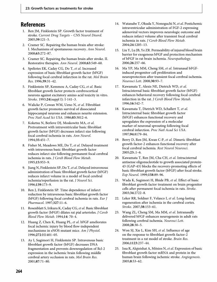
References1. Ren JM, Finklestein SP. Growth factor treatment of
stroke. Current Drug Targets – CNS Neurol Disord.2005;39:121–5.
2. Cramer SC. Repairing the human brain after stroke:I. Mechanisms of spontaneous recovery. Ann Neurol.2008;63:272–87.
3. Cramer SC. Repairing the human brain after stroke. II.Restorative therapies. Ann Neurol. 2008;63:549–60.
4. Speliotes EK, Caday CG, Do T, et al. Increasedexpression of basic fibroblast growth factor (bFGF)following focal cerebral infarction in the rat.Mol BrainRes. 1996;39:31–42.
5. Finklestein SP, Kemmou A, Caday CG, et al. Basicfibroblast growth factor protects cerebrocorticalneurons against excitatory amino acid toxicity in vitro.Stroke. 1993;24(suppl I): I-141–3.
6. Walicke P, Cowan WM, Ueno N, et al. Fibroblastgrowth factor promotes survival of dissociatedhippocampal neurons and enhances neurite extension.Proc Natl Acad Sci USA. 1986;83:3012–6.
7. Koketsu N, Berlove DJ, Moskowitz MA, et al.Pretreatment with intraventricular basic fibroblastgrowth factor (bFGF) decreases infarct size followingfocal cerebral ischemia in rats. Ann Neurol.1994;35:451–7.
8. Fisher M, Meadows ME, Do T, et al. Delayed treatmentwith intravenous basic fibroblast growth factorreduces infarct size following permanent focal cerebralischemia in rats. J Cereb Blood Flow Metab.1995;15:953–9.
9. Jiang N, Finklestein SP, Do T, et al. Delayed intravenousadministration of basic fibroblast growth factor (bFGF)reduces infarct volume in a model of focal cerebralischemia/reperfusion in the rat. J Neurol Sci.1996;139:173–9.
10. Ren J, Finklestein SP. Time dependence of infarctreduction by intravenous basic fibroblast growth factor(bFGF) following focal cerebral ischemia in rats. Eur JPharmacol. 1997;327:11–6.
11. Rosenblatt S, Irikura K, Caday CG, et al. Basic fibroblastgrowth factor (bFGF) dilates rat pial arterioles. J CerebBlood Flow Metab. 1994;14: 70–4.
12. Huang Z, Chen K, Huang PL, et al. bFGF amelioratesfocal ischemic injury by blood flow-independentmechanisms in eNOS mutant mice. Am J Physiol.1996;272:H1401–05.
13. Ay I, Sugimori H, Finklestein SP. Intravenous basicfibroblast growth factor (bFGF) decreases DNAfragmentation and prevents downregulation of Bcl-2expression in the ischemic brain following middlecerebral artery occlusion in rats. Mol Brain Res.2001;87:71–80.
14. Watanabe T, Okuda Y, Nonoguchi N, et al. Postichemicintraventricular administration of FGF-2 expressingadenoviral vectors improves neurologic outcome andreduces infarct volume after transient focal cerebralischemia in rats. J Cereb Blood Flow Metab.2004;24:1205–13.
15. Liu Y, Lu JB, Ye ZR. Permeability of injured blood brainbarrier for exogenous bFGF and protection mechanismof bFGF in rat brain ischemia. Neuropathology.2006;26:257–66.
16. Ma YP, Ma MM, Cheng SM, et al. Intranasal bFGF-induced progenitor cell proliferation andneuroprotection after transient focal cerebral ischemia.Neurosci Lett. 2008;30:93–7.
17. Kawamata T, Alexis NE, Dietrich WD, et al.Intracisternal basic fibroblast growth factor (bFGF)enhances behavioral recovery following focal cerebralinfarction in the rat. J Cereb Blood Flow Metab.1996;16:542–7.
18. Kawamata T, Dietrich WD, Schallert T, et al.Intracisternal basic fibroblast growth factor(bFGF) enhances functional recovery andupregulates the expression of a molecularmarker of neuronal sprouting following focalcerebral infarction. Proc Natl Acad Sci USA.1997;94:8179–84.
19. Berry D, Ren JM, Kwan C-P, et al. Dimeric fibroblastgrowth factor-2 enhances functional recovery afterfocal cerebral ischemia. Rest Neurol Neurosci.2005;23:.1–6.
20. Kawamata T, Ren JM, Cha CH, et al. Intracisternalantisense oligonucleotide to growth associated protein-43 (GAP-43) blocks the recovery-promoting effects ofbasic fibroblast growth factor (bFGF) after focal stroke.Exp Neurol. 1999;158:89–96.
21. Wada K, Sugimori H, Bhide PB, et al. Effect of basicfibroblast growth factor treatment on brain progenitorcells after permanent focal ischemia in rats. Stroke.2003;34:2722–8.
22. Leker RR, Soldner F, Velasco I, et al. Long-lastingregeneration after ischemia in the cerebral cortex.Stroke. 2007;38:153–61.
23. Wang ZL, Cheng SM, Ma MM, et al. Intranasallydelivered bFGF enhances neurogenesis in adult ratsfollowing cerebral ischemia. Neurosci Lett.2008;28:30–5.
24. Won SJ, Xie L, Kim SH, et al. Influence of ageon the response to fibroblast growth factor-2treatment in a rat model of stroke. Brain Res.2006;1123:237–44.
25. Issa R, Alqteishat A,Mitsios N, et al. Expression of basicfibroblast growth factor mRNA and protein in thehuman brain following ischemic stroke. Angiogenesis.2005;8:53–62.
23: Growth factors as treatments for stroke
264
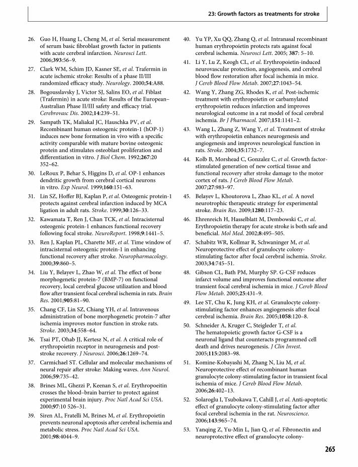
26. Guo H, Huang L, Cheng M, et al. Serial measurementof serum basic fibroblast growth factor in patientswith acute cerebral infarction. Neurosci Lett.2006;393:56–9.
27. Clark WM, Schim JD, Kasner SE, et al. Trafermin inacute ischemic stroke: Results of a phase II/IIIrandomized efficacy study. Neurology. 2000;54:A88.
28. Bogousslavsky J, Victor SJ, Salins EO, et al. Fiblast(Trafermin) in acute stroke: Results of the European–Australian Phase II/III safety and efficacy trial.Cerebrovasc Dis. 2002;14:239–51.
29. Sampath TK, Maliakal JC, Hauschka PV, et al.Recombinant human osteogenic protein-1 (hOP-1)induces new bone formation in vivo with a specificactivity comparable with mature bovine osteogenicprotein and stimulates osteoblast proliferation anddifferentiation in vitro. J Biol Chem. 1992;267:20352–62.
30. LeRoux P, Behar S, Higgins D, et al. OP-1 enhancesdendritic growth from cerebral cortical neuronsin vitro. Exp Neurol. 1999;160:151–63.
31. Lin SZ, Hoffer BJ, Kaplan P, et al. Osteogenic protein-1protects against cerebral infarction induced by MCAligation in adult rats. Stroke. 1999;30:126–33.
32. Kawamata T, Ren J, Chan TCK, et al. Intracisternalosteogenic protein-1 enhances functional recoveryfollowing focal stroke. NeuroReport. 1998;9:1441–5.
33. Ren J, Kaplan PL, Charette MF, et al. Time window ofintracisternal osteogenic protein-1 in enhancingfunctional recovery after stroke. Neuropharmacology.2000;39:860–5.
34. Liu Y, Belayev L, Zhao W, et al. The effect of bonemorphogenetic protein-7 (BMP-7) on functionalrecovery, local cerebral glucose utilization and bloodflow after transient focal cerebral ischemia in rats. BrainRes. 2001;905:81–90.
35. Chang CF, Lin SZ, Chiang YH, et al. Intravenousadministration of bone morphogenetic protein-7 afterischemia improves motor function in stroke rats.Stroke. 2003;34:558–64.
36. Tsai PT, Ohab JJ, Kertesz N, et al. A critical role oferythropoietin receptor in neurogenesis and post-stroke recovery. J Neurosci. 2006;26:1269–74.
37. Carmichael ST. Cellular and molecular mechanisms ofneural repair after stroke: Making waves. Ann Neurol.2006;59:735–42.
38. Brines ML, Ghezzi P, Keenan S, et al. Erythropoeitincrosses the blood–brain barrier to protect againstexperimental brain injury. Proc Natl Acad Sci USA.2000;97:10 526–31.
39. Siren AL, Fratelli M, Brines M, et al. Erythropoietinprevents neuronal apoptosis after cerebral ischemia andmetabolic stress. Proc Natl Acad Sci USA.2001;98:4044–9.
40. Yu YP, Xu QQ, Zhang Q, et al. Intranasal recombinanthuman erythropoietin protects rats against focalcerebral ischemia. Neurosci Lett. 2005; 387: 5–10.
41. Li Y, Lu Z, Keogh CL, et al. Erythropoietin-inducedneurovascular protection, angiogenesis, and cerebralblood flow restoration after focal ischemia in mice.J Cereb Blood Flow Metab. 2007;27:1043–54.
42. Wang Y, Zhang ZG, Rhodes K, et al. Post-ischemictreatment with erythropoietin or carbamylatederythropoietin reduces infarction and improvesneurological outcome in a rat model of focal cerebralischemia. Br J Pharmacol. 2007;151:1141–2.
43. Wang L, Zhang Z, Wang Y, et al. Treatment of strokewith erythropoietin enhances neurogenesis andangiogenesis and improves neurological function inrats. Stroke. 2004;35:1732–7.
44. Kolb B, Morshead C, Gonzalez C, et al. Growth factor-stimulated generation of new cortical tissue andfunctional recovery after stroke damage to the motorcortex of rats. J Cereb Blood Flow Metab.2007;27:983–97.
45. Belayev L, Khoutorova L, Zhao KL, et al. A novelneurotrophic therapeutic strategy for experimentalstroke. Brain Res. 2009;1280:117–23.
46. Ehrenreich H, Hasselblatt M, Dembowski C, et al.Erythropoietin therapy for acute stroke is both safe andbeneficial. Mol Med. 2002;8:495–505.
47. Schabitz WR, Kollmar R, Schwaninger M, et al.Neuroprotective effect of granulocyte colony-stimulating factor after focal cerebral ischemia. Stroke.2003;34:745–51.
48. Gibson CL, Bath PM, Murphy SP. G-CSF reducesinfarct volume and improves functional outcome aftertransient focal cerebral ischemia in mice. J Cereb BloodFlow Metab. 2005;25:431–9.
49. Lee ST, Chu K, Jung KH, et al. Granulocyte colony-stimulating factor enhances angiogenesis after focalcerebral ischemia. Brain Res. 2005;1058:120–8.
50. Schneider A, Kruger C, Steigleder T, et al.The hematopoietic growth factor G-CSF is aneuronal ligand that counteracts programmed celldeath and drives neurogenesis. J Clin Invest.2005;115:2083–98.
51. Komine-Kobayashi M, Zhang N, Liu M, et al.Neuroprotective effect of recombinant humangranulocyte colony-stimulating factor in transient focalischemia of mice. J Cereb Blood Flow Metab.2006;26:402–13.
52. Solaroglu I, Tsubokawa T, Cahill J, et al. Anti-apoptoticeffect of granulocyte colony-stimulating factor afterfocal cerebral ischemia in the rat. Neuroscience.2006;143:965–74.
53. Yanqing Z, Yu-Min L, Jian Q, et al. Fibronectin andneuroprotective effect of granulocyte colony-
23: Growth factors as treatments for stroke
265
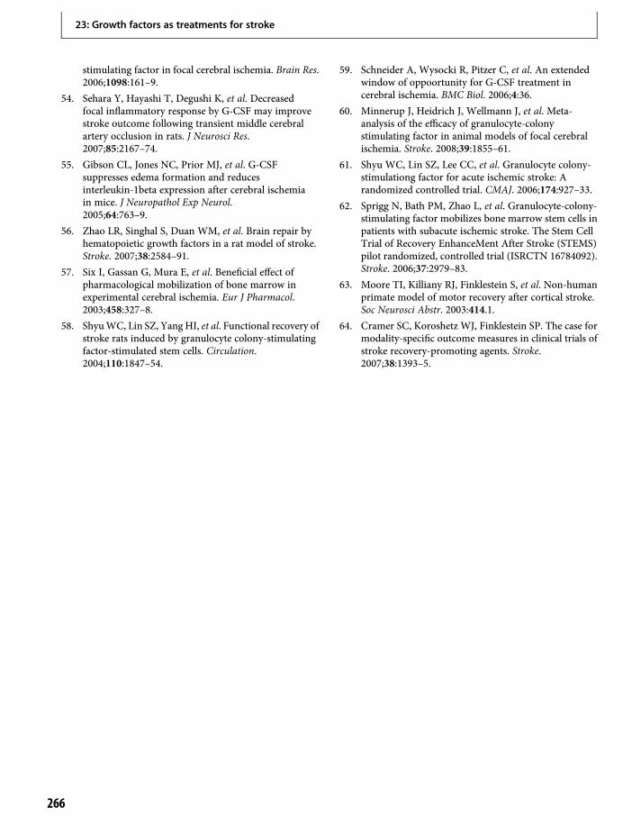
stimulating factor in focal cerebral ischemia. Brain Res.2006;1098:161–9.
54. Sehara Y, Hayashi T, Degushi K, et al. Decreasedfocal inflammatory response by G-CSF may improvestroke outcome following transient middle cerebralartery occlusion in rats. J Neurosci Res.2007;85:2167–74.
55. Gibson CL, Jones NC, Prior MJ, et al. G-CSFsuppresses edema formation and reducesinterleukin-1beta expression after cerebral ischemiain mice. J Neuropathol Exp Neurol.2005;64:763–9.
56. Zhao LR, Singhal S, Duan WM, et al. Brain repair byhematopoietic growth factors in a rat model of stroke.Stroke. 2007;38:2584–91.
57. Six I, Gassan G, Mura E, et al. Beneficial effect ofpharmacological mobilization of bone marrow inexperimental cerebral ischemia. Eur J Pharmacol.2003;458:327–8.
58. ShyuWC, Lin SZ, Yang HI, et al. Functional recovery ofstroke rats induced by granulocyte colony-stimulatingfactor-stimulated stem cells. Circulation.2004;110:1847–54.
59. Schneider A, Wysocki R, Pitzer C, et al. An extendedwindow of oppoortunity for G-CSF treatment incerebral ischemia. BMC Biol. 2006;4:36.
60. Minnerup J, Heidrich J, Wellmann J, et al. Meta-analysis of the efficacy of granulocyte-colonystimulating factor in animal models of focal cerebralischemia. Stroke. 2008;39:1855–61.
61. Shyu WC, Lin SZ, Lee CC, et al. Granulocyte colony-stimulationg factor for acute ischemic stroke: Arandomized controlled trial. CMAJ. 2006;174:927–33.
62. Sprigg N, Bath PM, Zhao L, et al. Granulocyte-colony-stimulating factor mobilizes bone marrow stem cells inpatients with subacute ischemic stroke. The Stem CellTrial of Recovery EnhanceMent After Stroke (STEMS)pilot randomized, controlled trial (ISRCTN 16784092).Stroke. 2006;37:2979–83.
63. Moore TI, Killiany RJ, Finklestein S, et al. Non-humanprimate model of motor recovery after cortical stroke.Soc Neurosci Abstr. 2003:414.1.
64. Cramer SC, Koroshetz WJ, Finklestein SP. The case formodality-specific outcome measures in clinical trials ofstroke recovery-promoting agents. Stroke.2007;38:1393–5.
23: Growth factors as treatments for stroke
266

24 Cellular approaches to stroke recoveryYi Li & Michael Chopp
Stroke is a leading cause of long-term disability.Recovery from stroke is a challenging journey thatcan last a lifetime. Therapeutic options have untilnow been limited to the first hours after diseaseonset. However, once the damage from a stroke eventhas maximized, little can be done to recover premor-bid function. Experimental animal data suggest thatrestorative cell approaches may be an effective alter-native to conventional disease management strategiesof ischemic stroke. Therefore, this chapter focuses onthe use of cellular approaches in the restorative treat-ment of stroke.
Endogenous stem cellsand progenitor cells in the adultbrain after strokeStem cells are undifferentiated cells capable ofself-renewal, production of a large number of differ-entiated progeny, and regeneration of tissues.Generally, it has been thought that damage to thebrain could not be repaired, as adult neurons couldnot be replaced. However, convincing evidence hasemerged showing that neurogenesis in the adult cen-tral nervous system (CNS) is an ongoing physiologicalprocess [1]. Neural stem cells (NSCs) and neural pro-genitor cells (NPCs), the source of the new neurons,are present mainly at two sites in the adult brain. Oneis the subventricular zone (SVZ) in the lateral ven-tricles. The SVZ feeds new neurons into the olfactorybulb to continuously maintain the sense of smell [2].The other site is the subgranular zone (SGZ) of thedentate gyrus in the hippocampus, a brain regioninvolved in learning and memory [3]. AlthoughNSCs/NPCs are a continuous source for new neuronsin the brain, the turnover of these cells is vanishinglylow under physiological conditions. Moreover, theturnover rate of NSCs is influenced by pathological
conditions in the brain. After ischemic brain injury,significantly more neurons are generated in the neuro-genic zones and these new neurons tend to migratetoward the site of damage [4]. It seems that the braintries to repair itself, possibly but not exclusively byreplacing the damaged cells with an amplified endog-enous pool of stem and progenitor cells. The replace-ment, though, is not extensive. After a few weeks, only0.2% of the cells destroyed by ischemic attack arereplaced and as many as 80% of the new neuronsproduced die [5].
It is necessary to amplify the endogenous neuro-restorative response of the brain to stroke, to stimulateintrinsic neurorestorative pathways so that we canfurther improve neurological function after stroke.Preclinical data demonstrate that after stroke, thebrain expresses an array of developmental genes andproteins, particularly in the boundary of the ischemiclesion, reminiscent of the developing brain [6].We cancapitalize on this attempted return to youth andamplify these restorative processes to rewire andrestructure the CNS so as to minimize loss of function.
Stem and progenitor cells respond to key growth,differentiation and migration factors [7]. These factorsare likely to differ depending on the tissue, degree ofinjury, and the stem cells involved [7]. Various mole-cules can enhance the intrinsic neurogenic capacity.Pharmacological intervention targeting enhancementof endogenous neurogenesis has become an importantstrategy in stroke research. For example, biologicalmolecules capable of increasing endogenous neuro-genesis after stroke in animal models include brain-derived neurotrophic factor (BDNF), epidermalgrowth factor (EGF), fibroblast growth factor 2(bFGF), stem cell factor (SCF), vascular endothelialgrowth factor (VEGF), and others [8]. Moreover, sim-ilar effects on endogenous neurogenesis have beenreported using clinically available drugs such as
Brain Repair After Stroke, ed. S. C. Cramer and R. J. Nudo. Published by Cambridge University Press.© Cambridge University Press 2010.
267
statins, sildenafil, and erythropoietin (EPO) and car-bamylated erythropoietin (CEPO) after middle cere-bral artery occlusion (MCAo) in adult rodents [9].Recently, the benefits of high density lipoproteins(HDL) as restorative factors have been investigated,with evidence demonstrating that Niaspan, aslow-release form of niacin which increases HDL,improves neurogenesis and functional outcome whenadministered well after stroke onset [9]. These datasupport the hypothesis that increasing neurogenesis isrelated to recovery of function.
Interestingly, many of the aforementioned agents,if not all, increase neurogenesis, synaptogenesis, andangiogenesis. Acute ischemic stroke causes a disturb-ance of neuronal circuitry and disruption of theblood–brain barrier that lead to functional disabilities.Angiogenic events are highly coupled to neurogenesisand synaptic activity [9]. Newly formed vasculatureexpresses BDNF, which acts to recruit neuroblastsfrom the SVZ to the site of vascular alteration [10].Angiopoietin 1 and its receptor Tie2 are also upregu-lated in the ischemic brain in response to BMSC ther-apy and contribute to the maturation and stabilizationof this newly formed vasculature [11]. Indeed, therelationship between angiogenesis and neurogenesisis under intense investigation. Goldman and his col-leagues found that new neurons localize to angiogenicvessels within the parenchyma [12]. Palmer and otherssuggested that migrating neuroblasts and newlyformed vessels are closely coupled, and the lattermight provide a “vascular niche” for the survival ofstem cells and their descendants [13]. Taken together,these experimental data suggest that endothelial cellsfoster conditions leading to the local proliferation andsurvival of neural stem cells. Therefore, promotingangiogenesis may prompt endogenous neurogenesisafter stroke. Future drug development may considerthat neurogenesis, synaptogenesis, and angiogenesisact in concert to stimulate repair and enhance recoveryfrom stroke.
Exogenous stem cells andprogenitor cells in the adultbrain after stroke
Tissue-specific neural stem cellsStem cells from the CNS provide a source ofwell-characterized tissue-specific cells. They can becategorized into embryonic, fetal, and adult stem
cells according to their source. Embryonic neural pro-genitor cells transplanted in a rat model of MCAodemonstrated potent therapeutic effects examinedbehaviorally along with neuroradiological assessmentusing magnetic resonance imaging (MRI) [14]. Fetalcortical cells survive after stroke in adult rats, and theadult hosts have a regenerative capacity sufficient toinnervate the grafted tissue [15]. Because of ethicaldilemmas and practical concerns regarding teratomaformation with embryonic and fetal stem cells,increased attention has been directed to adult-derivedcells for therapeutic application to neural injury. Thishas been fueled by primary observations that adultbrain stem cells can be harvested, expanded, and thenreimplanted and may have the capacity to integrateinto the injured brain microenvironment and trans-differentiate into neural cells or, possibly and moreimportantly, serve as a source for beneficial growthand restorative factors [16].
Following the discovery of the ability to generateneurons and astrocytes from isolated cells of the adultmammalian brain in 1992 [17], NSCs/NPCs werefound to be continuously generated in the adult rodentbrain. Endogenous NPCs are activated in response toischemia, both in rodents [5] and humans [18]. Wedelivered ferromagnetic labeled adult SVZ cells intra-cisternally into rats 48 h after MCAo [19]. MRI anal-ysis showed that SVZ cells targeted the ischemicboundary zone, and that these cells also selectivelymigrated within the cerebrospinal fluid into paren-chyma at a mean speed of 65±14.6 microns/h.Neurological function evaluation showed that the def-icit ameliorating effect of SVZ cell treatment was appa-rent at 28 days after stroke. Other studies alsodemonstrate that transplanted NSC-generated cellsmigrated to the lesion, differentiated into neuronswith axons that projected to appropriate targets, andexpressed appropriate neurotransmitters and recep-tors [20]. Tissue-specific neural stem cells under inves-tigation for reduction of neurological deficits afterstroke in rat, mouse, gerbil, and monkey includeimmortalized cell lines (for review see [21]), includingC17.2-mouse neural precursors, MHP36-mouse hip-pocampal stem cells, NT2N-human postmitotic neu-rons, porcine fetal cells–striatal progenitors, andvarious stem or progenitor cells. As a source of newneurons throughout the life span of mammals, NSCs/NPCs are good candidates for the treatment of stroke.The first cell-based therapy for the treatment of strokeemployed cells (the Ntera 2/ce.D1 human embryonic
24: Cellular approaches to stroke recovery
268
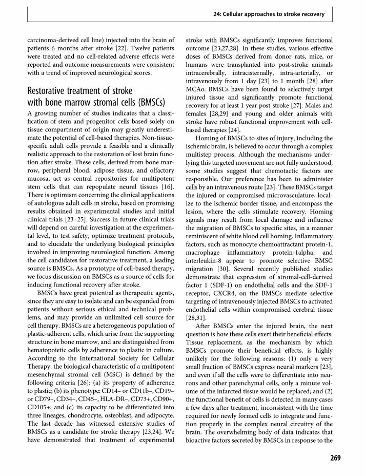
carcinoma-derived cell line) injected into the brain ofpatients 6 months after stroke [22]. Twelve patientswere treated and no cell-related adverse effects werereported and outcome measurements were consistentwith a trend of improved neurological scores.
Restorative treatment of strokewith bone marrow stromal cells (BMSCs)A growing number of studies indicates that a classi-fication of stem and progenitor cells based solely ontissue compartment of origin may greatly underesti-mate the potential of cell-based therapies. Non-tissue-specific adult cells provide a feasible and a clinicallyrealistic approach to the restoration of lost brain func-tion after stroke. These cells, derived from bone mar-row, peripheral blood, adipose tissue, and olfactorymucosa, act as central repositories for multipotentstem cells that can repopulate neural tissues [16].There is optimism concerning the clinical applicationsof autologous adult cells in stroke, based on promisingresults obtained in experimental studies and initialclinical trials [23–25]. Success in future clinical trialswill depend on careful investigation at the experimen-tal level, to test safety, optimize treatment protocols,and to elucidate the underlying biological principlesinvolved in improving neurological function. Amongthe cell candidates for restorative treatment, a leadingsource is BMSCs. As a prototype of cell-based therapy,we focus discussion on BMSCs as a source of cells forinducing functional recovery after stroke.
BMSCs have great potential as therapeutic agents,since they are easy to isolate and can be expanded frompatients without serious ethical and technical prob-lems, and may provide an unlimited cell source forcell therapy. BMSCs are a heterogeneous population ofplastic-adherent cells, which arise from the supportingstructure in bone marrow, and are distinguished fromhematopoietic cells by adherence to plastic in culture.According to the International Society for CellularTherapy, the biological characteristic of a multipotentmesenchymal stromal cell (MSC) is defined by thefollowing criteria [26]: (a) its property of adherenceto plastic; (b) its phenotype: CD14– or CD11b–, CD19–orCD79–, CD34–, CD45–, HLA-DR–, CD73+, CD90+,CD105+; and (c) its capacity to be differentiated intothree lineages, chondrocyte, osteoblast, and adipocyte.The last decade has witnessed extensive studies ofBMSCs as a candidate for stroke therapy [23,24]. Wehave demonstrated that treatment of experimental
stroke with BMSCs significantly improves functionaloutcome [23,27,28]. In these studies, various effectivedoses of BMSCs derived from donor rats, mice, orhumans were transplanted into post-stroke animalsintracerebrally, intracisternally, intra-arterially, orintravenously from 1 day [23] to 1 month [28] afterMCAo. BMSCs have been found to selectively targetinjured tissue and significantly promote functionalrecovery for at least 1 year post-stroke [27]. Males andfemales [28,29] and young and older animals withstroke have robust functional improvement with cell-based therapies [24].
Homing of BMSCs to sites of injury, including theischemic brain, is believed to occur through a complexmultistep process. Although the mechanisms under-lying this targeted movement are not fully understood,some studies suggest that chemotactic factors areresponsible. Our preference has been to administercells by an intravenous route [23]. These BMSCs targetthe injured or compromised microvasculature, local-ize to the ischemic border tissue, and encompass thelesion, where the cells stimulate recovery. Homingsignals may result from local damage and influencethe migration of BMSCs to specific sites, in a mannerreminiscent of white blood cell homing. Inflammatoryfactors, such as monocyte chemoattractant protein-1,macrophage inflammatory protein-1alpha, andinterleukin-8 appear to promote selective BMSCmigration [30]. Several recently published studiesdemonstrate that expression of stromal-cell-derivedfactor 1 (SDF-1) on endothelial cells and the SDF-1receptor, CXCR4, on the BMSCs mediate selectivetargeting of intravenously injected BMSCs to activatedendothelial cells within compromised cerebral tissue[28,31].
After BMSCs enter the injured brain, the nextquestion is how these cells exert their beneficial effects.Tissue replacement, as the mechanism by whichBMSCs promote their beneficial effects, is highlyunlikely for the following reasons: (1) only a verysmall fraction of BMSCs express neural markers [23],and even if all the cells were to differentiate into neu-rons and other parenchymal cells, only a minute vol-ume of the infarcted tissue would be replaced; and (2)the functional benefit of cells is detected in many casesa few days after treatment, inconsistent with the timerequired for newly formed cells to integrate and func-tion properly in the complex neural circuitry of thebrain. The overwhelming body of data indicates thatbioactive factors secreted by BMSCs in response to the
24: Cellular approaches to stroke recovery
269
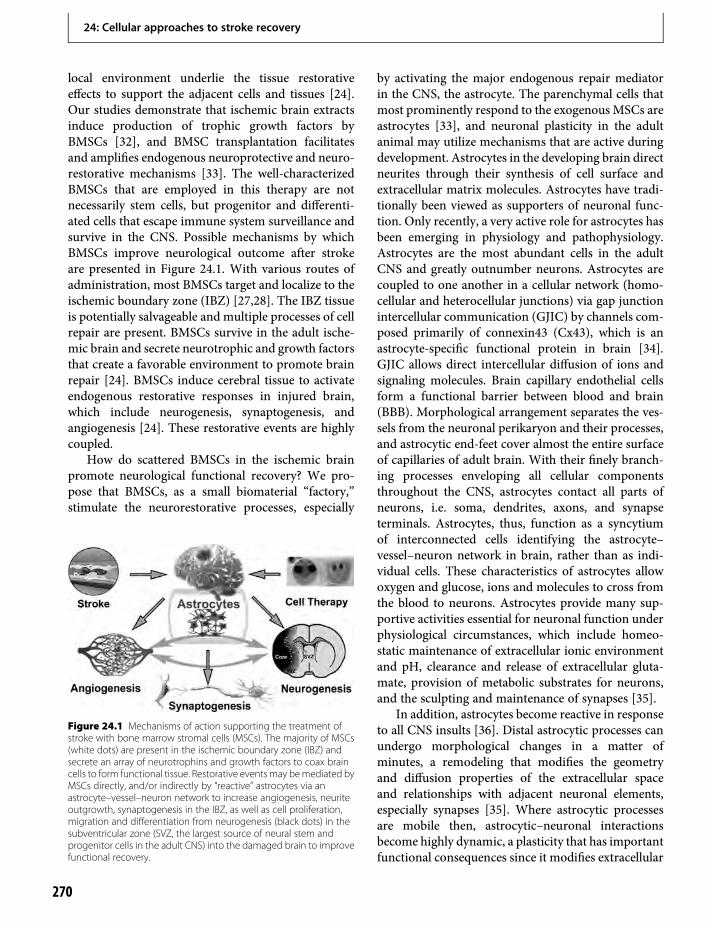
local environment underlie the tissue restorativeeffects to support the adjacent cells and tissues [24].Our studies demonstrate that ischemic brain extractsinduce production of trophic growth factors byBMSCs [32], and BMSC transplantation facilitatesand amplifies endogenous neuroprotective and neuro-restorative mechanisms [33]. The well-characterizedBMSCs that are employed in this therapy are notnecessarily stem cells, but progenitor and differenti-ated cells that escape immune system surveillance andsurvive in the CNS. Possible mechanisms by whichBMSCs improve neurological outcome after strokeare presented in Figure 24.1. With various routes ofadministration, most BMSCs target and localize to theischemic boundary zone (IBZ) [27,28]. The IBZ tissueis potentially salvageable and multiple processes of cellrepair are present. BMSCs survive in the adult ische-mic brain and secrete neurotrophic and growth factorsthat create a favorable environment to promote brainrepair [24]. BMSCs induce cerebral tissue to activateendogenous restorative responses in injured brain,which include neurogenesis, synaptogenesis, andangiogenesis [24]. These restorative events are highlycoupled.
How do scattered BMSCs in the ischemic brainpromote neurological functional recovery? We pro-pose that BMSCs, as a small biomaterial “factory,”stimulate the neurorestorative processes, especially
by activating the major endogenous repair mediatorin the CNS, the astrocyte. The parenchymal cells thatmost prominently respond to the exogenous MSCs areastrocytes [33], and neuronal plasticity in the adultanimal may utilize mechanisms that are active duringdevelopment. Astrocytes in the developing brain directneurites through their synthesis of cell surface andextracellular matrix molecules. Astrocytes have tradi-tionally been viewed as supporters of neuronal func-tion. Only recently, a very active role for astrocytes hasbeen emerging in physiology and pathophysiology.Astrocytes are the most abundant cells in the adultCNS and greatly outnumber neurons. Astrocytes arecoupled to one another in a cellular network (homo-cellular and heterocellular junctions) via gap junctionintercellular communication (GJIC) by channels com-posed primarily of connexin43 (Cx43), which is anastrocyte-specific functional protein in brain [34].GJIC allows direct intercellular diffusion of ions andsignaling molecules. Brain capillary endothelial cellsform a functional barrier between blood and brain(BBB). Morphological arrangement separates the ves-sels from the neuronal perikaryon and their processes,and astrocytic end-feet cover almost the entire surfaceof capillaries of adult brain. With their finely branch-ing processes enveloping all cellular componentsthroughout the CNS, astrocytes contact all parts ofneurons, i.e. soma, dendrites, axons, and synapseterminals. Astrocytes, thus, function as a syncytiumof interconnected cells identifying the astrocyte–vessel–neuron network in brain, rather than as indi-vidual cells. These characteristics of astrocytes allowoxygen and glucose, ions and molecules to cross fromthe blood to neurons. Astrocytes provide many sup-portive activities essential for neuronal function underphysiological circumstances, which include homeo-static maintenance of extracellular ionic environmentand pH, clearance and release of extracellular gluta-mate, provision of metabolic substrates for neurons,and the sculpting and maintenance of synapses [35].
In addition, astrocytes become reactive in responseto all CNS insults [36]. Distal astrocytic processes canundergo morphological changes in a matter ofminutes, a remodeling that modifies the geometryand diffusion properties of the extracellular spaceand relationships with adjacent neuronal elements,especially synapses [35]. Where astrocytic processesare mobile then, astrocytic–neuronal interactionsbecome highly dynamic, a plasticity that has importantfunctional consequences since it modifies extracellular
Figure 24.1 Mechanisms of action supporting the treatment ofstroke with bone marrow stromal cells (MSCs). The majority of MSCs(white dots) are present in the ischemic boundary zone (IBZ) andsecrete an array of neurotrophins and growth factors to coax braincells to form functional tissue. Restorative eventsmay bemediated byMSCs directly, and/or indirectly by “reactive” astrocytes via anastrocyte–vessel–neuron network to increase angiogenesis, neuriteoutgrowth, synaptogenesis in the IBZ, as well as cell proliferation,migration and differentiation from neurogenesis (black dots) in thesubventricular zone (SVZ, the largest source of neural stem andprogenitor cells in the adult CNS) into the damaged brain to improvefunctional recovery.
24: Cellular approaches to stroke recovery
270
ionic homeostasis, neurotransmission, gliotransmis-sion, and ultimately neuronal function at the cellularand system levels. Astrocytes influence long-termrecovery after brain injury, through neurite out-growth, synaptic plasticity, or neuron regeneration,which are mediated by astrocyte surface moleculeexpression and trophic factor release. The roles reac-tive gliosis take after brain injury are varied. Rapidlyexpanding astrocytic processes create both physicaland functional walls surrounding the ischemic core,which extend the time available for marshallingendogenous repair mechanisms, e.g. redirection ofblood flow to still salvageable parts of the brain andredirection of neurite sprouting and synapse forma-tion to build a new circuitry. However, astrocytes canalso form scars that inhibit axonal regeneration [36].Our data suggest that astrocytes promote brain plasti-city and recovery from stroke [33], and that the bene-ficial effects of reactive astrocytes are enhanced in theischemic brain after BMSC transplantation, andthereby promote appropriate neurite outgrowth andextension and myelination of axons to the injuredhemisphere. The glial scar adjacent to the infarct issignificantly reduced and more permeable than incontrol non-treated rats subjected to ischemic stroke[37]. Cells reduce scar tissue formation and veryimportantly reduce inhibitory glycoproteins. Wheninhibited, these proteins are permissive of neuriteoutgrowth and axonal remodeling in both the brainand spinal cord. In the adult animal after stroke, axonsmay also acquire their potential for outgrowth fromneighboring astrocytes and establish contacts withexisting circuits in the CNS.
Our studies demonstrate that white matter areas,in the striatum and the corpus callosum, are moreintact and enlarged after BMSC treatment than incontrol non-treated rats subjected to MCAo [37].A somewhat neglected but obviously important areaof interest is the response of the spinal cord to strokeand restorative cell therapy. Motor and somatosensoryresponse requires communication with the spinalcord, via the corticospinal tract (CST). Thus, recoveryof function may be associated with plasticity in theCST and the spinal cord. Anterograde and retrogradelabeling of the CST demonstrates a remarkable patternof neurite outgrowth from the intact to the denervatedspinal cord which significantly correlates with somato-sensory functional recovery [38]. Retrograde labelingof bilateral forelimbs also demonstrates crossed con-nections in the contralateral and ipsilateral brain
hemispheres amplified by BMSCs. Downregulationof inhibitory glycoproteins may contribute to thisrobust rewiring in the brain and spinal cord. To sumup, constitutive reparative responses are facilitated bya series of interactions between BMSCs and host cellsafter stroke, fostering the remodeling of brain tissue,reducing scar tissue, and facilitating synaptic andvascular reconstitution.
Changes in white matter in response to either acellular or a pharmacological restorative therapy canbe readily monitored using magnetic resonanceimaging–diffusion tensor imaging techniques(MRI–DTI) [39]. Tissue, in which the diffusion tensorfor water is isotropic, is cavitated. The more aniso-tropic the diffusion constant for water, the more struc-ture is present in the tissue [40]. Water moves easilyalong white matter fibers, and these structural changesin white matter and axonal growth may become evi-dent using DTI [40]. Preclinical data demonstrate thatcell therapy evokes white matter changes in the corpuscallosum, the striatum, and in the boundary region ofthe ischemic lesion which is sensitive to the DTI, andsignificant correlations between functional recoveryand a DTI-based parameter, Fractional Anisotropy(FA), may find clinical application [39].
Other cell sources for restorative treatmentof stroke: view within articleOther sources of non-tissue-specific adult stem cellsfor use in stroke therapy include cells derived fromhematological stem cells, peripheral blood, umbilicalcord blood, adipose cells, and olfactory mucosa.
Bone marrow-derived pluripotent hematopoieticstem cells (HSCs) have the ability to reconstitute allcells of the blood. It has been suggested, based onstatistical arguments, that individual stem cells cangive rise to clones with full hematopoietic capacity.The hematopoietic stem cell can be highly enrichedup to 10 000-fold and delivered to marrow-ablatedrecipients to fully reconstitute the blood [41].Systemically applied HSCs reduce cerebral post-ischemic inflammation, attenuate peripheral immuneactivation, and mediate neuroprotection after ische-mic stroke [42].
Filgratism (granulocyte colony stimulating factor,G-CSF)-mobilized peripheral blood progenitor cells(PBPCs) have been employed as a preferred source ofautologous stem cells, in light of the faster hematologicalrecovery and lesser supportive care requirement
24: Cellular approaches to stroke recovery
271
exhibited by PBPC transplants [43]. TransplantedG-CSF-mobilized PBPCs in rats after MCAo signifi-cantly reduced the stroke-induced hyperactivity com-pared with nontransplanted stroked animals [43].Preclinical data suggest that peripheral blood-derivedHSCs are as efficient in promoting functional improve-ment following MCAo as intravenous transplantationin rats [43].
The identification of endothelial progenitor cells(EPCs) in peripheral blood with the ability to differ-entiate into endothelial cells broke the paradigm thatvasculogenesis was only an embryogenic process. Use ofEPCs in experimental models [44] and patients [45] ofcerebral ischemia suggest EPCs can form new vessels inischemic areas and thus promote recovery after ische-mic events. The increase of circulating EPC after acuteischemic stroke was associated with good functionaloutcome and reduced infarct growth in 25 patientscompared with 23 patients with poor outcome [45].These findings suggest that EPCs participate in neuro-repair after ischemic stroke. These studies suggest thatEPC can be used as a potential cell-based therapeuticagent in stroke, since EPCs may play an important rolein tissue vascularization and endothelium homeostasis.
Umbilical cord blood cells (UCBCs) are a promisingsource of HSCs because of their availability, weakimmunogenicity, and low risk of mediating viral trans-mission. Infusion of human UCBCs into rats afterMCAo improved behavioral recovery [46]. UCBCssecrete neuroprotectivemolecules, enhance neovascula-rization and endogenous neurogenesis, and reduceinflammation in the ischemic brain [47]. These afore-mentioned effects, not cell replacement, may underliethe therapeutic benefits of UCBCs in ischemic brain.
Adipose tissue-derived stem cells (ADSCs) sharemany of the characteristics of the BMSCs, includingextensive proliferative potential and the ability toundergo multilineage differentiation. To test theeffects of ADSCs on experimental stroke, these cellswere directly implanted into the brain of rats afterMCAo [48]. ADSCs migrated toward the ischemicarea, and they also traveled far distances to the con-tralateral cortex. Transplanted rats had significantlybetter recovery in motor and somatosensory behaviorcompared with animals that only received saline. Thebenefits strongly support current hypotheses thattransplanted stem cells after stroke provide and inducetrophic support of injured brain.
Murine olfactory ensheathing cells (OECs)promote central nervous system axonal regeneration
in a murine model of stroke [49]. OECs also inducea neuroplastic effect which reduces neurologicaldysfunction caused by hypoxic/ischemic stress. Ratswith intracerebral implantation of human OECs/olfactory nerve fibroblasts (hOECs/ONFs) showedmore improvement on behavioral measures of neuro-logical deficit following stroke than control rats.[18F]fluoro-2-deoxyglucose PET (FDG-PET) showedincreased glucose metabolic activity in the hOEC/ONF-treated group compared with controls. BothhOECs/ONFs and endogenous homing stem cellsenhanced neuroplasticity in the rat and mouse ische-mic brain.
ConclusionThe non-tissue-specific adult stem cells, whether they beMSC, HSCs, PBPCs, EPCs, UCBCs, ADSCs, or OECs,when injected into the adult do not repopulate the adultbrain tissue; they produce an array of factors includingangiogenic and neurotrophic factors which initiate arestorative cascade of recovery. The targets of cellulartherapy are distinct fromneuroprotective effects of acuteintervention to reduce the volume of cerebral infarctionand the sequelae of secondary cell death, whether bynecrosis or apoptosis. After many years of failed clinicaltrials of neuroprotective drugs and in light of the com-pelling scientific evidence supporting neurorestoration,cell-based therapy is poised to be tested further in atranslational setting for stroke patients. Among variouscell sources, adult-derived cells stand out because:(1) cells can be obtained readily and expanded in culture;(2) autologous transplantation circumvents the risk ofrejection and graft-versus-host reaction; some cells, e.g.BMSCs, are not rejected and can be administered allo-geneically; (3) intravenous transplantation is an essen-tially non-invasive way to administer cells; and (4) safetyof cell administration has been demonstrated in exper-imental animals. Indeed, Bang and others have reporteddata from a clinical trial [25], in which 1×10⁸ cultureexpanded autologous BMSCs were administered intra-venously into 5 patients with ischemic stroke.Neurological outcome was significantly improved intreated patients and no adverse effects were detected.Additional trials for stroke are under way, and theseearly studies will hopefully spur application of thispromising restorative therapy.
We now know that the injured brain is highlymalleable and the entire intact brain responds toinjury and stroke, by producing new brain cells(neurogenesis), new vasculature (angiogenesis and
24: Cellular approaches to stroke recovery
272
arteriogenesis), and new wiring (synaptogenesis andaxonal growth), and these events collectively improveneurological function after stroke [50]. Although acomplete picture of the cellular approach acting as acatalyst endogenous mechanism is still lacking, someaspects have been identified, notably, stimulating bio-active factors to enhance neurite outgrowth, angiogen-esis, neurogenesis, and synaptogenesis [24]. Cell-basedtherapy induces recovery of function post-stroke bystimulating endogenous restorative mechanisms andnot by replacing infarcted tissue. Adult cerebral tissuesare highly plastic and amenable to change given theappropriate microenvironment. The field of restora-tive neurology for the treatment of stroke is rapidlyprogressing and cell approaches can be used to amplifyrecovery of function in the injured brain. Cell-basedrestorative therapies initiated days or weeks afterstroke hold tremendous promise for millions ofdisabled stroke survivors.
AcknowledgmentsThis work was supported by NINDS grants PO1NS42345 and PO1 NS23393.
References1. Gage FH. Mammalian neural stem cells. Science.
2000;287:1433–8.
2. Alvarez-Buylla A, Seri B, Doetsch F. Identification ofneural stem cells in the adult vertebrate brain. Brain ResBull. 2002;57:751–8.
3. Eriksson PS, Perfilieva E, Bjork-Eriksson T, et al.Neurogenesis in the adult human hippocampus.NatureMed. 1998;4:1313–7.
4. Zhang R, Zhang Z, Wang L, et al. Activated neural stemcells contribute to stroke-induced neurogenesis andneuroblast migration toward the infarct boundary inadult rats. J Cereb Blood Flow Metab. 2004;24:441–8.
5. Arvidsson A, Collin T, Kirik D, et al. Neuronalreplacement from endogenous precursors in the adultbrain after stroke. Nature Med. 2002;8:963–70.
6. Cramer SC, Chopp M. Recovery recapitulatesontogeny. Trends Neurosci. 2000;23:265–71.
7. Blau HM, Brazelton TR, Weimann JM. The evolvingconcept of a stem cell: Entity or function? Cell.2001;105:829–41.
8. Wiltrout C, Lang B, Yan Y, et al. Repairing brain afterstroke: A review on post-ischemic neurogenesis.Neurochem Int. 2007;50:1028–41.
9. Chopp M, Li Y, Chen J, et al. Brain repair and recoveryfrom stroke. Eur Neurol. 2008;3:2–5.
10. Wang L, Zhang Z, Wang Y, et al. Treatment of strokewith erythropoietin enhances neurogenesis andangiogenesis and improves neurological function inrats. Stroke. 2004;35:1732–7.
11. Zacharek A, Chen J, Cui X, et al. Angiopoietin1/Tie2and VEGF/Flk1 induced by MSC treatment amplifiesangiogenesis and vascular stabilization after stroke.J Cereb Blood Flow Metab. 2007;27:1684–91.
12. Leventhal C, Rafii S, Rafii D, et al. Endothelial trophicsupport of neuronal production and recruitment fromthe adult mammalian subependyma.Mol Cell Neurosci.1999;13:450–64.
13. Palmer TD, Willhoite AR, Gage FH. Vascular niche foradult hippocampal neurogenesis. J Comp Neurol.2000;425:479–94.
14. Takahashi K, Yasuhara T, Shingo T, et al. Embryonicneural stem cells transplanted in middle cerebral arteryocclusion model of rats demonstrated potenttherapeutic effects, compared to adult neural stem cells.Brain Res. 2008;1234:172–82.
15. Grabowski M, Brundin P, Johansson BB. Fetalneocortical grafts implanted in adult hypertensive ratswith cortical infarcts following a middle cerebral arteryocclusion: Ingrowth of afferent fibers from the hostbrain. Exp Neurol. 1992;116:105–21.
16. Roh JK, Jung KH, Chu K. Adult stem celltransplantation in stroke: Its limitations and prospects.Curr Stem Cell Res Ther. 2008;3:185–96.
17. Reynolds BA, Weiss S. Generation of neurons andastrocytes from isolated cells of the adult mammaliancentral nervous system. Science. 1992;255:1707–10.
18. Jin K, Wang X, Xie L, et al. Evidence for stroke-inducedneurogenesis in the human brain. Proc Natl Acad SciUSA. 2006;103:13 198–202.
19. Zhang ZG, Jiang Q, Zhang R, et al. Magnetic resonanceimaging and neurosphere therapy of stroke in rat. AnnNeurol. 2003;53:259–63.
20. Magavi SS, Macklis JD. Induction of neuronaltype-specific neurogenesis in the cerebral cortex ofadult mice: Manipulation of neural precursors in situ.Brain Res Dev Brain Res. 2002;134:57–76.
21. Bacigaluppi M, Pluchino S, Martino G, et al. Neuralstem/precursor cells for the treatment of ischemicstroke. J Neurol Sci. 2008;265:73–7.
22. Kondziolka D, Wechsler L, Goldstein S, et al.Transplantation of cultured human neuronal cells forpatients with stroke. Neurology. 2000;55:565–9.
23. Chen J, Li Y, Wang L, et al. Therapeutic benefit ofintravenous administration of bone marrow stromalcells after cerebral ischemia in rats. Stroke.2001;32:1005–11.
24. Chopp M, Li Y. Treatment of neural injury withmarrow stromal cells. Lancet Neurol. 2002;1:92–100.
24: Cellular approaches to stroke recovery
273
25. Bang OY, Lee JS, Lee PH, et al. Autologousmesenchymal stem cell transplantation in strokepatients. Ann Neurol. 2005;57:874–82.
26. Dominici M, Le Blanc K, Mueller I, et al. Minimalcriteria for defining multipotent mesenchymal stromalcells. The International Society for Cellular Therapyposition statement. Cytotherapy. 2006;8:315–7.
27. Shen LH, Li Y, Chen J, et al. One-year follow-up afterbone marrow stromal cell treatment in middle-agedfemale rats with stroke. Stroke. 2007;38:2150–6.
28. Shen LH, Li Y, Chen J, et al. Therapeutic benefit of bonemarrow stromal cells administered 1 month afterstroke. J Cereb Blood Flow Metab. 2007;27:6–13.
29. Li Y, McIntosh K, Chen J, et al. Allogeneic bonemarrow stromal cells promote glial–axonal remodelingwithout immunologic sensitization after stroke in rats.Exp Neurol. 2006;198:313–25.
30. Wang L, Li Y, Chen J, et al. Ischemic cerebral tissue andMCP-1 enhance rat bone marrow stromal cell migrationin interface culture. Exp Hematol. 2002;30:831–6.
31. Cui X, Chen J, Zacharek A, et al. Nitric oxide donorupregulation of stromal cell-derived factor-1/chemokine (CXC motif) receptor 4 enhances bonemarrow stromal cell migration into ischemic brain afterstroke. Stem Cells (Dayton, Ohio). 2007;25:2777–85.
32. Qu R, Li Y, Gao Q, et al. Neurotrophic and growthfactor gene expression profiling of mouse bone marrowstromal cells induced by ischemic brain extracts.Neuropathology. 2007;27:355–63.
33. Gao Q, Li Y, Chopp M. Bone marrow stromal cellsincrease astrocyte survival via upregulation ofphosphoinositide 3-kinase/threonine protein kinaseand mitogen-activated protein kinase kinase/extracellular signal-regulated kinase pathways andstimulate astrocyte trophic factor gene expressionafter anaerobic insult. Neuroscience.2005;136:123–34.
34. Rouach N, Glowinski J, Giaume C. Activity-dependentneuronal control of gap-junctional communication inastrocytes. J Cell Biol. 2000;149:1513–26.
35. Theodosis DT, PoulainDA,Oliet SH. Activity-dependentstructural and functional plasticity of astrocyte–neuroninteractions. Physiol Rev. 2008;88:983–1008.
36. Sofroniew MV. Reactive astrocytes in neural repair andprotection. Neuroscientist. 2005;11:400–07.
37. Shen LH, Li Y, Chen J, et al. Intracarotidtransplantation of bone marrow stromal cells increasesaxon–myelin remodeling after stroke. Neuroscience.2006;137:393–9.
38. Liu Z, Li Y, Qu R, et al. Axonal sprouting into thedenervated spinal cord and synaptic and postsynapticprotein expression in the spinal cord aftertransplantation of bone marrow stromal cell in strokerats. Brain Res. 2007;1149:172–80.
39. Jiang Q, Zhang ZG, Ding GL, et al. MRI detects whitematter reorganization after neural progenitor celltreatment of stroke. NeuroImage. 2006;32:1080–9.
40. Beaulieu C. The basis of anisotropic water diffusion inthe nervous system – A technical review. NMR Biomed.2002;15:435–55.
41. Morrison SJ,Weissman IL. The long-term repopulatingsubset of hematopoietic stem cells is deterministic andisolatable by phenotype. Immunity. 1994;1:661–73.
42. Schwarting S, Litwak S, Hao W, et al. Hematopoieticstem cells reduce postischemic inflammation andameliorate ischemic brain injury. Stroke.2008;39:2867–75.
43. Willing AE, Vendrame M, Mallery J, et al. Mobilizedperipheral blood cells administered intravenouslyproduce functional recovery in stroke. Cell Transplant.2003;12:449–54.
44. Rouhl RP, van Oostenbrugge RJ, Damoiseaux J, et al.Endothelial progenitor cell research in stroke:A potential shift in pathophysiological andtherapeutical concepts. Stroke. 2008;39:2158–65.
45. Sobrino T, Hurtado O, Moro MA, et al. The increase ofcirculating endothelial progenitor cells after acuteischemic stroke is associated with good outcome.Stroke. 2007;38:2759–64.
46. Newcomb JD, Ajmo CT, Jr, Sanberg CD, et al. Timingof cord blood treatment after experimental strokedetermines therapeutic efficacy. Cell Transplant.2006;15:213–23.
47. Vendrame M, Gemma C, de Mesquita D, et al. Anti-inflammatory effects of human cord blood cells in a ratmodel of stroke. Stem Cells Dev. 2005;14:595–604.
48. Kang S, Lee D, Bae Y, et al. Improvement ofneurological deficits by intracerebral transplantationof human adipose tissue-derived stromal cellsafter cerebral ischemia in rats. Exp Neurol.2003;183:355–66.
49. Shyu WC, Liu DD, Lin SZ, et al. Implantation ofolfactory ensheathing cells promotes neuroplasticity inmurine models of stroke. J Clin Invest.2008;118:355–66.
50. Lindvall O, Kokaia Z, Martinez-Serrano A. Stem celltherapy for human neurodegenerative disorders –Howto make it work. Nature Med. 2004;10:S42–50.
24: Cellular approaches to stroke recovery
274
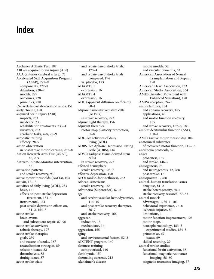
Index
Aachener Aphasie Test, 187ABI see acquired brain injury (ABI)ACA (anterior cerebral artery), 71Accelerated Skill Acquisition Program
(ASAP), 227–9components, 227–8definition, 228–9models, 227outcomes, 228principles, 228
{N-}acetylaspartate–creatine ratios, 151acetylcholine, 188acquired brain injury (ABI)impacts, 233incidence, 233rehabilitation treatments, 233–4survivors, 233acrobatic tasks, rats, 28–9
acrobatic trainingefficacy, 28–9
action observationin post-stroke motor learning, 237–8
Action Research Arm Test (ARAT),186, 239
Activate-Initiate-Monitor intervention,156
activation patternsand stroke recovery, 95
active motor thresholds (AMTs), 104activin, 12–13activities of daily living (ADL), 233basic, 151effects on post-stroke depression
treatment, 153–4instrumental, 151post-stroke depression effects on,
151–2, 154–5acute strokebrain eventsand subsequent repair, 87–96
acute stroke survivorsrobotic therapy, 197
acute stroke therapiesgoals, 259and nature of stroke, 167recanalization strategies, 89selection issues, 82thrombolysis, 88timing issues, 87
acute stroke trials
and repair-based stroke trials,173–4
and repair-based stroke trialscompared, 174
vs. placebo, 173ADAMTS 1expression, 16
ADAMTS 4expression, 16
ADC (apparent diffusion coefficient),60–1
adipose tissue-derived stem cells(ADSCs)
in stroke recovery, 272adjunct light therapy, 156adjuvant therapiesmotor map plasticity promotion,
7–8ADL, See activities of daily
living (ADL)ADRS. See Aphasic Depression Rating
Scale (ADRS), 146ADSCs (adipose tissue-derived stem
cells)in stroke recovery, 272
affected hemispherestroke recovery, 105–7
affective depression, 150AFOs (ankle–foot-orthoses), 252African-Americansstroke recovery, 166
Afrotheria (Superorder), 67–8ageand cerebrovascular hemodynamics,
114–15and post-stroke recovery therapies,
36–7and stroke recovery, 166
aggrecaninduction, 15mechanisms, 14
aggression, 155agingand environmental factors, 52–3
AIXTENT program, 140alertness trainingcomputerized, 140
alexithymia, 150alternating currents, 213Alzheimer’s disease
mouse models, 52and vascular dementia, 52
American Association of NeuralTransplantation and Repair,190
American Heart Association, 233American Stroke Association, 164AMES (Assisted Movement with
Enhanced Sensation), 198AMPA receptors, 24–5amphetamines, 184and aphasia recovery, 185applications, 40and motor function recovery,
185and stroke recovery, 167–8, 185
amplitude/stimulus function (ASF),150–1
AMTs (active motor thresholds), 104anatomical substratesof recovered motor function, 115–16
anesthesia protocols, 59angerproneness, 155and stroke, 146–7
angiogenesis, 73and neurogenesis, 12, 268post-stroke, 17
angiopoietin 1, 268animal–human translation issuesdrug use, 81–2stroke heterogeneity, 80–1stroke recovery research, 77–82
animal modelsadvantages, 1, 80–1, 103behavioral experience, 27–8ischemic injuries, 80limitations, 1motor function improvement, 105motor maps, 1neuropharmacology, 183–5experimental studies, 184–5
primates as, 69issues, 69
skilled reaching, 29animal stroke studiesfunctional brain activation, 58functional magnetic resonance
imaging, 58–60magnetic resonance imaging, 57
275
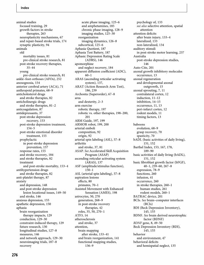
animal studiesfocused training, 29growth factors in stroke
therapies, 263neuroplasticity mechanisms, 67and repair-based stroke trials, 174synaptic plasticity, 94
animalsoldmortality issues, 81pre-clinical stroke research, 81
post-stroke recovery therapies,35–44
youngpre-clinical stroke research, 81
ankle–foot-orthoses (AFOs), 252anosognosia, 154anterior cerebral artery (ACA), 71anthropoid primates, 68–9anticholesterol drugsand stroke therapies, 82
anticholinergic drugsand stroke therapies, 81–2
anticoagulation, 87antidepressants, 37post-stroke depression
recovery, 153post-stroke depression treatment,
154–5post-stroke emotional disorder
treatment, 155prophylacticin post-stroke depressionprevention, 157
response rates, 155and stroke recovery, 184and stroke therapies, 82treatmentand post-stroke mortality, 153–4
antihypertension drugsand stroke therapies, 82
anti-platelet therapy, 87anxietyand depression, 148and post-stroke depressionlesion locational issues, 149–50
and stroke, 146anxious depression, 155apathetic depression, 150aphasiabrain reorganizationtherapy impacts, 129
conduction, 129–30constraint-induced therapy, 129future research, 130longitudinal studies, 127–8measures, 146and network approach, 129–30neuroimaging trials, 187–8recovery
acute phase imaging, 125–6and amphetamines, 185chronic phase imaging, 128–9imaging studies, 125–30
reorganizationimaging dynamics, 126–8
subcortical, 125–6Aphasia Quotient, 187Aphasia Test Battery, 187Aphasic Depression Rating Scale
(ADRS), 146apomorphineand neglect recovery, 184
apparent diffusion coefficient (ADC),60–1
ARAS (ascending reticular activatingsystem), 137,
ARAT (Action Research Arm Test),186, 239
Archonta (Superorder), 67–8areaand dexterity, 2–3
arm exerciserobotic therapy, 197robotic vs. other therapies, 198–200,
201ARM Guide, 197, 199ARMOR robot, 199, 200arterial embolicomposition, 92origin, 92
arterial spin labeling (ASL), 57–8arthritisand stroke, 37, 81
ASAP. SeeAccelerated Skill AcquisitionProgram (ASAP)
ascending reticular activating system(ARAS), 137
ASF (amplitude/stimulus function),150–1
ASL (arterial spin labeling), 57–8aspiration lesionseffects, 80primates, 70–1
Assisted Movement with EnhancedSensation (AMES), 198
astrocytes, 50, 270generation, 268–9in post-stroke recovery
therapies, 42roles, 35, 50, 270–1
ATF3, 14atherosclerosisand stroke, 37
attention,brain mapping
after stroke, 133–41and brain reorganization, 141functional mapping studies,
136–9
psychology of, 133see also selective attention, spatialattention
attention deficitsafter brain injury, 133–4lateralized, 133non-lateralized, 134
auditory stimuliin post-stroke motor learning, 237
Australiapost-stroke depression studies,
146Auto-Cite, 201axonal growth inhibitory moleculesoccurrence, 13
axonal regenerationand developmental axonal
outgrowth, 15axonal sprouting, 7, 11contralateral cortex, 12induction, 11–12inhibition, 14–15occurrence, 11, 13peri-infarct cortex, 12rodent models, 11timing factors, 13
baboonsevolution, 68–9grasp recovery, 70spasticity, 70
bADL (basic activities of daily living),151, 152
Barthel Index, 153, 167, 178,189–90
basic activities of daily living (bADL),151, 152
basic fibroblast growth factor (bFGF),40–1, 259–60, 267–8
expression, 78–9functions, 260infusion, 41occurrence, 260in stroke therapies, 260–1human studies, 261rodent models, 260–1
BATRAC device, 201BCIs. See brain–computer interfaces
(BCIs)BDI (Beck Depression Inventory),
145, 155BDNF. See brain derived neurotrophic
factor (BDNF)BDNF gene, 8, 49–50Beck Depression Inventory (BDI),
145, 155behaviorand environment, 47
behavioral deficitsand hemispatial neglect, 135
Index
276

behavioral experienceanimal models, 27–8and brain injuries, 25and environmental complexity,
27–8behavioral experience manipulationsand degenerative responses,
23–4and degenerative–regenerative
cascade, 26and regenerative responses,
23–4behavioral impairmentsand stroke, 1
behavioral influenceson post-stroke neuronal events,
23–31behavioral manipulationsand other interventions combined,
29–30behavioral repertoiresmammals, 39–40
behavioral trainingand post-injury pharmacological
manipulations, 29–30benzodiazepine, 52and motor recovery, 184neuroprotectant activity, 184
Betz cells, 72, See also corticospinal(CS) neurons
bFGF. See basic fibroblast growth factor(bFGF)
bicuculline, 60bilateral training, 202BiManuTrac, 198, 200biomarkersand repair-based stroke trials,
176–8block myelin-associated growth
inhibitors, 12blood glucose levelsand stroke recovery, 167
blood oxygenation level-dependent(BOLD) fMRI, 57–8, 88
activity, 138age factors, 114–15applications, 58, 177in cerebrovascular disease,
113–15correlation approaches, 114limitations, 114low frequency signals, 60principles, 113signal fluctuations, 139signal reduction, 114signal shape, 113–14
BMP-7. See osteogenic protein-1(OP-1)
BMSCs. See bone marrow stromal cells(BMSCs)
Bobath approach, 220, 221, 223and stroke outcomes, 222treatment–outcome relationships,
221–2BOLD fMRI. See blood oxygenation
level-dependent (BOLD)fMRI
bone marrow stromal cells (BMSCs),270
advantages, 269homing, 269in stroke recovery, 269–71
mechanisms, 269–71trials, 272
braindegenerative processes, 24, 43–4regenerative processes, 24reparative processes, 43–4
brain activityhyperactivity, 128lateralization, 119
brain adaptationfuture research, 130
brain centersfunctional independence issues,
129–30brain–computer interfaces (BCIs)development, 253–4field of study, 253
brain damage. See brain injuriesbrain derived neurotrophic factor
(BDNF), 37, 40, 148,267–8
and environmental enrichment,49–50
expression, 268polymorphisms, 166Val66Met polymorphism,
49–50brain disordersand environmental enrichment, 52
brain eventsin acute stroke period
and subsequent repair, 87–96brain functionsloss, 130organization, 129
brain infarctsdeterminants, 88magnetic resonance imaging,
88–9pathogenesis, 89recovery, 87–8, 94–5
brain injuries. See also acquired braininjury (ABI), See also traumaticbrain injury (TBI)
and attention deficits, 133–4and behavioral experience, 25functional recovery
lesion-dependent, 80
locational issues, 80and motor system reconfiguration,
120responsesgender differences, 38
tissue losscompensation strategies, 37
unilateral, 27brain ischemiaetiology, 87and stroke, 87
brain lesionsand post-stroke depressionlocational issues,149–50
brain mappingof attentionafter stroke, 133–41
of neglectafter stroke, 133–41
post-strokemotor system, 113–20
rats, 63brain plasticityand environmental enrichment,
52occurrence, 23
brain reorganization, See also post-stroke brain reorganization
aphasiaimaging dynamics, 126–8therapy impacts, 129
and attention, 141environmental enrichment, 23optimization, 23and post-stroke recovery,
116–17and rehabilitative training, 23and self-taught behavioral change,
26–7brain repairand perfusion–diffusion mismatch,
88–9brain repair after strokeclinical applications, 173clinical trial methodologiesissues, 173–8
definition, 173mammals, 77and potential clinical applications,
173therapiesclasses, 178
tissue targeting issues, 173brain tissuepreservationand stroke recovery, 168
brain-controlled interfacesneuroprostheses, 253–4
BrainGate, 253
Index
277
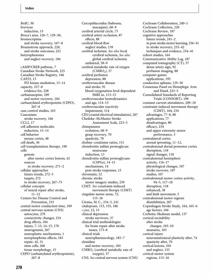
BrdU, 50brevicaninduction, 15
Broca’s area, 126–7, 129–30,bromocriptineand stroke recovery, 167–8
Brunnstrom approach, 220,and stroke outcomes, 221
butyrophenonesand neglect recovery, 184
cAMP/CREB pathway, 7Canadian Stroke Network, 225Canadian Stroke Registry, 166CAP23, 13PI3 kinase mediation, 13–14
capacity, 227–8evidence for, 228
carbamazepine, 109and motor recovery, 184
carbamylated erythropoietin (CEPO),267–8
case control studies, 163Caucasiansstroke recovery, 166
CCL2, 17cell adhesion moleculesinduction, 13–14
cell behaviormouse cortex, 48
cell death, 36cell transplantation therapy, 190cellsgenesisafter motor cortex lesions, 42
sourcesin stroke recovery, 271–2
cellular approachesfuture trends, 272–3targets, 272to stroke recovery, 267–73
cellular conceptsof neural repair after stroke,
11–12Centers for Disease Control and
Prevention, 233central motor conduction time, 109central nervous system (CNS)astrocytes, 270connectivity changes, 234drug effects, 184injury, 7, 13, 14–15, 178neurogenesis, 267neuroplastic mechanisms, 1norepinephrine effects, 183repair, 42, 51stem cells, 268tissue morphology, 15
CEPO (carbamylated erythropoietin),267–8
Cercopithecoidea (baboons,macaques), 68–9
cerebral arterial circle, 75cerebral artery occlusion, 87effects, 88
cerebral blood flowneglect studies, 139
cerebral ischemia. See also focalcerebral ischemia, See alsoglobal cerebral ischemia
unilateral, 58–9cerebral metabolic rate of oxygen
(CMRO2), 57cerebral perfusiondepression, 88
cerebrovascular diseaseand stroke, 91blood oxygenation level-dependent
fMRI in, 113–15cerebrovascular hemodynamicsand age, 114–15
cerebrovascular reactivityimpairment, 114
CES (cranial electrical stimulation), 207Chedoke–McMaster Stroke
Assessment Scale, 223–5chimpanzeesevolution, 68–9grasp recovery, 70spasticity, 70
choline–creatinine ratios, 151chondroitin sulfate proteoglycan
neurocansinduction, 13
chondroitin sulfate proteoglycans(CSPGs), 14–15
mechanisms, 14post-stroke responses, 15
chromatin, 52chronic strokemotor imagery studies, 239
CIMT. See constraint-inducedmovement therapy (CIMT)
cingulate motor areas, 72,115–16
Cirstea, M. C., 234–5, 241citalopram, 153, 155, 186c-jun, 12, 13clinical depressionstroke survivors, 37
clinical trial methodologiesfor brain repair after stroke
issues, 173–8clinical trialsneuropharmacology, 185–7
clonidineand motor recovery, 184
CMRO2 (cerebral metabolic rate ofoxygen), 57
CNS. See central nervous system (CNS)
Cochrane Collaboration, 240–1Cochrane Collection, 220Cochrane Review, 187cognitive approachesfuture trends, 241–2in post-strokemotor learning, 236–41to stroke recovery, 233–42techniques and evidence, 234–41
cohort studies, 164Communicative Ability Log, 187computed tomography (CT), 57dense artery sign, 92perfusion imaging, 88
computer gamesapplications, 195
conduction aphasia, 129–30Consensus Panel on Hemiplegic Arm
and Hand, 223–5Consolidated Standards of Reporting
Trials (CONSORT), 190constant current stimulators, 209–10constraint-induced movement therapy
(CIMT), 164, 234advantages, 77–8, 80applications, 77disadvantages, 80efficacy, 234and upper extremity motor
performance, 3contralateral cortexaxonal sprouting, 11–12,
contralesional dorsal premotor cortexdisruption, 118signal changes, 118
contralesional hemisphereactivity, 116–17physiological changes, 107stroke recovery, 107studies, 107
contralesional motor cortex activity,94–5, 117–18
disruption, 118enhanced, 58and limb movement, 5
contralesional motor regionsdisinhibition, 207
Copenhagen Stroke Study, 164, 165–6age factors, 166
Corbetta–Shulman model, 137cortical excitabilityafter strokechanges, 103–10
measures, 103cortical injuryneuroanatomical plasticity after, 74spasticity after, 70
cortical lesions, 104and neglect, 135
cortical motor systemregions, 115–16
Index
278
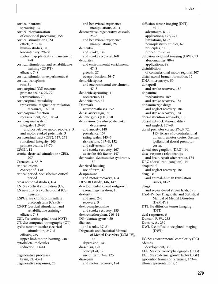
cortical neuronssprouting, 13
cortical reorganizationof emotional processing, 158
cortical stimulation (CS)effects, 213–14human studies, 30low-intensity, 29–30motor map plasticity enhancement,
7–8cortical stimulation and rehabilitative
training (CS-RT)efficacy, 7–8
cortical stimulation experiments, 6cortical transplantsrats, 51
corticospinal (CS) neuronsprimate brains, 70, 72terminations, 70
corticospinal excitabilitytranscranial magnetic stimulation
measures, 109–10corticospinal functionmeasurement, 2–3, 103–4
corticospinal systemintegrity, 119–20and post-stroke motor recovery, 3
and motor evoked potentials, 3corticospinal tract (CST), 117, 271functional integrity, 103primate brains, 69–70
CPG21, 12cranial electrical stimulation (CES),
207Cretaceous, 68–9critical lesionsconcept of, 130
critical period. See ischemic criticalperiod
cross sectional studies, 164CS. See cortical stimulation (CS)CS neurons. See corticospinal (CS)
neuronsCSPGs. See chondroitin sulfate
proteoglycans (CSPGs)CS-RT (cortical stimulation and
rehabilitative training)efficacy, 7–8
CST. See corticospinal tract (CST)CT. See computed tomography (CT)cyclic neuromuscular electrical
stimulation, 247–8efficacy, 249upper limb motor learning, 248
cytoskeletal moleculesinduction, 13–14
degenerative processesbrain, 24, 43–4
degenerative responses, 23
and behavioral experiencemanipulations, 23–4
degenerative–regenerative cascade,25–6
and behavioral experiencemanipulations, 26
dementiaand stroke, 149and stroke recovery, 168
dendritesand environmental enrichment,
47–8growth, 27overproduction, 26–7
dendritic spinesand environmental enrichment,
47–8dendritic sprouting, 11occurrence, 11
dendritic tree, 47Denmarkneuroprostheses, 252
dense artery sign, 92dentate gyrus (DG), 50depression. See also post-stroke
depressionand anxiety, 148prevalence, 157rating scales, 145–6risk factors, 147–8, 152and self-esteem, 148and stroke recovery, 167as stroke risk factor, 147
depression-dysexecutive syndrome,150
deprived housinguse of term, 47
desipramineand motor recovery, 184
DESTRO study, 146, 147developmental axonal outgrowthaxonal regeneration, 15
dexterityand area, 2–3recovery, 5
dextroamphetamineand stroke recovery, 185
dextromethorphan, 210–11DG (dentate gyrus), 50diabetesand stroke, 37, 81
Diagnostic and Statistical Manualof Mental Disorders (DSM-IV),145
depression, 145diaschisis, 128concept of, 125use of term, 3–4, 125
diazepamand motor recovery, 184
diffusion tensor imaging (DTI),60–1
advantages, 61–2applications, 177, 271limitations, 61–2neuroplasticity studies, 62principles, 61procedures, 61–2
diffusion weighted imaging (DWI), 93abnormalities, 88–9applications, 88
disinhibitionof contralesional motor regions, 207
distal axonal branch formation, 12DNA microarrays, 50donepeziland stroke recovery, 187
dopaminemechanisms, 189and stroke recovery, 184
dopaminergic drugsand neglect recovery, 184and stroke recovery, 184
dorsal attention networks, 135dorsal network abnormalitiesand neglect, 137–9
dorsal premotor cortex (PMd), 72,115–16, See also contralesionaldorsal premotor cortex, See alsoipsilesional dorsal premotorcortex
dorsal root ganglion (DRG), 14dose–response relationshipsand brain repair after stroke, 174
DRG (dorsal root ganglion), 14droperidoland neglect recovery, 184
drug useand animal–human translation
issues, 81–2drugsand repair-based stroke trials, 175
DSM-IV. See Diagnostic and StatisticalManual of Mental Disorders(DSM-IV)
DTI. See diffusion tensor imaging(DTI)
dual responses, 6Duncan, P.W., 233Dunsky, A., 239DWI. See diffusion weighted imaging
(DWI)
EC. See environmental complexity (EC)edemasdevelopment, 35
EEG. See electroencephalography (EEG)EGF. See epidermal growth factor (EGF)egocentric frames of reference, 133–4elbow representations, 6
Index
279
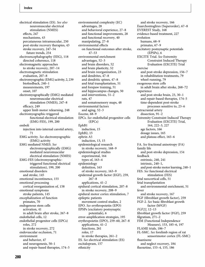
electrical stimulation (ES). See alsoneuromuscular electricalstimulation (NMES)
effects, 247mechanisms, 43percutaneous intramuscular, 250post-stroke recovery therapies, 43stroke recovery, 247–54future trends, 254
electroencephalography (EEG), 118directed coherence, 118
electromagnetic approachesto stroke recovery, 207–14
electromagnetic stimulationevaluation, 207–8
electromyographic (EMG) activity, 2, 239biofeedback, 240–1measurements, 197onset, 107
electromyographically (EMG) mediatedneuromuscular electricalstimulation (NMES), 247–8
efficacy, 249upper limb motor relearning, 248
electromyographic-triggeredfunctional electrical stimulation(EMG-FES), 199, 200
emboliinjection into internal carotid artery,
71EMG activity. See electromyographic
(EMG) activityEMG mediated NMES. See
electromyographically (EMG)mediated neuromuscularelectrical stimulation (NMES)
EMG-FES (electromyographic-triggered functional electricalstimulation), 199, 200
emotional disordersand stroke, 145
emotional incontinence, 155emotional processingcortical reorganization of, 158
emotional symptomsstroke patients, 145
encephalization of functionprimates, 70
endogenous stem cellsactivation, 41in adult brain after stroke, 267–8
endothelial cells, 12endothelial progenitor cells (EPCs)roles, 272in stroke recovery, 272
endovascular occlusion, 71environmentand behavior, 47and neurogenesis, 50–1and repair-based therapies, 174–5
environmental complexity (EC)advantages, 28and behavioral experience, 27–8and functional improvements, 28and functional recovery, 40manipulating, 27–8
environmental effectson functional outcomes after stroke,
47–53environmental enrichmentadvantages, 52–3and brain disorders, 52and brain plasticity, 52and brain reorganization, 23and dendrites, 47–8and dendritic spines, 47–8and fetal transplantation, 51and forepaw training, 51and hippocampus changes, 50and interventions, 51–2rats, 47and somatosensory maps, 48
environmental factorsand aging, 52–3
Eocene, 68–9EPCs. See endothelial progenitor cells
(EPCs)EphB1induction, 15
EphB2, 15ephrin A5induction, 13, 15
epidemiological researchin stroke recovery, 168–9
epidemiological studiesexperimental, 164types of, 163–4
epidemiologydefinition, 163of stroke recovery, 163–9
epidermal growth factor (EGF), 259,267–8
applications, 41–2epidural cortical stimulation, 207–8in stroke recovery, 208–9
epidural motor cortex stimulation, 7epileptic patientsmovement control studies, 2
EPO. See erythropoietin (EPO)EPSPs (excitatory postsynaptic
potentials), 6error-amplification strategies, 195erythropoietin (EPO), 259–60, 267–8applications, 41–2functions, 261roles, 17in stroke therapies, 261–2
ES. See electrical stimulation (ES)escitalopram, 157ethnicity
and stroke recovery, 166Euarchontoglires (Superorder), 67–8EVEREST Study, 168evidence-based treatment, 227evolutionhumans, 68–9primates, 67–9
excitatory postsynaptic potentials(EPSPs), 6
EXCITE Trial. See ExtremityConstraint Induced TherapyEvaluation (EXCITE) Trial
exerciseand post-stroke depression, 156–7in rehabilitation treatments, 79wheel-running, 79
exogenous stem cellsin adult brain after stroke, 268–72
experienceand post-stroke brain, 23, 30–1and repair-based therapies, 174–5time-dependent post-stroke
processes sensitive to, 23–6extracranial arterydissection, 91–2
Extremity Constraint Induced TherapyEvaluation (EXCITE) Trial,164, 222–3, 227
age factors, 166dosage issues, 165and plateau effect, 165–6
FA. See fractional anisotropy (FA)family lifeand post-stroke depression, 154
feedbackextrinsic, 240, 241intrinsic, 240–1,and post-strokemotor learning, 240–1
FES. See functional electricalstimulation (FES)
fetal neocortical cells, 51fetal transplantationand environmental enrichment, 51
feverand stroke recovery, 167
FGF (fibroblast growth factor), 259FGF-2. See basic fibroblast growth
factor (bFGF)FGF22, 12–13fibroblast growth factor (FGF), 259filgratism, 271–2FIM (Functional Independence
Measure), 153, 185–6, 197FLAME trials, 186–7FL-SMC. See forelimb region of rat
sensorimotor cortex (FL-SMC)fluanisoneand neglect recovery, 184
fluoxetine, 153–4, 155, 186
Index
280

applications, 37, 60, 155and hemiparesis, 188
FM score. See Fugl-Meyer (FM) scorefMRI. See functional magnetic
resonance imaging (fMRI)focal cerebral ischemia, 259and global cerebral ischemia
compared, 259focal electrocoagulationprimates, 71–2
focal ischemic infarcts, 43consequences, 126
focused traininganimal studies, 29efficacy, 29
forelimb impairmentfunctional recovery, 51neural outcomes, 78
forelimb movement studiesanimals, 26rats, 4, 27, 61, 79
forelimb region of rat sensorimotorcortex (FL-SMC)
electrolytic lesions, 77–8injury, 77, 78–9, 80
forelimbsdisuse, 78–9overuse, 78stimulation, 59
forepaw trainingand environmental enrichment, 51
fractional anisotropy (FA), 61increase, 62reduced, 61–2stroke recovery studies, 168
fractured somatotopy, 2Framingham Heart Study, 147Fugl-Meyer (FM) scoreupper extremity, 196–7, 198
Fugl-Meyer Assessment Scale, 220,238, 239
Fugl-Meyer Motor Scale, 185–6Fugl-Meyer scale, 152functional brain activationanimal stroke studies, 58
functional brain imagingcontributions, 113, 125
functional brain responseschanges, 60
functional electrical stimulation(FES)
motor cortex-controlled, 253–4use of term, 247
functional goalsvs. treatment, 226
functional imaging, 135early studies, 115
functional imaging studies, 94–5Functional Independence Measure
(FIM), 153, 185–6, 197
functional magnetic resonance imaging(fMRI)
alternative approaches, 60with anesthesia protocols, 59animal stroke studies, 58–60aphasia studies, 125, 126–7, 128applications, 57–8hand grip studies, 116, 117hemiparesis, 188language-specific activation, 127limitations, 59longitudinal studies, 116–17motor system studies, 115of post-stroke brain reorganization,
57–64post-stroke studies, 119–20principles, 57–8procedures, 57–60resting-state, 60stroke rehabilitation studies, 96transcranial direct current
stimulation studies, 213treatment strategy effects, 60
functional mapping studiesof attention, 136–9of spatial neglect, 136–9
functional motor recoverymechanistic bases, 67
functional outcomes after strokeenvironmental effects, 47–53
functional recoveryand environmental complexity, 40factors affecting, 26following motor cortex injury, 67lesion-dependent, 80and neurophysiological map
plasticity, 72–4functional representationschanges, 94–5
GABA. See gamma-aminobutyric acid(GABA)
GABA-benzodiazepine-receptorexpression, 94
GABAergic agonists, 106and stroke therapies, 81–2
GABAergic inhibitionstudies, 107
GABAergic neurotransmission, 95GADD45activation, 14
galaninactivation, 14
gamma-aminobutyric acid (GABA)expression, 94and stroke recovery, 184
gap junction intercellularcommunication (GJIC), 270
GAP43, 13expression
decrease, 80PI3 kinase mediation, 13–14
G-CSF. See granulocyte colony-stimulating factor (G-CSF)
genderand stroke recovery, 166–7
gender differencesbrain injury responses, 38post-stroke recovery
therapies, 38gene expression, 48–50changes, 12–13
gene expression profilingand biological meaning, 12–13development, 12
gene–environment interactions, 47genetic factorsmotor map plasticity enhancement, 8
geneticsand stroke recovery, 166
Genetics and Environmental RiskFactors for HemorrhagicStroke, 163
Gentle-S robot, 200GJIC (gap junction intercellular
communication), 270gliainjury-induced proliferation, 26
glial cells, 41global cerebral ischemiaand focal cerebral ischemia
compared, 259glovespneumatic, 199
glutamateexpression, 94
glutamate/glutamine–creatinine ratios,151
glutaminergic synapses, 47granulocyte colony-stimulating factor
(G-CSF), 259–60functions, 262in stroke therapies, 262–3
grasp recoveryprimates, 70
gray matterdamage, 87–8
Greater Cincinnati/Northern KentuckyStroke Study, 166, 167
infection studies, 167growth factorsapplications, 40–1classification, 259clinical trial designs, 263definition, 259expression, 259in stroke therapies, 259–63animal studies, 263future trends, 263mechanisms, 263
Index
281

growth inhibitory molecular programspost-stroke neuronal sprouting,
14–15growth promoting molecular programspost-stroke neuronal sprouting,
13–14
HADS. See Hospital Anxiety andDepression Scale (HADS)
haloperidoland neglect recovery, 184
Hamilton Rating Scale for Depression(HRSD), 145, 155, 156
hand gripfunctional magnetic resonance
imaging studies, 116, 117hand movementsand interhemispheric inhibition,
107–8maps, 4
hand neuroprostheses, 251hand representations, 73site changes, 115in supplementary motor area, 73–4
Hand Wrist Assistive RehabilitationDevice (HWARD), 196,197, 200
hand/wrist/forearm exerciserobotic therapy, 197–8robotic vs. non-robotic technologies,
201robotic vs. other therapies,
199, 200HDLs (high density lipoproteins), 268health care managementpost-stroke depression, 156
health care utilizationand post-stroke depression, 154
Heart and Stroke Foundation ofOntario, 223–5
heart diseaseand stroke, 81
Heilman–Mesulam model, 137hematopoietic growth factor, 259hematopoietic stem cells (HSCs)peripheral blood-derived, 272sources, 272in stroke recovery, 271
hemidecortication, 70hemiparesisfunctional magnetic resonance
imaging, 188neuroimaging trials, 188–9pharmacology mechanisms, 189transcranial magnetic stimulation,
188–9hemiplegiagait deviation, 248–9neuroprostheses, 251
hemiplegic shoulder pain, 250
hemispatial neglect, 133and behavioral deficits, 135left, 139right, 139
hemispherestargeting, 211
hemispheric balancechanging, 117–19
hemodynamic insufficiency, 114heparin sulfate proteoglycans,
14–15HFAC block. See high frequency
alternating current (HFAC)block
Hif1, 13–14high density lipoproteins (HDLs), 268high frequency alternating current
(HFAC) blockmechanisms, 254phases, 254
high frequency blockneuroprostheses, 254
hippocampusastrocytes, 50changes
and environmental enrichment, 50histoneacetylation, 52
hNT cellsimplantation, 190
Hodgkin–Huxley equations, 254Hominoidea (apes, humans), 68–9homotopic contralateral
somatosensory cortex, 47Hospital Anxiety and Depression Scale
(HADS), 146emotional status, 152
HRSD (Hamilton Rating Scale forDepression), 145, 155
HSCs. See hematopoietic stem cells(HSCs)
[5-]HT. See serotonin5-HTTLPR, 150, See inability to control
anger or aggression (ICAA)humanscerebral arterial circle, 75corticospinal neuron terminations,
70evolution, 68–9motor cortex lesions, 67and primates compared, 70, 74–5
HWARD (Hand Wrist AssistiveRehabilitation Device), 196,197, 200
hypercapnia, 114hypertensionand stroke, 37, 81
hypokinesiadirectional, 133
hypometabolism, 139
hypothermiain stroke therapies, 92
iADL (instrumental activities of dailyliving), 151
IBZ (ischemic boundary zone), 269–70ICA (internal carotid artery)emboli injection, 71
ICAA (inability to control anger oraggression), 146–7, 150
ICD-10 (International Classification ofDiseases), 145
ICF (intracortical facilitation), 110ICMS. See intracortical
microstimulation (ICMS)IDAPs (intensity dependence of the
auditory-evoked potentials),150–1
idazoxanand motor recovery, 183–4
IFG (inferior frontal gyrus), 125IGF1, 12–13IGF2, 12–13IHI. See interhemispheric inhibition
(IHI)imagingaphasia recovery studies, 125–30
immunosuppressionand lesion growth, 93–4
impairmentstraining focused on, 28–9
IN-1, 7inability to control anger or aggression
(ICAA), 146–7, 150infectionsand stroke recovery, 167
inferior frontal gyrus (IFG), 125inflammationpostischemic, 93and post-stroke neurogenesis, 17
inflammatory cytokine/chemokinesignaling
and post-stroke neurogenesis, 17information provisionand post-stroke depression,
156–7injuriesand synaptic changes, 24–5
InMotion2, 197inosine, 12instrumental activities of daily living
(iADL), 151insulainfarcts, 89–91
intensityconcept of, 219, 229contemporary approaches, 222–3,
224definitions, 219issues, 223–5
Index
282
literature searches, 220perspectives, 225–9and stroke rehabilitation, 219–20treatment–outcome relationships,
229Bobath approach, 221–2Brunnstrom approach, 220proprioceptive neuromuscularfacilitation, 221
intensity dependence of the auditory-evoked potentials (IDAPs),150–1
intensity of practice, 225intensive physical therapeutic
approachesto stroke recovery, 219–29
interconnectivity, 2interhemispheric inhibition (IHI), 110brain models, 212characteristics, 107and hand movements, 107–8
interhemispheric inhibitoryinteractions, 211
interhemispheric interactionsafter strokechanges, 103–10
and stroke recovery, 107–8internal carotid artery (ICA)emboli injection, 71
International Classification of Diseases(ICD-10), 145
International Society for CellularTherapy, 269
inter-regional interactionsmeasures, 110
interventionsand behavioral manipulations
combined, 29–30and environmental enrichment,
51–2intracortical excitabilitymeasures, 110
intracortical facilitation (ICF), 110intracortical microstimulation (ICMS),
2, 3, 73applications, 72hand movement studies, 4primary motor cortex studies, 6
intrinsic motivation, 228ipsilateral corticospinal projectionssprouting, 26
ipsilateral motor cortexactive, 125
ipsilesional activity, 116–17ipsilesional dorsal premotor cortexactivity, 5disruption, 118
ipsilesional forelimbimpairments, 27training, 27
ipsilesional hemisphere, 208activity, 117
ipsilesional motor cortexincreased excitability, 208
ipsilesional motor regionsincreased inhibition, 207
ipsilesional sensorimotor cortexdiminished activation, 58tissue injury, 58–9
irritability, 155ischemia. See also cerebral ischemiaand language function, 125permanent vs. temporary, 259
ischemic boundary zone (IBZ), 269–70,ischemic brain damagediffusion-weighted MRI studies,
60–1ischemic cortexreactivation, 90
ischemic critical period. See alsojuvenile critical period
characterization, 16future research, 16importance of, 17–18post-stroke neuronal sprouting, 16
ischemic injuriesanimal models, 80
Italypost-stroke depression studies,
146, 147
juvenile critical period, 16
knowledge of performance, 240, 241modalities, 241
knowledge of results, 241feedback, 241
L300, 252lacunar infarcts, 91language functionand ischemia, 125neural reorganization, 126–7
language networkdysfunction, 125function, 125
language pathways, 129language recoverymodels, 128
language-specific activationfunctional magnetic resonance
imaging, 127laughingantidepressant treatment, 155
Laurasiatheria (Superorder), 67–8layer 2/3 pyramidal neuronsabundance, 48
L-dopa, 213left temporal infarctionlongitudinal studies, 127–8
lentiform nucleusinfarcts, 89–91
lesion growthand immunosuppression, 93–4
lesion–behavior interactions, 27leukoaraiosisand stroke recovery, 168
levodopaand hemiparesis, 189and stroke recovery, 186chronic dosage, 186single dosage, 186
LHS. See LondonHandicap Scale (LHS)LICI (long interval intracortical
inhibition), 106–7, 110Lokomat robot, 196, 200London Handicap Scale (LHS), 152applications, 152
long interval intracortical inhibition(LICI), 106–7, 110
long term depression (LTD), 6, 210–11long term potentiation (LTP), 6, 210–11blocking, 212–13
lower limb motor relearning, 248–9lower limbsneuroprostheses, 252
LTD (long term depression), 6, 210–11LTP. See long term potentiation (LTP)lymphocytesaccumulation, 93–4
M1. See primary motor cortexmacaquescerebral arterial circle, 75corticospinal neuron terminations, 70endovascular occlusion, 71evolution, 68–9grasp recovery, 70spasticity, 70
macrophagesaccumulation, 93–4
MADRS. See Montgomery–AsbergDepression Rating Scale(MADRS)
MAG. See myelin-associatedglycoprotein (MAG)
magnetic resonance imaging (MRI), 57,See also functional magneticresonance imaging (fMRI),See also structural magneticresonance imaging
activity-induced manganese-dependent, 60
advantages, 64animal stroke studies, 57brain infarcts, 88–9future trends, 64magnetic-source, 60measures, 177perfusion imaging, 88
Index
283

magnetic resonance imaging (MRI),(cont.)post-stroke depression studies, 149repair-based stroke trial endpoints,
176maladaptive behavioral experience, 30mammals. See also rodentsbehavioral repertoires, 39–40brain repair after stroke, 77cladograms, 68phyletic relationships, 67Superorders, 67–8
manganese-enhanced magneticresonance imaging (MEMRI),60–1
applications, 63–4limitations, 64principles, 62procedures, 62–4variations, 63
maprotiline, 186MARCKSinduction, 13–14
marmosetscorticospinal neuron terminations, 70evolution, 69NXY-059 studies, 74
matrix metalloproteinasesexpression, 16secretion, 17
MCA. See middle cerebral artery(MCA)
MCAo. See middle cerebral arteryocclusion (MCAo)
MCP-1, 17medial prefrontal cortex regenerationrats, 41
MEMOS, 197MEMRI. See manganese-enhanced
magnetic resonance imaging(MEMRI)
MEPs. See motor evoked potentials(MEPs)
mesenchymal stromal cells (MSCs), 269Mesulam model, 137Mesulam target cancellation task, 134metabolic changesin post-stroke depression, 151
methylphenidateand hemiparesis, 188and motor recovery, 183–4and stroke recovery, 185–6
MI. See also motor imagery (MI)Michigan Stroke Registry, 166microaneurysm clips, 71microarray analysis, 12microglia, 50–1roles, 35
middle cerebral artery (MCA)ligation, 47
occlusion, 38, 53, 89, 93embolic, 89–91permanent, 92–3
perfusion–diffusion mismatch, 88unilateral occlusion, 58, 63–4
middle cerebral artery occlusion (MCAo)advantages, 71approaches, 71endovascular, 71growth factor studies, 260–1, 262NXY-059 studies, 74primates, 71rodents, 49, 267–8timing factors, 80
MIME device, 198–9Mini-Mental State Examination
(MMSE), 146Miocene, 68–9miotic cells, 50mirtazapine, 157MIT-MANUS, 196, 197, 199, 200MMSE (Mini-Mental State
Examination), 146moclobemideand stroke recovery, 187
modified Rankin Scale, 153, 178moesinactivation, 14
molecular mechanismsof neuronal repair after stroke,
11–18monkeys, See also squirrel monkeys;
macaques; marmosetsevolution, 68–9hand movement studies, 4New World, 68–9Old World, 68–9stroke studies, 263
monoaminergic drugsand hemiparesis, 188
monoaminesroles, 189
Montgomery–Asberg DepressionRating Scale (MADRS), 145
reliability, 146motivation, 227–8enhancement, 228intrinsic, 228psychological theories of, 228
motor areasretraining, 5–6roles, 4–5
motor behavioranimal models, 1
motor compensationuse of term, 3
motor cortexfocal lesions, 6and motor relearning, 247
motor cortex injury
functional recovery, 67motor cortex lesionscellular genesis, 42humans, 67
motor cortex organizationprimates, 72
motor cortex reorganizationneural bases ofsynaptic plasticity, 6
motor evoked potentials (MEPs), 2, 94,95, 177, 207
amplitude, 103–4, 109reduction, 104
and corticospinal system, 3interpretation issues, 103latency, 109maximal, 109in post-stroke recovery studies, 119production, 103recording, 109
motor functionrecoveredanatomical substrates of, 115–16
recoveryand amphetamines, 185
rehabilitation, 234–5motor function improvementanimal models, 105neural substrates for, 1–8
motor imagery (MI), 238–40affected arm impairment reduction,
238affected leg retraining after stroke,
239chronic stroke studies, 239definition, 238efficacyfor affected arm in stroke,238–9
mechanisms, 239–40neural mechanisms, 238reaching kinematics improvement,
238–9motor improvementand motor map reorganization, 6neural strategies, 4
motor learning. See also post-strokemotor learning
abilitystudies, 235–6
implicit, 235in stroke rehabilitation, 236implications, 236
motor map organizationprinciples, 2–3
motor map plasticity, 1–8importance of, 8,as modelfor post-stroke functionalimprovement studies, 1
Index
284

promotionadjuvant therapies, 7–8
and synaptogenesis, 6motor map plasticity enhancementcortical stimulation, 7–8pharmacological stimulation, 7
motor map reorganizationdeterminants, 6and motor improvement, 6
motor mapsanimal models, 1post-stoke, 113–20recruitment, 6restoration, 4, 6retraining, 6
motor practice, 119, 233motor recoveryand norepinephrine, 183stroke rehabilitationactive participation, 234–5
use of term, 3motor rehabilitation, 4, 5, 7roles, 4
motor relearning, 247–9definition, 247future trends, 249lower limb, 248–9and motor cortex, 247principles, 247–8theoretical issues, 247–8upper limb, 248
motor representationschanges, 106
motor skill learningand synaptic potentiation, 6
motor skill trainingefficacy, 29
motor system. See also cortical motorsystem
functional imagingearly studies, 115
physiology, 108post-stroke brain reorganization,
115post-stroke human brain mapping,
113–20reconfigurationand brain injuries, 120
motor threshold (MT), 103–4, 109changesfactors affecting, 104
motor trainingeffects on primary motor cortex
functional topography, 72–3squirrel monkeys, 72–3
mouse cortexcell behavior, 48
mouse modelsAlzheimer’s disease, 52
movement
post-stroke rehabilitation, 195movement controlearly studies, 2
movement representationsreorganization, 7restoration, 7and retraining, 6topography, 1
movement tracking games, 201movement trainingrobotic vs. non-robotic, 196
movementsassisting patients to complete, 202coding, 133
moyamoya disease, 92MRI. See magnetic resonance imaging
(MRI)MSCs (mesenchymal stromal cells), 269MT. See motor threshold (MT)musicand post-stroke depression, 156–7
myelin-associated glycoprotein(MAG), 14–15
induction, 15myelin-associated inhibitory factors, 7
NARIs. See noradrenaline reuptakeinhibitors (NARIs)
NARP, 12National Institute of Neurological
Disorders and Stroke (NINDS)tPA Study Group, 167
NDT (neurodevelopmental treatment),219–20
neglect. See also spatial neglectanatomical bases, 135–9anatomical studies, 135brain mapping
after stroke, 133–41cerebral blood flow studies, 139and dorsal network abnormalities,
137–9future research, 140–1occurrence, 133–4post-stroke recovery, 140
neural correlates, 139subtypes, 133, 135, 140treatment
and neuroimaging, 141,and ventral network damage, 137–9
neglect recoveryand dopaminergic drugs, 184
neocortexneuroblasts in, 50–1
Neogene, 68–9NeReBot, 197nerve growth factor (NGF)applications, 40–1
nerve growth factor-induced gene A(NGFI-A)
down regulation, 49–50expression, 48–9
nerve growth factor-induced gene B(NGFI-B)
expression, 48–9network approachand aphasia, 129–30
neural mechanismsand electrical stimulation, 43
neural plasticityanimal studies, 26–7
neural progenitor cells (NPCs)generation, 268–9localization, 267
neural reorganizationof language function, 126–7
neural repairafter strokecellular concepts, 11–12
mechanisms, 17–18neural stem cells (NSCs)applications, 41generation, 268–9localization, 267tissue-specificin stroke recovery, 268–9
trials, 189–90neural strategiesfor post-stroke motor improvement,
3–6neural substratesfor motor function improvement,
1–8neural tissueloss of, 1
neuroanatomical plasticityafter cortical injury, 74
neuroblastsin neocortex, 50–1
neurocaninduction, 15
neurochemicalsand synaptic plasticity, 7
neurodegenerative cascade,35–6,
neurodevelopmental treatment (NDT),219–20
neurogenesis, 11, 48–50and angiogenesis, 268effects on, 267–8and environment, 50–1induction, 12inhibition, 17mechanisms, 267
neurogenesis and angiogenesis, 12neurogliaclassification, 50–1
neuroimagingand neglect treatment, 141,and pharmacology, 187–9
Index
285
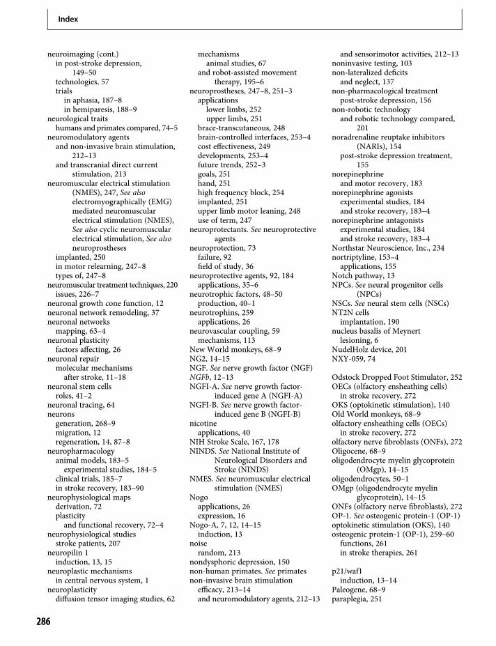
neuroimaging (cont.)in post-stroke depression,
149–50technologies, 57trialsin aphasia, 187–8in hemiparesis, 188–9
neurological traitshumans and primates compared, 74–5
neuromodulatory agentsand non-invasive brain stimulation,
212–13and transcranial direct current
stimulation, 213neuromuscular electrical stimulation
(NMES), 247, See alsoelectromyographically (EMG)mediated neuromuscularelectrical stimulation (NMES),See also cyclic neuromuscularelectrical stimulation, See alsoneuroprostheses
implanted, 250in motor relearning, 247–8types of, 247–8
neuromuscular treatment techniques, 220issues, 226–7
neuronal growth cone function, 12neuronal network remodeling, 37neuronal networksmapping, 63–4
neuronal plasticityfactors affecting, 26
neuronal repairmolecular mechanismsafter stroke, 11–18
neuronal stem cellsroles, 41–2
neuronal tracing, 64neuronsgeneration, 268–9migration, 12regeneration, 14, 87–8
neuropharmacologyanimal models, 183–5experimental studies, 184–5
clinical trials, 185–7in stroke recovery, 183–90
neurophysiological mapsderivation, 72plasticityand functional recovery, 72–4
neurophysiological studiesstroke patients, 207
neuropilin 1induction, 13, 15
neuroplastic mechanismsin central nervous system, 1
neuroplasticitydiffusion tensor imaging studies, 62
mechanismsanimal studies, 67
and robot-assisted movementtherapy, 195–6
neuroprostheses, 247–8, 251–3applications
lower limbs, 252upper limbs, 251
brace-transcutaneous, 248brain-controlled interfaces, 253–4cost effectiveness, 249developments, 253–4future trends, 252–3goals, 251hand, 251high frequency block, 254implanted, 251upper limb motor leaning, 248use of term, 247
neuroprotectants. See neuroprotectiveagents
neuroprotection, 73failure, 92field of study, 36
neuroprotective agents, 92, 184applications, 35–6
neurotrophic factors, 48–50production, 40–1
neurotrophins, 259applications, 26
neurovascular coupling, 59mechanisms, 113
New World monkeys, 68–9NG2, 14–15NGF. See nerve growth factor (NGF)NGFb, 12–13NGFI-A. See nerve growth factor-
induced gene A (NGFI-A)NGFI-B. See nerve growth factor-
induced gene B (NGFI-B)nicotineapplications, 40
NIH Stroke Scale, 167, 178NINDS. See National Institute of
Neurological Disorders andStroke (NINDS)
NMES. See neuromuscular electricalstimulation (NMES)
Nogoapplications, 26expression, 16
Nogo-A, 7, 12, 14–15induction, 13
noiserandom, 213
nondysphoric depression, 150non-human primates. See primatesnon-invasive brain stimulationefficacy, 213–14and neuromodulatory agents, 212–13
and sensorimotor activities, 212–13noninvasive testing, 103non-lateralized deficitsand neglect, 137
non-pharmacological treatmentpost-stroke depression, 156
non-robotic technologyand robotic technology compared,
201noradrenaline reuptake inhibitors
(NARIs), 154post-stroke depression treatment,
155norepinephrineand motor recovery, 183
norepinephrine agonistsexperimental studies, 184and stroke recovery, 183–4
norepinephrine antagonistsexperimental studies, 184and stroke recovery, 183–4
Northstar Neuroscience, Inc., 234nortriptyline, 153–4applications, 155
Notch pathway, 13NPCs. See neural progenitor cells
(NPCs)NSCs. See neural stem cells (NSCs)NT2N cellsimplantation, 190
nucleus basalis of Meynertlesioning, 6
NudelHolz device, 201NXY-059, 74
Odstock Dropped Foot Stimulator, 252OECs (olfactory ensheathing cells)in stroke recovery, 272
OKS (optokinetic stimulation), 140Old World monkeys, 68–9olfactory ensheathing cells (OECs)in stroke recovery, 272
olfactory nerve fibroblasts (ONFs), 272Oligocene, 68–9oligodendrocyte myelin glycoprotein
(OMgp), 14–15oligodendrocytes, 50–1OMgp (oligodendrocyte myelin
glycoprotein), 14–15ONFs (olfactory nerve fibroblasts), 272OP-1. See osteogenic protein-1 (OP-1)optokinetic stimulation (OKS), 140osteogenic protein-1 (OP-1), 259–60functions, 261in stroke therapies, 261
p21/waf1induction, 13–14
Paleogene, 68–9paraplegia, 251
Index
286
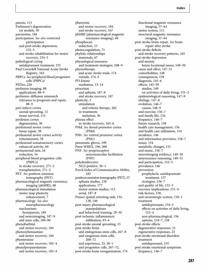
paresis, 113Parkinson’s degenerationrat models, 30
paroxetine, 184participation. See also restricted
participationand post-stroke depression,
152–3and stroke rehabilitation for motor
recovery, 234–5pathological cryingantidepressant treatment, 155
Paul Coverdell National Acute StrokeRegistry, 163
PBPCs. See peripheral blood progenitorcells (PBPCs)
PEDro, 220perfusion imaging, 88applications, 88–9
perfusion–diffusion mismatchrelevance to prognosis and repair,
88–9peri-infarct cortexaxonal sprouting, 12tissue survival, 115
perilesion cortexdegeneration, 30
perilesional motor cortextissue repair, 58
perilesional motor cortex activityreinstatement, 58
perilesional somatosensory cortexenhanced activity, 60
perineuronal nets, 16reduction, 16
peripheral blood progenitor cells(PBPCs)
in stroke recovery, 271–2transplantation, 271–2
PET. See positron emissiontomography (PET)
pharmacological magnetic resonanceimaging (phMRI), 60
pharmacological stimulationmotor map plasticity
enhancement, 7pharmacology. See also
neuropharmacologymechanismshemiparesis, 189
and neuroimaging, 187–9and stem cells, 189–90
phenobarbitaland motor recovery, 184
phenoxybenzamineand motor recovery, 184
phentermineand motor recovery, 183–4
phenylpropanolamineand motor recovery, 183–4
phenytoinand motor recovery, 184and stroke recovery, 167
phMRI (pharmacological magneticresonance imaging), 60
phosphocaninduction, 15
photocoagulation, 71phyletic relationshipsmammals, 67
physiological measuresand treatment strategies, 108–9
physiotherapyand acute stroke trials, 174outside, 174–5
PI3 kinasemediation, 13–14
piracetamand aphasia, 187–8and stroke recovery, 187
plasticity, 3stimulation
and robotic therapy, 202in treatments
initiation, 36plateau effectin stroke recovery, 165–6
PMd. See dorsal premotor cortex(PMd)
PMv. See ventral premotor cortex(PMv)
pneumatic gloves, 199Pneu-WREX, 196, 200PNF. See proprioceptive
neuromuscular facilitation(PNF)
polydendrocytesNG2-positive, 50–1
Porch Index of Communicative Ability,185
positron emission tomography (PET), 57aphasia studies, 128applications, 177motor system studies, 115serial, 187–8
Posner spatial orienting task, 134,138, 140
post-injury pharmacologicalmanipulations
and behavioral training, 29–30post-ischemic inflammatory
infiltration, 93–4post-stroke axonal sprouting, 15post-stroke brainand endogenous stem cells, 267–8and exogenous stem cells,
268–72and experience, 23, 30–1and progenitor cells, 267–72,
post-stroke brain reorganization, 174
functional magnetic resonanceimaging, 57–64
motor system, 115structural magnetic resonance
imaging, 57–64post-stroke brain repair. See brain
repair after strokepost-stroke deficitsand stroke recovery patterns, 165
post-stroke depressionand anxietylesion locational issues, 149–50
cause and effect, 147–51comorbidities, 148consequences, 158diagnosis, 145–6effects, 145–59studies, 149on activities of daily living, 151–2
epidemiological reasoning, 147–9etiology, 147–8evolution, 146–7causes, 148–9
and exercise, 156–7and family life, 154frequency, 146–7future research, 158health care management, 156and health care utilization, 154incidence, 146and information provision, 156–7issues, 154metabolic changes, 151and music, 156–7neuroimaging evidence, 149–50neuroscience reasoning, 149–51and participation, 152–3prevalence, 146preventionprophylactic antidepressanttreatment, 157
strategies, 156–7and quality of life, 152–3recovery implications, 151–4risk factors, 158,and serotonergic system, 150–1treatmentantidepressants, 154–5effects on activities of daily living,153–4
non-pharmacological, 156options, 154–7, 158
post-stroke effectsdegenerative responses, 23regenerative responses, 23
post-stroke emotional disorderstreatmentantidepressant, 155
post-stroke emotional symptomsfrequency, 146–7
Index
287

post-stroke fatigueetiology, 147–8frequency, 147risk factors, 148studies, 155
post-stroke functional improvementsstudiesmotor map plasticity as modelfor, 1
post-stroke interventionsprimate studies, 74
post-stroke mortalityand antidepressant treatment, 153–4
post-stroke motor improvementmotor map plasticity, 1–8neural strategies for, 3–6
post-stroke motor learning, 235–6action observation, 237–8auditory stimuli, 237cognitive approaches, 236–41and feedback, 240–1information/instruction provision,
236–40verbal information, 236–7
post-stroke neurogenesis, 16–17induction, 12and inflammation, 17and inflammatory cytokine/
chemokine signaling, 17occurrence, 17
post-stroke neuronal eventsbehavioral influences, 23–31
post-stroke neuronal sproutinggrowth inhibitory molecular
programs, 14–15growth promoting molecular
programs, 13–14ischemic critical period, 16
post-stroke personality changes, 147post-stroke recoveryavailability, 38and brain reorganization, 116–17cause and effects, 158mechanisms, 116from neglect, 140predicting, 119–20treatment, 158upper limb function, 113, 117
post-stroke recovery researchrodents, 35
post-stroke recovery therapies,end point measures, 38–9age factors, 36–7in animals, 35–44astrocytes, 42cell-based, 41–3electrical stimulation, 43enrichment issues, 40experience issues, 40gender differences, 38
hormonal status, 38intensity issues, 35–6issues, 35–40measurement issues, 38–40organismal factors, 36–8pharmacotherapy, 40–1plasticity, 36sex status, 38timing issues, 35–6and vascular system, 42–3
post-stroke rehabilitationof movement, 195
post-stroke shoulder pain, 249–51electrical stimulation
future trends, 250–1intramuscular systems,249–50
surface systems, 249theoretical issues, 249
prazosinand motor recovery, 184
pre-clinical stroke researchold animals, 81young animals, 81,
premotor cortex, 115–16prenatal infarct, 115pre-stroke fatiguerisk factors, 148
primary motor cortex, 115–16BOLD responses, 113–14functional organization, 6functional topography
and motor training, 72–3functions, 1infarctions, 5lesions, 5movement representations, 1premotor areas, 72primates, 72
primary sensorimotor cortexdamage, 120ischemic damage prevention, 60
primate brainscorticospinal neurons, 70, 72corticospinal tract, 69–70injury, 69size, 69stroke recovery research, 69–70white matter–gray matter ratios, 69
primates. See also monkeysas animal models, 69
issues, 69anthropoid, 68–9aspiration lesions, 70–1cladograms, 68encephalization of function, 70evolution, 67–9focal electrocoagulation, 71–2grasp recovery, 70and humans compared, 70, 74–5
middle cerebral artery occlusion, 71motor cortex organization, 72post-stroke intervention studies, 74primary motor cortex, 72secondary motor areas, 115–16spasticity, 70strepsirrhine, 68–9stroke models, 70–2stroke recoverycomparative perspectives, 67–75
stroke recovery studies, 67–70practical and ethical issues, 69
progenitor cells. See also endothelialprogenitor cells (EPCs), See alsoperipheral blood progenitorcells (PBPCs)
in adult brain after stroke, 267–72,factors affecting, 267–8
prognosisand perfusion–diffusion mismatch,
88–9proprioceptive neuromuscular
facilitation (PNF)and stroke outcomes, 221treatment–outcome relationships, 221
propriospinal projections, 116protease activityroles, 17upregulation, 16
proton magnetic resonancespectroscopy, 151
psychoactive drugsand stroke therapies, 81–2
psychomotor stimulantsapplications, 40
pyramidal neuronsdendritic morphology, 48
pyrexia, 167
quality of life (QoL)factors affecting, 53and post-stroke depression, 152–3
raceand stroke recovery, 166
random effects analysis, 115random noise, 213range of motion (ROM)lateral, 249
rat modelsParkinson’s degeneration, 30
ratsacrobatic tasks, 28–9, 49brain imaging, 62brain maps, 63cortical stimulation studies, 29–30cortical transplants, 51and environmental enrichment, 47exerciseforced vs. voluntary, 79
Index
288
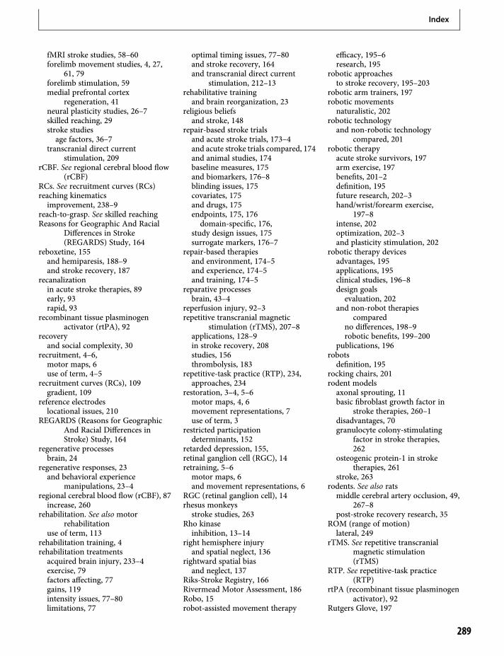
fMRI stroke studies, 58–60forelimb movement studies, 4, 27,
61, 79forelimb stimulation, 59medial prefrontal cortex
regeneration, 41neural plasticity studies, 26–7skilled reaching, 29stroke studiesage factors, 36–7
transcranial direct currentstimulation, 209
rCBF. See regional cerebral blood flow(rCBF)
RCs. See recruitment curves (RCs)reaching kinematicsimprovement, 238–9
reach-to-grasp. See skilled reachingReasons for Geographic And Racial
Differences in Stroke(REGARDS) Study, 164
reboxetine, 155and hemiparesis, 188–9and stroke recovery, 187
recanalizationin acute stroke therapies, 89early, 93rapid, 93
recombinant tissue plasminogenactivator (rtPA), 92
recoveryand social complexity, 30
recruitment, 4–6,motor maps, 6use of term, 4–5
recruitment curves (RCs), 109gradient, 109
reference electrodeslocational issues, 210
REGARDS (Reasons for GeographicAnd Racial Differences inStroke) Study, 164
regenerative processesbrain, 24
regenerative responses, 23and behavioral experience
manipulations, 23–4regional cerebral blood flow (rCBF), 87increase, 260
rehabilitation. See also motorrehabilitation
use of term, 113rehabilitation training, 4rehabilitation treatmentsacquired brain injury, 233–4exercise, 79factors affecting, 77gains, 119intensity issues, 77–80limitations, 77
optimal timing issues, 77–80and stroke recovery, 164and transcranial direct current
stimulation, 212–13rehabilitative trainingand brain reorganization, 23
religious beliefsand stroke, 148
repair-based stroke trialsand acute stroke trials, 173–4and acute stroke trials compared, 174and animal studies, 174baseline measures, 175and biomarkers, 176–8blinding issues, 175covariates, 175and drugs, 175endpoints, 175, 176
domain-specific, 176,study design issues, 175surrogate markers, 176–7
repair-based therapiesand environment, 174–5and experience, 174–5and training, 174–5
reparative processesbrain, 43–4
reperfusion injury, 92–3repetitive transcranial magnetic
stimulation (rTMS), 207–8applications, 128–9in stroke recovery, 208studies, 156thrombolysis, 183
repetitive-task practice (RTP), 234,approaches, 234
restoration, 3–4, 5–6motor maps, 4, 6movement representations, 7use of term, 3
restricted participationdeterminants, 152
retarded depression, 155,retinal ganglion cell (RGC), 14retraining, 5–6motor maps, 6and movement representations, 6
RGC (retinal ganglion cell), 14rhesus monkeysstroke studies, 263
Rho kinaseinhibition, 13–14
right hemisphere injuryand spatial neglect, 136
rightward spatial biasand neglect, 137
Riks-Stroke Registry, 166Rivermead Motor Assessment, 186Robo, 15robot-assisted movement therapy
efficacy, 195–6research, 195
robotic approachesto stroke recovery, 195–203
robotic arm trainers, 197robotic movementsnaturalistic, 202
robotic technologyand non-robotic technology
compared, 201robotic therapyacute stroke survivors, 197arm exercise, 197benefits, 201–2definition, 195future research, 202–3hand/wrist/forearm exercise,
197–8intense, 202optimization, 202–3and plasticity stimulation, 202
robotic therapy devicesadvantages, 195applications, 195clinical studies, 196–8design goalsevaluation, 202
and non-robot therapiescompared
no differences, 198–9robotic benefits, 199–200
publications, 196robotsdefinition, 195
rocking chairs, 201rodent modelsaxonal sprouting, 11basic fibroblast growth factor in
stroke therapies, 260–1disadvantages, 70granulocyte colony-stimulating
factor in stroke therapies,262
osteogenic protein-1 in stroketherapies, 261
stroke, 263rodents. See also ratsmiddle cerebral artery occlusion, 49,
267–8post-stroke recovery research, 35
ROM (range of motion)lateral, 249
rTMS. See repetitive transcranialmagnetic stimulation(rTMS)
RTP. See repetitive-task practice(RTP)
rtPA (recombinant tissue plasminogenactivator), 92
Rutgers Glove, 197
Index
289
SADQ (Stroke Aphasic DepressionQuestionnaire), 146
saliency maps, 133Scandinavian Stroke Scale, 153SCF (stem cell factor), 267–8SCG10, 13–14, 15SCILT (Spinal Cord Injury Locomotor
Trial), 222–3SCLIP, 13–14SDF-1 (stromal-cell-derived factor 1),
17, 269secondary motor areasprimates, 115–16projections, 116
seizuresand stroke, 37–8
selection, 133selective attentionuse of term, 133
selective serotonin reuptake inhibitors(SSRIs), 151, 154
applications, 37post-stroke depression treatment,
155and stroke recovery, 186–7chronic dosage, 186pending studies, 186–7single dosage, 186
self-efficacy, 228self-esteemand depression, 148
self-taught behavioral changeand brain reorganization, 26–7
semaphorin 3ainduction, 15,
Sensorimotor Active RehabilitationTraining (SMART) Arm, 201
sensorimotor activitiesand non-invasive brain stimulation,
212–13serotonergic pathwaystudies, 188
serotonergic systemand post-stroke depression, 150–1
serotoninroles, 189
sertraline, 157SGZ (subgranular zone), 267short interval intracortical inhibition
(SICI), 106, 110contralesional hemisphere studies,
107spatial distribution, 106–7
shoulder pain. See also post-strokeshoulder pain
hemiplegic, 250SICI. See short interval intracortical
inhibition (SICI)signal-to-noise ratio (SNR)reduced, 114–15
sildenafil, 62, 267–8silent perioduse of term, 95
skilled reachinganimal models, 29improvements, 29
skills, 227–8SLF (superior longitudinal fasciculus)lesions, 135
Slit, 15SMA. See supplementary motor area
(SMA)SMART (Sensorimotor Active
Rehabilitation Training) Arm,201
SNR (signal-to-noise ratio)reduced, 114–15
social complexityand recovery, 30
socio-economic factorsand stroke recovery, 168
somatosensory evoked potentials, 95somatosensory mapsand environmental enrichment, 48
spasticityafter cortical injury, 70primates, 70
spatial attentiondefinition, 133
spatial neglect, 133–4, See alsohemispatial neglect
functional mapping studies, 136–9models, 137and right hemisphere injury, 136tests, 134
spatial orientingbiases, 139
Spaulding, S. J., 234speechspontaneous, 129–30
Spinal Cord Injury Locomotor Trial(SCILT), 222–3
SPIO (superparamagnetic iron oxide)particles, 190
spiroperidoland neglect recovery, 184
SPRR1, 12, 13SPRR1aactivation, 14
squirrel monkeyscorticospinal neuron terminations,
70evolution, 69forelimb movement studies, 26motor training, 72–3primary motor cortex infarctions, 5,
74skilled reaching, 29
SSRIs. See selective serotonin reuptakeinhibitors (SSRIs)
STAIR consensus conference, 74Stat3, 14statins, 267–8stem cell factor (SCF), 267–8stem cells. See also neural stem cells
(NSCs), See also hematopoieticstem cells (HSCs)
adipose tissue-derived, 272characterization, 268definition, 267factors affecting, 267–8and pharmacology, 189–90tagging, 190trials, 189–90
stimulation targetsdefining, 208
stimulus–response mapping, 133strepsirrhine primates, 68–9strokeacute period, 87age factors, 36–7and anger, 146–7and anxiety, 146and behavioral impairments, 1and brain ischemia, 87classification, 89comorbidities, 37–8, 81and stroke recovery, 167–8
damage quantification, 103–5and dementia, 149and emotional disorders, 145epidemiological studiestypes of, 163–4
incidence, 77, 173mechanisms, 35modeling, 38molecular mechanisms of neuronal
repair, 11–18nature ofand acute treatment, 167
prevalence, 173and religious beliefs, 148risk factorsdepression, 147
rodent models, 263and seizures, 37–8types of, 89–92pathogenesis, 90
and vascular dementia, 52Stroke Aphasic Depression
Questionnaire (SADQ), 146stroke evolutiondeterminants, 88–9
stroke heterogeneityanimal–human translation issues,
80–1stroke modelsprimates, 70–2
stroke outcomesand Bobath approach, 222
Index
290

and Brunnstrom approach, 221models, 169and proprioceptive neuromuscular
facilitation, 221stroke pathophysiologyimaging technologies, 57
stroke patientscontralesionalmotor cortex activity, 5emotional symptoms, 145neurophysiological studies, 207survival issues, 81
stroke recoveryand activation patterns, 95affected hemisphere, 105–7bone marrow stromal cells in, 269–71cellular approaches, 267–73cognitive approaches, 233–42contralesional hemisphere, 107electrical stimulation, 247–54future trends, 254
electromagnetic approaches, 207–14epidemiological researchimportance of, 168–9
epidemiology, 163–9definition, 163
epidural cortical stimulation, 208–9intensive physical therapeutic
approaches, 219–29interhemispheric interactions, 107–8neuropharmacology in, 183–90outcomes, 169patterns, 164–6and post-stroke deficits, 165
plateau effect, 165–6predictors, 168–9,primate brain studies, 69–70primate studies, 67–70practical and ethical issues, 69
primatescomparative perspectives, 67–75
and rehabilitation treatments, 164repetitive transcranial magnetic
stimulation, 208robotic approaches, 195–203specific factors, 166–8integration, 168
statistics, 164therapeutic options, 267time factors, 164–5tissue-specific neural stem cells in,
268–9transcranial direct current
stimulation, 209–11transcranial magnetic stimulation
studies, 105–8stroke recovery researchanimal–human translation issues,
77–82stroke recovery-promoting agents, 259stroke registries
epidemiological studies, 163stroke rehabilitationapproaches, 108early commencement, 95–6and intensity, 219–20motor learning strategies, 236
implications, 236for motor recovery
active participation, 234–5requirements, 52
stroke survivorsimpairment variations, 38medications, 37
stroke therapies. See also acute stroketherapies
approved, 173dosage issues, 165drug issues, 81–2growth factors, 259–63selection issues, 82timing issues, 87
stroke unitsadvantages, 53
stromal-cell-derived factor 1 (SDF-1),17, 269
structural magnetic resonance imagingof post-stroke brain reorganization,
57–64procedures, 60–4
structural plasticity, 11subcortical aphasia, 125–6subgranular zone (SGZ), 267subventricular zone (SVZ), 12, 267, 270astrocytes, 50cell proliferation, 16–17, 268–9proliferating cells, 50
Sunnybrook Stroke Study, 167superior longitudinal fasciculus (SLF)lesions, 135
superparamagnetic iron oxide (SPIO)particles, 190
supplementary motor area (SMA), 72,115–16
hand representations, 73–4surface peroneal nerve stimulation, 252SVZ. See subventricular zone (SVZ)Swedenpost-stroke depression studies,
146, 148Riks-Stroke Registry, 166
synapsesgrowth, 27
synaptic changesand injuries, 24–5
synaptic efficacychanges, 6
synaptic plasticity, 7, 103animal studies, 94and motor cortex reorganization, 6signaling pathways, 7
synaptic potentiationand motor skill learning, 6
synaptic strengthchanges, 6
synaptogenesis, 268and motor map plasticity, 6
task parameter modulationand task-related activities, 118
task-related activitiesand task parameter modulation, 118
task-specific training, 229TBI (traumatic brain injury), 233tDCS. See transcranial direct cortical
stimulation (tDCS)temporo-parietal junction (TPJ) cortex,
136, 140damage, 135
tenascin, 14–15tetraplegia, 251neuroprostheses, 251,brain-controlled interfaces, 253
TGFb pathway, 13TGFb1, 12–13TGF-β (transforming growth factor-β),
259therapeutic electricityconcept of, 207historical background, 207
thrombolysis, 87, 91, 92in acute stroke therapies, 88
Tie2, 268time-dependent post-stroke processesexperience sensitive, 23–6
tissue losscompensation strategies, 37
TMS. See transcranial magneticstimulation (TMS)
TOAST Study, 167total map area, 109–10total map volume, 109–10tPAmechanisms, 16
TPJ cortex. See temporo-parietaljunction (TPJ) cortex
trainingbilateral, 202focused on impairments, 28–9and repair-based therapies,
174–5task-specific, 229timing factors, 29
transcallosal disinhibition, 107transcallosal projectionsdegeneration, 27
transcranial direct current stimulation(tDCS)
functional magnetic resonanceimaging studies, 213
setup, 210
Index
291
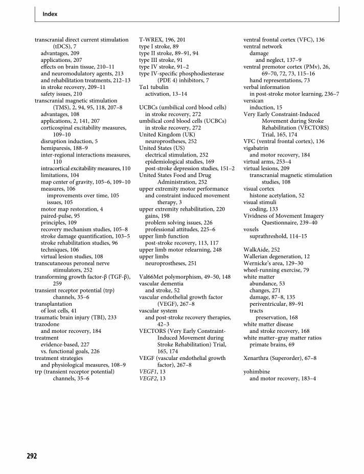
transcranial direct current stimulation(tDCS), 7
advantages, 209applications, 207effects on brain tissue, 210–11and neuromodulatory agents, 213and rehabilitation treatments, 212–13in stroke recovery, 209–11safety issues, 210
transcranial magnetic stimulation(TMS), 2, 94, 95, 118, 207–8
advantages, 108applications, 2, 141, 207corticospinal excitability measures,
109–10disruption induction, 5hemiparesis, 188–9inter-regional interactions measures,
110intracortical excitabilitymeasures, 110limitations, 104map center of gravity, 105–6, 109–10measures, 106improvements over time, 105issues, 105
motor map restoration, 4paired-pulse, 95principles, 109recovery mechanism studies, 105–8stroke damage quantification, 103–5stroke rehabilitation studies, 96techniques, 106virtual lesion studies, 108
transcutaneous peroneal nervestimulators, 252
transforming growth factor-β (TGF-β),259
transient receptor potential (trp)channels, 35–6
transplantationof lost cells, 41
traumatic brain injury (TBI), 233trazodoneand motor recovery, 184
treatmentevidence-based, 227vs. functional goals, 226
treatment strategiesand physiological measures, 108–9
trp (transient receptor potential)channels, 35–6
T-WREX, 196, 201type I stroke, 89type II stroke, 89–91, 94type III stroke, 91type IV stroke, 91–2type IV-specific phosphodiesterase
(PDE 4) inhibitors, 7Tα1 tubulinactivation, 13–14
UCBCs (umbilical cord blood cells)in stroke recovery, 272
umbilical cord blood cells (UCBCs)in stroke recovery, 272
United Kingdom (UK)neuroprostheses, 252
United States (US)electrical stimulation, 252epidemiological studies, 169post-stroke depression studies, 151–2
United States Food and DrugAdministration, 252
upper extremity motor performanceand constraint induced movement
therapy, 3upper extremity rehabilitation, 220gains, 198problem solving issues, 226professional attitudes, 225–6
upper limb functionpost-stroke recovery, 113, 117
upper limb motor relearning, 248upper limbsneuroprostheses, 251
Val66Met polymorphism, 49–50, 148vascular dementiaand stroke, 52
vascular endothelial growth factor(VEGF), 267–8
vascular systemand post-stroke recovery therapies,
42–3VECTORS (Very Early Constraint-
Induced Movement duringStroke Rehabilitation) Trial,165, 174
VEGF (vascular endothelial growthfactor), 267–8
VEGF1, 13VEGF2, 13
ventral frontal cortex (VFC), 136ventral networkdamageand neglect, 137–9
ventral premotor cortex (PMv), 26,69–70, 72, 73, 115–16
hand representations, 73verbal informationin post-stroke motor learning, 236–7
versicaninduction, 15
Very Early Constraint-InducedMovement during StrokeRehabilitation (VECTORS)Trial, 165, 174
VFC (ventral frontal cortex), 136vigabatrinand motor recovery, 184
virtual arms, 253–4virtual lesions, 209transcranial magnetic stimulation
studies, 108visual cortexhistone acetylation, 52
visual stimulicoding, 133
Vividness of Movement ImageryQuestionnaire, 239–40
voxelssuprathreshold, 114–15
WalkAide, 252Wallerian degeneration, 12Wernicke’s area, 129–30wheel-running exercise, 79white matterabundance, 53changes, 271damage, 87–8, 135periventricular, 89–91tractspreservation, 168
white matter diseaseand stroke recovery, 168
white matter–gray matter ratiosprimate brains, 69
Xenarthra (Superorder), 67–8
yohimbineand motor recovery, 183–4
Index
292

0 50 100 150 (s)
–5
0
5
10
15
–5
0
5
10
15
–5
0
5
10
15
Δ C
BV
(%
of
bas
elin
e)
fMRI during forelimb stimulation in rats
14 days post-stroke
3 days post-stroke
–log P 135
control
Figure 6.1 Left: T2-weighted MR images of arat brain slice overlaid by statistical activationmaps calculated from contrast-enhancedCBV-weighted fMRI in combination with anelectrical forelimb stimulation paradigm asdescribed by Dijkhuizen et al. [17]. Right: Time-course of CBV changes in the rightsensorimotor cortex in response to leftforelimb stimulation (the stimulation period isindicated by the black bar) (mean ± SD, n = 6).Left forelimb stimulation induced significantactivation responses in the right sensorimotorcortex in control rats. At 3 days after right-sided stroke, activation responses in the right,ipsilesional sensorimotor cortex were largelyabsent; however, responses were found in theleft, contralesional hemisphere. After 14 days,partial restoration of activation was detectedin the right, ipsilesional sensorimotor cortex.The unilateral stroke lesion is characterized byincreased T2-weighted signal intensity in theright somatosensory cortex and lateralstriatum.
Figure 1.2 Intracortical microstimulation (ICMS) technique used to derivemaps of movement representation in rat motor cortex.A. Motormapshowing areas of the cortex in which stimulation evokedmovement. B. Movements are produced by lowering a stimulating electrode into layerV of the motor cortex and activating corticospinal neurons. C. Corticospinal neurons are activated trans-synaptically. These neurons then projectto motor neurons in the spinal cord that in turn cause muscle contractions in the periphery.
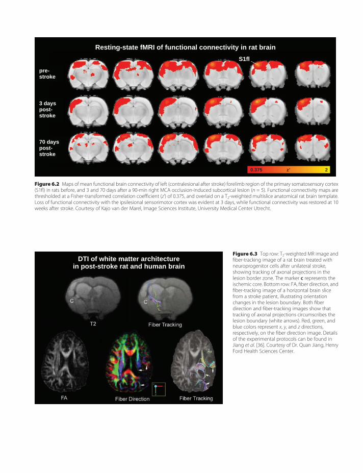
pre-stroke
3 dayspost-stroke
70 dayspost-stroke
S1fl
z’0.375 2
Resting-state fMRI of functional connectivity in rat brain
Figure 6.2 Maps of mean functional brain connectivity of left (contralesional after stroke) forelimb region of the primary somatosensory cortex(S1fl) in rats before, and 3 and 70 days after a 90-min right MCA occlusion-induced subcortical lesion (n = 5). Functional connectivity maps arethresholded at a Fisher-transformed correlation coefficient (z’) of 0.375, and overlaid on a T2-weighted multislice anatomical rat brain template.Loss of functional connectivity with the ipsilesional sensorimotor cortex was evident at 3 days, while functional connectivity was restored at 10weeks after stroke. Courtesy of Kajo van der Marel, Image Sciences Institute, University Medical Center Utrecht.
DTI of white matter architecturein post-stroke rat and human brain
Figure 6.3 Top row: T2-weighted MR image andfiber-tracking image of a rat brain treated withneuroprogenitor cells after unilateral stroke,showing tracking of axonal projections in thelesion border zone. The marker c represents theischemic core. Bottom row: FA, fiber direction, andfiber-tracking image of a horizontal brain slicefrom a stroke patient, illustrating orientationchanges in the lesion boundary. Both fiberdirection and fiber-tracking images show thattracking of axonal projections circumscribes thelesion boundary (white arrows). Red, green, andblue colors represent x, y, and z directions,respectively, on the fiber direction image. Detailsof the experimental protocols can be found inJiang et al. [36]. Courtesy of Dr. Quan Jiang, HenryFord Health Sciences Center.
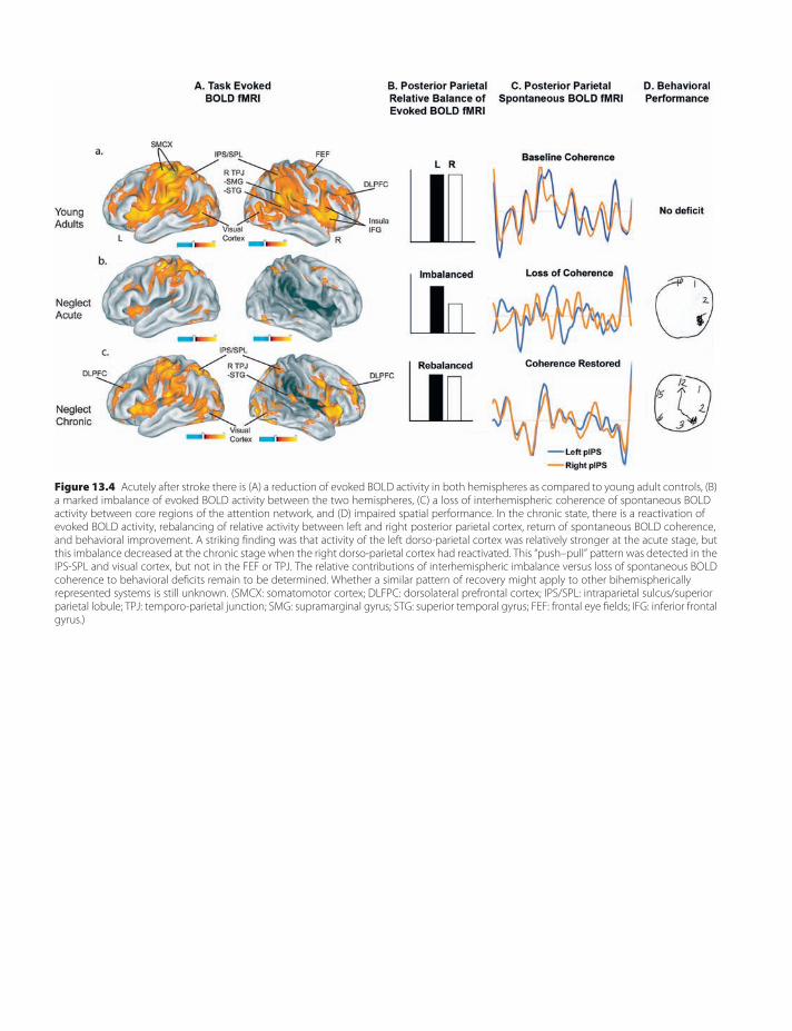
Figure 13.4 Acutely after stroke there is (A) a reduction of evoked BOLD activity in both hemispheres as compared to young adult controls, (B)a marked imbalance of evoked BOLD activity between the two hemispheres, (C) a loss of interhemispheric coherence of spontaneous BOLDactivity between core regions of the attention network, and (D) impaired spatial performance. In the chronic state, there is a reactivation ofevoked BOLD activity, rebalancing of relative activity between left and right posterior parietal cortex, return of spontaneous BOLD coherence,and behavioral improvement. A striking finding was that activity of the left dorso-parietal cortex was relatively stronger at the acute stage, butthis imbalance decreased at the chronic stage when the right dorso-parietal cortex had reactivated. This “push–pull” pattern was detected in theIPS-SPL and visual cortex, but not in the FEF or TPJ. The relative contributions of interhemispheric imbalance versus loss of spontaneous BOLDcoherence to behavioral deficits remain to be determined. Whether a similar pattern of recovery might apply to other bihemisphericallyrepresented systems is still unknown. (SMCX: somatomotor cortex; DLFPC: dorsolateral prefrontal cortex; IPS/SPL: intraparietal sulcus/superiorparietal lobule; TPJ: temporo-parietal junction; SMG: supramarginal gyrus; STG: superior temporal gyrus; FEF: frontal eye fields; IFG: inferior frontalgyrus.)
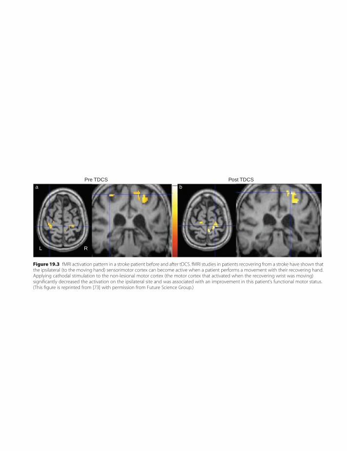
Pre TDCSa
L R
bPost TDCS
Figure 19.3 fMRI activation pattern in a stroke patient before and after tDCS. fMRI studies in patients recovering from a stroke have shown thatthe ipsilateral (to the moving hand) sensorimotor cortex can become active when a patient performs a movement with their recovering hand.Applying cathodal stimulation to the non-lesional motor cortex (the motor cortex that activated when the recovering wrist was moving)significantly decreased the activation on the ipsilateral site and was associated with an improvement in this patient’s functional motor status.(This figure is reprinted from [73] with permission from Future Science Group.)