Monitoring of percolation water to discriminate surficial inputs in a karst aquifer
Brain Magnetic Resonance Imaging White Matter Lesions AreFrequent in HTLV-I Carriers and Do Not Discriminate from HAM/TSP
DANIEL J. MORGAN1, MARINA F. CASKEY1, CRISTIANE ABBEHUSEN2, JAMARYOLIVEIRA-FILHO3, CESAR ARAUJO2, AURELIA F. PORTO3, SILVANE BRAGA SANTOS3,GLORIA O. ORGE3, MARIA JOSE JOIA3, ANDRE L. MUNIZ3, ISADORA SIQUEIRA3,MARSHALL J. GLESBY1, and EDGAR CARVALHO3
1 Department of International Medicine and Infectious Disease, Weill Medical College of Cornell University,New York, New York 10021
2 Image Memorial Radiology Clinic, Salvador, Bahia, Brazil
3 Universidade Federal da Bahia, Salvador, Bahia, Brazil
AbstractHuman T lymphotropic virus (HTLV)-I is known to cause HTLV-I-associated myelopathy/tropicalspastic paraparesis (HAM/TSP) and other pronounced disease in less than 4% of those infected.However, evidence is accumulating that a proportion of HTLV-I carriers have neurological andurological symptoms without fulfilling criteria for HAM/TSP. Brain white matter (WM) lesions onmagnetic resonance imaging (MRI) are frequently seen in HAM/TSP. HTLV-I carriers with MRIscans for other neurological diagnoses have WM lesions more frequently than expected. We studied10 patients with HAM/TSP and 20 HTLV-I carriers without overt neurological disease and evaluatedclinical characteristics, viral load, total, small, large, confluent WM lesion number, and lesion volumeon MRI. Cerebral WM lesions were found in of 85% of HTLV-I carriers and 80% of HAM/TSPpatients. Lesion number, size or location was no different between carriers and HAM/TSP. Cognitivefunction was lower in HAM/TSP (p = 0.045) but did not correlate with WM lesion number. Viralload and peripheral blood mononuclear cell interferon-γ production correlated positively (p = 0.001)but did not correlate with lesion number or volume. Conventional brain MRI frequently shows WMlesions in HTLV-I-infected individuals suggesting potential early central nervous systeminflammation with rare development of progressive disease.
INTRODUCTIONHuman T lymphotrophic virus (HTLV)-I is a retrovirus that infects 10–20 million peopleworldwide.1 The majority of those infected are carriers, with fewer than 4% developing HTLV-I-associated myelopathy/tropical spastic paraparesis (HAM/TSP),2 acute T cell lymphomaleukemia (ATLL),3 or infective dermatitis.4 Recent reports have documented urinary,neurological, and inflammatory symptoms, similar to those seen in HAM/TSP but milder innature, among the 95% of HTLV-I-infected individuals traditionally considered carriers.5–7Laboratory findings that generally distinguish between populations of HTLV-I carriers andthose with HAM/TSP also overlap among a subset of carriers. HAM/TSP has been associatedin general with a higher proviral load than carriers and increased production of inflammatory
Address reprint requests to: Edgar M. Carvalho, Servico de Immunologia, Universidade Federal de Bahia, Salvador, Bahia, Brazil, E-mail: [email protected].
NIH Public AccessAuthor ManuscriptAIDS Res Hum Retroviruses. Author manuscript; available in PMC 2008 December 3.
Published in final edited form as:AIDS Res Hum Retroviruses. 2007 December ; 23(12): 1499–1504. doi:10.1089/aid.2007.0077.
NIH
-PA Author Manuscript
NIH
-PA Author Manuscript
NIH
-PA Author Manuscript

cytokines in peripheral blood mononuclear cells (PBMC).8,9 High proviral load and interferon-γ (IFN-γ) production, however, are found in a small number of HTLV-I carriers at levels similarto those seen in HAM/TSP.9
Magnetic resonance imaging (MRI) has been proposed as a tool to aid in the diagnosis of HAM/TSP,10 to follow the response to therapy,11 or to distinguish HAM/TSP from multiple sclerosis(MS).12,13 Although the typical clinical presentation of HAM/TSP involves lower extremityparalysis and urological symptoms, which localize to a lesion in the thoracic spine,corresponding MRI lesions are seen in a minority of patients.10,13,14 The incidence of spinalcord atrophy varies from 6% to 74%15,16 as no validated radiological criteria exist. In contrast,small white matter (WM) lesions are frequently seen in subcortical and periventricular areasin patients with HAM/TSP10–21 at a higher frequency than seen in noninflammatoryneurological diseases.11,21 The quantity and distribution of small WM lesions do notcorrespond to symptoms.10,11,15 The duration or grade of HAM/TSP has been shown tocorrelate with MRI changes by some11 but not others.10
The limited number of series that included HTLV-I carriers who underwent MRI for otherneurological diseases has shown a frequency of WM lesions in HTLV-I carriers similar to thatseen in patients with HAM/TSP.11,13 No studies have examined HTLV-I carriers withoutindications for MRI. Moreover, no studies have tried to correlate immunological measures withWM lesions. We performed brain MRIs on 10 patients with HAM/TSP and 20 age/sex-matchedHTLV-I carriers from an endemic area without other neurological diseases.
MATERIALS AND METHODSThe study was performed in Salvador, Brazil, a city of 2.5 million inhabitants in NortheasternBrazil. The population is primarily of African/Portuguese descent and is characterized bymarked socioeconomic disparities.22
HAM/TSP and HTLV-I carriers were recruited from the HTLV-I multidisciplinary clinic atthe Hospital Universitário Prof. Edgard Santos in Salvador. Patients are referred to the clinicfrom blood banks as well as dermatology or neurology clinics. Diagnosis of HTLV-I was madeby enzyme-linked immunosorbent assay (ELISA) (Cambridge Biotech Corp., Worcester, MA)with confirmation by Western blot (HTLV blot 2.4, Genelab, Singapore). HAM/TSP wasdiagnosed by neurological examination using Kurtzke’s Expanded Disability Status Scale(EDSS)23 and Osame’s Motor Disability Score (OMDS) in accordance with WHO criteria.24 Anti-HTLV-I antibodies were documented in cerebrospinal fluid (CSF) and based onclinical and radiological findings patients did not have other diseases to explain theirneurological symptoms.
A convenience sample of patients with confirmed HAM/TSP between the ages of 18 and 65years was identified. HAM/TSP patients were matched by age and sex with control seropositiveHTLV-I carriers from the clinic population selected without regard to symptoms orcomorbidities beyond exclusion for diabetes, known cerebrovascular disease, HIV, or activesyphilis. Every HAM/TSP patient was matched with two HTLV-I carriers. HTLV-I carrierswere selected from 103 carriers who had completed PBMC cultures with IFN-γ determinationin a descriptive study of the clinic population. Our cross-sectional study was originallydesigned, in part, to compare carriers based on IFN-γ production. Consequently, carriers werestratified by IFN-γ production into two groups of >1500 pg/ml or <500 pg/ml. Within thesestrata, one high IFN-γ and one low IFN-γ control per HAM/TSP subject matched on sex andage ±5 years was selected. Since preliminary analyses revealed no differences in MRI findingsin the two strata, the groups were combined to form a group of 20 HTLV-I carriers. All HAM/TSP patients and HTLV-I carriers were submitted to a full general physical and neurological
MORGAN et al. Page 2
AIDS Res Hum Retroviruses. Author manuscript; available in PMC 2008 December 3.
NIH
-PA Author Manuscript
NIH
-PA Author Manuscript
NIH
-PA Author Manuscript
examination as well as completing an extensive questionnaire of neurological, urological,rheumatological, and odontological symptoms as described previously.5
The study was reviewed and approved by the Institutional Review Boards of the Oswaldo CruzFoundation and the Weill Medical College of Cornell University. Informed consent wasobtained from all subjects enrolled in the study.
Mononuclear cell separation and determination of IFN-γPBMCs were obtained from heparinized venous blood by density gradient centrifugation usinglymphocyte separation media (LSM; Organon Tecnika Corporation, Durham, NC). Afterwashing in saline, the cells were adjusted to 3 × 10⁶ ml in RPMI-1640 (GIBCO, Grand Island,NY) containing 100 U penicillin G and 10 μ/ml of streptomycin and supplemented with 10%AB serum. All cultures were incubated without stimulus at 37°C in 5% CO2 for 72 h.Supernatants were collected and stored at −20°C. IFN-γ was measured by commercial ELISAfollowing the manufacturer’s instructions (PharMingen).
Proviral loadThe HTLV-I proviral load in PBMC was quantified using a real-time TaqMan polymerasechain reaction (PCR) method.25 Briefly, SK110/SK111 primers were used to amplify a 186fragment of the pol gene and the dual-labeled TaqMan probe (5′-FAM and 3′-TAMRA) locatedat 4829–4858 bp of the HTLV-I reference sequence (HTLVATK). Albumin DNA wasquantified in parallel to determine the input cell number as an endogenous reference. BothHTLV-I and albumin gene amplifications were performed on 10 μl of DNA extract using Taq-MAn PCR core reagent kit (Applied Biosystems). Amplification and data acquisition werecarried out using the ABI Prism 7700 Sequence detector system (Applied Biosystems).Standard curves were generated using 10-fold serial dilution of a double standard plasmid(pcHTLV-ALB). The HTLV-I-infected human lymphocyte line MT2 was used as a control forquantification. All standard dilution, controls, and patients samples were run in duplicate. Thenormalized value of the HTLV-I proviral load was calculated as the ratio of (HTLV-I DNAaverage copy number/albumin DNA average copy number) × 2 × 10⁶ and expressed as thenumber of HTLV-I copies per 106 PBMCs.
Magnetic resonance imagingMR imaging was performed on a 1.5-T whole body scanner (Siemens, Erlangen, Germany).
The acquisition MR imaging protocol included a sagittal localizer followed by axial T1 [spin-echo (SE) echo time (TE) = 16 msec, repetition time (TR) = 65 msec], T2 [fast spin-echo time(FSE) = 102 msec, TR = 42 msec], and fluid-attenuated inversion recovery (FLAIR) (TE =141 msec, TR = 10,002 sec, TI = 2200 msec) sequences, all with 20 contiguous 5-mm-thicksections with 256 × 192 pixels across a 24-cm field of view (FOV). Examinations wereevaluated by a neurologist (J.O.F.) and a neuroradiologist (C.A.) independently withoutknowledge of the disease state of an individual with consensus achieved on number of lesionswhen initial disagreement occurred. A lesion was considered present if its size was >3 mm.Small lesions were 3–6 mm, large lesions >6 mm in largest dimension, and confluent lesionswere irregular in shape and >6 mm. Brain atrophy was quantified by visual inspection asdescribed by Victoroff et al. 26 Briefly, three brain regions (frontal, parietal, and temporallobes) were rated from zero (no atrophy) to three (severe atrophy) and the total score was thesum of the three regions (possible score range zero to nine). Total lesion volume was calculatedfrom the summed cross-sectional area of FLAIR hyperintense lesions using ImageJ software.
MORGAN et al. Page 3
AIDS Res Hum Retroviruses. Author manuscript; available in PMC 2008 December 3.
NIH
-PA Author Manuscript
NIH
-PA Author Manuscript
NIH
-PA Author Manuscript
Statistical analysisDifferences between carriers and HAM/TSP patients were calculated using the Mann–WhitneyU-test (Wilcoxon rank sum test) or the Fishers exact test. Correlations were performed withSpearman’s rank test. All calculations were performed using Stata 7 software (StataCorporation, College Station, TX). A p value <0.05 was considered significant.
RESULTSTwelve patients with HAM/TSP were contacted from the HTLV-I clinic. One was excludedfor diabetes and one for stroke. Twenty-one matched carriers were identified, one of whomwas excluded for possible latent syphilis. Patients were 70% female and their median age was47 years (HAM/TSP) and 49 years (controls). The median duration of HAM/TSP was 3 years(range 2–20 years). The median EDSS was 4 (range 2–8) in HAM/TSP and 0 (range 0–1) incontrols. The median OMDS score was 4 (range 3–11) in HAM/TSP and 0 in controls (bydefinition). Hyperreflexia was present in 100% of HAM/TSP and 15% (3/20) of carriers.Hypertension was present in 3/10 (30%) of HAM/TSP and 5/20 (25%) of HTLV-I carriers. NoHAM/TSP patients and 1/20 (5%) of HTLV-I carriers reported smoking tobacco. No patientsin either group had known cardiovascular disease.
The median log of proviral load was 4.69 log10 copies/106 PBMCs (range 0–5.52) in HAM/TSP and 4.81 log10 copies/106 PBMCs (range 1.07–5.52) in carriers, which was not statisticallydifferent (p = 0.89).
Urinary incontinence was reported by 50% (5/10) of HAM/TSP and 20% (4/20) of HTLV-Icarriers. Difficulty walking was reported by 90% (9/10) of HAM/TSP and 5% (1/20) of HTLV-I carriers. Leg weakness was reported by 80% (8/10) of HAM/TSP and 20% (4/20) of HTLV-I carriers. Dry eyes and dry mouth were reported by 40% (4/10) of HAM/TSP and 20% (4/20)of HTLV-I carriers. Overall, 55% (11/20) of HTLV-I carriers had at least one of theaforementioned symptoms.
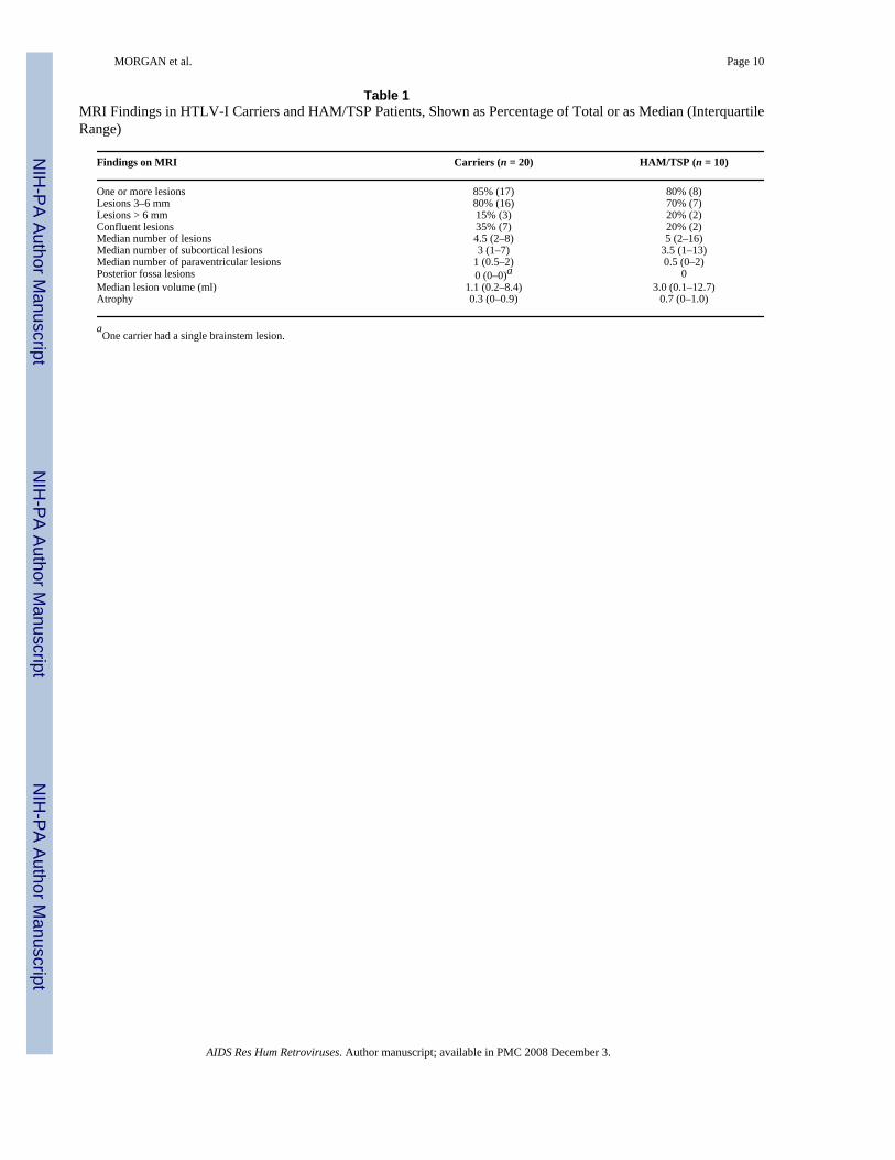
Table 1 displays MRI findings by group. The median number of lesions was 5 (Interquartilerange (IQR) 2–16) in HAM/TSP and 4.5 (IQR 2–8) in HTLV-I carriers (p = 0.64). The medianlesion volume was 3.0 ml (IQR 1.0–12.7 ml) in HAM/TSP and 1.0 ml (IQR 0.2–8.4 ml) incarriers (p = 0.49). No statistical difference between HTLV-I carriers and HAM/TSP was seenwith respect to any variable presented in Table 1. An example of a WM lesion is presented inFig. 1.
The number of WM lesions seen in carriers and HAM/TSP patients is shown in Fig. 2. Lesionnumber and volume did not correlate with EDSS or OMDS or duration of HAM/TSP. Themedian IFN-γ was 27 pg/ml (range 0–435) in 10 low IFN-γ carriers, 2971 pg/ml (range 1936–4980) in 10 high IFN-γ carriers, and 2329 pg/ml (range 306–4075) in HAM/TSP. No differencein lesion number was seen between high and low IFN-γ groups (p = 0.88). No correlationbetween IFN-γ and WM lesion number or volume was observed. No association was foundbetween the presence of any symptoms and IFN-γ level (p = 0.19). No correlation between thepresence of any symptoms and number of WM lesions was found. Proviral load did notcorrelate with EDSS (p = 0.77), OMDS (p = 0.91), total lesion number (p = 0.96), or lesionvolume (p = 0.48). Proviral load significantly correlated with PBMC IFN-γ production (p =0.001, r2 = 0.42). The Mini-Mental State Exam score was significantly lower in HAM/TSP,25, than in HTLV-I carriers, 28 (p = 0.045) and did not correlate with lesion number or volume(p = 0.71 and p = 0.92). Lesion number was positively correlated with age for all HTLV-I-infected patients (R2 = 0.25, p = 0.005).
MORGAN et al. Page 4
AIDS Res Hum Retroviruses. Author manuscript; available in PMC 2008 December 3.
NIH
-PA Author Manuscript
NIH
-PA Author Manuscript
NIH
-PA Author Manuscript
DISCUSSIONIn the first study to compare HAM/TSP to HTLV-I carriers who did not undergo MRI for otherneurological reasons, we found a high rate of WM lesions in carriers. The distribution andquantity of WM lesions were not different between groups. No correlations with clinicalfindings, including EDSS or OMDS scores, were found.
WM lesions in HTLV-I have been postulated as evidence of a small vessel vasculitis.11 SmallWM lesions have been observed more frequently in HTLV-I infection than in seronegativepatients with other noninflammatory neurological diseases.11,21 As others have described,11,13 our results confirm that a large number of both HAM/TSP and HTLV-I carriers haveWM lesions. The frequency appears higher than that reported in healthy adult volunteers.27In one study by Fazekas, WM lesions were found in 37% of healthy volunteers <70 years ofage,27 which would suggest that the rate of WM lesions in HTLV-infected patients in our studywas higher than normal. Some have found more lesions in HAM/TSP than carriers. In onestudy, when >10 WM lesions was used as a definition, 38% of HAM/TSP patients as comparedto 10% of HTLV-I carriers were identified.11 We found no difference with this cutoff in ourpopulation. Our study was limited by not including HTLV-I-negative controls although fewWM lesions are expected in nonelderly subjects without neurological complaints. We found acorrelation between age and number of WM lesions, which have been described in a HTLV-I-seronegative population.27 Prior studies of HTLV-I have not found this correlation.11,21
The clinical significance of cerebral WM lesions has also been debated. Cerebral WM lesionsare the most common changes in HAM/TSP, occurring in 33–100% of patients,10–21 althoughWM lesion number does not correlate with symptoms. The high frequency of WM lesionsobserved in our study is likely due to highly sensitive modern MRI techniques as well as apossible bias to identify lesions that may be overlooked in retrospective reviews of MRIsobtained for clinical purposes, as ours was a prospective research protocol. Although a segmentof HTLV-I carriers reported symptoms such as incontinence (20%) and lower extremityweakness (20%) or were found to have hyperreflexia (15%), these findings did not correlatewith the presence or number of WM lesions. There was also no correlation between IFN-γlevels and WM lesions. There is a known association between proviral load and HAM/TSP.8 In this study we found no correlation between WM lesion number or volume and proviralload. We did, however, find a significant correlation between IFN-γ level and proviral load.The possibility exists that there is a less robust association between these and WM lesions thatwas not seen because of the small sample in this study.
Among our patients, two with advanced HAM/TSP (both EDSS 8) had diffuse WM changesthat were more severe than MRIs of any HTLV-I carriers. Others have found large lesions tobe predictive of MS rather than HAM/TSP in the appropriate clinical setting.12,13 Both ofthese patients had definite clinical HAM/TSP, with no evidence of cognitive or other cerebraldysfunction.
In summary, our prospective, cross-sectional study using HTLV-I carriers without otherneurological disease demonstrates that cerebral WM lesions occur in a large percentage of allHTLV-I-infected individuals. A large proportion of HTLV-I carriers has neurological andurological manifestations similar to those observed in HAM/TSP.5,6,7,28 Recently proposedcriteria for the diagnosis of HAM/TSP add urological symptoms and neurological symptomsto OMDS and EDSS measures.29 It is apparent that morbidity associated with HTLV-Iinfection is higher than has been previously reported. Our findings, that cerebral WM lesionsoccur in the majority of HTLV-I carriers, suggest that neurological damage with or withouttranslation to symptoms is documented in the majority of HTLV-I carriers. Longitudinal
MORGAN et al. Page 5
AIDS Res Hum Retroviruses. Author manuscript; available in PMC 2008 December 3.
NIH
-PA Author Manuscript
NIH
-PA Author Manuscript
NIH
-PA Author Manuscript
studies are needed to delineate the natural history of cerebral MRI abnormalities in thispopulation.
AcknowledgementsThis work was supported by the National Institutes of Health (NIH), 1 RO3-AI60830 and T32–AI07613 (traininggrant to D.J.M. and M.F.C.), Brazilian National Research Council (CNPq), and Fundação de Amparo à Pesquisa doEstado da Bahia (FAPESB). E.M.C. is a senior investigator of CNPq.
References1. De The G, Bomford R. An HTLV-I vaccine: Why how, for whom? AIDS Res Hum Retroviruses
1993;9:381–386. [PubMed: 8318266]2. Orland JR, Engstrom J, Fridey J, et al. Prevalence and clinical features of HTLV neurologic disease
in the HTLV outcomes study. Neurology 2003;61:1588–1594. [PubMed: 14663047]3. Hinuma Y, Nagata K, Hanaoka M, et al. Adult T-cell leukemia: Antigen in an ATL cell line and
detection of antibodies to the antigen in human sera. Proc Natl Acad Sci USA 1981;78(10):6476–6480.[PubMed: 7031654]
4. LaGrenade L, Hanchard B, Fletcher V, Cranston B, Blattner W. Infective dermatitis of Jamaicanchildren: A marker for HTLV-I infection. Lancet 1990;336(8727):1345–1347. [PubMed: 1978165]
5. Caskey MF, Morgan DJ, Porto MAF, Giozza SP, Muniz AL, Orge MG, Travassos MJ, Baron Y,Carvalho EM, Glesby MJ. Clinical manifestations associated with HTLV type I infection: A cross-sectional study. AIDS Res Hum Retroviruses 2007;23(3):365–371. [PubMed: 17411369]
6. Zunt JR, Alacron JO, Montano S, Longstreth WT Jr, Price R, Holmes KK. Quantitative assessment ofsubclinical spasticity in human T-cell lymphotropic virus type I infection. Neurology 1999;53(2):386–390. [PubMed: 10430431]
7. Castro N, Oliveira P, Junior WR, Muniz A, Carvalho E. Erectile dysfunction and HTLV-I infection:A silent problem. Int J Impot Res 2005;17:364–369. [PubMed: 15875060]
8. Nagai M, Usuku K, Matsumoto W, et al. Analysis of HTLV-I proviral load in 202 HAM/TSP patientsand 243 asymptomatic HTLV-I carriers: High proviral load strongly predisposes to HAM/TSP. JNeurovirol 1998;4:586–593. [PubMed: 10065900]
9. Santos SB, Porto AF, Muniz AL, et al. Exacerbated inflammatory cellular immune responsecharacteristic of HAM/TSP is observed in a large proportion of HTLV-I asymptomatic carriers. BMCInfect Dis 2004;2:4–7.
10. Bagnato F, Butman JA, Mora CA, et al. Conventional magnetic resonance imaging features in patientswith tropical spastic paraparesis. J Neurovirol 2005;11:525–534. [PubMed: 16338746]
11. Kira J, Fujihara K, Itoyama Y, et al. Leukoencephalopathy in HTLV-I associated myelopathy/tropicalspastic paraparesis: MRI analysis and a two year follow-up study after corticosteroid therapy. JNeurol Sci 1991;106:41–49. [PubMed: 1779238]
12. Kuroda Y, Matsui M, Yukitake M, et al. Assessment of MRI criteria for MS in Japanese MS andHAM/TSP. Neurology 1995;45:30–33. [PubMed: 7824129]
13. Howard AK, Li DK, Oger J. MRI contributes to the differentiation between MS and HTLV-Iassociated myelopathy in British Columbian coastal natives. Can J Neurol Sci 2003;30:41–48.[PubMed: 12619783]
14. Gessain A, Gout O. Chronic myelopathy associated with human T-lymphotropic virus type I (HTLV-I). Ann Intern Med 1992;117:933–946. [PubMed: 1443956]
15. Ferraz AC, Gabbai AA, Abdala N, Nogueira RG. Magnetic resonance in HTLV-1 associatedmyelopathy [Portuguese]. Arq Neuropsiquiatr 1997;55(4):728–736. [PubMed: 9629331]
16. Milagres AC, Jorge ML, Marchiori PE, Segurado AA. Human T cell lymphotropic virus type 1-associated myelopathy in Sao Paulo, Brazil. Epidemiologic and clinical features of a universityhospital cohort. Neuroepidemiology 2002;21(3):153–158. [PubMed: 12006779]
17. Alcindor F, Valderrama R, Canavaggio M, Lee H, et al. Imaging of human T-lymphotropic virus typeI-associated chronic progressive myeloneuropathies. Neuroradiology 1992;35(1):69–74. [PubMed:1289743]
MORGAN et al. Page 6
AIDS Res Hum Retroviruses. Author manuscript; available in PMC 2008 December 3.
NIH
-PA Author Manuscript
NIH
-PA Author Manuscript
NIH
-PA Author Manuscript
18. Melo A, Gomes I, Mattos K. HTLV-1 associated myelopathies in the city of Salvador, Bahia[Portuguese]. Arq Neuropsiquiatr 1994;52(3):320–325. [PubMed: 7893204]
19. Rudge P, Ali A, Cruichshank JK. Multiple sclerosis, tropical spastic paraparesis and HTLV-1infection in Afro-Caribbean patients in the United Kingdom. J Neurol Neurosurg Psychiatry 1991;54(8):689–694. [PubMed: 1940939]
20. Godoy AJ, Kira J, Hasou K, Goto I. Characterization of cerebral white matter lesions of HTLV-I-associated myelopathy/tropical spastic paraparesis in comparison with multiple sclerosis andcollagen-vasculitis: A semiquantitative MRI study. J Neurol Sci 1995;133:102–111. [PubMed:8583211]
21. Kira J, Minato S, Itoyama Y, Goto I, Kato M, Hasuo K. Leukoencephalopathy in HTLV-I-associatedmyelopathy: MRI and EEG data. J Neurol Sci 1988:221–232. [PubMed: 3210034]
22. Azevedo ES, Fortuna CM, Santos MG, et al. Spread and diversity of human populations in Bahia,Brazil. Hum Biol 1982;54:329–341. [PubMed: 7095799]
23. Kurtzke JF. Rating neurologic impairment in multiple sclerosis: An expanded disability status scale(EDSS). Neurology 1983;33:1444–1452. [PubMed: 6685237]
24. Osame, M. Review of WHO Kagoshima meeting and diagnostic guidelines for HAM/TSP. In:Blattner, W., editor. Human Retrovirology: HTLV. Raven Press; New York: 1990. p. 191-197.
25. Dehee A, Cesaire R, Desire N, et al. Quantitation of HTLV-I proviral load by a TaqMan real-timePCR assay. J Virol Methods 2002;102:37–51. [PubMed: 11879691]
26. Victoroff J, Mack WJ, Grafton ST, Schreiber SS, Chui HC. A method to improve interrater reliabilityof visual inspection of brain MRI. Neurology 1994;44(12):2267–2276. [PubMed: 7991111]
27. Fazekas F. Magnetic resonance signal abnormalities in asymptomatic individuals: Their incidenceand functional correlates. Eur Neurol 1989;29:164–168. [PubMed: 2731564]
28. Castro NM, Freitas DM, Junior WR, Muniz A, Oliveira P, Carvalho E. Urodynamic features of voidingdysfunction in HTLV-I infected individuals. Int Braz J Urol 2007;33(2):238–244. [PubMed:17488545]discussion 244–245
29. Castro-Costa CM, Ara jo AQC, Barreto MM, et al. Proposal for diagnostic criteria of tropical spasticparaparesis/HTLV-I- associated myelopathy (TSP/HAM). AIDS Res Hum Retroviruses 2006;22(10):931–935. [PubMed: 17067261]
MORGAN et al. Page 7
AIDS Res Hum Retroviruses. Author manuscript; available in PMC 2008 December 3.
NIH
-PA Author Manuscript
NIH
-PA Author Manuscript
NIH
-PA Author Manuscript
FIG. 1.FLAIR-enhanced image showing confluent lesions seen in some HTLV-I-infected patients(arrows indicate lesions, which extend from periventricular to subcortical locations).
MORGAN et al. Page 8
AIDS Res Hum Retroviruses. Author manuscript; available in PMC 2008 December 3.
NIH
-PA Author Manuscript
NIH
-PA Author Manuscript
NIH
-PA Author Manuscript
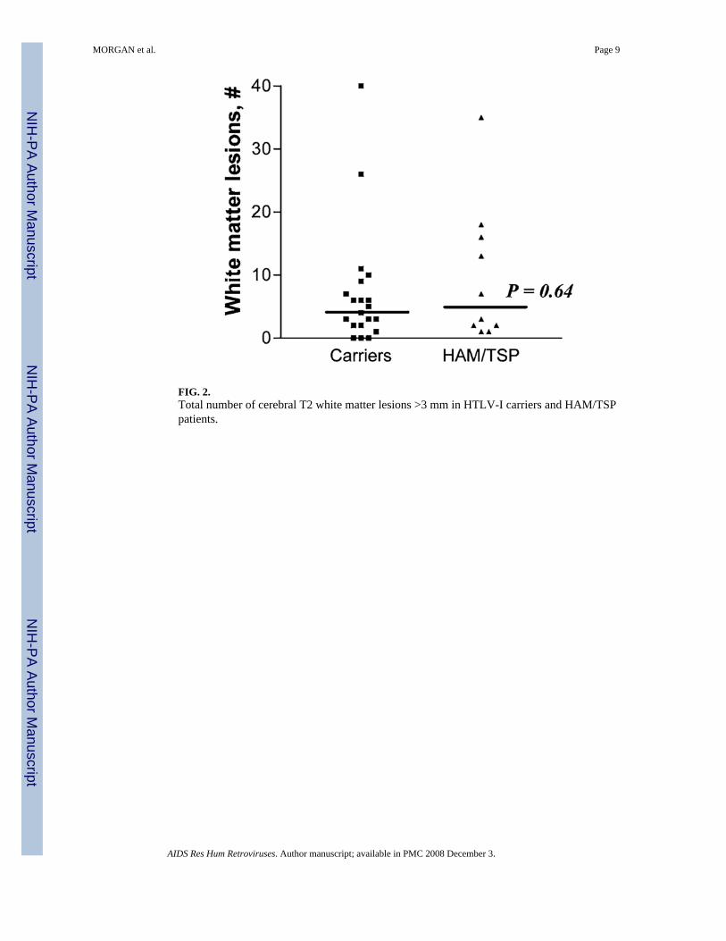
FIG. 2.Total number of cerebral T2 white matter lesions >3 mm in HTLV-I carriers and HAM/TSPpatients.
MORGAN et al. Page 9
AIDS Res Hum Retroviruses. Author manuscript; available in PMC 2008 December 3.
NIH
-PA Author Manuscript
NIH
-PA Author Manuscript
NIH
-PA Author Manuscript
NIH
-PA Author Manuscript
NIH
-PA Author Manuscript
NIH
-PA Author Manuscript
MORGAN et al. Page 10
Table 1MRI Findings in HTLV-I Carriers and HAM/TSP Patients, Shown as Percentage of Total or as Median (InterquartileRange)
Findings on MRI Carriers (n = 20) HAM/TSP (n = 10)
One or more lesions 85% (17) 80% (8)Lesions 3–6 mm 80% (16) 70% (7)Lesions > 6 mm 15% (3) 20% (2)Confluent lesions 35% (7) 20% (2)Median number of lesions 4.5 (2–8) 5 (2–16)Median number of subcortical lesions 3 (1–7) 3.5 (1–13)Median number of paraventricular lesions 1 (0.5–2) 0.5 (0–2)Posterior fossa lesions 0 (0–0)a 0Median lesion volume (ml) 1.1 (0.2–8.4) 3.0 (0.1–12.7)Atrophy 0.3 (0–0.9) 0.7 (0–1.0)
aOne carrier had a single brainstem lesion.
AIDS Res Hum Retroviruses. Author manuscript; available in PMC 2008 December 3.
Copyright © 2022 FDOKUMEN