Multivariate Activation and Connectivity Patterns Discriminate ...
12
Multivariate Activation and Connectivity Patterns Discriminate Speech Intelligibility in Wernicke’s, Broca’s, and Geschwind’s Areas Daniel A. Abrams 1 , Srikanth Ryali 1 , Tianwen Chen 1 , Evan Balaban 4 , Daniel J. Levitin 4 and Vinod Menon 1,2,3 1 Department of Psychiatry and Behavioral Sciences, 2 Program in Neuroscience and 3 Department of Neurology and Neurological Sciences, Stanford University School of Medicine, Stanford, CA, USA and 4 Department of Psychology, McGill University, Montreal, QC, Canada Address correspondence to Daniel A. Abrams and Vinod Menon, Department of Psychiatry and Behavioral Sciences, Stanford University School of Medicine, 401 Quarry Rd., Stanford, CA 94305-5719, USA. Email: [email protected] (Daniel A. Abrams) and [email protected] (Vinod Menon). The brain network underlying speech comprehension is usually de- scribed as encompassing fronto–temporal–parietal regions while neuroimaging studies of speech intelligibility have focused on a more spatially restricted network dominated by the superior tem- poral cortex. Here we use functional magnetic resonance imaging with a novel whole-brain multivariate pattern analysis (MVPA) to more fully characterize neural responses and connectivity to intelli- gible speech. Consistent with previous univariate findings, intelligi- ble speech elicited greater activity in bilateral superior temporal cortex relative to unintelligible speech. However, MVPA identified a more extensive network that discriminated between intelligible and unintelligible speech, including left-hemisphere middle temporal gyrus, angular gyrus, inferior temporal cortex, and inferior frontal gyrus pars triangularis. These fronto–temporal–parietal areas also showed greater functional connectivity during intelligible, compared with unintelligible, speech. Our results suggest that speech intelligi- bly is encoded by distinct fine-grained spatial representations and within-task connectivity, rather than differential engagement or dis- engagement of brain regions, and they provide a more complete view of the brain network serving speech comprehension. Our find- ings bridge a divide between neural models of speech comprehen- sion and the neuroimaging literature on speech intelligibility, and suggest that speech intelligibility relies on differential multivariate response and connectivity patterns in Wernicke’s, Broca’s, and Geschwind’s areas. Keywords: Angular gyrus, Auditory cortex, Broca’s area, Inferior frontal gyrus, Speech perception Introduction Studies investigating the neural basis for sentence-level speech comprehension have highlighted a broad array of brain structures. Classical accounts from neuropsychological evaluations in patients with focal lesions of the cortex have implicated left-hemisphere temporal (Wernicke 1874) and parietal regions (Geschwind 1970) associated with putative Wernicke’s and Geschwind’s areas for speech comprehension, while other neuropsychological studies have identified pre- frontal contributions associated with classically defined Broca’s area in the inferior frontal gyrus (IFG; Bates et al. 2003). Functional imaging studies in healthy adults have pro- vided corroborating evidence (Vigneau et al. 2006): left-hemisphere superior temporal cortex (Ben-Shachar et al. 2004) and IFG regions (Friederici et al. 2006) have been im- plicated in sentence-level syntactic analysis, whereas left inferior parietal cortex regions, most notably the angular gyrus (AG; Humphries et al. 2006) and Broca’s area as well as other subdivisions of the IFG (Rodd et al. 2005), have been implicated in sentence-level semantic processing. Collectively, a distributed left-hemisphere network involving the superior temporal, inferior parietal, and IFG have been implicated in sentence-level speech perception (Tyler and Marslen-Wilson 2008; Peelle, Johnsrude, et al. 2010; Price 2010). Neverthe- less, individual studies of speech processing, notably those in- volving speech intelligibility, have often diverged from these models. Distinguishing intelligible from unintelligible speech is an influential paradigm for investigating the neural correlates of auditory sentence comprehension. In this approach, brain responses elicited by intelligible speech are examined relative to acoustically matched “unintelligible” speech. Intelligible speech is hypothesized to recruit brain structures supporting phonology, word-form recognition, semantics, and syntax (Scott et al. 2000), while carefully controlled unintelligible speech retains many speech-like qualities (Azadpour and Balaban 2008), including frequency and amplitude modulations and formant-like features, but is not thought to provide direct access to linguistic or semantic representations in the brain. Imaging studies of speech intelligibility have yielded incon- sistent results. Table 1 summarizes the various speech and control stimuli used in this literature as well as the brain regions identified in these studies as a main effect of speech intelligibility. For example, in a seminal positron emission tomography study, results showed that left anterior superior temporal sulcus (aSTS) was sensitive to intelligible speech (Scott et al. 2000), a result which has been corroborated in subsequent functional magnetic resonance imaging (fMRI) studies (Narain et al. 2003; Obleser and Kotz 2010). Other speech intelligibility studies have variably identified additional left-hemisphere regions, including the IFG (Davis and Johnsrude 2003; Eisner et al. 2010; Okada et al. 2010; Davis et al. 2011; McGettigan, Faulkner, et al. 2012) and the AG of the inferior parietal lobe (Davis and Johnsrude 2003; Obleser et al. 2007). In one of these studies, it was shown that activity in left IFG, STS, and AG are correlated with increasing speech intelligibility (Davis and Johnsrude 2003). In contrast, a second study showed that the AG becomes active only during sentence comprehension when the speech signal is sufficiently degraded, and that, consistent with earlier reports (Scott et al. 2000; Narain et al. 2003), sentence processing under more favorable listening conditions is served predomi- nantly by superior temporal cortex (Obleser et al. 2007). It remains unclear why studies of speech intelligibility have failed to consistently identify main effects of intelligibility in a © The Author 2012. Published by Oxford University Press. All rights reserved. For Permissions, please e-mail: [email protected] Cerebral Cortex July 2013;23:1703–1714 doi:10.1093/cercor/bhs165 Advance Access publication June 12, 2012 at Stanford University on July 17, 2013 http://cercor.oxfordjournals.org/ Downloaded from
-
Upload
khangminh22 -
Category
Documents
-
view
0 -
download
0
Transcript of Multivariate Activation and Connectivity Patterns Discriminate ...

Multivariate Activation and Connectivity Patterns Discriminate Speech Intelligibilityin Wernicke’s, Broca’s, and Geschwind’s Areas
Daniel A. Abrams1, Srikanth Ryali1, Tianwen Chen1, Evan Balaban4, Daniel J. Levitin4 and Vinod Menon1,2,3
1Department of Psychiatry and Behavioral Sciences, 2Program in Neuroscience and 3Department of Neurology and NeurologicalSciences, Stanford University School of Medicine, Stanford, CA, USA and 4Department of Psychology, McGill University,Montreal, QC, Canada
Address correspondence to Daniel A. Abrams and Vinod Menon, Department of Psychiatry and Behavioral Sciences, Stanford UniversitySchool of Medicine, 401 Quarry Rd., Stanford, CA 94305-5719, USA. Email: [email protected] (Daniel A. Abrams) and [email protected](Vinod Menon).
The brain network underlying speech comprehension is usually de-scribed as encompassing fronto–temporal–parietal regions whileneuroimaging studies of speech intelligibility have focused on amore spatially restricted network dominated by the superior tem-poral cortex. Here we use functional magnetic resonance imagingwith a novel whole-brain multivariate pattern analysis (MVPA) tomore fully characterize neural responses and connectivity to intelli-gible speech. Consistent with previous univariate findings, intelligi-ble speech elicited greater activity in bilateral superior temporalcortex relative to unintelligible speech. However, MVPA identified amore extensive network that discriminated between intelligible andunintelligible speech, including left-hemisphere middle temporalgyrus, angular gyrus, inferior temporal cortex, and inferior frontalgyrus pars triangularis. These fronto–temporal–parietal areas alsoshowed greater functional connectivity during intelligible, comparedwith unintelligible, speech. Our results suggest that speech intelligi-bly is encoded by distinct fine-grained spatial representations andwithin-task connectivity, rather than differential engagement or dis-engagement of brain regions, and they provide a more completeview of the brain network serving speech comprehension. Our find-ings bridge a divide between neural models of speech comprehen-sion and the neuroimaging literature on speech intelligibility, andsuggest that speech intelligibility relies on differential multivariateresponse and connectivity patterns in Wernicke’s, Broca’s, andGeschwind’s areas.
Keywords: Angular gyrus, Auditory cortex, Broca’s area, Inferior frontalgyrus, Speech perception
Introduction
Studies investigating the neural basis for sentence-levelspeech comprehension have highlighted a broad array ofbrain structures. Classical accounts from neuropsychologicalevaluations in patients with focal lesions of the cortex haveimplicated left-hemisphere temporal (Wernicke 1874) andparietal regions (Geschwind 1970) associated with putativeWernicke’s and Geschwind’s areas for speech comprehension,while other neuropsychological studies have identified pre-frontal contributions associated with classically definedBroca’s area in the inferior frontal gyrus (IFG; Bates et al.2003). Functional imaging studies in healthy adults have pro-vided corroborating evidence (Vigneau et al. 2006):left-hemisphere superior temporal cortex (Ben-Shachar et al.2004) and IFG regions (Friederici et al. 2006) have been im-plicated in sentence-level syntactic analysis, whereas leftinferior parietal cortex regions, most notably the angular
gyrus (AG; Humphries et al. 2006) and Broca’s area as well asother subdivisions of the IFG (Rodd et al. 2005), have beenimplicated in sentence-level semantic processing. Collectively,a distributed left-hemisphere network involving the superiortemporal, inferior parietal, and IFG have been implicated insentence-level speech perception (Tyler and Marslen-Wilson2008; Peelle, Johnsrude, et al. 2010; Price 2010). Neverthe-less, individual studies of speech processing, notably those in-volving speech intelligibility, have often diverged from thesemodels.
Distinguishing intelligible from unintelligible speech is aninfluential paradigm for investigating the neural correlates ofauditory sentence comprehension. In this approach, brainresponses elicited by intelligible speech are examined relativeto acoustically matched “unintelligible” speech. Intelligiblespeech is hypothesized to recruit brain structures supportingphonology, word-form recognition, semantics, and syntax(Scott et al. 2000), while carefully controlled unintelligiblespeech retains many speech-like qualities (Azadpour andBalaban 2008), including frequency and amplitude modulationsand formant-like features, but is not thought to provide directaccess to linguistic or semantic representations in the brain.
Imaging studies of speech intelligibility have yielded incon-sistent results. Table 1 summarizes the various speech andcontrol stimuli used in this literature as well as the brainregions identified in these studies as a main effect of speechintelligibility. For example, in a seminal positron emissiontomography study, results showed that left anterior superiortemporal sulcus (aSTS) was sensitive to intelligible speech(Scott et al. 2000), a result which has been corroborated insubsequent functional magnetic resonance imaging (fMRI)studies (Narain et al. 2003; Obleser and Kotz 2010). Otherspeech intelligibility studies have variably identifiedadditional left-hemisphere regions, including the IFG (Davisand Johnsrude 2003; Eisner et al. 2010; Okada et al. 2010;Davis et al. 2011; McGettigan, Faulkner, et al. 2012) and theAG of the inferior parietal lobe (Davis and Johnsrude 2003;Obleser et al. 2007). In one of these studies, it was shown thatactivity in left IFG, STS, and AG are correlated with increasingspeech intelligibility (Davis and Johnsrude 2003). In contrast,a second study showed that the AG becomes active onlyduring sentence comprehension when the speech signal issufficiently degraded, and that, consistent with earlier reports(Scott et al. 2000; Narain et al. 2003), sentence processingunder more favorable listening conditions is served predomi-nantly by superior temporal cortex (Obleser et al. 2007).
It remains unclear why studies of speech intelligibility havefailed to consistently identify main effects of intelligibility in a
© The Author 2012. Published by Oxford University Press. All rights reserved.For Permissions, please e-mail: [email protected]
Cerebral Cortex July 2013;23:1703–1714doi:10.1093/cercor/bhs165Advance Access publication June 12, 2012
at Stanford University on July 17, 2013
http://cercor.oxfordjournals.org/D
ownloaded from

distributed temporal–frontal–parietal network that is widelyaccepted in current neural models of speech comprehension.While differences in experimental designs could potentiallyexplain this discrepancy, another possibility is that conven-tional univariate fMRI methods, which identify voxels in thebrain that have larger responses for one stimulus conditionrelative to another, are unable to distinguish responses in keynodes of the speech comprehension network. An alternativeapproach is multivariate pattern analysis (MVPA), whichidentifies spatial patterns of fMRI activity sufficient to dis-criminate between experimental conditions (Haynes and Rees2006; Norman et al. 2006; Kriegeskorte and Bandettini 2007).Univariate and MVPA techniques provide complementaryinformation regarding the neural substrates underlying cogni-tive processes (Schwarzlose et al. 2008). A plausible neuro-physiological basis for MVPA is that specific and consistentpatterns of neural activity measured across neuronal popu-lations may represent an essential spatial coding mechanism.Moreover, unlike differences in regional signal level, spatialpatterns of neural activity have the capacity to represent alarge number of stimulus attributes, categories, and cognitivestates (Haxby et al. 2001). In support of the utility of MVPA,previous studies of the visual system have shown that multi-variate patterns of activity within higher levels of the visualsystem (i.e. ventral temporal cortex) are more sensitive to cat-egory discrimination relative to univariate measures (Haxbyet al. 2001).
The vast majority of studies to date have used univariatemeasures to localize brain regions sensitive to intelligiblespeech. Only 2 studies have probed speech intelligibilityusing MVPA (Okada et al. 2010; McGettigan, Evans, et al.2012). Univariate results in these studies showed that mul-tiple regions of superior temporal and inferior frontal cortexhad greater activity for intelligible than unintelligible con-ditions, and MPVA results showed that bilateral anterior andposterior STG/STS could discriminate between intelligibleand unintelligible speech. However, in both studies, MVPAwas restricted to the temporal lobe (and an occipital lobecontrol region in one study: McGettigan, Evans, et al. 2012),and it is currently unknown what additional brain regionsdiscriminate between these speech conditions based onspatial patterns. Given that superior temporal cortex is theonly brain region that is consistently identified as showingsignal-level differences in univariate studies of speech intel-ligibility, it may be the case that regions beyond temporalcortex, including the IFG and AG, additionally reflect sensi-tivity to the intelligibility of speech based on consistentspatial patterns of regional activity.
Here, we use the fMRI data from 20 normal healthy adultsand applied a novel whole-brain MVPA to examine this ques-tion. We identified 2 goals for the analysis of these data. First,we used a searchlight MVPA (Kriegeskorte et al. 2006;Abrams et al. 2011) to discriminate responses to well-matchedintelligible and unintelligible speech conditions and
Table 1Summary of previous studies of speech intelligibility
Study Experimental stimuli Control stimuli Univariate analysis and main effect of intelligibility Multivariate analysis andintelligibility-based classification
Scott et al.(2000)
Normal and noise- vocoded sentences Spectrally rotated sentences andspectrally rotated/vocoded sentences
Subtraction analysis: Left anterior STS N/A
Narain et al.(2003)
Normal and noise-vocoded sentences Spectrally-rotated sentences andspectrally-rotated/vocoded sentences
Subtraction Analysis: Left posterior, mid, and anteriorSTS (non-contiguous clusters)
N/A
Davis andJohnsrude (2003)
Sentences that were distorted using 3different methods and 3 levels ofdistortion
Correlation Analysis: Bilateral MTG, Left hippocampus,Left posterior MTG, Left AG, Left IFG (BA 44), Left SFS,Left precuneus
N/A
Obleser et al.2007
Sentences of high/low predictability thatwere noise vocoded at 2, 8, 32 bands
Correlation Analysis: Bilateral STS, Bilateral precuneus,Left MTG, Right MFG, Left SMG
N/A
Obleser and Kotz(2010)
Sentences of high/low predictability thatwere noise vocoded at 1, 4, 16 bands
Correlation Analysis: Bilateral STS, Left AG N/A
Adank and Devlin(2010)
Time-compressed sentences Normal sentences Subtraction Analysis: Bilateral anterior and posterior STS/STG, Bilateral pre-SMA, Bilateral cingulate sulcus
N/A
Eisner et al.(2010)
“Learnable” noise- vocoded andspectrally-shifted sentences
Spectrally-inverted sentences Subtraction Analysis: Left IFG (BA 44), Left STS N/A
Okada et al.(2010)
Normal and noise –vocoded sentences Spectrally-rotated sentences andspectrally-rotated/vocoded sentences
Subtraction Analysis: Bilateral MTG, Bilateral fusiformgyrus, Left parahippocampal gyrus, Left SMG, Left IFG(BA 45), Left MTG, Right medial temporal lobe, Rightcerebellum, Right ITG
ROI-based MVPA: BilateralHeschl’s gyrus, Bilateral anteriorand posterior STS, Right mid STS
Davis et al.(2011)
Sentences with 9 levels ofsignal-correlated-noise
Correlation Analysis: Bilateral anterior and posterior MTG,Bilateral mid STG, Bilateral temporal pole, Left IFG (BAs44, 45, and 47), Right Heschl’s gyrus, Right posteriorSTG, Left putamen, Bilateral hippocampus, Rightamygdala, Left IC, Left fusiform, Left ITG
N/A
McGettigan,Faulkner, et al.(2012a)
High and low-predictability sentencesnoise-vocoded in 3 bands
Correlation Analysis: Bilateral anterior and posterior STS/STG, Left IFG (BAs 44 and 45), Right IFG (BA 47), Leftfusiform gyrus,
N/A
McGettigan,Evans, et al.(2012b)
Dynamic frequency and amplitudevariations in sine-wave speech takenfrom the same original sentence
Dynamic frequency and amplitudevariations in sine-wave sentencestaken from 2 different sentences
Subtraction Analysis: Bilateral STS, Left IFG (BA 47),Right IFG (BA 45), Left precentral gyrus
ROI-based MVPA: Bilateral STG/MTG, Right inferior occipital gyrus
1704 Distributed Multivariate Patterns for Speech • Abrams et al.
at Stanford University on July 17, 2013
http://cercor.oxfordjournals.org/D
ownloaded from

compared these results with the results from a conventionalunivariate analysis. Based on the extant literature, we pre-dicted that univariate analysis would reveal signal-level differ-ences in superior temporal cortex for intelligible versusunintelligible speech. Critically, we hypothesized that MVPAwould reveal distinct neural representations for these speechconditions in inferior prefrontal and parietal areas known tobe important for speech comprehension (Bates et al. 2003).The second goal of this work was to examine functional con-nectivity between brain regions identified using MVPA andhow they are modulated by the intelligibility of speech. Themotivation for this analysis is that it addresses whether the co-ordinated activity across brain regions identified with MVPAcharacterizes the processing of intelligible speech.
Materials and Methods
ParticipantsParticipants were 20 (11 males) right-handed Stanford Universityundergraduate and graduate students with no psychiatric or neuro-logical disorders, as assessed by self-report and the SCL-90-R (Deroga-tis 1992). All participants were native English speakers and werebetween the ages of 19 and 22 (mean age = 21.2 years). The partici-pants received $50 in compensation for participation. The StanfordUniversity School of Medicine Human Subjects committee approvedthe study, and informed consent was obtained from all participants.
StimuliSpeech stimuli were 23–27 s excerpts of familiar and unfamiliarspeeches (e.g. Martin Luther King, President Roosevelt) selected froma compilation of famous speeches of the 20th century (Various 1991).All speech stimuli were digitized at 22 050 Hz sampling rate with16-bit dynamic range. Consistent with previous reports (Scott et al.2000; Narain et al. 2003; Okada et al. 2010), we used natural speechsentences (Speech condition) as our intelligible speech condition andspectrally rotated speech stimuli (rSpeech condition; Blesser 1972) asour unintelligible speech condition. To perform spectral rotation onthe sentence stimuli, each speech file was low-pass filtered 5 times at2400 Hz (5-pole elliptic filter, slope 100 dB/octave, max attenuation60 dB), multiplied by a 2500 Hz sine wave, and low-pass filtered 5more times at 2400 Hz to prevent aliasing. The original signal and therotated signal were then each low-pass filtered at 1850 Hz, normalized(largest amplitude set to 1), amplified (largest amplitude set to 8 V)and outputted as wave files. The reason for low-pass filtering at thisrelatively low frequency (1850 Hz) is that the speech material wastaken from a commercial recording of great speeches spanning the20th century, and the originals (some of which had been recorded inthe 1940s) differed in their frequency range. The filtering at 1850 Hzwas carried out in order to restrict all of the excerpts to the same fre-quency range as the excerpt with the narrowest range, so that theywould be rendered acoustically comparable. As a further verification,the filtered versions were played for 5 native speakers unfamiliar withthe excerpts, and each of these individuals was able to accuratelyrepeat back the text of the speeches.
fMRI TaskSpeech and rSpeech stimuli were presented in 2 separate runs eachlasting ∼7 min; the order of runs was randomized across participants.One run consisted of 18 blocks of alternating Speech, temporally re-ordered Speech, and Rest. The other run consisted of 18 alternatingblocks of rSpeech, temporally reordered rSpeech, and Rest. Eachstimulus lasted 23–27 s. Data from temporally reordered stimuli wereoriginally included to compare temporal structure processing inspeech and music (Abrams et al. 2011) and are not presented heresince the goal of the current work study is to examine univariate andmultivariate differences in brain response related to speech
intelligibility. The block order and the order of the individual excerptswere counterbalanced across participants. Participants were instructedto press a button on an MRI-compatible button box whenever asound excerpt ended. All participants reported listening attentively tothe speech stimuli. Speech stimuli were presented to participants inthe scanner using Eprime V1.0 (Psychological Software Tools, 2002).Participants wore custom-built headphones designed to reduce thebackground scanner noise to ∼70 dBA (Menon and Levitin 2005;Abrams et al. 2011). Because of the temporally extended stimuli usedhere, fMRI data were acquired during continuous scanning, ratherthan clustered acquisition sequences preferred for brief auditorystimuli (Gaab et al. 2007; Peelle, Eason, et al. 2010). Given that thebackground scanner noise was present for both Speech and rSpeechconditions, it is unlikely that scanner noise had a significant influenceon the reported results.
fMRI Data AcquisitionImages were acquired on a 3 T GE Signa scanner using a standard GEwhole-head coil (software Lx 8.3). A custom-built head holder wasused to prevent head movement during the scan. Twenty-eight axialslices (4.0 mm thick, 1.0 mm skip) parallel to the AC/PC line and cov-ering the whole brain were imaged with a temporal resolution of 2 susing a T2*-weighted gradient-echo spiral in–out pulse sequence (TR= 2000 ms, TE = 30 ms, flip angle = 80°). The field of view was 200 ×200 mm, and the matrix size was 64 × 64, providing an in-planespatial resolution of 3.125 mm. To reduce blurring and signal lossarising from field inhomogeneities, an automated high-order shim-ming method based on spiral in–out acquisitions was used beforeacquiring functional MRI scans (Kim et al. 2000).
fMRI Data Analysis
PreprocessingThe first 2 volumes were not analyzed to allow for signal equili-bration. A linear shim correction was applied separately for each sliceduring reconstruction using a magnetic field map acquired automati-cally by the pulse sequence at the beginning of the scan (Glover andLai 1998). The fMRI data were then analyzed using SPM8 analysissoftware (http://www.fil.ion.ucl.ac.uk/spm). Images were realignedto correct for motion, corrected for errors in slice-timing, spatiallytransformed to standard stereotaxic space (based on the MontrealNeurologic Institute [MNI] coordinate system), resampled every 2 mmusing sinc interpolation and smoothed with a 6 mm full-width half-maximum Gaussian kernel to decrease spatial noise prior to statisticalanalysis. Translational movement in millimeters (x, y, z) and rotationalmotion in degrees (pitch, roll, yaw) was calculated based on theSPM8 parameters for motion correction of the functional images ineach participant. No participants had movement >3 mm translation or3° of rotation; therefore, none were excluded from further analysis.
Quality ControlAs a means of assessing the validity of individual participants’ fMRIdata, we performed an initial analysis that identified images withpoor image quality or artifacts. We find that scrutinizing functionaldata in this manner is key to ensuring high-quality results. To thisend, we calculated the standard deviation of each participant’s t-mapimage for the [Speech – rSpeech] contrast, where the standard devi-ation is calculated by the sum of the squared distance of each imagefrom the sample mean (VBM toolboxes: http://dbm.neuro.uni-jena.de/vbm/). This analysis is based on the assumption that a large stan-dard deviation may indicate the presence of artifacts in the image.The squared distance to the mean was calculated for each partici-pant’s data. We used a cut-off of 3 standard deviations to identify out-liers. Results revealed no outliers among the 20 participants.
Univariate Statistical AnalysisTask-related brain activation was identified using a general linearmodel (GLM) and the theory of Gaussian random fields asimplemented in SPM8. Individual subject analyses were first
Cerebral Cortex July 2013, V 23 N 7 1705
at Stanford University on July 17, 2013
http://cercor.oxfordjournals.org/D
ownloaded from

performed by modeling task-related conditions as well as 6 movementparameters from the realignment procedure mentioned above, and re-gressors were entered separately for each session, with separatecolumns to account for session effects. Brain activity related to the 4task conditions (Speech, rSpeech, Reordered Speech, and ReorderedrSpeech) was modeled using boxcar functions convolved with a cano-nical hemodynamic response function and a time derivative toaccount for voxel-wise latency differences in hemodynamic response.Low-frequency drifts at each voxel were removed using a high-passfilter (0.5 cycles/min) and serial correlations were accounted for bymodeling the fMRI time series as a first-degree autoregressive process(Friston et al. 1997). Voxel-wise t-statistics maps for each conditionwere generated for each participant using the GLM, along with therespective contrast images. Group-level activation was determinedusing individual subject contrast images and a second-level analysisof variance. The 3 main contrasts of interest were [Speech – rSpeech],[Speech – Rest], and [rSpeech – Rest]. We also examined omnibus acti-vation for [Speech + rSpeech] minus [Rest] as well as the contrast[Rest] minus [Speech + rSpeech]. Since rest was an implicit conditionin the SPM design matrix, these contrasts were constructed using avalue of 0.5 for both Speech and rSpeech in the SPM T-contrast vectorfor the [Speech + rSpeech] minus [Rest] contrast, and a value of −0.5for the [Rest] minus [Speech + rSpeech] contrast. Significant clusters ofactivation were determined using a voxel-wise statistical heightthreshold of P < 0.005, with family-wise error corrections for multiplespatial comparisons (P < 0.05; 70 voxels) determined using MonteCarlo simulations (Forman et al. 1995; Ward 2000) using a customMatlab script. Activation foci were superimposed on high-resolutionT1-weighted images and their locations were interpreted using knownfunctional neuroanatomical landmarks (Duvernoy 1995; Duvernoyand Bourgouin 1999) as has been done in our previous studies(Menon and Levitin 2005; Abrams et al. 2011). Anatomical localiz-ations were cross-validated with the atlas of Mai et al. (2004).
Multivariate Pattern AnalysisA multivariate statistical pattern recognition-based method was usedto find brain regions that discriminated between intelligible and unin-telligible speech (Abrams et al. 2011) utilizing a nonlinear classifierbased on support-vector machine algorithms with radial basis func-tion (RBF) kernels (Muller et al. 2001). Briefly, at each voxel vi, a 3 ×3 × 3 neighborhood centered at vi was defined. The spatial pattern ofvoxels in this block was defined by a 27-dimensional vector. Supportvector machine (SVM) classification was performed using LIBSVMsoftware (www.csie.ntu.edu.tw/~cjlin/libsvm). We used an SVM clas-sifier for 4 reasons: first, it is a widely used method in machine learn-ing literature for classification; second, SVM is robust to outliers;third, SVM provides regularization when the number of features islarger than observations; fourth, one can design robust nonlinear clas-sifiers using kernels such as SVM-RBF. For the non-linear SVM classi-fier, we needed to specify 2 parameters, C (regularization) and α(parameter for RBF kernel), at each searchlight position. We esti-mated optimal values of C and α and the generalizability of the classi-fier at each searchlight position by using a combination of grid searchand cross-validation procedures. In earlier approaches (Haynes et al.2007), linear SVM was used and the free parameter, C, was arbitrarilyset. In the current work, however, we have optimized the free par-ameters (C and α) based on the data, thereby designing an optimalclassifier. In M-fold cross-validation procedure, the data are randomlydivided into M-folds. M – 1 folds were used for training the classifierand the remaining fold was used for testing. This procedure is re-peated M times wherein a different fold was left out for testing. Weestimated class labels of the test data at each fold and computed theaverage classification accuracy obtained at each fold, termed here asthe cross-validation accuracy (CVA). The optimal parameters werefound by grid searching the parameter space and selecting the pair ofvalues (C, α) at which the M-fold CVA is maximum. In order to searchfor a wide range of values, we varied the values of C and α from0.125 to 32 in steps of 2 (0.125, 0.25, 0.5,… ,16, 32). Here we used aleave-one-out cross-validation procedure where M =N (where N is thenumber of data samples in each condition/class). The resulting 3-Dmap of CVA at every voxel was used to detect brain regions that
discriminated between the individual subjects’ t-score maps for eachof the 2 experimental conditions: [Speech – Rest] and [rSpeech – Rest].Under the null hypothesis that there is no difference between the 2conditions, the CVAs were assumed to follow the binomial distri-bution Bi(N, p) with parameters N equal to the total number of par-ticipants and p equal to 0.5 (under the null hypothesis, theprobability of each condition is equal; Pereira et al. 2009). The CVAswere then converted to p-values using the binomial distribution, thre-sholded for height at P < 0.05 and a cluster extent of 50 voxels. Whilemany published results using the “searchlight” MVPA approach havenot included any extent thresholding (Haynes et al. 2007; Abramset al. 2011), we used a 50-voxel extent threshold to eliminate isolatedsuprathreshold voxels.
Interpretation of MVPAThe results from the multivariate analysis are interpreted in a funda-mentally different manner as those described for traditional univariateresults. Univariate results show which voxels in the brain have greatermagnitude of activation for one stimulus condition (or contrast) rela-tive to another. Multivariate results show whether local patterns offMRI activity across a predetermined number of voxels (a 3 × 3 × 3volume of voxels in the current study) discriminate between stimulusconditions, and this is analogous to a population code in near-fieldresponses (Wang et al. 1995). It is critical to note that, unlike the uni-variate method, MVPA does not provide information about whichvoxels “prefer” a given stimulus condition relative to second con-dition. Our multivariate analyses identify the location of voxels thatconsistently demonstrate a fundamentally different spatial pattern ofactivity for one stimulus condition relative to another (Haynes andRees 2006; Kriegeskorte et al. 2006; Schwarzlose et al. 2008; Pereiraet al. 2009; Abrams et al. 2011).
Anatomical ROIsWe used the Harvard–Oxford probabilistic structural atlas (Smithet al. 2004) to determine classification accuracies within specificfrontal and temporal cortex ROIs. A probability threshold of 25% wasused to define frontal and temporal lobe ROIs. To enable a greaterlevel of anatomical specificity for suprathreshold classification accu-racies within the parietal lobe (i.e. PGa and PGp), we used probabilis-tic maps developed based on observer-independent definitions ofcytoarchitectonic borders (Caspers et al. 2006).
ROI AnalysisThe aim of this analysis was to determine whether voxels that showedsuprathreshold classification in the MVPA for [Speech – Rest] versus[rSpeech – Rest] also differed in activation levels. This analysis doesnot violate rules of non-independence since MVPA results identifyingan ROI are ambiguous with regards to whether a univariate differenceexists within that ROI. This post-hoc analysis was performed usingthe 6 left-hemisphere frontal, temporal, and parietal ROIs found inthe MVPA, including pars triangularis (BA 45), anterior (aMTG), andposterior middle temporal gyrus (pMTG), posterior inferior temporalcortex (pITC), and PGa and PGp of the AG. This ROI analysis wasrestricted to voxels that showed suprathreshold classification in theMVPA for [Speech – Rest] versus [rSpeech – Rest]. Within theseregions, ANOVAs were used to compare the mean activation levels forthe [Speech – rSpeech] contrast. ROI analyses were conducted usingthe MarsBaR toolbox (http://marsbar.sourceforge.net).
Functional Connectivity AnalysesFunctional connectivity analyses were conducted by computing thecorrelation between activation in specific regions of interest. Individ-ual time series were extracted from the IFG (BA 45), aMTG, pMTG,PGa, and PGp voxels within a 6-mm radius of the peak classificationaccuracy in each of these regions. This analysis was limited toleft-hemisphere brain structures whose role in sentence-level speechcomprehension has been well described (Tyler and Marslen-Wilson2008; Peelle, Johnsrude, et al. 2010; Price 2010). Given that currentmodels of speech comprehension have not delineated clear roles forthe other brain regions identified with MVPA (e.g. left ITC and righthemisphere AG), these regions were not included in the functional
1706 Distributed Multivariate Patterns for Speech • Abrams et al.
at Stanford University on July 17, 2013
http://cercor.oxfordjournals.org/D
ownloaded from

connectivity analysis. For each subject, fMRI time series (1 for eachROI and subject) were averaged separately across voxels within theseROIs after high-pass filtering (f < 1/120 Hz) the low-frequency drift.The first 2 TRs of each block were removed from the resulting timeseries and inter-regional cross-correlation was computed separatelyfor the 2 conditions (Speech, rSpeech). The correlation coefficientsbetween regions i and j, ri j, were transformed to a normal distributionusing Fisher’s r-to-z transformation: zi,j = 0.5 × ln((1 + ri,j)/(1− ri,j)).One-sample t-tests, with Bonferroni corrections for multiple compari-sons, were first performed on Z-scores to identify significant networkconnections in each task condition. Z-scores were exported to SPSS(SPSS Inc., Chicago, IL, USA) and repeated-measures ANOVAs, withfactors stimulus condition (Speech, rSpeech) and ROIs, were used toexamine between-condition differences in functional connectivity.
Results
Omnibus Responses to Speech and rSpeechOur first goal was to identify brain structures that showeddifferential activity in response to the 2 speech conditionscompared with rest. We first examined the omnibus GLMresults for the [Speech + rSpeech] minus [Rest] comparison toidentify brain structures that showed greater activation to the2 speech conditions compared with rest (Fig. 1, heat map).Activation to the speech conditions was evident throughoutbilateral superior temporal cortex and pMTG (Table 2).Superior temporal lobe activation extended from Heschl’sgyrus, which contains primary auditory cortex, into posteriorauditory association structures, including planum temporaleand posterior superior temporal gyrus (pSTG), as well asmore anterior structures including planum polare, anteriorsuperior temporal gyrus (aSTG), and the temporal pole.
Activation extended into bilateral IFG, with more extensiveactivity in the right-hemisphere. Specifically, bilateral pars op-ercularis (BA 44) and triangularis (BA 45) were activated bythe speech conditions relative to rest, with additional acti-vation in right-hemisphere orbital cortex (BA 47), middlefrontal gyrus, and frontal pole. Bilateral anterior insulae werealso activated as well as right-hemisphere hippocampus, cer-ebellum, and putamen.
Next, we identified the brain structures that showed “deac-tivation,” that is, more activity for rest compared with the 2speech conditions by examining the [Rest] minus [Speech +rSpeech] comparison (Fig. 1, blue). Brain structures thatshowed deactivation included bilateral anterior cingulate(ACC) and right posterior cingulate cortex, separate dorsaland ventral clusters in bilateral precuneus, and bilateralcuneal, fusiform, and angular gyri. Unilateral deactivation wasalso evident in left anterior supramarginal gyrus and rightpost-central gyrus.
Univariate Responses to Speech versus rSpeechOur next goal was to identify brain regions that showedgreater activation for the Speech condition relative to rSpeech(Fig. 2; Table 2). Consistent with previous results (Obleseret al. 2007; Okada et al. 2010), intelligible speech elicitedgreater activity in bilateral pSTG and aSTG, including exten-sive activation into the superior temporal sulcus (STS). More-over, effects of intelligibility were more pronounced in theleft-hemisphere relative to the right: activity in theleft-hemisphere extended into in the superior temporal planeat the border of the planum temporale and planum polarelateral to Heschl’s gyrus and also extended more posteriorlyin STG and STS. Importantly, suprathreshold voxels were re-stricted to auditory association cortex: there were no supra-threshold voxels in either Heschl’s gyrus, which contains
Figure 1. Omnibus responses to Speech and rSpeech. Surface rendering, coronal(Y =− 59), and axial (Z = 6) slices of cortical regions activated during speech androtated speech conditions (heat map) and deactivated during rest (blue). Imageswere thresholded using a voxel-wise statistical height threshold of P< 0.005, withcorrections for multiple spatial comparisons at the cluster level (P<0.05). BA 44,pars opercularis of the inferior frontal gyrus; BA 45, pars triangularis of the inferiorfrontal gyrus; BA 47, pars orbitalis of the inferior frontal gyrus; AG, angular gyrus;STG, superior temporal gyrus; STS, superior temporal sulcus.
Table 2Whole-brain univariate results
Regions Peak MNI coordinates Peak Z-score No. of voxels
Omnibus activation and deactivation(A) [Speech + rSpeech] minus restR STG, MTG, IFG, MFG, PCG 64, −6, −4 5.8 8676L STG, MTG, IFG −56, −24, 0 5.7 5072L Cerebellum (lobule VIIb, Crus II) −28, −64, −52 4.1 1064R SMG 48, −34, 46 3.3 84R Pallidum 20, 2, 2 3.1 99R Amygdala 16 −6 −18 3.0 72
(B) Rest minus [Speech + rSpeech]L, R Precuneus, LOC −8 −64 50 4.3 5260R Postcentral Gyrus 12 −28 46 3.7 514L Postcentral Gyrus −6 −32 44 3.7 184L Temporal Fusiform Gyrus −36 −36 −14 3.7 456L Parietal Operculum/SMG −52 −32 28 3.6 161R Temporal Fusiform Gyrus 34 −44 −10 3.6 124R PCG 40 −10 46 3.5 139L, R Anterior Cingulate −4 32 −4 3.4 632L Superior Frontal Gyrus −18 28 36 3.4 141L Frontal Pole −18 42 −16 3.4 229L SFG / MFG −24 8 42 3.0 110
Speech versus rSpeech(A) Speech minus rSpeechL MTG, STG −64 −6 −10 4.2 1093R MTG, STG 60 −6 −10 4.1 367
(B) rSpeech minus Speechns
STG, superior temporal gyrus; MTG, middle temporal gyrus; IFG, inferior frontal gyrus; MFG,middle frontal gyrus; PCG, precentral gyrus; SMG, supramarginal gyrus; LOC, lateral occipitalcortex; SFG, superior frontal gyrus.
Cerebral Cortex July 2013, V 23 N 7 1707
at Stanford University on July 17, 2013
http://cercor.oxfordjournals.org/D
ownloaded from

primary auditory cortex, or in any other brain regions outsidethe superior temporal cortex for this contrast. When we exam-ined the [rSpeech – Speech] contrast, no voxels survived ourthresholding criteria.
MVPA of Speech versus rSpeechOur next goal was to examine whether multi-voxel patterns offMRI activity measured throughout the entire brain were suffi-cient to discriminate between Speech and rSpeech conditions.MVPA results indicated that distributed brain structures, bothwithin and beyond superior temporal cortex, were able to dis-criminate between these 2 speech conditions (Fig. 3). In theleft-hemisphere superior temporal cortex, 2 separate clustersshowed significant discriminability between Speech andrSpeech conditions, with one cluster located in aSTS extend-ing into aMTG and one cluster located in pMTG. A third sig-nificant temporal lobe cluster was evident in thetemporo-occipital portion of the left pITC. Importantly, therewere a number of clusters beyond the temporal cortex. In thefrontal lobe, MVPA identified a region of the left inferior tem-poral gyrus in dorsal pars triangularis (BA 45) that extendedinto the middle frontal gyrus, as well as 2 clusters in the right-hemisphere frontal pole, and a cluster in the right superiorfrontal sulcus. In the parietal lobe, suprathreshold classifi-cation was also evident in bilateral angular gyrus and precu-neus as well as the left hemisphere postcentral gyrus.Suprathreshold classification accuracies within the left AG ex-tended into its 2 cytoarchitectonically distinct anterior (PGa)and posterior subdivisions (PGp; Caspers et al. 2006). De-scriptive statistics for classification accuracies in theseleft-hemisphere brain regions are displayed in Table 3.
Overlap Between Univariate and Multivariate Responsesto Speech versus rSpeechBecause GLM and MVPA provide complementary infor-mation about the neural substrates of cognitive processes(Schwarzlose et al. 2008), we examined the extent to whichresults from these analyses revealed overlapping corticalstructures. First, we examined overlap between temporallobe regions identified in the [Speech – rSpeech] GLM andthe temporal lobe clusters identified with MVPA. Resultsindicate that nearly all of the voxels in the pMTG regionidentified in the MVPA overlap with the GLM results whilethe aMTG cluster from the MVPA partially overlaps withthe anterior-most portion of the GLM results (Fig. 4, green).
Given the partially overlapping left-hemisphere GLM andMVPA results, our next goal was to examine the extent towhich signal-level differences between Speech and rSpeechconditions could have been driving MVPA results in the
Figure 2. Univariate responses to Speech versus rSpeech. Surface rendering andaxial (Z=− 6) slice of cortical regions with greater activation during intelligiblerelative to unintelligible speech (the [Speech – rSpeech] contrast). Images werethresholded using a voxel-wise statistical height threshold of P< 0.005, withcorrections for multiple spatial comparisons at the cluster level (P<0.05). No voxelssurvived this criteria for the [rSpeech – Speech] contrast. STG, superior temporalgyrus; STS, superior temporal sulcus.
Figure 3. Multivariate pattern analysis of Speech versus rSpeech. Classificationmaps show brain regions whose activity patterns discriminated between [Speech –
Rest] and [rSpeech – Rest] conditions. Maximum classification accuracy ranged from80% to 85% across all brain regions identified by MVPA. AG, angular gyrus; FP, frontalpole; aMTG, anterior middle temporal gyrus; pMTG, posterior middle temporal gyrus;BA 45, pars triangularis of the inferior frontal gyrus.
Table 3Multivariate classification accuracy in left-hemisphere ROIs
CorticalStructure
Coordinate of maximumclassification accuracy(MNI)
Size(voxels)
Meanclassificationaccuracy (%)
Maximumclassificationaccuracy (%)
BA 45 −44, 28, 16 90 75.3 85.0aMTG −56, 6, −28 65 74.4 82.5pMTG −60, −34, −6 50 76.7 85.0pITC −50, −54, −16 54 74.1 80.0PGa −48, −62, 38 47 73.7 80.0PGp −38, −70, 20 88 74.1 82.5
1708 Distributed Multivariate Patterns for Speech • Abrams et al.
at Stanford University on July 17, 2013
http://cercor.oxfordjournals.org/D
ownloaded from

previously identified clusters. Consistent with the overlappingresults in the temporal cortex, results show statistically signifi-cant [Speech – rSpeech] signal-level differences for the aMTGand pMTG clusters based on t-score average, beta average,and mean signal change measures (P≤ 0.01 for all measuresin both aMTG and pMTG clusters; Fig. 5). Importantly, signallevels were statistically similar for the [Speech – rSpeech] com-parison in all the frontal and parietal nodes as well as thepITC (P > 0.35 for all signal level measures in BA 45, pITC,PGa and PGp clusters). This latter finding strongly suggests
that patterns of BOLD activity identified by MVPA in theinferior frontal and parietal clusters were not driven by signal-level differences. We also performed 1-sample t-tests on thebeta average and the mean signal change for each ROI andstimulus condition combination to examine whether they dif-fered significantly from baseline. Results showed that pMTGhad greater activity relative to the baseline for both Speechand rSpeech conditions (P≤ 0.011 for both signal levelmeasures and stimulus conditions), while none of the otherROI and stimulus condition combinations had values that dif-fered significantly from baseline (P > 0.05 for all other signallevel measures and stimulus conditions combinations for BA45, aMTG, pITC, PGa, and PGp).
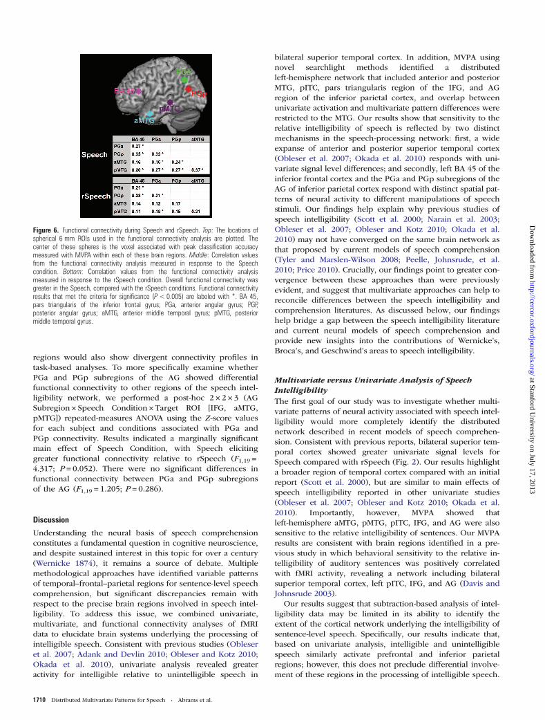
Functional Connectivity during Speech and rSpeechTo examine the functional relationships between the brainregions identified using MVPA during the processing of bothSpeech and rSpeech, we performed a functional connectivityanalysis on the time series data from 6-mm spheres centeredat the 5 classification peaks (IFG, aMTG, pMTG, PGa, andPGp) collected during these stimulus conditions. For theSpeech condition, 9 of the 10 connections (P = 0.02, binomialtest) between MVPA regions were significantly connected(Fig. 6). In contrast, for the rSpeech condition, only 4 of the10 connections (P = 0.75, binomial test) we examined reachedsignificance (Fig. 6). A direct comparison between Speechand rSpeech conditions using repeated-measures ANOVA withfactors stimulus condition (Speech, rSpeech) and ROI (IFG,aMTG, pMTG, PGa, and PGp) revealed a significant maineffect of stimulus (F1,19 = 6.553; P = 0.019), with greater con-nectivity in the Speech, compared with rSpeech, condition.There were no significant interaction between stimulus andROI (F9,171 = 0.760; P = 0.654).
A recent intrinsic connectivity analysis showed that PGaand PGp have different intrinsic connectivity profiles(Uddin et al. 2010) and it is unknown whether these AG
Figure 4. Overlap between univariate and multivariate responses to Speech versus rSpeech. Anterior and posterior left middle temporal gyrus (MTG) regions showed significantoverlap in multivariate and univariate responses to speech intelligibility. Outside the left MTG, there was no overlap between the MVPA and univariate results. aMTG, anteriormiddle temporal gyrus; pMTG, posterior middle temporal gyrus; AG, angular gyrus.
Figure 5. Signal levels in left-hemisphere ROIs. Signal level differences for Speechand rSpeech conditions are plotted in the bar graph for 6 left-hemisphere regionsidentified by MVPA. The filled and empty bars in the bar graph represent signal levelsfor the Speech and rSpeech conditions, respectively. Signal-level differences in theanterior and posterior MTG showed significant signal-level differences betweenSpeech and rSpeech; the pars triangularis of the inferior frontal gyrus (BA 45), theposterior inferior temporal cortex (pITC), and the anterior (PGa) and posterior (PGp)regions of the angular gyrus did not differ in signal level.
Cerebral Cortex July 2013, V 23 N 7 1709
at Stanford University on July 17, 2013
http://cercor.oxfordjournals.org/D
ownloaded from
regions would also show divergent connectivity profiles intask-based analyses. To more specifically examine whetherPGa and PGp subregions of the AG showed differentialfunctional connectivity to other regions of the speech intel-ligibility network, we performed a post-hoc 2 × 2 × 3 (AGSubregion × Speech Condition × Target ROI [IFG, aMTG,pMTG]) repeated-measures ANOVA using the Z-score valuesfor each subject and conditions associated with PGa andPGp connectivity. Results indicated a marginally significantmain effect of Speech Condition, with Speech elicitinggreater functional connectivity relative to rSpeech (F1,19 =4.317; P = 0.052). There were no significant differences infunctional connectivity between PGa and PGp subregionsof the AG (F1,19 = 1.205; P = 0.286).
Discussion
Understanding the neural basis of speech comprehensionconstitutes a fundamental question in cognitive neuroscience,and despite sustained interest in this topic for over a century(Wernicke 1874), it remains a source of debate. Multiplemethodological approaches have identified variable patternsof temporal–frontal–parietal regions for sentence-level speechcomprehension, but significant discrepancies remain withrespect to the precise brain regions involved in speech intel-ligibility. To address this issue, we combined univariate,multivariate, and functional connectivity analyses of fMRIdata to elucidate brain systems underlying the processing ofintelligible speech. Consistent with previous studies (Obleseret al. 2007; Adank and Devlin 2010; Obleser and Kotz 2010;Okada et al. 2010), univariate analysis revealed greateractivity for intelligible relative to unintelligible speech in
bilateral superior temporal cortex. In addition, MVPA usingnovel searchlight methods identified a distributedleft-hemisphere network that included anterior and posteriorMTG, pITC, pars triangularis region of the IFG, and AGregion of the inferior parietal cortex, and overlap betweenunivariate activation and multivariate pattern differences wererestricted to the MTG. Our results show that sensitivity to therelative intelligibility of speech is reflected by two distinctmechanisms in the speech-processing network: first, a wideexpanse of anterior and posterior superior temporal cortex(Obleser et al. 2007; Okada et al. 2010) responds with uni-variate signal level differences; and secondly, left BA 45 of theinferior frontal cortex and the PGa and PGp subregions of theAG of inferior parietal cortex respond with distinct spatial pat-terns of neural activity to different manipulations of speechstimuli. Our findings help explain why previous studies ofspeech intelligibility (Scott et al. 2000; Narain et al. 2003;Obleser et al. 2007; Obleser and Kotz 2010; Okada et al.2010) may not have converged on the same brain network asthat proposed by current models of speech comprehension(Tyler and Marslen-Wilson 2008; Peelle, Johnsrude, et al.2010; Price 2010). Crucially, our findings point to greater con-vergence between these approaches than were previouslyevident, and suggest that multivariate approaches can help toreconcile differences between the speech intelligibility andcomprehension literatures. As discussed below, our findingshelp bridge a gap between the speech intelligibility literatureand current neural models of speech comprehension andprovide new insights into the contributions of Wernicke’s,Broca’s, and Geschwind’s areas to speech intelligibility.
Multivariate versus Univariate Analysis of SpeechIntelligibilityThe first goal of our study was to investigate whether multi-variate patterns of neural activity associated with speech intel-ligibility would more completely identify the distributednetwork described in recent models of speech comprehen-sion. Consistent with previous reports, bilateral superior tem-poral cortex showed greater univariate signal levels forSpeech compared with rSpeech (Fig. 2). Our results highlighta broader region of temporal cortex compared with an initialreport (Scott et al. 2000), but are similar to main effects ofspeech intelligibility reported in other univariate studies(Obleser et al. 2007; Obleser and Kotz 2010; Okada et al.2010). Importantly, however, MVPA showed thatleft-hemisphere aMTG, pMTG, pITC, IFG, and AG were alsosensitive to the relative intelligibility of sentences. Our MVPAresults are consistent with brain regions identified in a pre-vious study in which behavioral sensitivity to the relative in-telligibility of auditory sentences was positively correlatedwith fMRI activity, revealing a network including bilateralsuperior temporal cortex, left pITC, IFG, and AG (Davis andJohnsrude 2003).
Our results suggest that subtraction-based analysis of intel-ligibility data may be limited in its ability to identify theextent of the cortical network underlying the intelligibility ofsentence-level speech. Specifically, our results indicate that,based on univariate analysis, intelligible and unintelligiblespeech similarly activate prefrontal and inferior parietalregions; however, this does not preclude differential involve-ment of these regions in the processing of intelligible speech.
Figure 6. Functional connectivity during Speech and rSpeech. Top: The locations ofspherical 6 mm ROIs used in the functional connectivity analysis are plotted. Thecenter of these spheres is the voxel associated with peak classification accuracymeasured with MVPA within each of these brain regions. Middle: Correlation valuesfrom the functional connectivity analysis measured in response to the Speechcondition. Bottom: Correlation values from the functional connectivity analysismeasured in response to the rSpeech condition. Overall functional connectivity wasgreater in the Speech, compared with the rSpeech conditions. Functional connectivityresults that met the criteria for significance (P<0.005) are labeled with *. BA 45,pars triangularis of the inferior frontal gyrus; PGa, anterior angular gyrus; PGP,posterior angular gyrus; aMTG, anterior middle temporal gyrus; pMTG, posteriormiddle temporal gyrus.
1710 Distributed Multivariate Patterns for Speech • Abrams et al.
at Stanford University on July 17, 2013
http://cercor.oxfordjournals.org/D
ownloaded from

Consistent with this view, we find that left inferior prefrontaland parietal cortex support intelligible speech processing,and processing in these regions is reflected by spatially dis-tributed patterns of neural activity rather than average activitylevels. Importantly, there is a striking overlap between thebrain regions identified in our MVPA results and the distribu-ted regions highlighted in a recent meta-analysis of thebrain’s semantic network (Binder et al. 2009), suggesting thatthe temporal, frontal, and parietal regions identified in ourstudy may all be engaged in differential semantic-level proces-sing of intelligible, compared with unintelligible, speechdespite showing no differences in intensity of activation tothese stimuli.
It is important to note that relatively similar study designsand univariate analyses of speech intelligibility data in pre-vious works have seldom revealed consistent results whenconsidering main effects of speech intelligibility (seesummary Table 1). For example, the table shows that brainregions such as the left IFG and left inferior parietal cortexare only occasionally identified as a main effect of intelligibil-ity. Many factors may contribute to these inconsistencies, in-cluding differences in stimuli, the number of participantsused in each study, data acquisition methods (Peelle, Johns-rude, et al. 2010), and data analysis considerations, includingthe thresholding of univariate results. Results from the currentstudy suggest that the use of new analytic tools, such aswhole-brain MVPA, may help resolve some of the inconsisten-cies in this literature by bringing a new level of sensitivity tothe data analysis and highlighting the role of fine-grain dis-tributed neural representations. The methods and approachused here may provide a more complete view of brainsystems underlying different aspects of speech and languageprocessing, including targeted studies of speech intelligibilityin the context of cochlear implant simulations (Eisner et al.2010), speech in noise (Davis and Johnsrude 2003), and sine-wave speech (McGettigan, Evans, et al. 2012).
Speech Intelligibility and the Left IFGMVPA results from the current study identified a dorsal regionof pars triangularis of the left IFG as a structure whosepattern of fMRI activity can reliably discriminate between in-telligible and unintelligible speech. This region shares exten-sive connections with auditory regions in the temporal lobe(Romanski et al. 1999) and has been implicated in manyaspects of speech comprehension, including phonological(Poldrack et al. 1999), syntactic (Ben-Shachar et al. 2004), andsemantic (Wagner et al. 2001) processes. The precise role ofthe IFG in sentence processing remains unknown; however,recent studies have suggested a role for semantic integration(Obleser and Kotz 2010) and working memory necessary forsentence comprehension (Eisner et al. 2010). Our results areconsistent with the view that the left IFG supports semanticand/or syntactic analysis of sentences (Caplan et al. 2008), se-quencing of speech input (Gelfand and Bookheimer 2003), orauditory working memory demands imposed by naturalisticsentences over time (Schulze et al. 2010). Although the IFGhas been implicated in some studies of speech intelligibility,there is no consensus with respect to its localization to BA 44,45, and 47 (Table 1). Recent receptor mapping studies haveprovided new insights into the organization of languageregions in the IFG (Amunts et al. 2010). Critically, based on
these studies, our study pin points for the first time theanterior aspects of Broca’ area (BA 45a) as the main locus ofspeech intelligibility in the IFG. Our findings suggest thatmore targeted investigations of speech processing usingMVPA will help to further clarify the role of specific subdivi-sions of the left IFG in sentence processing.
In the context of these left hemisphere findings, it is inter-esting to contrast univariate and multivariate response pat-terns in the right IFG. One initial finding was a surprisinglylarge response of the right IFG relative to the left in theomnibus activation to Speech and rSpeech, with respect tothe resting baseline (Fig. 1). A recent meta-analysis investi-gating right-hemisphere involvement in sentence-level proces-sing has indicated that right IFG activity is not uncommonand is typically associated with executive functions that arenot specific to language function, such as auditory selectiveattention and working memory (Vigneau et al. 2011). Criti-cally, however, neither univariate nor multivariate responsepatterns in the right IFG distinguished between speech androtated speech, further emphasizing the specific contributionsof different multivariate patterns in the left IFG to speechintelligibility.
Speech Intelligibility and the Left AGGiven the prominence of the left AG in neural models ofspeech comprehension (Tyler and Marslen-Wilson 2008;Peelle, Johnsrude, et al. 2010; Price 2010) and language pro-cesses (Geschwind 1970; Mesulam 1998), and its ubiquity instudies examining semantic processes (Binder et al. 2009), ithas been surprising that speech intelligibility paradigms havenot typically identified the AG. As previously mentioned, theAG has been implicated in a parametric study of speech intel-ligibility in which a positive correlation between the intellig-ibility of sentences and AG activity was reported (Davis andJohnsrude 2003); however a recent study showed the oppo-site effect in which AG activity was negatively correlated withsentence intelligibility (McGettigan, Faulkner, et al. 2012).The one subtraction–based intelligibility study that identifiedthe left AG involved an experimental design in which boththe intelligibility and semantic predictability of sentencestimuli were varied (Obleser et al. 2007). Results from thismanipulation showed that the AG becomes active relative tospectrally rotated speech only when highly predictable sen-tences are sufficiently acoustically degraded but was not re-vealed as a main effect of intelligibility. The implication ofthis result is that the AG is not necessary for speech compre-hension under ideal listening conditions, which would appearto contradict the hypothesis that the AG is essential for se-mantic processing of speech stimuli irrespective of acousticalconsiderations (Binder et al. 2009). Importantly, the AG intel-ligibility × predictability interaction finding shown in theObleser et al. (2007) study was not replicated in a more recentwork using a very similar design (Davis et al. 2011).
Our study helps reconcile discrepancies in the literaturesummarized above. Critically, we found that despite its deacti-vation (Binder et al. 1999; Greicius et al. 2003), relative to“rest” baseline, and despite its lack of differential engagementto Speech and rSpeech, the AG was sensitive to the intellig-ibility of speech based on distinct multivariate patterns ofneural activity. Cytoarchitectonic mapping revealed thatspeech intelligibility effects in the parietal cortex were
Cerebral Cortex July 2013, V 23 N 7 1711
at Stanford University on July 17, 2013
http://cercor.oxfordjournals.org/D
ownloaded from

localized to the AG (Caspers et al. 2006), with no overlap inthe supramarginal gyrus and the intra-parietal sulcus. Weshow that the AG involvement in the processing of speech in-telligibility involves both the PGa and PGp, its cytoarchitecto-nically distinct anterior and posterior subdivisions (Casperset al. 2006). The anatomical specificity of speech-related pro-cessing in circumscribed regions within the AG have impli-cations for reinterpreting findings in the neuropsychologicalliterature indicating that lesions to the parietal cortex do notalways produce speech comprehension deficits (Caplan et al.1996). Specifically, our results suggest that only parietallesions localized to the PGa or PGp may impair speech com-prehension. Taken together, these results provide a new levelof anatomical specificity with regards to speech intelligibilityin the human parietal cortex.
Our findings of AG involvement in processing of intelligi-ble speech comprehension is consistent with the view that theAG may be critical for higher-level processing of abstract fea-tures of the speech signal (Davis and Johnsrude 2003), andmost importantly, its semantic content (Binder et al. 2009;Seghier et al. 2010). We hypothesize that the AG, togetherwith Broca’s area, forms a tightly coupled network importantfor speech comprehension. Consistent with this view, recentanatomical studies have revealed direct white matter pathwaysbetween the AG and Broca’s area via the superior longitudinalfasciculus in humans and macaques (Makris et al. 2005; Pet-rides and Pandya 2009) and fMRI studies have highlightedstrong intrinsic functional connectivity in this fronto-parietalcircuit (Turken and Dronkers 2011).
Functional Connectivity in the Speech IntelligibilityNetworkThe second goal of this work was to examine differentialfunctional connectivity between the nodes identified withMVPA during the processing of intelligible and unintelligiblespeech. Importantly, we computed functional connectivity byremoving the first 4 s of transient changes in each block,thereby capturing temporal correlations within the Speechand rSpeech task blocks that do not reflect transitionsbetween high and low levels of activation. We found signifi-cant connectivity between nearly all nodes of the speech intel-ligibility network during the processing of Speech, withsubthreshold connectivity between many of these nodesduring the rSpeech condition (Fig. 6). In direct comparisonsbetween Speech and rSpeech conditions, connectivity for in-telligible speech was significantly greater than unintelligiblespeech. This finding is strengthened by the fact that themajority of nodes in this distributed network were identifiedusing MVPA and did not show univariate signal-level differ-ences between conditions, thus avoiding the possibility thatfunctional connectivity results were driven by signal-levelfluctuations associated with task on-off states.
Our results demonstrate, for the first time, that processingof intelligible speech drives increased functional connectivityrelative to unintelligible speech across the distributed tem-poral–frontal–parietal network encompassing putativeBroca’s, Wernicke’s, and Geschwind’s areas. This is an impor-tant finding as it suggests that the coordinated activity acrossestablished and distributed nodes of the speech comprehen-sion network is necessary for the processing of intelligiblespeech, independent of overall changes in task on-off
activation. This finding builds on a previous findings showingsignificant connectivity between the IFG and AG during theprocessing of intelligible speech (Eisner et al. 2010). Anotherprevious work reported significant functional connectivityduring processing of intelligible speech (Obleser et al. 2007);however, the nodes examined in that analysis included dorso-lateral prefrontal cortex and posterior cingulate cortex, whoseroles in speech processing are poorly understood, and did notinclude temporal lobe structures, whose role is speech proces-sing is well established. Given that our results describe pat-terns of functional connectivity across a well-describednetwork encompassing left MTG, IFG, and AG, an additionalstrength of this work is the interpretability of the findingswithin the context of the broader speech comprehensionliterature.
Conclusions
We have shown that intelligible speech is processed by dis-tinct spatial patterns of neuronal activity in a distributed corti-cal network that extends beyond superior temporal cortexand includes the left temporal, frontal, and parietal regions.Our results also show that univariate methods used in manyprevious intelligibility studies are insensitive to effects outsidethe temporal lobe, which are manifest as differences in multi-voxel patterns of brain activity. Moreover, functional connec-tivity between nodes identified with MVPA was greater duringthe processing of intelligible speech even though most of theregions within the network did not show signal-level differ-ences between intelligible and unintelligible speech. Morebroadly, our findings help to bridge a gap between speechcomprehension and speech intelligibility literatures, andsuggest a role for classical Wernicke’s, Broca’s, and Gesch-wind’s areas in the processing of intelligible speech.
Funding
This work was supported by the National Institute on Deaf-ness and Other Communication Disorders at the National In-stitutes of Health (grant number F32DC010322) to D.A.A. and1R21DC011095 to V.M.; National Science Foundation (grantnumber BCS0449927) to V.M. and D.J.L.; Natural Sciences andEngineering Research Council of Canada (grant numbers223210, 298612) to D.J.L. and E.B., respectively; and CanadaFoundation for Innovation (grant number 9908) to E.B.
NotesWe thank Jason Hom for assistance with data acquisition and DrsLucina Uddin and Miriam Rosenberg-Lee for critical comments on thiswork. We are grateful to two anonymous reviewers for their valuablesuggestions. Conflict of Interest: None declared
ReferencesAbrams DA, Bhatara A, Ryali S, Balaban E, Levitin DJ, Menon V. 2011.
Decoding temporal structure in music and speech relies on sharedbrain resources but elicits different fine-scale spatial patterns.Cereb Cortex. 21:1507–1518.
Adank P, Devlin JT. 2010. On-line plasticity in spoken sentence com-prehension: adapting to time-compressed speech. Neuroimage.49:1124–1132.
1712 Distributed Multivariate Patterns for Speech • Abrams et al.
at Stanford University on July 17, 2013
http://cercor.oxfordjournals.org/D
ownloaded from

Amunts K, Lenzen M, Friederici AD, Schleicher A, Morosan P,Palomero-Gallagher N, Zilles K. 2010. Broca’s region: novel organ-izational principles and multiple receptor mapping. PLoS Biol.8:1–16.
Azadpour M, Balaban E. 2008. Phonological representations are un-consciously used when processing complex, non-speech signals.PLoS One. 3:1–7.
Bates E, Wilson SM, Saygin AP, Dick F, Sereno MI, Knight RT, Dron-kers NF. 2003. Voxel-based lesion-symptom mapping. Nat Neuro-sci. 6:448–450.
Ben-Shachar M, Palti D, Grodzinsky Y. 2004. Neural correlates of syn-tactic movement: converging evidence from two fMRI exper-iments. Neuroimage. 21:1320–1336.
Binder JR, Desai RH, Graves WW, Conant LL. 2009. Where is the se-mantic system? A critical review and meta-analysis of 120 func-tional neuroimaging studies. Cereb Cortex. 19:2767–2796.
Binder JR, Frost JA, Hammeke TA, Bellgowan PS, Rao SM, Cox RW.1999. Conceptual processing during the conscious resting state. Afunctional MRI study. J Cogn Neurosci. 11:80–95.
Blesser B. 1972. Speech perception under conditions of spectral trans-formation.1. Phonetic characteristics. J Speech Hear Res. 15:5–41.
Caplan D, Hildebrandt N, Makris N. 1996. Location of lesions instroke patients with deficits in syntactic processing in sentencecomprehension. Brain. 119(Pt 3):933–949.
Caplan D, Stanczak L, Waters G. 2008. Syntactic and thematic con-straint effects on blood oxygenation level dependent signal corre-lates of comprehension of relative clauses. J Cogn Neurosci.20:643–656.
Caspers S, Geyer S, Schleicher A, Mohlberg H, Amunts K, Zilles K.2006. The human inferior parietal cortex: cytoarchitectonic parcel-lation and interindividual variability. Neuroimage. 33:430–448.
Davis MH, Ford MA, Kherif F, Johnsrude IS. 2011. Does semanticcontext benefit speech understanding through “Top-Down” pro-cesses? Evidence from time-resolved sparse fMRI. J Cogn Neurosci.23:3914–3932.
Davis MH, Johnsrude IS. 2003. Hierarchical processing in spokenlanguage comprehension. J Neurosci. 23:3423–3431.
Derogatis LR. 1992. SCL-90-R: administration, scoring, and proceduresmanual. 2nd edition. Baltimore (MD): Clinical PsychometricResearch.
Duvernoy HM. 1995. The human brain stem and cerebellum: surface,structure, vascularization, and three-dimensional sectionalanatomy with MRI. New York: Springer-Verlag.
Duvernoy HM, Bourgouin P. 1999. The human brain: surface, three-dimensional sectional anatomy with MRI, and blood supply.New York: Springer.
Eisner F, McGettigan C, Faulkner A, Rosen S, Scott SK. 2010. Inferiorfrontal gyrus activation predicts individual differences in percep-tual learning of cochlear-implant simulations. J Neurosci.30:7179–7186.
Forman SD, Cohen JD, Fitzgerald M, Eddy WF, Mintun MA, Noll DC.1995. Improved assessment of significant activation in functionalmagnetic resonance imaging (fMRI): use of a cluster-sizethreshold. Magn Reson Med. 33:636–647.
Friederici AD, Fiebach CJ, Schlesewsky M, Bornkessel ID, vonCramon DY. 2006. Processing linguistic complexity and grammati-cality in the left frontal cortex. Cereb Cortex. 16:1709–1717.
Friston KJ, Buechel C, Fink GR, Morris J, Rolls E, Dolan RJ. 1997.Psychophysiological and modulatory interactions in neuroima-ging. Neuroimage. 6:218–229.
Gaab N, Gabrieli JD, Glover GH. 2007. Assessing the influence ofscanner background noise on auditory processing. II. An fMRIstudy comparing auditory processing in the absence and presenceof recorded scanner noise using a sparse design. Hum BrainMapp. 28:721–732.
Gelfand JR, Bookheimer SY. 2003. Dissociating neural mechanisms oftemporal sequencing and processing phonemes. Neuron.38:831–842.
Geschwind N. 1970. The organization of language and the brain.Science. 170:940–944.
Glover GH, Lai S. 1998. Self-navigated spiral fMRI: interleaved versussingle-shot. Magn Reson Med. 39:361–368.
Greicius MD, Krasnow B, Reiss AL, Menon V. 2003. Functional con-nectivity in the resting brain: a network analysis of the defaultmode hypothesis. Proc Natl Acad Sci USA. 100:253–258.
Haxby JV, Gobbini MI, Furey ML, Ishai A, Schouten JL, Pietrini P.2001. Distributed and overlapping representations of faces andobjects in ventral temporal cortex. Science. 293:2425–2430.
Haynes JD, Rees G. 2006. Decoding mental states from brain activityin humans. Nat Rev Neurosci. 7:523–534.
Haynes JD, Sakai K, Rees G, Gilbert S, Frith C, Passingham RE. 2007.Reading hidden intentions in the human brain. Curr Biol.17:323–328.
Humphries C, Binder JR, Medler DA, Liebenthal E. 2006. Syntacticand semantic modulation of neural activity during auditory sen-tence comprehension. J Cogn Neurosci. 18:665–679.
Kim SH, Adalsteinsson E, Glover GH, Spielman S. 2000. SVD regular-ization algorithm for improved high-order shimming. In: Proceed-ings of the 8th annual meeting of ISMRM. Denver.
Kriegeskorte N, Bandettini P. 2007. Analyzing for information, notactivation, to exploit high-resolution fMRI. Neuroimage.38:649–662.
Kriegeskorte N, Goebel R, Bandettini P. 2006. Information-basedfunctional brain mapping. Proc Natl Acad Sci USA.103:3863–3868.
Mai JK, Assheur J, Paxinos G. 2004. Atlas of the human brain.Amsterdam: Elsevier.
Makris N, Kennedy DN, McInerney S, Sorensen AG, Wang R, CavinessVS, Pandya DN. 2005. Segmentation of subcomponents within thesuperior longitudinal fascicle in humans: a quantitative, in vivo,DT-MRI study. Cereb Cortex. 15:854–869.
McGettigan C, Evans S, Rosen S, Agnew ZK, Shah P, Scott SK. 2012.An application of univariate and multivariate approaches in FMRIto quantifying the hemispheric lateralization of acoustic and lin-guistic processes. J Cogn Neurosci. 24:636–652.
McGettigan C, Faulkner A, Altarelli I, Obleser J, Baverstock H, ScottSK. 2012. Speech comprehension aided by multiple modalities:behavioural and neural interactions. Neuropsychologia.50:762–776.
Menon V, Levitin DJ. 2005. The rewards of music listening: responseand physiological connectivity of the mesolimbic system. Neuro-image. 28:175–184.
Mesulam MM. 1998. From sensation to cognition. Brain. 121(Pt6):1013–1052.
Muller KR, Mika S, Ratsch G, Tsuda K, Scholkopf B. 2001. An intro-duction to kernel-based learning algorithms. IEEE Trans NeuralNetw. 12:181–201.
Narain C, Scott SK, Wise RJ, Rosen S, Leff A, Iversen SD, MatthewsPM. 2003. Defining a left-lateralized response specific to intelligi-ble speech using fMRI. Cereb Cortex. 13:1362–1368.
Norman KA, Polyn SM, Detre GJ, Haxby JV. 2006. Beyond mind-reading: multi-voxel pattern analysis of fMRI data. Trends CognSci. 10:424–430.
Obleser J, Kotz SA. 2010. Expectancy constraints in degraded speechmodulate the language comprehension network. Cereb Cortex.20:633–640.
Obleser J, Wise RJ, Alex Dresner M, Scott SK. 2007. Functional inte-gration across brain regions improves speech perception underadverse listening conditions. J Neurosci. 27:2283–2289.
Okada K, Rong F, Venezia J, Matchin W, Hsieh IH, Saberi K, SerencesJT, Hickok G. 2010. Hierarchical organization of human auditorycortex: evidence from acoustic invariance in the response to intel-ligible speech. Cereb Cortex. 20:2486–2495.
Peelle JE, Eason RJ, Schmitter S, Schwarzbauer C, Davis MH. 2010.Evaluating an acoustically quiet EPI sequence for use in fMRIstudies of speech and auditory processing. Neuroimage.52:1410–1419.
Peelle JE, Johnsrude IS, Davis MH. 2010. Hierarchical processing forspeech in human auditory cortex and beyond. Front Hum Neuro-sci. 4:51.
Cerebral Cortex July 2013, V 23 N 7 1713
at Stanford University on July 17, 2013
http://cercor.oxfordjournals.org/D
ownloaded from

Pereira F, Mitchell T, Botvinick M. 2009. Machine learningclassifiers and fMRI: a tutorial overview. Neuroimage. 45:S199–S209.
Petrides M, Pandya DN. 2009. Distinct parietal and temporal pathwaysto the homologues of Broca’s area in the monkey. PLoS Biol. 7:e1000170.
Poldrack RA, Wagner AD, Prull MW, Desmond JE, Glover GH, Gabrie-li JDE. 1999. Functional specialization for semantic and phonologi-cal processing in the left inferior prefrontal cortex. Neuroimage.10:15–35.
Price CJ. 2010. The anatomy of language: a review of 100 fMRIstudies published in 2009. Ann N Y Acad Sci. 1191:62–88.
Rodd JM, Davis MH, Johnsrude IS. 2005. The neural mechanisms ofspeech comprehension: fMRI studies of semantic ambiguity. CerebCortex. 15:1261–1269.
Romanski LM, Tian B, Fritz J, Mishkin M, Goldman-Rakic PS,Rauschecker JP. 1999. Dual streams of auditory afferents targetmultiple domains in the primate prefrontal cortex. Nat Neurosci.2:1131–1136.
Schulze K, Zysset S, Mueller K, Friederici AD, Koelsch S. 2010. Neu-roarchitecture of verbal and tonal working memory in nonmusi-cians and musicians. Hum Brain Mapp. 32:771–783.
Schwarzlose RF, Swisher JD, Dang S, Kanwisher N. 2008. The distri-bution of category and location information across object-selectiveregions in human visual cortex. Proc Natl Acad Sci USA.105:4447–4452.
Scott SK, Blank CC, Rosen S, Wise RJ. 2000. Identification of apathway for intelligible speech in the left temporal lobe. Brain.123(Pt 12):2400–2406.
Seghier ML, Fagan E, Price CJ. 2010. Functional subdivisions in theleft angular gyrus where the semantic system meets and divergesfrom the default network. J Neurosci. 30:16809–16817.
Smith SM, Jenkinson M, Woolrich MW, Beckmann CF, Behrens TE,Johansen-Berg H, Bannister PR, De Luca M, Drobnjak I, FlitneyDE et al. 2004. Advances in functional and structural MR image
analysis and implementation as FSL. Neuroimage. 23(Suppl 1):S208–S219.
Turken AU, Dronkers NF. 2011. The neural architecture of thelanguage comprehension network: converging evidence fromlesion and connectivity analyses. Front Syst Neurosci. 5:1.
Tyler LK, Marslen-Wilson W. 2008. Fronto-temporal brain systemssupporting spoken language comprehension. Philos Trans R SocLond B Biol Sci. 363:1037–1054.
Uddin LQ, Supekar K, Amin H, Rykhlevskaia E, Nguyen DA, GreiciusMD, Menon V. 2010. Dissociable connectivity within humanangular gyrus and intraparietal sulcus: evidence from functionaland structural connectivity. Cereb Cortex. 20:2636–2646.
Various. 1991. Great speeches of the 20th century. Los Angeles:Rhino Records.
Vigneau M, Beaucousin V, Herve PY, Duffau H, Crivello F, Houde O,Mazoyer B, Tzourio-Mazoyer N. 2006. Meta-analyzing left hemi-sphere language areas: phonology, semantics, and sentence pro-cessing. Neuroimage. 30:1414–1432.
Vigneau M, Beaucousin V, Herve PY, Jobard G, Petit L, Crivello F,Mellet E, Zago L, Mazoyer B, Tzourio-Mazoyer N. 2011. What isright-hemisphere contribution to phonological, lexico-semantic,and sentence processing? Insights from a meta-analysis. Neuro-image. 54:577–593.
Wagner AD, Pare-Blagoev EJ, Clark J, Poldrack RA. 2001. Recoveringmeaning: left prefrontal cortex guides controlled semantic retrie-val. Neuron. 31:329–338.
Wang X, Merzenich MM, Beitel R, Schreiner CE. 1995. Representationof a species-specific vocalization in the primary auditory cortex ofthe common marmoset: temporal and spectral characteristics.J Neurophysiol. 74:2685–2706.
Ward BD. 2000. Simultaneous inference for fMRI data. AFNI 3dDe-convolve documentation, Milwaukee (WI): Medical College ofWisconsin.
Wernicke C. 1874. Der Aphasische Symptomencomplex. Breslau:Cohn and Weigert.
1714 Distributed Multivariate Patterns for Speech • Abrams et al.
at Stanford University on July 17, 2013
http://cercor.oxfordjournals.org/D
ownloaded from