Current concepts regarding the HTLV-1 receptor complex
11
REVIEW Open Access Current concepts regarding the HTLV-1 receptor complex David Ghez 1,2* , Yves Lepelletier 1 , Kathryn S Jones 3 , Claudine Pique 4,5 , Olivier Hermine 1,6* Abstract The identity of the Human T lymphotropic Virus type 1 (HTLV-1) receptor remained an unsolved puzzle for two decades, until the recent demonstration that three molecules, Glucose Transporter 1, Neuropilin-1 and Heparan Sul- fate Proteoglycans are involved in HTLV-1 binding and entry. Despite these advances, several questions remain unanswered, including the precise role of each of these molecules during virus entry. In light of the most recent data, we propose a model of the HTLV-1 receptor complex and discuss its potential impact on HTLV-1 infection. Introduction Since its identification in 1979, the Human T-Lympho- tropic Virus type 1 (HTLV-1) has been the subject of extensive research. Despite abundant experimental data, several unsolved mysteries still surround HTLV-1. Amongst those, the identity of the HTLV-1 cellular receptor has eluded scientists for over two decades. Since 2003, data from several independent groups have shed light on the identity of the molecules that appear to be directly involved in the process of HTLV-1 entry. These include Glucose Transporter 1 (GLUT1) [1,2], Neuropilin-1 (NRP-1) [3-5] and Heparan Sulfate Proteo- glycans (HSPG) [6,7]. Despite these advances, our cur- rent knowledge of their precise roles during HTLV-1 entry remains limited. In particular, little is known about interaction between these molecules, including whether they form a multimolecular complex similar to that of the well-studied Human Immunodeficiency Virus type 1 (HIV-1) receptor. In this review, we will summarize the data pertaining to HTLV-1 entry mole- cules, propose a hypothetical model of the HTLV-1 receptor and discuss its possible impact on the patho- biology of HTLV-1 infection. The HTLV-1 Receptor Enigma HTLV-1 entry It is widely believed that HTLV-1 enters cells in a man- ner similar to that of other retroviruses including the well-studied HIV-1. HTLV-1 infection of target cells is believed to require the two virally encoded envelope gly- coproteins (Env), the surface subunit (SU) gp46 and the transmembrane subunit (TM) gp21, generated from the cleavage of a polyprecursor (gp61) in the Golgi appara- tus [8]. As has previously been shown for the SU (gp120) and TM (gp41) of HIV-1 [9], the SU and TM of HTLV-1 are believed to function successively during the entry process. The gp46/SU, which does not contain a transmembrane region, is tethered to the cell surface through interactions with the gp21, and is involved in direct interactions with the cell surface receptors. Like the HIV-1 gp41, the gp21 of HTLV-1 contains a trans- membrane region and a N-terminal hydrophobic fusion peptide that plays a crucial role in the fusion of the viral and cellular membrane during the final steps of HTLV- 1 entry [10,11]. Earlier work showed that both the presence of certain mutations in the env gene and anti- bodies directed against gp46/SU partially or totally abol- ished HTLV-1 infection in vitro [12,13]. Infection was also reduced by exposure of target cells to a soluble recombinant protein containing soluble full-length SU, indicating that it could compete with the SU on the sur- face of the virus for the binding to yet unknown mole- cules at the cell surface [14]. Altogether, these experiments confirmed that the two Env glycoproteins play crucial role during HTLV-1 entry and indicated that, as for other retroviruses, this process depends on the expression of cellular receptor(s) at the surface of target cells. Interaction between gp46/SU and its cellular receptor(s) induces a conformational change that * Correspondence: [email protected]; [email protected] 1 CNRS UMR8147, Universite Rene Descartes, Paris 5, 161 Rue de Sèvres, 75743 Paris Cedex 15, France Full list of author information is available at the end of the article Ghez et al. Retrovirology 2010, 7:99 http://www.retrovirology.com/content/7/1/99 © 2010 Ghez et al; licensee BioMed Central Ltd. This is an Open Access article distributed under the terms of the Creative Commons Attribution License (http://creativecommons.org/licenses/by/2.0), which permits unrestricted use, distribution, and reproduction in any medium, provided the original work is properly cited.
-
Upload
independent -
Category
Documents
-
view
0 -
download
0
Transcript of Current concepts regarding the HTLV-1 receptor complex

REVIEW Open Access
Current concepts regarding the HTLV-1 receptorcomplexDavid Ghez1,2*, Yves Lepelletier1, Kathryn S Jones3, Claudine Pique4,5, Olivier Hermine1,6*
Abstract
The identity of the Human T lymphotropic Virus type 1 (HTLV-1) receptor remained an unsolved puzzle for twodecades, until the recent demonstration that three molecules, Glucose Transporter 1, Neuropilin-1 and Heparan Sul-fate Proteoglycans are involved in HTLV-1 binding and entry. Despite these advances, several questions remainunanswered, including the precise role of each of these molecules during virus entry. In light of the most recentdata, we propose a model of the HTLV-1 receptor complex and discuss its potential impact on HTLV-1 infection.
IntroductionSince its identification in 1979, the Human T-Lympho-tropic Virus type 1 (HTLV-1) has been the subject ofextensive research. Despite abundant experimental data,several unsolved mysteries still surround HTLV-1.Amongst those, the identity of the HTLV-1 cellularreceptor has eluded scientists for over two decades.Since 2003, data from several independent groups haveshed light on the identity of the molecules that appearto be directly involved in the process of HTLV-1 entry.These include Glucose Transporter 1 (GLUT1) [1,2],Neuropilin-1 (NRP-1) [3-5] and Heparan Sulfate Proteo-glycans (HSPG) [6,7]. Despite these advances, our cur-rent knowledge of their precise roles during HTLV-1entry remains limited. In particular, little is knownabout interaction between these molecules, includingwhether they form a multimolecular complex similar tothat of the well-studied Human ImmunodeficiencyVirus type 1 (HIV-1) receptor. In this review, we willsummarize the data pertaining to HTLV-1 entry mole-cules, propose a hypothetical model of the HTLV-1receptor and discuss its possible impact on the patho-biology of HTLV-1 infection.
The HTLV-1 Receptor EnigmaHTLV-1 entryIt is widely believed that HTLV-1 enters cells in a man-ner similar to that of other retroviruses including the
well-studied HIV-1. HTLV-1 infection of target cells isbelieved to require the two virally encoded envelope gly-coproteins (Env), the surface subunit (SU) gp46 and thetransmembrane subunit (TM) gp21, generated from thecleavage of a polyprecursor (gp61) in the Golgi appara-tus [8]. As has previously been shown for the SU(gp120) and TM (gp41) of HIV-1 [9], the SU and TM ofHTLV-1 are believed to function successively during theentry process. The gp46/SU, which does not contain atransmembrane region, is tethered to the cell surfacethrough interactions with the gp21, and is involved indirect interactions with the cell surface receptors. Likethe HIV-1 gp41, the gp21 of HTLV-1 contains a trans-membrane region and a N-terminal hydrophobic fusionpeptide that plays a crucial role in the fusion of the viraland cellular membrane during the final steps of HTLV-1 entry [10,11]. Earlier work showed that both thepresence of certain mutations in the env gene and anti-bodies directed against gp46/SU partially or totally abol-ished HTLV-1 infection in vitro [12,13]. Infection wasalso reduced by exposure of target cells to a solublerecombinant protein containing soluble full-length SU,indicating that it could compete with the SU on the sur-face of the virus for the binding to yet unknown mole-cules at the cell surface [14]. Altogether, theseexperiments confirmed that the two Env glycoproteinsplay crucial role during HTLV-1 entry and indicatedthat, as for other retroviruses, this process depends onthe expression of cellular receptor(s) at the surface oftarget cells. Interaction between gp46/SU and its cellularreceptor(s) induces a conformational change that
* Correspondence: [email protected]; [email protected] UMR8147, Universite Rene Descartes, Paris 5, 161 Rue de Sèvres,75743 Paris Cedex 15, FranceFull list of author information is available at the end of the article
Ghez et al. Retrovirology 2010, 7:99http://www.retrovirology.com/content/7/1/99
© 2010 Ghez et al; licensee BioMed Central Ltd. This is an Open Access article distributed under the terms of the Creative CommonsAttribution License (http://creativecommons.org/licenses/by/2.0), which permits unrestricted use, distribution, and reproduction inany medium, provided the original work is properly cited.

unmasks the fusion domain within the TM, allowingfusion between the viral and cellular membranes [15].
The search for the HTLV receptorExperimental demonstration that specific moleculesfunction as a virus receptor often is a challenge [16].This is illustrated by the fact that there are a great num-ber of viruses, including certain retroviruses, for whichno receptors have been identified. In addition, for cer-tain viruses, it has been shown that more than onemolecule may function as entry receptors. Some virusesuse different receptors on different cell types, whileothers require the presence of both molecules on agiven target cell to facilitate entry. In the case of HTLV-1, several peculiar features of the virus and its receptor(s) have considerably hampered research. First, freeHTLV-1 virions are poorly infectious for most cell typesand efficient infection of T cells requires a cellular con-tact [17]. Secondly, infected lymphocytes produce a lim-ited amount of viral particles, amongst which 1 out of105 are actually infectious [18]. Lastly, the fact thatmolecules capable of binding and allowing HTLV-1entry are expressed on nearly all available establishedcell lines has prevented the use of classical strategiessuch as the screening of a cDNA bank in receptor-nega-tive cells.
In vivo versus in vitro entry tropism: the paradox of theHTLV-1 receptorVirus tropism, the ability of a virus to replicate in parti-cular cells, depends on interactions between viral com-ponents and cellular factors at each step of the viralcycle from the initial entry to the ultimate release andtransmission of virions. Here, we will focus on the cellu-lar factors that allow HTLV-1 entry, which is deter-mined by the distribution of the receptor molecule(s).In vivo entry tropismAlthough the main targets of HTLV-1 are CD4+ T cells[19], the virus has been found in other cell types in vivoincluding as CD8+ T cells [20], monocytes and B cells[21], macrophages [22], dendritic cells (DC) [23,24] andendothelial cells [25]. In vivo, the only known targets ofvirus-induced transformation are CD4+ CD45RO+ mem-ory T cells [26]. Recently, it has been hypothesized thatnatural regulatory T cells (Tregs) can be infected. Thiswas based on the fact that HTLV-1 infected T cells andTregs have a strikingly close phenotype: CD25+, (whichis a direct consequence of HTLV-1 Tax synthesis [27]),GITR+ and FoxP3+ [28,29]. If true, this could partlyexplain the frequent immune dysregulation observed inHTLV-1-infected individuals. However, studies fromother laboratories suggest that Tregs are not infected bythe virus [30,31]. This is consistent with other workshowing potent suppressive activity of HTLV-1-infected
or transformed T cells in only a portion of HTLV-1-infected patients [32,33]. This could be due to theimpairment of FoxP3 function by the HTLV-1 Tax pro-tein, as shown by Jacobson’s group [34]. In contrast, ithas been suggested that HTLV-1 may have an indirecteffect on Tregs since Bangham’s group recently reportedthat the frequency of uninfected functional Tregs isabnormally high in HTLV-1-infected individuals [35],which may account as well for the immunosuppressionassociated with HTLV-1 infection.In vitro entry tropismIn striking contrast to the limited number of cell-typesin which HTLV-1 is detected in vivo, molecules capableof supporting the initial steps in infection, binding andentry, appear to be expressed on nearly all establishedcell lines. As discussed below, this is consistent with thefact that molecules in the receptor complex are up-regu-lated on most transformed cells. Moreover, the HTLV-1receptor complex is present on cell lines from nearly allknown vertebrate species [36]: most available establishedcell lines are able to form multinucleated giant cells(syncytia) when cultured with Env expressing cells, aphenomenon that is dependent on Env/Receptor inter-actions [37]. Further evidence that HTLV-1 receptorsare widely distributed comes from observations thatHTLV-1 Env-pseudotyped particles can infect a numberof established cell lines [38]. More recently, binding stu-dies with soluble, full-length HTLV-1 SU (SU-Fc) hasnot only confirmed the results obtained with infectionand fusion experiments but also showed that a widerrange of established cell lines expressed molecules cap-able of specifically binding the SU protein [1]. Workfrom the Brighty laboratory found that the SU-Fc pro-tein could bind to a vast number of vertebrate cell linesincluding some that were originally thought to be recep-tor negative due to their resistance to Env-mediated cellto cell fusion or infection [39]. This study identified onecell line, the drosophila cell line S2, that lacked mole-cules capable of specifically binding HTLV SU on thecell surface. Unfortunately, these cells could not be usedto identify HTLV-1 receptors, since they have post-entryblocks to HTLV-1 and other retroviruses.
Properties of the HTLV-1 receptor: indirect evidenceAlthough its ubiquitous nature considerably complicatedits identification, several properties of the HTLV-1receptor, in particular its expression pattern on primarycells, were characterized using indirect approaches. In2003, two independent groups generated soluble SUproteins by generating recombinant SU-Ig-Fc fusionproteins containing either the full-length SU [22] or theN-terminal portion of SU (Receptor Binding Domain,see section 3.1) [40]. Their findings showed that thereceptor was not present at the surface of resting CD4+
Ghez et al. Retrovirology 2010, 7:99http://www.retrovirology.com/content/7/1/99
Page 2 of 11

T cells but was rapidly upregulated upon activation. Thereceptor was also absent on naive T cells isolated fromcord blood but could similarly be upregulated after sti-mulation by interleukin-7. Finally, it was reported thatthe SU-Fc could inhibit a mixed lymphocyte reaction[22]. This last property suggested that one of the mole-cules involved in the HTLV-1 receptor was a memberof the immune synapse. Surface expression of the recep-tor was found to be not solely dependent on T cell acti-vation. It could also be upregulated upon exposure toTGF-b, a potent negative regulator of the immuneresponse [41]. The pattern of surface expression follow-ing exposure to TGF-b, as determined from binding ofthe soluble SU, was very similar to that observed afteractivation with PHA/IL-2, although with slightly slowerkinetics. It depended on the TGF-b classical signallingpathway as it was abolished in the presence of Smadinhibitors. TGF-b not only triggered receptor expressionat the cell surface but also increased the titer of lenti-viral vectors pseudotyped with HTLV-1 Env in vitro,demonstrating that molecules capable of both bindingand fusion were expressed [41]. TGF-b-treated T cellsremained in a resting state, demonstrating that T cellactivation per se was not required for receptor expres-sion. This might constitute a strategy for the virus toincrease its infectivity [42] as HTLV-1-infected CD4+ Tcells are known to abundantly secrete TGF-b.
Candidate receptors whose role was ruled outOver the years, several molecules were proposed as candi-date HTLV-1 receptors. It was proposed that the HTLV-1receptor is encoded by chromosome 17q23.2-23.5, whichwas questioned in later studies [38,43]. Most of the candi-date receptors were identified as antigens against whichspecific antibodies could inhibit Env-mediated cell fusion[43-46]. It was later shown that some antibodies may inhi-bit this process through non-specific protein crowding,rather than directly competing for binding, making theresults difficult to interpret [47]. Adhesion molecules,which strengthen and stabilize the cell-to-cell contact, canalso augment cell fusion in a non-specific manner [48].Most importantly, none of these molecules had demon-strated one fundamental property of the receptor, that is,the ability to bind the HTLV-1 SU. In contrast, as it couldbind the 197-216 region of the SU, the heat shock cognateprotein HSC70 was proposed as a receptor [49]. However,later studies showed that, while HSC70 modulates HTLV-1-induced syncytia formation, it was dispensable forHTLV-1 infection [50], ruling out that this molecule wasan entry receptor. The tetraspanin CD82 was also shownto bind the HTLV-1 Env protein but was excluded as anentry receptor on the fact that its overexpression inhibited,rather than enhanced, syncytia formation and HTLV-1transmission [51].
Not one but Three Receptor Molecules: GLUT1,HSPG and NRP-1The glucose transporter GLUT1It was not until 2003 that work from Sitbon and Batti-ni’s laboratory identified a candidate that matched allthe prerequisites to be a HTLV-1 receptor [2]. Further-more, this group determined that this molecule inter-acted with both HTLV-1 and the related virus HTLV-2,believed to use a common entry receptor. After noticingthat overexpression of either the HTLV-1 or HTLV-2Env in cells prevented acidification of the culture med-ium, the authors hypothesized that the HTLV-1 receptormight be related to the proton-dependent lactate pro-duction. Based on their previous identification of thereceptor binding domain (RBD) of HTLV-1 or HTLV-2,these authors further reported that the overexpressionof either the H1-RBD (first 215 residues of the HTLV-1SU) or the H2-RBD (178 residues of the HTLV-2)[40,52,53] altered glucose metabolism, which was consis-tent with the frequent use of metabolite transporters asentry receptors by retroviruses [16]. They focused onthe ubiquitous glucose transporter 1 (GLUT1), which isupregulated in activated T cells, and found that overex-pression of GLUT-1 in cells increased the level of bind-ing of both the H1- or H2-RBD. In the absence ofavailable GLUT1-negative cell lines, they used differentapproaches, in particular small interfering RNA (siRNA),to demonstrate that GLUT1 played a role in HTLV-1entry. Down regulation of GLUT1 by siRNA both inhib-ited the binding of the H1- or H2-RBD and infection byretroviral vectors pseudotyped with either the HTLV-1or -2 Env. Both were restored after cotransfection ofGLUT1 but not GLUT3, a related glucose transporter.They later extended their findings by that identifying aspecific residue in the HTLV-1 SU (Y114) of the H1-RBD that appeared critical to the interaction [52], whichinteracted with the 6th extracellular loop (ECL6) ofGLUT1 [54].Further data from other laboratories confirmed that
GLUT1 plays a role in HTLV-1 entry. TGF-b, whichhad previously been shown to upregulate molecules cap-able of binding HTLV SU, induces GLUT1 at the sur-face of T cells [41]. Overexpression of GLUT1 in thebovine MDBK cell line, which is relatively resistant toHTLV-1 infection, increases two-fold the infectious titreof a HTLV-1 Env pseudotyped lentivirus vector [55]. Asimilar result was obtained in the NIH3T3 cell line,which is also poorly susceptible to infection by HTLV-1pseudotyped vectors [1]. Antibodies raised againstGLUT1 ECL1 (GLUT-IgY) block both Env-mediatedfusion and infection. Furthermore, blocking interactionswith GLUT1 ECL1, either by replacing the GLUT1ECL1 with that of GLUT3, or by blocking with ECL1-derived peptides, inhibits HTLV-1-induced cell fusion,
Ghez et al. Retrovirology 2010, 7:99http://www.retrovirology.com/content/7/1/99
Page 3 of 11
suggesting that ECL1 is critical for the receptor activityof GLUT1 [1]. This confirmed earlier data showing that,although only the ECL6 was required for the binding ofthe H1-RBD to GLUT1, infection by HTLV-2-pseudo-typed viruses also required residues located in ECL1 andECL5 of this molecule [54]. This indicates that GLUT1contains multiple functional determinants: thoserequired for interactions with the RBD portion of theHTLV SU, located in ECL6, and others required forinteractions of the entire Env, consisting of the full-length SU protein and the TM protein, located in ECL1and ECL5. Thus, current data indicate that GLUT1plays a role in HTLV-1 entry.However, several studies suggested that the identifica-
tion of GLUT1 was not the end of the story. The astro-cytoma/glioblastoma U87 cell line, which expresses verylow levels of GLUT-1 due to a transdominant negativemutation in one of the alleles of GLUT1 (GLUT-1 D5),is easily infected by HTLV-pseudotyped viruses[3,56].Moreover, blocking interactions with GLUT1 by eithersiRNA-mediated down-regulation, or by incubating withGLUT-IgY blocking antibodies, has no effect on thelevel of infection with HTLV-Env-pseudotyped virus orHTLV Env-mediated fusion or infection in this cell line,suggesting that molecules other than GLUT1 might beinvolved [56]. An earlier study reported that overexpres-sion of GLUT1 in a cell line (COS-7) increased cell-celltransmission, but did not increase the level of binding ofthe SU-Fc protein [57]. Similarly, more recent studiesusing clones of the U87 cell line that express differentlevels of GLUT1 have observed that the level of cell sur-face GLUT1 correlates with the titer of HTLV-1 Envpseudotyped viruses, but not with SU-Fc binding [58].All these findings suggested that molecules other thanGLUT1 are also involved in HTLV-1 entry, especially atthe binding step.
Heparan Sulfate Proteoglycans modulate HTLV-1attachment and entryMolecules of the HSPG family are composed of a coreprotein associated with one or more sulphated polysac-charide side chains called heparan sulfate glycosamino-glycans. Because of their highly negative charge, HSPGbind through electrostatic interactions to a plethora ofproteins including growth factors and their receptors,chemokines, cytokines and numerous proteins of theextracellular matrix or the plasma, thereby playing amajor role in mammalian physiology [59]. Pathogens,including many viruses, have been shown to hijackHSPG [60]. HSPG generally enhance infection by facili-tating the attachment of the particles on target cellsand/or allowing their clustering at the cell surfacebefore specific interactions between viral proteins andtheir receptors that lead to fusion. For example, prior to
interaction of the HIV SU (gp120) with CD4, the initialattachment of the virus to target cells involves specificinteractions between gp120 and cellular HSPG [61].Rarely, a specific role of HSPG in the fusion processhas been observed, in particular with Herpes Simplexvirus [62].The important role of HSPG in mediating attachment
and entry has also been demonstrated for HTLV-1.Enzymatic removal of HSPG or inhibition of electro-static interactions with dextran sulfate decreases thebinding of full length soluble SU, Env-mediated syncy-tium formation and infection with pseudotyped viruses[7]. These initial findings, obtained in non-lymphoid celllines expressing high levels of HSPG, were later con-firmed in CD4+ T cells in Jones and Ruscetti’s labora-tory [6]. Although HSPG are barely detectable onquiescent T cells [63], they are rapidly upregulated uponactivation. In CD4+ T cells, HSPG augment both thebinding of the SU-Fc and the infectious titer of pseudo-typed viruses [6]. The importance of HSPG was sug-gested by studies showing that removing HSPG reducedbinding and entry of HTLV-1 virus into CD4+ T cellsand dendritic cells [4,6] and significantly reduced thelevel of HTLV-1 infection.Other recent studies suggest another role for HSPG
during cell-cell transmission of the virus. It was discov-ered that HTLV-1 virions are stored outside the cell,within a protective microenvironment enriched for spe-cific components of the extracellular matrix, includinglectins and HSPGs [64]. Following contact between Tcells, these structures are rapidly transferred frominfected to uninfected cells, ultimately resulting in infec-tion of the target cell. This suggests that HSPG couldplay a broader role in HTLV-1 transmission by facilitat-ing both the transfer of newly-formed virions from aninfected T cells and the subsequent entry into the targetT cell. The galactose-binding lectin Galactin-1, whichhas been shown to increase HTLV-1-Env mediatedinfection [65], might also have a similar function.Interestingly, HSPG are important for infection with
HTLV-1 but not HTLV-2 [66], showing that these twoviruses, initially considered to share the same receptor[67], may share some but not all molecular determinantsfor entry. It has previously been reported that HTLV-1SU has the ability to bind HSPG; this ability maps tothe C-terminal region of HTLV-1 SU, located down-stream the RBD and the proline rich region [66]. Inter-estingly, the Green laboratory has previously reportedthat the preferential tropism of HTLV-1 and HTLV-2 totransform CD4+ T cells or CD8+ T cells, respectively, isgoverned by Env [68]. The difference in the requirementof HTLV-1 and HTLV-2 two SU for HSPG mightexplain the different tropisms of the two retroviruses.Indeed, CD4+ T cells express a high level of HSPG and
Ghez et al. Retrovirology 2010, 7:99http://www.retrovirology.com/content/7/1/99
Page 4 of 11

a lower level of GLUT1 whereas CD8+ T cells express ahigh level of GLUT1 but very few HSPG. In this model,the differences between HTLV-1 and HTLV-2 would besimilar to what has been described in CD4-dependentand independent HIV-2 or SIV strains, which differ intheir requirements for the binding receptor (CD4) butshare similar fusion receptors [69].
The third player: Neuropilin-1In 2006, the Hermine and Pique laboratories showedthat Neuropilin-1 (NRP-1) also displayed propertiesexpected for a HTLV-1 receptor [5]. This hypothesiswas confirmed in a recent study [3]. NRP-1 is a 130kDa single membrane spanning glycoprotein which actsas the co-receptor for Semaphorin 3a and VEGFA165.NRP-1 was initially identified as a critically importantfactor in embryonic neuron guidance, and later shownto be a key player in the regulation of angiogenesis. Inaddition, Romeo and Hermine’s laboratories were thefirst to demonstrate that NRP-1 is also involved in theregulation of the immune response [70]. NRP-1 is highlyconserved amongst vertebrate species but there are noNRP-1 homologs in insects [71]. NRP-1 is mainlyexpressed in T cells and DC, which are targets ofHTLV-1 in vivo. NRP-1 is highly expressed on plasma-cytoid DC (pDC), which can be infected by cell-freevirus in vitro [72]. Endothelial cells, in which HTLV-1proviral DNA has been detected in vivo [25], alsoexpress NRP-1 [73]. NRP-1 is absent on resting T cellsbut is rapidly upregulated following activation [72,74].In contrast with its relatively limited expression in vivo,NRP-1 is found in many tumor cells [73] and hence isexpressed on an extremely broad range of establishedcell lines. Finally, NRP-1 plays a role in cytoskeletalrearrangement, a phenomenon that has been shown tobe important for transmission of different retrovirusesincluding HTLV-1 [75].Ghez et al. reported that NRP-1 was able to directly
interact with the HTLV-1 and -2 SU [5]. This interac-tion appeared functionally relevant since NRP-1 overex-pression enhanced syncytium formation and the titer ofHTLV-1 or -2 Env pseudotyped viruses whereas siRNA-mediated NRP-1 downmodulation had the oppositeeffect [5]. A strong polarization of NRP-1 and Env oneither side of the interface between an infected cell anda target T cell was observed using confocal microscopy.This phenomenon was also observed with GLUT1,though GLUT1 was also found to colocalize at themembrane junction of two uninfected T cells, thus in anEnv-independent manner. Both polarization and co-localization of the three molecules were particularlyintense at regions where partial membrane fusion wastaking place. It was also observed that GLUT1 andNRP-1 could form intracytoplasmic complexes in
transfected cells, an association that was greatlyenhanced in the presence of Env [5].
A new understanding of HTLV-1 tropismThe identification of three new molecules involved inthe process of HTLV-1 entry allows one to revisit theHTLV-1 tropism paradox mentioned above. WhileHSPG usage appears to distinguish HTLV-1 and HTLV-2, their ubiquitous expression cannot explain the limitedtropism of HTLV-1 in vivo. This is also the case ofGLUT1, whose expression is ubiquitous as well. In con-trast, the distinct pattern of NRP-1 expression in vivoand in vitro may explain the disparity between the invivo and in vitro tropisms. In primary cells, NRP-1 ismainly expressed on endothelial cells, activated CD4+ Tcells and DC, all of which have been observed to beinfected in vivo. However, NRP-1 overexpression uponcell transformation renders it nearly ubiquitous amongestablished cell lines. Moreover, the nrp-1 gene isstrongly conserved between mammalian species and hasno homolog in insects. These findings strongly favor thenotion that the in vivo HTLV-1 tropism is mainly dic-tated by NRP-1 expression.
At Last, Several Receptors or a ReceptorComplex?Thus, after more than 20 years, three different mole-cules have proven to be important for HTLV-1 entry. Itseems clear that HSPG, as for nearly all other viruses, isinvolved in binding but not in fusion. The propertiesdisplayed by both GLUT1 and NRP-1, i.e. binding to theHTLV-1 SU and modulation of infection, are consistentwith those expected for a receptor but, as mentionedabove, many questions on their exact function remainunsolved. In particular, it is not clear whether GLUT1and NRP-1 cooperate to promote HTLV-1 entry. Wewill review very recent data that have started to clarifythe respective roles of HSPG, NRP-1 and GLUT1 inHTLV-1 infection.
GLUT1 and NRP-1 bind distinct regions of the HTLV-1 SUAs was hypothesized previously, there is now definitiveevidence that GLUT1 and NRP-1 bind to distinctregions of SU. Kim et al. determined that the minimalRBD is located within the first 183 amino terminal resi-dues of the HTLV-1 SU [52]. Mutation of the tyrosineresidue 114 completely abolished the binding of H1-RBD, suggesting it plays a critical role. However, theresults of binding studies using RBD should be inter-preted with caution, as they might not truly representwhat occurs with the full length SU or the SU/TM com-plex in the native Env. Several lines of evidence suggestthat this may be the case. For example, over expressionof GLUT1 strongly increased binding of the H1-RBD
Ghez et al. Retrovirology 2010, 7:99http://www.retrovirology.com/content/7/1/99
Page 5 of 11
[2], but has little impact on the binding of full-lengthHTLV-1 SU [57]. It is thus possible that shortening theSU modifies its quaternary structure and mimics a con-formational change resulting in an “active” form capableof binding directly to GLUT1, possibly by exposing thetyrosine residue 114 [52]. It is tempting to speculatethat the conformational change allowing the associationto GLUT1 might be dependent on the initial SU bindingto NRP-1. This would be analogous to what occurs forHIV, where SU (gp120) binding to CD4 induces a con-formational change that allows binding to the co recep-tors, which facilitates fusion.The HSPG-interacting SU region is located outside
the GLUT1 and NRP-1 binding sites, and thus shouldnot decrease its affinity to the other two molecules; thisinterpretation is consistent with the data showing thatincreasing cell surface levels of HSPG increases SUbinding to receptor-expressing cells [6,7]. Blocking theability of SU to interact with HSPG, either by enzymaticremoval or incubation with a peptide mimicking theHSPG-binding domain of VEGFA165 (exon-7 peptide),decreased the DC-mediated transfer of HTLV-1 to CD4+ T cells by 50%. Moreover, blocking both HSPG andNRP-1 interactions decreased HTLV-1 infection by 80%[4,72]. Thus, HSPG are important for the efficiency ofHTLV-1 infection, likely by increasing and stabilizingthe initial SU binding to NRP-1, a phenomenon alreadydescribed for gp120 and CD4 [76].In 2009, Lambert et al. found that the binding of the
HTLV-1 SU to NRP-1 involved two distinct types ofinteraction: an indirect interaction mediated by HSPGand a direct interaction mediated by the amino-acids90-94 of the SU [4]. Moreover, they identified a regionof SU (aa 90-94) that is homologous in both sequenceand function to the exon 8 domain of the NRP-1 ligandVEGFA165, [77]. This demonstrates that the SU binds toNRP-1 using molecular mimicry of VEGFA165. Interest-ingly, the aa 90-94 motif is conserved in the SU proteinsof HTLV-2, HTLV-3 and related Simian T-Lymphotro-pic Viruses but not other retroviruses [4] and is the tar-get of neutralizing antibodies [78]. Strikingly, Kim et al.showed that mutating either aa 94 (R) or 114 (Y), whichinhibits direct interaction with NRP-1 or GLUT1respectively, is equally efficient in preventing interfer-ence due to overexpression of the H1-RBD in targetcells [52]. This reinforces the notion that the directinteraction of SU with both NRP-1 and GLUT1 isrequired for HTLV-1 entry.
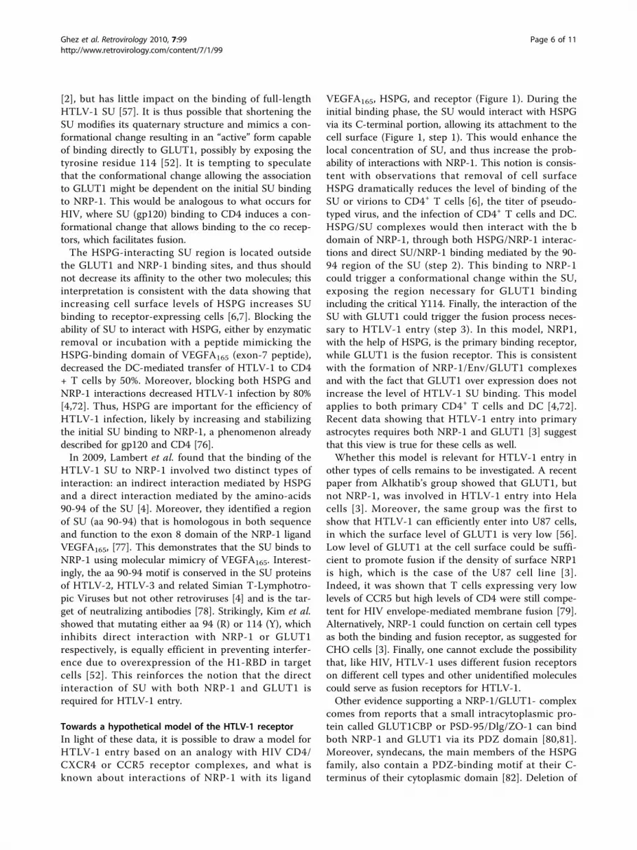
Towards a hypothetical model of the HTLV-1 receptorIn light of these data, it is possible to draw a model forHTLV-1 entry based on an analogy with HIV CD4/CXCR4 or CCR5 receptor complexes, and what isknown about interactions of NRP-1 with its ligand
VEGFA165, HSPG, and receptor (Figure 1). During theinitial binding phase, the SU would interact with HSPGvia its C-terminal portion, allowing its attachment to thecell surface (Figure 1, step 1). This would enhance thelocal concentration of SU, and thus increase the prob-ability of interactions with NRP-1. This notion is consis-tent with observations that removal of cell surfaceHSPG dramatically reduces the level of binding of theSU or virions to CD4+ T cells [6], the titer of pseudo-typed virus, and the infection of CD4+ T cells and DC.HSPG/SU complexes would then interact with the bdomain of NRP-1, through both HSPG/NRP-1 interac-tions and direct SU/NRP-1 binding mediated by the 90-94 region of the SU (step 2). This binding to NRP-1could trigger a conformational change within the SU,exposing the region necessary for GLUT1 bindingincluding the critical Y114. Finally, the interaction of theSU with GLUT1 could trigger the fusion process neces-sary to HTLV-1 entry (step 3). In this model, NRP1,with the help of HSPG, is the primary binding receptor,while GLUT1 is the fusion receptor. This is consistentwith the formation of NRP-1/Env/GLUT1 complexesand with the fact that GLUT1 over expression does notincrease the level of HTLV-1 SU binding. This modelapplies to both primary CD4+ T cells and DC [4,72].Recent data showing that HTLV-1 entry into primaryastrocytes requires both NRP-1 and GLUT1 [3] suggestthat this view is true for these cells as well.Whether this model is relevant for HTLV-1 entry in
other types of cells remains to be investigated. A recentpaper from Alkhatib’s group showed that GLUT1, butnot NRP-1, was involved in HTLV-1 entry into Helacells [3]. Moreover, the same group was the first toshow that HTLV-1 can efficiently enter into U87 cells,in which the surface level of GLUT1 is very low [56].Low level of GLUT1 at the cell surface could be suffi-cient to promote fusion if the density of surface NRP1is high, which is the case of the U87 cell line [3].Indeed, it was shown that T cells expressing very lowlevels of CCR5 but high levels of CD4 were still compe-tent for HIV envelope-mediated membrane fusion [79].Alternatively, NRP-1 could function on certain cell typesas both the binding and fusion receptor, as suggested forCHO cells [3]. Finally, one cannot exclude the possibilitythat, like HIV, HTLV-1 uses different fusion receptorson different cell types and other unidentified moleculescould serve as fusion receptors for HTLV-1.Other evidence supporting a NRP-1/GLUT1- complex
comes from reports that a small intracytoplasmic pro-tein called GLUT1CBP or PSD-95/Dlg/ZO-1 can bindboth NRP-1 and GLUT1 via its PDZ domain [80,81].Moreover, syndecans, the main members of the HSPGfamily, also contain a PDZ-binding motif at their C-terminus of their cytoplasmic domain [82]. Deletion of
Ghez et al. Retrovirology 2010, 7:99http://www.retrovirology.com/content/7/1/99
Page 6 of 11

the PDZ-binding motif of GLUT1 was shown to pre-clude GLUT1 clustering at the plasma membrane [83].GLUT1CBP could therefore act as a “bridge” to stabilizethe entire tripartite receptor complex. It could also pro-vide cues for cytoskeletal polarisation, since it canassociate with several cytoskeletal proteins [80] (Figure1, step 3). Altogether, this model not only reconcilesnearly all of the recent data obtained on the HTLV-1receptor but also appears to explain the in vivo tropismof the virus better than the GLUT1-only hypothesis.
PerspectivesGiven its role in the immune system, the discovery ofNRP-1 as an entry molecule for HTLV-1 opens interest-ing perspectives on viral transmission and the physiol-ogy of HTLV-1 related diseases.
Targeting dendritic cells and disturbing the immuneresponsePlasmacytoid DC (pDC), and to a lesser extent myeloiddendritic cells (myDC), constitutively express high levelsof NRP-1, which is involved in the regulation of DC-Tcell interactions at the immune synapse [70]. Shakingthe commonly accepted idea that HTLV-1 could only betransmitted via cell to cell contact, it was recently foundthat free HTLV-1 virions could efficiently infect DC[72]. The contact of HTLV-1-infected DC with CD4+ Tcells upregulates NRP-1 and HSPG within an hour fol-lowed by transmission of HTLV-1 to T cells and pro-ductive infection. This phenomenon appears to bedependent on both NRP-1 and HSPG expression on thetarget T cells [72]. Viral transmission may occur in cis(from virus produced from infected DCs) but also in
NRP 1
HSPGs
HTLV 1 SU
GLUT1
GLUT1CBP
?
2 Binding ofHSPG/SU l t t
1 Virus attachmentand concentration
3 Stable binding of Env toHSPG/NRP 1 complexes/
4 Recruitment ofGLUT1HSPG/SU clusters to
NRP1and concentration
on HSPGsHSPG/NRP 1 complexes/
NRP 1 dimerizationGLUT1.
Conformationalchange and
exposition of Y114Fusion
Attachment/Binding Binding/FusionFigure 1 Hypothetical model of the HTLV receptor complex and HTLV entry: 1- HSPG interaction with the SU allow the initial attachmentand concentration of the virions at the cell surface. 2- HSPG facilitates the recruitment of the SU to NRP-1. 3- Using molecular mimicry ofVEGFA165, the SU region 90-94 interacts with the b domain of NRP-1, an interaction that is stabilized by HSPG. 4- SU-NRP-1 interaction triggers aconformational change within the SU, allowing exposure of the tyrosine 114 that is critical for binding to GLUT1. Interaction between GLUT1and the SU triggers a conformational change allowing the unmasking of the TM fusion peptide (not depicted). The small adaptor proteinGLUT1CBP might form a link between NRP1 and GLUT1 and stabilize the receptor complex.
Ghez et al. Retrovirology 2010, 7:99http://www.retrovirology.com/content/7/1/99
Page 7 of 11

trans (prior to infection of the DC), as has been pre-viously shown for DC-mediated HIV infection. Thisraises the question of whether DC (in particular pDC)could constitute a reservoir for the virus. Several labora-tories have reported detecting HTLV-1 sequences in DCin vivo [84]. It would be interesting to determinewhether, in contrast to T cells, an active replicationcycle takes place in pDC in vivo.Targeting DC and interfering with NRP-1 signalling
might have several other consequences on viral infec-tion. HTLV-1 infection results in a depletion of myDCas well as pDC and an impairment of their IFNa-produ-cing capacity [85]. Furthermore, the number of pDC isnegatively correlated to the proviral load, suggesting acertain degree of impairment of the antiviral response.It is still not known whether this pDC decrease is dueto true depletion or, as in HIV infection, to their migra-tion into lymphoid tissues. Recent in vitro data demon-strated that cell-free HTLV-1 generates a pDC innateimmune response by an induction of costimulatorymolecules, production of massive levels of IFN-a, andrapid expression of the apoptotic ligand TRAIL. Therole of NRP-1 in this process remains to be clarified.Furthermore, HTLV-1-stimulated pDC was shown toinduce apoptosis of CD4+ T cells expressing DR5, trans-forming pDC into Interferon-producing Killer pDC(IKpDC) [86]. Interaction of HTLV-1 SU binding andNRP-1 in infected cells might by itself disturb DC-Tcells interactions, and thus impair the immune response.This is consistent with previous reports that solubleHTLV-1 SU has an inhibitory effect on a mixed lym-phocyte reaction [22]. This phenomenon might be ofparticular significance during primary infection whenthere is an active transcription of viral products includ-ing Env, which could thus dampen the primary antiviralresponse. Alternatively, IKpDC may reduce spreading ofinfection by killing infected T cells and allowing theemergence of few infected T cell clones in the infectedhost. Finally, infection of immature myDC might disturbT cell homeostasis and lead to an increased risk of auto-immune phenomena.
Potential effects of NRP-1 ligandsVEGFA165
Data clearly show that VEGFA165 is a potent competitorof HTLV-1 SU binding and HTLV-1 infection of pri-mary T cells and DC in vitro [4,72]. VEGFA165 secretionby HTLV-1-infected cells [87] might explain the phe-nomenon of receptor interference. By preventing thegeneralised spreading of the virus, it might also explainthe peculiar course of HTLV-1 infection in which a lim-ited number of T cell clones are infected following pri-mary infection. Because it increases NRP-1 expression atthe surface of target cells [88], VEGFA165 might not
only have a negative effect. Sherer et al. showed in anelegant model of MLV infection that target cells pro-jected filopodiae towards MLV infected cells [89]. Theseexpansions, which the authors termed viral cytonemes,create an intercellular bridge that allows the virus to“slide” towards its target cell and bind to its receptor.Endothelial cells, which have been found to be infectedin vivo [25] and coexpress NRP-1 and VEGFR2, are ableto project similar cytoplasmic expansions, or filopodiae,towards VEGFA165 [90]. Although the relevance of thisphenomenon in other retroviral infections remains to bestudied, a similar mechanism driven by the interactionbetween NRP-1 and either VEGFA165 or Env itselfmight be envisaged with HTLV-1.In ATLL it has been shown that VEGFA165 produc-
tion may participate in cell growth and angiogenesis[91,92]. At this stage of the disease, the NRP-1/VEGFA165 interaction may occur because the virus Envis no longer expressed in vivo.Semaphorin-3aAnother major NRP-1 ligand, sema3a, could also play arole in the regulation of HTLV-1 infection. Polarizationof the cytoskeleton is a critical determinant of transmis-sion in a number of retroviruses including HIV-1 andHTLV-1 [75]. Although Tax has been shown to accu-mulate at the cell-cell junction and be involved in themicrotubule reorganization [84], little is known aboutthe precise mechanisms responsible for this phenom-enon. In the central nervous system, sema3a blocks F-actin polymerization, which results in the repulsion ofthe axon growth cone [93]. In the immune system,sema3a-NRP-1 interactions inhibit the cytoskeletalpolarization following the contact between a DC and aT cell [74]. This process may provide the negative feed-back cues to disorganize the immune synapse, allowingT cells to migrate from DC. In HTLV-1 infection,sema3a could play a negative role by inhibiting thecytoskeletal rearrangement that is necessary for HTLV-1entry. Thus, interaction between the Env and NRP-1may prevent the action of Sema3A on the activated Tcell target, thus allowing HTLV-1 infection.
ConclusionThe recent discovery that GLUT1, NRP-1 and HSPG areinvolved in HTLV-1 entry sheds new light on HTLV-1infection and the physiopathology of HTLV-1-relateddiseases. The demonstration that HTLV-1 uses mole-cules involved in the immune response and can infectdendritic cells more efficiently than CD4+ T cells pro-vides fascinating perspectives for future research.Further work is clearly needed to better define the pre-cise role of the elements of this “ménage à trois” and itsconsequence on infected individuals. A better under-standing of the effect of HTLV-1 infection on the
Ghez et al. Retrovirology 2010, 7:99http://www.retrovirology.com/content/7/1/99
Page 8 of 11

immune system could lead to the development of alter-native strategies in the treatment of HTLV-1-related dis-eases early in the course of the infection or even at alater stage, as in ATLL.
AcknowledgementsThe authors would like to thank the French Foundation “Cent pour sang laVie”, Institut National du cancer (INCA), Association de lutte contre le cancer,ligue nationale contre le cancer, cancéropole d’ile de France, foundationpour la recherche médicale, foundation de France for the financial supportof this work.
Author details1CNRS UMR8147, Universite Rene Descartes, Paris 5, 161 Rue de Sèvres,75743 Paris Cedex 15, France. 2Service d’Hématologie, Institut GustaveRoussy, 39 rue Camille Desmoulins 94805 Villejuif, France. 3SAIC-Frederick,Inc., NCI-Frederick, Frederick, MD, USA. 4Institut Cochin, Université ParisDescartes, CNRS (UMR 8104), Paris, France. 5Inserm, U1016, Paris, France.6Service d’Hématologie Adulte, Hôpital Necker, 161 Rue de Sèvres, 75743Paris CEDEX 15, France.
Authors’ contributionsOH and DG designed the manuscript. DG wrote the manuscript. YL, KSJ, CPrevised and added new information to the manuscript. OH coordinated thework and finalized the manuscript with DG.
Competing interestsThe authors declare that they have no competing interests.
Received: 24 April 2010 Accepted: 29 November 2010Published: 29 November 2010
References1. Jin Q, Agrawal L, VanHorn-Ali Z, Alkhatib G: Infection of CD4+ T
lymphocytes by the human T cell leukemia virus type 1 is mediated bythe glucose transporter GLUT-1: evidence using antibodies specific tothe receptor’s large extracellular domain. Virology 2006, 349:184-196.
2. Manel N, Kim FJ, Kinet S, Taylor N, Sitbon M, Battini JL: The ubiquitousglucose transporter GLUT-1 is a receptor for HTLV. Cell 2003, 115:449-459.
3. Jin Q, Alkhatib B, Cornetta K, Alkhatib G: Alternate receptor usage ofneuropilin-1 and glucose transporter protein 1 by the human T cellleukemia virus type 1. Virology 2009, 396:203-212.
4. Lambert S, Bouttier M, Vassy R, Seigneuret M, Petrow-Sadowski C, Janvier S,Heveker N, Ruscetti FW, Perret G, Jones KS, Pique C: HTLV-1 uses HSPGand neuropilin-1 for entry by molecular mimicry of VEGF165. Blood 2009,113:5176-5185.
5. Ghez D, Lepelletier Y, Lambert S, Fourneau JM, Blot V, Janvier S, Arnulf B,van Endert PM, Heveker N, Pique C, Hermine O: Neuropilin-1 is involved inhuman T-cell lymphotropic virus type 1 entry. J Virol 2006, 80:6844-6854.
6. Jones KS, Petrow-Sadowski C, Bertolette DC, Huang Y, Ruscetti FW: Heparansulfate proteoglycans mediate attachment and entry of human T-cellleukemia virus type 1 virions into CD4+ T cells. J Virol 2005,79:12692-12702.
7. Pinon JD, Klasse PJ, Jassal SR, Welson S, Weber J, Brighty DW, Sattentau QJ:Human T-cell leukemia virus type 1 envelope glycoprotein gp46interacts with cell surface heparan sulfate proteoglycans. J Virol 2003,77:9922-9930.
8. Delamarre L, Rosenberg AR, Pique C, Pham D, Callebaut I, Dokhelar MC:The HTLV-I envelope glycoproteins: structure and functions. J AcquirImmune Defic Syndr Hum Retrovirol 1996, 13(Suppl 1):S85-91.
9. Wyatt R, Sodroski J: The HIV-1 envelope glycoproteins: fusogens,antigens, and immunogens. Science 1998, 280:1884-1888.
10. Kobe B, Center RJ, Kemp BE, Poumbourios P: Crystal structure of human Tcell leukemia virus type 1 gp21 ectodomain crystallized as a maltose-binding protein chimera reveals structural evolution of retroviraltransmembrane proteins. Proc Natl Acad Sci USA 1999, 96:4319-4324.
11. Maerz AL, Center RJ, Kemp BE, Kobe B, Poumbourios P: Functionalimplications of the human T-lymphotropic virus type 1 transmembraneglycoprotein helical hairpin structure. J Virol 2000, 74:6614-6621.
12. Delamarre L, Pique C, Pham D, Tursz T, Dokhelar MC: Identification offunctional regions in the human T-cell leukemia virus type I SUglycoprotein. J Virol 1994, 68:3544-3549.
13. Pique C, Tursz T, Dokhelar MC: Mutations introduced along the HTLV-Ienvelope gene result in a non-functional protein: a basis for envelopeconservation? Embo J 1990, 9:4243-4248.
14. Jassal SR, Lairmore MD, Leigh-Brown AJ, Brighty DW: Soluble recombinantHTLV-1 surface glycoprotein competitively inhibits syncytia formationand viral infection of cells. Virus Res 2001, 78:17-34.
15. Li K, Zhang S, Kronqvist M, Wallin M, Ekstrom M, Derse D, Garoff H:Intersubunit disulfide isomerization controls membrane fusion of humanT-cell leukemia virus Env. J Virol 2008, 82:7135-7143.
16. Sommerfelt MA: Retrovirus receptors. J Gen Virol 1999, 80(Pt 12):3049-3064.17. Fan N, Gavalchin J, Paul B, Wells KH, Lane MJ, Poiesz BJ: Infection of
peripheral blood mononuclear cells and cell lines by cell-free human T-cell lymphoma/leukemia virus type I. J Clin Microbiol 1992, 30:905-910.
18. Derse D, Hill SA, Lloyd PA, Chung H, Morse BA: Examining human T-lymphotropic virus type 1 infection and replication by cell-free infectionwith recombinant virus vectors. J Virol 2001, 75:8461-8468.
19. Richardson JH, Edwards AJ, Cruickshank JK, Rudge P, Dalgleish AG: In vivocellular tropism of human T-cell leukemia virus type 1. J Virol 1990,64:5682-5687.
20. Hanon E, Stinchcombe JC, Saito M, Asquith BE, Taylor GP, Tanaka Y,Weber JN, Griffiths GM, Bangham CR: Fratricide among CD8(+) Tlymphocytes naturally infected with human T cell lymphotropic virustype I. Immunity 2000, 13:657-664.
21. Koyanagi Y, Itoyama Y, Nakamura N, Takamatsu K, Kira J, Iwamasa T, Goto I,Yamamoto N: In vivo infection of human T-cell leukemia virus type I innon-T cells. Virology 1993, 196:25-33.
22. Nath MD, Ruscetti FW, Petrow-Sadowski C, Jones KS: Regulation of thecell-surface expression of an HTLV-I binding protein in human T cellsduring immune activation. Blood 2003, 101:3085-3092.
23. Knight SC, Macatonia SE, Cruickshank K, Rudge P, Patterson S: Dendriticcells in HIV-1 and HTLV-1 infection. Adv Exp Med Biol 1993, 329:545-549.
24. Macatonia SE, Cruickshank JK, Rudge P, Knight SC: Dendritic cells frompatients with tropical spastic paraparesis are infected with HTLV-1 andstimulate autologous lymphocyte proliferation. AIDS Res Hum Retroviruses1992, 8:1699-1706.
25. Setoyama M, Kerdel FA, Elgart G, Kanzaki T, Byrnes JJ: Detection of HTLV-1by polymerase chain reaction in situ hybridization in adult T-cellleukemia/lymphoma. Am J Pathol 1998, 152:683-689.
26. Suzuki M, Matsuoka H, Yamashita K, Maeda K, Kawano K, Uno H,Tsubouchi H: CD45RO expression on peripheral lymphocytes as aprognostic marker for adult T-cell leukemia. Leuk Lymphoma 1998,28:583-590.
27. Ruben S, Poteat H, Tan TH, Kawakami K, Roeder R, Haseltine W, Rosen CA:Cellular transcription factors and regulation of IL-2 receptor geneexpression by HTLV-I tax gene product. Science 1988, 241:89-92.
28. Yamano Y, Takenouchi N, Li HC, Tomaru U, Yao K, Grant CW, Maric DA,Jacobson S: Virus-induced dysfunction of CD4+CD25+ T cells in patientswith HTLV-I-associated neuroimmunological disease. J Clin Invest 2005,115:1361-1368.
29. Lohr J, Knoechel B, Abbas AK: Regulatory T cells in the periphery.Immunol Rev 2006, 212:149-162.
30. Toulza F, Nosaka K, Takiguchi M, Pagliuca T, Mitsuya H, Tanaka Y, Taylor GP,Bangham CR: FoxP3+ regulatory T cells are distinct from leukemia cellsin HTLV-1-associated adult T-cell leukemia. Int J Cancer 2009,125:2375-2382.
31. Abe M, Uchihashi K, Kazuto T, Osaka A, Yanagihara K, Tsukasaki K,Hasegawa H, Yamada Y, Kamihira S: Foxp3 expression on normal andleukemic CD4+CD25+ T cells implicated in human T-cell leukemia virustype-1 is inconsistent with Treg cells. Eur J Haematol 2008, 81:209-217.
32. Yano H, Ishida T, Inagaki A, Ishii T, Kusumoto S, Komatsu H, Iida S,Utsunomiya A, Ueda R: Regulatory T-cell function of adult T-cellleukemia/lymphoma cells. Int J Cancer 2007, 120:2052-2057.
33. Chen S, Ishii N, Ine S, Ikeda S, Fujimura T, Ndhlovu LC, Soroosh P, Tada K,Harigae H, Kameoka J, Kasai N, Sasaki T, Sugamura K: Regulatory T cell-likeactivity of Foxp3+ adult T cell leukemia cells. Int Immunol 2006, 18:269-277.
34. Grant C, Oh U, Yao K, Yamano Y, Jacobson S: Dysregulation of TGF-betasignaling and regulatory and effector T-cell function in virus-inducedneuroinflammatory disease. Blood 2008, 111:5601-5609.
Ghez et al. Retrovirology 2010, 7:99http://www.retrovirology.com/content/7/1/99
Page 9 of 11

35. Toulza F, Heaps A, Tanaka Y, Taylor GP, Bangham CR: High frequency ofCD4+FoxP3+ cells in HTLV-1 infection: inverse correlation with HTLV-1-specific CTL response. Blood 2008, 111:5047-5053.
36. Trejo SR, Ratner L: The HTLV receptor is a widely expressed protein.Virology 2000, 268:41-48.
37. Okuma K, Nakamura M, Nakano S, Niho Y, Matsuura Y: Host range ofhuman T-cell leukemia virus type I analyzed by a cell fusion-dependentreporter gene activation assay. Virology 1999, 254:235-244.
38. Sutton RE, Littman DR: Broad host range of human T-cell leukemia virustype 1 demonstrated with an improved pseudotyping system. J Virol1996, 70:7322-7326.
39. Jassal SR, Pohler RG, Brighty DW: Human T-cell leukemia virus type 1receptor expression among syncytium-resistant cell lines revealed bya novel surface glycoprotein-immunoadhesin. J Virol 2001,75:8317-8328.
40. Manel N, Kinet S, Battini JL, Kim FJ, Taylor N, Sitbon M: The HTLV receptoris an early T-cell activation marker whose expression requires de novoprotein synthesis. Blood 2003, 101:1913-1918.
41. Jones KS, Akel S, Petrow-Sadowski C, Huang Y, Bertolette DC, Ruscetti FW:Induction of human T cell leukemia virus type I receptors on quiescentnaive T lymphocytes by TGF-beta. J Immunol 2005, 174:4262-4270.
42. Moriuchi M, Moriuchi H: Transforming growth factor-beta enhanceshuman T-cell leukemia virus type I infection. J Med Virol 2002,67:427-430.
43. Sommerfelt MA, Williams BP, Clapham PR, Solomon E, Goodfellow PN,Weiss RA: Human T cell leukemia viruses use a receptor determined byhuman chromosome 17. Science 1988, 242:1557-1559.
44. Agadjanyan MG, Chattergoon MA, Petrushina I, Bennett M, Kim J, Ugen KE,Kieber-Emmons T, Weiner DB: Monoclonal antibodies define a cellularantigen involved in HTLV-I infection. Hybridoma 1998, 17:9-19.
45. Imai T, Fukudome K, Takagi S, Nagira M, Furuse M, Fukuhara N,Nishimura M, Hinuma Y, Yoshie O: C33 antigen recognized by monoclonalantibodies inhibitory to human T cell leukemia virus type 1-inducedsyncytium formation is a member of a new family of transmembraneproteins including CD9, CD37, CD53, and CD63. J Immunol 1992,149:2879-2886.
46. Imai T, Yoshie O: C33 antigen and M38 antigen recognized bymonoclonal antibodies inhibitory to syncytium formation by human Tcell leukemia virus type 1 are both members of the transmembrane 4superfamily and associate with each other and with CD4 or CD8 in Tcells. J Immunol 1993, 151:6470-6481.
47. Hildreth JE: Syncytium-inhibiting monoclonal antibodies producedagainst human T-cell lymphotropic virus type 1-infected cells recognizeclass II major histocompatibility complex molecules and block byprotein crowding. J Virol 1998, 72:9544-9552.
48. Daenke S, McCracken SA, Booth S: Human T-cell leukaemia/lymphomavirus type 1 syncytium formation is regulated in a cell-specific mannerby ICAM-1, ICAM-3 and VCAM-1 and can be inhibited by antibodies tointegrin beta2 or beta7. J Gen Virol 1999, 80(Pt 6):1429-1436.
49. Sagara Y, Ishida C, Inoue Y, Shiraki H, Maeda Y: 71-kilodalton heat shockcognate protein acts as a cellular receptor for syncytium formationinduced by human T-cell lymphotropic virus type 1. J Virol 1998,72:535-541.
50. Fang D, Haraguchi Y, Jinno A, Soda Y, Shimizu N, Hoshino H: Heat shockcognate protein 70 is a cell fusion-enhancing factor but not an entryfactor for human T-cell lymphotropic virus type I. Biochem Biophys ResCommun 1999, 261:357-363.
51. Pique C, Lagaudriere-Gesbert C, Delamarre L, Rosenberg AR, Conjeaud H,Dokhelar MC: Interaction of CD82 tetraspanin proteins with HTLV-1envelope glycoproteins inhibits cell-to-cell fusion and virus transmission.Virology 2000, 276:455-465.
52. Kim FJ, Manel N, Garrido EN, Valle C, Sitbon M, Battini JL: HTLV-1 and -2envelope SU subdomains and critical determinants in receptor binding.Retrovirology 2004, 1:41.
53. Kim FJ, Seiliez I, Denesvre C, Lavillette D, Cosset FL, Sitbon M: Definition ofan amino-terminal domain of the human T-cell leukemia virus type 1envelope surface unit that extends the fusogenic range of an ecotropicmurine leukemia virus. J Biol Chem 2000, 275:23417-23420.
54. Manel N, Battini JL, Sitbon M: Human T cell leukemia virus envelopebinding and virus entry are mediated by distinct domains of theglucose transporter GLUT1. J Biol Chem 2005, 280:29025-29029.
55. Coskun AK, Sutton RE: Expression of glucose transporter 1 conferssusceptibility to human T-cell leukemia virus envelope-mediated fusion.J Virol 2005, 79:4150-4158.
56. Jin Q, Agrawal L, Vanhorn-Ali Z, Alkhatib G: GLUT-1-independent infectionof the glioblastoma/astroglioma U87 cells by the human T cell leukemiavirus type 1. Virology 2006, 353:99-110.
57. Takenouchi N, Jones KS, Lisinski I, Fugo K, Yao K, Cushman SW, Ruscetti FW,Jacobson S: GLUT1 is not the primary binding receptor but is associatedwith cell-to-cell transmission of human T-cell leukemia virus type 1. JVirol 2007, 81:1506-1510.
58. Jones KS, Huang YK, Chevalier SA, Afonso PV, Petrow-Sadowski C,Bertolette DC, Gessain A, Ruscetti FW, Mahieux R: The receptor complexassociated with human T-cell lymphotropic virus type 3 (HTLV-3) Env-mediated binding and entry is distinct from, but overlaps with, thereceptor complexes of HTLV-1 and HTLV-2. J Virol 2009, 83:5244-5255.
59. Bishop JR, Schuksz M, Esko JD: Heparan sulphate proteoglycans fine-tunemammalian physiology. Nature 2007, 446:1030-1037.
60. Vives RR, Lortat-Jacob H, Fender P: Heparan sulphate proteoglycans andviral vectors: ally or foe? Curr Gene Ther 2006, 6:35-44.
61. Mondor I, Ugolini S, Sattentau QJ: Human immunodeficiency virus type 1attachment to HeLa CD4 cells is CD4 independent and gp120dependent and requires cell surface heparans. J Virol 1998, 72:3623-3634.
62. Shukla D, Liu J, Blaiklock P, Shworak NW, Bai X, Esko JD, Cohen GH,Eisenberg RJ, Rosenberg RD, Spear PG: A novel role for 3-O-sulfatedheparan sulfate in herpes simplex virus 1 entry. Cell 1999, 99:13-22.
63. Ibrahim J, Griffin P, Coombe DR, Rider CC, James W: Cell-surface heparansulfate facilitates human immunodeficiency virus Type 1 entry intosome cell lines but not primary lymphocytes. Virus Res 1999, 60:159-169.
64. Pais-Correia AM, Sachse M, Guadagnini S, Robbiati V, Lasserre R, Gessain A,Gout O, Alcover A, Thoulouze MI: Biofilm-like extracellular viral assembliesmediate HTLV-1 cell-to-cell transmission at virological synapses. Nat Med2009, 16:83-89.
65. Gauthier S, Pelletier I, Ouellet M, Vargas A, Tremblay MJ, Sato S, Barbeau B:Induction of galectin-1 expression by HTLV-I Tax and its impact onHTLV-I infectivity. Retrovirology 2008, 5:105.
66. Jones KS, Fugo K, Petrow-Sadowski C, Huang Y, Bertolette DC, Lisinski I,Cushman SW, Jacobson S, Ruscetti FW: Human T-cell leukemia virus type1 (HTLV-1) and HTLV-2 use different receptor complexes to enter T cells.J Virol 2006, 80:8291-8302.
67. Sommerfelt MA, Weiss RA: Receptor interference groups of 20retroviruses plating on human cells. Virology 1990, 176:58-69.
68. Xie L, Green PL: Envelope is a major viral determinant of the distinct invitro cellular transformation tropism of human T-cell leukemia virus type1 (HTLV-1) and HTLV-2. J Virol 2005, 79:14536-14545.
69. Reeves JD, Hibbitts S, Simmons G, McKnight A, Azevedo-Pereira JM, Moniz-Pereira J, Clapham PR: Primary human immunodeficiency virus type 2(HIV-2) isolates infect CD4-negative cells via CCR5 and CXCR4:comparison with HIV-1 and simian immunodeficiency virus andrelevance to cell tropism in vivo. J Virol 1999, 73:7795-7804.
70. Tordjman R, Lepelletier Y, Lemarchandel V, Cambot M, Gaulard P,Hermine O, Romeo PH: A neuronal receptor, neuropilin-1, is essential forthe initiation of the primary immune response. Nat Immunol 2002,3:477-482.
71. Ellis LM: The role of neuropilins in cancer. Mol Cancer Ther 2006,5:1099-1107.
72. Jones KS, Petrow-Sadowski C, Huang YK, Bertolette DC, Ruscetti FW: Cell-free HTLV-1 infects dendritic cells leading to transmission andtransformation of CD4(+) T cells. Nat Med 2008, 14:429-436.
73. Soker S, Takashima S, Miao HQ, Neufeld G, Klagsbrun M: Neuropilin-1 isexpressed by endothelial and tumor cells as an isoform-specific receptorfor vascular endothelial growth factor. Cell 1998, 92:735-745.
74. Lepelletier Y, Moura IC, Hadj-Slimane R, Renand A, Fiorentino S, Baude C,Shirvan A, Barzilai A, Hermine O: Immunosuppressive role of semaphorin-3A on T cell proliferation is mediated by inhibition of actin cytoskeletonreorganization. Eur J Immunol 2006, 36:1782-1793.
75. Igakura T, Stinchcombe JC, Goon PK, Taylor GP, Weber JN, Griffiths GM,Tanaka Y, Osame M, Bangham CR: Spread of HTLV-I between lymphocytesby virus-induced polarization of the cytoskeleton. Science 2003,299:1713-1716.
76. Crublet E, Andrieu JP, Vives RR, Lortat-Jacob H: The HIV-1 envelopeglycoprotein gp120 features four heparan sulfate binding domains,
Ghez et al. Retrovirology 2010, 7:99http://www.retrovirology.com/content/7/1/99
Page 10 of 11

including the co-receptor binding site. J Biol Chem 2008,283:15193-15200.
77. Vander Kooi CW, Jusino MA, Perman B, Neau DB, Bellamy HD, Leahy DJ:Structural basis for ligand and heparin binding to neuropilin B domains.Proc Natl Acad Sci USA 2007, 104:6152-6157.
78. Palker TJ, Riggs ER, Spragion DE, Muir AJ, Scearce RM, Randall RR,McAdams MW, McKnight A, Clapham PR, Weiss RA, et al: Mapping ofhomologous, amino-terminal neutralizing regions of human T-celllymphotropic virus type I and II gp46 envelope glycoproteins. J Virol1992, 66:5879-5889.
79. Chanel C, Staropoli I, Baleux F, Amara A, Valenzuela-Fernandez A,Virelizier JL, Arenzana-Seisdedos F, Altmeyer R: Low levels of co-receptorCCR5 are sufficient to permit HIV envelope-mediated fusion with restingCD4 T cells. Aids 2002, 16:2337-2340.
80. Bunn RC, Jensen MA, Reed BC: Protein interactions with the glucosetransporter binding protein GLUT1CBP that provide a link betweenGLUT1 and the cytoskeleton. Mol Biol Cell 1999, 10:819-832.
81. Cai H, Reed RR: Cloning and characterization of neuropilin-1-interactingprotein: a PSD-95/Dlg/ZO-1 domain-containing protein that interactswith the cytoplasmic domain of neuropilin-1. J Neurosci 1999,19:6519-6527.
82. Grootjans JJ, Zimmermann P, Reekmans G, Smets A, Degeest G, Durr J,David G: Syntenin, a PDZ protein that binds syndecan cytoplasmicdomains. Proc Natl Acad Sci USA 1997, 94:13683-13688.
83. Yoshida S, Higuchi M, Shoji T, Yoshita M, Ishioka K, Takahashi M, Oie M,Tanaka Y, Uchiyama M, Fujii M: Knockdown of synapse-associated proteinDlg1 reduces syncytium formation induced by human T-cell leukemiavirus type 1. Virus Genes 2008, 37:9-15.
84. Azakami K, Sato T, Araya N, Utsunomiya A, Kubota R, Suzuki K, Hasegawa D,Izumi T, Fujita H, Aratani S, Fujii R, Yagishita N, Kamijuku H, Kanekura T,Seino K, Nishioka K, Nakajima T, Yamano Y: Severe loss of invariant NKTcells exhibiting anti-HTLV-1 activity in patients with HTLV-1-associateddisorders. Blood 2009 114:3208-15.
85. Hishizawa M, Imada K, Kitawaki T, Ueda M, Kadowaki N, Uchiyama T:Depletion and impaired interferon-alpha-producing capacity of bloodplasmacytoid dendritic cells in human T-cell leukaemia virus type I-infected individuals. Br J Haematol 2004, 125:568-575.
86. Colisson R, Barblu L, Gras C, Raynaud F, Hadj-Slimane R, Pique C,Hermine O, Lepelletier Y, Herbeuval JP: Free HTLV-1 induces TLR7-dependent innate immune response and TRAIL relocalization in killerplasmacytoid dendritic cells. Blood 2009, 115:2177-2185.
87. El-Sabban ME, Merhi RA, Haidar HA, Arnulf B, Khoury H, Basbous J, Nijmeh J,de The H, Hermine O, Bazarbachi A: Human T-cell lymphotropic virus type1-transformed cells induce angiogenesis and establish functional gapjunctions with endothelial cells. Blood 2002, 99:3383-3389.
88. Oh H, Takagi H, Otani A, Koyama S, Kemmochi S, Uemura A, Honda Y:Selective induction of neuropilin-1 by vascular endothelial growth factor(VEGF): a mechanism contributing to VEGF-induced angiogenesis. ProcNatl Acad Sci USA 2002, 99:383-388.
89. Sherer NM, Lehmann MJ, Jimenez-Soto LF, Horensavitz C, Pypaert M,Mothes W: Retroviruses can establish filopodial bridges for efficient cell-to-cell transmission. Nat Cell Biol 2007, 9:310-315.
90. Ruhrberg C, Gerhardt H, Golding M, Watson R, Ioannidou S, Fujisawa H,Betsholtz C, Shima DT: Spatially restricted patterning cues provided byheparin-binding VEGF-A control blood vessel branching morphogenesis.Genes Dev 2002, 16:2684-2698.
91. Bazarbachi A, Abou Merhi R, Gessain A, Talhouk R, El-Khoury H, Nasr R,Gout O, Sulahian R, Homaidan F, de Thé H, Hermine O, El-Sabban ME:Human T-cell lymphotropic virus type I-infected cells extravasatethrough the endothelial barrier by a local angiogenesis-like mechanism.Cancer Res 2004, 15:2039-46.
92. El-Sabban ME, Merhi RA, Haidar HA, Arnulf B, Khoury H, Basbous J, Nijmeh J,de Thé H, Hermine O, Bazarbachi A: Human T-cell lymphotropic virus type1-transformed cells induce angiogenesis and establish functional gapjunctions with endothelial cells. Blood 2002, 99:3383-9.
93. Nakamura F, Tanaka M, Takahashi T, Kalb RG, Strittmatter SM: Neuropilin-1extracellular domains mediate semaphorin D/III-induced growth conecollapse. Neuron 1998, 21:1093-1100.
doi:10.1186/1742-4690-7-99Cite this article as: Ghez et al.: Current concepts regarding the HTLV-1receptor complex. Retrovirology 2010 7:99.
Submit your next manuscript to BioMed Centraland take full advantage of:
• Convenient online submission
• Thorough peer review
• No space constraints or color figure charges
• Immediate publication on acceptance
• Inclusion in PubMed, CAS, Scopus and Google Scholar
• Research which is freely available for redistribution
Submit your manuscript at www.biomedcentral.com/submit
Ghez et al. Retrovirology 2010, 7:99http://www.retrovirology.com/content/7/1/99
Page 11 of 11