Different Fonts Increase Reading Speed for Different Individuals

Cell Transplantation, Vol. 20, pp. 1179–1192, 2011 0963-6897/11 $90.00 + .00Printed in the USA. All rights reserved. DOI: http://dx.doi.org/10.3727/096368910X552826Copyright 2011 Cognizant Comm. Corp. E-ISSN 1555-3892
www.cognizantcommunication.com
Bone Marrow Contributes Simultaneously to Different Neural Types inthe Central Nervous System Through Different Mechanisms of Plasticity
Javier S. Recio,*1 Manuel Alvarez-Dolado,†1 David Dıaz,* Fernando C. Baltanas,*Marina Piquer-Gil,† Jose R. Alonso,*‡ and Eduardo Weruaga*
*Laboratory of Neuronal Plasticity and Neurorepair, Institute for Neuroscience of Castilla y Leon,Universidad de Salamanca, Salamanca, Spain
†Laboratory of Cell Therapy for Neuropathologies, Andalucian Center for Molecular Biology and Regenerative Medicine,CABIMER, Seville, Spain
‡Universidad de Tarapaca en Arica, Arica, Chile
Many studies have reported the contribution of bone marrow-derived cells (BMDC) to the CNS, raising thepossibility of using them as a new source to repair damaged brain tissue or restore neuronal function. Thisprocess has mainly been investigated in the cerebellum, in which a degenerative microenvironment has beensuggested to be responsible for its modulation. The present study further analyzes the contribution of BMDCto different neural types in other adult brain areas, under both physiological and neurodegenerative condi-tions, together with the mechanisms of plasticity involved. We grafted genetically marked green fluorescentprotein/Cre bone marrow in irradiated recipients: a) the PCD (Purkinje Cell Degeneration) mutant mice,suffering a degeneration of specific neuronal populations at different ages, and b) their corresponding healthycontrols. These mice carried the conditional lacZ reporter gene to allow the identification of cell fusionevents. Our results demonstrate that BMDC mainly generate microglial cells, although to a lesser extent aclear formation of neuronal types also exists. This neuronal recruitment was not increased by the neurode-generative processes occurring in PCD mice, where BMDC did not contribute to rescuing the degeneratedneuronal populations either. However, an increase in the number of bone marrow-derived microglia wasfound along the life span in both experimental groups. Six weeks after transplantation more bone marrow-derived microglial cells were observed in the olfactory bulb of the PCD mice compared to the controlanimals, where the degeneration of mitral cells was in process. In contrast, this difference was not observedin the cerebellum, where Purkinje cell degeneration had been completed. These findings demonstrated thatthe degree of neurodegenerative environment can foster the recruitment of neural elements derived frombone marrow, but also provide the first evidence that BMDC can contribute simultaneously to differentencephalic areas through different mechanisms of plasticity: cell fusion for Purkinje cells and differentiationfor olfactory bulb interneurons.
Key words: Bone marrow transplantation; Cell fusion; Neural repair; Neurodegeneration;Neural differentiation
INTRODUCTION tic strategies to treat degenerative, posttraumatic, andhereditary diseases (3,9). Thus, the contribution of BMDCto diverse tissues has been described under normal andIn recent years, it has been shown that bone marrow-
derived cells (BMDC) owe their plasticity to changes in pathological conditions. The contribution rate was al-ways very scarce (2,42), but this low “basal” frequency,their fate, which allows their contribution to different
cell populations in diverse organs (7,19,22,26,40,41,48), described under physiological conditions, was signifi-cantly increased when the target organs underwent in-including the central nervous system (CNS) (2,8,9,34,
54). This surprising capacity has been considered a po- jury (2,27,34,56), especially when accompanied by in-flammation (23,38).tential tool for the development of innovative therapeu-
Received February 1, 2010; final acceptance November 22, 2010. Online prepub date: February 3, 2011.1These authors provided equal contribution to this work.Address correspondence to Eduardo Weruaga, Ph.D., Laboratorio de Plasticidad Neuronal y Neurorreparacion, Instituto de Neurociencias de Castillay Leon, Universidad de Salamanca, C/ Pintor Fernando Gallego, No. 1, E-37007 Salamanca, Spain. Tel: +34-923-294500, ext. 5324; Fax: +34-923294549; E-mail: [email protected]
1179

1180 RECIO ET AL.
In the CNS, the arrival of BMDC at the brain has nism involved in this process, the PCD mouse line wascrossed with the conditional Cre reporter mouse line,mainly been studied in the cerebellum, where they con-
tribute to the Purkinje cell population in animal models R26R (2,31). In this line, the lacZ reporter gene is ex-pressed exclusively after CRE recombinase excision of(2,43,50,54) and humans (35,53). Similar to other or-
gans, this contribution of BMDC seems to be increased a loxP-flanked stop cassette. As bone marrow donors weused mice constitutively expressing CRE recombinaseby a neurodegenerative or injured microenvironment
(3,30). The underlying mechanism responsible for this and green fluorescent protein (GFP) under the control ofthe β-actin promoter. This transplant strategy has beenprocess has long been a very controversial issue, and
two main possible plasticity mechanisms have been pro- used previously with success to discern between cell fu-sion and differentiation events (2,31).posed: differentiation (24,40,42) and cell fusion (2,39,
47,51,54). They are not mutually exclusive. However, Our data indicated that the contribution of BMDC tothe formation of new neuronal elements in the encepha-despite this, the lack of studies in which both processes
are analyzed together and discerned under exactly the lon of adult mice was not restricted to the Purkinje cellsof the cerebellum, but also took place in OB interneur-same conditions has reinforced the controversy. Recent
evidence supports the cell-fusion mechanism for the ons. Unexpectedly, the neurodegenerative environmenthad no effect on the behavior of BMDC. In contrast, andgeneration of Purkinje cells (23,30,38). However, very
little is known about the possible plasticity mechanisms very interestingly, the contribution of BMDC occurredthrough two different plasticity mechanisms, dependingfor other neuronal types. Thus, encephalic regions other
than the cerebellum require a more extensive review. on the region and cellular type: cell fusion for the Pur-kinje cells of the cerebellum, and differentiation for theThis is the case of the olfactory bulb (OB), where previ-
ous works studying the contribution of BMDC are OB interneurons. This strongly suggests that plasticitymechanisms may be modulated by region- and cell type-scarce and have provided confusing or inconsistent data
(8,11,32,43). This region is characterized by a continu- specific factors.ous recruitment of new neurons along adulthood, whichalso may influence the behavior, differentiation, and ar- MATERIALS AND METHODSrival of BMDC. Moreover, no previous studies have
Animalsbeen performed in animals suffering from neurodegener-ation in the OB, and hence an exhaustive study of the Recipients were obtained from the PCD-R26R mouse
colony. This colony was established at the Animal Re-behavior of BMDC after their arrival at this highly plas-tic encephalic region, in comparison with others, under search Facility of the University of Salamanca in two
steps. First, in order to be able to genotype the pcd1Jboth healthy and diseased conditions should help us tobetter understand the contribution of BMDC to the brain. mutation, heterozygous C57BL/6J mice (+/pcd) from a
colony originally obtained from Jackson LaboratoriesAccordingly, in this work we further analyzed the ef-fect of different neurodegenerative stages on the contri- were mated with wild-type DBA/2J females, establish-
ing the PCD colony. Thus, the pcd1J allele was associ-bution of BMDC to several brain regions, together withthe plasticity mechanism(s) involved. As in previous ated with the genetic background of the C57BL/6J
strain, and the wild-type one with the genetic back-work in the field, we performed intravenous transplanta-tion of genetically marked bone marrow in lethally irra- ground of the DBA/2J strain. Mice were genotyped by
PCR amplification of specific microsatellite sequencesdiated mice. The recipients were both wild-type and mu-tant mice from the Purkinje Cell Degeneration (PCD) that show intraspecific divergences. Second, in order to
obtain wild-type and pcd/pcd animals carrying the CREmouse line. Homozygous recessive mice (pcd/pcd) un-dergo a selective postnatal neurodegeneration in several reporter lacZ gene (2,31), +/pcd animals (from the PCD
colony) were crossed with R26R mice (JAX 003474).encephalic areas at different ages (16,36,44). They losethe Purkinje cells around postnatal day 20, which causes Donors were obtained from the PCD-(CRE-GFP)
mouse colony. Homozygous mice expressing CRE re-severe ataxia (36). In addition, they have a selective lossof OB mitral cells, starting around 60 days after birth combinase under the control of a hybrid regulatory ele-
ment of the cytomegalovirus enhancer β-actin promoter(18). Prior to this age, the cellular and synaptic organiza-tion in the OB seems to be normal (4,17,18). Thus, anal- (28), and homozygous mice expressing GFP under the
same promoter (20) were bred to generate β-actin CRE-ysis of the cerebellum and the OB of these mutant ani-mals grafted with wild-type bone marrow allowed us to GFP mice. Furthermore, to prevent allograft rejection,
the mice were crossed with +/pcd animals (from thestudy the possible effect of different neurodegenerativeenvironments on the arrival and contribution of the PCD colony), increasing the consanguinity between do-
nors and recipients (Fig. 1). The experiments were car-BMDC to these areas.Additionally, in order to discern the plasticity mecha- ried out in accordance with the guidelines of the Euro-

DIFFERENT WAYS OF BONE MARROW CONTRIBUTION TO THE CNS 1181
Figure 1. Chart depicting the work schema to demonstrate both the destination and cell mechanism for BMDC to be integratedinto nervous tissue. Donor mice contain GFP, the Cre transgene, or both, while the recipient mice carry the conditional floxedreporter gene lacZ in the R26R loci. Recipient mice were wild-type or exhibit a natural neurodegenerative process (PCD). Afterirradiation, all the recipient mice were transplanted with donor bone marrow, and the brains of the resultant chimeric mice wereanalyzed and compared.
pean Community Council (86/609/EEC) and current min of incubation in ice-cold ammonium chloride (140mM in Tris 17 mM). After a double wash and mild cen-Spanish legislation for the use and care of laboratory
animals (RD 1201/2005). The protocols for animal care trifugation, 7.5 × 10⁶ bone marrow cells were injectedintravenously into the tail vein in both the wild-type andand manipulation used were approved by the Bioethics
Committee of the University of Salamanca. pcd/pcd R26R mice. Animals undergoing bone marrowtransplantation (BMT) were lethally irradiated to re-
Bone Marrow Transplantation move their own bone marrow 24 h before grafting. Ow-ing to their particular characteristics (fairly young—Wild-type bone marrow cells were collected from
4–8-week-old donor mice by flushing tibias and femurs P19—and some of them PCD mutant animals), wepreviously tested different doses of irradiation (unpub-with Iscove’s modified Dulbecco’s medium (IMDM; In-
vitrogen). Red blood cells were subsequently lysed by 5 lished data) in order to select the minimum lethal dose

1182 RECIO ET AL.
for these P19 animals (7.5 Gy). Moreover, to ensure the sections were obtained with a freezing microtome andwere collected in alternating series. Representative sec-survival of the transplanted PCD mutant mice, we per-
formed whole BMT, without sorting its populations. tions were stained with X-gal staining or immunohisto-chemistry. Medial blocks were frozen and stored at −80°C
Flow Cytometry for future experiments.Donor engraftment was determined by flow cytome-
X-gal Staining and Immunohistochemistrytry of peripheral blood (FACS Calibur, BD Biosci-ences). Peripheral blood was subjected to red blood cell To identify cell-fusion events, sections were pro-
cessed with X-gal staining by placing them in PB con-lysis by treatment with ammonium chloride (see above),washed, and resuspended in phosphate buffered saline taining 10 mM K3Fe(CN)6 and 10 mM K4Fe(CN)6
together with the β-gal substrate, 5-bromo-4-chloro-3-(PBS). Cytometric analysis was performed twice: 2weeks after grafting and on the day of sacrifice (see be- indolyl-β-D-galactopyranoside (X-gal; 1 mg/ml; Molec-
ular Probes), at 37°C overnight. Then, to characterizelow). Recipients showing a donor chimerism <50% inthe first sample were rejected. As indicated in Table 1, the X-gal-positive cells, selected sections were pro-
cessed immunohistochemically with anti-Iba1 antibodyall transplanted mice analyzed in this study showed sig-nificant levels of multilineage chimerism of peripheral [goat (1:500), from Everest Biotech, or rabbit (1:750),
from Dako], or anti-GFAP [rabbit (1:2,000), fromblood leukocytes.Sigma], or anti-calbindin [CB; mouse (1:2,000) or rabbit
Tissue Collection (1:7,000), from Swant], or anti-Reelin [mouse (1:1,000),At P60, P110, and P150, groups of experimental ani- from Chemicon] in a medium containing 0.2% Triton
mals were deeply anaesthetized intraperitoneally with a X-100 (Probus) and 5% normal donkey serum (Vector)solution composed of ketamine [120 µg/g body weight in PBS overnight at RT. Biotinylated donkey anti-goat,(b.wt.)] and xylazine (10 µg/g b.wt.) and were perfused rabbit, or mouse IgG secondary antibodies (1:200) werethrough the ascending aorta, first with 0.9% saline for from Jackson ImmunoResearch, and avidin-biotin-1 min and then with 50 ml of fixative containing 4% peroxidase complex (Kit Elite ABC; 1:200) was fromparaformaldehyde (for general immunofluorescence) or Vector. The reaction product was visualized by incubat-2% paraformaldehyde + 0.25% glutaraldehyde (for X- ing sections in 0.02% 3,3′-diaminobenzidine (DAB) andgal histochemistry) in 0.1 M phosphate buffer (PB). 0.003% H2O2 in 0.1 M Tris-HCl buffer.Encephala were dissected out, cut into blocks (rostral,
Immunofluorescenceincluding the OB; medial and caudal, including the cere-bellum) and immersed in the same fixative solution for Double or triple immunofluorescence was used to
identify GFP+ cells, along with other cellular antigens20 min at room temperature (RT). Then, the blocks wererinsed in PB and cryoprotected with 30% sucrose in PB for both neurons (CB and Reelin) and glial cells (GFAP
and Iba1). Sections were incubated with 0.2% Triton X-overnight at 4°C. Rostral and caudal blocks were cut in40-µm coronal or sagittal sections, respectively; serial 100 and 5% normal serum and were costained with anti-
Table 1. Summary of the Different Experimental Animals Employed and the Experimental ProceduresFollowed in the Present Study
No. of % GFP+ Blood Degenerative StageNo. of Transplanted Sacrifice Cells at DeathAnimals Genotype Treatment Cells Age (Mean ± SEM) Cerebellum OB
4 +/+ BMT P20 7.5 × 10⁶ P60 59.5 ± 2.4 N N4 +/+ BMT P20 7.5 ± 106 P110 65.7 ± 2.9 N N4 +/+ BMT P20 7.5 × 10⁶ P150 74.8 ± 1.7 N N4 pcd/pcd NT — P60 — PD N4 pcd/pcd NT — P110 — PD D4 pcd/pcd NT — P150 — PD PD4 pcd/pcd BMT P20 7.5 × 10⁶ P60 58.2 ± 0.9 PD N4 pcd/pcd BMT P20 7.5 × 10⁶ P110 64.8 × 1.8 PD D4 pcd/pcd BMT P20 7.5 × 10⁶ P150 72.7 ± 4.9 PD PD
Genotype: +/+, wild-type; pcd/pcd, PCD. Treatment: BMT, bone marrow transplantation; NT, not treated. Degenera-tive stage at sacrifice age of both Purkinje cells (cerebellum) and mitral cells (OB): N, normal; D, degeneration; PD,postdegeneration.

DIFFERENT WAYS OF BONE MARROW CONTRIBUTION TO THE CNS 1183
1.35) equipped with an Olympus DP70 digital camera.Selected sections were also examined with a confocalmicroscope (Leica TCS SP spectral confocal micro-scope; pinhole opening up to 1.5 Airy; Ar/ArKr and He/Ne laser excitation and Planapochromatic objectives upto 63× N.A. 1.4). Digital images were processed withAdobe Photoshop 7.0 (Adobe Systems) to adjust con-trast slightly and assemble the final plates.
Cell Counting and Statistical Comparison
The same animals were used for analyzing both theolfactory bulb and cerebellum. All GFP+ Purkinje cellsand OB interneurons were counted immediately after theobservation of all sections, since their numbers werelow. However, to compare the quantities of GFP+ micro-glial cells anatomically comparable sections were se-lected. Both the OB and cerebella of PCD mice undergoa dramatic shrinkage (4,14,25,33), which would makethe comparison of cell density values unfounded foranalyses. Thus, we chose the number of cells per sectionas the morphometric value for comparisons between ex-perimental groups. For the cerebellum, two purely sagit-tal sections were chosen (where the 10 cerebellar lobuleswhere clearly seen). Regarding the OB, a central coronalsection was chosen for comparisons, as previously de-scribed (14,55). Four animals from each experimentalgroup were analyzed. Therefore, these values representan estimation of the arrival of BMDC to specific re-gions, without the influence of their shrinkage due tothe pcd1J mutation. GFP+ cells were counted with the
Figure 2. Epifluorescence microscopy images from encephalic Neurolucida and Neuroexplorer programs (MicroBrigh-sections of a P150 wild-type mouse transplanted with GFP Field, Magdeburg, Germany).bone marrow. These images show the contribution of the bone All counts were performed by the same personmarrow to the microglial cell population, both resting microg-
(D.D.), following the same criterion and using a double-lial cells (A–C) and activated ones (D–F). Resting and acti-blind study. Once homoscedasticity had been checkedvated cells were identified by their morphological characteris-
tics. Upper images (A, D) show GFP labeling, which allows with the Kolmogorov-Smirnov test, a one-way ANOVAthe identification of the newly generated cells coming from the test was employed to analyze possible differencestransplanted bone marrow. Images in the central portion (B, among different graft survival times within each geneticE) exhibit the Iba1 signal (microglial cell specific marker).
group; Bonferroni post hoc test was performed to makeLower photographs (C, F) correspond to the merging of boththe simple comparisons. Following this, Student’s t-testprevious images for each kind of microglial cell. Scale bar: 20
µm. was performed in order to assess possible differencesbetween PCD and wild-type mice within each survivaltime.
GFP antibody [goat (1:2,000) or rabbit (1:4,000), fromRESULTSAbCam] and the above described primary antibodies.
Secondary antibodies (Cy-2, Cy-3, Cy-5 conjugated) In order to analyze the effect of different neurodegen-erative stages on the contribution of BMDC to the cere-were used at a dilution 1:500 (from Jackson Immunore-
search). Nuclei were counterstained with DAPI (Gibco- bellum and olfactory bulb, we performed BMT in 20-day-old normal and PCD mice (see Materials and MethodsBRL) or propidium iodide (PI; Sigma).and Fig. 1). At this age, Purkinje cell degeneration in
Microscopy Analysis PCD animals has just started (36), while OB mitral cellsare still alive (4,17). After transplantation, the animalsProcessed sections were analyzed for the coexpres-
sion of the indicated markers under an Olympus Provis were sacrificed at three different ages: P60, P110, andP150. At P60 (40 days posttransplantation), PurkinjeAX70 microscope (UPlanSApo lens up to 60× N.A.
1184 RECIO ET AL.
Table 2. Number of GFP+ Microglial Cells After Bone Marrow Transplantation
Cerebellum (n = 4) Olfactory Bulb (n = 4)
Age Wild-Type PCD Wild-Type PCD
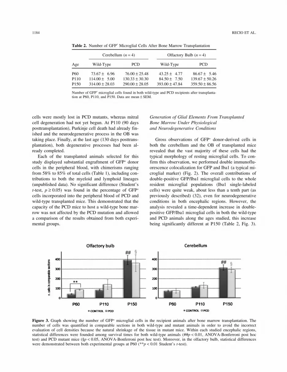
P60 73.67 ± 6.96 76.00 ± 25.48 43.25 ± 4.77 86.67 ± 5.46P110 114.00 ± 5.00 130.33 ± 30.30 84.50 ± 7.50 139.67 ± 50.26P150 314.00 ± 28.03 290.00 ± 28.05 393.00 ± 47.84 359.50 ± 86.56
Number of GFP+ microglial cells found in both wild-type and PCD recipients after transplanta-tion at P60, P110, and P150. Data are mean ± SEM.
cells were mostly lost in PCD mutants, whereas mitral Generation of Glial Elements From TransplantedBone Marrow Under Physiologicalcell degeneration had not yet begun. At P110 (90 days
posttransplantation), Purkinje cell death had already fin- and Neurodegenerative Conditionsished and the neurodegenerative process in the OB wastaking place. Finally, at the last age (130 days posttrans- Gross observations of GFP+ donor-derived cells in
both the cerebellum and the OB of transplanted miceplantation), both degenerative processes had been al-ready completed. revealed that the vast majority of these cells had the
typical morphology of resting microglial cells. To con-Each of the transplanted animals selected for thisstudy displayed substantial engraftment of GFP+ donor firm this observation, we performed double immunoflu-
orescence colocalization for GFP and Iba1 (a typical mi-cells in the peripheral blood, with chimerisms rangingfrom 58% to 85% of total cells (Table 1), including con- croglial marker) (Fig. 2). The overall contributions of
double-positive GFP/Iba1 microglial cells to the wholetributions to both the myeloid and lymphoid lineages(unpublished data). No significant difference (Student’s resident microglial populations (Iba1 single-labeled
cells) were quite weak, about less than a tenth part (ast-test, p ≥ 0.05) was found in the percentage of GFP+
cells incorporated into the peripheral blood of PCD and previously described) (32), even for neurodegenerativeconditions in both encephalic regions. However, thewild-type transplanted mice. This demonstrated that the
capacity of the PCD mice to host a wild-type bone mar- analysis revealed a time-dependent increase in double-positive GFP/Iba1 microglial cells in both the wild-typerow was not affected by the PCD mutation and allowed
a comparison of the results obtained from both experi- and PCD animals along the ages studied, this increasebeing significantly different at P150 (Table 2, Fig. 3).mental groups.
Figure 3. Graph showing the number of GFP+ microglial cells in the recipient animals after bone marrow transplantation. Thenumber of cells was quantified in comparable sections in both wild-type and mutant animals in order to avoid the incorrectevaluation of cell densities because the natural shrinkage of the tissue in mutant mice. Within each studied encephalic regions,statistical differences were founded among survival times for both wild-type animals (##p < 0.01, ANOVA-Bonferroni post hoctest) and PCD mutant mice (§p < 0.05, ANOVA-Bonferroni post hoc test). Moreover, in the olfactory bulb, statistical differenceswere demonstrated between both experimental groups at P60 (**p < 0.01 Student’s t-test).

DIFFERENT WAYS OF BONE MARROW CONTRIBUTION TO THE CNS 1185
Figure 4. Epifluorescence microscopy images of cerebellum sections from a P150 wild-type mouse (A, B) and a P150 PCD mouse,both grafted with GFP bone marrow, showing the GFP+ cells. (A) Panoramic image showing the distribution and abundance of thecells newly generated from the transplanted bone marrow in the cerebellum. A GFP+ Purkinje cell can be observed. (B) Magnifica-tion of the previous image and allows the assessment in greater detail of both the GFP+ Purkinje cell and the surrounding microglialcells. This GFP+ Purkinje cell has a very complex dendritic tree and a long axon (indicated with arrows), presenting direct evidenceof its integration in the cerebellum. Moreover, the axon presents the typical recurrent collaterals (arrowheads) that characterizePurkinje cells. Furthermore, microglial cells were basically located in a peripheral position. Some of these microglial cells areindicated with asterisks. (C) The same region of a transplanted PCD mouse, showing the location of the newly generated cells inthe cerebellum of these mutant animals. Scale bars: 200 µm (A) and 100 µm (B, C).
Most of these cells were located peripherally, close to To investigate whether GFP+ microglial cells weregenerated by cell fusion, we performed X-gal stainingthe meninges, but also in perivascular locations of the
cerebellum and the OB (Fig. 4). Furthermore, in the to detect the lacZ expression resulting from the recombi-nation of floxed sequences after a fusion event. Wetransplanted PCD mice, the neurodegenerative environ-
ment of the OB at P60, and to a lesser extent at P110, never observed GFP+ microglial cells colocalized withX-Gal staining in any brain region of either experimen-led to significant differences in the contribution of
BMDC to the generation of microglial cells in compari- tal group. Accordingly, the contribution of BMDC tomicroglial cells must occur through a differentiation pro-son with age-mated transplanted wild-type animals (p <
0.01 at P60) (Fig. 3), coinciding with the time window cess.Finally, the possible formation of astrocytes from theof mitral cells degeneration (18,49).
1186 RECIO ET AL.
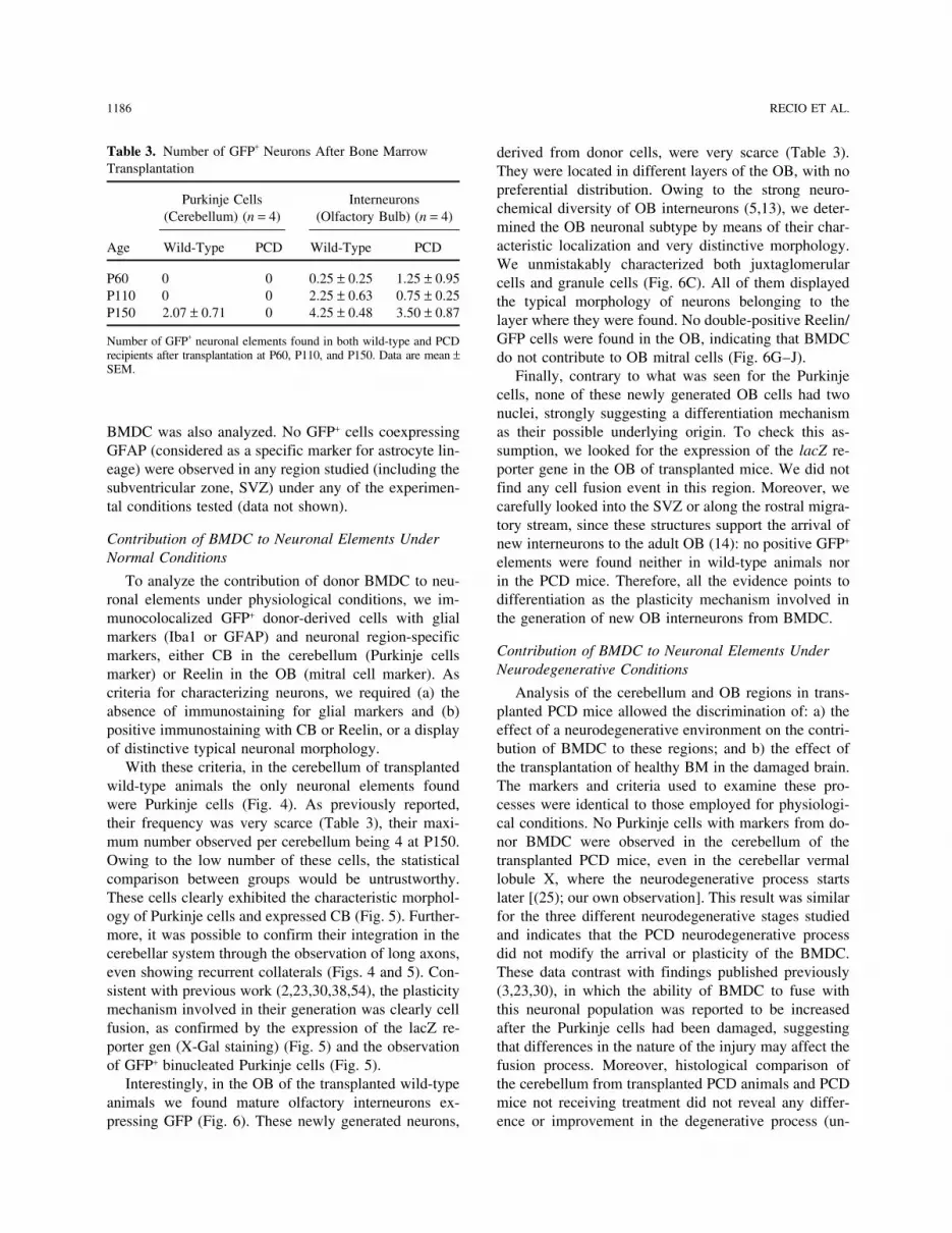
Table 3. Number of GFP+ Neurons After Bone Marrow derived from donor cells, were very scarce (Table 3).Transplantation They were located in different layers of the OB, with no
preferential distribution. Owing to the strong neuro-Purkinje Cells Interneurons
chemical diversity of OB interneurons (5,13), we deter-(Cerebellum) (n = 4) (Olfactory Bulb) (n = 4)mined the OB neuronal subtype by means of their char-acteristic localization and very distinctive morphology.Age Wild-Type PCD Wild-Type PCDWe unmistakably characterized both juxtaglomerular
P60 0 0 0.25 ± 0.25 1.25 ± 0.95 cells and granule cells (Fig. 6C). All of them displayedP110 0 0 2.25 ± 0.63 0.75 ± 0.25 the typical morphology of neurons belonging to theP150 2.07 ± 0.71 0 4.25 ± 0.48 3.50 ± 0.87 layer where they were found. No double-positive Reelin/
GFP cells were found in the OB, indicating that BMDCNumber of GFP+ neuronal elements found in both wild-type and PCDrecipients after transplantation at P60, P110, and P150. Data are mean ± do not contribute to OB mitral cells (Fig. 6G–J).SEM. Finally, contrary to what was seen for the Purkinje
cells, none of these newly generated OB cells had twonuclei, strongly suggesting a differentiation mechanismas their possible underlying origin. To check this as-BMDC was also analyzed. No GFP+ cells coexpressing
GFAP (considered as a specific marker for astrocyte lin- sumption, we looked for the expression of the lacZ re-porter gene in the OB of transplanted mice. We did noteage) were observed in any region studied (including the
subventricular zone, SVZ) under any of the experimen- find any cell fusion event in this region. Moreover, wecarefully looked into the SVZ or along the rostral migra-tal conditions tested (data not shown).tory stream, since these structures support the arrival of
Contribution of BMDC to Neuronal Elements Under new interneurons to the adult OB (14): no positive GFP+
Normal Conditions elements were found neither in wild-type animals norin the PCD mice. Therefore, all the evidence points toTo analyze the contribution of donor BMDC to neu-
ronal elements under physiological conditions, we im- differentiation as the plasticity mechanism involved inthe generation of new OB interneurons from BMDC.munocolocalized GFP+ donor-derived cells with glial
markers (Iba1 or GFAP) and neuronal region-specificContribution of BMDC to Neuronal Elements Undermarkers, either CB in the cerebellum (Purkinje cellsNeurodegenerative Conditionsmarker) or Reelin in the OB (mitral cell marker). As
criteria for characterizing neurons, we required (a) the Analysis of the cerebellum and OB regions in trans-planted PCD mice allowed the discrimination of: a) theabsence of immunostaining for glial markers and (b)
positive immunostaining with CB or Reelin, or a display effect of a neurodegenerative environment on the contri-bution of BMDC to these regions; and b) the effect ofof distinctive typical neuronal morphology.
With these criteria, in the cerebellum of transplanted the transplantation of healthy BM in the damaged brain.The markers and criteria used to examine these pro-wild-type animals the only neuronal elements found
were Purkinje cells (Fig. 4). As previously reported, cesses were identical to those employed for physiologi-cal conditions. No Purkinje cells with markers from do-their frequency was very scarce (Table 3), their maxi-
mum number observed per cerebellum being 4 at P150. nor BMDC were observed in the cerebellum of thetransplanted PCD mice, even in the cerebellar vermalOwing to the low number of these cells, the statistical
comparison between groups would be untrustworthy. lobule X, where the neurodegenerative process startslater [(25); our own observation]. This result was similarThese cells clearly exhibited the characteristic morphol-
ogy of Purkinje cells and expressed CB (Fig. 5). Further- for the three different neurodegenerative stages studiedand indicates that the PCD neurodegenerative processmore, it was possible to confirm their integration in the
cerebellar system through the observation of long axons, did not modify the arrival or plasticity of the BMDC.These data contrast with findings published previouslyeven showing recurrent collaterals (Figs. 4 and 5). Con-
sistent with previous work (2,23,30,38,54), the plasticity (3,23,30), in which the ability of BMDC to fuse withthis neuronal population was reported to be increasedmechanism involved in their generation was clearly cell
fusion, as confirmed by the expression of the lacZ re- after the Purkinje cells had been damaged, suggestingthat differences in the nature of the injury may affect theporter gen (X-Gal staining) (Fig. 5) and the observation
of GFP+ binucleated Purkinje cells (Fig. 5). fusion process. Moreover, histological comparison ofthe cerebellum from transplanted PCD animals and PCDInterestingly, in the OB of the transplanted wild-type
animals we found mature olfactory interneurons ex- mice not receiving treatment did not reveal any differ-ence or improvement in the degenerative process (un-pressing GFP (Fig. 6). These newly generated neurons,

DIFFERENT WAYS OF BONE MARROW CONTRIBUTION TO THE CNS 1187
Figure 5. Fluorescence microscopy images of cerebellum sections from wild-type mice grafted with wild-type bone marrow fromCRE-GFP donors showing some examples of Purkinje cells originated from the transplanted bone marrow. (A, D) Two perfectlydifferentiated different GFP+ Purkinje cells. The arrowhead in (A) indicates the axon. (B, E) The calbindin (CB) labeling for theprevious images, and (C, F) the corresponding merging. Purkinje cells produced from the grafted bone marrow not only show thetypical morphology of this specific cell type, but also express CB, like the other Purkinje cells. (G–I) The X-gal staining of acerebellar section from a wild-type R26R mouse grafted with CRE-GFP bone marrow. (G) A Purkinje cell that has undergone afusion cell event with a BMC. (H) Magnification of the fused cell in (G), and (I) the double staining of that Purkinje cell with X-gal and CB (with the DAB immunohistochemical technique). Images at the bottom (J–L) represent high magnifications of the somafrom a GFP+ Purkinje cell (J), the staining of this region with the nuclear marker propidium iodide (PI; K), and the merging ofboth stainings (L). Arrowheads in (K) indicate the two nuclei belonging to the previous GFP+ Purkinje cell, suggesting that it is abinucleate and presumably polyploid cell. Scale bar in (C): 100 µm for (A–F), 200 µm for (G), and 50 µm for (H, I). Scale bar in(L): 20 µm for (J–L).
published data). This was confirmed by the absence of generation either, since the results were similar to thoseobtained in wild-type transplanted mice (Table 3): inPurkinje cells (CB positive) in all the mutant mice ana-
lyzed, with or without BMDC transplantation. normal mice mature olfactory interneurons expressingGFP were also observed in the OB of some transplantedAdditionally, analysis of the OB of transplanted PCD
mice (Fig. 6) suggested that the sparse formation of neu- PCD mice at similar ages (Fig. 6C, F). However, noneof these newly generated cells expressed Reelin or dis-rons from BMDC was not influenced by mitral cell de-

1188 RECIO ET AL.
Figure 6. Fluorescence microscopy images of olfactory bulb (OB) sections from wild-type (A–C, G–J) and PCD (D–F) micegrafted with wild-type bone marrow from a CRE-GFP donor showing some examples of the typical neuronal types of this regionoriginated from the transplanted bone marrow. (A, D) The newly generated cells (GFP+) in the different OB layers in both wild-type (A) and PCD (D) recipients. (B, E) The same sections counterstained with DAPI to display the location of GFP+ cells and thelaminar organization of this region in both experimental groups. (C) A GFP+ cell from a grafted wild-type mouse that can beclassified as a granule cell, considering its typical morphology and location in the OB. The white arrow points to its soma and thewhite arrowheads to its typical prolongation. (F) A GFP+ cell from a grafted PCD mouse that can be sorted, by its location andmorphology in the OB, as a juxtaglomerular cell. The white arrow points to its soma and the white arrowheads to its typicalprolongation towards an olfactory glomerulus. (G–J) Laser scanning confocal images from the OB of a mouse grafted with GFPbone marrow. (G) The GFP labeling to identify donor cells. (H) The mitral cell layer (Reelin positive). (I) The Iba1 labeling toshow the microglial cells. (J) The merging of the previous confocal images. Taking into account its characteristics, this newlygenerated cell can be classified as a granule cell. MCL: mitral cell layer, GL: glomerular layer, GCL: granule cell layer, EPL:external plexiform layer. Scale bars: 100 µm (A, B, D, E), 50 µm (C, F), 25 µm (G–J).
played the typical mitral cell morphology. Therefore, de- Similar to the results observed in wild-type graftedanimals, none of the newly generated GFP+ OB cellsspite the mitral cell degeneration in the OB of the PCD
mice, BMDC did not contribute to this neuronal popula- had two nuclei nor expressed the lacZ reporter gene,supporting their origin through a differentiation mecha-tion under either normal or degenerative conditions.

DIFFERENT WAYS OF BONE MARROW CONTRIBUTION TO THE CNS 1189
nism. Finally, histological comparison of the OB of staining in this cell type clearly showed that this phe-nomenon occurred through differentiation. Nonetheless,transplanted PCD mice and PCD mice not receiving
treatment revealed no difference in the remaining mitral we cannot discard a silencing or an uncompleted CRErecombination process of the conditional reporter genecell population at the different ages analyzed. This sug-
gested that the transplantation of wild-type BM had no in some cell types, which would mask some of the fu-sion events (10,29).effect on the degenerative process of the OB, as in the
cerebellum. Finally, no astrocytes were observed expressing anymarker from donor bone marrow. This absence is in ac-
DISCUSSION cordance with most previous studies (8,32,37,42,52) andThe Formation of Microglial Cells From demonstrates that BMDC do not contribute to astrocytethe Bone Marrow Is Influenced by Selective formation under either physiological or neurodegenera-Postnatal Neurodegeneration tive conditions.
Analysis of the contribution of BMDC to the cerebel-BMDC Contribute to Different Neuronal Typeslum and the OB of both wild-type and PCD transplantedThrough Two Different Plasticity Mechanismsanimals clearly demonstrated a stable generation of new
neural elements from donor bone marrow under normal Our results concerning the contribution of BMDC toneuronal elements under physiological conditions cor-physiological conditions and along the different stages
of the selective postnatal neurodegenerative processes. roborate previous studies addressing this phenomenon inthe cerebellum (2,12,23,30,38,43,50,54). The frequencyThe most common cell type generated from the BMDC
was microglia, in accordance with previous studies of Purkinje cells expressing donor markers and their re-lationship with the posttransplantation time lie within(1,6,12,15,21,32,43,45,50). All the experimental groups
underwent the same procedure (irradiation), were the values described previously in works using a similarBMT procedure (irradiation for removing the recipient’sgrafted with the same wild-type bone marrow, and had
similar percentages of GFP+ cells in their peripheral bone marrow, intravenous graft, and no additional con-ditioning). In addition, here we report new anatomicalblood. Along time, the number of engrafted GFP+ micro-
glial cells increased in both the wild-type and PCD re- information (the existence of long axons with recurrentcollaterals) concerning the integration of Purkinje cellscipients, suggesting that donor-derived bone marrow
generates permanent new microglial cells for the en- expressing BMDC markers in the cerebellum. More-over, we can confirm that BMDC fuse with Purkinjecephalon throughout the life span of the animals, accord-
ing to previous findings (6,15,21,32). cells (2,23,30,38,54).To our knowledge this is the first work reporting theThe PCD animals showed difference in the degree
of neurodegeneration in each of the encephalic regions contribution of BMDC to the olfactory interneurons.This important result clarifies those reported in previousstudied (16,18,27). Interestingly, the percentage of con-
tribution of BMDC to the microglial population was publications in which the contribution of BMDC to theOB appeared controversial, since the neural nature ofhigher in the OB neurodegenerative environment of P60
PCD mice, but not later on. In contrast, this increase in the differentiated GFP+ elements remained unclear (8,11,32,43); our observations provide a direct and consis-the arrival of BMDC was not found in the cerebellum.
This observation fits with the degenerative time course tent demonstration of the contribution of BMDC to per-fectly differentiated OB neurons. Moreover, we also dis-in both encephalic regions: while mitral cells in the OB
undergo degeneration at P60, Purkinje cells in the cere- card the possibility that the newly formed interneuronsmight be generated in the SVZ, since we did not detectbellum have almost completely disappeared at that age.
Previous reports have claimed that microglial cells, GFP+ neural precursors in this neurogenic region or inthe rostral migratory stream.which proliferate after injury to the CNS, do not origi-
nate from the bone marrow but are resident in the en- Finally, we show for the first time how BMDC con-tribute to the CNS in different ways in the same animal,cephalon (1,32,46). Alternatively, the neurodegenerative
microenvironment would also attract more BMDC in- depending on the region and cell-specific factors: cellfusion for Purkinje cells and differentiation for olfactorycreasing the resulting microglial population (21,32).
Notwithstanding these observations, the significant rise interneurons. Future studies should clarify whether theplasticity mechanism involved depends on a) the typeof microglial cells observed in the ongoing boiling de-
generation in the OB at P60 could be the result of both of participating BMDC or the resulting cellular type; b)different plasticity properties of the BMDC or fusingphenomena.
Regarding the plasticity mechanism(s) involved in partner; and/or c) the niche where the phenomenon oc-curs.the formation of microglial cells, the absence of X-gal

1190 RECIO ET AL.
Despite Their Neurodegeneration, Wild-Type BMDC butions are limited but take place under physiologicalconditions. The former occurs through cell fusion andDo Not Contribute to the Cellular Populations Affected
by the pcd1J Mutation hence depends on the existence of the cellular type gen-erated and its fusogenic capability (39). The latter takesWe never observed Purkinje cells, either originatedplace by differentiation and is not affected by the neuro-from grafted bone marrow or belonging to the recipients,degeneration of surrounding cells. Future experimentsin any of the transplanted PCD animals at the three dif-should be performed to discern the physiological impor-ferent ages studied. However, previous work has reportedtance of both processes in normal homeostasis and inthat neurodegeneration has an important effect in pro-cell replacement therapies for the adult CNS.moting the arrival of BMDC and their fusion with Pur-
Moreover, despite the strong neurodegenerative envi-kinje cells (3,23,30,38). This difference in the resultsronment in both the cerebellum and the OB of PCD mu-may be the consequence of differences in the nature andtant mice, neither Purkinje cells nor mitral cells weretime course of the neurodegenerative processes and informed from the transplanted bone marrow. Therefore,the transplantation system (intracranial vs. intravenous).the scarce neural elements generated from BMDC, the re-An alternative explanation could be the early onset ofstriction of the neuronal and glial types formed, and thePurkinje cell degeneration in PCD mice, or its high ratelack of response against selective neuronal loss, as is ob-of progression (25,27). It is likely that when transplanta-served in many neurodegenerative diseases, recommendtion is performed (P20; earlier transplantation is not via-that further basic studies be continued with experimentalble because it strongly increases the mortality and mor-models that will clearly differentiate regional- and neural-bidity of PCD mutant mice and induces morphologicaltype variables before planning clinical applications.abnormalities in the cerebellum) the neurodegenerative
process is already very advanced and cannot be reversed. ACKNOWLEDGMENTS: This work was supported by theMinisterio de Ciencia e Innovacion (BFU2010-18284), theThis is similar to many human neurodegenerative dis-Ministerio de Sanidad, Polıtica Social e Igualdad (Plan Nacio-eases, which are often diagnosed when the degenerativenal Sobre Drogas), the Junta de Castilla y Leon, “MMA,”
process is very advanced, thus limiting possibilities for “Samuel Solorzano Barruso,” and the “Alicia Koplowitz”improvement. Consistent with this, and since the plastic- Foundations, and Centre for Regenerative Medicine and Cell
Therapy of Castilla y Leon.ity mechanism involved in the contribution of BMDC tothe Purkinje cell population is cell fusion, the absence
REFERENCESof one of the partners in the fusion process did not pro-1. Ajami, B.; Bennett, J. L.; Krieger, C.; Tetzlaff, W.; Rossi,duce any fusion event. Accordingly, the therapeutic use
F. M. Local self-renewal can sustain CNS microglia main-of BMDC in neurodegenerative pathologies should betenance and function throughout adult life. Nat. Neurosci.considered with caution, depending on the etiology of 10:1538–1543; 2007.
the disease studied, the age/stage of the disease, and the 2. Alvarez-Dolado, M.; Pardal, R.; Garcıa-Verdugo, J. M.;type of degenerative process to be treated. Moreover, Fike, J. R.; Lee, H. O.; Pfeffer, K.; Lois, C.; Morrison,
S. J.; Alvarez-Buylla, A. Fusion of bone marrow derivedthe encephalic area affected must be taken into account.cells with Purkinje neurons, cardiomyocytes and hepato-Our results concerning the OB of transplanted PCD ani-cytes. Nature 425:968–973; 2003.mals, where BMDC did not contribute to the mitral cell 3. Bae, J. S.; Furuya, S.; Shinoda, Y.; Endo, S.; Schuchman,
population despite their degeneration, show that there E. H.; Hirabayashi, Y.; Jin, H. K. Neurodegeneration aug-must be region-specific factors that modulate the plastic- ments the ability of bone marrow derived mesenchymal
stem cells to fuse with Purkinje neurons in Niemann Pickity properties of BMDC.type C mice. Hum. Gene Ther. 16:1006–1011; 2005.Similarly, Purkinje cells might have a special propen-
4. Baker, H.; Greer, C. A. Region specific consequences ofsity to undergo cell fusion events with BMDC (38,50).pcd gene expression in the olfactory system. J. Comp.
Further analyses of what renders them fusogenic would Neurol. 293:125–133; 1990.provide new guidelines for exploiting this mechanism in 5. Baltanas, F. C.; Weruaga, E.; Airado, C.; Valero, J.;
Recio, J. S.; Dıaz, D.; Alonso, J. R. Chemical heterogene-regenerative medicine.ity of the periglomerular neurons in the olfactory bulb. AThe results of the present study also demonstrate thatreview. Eur. J. Anat. 11:123–147; 2007.the formation of new interneurons in the adult OB from 6. Biffi, A.; De Palma, M.; Quattrini, A.; Del Carro, U.;
the bone marrow seems not to be affected by mitral cell Amadio, S.; Visigalli, I.; Sessa, M.; Fasano, S.; Brambilla,degeneration. Instead, it seems to be connected to the R.; Marchesini, S.; Bordignon, C.; Naldini, L. Correction
of metachromatic leukodystrophy in the mouse model byplasticity mechanism involved, and does not depend ontransplantation of genetically modified hematopoieticcells other than BMDC.stem cells. J. Clin. Invest. 113:1118–1129; 2004.In light of our observations, we conclude that BMDC 7. Bittner, R. E.; Schofer, C.; Weipoltshammer, K.; Ivanova,
do not only contribute to the cerebellar Purkinje cell S.; Streubel, B.; Hauser, E.; Freilinger, M.; Hoger, H.;population, but also to the OB interneurons. Both contri- Elbe-Burger, A.; Wachtler, F. Recruitment of bone mar-

DIFFERENT WAYS OF BONE MARROW CONTRIBUTION TO THE CNS 1191
row derived cells by skeletal and cardiac muscle in adult ischemic cardiac muscle and vascular endothelium byadult stem cells. J. Clin. Invest. 107:1395–1402; 2001.dystrophic mdx mice. Anat. Embryol. 199:391–396; 1999.
8. Brazelton, T. R.; Rossi, F. M.; Keshet, G. I.; Blau, H. M. 23. Johansson, C. B.; Youssef, S.; Koleckar, K.; Holbrook,C.; Doyonnas, R.; Corbel, S. Y.; Steinman, L.; Rossi,From marrow to brain: Expression of neuronal phenotypes
in adult mice. Science 290:1775–1779; 2000. F. M.; Blau, H. M. Extensive fusion of haematopoieticcells with Purkinje neurons in response to chronic inflam-9. Cho, S. R.; Kim, Y. R.; Kang, H. S.; Yim, S. H.; Park,
C. I.; Min, Y. H.; Lee, B. H.; Shin, J. C.; Lim, J. B. Func- mation. Nat. Cell Biol. 10:575–583; 2008.24. Krause, D. S.; Theise, N. D.; Collector, M. I.; Henegariu,tional recovery after the transplantation of neurally differ-
entiated mesenchymal stem cells derived from bone mar- O.; Hwang, S.; Gardner, R.; Neutzel, S.; Sharkis, S. J.Multi organ, multi lineage engraftment by a single bonerow in a rat model of spinal cord injury. Cell Transplant.
18:1359–1368; 2009. marrow derived stem cell. Cell 105:369–377; 2001.25. Kyuhou, S.; Kato, N.; Gemba, H. Emergence of endoplas-10. Cohen-Tannoudji, M.; Babinet, C.; Morello, D. LacZ and
ubiquitously expressed genes: Should divorce be pro- mic reticulum stress and activated microglia in Purkinjecell degeneration mice. Neurosci. Lett. 396:91–96; 2006.nounced? Transgenic Res. 9:233–235; 2000.
11. Corti, S.; Locatelli, F.; Donadoni, C.; Guglieri, M.; Papad- 26. Lagasse, E.; Connors, H.; Al Dhalimy, M.; Reitsma, M.;Dohse, M.; Osborne, L.; Wang, X.; Finegold, M.; Weiss-imitriou, D.; Strazzer, S.; Del Bo, R.; Comi, G. P. Wild
type bone marrow cells ameliorate the phenotype of man, I. L.; Grompe, M. Purified hematopoietic stem cellscan differentiate into hepatocytes in vivo. Nat. Med. 6:SOD1-G93A ALS mice and contribute to CNS, heart and
skeletal muscle tissues. Brain 127:2518–2532; 2004. 1229–1234; 2000.27. Landis, S. C.; Mullen, R. J. The development and degen-12. Corti, S.; Locatelli, F.; Strazzer, S.; Salani, S.; Del Bo, R.;
Soligo, D.; Bossolasco, P.; Bresolin, N.; Scarlato, G.; eration of Purkinje cells in pcd mutant mice. J. Comp.Neurol. 177:125–143; 1978.Comi, G. P. Modulated generation of neuronal cells from
bone marrow by expansion and mobilization of circulating 28. Lewandoski, M.; Meyers, E. N.; Martin, G. R. Analysisof Fgf8 gene function in vertebrate development. Coldstem cells with in vivo cytokine treatment. Exp. Neurol.
177:443–452; 2002. Spring Harb. Symp. Quant. Biol. 62:159–168; 1997.29. Long, M. A.; Rossi, F. M. Silencing inhibits Cre-mediated13. Crespo, C.; Alonso, J. R.; Brinon, J. G.; Weruaga, E.;
Porteros, A.; Arevalo, R.; Aijon, J. Calcium-binding pro- recombination of the Z/AP and Z/EG reporters in adultcells. PLoS One 4:e5435; 2009.teins in the periglomerular region of typical and atypical
olfactory glomeruli. Brain Res. 745:293–302; 1997. 30. Magrassi, L.; Grimaldi, P.; Ibatici, A.; Corselli, M.; Ciar-delli, L.; Castello, S.; Podesta, M.; Frassoni, F.; Rossi, F.14. Dıaz, D.; Valero, J.; Airado, C.; Baltanas, F. C.; Weruaga,
E.; Alonso, J. R. Sexual dimorphic stages affect both pro- Induction and survival of binucleated Purkinje neurons byselective damage and aging. J. Neurosci. 27:9885–9892;liferation and serotonergic innervation in the adult rostral
migratory stream. Exp. Neurol. 216:357–364; 2009. 2007.31. Mao, X.; Fujiwara, Y.; Orkin, S. H. Improved reporter15. Eglitis, M. A.; Mezey, E. Hematopoietic cells differentiate
into both microglia and macroglia in the brains of adult strain for monitoring Cre recombinase mediated DNA ex-cisions in mice. Proc. Natl. Acad. Sci. USA 96:5037–5042;mice. Proc. Natl. Acad. Sci. USA 94:4080–4085; 1997.
16. Fernandez-Gonzalez, A.; La Spada, A. R.; Treadaway, J.; 1999.32. Massengale, M.; Wagers, A. J.; Vogel, H.; Weissman,Higdon, J. C.; Harris, B. S.; Sidman, R. L.; Morgan, J. I.;
Zuo, J. Purkinje cell degeneration (pcd) phenotypes I. L. Hematopoietic cells maintain hematopoietic fatesupon entering the brain. J. Exp. Med. 201:1579–1589;caused by mutations in the axotomy induced gene, Nna1.
Science 295:1904–1906; 2002. 2005.33. Matsuda, R.; Yoshikawa, M.; Kimura, H.; Ouji, Y.;17. Greer, C. A.; Halasz, N. Plasticity of dendrodendritic mi-
crocircuits following mitral cell loss in the olfactory bulb Nakase, H.; Nishimura, F.; Nonaka, J.; Toriumi, H.;Yamada, S.; Nishiofuku, M.; Moriya, K.; Ishizaka, S.;of the murine mutant Purkinje cell degeneration. J. Comp.
Neurol. 256:284–298; 1987. Nakamura, M.; Sakaki, T. Cotransplantation of mouse em-bryonic stem cells and bone marrow stromal cells follow-18. Greer, C. A.; Shepherd, G. M. Mitral cell degeneration
and sensory function in the neurological mutant mouse ing spinal cord injury suppresses tumor development. CellTransplant. 18:39–54; 2009.Purkinje cell degeneration (pcd). Brain Res. 235:156–
161; 1982. 34. Mezey, E.; Chandross, K. J.; Harta, G.; Maki, R. A.;McKercher, S. R. Turning blood into brain: Cells bearing19. Gussoni, E.; Soneoka, Y.; Strickland, C. D.; Buzney,
E. A.; Khan, M. K.; Flint, A. F.; Kunkel, L. M.; Mulligan, neuronal antigens generated in vivo from bone marrow.Science 290:1779–1782; 2000.R. C. Dystrophin expression in the mdx mouse restored
by stem cell transplantation. Nature 401:390–394; 1999. 35. Mezey, E.; Key, S.; Vogelsang, G.; Szalayova, I.; Lange,G. D.; Crain, B. Transplanted bone marrow generates new20. Hadjantonakis, A. K.; Gertsenstein, M.; Ikawa, M.;
Okabe, M.; Nagy, A. Generating green fluorescent mice neurons in human brains. Proc. Natl. Acad. Sci. USA 100:1364–1369; 2003.by germline transmission of green fluorescent ES cells.
Mech. Dev. 76:79–90; 1998. 36. Mullen, R. J.; Eicher, E. M.; Sidman, R. L. Purkinje celldegeneration, a new neurological mutation in the mouse.21. Hess, D. C.; Abe, T.; Hill, W. D.; Studdard, A. M.;
Carothers, J.; Masuya, M.; Fleming, P. A.; Drake, C. J.; Proc. Natl. Acad. Sci. USA 73:208–212; 1976.37. Nakano, K.; Migita, M.; Mochizuki, H.; Shimada, T. Dif-Ogawa, M. Hematopoietic origin of microglial and peri-
vascular cells in brain. Exp. Neurol. 186:134–144; 2004. ferentiation of transplanted bone marrow cells in the adultmouse brain. Transplantation 71:1735–1740; 2001.22. Jackson, K. A.; Majka, S. M.; Wang, H.; Pocius, J.; Hart-
ley, C. J.; Majesky, M. W.; Entman, M. L.; Michael, 38. Nygren, J. M.; Liuba, K.; Breitbach, M.; Stott, S.; Thoren,L.; Roell, W.; Geisen, C.; Sasse, P.; Kirik, D.; Bjorklund,L. H.; Hirschi, K. K.; Goodell, M. A. Regeneration of

1192 RECIO ET AL.
A.; Nerlov, C.; Fleischmann, B. K.; Jovinge, S.; Jacob- type of other cells by spontaneous cell fusion. Nature 416:542–545; 2002.sen, S. E. Myeloid and lymphoid contribution to non
haematopoietic lineages through irradiation induced het- 48. Theise, N. D.; Badve, S.; Saxena, R.; Henegariu, O.; Sell,S.; Crawford, J. M.; Krause, D. S. Derivation of hepato-erotypic cell fusion. Nat. Cell Biol. 10:584–592; 2008.
39. Ogle, B. M.; Cascalho, M.; Platt, J. L. Biological implica- cytes from bone marrow cells in mice after radiation in-duced myeloablation. Hepatology 31:235–240; 2000.tions of cell fusion. Nat. Rev. Mol. Cell Biol. 6:567–575;
2005. 49. Valero, J.; Weruaga, E.; Murias, A. R.; Recio, J. S.; Curto,G. G.; Gomez, C.; Alonso, J. R. Changes in cell migration40. Orlic, D.; Kajstura, J.; Chimenti, S.; Jakoniuk, I.; Ander-
son, S. M.; Li, B.; Pickel, J.; McKay, R.; Nadal-Ginard, and survival in the olfactory bulb of the pcd/pcd mouse.Dev. Neurobiol. 67:839–859; 2007.B.; Bodine, D. M.; Leri, A.; Anversa, P. Bone marrow
cells regenerate infarcted myocardium. Nature 410:701– 50. Wagers, A. J.; Sherwood, R. I.; Christensen, J. L.; Weiss-man, I. L. Little evidence for developmental plasticity of705; 2001.
41. Petersen, B. E.; Bowen, W. C.; Patrene, K. D.; Mars, adult hematopoietic stem cells. Science 297:2256–2259;2002.W. M.; Sullivan, A. K.; Murase, N.; Boggs, S. S.; Green-
berger, J. S.; Goff, J. P. Bone marrow as a potential source 51. Wang, X.; Willenbring, H.; Akkari, Y.; Torimaru, Y.;Foster, M.; Al Dhalimy, M.; Lagasse, E.; Finegold, M.;of hepatic oval cells. Science 284:1168–1170; 1999.
42. Priller, J.; Flugel, A.; Wehner, T.; Boentert, M.; Haas, Olson, S.; Grompe, M. Cell fusion is the principal sourceof bone marrow derived hepatocytes. Nature 422:897–C. A.; Prinz, M.; Fernandez-Klett, F.; Prass, K.; Bech-
mann, I.; de Boer, B. A.; Frotscher, M.; Kreutzberg, 901; 2003.52. Wehner, T.; Bontert, M.; Eyupoglu, I.; Prass, K.; Prinz,G. W.; Persons, D. A.; Dirnagl, U. Targeting gene modi-
fied hematopoietic cells to the central nervous system: M.; Klett, F. F.; Heinze, M.; Bechmann, I.; Nitsch, R.;Kirchhoff, F.; Kettenmann, H.; Dirnagl, U.; Priller, J.Use of green fluorescent protein uncovers microglial en-
graftment. Nat. Med. 7:1356–1361; 2001. Bone marrow derived cells expressing green fluorescentprotein under the control of the glial fibrillary acidic pro-43. Priller, J.; Persons, D. A.; Klett, F. F.; Kempermann, G.;
Kreutzberg, G. W.; Dirnagl, U. Neogenesis of cerebellar tein promoter do not differentiate into astrocytes in vitroand in vivo. J. Neurosci. 23:5004–5011; 2003.Purkinje neurons from gene marked bone marrow cells in
vivo. J. Cell Biol. 155:733–738; 2001. 53. Weimann, J. M.; Charlton, C. A.; Brazelton, T. R.; Hack-man, R. C.; Blau, H. M. Contribution of transplanted bone44. Recio, J. S.; Weruaga, E.; Gomez, C.; Valero, J.; Brinon,
J. G.; Alonso, J. R. Changes in the connections of the marrow cells to Purkinje neurons in human adult brains.Proc. Natl. Acad. Sci. USA 100:2088–2093; 2003.main olfactory bulb after mitral cell selective neurodegen-
eration. J. Neurosci. Res. 85:2407–2421; 2007 54. Weimann, J. M.; Johansson, C. B.; Trejo, A.; Blau, H. M.Stable reprogrammed heterokaryons form spontaneously45. Simard, A. R.; Rivest, S. Bone marrow stem cells have
the ability to populate the entire central nervous system in Purkinje neurons after bone marrow transplant. Nat.Cell Biol. 5:959–966; 2003.into fully differentiated parenchymal microglia. FASEB J.
18:998–1000; 2004. 55. Weruaga, E.; Brinon, J. G.; Barbado, V.; Aijon, J.;Alonso, J. R. A standardized model for the anatomical di-46. Solomon, J. N.; Lewis, C. A.; Ajami, B.; Corbel, S. Y.;
Rossi, F. M.; Krieger, C. Origin and distribution of bone vision of the rodent olfactory bulb. Eur. J. Anat. 3:27–34;1999.marrow derived cells in the central nervous system in a
mouse model of amyotrophic lateral sclerosis. Glia 53: 56. Yang, T.; Tsang, K. S.; Poon, W. S.; Ng, H. K. Neuro-trophism of bone marrow stromal cells to embryonic stem744–753; 2006.
47. Terada, N.; Hamazaki, T.; Oka, M.; Hoki, M.; Mastalerz, cells: Noncontact induction and transplantation to a mouseischemic stroke model. Cell Transplant. 18:391–404; 2009.D. M.; Nakano, Y.; Meyer, E. M.; Morel, L.; Petersen,
B. E.; Scott, E. W. Bone marrow cells adopt the pheno-
Copyright © 2022 FDOKUMEN