Biomechanics of forearm rotation: force and efficiency of pronator teres
9
Biomechanics of Forearm Rotation: Force and Efficiency of Pronator Teres Pere Iba ´n ˜ ez-Gimeno 1 , Ignasi Galte ´s 2,3 , Xavier Jordana 4 , Assumpcio ´ Malgosa 1 , Joan Manyosa 5 * 1 Unitat d’Antropologia Biolo ` gica, Departament de Biologia Animal, Biologia Vegetal i Ecologia, Universitat Auto ` noma de Barcelona, Bellaterra, Barcelona, Catalonia, Spain, 2 Centre de Patologia Forense de Collserola, Institut de Medicina Legal de Catalunya, Montcada i Reixach, Barcelona, Catalonia, Spain, 3 Unitat de Medicina Legal i Forense, Departament de Psiquiatria i de Medicina Legal, Universitat Auto ` noma de Barcelona, Bellaterra, Barcelona, Catalonia, Spain, 4 Departament de Paleobiologia, Institut Catala ` de Paleontologia Miquel Crusafont (ICP), Universitat Auto ` noma de Barcelona, Bellaterra, Barcelona, Catalonia, Spain, 5 Unitat de Biofı ´sica, Departament de Bioquı ´mica i de Biologia Molecular, and Centre d’Estudis en Biofı ´sica, Universitat Auto ` noma de Barcelona, Bellaterra, Barcelona, Catalonia, Spain Abstract Biomechanical models are useful to assess the effect of muscular forces on bone structure. Using skeletal remains, we analyze pronator teres rotational efficiency and its force components throughout the entire flexion-extension and pronation-supination ranges by means of a new biomechanical model and 3D imaging techniques, and we explore the relationship between these parameters and skeletal structure. The results show that maximal efficiency is the highest in full elbow flexion and is close to forearm neutral position for each elbow angle. The vertical component of pronator teres force is the highest among all components and is greater in pronation and elbow extension. The radial component becomes negative in pronation and reaches lower values as the elbow flexes. Both components could enhance radial curvature, especially in pronation. The model also enables to calculate efficiency and force components simulating changes in osteometric parameters. An increase of radial curvature improves efficiency and displaces the position where the radial component becomes negative towards the end of pronation. A more proximal location of pronator teres radial enthesis and a larger humeral medial epicondyle increase efficiency and displace the position where this component becomes negative towards forearm neutral position, which enhances radial curvature. Efficiency is also affected by medial epicondylar orientation and carrying angle. Moreover, reaching an object and bringing it close to the face in a close-to-neutral position improve efficiency and entail an equilibrium between the forces affecting the elbow joint stability. When the upper-limb skeleton is used in positions of low efficiency, implying unbalanced force components, it undergoes plastic changes, which improve these parameters. These findings are useful for studies on ergonomics and orthopaedics, and the model could also be applied to fossil primates in order to infer their locomotor form. Moreover, activity patterns in human ancient populations could be deduced from parameters reported here. Citation: Iba ´n ˜ ez-Gimeno P, Galte ´ s I, Jordana X, Malgosa A, Manyosa J (2014) Biomechanics of Forearm Rotation: Force and Efficiency of Pronator Teres. PLoS ONE 9(2): e90319. doi:10.1371/journal.pone.0090319 Editor: David Carrier, University of Utah, United States of America Received October 1, 2013; Accepted January 28, 2014; Published February 28, 2014 Copyright: ß 2014 Iba ´n ˜ ez-Gimeno et al. This is an open-access article distributed under the terms of the Creative Commons Attribution License, which permits unrestricted use, distribution, and reproduction in any medium, provided the original author and source are credited. Funding: This work is supported by grants from the Ministerio de Ciencia e Innovacio ´ n (grant no. CGL2008-00800/BOS), the Generalitat de Catalunya (grant no. 2009 SGR 566), the Programa de Formacio ´ n de Profesorado Universitario, Ministerio de Educacio ´ n, Cultura y Deporte (grant no. AP2009-5102) awarded to Pere Iba ´n ˜ ez-Gimeno and the Programa Juan de la Cierva, Ministerio de Economı ´a y Competitividad (grant no. JCI-2010-08157) awarded to Xavier Jordana. The funders had no role in study design, data collection and analysis, decision to publish, or preparation of the manuscript. Competing Interests: The authors have declared that no competing interests exist. * E-mail: [email protected] Introduction The effect of muscular forces on bone structure can be predicted from osteometric parameters using biomechanical models if the line of action and the origins of the tendon are known [1,2]. The rotational efficiency (E rot ) of the pronator teres (PT) is a measure of its rotational capacity as a function of the applied force [3]. The PT E rot can be calculated from several osteometric parameters of the upper-limb skeleton, basically related to the curvature of the diaphysis of the radius bone, the size and shape of the humeral medial epicondyle, and the proximo-distal location of PT radial enthesis [3–5]. During resisted pronation, PT acts as the primary agonist, displaying relatively higher activity levels than pronator quadratus [6]. In contrast to the latter, PT is largely affected by elbow and forearm position [6,7]. The positioning of the forearm may also influence the components of PT force, which in turn can entail structural changes with different functional implications. There- fore, it can be hypothesized that each component influences the skeletal structure in a different way, e.g. a component may enhance the curvature of the radius when it exerts a bending loading on its shaft [3]. However, the biomechanical behavior and structural effects of these components in the pronation-supination and flexion-extension ranges have never been examined. This study aims to analyze PT E rot for a full range of elbow flexion angles in order to (i) analyze the relationship between the components of the force vector and the skeletal structure, (ii) assess how E rot is modified by changes in several osteometric parameters and (iii) get further knowledge about the functional implications of the variation of this parameter in the flexion-extension range. The results of this biomechanical analysis will lay the foundations for future studies about comparative and evolutionary anatomy, as well as works on applied sciences, such as ergonomics, sports medicine and orthopaedics, providing relevant information about PLOS ONE | www.plosone.org 1 February 2014 | Volume 9 | Issue 2 | e90319
Transcript of Biomechanics of forearm rotation: force and efficiency of pronator teres
Biomechanics of Forearm Rotation: Force and Efficiencyof Pronator TeresPere Ibanez-Gimeno1, Ignasi Galtes2,3, Xavier Jordana4, Assumpcio Malgosa1, Joan Manyosa5*
1 Unitat d’Antropologia Biologica, Departament de Biologia Animal, Biologia Vegetal i Ecologia, Universitat Autonoma de Barcelona, Bellaterra, Barcelona, Catalonia, Spain,
2 Centre de Patologia Forense de Collserola, Institut de Medicina Legal de Catalunya, Montcada i Reixach, Barcelona, Catalonia, Spain, 3 Unitat de Medicina Legal i
Forense, Departament de Psiquiatria i de Medicina Legal, Universitat Autonoma de Barcelona, Bellaterra, Barcelona, Catalonia, Spain, 4 Departament de Paleobiologia,
Institut Catala de Paleontologia Miquel Crusafont (ICP), Universitat Autonoma de Barcelona, Bellaterra, Barcelona, Catalonia, Spain, 5 Unitat de Biofısica, Departament de
Bioquımica i de Biologia Molecular, and Centre d’Estudis en Biofısica, Universitat Autonoma de Barcelona, Bellaterra, Barcelona, Catalonia, Spain
Abstract
Biomechanical models are useful to assess the effect of muscular forces on bone structure. Using skeletal remains, weanalyze pronator teres rotational efficiency and its force components throughout the entire flexion-extension andpronation-supination ranges by means of a new biomechanical model and 3D imaging techniques, and we explore therelationship between these parameters and skeletal structure. The results show that maximal efficiency is the highest in fullelbow flexion and is close to forearm neutral position for each elbow angle. The vertical component of pronator teres forceis the highest among all components and is greater in pronation and elbow extension. The radial component becomesnegative in pronation and reaches lower values as the elbow flexes. Both components could enhance radial curvature,especially in pronation. The model also enables to calculate efficiency and force components simulating changes inosteometric parameters. An increase of radial curvature improves efficiency and displaces the position where the radialcomponent becomes negative towards the end of pronation. A more proximal location of pronator teres radial enthesis anda larger humeral medial epicondyle increase efficiency and displace the position where this component becomes negativetowards forearm neutral position, which enhances radial curvature. Efficiency is also affected by medial epicondylarorientation and carrying angle. Moreover, reaching an object and bringing it close to the face in a close-to-neutral positionimprove efficiency and entail an equilibrium between the forces affecting the elbow joint stability. When the upper-limbskeleton is used in positions of low efficiency, implying unbalanced force components, it undergoes plastic changes, whichimprove these parameters. These findings are useful for studies on ergonomics and orthopaedics, and the model could alsobe applied to fossil primates in order to infer their locomotor form. Moreover, activity patterns in human ancientpopulations could be deduced from parameters reported here.
Citation: Ibanez-Gimeno P, Galtes I, Jordana X, Malgosa A, Manyosa J (2014) Biomechanics of Forearm Rotation: Force and Efficiency of Pronator Teres. PLoSONE 9(2): e90319. doi:10.1371/journal.pone.0090319
Editor: David Carrier, University of Utah, United States of America
Received October 1, 2013; Accepted January 28, 2014; Published February 28, 2014
Copyright: � 2014 Ibanez-Gimeno et al. This is an open-access article distributed under the terms of the Creative Commons Attribution License, which permitsunrestricted use, distribution, and reproduction in any medium, provided the original author and source are credited.
Funding: This work is supported by grants from the Ministerio de Ciencia e Innovacion (grant no. CGL2008-00800/BOS), the Generalitat de Catalunya (grantno. 2009 SGR 566), the Programa de Formacion de Profesorado Universitario, Ministerio de Educacion, Cultura y Deporte (grant no. AP2009-5102) awarded toPere Ibanez-Gimeno and the Programa Juan de la Cierva, Ministerio de Economıa y Competitividad (grant no. JCI-2010-08157) awarded to Xavier Jordana. Thefunders had no role in study design, data collection and analysis, decision to publish, or preparation of the manuscript.
Competing Interests: The authors have declared that no competing interests exist.
* E-mail: [email protected]
Introduction
The effect of muscular forces on bone structure can be predicted
from osteometric parameters using biomechanical models if the
line of action and the origins of the tendon are known [1,2]. The
rotational efficiency (Erot) of the pronator teres (PT) is a measure of
its rotational capacity as a function of the applied force [3]. The
PT Erot can be calculated from several osteometric parameters of
the upper-limb skeleton, basically related to the curvature of the
diaphysis of the radius bone, the size and shape of the humeral
medial epicondyle, and the proximo-distal location of PT radial
enthesis [3–5].
During resisted pronation, PT acts as the primary agonist,
displaying relatively higher activity levels than pronator quadratus
[6]. In contrast to the latter, PT is largely affected by elbow and
forearm position [6,7]. The positioning of the forearm may also
influence the components of PT force, which in turn can entail
structural changes with different functional implications. There-
fore, it can be hypothesized that each component influences the
skeletal structure in a different way, e.g. a component may
enhance the curvature of the radius when it exerts a bending
loading on its shaft [3]. However, the biomechanical behavior and
structural effects of these components in the pronation-supination
and flexion-extension ranges have never been examined.
This study aims to analyze PT Erot for a full range of elbow
flexion angles in order to (i) analyze the relationship between the
components of the force vector and the skeletal structure, (ii) assess
how Erot is modified by changes in several osteometric parameters
and (iii) get further knowledge about the functional implications of
the variation of this parameter in the flexion-extension range. The
results of this biomechanical analysis will lay the foundations for
future studies about comparative and evolutionary anatomy, as
well as works on applied sciences, such as ergonomics, sports
medicine and orthopaedics, providing relevant information about
PLOS ONE | www.plosone.org 1 February 2014 | Volume 9 | Issue 2 | e90319
the relationship between the skeletal structure and PT function-
ality.
Materials and Methods
MaterialsThe skeletal remains used in this study are part of an
osteological collection housed at Unitat d’Antropologia Biologica
(Universitat Autonoma de Barcelona). The skeletons in this
collection were ceded to Unitat d’Antropologia Biologica (Uni-
versitat Autonoma de Barcelona) by Cementiri de Granollers
(Ajuntament de Granollers). This cession was subjected to prior
agreement between both institutions. Moreover, this study is part
of a project (CGL2008-00800/BOS), which was approved by
Ministerio de Ciencia e Innovacion (Gobierno de Espana).
Rotational efficiency was calculated for the right upper-limb
skeleton (humerus, radius and ulna) of a 32-year-old male from the
abovementioned collection. The Erot of the studied individual was
previously shown to be within to the human pattern of variation,
calculated for full elbow extension (180u) and intermediate flexion
(90u) [5]. The biomechanical model used in the current study
enables to calculate Erot as function of the elbow flexion angle by
using 3D imaging techniques. NextEngine’s 3D Scanner was used
to obtain a three-dimensional image of the humerus, which was
processed with ScanStudio HD software [8]. The image was
exported to the modeling software Rhinoceros 4.0 SR1 [9], where
planes and axes were defined and measurements were taken.
Calculation of pronator teres rotational efficiencyGaltes et al. [3] developed the biomechanical model to calculate
the rotational efficiency (Erot) of the pronator teres (PT), which was
initially based on the use of CT scan images of the upper-limb. In
accordance with this work, Erot is defined by the expression:
Erot~ cos a| cos b|AO0
The biomechanical model was later adapted to calculate Erot
using skeletal remains by means of photographs of the distal
epiphysis of humerus [4]. Although this methodology was simple
and practical, the technique was limited since the spatial
representation of the humeral planes was not possible. Therefore,
some assumptions about the arm and forearm mechanics and the
representation of the geometrical points used to calculate Erot had
to be made. The use of 3D imaging, basically to define the
humeral planes and axes and to determine important details of the
medial epicondyle, enabled to obtain more accurate calculations of
Erot [5]. As a matter of fact, the calculation of Erot at any elbow
position described in the current paper is based on the approach
developed by Galtes et al. [4] and Ibanez-Gimeno et al. [5]. In
order to calculate angles a and b at a given forearm and elbow
position, several parameters are required (Table 1) [5]. Two of
these parameters, l1 distance and O0B0 distance, depend on the
value of carrying angle (l) and thus vary throughout the flexion-
extension range (Fig. 1) [10]. The carrying angle is the angle
between the arm and the forearm long axes (Fig. 1). Assuming a
linear variation of this angle between a maximum value (lm) in full
elbow extension (180u), which is measured on dry bones [11], and
l= 0u in full elbow flexion (40u) [10,12–14], and using XB and
XC values (Fig. 1B) obtained from the three-dimensional image of
the humerus, we can obtain:
Dl1 lmð Þ~
l1 1800ð Þ{l1 400ð Þ~CB|sin vzlð Þ{CB|sinvðEq:1Þ
DOB0 lmð Þ~
OB0 1800ð Þ{OB0 400ð Þ~CB|cos vzlð Þ{CB|cosvðEq:2Þ
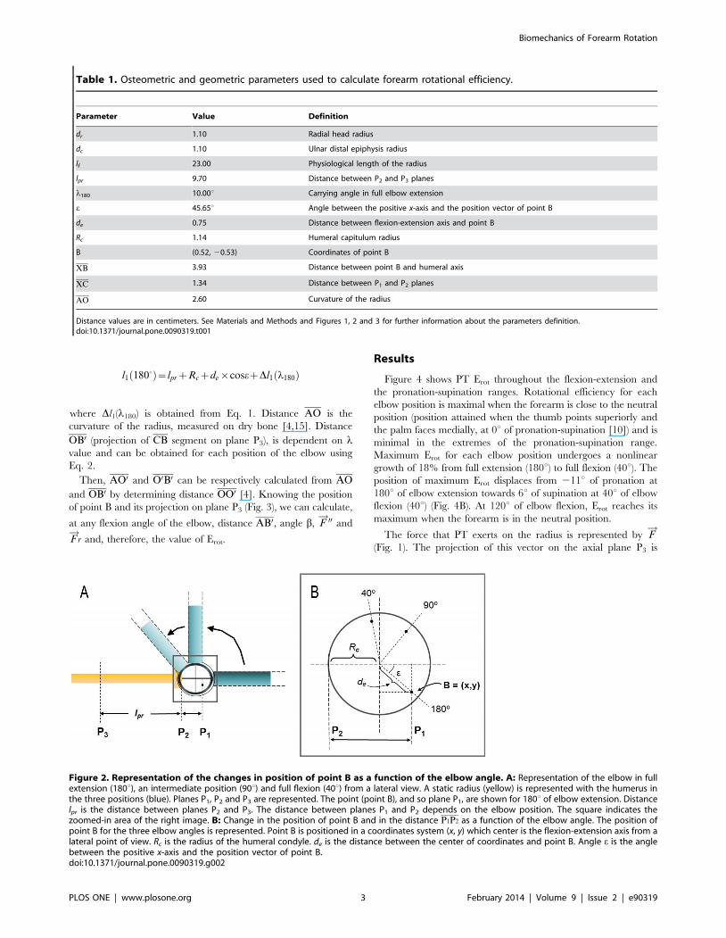
Distance l1 (P1P3) is the sum of P2P3 and P1P2 (Figs. 1A and 2A).
P2P3 can be measured directly on the radius bone (Figs. 1A and
2A, lpr), and P1P2 can be obtained from: (i) geometric parameters
obtained from the three-dimensional image of the humerus (Rc, de,
e, Fig. 2B); (ii) the position of the humeral medial epicondyle at
each flexion angle (Fig. 2B), and (iii) the position of the elbow
flexion axis, which depends on the carrying angle (l) and varies
throughout the flexion-extension range (see Fig. 1 and Eq. 1) [10].
Then, the distance l1 in maximum elbow extension can be
calculated as follows:
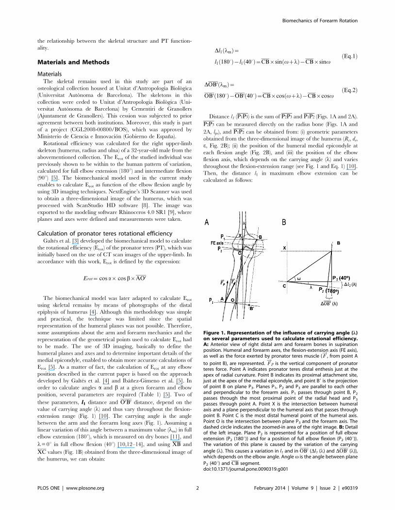
Figure 1. Representation of the influence of carrying angle (l)on several parameters used to calculate rotational efficiency.A: Anterior view of right distal arm and forearm bones in supinationposition. Humeral and forearm axes, the flexion-extension axis (FE axis),as well as the force exerted by pronator teres muscle ( F
!, from point A
to point B), are represented. F!
p is the vertical component of pronatorteres force. Point A indicates pronator teres distal enthesis just at theapex of radial curvature. Point B indicates its proximal attachment site,just at the apex of the medial epicondyle, and point B9 is the projectionof point B on plane P3. Planes P1, P2 and P3 are parallel to each otherand perpendicular to the forearm axis. P1 passes through point B, P2
passes through the most proximal point of the radial head and P3
passes through point A. Point X is the intersection between humeralaxis and a plane perpendicular to the humeral axis that passes throughpoint B. Point C is the most distal humeral point of the humeral axis.Point O is the intersection between plane P3 and the forearm axis. Thedashed circle indicates the zoomed-in area of the right image. B: Detailof the left image. Plane P2 is represented for a position of full elbowextension (P2 (180u)) and for a position of full elbow flexion (P2 (40u)).The variation of this plane is caused by the variation of the carrying
angle (l). This causes a variation in l1 and in OB0 (Dl1 (l) and DOB0 (l)),which depends on the elbow angle. Angle v is the angle between plane
P2 (40u) and CB segment.doi:10.1371/journal.pone.0090319.g001
Biomechanics of Forearm Rotation
PLOS ONE | www.plosone.org 2 February 2014 | Volume 9 | Issue 2 | e90319
l1 1800ð Þ~lprzRczde|cosezDl1 l180ð Þ
where Dl1(l180) is obtained from Eq. 1. Distance AO is the
curvature of the radius, measured on dry bone [4,15]. Distance
OB0 (projection of CB segment on plane P3), is dependent on lvalue and can be obtained for each position of the elbow using
Eq. 2.
Then, AO0 and O0B0 can be respectively calculated from AO
and OB0 by determining distance OO0 [4]. Knowing the position
of point B and its projection on plane P3 (Fig. 3), we can calculate,
at any flexion angle of the elbow, distance AB0, angle b, F!00 and
F!
r and, therefore, the value of Erot.
Results
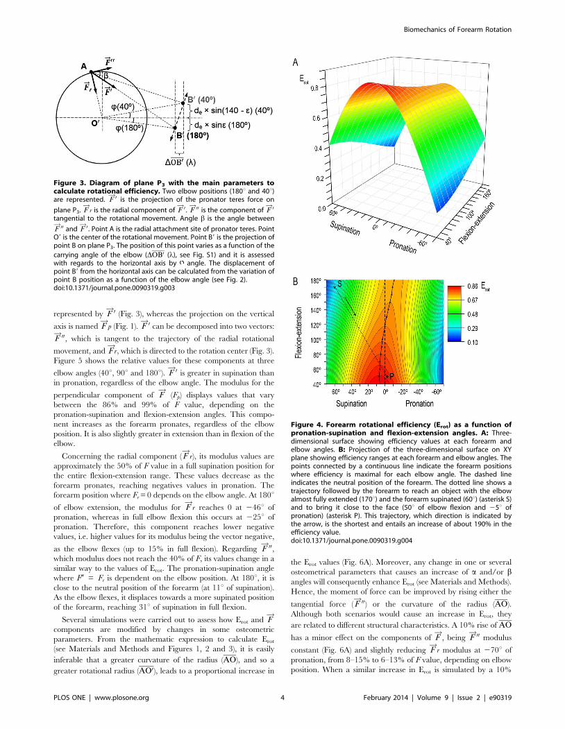
Figure 4 shows PT Erot throughout the flexion-extension and
the pronation-supination ranges. Rotational efficiency for each
elbow position is maximal when the forearm is close to the neutral
position (position attained when the thumb points superiorly and
the palm faces medially, at 0u of pronation-supination [10]) and is
minimal in the extremes of the pronation-supination range.
Maximum Erot for each elbow position undergoes a nonlinear
growth of 18% from full extension (180u) to full flexion (40u). The
position of maximum Erot displaces from 211u of pronation at
180u of elbow extension towards 6u of supination at 40u of elbow
flexion (40u) (Fig. 4B). At 120u of elbow flexion, Erot reaches its
maximum when the forearm is in the neutral position.
The force that PT exerts on the radius is represented by F!
(Fig. 1). The projection of this vector on the axial plane P3 is
Table 1. Osteometric and geometric parameters used to calculate forearm rotational efficiency.
Parameter Value Definition
dr 1.10 Radial head radius
dc 1.10 Ulnar distal epiphysis radius
lf 23.00 Physiological length of the radius
lpr 9.70 Distance between P2 and P3 planes
l180 10.00u Carrying angle in full elbow extension
e 45.65u Angle between the positive x-axis and the position vector of point B
de 0.75 Distance between flexion-extension axis and point B
Rc 1.14 Humeral capitulum radius
B (0.52, 20.53) Coordinates of point B
XB 3.93 Distance between point B and humeral axis
XC 1.34 Distance between P1 and P2 planes
AO 2.60 Curvature of the radius
Distance values are in centimeters. See Materials and Methods and Figures 1, 2 and 3 for further information about the parameters definition.doi:10.1371/journal.pone.0090319.t001
Figure 2. Representation of the changes in position of point B as a function of the elbow angle. A: Representation of the elbow in fullextension (180u), an intermediate position (90u) and full flexion (40u) from a lateral view. A static radius (yellow) is represented with the humerus inthe three positions (blue). Planes P1, P2 and P3 are represented. The point (point B), and so plane P1, are shown for 180u of elbow extension. Distancelpr is the distance between planes P2 and P3. The distance between planes P1 and P2 depends on the elbow position. The square indicates thezoomed-in area of the right image. B: Change in the position of point B and in the distance P1P2 as a function of the elbow angle. The position ofpoint B for the three elbow angles is represented. Point B is positioned in a coordinates system (x, y) which center is the flexion-extension axis from alateral point of view. Rc is the radius of the humeral condyle. de is the distance between the center of coordinates and point B. Angle e is the anglebetween the positive x-axis and the position vector of point B.doi:10.1371/journal.pone.0090319.g002
Biomechanics of Forearm Rotation
PLOS ONE | www.plosone.org 3 February 2014 | Volume 9 | Issue 2 | e90319
represented by F!0 (Fig. 3), whereas the projection on the vertical
axis is named F!
p (Fig. 1). F!0 can be decomposed into two vectors:
F!00, which is tangent to the trajectory of the radial rotational
movement, and F!
r, which is directed to the rotation center (Fig. 3).
Figure 5 shows the relative values for these components at three
elbow angles (40u, 90u and 180u). F!0 is greater in supination than
in pronation, regardless of the elbow angle. The modulus for the
perpendicular component of F!
(Fp) displays values that vary
between the 86% and 99% of F value, depending on the
pronation-supination and flexion-extension angles. This compo-
nent increases as the forearm pronates, regardless of the elbow
position. It is also slightly greater in extension than in flexion of the
elbow.
Concerning the radial component ( F!
r), its modulus values are
approximately the 50% of F value in a full supination position for
the entire flexion-extension range. These values decrease as the
forearm pronates, reaching negatives values in pronation. The
forearm position where Fr = 0 depends on the elbow angle. At 180uof elbow extension, the modulus for F
!r reaches 0 at 246u of
pronation, whereas in full elbow flexion this occurs at 225u of
pronation. Therefore, this component reaches lower negative
values, i.e. higher values for its modulus being the vector negative,
as the elbow flexes (up to 15% in full flexion). Regarding F!00,
which modulus does not reach the 40% of F, its values change in a
similar way to the values of Erot. The pronation-supination angle
where F0 = Fr is dependent on the elbow position. At 180u, it is
close to the neutral position of the forearm (at 11u of supination).
As the elbow flexes, it displaces towards a more supinated position
of the forearm, reaching 31u of supination in full flexion.
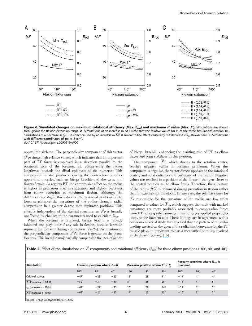
Several simulations were carried out to assess how Erot and F!
components are modified by changes in some osteometric
parameters. From the mathematic expression to calculate Erot
(see Materials and Methods and Figures 1, 2 and 3), it is easily
inferable that a greater curvature of the radius (AO), and so a
greater rotational radius (AO0), leads to a proportional increase in
the Erot values (Fig. 6A). Moreover, any change in one or several
osteometrical parameters that causes an increase of a and/or bangles will consequently enhance Erot (see Materials and Methods).
Hence, the moment of force can be improved by rising either the
tangential force ( F!00) or the curvature of the radius (AO).
Although both scenarios would cause an increase in Erot, they
are related to different structural characteristics. A 10% rise of AO
has a minor effect on the components of F!
, being F!00 modulus
constant (Fig. 6A) and slightly reducing F!
r modulus at 270u of
pronation, from 8–15% to 6–13% of F value, depending on elbow
position. When a similar increase in Erot is simulated by a 10%
Figure 3. Diagram of plane P3 with the main parameters tocalculate rotational efficiency. Two elbow positions (180u and 40u)are represented. F
!0 is the projection of the pronator teres force on
plane P3. F!
r is the radial component of F!0. F!00 is the component of F
!0tangential to the rotational movement. Angle b is the angle between
F!00 and F
!0. Point A is the radial attachment site of pronator teres. PointO9 is the center of the rotational movement. Point B9 is the projection ofpoint B on plane P3. The position of this point varies as a function of the
carrying angle of the elbow (DOB0 (l), see Fig. S1) and it is assessedwith regards to the horizontal axis by angle. The displacement ofpoint B9 from the horizontal axis can be calculated from the variation ofpoint B position as a function of the elbow angle (see Fig. 2).doi:10.1371/journal.pone.0090319.g003
Figure 4. Forearm rotational efficiency (Erot) as a function ofpronation-supination and flexion-extension angles. A: Three-dimensional surface showing efficiency values at each forearm andelbow angles. B: Projection of the three-dimensional surface on XYplane showing efficiency ranges at each forearm and elbow angles. Thepoints connected by a continuous line indicate the forearm positionswhere efficiency is maximal for each elbow angle. The dashed lineindicates the neutral position of the forearm. The dotted line shows atrajectory followed by the forearm to reach an object with the elbowalmost fully extended (170u) and the forearm supinated (60u) (asterisk S)and to bring it close to the face (50u of elbow flexion and 25u ofpronation) (asterisk P). This trajectory, which direction is indicated bythe arrow, is the shortest and entails an increase of about 190% in theefficiency value.doi:10.1371/journal.pone.0090319.g004
Biomechanics of Forearm Rotation
PLOS ONE | www.plosone.org 4 February 2014 | Volume 9 | Issue 2 | e90319

decrease of lpr, a completely different behavior of F!
vectors can be
observed: F!00 modulus increases proportionally to Erot during the
entire flexion range (Fig. 6B), whereas F!
r modulus rises from 46–
48% to 49–51% of F value at 70u of supination and from 8–15%
to 10–19% at 270u of pronation. A 10% increase of XB has
broadly the same effect on Erot and F!00 and F
!r moduli than the
reported 10% decrease of lpr. F!
p is not meaningfully modified by
changes in the abovementioned parameters, whereas the forearm
positions where F!
r modulus reaches 0, where F!00 and F
!r become
equal and where Erot is maximal are considerably affected by these
changes. All this information is summarized in Table 2.
Conversely to the abovementioned simulations, the direction of
the change in Erot and F!00 modulus (increase or decrease) caused
by a change in the position of point B depends on the elbow angle
(Fig. 6C). When coordinate x increases, maximal Erot and F!00
modulus rise in flexion of the elbow and decrease in extension.
When this coordinate is lower, the values for these parameters
decrease in flexion and rise in extension. When coordinate y is
closer to 0, Erot and F!00 modulus increase in flexion and fall in
extension, whereas when this coordinate is lower, these parameters
decrease in flexion and rise in extension.
Efficiency is also lower when the carrying angle (l) is greater. A
change of 5u in this angle leads to a variation of about 6.5% of the
maximum Erot value at 180u of elbow extension. This effect is
lesser as the elbow flexes and it is null in full flexion (40u).
Discussion
Rotational efficiency, force components and structuralimplications
In the current study, PT Erot has been assessed throughout the
entire flexion-extension range using three-dimensional technology.
The variation of Erot obtained from our innovative biomechanical
model is in agreement with the results of kinematic studies using
cadaveric specimens [16,17] and virtual and resin models of the
upper-limb skeleton [18–20], as well as with analysis on forearm
discomfort [21] and on electromyographic signals of the forearm
pronators [7].
Pronator teres Erot is dependent on the skeletal structure of the
arm, elbow and forearm, which in turn can be modified by the
usage of this muscle. In this regard, the analysis of Erot has enabled
to study the effect of the components of PT force vector on the
Figure 5. Relative values for F!
components and forearm rotational efficiency (Erot) in three elbow positions. Full extension (180u),intermediate position (90u) and full elbow flexion (40u) scenarios are shown.doi:10.1371/journal.pone.0090319.g005
Biomechanics of Forearm Rotation
PLOS ONE | www.plosone.org 5 February 2014 | Volume 9 | Issue 2 | e90319
upper-limb skeleton. The perpendicular component of this vector
( F!
p) shows high relative values, which indicates that an important
part of PT force is employed in a direction parallel to the
rotational axis of the forearm, i.e. compressing the radius
lengthwise towards the distal epiphysis of the humerus. This
compression is also produced during the contraction of other
upper-limb muscles, such as biceps brachii and the wrist and
fingers flexors. As regards PT, the compressive effect on the radius
is higher in pronation than in supination and slightly decreases
from elbow extension to maximum flexion. Although the
differences are slight, this indicates that pronated positions of the
forearm enhance the curvature of the radius through radial
compression in a greater degree than supinated positions. This
effect is independent of the skeletal structure, as F!
p is broadly
unaffected by changes in the parameters used to calculate Erot.
When the forearm is pronated, biceps brachii is reflexly
inhibited and plays little if any role in flexion, because it would
supinate the forearm during contraction [22–24]. As mentioned,
the perpendicular component of PT force is greater on the prone
forearm. This increase may partially compensate the lack of action
of biceps brachiii, enhancing the assisting role of PT as elbow
flexor and joint stabilizer in this position.
The component F!
r, which directs to the rotation center,
reaches negative values in forearm pronation. When this
component is negative, the vector directs opposite to the rotational
center, and so it enhances the curvature of the radius. Negative
values are reached in a position of the forearm that gets closer to
the neutral position as the elbow flexes. Therefore, the curvature
of the radius (AO) is enhanced during pronation in flexion rather
than in extension of the elbow. In any case, the relative values for
F!
r responsible for the curvature of the radius are low when
compared to values for F!
p, which suggests that radii with marked
curvatures are more probably associated to compression forces
from PT, among other muscles, than to forces applied perpendic-
ularly to the forearm axis. These findings are in agreement with a
previous empirical study that revealed that the pattern of muscular
loading exerted on the apex of the radial shaft curvature by the PT
muscle plays an important role as a mechanical stimulus involved
in diaphyseal bowing [15].
Figure 6. Simulated changes on maximum rotational efficiency (Max. Erot) and maximum F0 value (Max. F0). Simulations are shownthroughout the flexion-extension range. A: Simulations of an increase in AO. Note that the relative values for F0 of the three simulations overlap. B:
Simulations of a decrease in lpr. The effect caused by an increase in XB is similar to the effect caused by the decrease in lpr shown here. C: Simulationswith different coordinates of point B (cm).doi:10.1371/journal.pone.0090319.g006
Table 2. Effect of the simulations on F!
components and rotational efficiency (Erot) for three elbow positions (180u, 90u and 40u).
Simulation Forearm position where Fr = 0 Forearm position where F0 = Fr
Forearm position where Erot ismaximal
180u 90u 40u 180u 90u 40u 180u 90u 40u
Original values 247u 229u 225u 11u 28u 31u 211u 4u 6u
AO increase (+10%) 252u 234u 230u 8u 25u 28u 211u 4u 6u
lpr decrease (210%) 244u 227u 223u 13u 29u 34u 211u 3u 5u
XB increase (+10%) 242u 226u 223u 14u 29u 33u 210u 3u 5u
doi:10.1371/journal.pone.0090319.t002
Biomechanics of Forearm Rotation
PLOS ONE | www.plosone.org 6 February 2014 | Volume 9 | Issue 2 | e90319
The effect that the skeletal structure and form have on Erot and
on the force vectors has also been assessed. The results show that a
greater bowing of the radius entails an increase of Erot. This is
consistent with previous studies suggesting an enhancement of PT
action and forearm rotational power by a markedly bowed radius
[3,25–28]. An increase of the radial curvature also causes that F!
r
becomes negative in a more pronated position of the forearm, and
so its modulus reach lower values in full pronation. Therefore, the
radius is more easily bowed when its curvature is low.
The radial location of PT muscle also affects Erot: at any elbow
angle, Erot increases when this enthesis is more proximally located,
which is consistent with previous observations in full elbow
extension [3]. Moreover, radii with a more proximal enthesis for
PT muscle lead to the reach of negative values of F!
r closer to the
neutral position of the forearm. Therefore, these radii entail lower
negative values for this component, i.e. higher values for its
modulus being the vector negative, which stimulates radial
curvature.
Concerning the humeral medial epicondyle, Erot also tends to
increase in all elbow positions as this structure enlarges. Even
though the current analysis uses a different approach to quantify
humeral medial epincondylar projection (XB), the results are
consistent with previous studies [3,5,29]. Moreover, an enlarge-
ment of the medial epicondyle has the same effect on F!
r than a
more proximally located radial enthesis for PT, and therefore a
more medially projected epicondyle enhances radial curvature.
The orientation of the medial epicondyle is also relevant for the
determination of Erot. The alteration of this orientation causes
changes in F!00 that lead to changes in Erot. A more posteriorly
oriented epicondyle, i.e. more retroflexed [5,30], is associated with
a greater Erot in full extension of the elbow, whereas an epicondyle
with a lower degree of retroflexion has greater values of Erot in full
flexion. Moreover, a more proximally oriented epicondyle
enhances Erot in full flexion, whereas when it is more distally
oriented, Erot increases in extension. The orientation of the medial
epicondyle was reported to present a partially activity-dependent
plasticity, and so it was hypothesized that it can be modified to
enhance certain abilities [30]. In this regard, and in agreement
with the abovementioned relationship between Erot and epicon-
dylar orientation, a simple observation of the upper-limb
positioning shows that a habitual and continued contraction of
PT in full elbow flexion would reorient the epicondyle towards a
more proximal position. Conversely, this reorientation would
occur distally in full elbow extension. The reported association
between the orientation of the medial epicondyle and Erot may
have important implications on the evolutionary pathway of the
upper-limb skeleton, as this structure plays a central role in the
locomotor diversity of primates [5,31–33].
Regarding the carrying angle of the elbow [11], this is the first
quantitative insight into its biomechanical implications on forearm
rotation. An increase in this angle leads to lower values of Erot.
This effect is the greatest in full elbow extension and becomes
lower as it is flexed, given that carrying angle is maximal in full
extension, decreases during flexion and reaches 0u when the elbow
is completely flexed [10,12–14].
Functional implications of forearm rotationThe biomechanical model described here enables to make a
thorough quantitative approach to clarify some mechanical aspects
about the relationship between PT and forearm and elbow
motion. For instance, the results indicate that maximal Erot for
each elbow angle is close to the neutral position of the forearm.
This position has been commonly associated to the functional
position, which minimizes the expenditure of muscular energy as it
implies a natural equilibrium between antagonist muscles [10].
Moreover, it entails an enhancement of the precision of the grip,
because the forearm axis is in line with the pronation-supination
axis [10,34].
Although skeletal structures with different morphologies can
have the same Erot, they do not have to imply the same abilities in
terms of precision. For instance, if the same force is exerted by PT
on two upper-limbs with the same Erot, the one with the greatest
curvature will be able to reach a lower rotational angle (File S1
and Fig. S1), and thus a greater rotational precision.
The stability of the elbow joint has relevant implications for
sports medicine and other pathological conditions [14,35,36].
During pronation, F!0 plays an important role in the stability of the
radio-ulnar joint (see File S1 and Fig. 3), whereas F!
p participates
in the radio-humeral stability (see Fig. 1A). The global stability of
the elbow is thus enhanced by the combination of both
components. Although F!
p is slightly lower when the forearm is
supinated, the great F!0 value in this position leads to a better
fitting between the radius and the ulna than in pronation [37].
Rotational efficiency and the forces that act during pronation
may be associated to the estimations of the discomfort levels done
by Mukhopadhyay et al. [38], as the movements implying a high
level of discomfort correspond to positions where Erot is low.
Moreover, the concept ‘‘efficiency’’ provides information about
the rotational stability of a given position, as the work needed to
modify the forearm rotational angle depends directly on the value
of Erot in each position (see File S1). For a given pronation
movement, the model can be used to determine the trajectories of
the hand that entail the minimal expenditure of energy for PT. In
humans, pronation is usually performed when an object has been
reached with the hand supinated and the elbow extended and is
then brought closer to the body. The final position of this
movement will entail a full flexion of the elbow and a close-to-
neutral position of the forearm. This movement implies an
increase of PT Erot, which indicates that the energy expenditure of
this muscle diminishes when the elbow is flexed (see Fig. 4B). The
results show that a great part of this trajectory entails upper-limb
positions where F!00 and F
!r have a similar value, i.e. around the
forearm position where F0 = Fr. This indicates that during this
trajectory the forces that the elbow joint is submitted to are quite
equilibrated.
These two conditions (equilibrium between forces and increase
of Erot) are not observed for other trajectories, such as those where
the end point is close to full pronation with the elbow flexed. In
this case, the final part of the trajectory entails a decrease of Erot
and very different values of these two force components. If the
forearm was very regularly used to perform this movement with
important mechanical loads, the disequilibrium could have
pathological consequences, as a result of heavy tensions in the
radio-ulnar proximal joint. In order to minimize the expenditure
of energy and to rebalance the forces, the radial curvature could
increase. As reported by our simulations, a rise of the curvature of
the radius causes an increase of Erot and a displacement of the
position where Fr = 0 towards full pronation, as well as a
displacement of the position where F!
r and F!00 become equal
from supination towards a position closer to the neutral.
Therefore, the results of the current study also indicate that the
upper-limb skeleton may experience plastic changes as a result of
PT activity when it is used in positions where Erot is low and forces
are not equilibrated. These changes entail an improvement of
these parameters, in order to adjust to the unfavorable conditions.
Biomechanics of Forearm Rotation
PLOS ONE | www.plosone.org 7 February 2014 | Volume 9 | Issue 2 | e90319

Conversely, if the upper-limb is mainly used in positions with high
Erot values, PT will not trigger changes in the skeletal structure.
Overall, Erot and its relationship with energy expenditure and
rotational stability, as well as the relative values of the force
components, which are linked to the joint stability, provide a
global insight into pronation and its association to the forearm
skeletal structure. These findings will be useful for future analyses
on ergonomics and orthopaedics of the upper-limb skeleton, as
they provide relevant information about the relationship between
PT functionality and the skeletal structure.
Applying this biomechanical model on studies about compar-
ative anatomy of primate taxa would provide valuable information
about the functional meaning of the upper-limb skeletal interspe-
cific differences, which will probably be associated to the
locomotor abilities of each taxon. Moreover, an analysis of PT
Erot and force components on a fossil primate would very useful for
inferring its upper-limb involvement in locomotion.
Some skeletal parameters essential for the determination of PT
functionality, such as the radial curvature and the medial
epicondylar form, are reported to be plastic and related to the
usage of PT muscle [15,30]. Therefore, this biomechanical model
would also be of great interest if applied to ancient human
populations. Differences in parameters derived or related to Erot
between sexes, groups or populations would account for differ-
ences in occupational and habitual activities.
Supporting Information
Figure S1 Diagrams showing the displacement causedby a force in different scenarios. A: Linear displacement (Ds)
and rotational angle (Dh) caused by a force F!
on a rotating body,
being the center of rotation O and the rotational radius R. B:
Representation of the differences in the force (F!
1 and F!
2) that are
needed to obtain the same rotational angle when the rotation
performed by pronator teres muscle starts at a different point (A1
and A2) (see Fig. 3). C: Representation of the differences in the
rotational angle (Dh1 and Dh2) obtained when the same force ( F!
)
is applied by pronator teres muscle on two radii with different
rotational radius (r1 and r2) (see Fig. 3). Note that the same linear
displacement (Ds) is obtained in both cases.
(TIF)
File S1 Rotating work during forearm pronation,stability and muscular energy expenditure.
(DOCX)
Acknowledgments
The authors are grateful to Antoni Morros for his assistance in the
preparation of the artwork. They are also indebted to the Academic Editor,
David Carrier, and an anonymous reviewer for their comments on an
earlier version of the manuscript.
Author Contributions
Conceived and designed the experiments: PI IG JM. Performed the
experiments: PI JM. Analyzed the data: PI IG XJ AM JM. Contributed
reagents/materials/analysis tools: XJ AM. Wrote the paper: PI JM.
Revised the paper: PI IG XJ AM JM.
References
1. Murray WM, Buchanan TS, Delp SL (2002) Scaling of peak moment arms of
elbow muscles with upper extremity bone dimensions. J Biomech 35: 19–26.
2. Tsaopoulos DE, Maganaris CN, Baltzopoulos V (2007) Can the patellar tendon
moment arm be predicted from anthropometric measurements? J Biomech 40:645–651.
3. Galtes I, Jordana X, Cos M, Malgosa A, Manyosa J (2008) Biomechanical modelof pronation efficiency: New insight into skeletal adaptation of the hominoid
upper limb. Am J Phys Anthropol 135: 293–300.
4. Galtes I, Jordana X, Malgosa A, Manyosa J (2009) Technical note: Forearm
pronation efficiency analysis in skeletal remains. Am J Phys Anthropol 140: 589–594.
5. Ibanez-Gimeno P, Jordana X, Manyosa J, Malgosa A, Galtes I (2012) 3Danalysis of the forearm rotational efficiency variation in humans. Anat Rec 295:
1092–1100.
6. Gordon KD, Pardo RD, Johnson JA, King GJW, Miller TA (2004)
Electromyographic activity and strength during maximum isometric pronation
and supination efforts in healthy adults. J Orthop Res 22: 208–213.
7. O’Sullivan LW, Gallwey TJ (2002) Upper-limb surface electro-myography at
maximum supination and pronation torques: the effect of elbow and forearmangle. J Electromyogr Kines 12: 275–285.
8. ScanStudio HD 1.2.1 (2006) Software for Windows. Santa Monica: NextEngine,Inc.
9. Rhinoceros 4.0 SR1 (2007) Educational Edition for Windows. Seattle: RobertMcNeel & Associates.
10. Kapandji AI (2002) Fisiologıa articular, miembro superior. Madrid: EditorialMedica Panamericana.
11. Knussmann R (1967) Humerus, ulna und radius der Simiae. Bibl Primatol 5: 1–399.
12. Morrey BF, Chao EYS (1976) Passive motion of the elbow joint. J Bone JointSurg Am 58: 501–508.
13. Rouviere H, Delmas A (1988) Anatomıa humana: Descriptiva, topografica yfuncional, tomo III, 9th ed. Barcelona: Masson.
14. Goto A, Moritomo H, Murase T, Oka K, Sugamoto K, et al. (2004) In vivoelbow biomechanical analysis during flexion: Threedimensional motion analysis
using magnetic resonance imaging. J Shoulder Elbow Surg 13: 441–447.
15. Galtes I, Jordana X, Manyosa J, Malgosa A (2009) Functional implications of
radial diaphyseal curvature. Am J Phys Anthropol 138: 286–292.
16. Murray WM, Delp SL, Buchanan TS (1995) Variation of muscle moment arms
with elbow and forearm position. J Biomech 28: 513–525.
17. Haugstvedt JR, Berger RA, Berglund LJ (2001) A mechanical study of the
moment-forces of the supinators and pronators of the forearm. Acta Orthop
Scand 72: 629–634.
18. Lan N, Baker L (2004) Biomechanical couplings between elbow and forearm
movements. Conf Proc IEEE Eng Med Biol Soc 6: 4661–4664.
19. Bremer AK, Sennwald GR, Favre P, Jacob HAC (2006) Moment arms of
forearm rotators. Clin Biomech 21: 683–691.
20. Hale R, Dorman D, Gonzalez RV (2011) Individual muscle force parameters
and fiber operating ranges for elbow flexion-extension and forearm pronation-supination. J Biomech 44: 650–656.
21. O’Sullivan LW, Gallwey TJ (2001) Forearm discomfort for repeated isometrictorque exertions in pronation and supination. In: Hanson MA, editor.
Contemporary ergonomics 2001. London: Taylor and Francis. pp. 79–83.
22. Basmajian JV, Latif A (1957) Integrated actions and functions of the chief flexors
of the elbow: A detailed electromyographic analysis. J Bone Joint Surg Am 39-A:
1106-1118.
23. Naito A, Shimizu Y, Handa Y, Ichie M, Hoshimiya N (1991) Functional
anatomical studies of the elbow movements I: Electromyographic (EMG)analysis. Okajimas Folia Anat Jpn 68: 283–288.
24. Latarjet M, Ruiz Liard A, Pro E (2004) Anatomıa humana. Buenos Aires:Medica Panamericana.
25. Trinkaus E, Churchill SE (1988) Neandertal radial tuberosity orientation.Am J Phys Anthropol 75: 15–21.
26. Aiello L, Dean C (1990) An introduction to human evolutionary anatomy.London: Academic Press.
27. Tuttle RH, Hollowed JR, Basmajian JV (1992) Electromyography of pronators
and supinators in great Apes. Am J Phys Anthropol 87: 215–226.
28. Rose MD (1993). Functional anatomy of the elbow and forearm in primates. In:
Gebo DL, editor. Postcranial adaptation in nonhuman primates. DeKalb:Northern Illinois University Press. pp. 70–95.
29. Jablonski NG, Leakey MG, Kiarie C, Anton M (2002) A new skeleton ofTheropithecus brumpti (Primates: Cercopithecidae) from Lomekwi, West Turkana,
Kenya. J Hum Evol 43: 887–923.
30. Ibanez-Gimeno P, Galtes I, Jordana X, Fiorin E, Manyosa J, et al. (2013)
Entheseal changes and functional implications of the humeral medial
epicondyle. Int J Osteoarchaeol 23: 211–220.
31. Seiffert ER, Simons EL, Fleagle JG (2000) Anthropoid humeri from the late
Eocene of Egypt. Proc Natl Acad Sci 97: 10062–10067.
32. Ciochon RL, Gunnell GF (2004) Eocen large-bodied primates of Myanmar and
Thailand: Morphological considerations and phylogenetic affinities. In: Ross CF,Kay RF, editors. Anthropoid origins: New visions. New York: Kluwer
Academic/Plenum Publishers. pp. 249–282.
33. MacPhee R, Meldrum J (2006) Postcranial remains of the extinct monkeys of the
Greater Antilles, with evidence for semiterrestriality in Paralouatta. Am MusNovit 3516: 3–65.
Biomechanics of Forearm Rotation
PLOS ONE | www.plosone.org 8 February 2014 | Volume 9 | Issue 2 | e90319
34. Marzke MW (1997) Precision grips, hand morphology, and tools. Am J Phys
Anthropol 102: 91–110.35. Shiba R, Sorbie C, Siu DW, Bryant JT, Cooke TDV, et al. (1988) Geometry of
the humeroulnar joint. J Orthop Res 6: 897–906.
36. Richards RR (1996) Chronic disorders of the forearm. J Bone Joint Surg Am 78:916–930.
37. Morrey BF, An KN, Stormont TJ (1988) Force transmission through the radial
head. J Bone Joint Surg Am 70: 250–256.38. Mukhopadhyay P, O’Sullivan L, Gallwey TJ (2007) Estimating upper limb
discomfort level due to intermittent isometric pronation torque with various
combinations of elbow angles, forearm rotation angles, force and frequency withupper arm at 90u abduction. Int J Ind Ergonom 37: 313–325.
Biomechanics of Forearm Rotation
PLOS ONE | www.plosone.org 9 February 2014 | Volume 9 | Issue 2 | e90319