BIOLOGY - Сельскохозяйственная биология
172
ISSN 2412-0324 (English ed. Online) ISSN 0131-6397 (Russian ed. Print) ISSN 2313-4836 (Russian ed. Online) Since January, 1966 ANIMAL BIOLOGY Vol. 55, Issue 4 July-August 2020 Moscow BIOLOGY AGRICULTURAL
-
Upload
khangminh22 -
Category
Documents
-
view
2 -
download
0
Transcript of BIOLOGY - Сельскохозяйственная биология

ISSN 2412-0324 (English ed. Online) ISSN 0131-6397 (Russian ed. Print) ISSN 2313-4836 (Russian ed. Online)
Since January, 1966
ANIMAL BIOLOGY
Vol. 55, Issue 4 July-August 2020 Moscow
BIOLOGY AGRICULTURAL

EDITORIAL BOARD
V.I. FISININ (Sergiev Posad, Russia) — Chairman (animal biology)
BAGIROV V.A. (Moscow, Russia) BORISOVA E.M. (Moscow, Russia) BREM G. (Vienna, Austria) EGOROV I.A. (Sergiev Posad, Russia) FEDOROV Yu.N. (Moscow, Russia) FEDOROVA L.M. (editor-in-chief) (Moscow, Russia) KOSOLAPOV V.M. (Lobnya, Russia)
LAPTEV G.Yu. (St. Petersburg, Russia) LUSHENG HUANG (China) PANIN A.N. (Moscow, Russia) SMIRNOV A.M. (Moscow, Russia) SURAI P.F. (Ayr, Scotland, UK) SHEVELEV N.S. (Moscow, Russia) ZINOVIEVA N.A. (Dubrovitsy, Russia)
A peer-reviewed academic journal for delivering current original research results and reviews on classic and modern biology of agricultural plants, animals and microorganisms Covered in Scopus, Web of Science (BIOSIS Previews, Biological Abstracts, CAB Abstracts, Russian Science Citation Index), Agris Science editors: E.V. Karaseva, L.M. Fedorova Publisher: Agricultural Biology Editorial Office NPO Address: build. 16/1, office 36, pr. Polesskii, Moscow, 125367 Russia Tel: + 7 (916) 027-09-12 E-mail: [email protected], [email protected] Internet: http://www.agrobiology.ru
For citation: Agricultural Biology, Сельскохозяйственная биология, Sel’skokhozyaistvennaya biologiya ISSN 0131-6397 (Russian ed. Print) ISSN 2313-4836 (Russian ed. Online)
© Agricultural Biology Editorial Office (Редакция журнала «Сельскохозяйственная биология»), 2020
ISSN 2412-0324 (English ed. Online)

843
SEL’SKOKHOZYAISTVENNAYA BIOLOGIYA [AGRICULTURAL BIOLOGY], 2020, Vol. 55, ¹ 4
CONTENTS
REVIEWS, CHALLENGES Shchukina E.S., Kosovsky G.Yu., Glazko V.I. et al. Domestic rabbit Oryctolagus cuniculus
var. domestica L. as a model in the study of domestication and biomedical re-searches (review) . . . . . . . . . . . . . . . . . . . . . . . . 643
Savchenkova I.P. The role of microenvironment in the directed in vitro hematopoietic differentiation of mouse embryonic stem cell (review) . . . . . . . 659
MICROBIOMES Alekseeva E.I., Dubrovin A.V., Laptev G.Yu. et al. Results of the research of intestinal mi-
crobial profiles of Equus ferus caballus by NGS sequencing . . . . . . . . 671 Bagirov V.A., Ushakov A.S., Duskaev G.K. et al. Metagenomic analysis of intestinal mi-
crobiome and biochemical composition of broiler meat upon use of Quercus cortex extract dietary additive . . . . . . . . . . . . . . . . . . . . . 682
Ilina L.A., Filippova V.A., Layshev K.A. et al. Variation in the Russian Arctic reindeer (Rangifer tarandus) rumen microbiome related to season change . . . . . . . 697
PHYSIOLOGY, BIOCHEMISTRY, NUTRITION Vasilevsky N.V., Yeletskaya T.A. Food particle size as an indicator of its structural com-
position and a key aspect of the development of the nutrition theory paradigm . 714 Vertiprakhov V.G., Grozina A.A., Fisinin V.I. The exocrine pancreatic function in chick-
en (Gallus gallus L.) fed diets supplemented with different vegetable oils . . . . 726 DIETARY ADDITIVES Kavtarashvili A.Sh., Novotorov E.N., Kodentsova V.M. et al. The role of carotenoids in
the biofortification of table chicken (Gallus gallus L.) eggs with ω-3 polyunsaturat-ed fatty acids, vitamin E, and selenium . . . . . . . . . . . . . . . . 738
Fomichev Yu.P., Bogolyubova N.V., Nekrasov R.V. et al. Physiological and biochemical effects of two feed antioxidants in modeling technological stress in pigs (Sus scrofa domesticus Erxleben, 1777) . . . . . . . . . . . . . . . . . . . . 750
Fomichev Yu.P., Bogolyubova N.V., Romanov V.N. et al. Comparative assessment of nat-ural feed additives for functional effects on the digestive processes in the rumen of sheep (Ovis aries) . . . . . . . . . . . . . . . . . . . . . . . 770
REPRODUCTION, DEVELOPMENTAL PHYSIOLOGY Kuzmina T.I., Chistyakova I.V., Tatarskaya D.N. The influence of highly dispersed silica
nanoparticles on the functional activity of mitochondria and chromatin state in na-tive and devitrified Bos taurus oocytes . . . . . . . . . . . . . . . . 784
Dolgorukova A.M., Titov V.Yu., Kochish I.I. et al. The embryonic metabolism of nitric oxide and its interrelation with postembryonic development in chicken (Gallus gallus domesticus L.) and quails (Coturnix coturnix L.) . . . . . . . . . . . . . . 794
VETERINARY MICROBIOLOGY Potehin А.V., Shadrova N.B., Pruntova О.V. et al. Biochemical, antigenic and proteomic
properties of isolates and strains of the causative agent of chicken infectious coryza Avibacterium paragallinarum (Biberstein and White 1969) Blackall et al. 2005 . . . 804
Laptev G.Y., Yildirim E.A., Dunyashev T.P. et al. Genomic and phenotypical potential of antimicrobial activity of a bacillus strain Bacillus megaterium В-4801 . . . . . 816
ANTHELMINTICS Varlamova A.I., Arkhipov I.A. Biological activity of fenbendazole based on supramolecu-
lar delivery system with disodium salt of glycyrrhizic acid . . . . . . . . . 830

643
AGRICULTURAL BIOLOGY, ISSN 2412-0324 (English ed. Online)
2020, V. 55, ¹ 3, pp. 643-658 (SEL’SKOKHOZYAISTVENNAYA BIOLOGIYA) ISSN 0131-6397 (Russian ed. Print)
ISSN 2313-4836 (Russian ed. Online)
Reviews, challenges UDC 636.92+639.112.1]:636.01:574/577 doi: 10.15389/agrobiology.2020.4.643eng
doi: 10.15389/agrobiology.2020.4.643rus
DOMESTIC RABBIT Oryctolagus cuniculus var. domestica L. AS A MODEL IN THE STUDY OF DOMESTICATION AND BIOMEDICAL
RESEARCHES (review)
E.S. SHCHUKINA1, G.Yu. KOSOVSKY1, V.I. GLAZKO1, 2, I.S. KASHAPOVA1, T.T. GLAZKO1, 2
1Afanas’ev Research Institute of Fur-Bearing Animal Breeding and Rabbit Breeding, 6, ul. Trudovaya, pos. Rodniki, Ramenskii Region, Moscow Province, 140143 Russia, e-mail [email protected], [email protected], [email protected] 2Timiryazev Russian State Agrarian University—Moscow Agrarian Academy, 49, ul. Timiryazevskaya, Moscow, 127550 Russia, e-mail [email protected] ( corresponding author), [email protected], e-mail [email protected] ORCID: Shchukina E.S. orcid.org/0000-0002-2465-7184 Kashapova I.S. orcid.org/0000-0002-6360-0025 Kosovskii G.Yu. orcid.org/0000-0003-3808-3086 Glazko T.T. orcid.org/0000-0002-3879-6935 Glazko V.I. orcid.org/0000-0002-8566-8717 The authors declare no conflict of interests Received January 14, 2020
A b s t r a c t
The domestic rabbit (Oryctolagus cuniculus var. domestica L.) belongs to the few domesti-cated species in which the wild ancestral species exists simultaneously with the domesticated one (M. Carneiro, 2014) that allows us to study the mechanisms underlying the processes of domestication. It should be noted that the genetic basis of domestication syndrome is still insufficiently studied (M.A. Zeder, 2006-2017). It is assumed that domestication is a unique form of symbiosis between humans and domesticated species that form a common habitat niche (M.A. Zeder, 2012). Research of symbiotic partners of such a niche allows us to accumulate information about the mechanisms of adaptation to it, including humans. In this regard, it is difficult to overestimate the importance of studying the domestic rabbit, because it has remained one of the main models in biomedical research for many decades (K.M. El-Bayomi, 2013). The unique physiological features of the rabbit explain its widespread use in the study of many human diseases. At the same time, we have not found any works that systematize current information on the fundamental biology of this domesticated species in com-parison with its wild ancestral form. The purpose of this review is to summarize data on the population genetic structure (M. Carneiro, 2014; A.D. Stock, 1976), distribution of genomic elements (M. Car-neiro, 2011), composition of microbiomes (M.S. Gómez-Conde, 2009), morphometric characteristics and physiological features (S.N. Bogolyubskii, 1959) of the domestic rabbit and ancestral subspecies of the European rabbit, including those that determine the value of O. cuniculus var. domestica not only as an economically valuable species, but also as a model object in various fields of biomedicine. The presented comparative analysis allows us to identify a number of phenotypic characteristics (J.L. Hen-drikse, 2007; I. Brusini, 2018; P.S. Ungar, 2010), as well as a group of molecular genetic markers of genomic DNA, differentiating the domestic rabbit from the ancestral species (M. Sparwel, 2019). Distribution of alleles of different mobile genetic elements, microsatellites, separate structural genes involved in the domestication process of domestic rabbit, can improve the efficiency of genetic re-sources management of not only this species but also other objects that are used in biomedical research, and for solving problems of selection work.
Keywords: domestication syndrome, wild rabbits, domestic rabbits, DNA markers, endoge-nous retroviruses, polylocus genotyping, microbiota
The study of the genetic structure of domesticated species is a prerequisite for the development of methods for managing the genetic resources of economi-cally valuable animals. Domestic rabbit Oryctolagus cuniculus var. domestica L. belongs to those rare domesticated species in which the wild ancestral species exists simultaneously with the domesticated one, which allows studying the mechanisms

644
underlying domestication. Rabbit breeding is currently actively developing, and, according to In-
dexBox, Inc. (Great Britain), the growth of world production of rabbit meat will maintain the current trend with the expected annual growth of the market about +2.3% (up to 1.8 million tons by 2025) (https://meatinfo.ru). At the same time, we did not find any works summarizing modern information on the fundamental biology of this domesticated species.
This review aimed to compare the population genetic structure, distribution of genomic elements and phenotypic features of the domestic rabbit and its ancestral subspecies, the European rabbit, and also to summarize data on microbiome com-position and physiological characteristics due to which the domestic rabbit is a model in various fields of biomedicine
Domes t i c ra bb i t a s an ob je c t o f re s ear ch and u s e. The do-mestic rabbit (Oryctolagus cuniculus var. domestica L.) has shared a common niche with humans for a long time. Domestication is a quantitative trait which varies from animals experiencing anthropogenic pressure to the most domesti-cated and forming a common niche with humans [1]. Today, there is no con-sensus on what domestication and domestication syndrome are, though this syn-drome is common for taxonomically distant species [2-4]. According to a num-ber of researchers [2], domestication is a unique symbiosis between humans and domesticated species that coexist in a single ecological niche.
The new geological period in which human activity turned into a planetary transforming force, called the Anthropocene [5], affects the adaptation of animals to habitat conditions via interfering with their life cycles. In general, the domesti-cation model indicates that the target of selection is not one species, but their community, that is, there is a coevolution of animals, humans, and other symbi-onts, including those that are part of the microbiome in different species [1]. The study of the mechanisms of domestication makes a significant contribution to understanding appearance of new forms, artificial selection, methodology for man-aging gene pools, breed formation and other microevolutionary processes.
The European rabbit (Oryctolagus cuniculus L.) is the only recognized an-cestor of the domestic rabbit, which, in its turn, is an important agricultural species with high-value dietary meat 90% of which can be utilized by the human, high productivity, early maturity, relatively simple use in fur farming, and also a unique biomedical model due to the peculiarities of physiology [6]. Rabbits as laboratory mammals are much closer genetically and physiologically to humans. In addition, high fertility and a short reproductive period make them the most convenient model for research as compared to other domesticated mammalian species.
Due to the short life expectancy, relatively short gestation periods, multi-ple births, low cost and availability of genomic and proteomic information, the domestic rabbit fills the gap between small laboratory animals, the mice and rats, and larger animals, the dogs and monkeys, in extrapolation of model experimental data to human. In some cases, this plays a crucial role, for example, in preclinical testing of drugs and diagnostic methods [7]. One of the visual contributions of rabbits to medicine is the discovery of statin, the most powerful lipid-lowering drug [8]. With the development of therapeutic methods, it became obvious that many human diseases cannot be properly investigated in small mouse-like rodents. Many clinical trials have been unsuccessful, perhaps due to use of these models in the experiments. Rabbits are models for studying human diseases and elucidating those specific issues that cannot be solved in rodents, which makes rabbit valuable in both biomedical and fundamental research [9]. An example is hereditary dis-eases that are widespread in humans (aortic atherosclerosis, cataracts, hyperten-sion, hypertrophic cardiomyopathy, epilepsy, osteoporosis, etc.). The production
645
of transgenic rabbits and those with knockout genes is a new impetus for the development of both therapeutic and diagnostic strategies in the future [10].
It can be expected that a comparative analysis of the genome of the rabbit and other mammals will further increase its usefulness as a biological model. The study of epigenetic changes in regulatory genomic elements will contribute to the detection of gene networks underlying the adaptation of animals to environmental stress factors, and sequencing of rabbit genomes will make it possible to identify and compare critical regulatory elements of this process, structural genes and their interactions in rodents, lagomorphs and primates.
The domestic rabbit is one of the youngest domestic species. It is char-acterized by an exceptionally high phenotypic diversity. More than 200 breeds of rabbits are known [11], which are bred for both commercial and research purposes [12-15]. Commercial interests include the production of meat, fur, wool, and therapeutic proteins; in addition, rabbits are used as pets and com-panions [16-18]. At present, breeding continues, including a significant contri-bution of marker assistance selection (MAS) and genomic selection based on identification of SNP polymorphisms of structural genes and regulatory genomic elements controlling various metabolic pathways associated with meat and wool productivity, reproduction, and resistance to various diseases [19, 20]. To date, complete sequencing of the genome of the domestic rabbit has been performed (https://www.ncbi.nlm.nih.gov/assembly/GCF_000003625.3#/def, the reference genome deposited in NCBI GenBank), the whole genome sequences of domestic and wild rabbits have been compared, single nucleotide polymorphisms (SNPs) have been revealed, and genomic regions with polymorphisms associated with the variability of phenotypic characteristics have been identified and described [21, 22].
Taxonomy of the domest ic rabb i t. Rabbits and hares belong to class Mammalia Linnaeus, 1758 (mammals), the order Lagomorph Brandt, 1855 (hare-like) (91 living species in total), divided into two families, Ochotonidae Thomas, 1897 (pikas) and Leporidae Fischer, 1817 (hares, rabbits ) [23], which evolved at the border of the Cretaceous and Paleogene periods about 53 million years ago and are in the same main group of mammals as rodents and primates [24]. The specific features of organs and body systems are the basis for dividing rabbits and hares into two very similar externally, but separate species. The kary-otype (2n) in these two species is different, i.e. 44 chromosomes in rabbits and 48 chromosomes in hares [25-28].
The h is tory o f the or ig in o f the domest ic r abb i t. It is assumed that the domestication of rabbits is began about 12 thousand years ago [29]. The Romans were the first to documentarily record the wild ancestors of the domestic rabbit, involved in domestication from a geographically limited population of the Iberian Peninsula and southwestern France. Archaeological data show that rabbits were widely used in these areas during the Paleolithic, Mesolithic and early Neo-lithic periods [30, 31].
There is historical evidence that rabbits were the first animals to be kept in captivity in large enclosures for meat production in the Iberian Peninsula during the Roman occupation in the 1st century BC [32]. Marcus Terentius Varro, Ro-man encyclopedic scholar and writer of the 1st century BC, kept rabbits together with hares in leporaria, the cages for keeping wild animals [33] and fattened them before slaughter [34], but this form of keeping did not significantly affect the behavioral characteristics of animals [35].
Historical records suggest that directed breeding of rabbits probably began around AD 600 in French monasteries by the decree of Pope Gregory I the Great (Gregorius PP. I), who argued that the carcasses of newborn rabbits should not

646
be implied as meat, so could be eaten during fasting [11, 32, 36]. Later, numerous errors in the citation of the late 6th century manuscript written in Latin were revealed. Thus, the idea that rabbit meat was popular during the fast is not docu-mented [37].
It is known that rabbits were deliberately brought to Europe in the mid-dle of the 10th century, since even then their meat was considered a delicacy [36]. The first morphological changes in the skeleton, involving occipital bones, xiphoid processes of the sternum, acromion of the scapula, coincide with the early data on rabbits as domestic animals in the 18th century [36].
Domestication of rabbits, like other species, was the result of a continuous dynamic process that reflects gradual interactions between humans and animals [38]. It is necessary to consider domestication and associated biological changes as a single process [3]. It includes the relationship between humans and domesti-cated animals with spatial and temporal transformations of these relationships, including the intensity of the pressure of artificial selection, which entails both changes in the genetic structure and the emergence of new morphological forms. Rabbits were hunted in the II millennium BC, placed in Roman leporaria, trans-ported to the Mediterranean islands, kept in artificial conditions, and reproduced upon cage keeping. As a result, it was only in the 18th century that rabbits acquired the first phenotypic traits of domesticated ones, distinguishing these individuals from wild ones, and were first used as domestic animals. None of the listed stages can be classified as a special “step” of domestication, but in aggregate they formed in rabbits a complex of traits corresponding to domestic animals [37].
From the beginning of the 9th century, thanks to Phoenician traders in the Mediterranean (Fertile Crescent), the global distribution of rabbits as domestic animals bred for meat and skins began. Later (in the Middle Ages) rabbits were brought to the British and other islands of the northeastern Atlantic Ocean, as well as to Australia, Chili, New Zealand, North and South Africa.
Modern rabbit breeds are characterized by a wide phenotypic diversity associated with complex molecular genetic mechanisms [11]. Domestic rabbits differ significantly from wild ancestors and have many morphological variations in body weight, constitution, quality and color of the hairline, ear length, skull struc-ture, changes in the size of the brain, etc., as well as in behavioral traits such as reducing the level of fear and aggressiveness [39].
Morpholog ica l and anatomical d if ferences between domes-t icated rabbit and wild ancestor. Domesticated forms of rabbits differ from their wild ancestor in the morphology of the occipital bones, the xiphoid process of the sternum, the acromion of the shoulder blades, vertebrae (the processes are more branched and thickened), the lower jaw, and the position of the auditory meatus in rabbits with one drooping ear (half-lop rabbits). The ratio for live weight of wild and domesticated rabbits is 1:2.17, for body length 1:1.41, and for skull volume 1:1.15. Thus, the size of the skull and, consequently, the brain, as shown by body measurements in wild and domestic individuals, increased insignificantly, which is explained by the small width of the skull relative to its length in all domestic rabbits. Domesticated species are characterized by the absence of pro-nounced tubercles and roughness on the bones in the places of muscle attachment, which is due to a general weakening of the muscles [40].
With an increase in body size in rabbits, changes in the cervical vertebrae occurs, that is, the third vertebra, due to the development of the transverse pro-cesses, becomes similar to the fourth, which, in turn, approaches the fifth vertebra [40].
Morphogene t ic processe s and morphologica l d i f f erence s. The skull, given its complex structure already during embryonic development

647
(neu-ral crest, pharyngeal arch, dermatocranium, and endocranium), the most informa-tively characterizes morphological diversity [40, 41]. In evolutionary time scales, the total number of heterochronous events (i.e. occurring unevenly with a temporal discrepancy) which lead to changes in the size, shape and functions of organs is large in a dog, cat, domestic horse, sheep, llama, and rabbit [42]. Since morphological transformations during the transition from a wild ancestor to a domesticated form are mediated events, involve, in particular, some species-spe-cific processes, and can be manifested with varying intensity, the established gen-eral anatomical features characteristic of domestic animals should not be recog-nized as a “domestication syndrome” [43]. The species-specific structure of the skull and the change in its proportions during growth is probably one of the most important factors providing morphological diversity [44].
Non-isometric (or allometric) growth forms the potential for morpholog-ical variability [45], since even with minor changes in body size it leads to different proportions in animals [46]. In contrast, isometric growth means that two indi-viduals of different sizes tend to be similar in body proportions. The difference in the skull sizes of rabbits domesticated in the Middle Ages [32] has not been quan-titatively determined, but their skulls differ significantly phenotypically [11]. The height of the coronal suture indicates a positive allometry in all studied individuals, which is presumably associated with accelerated growth in the postnatal period. Hence, it follows that the domestication syndrome for a rabbit is apparently char-acteristic only during embryogenesis [47].
Comparative morphometry of wild (Oryctolagus cuniculus L.) and domesticated (Oryctolagus cuniculus var. domestica L.) rabbits (M±SEM) [48]
Species Live weight, kg Brain volume, ml Amygdala reduction, % Medial frontal cortex volume, %
Domestic rabbit 4.12±0.25 9.55±0.35 10.1 12.1 Wild ancestor 1.07±0.04 7.98±0.26 8.7 11.1
The proportion of brain volume to the skull size in domestic animals com-pared to their wild ancestors was found to decrease [48]. So, in spite of the large live weight of domestic rabbits as compared to wild ones (Table), they have a slightly larger absolute brain size (see Table), the contraction of the right and left amygdala in the domesticated rabbit is greater, the volume of the right and left medial frontal cortex increases. This may be one of the factors reducing fear and aggressiveness towards humans in domesticated species, since a decrease in the size of the amygdala with a relative increase in the medial prefrontal cortex in domestic animals, including rabbits, compared to wild individuals, entails changes of unconditioned reflex behavior [49, 50)]. For example, in rabbits adapted to life in captivity and to close contact with humans, the manifestation of the protective reflex is reduced and mediated by the absence of the need for the “fight or flight” response [51].
Data on the average size of the skull and dental arches indicate that the skull of wild rabbits is somewhat wider and shorter than that of domestic rabbits. Domestic rabbits have a relatively long skull with a nasal bone protruding forward above the incisors, while wild rabbits have a relatively short skull and nasal bone. Elongation of the roots of incisors and diseases such as periodontal disease are more often observed in domestic rabbits [52-54]. Radiographs reveal relatively high crowns in domestic rabbits as compared to wild animals, which is due to the different diets [55, 56] and, possibly, also depends on anthropogenic factors af-fecting the animals [55]. Teeth with long crowns and short roots are compensated for by intense abrasion during food intake, typical of rodents, and are considered as an evolutionary adaptation to the high rigidity (abrasiveness) of plants due to

648
the increased content of silica characteristic of phytoliths in herbs [52, 57-59]. In a domestic rabbit, there is a displacement of the points of muscle at-
tachment, for example, the position of the occipital tubercle. The antegonial notch of the mandible is located on a vertical line relative to the last molars in wild rabbits, while in domestic rabbits it is located behind. The diastema between the two anterior incisors is also affected by changes in the shape of the skull. The evolution of the skull and lower jaw in rabbits was regulated by ecological adapta-tion [60], including locomotion (movement of animals in space due to their active actions) [61] and types of nutrition [62, 63]. Constant wear of teeth with long crowns and short roots is mainly associated with abrasive nutrition due to the increased amounts of lignin, cellulose, and hard silicate phytoliths in grasses and other plants [64]. The ramus of the lower jaw is higher relative to the position of the angular process, which is displaced dorsally, which leads to a decrease in the distance between the joints of the jaw and the muscles of the angular process (deep and superficial musculus masseter) in domestic rabbits compared to wild ones [65]. The part of the lower jaw that lies ventral-caudal to the notch of the lower jaw (reaches the end of the posterior dorsal point of the angular process) is more pronounced in domestic rabbits than in their wild ancestors. Wild rabbits differ from domestic rabbits by highly developed jaw muscles and increased bite force, which is provided by a shorter skull length and vertically located jaw muscles, while in an elongated skull the muscles are located at an angle and the bite force decreases [66]. Due to the consumption of large amounts of hay by rabbits [67], retrograde lengthening of the tooth root occurs, which leads to various patholog-ical processes and a decrease in appetite [68, 69].
Divers i ty of the inte st inal microb io ta of w ild and domest i-cated rabb it s. The development of the mammalian gut microbiota begins with the colonization of the sterile gastrointestinal tract of a newborn animal with bacteria through vertical transfer from mother to offspring [70]. Bifidobacteria play a key role in various biological processes, such as suppression of putrefactive and pathogenic microorganisms, as well as the capability of carbohydrate diges-tion [71].
Bifidobacterium longum and Bifidobacterium adolescentis are present in 95.5 and 91.0% of all mammalian species, Bifidobacterium pseudolongum and Bifidobac-terium bifidum in 85.0%. It was found that bifidobacterial biodiversity, including the abundance in the microbiome of species B. magnum, B. bifidum, B. boum, B. mongoliense, B. new_taxa_10, B. new_taxa_50, B. new_taxa_23, B. new_taxa_59, B. new_taxa_54 [72], is higher in domesticated species than in wild ones, which confirms the hypothesis that contact with humans, life in captivity, and pressure of artificial selection contribute to the acquisition of new bifidobacterial taxa by mammals.
In wild rabbits, 58 different types of microbiome have been characterized [72], which differ from those in domesticated ones. The feeding habits of wild rabbits are largely determined by their area [73], the availability of forage re-sources, pressure from predators and population density. Herbs with a high content of structural polysaccharides are the main food for them [74].
Enzymatic profiles, abundance and diversity of gut bacterial community of wild and domestic rabbits have significant differences, e.g. the pH of the cecum content in wild rabbits is more acidic, the ammonia content is lower, and the level of volatile fatty acids is higher compared to those of domestic rabbits [75].
Valeric acid produced by the gut microbiome is found in 87% of domestic rabbits and only in 68% of wild rabbits. The presence of isobutyrate and isovaleric acid is characteristic only of wild rabbits, and is detected in only 25% of animals. Despite the fact that the molar fraction of acetates in wild rabbits is lower, the

649
proportion of butyrates is higher compared to domesticated rabbits [75]. The amount of soluble fiber in the diet of domestic rabbits is known to
influence the bacterial diversity [75]. Bacterial profiles differ not only between wild and domestic animals, but also between groups with a different type of diet, i.e. with low and high levels of soluble fiber. Differences in the abundance of bacteria in domestic rabbits depend on the proportion of soluble fiber in the diet, i.e. a large intake of easily digestible substances into the cecum promotes the reproduc-tion of bacteria) [76, 77]. In wild individuals, dry matter assimilates by better than in domesticated rabbits (58 and 37 g of dry matter per 1 kg of live weight, respec-tively, or by 55%) [78].
Gene t ic mod if ica t ions in the course o f domest ica t ion. As noted above, the European rabbit (O. cuniculus) is the only recognized ancestor of domestic rabbits. This species is widespread in the Iberian Peninsula, where about 1.8 million years ago it diverged into two subspecies, the O. cuniculus algirus which lived in the southwestern part of the Iberian Peninsula, and O. cuniculus cuniculus which area included the northeast of the Iberian Peninsula and France [21].
Despite the fact that secondary contact in the Pleistocene led to the genetic similarity of both subspecies, O. c. algirus and O. page. cuniculus retain pronounced distinctions [79]. There are significant differences in the polymor-phism of chromosome X regions in the pericentromeric region and distal regions adjacent to the telomeres. It is assumed that the pericentromeric region of the X chromosome that can be involved in the determination of reproductive isola-tion between the two subspecies [80].
It is known that the level of intrabreed and species genetic polymorphism for some DNA markers in rabbits is 0.2% [9], whereas the modern rabbit differs from its wild ancestor by 60%. Rabbit breeds are relatively young, the coefficient of inbreeding of subpopulations relative to the entire population (correlation be-tween randomly selected gametes within the subpopulation, FST) [81] is 17.9%. This suggests that rabbits which were the predecessors of the breeds constituted the closed gene pools, which contributed to the accumulation population genetic differences in breeds [21]. A retrospective analysis of population genetic processes shows [82] that the initial population of rabbits involved in domestication num-bered less than 1200 individuals [9].
The changes found in structural genes, e.g. GPC3 (https://www.gene-cards.org/cgi-bin/carddisp.pl?gene=GPC3) and GPC4 (https://www.genecards.org/cgi-bin/carddisp.pl? gene=GPC4), encoding proteins Glypican-3 and Glypican-4 in-volved in the control of cell division, indicate the effect of artificial selection [83, 84]. The gene networks involved in the control of cell division, including the GPC3 and GPC4 genes, can be an indirect target of selection, since body size has his-torically been the first selectable trait in rabbits [32].
The domestication of the rabbit, as per the available data on the haplotypes of the mitochondrial DNA D-loop, apparently caused a noticeable loss of genetic diversity, as in most domesticated species. The bottleneck effect is a common feature of domestication leading to a decrease in genetic variability in mitochon-drial DNA, which correlates with a decrease in selection efficiency [85]. There is a constant decrease in genetic variability at microsatellite loci, mitochondrial DNA and the gene encoding the transcription factor (Sex-Determining Region Y Pro-tein, SRY) (https://www.ge-necards.org/cgi-bin/carddisp.pl?gene=SRY, 86), which is probably due to the small populations of rabbits historically bred in isolation [9]. The domestic rabbit is characterized by increased expression of genes sox6 (transcriptional regulation factor SOX6) (https://www.genecards.org/cgi-bin/carddisp.pl?gene=SOX6), as well as prom1 (Prominin 1) (https://www.ge-

650
necards.org/cgi-bin/carddisp.pl?gene=PROM1&keywords=PROM1) encoding the CD133 antigen. These two genes are involved in modulation of brain development, and their expression levels were higher in domesticated species [87].
Some of the known genetic processes associated with domestication and phenotype occur in the same genes in different types of domestic animals. For example, a certain coat color in dogs [88], pigs [89], horses [90], and feathers in chickens [91] is associated with mutations in the gene encoding the agouti-mela-nocortin 1 receptor (MC1R) [92]. In laboratory mouse strains, a mutation in the promoter region of ASIP (Agouti Signaling Protein) gene was found which is as-sociated with a retroviral insert and leads to the appearance of a black-brown phenotype. In rabbits, it is believed that there are three ASIP alleles, including the at allele which determines the black-brown coat color [93].
In domestic rabbits, an increased expression is typical for Periplakin (PPL) (cytoskeleton-associated protein) [94], with a decreased expression for myosin 5C (MYO5C, a fibrillar protein, one of the main components of contractile fibers of muscle tissue) [95]. Despite this, changes in the sequences of cis elements that regulate the expression of these genes have not been identified [87, 96]. The data on linkage disequilibrium of allelic variants of a number of microsatellites indicate that the values of genetic variability parameters (heterozygosity, proportion of pol-ymorphic loci, genetic distances) in the domestic rabbit are lower than in wild ones [9, 97]. At the same time, for a number of other genomic elements, increased polymorphism is observed. E.g., in some lines of the domestic rabbit, a large num-ber of allelic variants for sperm proteins have been identified [98].
The difference in the expression of some genes in domestic animals and their wild relatives is likely associated with genetic transformations of gene net-works, changed predominantly under the influence of artificial selection. In addi-tion, since the earliest genomic studies of domesticated species in comparison with closely related wild species, it has been found that artificial selection involves in reproduction animals with certain characteristics of genes associated with the func-tions of the immune system [99].
Immunoglobulins (IgG) are a key component of the adaptive immune system, linking antigen recognition to its elimination through several effector func-tions. IgG is the predominant serum immunoglobulin with a wide spectrum of functional activity, including binding to antigens on the cell surface and interac-tion with the complement system [100, 101]. The assessment of the genetic diver-sity of wild populations and domesticated breeds for IgG was previously carried out using serological analysis of their polymorphism using the antigen spectra [102, 103], on the basis of which a high genetic diversity was proved in the populations of Iberian rabbits.
In mammalian genomes, among the dispersed repeats, endogenous retro-viruses are widely represented which are derivatives of exogenous viruses that have lost their infectious usefulness, but retained the ability to reproduce through own reverse transcriptase and to move to new genomic regions. Comparison of distri-bution of endogenous retrovirus (ERV) in domestic rabbit and the ancestral Eu-ropean subspecies, as a rule, indicates the closeness of their origin [21]. Retrovi-ruses integrate a proviral copy of DNA into the host germ line and are thus in-herited [104]. ERVs are identified in the host genomes by their similarity to the sequences of exogenous retroviruses of the same genus [105]. The presence and movement of ERVs in the host genome leads to a rearrangement of genomic sequences, which, in particular, promotes the formation of recombinants of en-dogenous retroviruses, as well as the preservation of specific retroviral regions, for example, single long terminal repeats (Long Terminal Repeats, solo-LTR) [106]. The wide distribution of ERVs in mammalian genomes makes it possible to use

651
homologous sequences to reconstruct phylogenetic relationships, including for dif-ferent groups of rabbits [107]. In particular, a comparative genomic analysis of single nucleotide substitutions (SNP) and the distribution of endogenous retrovi-ruses (ERV) in two subspecies of the wild rabbit (French and Spanish, O. c. cu-niculus and O. c. algirus) and in the domestic rabbit revealed a high diversity of ERV in the European rabbit which is due to numerous evolutionary events (do-mestication, hybridization, and breed formation) [108]. Relatively greater similar-ity in the ERV distribution was found between the French subspecies and the domestic rabbit compared to the Spanish subspecies. Overall, the wild species has a greater ERV diversity than the domestic rabbit. At the same time, certain ERV families predominantly reproduced in domesticated animals in contrast to the original subspecies.
Molecular methods give new tools in animal husbandry, which make it possible to quickly and accurately identify animals, as well as assess their consol-idation and population genetic features of formation; the uniqueness of gene pol-ymorphism and ERV distribution can contribute to the development of methods for managing genetic resources [109].
So, the domestic rabbit is widely used for various agricultural and biomed-ical purposes. In addition, it is one of the rare examples of a domesticated species living concurrently with an ancestral wild species, which opens up unique oppor-tunities for researching the domestication processes and the “domestication syn-drome” common for species from remote taxa. Managing the genetic resources of this unique species depends on clarification of the phenotypic, population genetic and other biological parameters that distinguish the domestic rabbit from the an-cestral European subspecies. The comparative analysis allowed identification of a number of phenotypic characteristics that differentiate the domestic rabbit from the ancestral species. We also highlighted a group of genomic DNA markers as a tool for animal identification and gene pool consolidation in order to control ge-netic resources and to involve valuable donors in breeding based on the modern methods. The revealed patterns can be extended to other domestic animals, which is necessary both in biomedical research and in addressing the problems of food production and processing.
R E F E R E N C E S
1. Glazko V.I. Gene and genomic levels of domestication signature (review). Agricultural Biology
[Sel'skokhozyaistvennaya biologiya], 2018, 53(4): 659-672 (doi: 10.15389/agrobiol-ogy.2018.4.659eng).
2. Zeder M.A., Emshwiller E., Smith B.D., Bradley D.G. Documenting domestication: the in-tersection of genetics and archaeology. Trends in Genetics, 2006, 22(3): 139-155 (doi: 10.1016/j.tig.2006.01.007).
3. Zeder M.A. The domestication of animals. Journal of Anthropological Research, 2012, 68(2): 161-190 (doi: 10.3998/jar.0521004.0068.201).
4. Zeder M.A. Domestication as a model system for the extended evolutionary synthesis. Interface Focus, 2017, 7(5): 20160133 (doi: 10.1098/rsfs.2016.0133).
5. Lewis S.L., Maslin M.A. Defining the Anthropocene. Nature, 2015, 519(7542): 171-180 (doi: 10.1038/nature14258).
6. El-Bayomi Kh.M., Awad A., Saleh A.A. Genetic diversity and phylogenetic relationship among some rabbit breeds using random amplified polymorphic DNA markers. Life Science Journal, 2013, 10(1): 1449-1457.
7. Rybakova A.V., Makarova M.N., Makarov V.G. Mezhdunarodnyi vestnik veterinarii, 2016, 4: 102-106 (in Russ.).
8. Sergienko I.V. Ateroskleroz i dislipidemii, 2011, 1: 57-65 (in Russ.). 9. Carneiro M., Afonso S., Geraldes A., Garreau H., Bolet G., Boucher S., Tircazes A., Queney G.,
Nachman M.W., Ferrand N. The genetic structure of domestic rabbits. Molecular Biology and Evolution, 2011, 28(6): 1801-1816 (doi: 10.1093/molbev/msr003).
10. Maksimenko O.G., Deikin A.V., Khodarovich Yu.M., Georgiev P.G. Acta Naturae, 2013, 1(16): 33-47 (in Russ.).

652
11. Whitman B.D. Domestic rabbits & their histories: breeds of the world. Overland Park, KS: Leathers Publishing, 2004.
12. Weisbroth S.H. Chapter 14 Neoplastic diseases. In: The biology of the laboratory rabbit. S.H. Weisbroth, R.E. Flatt, A.L. Kraus (eds.). Academic Press, New York, 1974: 332-376.
13. Lindsey J., Fox R. Inherited disease and variations. In: The biology of the laboratory rabbit. P. Manning (ed.), D. Ringler, C. Newcomer (series eds.). Academic Press, San Diego (CA), 1994: 293-319.
14. Lebas F., Coudert P., de Rochambeau H., Thébault R.G. The rabbit: husbandry, health and production. FAO Animal Production and Health Series № 21. FAO, Rome, 1997.
15. Bosze Z., Hiripi L., Carnwath J.W., Niemann H. The transgenic rabbit as model for human diseases and as a source of biologically active recombinant proteins. Transgenic Research, 2003, 12(5): 541-553 (doi: 10.1023/A:1025816809372).
16. Fan J.L., Watanabe T. Transgenic rabbits as therapeutic protein bioreactors and human dis-ease models. Pharmacology & Therapeutics, 2003, 99(3): 261-282 (doi: 10.1016/S0163-7258(03)00069-X).
17. Houdebine L.M., Jolivet G., Ripoli P.J. Transgenic rabbits to prepare pharmaceutical proteins. In: Rabbit biotechnology: rabbit genomics, transgenesis, cloning and models. L.M. Houdebine, J. Fan (eds.). Springer, Dordrecht, 2009: 65-75 (doi: 10.1007/978-90-481-2227-1_8).
18. Rogel-Gaillard C., Ferrand N., Hayes H. Rabbit. In: Genome mapping and genomics in domestic animals. N.E. Cockett, C. Kole. Springer, 2009: 165-230.
19. El-Sabrout K., Aggag S., JBF de Souza Jr. Some recent applications of rabbit biotechnology — a review. Animal Biotechnology, 2018, 31(1): 76-80 (doi: 10.1080/10495398.2018.1539005).
20. Gunia M., David I., Hurtaud J., Maupin M., Gilbert H., Garreau H. Genetic parameters for resistance to non-specific diseases and production traits measured in challenging and selection environments; application to a rabbit case. Frontiers in Genetics, 2018, 9: 467 (doi: 10.3389/fgene.2018.00467).
21. Carneiro M., Rubin C., Palma F., Albert F., Alföldi J., Barrio A.M., Pielberg G., Rafati N., Sayyab S., Turner-Maier J., S Younis., Afonso S., Aken B., Alves J.M., Barrell D., Bolet G., Boucher S., Burbano H.A., Campos R., Chang J.L., Duranthon V., Fontanesi L., Garreau H., Heiman D., Johnson J., Mage R.G., Peng Z., Queney G., Rogel-Gaillard C., Ruffier M., Searle S., Villafuerte R., Xiong A., Young S., Forsberg-Nilsson K., Good J.M., Lander E.S., Ferrand N., Lindblad-Toh K., Andersson L. Rabbit genome analysis reveals a polygenic basis for phenotypic change during domestication. Science, 2014, 345(6200): 1074-1079 (doi: 10.1126/sci-ence.1253714).
22. Fontanesi L., Di Palma F., Flicek P., Smith A.T., Thulin C.G., Alves P.C., Lagomorph Genomics Consortium. LaGomiCs-Lagomorph Genomics Consortium: an international collaborative effort for sequencing the genomes of an entire Mammalian order. Journal of Heredity, 2016, 107(4): 295-308 (doi: 10.1093/jhered/esw010).
23. Linnaeus C. Systema naturae, per regna tria naturae, secundum classes, ordines, genera, species, cum characteribus, differentiis, synonymis, locis. Tomus I. Holmiae, Impensis Direct Laurentii Salvii, 1758 (doi: 10.5962/bhl.title.542).
24. O’Leary M.A., Bloch J.I., Flynn J.J., Gaudin T.J., Giallombardo A., Giannini N.P., Gold-berg S.L., Kraatz B.P., Luo Z.X., Meng J., Ni X., Novacek M.J., Perini F.A., Randall Z.S., Rougier G.W., Sargis E.J., Silcox M.T., Simmons N.B., Spaulding M., Velazco P.M., We-ksler M., Wible J.R., Cirranello A.L. The placental mammal ancestor and the post-K-Pg radiation of placentals. Science, 2013, 339(6120): 662-667 (doi: 10.1126/science.1229237).
25. Stock A.D. Chromosome banding pattern relationships of hares, rabbits, and pikas (order Lago-morpha). Cytogenetic and Genome Research, 1976, 17(2): 78-88 (doi: 10.1159/000130692).
26. Korstanje R., O’Brien P.C.M., Yang F., Rens W., Bosma A.A., van Lith H.A., Ferguson-Smith M.A. Complete homology maps of the rabbit (Oryctolagus cuniculus) and human by recip-rocal chromosome painting. Cytogenetic and Genome Research, 1999, 86(3-4): 317-322 (doi: 10.1159/000015325).
27. Robinson T.J., Yang F., Harrison W.R. Chromosome painting refines the history of genome evolution in hares and rabbits (order Lagomorpha). Cytogenetic and Genome Research, 2002, 96(1-4): 223-227 (doi: 10.1159/000063034).
28. Beklemisheva V.R., Romanenko S.A., Biltueva L.S., Trifonov V.A., Vorobieva N.V., Serdu-kova N.A., Rubtsova N.V., Brandler O.V., O’Brien P.C., Yang F., Stanyon R., Ferguson-Smith M.A., Graphodatsky A.S. Reconstruction of karyotype evolution in core Glires. I. The genome homology revealed by comparative chromosome painting. Chromosome Res., 2011, 19(4): 549-565 (doi: 10.1007/s10577-011-9210-y).
29. Ho S., Larson G. Molecular clocks: when times are a-changin. Trends Genet., 2006, 22(2): 79-83 (doi: 10.1016/j.tig.2005.11.006).
30. Sana M. Domestication of animals in the Iberian Peninsula. In: The origins and spread of domestic animals in Southwest Asia and Europe /S. Colledge, J. Conolly, K. Dobney, K. Manning, S. Shennan (eds). Left Coast Press, Inc, 2013, 195-221.
31. Quintana J., Ramis D., Bover P. Primera datació d’un mamífer no autòcton (Oryctolagus cuniculus

653
[Linnaeus, 1758]) (Mammalia: Lagomorpha) del jaciment holocènic del Pas d’en Revull (barranc d’Algendar, Ferreries). Revista de Menorca, 2016, 95: 185-200.
32. Clutton-Brock J. A natural history of domesticated mammals. Cambridge University Press, Cam-bridge, UK, 1999.
33. Lewis S.T., Short C. A Latin dictionary (importacion). Oxford University Press, 1963. 34. Varronis M.T. Rerum Rusticarum Libri Tres /G. Goetz (ed.). Lipsiae, Teubner, 1929. 35. Nachtsheim H. Vom Wildtier zum Haustier. Berlin: Alfred Metzner, 1936. 36. Méniel P. Callou C. (2003) De la garenne au clapier: etude archeozoologique du Lapin en Europe
occidentale. Bulletin de la Société préhistorique française, 2004, 101(2): 371-372. 37. Irving-Pease E.K., Frantz L.A.F., Sykes N., Callou C., Larson G. Rabbits and the specious
origins of domestication. Trends in Ecology & Evolution, 2018, 33(3): 149-152 (doi: 10.1016/j.tree.2017.12.009).
38. Vigne J.-D. The origins of animal domestication and husbandry: a major change in the history of humanity and the biosphere. Comptes Rendus Biologies, 2011, 334(3): 171-181 (doi: 10.1016/j.crvi.2010.12.009).
39. Price E.O. Behavioral development in animals undergoing domestication. Applied Animal Behav-iour Science, 1999, 65(3): 245-271 (doi: 10.1016/S0168-1591(99)00087-8).
40. Bogolyubskii S.N. Proiskhozhdenie i preobrazovanie domashnikh zhivotnykh [The origin and trans-formation of domestic animals]. Moscow, 1959 (in Russ.).
41. Moore W.J. The mammalian skull. Cambridge University Press, Cambridge, UK, 1981. 42. Sánchez-Villagra M.R., Segura V., Geiger M., Heck L., Veitschegger K., Flores D. On the
lack of a universal pattern associated with mammalian domestication: differences in skull growth trajectories across phylogeny. Royal Society Open Science, 2017, 4(10): 170876 (doi: 10.1098/rsos.170876).
43. Wilkins A.S., Wrangham R.W., Fitch W.T. The ‘domestication syndrome’ in mammals: a unified explanation based on neural crest cell behavior and genetics. Genetics, 2014, 197(3): 795-808 (doi: 10.1534/genetics.114.165423).
44. Hendrikse J.L., Parsons T.E., Hallgrímsson B. Evolvability as the proper focus of evolutionary developmental biology. Evolution & Development, 2007, 9(4): 393-401 (doi: 10.1111/j.1525-142X.2007.00176.x).
45. Wayne R.K. Cranial morphology of domestic and wild canids: the influence of development on morphological change. Evolution, 1986, 40(2): 243-261 (doi: 10.1111/j.1558-5646.1986.tb00467.x).
46. Gilyarov M.S. Biologicheskii entsiklopedicheskii slovar' [Encyclopedic dictionary of Biology]. Mos-cow, 1986 (in Russ.).
47. Richardson M.K. Vertebrate evolution: the developmental origins of adult variation. BioEssays, 1999, 21(7): 604-613 (doi: 10.1002/(SICI)1521-1878(199907)21:7<604::AID-BIES9>3.0.CO;2-U).
48. Kruska D.C.T. On the evolutionary significance of encephalization in some eutherian mammals: effects of adaptive radiation, domestication, and feralization. Brain, Behavior and Evolution, 2005, 65(2): 73-108 (doi: 10.1159/000082979).
49. Agren T., Engman J., Frick A., Björkstrand J., Larsson E., Furmark T., Fredrikson M. Disruption of reconsolidation erases a fear memory trace in the human amygdala. Science, 2012, 337(6101): 1550-1552 (doi: 10.1126/science.1223006).
50. Davidson R.J., Putnam K.M., Larson C.L. Dysfunction in the neural circuitry of emotion regu-lation: a possible prelude to violence. Science, 2000, 289(5479): 591-594 (doi: 10.1126/sci-ence.289.5479.591).
51. Brusini I., Carneiro M., Wang C., Rubin C., Ring H., Afonso S., Blanco-Aguiar J.A., Ferrand N., Rafati N., Villafuerte R., Smedby Ö., Damberg P., Hallböök F., Fredrikson M., Andersson L. Changes in brain architecture are consistent with altered fear processing in domestic rabbits. PNAS, 2018, 115(28): 7380-7385 (doi: 10.1073/pnas.1801024115).
52. Ungar P.S. Mammal. Teeth: origin, evolution, and diversity. The Johns Hopkins University Press: Baltimore, 2010.
53. Damuth J., Janis C.M. On the relationship between hypsodonty and feeding ecology in ungulate mammals, and its utility in palaeoecology. Biological Reviews, 2011, 86(3): 733‐758 (doi: 10.1111/j.1469-185x.2011.00176.x).
54. Williams S.H., Kay R.F. A comparative test of adaptive explanations for hypsodonty in ungulates and rodents. Journal of Mammalian Evolution, 2001, 8(3): 207-229 (doi: 10.1023/A:1012231829141).
55. Böhmer E. Warum Leiden Hauskaninchen so Häufig an Gebiss-und Verdauungsproblemen? Ein Ratgeber für die Ernährung von Kaninchen. Curoxray, München, 2014.
56. Okuda A., Hori Y., Ichihara N., Asari M., Wiggs R.B. Comparative observation of skeletal-dental abnormalities in wild, domestic, and laboratory rabbits. J. Vet. Dent., 2007, 24(4): 224-229 (doi: 10.1177/089875640702400403).
57. Koenigswald W.V. Diversity of hypsodont teeth in mammalian dentitions — construction and classification. Palaeontographica, Abt. A: Palaeozoology — Stratigraphy, 2011, 294(1-3): 63-94 (doi:10.1127/pala/294/2011/63).
58. Evolution of the Rodents: advances in phylogeny, functional morphology and development. P.G. Cox, L. Hautier (eds.). Cambridge University Press, Cambridge, 2015 (doi: 10.1017/CBO9781107360150).

654
59. Schmidt-Kittler N. Feeding specializations in rodents. Senckenbergiana Lethaea, 2002, 82(1): 141-152 (doi: 10.1007/BF03043780).
60. Ge D., Yao L., Xia L., Zhang Z., Yang Q. Geometric morphometric analysis of skull morphology reveals loss of phylogenetic signal at the generic level in extant lagomorphs (Mammalia: Lagomor-pha). Contributions to Zoology, 2015, 84(4): 267-284 (doi: 10.1163/18759866-08404001).
61. Kraatz B.P., Sherratt E., Bumacod N., Wedel M.J. Ecological correlates to cranial morphology in Leporids (Mammalia, Lagomorpha). PeerJ, 2015, 3: e844 (doi: 10.7717/peerj.844).
62. Koenigswald W.V, Anders U., Engels S., Schultz J.A., Ruf I. Tooth morphology in fossil and extant Lagomorpha (Mammalia) reflects different mastication patterns. Journal of Mammalian Evolution, 2010, 17(4): 275-299 (doi: 10.1007/s10914-010-9140-z).
63. Watson P.J., Groning F., Curtis N., Fitton L.C., Herrel A., McCormack S.W., Fagan M.J. Mas-ticatory biomechanics in the rabbit: a multi-body dynamics analysis. J. R. Soc. Interface, 2014, 11(99): 20140564 (doi: 10.1098/rsif.2014.0564).
64. Ardran G.M., Kemp F.H., Ride W.D.L. A radiographic analysis of mastication and swallowing in the domestic rabbit: Oryctolagus cuniculus. Proceedings of the Zoological Society of London, 1958, 130(2): 257-274 (doi: 10.1111/j.1096-3642.1958.tb00573.x).
65. Weijs W.A., Dantuma R. Functional anatomy of the masticatory apparatus in the rabbit (Oryctolagus cuniculus L.). Netherlands Journal of Zoology, 1981, 31(3): 99-147 (doi: 10.1163/002829680X00212).
66. Weijs W.A., Brugman P., Grimbergen C.A. Jaw movements and muscle activity during mastica-tion in growing rabbits. Anat. Rec., 1989, 224(3): 407-416 (doi: 10.1002/ar.1092240309).
67. Ravosa M.J., Scott J.E., McAbee K.R., Veit A.J., Fling A.L. Chewed out: an experimental link between food material properties and repetitive loading of the masticatory apparatus in mammals. PeerJ, 2015, 3: e1345 (doi: 10.7717/peerj.1345).
68. Böhmer E. Dentistry in rabbits and rodents. Wiley Blackwell, Chichester, 2015. 69. Böhmer C., Böhmer E. Shape variation in the craniomandibular system and prevalence of dental
problems in domestic rabbits: a case study in evolutionary veterinary science. Vet Sci., 2017, 4(1): 5 (doi: 10.3390/vetsci4010005).
70. Yatsunenko T., Rey F.E., Manary M.J., Trehan I., Dominguez-Bello M.G., Contreras M., Ma-gris M., Hidalgo G., Baldassano R N., Anokhin A.P., Heath A.C., Warner B., Reeder J., Kuczyn-ski J., Caporaso J.G., Lozupone C.A., Lauber C., Clemente J.C., Knights D., Knigh, R., Gor-don J.I. Human gut microbiome viewed across age and geography. Nature, 2012, 486(7402): 222-227 (doi: 10.1038/nature11053).
71. Ventura M., Turroni F., Motherway M.O., MacSharry J., van Sinderen D. Host-microbe inter-actions that facilitate gut colonization by commensal bifidobacteria. Trends Microbiol., 2012, 20(10): 467-476 (doi: 10.1016/j.tim.2012.07.002).
72. Milani C., Mangifesta M., Mancabelli L., Lugli G.A., James K., Duranti S., Turron, F., Fer-rario C., Ossiprandi M.C., van Sinderen D., Ventura M. Unveiling bifidobacterial biogeography across the mammalian branch of the tree of life. ISME J., 2017, 11(12): 2834-2847 (doi: 10.1038/ismej.2017.138).
73. Stott P. Use of space by sympatric European hares (Lepus europaeus) and European rabbits (Oryctolagus cuniculus) in Australia. Mammalian Biology., 2003, 68(5): 317-327 (doi: 10.1078/1616-5047-00099).
74. Wallage-Drees J.M., Deinum B. Quality of the diet selected by wild rabbits (Oryctolagus cuniculus L.) in autumn and winter. Netherlands Journal of Zoology, 1986, 36(4): 438-448 (doi: 10.1163/002829686X00162).
75. Abecia L., Rodríguez-Romero N., Yañez-Ruiz D.R., Fondevila M. Biodiversity and fermentative activity of caecal microbial communities in wild and farm rabbits from Spain. Anaerobe, 2012, 18(3): 344-349 (doi: https://doi.org/10.1016/j.anaerobe.2012.04.004).
76. Gómez-Conde M.S., Pérez de Rozas A., Badiola I., Pérez-Alba L., de Blas C., Carabaño R., García J. Effect of neutral detergent soluble fibre on digestion, intestinal microbiota and perfor-mance in twenty-fiveday old weaned rabbits. Livestock Science, 2009, 125(2-3): 192-198 (doi: 10.1016/j.livsci.2009.04.010).
77. Gidenne T., Jehl N., Lapanouse A., Segura M. Inter-relationship of microbial activity, digestion and gut health in the rabbit: effect of substituting fibre by starch in diets having a high proportion of rapidly fermentable polysaccharides. British Journal of Nutrition, 2004, 92(1): 95-104 (doi: 10.1079/BJN20041173).
78. Pinheiro V., Outor-Monteiro D., Mourão J.L., Cone J.W., Lourenço A.L. Effects of animal type (wild vs. domestic) and diet alfalfa level on intake and digestibility of European adult rabbits (Oryctolagus cuniculus). J. Anim. Physiol. Anim. Nutr., 2018, 102(1): e460-e467 (doi: 10.1111/jpn.12774).
79. Carneiro M., Ferrand N., Nachman M.W. Recombination and speciation: loci near centromeres are more differentiated than loci near telomeres between subspecies of the European rabbit (Oryctolagus cuniculus). Genetics, 2009, 181(2): 593-606 (doi: 10.1534/genetics.108.096826).
80. Geraldes A., Ferrand N., Nachman M.W. Contrasting patterns of introgression at X-linked loci across the hybrid zone between subspecies of the European rabbit (Oryctolagus cuniculus). Genet-ics, 2006, 173(2): 919-933 (doi: 10.1534/genetics.105.054106).

655
81. Kuznetsov V.M. Problemy biologii produktivnykh zhivotnykh, 2014, 4: 80-104 (in Russ.). 82. Anderson C.N.K., Ramakrishnan U., Chan Y.L., Hadly E.A. A population genetics model for
data from multiple populations and points in time. Bioinformatics, 2004, 21(8): 1733-1734 (doi: 10.1093/bioinformatics/bti154).
83. Oliver F., Christians J.K., Liu X., Rhind S., Verma V., Davison C., Brown S.D., Denny P., Keightley P.D. Regulatory variation at glypican-3 underlies a major growth QTL in mice. PLoS Biology, 2005, 3(5): e135 (doi: 10.1371/journal.pbio.0030135).
84. Pilia G., Hughes-Benzie R.M., MacKenzie A., Baybayan P., Chen E.Y., Huber R., Neri G., Cao A., Forabosco A., Schlessinger D. Mutations in GPC3, a glypican gene, cause the Simpson-Golabi-Behmel overgrowth syndrome. Nat. Genet., 1996, 12(3): 241-247 (doi: 10.1038/ng0396-241).
85. Brandvain Y., Wright S.I. The limits of natural selection in a nonequilibrium world. Trends Genet., 2016, 32(4): 201-210 (doi: 10.1016/j.tig.2016.01.004).
86. Geraldes A., Rogel‐Gaillard C., Ferrand N. High levels of nucleotide diversity in the European rabbit (Oryctolagus cuniculus) SRY gene. Animal Genetics, 2005, 36(4): 349-351 (doi: 10.1111/j.1365-2052.2005.01300.x).
87. Khaitovich P., Hellmann I., Enard W., Nowick K., Leinweber M., Franz H., Weiss G., Lach-mann M., Pääbo S. Parallel patterns of evolution in the genomes and transcriptomes of humans and chimpanzees. Science, 2005, 309(5742): 1850-1854 (doi: 10.1126/science.1108296).
88. Candille S.I., Kaelin C.B., Cattanach B.M., Yu B., Thompson D.A., Nix M.A., Kerns J.A., Schmutz S.M., Millhauser G.L., Barsh G.S. A beta-defensin mutation causes black coat color in domestic dogs. Science, 2007, 318(5855): 1418-1423 (doi: 10.1126/science.1147880).
89. Fang M., Larson G., Soares Ribeiro H., Li N., Andersson L. Contrasting mode of evolution at a coat color locus in wild and domestic pigs. PLoS Genet., 2009, 5(1): e1000341 (doi: 10.1371/jour-nal.pgen.1000341).
90. Ludwig A., Pruvost M., Reissmann M., Benecke N., Brockmann G.A., Castaños P., Cieslak M., Lippold S., Llorente L., Malaspinas A., Slatkin M., Hofreiter M. Coat color variation at the beginning of horse domestication. Science, 2009, 324(5926): 485 (doi: 10.1126/science.1172750).
91. Kerje S., Lind J., Schütz K., Jensen P., Andersson L. Melanocortin 1-receptor (MC1R) mutations are associated with plumage color in chicken. Animal Genetics, 2003, 34(4): 241-248 (doi: 10.1046/j.1365-2052.2003.00991.x).
92. Stern D.L., Orgogozo V. The loci of evolution: how predictable is genetic evolution. Evolution, 2008, 62(9): 2155-2177 (doi: 10.1111/j.1558-5646.2008.00450.x).
93. Letko A., Ammann B., Jagannathan V., Henkel J., Leuthard F., Schelling C., Carneiro M., Drögemüller C., Leeb T. A deletion spanning the promoter and first exon of the hair cycle-specific ASIP transcript isoform in black and tan rabbits. Animal Genetics, 2019, 51(1): 137-140 (doi: 10.1111/age.12881).
94. Leung C.L., Green K.J., Liem R.K. Plakins: a family of versatile cytolinker proteins. Trends in Cell Biology, 2002, 12(1): 37-45 (doi: 10.1016/s0962-8924(01)02180-8).
95. Jacobs D.T., Weigert R., Grode K.D., Donaldson J.G., Cheney R.E. Myosin Vc is a molecular motor that functions in secretory granule trafficking. Molecular Biology of the Cell, 2009, 20(21): 4471-4488 (doi: 10.1091/mbc.E08-08-0865).
96. Wray G.A. The evolutionary significance of cis-regulatory mutations. Nature Reviews Genetics, 2007, 8(3): 206-216 (doi: 10.1038/nrg2063).
97. Lindblad-Toh K., Wade C.M., Mikkelsen T.S., Karlsson E.K., Jaffe D.B., Kamal M., Clamp M., Chang J.L., Kulbokas 3rd E.J., Zody M.C., Mauceli E., Xie X., Breen M., Wayne R.K., Os-trander E.A., Ponting C.P., Galibert F., Smith D.R., DeJong P.J., Kirkness E., Alvarez P., Biagi T., Brockman W., Butler J., Chin C., Cook A., Cuff J., Daly M. J., DeCaprio D., Gnerre S., Grabherr M., Kellis M., Kleber M., Bardeleben C., Goodstadt L., Heger A., Hitte C., Kim L., Koepfli K., Parker H.G., Pollinger J.P., Searle S.M.J., Sutter N.B., Thomas R., Web-ber C., Baldwin J., Abebe A., Abouelleil A., Aftuck L., Ait-Zahra M., Aldredge T., Allen N., An P., Anderson S., Antoine C., Arachchi H., Aslam A., Ayotte L., Bachantsang P., Barry A., Bayul T., Benamara M., Berlin A., Bessette D., Blitshteyn B., Bloom T., Blye J., Boguslavskiy L., Bonnet C., Boukhgalter B., Brown A., Cahill P., Calixte N., Camarata J., Cheshatsang Y., Chu J., Citroen M., Collymore A., Cooke P., Dawoe T., Daza R., Decktor K., DeGray S., Dhargay N., Dooley K., Dooley K., Dorje P., Dorjee K., Dorris L., Duffey N., Dupes A., Egbiremolen O., Elong R., Falk J., Farina A., Faro S., Ferguson D., Ferreira P., Fisher S., FitzGerald M., Fo-ley K., Foley C., Franke A., Friedrich D., Gage D., Garber M., Gearin G., Giannoukos G., Goode T., Goyette A., Graham J., Grandbois E., Gyaltsen K., Hafez N., Hagopian D., Hagos B., Hall J., Healy C., Hegarty R., Honan T., Horn A., Houde N., Hughes L., Hunnicutt L., Husby M., Jester B., Jones C., Kamat A., Kanga B., Kells C., Khazanovich D., Kieu A.C., Kisner P., Kumar M., Lance K., Landers T., Lara M., Lee W., Leger J., Lennon N., Leuper L., LeVine S., Liu J., Liu X., Lokyitsang Y., Lokyitsang T., Lui A., Macdonald J., Major J., Mara-bella R., Maru K., Matthews C., McDonough S., Mehta T., Meldrim J., Melnikov A., Me-neus L., Mihalev A., Mihova T., Miller K., Mittelman R., Mlenga V., Mulrain L., Munson G., Navidi A., Naylor J., Nguyen T., Nguyen N., Nguyen C., Nguyen T., Nicol R., Norbu N., Norbu C., Novod N., Nyima T., Olandt P., O’Neill B., O’Neill K., Osman S., Oyono L., Patti C., Perrin D., Phunkhang P., Pierre F., Priest M., Rachupka A., Raghuraman S., Rameau R., Ray

656
V., Raymond C., Rege F., Rise C., Rogers J., Rogov P., Sahalie J., Settipalli S., Sharpe T., Shea T., Sheehan M., Sherpa N., Shi J., Shih D., Sloan J., Smith C., Sparrow T., Stalker J., Stange-Thomann N., Stavropoulos S., Stone C., Stone S., Sykes S., Tchuinga P., Tenzing P., Tesfaye S., Thoulutsang D., Thoulutsang Y., Topham K., Topping I., Tsamla T., Vassiliev H., Venkata-raman V., Vo A., Wangchuk T., Wangdi T., Weiand M., Wilkinson J., Wilson A., Yadav S., Yang S., Yang X., Young G., Yu Q., Zainoun J., Zembek L., Zimmer A., Lander E.S. Genome se-quence, comparative analysis and haplotype structure of the domestic dog. Nature, 2005, 438(7069): 803-819 (doi: 10.1038/nature04338).
98. Casares-Crespo L., Fernández-Serrano P., Viudes-de-Castro M.P. Proteomic characterization of rabbit (Oryctolagus cuniculus) sperm from two different genotypes. Theriogenology, 2019, 128: 140-148 (doi: 10.1016/j.theriogenology.2019.01.026).
99. Diamond J. Evolution, consequences and future of plant and animal domestication. Nature, 2002, 418(6898): 700-707 (doi: 10.1038/nature01019).
100. Pinheiro A., Woof J.M., Almeida T., Abrantes J., Alves P. C., Gortázar C., Esteves P.J. Leporid immunoglobulin G shows evidence of strong selective pressure on the hinge and CH3 domains. Open Biology, 2014, 4(9): 140088 (doi: 10.1098/rsob.140088).
101. Pinheiro A., Almeida T., Esteves P.J. Survey of genetic diversity of IgG in wild and domestic rabbits. International Journal of Immunogenetics, 2015, 42(5): 364-367 (doi: 10.1111/iji.12222).
102. Dubiski S. Immunochemistry and genetics of a “new” allotypic specificity Ae14 of rabbit gamma-G immunoglobulins: recombination in somatic cells. J. Immunol., 1969, 103(1): 120-128.
103. Esteves P.J., Carmo C., Godinho R., van der Loo W. Genetic diversity at the hinge region of the unique immunoglobulin heavy gamma (IGHG) gene in leporids (Oryctolagus, Sylvilagus and Lepus). International Journal of Immunogenetics, 2006, 33(3): 171-177 (doi: 10.1111/j.1744-313x.2006.00588.x).
104. Jern P., Coffin J.M. Effects of retroviruses on host genome function. Annual Review of Genetics, 2008, 42: 709-732 (doi: 10.1146/annurev.genet.42.110807.091501).
105. Sperber G.O., Airola, T., Jern, P., Blomberg, J. Automated recognition of retroviral se-quences in genomic data—RetroTector. Nucleic Acids Research, 2007, 35(15): 4964-4976 (doi: 10.1093/nar/gkm515).
106. Rivas-Carrillo S.D., Pettersson M.E., Rubin C., Jern P. Whole-genome comparison of endoge-nous retrovirus segregation across wild and domestic host species populations. PNAS, 2018, 115(43): 11012-11017 (doi: 10.1073/pnas.1815056115).
107. Sparwel M., Doronina L., Churakov G., Stegemann A., Brosius J., Robinson T.J., Schmitz J. The volcano rabbit in the phylogenetic network of Lagomorphs. Genome Biology and Evolution, 2019, 11(1): 11-16 (doi: 10.1093/gbe/evy257).
108. Rafati N., Blanco-Aguiar J.A., Rubin C J., Sayyab S., Sabatino S.J., Afonso S., Feng C., Alves P.C., Villafuerte R., Ferrand N., Andersson L., Carneiro M. A genomic map of clinal variation across the European rabbit hybrid zone. Molecular Ecology, 2018, 27(6): 1457-1478 (doi: 10.1111/mec.14494).
109. Fulton J.E. Molecular genetics in a modern poultry breeding organization. World’s Poultry Sci-ence Journal, 2008, 64(2): 171-176 (doi: 10.1017/S0043933907001778).

659
AGRICULTURAL BIOLOGY, ISSN 2412-0324 (English ed. Online)
2020, V. 55, ¹ 3, pp. 659-670 (SEL’SKOKHOZYAISTVENNAYA BIOLOGIYA) ISSN 0131-6397 (Russian ed. Print)
ISSN 2313-4836 (Russian ed. Online) UDC 576.535:57.085.23 doi: 10.15389/agrobiology.2020.4.659enf
doi: 10.15389/agrobiology.2020.4.659rus
THE ROLE OF MICROENVIRONMENT IN THE DIRECTED in vitro HEMATOPOIETIC DIFFERENTIATION
OF MOUSE EMBRYONIC STEM CELL (review)
I.P. SAVCHENKOVA
Federal Science Center Skryabin and Kovalenko All-Russian Research Institute of Experimental Veterinary RAS, 24/1, Ryazanskii pr., Moscow, 109428 Russia, e-mail [email protected] ( corresponding author) ORCID: Savchenkova I.P. orcid.org/0000-0003-3560-5045 The authors declare no conflict of interests Acknowledgements: The work was carried out for the topic of research studies No. 0578-2014-0028 “To study stem cells as a new cellular system for animal lentiviruses in vitro” Received May 15, 2020
A b s t r a c t
Monocytes and macrophages are the targets for many animal lentiviruses, including the eq-uine infectious anemia virus (I.P. Savchenkova et al., 2017). The complexity of the pathogenesis and insufficient knowledge of retroviral infections necessitate the search for an adequate cellular model for their in vitro study. In this regard, obtaining macrophages via directed differentiation of embryonic stem cells (ESCs) in vitro, including those genetically transformed with equine gene, is of interest for veterinary medicine (I.P. Savchenkova et al., 2016). Mouse ESCs isolated from preimplantation em-bryos (M.J. Evans et al., 1981; G.R. Martin, 1981) have unique properties compared to other cell types (T.C. Doetschman et al., 1985; I.P. Savchenkova et al., 1996; A.M. Wobus et al., 2003), namely an unlimited capacity to proliferate and form all types of cells of the embryo and adult organism in vitro. They can be a valuable source for in vitro production of all types of mammalian tissues and organs for experimental research, including for the study and modeling of early hematopoiesis in vitro. The review discusses issues related to the in vitro hematopoietic differentiation of ESCs (A.L. Olsen et al., 2006; I. Orlovskaya et al., 2008; J.A. Briggs et al., 2017). For this, various methodological ap-proaches are used, which have advantages and disadvantages. Effects of cytokines, hematopoietic growth factors, and feeder layers, e.g. a monolayer of stromal cells, on differentiation in vitro of ESCs are under consideration. The attention extremely focuses on indirect method of differentiation by creating embryonic bodies (EBs) in vitro and simulating a microenvironment for differentiation. The microenvironment is shown to activate the hematopoietic cytodifferentiation pathways in mouse ESCs. It has been demonstrated that the conditions of culture and differentiation in vitro closest to those enabling hematopoiesis development in vivo, increases the efficiency of hematopoietic differentiation of ESCs. It is necessary to continue the search for a panel of factors that selectively direct the devel-opment of ESCs in the mesoderm and prevent their differentiation into ectoderm and endoderm. Obtaining new data will improve existing and develop new methods for creating specialized homoge-neous populations of blood cells and the immune system in vitro with desired properties. Methods are currently being developed that make it possible to obtain macrophages in culture from ESCs (A. Subramanian et al., 2009; L. Zhuang et al., 2012; M. Pittet et al., 2014). Data are presented, including the author’s own findings, on the role of the microenvironment in the differentiation of ESCs into macrophages in vitro. An indirect method of ESC differentiation through the creation of EBs in vitro and imitation of the microenvironment (addition of recombinant cytokines, the interleukin 3 and granulocyte macrophage colony-stimulating factor) can be considered as a more promising way to obtain macrophages in vitro. An understanding of the regulatory mechanisms that drive the innate immune system may contribute to more effective research on lentiviruses with tropism for these cells. Obtaining a homogeneous cell population of monocytes and macrophages from ESCs in culture opens up new opportunities for studying the dependence of replication lentiviruses on the degree of cell differentiation.
Keywords: mouse, embryonic stem cells, embryonic bodies, differentiation, hematopoietic niche, hematopoietic stem cells, growth factors, cytokines, mononuclear phagocyte system, lentivi-ruses, macrophages, production, in vitro

660
The complexity of the pathogenesis and insufficient knowledge of retrovi-ral infections necessitate the search for an adequate cellular model for their study in vitro. Monocytes and macrophages are targets for many animal lentiviruses, including equine infectious anemia virus [1]. For many years, macrophages for research were derived from monocytes isolated from animal peripheral blood, which required a significant blood amount. The use of several donors led to the need for multiple blood sampling, since diploid macrophages multiply in culture for a limited time. Attempts have been made to create immortal cultures of canine (DH82) [2] and horse (EML-3C, e-Cas) macrophages [3, 4] sensitive to infectious anemia, which have been deposited to in the American Type Culture Collection (ATCC®, https://www.lgcstandards-atcc.org/Products/Cells_and_Mi-croorganisms/Cell_Lines.aspx?geo_country=ru). However, the continuous e-Cas line of horse macrophages turned out to be murine macrophages [5]. Therefore, generating macrophages via directed differentiation of mouse embryonic stem cells (ESCs) in vitro, including genetically transformed with horse genes, is of interest for veterinary medicine.
Mammalian ESCs are a promising in cytodifferentiation research [6]. Mouse ESCs isolated from pre-implantation embryos in 1981 [7, 8] have unique properties [9-11] compared to other types of cells. In ESCs, a huge library of pre-synthesized mRNA for genes of early embryogenesis and organogenesis has been identified. ESCs are capable to respond to all signals that regulate embryogenesis, and the timing of activated expression of the main developmental genes coincides in post-implantation embryos and in the culture of embryonic bodies (EB) [12]. This makes it possible to create in vitro model systems that repeat embryonic events in order to identify genes and molecular signals responsible for the fate of cell specialization and proliferation, which opens up tremendous opportunities for studying the functional programs of the mammalian genome. ESCs have unlimited ability to form in vitro all types of cells of the embryo and adult organism, includ-ing trophoblast and germ cells [13]. They can be considered as a valuable source for in vitro production of all types of mammalian tissues and organs for experi-mental analysis [14-16], including for the study and modeling of early hematopoi-esis in culture [17-19].
The first experiments which tried to achieve the development of hemato-poiesis in murine ESCs included generation of hematopoietic stem cells (HSCs) [20] and determination of the role of different factors in their formation [21-23]. The experimental approaches used were empirical, and the knowledge about the hematopoietic system ontogenesis was not applied. Assessment of ESC differenti-ation in HSC was based on morphological changes and the study of gene expres-sion of hematopoietic markers [24, 25]. Cell cloning and in vitro analysis have rarely been used to assess production of HSCs and more specialized blood cells. The lack of knowledge about the cellular structure of the hematopoietic niche in mammals due to its complexity explains the inability to restore the microenviron-ment in vitro to maintain and expand HSCs and their derivatives.
Let’s recall that the concept of the hematopoietic niche was introduced more than three decades ago [26]. Since then, our understanding of niche biology has expanded significantly [27-29]. At present, it is generally accepted that the bone marrow stroma, i.e. its cellular and extracellular components, plays a key role in the regulation of HSC self-renewal and specialization. Experimental in-duction of ESC differentiation into a hematopoietic line is based on the use of feeder layers from a monolayer of cells of various origins, including those produc-ing hematopoietic factors, on indirect differentiation through the formation of EB in culture, on application of mixtures of growth factors, or on various combina-tions of these protocols [30-32].

661
This paper reviews approaches to hematopoietic differentiation of embry-onic stem cells vial in vitro simulation of a hematopoietic niche, and discusses the opportunities of obtaining macrophages from ESCs.
Dif f e rent ia t ion o f mouse ESCs us ing f eeder laye r s. The func-tion of niche cells is mediated by molecules associated with the cell membrane, soluble factors, and extracellular matrix molecules that are produced by these same cells. Attempts to restore a functional hematopoietic niche in vitro have not yet been crowned with success, but have led to the creation of cell lines supporting hematopoiesis. It has been shown that osteoblasts, endothelial and fibroblast-like cells are involved in the regulation of HSC self-renewal in the bone marrow [33-35]. Some of these cell lines have been successfully used as inducers of hemato-poietic differentiation of mouse ESCs [36, 37]. The use of supporting cell mono-layers (feeders) will help identify molecules that are important for the differentia-tion of ESCs. Currently, various stromal cells isolated from the embryonic liver, bone marrow, and the stromal-vascular fraction of subcutaneous adipose tissue are used as feeder layers. Such cultures, alone or in combination with growth factors, are methodologically successful for the induction of hematopoietic in vitro differ-entiation of ESCs.
OP9 stromal cell line derived from the bone marrow of mutant mice [23] was one of the first cell lines used to induce the differentiation of murine ESCs into hematopoietic cells. Due to a mutation in the M-CSF gene, cells do not produce a functional macrophage colony-stimulating factor (M-CSF). M-CSF is a cytokine involved in the proliferation, differentiation and maintenance of mon-ocytes and macrophages. Cells with a defective M-CSF gene do not secrete it; therefore, their use as a feeder layer could prevent the differentiation of ESCs into macrophages. Later, it was demonstrated that M-CSF does not affect the ability of feeder cells to maintain hematopoietic differentiation of ESCs [24]. Cultivation of ESCs on feeder layers from OP9 cells led to the formation of HSCs in culture, from which erythrocytes, myeloid and lymphoid cells were then obtained [38]. Hematopoietic differentiation of ESCs using OP9 feeder was more effective when growth factors that support hematopoiesis were added to the nutrient medium. Thus, the culture of ESCs on a monolayer of OP9 cells in combination with thrombopoietin and interleukins 6 and 11 (IL-6 and IL-11), which maintain the megakaryocyte line in the bone marrow, led to in vitro formation of platelet-producing megakaryocytes [39].
There are reports of the successful use of bone marrow stromal cells of the MS-5 line as a feeder layer for the induction of ESC differentiation into megakar-yocytes. The growth medium was supplemented with thrombopoietin (Tpo), fi-broblast growth factor 2 (FGF-2), erythropoietin (Epo), hepatocyte growth factor (HGF), stem cell growth factor (SCF), a mixture of interleukins 3, 6, 11 (IL-3 , IL-6, IL-11) and granulocyte colony-stimulating growth factor (G-CSF) [40-42].
It was also reported about the use of the stromal ST2 cell line derived from the bone marrow of mice to induce ESC differentiation [43]. At the first stage, ESCs were cultured in a semi-liquid methylcellulose medium (MT), then the cells were washed and cultured on a monolayer of ST2 cells in the presence of inter-leukin 7 (IL-7), a factor that is known to direct the development of adult HSCs into lymphoid line. The authors showed that in this co-culture, ESCs are able to form immature precursors of lymphocytes which can further specialize in vitro into mature B and T lymphocytes.
PA6 cells from the stromal-vascular fraction of subcutaneous fat were suc-cessfully used as a feeder layer [44, 45[. Data on the study of multipotent mesen-chymal stem cells as feeder layers for directed induction of ESC hematopoietic differentiation are of interest [46-48].

662
D i f f e r e n t i a t i o n o f ESCs b y c r e a t i n g e m b r y o n i c b o d i e s w i t h s u b s e q u e n t m i c r o e n v i r o n m e n t i m i t a t i o n. ESC differentia-tion depends not only on certain molecular stimuli produced by feeder layers and provided by mixtures of cytokines, but also on the specific physical conditions in which the cells are cultured. It has been described, including by us [49], that ESCs in culture strive to create three-dimensional structures that resemble the early development of embryos. In these structures, called embryonic bodies (EBs), var-ious types of cells develop, including hemangioblast stem cells, precursors of HSCs (hemocytoblast), and blood vessel endothelial stem cells (angioblast). The main feature of murine ESCs in vitro differentiation is its staging. All differentiation of ESCs occurs through the formation of initially simple, then complex cystic EB [9, 10]. In contrast to murine ESCs, most human ESC lines do not have the stage of formation of simple EBs in vitro due to the heterogeneity of colonies [50].
Various methods used to obtain EBs include culture depletion, high cell concentration of inoculum, blocking cell adhesion in Petri dishes with ultra-low attachment [51, 52]; the use of suspended drip cultures [53-55]; the use of methyl-cellulose (MT) or other semi-liquid media, or culturing in porous sponges [56, 57]. At the first stage, in all of the above methods, ESCs are induced to differen-tiate by changing the culture conditions via removal of the feeder layer and factors that prevent differentiation, for example LIF, the leukemia inhibitory factor. Changes in the concentration of cells for inoculation or density of the feeder layer, unusual methods of removing ESCs from the substrate, and suspension cultures are also used. In four days, all of the above manipulations lead to the formation of simple EBs, the three-dimensional spherical structures consisting of cells at the initial stages of differentiation. Endodermal cells make the outer layer of such bodies. They form a basal membrane resembling Reichart’s membrane, the com-ponents of which are synthesized in normal embryogenesis by the cells of the parietal endoderm. The population of undifferentiated ESCs, which continue to divide, remains at the center of simple EBs. If cultivation in suspension lasts more than four days, the cystic EBs are formed from simple EBs. They are characterized by the presence of a cavity inside, which is filled with liquid, and the inner surface of the EB is lined with ectodermal cylindrical cells. If such aggregates are trans-ferred to a surface that promotes adhesion, for example, coated with gelatin, then cystic EBs attach to the substrate, and the process of cell migration begins. Within 9 days or more, many types of cells are formed that are determined to differentiate. In this case, the process of differentiation is chaotic [58]. The disadvantage of such differentiation is that different types of cells are present in EB, and it is difficult to produce many cells of one specialized type. Cells at the moment of migration from EB, when the latter are “sprawling” over the gelatinized surface, become sensitive to the effects of various inducers of cytodifferentiation. Therefore, at this stage, treatment with directed differentiation inductors is carried out [14[. Today, there are protocols for obtaining EB from ESCs with high efficiency [59-61]. Hematopoietic differentiation of ESCs through EB culture is considered an indi-rect method of differentiation [62] and has an advantage over other methods [63-65]. Interestingly, the frequency of the formation of hematopoietic precursors us-ing different methods of EB formation is similar [63]. Studies have shown that cultivation of cells on microcarriers represented by polymers provides a significant increase in the efficiency of ESC differentiation [66, 67].
ESCs can be cultured as single cells, clusters (20 cells) and colonies (over 200 cells). The number of ESCs can be controlled and ensure the formation of EBs of the desired size [68, 69]. EBs are cultured in media with or without the addition of ESC differentiation inducers to assess spontaneous ESC differentiation. The production of HSCs from murine EBs was more efficient when IL-6 was

663
added, alone or in combination with IL-3 and SCF, to the induction medium [70]. The addition of Epo during EB culturing in MT-based semi-liquid medium, significantly activated the differentiation of ESCs into erythrocytes as compared to that induced in a medium without Epo. The formation of myeloid lineage from EB was enhanced by IL-3 [71] and in combination with IL-1 and M-CSF or GM-CSF (granulocyte macrophage colony stimulating factor) [72].
Thus, knowledge of the factors that are involved in the regulation of the stages of the hematopoietic system development in ontogenesis, including the fac-tors regulating mesoderm induction and the subsequent formation of hemangio-blast and HSC, plays a key role in the hematopoiesis induction of ESCs. Obtaining new data will improve the existing methods and provide development of new tech-niques for creating specialized homogeneous populations of blood and immune cells in vitro, including those with desired properties. All types of blood and im-mune cells were created from ESCs, i.e. erythrocytes, megakaryocytes, granulo-cytes, mast cells, eosinophils, T- and B-lymphocytes, dendritic cells and macro-phages [73-75], including human cells [76].
O b t a i n i n g m a c r o p h a g e s f r o m ESCs in vitro. Recently, the main ideas about the mononuclear system of phagocytes (MSF) have been chal-lenged due to the accumulation of new experimental data [77, 78], which include the existence of a separate line of embryonic phagocytes, the ability to transdif-ferentiate (the process of direct transformation) and fusion of MSF cells with other types of cells, evidence of local renewal of tissue macrophage populations in con-trast to monocytes, and the discovery of dendritic cells as a separate line of mon-onuclear phagocytes, specializing in antigen presentation to T cells, initiation and control of immunity. Previously, the MSF system was defined as a hematopoietic cell line derived from progenitor cells in the bone marrow. The concept of a cellular system, based on a single cellular origin, was attractive due to the fact that it combined many aspects of the study of innate immunity. Currently, there are two hypotheses, one of which assumes fragmentation of MSF into subsets with different specializations and states of activity, and the other postulates that the boundaries between mononuclear phagocytes and other myeloid cells, even other types of mesodermal cells, are blurred. Nevertheless, it is believed that MSF in-cludes populations of monocytes, macrophages, and dendritic cells at different stages of differentiation and activation [79, 80]. Tissue macrophages, the highly specialized cells widely distributed in all tissues, are a key component of the im-mune system. They are actively involved in tissue repair in ischemic organ damage, vascular injury and antigen presentation, and in different tissues can exhibit sig-nificant heterogeneity in phenotype, homeostatic metabolism, and function. Ques-tions about the origin and renewal of tissue macrophage subsets remain contro-versial [79].
Understanding the regulatory mechanisms governing the innate immune system may make the study of lentiviruses that are macrophage-tropic more effec-tive. Obtaining from ESCs culture a homogeneous cellular population represented by monocytes or macrophages opens up new opportunities for studying the de-pendence of lentivirus replication on the level of cytodifferentiation. An analysis of the literature data showed that this research is underway [81]. A method for obtaining functional monocytes and macrophages from ESCs has been described, which includes spontaneous differentiation of ESCs into EB followed by directed differentiation to the myeloid line [82]. Recombinant cytokines IL-3 and M-CSF were added to the medium to obtain a homogeneous population of monocytes, from which macrophages were further formed. In their properties, i.e. phenotype and functional performance, they were similar to macrophages obtained from blood monocytes. Using this method, more than 1½107 monocytes from a 6-well

664
plate can be produced within 1-3 weeks, but then the efficiency decreases sharply. In addition, the dependence was revealed of the number of monocytes on the ESC line used for these purposes.
To date, methods have been developed for obtaining macrophages in cul-ture from both mouse [81, 83, 84] and human ESCs [85]. These include culturing ESCs on mouse stromal cells (e.g., OP9) and/or purifying progenitor cells from partially differentiated cultures at the stage of differentiation into monocytes. However, none of these protocols lends itself to scaling due to the fact that the conditions for producing macrophages in culture are not fully defined.
Abcam Inc.’s website (http://www.abcam.com, Great Britain) published a step-by-step protocol for production of macrophages from mouse ESCs of the E14 line according the paper of L. Zhuang et al. [84]. This method suggests using 15% conditioned medium (CM) collected from the cell culture of the murine fibrosar-coma L929, which contains colony growth stimulating factor 1 (CGF-1), also known as M-CSF. The culture medium composition for ESCs differentiation into macrophages is not clear, namely adding LIF which allows ESCs cultivation, pre-serving their embryonic phenotype undifferentiated. The idea of multiple collec-tion of the medium from the culture of EB containing cells with the macrophage phenotype in suspension is original. The method allows production of 12½106-24½106 macrophages from one Petri dish within 10-20 days by multiple accu-mulation.
In our research we also described the protocol for production of macro-phages from mouse ESCs through differentiation [86, 87] with the use of cell line D3. Differentiation was carried out through EB formation in culture. The deple-tion of ESCs culture and transfer to the suspension state led to EB formation on day 2 with high efficiency (99±0.02%). Culturing EBs for 12 days in suspension in a medium that contained 25% CM from mouse cells isolated from the bone marrow stroma promoted EBs hematopoietic differentiation. On day 12 of culture, EBs were collected and treated with enzymes to obtain individual cells. The pro-portion of cells positively stained with antibodies (ABs) against antigens the ex-pression of which is specific for hematopoietic cells CD34 (sialomucin) and CD45 (total leukocyte antigen) was 37 and 5%, respectively. A semi-liquid MT medium additionally supplemented with 25% CM was used to confirm differentiation. On day 14, clones with different morphology appeared with the efficiency of 0.11% (11±0.4 per 10,000 cells). In three of the seven selected clones, cells stained pos-itively with antibodies against F4/80 antigen the expression of which is specific for macrophages. When recombinant IL-3 and GM-CSF were added to the medium instead of CM, the proportion of cells positively stained with ABs against the CD34 and CD45 antigens was 43 and 25%, respectively. The efficiency for col-onies with morphology similar to that of macrophages in MT medium with cy-tokines increased threefold. Our results indicate the possibility of obtaining cells with a phenotype similar to macrophages from ESCs through indirect differen-tiation of populations.
Thus, mouse embryonic stem cells isolated from pre-implantation embryos have unique properties and are valuable for studying and modeling early hemato-poiesis in culture. For this, various methodological approaches are used, which have their own advantages and disadvantages. Cytokines, hematopoietic growth factors and feeder layers, represented by a monolayer of stromal cells, play a key role in the induction of hematopoietic differentiation of mouse ESCs. An indirect method of ESC differentiation in vitro via embryonic bodies and imitation of microenvironment by adding recombinant cytokines is a more promising method for production of macrophages in culture. It can be concluded that the maximum approximation of the conditions of culturing and differentiation in vitro to those

665
during hematopoiesis in vivo increases the efficiency of hematopoietic differenti-ation of ESCs. Despite the huge interest in the discussed issues and the increasing number of methods, the problem of low efficiency of ESC differentiation into hematopoietic lines, including macrophages, remains unresolved. It is necessary to continue the search for a panel of factors that selectively direct the development of ESCs to the mesoderm and prevent the formation of ectoderm and endoderm. It is necessary to learn how to govern this process in order to stimulate selective differentiation of the mesoderm into hemangioblast and then into hematopoietic stem cells, possibly by selecting an appropriate microenvironment that will regulate the expression of the desired genes involved in control of hematopoiesis.
R E F E R E N C E S
1. Savchenkova I.P., Yurov K.P. Veterinariya i kormlenie, 2017, 6: 6-10 (in Russ.). 2. Hines R., Maury W. DH82 cells: a macrophage cell line for the replication and study of equine
infectious anemia virus. Journal of Virological Methods, 2001, 95(1-2): 47-56 (doi: 10.1016/s0166-0934(01)00288-9).
3. Fidalgo-Carvalho I., Craigo J.K., Barnes S., Costa-Ramos C., Montelaro R.C. Characterization of an equine macrophage cell line: application to studies of EIAV infection. Veterinary Microbi-ology, 2009, 136(1-2): 8-19 (doi: 10.1016/j.vetmic.2008.10.010).
4. Werners A.H., Bull S., Fink-Gremmels J., Bryant C.E. Generation and characterisation of an equine macrophage cell line (e-CAS cells) derived from equine bone marrow cells. Veterinary Immunology and Immunopathology, 2004, 97(1-2): 65-76 (doi: 10.1016/j.vetimm.2003.08.012).
5. Evans E., Paillot R., López-Álvarez M.R. A comprehensive analysis of e-CAS cell line reveals they are mouse macrophages. Sci. Rep., 2018, 8(1): 8237 (doi: 10.1038/s41598-018-26512-3).
6. Savchenkova I.P., Alekseenkova S.V., Yurov K.P. Voprosy virusologii, 2016, 61(3): 107-111 (doi: 10.18821/0507-4088-2016-61-3-107-111) (in Russ.).
7. Evans M.J., Kaufman M.H. Establishment in culture of pluripotential cells from mouse embryos. Nature, 1981, 292: 154-156 (doi: 10.1038/292154a0).
8. Martin G.R. Isolation of a pluripotent cell line from early mouse embryos cultured in medium conditioned by teratocarcinoma stem cells. Proceedings of the National Academy of Sciences, 1981, 78(12): 7634-7638 (doi: 10.1073/pnas.78.12.7634).
9. Bradley A., Evans M., Kaufman M.H., Robertson E. Formation of germ-line chimaeras from embryo-derived teratocarcinoma cell lines. Nature, 1984, 309: 255-256 (doi: 10.1038/309255a0).
10. Doetschman T.C., Eistetter H., Katz M., Schmidt W., Kemler R. The in vitro development of blastocyst-derived embryonic stem cell lines: formation of visceral yolk sac, blood islands and myocardium. J. Embryol. Exp. Morphol., 1985, 87: 27-45.
11. Savchenkova I.P., Zinov'eva N.A., Bulla I., Brem G. Uspekhi sovremennoi biologii, 1996, 116(1): 78-91 (in Russ.).
12. Savchenkova I.P. Embrional'nye stvolovye kletki v biologii: nastoyashchee i budushchee [Embryonic stem cells in biology: the current state of the art and future challenges]. Dubrovitsy, 1999 (in Russ.).
13. Savchenkova I.P. Problemy reproduktsii, 2009, 15(3): 54-59 (in Russ.). 14. Wobus A.M., Boheler K.R. Embryonic stem cells: prospects for developmental biology and cell
therapy. Physiological Reviews, 2005, 85(2): 635-678 (doi: 10.1152/physrev.00054.2003). 15. Nakano T. Hematopoietic stem cells: generation and manipulation. Trends Immunol., 2003,
24(11): 589-594 (doi: 10.1016/j.it.2003.09.003). 16. Kennedy M., Keller G.M. Hematopoietic commitment of ES cells in culture. Methods in Enzy-
mology, 2003, 365: 39-59 (doi: 10.1016/s0076-6879(03)65003-2). 17. Olsen A.L., Stachura D.L., Weiss M.J. Designer blood: creating hematopoietic lineages from
embryonic stem cells. Blood, 2006, 107(4): 1265-1275 (doi: 10.1182/blood-2005-09-3621). 18. McKinney-Freeman S.L., Daley G.Q. Towards hematopoietic reconstitution from embryonic
stem cells: a sanguine future. Current Opinion in Hematology, 2007, 14(4): 343-347 (doi: 10.1097/MOH.0b013e3281900edd).
19. Tian X., Kaufman D.S. Differentiation of embryonic stem cells towards hematopoietic cells: progress and pitfalls. Current Opinion in Hematology, 2008, 15(4): 312-318 (doi: 10.1097/MOH.0b013e328302f429).
20. Wiles M.V., Keller G. Multiple hematopoietic lineages develop from embryonic stem (ES) cells in culture. Development, 1991, 111(2): 259-267.
21. Burkert U., von Rüden R.T., Wagner E.F. Early fetal hematopoietic development from in vitro differentiated embryonic stem cells. The New Biologist, 1991, 3: 698-708.
22. Schmitt R.M., Bruyns E., Snodgrass H.R. Hematopoietic development of embryonic stem cells in vitro: cytokine and receptor gene expression. Genes & Dev., 1991, 5: 728-740 (doi:

666
10.1101/gad.5.5.728). 23. Nakano T., Kodama H., Honjo T. Generation of lymphohematopoietic cells from embryonic
stem cells in culture. Science, 1994, 265(5175): 1098-1101 (doi: 10.1126/science.8066449). 24. Nakano T. In vitro development of hematopoietic system from mouse embryonic stem cells:
a new approach for embryonic hematopoiesis. Int. J. Hematol., 1996, 65(1): 1-8 (doi: 10.1016/s0925-5710(96)00531-2).
25. Ling V., Neben S. In vitro differentiation of embryonic stem cells: immunophenotypic analysis of cultured embryoid bodies. J. Cell Physiol., 1997, 171(1): 104-115 (doi: 10.1002/(SICI)1097-4652(199704)171:1<104::AID-JCP12>3.0.CO;2-G).
26. Schofield R. The stem cell system. Biomedicine & Pharmacotherapy, 1983, 37(8): 375-380. 27. Spangrude G.J. Future challenges for hematopoietic stem cell research. Biotechniques, 2003, 35(6):
1273-1279 (doi: 10.2144/03356ss07). 28. Beerman I., Luis T.C., Singbrant S., Lo Celso C., Méndez-Ferrer S. The evolving view of the
hematopoietic stem cell niche. Exp. Hematol., 2017, 50: 22-26 (doi: 10.1016/j.exphem.2017.01.008). 29. Szade K., Gulati G.S., Chan C.K.F., Kao K.S., Miyanishi M., Marjon K.D., Sinha R., George
B.M., Chen J.Y., Weissman I.L. Where hematopoietic stem cells live: the bone marrow niche. Antioxidants & Redox Signaling, 2018, 29(2): 191-204 (doi: 10.1089/ars.2017.7419).
30. Kearney J.B., Bautch V.L. In vitro differentiation of mouse ES cells: hematopoietic and vascular development. Methods in Enzymology, 2003, 365: 83-98 (doi: 10.1016/s0076-6879(03)65006-8).
31. Fraser S.T., Yamashita J., Jakt L.M., Okada M., Ogawa M., Nishikawa S., Nishikawa S.-I. In vitro differentiation of mouse embryonic stem cells: hematopoietic and vascular cell types. Meth-ods in Enzymology, 2003, 365: 59-72 (doi: 10.1016/s0076-6879(03)65004-4).
32. Orlovskaya I., Schraufstatter I., Loring J., Khaldoyanidi S. Hematopoietic differentiation of em-bryonic stem cells. Methods, 2008, 45(2): 159-167 (doi: 10.1016/j.ymeth.2008.03.002).
33. Weisel K.C., Gao Y., Shieh J.H., Moore M.A. Stromal cell lines from the aorta-gonado-meso-nephros region are potent supporters of murine and human hematopoiesis. Exp. Hematol., 2006, 34(11): 1505-1516 (doi: 10.1016/j.exphem.2006.06.013).
34. Calvi L.M., Adams G.B., Weibrecht K.W., Weber J.M., Olson D.P., Knight M.C., Martin R.P., Schipani E., Divieti P., Bringhurst F.R., Milner L.A., Kronenberg H.M., Scadden D.T. Osteo-blastic cells regulate the haematopoietic stem cell niche. Nature, 2003, 425(6960): 841-846 (doi: 10.1038/nature02040).
35. Kiel M.J., Yilmaz O.H., Iwashita T., Yilmaz O.H., Terhorst C., Morrison S.J. SLAM family receptors distinguish hematopoietic stem and progenitor cells and reveal endothelial niches for stem cells. Cell, 2005, 121(7): 1109-1121 (doi: 10.1016/j.cell.2005.05.026).
36. Taichman R.S., Reilly M.J., Emerson S.G. The hematopoietic microenvironment: osteoblasts and the hematopoietic microenvironment. Hematology, 2000, 4(5): 421-426.
37. Filippi M.D., Porteu F., Le Pesteur F., Rameau P., Nogueira M.M., Debili N., Vainchenker W., de Sauvage F.J., Kupperschmitt A.D., Sainteny F. Embryonic stem cell differentiation to hema-topoietic cells: a model to study the function of various regions of the intracytoplasmic domain of cytokine receptors in vitro. Exp. Hematol., 2000, 28(12): 1363-1372 (doi: 10.1016/s0301-472x(00)00549-x).
38. Nakano T., Era T., Takahashi T., Kodama H., Honjo T. Development of erythroid cells from mouse embryonic stem cells in culture: potential use for erythroid transcription factor study. Leukemia, 1997, 11(Suppl. 3): 496-500.
39. Kitajima K., Tanaka M., Zheng J., Sakai-Ogawa E., Nakano T. In vitro differentiation of mouse embryonic stem cells to hematopoietic cells on an OP9 stromal cell monolayer. Methods Enzymol., 2003, 365: 72-83 (doi: 10.1016/s0076-6879(03)65005-6).
40. Uzan G., Prandini M.H., Rosa J.P., Berthier R. Hematopoietic differentiation of embryonic stem cells: an in vitro model to study gene regulation during megakaryocytopoiesis. Stem Cells, 1996, 14(Suppl. 1): 194-199 (doi: 10.1002/stem.5530140725).
41. Berthier R., Prandini M.H., Schweitzer A., Thevenon D., Martin-Sisteron H., Uzan G. The MS-5 murine stromal cell line and hematopoietic growth factors synergize to support the megakaryo-cytic differentiation of embryonic stem cells. Exp. Hematol., 1997, 25(6): 481-49.
42. Eto K., Murphy R., Kerrigan S.W., Bertoni A., Stuhlmann H., Nakano T., Leavitt A.D., Shat-til S.J. Megakaryocytes derived from embryonic stem cells implicate CalDAG-GEFI in integrin signaling. Proceedings of the National Academy of Sciences, 2002, 99(20): 12819-12824 (doi: 10.1073/pnas.202380099).
43. Nisitani S., Tsubata T., Honjo T. Lineage marker-negative lymphocyte precursors derived from embryonic stem cells in vitro differentiate into mature lymphocytes in vivo. International Immu-nology, 1994, 6(6): 909-916 (doi: 10.1093/intimm/6.6.909).
44. Shimizu N., Noda S., Katayama K., Ichikawa H., Kodama H., Miyoshi H. Identification of genes potentially involved in supporting hematopoietic stem cell activity of stromal cell line MC3T3-G2/PA6. Int. J. Hematol., 2008, 87(3): 239-245 (doi: 10.1007/s12185-008-0048-9).
45. Zhang W.J., Park C., Arentson E., Choi K. Modulation of hematopoietic and endothelial cell
667
differentiation from mouse embryonic stem cells by different culture conditions. Blood, 2005, 105: 111-114 (doi: 10.1182/blood-2004-04-1306).
46. Lengerke C., Daley G.Q. Patterning definitive hematopoietic stem cells from embryonic stem cells. Experimental Hematology, 2005, 33(9): 971-979 (doi: 10.1016/j.exphem.2005.06.004).
47. Gordon-Keylock S.A., Jackson M., Huang C., Samuel K., Axton R.A., Oostendorp R.A., Tay-lor H., Wilson J., Forrester L.M. Induction of hematopoietic differentiation of mouse embryonic stem cells by an AGM-derived stromal cell line is not further enhanced by overexpression of HOXB4. Stem Cells and Development, 2010, 19(11): 1687-1698 (doi: 10.1089/scd.2009.0467).
48. Chen D., Lewis R.L., Kaufman D.S. Mouse and human embryonic stem cell models of hemato-poiesis: past, present, and future. Biotechniques, 2003, 35(6): 1253-1261 (doi: 10.2144/03356ss05).
49. Savchenkova I.P., Flyaishmann M., Bulla I., Brem G. Tsitologiya, 1996, 38(10): 1118-1123 (in Russ.).
50. Savchenkova I.P. V knige: Zhivotnaya kletka v kul'ture (metody i primenenie v biotekhnologii) (2-e izd., dopolnennoe). Pod redaktsiei L.P. D’yakonova [Animal cell culture (methods and applications in biotechnology) (2nd ed., revised). L.P. D’yakonov (ed.)]. Moscow, 2009: 347-379 (in Russ.).
51. Konno T., Akita K., Kurita K., Ito Y. Formation of embryoid bodies by mouse embryonic stem cells on plastic surfaces. Journal of Bioscience and Bioengineering, 2005, 100(1): 88-93 (doi: 10.1263/jbb.100.88).
52. Kurosawa H. Methods for inducing embryoid body formation: in vitro differentiation system of embryonic stem cells. Journal of Bioscience and Bioengineering, 2007, 103(5): 389-398 (doi: 10.1263/jbb.103.389).
53. Behringer R., Gertsenstein M., Nagy K.V., Nagy A. Differentiating mouse embryonic stem cells into embryoid bodies by hanging-drop cultures. Cold Spring Harb. Protoc., 2016, 1(12) (doi: 10.1101/pdb.prot092429).
54. Wang X., Yang P. In vitro differentiation of mouse embryonic stem (mES) cells using the hanging drop method. J. Vis. Exp., 2008, 23(17): e825 (doi: 10.3791/825).
55. Carpenedo R.L., Sargent C.Y., McDevitt T.C. Rotary suspension culture enhances the efficiency, yield, and homogeneity of embryoid body differentiation. Stem Cells, 2007, 25(9): 2224-2234 (doi: 10.1634/stemcells.2006-0523).
56. Wu H.-W., Hsiao Y.-H., Chen C.-C., Yet S.-F., Hsu C.-H. A PDMS-based microfluidic hanging drop chip for embryoid body formation. Molecules, 2016, 21(7): 882 (doi: 10.3390/mole-cules21070882).
57. Liu J.F, Chen Y.M., Yang J.J., Kurokawa T., Kakugo A., Yamamoto K., Gong J.P. Dynamic behavior and spontaneous differentiation of mouse embryoid bodies on hydrogel substrates of different surface charge and chemical structures. Tissue Engineering Part A, 2011, 17(17-18): 2343-2357 (doi: 10.1089/ten.TEA.2011.0034).
58. Savchenkova I.P. V knige: Zhivotnaya kletka v kul'ture (metody i primenenie v biotekhnologii) /Pod redaktsiei L.P. D’yakonova, V.I. Sit'kova [In: Animal cell culture (methods and applications in biotechnology). L.P. D’yakonov, V.I. Sit’kov (eds.)]. Moscow, 2000: 244-273 (in Russ.).
59. Koike M., Sakaki S., Amano Y., Kurosawa H. Characterization of embryoid bodies of mouse embryonic stem cells formed under various culture conditions and estimation of differentiation status of such bodies. Journal of Bioscience and Bioengineering, 2007, 104(4): 294-299 (doi: 10.1263/jbb.104.294).
60. Brickman J.M., Serup P. Properties of embryoid bodies. WIREs Dev. Biol., 2017, 6(2): e259 (doi: 10.1002/wdev.259).
61. Briggs J.A., Li V.C., Lee S., Woolf C.J., Klein A., Kirschner M.W. Mouse embryonic stem cells can differentiate via multiple paths to the same state. eLife, 2017, 6: e26945 (doi: 10.7554/eLife.26945).
62. Park C., Lugus J.J., Choi K. Stepwise commitment from embryonic stem to hematopoietic and endothelial cells. Current Topics in Developmental Biology, 2005, 66: 1-36 (doi: 10.1016/S0070-2153(05)66001-2).
63. Dang S.M., Kyba M., Perlingeiro R., Daley G.Q., Zandstra P.W. Efficiency of embryoid body formation and hematopoietic development from embryonic stem cells in different culture systems. Biotechnol Bioeng., 2002, 78(4): 442-53 (doi: 10.1002/bit.10220).
64. Choi K., Chung Y.S., Zhang W.J. Hematopoietic and endothelial development of mouse embry-onic stem cells in culture. In: Developmental hematopoiesis. Methods in molecular medicine, vol. 105. M.H. Baron (ed.). Humana Press, Totowa, NJ, 2005: 359-368 (doi: 10.1385/1-59259-826-9:359).
65. Shen J., Qu C.K. In vitro hematopoietic differentiation of murine embryonic stem cells. In: Hematopoietic stem cell protocols. Methods in molecular biology™, vol. 430. K.D. Bunting (ed.). Humana Press, 2008: 103-118 (doi: 10.1007/978-1-59745-182-6_7).
66. Liu H., Roy K. Biomimetic three-dimensional cultures significantly increase hematopoietic dif-ferentiation efficacy of embryonic stem cells. Tissue Engineering, 2005, 11(1-2): 319-330 (doi: 10.1089/ten.2005.11.319).
67. Taqvi S., Roy K. Influence of scaffold physical properties and stromal cell coculture on

668
hematopoietic differentiation of mouse embryonic stem cells. Biomaterials, 2006, 27(36): 6024-6031 (doi: 10.1016/j.biomaterials.2006.05.052).
68. Kinney M.A., Saeed R., McDevitt T.C. Systematic analysis of embryonic stem cell differentiation in hydrodynamic environments with controlled embryoid body size. Integrative Biology, 2012, 4(6): 641-650 (doi: 10.1039/c2ib00165a).
69. Dias A.D., Unser A.M., Xie Y., Chrisey D.B., Corr D.T. Generating size-controlled embry-oid bodies using laser direct-write. Biofabrication, 2014, 6(2): 025007 (doi: 10.1088/1758-5082/6/2/025007).
70. Nakano Y., Iwanaga S., Mizumoto H., Kajiwara T. Evaluation of hollow fiber culture for large-scale production of mouse embryonic stem cell-derived hematopoietic stem cells. Cytotechnology, 2018, 70(3): 975-982 (doi: 10.1007/s10616-018-0210-z).
71. Biesecker L.G., Emerson S.G. Interleukin-6 is a component of human umbilical cord serum and stimulates hematopoiesis in embryonic stem cells in vitro. Exp. Hematol., 1993, 21(6): 774-778.
72. Lieschke G.J., Dunn A.R. Development of functional macrophages from embryonal stem cells in vitro. Exp. Hematol., 1995, 23(4): 328-334.
73. Matsumoto K., Isagawa T., Nishimura T., Ogaeri T., Eto K., Miyazaki S., Miyazaki J., Abura-tani H., Nakauchi H., Ema H. Stepwise development of hematopoietic stem cells from embryonic stem cells. PLoS ONE, 2009, 4(3): e4820 (doi: 10.1371/journal.pone.0004820).
74. Burt R.K., Verda L., Kim D.A., Oyama Y., Luo K., Link C. Embryonic stem cells as an alternate marrow donor source: engraftment without graft-versus-host disease. J. Exp. Med., 2004, 199(7): 895-904 (doi: 10.1084/jem.20031916).
75. Dang S.M., Gerecht-Nir S., Chen J., Itskovitz-Eldor J., Zandstra P.W. Controlled, scalable em-bryonic stem cell differentiation culture. Stem Cells, 2004, 22(3): 275-282 (doi: 10.1634/stem-cells.22-3-275).
76. de Souza G.T., Maranduba C.P., de Souza C.M., do Amaral D.L., da Guia F.C., Zanette Rde S., Rettore J.V., Rabelo N.C., Nascimento L.M., Pinto Í.F., Farani J.B., Neto A.E., Silva Fde S., Maranduba C.M., Atalla A. Advances in cellular technology in the hematology field: What have we learned so far? World J. Stem Cells, 2015, 7(1): 106-115 (doi: 10.4252/wjsc.v7.i1.106).
77. Hume D. The mononuclear phagocyte system. Current Opinion in Immunology, 2006, 18(1): 49-53 (doi: 10.1016/j.coi.2005.11.008).
78. Geissmann F., Manz M.G., Jung S., Sieweke M.H., Merad M., Ley K. Development of mono-cytes, macrophages and dendritic cells. Science, 2010, 327(5966): 656-661 (doi: 10.1126/sci-ence.1178331).
79. Ginhoux F., Guilliams M. Tissue-resident macrophage ontogeny and homeostasis. Immunity, 2016, 44(3): 439-449 (doi: 10.1016/j.immuni.2016.02.024).
80. McGrath K.E., Frame J.M., Palis J. Early hematopoiesis and macrophage development. Seminars in Immunology, 2015, 27(6): 379-387 (doi: 10.1016/j.smim.2016.03.013).
81. Pittet M.J., Nahrendorf M., Swirski F.K. The journey from stem cell to macrophage. Ann. N. Y. Acad. Sci., 2014, 1319(1): 1-18 (doi: 10.1111/nyas.12393).
82. Young D.A., Lowe L.D., Clark S.C. Comparison of the effects of IL-3, granulocyte-macrophage colony-stimulating factor, and macrophage colony-stimulating factor in supporting monocyte dif-ferentiation in culture. Analysis of macrophage antibody-dependent cellular cytotoxicity. Journal of Immunology, 1990, 145(2): 607-615.
83. Moore K.J., Fabunmi R.P., Andersson L.P., Freeman M.W. In vitro-differentiated embryonic stem cell macrophages: a model system for studying atherosclerosis-associated macrophage func-tions. Arteriosclerosis, Thrombosis, and Vascular Biology, 1998, 18(10): 1647-1654 (doi: 10.1161/01.atv.18.10.1647).
84. Zhuang L., Pound J.D., Willems J.J., Taylor A.H., Forrester L.M., Gregory C.D. Pure popula-tions of murine macrophages from cultured embryonic stem cells. Application to studies of chem-otaxis and apoptotic cell clearance. Journal of Immunological Methods, 2012, 385(1-2): 1-14 (doi: 10.1016/j.jim.2012.06.008).
85. Subramanian A., Guo B, Marsden M.D., Galic Z., Kitchen S., Kacena A., Brown H.J., Cheng G., Zack J.A. Macrophage differentiation from embryoid bodies derived from human em-bryonic stem cells. J. Stem Cells, 2009, 4: 29-45.
86. Savchenkova I.P., Savchenkova E.A., Osipova Yu.A. Geny i kletki, 2019, 14(S1): 202-203 (in Russ.).
87. Savchenkova I.P., Savchenkova E.A. Vestnik transplantologii i iskusstvennykh organov, 2019, XXI(S): 160 (in Russ.).

671
AGRICULTURAL BIOLOGY, ISSN 2412-0324 (English ed. Online)
2020, V. 55, ¹ 3, pp. 671-681 (SEL’SKOKHOZYAISTVENNAYA BIOLOGIYA) ISSN 0131-6397 (Russian ed. Print)
ISSN 2313-4836 (Russian ed. Online)
Microbiomes UDC 636.1:579.6:577.2 doi: 10.15389/agrobiology.2020.4.671eng
doi: 10.15389/agrobiology.2020.4.671rus
RESULTS OF THE RESEARCH OF INTESTINAL MICROBIAL PROFILES OF Equus ferus caballus BY NGS SEQUENCING
E.I. ALEKSEEVA1, A.V. DUBROVIN2, G.Yu. LAPTEV2, E.A. YILDIRIM2, L.A. ILYINA2, E.A. BRAZHNIK2, V.A. FILIPPOVA2, N.I. NOVIKOVA2,
D.G. TYURINA2, T.P. DUNYASHEV2, N.V. TARLAVIN2
1Saint Petersburg State Agrarian University, 2, lit A, Peterburgskoe sh., St. Petersburg—Pushkin, 196601 Russia, e-mail [email protected]; 2JSC «Biotrof+», 19, korp. 1, Zagrebskii bulv., St. Petersburg, 192284 Russia, e-mail [email protected], [email protected], [email protected] (corresponding author ), [email protected], [email protected], [email protected], [email protected], [email protected], [email protected], [email protected] ORCID: Alekseeva E.I. orcid.org/0000-0002-7119-5103 Filippova V.A. orcid.org/0000-0001-8789-9837 Dubrovin A.V. orcid.org/0000-0001-8424-4114 Novikova N.I. orcid.org/0000-0002-9647-4184 Laptev G.Yu. orcid.org/0000-0002-8795-6659 Tyurina D.G. orcid.org/0000-0001-9001-2432 Yildirim E.A. orcid.org/0000-0002-5846-4844 Dunyashev T.P. orcid.org/0000-0002-3918-0948 Ilyina L.A. orcid.org/0000-0003-2490-6942 Tarlavin N.V. orcid.org/0000-0002-6474-9171 Brazhnik E.A. orcid.org/0000-0003-2178-9330 The authors declare no conflict of interests Received June 9, 2020
A b s t r a c t
The symbiotic microbiome of the gastrointestinal tract of animals plays a vital role in the digestion and assimilation of feed nutrients, the development of immunity, disease resistance, and the breakdown of toxins. Significant amounts of starch are introduced into the diet of horses specialized for riding, in some cases (for example, before participating in exhibitions). This can lead to serious dysbiotic disorders of the microbiome. Disorders of the microbial community of the intestine can adversely affect animal health that become the cause of metabolic disorders, such as acidosis, a decrease in the digestibility of diet components, primarily fiber, hoof diseases, etc. The digestive system of Equus ferus caballus has a number of unique features compared to other mammals. In this work, for the first time in Russia, the diversity of the equine intestinal microbiome composition was demonstrated using the 16S metagenomics method. The study aimed to evaluate the microbiomes of the contents of the rectum of horses of different ages, physiological status, diets, sexes and breeds using NGS sequencing. The experiment was carried out in the summer 2017 at the Malanichevs’ Farm (Grishkino settlement, Leningrad Province, Tosnensky District) with horses (Equus ferus caballus) specialized for riding and hippodrome trials. Samples of 10-50 g (in triplicate) were taken from the rectum of three stallions of the Hanoverian breed (3 years old), a mare (6 years old) and a stallion (7 years old) of the Trakehner breed. Five days before sampling, the mare was foaled. The diets of stallions and mares were different. The stallions’ diet included grass (20 kg), hay (9 kg), carrots (1 kg), oats (3 kg), table salt (29 g). The mares’ diet consisted of grass (26 kg), carrots (1 kg), rolled oats (2.5 kg), table salt (27 g). Total DNA from the samples was extracted using Genomic DNA Purification Kit (Fermentas Inc., Lithuania). Amplification for subsequent NGS sequencing was carried out on a Verity DNA amplifier (Life Tech-nologies, Inc., USA) using eubacterial primers (IDT) 343F 343F (5-CTCCTACGGRRSGCAG-CAG-3) and 806R (5-GGACTACNVGGGTWTCTAAT-3) flanking the V1V3 region of the 16S rRNA gene. Metagenomic sequencing was performed on a MiSeq instrument (Illumina, Inc., USA). The taxonomic affiliation of microorganisms to genus was determined using the RDP Classifier program (https://rdp.cme.msu.edu/classifier/classifier.jsp). In five different studied individuals of E. ferus caballus, fairly similar microbiomes of intestinal profiles were revealed, regardless of the type of nutri-tion, physiological status, age, gender, and breed. High values of the Shannon and Simpson diversity indices testified to the species richness and biodiversity of the intestinal contents of horses. In the rectum, 25 phyla of microorganisms were found. The dominant phyla were Firmicutes (ranged from 32±1.9 to 40±3.8 %) and Bacteroidetes (from 34±2.1 to 40±4.7 %). It is important to emphasize that we revealed in the microflora a significant number of microorganisms associated with feed digestion, especially those decomposing cellulose. So, the content of bacteria synthesizing cellulases reached

672
significant values, up to 23.8±1.30 % for Bacteroidales, up to 14.7±2.80 % for Lachnospiraceae, up to 10.2±3.30 % for Ruminococcaceae, and up to 6.6±0.6 % for Clostridiaceae. A number of microorgan-isms were identified that can be associated with various diseases, e.g. horse with colic, acidosis, lami-nitis, etc. For example, in all samples of the rectum contents, we detected undesirable members of the order Lactobacillales, such as Streptococcus equinus and Str. bovis, which are associated with the oc-currence of acidosis and laminitis in horses. The genus Treponema bacteria was revealed (from 2.2±0.22 to 6.5±0.40 %) which are associated with the occurrence of periodontitis in horses. The enterobacteria of the genera Enterobacter, Serratia, and Escherichia were detected, among which gastroenteritis path-ogens can be often found. Further study of the intestinal microbiota profiles may contribute to the improvement of diagnosis and treatment of equine diseases.
Keywords: Equus ferus caballus, intestinal microbiome, Bacteroidales, Lachnospiraceae, Rumi-nococcaceae, Clostridiaceae, Streptococcus equinus, Streptococcus bovis, Treponema, Enterobacter, Serratia, Escherichia, NGS sequencing, BIOTROF, molecular methods
Gut microbiota (the gastrointestinal tract, GIT) plays a special role in herbivores, since the digestion of plant fibers with transformation to volatile fatty acids occurs with the participation of symbiont microorganisms [1]. The digestive system of the domestic horse (Equus ferus caballus) has a number of unique fea-tures as compared to other mammals. Thus, it is believed that cattle show a slightly greater resistance to the consumption of toxic feed [2] due to the active detoxifi-cation activity of the cicatricial normobiota before the feed is transferred to the small intestine. However, in horses that do not have a proventriculus, the intake of pathogens and toxins with food is one of the reasons for a dangerous disease, the symptom complex of colic leading to death [3, 4].
The large intestine of horses is well-formed, makes up approximately 64% of the gastrointestinal tract volume, and includes three sections, the cecum, the colon, and the rectum [5]. The cecum in horses (length about 1 m, volume of 30-35 l) is considered an analogue of the rumen of ruminants, since 40-50% of all fiber and up to 40% of protein are digested here [6, 7] with the participation of a variety of symbiotic microflora: bacteria, archaea, micromycetes, protozoa, and bacteriophages [1]. Microorganisms have a variety of functional activities neces-sary to facilitate feed digestion, e.g. hemicellulolytic and cellulolytic, amylolytic, proteolytic properties, and can utilize lactate [1]. The digestive system of E. ferus caballus, in comparison with that of wild relatives, is under severe anthropogenic pressure which has negative consequences for health. For example, an increased amount of dry matter in feed is an additional factor provoking colic symptoms, since, due to anatomical features, it significantly slows down chyme flow through the caecum [1]. Dysbiotic disorders of the microflora of the blind processes of the intestine, which takes an active part in digestion, together with unbalanced diets, increase the risk of colic symptom complex [1].
In special cases, e.g., before exhibitions, riding horses are fed high starch diets. By analogy with similar processes in cattle [8], this increases in the abun-dance of amylolytic bacteria that produce lactic acid in the gastrointestinal tract, primarily in the large intestine, which, in turn, causes a decrease in pH and triggers cascade mechanisms [1]. The abundance of cellulolytic forms which are pH-sen-sitive decreases, and the fiber assimilation processes in the cecum are disrupted, which becomes a provocative factor for the occurrence of colic [1].
This work is the Russia’s first to disclose the diversity of the horse intestinal microbiome by 16S metagenomic sequencing method. Metagenomic analysis re-vealed a significant number of microorganisms associated with digestion of feed, primarily fiber, and a number of microorganisms related to occurrence of various diseases, e.g. colic, acidosis, and laminitis.
This research aimed to reveal profiles of rectal microbiomes in horses with regard to their age, sex, breed and diet, using high throughput sequencing tech-nology.

673
Material and methods. The experiment was carried out in the summer 2017 at the Malanichevs’ Farm (Grishkino settlement, Leningrad Province, Tosnensky District) on horses (Equus ferus caballus) specialized for riding and racetrack trials.
Samples of 10-50 g (in triplicate) from the rectum of three stallions of the Hanoverian breed (3 years old), a Trakehner mare (6 years old) and a Trakehner stallion (7 years old) were taken manually aseptically using sterile rubber gloves. Five days before sampling, the mare was foaled.
The stallions’ and mare’s diets differed. Stallions were fed rations with grass (20 kg), hay (9 kg), carrots (1 kg), oats (3 kg), and table salt (29 g); 9.89 energy feed units (EFU). The mare’s ration consisted of grass (26 kg), carrots (1 kg), rolled oats (2.5 kg), and table salt (27 g); 9.87 EFU.
Total DNA was extracted with Genomic DNA Purification Kit (Fermen-tas, Inc., Lithuania) according to the attached instructions. Amplification for NGS sequencing was performed using a Verity DNA amplifier (Life Technologies, Inc., USA) with eubacterial primers (IDT) 343F (5-CTCCTACGGRRSGCAGCAG-3) and 806R (5-GGACTACNVGGGTWTCTAAT-3) flanking V1V3 region of the 16S rRNA gene. The following amplification mode was used: 3 min at 95 С (1 cycle); 30 s at 95 С, 30 s at 55 С, 30 s at 72 С (25 cycles); 5 min at 72 С (1 cycle).
Metagenomic sequencing (MiSeq, Illumina, Inc., USA) was performed with the MiSeq Reagent Kit v3 (Illumina, Inc., USA). The maximum length of the obtained sequences was 2½300 bp. Chimeric sequences were excluded from analysis using the USEARCH 7.0 program (http://drive5.com/usearch/). The ob-tained 2½300 bp reads were processed using the bioinformatics platform CLC Bio GW 7.0 (Qiagen, the Netherlands). The processing included overlap, filtration by quality (quality value, QV > 15), and primer trimming. The taxonomic affiliation of microorganisms to genus was determined using the RDP Classifier program (https://rdp.cme.msu.edu/classi-fier/classifier.jsp).
Mathematical and statistical processing was performed using the software packages Microsoft Office Excel 2003, PAST, R-Studio (Version 1.1.453) (PAST, 2011, https://rstudio.com) [9, 10]. The results were considered signifi-cant at p < 0.05. Numerical data are presented as means (M) and standard errors of the mean (±SEM). The biodiversity indices of Chao1, Shannon (H), Simpson (D) were calculated as per [11].
Results. Analysis of -biodiversity parameters calculated from the results of profiling microbial community of the horse intestine by NGS-sequencing did not revealed differences between animals (Table 1). The values of the Shannon and Simpson indices of species diversity turned out to be rather high compared to those established for cattle. Thus, for fattening gobies fed various diets, the Shan-non index of rumen microbiota varied from 7.43±0.66 to 8.48±0.28, and the Simpson index from 0.975±0.002 to 0.985±0.001 [12]. It is known that the con-ditions in the rumen are optimal for the development of a variety of microflora, while the food stays here for 24-48 hours, which contributes to favorable condi-tions for microbiological processes and determines a high biodiversity level [13]. Nevertheless, the Chao1 index, which indicates the species richness of the micro-biome, in the analysis of the horse intestinal microbiota was, on average, 5 times lower than in the study of cattle rumen [12].
The rectal microbiome of the examined specimens contained 25 phyla of microorganisms (Fig. 1). Firmicutes (from 32±1.9 to 40±3.8%) and Bacteroidetes (from 34±2.1 to 40±4.7%) were dominant, Verrucomicrobia, Proteobacteria, Spi-rochaetes, Fibrobacteres were rather abundant, the rest of the phyla turned out to be minor. Earlier, other researchers noted the predominance of the phylum

674
Firmicutes in the feces of healthy horses together with a decrease in the number of the Bacteroidetes members [14]. At the same time, a decrease in the percentage of the phylum Firmicutes representatives with an increase in the percentage of the phylum Verrucomicrobia bacteria was associated with equine laminitis [15, 16]. A decrease in the proportion of phylum Firmicutes was also observed in horses with colitis and in those with symptoms of diarrhea. The work of P.K. Morrison et al. [17] demonstrated that in the feces of healthy horses, bacteria of the phylum Bac-teroidetes predominated in percentage, while Firmicutes and Fibrobacteres were less represented.
-Diversity of the rectal microflora of horses (Equus ferus caballus) depending of the age, sex, breed and diet as calculated from NGS-sequencing data (M±SEM, Grish-kino settlement, Leningrad Province, Tosnensky District, 2017)
Sample No. Chao1 index Shannon index (H) Simpson index (D) 1 255.6±18.235 7.5±0.68 0.99±0.063 2 259.5±12.442 7.6±0.44 0.99±0.073 3 341.8±16.329 7.9±0.42 0.99±0.047 4 324.1±15.321 7.9±0.39 0.99±0.054 5 224.3±12.637 7.3±0.53 0.99±0.068 N o t е. 1 — Trakehner stallion (7 years old), 2 — Trakehner mare (6 years old), 3, 4, 5 — Hanoverian stallions (3 years old). For animal diets, see Material and methods.
Fig. 1. Predominance of bacterial phyla in the rectal contents of horses (Equus ferus caballus) depending of the age, sex, breed and diet: 1 — Trakehner stallion (7 years old), 2 — Trakehner mare (6 years old), 3, 4, 5 — Hanoverian stallions (3 years old) (Grishkino settlement, Leningrad Province, Tos-nensky District, 2017). For animal diets, see Material and methods.
In general, the microbial profiles we obtained for higher taxonomic rank found in the rectum of horses were characteristic of quite similar microbiomes, regardless of the type of nutrition, physiological status, age, sex and breed. An exception was the mare that had foaled 5 days before the examination, with the highest (40±3.8%, p 0.05) abundance of bacterial phylum Firmicutes compared to other individuals. It is interesting that the work of A. Schoster et al. [18] also revealed an increase in the abundance of the Firmicutes phylum representatives, in particular Streptocococcaceae, in the intestine of mares after foaling, with a de-crease in the relative abundance of the phylum Proteobacteria when compared to fecal samples before the foal.
In the rectum microbiome of the horses, a high diversity of taxa is notable (Fig. 2), i.e. the families Ruminococcaceae, Lachnospiraceae, Clostridiaceae, Fla-vobacteriaceae, Prevotellaceae, Eubacteriaceae, the order Bacteroidales and the

675
phylum Fibrobacteres, among which there are cellulase-producing bacteria. The abundance of some taxa was significant, up to 23.8±1.30% for Bacteroidales, up to 14.7±2.80% for Lachnospiraceae, up to 10.2±3.30% for Ruminococcaceae, and up to 6.6±0.60% for Clostridiaceae. This is an important observation, since the digestion of non-starchy polysaccharides in the gut is an exclusively microbiolog-ical process [19]. Representatives of the only genus Fibrobacter of the phylum Fibrobacteres, previously classified as the genus Bacteroides, the F. succinogenes and F. intestinalis [20] are known for their high efficiency in the hydrolysis of plant cellulose [21]. S.E. Salem et al. [22] observed a significant increase in the relative abundance of the phylum Fibrobacters upon using haylage in the diet of horses.
It is interesting to find in the intestines of horses the members of phylum Eu-ryarchaeota, including meth-anogens [23, 24] that pro-duce methane, halophilic bacteria that remain viable at extreme salt concentrations, and thermoanaerophiles that survive at temperatures of 41-122 C [25]. We found a significantly higher abun-dance of the phylum Euryar-chaeota in horses aged 6-7 years of (from 0.31±0.020 to 0.44±0.020%) as compared
to those in horses of 3 years of age (from 0.03±0.001 to 0.05±0.002%) (at p 0.05). Among the representatives of Firmicutes, the counts of bacteria from the
order Lactobacillales were rather low in all samples, not exceeding 1.35±0.070% of the total abundance of microorganisms. At the same time, typical intestinal microorganisms of the Bifidobacteriaceae family [26] were almost completely ab-sent in the rectal contents.
On the one hand, the presence of lacto- and bifidobacteria in the intestine is an important marker of animal health. It is widely known that lactobacteria perform a number of significant functions. They protect the host organism from pathogens, exhibit immunomodulatory activity, and participate in the synthesis of vitamins and essential amino acids. Lacto- and bifidobacteria are also probiotics [18, 27, 28]. In our study, despite the low percentage of the genus Lactobacillus members (from 0.2±0.01 to 0.5±0.03%), their species diversity was quite wide, up to 25 species (Fig. 3).
On the other hand, we revealed unwanted members of the order Lactoba-cillales, such as Streptococcus equinus and Str. bovis, in all samples of the rectal contents (Fig. 4). The presence of Str. bovis in the horse digestive tract is associated with laminitis [29]. In horses [30], as in cattle [8], the counts of bacteria producing lactic acid increases in response to a significant proportion of starch in the diet, which often leads to lactic acidosis. This is accompanied by a decrease in the pH of the large intestine, a violation of the processes of fiber digestion and the im-proper quantitative ratio of volatile fatty acids. In many cases, as a result of aci-dosis in animals, hoof lesions, the laminitis can occur. As noted by C. Bergsten [31], in experimental models on horses and bulls, laminitis was easily provoked by an excessive amount of carbohydrates in the diet. It is assumed that high amounts
Fig. 2. Predominance of taxa which can comprise cellulase-pro-ducing bacteria in the rectal contents of horses (Equus ferus ca-ballus) depending of the age, sex, breed and diet: 1 — Trakehner stallion (7 years old), 2 — Trakehner mare (6 years old), 3, 4, 5 — Hanoverian stallions (3 years old) (Grishkino settlement, Leningrad Province, Tosnensky District, 2017). For animal di-ets, see Material and methods.

676
of endotoxins and histamine secreted by pathogenic forms, primarily F. necrophorum, which increase in abundance under acidic pH conditions, lead to damage to the mucous membrane of the digestive tract [31]. Endotoxins are an extremely pow-erful trigger for the prostaglandin cascade. This causes blood clots which clog up the small blood vessels (capillaries) of the laminar corium resulting in impaired circulation. Decreased oxygen and nutrient supply damages cells that form the keratinized laminae of the corium.
Рис. 3. Predominance of Lactobacillus bacteria in the rectal contents of horses (Equus ferus caballus) depending of the age, sex, breed and diet: 1 — Trakehner stallion (7 years old), 2 — Trakehner mare (6 years old), 3, 4, 5 — Hanoverian stallions (3 years old) (Grishkino settlement, Leningrad Province, Tosnensky District, 2017). For animal diets, see Material and methods.
The abundance of genus Bacillus bacteria, the microorganisms with broad antagonistic properties, in the rectal contents of the horses was also quite low and did not exceed 0.32±0.020%, despite the fact that these mi-croorganisms, due to spore formation are more resistant to aggressive environment of the gastrointestinal tract than many other forms [32]. Their functional role is associated with the ability to colonize the digestive tract, which en-
sures interaction with the intestinal epithelium of the host organism [33]. Genes as-sociated with the synthesis of antimicrobial compounds account for about at least 4-5% of the total genome of Bacillus sp. strains [34]. On average, 87% of anti-microbial bacterial metabolites of Bacillus sp. are organic acids, alcohols, ke-tones, alkanes, aldehydes, alkenes and 13% of the total pool of antimicrobial compounds are other substances, e.g. ribosomal peptides (bacteriocins and en-zymes), polyketides, nonribosomal peptides [35]. Most strains of bacilli also ex-hibit broad enzymatic activity and are involved in the metabolism of various nutrient substrates [32].
Fig. 4. Predominance of bacterial taxa related to animal pathol-ogy in the rectal contents of horses (Equus ferus caballus) de-pending of the age, sex, breed and diet: 1 — Trakehner stallion (7 years old), 2 — Trakehner mare (6 years old), 3, 4, 5 — Hanoverian stallions (3 years old) (Grishkino settlement, Len-ingrad Province, Tosnensky District, 2017). For animal diets, see Material and methods.
677
The presence of Treponema bacteria in the rectal contents of the examined animals (from 2.2±0.22 to 6.5±0.40%) deserves attention. We identified 11 Trep-onema species of which Treponema bryantii predominated. Treponema bacteria are associated with periodontitis in horses [36]. However, recent studies have reported the presence of 2-3% Treponema bacteria in healthy horses [14].
Identification of relatively new phylum Synergistetes in the intestinal mi-croflora of horses (see Fig. 3) is of special interest. Bacteria of this taxon are often detected in cysts and abscesses in humans [37, 38], and therefore they are classified as opportunistic pathogens [38, 39]. It is believed that some representatives of the phylum Synergistetes are also involved in occurrence of gastrointestinal infections (37) and, accordingly, may be associated with the colic symptom complex. Iden-tification of enterobacteria from genera Enterobacter, Serratia and Escherichia in the gastrointestinal microbiomes of the studied animals also worth attention, since among these bacteria causative agents of gastroenteritis often occur. In addition, we detected bacteria of the genus Clostridium. A significantly higher level of the genus Clostridium was found in horses aged 6-7 years (from 4.1±0.25 to 4.3±0.29%) compared to animals aged 3 years (from 3.1±0.18 to 3.3± 0.13%) (at p 0.05). In particular, we revealed Clostridium histolyticum, the causative agent of necrotic infections [40].
It should be emphasized that we have found a number of microorganisms in the horse gut microbiome that could accompany the onset of the colic symptom complex [3, 4], the main cause of horse mortality. Horses, especially those spe-cialized for horse riding and racetrack competitions, have a very high risk of lam-initis due to the significant load on the hooves [15, 16]. Maintaining a healthy gut microbiome and preventing dysbiosis and acidosis as the main causes of laminitis are effective in prevention of laminitis. When developing preventive measures to regulate the intestinal microbiome, one should bear in mind that the modern con-cept of pathogen control implies optimization of microecological niches based on the principles of self-regulation and ecologization [41]. Natural feed additives, such as bacterial strains with probiotic properties [42-44] and vegetable essential oils [45-47], can effectively maintain the balance of the normobiota of a horse digestive system.
Our findings on the equine microbiomes can be helpful to develop bi-omarkers as predictors of response to treatment with drugs, probiotics, and to changed diet composition.
Thus, high-throughput sequencing of rectal contents revealed rather sim-ilar intestinal microbiomes in five Equus ferus caballus individuals of different ages, sexes, and breeds, and fed different diets. Both normal microbiota and pathogenic microorganisms detected in the rectal contents show high biodiver-sity. It is worth to indicate a significant abundance of microorganisms associated with digestion of feed, primarily non-starchy polysaccharides, in the microbi-omes. This is of interest and will be further investigated. In addition, it can be assumed that the gut microbiome of horses is associated with their overall health status and wellbeing.
R E F E R E N C E S
1. Al Jassim R.A.M., Andrews F.M. The bacterial community of the horse gastrointestinal tract and
its relation to fermentative acidosis, laminitis, colic, and stomach ulcers. Veterinary Clinics of North America: Equine Practice, 2009, 25(2): 199-215 (doi: 10.1016/j.cveq.2009.04.005).
2. Fink-Gremmels J. The role of mycotoxins in the health and performance of dairy cows. The Veterinary Journal, 2008, 176(1): 84-92 (doi: 10.1016/j.tvjl.2007.12.034).
3. Bäverud V., Gustafsson A., Franklin A., Lindholm A., Gunnarsson A. Clostridium difficile asso-ciated with acute colitis in mature horses treated with antibiotics. Equine Veterinary Journal, 1997,

678
29(4): 279-284 (doi: 10.1111/j.2042-3306.1997.tb03124.x). 4. Chapman A.M. Acute diarrhea in hospitalized horses. Veterinary Clinics of North America: Equine
Practice, 2009, 25(2): 363-380 (doi: 10.1016/j.cveq.2009.05.001). 5. Kohnke J., Kelleher F., Trevor-Jones P. Feeding horses in Australia, a guide for horse owners and
managers. Union Offset, Canberra, ACT, Australia, 1999. 6. Santos A.S., Rodrigues M.A.M., Bessa R.J.B., Ferreira L.M., Martin-Rosset W. Understanding
the equine cecum-colon ecosystem: current knowledge and future perspectives. Animal, 2011, 5(1): 48-56 (doi: 10.1017/S1751731110001588).
7. Fliegerova K., Mura E., Mrázek J., Moniello G. A comparison of microbial profiles of different regions of the equine hindgut. Livestock Science, 2016, 190: 16-19 (doi: 10.1016/j.livsci.2016.05.015).
8. Nocek J.E. Bovine acidosis: implications on laminitis. Journal of Dairy Science, 1997, 80(5): 1005-1028 (doi: 10.3168/jds.S0022-0302(97)76026-0).
9. Sergeant M.J., Constantinidou C., Cogan T.A., Bedford M.R., Penn C.W., Pallen M.J. Extensive microbial and functional diversity within the chicken cecal microbiome. PLoS ONE, 2014, 9(3): e91941 (doi: 10.1371/journal.pone.0091941).
10. Stanley D., Geier M.S., Denman S.E., Haring V.R., Crowley T.M., Hughes R.J., Moore R.J. Identification of chicken intestinal microbiota correlated with the efficiency of energy extraction from feed. Veterinary Microbiology, 2013, 164(1-2): 85-92 (doi: 10.1016/j.vetmic.2013.01.030).
11. Kim B.R., Shin J., Guevarra R., Lee J.H., Kim D.W., Seol K.-H., Lee J.-H., Kim H.B., Isaac-son R. Deciphering diversity indices for a better understanding of microbial communities. Journal of Microbiology and Biotechnology, 2017, 27(12): 2089‐2093 (doi: 10.4014/jmb.1709.09027).
12. Tian K., Liu J., Sun Y., Wu Y., Chen J., Zhang R., He N., Dong G. Effects of dietary supple-mentation of inulin on rumen fermentation and bacterial microbiota, inflammatory response and growth performance in finishing beef steers fed high or low-concentrate diet. Animal Feed Science and Technology, 2019, 258: 114299 (doi: 10.1016/j.anifeedsci.2019.114299).
13. Il'ina L.A. Izuchenie mikroflory rubtsa krupnogo rogatogo skota na osnove molekulyarno-biolog-icheskogo metoda T-RFLP s tsel'yu razrabotki sposobov ee optimizatsii. Kandidatskaya dissertatsiya [Study of the cattle rumen microflora by T-RFLP technique for its optimization. PhD Thesis]. St. Petersburg, 2012 (in Russ.).
14. Husso A., Jalanka J., Alipour M.J., Huhti P., Kareskoski M., Pessa-Morikawa T., Iivanainen A., Niku M. The composition of the perinatal intestinal microbiota in horse. Scientific Reports, 2020, 10(1): 441 (doi: 10.1038/s41598-019-57003-8).
15. Steelman S.M., Chowdhary B.P., Dowd S., Suchodolski J., Janečka J.E. Pyrosequencing of 16S rRNA genes in fecal samples reveals high diversity of hindgut microflora in horses and potential links to chronic laminitis. BMC Veterinary Research, 2012, 8: 231 (doi: 10.1186/1746-6148-8-231).
16. Venable E.B., Bland S.D., McPherson J.L., Francis J. Role of the gut microbiota in equine health and disease, Animal Frontiers, 2016, 6(3): 43-49 (doi: 10.2527/af.2016-0033).
17. Morrison P.K., Newbold C.J., Jones E., Worgan H.J., Grove-White D.H., Dugdale A.H., Bar-foot C., Harris P.A., McG Argo C. The equine gastrointestinal microbiome: impacts of age and obesity. Frontiers in Microbiology, 2018, 9: 3017 (doi: 10.3389/fmicb.2018.03017).
18. Schoster A., Weese J.S., Guardabassi L. Probiotic use in horses — what is the evidence for their clinical efficacy? Journal of Veterinary Internal Medicine, 2014, 28(6): 1640–1652 (doi: 10.1111/jvim.12451).
19. Tarakanov B.V. Metody issledovaniya mikroflory pishchevaritel'nogo trakta sel'skokhozyaistvennykh zhivotnykh i ptitsy [Methods for studying microflora of the digestive tract of farm animals and poultry]. Moscow, 2006 (in Russ.).
20. Jewell K.A., Scott J.J., Adams S.M., Suen G. A phylogenetic analysis of the phylum Fibrobacteres. Systematic and Applied Microbiology, 2013, 36(6): 376-382 (doi: 10.1016/j.syapm.2013.04.002).
21. Ransom-Jones E., Jones D.L., McCarthy A.J., McDonald J.E. The Fibrobacteres: an important phylum of cellulose-degrading bacteria. Microbial Ecology, 2012, 63(2): 267-281 (doi: 10.1007/s00248-011-9998-1).
22. Salem S.E., Maddox T.W., Berg A., Antczak P., Ketley J.M., Williams N.J., Archer D.C. Vari-ation in faecal microbiota in a group of horses managed at pasture over a 12-month period. Scientific Reports, 2018, 8(1): 8510 (doi: 10.1038/s41598-018-26930-3).
23. Hogan C.M. “Archaea”, encyclopedia of Earth. E. Monosson, C. Cleveland (eds.). National Coun-cil for Science and the Environment, 2010.
24. Bomberg M., Timonen S. Distribution of cren- and euryarchaeota in scots pine mycorrhizo-spheres and boreal forest humus. Microbial Ecology, 2007, 54(3): 406-416 (doi: 10.1007/s00248-007-9232-3).
25. Lincoln S.A., Wai B., Eppley J.M., Church M.J., Summons R.E., DeLong E.F. Planktonic Eu-ryarchaeota are a significant source of archaeal tetraether lipids in the ocean. Proceedings of the National Academy of Sciences of the United States of America, 2014, 111(27): 9858-9863 (doi: 10.1073/pnas.1409439111).
26. Rychlik I. Composition and function of chicken gut microbiota. Animals, 2020, 10(1): 103 (doi:

679
10.3390/ani10010103). 27. Gotić J., Grden D., Babić N.P., Mrljak V. The use of probiotics in horses with gastrointestinal
disease. American Journal of Animal and Veterinary Sciences, 2017, 12(3): 159-168 (doi: 10.3844/ajavsp.2017.159.168).
28. Ishizaka S., Matsuda A., Amagai Y., Oida K., Jang H., Ueda Y., Takai M., Tanaka A., Matsuda H. Oral administration of fermented probiotics improves the condition of feces in adult horses. Journal of Equine Science, 2014, 25(4): 65-72 (doi: 10.1294/jes.25.65).
29. Bailey S.R., Baillon M.-L., Rycroft A.N., Harris P.A., Elliott J. Identification of equine cecal bacteria producing amines in an in vitro model of carbohydrate overload. Applied and Environ-mental Microbiology, 2003, 69(4): 2087-2093 (doi: 10.1128/aem.69.4.2087-2093.2003).
30. Al Jassim R.A.M. Supplementary feeding of horses with processed sorghum grains and oats. Ani-mal Feed Science and Technology, 2006, 125(1): 33-44 (doi: 10.1016/j.anifeedsci.2005.05.019).
31. Bergsten C. Causes, risk factors, and prevention of laminitis and related claw lesions. Acta Veter-inaria Scandinavica, 2003, 44: S157 (doi: 10.1186/1751-0147-44-S1-S157).
32. Bernardeau M., Lehtinen M.J., Forssten S.D., Nurminen P. Importance of the gastrointestinal life cycle of Bacillus for probiotic functionality. Journal of Food Science and Technology, 2017, 54(8): 2570-2584 (doi: 10.1007/s13197-017-2688-3).
33. Abriouel H., Franz C.M.A.P., Ben Omar N., Gálvez A. Diversity and applications of Bacillus bacteriocins. FEMS Microbiology Reviews, 2011, 35(1): 201-232 (doi: 10.1111/j.1574-6976.2010.00244.x).
34. Stein T. Bacillus subtilis antibiotics: structures, syntheses and specific functions. Molecular Micro-biology, 2005, 56(4): 845-857 (doi: 10.1111/j.1365-2958.2005.04587.x).
35. Caulier S., Nannan C., Gillis A., Licciardi F., Bragard C., Mahillon C. Overview of the antimi-crobial compounds produced by members of the Bacillus subtilis. Frontiers in Microbiology, 2019, 10: 302 (doi: 10.3389/fmicb.2019.00302).
36. Kennedy R., Lappin D.F., Dixon P.M., Buijs M.J., Zaura, E., Crielaard W., O’Donnell L., Bennett D., Brandt B.W., Riggio M.P. The microbiome associated with equine periodontitis and oral health. Veterinary Research, 2016, 47: 49 (doi: 10.1186/s13567-016-0333-1).
37. Jumas-Bilak E., Carlier J.P., Jean-Pierre H., Citron D., Bernard K., Damay A., Gay B., Teyssier C., Campos J., Marchandin H. Jonquetella anthropi gen. nov., sp. nov., the first member of the candidate phylum ‘Synergistete’' isolated from man. International Journal of Systematic and Evolutionary Microbiology, 2007, 57(12): 2743-2748 (doi: 10.1099/ijs.0.65213-0).
38. Vartoukian S.R., Palmer R.M., Wade W.G. The division “Synergistes”. Anaerobe, 2007, 13(3-4): 99-106 (doi: 10.1016/j.anaerobe.2007.05.004).
39. Marchandin H., Damay A., Roudière L., Teyssier C., Zorgniotti I., Dechaud, H., Jean-Pierre H., Jumas-Bilak E. Phylogeny, diversity and host specialization in the phylum Synergistetes with em-phasis on strains and clones of human origin. Research in Microbiology, 2010, 161(2): 91-100 (doi: 10.1016/j.resmic.2009.12.008).
40. Hatheway C.L. Toxigenic clostridia. Clinical Microbiology Reviews, 1990, 3(1): 66-98 (doi: 10.1128/cmr.3.1.66).
41. Bauer M.A., Kainz K., Carmona-Gutierrez D., Madeo F. Microbial wars: competition in eco-logical niches and within the microbiome. Microbial Sell, 2018, 5(5): 215-219 (doi: 10.15698/mic2018.05.628).
42. Markowiak P., Śliżewska K. The role of probiotics, prebiotics and synbiotics in animal nutrition. Gut Pathogens, 2018, 10: 21 (doi: 10.1186/s13099-018-0250-0).
43. Laptev G.Yu., Novikova N.I., Iyldyrym E.A., Il'ina L.A., Tarlavin N.V. Mikrobiom sel'skokho-zyaistvennykh zhivotnykh: svyaz' so zdorov'em i produktivnost'yu [The microbiome of farm animals: a link to health and productivity performance]. St. Petersburg, 2020 (in Russ.).
44. Fernández S., Fraga M., Silveyra E., Trombert A.N., Rabaza A., Pla M., Zunino P. Probiotic properties of native Lactobacillus spp. strains for dairy calves. Beneficial Microbes, 2018, 9(4): 613-624 (doi: 10.3920/BM2017.0131).
45. Iyldyrym E.A., Il’ina L.A., Laptev G.Yu., Zaitsev S.Yu. Influence of Zaslon®-Fito enterosorbent of mycotoxins on rumen microbiome and productivity of dairy cows. Agricultural Biology [Sel'skokhozyaistvennaya biologiya], 2019, 54(6): 1144-1153 (doi: 10.15389/agrobiol-ogy.2019.6.1144eng).
46. Nuerta B., Barrero-Domínguez B., Galán-Relaño Á., Tarradas C., Maldonado A., Luque I. Es-sential oils in the control of infections by Staphylococcus xylosus in Horses. Journal of Equine Veterinary Science, 2016, 38: 19-23 (doi: 10.1016/j.jevs.2015.11.011).
47. Laptev G.Y., Filippova V.A., Kochish I.I., Yildirim E.A., Ilina L.A., Dubrovin A.V., Bra-zhnik EA., Novikova N.I., Novikova O.B., Dmitrieva M.E., Smolensky V.I., Surai P.F., Grif-fin D.K., Romanov M.N. Examination of the expression of immunity genes and bacterial profiles in the caecum of growing chickens infected with Salmonella enteritidis and fed a phytobiotic. Animals, 2019, 9(9): 615 (doi: 10.3390/ani9090615).

682
AGRICULTURAL BIOLOGY, ISSN 2412-0324 (English ed. Online)
2020, V. 55, ¹ 3, pp. 682-696 (SEL’SKOKHOZYAISTVENNAYA BIOLOGIYA) ISSN 0131-6397 (Russian ed. Print)
ISSN 2313-4836 (Russian ed. Online) UDC 636.5:591.05:539:577.2 doi: 10.15389/agrobiology.2020.4.682eng
doi: 10.15389/agrobiology.2020.4.682rus
METAGENOMIC ANALYSIS OF INTESTINAL MICROBIOME AND BIOCHEMICAL COMPOSITION OF BROILER MEAT UPON USE
OF Quercus cortex EXTRACT DIETARY ADDITIVE
V.A. BAGIROV1, A.S. USHAKOV1, G.K. DUSKAEV2, O.V. KVAN2, Sh.G. RAKHMATULLIN2, E.V. YAUSHEVA2, I.A. VERSHININA2
1Ernst Federal Science Center for Animal Husbandry, 60, pos. Dubrovitsy, Podolsk District, Moscow Province, 142132 Russia, e-mail [email protected], [email protected]; 2Federal Research Centre of Biological Systems and Agrotechnologies RAS, 29, ul. 9 Yanvarya, Orenburg, 460000 Russia, e-mail [email protected] ( corresponding author), [email protected], [email protected], [email protected], [email protected] ORCID: Bagirov V.A. orcid.org/0000-0001-5398-8815 Rakhmatullin Sh.G. orcid.org/0000-0003-0143-9499 Ushakov A.S. orcid.org/0000-0001-5253-6083 Yausheva E.V. orcid.org/0000-0002-1589-2211 Duskaev G.K. orcid.org/0000-0002-9015-8367 Vershinina I.A. orcid.org/0000-0002-9377-7673 Kvan O.V. orcid.org/0000-0003-0561-7002 The authors declare no conflict of interests Acknowledgements: Supported financially by Russian Scienсe Foundation (project No. 16-16-10048 P) Received January 11, 2020
A b s t r a c t
Today, the use of antibiotics in veterinary medicine, as well as growth stimulants in animal husbandry, is considered the main reason for the development of bacterial resistance to antibiotics. Plant-based water extracts can provide simple new approaches to control pathogenic bacteria. Active search for natural alternative sources of antimicrobials, including wild plants, are almost unlimited source of phytochemicals. Plant-based water extracts can also provide simple new approaches to con-trolling pathogenic bacteria. Some authors suggest that an increase in broiler growth after adding active components of plants may be associated with an improvement in the microbial composition of the intestine and metabolic function. Other plant substances can improve the profile of unsaturated fatty acids and amino acids in meat. Thus, in order to get a more complete picture of the potential use of plant extracts for the prevention or control of bacterial infections, the most significant studies concern the evaluation of the activity of plant extracts in relation to the quality of products and the intestinal microbiome of farm animals and poultry. The aim of our experiment was to study the effect of Quercus cortex extract on biochemical composition of broiler chicken meat and intestinal microbiomes. The studies were carried out with Smena 8 broiler chickens (the Common Use Center for the scientific equipment of the BST RAS, in 2019). In the experiment, 120 broiler chickens aged 7-day were ran-domly assigned to 4 groups (n = 30 each, in 4 repetitions). The control broilers were fed the Basic Diet (BD); group I — BD + Quercus cortex extract 1 (1 ml/kg lw); group II — BD + Quercus cortex extract 2 (2 ml/kg lw); group III — BD + Quercus cortex extract 3 (3 ml/kg lw). Analysis of chemical composition of broiler chicken meat showed that the additional inclusion of oak bark extract at a dose of 1 ml/kg of live weight in the diet of the studied poultry helps to improve the quality of meat due to a 27.3 % (р 0.01) increase in moisture, crude protein and ash, while reducing the level of crude fat. The dietary oak bark extract contributed to an increase in the amount of essential amino acids, for lysine by 1.63-3.43 % (p ≤ 0,01, for leucine-isoleucine by 2.20-5.00 % (p 0.05), for methionine by 0.55-1.93 % (p 0.05), for valine by 1.06-1.95 % (p 0.05), for phenylalanine (group I and II) by 0.45 % and 1.14 %, respectively (p ≤ 0.05), for threonine (group I and II) by 1.07 % (p ≤ 0.05) and 1.82 % (p 0.01). Levels of non-essential amino acids in the pectoral muscles of broiler chickens compared to control also changed, with the maximum observed for a dosage of 2 ml/kg lw of oak bark extract. The content of unsaturated fatty acids in groups I and III increased compared to the control (for palmitoleic acid by 1.00 and 0.70 %, respectively, p 0.05). Different dosages of dietary Quercus cortex extract have a significant effect on microbiota of the blind intestine. Changes affect phyla Fir-micutes and Bacteroidetes, involved in metabolic energy resorption and degradation of proteins and polysaccharides. The abundance of phylum Bacteroidetes increased 3.96-fold and 2.10-fold in groups I and III compared to the control (p 0.05), while in group II these bacteria were not found. The

683
number of members of Firmicutes phylum decreased 3.60-fold and 1.47-fold (in groups I and III vs. the control, p 0.05) while increased 1.26 times in group II vs. the control, p 0.05 ) Thus, broilers fed 1-3 ml/kg dietary Quercus cortex extract were superior to other birds in terms of amino acid and unsaturated fatty acid levels in carcass due intensification of digestion in intestine, which improves consumer quality of meat.
Keywords: microbiome, broiler chickens, oak bark extract, fatty acids, amino acids
Today, the use of antibiotics as drugs in veterinary medicine and stimulants in animal husbandry is considered the main reason for emerging bacterial re-sistance to antibiotics. The World Health Organization (WHO) has compiled a list of antibiotic-resistant priority pathogenic microorganisms requiring new infection control strategies [1]. This stimulates an active search for natural alternatives to antimicrobial drugs [2]. Wild plants are a virtually unlimited source of phytochem-icals [3]. Assessment of the therapeutic potential of plant extracts against poultry bacteria revealed sensitivity of Salmonella enteritidis (63.64%) [4], Pseudomonas aeruginosa (81.81%) and Escherichia coli (27.27%) to leaf extracts from Mangifera indica L. cv. Julie, Euadenia eminens Hook f. and the bark of Euadenia trifoliata (Vahl) Oliv. [5, 6]. Aqueous extracts of plant origin can provide new simple ap-proaches to combat pathogenic bacteria [7].
An increase in growth rates in broilers fed active plant components may be associated with an improved intestine microbiome composition and, thence, better metabolic function [8, 9]. Also, plant substances are known which improve the profiles of unsaturated fatty acids [10, 11] and amino acids [12, 13] in meat.
Addressing the problems of bacterial infections of farm animals and poul-try necessitates more complete elucidation of the potential effects of plant extract on intestinal microbiomes and the quality of the food products. This information is expected to be helpful in the development of more efficient and simple appli-cation of natural therapeutic agents against bacteria, which additionally will in-crease livestock and poultry productivity in general.
Currently, studies of various plant extracts indicate the broad prospects [14-16]. However, papers concerning such effects in oak bark are relatively few [17-19], although oak bark is known to possess antibacterial properties and anti-quorum effects [20]. Previously, we showed that Quercus cortex extract combined with a probiotic [21] and an enzyme preparation [22] and depending on its com-position [23] has a positive effect on the immunity and productivity of broilers.
Here, we present data characterizing the effect of Quercus cortex extract on the productive performance, amino acid and fatty acid profiles of meat, and on the structure and abundance of the cecum microbiocenosis in broiler chickens. We are not aware of such studies in the available literature.
The aim of the work was a metagenomic analysis of the intestinal micro-biota of broiler chickens fed various doses of dietary Quercus cortex extract to link the microbiome profiles with the broiler productivity and meat quality parameters.
Materials and methods. The work was performed in an experimental bio-logical clinic (vivarium) (Federal Research Center of Biological Systems and Agro-technologies RAS, 2019). One hundred and twenty 7-day-old broiler chickens of Smena 8 cross were assigned to four treatments in a 35-day experiment: 30 broilers were fed basal diet (BD) throughout the experiment (control group), 30 broilers were fed BD + Quercus cortex, 1 ml/kg live weight (group I), 30 broilers were fed BD + oak bark extract (Quercus cortex), 2 ml/kg live weight (group II), and 30 broilers were fed BD + Quercus cortex), 3 ml/kg live weight (group III). Poultry housing and manipulations during the experiments complied with the requirements of the instructions and recommendations of the Russian regulations (Order of the Ministry of Health of the USSR ¹ 755 of 08/12/1977), as well as “The Guide for Care and Use of Laboratory Animals (National Academy Press, Washington,
684
DC, 1996)”. Сarcass quality parameters were assessed at the end of the experiment in accordance with the state standard GOST 31962-2013.
For extract preparation, oak bark was crushed, and heated with distilled water (1:1) in a water bath for 30 min, then the bark fragments were separated and crude extract was filtered (ash-free filters “white ribbon”, d = 70 mm, Reakon Plus LLC, Russia).
The microbial biodiversity in bird cecum was assessed on day 42. The collected samples of cecum contents were incubated at 37 С for 30 min in 300 μl of sterile lysis buffer (20 mM EDTA, 1400 mM NaCl, 100 mM Tris-HCl, pH 7.5; 50 μl of a 100 mg/ml stock solution of lysozyme). Proteinase K (Thermo Fisher Scientific, Inc., USA, 10 μl of a 10 mg/ml solution) and SDS (the final concentration of 1.0%) were added, and the mixture was incubated for 30 min at 60 С. DNA was purified with a mixture of phenol and chloroform (1:1), precip-itated by sodium acetate (3 M, up to 10% by volume) with three volumes of absolute ethanol at 20 С for 4 hours. After extractions with mixtures of phenol: chloroform:isoamyl alcohol (25:24:1) and chloroform:isoamyl alcohol (24:1) DNA was precipitated from the aqueous phase with 1 M ammonium acetate (up to 10% by volume) and 3 volumes of anhydrous ethanol overnight at 20 С. The DNA precipitate was separated by centrifugation (12000 rpm, 10 min), washed twice with 80% ethanol, dried and dissolved in TE buffer (1 ml of 1 M Tris-HCl, pH 8.0, 200 μl of 0.5 M EDTA, pH 8.0, Н2О to 100 ml; Evrogen, Russia). The purity during DNA extraction was assessed by the negative control (100 μl of autoclaved deionized water). The purity of the obtained DNA preparations was controlled electrophoretically in 1.5% agarose gel with photometry (NanoDrop 8000, Thermo Fisher Scientific, Inc., USA). The DNA concentration was measured fluoromet-rically (Qubit 2.0 instrument with high sensitivity for dsDNA measurement, Life Technologies, USA).
DNA libraries for sequencing were constructed as per the Illumina, Inc. (USA) protocol with primers S-D-Bact-0341-b-S-17 and S-D-Bact-0785-a-A-21 to the variable region V3-V4 of the 16S rRNA gene [24]. NGS sequencing was performed using a MiSeq platform (Illumina, Inc., USA) with MiSeq Re-agent Kit V3 PE600 (Illumina, Inc., USA) (the Center for Shared Use of Scientific Equipment “Persistence of Microorganisms”, Institute of Cellular and Intracellular Symbiosis, UB RAS). The operational taxonomic units (OTU) were classified using an interactive VAMPS tool and the RDP database (http://rdp.cme.msu.edu). Some OTUs were aligned using the BLAST algorithm (http://blast.ncbi.nlm.nih.gov/Blast.cgi) and the databases of nucleotide se-quence nr/nt (National Center for Biotechnological Information, NCBI, https://www.ncbi.nlm.nih.gov/) and aligned sequences of ribosomal RNA genes SILVA (https://www.arb-silva.de).
Chemical analysis of meat samples included assay of fat fraction according to GOST 23042-86, of ash according to GOST 15113-77, of protein by the Kjeldahl method according to GOST 23327-78 with preliminary mineralization, of amine nitrogen content by formol titration, and moisture mass fraction accord-ing to GOST 15113.8-77.
To assess meat biological value, the content of nonessential and essential amino acids was determined in a bulk sample and in the longissimus muscle by capillary electrophoresis (Kapel-105 M system, Lumex, Russia; GOST R 55569-2013) in accordance with the manufacturer’s protocol. Sample preparation in-cluded tissue homogenization, drying at 60-70 С, grinding, acid or alkaline (only for tryptophan) hydrolysis at 110 С for 14-16 hours. After acid hydrolysis, the samples were filtered (upon alkaline hydrolysis no filtration required). The hydrolysates were evaporated in warm air stream. The dry residue was diluted in

685
distilled water and clarified by centrifugation. The supernatant was used for anal-ysis by capillary electrophoresis.
The fatty acid composition of muscle tissue lipids was assayed by gas chro-matography method (Kristall Lux 4000 chromatograph, OOO NPF Meta-chro-mium, Russia; GOST R 55483-2013). The analytical standard was a mixture of fatty acid esters Supelco® 37 Component FAME Mix (Sigma-Aldrich, USA).
The data were statistically processed with the Statistica 10.0 program (StatSoft, Inc., USA). Results are presented as arithmetic mean (M) and standard error of the mean (±SEM). Differences were considered statistically significant at p 0.05 [25]. For bioinformatic processing of sequencing results, the USEARCH v8.0.1623_win32 software package (https://www.drive5.com/usearch/down-load.html) was used. The procedure included the fusion of paired reads in opera-tional taxonomic units, read filtering by quality and length (300 bp minimum size), removal of chimeras, doubletones and singletones, and clustering of reads in OTUs at a similarity level of 97% [26].
Results. Dietary biological additives directly affect quality of poultry meat, which ultimately can allow production of muscle tissue with high nutri-tional and/or biological value, and, if necessary, fatty tissue production [27, 28. In broiler growing and when assessing chicken meat for sale, the quality of both carcasses as a whole and their parts is accounted, for which the energy value is determined and the economic effect of the feed additives is calculated (Table 1).
1. Carcass quality parameters of Smena 8 broiler chicken fed plant extract Quercus cortex (vivarium, Federal Research Centre of Biological Systems and Agrotech-nologies RAS, Orenburg, 2019)
Parameter Control Group I Group II Group III Slaughter weight, g 2448.5±104.4 2597.3±204.9* 2257.6±100.7** 2176.5±78.5* Semi-eviscerated weight, g 2053.0±101.9 2188.7±184.9 1899.2±99.5 1856.5±73.3* Semi-eviscerated percentage 83.8±2.30 84.3±3.10 84.1±2.60 85.3±4.60* Eviscerated weight, г 1595.5±98.8 1725.3±165.3* 1490.0±77.3 1444.0±71.1* Dressing out percentage 65.2±1.61 67.4±1.18* 66.0±1.08 66.4±1.15 Meat quality index 2.39 2.36 2.27 2.31 Meat to bone index 2.10 2.08 2.23 2.49 N o t е. For description of groups, see Materials and methods. *, ** Differences between the treatment and control are statistically significant at р 0.05 and р 0.01, respectively.
Upon good development of muscle tissue, the yield of edible parts of car-casses will be higher, as a result, the meat quality index (the ratio of fat and total protein content) and the meat to bone index (the ratio of muscle and bone weight) will change. In all test groups, the index of meat qualities was inferior to that in the control, but at the same time in group I it was higher than in group II and group III. The meat to bone index was maximum in group III (15.7% was higher than in the control), while in group I it was 0.95% lower than in the control. In general, the dietary oak bark extract contributed to an increase in the total meat yield compared to the control group, which is consistent with data of Jamroz et al. [29].
The maximum moisture level was noted in group I (1.01% higher than the control). The crude fat in group I significantly decreased compared to the control (by 27.3%, p 0.01), while in group II it significantly increased (1.54 times, p 0.01) compared to the control. The crude protein levels in test groups varied from 18.78 to 19.90% with the maximum values also in test group I as compared to the control and other test groups (p 0.05). For ash content, distri-bution was similar with unreliable growth in group I (by 0.03% compared to the control).

686
One of the important chicken broiler meat quality criteria is its biological value determined by the amino acid and fatty acid composition. A decrease or a sharp increase in the accumulation of amino acids may indicate a negative effect of the introduced drug or biologically active substance, which, in turn, will affect the organoleptic properties of products which will no longer meet the requirements of GOST.
2. Amino acid content (%) in pectoral muscles of Smena 8 broiler chicken fed plant extract Quercus cortex (vivarium, Federal Research Centre of Biological Systems and Agrotechnologies RAS, Orenburg, 2019)
Amino acid Control Group I Group II Group III E s s e n t i a l a m i n o a c i d s
Lysine 9.67±0.31 11,30±0,28* 13,10±0,18** 11,1±0,09* Phenylalanine 3.58±0.21 4,03±0,19* 4,72±0,25* 3,83±0,17 Leucine Isoleucine 10.7±0.21 14,10±0,15* 15,70±0,60* 12,9±0,11* Methionine 2.51±0.19 3,06±0,14* 4,44±0,21** 3,48±0,15* Valine 3.46±0.15 4,55±0,31* 5,41±0,32* 4,52±0,21* Threonine 3.97±0.24 5,04±0,33* 5,79±0,32** 4,96±0,26
Total 33.9±4.60 42,10±3,70 49,20±4,70 40,80±6,20 N o n e s s e n t i a l a m i n o a c i d s
Arginine 4.98±0.19 6,83±0,24* 7,41±0,36* 7,16±0,41* Tyrosine 3.87±0.40 5,77±0,32* 6,17±0,31* 5,26±0,24* Giscidin 2.45±0.31 3,27±0,21 3,68±0,25* 2,89±0,12 Proline 2.63±0.16 3,30±0,23 3,75±0,32* 3,23±0,16 Serine 2.96±0.18 4,10±0,21** 5,24±0,24** 4,48±0,24* Alanin 5.94±0.41 8,35±0,36* 9,68±0,34* 8,11±0,51* Glycine 3.82±0.21 4,97±0,29 5,70±0,37* 4,82±0,32
Total 26.7±3.80 36,60±4,50 41,60±7,50 35,95±4,90 N o t е. For description of groups, see Materials and methods. *, ** Differences between the treatment and control are statistically significant at р 0.05 and р 0.01, respectively.
The accumulation of essential acids in the pectoral muscles of broilers in the test groups increased. So, for lysine, the indicators in the experimental groups exceeded the control ones, by 1.63% (p 0.05) in group I, by 3.43% (p 0.01) in group II, and by 1.43% (p 0.01) in group III (Table 2). A similar pattern was observed for leucine-isoleucine, methionine and valine, on average, by 2.20-5.00% (p 0.05), 0.55-1.93% (p 0.05), and 1.06-1.95% (p 0.05), respectively, compared to control. The phenylalanine level significantly exceeded the control, by 0.45% (p 0.05) in group I, and by 1.14% (p 0.05) in group II. The threonine accumulation in group I and group II significantly exceeded the control, by 1.07% (p 0.05) and 1.82% (p 0.01), respectively. In group II (see Table 2) there was a significant (p 0.05) increase in the content of all nonessential amino acids compared to control, by 2.43% for arginine, by 2.30% for tyrosine, by 1.23% for histidine, by 1.12% for proline, by 2.28% for serine, by 3.74% for alanine, and by 1.88% for glycine. In group I and group III, the amount of the following amino acids significantly increased (p 0.05) compared to control, by 1.85 and 2.18%, respectively, for arginine, by 1.90 and 1.39% for tyro-sine, by 1.14 and 1.52% for serine, and by 2.41 and 2.17% for alanine (p 0.05).
Thus, dietary oak bark extract as a rule contributed to an increase in the amount of both essential and nonessential amino acids in the pectoral muscles, with the maximum positive effect in the group with a dosage oak bark extract of 2 ml/kg live weight.
In the thigh muscles, the concentration of essential amino acids did not differ significantly from the control. Thus, in group II, only the content of lysine and leucine-isoleucine was significantly lower than the control (by 1.84 and 2.16%, respectively, p 0.05). In group III, only lysine accumulation significantly decreased compared to the control (by 1.66%, p 0.05), all other changes were unreliable. The patterns for nonessential amino acids were similar, a 1.65% decrease in lysine

687
(p 0.05) in group I, a 2.13% decrease in lysine (p 0.01) and an 0.65% decrease in glycine (p 0.05) in group II as compared to the control. As to pectoral mus-cles, it should be noted that the amino acid levels in the test groups did not exceed the maximum permissible concentration, remaining within the normal range.
Unsaturated fatty acids, which play an important role in metabolism in animals and humans, are of a special biological value [30]. In our experiment (Fig. 1), in poultry fed dietary oak bark extract at 1 ml/kg of live weight the accumula-tion of arachidic acid significantly increased (by 0.60% at p 0.01) compared to the control, and the dose of 2 ml/kg increased the concentrations of stearic and arachidic acids by 1.39 and 1.00%, respectively (p 0.05) compared to the control.
Fig. 1. Fatty acid profiles of pectoral muscles of Smena 8 broiler chicken fed plant extract Quercus cortex (vivarium, Federal Research Centre of Biological Systems and Agrotechnologies RAS, Oren-burg, 2019): А — saturated fatty acids, B — unsaturated fatty acids; a — control, b — group I, c — group II, d — group III; 1 — palmitic, 2 — stearic, 3 — arachidic, 4 — gondoic, 5 — linoleic, 6 — oleic, 7 — palmitoleic, 8 — linolenic. Asterisks (*, **) mark statistically significant differences vs. the control at р 0.05 and р 0.01, respectively.
In the unsaturated fatty acid profiles of the pectoral muscles, the pal-mitoleic acid levels significantly went up, by 1.0 and 0.7% (p 0.05), respec-tively, in groups I and III compared to the control. In group II, we noted only a significant decrease in linoleic acid, by 4.6% (p 0.05).
The fatty acid composition also changed in the thigh muscles of broiler chickens (Fig. 2).
Рис. 2. Fatty acid profiles of thigh muscles of Smena 8 broiler chicken fed plant extract Quercus cortex (vivarium, Federal Research Centre of Biological Systems and Agrotechnologies RAS, Orenburg, 2019): А — saturated fatty acids, B — unsaturated fatty acids; a — control, b — group I, c — group II, d — group III; 1 — palmitic, 2 — stearic, 3 — arachidic, 4 — gondoic, 5 — linoleic, 6 — oleic, 7 — palmitoleic, 8 — linolenic. Asterisks (*, ** and ***) mark statistically significant differences vs. the control at р 0.05, р 0.01 and р 0.001, respectively.
Changes in concentration of saturated fatty acids, such as palmitin and stearic, were insignificant and unreliable. The level of arachidic acid in group I significantly declined, by 0.3% compared to the control (p 0.05). A significant increase in the amount of gondoic acid upon all treatments should also be men-tioned (by 0.5-1.5%, p 0.01). S.T. Ahmed et al. [31[ noted similar pattern when

688
used pomegranate processing by-products as feed additives. Profiling chicken cecum microbiota by metagenomic sequencing (Table
3) allows us to classify a total of 425 OTUs in the control. Comparison with the SILVA database showed that these OTUs belonged to 19 phyla, 34 classes, 71 orders, 146 families, 247 genera and 297 species.
For group I, we identified 277 OTUs. Comparison with the SILVA data-base showed that these OTUs belonged to 21 phyla, 38 classes, 74 orders, 145 families, 230 genera and 284 species. In group II, bacteria accounted for 99.8%, and remaining 0.2% of microorganisms were unclassified. Here, 389 OTUs were found, of which 2 OTUs were removed as contaminants. Comparison with the SILVA database assigned the remaining 387 OTUs to 19 phyla, 30 classes, 64 orders, 130 families, 227 genera and 310 species. In group III, 406 OTUs were classified, which belong to 19 phyla, 34 classes, 70 orders, 142 families, 240 genera and 294 species.
These results showed that with an increase in the dose of oak bark extract, the phylum Bacteroidetes first increased in abundance (group I), then were not detected (group II) and increased аgain (group III) (see Table 3). Of the members of the phylum Bacteroidetes, the Rikenellaceae was dominant in group I and group III. Note that other studies [32, 33] note an opposite effect for Ruminococcaceae and Lachnospiraceae. In groups I and III, the Clostridia class was less abundant. Previous studies have also indicated the positive effect of tannin-containing sub-stances, which inhibit the development of microorganisms of the Clostridia class [34, 35].
3. Taxonomic profile (%) of cecum microbiome of Smena 8 broiler chicken fed plant extract Quercus cortex (M±SEM, vivarium, Federal Research Centre of Biolog-ical Systems and Agrotechnologies RAS, Orenburg, 2019)
Phylum Class Family Genus C o n t r o l (n = 5)
Bacteroidetes (16.7±0.75) Bacteroidia (16.7±0.75) Rikenellaceae (14.6±0.61) Alistipes (14.6±0.61) Bacteroidaceae (2.1±0.08) Bacteroides (2.1±0.08)
Firmicutes (73.8±2.68) Bacilli (2.68±0.13) Lactobacillaceae (2.67±0.12) Lactobacillus (2.67±0.12) Clostridia (71.0±2.5) Ruminococcaceae (11.1±0.47) –
Clostridiaceae (20.8±0.84) Clostridium (17.5±0.59) Faecalibacterium (3.1±0.23)
Lachnospiraceae (3.88±0.14) Blautia (2.19±0.05) Другие (35.2±0.11) –
Proteobacteria (3.22±0.13) Deltaproteobacteria (2.32±0.09) Desulfovibrionaceae (2.32±0.09) – G r o u p I (n = 5)
Bacteroidetes (66.2±3.20*) Bacteroidia (66.2±3.20*) Rikenellaceae (56.2±2.44*) Alistipes (56.2±2.44*) Bacteroidaceae (9.95±0.56*) Bacteroides (9.95±0.56*)
Firmicutes (20.5±0.92*) Clostridia (19.9±0.88*) Ruminococcaceae (8.85±0.51) – Clostridiaceae (2.85±0.12*) Faecalibacterium
(2.85±0.12) Lachnospiraceae (2.19±0.16) –
Actinobacteria (2.1±0.14) Actinobacteria (2.1±0.14) Micrococcaceae (2.1±0.14) Rothia (2.1±0.14) Proteobacteria (2.59±0.36) Gammaproteobacteria (2.59±0.36) – –
G r o u p II (n = 5) Firmicutes (93.7±3.78*) Clostridia (75.1±2.69) Ruminococcaceae (24.5±0.87*) Subdoligranulum
(2.64±0.09) Ruminococcus (8.24±0.35)
Clostridiaceae (22.7±0.94) Faecalibacterium (13.4±0.53*) Clostridium (7.17±0.28) Butyricicoccus (2.17±0.10)
Lachnospiraceae (24.4±0.76*) Blautia (3.77±0.14) Coprococcus (2.56±0.21) Fusicatenibacter (7.65±0.36)
Bacilli (18.6±0.77*) Lactobacillaceae (17.7±0.72*) Lactobacillus (17.7±0.72*)

689
Continued Table 3
G r o u p III (n = 5) Proteobacteria (2.09±0.24) Gammaproteobacteria (2.01±0.24) – – Firmicutes (49.9±2.47)* Clostridia (46.8±2.41) Ruminococcaceae (18.6±0.83) Subdoligranulum
(4.59±0.17) Ruminococcus (2.28±0.31)
Clostridiaceae (20.4±0.98) Clostridium (2.39±0.21*) Faecalibacterium (17±0.72*)
Lachnospiraceae (2.85±0.18) – Bacilli (2.2±0.27) Lactobacillaceae (2.2±0.27) Lactobacillus
(2.09±0.23) Bacteroidetes (35.2±1.21*) Bacteroidia (35.1±1.21*) Rikenellaceae (30.7±1.12) Alistipes (30.7±1.12)
Bacteroidaceae (4.36±0.08) Bacteroides (4.36±0.08) N o t е. For description of groups, see Materials and methods. Dashes indicate that the marked taxa have not been classified. Microorganisms assigned to other unidentified taxa were not counted. * Differences between the treatment and control are statistically significant at р 0.05.
Phylum Firmicutes, with the classes Clostridia and Bacilli, dominated in group II while the phylum Bacteroidetes was absent. Similar effects were observed when supplementing broiler feed with grape extract [36]. Therefrom, it can be con-cluded that the dietary Quercus cortex directly affects microbial profiles of the broiler chicken cecum with regard to taxa Firmicutes, Bacteroidetes, Bacilli, and Clostridia, which can change metabolic processes in the body, in particular energy resorption or degradation of proteins and polysaccharides.
There were reports that the increase in broiler weight correlated with changes in the gut microbiota composition, especially with the abundance of Bac-teroidetes and Firmicutes members [37]. With an increase in growth rates, a sharp increase in the abundance of Bacteroidetes representatives and a decrease in the proportion of Firmicutes were observed. This is due to the fact that the bacteria of the phylum Bacteroidetes are mainly able to stimulate digestion in the intestine, since they play a central role in the hydrolysis of complex molecules to simpler ones [38]. It is also important that bacteria of the genus Alistipes (family Rikenellaceae) are resistant to bile and necessary for the intestine, as they are capable of producing fibrinolysin, digesting gelatin, and fermenting carbohydrates to form acetic acid [39]. The genus Odoribacter (family Porphyromonadaceae) can ferment water carbohy-drates to form short-chain fatty acids, which is important for the growth of both microorganisms and epithelial cells of the host organism [40]. Consequently, the improvement in the growth performance of broilers may be associated with an in-crease in the relative proportion of Bacteroidetes phylum bacteria in the cecum mi-crobiota. Note also that the bacteria of the phylum Firmicutes, in turn, are important for feed digestion [41].
Our results of taxonomic analysis based on sequencing of the 16S rRNA gene are consistent with the data of a number of studies. Thus, high-throughput sequencing showed that phyla Firmicutes [42] and Bacteroidetes [43] predominate in the cecum of broiler chickens. However, it has also been reported that the predominance of the phylum Proteobacteria is possible [44]. Such variability in the dominance of representatives of different phyla can be associated with both the peculiarities of the sequencing methodology (different studies can use differ-ent primers, which, despite their universality, are more specific to certain se-quences of the 16S rRNA gene in microorganisms), and with external factors (changes in climatic conditions, diet, age and breed of the studied poultry). It is worth noting that the Proteobacteria taxon includes representatives of the oppor-tunistic group of intestinal bacteria. In our study, there was a decrease in the counts of Proteobacteria representatives, by 19.56% in group I and by 35.09% in
690
group III as compared to the control. Though these differences are not statisti-cally significant, the trend we revealed may be of significant interest.
It is assumed that phenolic compounds contained in plant extracts, in-cluding oak bark extract, can damage the cell membrane of bacteria by interacting with membrane proteins, or be involved in interaction with cellular enzymes, which directly or indirectly causes metabolic dysfunction and death of bacteria [45]. Phenolic compounds are also able to inhibit the Quorum Sensing signal receptors and reduce toxin secretion [46]. Nevertheless, the control of pathogenic intestinal microflora with plant extracts, according to a number of reports, may differ from the direct action of antibacterial drugs and involve other mechanisms. Thus, it was shown that a dietary extract containing capsaicin, carvacrol and cin-namaldehyde significantly increases the number of Lactobacillus spp. [47]. In rats, this was associated with the stimulation of Lactobacillus growth and the production of lactic acid in the presence of carvacrol [48].
Discussing the increase in threonine content, it should be noted that, as reported [49], endogenous losses of some amino acids, if assessed by their amount in excrement, increases under the influence of tannic acid found in plant extracts. This was the case for methionine, histidine and lysine, while excretion was the smallest for threonine, cysteine and valine. Tannins are also suggested to influence the reabsorption of amino acids from the intestinal lumen. There are reports of adverse in vitro and in vivo effects of pure tannins and plant extracts on intestinal absorption and transport of amino acids such as proline, methionine, alanine, and phenyalanine [50].
The increase in the content of polyunsaturated fatty acids revealed by us is consistent with the results of Koreleski et al. [51]. They showed that the use of sage extract increases the content of araquinonic acid in the pectoral muscles of chickens. In addition, these same authors have described the different effects of sage and rudbeckia extracts. Thus, a dose of 560 mg added to poultry feed con-tributed to a change in the fatty acid profile. The addition of sage extract to the diet reduced the accumulation of polyunsaturated fatty acids in the pectoral mus-cles compared to control.
It is known that some medicinal plants have a positive effect on the pro-ductive and economic indicators in growing poultry, in particular on lipid metab-olism in the liver and the antioxidant status of the body [52]. It was reported [53] that in the groups that received supplements containing polyphenols, the protein content in poultry pectoral muscles increased. Metabolites of phenolic com-pounds, the tannins and other substances, including those contained in Quercus cortex extract, possess antioxidant properties [54] which can affect the profile of fatty acids in muscle tissue. The low lipid content in poultry meat and the relatively high content of polyunsaturated fatty acids are recognized as one of the main beneficial properties of this valuable food product [55].
However, it should be noted that some bioactive substances contained in plant extracts and performing protective functions in plant tissues can have an ambiguous effect on the animal body [56], in particular on such essential processes as protein and fat metabolism. It is important to remember that in the composition of diets, trace elements interact with each other [57]. The property of tannins to bind to enzymes is known, and differences in the chemical structure of these polyphenols can affect such interactions [58, 59] and, as a consequence, metabolic processes which also change in different periods of poultry growth. Nevertheless, at a certain dose, tannins have a positive effect on productivity [60, 61].
So, our studies have shown that when using oak bark extract as a feed additive, the content of amino acids and unsaturated fatty acids in broiler meat

691
increases compared to the control poetry not fed the additive. At 1 ml/kg live weight, oak bark extract reduces the carcass fat content. The extract also has a positive effect on the cecum microbiota, increasing the abundance of microorgan-isms from the phyla Bacteroidetes and Firmicutes, which are necessary for the nor-mal digestion of feed in the intestines of poultry. Thence, the use of oak bark extract in feeding broiler chickens at a dosage of 1-3 ml/kg live weight improve digestion processes in the intestine and promotes an increase in the content of amino acids and unsaturated fatty acids in the carcass meat, which, in turn, im-proves the consumer properties of the meat products.
R E F E R E N C E S
1. Abiala M., Olayiwola J., Babatunde O., Aiyelaagbe O., Akinyemi S. Evaluation of therapeutic
potentials of plant extracts against poultry bacteria threatening public health. BMC complementary and alternative medicine, 2016, 16: 417 (doi: 10.1186/s12906-016-1399-z).
2. Ali S.M., Siddiqui R., Khan N.A. Antimicrobial discovery from natural and unusual sources. Journal of Pharmacy and Pharmacology, 2018, 70(10): 1287-1300 (doi: 10.1111/jphp.12976).
3. Al-Tohamy R., Ali S.S., Saad-Allah K., Fareed M., Ali A., El-Badry A., El-Zawawyb N. A., Wua J., Suna J., Maof G., Rupani P.F. Phytochemical analysis and assessment of antioxidant and antimicrobial activities of some medicinal plant species from Egyptian flora. Journal of Applied Biomedicine, 2018, 16(4): 289-300 (doi: 10.1016/j.jab.2018.08.001).
4. Santos S.A., Martins C., Pereira C., Silvestre A.J., Rocha S.M. Current challenges and perspectives for the use of aqueous plant extracts in the management of bacterial infections: the case-study of Salmonella enterica Serovars. Int. J. Mol. Sci., 2019, 20(4): 940 (doi: 10.3390/ijms20040940).
5. Suurbaar J., Mosobil R., Donkor A.M. Antibacterial and antifungal activities and phytochemical profile of leaf extract from different extractants of Ricinus communis against selected pathogens. BMC Res. Notes, 2017, 10(1): 660 (doi: 10.1186/s13104-017-3001-2).
6. Zhu N., Wang J., Yu L., Zhang Q., Chen K., Liu B. Modulation of growth performance and intestinal microbiota in chickens fed plant extracts or virginiamycin. Front. Microbiol., 2019, 10: 1333 (doi: 10.3389/fmicb.2019.01333).
7. Diaz Carrasco J.M., Redondo L.M., Redondo E.A., Dominguez J.E., Chacana A.P., Fernandez Miyakawa M.E. Use of plant extracts as an effective manner to control Clostridium perfringens induced necrotic enteritis in poultry. BioMed Research International, 2016, 2016: 3278359 (doi: 10.1155/2016/3278359).
8. Ezzat H.N., Abood S.S., Jawad H.S. A review on the effects of neem (Azadirachta indica) as feed additive in poultry production. Jornal of Entomology and Zoology Studies, 2018, 6(1): 1331-1333.
9. Oyeagu C.E., Mlambo V., Muchenje V., Marume U. Effect of dietary supplementation of asper-gillus xylanase on broiler chickens performance. Iranian Journal o Applied Animal Science, 2019, 9(4): 693-708.
10. Kishawy A.T., Amer S.A., El-Hack M.E., Saadeldin I.M., Swelum A.A. The impact of dietary linseed oil and pomegranate peel extract on broiler growth, carcass traits, serum lipid profile, and meat fatty acid, phenol, and flavonoid contents. Asian-Australasian Journal of Animal Sciences, 2019, 32(8): 1161-1171 (doi: 10.5713/ajas.18.0522).
11. Mpofu D., Marume U., Mlambo V., Hugo A. The effects of Lippia javanica dietary inclusion on growth performance, carcass characteristics and fatty acid profiles of broiler chickens. Animal Nutrition, 2016, 2(3): 160-167 (doi: 10.1016/j.aninu.2016.05.003).
12. Díaz Carrasco J.M., Redondo E.A., Pin Viso N.D., Redondo L.M., Farber M.D., Fernández Miyakawa M.E. Tannins and bacitracin differentially modulate gut microbiota of broiler chickens. BioMed Research International, 2018, 2018: 1879168 (doi: 10.1155/2018/1879168).
13. Tosi G., Massi P., Antongiovanni M., Buccioni A., Minieri S., Marenchino L., Mele M. Efficacy test of a hydrolysable tannin extract against necrotic enteritis in challenged broiler chickens. Italian Journal of Animal Science, 2013, 12: e62 (doi: 10.4081/ijas.2013.e62).
14. Shokraneh M., Ghalamkari G., Toghyani M., Landy N. Influence of drinking water containing Aloe vera (Aloe barbadensis Miller) gel on growth performance, intestinal microflora, and humoral immune responses of broilers. Veterinary World, 2016, 9(11): 1197-1203 (doi: 10.14202/vet-world.2016.1197-1203).
15. Starčević K., Krstulović L., Brozić D., Maurić M., Stojević Z., Mikulec Ž., Mikulec M., Bajic M., Mašek T. Production performance, meat composition and oxidative susceptibility in broiler chicken fed with different phenolic compounds. J. Sci. Food Agric., 2015, 95(6): 1172-1178 (doi: 10.1002/jsfa.6805).
16. Bourre J.M. Where to find omega-3 fatty acids and how feeding animals with diet enriched in omega-3 fatty acids to increase nutritional value of derived products for human: what is actually useful. J. Nutr. Health Aging, 2005, 9(4): 232-242.

692
17. Karimi A., Moradi M.T. Total phenolic compounds and in vitro antioxidant potential of crude methanol extract and the correspond fractions of Quercus brantii L. acorn. Journal of HerbMed Pharmacology, 2015, 4(1): 35-39.
18. Gessner D.K., Ringseis R., Eder K. Potential of plant polyphenols to combat oxidative stress and inflammatory processes in farm animals. J. Anim. Physiol. Anim. Nutr., 2017, 101(4): 605-628 (doi: 10.1111/jpn.12579).
19. Mondal S., Haldar S., Saha P., Ghosh T.K. Metabolism and tissue distribution of trace elements in broiler chickens' fed diets containing deficient and plethoric levels of copper, manganese, and zinc. Biol. Trace Elem. Res., 2010, 137(2): 190-205 (doi: 10.1007/s12011-009-8570-z).
20. Schiavone A., Guo K., Tassone S., Gasco L., Hernandez E., Denti R., Zoccarato I. Effects of a natural extract of chestnut wood on digestibility, performance traits, and nitrogen balance of broiler chicks. Poultry Science, 2008, 87(3): 521-527 (doi: 10.3382/ps.2007-00113).
21. Tonsor G.T., Wolf C.A. US farm animal welfare: an economic perspective. Animals, 2019, 9(6): 367 (doi: 10.3390/ani9060367).
22. Wilson R.T. Domestic livestock in African cities: production, problems and prospects. Open Urban Studies and Demography Journal, 2018, 4(1): 1-14 (doi: 10.2174/2352631901804010001).
23. Nkukwana T.T. Global poultry production: current impact and future outlook on the South Af-rican poultry industry. South African Journal of Animal Science, 2018, 48(5): 869-884 (doi: 10.4314/sajas.v48i5.7).
24. Mancabelli L., Ferrario C., Milani C., Mangifesta M., Turroni F., Duranti S., Ventura M. In-sights into the biodiversity of the gut microbiota of broiler chickens. Environmental Microbiology, 2016, 18(12): 4727-4738 (doi: 10.1111/1462-2920.13363).
25. Duskaev G.K., Rakhmatullin S.G., Kazachkova N.M., Sheida Y.V. Mikolaychik I.N., Moro-zova L.A., Galiev B.H. Effect of the combined action of Quercus cortex extract and probiotic substances on the immunity and productivity of broiler chickens. Veterinary World, 2018, 11(10): 1416-1422 (doi: 10.14202/vetworld.2018.1416-1422).
26. Meehan C.J., Beiko R.G. A phylogenomic view of ecological specialization in the Lachnospi-raceae, a family of digestive tract-associated bacteria. Genome Biology and Evolution, 2014, 6: 703-713 (doi: 10.1093/gbe/evu050).
27. Bagirov V.A., Duskaev G.K., Kazachkova N.M., Rakhmatullin S.G., Yausheva E.V., Ko-syan D.B., Makaev S.A., Dusaeva K.B. Addition of Quercus cortex extract to broiler diet changes slaughter indicators and biochemical composition of muscle tissue. Agricultural Biology, 2018, 53(4): 799-810 (doi: 10.15389/agrobiology.2018.4.799eng).
28. Wang J., Wang X., Li J., Chen Y., Yang W., Zhang L. Effects of dietary coconut oil as a medium-chain fatty acid source on performance, carcass composition and serum lipids in male broilers. Asian-Australasian Journal of Animal Sciences, 2015, 28(2): 223-230 (doi: 10.5713/ajas.14.0328).
29. Jamroz D., Orda J., Kamel C., Wiliczkiewicz A., Wertelecki T., Skorupinska J. The influence of phytogenic extracts on performance, nutrient digestibility, carcass characteristics, and gut micro-bial status in broiler chickens. Journal of Animal and Feed Sciences, 2003, 12(3): 583-596 (doi: 10.22358/jafs/67752/2003).
30. Tvrzicka E., Kremmyda L.S., Stankova B., Zak, A. Fatty acids as biocompounds: their role in human metabolism, health and disease — a review. Part 1: classification, dietary sources and biological functions. Biomedical Papers of the Medical Faculty of Palacky University in Olomouc, 2011, 155(2): 117-130 (doi: 10.5507/bp.2011.038).
31. Ahmed S.T., Islam M.M., Bostami A.R., Mun H.S., Kim Y.J., Yang C.J. Meat composition, fatty acid profile and oxidative stability of meat from broilers supplemented with pomegranate (Punica granatum L.) by-products. Food Chemistry, 2015, 188: 481-488 (doi: 10.1016/j.food-chem.2015.04.140).
32. Ran C., Hu J., Liu W., Liu Z., He S., Dan B.C., Diem N.N., Ooi E.L., Zhou Z. Thymol and carvacrol affect hybrid tilapia through the combination of direct stimulation and an intestinal microbiota-mediated effect: insights from a germ-free zebrafish model. The Journal of Nutrition, 2016, 146(5): 1132-1140 (doi: 10.3945/jn.115.229377).
33. Chen W.L., Tang S.G., Jahromi M.F., Candyrine S.C., Idrus Z., Abdullah N., Liang J.B. Met-agenomics analysis reveals significant modulation of cecal microbiota of broilers fed palm kernel expeller diets. Poultry Science, 2019, 98(1): 56-68 (doi: 10.3382/ps/pey366).
34. Xiao Y., Xiang Y., Zhou W., Chen J., Li K., Yang, H. Microbial community mapping in intestinal tract of broiler chicken. Poultry Science, 2017, 96(5): 1387-1393 (doi: 10.3382/ps/pew372).
35. Singh K., Shah T., Deshpande S., Jakhesara S., Koringa P., Rank D. High through put 16S rRNA gene-based pyrosequencing analysis of the fecal microbiota of high FCR and low FCR broiler growers. Molecular Biology Reports, 2012, 39: 10595-10602 (doi: 10.1007/s11033-012-1947-7).
36. Viveros A., Chamorro S., Pizarro M., Arija I., Centeno C., Brenes A. Effects of dietary polyphe-nol-rich grape products on intestinal microflora and gut morphology in broiler chicks. Poultry Science, 2011; 90(3): 566-578 (doi: 10.3382/ps.2010-00889).
37. Molnar A.K., Podmaniczky B., Kurti P., Tenk I., Glavits R., Virag G., Szabo Z. Effect of dif-ferent concentrations of Bacillus subtilis on growth performance, carcase quality, gut microflora

693
and immune response of broiler chickens. British Poultry Science, 2011, 52: 658-665 (doi: 10.1080/00071668.2011.636029).
38. Wexler H.M. Bacteroides: the good, the bad, and the Nitty-Gritty. Clinical Microbiology Reviews, 2007, 20: 593-621 (doi: 10.1128/CMR.00008-07).
39. Klindworth A., Pruesse E., Schweer T., Peplies J., Quast C., Horn M., Glöckner F. Evaluation of general 16S ribosomal RNA gene PCR primers for classical and next-generation sequencing-based diversity studies. Nucleic Acids Research, 2013, 41(1): 1-11 (doi:10.1093/nar/gks808).
40. Fisinin V.I., Ushakov A.S., Duskaev G.K., Kazachkova N.M., Nurzhanov B.S., Rakhmat-ullin S.G., Levakhin G.I. Mixtures of biologically active substances of oak bark extracts change immunological and productive indicators of broilers. Agricultural Biology [Sel’skokhozyaistvennaya Biologiya], 2018, 53(2): 385-392 (doi: 10.15389/agrobiology.2018.2.385eng).
41. Wagle B.R., Upadhyay A., Arsi K., Shrestha S., Venkitanarayanan K., Donoghue A.M., Do-noghue D.J. Application of β-resorcylic acid as potential antimicrobial feed additive to reduce Campylobacter colonization in broiler chickens. Frontiers in Microbiology, 2017, 8: 599 (doi: 10.3389/fmicb.2017.00599).
42. Olnood C.G., Beski S.S., Choct M., Iji P.A. Novel probiotics: their effects on growth perfor-mance, gut development, microbial community and activity of broiler chickens. Animal Nutrition, 2015, 1(3): 184-191 (doi: 10.1016/j.aninu.2015.07.003).
43. Hartmann M., Berditsch M., Hawecker J., Ardakani M.F., Gerthsen D., Ulrich A.S. Damage of the bacterial cell envelope by antimicrobial peptides gramicidin S and PGLa as revealed by trans-mission and scanning electron microscopy. Antimicrobial Agents and Chemotherapy, 2010, 54: 3132-3142 (doi: 10.1128/AAC.00124-10).
44. Li Y., Xu Q., Huang Z., Lv L., Liu X., Yin C., Yuan J. Effect of Bacillus subtilis CGMCC 1.1086 on the growth performance and intestinal microbiota of broilers. Journal of Applied Microbiology, 2016, 120(1): 195-204 (doi: 10.1111/jam.12972).
45. Deryabin D., Tolmacheva A. Antibacterial and anti-quorum sensing molecular composition de-rived from Quercus cortex (Oak bark) extract. Molecules, 2015, 20(9): 17093-17108 (doi: 10.3390/molecules200917093).
46. Cushnie T.P.T., Lamb A.J. Recent advances in understanding the antibacterial properties of fla-vonoids. International Journal of Antimicrobial Agents, 2011, 38(2): 99-107 (doi: 10.1016/j.ijantimi-cag.2011.02.014).
47. Manzanilla E.G., Perez J.F., Martin M., Kamel C., Baucells F., Gasa J. Effect of plant extracts and formic acid on the intestinal equilibrium of early-weaned pigs. Journal of Animal Science, 2004, 82(11): 3210-3218 (doi: 10.2527/2004.82113210x).
48. Wei H., Chen G., Wang R., Peng, J. Oregano essential oil decreased susceptibility to oxidative stress-induced dysfunction of intestinal epithelial barrier in rats. Journal of Functional Foods, 2015, 18: 1191-1199 (doi: 10.1016/j.jff.2015.02.035).
49. Mansoori B., Acamovic T. The effect of tannic acid on the excretion of endogenous methionine, histidine and lysine with broilers. Animal Feed Science and Technology, 2007, 134(3-4): 198-210 (doi: 10.1016/j.anifeedsci.2006.07.00).
50. Kreydiyyeh S.I. Inhibitors in tea of intestinal absorption of phenylalanine in rats. Comparative Biochemistry and Physiology Part C: Pharmacology, Toxicology and Endocrinology, 1996, 113(1): 67-71 (doi: 10.1016/0742-8413(95)02034-9).
51. Koreleski J., Swiatkiewicz S. Dietary supplementation with plant extracts, xantophylls and synthetic antioxidants: effect on fatty acid profile and oxidative stability of frozen stored chicken breast meat. Journal of Animal and Feed Sciences, 2007, 16(3): 463-471 (doi: 10.22358/jafs/66802/2007).
52. Wang Y., Dong Z., Song D., Zhou H., Wang W., Miao H., Li. A. Effects of microencapsulated probiotics and prebiotics on growth performance, antioxidative abilities, immune functions, and caecal microflora in broiler chickens. Food and Agricultural Immunology, 2018, 29(1): 859-869 (doi: 10.1080/09540105.2018.1463972).
53. Cui Y.M., Wang J., Lu W., Zhang H.J., Wu S.G., Qi G.H. Effect of dietary supplementation with Moringa oleifera leaf on performance, meat quality, and oxidative stability of meat in broilers. Poultry Science, 2018, 97(8): 2836-2844 (doi: 10.3382/ps/pey122).
54. Kumar P., Patra A.K., Mandal G.P., Debnath B.C. Carcass characteristics, chemical and fatty acid composition and oxidative stability of meat from broiler chickens fed black cumin (Nigella sativa) seeds. J. Anim. Physiol. Anim. Nutr., 2018, 102(3): 769-779 (doi: 10.1111/jpn.12880).
55. Ubua J.A., Ozung P.O., Inagu P.G., Aboluja B.A. Blood characteristics of broiler chickens as influenced by dietary inclusion of neem (Azadirachta indica) leaf meal. Canadian Journal of Agri-culture and Crops, 2018, 3(2): 72-80 (doi: 10.20448/803.3.2.72.80).
56. Patel A.P., Bhagwat S.R., Pawar M.M., Prajapati K.B., Chauhan H.D., Makwana R.B. Evalua-tion of Emblica officinalis fruit powder as a growth promoter in commercial broiler chickens. Veterinary World, 2016, 9(2): 207-210 (doi: 10.14202/vetworld.2016.207-210).
57. Sizova E., Miroshnikov S., Lebedev S., Kudasheva A., Ryabov N. To the development of inno-vative mineral additives based on alloy of Fe and Co antagonists as an example. Agricultural

694
Biology [Sel’skokhozyaistvennaua Biologiya], 2016, 51(4): 553-562. (doi: 10.15389/agrobiol-ogy.2016.4.553eng).
58. Nworgu F.C., Ogungbenro S.A., Solesi K.S. Performance and some blood chemistry indices of broiler chicken served fluted pumpkin (Telfaria occidentalis) leaves extract supplement. American-Eurasian J. Agric & Environ. Sci., 2007, 2(1): 90-98.
59. Alagawany M., El-Hack M.E., Farag M.R., Sachan S., Karthik K., Dhama K. The use of probi-otics as eco-friendly alternatives for antibiotics in poultry nutrition. Environmental Science and Pollution Research, 2018, 25(11): 10611-10618 (doi: 10.1007/s11356-018-1687-x).
60. Gotep J.G., Tanko J.T., Forcados G.E., Muraina I.A., Ozele N., Dogonyaro B.B., Oladipo O.O., Makoshi M.S., Akanbi O.B., Kinjir H., Samuel A.L., Onyiche T.E., Ochigbo G.O., Aladelokun O.B., Ozoani H.A., Viyoff V.Z., Dapuliga C.C., Atiku A.A., Okewole P.A., Shamaki D., Ahmed M.S., Nduaka C.I. Therapeutic and safety evaluation of combined aqueous extracts of azadirachta indica and Khaya senegalensis in chickens experimentally infected with eimeria oocysts. Journal of Parasitology Research, 2016, 2016: 4692424 (doi: 10.1155/2016/4692424).
61. Heymsfield S.B., Wadden T.A. Mechanisms, pathophysiology, and management of obesity. New England Journal of Medicine, 2017, 376(3): 254-266 (doi: 10.1056/NEJMra1514009).

697
AGRICULTURAL BIOLOGY, ISSN 2412-0324 (English ed. Online)
2020, V. 55, ¹ 3, pp. 697-713 (SEL’SKOKHOZYAISTVENNAYA BIOLOGIYA) ISSN 0131-6397 (Russian ed. Print)
ISSN 2313-4836 (Russian ed. Online) UDC 636.294:579.62:577.2 doi: 10.15389/agrobiology.2020.4.697eng
doi: 10.15389/agrobiology.2020.4.697rus VARIATION IN THE RUSSIAN ARCTIC REINDEER (Rangifer tarandus) RU-
MEN MICROBIOME RELATED TO SEASON CHANGE
L.A. ILINA1, V.A. FILIPPOVA1, K.A. LAYSHEV2, E.A. YILDIRIM1, T.P. DUNYASHEV1, E.A. BRAZHNIK1, A.V. DUBROVIN1, D.V. SOBOLEV1,
D.G. TIURINA1, N.I. NOVIKOVA1, G.Yu. LAPTEV1, A.A. YUZHAKOV2, T.M. ROMANENKO3, Yu.P. VYLKO3
1JSC Biotrof+, 19, korp. 1, Zagrebskii bulv., St. Petersburg, 192284 Russia, e-mail [email protected] ( corresponding author), [email protected], [email protected], [email protected], [email protected], [email protected], [email protected], [email protected]; 2Northwest Center for Interdisciplinary Research of Food Security Problems, 7, sh. Podbel’skogo, St. Petersburg—Push-kin, 196608 Russia, e-mail [email protected], [email protected]; 3Laverov Federal Center for Integrated Arctic Research (FCIARctic) RAS, Naryan-Mar Agro-Experimental Station, 1a, ul. Rybnikov, Naryan-Mar, Nenets AO, 166004 Russia, e-mail [email protected], [email protected] ORCID: Ilina L.A. orcid.org/0000-0003-2789-4844 Sobolev D.V. orcid.org/0000-0002-3238-979X Laishev K.A. orcid.org/0000-0003-2490-6942 Novikova N.I. orcid.org/0000-0002-9647-4184 Yildirim E.A. orcid.org/0000-0002-5846-5105 Laptev G.Yu. orcid.org/0000-0002-8795-6659 Filippova V.A. orcid.org/0000-0001-8789-9837 Yuzhakov A.A. orcid.org/0000-0002-0633-4074 Dunyashev T.P. orcid.org/0000-0002-3918-0948 Romanenko Т.М. orcid.org/0000-0003-0034-7453 Dubrovin A.V. orcid.org/0000-0001-8424-4114 Vylko Yu.P. orcid.org/0000-0002-6168-8262 The authors declare no conflict of interests Acknowledgements: Supported financially by Russian Science Foundation for project No. 17-76-20026 “Rumen microbiocenosis in Ran-gifer tarandus of the Russian Arctic as a fundamental for promising animal biotechnologies” Received April 15, 2020
A b s t r a c t
Reindeer (Rangifer tarandus) is a large Holarctic herbivore animal, the habitat of which, including its existence at low temperatures and poor diets, has led to the evolutionary development of their unique rumen microbiota, which is necessary for the efficient assimilation of the Arctic flora. In winter, lichens rich in secondary metabolites which can influence the representatives of the microbial consortium of the digestive tract, make up a large proportion of reindeer fodder plants. The toxic effects of certain lichen metabolites (e.g., usnic acid) on a number of microorganisms (Clostridiales, Enterococcus, Staphylococcus aureus, Escherichia coli, etc.) as well as ruminants (elk) were previously reported. However, little is known about the effect of lichen consumption on the reindeer rumen microbiome. Using molecular analysis, we were the first to study the seasonal patterns of the formation of the microbial communities of the rumen of the reindeer Rangifer tarandus, living in the Russian Arctic. The purpose of the study was to compare the composition of the bacterial community of the reindeer rumen in the summer-autumn and winter-spring periods using the method of NGS-sequenc-ing. In the analysis of microbial communities, biodiversity, taxonomic structure, and the relationship of these indicators with the characteristics of reindeer nutrition in connection with seasonal changes were evaluated. Samples of the rumen content were collected in the summer-autumn and winter-spring periods in 2017-2018 from 20 Nenets reindeer (calves 4-8 months old and adult animals 3-6 years old, n≥= 3 per each age group) in the Nenets Autonomous District (AD). Seasonal differences, in contrast to gender and age, turned out to be the main factor influencing the reindeer rumen bacterial commu-nity, which, most likely, is due to differences in the composition of the pasture diet. In the summer-autumn period, a significant increase in the -biodiversity of the rumen microbiome was noted com-pared to the winter-spring time for the number of OTUs, Chao1 and Shannon indices. A comparison of the -diversity of the reindeer rumen microbiota composition has demonstrated the presence of pronounced cluster formation for samples collected in different seasons of the year. Despite the fact that in the winter period the diet of reindeer was mainly represented by lichens which are not typical food for other ruminants (such as cattle, sheep, etc.), it was interesting to note that, on the whole, the obtained microbiome profiles correspond to modern ideas about the ruminant rumen microbiota. Nev-ertheless, during different seasonal periods, significant changes in the representation of a number of

698
taxa were noted, the clearest of which were detected for microorganisms associated with feed polysac-charide fermentation. So, in the winter-spring season, a significant increase in microorganisms that decompose polysaccharides of lichens, including hemicellulose (Butyrivibrio, Ruminococcus), and li-chenin (Succiniclasticum, Paraprevotellaceae, and Prevotella). In the summer-autumn period, a signif-icant increase in the proportion of cellulolytic bacteria (Clostridium, Blautia, Clostridiales, Christensen-ellaceae Mogibacteriaceae, and Prevotellaceae) is noted. In addition, it has been shown that in the summer period a whole spectrum of microorganisms that belong to bacterial pathogens, including Erysipelotrichaceae, Coriobacteriaceae, Mycoplasmataceae, and Rickettsiales, proliferate in the reindeer rumen. On the whole, the results obtained allow us to conclude that the reindeer rumen microbiome is quite clearly associated with nutritional characteristics during various seasonal periods, which deter-mine adaptation to environmental conditions.
Keywords: Rangifer tarandus, reindeer, rumen, microbiome, seasonal changes, NGS, Russian Arctic
Scarcity of the diet, especially during the long cold season, aggravated by the severity of the weather conditions poses a serious challenge for reindeer phys-iology. The shortage of available feed determines one of the causes of death of reindeer in winter [1, 2]. The reindeer forage composition varies considerably by seasons. In the summer-autumn period, the diet can be based on up to 300 plant species, including cereals, sedges, leaves of willows, dwarf birches. In this period, lichens account for no more than 15%. In the winter-spring time, the share of lichens in the diet of reindeer increases to 75%, and the remaining 25-30% are the remains of green plants, mosses, twig feed and various impurities [2]. Lichens are extremely poor in nutrients, nitrogenous and mineral compounds, which leads to a slowdown in the growth and development of youngsters, depletion of animals, especially those who have been ill in the summer, pregnant cows and bulls [3, 4]. In addition, lichens produce secondary metabolites, the organic compounds with bactericidal activity, in particular usnic acid [5, 6]. Some researchers have noted its toxic effects on animals. Thus, it was reported about the death of 300 elks who ate lichens in the absence of alternative food [7]. It is also known that usnic acid in high concentration is toxic to sheep [8]. However, it was found that reindeer can consume lichens without negative consequences [9] due to the ability of an-aerobic microorganisms of the rumen to detoxify secondary phenolic metabolites of lichens [10]. Moreover, usnic acid and its metabolites are not found in the contents of the rumen, urine, and excrement of reindeer [10]. These facts give rise to interest in in-depth insight into physiological characterization of nutrition of these ruminants, and above all of their unique ruminal microbiota, necessary for the efficient utilization of the Arctic flora.
Previous characterization of various ruminants have revealed that the com-position of the rumen microbiome can depend on many factors, including the genotype of animals [11], age [12], habitat [13], season of the year [4, 14], diet and feeding regime [15], health status, use of antimicrobial compounds [16], day-light regime [17], stress [18)] and environmental conditions [19]. Therefore, no doubt, the study of the reindeer adaptability should be based on an assessment of the conditions of their habitat, nutrition, and other factors.
Little is known about changes in the composition of the rumen microbi-ome of reindeer in winter associated with an increase in the lichen consumption. The report of M.A. Olsen et al. (20), based on classical microbiological methods, demonstrated a decrease in the total number of microorganisms in the rumen of Rangifer tarandus platyrhynchus in winter compared to summer by an order of mag-nitude. Similar results on a decrease in the number of viable zoospores of chytridi-omycetes in winter have been described for other ruminant species [21, 22].
Molecular genetic prefiling of the microbiome of the gastrointestinal tract come into conflict with the data obtained with culture methods. Thus, a significant

699
change was reported in the abundance of some microorganisms in the rumen of a reindeer depending on the season [23]. It was shown that the proportion of cellu-lolytic bacteria Butyrivibrio fibrisolvens increased in winter (22% in summer and 30% in winter), while amylolytic bacteria Streptococcus bovis decreased (17% in summer and 4% in winter). Other authors, on the contrary, did not reveal signif-icant changes in the rumen biota in Rangifer tarandus platyrhynchus of the Svalbard archipelago with regard to the number of methanogens, bacteria, and protozoa due to the change in the composition of vegetation of natural pastures in autumn and spring [24]. Similar results were obtained by A. Salgado-Flores et al. [25] who assessed the differences in the microbiota of the rumen and cecum of reindeer Rangifer tarandus tarandus inhabiting the territory of Norway when feeding lichens and pelleted fodder. The authors established the absence of a significant effect of the diet on the number of the main groups of microorganisms (bacteria, fungi, archaea), but nevertheless showed the presence of significant differences in the composition of the bacterial and archaeal communities both in the rumen and in the cecum. In particular, in the rumen of animals that received a lichen diet, a significant decrease was noted in the proportion of some bacteria of the genus Ruminococcus of the order Bacteroidales, participating in the decomposition of plant fiber.
Here, we first applied molecular analysis to investigate the seasonal fea-tures of bacterial rumen communities in Rangifer tarandus reindeer from the Rus-sian Arctic. The results characterize the confinement of microbiota changes to the structure of the forage and their relationship with age and sex differences. For the first time, it has been shown that seasonal changes are among the key factors in the formation of the rumen microbiome in reindeer, which is probably related to the peculiarities of the animal feed base.
Our objective was to compare the bacterial community composition in the rumen of reindeer from the regions of the Russian Arctic zone in the summer-autumn and winter-spring periods using the NGS sequencing method.
Materials and methods. Specimens of the rumen content were sampled from 20 Nenets reindeer (Rangifer tarandus), including 4-8-month old calves and 3-6-year old adults, in summer-autumn season and winter-spring season of 2017 and 2018 (n = 3 for each age) in Nenets Autonomous Okrug (AO), Nelmin-Nos settlement (tundra climate zone). Simultaneously, samples were collected of pasture vegetation which comprised basal reindeer diet correspond-ing to the seasonal period. A botanical description of the vegetation samples was carried out according to the “Definition of forage plants for reindeer” [26], the ratio of different types of vegetation in the diet and its nutritional value were measured [27].
To profile the reindeer rumen bacterial community composition by NGS (Next generation sequencing) method, total DNA was extracte from the samples using Genomic DNA Purification Kit (Fermentas, Inc., Lithuania) as per the man-ufacturer’s recommendations. The final concentration of total DNA preparation was measured on a Qubit fluorometer (Invitrogen, Inc., USA) with Qubit dsDNA BR Assay Kit (Invitrogen, Inc., USA) according to the manufacturer’s description.
NGS sequencing was performed on the next generation sequencing plat-form MiSeq (Illumina, Inc., USA) with primers to the V3-V4 region of 16S rRNA gene, the forward primer 5´-TCGTCGGCAGCGTCAGATGTGTATAAGAGA-CAGCCTACGGGNGGCWGCAG-3´, the reverse primer 5´-GTCTCGTGGG-CTCGGAGATGTGTATAAGAGACAGGACTACHVGGGTATCTAATCC-3´.

700
The DNA libraries were constructed using Nextera® XT IndexKit (Illu-mina, Inc., USA). Agencourt AMPure XP kit (Illumina, Inc., USA) was used for purification of PCR products, and MiSeq® ReagentKit v2 (500 cycle) (Illumina, Inc, USA) was used for sequencing.
Processing of the reads (Q30 quality filtration, primer trimming) were per-formed using Illumina bioinformatics platform (Illumina, Inc, USA). The quality of reads and the taxonomic composition of bacteria were assessed using QIIME2 v.2019.10 software (https://docs.qiime2.org) with the Green-Genes database ver-sion 13.5 (https://greengenes.secondgenome.com).
To compare bacterial communities, indices of - and -diversity were calculated. To assess -diversity, the species richness indices (the number of Op-erational Taxonomic Units, OTUs, the Chao1 abundance index) and the Shannon diversity index were calculated using QIIME2 software [28] with default parame-ters. Additionally, the number of common and unique OTUs was determined for the samples, grouped according to the season, with the use of the VennDiagram package [29] in the R software (https://bioinfogp.cnb.csic.es/tools/venny/in-dex.html). -Diversity was assessed with the R software using the nonmetric multidimensional scaling (NMDS) algorithm [30] with the Bray-Curtis dis-tance metric from the ‘vegan’ package (RDocumentation. Package ‘vegan’, 2019, https://cran.r-project.org/web/packages/vegan/vegan.pdf).
Heatmaps characterizing differences in the composition of the rumen mi-crobiome in the winter and summer seasons were constructed using the ‘pheatmap’ Version 1.0.12 package for the R software (https://cran.r-project.org/web/pack-ages/pheatmap/pheatmap.pdf). When constructing heatmaps, the numerical matrix was centered and scaled by rows and hierarchical clustering was per-formed using the Ward’s method based on the matrix of the squared Euclidean distances.
The results of NGS se-quencing of the bacterial com-munity of the reindeer rumen were deposited in the NCBI (National Center for Biotech-nology Information) on the Bio-Project service at the Sequence Read Archive (SRA) under num-ber PRJNA576999.
Statistical analysis was carried out using Microsoft Ex-cel 2010 software. The average values of indicators (M) and their standard errors (±SEM) are given. The significance of differences was assessed using the Student’s t-test.
Results. Figure 1 shows the location in the Nenets Au-tonomous District where rein-deer rumen content samples were collected. Table 1 presents the averaged composition and nu-
tritional value of the summer-autumn and winter-spring pasture ration of reindeer.
Fig. 1. Region of sampling the rumen contents of the Nenets reindeer (Rangifer tarandus) (Nenets Autonomous Okrug, 2017-2018).

701
1. Seasonal averaged composition and nutritional value of the pasture ration of the Nenets reindeer (Rangifer tarandus) (M±SEM, Nenets Autonomous Okrug, 2017-2018)
Parameter Summer-autumn Winter-spring N u t r i t i o n a l v a l u e
Soluble carbohydrates (sugars), g/kg 66.86±3.50 40.22±1.82 Mass fraction of dry matter, % 82.04±1.46 79.15±3.01 Crude fat, g/kg 15.46±0.54 10.62±0.26 Crude protein, g/kg 64.03±3.50 40.69±1.40 Crude ash, g/kg 23.95±1.80 30.95±1.12 Crude fiber, g/kg 160.55±8.60 168.45±7.40
I n g r e d i e n t s Lichens (Cladonia) 10 75 Silver birch (Betula pendula Roth) 20 5 Boreal willow (Sаlix borеalis Fries) 15 – Bog bilberry (Vaccinium uliginosum L.) 5 5 Dwarf birch (Bеtula nаna L.) 20 – Perennial herbage 30 15 N o t е. Dashes indicate the absent of the plant in the sample.
Table 2 describes the rumen content samples we involved in NGS se-quencing.
2. Samples of Nenets reindeer (Rangifer tarandus) rumen content used in NGS se-quencing (Nenets Autonomous Okrug, 2017-2018)
No. Number of reads Season Age Sex 7SNAM 86227 Summer-autumn Adult Bull 12SNAF 84970 Summer-autumn Adult Cow 8SNAM 81850 Summer-autumn Adult Bull 16SNCM 77574 Summer-autumn Calf Bull 15SNCM 76548 Summer-autumn Calf Bull 14SNAF 57229 Summer-autumn Adult Cow 10SNAF 56651 Summer-autumn Adult Cow 13SNCM 54770 Summer-autumn Calf Bull 9SNAM 44070 Summer-autumn Adult Bull 11SNAF 41268 Summer-autumn Adult Cow 16WNAF 31759 Winter-spring Adult Cow 15WNAF 29221 Winter-spring Adult Cow 20WNAM 28141 Winter-spring Adult Bull 18WNCM 26760 Winter-spring Calf Bull 19WNCM 26538 Winter-spring Calf Bull 22WNAM 25629 Winter-spring Adult Bull 14WNAF 24944 Winter-spring Adult Cow 17WNCM 20437 Winter-spring Calf Bull 13WNAF 17683 Winter-spring Adult Cow 21WNAM 14443 Winter-spring Adult Bull
Reindeer are the only animals that can effectively use the scarce plant resources of vast areas of the tundra, forest-tundra, and northern taiga [31]. One of the features of the diet of these animals is the high proportion of lichens. The scarcity of the northern diet forces the reindeer, even in summer, to actively eat various types of lichens [1, 2], containing large amounts of toxic metabolites, for example, usnic acid [9]. Moreover, in the winter diet of reindeer lichens can reach 75% [2]. Mushrooms and algae are very different chemically and structurally from vascular plants. In plants, the cell walls consist mainly of cellulose (34-68%), hemicellulose (34-60%), and lignin (5-17%) [32], while in lichens hemicellulose and lichenin are the main component [3].
In ruminants the ruminal bacteria constitute the largest fraction which is diverse in taxonomic composition and spectrum of produced enzymes necessary for decomposition of plant polysaccharides [33-35]. In this work, we for the first time studied the seasonal changes in the structure of the bacterial community of the reindeer rumen. The high-throughput sequencing method was chosen, the use of which previously allowed a number of authors [11, 36] to significantly

702
expand, compared to classical culture methods, the knowledge about rumen mi-crobiomes of ruminants, including reindeer.
The NGS library we have constructed contained 906712 sequences. The average number of analyzed sequences (reads) in one sample was 45336, the minimum was 14443, and the maximum was 86227. The sequenced fragments were de novo clustered into operational taxonomic OTUs (OTUs) with a 97% identity threshold.
3. Characterization of -biodiversity of Nenets reindeer (Rangifer tarandus) rumen bacterial community based on NGS sequencing (M±SEM, Nenets Autonomous Okrug, 2017-2018)
Parameter Value for groups p-value F a c t o r o f s e a s o n a l d i f f e r e n c e s
Comparison group Winter-spring Summer-autumn Shannon index 7.68±0.09 8.26±0.06 0.0000523 OTUs 423.90±25.03 535.00±24.14 0.0075542 Chao1 index 435.71±25.87 548.82±25.09 0.0084612
F a c t o r o f a g e d i f f e r e n c e s Comparison group Calves Adults Shannon index 8.03±0.10 7.95±0.11 0.7685305 OTUs 497.67±7.87 471.64±30.19 0.5885066 Chao1 index 511.16±8.07 484.17±31.08 0.5963144
F a c t o r o f s e x d i f f e r e n c e s Comparison group Bulls Cows Shannon index 8.01±0.10 7.93±0.15 0.4976555 OTUs 482.33±24.90 475.13±39.74 0.8484414 Chao1 index 495.60±25.85 487.26±40.60 0.8311257 N o t е. For description of the groups, see Material and methods.
Table 3 shows the values of the -biodiversity parameters (OTUs, Chao1 and Shannon indices). The data indicate that statistically significant differences in the -biodiversity parameters of the reindeer rumen bacterial community occur between the groups of samples collected in different seasons. Thus, the biodiversity coefficients for the samples collected in the summer-spring period were signifi-cantly higher than those for the samples in the winter-spring period according to the Chao1 (p = 0.0084612) and Shannon (p = 0.0000523) indices and the OTUs number (p = 0.0075542). We did not reveal any significant differences in the -biodiversity depending on sex and age of the reindeer.
These biodiversity parameters indicate not only a significant expan-sion of the qualitative composition of species (OTUs) in the summer-autumn period, but also an increase in their rel-ative abundance (or evenness) reflected by the Shannon index which accounts for both the species richness and the uniformity of OTUs distribution [28]. The increased value of Chao1 index, which, in addition to the species rich-ness, gives more weight to rare species, also indicates an increase in the biodi-versity of the microbial community of the reindeer rumen in the summer-au-
tumn period compared to winter-spring time. Since the indices of -biodiversity indicate the greatest influence of sea-
sonal factor on the reindeer rumen microbial community, we calculated the num-ber of common and unique OTUs using the VennDiagram statistical package to
Fig. 2. Comparison of -biodiversity of Nenets rein-deer (Rangifer tarandus) rumen microbiomes in summer-autumn and winter-spring seasons by Venn graph analysis method (Nenets Autonomous Okrug, 2017-2018).
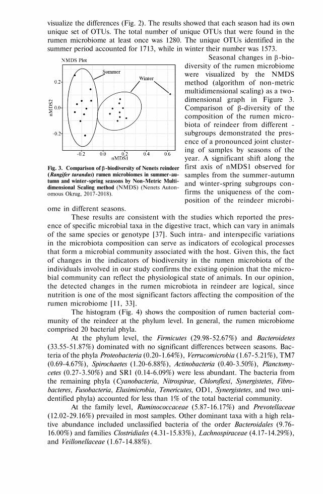
703
visualize the differences (Fig. 2). The results showed that each season had its own unique set of OTUs. The total number of unique OTUs that were found in the rumen microbiome at least once was 1280. The unique OTUs identified in the summer period accounted for 1713, while in winter their number was 1573.
Seasonal changes in -bio-diversity of the rumen microbiome were visualized by the NMDS method (algorithm of non-metric multidimensional scaling) as a two-dimensional graph in Figure 3. Comparison of β-diversity of the composition of the rumen micro-biota of reindeer from different -subgroups demonstrated the pres-ence of a pronounced joint cluster-ing of samples by seasons of the year. A significant shift along the first axis of nMDS1 observed for samples from the summer-autumn and winter-spring subgroups con-firms the uniqueness of the com-position of the reindeer microbi-
ome in different seasons. These results are consistent with the studies which reported the pres-
ence of specific microbial taxa in the digestive tract, which can vary in animals of the same species or genotype [37]. Such intra- and interspecific variations in the microbiota composition can serve as indicators of ecological processes that form a microbial community associated with the host. Given this, the fact of changes in the indicators of biodiversity in the rumen microbiota of the individuals involved in our study confirms the existing opinion that the micro-bial community can reflect the physiological state of animals. In our opinion, the detected changes in the rumen microbiota in reindeer are logical, since nutrition is one of the most significant factors affecting the composition of the rumen microbiome [11, 33].
The histogram (Fig. 4) shows the composition of rumen bacterial com-munity of the reindeer at the phylum level. In general, the rumen microbiome comprised 20 bacterial phyla.
At the phylum level, the Firmicutes (29.98-52.67%) and Bacteroidetes (33.55-51.87%) dominated with no significant differences between seasons. Bac-teria of the phyla Proteobacteria (0.20-1.64%), Verrucomicrobia (1.67-5.21%), TM7 (0.69-4.67%), Spirochaetes (1.20-6.88%), Actinobacteria (0.40-3.50%), Planctomy-cetes (0.27-3.50%) and SR1 (0.14-6.09%) were less abundant. The bacteria from the remaining phyla (Cyanobacteria, Nitrospirae, Chloroflexi, Synergistetes, Fibro-bacteres, Fusobacteria, Elusimicrobia, Tenericutes, OD1, Synergistetes, and two uni-dentified phyla) accounted for less than 1% of the total bacterial community.
At the family level, Ruminococcaceae (5.87-16.17%) and Prevotellaceae (12.02-29.16%) prevailed in most samples. Other dominant taxa with a high rela-tive abundance included unclassified bacteria of the order Bacteroidales (9.76-16.00%) and families Clostridiales (4.31-15.83%), Lachnospiraceae (4.17-14.29%), and Veillonellaceae (1.67-14.88%).
Fig. 3. Comparison of -biodiversity of Nenets reindeer (Rangifer tarandus) rumen microbiomes in summer-au-tumn and winter-spring seasons by Non-Metric Multi-dimensional Scaling method (NMDS) (Nenets Auton-omous Okrug, 2017-2018).

704
Fig. 4. Seasonal abundance of bacterial phyla in Nenets reindeer (Rangifer tarandus) rumen microbi-omes: SCM, SAM, SAF — summer-autumn samples from male calves, adult bulls and adult cows; WCM, WAM, WAF — winter-spring samples from male calves, adult bulls and adult cows (Nenets Autonomous Okrug, 2017-2018).
At the genus level, Prevotella (8.83-27.04%), Succiniclasticum (0.27-12.22%), unidentified bacteria of the genera from the families Veillonellaceae (0.21-2.71%), Lachnospiraceae (2.12-8.14%), Ruminococcaceae (5.02-14.14%) and from the orders Clostridiales (2.42-7.71%) and Bacteroidales (9.64-17.00%) occu-pied a dominant position.
Note, minor counts of microorganisms that traditionally belong to causative agents of various diseases of mammals, including members of the families Fusobac-teriaceae and Mycoplasmataceae were present in some individuals.
Despite the fact that the winter ration of reindeer was mainly the lichens, which are not typical feed for other ruminants (cattle, sheep, etc.), it is important to note that, in general, the obtained profiles of the microbiomes correspond to the modern understanding of rumen microbiota in ruminants. Our data serve as a direct confirmation of the results of G. Henderson et al. [11], who studied rumen microbiomes of different ruminants of 32 species and subspecies and showed the presence of a core community which remained stable in all studied species and subspecies. In the core community, these researchers detected bacteria of the gen-era Prevotella, Butyrivibrio, and Ruminococcus among the representatives of the phyla Firmicutes and Bacteroidetes. According to their data, the number of other microorganisms (e.g., bacteria of the families Lachnospiraceae, Ruminococcaceae, Bacteroidales, and Clostridiales) varied depending on the diet and the environment, thereby determining the uniqueness of each species of ruminant, while the differ-ences between groups which determine specificity of adaptation to environmental conditions were manifested at the level of minor taxa.
It is worth noting the important physiological role of microorganisms of the phyla Firmicutes and Bacteroidetes in the life of ruminants. Many microorgan-isms that are part of these taxa (for example, of the families Lachnospiraceae, Ruminococcaceae, Clostridiaceae, Bacteroidaceae, etc.) are active producers of en-zymes (cellulases, hemicellulases, xylanases, glycoside hydrolases, etc.) that the host’s body is not able to produce on its own. Microbial enzymes allow the body

705
of ruminants to assimilate a wide range of plant polysaccharides, providing the body of animals with volatile fatty acids (VFA), such as acetate, propionate, bu-tyrate, and other nutritive compounds [33, 34].
The proportion of phyla Firmicutes and Bacteroidetes in the rumen was reported to depend on the habitat and type of feed [13]. Thus, many bacteria of the phylum Firmicutes (genera Ruminococcus, Clostridium, Butirivibrio, etc.) ac-tively produce cellulolytic enzymes, which allows them to break down plant fi-ber, while representatives of Bacteroidetes synthesize mainly amylolytic enzymes and promote the utilization of easily fermented carbohydrates [34]. Therefore, a change in the profile of microorganisms of these groups can also lead to a change in the spectrum of metabolites produced by them, thereby influencing the host organism.
Our research revealed that the change of seasons caused significant differ-ences in the abundance of microorganisms in the phyla Firmicutes and Bacteroidetes, as well as in a number of representatives of other taxa. Figures 5 and 6 show heat maps reflecting prevalence of microorganisms the abundance of which in the rein-deer rumen significantly differed in the winter-spring and summer-autumn periods.
Figure 5 shows that in the summer-autumn period as compared to win-ter-spring time, the proportion between SR1 phyla (0.33±0.11% vs. 3.09±0.52%, r = 0.00004, p < 0.05) and Planctomycetes (0.01±0.006% vs. 0.24±0.15%, r = 0.02, p < 0.05) were significantly less.
Fig. 5. Heatmap of seasonal difference in the abundance of bacterial phyla in Nenets reindeer (Rangifer tarandus) rumen microbiomes: S — summer, W — winter, A — adult, С — calf, M — male, F — female; k — kingdom, p — phylum (Nenets Autonomous Okrug, 2017-2018).
In the winter-spring period, abundance of a number of phyla significantly decreased, including Actinobacteria (0.26±0.08% vs. 0.03±0.01%, r = 0.004), Chlor-oflexi (0.95±0.15% vs. 0.43±0.06%, r = 0.004), Tenericutes (3.10±0.08% vs. 0.05±0.02%, r = 0.006), Verrucomicrobia (0.27±0.06% vs. 0.09±0.03%, r = 0.01), unidentified phyla (1.96±0.32% vs.1.01±0.24%, r = 0.02); for all r p < 0.05. A lower proportion of phyla Actinobacteria, Verrucomicrobia, and Chloroflexi in the rumen in winter appeared to be a regularity; according to some research, these microorganisms are associated with soil and plant ecosystems [38].
Moreover, in general, NGS sequencing did no revealed significant sea-sonal differences in total proportion of the phyla Firmicutes and Bacteroidetes dom-inating in the reindeer rumen. However, as can be seen from Figure 6, at a lower taxonomic level, the abundance of some members of these taxa in the winter significantly increased. Interestingly, significant differences occurred primarily for microorganisms associated with fermentation of plant polysaccharides. Thus, in the winter there was a significant increase in the number of bacteria of the genera
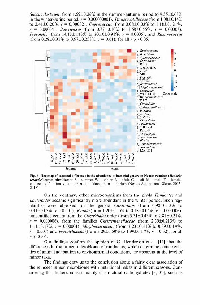
706
Succiniclasticum (from 1.59±0.26% in the summer-autumn period to 9.55±0.68% in the winter-spring period, r = 0.000000001), Paraprevotellaceae (from 1.08±0.14% to 2.41±0.20%, r = 0.00002), Coprococcus (from 0.08±0.03% to 1.18±0, 21%, r = 0.00004), Butyrivibrio (from 0.77±0.10% to 3.58±0.55%, r = 0.00007), Prevotella (from 14.13±1.13% to 20.10±0.91%, r = 0.0005), and Ruminococcus (from 0.28±0.01% to 0.97±0.253%, r = 0.01); for all r p <0.05.
Fig. 6. Heatmap of seasonal difference in the abundance of bacterial genera in Nenets reindeer (Rangifer tarandus) rumen microbiomes: S — summer, W — winter, A — adult, С — calf, M — male, F — female; g — genus, f — family, o — order, k — kingdom, p — phylum (Nenets Autonomous Okrug, 2017-2018).
On the contrary, other microorganisms from the phyla Firmicutes and Bacteroides became significantly more abundant in the winter period. Such reg-ularities were observed for the genera Clostridium (from 0.98±0.13% to 0.41±0.07%, r = 0.001), Blautia (from 1.20±0.15% to 0.18±0.04%, r = 0.000006), unidentified genera from the Clostridiales order (from 5.71±0.43% to 2.81±0.21%, r = 0.000006), from the families Christensenellaceae (from 2.39±0.213% to 1.11±0.17%, r = 0.00001), Mogibacteriaceae (from 2.23±0.41% to 0.89±0.19%, r = 0.007) and Prevotellaceae (from 3.29±0.50% to 1.99±0.17%, r = 0.02); for all r p <0.05.
Our findings confirm the opinion of G. Henderson et al. [11] that the differences in the rumen microbiome of ruminants, which determine characteris-tics of animal adaptation to environmental conditions, are apparent at the level of minor taxa.
The findings draw us to the conclusion about a fairly clear association of the reindeer rumen microbiome with nutritional habits in different seasons. Con-sidering that lichens consist mainly of structural carbohydrates [3, 32], such as

707
hemicellulose, it seems natural that bacteria of the genera Butyrivibrio and Rumi-nococcus capable of hydrolyzing this polysaccharide predominate in the rumen in the winter-spring period. It is known that the role of other ruminal bacteria, namely the members of genera Prevotella and Paraprevotella of phylum Bacteroide-tes, is associated with the degradation of starchy polysaccharides due to production of amylases [39]. An increase in the number of these bacteria in winter may be an expected consequence of the high content of lichen starch, the lichenin in the reindeer diet. In this regard, the discovered increase in the abundance of Prevotel-lus in the rumen of the Chinese sika deer, whose diet included a large amount of oak leaves containing a significant amount of secondary plant metabolites with antimicrobial action is also of interest [40].
Note that the abundance of another ruminal prevotellas from an uniden-tified genus of the family Prevotellaceae, on the contrary, was higher in the sum-mer-autumn period (see Fig. 6). This can be explained by the high genome vari-ability of the microorganisms of this family, as a result of which different genera and species of Prevotellaceae show the ability to synthesize a wide range of en-zymes that hydrolyze plant polysaccharides. Thus, an increase in the bacterial abundance of Prevotellus that we identified in the summer-autumn period is pos-sible due to its ability to form cellulases that break down plant fiber the proportion of which in the reindeer diet increases in summer.
The genera Clostridium and Blautia also belong to the cellulose-decom-posing ruminal bacteria, which explains their significantly higher level in the rein-deer rumen in summer and autumn and indicates an increase in the potential of the microbial community for the fermentation of plant polysaccharides. In addi-tion, a significant antimicrobial effect of lichen usnic acid was previously reported in a number of clostridial strains [5]. Note that the findings of T.H. Aagnes et al. [3] come into some contradiction with our data, since these authors reported an increase in the abundance of Clostridia in winter, which, obviously, can also be explained by the specific metabolic characteristics of various species of the genus Clostridium.
F. Li et al. [41] when analyzing the rumen microbiome in 709 cattle, revealed that the presence of a complex of bacteria, including unidentified bacteria of the order Clostridiales, families Christensenellaceae, Mogibacteriaceae, directly and positively correlates with the amount of acetate, while the genus Succiniclas-ticum abundance correlate with the propionate level. The data obtained by F. Li and colleagues indicate a certain metabolic relationship between these microor-ganisms. Thus, it is known that the formation of acetate in the rumen is associated with degradation of plant cellulose, while the formation of propionate is associated with the starch metabolism [34]. Thus, our results explain the increase in the abundance of bacteria synthesizing acetate in summer when plants are the main component of the reindeer diet, and those producing propionate in winter when the animals predominantly eat lichens.
In addition, we revealed a slight decrease in the proportion of the phylum Cyanobacteria (from 0.07±0.02% to 0%, r = 0.00004, p < 0.05) in the winter-spring period. In our study, as already noted, the presence of cyanobacteria in the rumen of reindeer inhabiting the territory of the Nenets Autonomous Okrug, both in the summer-autumn and winter-spring periods, was generally minor. Neverthe-less, other works [26, 42] mention a sufficiently high number of cyanobacteria in reindeer rumen microbial communities. This seems quite expectable, since cyano-bacteria are symbionts of lichens which constitute up 10-15% of reindeer grazing diet in summer and up to 75% in winter [43].

708
The patterns that we identified for the set of opportunistic microorgan-isms deserve special attention. NGS analysis showed that many microorganisms that are causative agents of infections preferably reproduce in reindeer rumen in summer and autumn. In winter and spring their abundance significantly decreases, from 1.12±0.17% to 0.50±0.06% (r = 0.003) for Erysipelotrichaceae, from 0.26±0.08% to 0.02±0.01% (r = 0.005) for Coriobacteriaceae, from 0.22±0.06% to 0.04±0.02% (r = 0.02) for Mycoplasmataceae, and from 0.39±0.11% to 0.10±0.02% (r = 0.017) for Rickettsiales; for all r p <0.05.
These results are to some extent confirmed by the study [44], in which the Coriobacteriaceae family was found to be the most sensitive indicators responding to changes in the rumen microbiome of ruminants. A significantly higher abun-dance of Mycoplasmataceae family comprising bacterial pathogens, that we re-vealed during the summer-autumn period, also corresponds to the available data on the higher incidence of reindeer in the summer season [45].
For cattle, a positive correlation was revealed between the abundance of Erysiopelotrichaceae family gram-positive bacteria and the feeding habits [46]. Like species of genus Lactobacillus, most members of Erysiopelotrichaceae ferment a wide variety of sugars to form mainly lactic acid, resulting in increased rumen acidity. It is known that an increase in the amount of lactic acid in the rumen leads to impaired digestion of fiber and the development of lactic acidosis. J.E. Nocek et al. [47] showed that higher lactic acid level in the rumen of cattle can lead to Fusobacterium necrophorum infection and necrobacteriosis as a result of the pathogen penetration into the blood through damaged rumen epithelium. Given that necrobacteriosis in reindeer herds is seasonal, i.e. occurs in the sum-mer, the increased abundance of Erysipelothrihaceae family that we have identified in the summer seems logical and noteworthy.
To summarize, the survey of Nenets reindeer (Rangifer tarandus) in the Russian Arctic showed a significantly higher -biodiversity level in the sum-mer-autumn period compared to the winter-spring period as per the number of OTUs (р = 0.0075542) and the values of Chao1 (p = 0.0084612) and Shannon (p = 0.0000523) indices. We did not find statistically significant differences in the biodiversity of the rumen bacterial community with regard to sex and age of the reindeer. Regardless of the season, the phyla Firmicutes and Bacteroidetes dominate in the rumen content, the phyla Proteobacteria, Actinobacteria, Ver-rucomicrobia, TM7, Spirochaete, Planctomycetes, and SR1 are less abundant, and other detected taxa are minor. Nevertheless, we revealed significant changes in the composition of certain taxonomic groups of bacteria depending on the season. In the winter-spring period, the number of ruminal bacteria involved in decomposition of lichen polysaccharides increase, including those decomposing hemicellulose (Butyrivibrio, r = 0.00007; Ruminococcus, r = 0.01) and lichenin (Succiniclasticum, r = 0.000000001; Paraprevotellaceae, r = 0.00002; Prevotella, r = 0.0005). During summer and autumn, the abundance of micro-organisms associated with the decomposition of plant fiber increases, including members of genera Clostridium (r = 0.001), Blautia (r = 0.000006), unidentified genera from the order Clostridiales (r = 0.000006) and from the families Chris-tensenellaceae (r = 0.00001), Mogibacteriaceae (r = 0.007), and Prevotellaceae (r = 0.02). The abundance of some infectious pathogens in the reindeer rumen is higher in summer, including bacteria of the families Erysipelotrichaceae (r = 0.003), Coriobacteriaceae (r = 0.005), Mycoplasmataceae (r = 0.02), and Rickettsiales (r = 0.017). For all r values, p < 0.05.
Thus, the obtained microbiome profiles of the reindeer rumen are gener-ally consistent with modern concepts of the rumen microbiota in ruminants. In the summer-autumn period, there is a significant increase in the indices of -

709
biodiversity of the rumen microbiome as comparison to the winter-spring period. Comparison of the -diversity of the reindeer rumen microbiota shows a pro-nounced clustering of the samples by seasons. In the winter-spring season, a sig-nificant increase occurs in the abundance of microorganisms that decompose li-chen polysaccharides, including hemicellulose (Butyrivibrio, Ruminococcus) and lichenin (Succiniclasticum, Paraprevotellaceae, Prevotella). In the summer-autumn period, there is a significant increase in the proportion of cellulolytic bacteria Clostridium, Blautia, Clostridiales, Christensenellaceae, Mogibacteriaceae, and Prevotellaceae. In addition, the summer period is preferable for the development of infectious agents (Erysipelotrichaceae, Coriobacteriaceae, Mycoplasmataceae, and Rickettsiales). In general, these findings allow us to conclude that there is a fairly clear association of the reindeer rumen microbiome with nutritional habits in different seasonal periods. The differences in the rumen microbiome of rumi-nants, which determine adaptation to environmental conditions, are apparent at the level of minor taxa. The data we obtained can be used to develop means facilitating the reindeer adaptation to the ecological conditions of the territory, as well as means for the prevention and treatment of diseases, one way or another associated with seasonal changes in the rumen microbiome.
R E F E R E N C E S
1. Morgavi D.P., Kelly W.J., Janssen P.H., Attwood G.T. Rumen microbial (meta)genomics and its
application to ruminant production. Animal, 2013, 7(1): 184-201 (doi: 10.1017/S1751731112000419). 2. Mathiesen S.D., Mackie R.I., Aschfalk A., Ringø E., Sundset M.A. Microbial ecology of the
digestive tract in reindeer: seasonal changes. In: Biology of growing animals. Vol. 2 Microbial ecology in growing animals. W.H. Holzapfel, P.J. Naughton, S.G. Pierzynowski, R. Zabielski, E. Salek (eds.). Elsevier Ltd., Edinburgh, 2005: 75-102 (doi: 10.1016/S1877-1823(09)70037-2).
3. Aagnes T.H., Sørmo W., Mathiesen S.D. Ruminal microbial digestion in free living, in captive lichen-fed and in starved reindeer (Rangifer tarandus tarandus) in winter. Applied and Environ-mental Microbiology, 1995, 61(2): 583-591.
4. Orpin C.G., Mathiesen S.D., Greenwood Y., Blix A.S. Seasonal changes in the ruminal micro-flora of the high-arctic Svalbard reindeer (Rangifer tarandus platyrhynchus). Applied and Environ-mental Microbiology, 1985, 50(1): 144-151.
5. Sundset M.A., Kohn A., Mathiesen S.D., Praesteng K.E. Eubacterium rangiferina, a novel usnic acid-resistant bacterium from the reindeer rumen. Naturwissenschaften, 2008, 95(8): 741-749 (doi: 10.1007/s00114-008-0381-0).
6. Kartsev V., Lichitsky B., Geronikaki A., Petrou A., Smiljkovic M., Kostic M., Radanovic O., Soković M. Design, synthesis and antimicrobial activity of usnic acid derivatives. Med. Chem. Commun., 2018, 9(5): 870-882 (doi: 10.1039/C8MD00076J).
7. Roach J.A.G., Musser S.M., Morehouse K., Woo J.Y.J. Determination of usnic acid in lichen toxic to elk by liquid chromatography with ultraviolet and tandem mass spectrometry determina-tion. J. Agric. Food. Chem., 2006, 54(7): 2484-2490 (doi: 10.1021/jf052767m).
8. Dailey R.N, Montgomery D.L., Ingram J.T., Siemion R., Vasquez M., Raisbeck M.F. Toxicity of lichen secondary metabolite (+)-Usnic acid in domestic sheep. Veterinary Pathology, 2008, 45(1): 19-25 (doi: 10.1354/vp.45-1-19).
9. Palo R.T. Usnic acid, a secondary metabolite of lichens and its effect on in vitro digestibility in reindeer. Rangifer, 1993, 13(1): 39-43 (doi: 10.7557/2.13.1.1071).
10. Sundset M.A., Barboza P.S., Green T.K., Folkow L.P., Blix A.S., Mathiesen S.D. Microbial degradation of usnic acid in the reindeer rumen. Naturwissenschaften, 2010, 97: 273-278 (doi: 10.1007/s00114-009-0639-1).
11. Henderson G., Cox F., Ganesh S., Jonker A., Young W., Global Rumen Census Collaborators, Janssen P.H. Rumen microbial community composition varies with diet and host, but a core microbiome is found across a wide geographical range. Scientific Reports, 2015, 5: 14567 (doi: 10.1038/srep14567).
12. Fonty G., Joblin K., Chavarot M., Roux R., Naylor G., Michallon F. Establishment and devel-opment of ruminal hydrogenotrophs in methanogen-free lambs. Applied and Environmental Mi-crobiology, 2007, 73(20): 6391-6403 (doi: 10.1128/AEM.00181-07).
13. Sundset M.A., Præsteng K.E., Cann I.K.O., Mathiesen S.D., Mackie R.I. Novel rumen bacterial diversity in two geographically separated sub-species of reindeer. Microb. Ecol., 2007, 54(3): 424-438 (doi: 10.1007/s00248-007-9254-x).

710
14. Crater A.R., Barboza P.S., Forster R. Regulation of rumen fermentation during seasonal fluctu-ations in food intake of muskoxen. Comparative Biochemistry and Physiology. Part A, Molecular & Integrative Physiology, 2007, 146(2): 233-241 (doi: 10.1016/j.cbpa.2006.10.019).
15. Rustomo B., AlZahal O., Odongo N., Duffield T.F., McBride B.W. Effects of rumen acid load from feed and forage particle size on ruminal pH and dry matter intake in the lactating dairy cow. Journal of Dairy Science, 2006, 89(12): 4758-4768 (doi: 10.3168/jds.S0022-0302(06)72525-5).
16. Kleen J.L., Hooijer G.A., Rehage J., Noordhuizen J.P. Subacute ruminal acidosis (SARA): a review. Journal of Veterinary Medicine Series A, 2003, 50(8): 406-414 (doi: 10.1046/j.1439-0442.2003.00569.x).
17. McEwan N.R., Abecia L., Regensbogenova M., Adam C.L., Findlay P.A., Newbold C.J. Rumen microbial population dynamics in response to photoperiod. Letters in Applied Microbiology, 2005, 41(1): 97-101 (doi: 10.1111/j.1472-765X.2005.01707.x)
18. Uyeno Y., Sekiguchi Y., Tajima K., Takenaka A., Kurihara M., Kamagata Y. An rRNA-based analysis for evaluating the effect of heat stress on the rumen microbial composition of Holstein heifers. Anaerobe, 2010, 16(1): 27-33 (doi: 10.1016/j.anaerobe.2009.04.006).
19. Romero-Pérez G.A., Ominski K.H., McAllister T.A., Krause D.O. Effect of environmental fac-tors and influence of rumen and hindgut biogeography on bacterial communities in steers. Applied and Environmental Microbiology, 2011, 77(1): 258-268 (doi: 10.1128/AEM.01289-09).
20. Olsen M.A., Aagnes T.H., Mathiesen S.D. The effect of timothy silage on the bacterial population in rumen fluid of reindeer (Rangifer tarandus tarandus) from natural summer and winter pasture. FEMS Microbiology Ecology, 1997, 24(2): 127-136 (doi: 10.1111/j.1574-6941.1997.tb00429.x).
21. Wang T.Y., Chen H.L., Lu M.J., Chen Y.C., Sung H.M., Mao C.T., Cho H.Y., Ke H.M., Hwa T.Y., Ruan S.K., Hung K.Y., Chen C.K., Li J.Y., Wu Y.C., Chen Y.H., Chou S.P., Tsai Y.W., Chu T.C., Shih C.A., Li W.H., Shih M.C. Functional characterization of cellulases identified from the cow rumen fungus Neocallimastix patriciarum W5 by transcriptomic and se-cretomic analyses. Biotechnol. Biofuels, 2011, 4: 24 (doi: 10.1186/1754-6834-4-24).
22. Fonty G., Joblin K.N. Rumen anaerobic fungi: their role and interactions with other rumen microorganisms in relation to fiber digestion. In: Physiological aspects of digestion and metabolism in ruminants. Academic Press, Toronto, ON, 1990: 665-680.
23. Sundset M.A., Edwards J.E., Cheng Y.F., Senosiain R.S., Fraile M.N., Northwood K.S., Præsteng K.E., Glad T., Mathiesen S.D., Wright A.D.G. Molecular diversity of the rumen mi-crobiome of Norwegian reindeer on natural summer pasture. Microb. Ecol., 2009, 57(2): 335-348 (doi: 10.1007/s00248-008-9414-7).
24. Sundset M.A., Edwards J.E., Cheng Y.F., Senosiain R.S., Fraile M.N., Northwood K.S., Praesteng K.E., Glad T., Mathiesen S.D., Wright A.D. Rumen microbial diversity in Svalbard reindeer, with particular emphasis on methanogenic archaea. FEMS Microbiology Ecology, 2009, 70(3): 553-562 (doi: 10.1111/j.1574-6941.2009.00750.x).
25. Salgado-Flores A., Hagen L.H., Ishaq S.L., Zamanzadeh M., Wright A.D.G., Pope P.B., Sundset M.A. Rumen and cecum microbiomes in reindeer (Rangifer tarandus tarandus) are changed in response to a lichen diet and may affect enteric methane emissions. PLoS ONE, 2016, 11(5): e0155213 (doi: 10.1371/journal.pone.0155213).
26. Polezhaev A.N., Berkutenko A.N. Opredelitel' kormovykh rastenii severnogo olenya: Magadanskaya oblast' [Keys to forage plants for reindeer: Magadan region]. Magadan, 1981 (in Russ.).
27. Motovilov K.Ya., Bulatov A.P, Poznyakovskii P.M., Lantseva N.N., Mikolaichik I.N. Ekspertiza kormov i kormovykh dobavok [Expertise of feed and feed additives]. Novosibirsk, 2004 (in Russ.).
28. Caporaso J.G., Kuczynski J., Stombaugh J., Bittinger K., Bushman F.D., Costello E.K., Fierer N., Pesa A.G., Goodrich J.K., Gordon J.I., Huttley G.A., Kelley S.T., Knights D., Koenig J.E., Ley R.E., Lozupone C.A., McDonald D., Muegge B.D., Pirrung M., Reeder J., Sevinsky J.R., Turnbaugh P.J., Walters W.A., Widmann J., Yatsunenko T., Zaneveld J., Knight R. QIIME allows analysis of high-throughput community sequencing data. Nature Methods, 2010, 7: 335-336 (doi: 10.1038/nmeth.f.303).
29. Oliveros J.C. Venny. An interactive tool for comparing lists with Venn’s diagrams. 2007. Accessed: http://bioinfogp.cnb.csic.es/tools/venny/index.html. No date.
30. Warton D.I., Wright T.W., Wang Y. Distance-based multivariate analyses confound location and dispersion effects. Methods in Ecology and Evolution, 2012, 3(1): 89-101 (doi: 10.1111/j.2041-210X.2011.00127.x).
31. Morgavi D.P., Kelly W.J., Janssen P.H., Attwood G.T. Rumen microbial (meta)genomics and its application to ruminant production. Animal, 2013, 7(1): 184-201 (doi: 10.1017/S1751731112000419).
32. Person S.J., White R.G., Luick J.R. Determination of nutritive value of reindeer-caribou range, In: Proceedings of the 2nd International Reindeer and Caribou Symposium. E. Reimers, E. Gaare, S. Skjenneberg (eds.). Direktoratet for vilt og ferskvannsfisk, Trondheim, Norway, 1980: 224-239.
33. Church D.C. Ruminant animal: digestive physiology and nutrition. Prentice Hall, New Jersey, 1993. 34. Hungate R.E. The rumen and its microbes. Academic Press, NY, 1966. 35. Hackmann T.J., Spain J.N. Invited review: ruminant ecology and evolution: perspectives useful
to ruminant livestock research and production. Journal of Dairy Science, 2010, 93(4): 1320-1334

711
(doi: 10.3168/jds.2009-2071). 36. Peng S., Yin J., Liu X., Jia B., Chang Z., Lu H., Jiang N., Chen Q. First insights into the
microbial diversity in the omasum and reticulum of bovine using Illumina sequencing. J. Appl. Genetics, 2015, 56(3): 393-401 (doi: 10.1007/s13353-014-0258-1).
37. Smith C.C.R., Snowberg L.K., Gregory C.J., Knight R., Bolnick D.I. Dietary input of microbes and host genetic variation shape among-population differences in stickleback gut microbiota. ISME J., 2015, 9(11): 2515-2526 (doi: 10.1038/ismej.2015.64).
38. Fiere N. Embracing the unknown: disentangling the complexities of the soil microbiome. Nature Reviews Microbiology, 2017, 15: 579-590 (doi: 10.1038/nrmicro.2017.87).
39. Shen J., Zheng L., Chen X., Han X., Cao Y., Yao J. Metagenomic analyses of microbial and carbohydrate-active enzymes in the rumen of dairy goats fed different rumen degradable starch. Frontiers in Microbiology, 2020, 11: 1003 (doi: 10.3389/fmicb.2020.01003).
40. Li Z.P., Liu H.L., Guang Y.L., Bao K., Wang K.Y., Xu C., Yang Y.F., Yang H.F., Wright A.D.G. Molecular diversity of rumen bacterial communities from tannin-rich and fiber-rich forage fed domestic Sika deer (Cervus nippon) in China. BMC Microbiol., 2013, 13: 151 (doi: 10.1186/1471-2180-13-151).
41. Li F., Li C., Chen Y., Liu J. Host genetics influence the rumen microbiota and heritable rumen microbial features associate with feed efficiency in cattle. Microbiome, 2019, 7: 92 (doi: 10.1186/s40168-019-0699-1).
42. Zielińska S., Kidawa D., Stempniewicz L., Łoś M., Łoś J.M. New insights into the microbiota of the Svalbard Reindeer Rangifer tarandus platyrhynchus. Frontiers in Microbiology, 2016, 7: 170 (doi: 10.3389/fmicb.2016.00170).
43. Mathiesen S.D, Haga Ø.E, Kaino T., Tyler N.J.C. Diet composition, rumen papillation and maintenance of carcass mass in female Norwegian reindeer (Rangifer tarandus tarandus) in winter. Journal of Zoology, 2000, 251(1): 129-138 (doi: 10.1111/j.1469-7998.2000.tb00598.x).
44. Zhou M., Peng Y., Chen Y., Klinger C., Masahito O., Liu J., Guan L. Assessment of microbiome changes after rumen transfaunation: implications on improving feed efficiency in beef cattle. Mi-crobiome, 2018, 6: 62 (doi: 10.1186/s40168-018-0447-y).
45. Kazanovskii E.S., Karabanov V.P., Klebenson K.A. Bolezni severnykh olenei [Diseases in reindeer]. Syktyvkar, 2011 (in Russ.).
46. Morita H., Shiratori C., Murakami M., Takami H., Toh H., Kato Y., Nakajima F., Takagi M., Akita H., Masaoka T., Hattori M. Sharpea azabuensis gen. nov., sp. nov., a Gram-positive, strictly anaerobic bacterium isolated from the faeces of thoroughbred horses. International Journal of Systematic and Evolutionary Microbiology, 2008, 58(12): 2682-2686 (doi: 10.1099/ijs.0.65543-0).
47. Nocek J.E. Bovine acidosis: implications on laminitis. Journal of Dairy Science, 1997, 80: 1005-1028 (doi: 10.3168/jds.S0022-0302(97)76026-0).
714
AGRICULTURAL BIOLOGY, ISSN 2412-0324 (English ed. Online)
2020, V. 55, ¹ 4, pp. 714-725 (SEL’SKOKHOZYAISTVENNAYA BIOLOGIYA) ISSN 0131-6397 (Russian ed. Print)
ISSN 2313-4836 (Russian ed. Online)
Physiology, biochemistry, nutrition UDC 636.2:591.132:57.01 doi: 10.15389/agrobiology.2020.4.714eng
doi: 10.15389/agrobiology.2020.4.714rus
Blessed memory of our Teacher Vasily V. Tsyupko is devoted
FOOD PARTICLE SIZE AS AN INDICATOR OF ITS STRUCTURAL COMPOSITION AND A KEY ASPECT
OF THE DEVELOPMENT OF THE NUTRITION THEORY PARADIGM
N.V. VASILEVSKY, T.A. YELETSKAYA
Institute of Animal Science of National academy of agrarian sciences of Ukraine, 3, 7-i Gvardeiskoi Armii st., Kharkov, 61120 Ukraine, e-mail [email protected] ( corresponding author), [email protected] ORCID: Vasilevskiy N.V. orcid.org/0000-0002-7437-2910 Yeletskaya T.A. orcid.org/0000-0001-8980-6972 The authors declare no conflict of interests Received October 8, 2019
A b s t r a c t
The using of modern feed-preparation machines and new technologies for the preparation and feeding of rations in animal husbandry has exacerbated the problem of the ratio in food of relatively large particles that capable to support the necessary motility of the digestive tract and relatively small ones, which provide high consumption of dry matter and its digestibility. In the presented work, we were the first to obtain data about the relationship between the particle size of the diet, the contents of the gastrointestinal tract and the feces of cattle. Based on the determination of the average particle size and dry matter content, a method for assessing the structure of feed has been proposed. The concepts of the informational component of food and the definition of rationed feeding have been introduced into the paradigm of nutrition theory. The aim of the work was to study the impact of the particle size of the diet on the particle size of the contents of the gastrointestinal tract and feces of cattle; to assess the relationship of these indicators with the digestibility of the main groups of nutrients; to develop a methodological approach to rationing the diet structure; to supplement the theory of rationed feeding with the necessary concepts. The studies were carried out at the Institute of Animal Science of National academy of agrarian sciences of Ukraine in 2017-2019 on two heifers (Bos taurus taurus) of the Ukrainian red-and-white breed with a live weight of 350 kg fitted with a duodenal lockable cannula of the beginning of the duodenum and a large-diameter rumen cannula. The animals were fed twice a day (at 8 a.m. and 5 p.m.) in equal shares. Chyme flux was measured during 9 h after morning feeding. The intake of chyme into the duodenum was measured within 14 days (n = 6). The measurements were interrupted for 1-2 days to give the animals rest. Samples of the contents of the rumen were collected through a cannula with a 100 mm diameter from the middle part of the rumen mat in triplicate with a 3 h interval between morning and evening feeding. The experiments were designed according to the balance test methodology with simultaneous assessment of the duodenal chyme flux and sampling of the rumen contents, duodenal chyme and feces. The main diet consisted of hay, silage, concentrates and provided the maintenance of the basal metabolism. The other rations were obtained by replacing one or two components of the main ration with additional amounts of the remaining components in an amount that was equal to the dry matter value of the interchangeable feed. The average particle size of the feed particles was determined by the arithmetic weighted average method by manual parsing and dry sieving, and the selected biological samples by the wet sieving method. The metabolic energy of the rations was calculated. It was found that the average particle size of rumen, chyme and feces decreased with an increase in the average particle size of the diet. The average particle size of the rumen (8.68±0.49 mm, n = 31) was an order of magnitude higher than the average particle size of the duodenal chyme and feces, that indicates the most intense crushing of feed in the proventriculus. The average particle size of feces (1.09±0.06 mm, n = 19, 0.1 > p > 0.05) exceeded the average particle size of the duodenal chyme (0.99±0.05 mm, n = 31), that is indicative of the predominant digestion of the small fraction in the intestine and the formation of feces from the large fraction of the chyme. The value of feed structure in our studies was 0.12-0.92 kgʺm/day vs. 0.033-0.062 kgʺm/day for the contents of the rumen, 0.0031-0.0057 kgʺm/day for duodenal chyme, and 0.0019-0.0028 kgʺm/day for feces. A relationship between the structure of the diet, the

715
availability of crude protein for digestion in the intestine and the size of feces particles was revealed, that theoretically suggests the possibility of assessing the state of digestive processes which based on the granulometric parameters of feces. The obtained data allow us to discuss the factors, affecting feed (and food) digestion and assimilation, which cannot be evaluated in a classical balance experiment by the difference in feed consumption and excretion. It has been proposed to collectively denote these factors as an information component which should be incorporated to express the concept of complex feed (food) structure. The information component is represented in part by various biologically active substances that are ingredients of the feed itself and/or products of the gastrointestinal microorganisms. Another part comprises physical factors, such as the physical parameters of food (temperature, humid-ity, particle size and stiffness), and non-feed factors (frequency and regularity of feeding, phased feed-ing, sound, light and other wave effects). We suggest the definition for rationed nutrition as a process of matter and information exchange between the external environment and the body, which ensures welfare and productive life.
Keywords: average particle size of food, structure of food, informational component of food, normalized feeding, theory of nutrition
A fully mixed ration technology is now widely used in the practice of feeding cattle [1, 2]. Possessing a number of indisputable advantages, this tech-nology includes several important aspects the ignoring of which can negatively affect the health and productivity performance of animals. First of all, this con-cerns the uniformity of mixing and the degree of crushing of high fiber coarse feed [3]. Poorly chopped parts of feed do not allow the necessary mixing uniformity to be achieved which is required to exclude the selective eating of individual compo-nents of the mixture by animals [4]. Excessive grinding leads to weaker response of the receptors to feed and, as a result, to a decrease in the rate of digestion and movement of masses along the digestive tract, up to digestive disorders [5-7]. The concept of food structure is used to assess the ability of the daily ration to maintain the necessary stimulation of the motility of the digestive tract. The effect of struc-ture on the intensity of digestion can be characterized by the degree of feed crush-ing expressed through the average particle size of the feed; frequency and duration of rumination and chewing; the density of the scar mat; the degree of digestion of dry matter; the content of neutral-detergent, acid-detergent or crude fiber [8)]. Some researchers use dimensionless indicators, e.g. percentages of or proportions to the reference feed [9].
This work for the first time shows data reflecting the relationship between the sizes of particles in the diet, in the gastrointestinal tract contents and in feces of cattle. A method for assessing the structure of feed is proposed which is based on the average particle size and dry matter content determination. These data allow us formulate a concept of the informational component of food and the definition of rationed feeding to be introduced into the paradigm of nutrition theory.
The objectives of the research were to reveal the effect of the particle size of the diet on the particle size in the contents of the gastrointestinal tract and feces in cattle, to assess the relationship of these indicators with the digestibility of the main groups of nutrients, to develop a methodological approach to the regulation of the structure of the diet, and to incorporate new definitions and concepts into the theory of rationed feeding.
Materials and methods. Two Ukrainian red-and-white heifers (Bos taurus taurus), 350 kg live weight, with a duodenal lockable cannula in the superior part of duodenum and a large-diameter ruminal cannula were involved in feeding trials (the Institute of Animal Science of the Ukrainian Academy of Agrarian Sciences, 2017-2019). The design of the cannulas ensured collection of chyme coming from the abomasum into the duodenum, its quantitation and return to the digestive system [10]. The animals were fed twice a day (at 8.00 and 17.00) in equal por-tions. Chyme flux was measured for 9 hours after morning feeding. The obtained values of the chyme volume for 9 hours were extrapolated to the daily interval,

716
which made it possible to calculate the digestibility of nutrients in the complex stomach after chemical analysis of the selected chyme samples and feed.
The adaptation of the digestion to the tested diet took 14 days. Then, within 14 days, the intake of chyme into the duodenum was measured (n = 6). Between measurements, the animals were allowed for 1-2-day rest. Rumen con-tents were sampled through a 100 mm cannula from the middle part of the rumen mat in 3 replicates with a 3-hour interval between morning and evening feeding. The experiments were carried out according to the methodology of balance exper-iments with a simultaneous assessment of the duodenal chyme flow and collection of rumen contents, duodenal chyme and feces.
Composition of feed, the gastrointestinal tract contents and feces was as-sessed using common analytical methods [11]. The main diet consisted of hay, silage, concentrates and provided the maintenance of the basal metabolism. The rest of the rations were obtained by replacing one or two components of the main ration with an additional amount of the remaining components equal to the dry matter content of the interchangeable feeds. The designed scheme met the re-quirements of data statistical processing by the dispersion method to quantify the influence of individual dietary ingredients on the structure of chyme and feces, and on the digestibility of the main groups of nutrients.
For feed, the average size of particles was determined by the arithmetic weighted average method after manual particle separation and dry sieving, for the collected biological samples the wet sieving method was used [12].
Dietary metabolizable energy (ME) we calculated as ME = 14.46 0.0007 ½ CP + 0.0168 ½ CF 0.0192 ½ CF* 0,00028 ½ NFE,
where CP, CF, CF*, and NFE are crude proteins, crude fat, crude fiver and ni-trogen-free extract (NFE), g/kg dietary dry matter (DM). The availability (b) of nutrients in the small intestine was calculated as
b = C/A ½ 100 %, where C is the amount of nutrient entered the small intestine per day, A is the amount of nutrient consumed with food per day [13].
Statistical calculations, construction of graphs and diagrams were performed using the licensed software package Office Standard 2010 32-bit Russian (license GGWA-A) (https://www.microsoft.com/ru-ru/download/office.aspx) by analysis of variance methods. The arithmetic mean values (M) and standard errors of the mean (±SEM) were calculated. The significance of differences was assessed using the paired Student’s t-test and the conjugate series approach (direct difference).
Results. Tables 1 and 2 present the characterization of the six calculated diets that were fed to the heifers.
The average particle size of the rumen content, duodenal chyme, and feces decreased as the average particle size of the diet increased (Fig. 1). An inverse relationship was found between the size of feed particles and the particle of the digestive tract content. The slope of the linear regression turned out to be close for chyme and feces (0.0044 and 0.0048 mm/mm, respectively), for the rumen contents of the degree of the food particles size influence was an order
1. Composition (kg natural feed per day) of the diets fed to Ukrainian red-and-white heifers (Bos taurus taurus) (Institute of Animal Science of UAAS, Kharkov, 2017-2019)
Feed Diet No.
1 2 3 4 5 6 Silage 16.00 20.00 24.40 19.74 Hay 2.00 3.00 8.39 4.14 Concentrates 2.00 2.96 4.04 N o t е. Dashes mean the absence of the ingredient in the diet.

717
of magnitude higher (0.0479). The average particle size of the fеces was slightly higher than that of the duodenal chyme, 1.09±0.06 and 0.99±0.05 mm, respec-tively. Statistical processing of the entire array of particles showed a difference at the level of tendency in their sizes in chyme and feces (0.1 > p > 0.05). The conjugate series approach to the same data analysis revealed a significant difference (p < 0.05). Most likely, in the small and large intestines, digestion preferentially occurs, the relatively small fraction of chyme particles disappears, and the feces in the rectum are formed from the remaining larger fraction.
2. Characterization of the diets fed to Ukrainian red-and-white heifers (Bos taurus taurus) (Institute of Animal Science of UAAS, Kharkov, 2017-2019)
Diet No. Dry matter, g
Crude fiber, g
Crude protein Metabolizable energy Basal metabolic energy supply, MJ/kg
amount, g concentration, %
total MJconcentration, MJ/kg
1 7685 1690 593 7.71 77.76 10.12 0.80 2 7894 2137 523 6.62 85.17 10.79 0.87 3 6666 1698 444 6.66 69.80 10.47 0.71 4 7899 1492 649 8.21 82.62 10.46 0.85 5 6797 2083 444 6.53 67.61 9.95 0.69 6 6775 1190 614 9.06 66.22 9.77 0.68
Fig. 1. Size of particle in the rumen content (1), chyme (2) and feces (3) of Ukrainian red-and-white heifers (Bos taurus taurus) depending on the dietary particle size (M±SEM, Institute of Animal Science of UAAS, Kharkov, 2017-2019). The linear regressions: for rumen y = 0.0479x + 11.624, R2 = 0.6311; for chyme y = 0.0044x + 1.2185, R2 = 0,806; for feces y = 0,0047x + 1,3979, R2 = 0,5951. For description of diets and design of the trial, see Materials and methods.
3. Particle size and structural orderliness of feed, gastrointestinal tract content and feces of Ukrainian red-and-white heifers (Bos taurus taurus) (M±SEM, Institute of Animal Science of UAAS, Kharkov, 2017-2019)
Sample Diet No. 1 2 3 4 5 6
P a r t i c l e s i z e, mm Feed 41.48 57.13 22.43 15.65 135.14 67.43 Rumen 9.05±0.32 10.26±0.67 12.61±0.36 9.69±0.75 5.60±0.55 6.28±0.62 Chyme 1.19±0.16 0.95±0.05 0.99±0.12 1.16±0.04 0.60±0.03 0.94±0.10 Feces 1.15±0.04 1.13±0.02 1.58±0.22 1.10±0.07 0.77±0.08 1.08±0.09
S t r u c t u r a l o r d e r l i n e s s, kgʺm/day Feed 0.319 0.451 0.150 0.120 0.919 0.457 Rumen 0.051 0.060 0.062 0.061 0.033 0.039 Chyme 0.0043 0.0036 0.0032 0.0057 0.0031 0.0052 Feces 0.0031 0.0025 0.0033 0.0030 0.0019 0.0029
Despite the fact that the average size of feed particles has a significant

718
effect on the activation of receptors in the digestive tract, it cannot serve as an adequate measure of feed structural orderliness, since the duration and degree of exposure also depend on the number of particles. In this regard, to assess the structural orderliness, we used an indicator calculated as the product of the average particle size (m) by the total amount of dry matter (kg) in the daily diet, the contents of the gastrointestinal tract, or feces (Table 3).
Fig. 2. Crude protein digestive availability in intestines (1) and concentration (2) depending on the structural orderliness of feed fed to Ukrainian red-and-white heifers (Bos taurus taurus) (M±SEM, Institute of Animal Science of UAAS, Kharkov, 2017-2019). The linear regressions for crude protein digestive availability y = 314.53x + 63.298, R2 = 0.74, for crude protein concen-tration y = 1.101x + 7.9101, R2 = 0,0946. For description of diets and design of the trial, see Materials and methods.
As structural orderliness increases in feed, it decreases in the rumen con-tent and feces, with the degree of determination R2 = 0.61 and R2 = 0.86, respectively. Regression analysis of indicators of the feed structural orderliness, on the one hand, and feed digestibility in the rumen, as well as visible digesti-bility throughout the gastrointestinal tract, on the other, showed the lack of association for all major groups of nutrients, with the exception of crude protein (negative association, R2 = 0.73 and R2 = 0.74). Moreover, this did not become a consequence of some fluctuations in the content of crude protein in the diet (see Table 2), since changes in the structural orderliness of the diets and the amount of crude protein in feed did not interrelate (R2 = 0.20). The intake of crude protein in the duodenum was higher than its amount taken with feed due to intensive microbial synthesis from endogenous nitrogen sources. A higher structural orderliness with a high degree of determination (R2 = 0.74) led to an increase in the availability of crude protein for digestion in the intestine calcu-lated as intake into the duodenum as a percentage of that consumed with the diet) (Fig. 2). The increase in the intake of crude protein in the intestine led to a slight decrease in its apparent digestibility throughout the gastrointestinal tract. Consequently, in the studied range of averaged feed particle sizes, a higher struc-tural orderliness did not cause significant changes in the digestibility of the main groups of nutrients, with some improvement in the conditions for the synthesis of microbial crude protein in the rumen.
The availability of crude protein for intestinal digestion is an indicator of the intensity of microbial processes in the rumen. Its increase can also indi-rectly indicate the acceleration of fractional outflow from the rumen. That is, the structure of the diet, on the one hand, has a decisive effect on the structure and size of feces particles, on the other, on the availability of crude protein for

719
digestion in the intestine. Given this, we examined the relationship between the particle size of feces and the availability of crude protein for digestion in the intestine. The obtained value of the coefficient of determination (R2 = 0.37) indicated a moderate degree of interdependence on the Cheddock scale between these indicators.
The obtained results do not allow us to identify the optimal range of the structural orderliness of the diet from the viewpoint of digestion conditions, since the energy consumption per unit of metabolic mass in the operated animals cannot reach the values typical for young fattening cattle, and even more so for lactating cows. The proposed methodological approach for assessing the structure of the diet makes it possible to correlate the indicators characterizing the digestive pro-cesses and the structural properties of feed. Research on highly productive animals is necessary to develop standardization of feed structure and rations for cattle. The main difficulty here is that the size and shape of particles cannot be considered within the framework of the theory of balanced nutrition as a food component, which, as a result of digestion and absorption, is assimilated by the body and consumed in the process of vital activity. Nevertheless, the effect of this factor on the digestion process does not raise doubts, and some modern rationing systems involve structural characteristics of feed and cattle diets [9, 14].
Studying the digestibility of nutrients from feed mixtures that are identical in chemical composition, but differ in the degree of grinding of roughage [15], we found a significant effect of the structural factor (particle size and their ability to resist mechanical grinding during chewing) on digestion in ruminants. However, the structural parameters of feed cannot be assessed in the classical balance exper-iment by the difference in consumption and excretion with feces and urine. The “apparently disappeared” structure is not absorbed into the internal environment of the body along with digested components determining structural charateris-tics, nevertheless, the “undigested structure” excreted in the feces has an effect on the digestive processes. Therefore, the concept of structural orderliness does not correspond to the main position of the theory of balanced nutrition, accord-ing to which the nutritional value of food is determined only by its digestible components.
Pokrovsky [16, 17] notes that nutrition is a complex process, the main goal of which is to ensure the growth and development of the child’s body, max-imum performance and well-being in adulthood, longevity and health in the el-derly and in senile period. Nutrition is a source of aesthetic pleasure and an im-portant healing factor [16, 17]. According to the author, it should not only com-pensate for the body’s expenses to maintain vital functions (and in farm animals also productivity), but also provide an emotional and therapeutic component.
Ugolev et al. [18, 19] showed the participation of the gastrointestinal tract in the formation of the individual hormonal profile of the organism in a proportion reaching up to 50% of the total amount of hormones. This means that food, through the formation of a hormonal pattern, is involved in the regulation of gene expression, which means that it affects how a specific phenotypic image of an organism will be formed. In other words, in matters of nutrition, one should not be limited to only considering the role of nutrients in providing endogenous met-abolic processes of the body with exogenous substrates; it is also necessary to recognize the regulatory function of food expressed at the neurohumoral level.
Within the framework of the existing paradigm of nutrition theory, it is impossible to explain the effect of phase feeding, which is sometimes used when fattening young cattle. The essence of the method is to periodically change the rate of feed delivery by 10-20%, alternately up and down from

720
100% of the need. This technique significantly increases live weight gain in comparison with a constant 100% ration delivery [20, 21].
Another example of the contradiction between the feeding practice and the theory of balanced (adequate) nutrition is the exchange of calcium in the body. It is known that the assimilation of this element is critically dependent on the availability of vitamin D [22, 23]. The synthesis of vitamin D, in turn, occurs when the body is exposed to ultraviolet radiation [24, 25]. That is, sunlight or other ultra-violet source should be counted as a “nutritive” element.
The current period in the development of nutrition science is characterized by a significant accumulation of facts that do not fit into the framework of the generally accepted concept, i.e. the classical theory of balanced nutrition. In our opinion, this information has reached a critical mass and prerequisites have been created for the formation of different views on the theory of nutrition and, ac-cordingly, approaches to normalized feeding of animals. A.M. Ugolev [26] gives the most detailed criticism of the theory of balanced nutrition. Nevertheless, until now, there is no universally recognized alternative to the theory of balanced nu-trition and, with the exception of some works [27]; most experts in animal nutri-tion rely on the paradigm of digestion and nutrient assimilation in the framework of the balanced nutrition theory. This is expressed, first of all, in the fact that in all modern systems of rationed feeding of farm animals the rationing is considered as compensation of the body expenses for maintaining vital activity and ensuring productivity.
The flow of substances vital for the body and coming from digested food components, in addition to traditional plastic and energy components, contains substances with zero and even negative energy value, the metabolization of which is associated with additional energy costs. Moreover, food has the ability to influ-ence the body not only through nutrients, but also through other factors of a chemical and physical nature. Such an effect leads not only to changes in the digestion and assimilation of the components of the food itself, but also affects other vital functions of the body, up to a change in the hormonal profile and regulation of genome replication [18, 19, 26].
We guess that the described problems can be easily addressed if we recog-nize the existence of information flows directed both into the internal environment of the organism and from it. In this case, a special role in digestion is played by the exchange of signals with the enteric environment, which is partially separated from the “truly external”. In our opinion, the paradigm of nutritional theory should be expanded with a number of concepts that characterize such information exchange.
Obviously, through complex management of factors that are practically not taken into account at present, it is possible to influence with the desired effect the physiological and biochemical processes in the body. Such factors include not only chemical (biologically active substances, phytohormones, neuropeptides, some amino acids, fatty acids, etc.) and physical (temperature, humidity, particle size and stiffness) parameters of the food itself, but also the frequency of feeding, phased feeding, sound and light effects. In aggregate, we propose to call them the informational component of food, without the rationing of which it is impossible to improve feeding of animals, especially highly productive ones.
From the practical problems, it is possible to limit the scope of the general theory of nutrition to the concept of rationed feeding. In this case, instead of the postulates of theories of balanced nutrition and adequate nutrition [26], we offer the following definition, which introduces the necessary and sufficient fundamen-tal concepts. Normalized nutrition is a process of material and information

721
exchange between the external environment and the body, which ensures the full-fledged productive life of the latter. The concept of “material and information exchange” means that this process involves both the actual nutrients of the feed and its undigested residues removed from the body, as well as information signals from chemical agents and physical factors associated with food (some amino acids, hormones and hormone-like substances, biologically active substances; food struc-ture, regularity and volume of serving portions), and from factors of non-food nature (insolation, temperature and humidity, acoustic accompaniment of the feeding process).
Information exchange is accompanied by a change in the informational state of the organism. The term “stress” is used to assess the informational state. There are three stages in the development of stress: mobilization of the body’s adaptive capabilities (anxiety stage), balanced expenditure of adaptation reserves (resistance stage), and exhaustion stage [28]. Exhaustion is not understood as the expenditure of metabolic resources, but a kind of “fatigue” of the nervous or, rather, the neurohumoral system. Currently, it is customary to assess stress by the effect of a stress factor on the activation of the hypothalamic-pituitary-adrenal axis (HPA axis) [29]. In various animal species, including ruminants, increased activity of the HPA axis is usually observed in connection with acute stress and is aimed at mobilizing the body’s resources to overcome the effects of the stress factor. Corticoliberin, a peptide hormone secreted by neuroendocrine cells of the hypothalamus, at the level of the central nervous system, is involved in the regu-lation of feed intake, as well as in behavioral responses to stress [30, 31]. At the same time, under repeated or long-term exposure to stressors in ruminants, the regulation of the HPA axis is not entirely understood and can be carried out by both the adrenal glands [32] and the pituitary gland [33]. Currently, primary at-tention is focused on studying the effect of nutrition on the HPA axis reactivity in ruminants in order to optimize the number of animals in the production group, the space for their feeding and rest [34, 35]. In our opinion, these studies are of particular interest in connection with the development of a methodology for as-sessing the current state of the body.
A significant part of the information flow from the body is directed to the lumen of the gastrointestinal tract and interacts with the microflora that lives there, which, in turn, interacts with the macroorganism, producing certain signaling sub-stances (and quite possibly through other communication channels).
A full-fledged vital activity means the expenditure of energy and metab-olites obtained from food to maintain vital functions, physical and intellectual activity, to increase body weight, growth and development, to reproduce (in agricultural animals, also to ensure productivity in breeding goals) for a suffi-ciently long time without harm to the body.
In the existing norms for human nutrition and feeding of agricultural animals, there is practically no study on rationing the information flow accom-panying feeding and digestion. Basically, the information component is partially standardized by medical and veterinary regulations, sanitary standards and re-quirements for working conditions and human life, as well as zootechnical re-quirements for the conditions of rearing farm animals. The present approach to rationing can be characterized as deterministic in the sense that, according to it, for the obtaining a certain amount of production, an animal a priori requires certain conditions of rearing, the amount of energy and plastic substances. In this case, neither the individual, nor the adaptive capabilities of the organism are taken into account. Obviously, the primary task in the development of ra-tioned feeding systems should not be the creation of averaged norms of need,

722
but the development of methods for assessing and predicting the state of the animal’s body as the controlled object [36].
Thus, the classical theory of balanced (adequate) nutrition requires cor-rection to incorporate in its paradigm the concepts of the informational compo-nent of food and the circulation of information between the environment and the body. Structural orderliness of the evaluated object (e.g., food, chyme, or feces) can be expressed as the product of the average particle size and the dry matter content. The average particle size of the diet in the range studied by us and feed structure are inversely related to the average size and structure of the contents of the gastrointestinal tract and feces. No relationship has been established between the mean particle size of the diet, the structure of food and rumen content, on the one hand, and the duration of chewing or the number of rumen contractions, on the other. An increase in structure did not have a significant effect on the digestibility of the main groups of nutrients in the feed, with some improvement in the conditions for the synthesis of microbial crude protein in the rumen.
R E F E R E N C E S
1. Moya D., Mazzenga A., Holtshausen L., Cozzi G., González L.A., Calsamiglia S., Gibb D.G.,
McAllister T.A., Beauchemin K.A., Schwartzkopf-Genswein K. Feeding behavior and ruminal acidosis in beef cattle offered a total mixed ration or dietary components separately. Journal of Animal Science, 2011, 89(2): 520-530 (doi: 10.2527/jas.2010-3045).
2. Kharitonov E. Myasnoe i molochnoe skotovodstvo, 2010, 1: 16-17 (in Russ.). 3. Tylutki T.P., Fox D.G., Durbal V.M., Tedeschi L.O., Russell J.B., Van Amburgh M.E., Over-
ton T.R., Chase L.E., Pell A.N. Net sarbohydrate and protein system: a model for precision feeding of dairy cattle. Animal Feed Science and Technology, 2008, 143(1): 174-202 (doi: 10.1016/j.anifeedsci.2007.05.010).
4. Hutjens M.F. Is a one TMR approach right? Western Dairy Management Conference. Reno, 2012: 195-190.
5. Fahey G.C., Novotny L., Layton B., Mertens D.R. Critical factors in determining fiber content of feeds and foods and their ingredients. Journal of AOAC INTERNATIONAL, 2019, 102(1): 52-62 (doi: 10.5740/jaoacint.18-0067).
6. Beauchemin K.A., Rode L.M., Eliason M.V. Chewing activities and milk production of dairy cows fed alfalfa as hay, silage, or dried cubes of hay or silage. J. Dairy Sci., 1997, 80(2): 324-333 (doi: 10.3168/jds.S0022-0302(97)75942-3).
7. Beauchemin K.A., Yang W.Z. Effects of physically effective fiber on intake, chewing activity, and ruminal acidosis for dairy cows fed diets based on corn silage. J. Dairy Sci., 2005, 88(6): 2117-2129 (doi: 10.3168/jds.S0022-0302(05)72888-5).
8. Woolpert M.E., Dann H.M., Cotanch K.W, Melilli C., Chase L.E., Grant R.J. Management practices, physically effective fiber, and ether extract are related to bulk tank milk de novo fatty acid concentration on Holstein dairy farms. J. Dairy Sci., 2017, 100(6): 5097-5106 (doi: 10.3168/jds.2016-12046).
9. Weiß H. von J., Pabst W., Granz S. Tierproduktion. Stuttgart, 2011. 10. Aliev A.A. Noveishie operativnye metody issledovaniya zhvachnykh zhivotnykh [The latest surgical
techniques in research studies of ruminants]. Moscow, 1985 (in Russ.). 11. Vlіzlo V.V., Fedoruk R.S., Ratich І.B., Vіshchur O.І., Sharan M.M., Vudmaska І.I.,
Fedorovich Є.Sh., Ostapіv D.D., Stalai P.V., Buchko O.M., Gunchak A.V., Saliga Yu.T., Stefanishin O.M., Gevkan І.І., Lesik Ya.V., Sіmonov M.R., Nevostruєva І.V., Khomin M.M., Smolyanіnov K.B., Gavrilyak V.V., Kolіsnik G.V., Petrukh І.M., Broda N.A., Luchka І.V., Koval'chuk І.І., Kropivka S.I., Paranyak N.M., Tkachuk V.M., Khrabko M.І., Shtapenko O.V., Dzen' Є.O., Maksimovich І.Ya., Fedorovich V.V., Yus'kіv L.L., Dolaichuk O.P., Іvanits'ka L.A., Sіrko Ya.M., Kistsіv V.O., Zagrebel'nii O.V., Sіmonov R.P., Stoyanovs'ka G.M., Kirilіv B.Ya., Kuzіv M.І., Maior Kh.Ya., Kuz'mіna N.V., Talokha N.І., Lіsna B.B., Klimishin D.O., Chokan T.V., Kamіns'ka M.V., Kozak M.R., Olіinik A.V., Golova N.V., Dubіns'kii V.V., Іskra R.Ya., Rіvіs I.F., Tsepko N.L., Kishko V.І., Oleksyuk N.P., Denis G.G., Slivchuk Yu.І., Martin Yu.V. Laboratornі metodi doslіdzhen' u bіologії, tvarinnitstvі ta veterinarnіi meditsinі: Dovіdnik /Za red. V.V. Vlіzla. Spolom, L'vіv, 2012.
12. Vasilevskii N.V., Eletskaya T.A., Greben' L.G., Berestovaya L.E. Problemy biologii produktivnykh zhivotnykh, 2017, 2: 88-100 (in Russ.).
13. Nehring K., Schiemann R., Hoffman L. A new system of energetic evaluation of food on basis of net energy for fattening. In: Energy metabolism of farm animals. Oriel Press, London, 1969.

723
14. National Research council. Nutrient requirements for dairy cattle: seventh revised edition. The National Academies Press, Washington DC, 2001: 381-333 (doi: 10.17226/9825).
15. Vasilevskii N., Eletskaya T., TSyupko V., Berestovaya L. Vliyanie tekhnologii skarmlivaniya kormov i urovnya syrogo proteina v ratsione na perevarimost' pitatel'nykh veshchestv u korov. Problemy biologii produktivnykh zhivotnykh, 2013, 1: 67-74.
16. Pokrovskii A.A. Besedy o pitanii [Nutritional conversations]. Moscow, 1964 (in Russ.). 17. Pokrovskii A.A. Metabolicheskie aspekty farmakologii i toksikologii pishchi [Metabolic aspects of
food pharmacological and toxicological effects]. Moscow, 1979 (in Russ.). 18. Ugolev A. M. Enterinovaya (kishechnaya gormonal'naya) Sistema [Enteric (intestinal hormonal)
system]. Leningrad, 1978 (in Russ.). 19. Ugolev A.M., Labusheva M.A., Vakhrushev Ya.M. Fiziologicheskii zhurnal SSSR im. I.M. Seche-
nova, 1989, 75(5): 609-618 (in Russ.). 20. Medvedev A.Yu. Usovershenstvovanie energosberegayushchei tekhnologii proizvodstva govyadiny v
molochnom skotovodstve. Doktorskaya dissertatsiya [Improvement of energy-saving technology for beef production in dairy farming]. Lugansk, 2015 (in Russ.).
21. Leibina T.I. Izvestiya Samarskoi gosudarstvennoi sel'skokhozyaistvennoi akademii, 2015, 1: 171-174 (in Russ.).
22. Meena K., Khazai N.B., Ziegler T.R., Nanes V.S., Abrams S.A., Tangpricha V. Vitamin D-mediated calcium absorption in patients with clinically stable Crohn’s disease: a pilot study. Mol. Nutr. Food Res., 2010, 54(8): 1085-1091 (doi: 10.1002/mnfr.200900351).
23. Kaushik R., Sachdeva B., Arora S., Kapila S., Wadhwa B.K. Bioavailability of vitamin D2 and calcium from fortified milk. Food Chemistry, 2014, 147: 307-11 (doi: 10.1016/j.foodchem.2013.09.150).
24. Nelson C.D., Powell J.L., Price D.M., Hersom M.J., Yelich J.V., Drewnoski M.E., Bird S.L., Bridges G.A. Assessment of serum 25-hydroxyvitamin D concentrations of beef cows and calves across seasons and geographical locations. Journal of Animal Science, 2016, 94(9): 3958-3965 (doi: 10.2527/jas.2016-0611).
25. Holcombe S.J., Wisnieski L., Gandy J., Norby B., Sordillo L.M. Reduced serum vitamin D concentrations in healthy early-lactation dairy cattle. Journal of Dairy Science, 2018, 101(2): 1488-1494 (doi: 10.3168/jds.2017-13547).
26. Ugolev A.M. Teoriya adekvatnogo pitaniya i trofologiya [The theory of adequate nutrition and trophology]. Leningrad, 1991 (in Russ.).
27. Vasilevskaya L.S. Rossiiskii zhurnal gastroenterologii, gepatologii, koloproktologii, 2001, 11(4-prilozhenie ¹ 14 Materialy XVI sessii Akademicheskoi shkoly-seminara imeni A.M. Ugoleva «Sovremennye problemy fiziologii i patologii pishchevareniya», Pushchino-na-Oke, 14-17 maya 2001 goda): 148-155 (in Russ.).
28. Sandor S., Yoshida M., Filakovszky J., Juhasz G. «Stress» is 80 years old: from Hans Selye original paper in 1936 to recent advances in gi ulceration. Current Pharmaceutical Design, 2017, 23(27): 4029-4041 (doi: 10.2174/1381612823666170622110046).
29. Robert L Spencer R.L., Deak T. A users guide to HPA axis research. Physiology & Behavior, 2017, 178: 43-65 (doi: 10.1016/j.physbeh.2016.11.014).
30. Dunn A.J., Berridge C.W. Physiological and behavioural responses to corticotrophin-releasing factor administration: is CRF a mediator of anxiety or stress responses? Brain Research Reviews, 1990, 15(2): 71-100 (doi: 10.1016/0165-0173(90)90012-d).
31. Koob G.F. Corticotropin-releasing factor, norepinephrine and stress. Biological Psychiatry, 1999, 46(9): 1167-1180 (doi: 10.1016/s0006-3223(99)00164-x).
32. Fisher A.D., Crowe M.A., Prendiville D.J., Enright W.J. Indoor space allowance, effects on growth, behaviour, adrenal and immune responses of finishing beef heifers. Animal Science, 1997, 64(1): 53-62 (doi: 10.1017/S135772980001554X).
33. Fisher A.D., Verkerk G.A., Morrow C.J., Matthews L.R. The effects of feed restriction and lying deprivation on pituitary-adrenal axis regulation in lactating cows. Livestock Production Science, 2002, 73(2-3): 255-263 (doi: 10.1016/S0301-6226(01)00246-9).
34. Munksgaard L., Ingvartsen K.L., Pedersen L.J., Nielsen V.K.M. Deprivation of lying down affects behaviour and pituitary-adrenal axis responses in young bull. Acta Agriculturae Scandinavica, Sec-tion A — Animal Science, 1999, 49(3): 172-178 (doi: 10.1080/090647099424088).
35. Munksgaard L., Herskin M.S., Lovendahl P., Andersen J.B. Effects of nutrition on stress reac-tivity. In: Ruminant physiology. Digestion, metabolism and impact of nutrition on gene expression, immunology and stress. K. Sejrsen, T. Hvelplund, M.O. Nielsen (eds.). Wageningen Academic Publishers, 2006: 511-525.
36. Vasilevskii N.V. Problemy biologii produktivnykh zhivotnykh, 2015, 2: 67-79 (in Russ.).

726
AGRICULTURAL BIOLOGY, ISSN 2412-0324 (English ed. Online)
2020, V. 55, ¹ 3, pp. 726-737 (SEL’SKOKHOZYAISTVENNAYA BIOLOGIYA) ISSN 0131-6397 (Russian ed. Print)
ISSN 2313-4836 (Russian ed. Online) UDC 636.52/.58:591.132:611.37:57.089.6 doi: 10.15389/agrobiology.2020.4.726eng
doi: 10.15389/agrobiology.2020.4.726rus THE EXOCRINE PANCREATIC FUNCTION IN CHICKEN
(Gallus gallus L.) FED DIETS SUPPLEMENTED WITH DIFFERENT VEGETABLE OILS
V.G. VERTIPRAKHOV, A.A. GROZINA, V.I. FISININ
Federal Scientific Center All-Russian Research and Technological Poultry Institute RAS, 10, ul. Ptitsegradskaya, Sergiev Posad, Moscow Province, 141311 Russia, e-mail [email protected] ( corresponding author), [email protected], [email protected] ORCID: Vertiprakhov V.G. orcid.org/0000-0002-3240-7636 Fisinin V.I. orcid.org/0000-0003-0081-6336 Grozina A.A. orcid.org/0000-0001-9654-7710 The authors declare no conflict of interests Acknowledgements: Supported financially from a subprogram “Study of adaptation mechanisms of the digestive system of mammals and poultry to diets with various ingredients” of the Program for Basic Research (Resolution of the Presidium RAS No. 132 dated 07/05/2017) Received September 19, 2019
A b s t r a c t
The efficiency of different vegetable oils in poultry nutrition is still a matter of interest for the theory and practice; numerous studies in broilers have evidenced that different oils can significantly affect the productive performance and metabolism. However, the mechanism of the influence of dif-ferent oils on the exocrine pancreatic function is understudied. In our study on fistulated chicken the innovative data are presented for the analysis of the effects of different dietary lipid sources on the secretion and enzymatic activities of pancreatic juice. The study was performed in 2019 on three laying hens (Gallus gallus L., cross Hisex White) with chronic fistulae of main pancreatic duct inserted by the method of Ts.Zh. Batoev (2001). The physiological trials were performed by the method of periods (7-10 days per period): during the control period the basic feed (commercial compound feed for layers PK-1) was supplemented with sunflower oil; during period 1 with soybean oil; during period 2 rapeseed oil; during period 3 flaxseed oil. The pancreatic juice and blood were sampled throughout these periods. The secretion rate of pancreatic juice and enzymatic activities in it (amylase, lipase, total proteases TP, alkaline phosphatase AP) were determined by standard methods. The activities of amylase, lipase, alanine (ALT) and aspartate (AST) transaminases in blood serum were determined on analyzer ChemWell 2900 (T) (Awareness Technology, USA) with corresponding reagent kits by Human GmbH, Germany; the activities of TP and AP and other biochemical indices in blood serum were determined on semi-automatic analyzer Sinnowa BS-3000P (SINNOWA Medical Science & Tech-nology Co., China) with BAPNA as the substrate (TP) and corresponding reagent kits by DIAKON-VET, Russia (AP and all other indices). It was found that the activity of lipase in pancreatic juice can adjust to the dietary oil used. The significantly highest lipase activity was found with unrefined sunflower oil (21345±652.8 μmol∕l1∕min1, p < 0.05), together with the increases in the activity of amylase (9254±440.3 mg∕ml1∕min1, p < 0.05). The increases in lipolytic activity in the first few postprandial minutes (corresponding to complex-reflex phase of regulation of the pancreatic secretion) with sunflower and flaxseed oils evidenced that these oils are the most palatable for chicken while the increase in the activity of lipase in neurohumoral phase of regulation with rapeseed and soybean oils evidenced their higher nutritive value. The strong negative correlation (r = 0.87, p < 0.05) was also found between the activities of TP and AP in the pancreatic juice. The phospha-tase-protease index, AP/TP ratio, is proposed as a significant physiological indicator of the adapta-tion of the digestive tract in poultry to the changes in diet composition. The most affected by oil type blood indices were the activities of TP, lipase, ALT, AST, and concentration of triglycerides. The dynamics of lipase activity in blood serum was similar to that in the pancreatic juice; the AP/TP ratio in serum was partially close to the values of this ratio in the pancreatic juice. The changes found in the exocrine pancreatic function in response to different dietary oils evidenced that the studies on the digestion should be continued for further optimization of the composition of com-mercial diets for poultry.
Keywords: exocrine pancreatic function, chickens, digestive enzymes in blood, sunflower oil, soybean oil, rapeseed oil, flaxseed oil
727
Issues of adaptation of the digestive glands of animals to different diets still remain discussible and relevant both for fundamental research and for the practice of poultry rearing. Experiments on birds established that gastric secretion and exocrine pancreatic function adapt to the chemical composition of food [1, 2]. In poultry farming, the use of vegetable oils in feeding is of considerable inter-est. Experiments on broilers [3, 4] indicate the advantage of soybean, camelina, and flaxseed oils compared to sunflower oils. Replacing sunflower oil with other oils, ensure an increase in live weight, a decrease in feed costs per unit of produc-tion, better meat quality and higher profitability. There is a viewpoint [5] that dietary sunflower oil improves poultry development and feed conversion compared to olive oil.
Compound feeds with rapeseed oil are not inferior in efficiency to those with sunflower oil [6]. According to some research, the positive effect of dietary rapeseed oil is expressed in an increase in the content of polyunsaturated fatty acids in the meat of broiler chickens [7]. Rapeseed oil is superior to palm oil as a source of lipids due to higher concentration of digestible fatty acids. In the meat of chickens fed with rapeseed oil, oleic acid predominated [8].
It is known [9] that digestion and absorption of various fats differ depend-ing on a lipid composition. A correlation was shown between the lipid profile of blood serum and the composition of fatty acids supplied in diet and metabolized in the body. This allowed the authors to suggest that the lipid composition of the diet can modulate digestion and absorption in the gastrointestinal canal [9].
A comparative study on sheep have shown that dietary rapeseed and flax-seed oils affect the activity of pancreatic enzymes. The secretion of bile and pan-creatic juice tended to increase in sheep fed rapeseed and flaxseed oil (69.5 and 68.5 ml/h, respectively) as compared to control (59.8 ml/h); lipase and trypsin activity was also higher compared to the control diet, 175 and 21.6 U/l and 179 and 23.2 U/l, respectively, vs. 128 and 13.1 U/l, respectively [10].
Thus, data on the effect of different vegetable oils on poultry productivity are controversial, and no information is available on the mechanisms of action of these nutrients on the digestive system.
This paper is the first to show for the first time that dietary unrefined sunflower oil, as compared to soybean, rapeseed or flaxseed oil, significantly in-creases the lipolytic and amylolytic activity of pancreatic juice, changes the phos-phatase-protease index and blood plasma biochemical profile which is consistent with indicators of the secretory function of the pancreas.
The work aimed to examine effects of various vegetable oils on the secre-tory function of the pancreas and blood biochemical parameters in laying hens.
Materials and methods. Three 28-46-week old Hisex White chickens (Gal-lus gallus domesticus L.) were fistulated according to the method of C.Zh. Batoev and S.Ts. Batoeva [2, 11]. A 4-5 cm segment was cut out of the duodenum, into which the main pancreatic duct was transplanted with the implantation of two L-shaped fistulas. An external anastomosis was formed, allowing the return of pan-creatic juice to the duodenum in the periods between feed trials. Chickens’ feeding and keeping were in compliance with requirements for the cross [12] (the vivarium of FSC VNITIP RAS, 2019). Physiological experiment began in the morning on an empty stomach of birds after 14-16 hours of fasting. The chickens were placed in a special facility, in which they were kept for 3 hours. A microtube for collecting pancreatic juice was attached to the fistula from an isolated section via a special rubber adapter. In the first 30 minutes, the juice was collected after starvation, then the birds were given 30 g of compound feed and collection of the juice continued in every 30 minutes. The treatments were as follows: basal diet according to the norms for the cross (BD, compound feed PK-1, Russia) [12] + 2.6% soybean

728
oil (experiment 1), BD + 2.6% rapeseed oil (experiment 2) and BD+ 2.6% linseed oil (experiment 3); BD + 2.6% sunflower oil (control). Feeding trials were de-signed in periods (7-10-day control period, 2-3-day transition period, 7-10-day test period). Each series of experiments was repeated thrice the least.
Amylase activity was determined by modified Smith-Roe assay for high amylolytic activity [2], proteolytic activity was quantified by hydrolysis of Ham-merstein Grade Casein with colorimetry ( = 450 nm) [2], lipase activity was measured using a semi-automatic biochemical analyzer Sinnowa BS3000P (SINNOWA Medical Science & Technology Co., Ltd, China) with a kit of diag-nostic reagents for blood lipase quantification in animals (DIAKON-VET, Russia).
Blood (2-3 ml) was sampled from hen’s axillary vein after fasting for 14 hours using sodium citrate as an anticoagulant and centrifuged at 5000 rpm for 5 min.
In the blood plasma, the activity of digestive enzymes trypsin, amylase and lipase was measured. N-benzoyl-DL-arginine-p-nitroanilide (BAPNA, ACROS ORGANICS, Switzerland) was a substrate for trypsin, the activity was determined with a Sinnowa BS3000P semi-automatic biochemical analyzer (China) by kinetic method [13]. To determine the activity of blood amylase and lipase, an automated biochemical analyzer Chem well 2900 (T) (USA) was used with the requisite reagent kit (Human GmbH, Germany).
JMP Trial 14.1.0 software (https://www.jmp.com/en_us/home.html) was used for statistical processing, the mean values (M), standard deviations (±SD), and Pearson’s (r) correlation coefficients were calculated. The significance of the differences was confirmed by the Student’s t-test, the differences were deemed statistically significant at p < 0.05.
Results. In-deep insight of the exocrine function of the pancreas is associ-ated with the experimental surgery methods developed by academician I.P. Pavlov [14]. In 1904 I.P. Pavlov was awarded the Nobel Prize for his “work on the phys-iology of digestion” which proposed a surgery method to isolate part of the stom-ach (ventricle) retaining the same innervation as the main stomach. This technique allows physiological phenomena to be studied on objects that are in a state of physiological norm. Another discovery of I.P. Pavlova is chronic experiments with surgically prepared animals with chronic fistulas or gastric pouches to abandon acute experiments by the end of which, as a rule, the animals died.
The feeding trials we performed used chickens with a chronic fistula of the pancreatic duct [2], from which we ob-tained secretions during feeding test for 180 min, and the rest of the time, connect-ing the cannulas with a rubber adapter, send secretions to the intestine (Fig. 1). This method, in our opinion, is best suited for studying the effect of different vegetable oils on digestion. The pancreas is very sen-sitive to the diet composition, thence per-forming tests on a healthy organism allows researchers to more accurately trace adap-tation to a new dietary ingredient and the most completely disclose its mechanisms.
Nowadays, one of the challenge of poultry farming is the need to replace some ingredients of the diet with cheaper components, however, little is known
Fig. 1. Hisex White hens (Gallus gallus domes-ticus L.) with a chronic fistula of the pancreatic duct: feeding trial of the effects of vegetable oils on digestion (the vivarium of FSC VNITIP RAS, 2019).

729
about their effect on the poultry body, and for some potential additives the problem is not studied at all. Along with protein sources [15], these components include vegetable oils.
We compared the effects of sunflower, soybean, rapeseed and unrefined linseed oils on the exocrine function of the chicken pancreas. The results show that the exocrine function of the laying hens adapts to each of the added vegetable oils (Table 1).
1. Exocrine pancreatic function of Hisex White laying hens (Gallus gallus domesticus L.) fed basal diet (BD) supplemented with various vegetable oils (M±SD, n = 9, the vivarium of FSC VNITIP RAS, 2019)
Parameter Control Test 1 Test 2 Test 3 Amount of pancreatic juice for 180 min of experiment, ml 3.5±0.13 3.8±0.13 3.5±0.14 3.4±0.16 Activity per 1 ml of pancreatic juice:
amylase, mg/(m∕min) 9254±440.3 6531±381.4* 6014±467.7* 7685±376.8* lipase, μmol/(l∕min) 21345±652.8 12347±594.8* 9048±486.4* 17264±1000.2* proteases, mg/(ml∕min) 391±16.0 400±14.2 401±18.4 415±23.5
Total activity for 180 min: amylase, mg/(m∕min) 34878±2347.3 26210±1882.9* 23652±2424.2* 25847±1221.4* lipase, μmol/(l∕min) 78435±6174.7 49959±2787.3* 32370±2956.5* 58148±2882.9* proteases, mg/(ml∕min) trypsin, U/l
1475±79.8 5709±233.6
1528±62.8 5840±204.4
1386±49.3 5855±268.6
1354±69.8 6059±343.1
alkaline phosphatase, U/l 5707±321.5 12159±566.0* 3961±188.1* 10791±423.6* Index ALP/proteases 1.0 2.1 0.7 1.8 N o t е. Control — BD + 2.6 % sunflower oil, test 1 — BD + 2.6% soybean oil, test 2 — BD + 2.6% rapeseed oil, test 3 — BD + 2.6% linseed oil; ALP — alkaline phosphatase. * Differences between the control and test periods are statistically significant at p < 0.05.
The obtained results (see Table 1) showed that different dietary vegetable oils do not change significantly the amount of pancreatic juice in the laying hens. The lipolytic activity is the greatest in the control period when using sunflower oil. Replacement of sunflower oil with soybean oil leads to a 42.2% decrease (p < 0.05) in the lipase activity. Rapeseed oil reduces the lipase activity by 57.6% compared to control (p < 0.05), linseed oil by 19.1% (p <0.05). Consequently, the lipase activity is adapted to the dietary vegetable oil used. The amylase activity also changes depending on the type of oil, with the highest level for sunflower oil. Soybean oil decreases amylolytic activity per 1 ml of pancreatic juice by 31.6% compared to control (p < 0.05), rapeseed oil by 35.1% (p < 0.05), linseed oil by 17.0% (p < 0.05). Note synchronous changes of amylolytic and lipolytic activity when a certain oil is added to the feed. The lowest amylase and lipase activities occur in response to rapeseed oil, then soybean, linseed and sunflower oil are ranked with a sequential increase. There is no unambiguous answer to the ques-tion why rapeseed oil has such a low stimulation towards pancreatic enzymes, but, apparently, this is due to the presence of a large number of unsaturated fatty acids (Table 2), and mainly oleic acid. For rapeseed oil, the quantitative ratio of the unsaturated + polyunsaturated fatty acids to saturated fatty acids is 11:1, whereas it is 8:1 for sunflower oil, 5:1 for soybean oil, and 9:1 for linseed oil (see Table 2).
2. Fatty acid composition of vegetable oils used in the feeding trial (the vivarium of FSC VNITIP RAS, 2019)
Fatty acid Sunflower Soybean Rapeseed Flaxseed Oil 0.01 0.01 0.01 0.01 Caprylic 0.02 Capric 0.01 Lauric 0.01 Tridecanoic 0.02 Myristic 0.06 0.06 0.05 0.03 Pentadecane 0.03 0.03 0.04 0.04

730
Continued Table 2
Palmitic 6.24 11.14 4.73 5.68 Palmitoleic 0.08 0.09 0.10 0.02 Heptadecanoic (margarine) 0.03 0.07 0.04 0.05 Margarolevaya 0.02 0.05 0.05 0.03 Stearic 3.70 3.90 2.13 4.14 Oleinovaya 31.06 29.41 66.50 14.44 Elaidinic 0.61 1.03 Linoleic 56.81 47.79 17.62 16.99 -Linolenic 0.04 5.08 6.57 58.14 Arachinic 0.26 0.45 0.62 0.15 Eicosenic (gondoinic) 0.20 0.34 1.00 0.06 Eicozoic 0.03 0.08 0.03 Arachidonic 0.04 Behenic 0.65 0.38 0.29 0.08 Tetracosan (lignocerol) 0.20 0.13 0.09 0.07 Nervonic 0.06 Total saturated fatty acids 11.18 16.21 8.10 10.28 Total unsaturated fatty acids 31.97 30.92 67.71 14.55 Polyunsaturated fatty acids:
total 56.85 52.87 24.19 75.17 -3 0.04 5.08 6.57 58.14 -6 56.81 47.79 17.62 17.03
N o t е. The assay was carried out according to GOST 30418 “Vegetable oils. Method for determination of fatty acid composition” at the Testing laboratory center VNIIPP (Rzhavki, Moscow Province). The values for acid peaks are indicated as a percentage of the total peak area of all fatty acids. Dashes indicate the absence of the corresponding fatty acid.
The tests have shown that the proteolytic activity of the pancreatic juice does not change significantly upon replacing dietary vegetable oils. The activity of alkaline phosphatase adapts to various oils, as a result, the phosphatase-protease ratio (ALP/protease index) increases 2.1-fold when replacing sunflower oil with soybean oil, and 1.8-fold for linseed oil. For rapeseed oil, the index is almost as much as for the control period, comprising 0.7. The ALP/protease index was cal-culated because in our previous work we revealed strong inverse correlation be-tween these indicators (r = 0.87; p < 0.05) [16].
Chemical composition of oils was consistent with that re-ported, where it is indicated that rapeseed oil has the highest oleic acid content among fatty acids [17]. It is known [18] that the -6 to -3 acid ratio affects fat deposition in broilers. The optimal -6 to -3 ratio of 3:1 is typical for rapeseed oil, sunflower oil has a very high ratio of 1420:1, the ratio for soy-bean oil is 9:1, while linseed oil, on the contrary, contains a 5-fold ex-cess of -3 acids over to -6 acids.
For a better insight into the effects of vegetable oils on ex-ocrine pancreatic function, the dy-namics of lipase activity should be evaluated after feed intake (Fig. 2). The tests showed that the basal ac-tivity was the lowest for rapeseed oil. That is, the oil obtained from
cold-pressed rapeseed does not have a strong stimulating effect on the exocrine pancreatic function. The feed containing rapeseed oil increases the enzyme secretion
Fig. 2. Dynamics of changing lipase activity in pancreatic juice of Hisex White laying hens (Gallus gallus domesticus L.) fed basal diet (BD) supplemented with various vege-table oils: 1 — sunflower oil, 2 — linseed oil, 3 — soybean oil, 4 — rapeseed oil; basal level corresponds to the value at the 30th minute (prior to feeding) (M±SD, n = 9, the vivarium of FSC VNITIP RAS, 2019).

731
1.4-fold during the postprandial phase for 60 minutes, and then the activity re-mains approximately at the same level until the end of the test (180 minutes). The curve showing the dynamics of lipase activity when soybean oil is used in feed looks similar. It is possible that such a response of the exocrine pancreatic function is due to oil emulsions formed in the alimentary canal, since in ducks, for example, the type of fat and emulsifiers has a positive effect on lipid metabolism [19].
In contrast to these graphs, linseed oil caused a sharp increase in lipase activity during the complex reflex phase of pancreatic regulation. The activity of the enzyme during the first 60 minutes after feeding increases 2.5 times, decreasing further 1.2 times in the neurochemical phase as compared to the maximum level. The observed effect, apparently, is due to the fact that linseed oil has a bitter taste and smells like fish oil (fish meal). This causes a reflex response of the feeding center in the brain to impulses generated by receptors in the oral cavity, which contributes to increased appetite. In 60 minutes after feeding, when the contents of the stomach enter the intestine in small portions, a more detailed discrimination of the quality of nutrients occurs, and the duodenum reacts by releasing hormones (secretin, cholecystokinin) more restrainedly than the oral cavity receptors.
The curve of lipase release in response to the consumption of sunflower oil has a sharp rise in the first 30 min of the postprandial phase, when the enzyme activity increases 1.9 times compared to the basal level (p < 0.05). Then a decline in lipase activity is observed, which is apparently due to the presence of a lipase inhibitor in sunflower oil [20] and no stimulating effect in the neurochemical regulation of pancreatic secretion.
Blood biochemistry reflects the metabolic state of an animal [21]; therefore, it is unlikely that changes in the exocrine pancreatic function of laying hens would not affect the blood biochemical parameters (Table 3).
3. Biochemical parameters Биохимические показатели крови у кур-несушек (Gal-lus gallus domesticus L.) породы Hisex White при добавлении в базовый рацион разных растительных масел (M±SD, n = 10; виварий ФНЦ ВНИТИП РАН, 2019 год)
Parameter Basal diet + 2.6% oil
sunflower soybean rapeseed linseed Trypsin, U/l 89±11.3 164±20.3* 167±11.5* 84±7.0 Amylase, U/l 336±51.3 319±29.5 341±30.1 394±27.6 Lipase, U/l 43±2.3 25±1.1* 22±1.4* 37±2.0 ALT, U/l 18±5.3 8±0.9* 7±0.9* 5±0.9* AST, U/l 129±6.0 153±7.2* 140±9.2 145±14.1 De Ritis ratio 7.2 19.1 20.0 29.0 Total protein, g/l 35±1.3 40±1.9 39±1.3 34±0.7 Triglycerides, mmol/l 2.3±0.30 5.3±0.64* 4.9±0.36* 2.8±0.17 Cholesterol, mmol/l 1.9±0.16 2.4±0.21 2.2±0.22 2.2±0.16 Alkaline phosphatase, U/l 2056±253.5 3040±439.5 2514±638.5 2701±178.1 Index ALP/trypsin 23.1 18.5 15.0 32.1 N o t е. ALT — alanine aminotransferase, AST — aspartate aminotransferase, ALP — alkaline phosphatase. * Differences between the control and test periods are statistically significant at p < 0.05.
The blood trypsin activity was maximum for soybean and rapeseed oils, exceeding the control level (for sunflower oil) by 84.3 and 87.6%, respectively (p < 0.05) and corresponded to the control in the case of linseed oil treatment. It should be noted that the trypsin activity when using soybean and rapeseed oil is at the upper limit of the physiological norm for laying hens [13], which may be caused by anti-nutritional factors (trypsin inhibitor) in these oils [20]. We did not note significant differences in the blood amylase activity of the hens. The blood lipase activity was is the highest upon treatment with sunflower oil, which is consistent with the state of exocrine pancreatic function responding to the sunflower oil con-sumption. The activity of lipase decreased by 41.9% (p < 0.05) for soybean oil, by

732
48.8% (p < 0.05) for rapeseed oil, and by 14.0% (p < 0.05) for linseed oil. That is, the change in the blood lipase activity follows the dynamics of the enzyme activity in pancreatic juice. Conjugated modulation of the activity of duodenal lipase and blood lipase was observed when bile acids were added to broiler diet [22], which is consistent with our findings. The activity of aminotransferases also indicates the dependance on the dietary lipid component (see Table 3). The most critical increase in the de Ritis ratio value calculated as the aspartate transaminase (AST) levels divided by alanine transaminase (ALT) levels occurs when BD was supplemented with linseed oil.
The activity of blood alkaline phosphatase in the hens did not differ signif-icantly in different test periods, although there was a tendency to an increase in the indicator when using soybean oil. An increase in the activity of alkaline phosphatase occurs as a result of general systemic response which is accompanied by violation of oxidative phosphorylation in organs and tissues and changed permeability of cell membranes [23].
Soybean and rapeseed oils markedly increase the concentration of blood triglycerides, which indicates a high availability of these fats. So, soybean oil in-creases the amount of blood lipids by 130.0%, and rapeseed oil by 113.0% com-pared to sunflower oil. In poultry, the lymphatic system is practically undeveloped; therefore, fat absorption occurs through the portal vein of the liver. It is charac-teristic that during the absorption and transport of fat, there is no change in its fatty acid composition. Therefore, there is a great similarity between the fat that is fed and deposited in the body. Triglycerides can also be synthesized from glucose during the absorption of carbohydrates. Thus, the final body fat in birds will consist of feed fats, endogenous fats from glucose, and fats obtained as a result of beta-oxidation of higher fatty acids when unsaturated acids are reduced to saturated ones [24].
The alkaline phosphatase/protease (trypsin) index (see Table 3) is con-sistent with the indicators characterizing the exocrine function of the pancreas (see Table 1) in that the lowest index was observed when using rapeseed oil. Data on the relationship between the activity of alkaline phosphatase in the intestine and blood were obtained by changing the components in the diet of broiler chick-ens [25]. Our tests have shown that the most efficient assimilation of feed protein occurs with the addition of rapeseed oil due to optimization of the function of digestive glands, the pancreas and liver. The results presented by us also confirm the “loaded” metabolism of the liver and pancreas when using linseed oil, in this case, the ALP/trypsin index increased to 32.1. Thence, the ratio of alkaline phos-phatase to proteases is of diagnostic value and can serve as an indicator of the physiological state of the alimentary canal.
Thus, our findings have shown that various lipid components of the diet affect the metabolism of laying hens through changes in the function of the pan-creas and the state of the gastrointestinal tract. The scholar literature reports on the effect of fats on glucose and lipid metabolism in pigs [26], on changes in the intestinal microbiota when replacing fats in the diet of sheep [27], on improving digestion, ruminal fermentation and rumen fatty acid profile in dairy cows under the influence of linseed oil [28]. It has been found that vegetable oils rich in polyunsaturated fatty acids improve productive and reproductive performance of dairy cows [29], rapeseed oil has a positive effect in diseases of the pancreas due to reduction of inflammation and oxidative stress [30]. There is evidence of an increase in pancreas and bile secretion with an increase in lipolytic and trypsin activity in sheep fed rapeseed and linseed oils [10]. However, we did not find information on the effect of different vegetable oils on the pancreas secretory

733
function of the in birds. The results we obtained on surgically prepared hens give the first insight into the mechanism of action of various lipid feed additives on the secretion of pancreatic juice and its enzymatic activity. The experiments revealed that the index of alkaline phosphatase/protease reliably reflects the physiological state of the digestive tract during bird adaptation to feed ingredient. In addition, the tests allowed us juxtapose pancreatic secretion with the blood biochemical parameters for each vegetable oil added to the diet. As a result, we have established that the change in blood lipase activity occurs similarly to the enzyme activity in pancreatic juice.
Our data complement the information that aromatic compounds of rape-seed oil affect the taste of food [31]. In general, the results of the study are con-sistent with the notion that the quality of livestock products depends on the veg-etable oil used in the diets. Thus, sunflower oil improves the fatty acid profile of milk and its oxidative stability in dairy cows [32], and in aquaculture, the addition of soy and linseed with different ω6-ω3 fatty acid ratios affects growth parameters, tissue composition, biosynthesis of fatty acids and expression of lipid genes in Atlantic salmon (Salmo salar) [33]. In laying hens, the size and quality of eggs and blood biochemical parameters depended on the used dietary oil, the fish oil, coconut and soybean oils [34], which is associated with the digestive function of the pancreas.
So, the study allows us to draw the following conclusions. In laying hens, the lipase activity is adapted to the dietary vegetable oil, and for unrefined sun-flower oil, the activity is the highest. Amylase activity also changes with the use of different oils, and the highest level is also observed for sunflower oil. Analysis of the dynamics of pancreatic lipase activity in the postprandial period indicates that sunflower and linseed oils are leading in taste, since in the first 60 minutes after feed intake (the period corresponding to the complex reflex phase of pancreatic secretion regulation), enzymatic activity increases 1.9-2.5-fold compared to the basal level. Rapeseed and soybean oils markedly increase the activity of lipase in the neuro-humoral phase, which indicates their high nutritional value. The activity of trypsin, lipase, aminotransferases and the concentration of triglycerides are the most labile blood biochemical parameters when replacing vegetable oil in the diet. The activity of blood lipase upon a change in the feed lipid component follows the activity in the pancreatic juice, and the alkaline phosphatase/protease index partially follows the indicator values in the pancreatic juice. The relation-ship between the activity of proteases and alkaline phosphatase in pancreatic juice and blood has been established, which makes it possible to propose a phos-phatase-protease index calculated from blood parameters as an estimate of the intestine physiological function in response to changes in the ingredient compo-sition of the diet. The revealed changes in the secretory function of the pancreas upon the addition of various dietary vegetable oils prompt us to conduct further in-depth studies of digestion processes to determine the optimal ingredients in poultry feeding.
R E F E R E N C E S
1. Perepelkina L.I., Berdnikov P.P., Samsonenko I.A. Vestnik Altaiskogo agrarnogo universiteta, 2012,
7(93): 67-68 (in Russ.). 2. Batoev Ts.Zh. Fiziologiya pishchevareniya ptits [Physiology of digestion in birds]. Ulan-Ude, 2001
(in Russ.). 3. Selina T.V. Ptitsevodstvo, 2015, 7: 43-46 (in Russ.). 4. Mal'tseva N.A., Yadrishchenskaya O.A., Selina T.V. Ptitsevodstvo, 2016, 7: 11-13 (in Russ.). 5. Baiгo N.C., Lara L.J.C. Oil and fat in broiler nutrition. Brazilian Journal of Poultry Science, 2005,

734
7(3): 129-141 (doi: 10.1590/S1516-635X2005000300001). 6. Gaganov A.P., Zverkova Z.N., Osipyan B.A. Vysokokontsentrirovannyi istochnik energii dlya
broilerov. Adaptivnoe kormoproizvodstvo, 2019, 4: 36-47 (doi: 10.33814/AFP-2222-5366-2019-4-36-47).
7. Abbasi M.A., Ghazanfari S., Sharifi S.D., Ahmadi Gavlighi H. Influence of dietary plant fats and antioxidant supplementations on performance, apparent metabolizable energy and protein digest-ibility, lipid oxidation and fatty acid composition of meat in broiler chicken. Veterinary Medicine and Science, 2020, 6(1): 54-68 (doi: 10.1002/vms3.212).
8. Skřivan M., Marounek M., Englmaierová M., Čermák L., Vlčková J., Skřivanová E. Effect of dietary fat type on intestinal digestibility of fatty acids, fatty acid profiles of breast meat and abdominal fat, and mRNA expression of lipid-related genes in broiler chickens. PLoS ONE, 2018, 13(4): e0196035 (doi: 10.1371/journal.pone.0196035).
9. Ye Z., Cao C., Li R., Cao P., Li Q., Liu Y. Lipid composition modulates the intestine digestion rate and serum lipid status of different edible oils: a combination of in vitro and in vivo studies. Food & Function, 2019, 10(3): 1490-1503 (doi: 10.1039/C8FO01290C).
10. Kowalik B., Majewska M.P., Miltko R., Bełżecki G. The effect of supplementing sheep with rapeseed and linseed oils on the activity of pancreatic digestive enzymes. Journal of Animal Phys-iology and Animal Nutrition, 2018, 102(5): 1194-1198 (doi: 10.1111/jpn.12936).
11. Vertiprakhov V.G., Egorov I.A. The influence of feed intake and conditioned reflex on exocrine pancreatic function in broiler chicks. Open Journal of Animal Sciences, 2016, 6(4): 298-303 (doi: 10.4236/Ojas.2016.64034).
12. Rukovodstvo po optimizatsii retseptov kombikormov dlya sel'skokhozyaistvennoi ptitsy /Pod redaktsiei V.I. Fisinina [Mixed feed optimization for for poultry — guidelines. V.I. Fisinin (ed.)]. Sergiev Posad, 2014: 3-4 (in Russ.).
13. Vertiprakhov V.G., Grozina A.A. Veterinariya, 2018, 12: 51-54 (doi: 10.30896/0042-4846.2018.21.12.51-54) (in Russ.).
14. Aliev A.A. Progress in digestion physiology of agricultural animals at the twentieth century (principal conception). Sel'skokhozyaistvennaya biologiya [Agricultural Biology], 2007, 2: 12-23 (in Russ.).
15. Fisinin V.I., Vertiprakhov V.G., Grozina A.A. The exocrine pancreatic function in chicken (Gallus gallus L.) fed diets containing different ingredients. Agricultural Biology [Sel'skokhozyaistvennaya biologiya], 2018, 53(4): 811-819 (doi: 10.15389/agrobiology.2018.4.811eng).
16. Vertiprakhov V.G., Kislova I.V. Mezhdunarodnyi vestnik veterinarii, 2019, 4: 118-124 (in Russ.). 17. Konuskan D.B., Arslan M., Oksuz A. Physicochemical properties of cold pressed sunflower, pea-
nut, rapeseed, mustard and olive oils grown in the Eastern Mediterranean region. Saudi Journal Biological Sciences, 2019, 26(2): 340-344 (doi: 10.1016/j.sjbs.2018.04.005).
18. Arkhipov A.V. Lipidnoe pitanie, produktivnost' ptitsy i kachestvo produktov ptitsevodstva [Lipid nu-trition, poultry productivity and quality of poultry products]. Moscow, 2007 (in Russ.).
19. Hu X.Q., Wang W.B., Liu L., Wang C., Feng W., Luo Q.P., Han R., Wang X.D. Effects of fat type and emulsifier in feed on growth performance, slaughter traits, and lipid metabolism of Cherry Valley ducks. Poultry Science, 2019, 98(11): 5759-5766 (doi: 10.3382/ps/pez369).
20. Andrianova E.N., Egorov I.A., Prisyazhnaya L.M., Grigor'eva E.N., Rebrakova T.M. Ptitsa i ptitseprodukty, 2016, 2: 39-41 (in Russ.).
21. Metreveli T.V. Biokhimiya zhivotnykh /Pod redaktsiei N.S. Sheveleva [Animal biochemistry]. St. Petersburg, 2005 (in Russ.).
22. Lai W., Huang W., Dong B., Cao A., Zhang W., Li J., Wu H., Zhang L. Effects of dietary supplemental bile acids on performance, carcass characteristics, serum lipid metabolites and in-testinal enzyme activities of broiler chickens. Poultry Science, 2018, 97(1): 196-202 (doi: 10.3382/ps/pex2880).
23. Kletikova L.V. Mezhdunarodnyi zhurnal prikladnykh i fundamental'nykh issledovanii, 2014, 1: 57-58 (in Russ.).
24. Lazareva N.Yu. Ptitsevodstvo, 2019, 2: 37-40 (in Russ.). 25. Barshan S., Khalaji S., Hedayati M., Yari M. Influence of bone meal degelatinisation and
calcium source and particle size on broiler performance, bone characteristics and digestive and plasma alkaline phosphatase activity. British Poultry Science, 2019, 60(3): 297-308 (doi: 10.1080/00071668.2019.1587151).
26. Gu Z., Mu H., Shen H., Deng K., Liu D., Yang M., Zhang Y., Zhang W., Mai K. High level of dietary soybean oil affects the glucose and lipid metabolism in large yellow croaker Larim-ichthys crocea through the insulin-mediated PI3K/AKT signaling pathway. Comparative Bio-chemistry and Physiology Part B: Biochemistry and Molecular Biology, 2019, 231: 34-41 (doi: 10.1016/j.cbpb.2018.12.003).
27. Muralidharan J., Galiè S., Hernández-Alonso P., Bulló M., Salas-Salvadó J. Plant-based fat, dietary patterns rich in vegetable fat and gut microbiota modulation. Frontiers in Nutrition, 2019, 6: 157 (doi: 10.3389/fnut.2019.00157).
28. Pi Y., Ma L., Pierce K.M., Wang H.R., Xu J.C., Bu D.P. Rubber seed oil and flaxseed oil

735
supplementation alter digestion, ruminal fermentation and rumen fatty acid profile of dairy cows. Animal, 2019, 13(12): 2811-2820 (doi: 10.1017/S175173111900137X).
29. Castro T., Martinez D., Isabel B., Cabezas A., Jimeno V. Vegetable oils rich in polyunsaturated fatty acids supplementation of dairy cows' diets: effects on productive and reproductive perfor-mance. Animals (Basel), 2019, 9(5): 2-16 (doi: 10.3390/ani9050205).
30. Atefi M., Pishdad G.R., Faghih S. The effects of canola and olive oils on insulin resistance, inflammation and oxidative stress in women with type 2 diabetes: a randomized and controlled trial. Journal of Diabetes & Metabolic Disorders, 2018, 17(2): 85-91 (doi: 10.1007/s40200-018-0343-9).
31. Zhou Q., Jia X., Yao Y.Z., Wang B., Wei C.Q., Zhang M., Huang F. Characterization of the aroma-active compounds in commercial fragrant rapeseed oils via monolithic material sorptive extraction. Journal of Agricultural and Food Chemistry, 2019, 67(41): 11454-11463 (doi: 10.1021/acs.jafc.9b05691).
32. Salles M.S.V., D’Abreu L.F., Júnior L.C.R., César M.C., Guimarгes J.G.L., Segura J.G., Ro-drigues C., Zanetti M.A., Pfrimer K., Netto A.S. Inclusion of sunflower oil in the bovine diet improves milk nutritional profile. Nutrients, 2019, 11(2): 2-15 (doi: 10.3390/nu11020481).
33. Katan T., Caballero-Solares A., Taylor R.G., Rise M.L., Parrish C.C. Effect of plant-based diets with varying ratios of ω 6 to ω3 fatty acids on growth performance, tissue composition, fatty acid biosynthesis and lipid-related gene expression in Atlantic salmon (Salmo salar). Com-parative Biochemistry and Physiology Part D: Genomics and Proteomics, 2019, 30: 290-304 (doi: 10.1016/j.cbd.2019.03.004).
34. Dong X.F., Liu S., Tong J.M. Comparative effect of dietary soybean oil, fish oil, and coconut oil on performance, egg quality and some blood parameters in laying hens. Poultry Science, 2018, 97(7): 2460-2472 (doi: 10.3382/ps/pey094).

738
AGRICULTURAL BIOLOGY, ISSN 2412-0324 (English ed. Online)
2020, V. 55, ¹ 3, pp. 738-749 (SEL’SKOKHOZYAISTVENNAYA BIOLOGIYA) ISSN 0131-6397 (Russian ed. Print)
ISSN 2313-4836 (Russian ed. Online)
Dietary additives UDC 636.52/.58:637.4.04/.07 doi: 10.15389/agrobiology.2020.4.738eng
doi: 10.15389/agrobiology.2020.4.738rus
THE ROLE OF CAROTENOIDS IN THE BIOFORTIFICATION OF TABLE CHICKEN (Gallus gallus L.) EGGS WITH ω-3 POLYUNSATURATED
FATTY ACIDS, VITAMIN E, AND SELENIUM
A.Sh. KAVTARASHVILI1, E.N. NOVOTOROV1, V.M. KODENTSOVA2, D.V. RISNIK3
1Federal Scientific Center All-Russian Research and Technological Poultry Institute RAS, 10, ul. Ptitsegradskaya, Sergiev Posad, Moscow Province, 141311 Russia, e-mail [email protected] ( corresponding author), [email protected]; 2Federal Research Centre of Nutrition, Biotechnology and Food Safety, 2/14, Ust’yinskii per., Moscow, 109240 Russia, e-mail [email protected]; 3Lomonosov Moscow State University, Faculty of Biology, 1-12, Leninskie Gory, Moscow, 119991 Russia, e-mail [email protected] ORCID: Kavtarashvili A.Sh. orcid.org/0000-0001-9108-1632 Kodentsova V.M. orcid.org/0000-0002-5288-1132 Novotorov E.N. orcid.org/0000-0003-4478-3206 Risnik D.V. orcid.org/0000-0002-3389-8115 The authors declare no conflict of interests Acknowledgements: Supported financially by Russian Science Foundation, grant No 16-16-04047 Received December 13, 2019
A b s t r a c t
Natural fortification of plant and animal derived foodstuffs with essential nutrients (bioforti-fication) is regarded by modern nutritional science as an effective alternative for the synthetic food additives. Biofortified eggs are usually enriched with one or two target micronutrients via layer diets. However, the nutrition of the large part of the World’s population (both adult and infant) is charac-terized by the simultaneous deficiencies of different micronutrients: vitamins, carotenoids, minerals, and polyunsaturated fatty acids (PUFAs). The combined enrichment of foodstuffs with balanced set of the essential nutrients can compensate for these deficiencies and control the secondary deficiencies of the nutrients which can become deficient due to the changes in the diets of the patients. In the study presented the combined biofortification of table chicken eggs with four deficient micronutrients with different chemical and physiological properties was examined. The trial was performed in 2019 on four treatments of cage-housed Leghorn laying hens (Gallus gallus L., cross SP 789, 30 birds per treatment) during 60 days of the productive period. The concentrations of ω-6 and ω-3 PUFAs (from flaxseed oil and cake) in the diet for control treatment 1 were 2.18 and 1.97 %, respectively, ω-6/ω-3 ratio 1.11:1; the diet was also supplemented with vitamin E (150 ppm, as concentrated D--tocoph-erol) and selenium (0.5 ppm as 1:1 mixture of organic (Sel-Pleх®) and inorganic (sodium selenite) forms); the background concentration of carotenoids in this diet was 7.5 ppm. Treatments 2, 3, and 4 were fed the same diet additionally supplemented with 10; 14 and 18 ppm of carotenoids (as extract of the marigold, Tagetes erecta), respectively. The supplementation with carotenoids of diets containing the constant combination of other three target micronutrients (that we have studied in our previous trials) did not significantly affect the egg production (45.1-46.9 eggs per hen during 60 days of the trial), feed conversion ratios (1.58-1.63 kg of feed per 10 eggs and 2.43-2.50 kg of feed per 1 kg of eggs laid), and egg weight and morphology (average egg weight 65.0-65.2 g, yolk weight 18.1-18.3 g, albu-men weight 39.3-39.7 g, eggshell weight 7.0-7.1 g) in layers. The eggs simultaneously fortified with the four micronutrients can be the valuable source of lutein, selenium, PUFAs, and vitamin E in human diet; these eggs could be also used as a component of multi-ingredient functional foodstuffs. The concentration of carotenoids in eggs increased with the increase in their concentration in layer diets. The increase in the concentration of the carotenoids in yolk enhanced the intensity of yolk coloration 1.66-1.84-fold, improving the market appearance and consumer attractibility of the eggs. The concen-tration of the four target micronutrients (per 100 g of edible part of the eggs) in the eggs from the treatment fed the highest dietary level of carotenoids were 0.7 mg for carotenoids, 62 μg for selenium, 10 mg for vitamin E, 417 mg for ω-3 PUFAs (with ω-6/ω-3 ratio 3.1:1), 3.96; 1.62; 4.37 and 2.42 times higher, respectively, in compare to “standard” (non-fortified) eggs. The daily consumption of

739
one fortified egg will provide a consumer with 7.5 and 12 % of recommended daily consumption of ω-6 and ω-3 PUFAs, respectively, as well as 9; 39; 51 and 8 % of recommended daily consumption of vitamin A, vitamin E, selenium, and easily digestible carotenoids (primarily, lutein), respectively.
Keywords: biofortification, lutein, chickens, eggs, vitamin E, selenium, ω-3 PUFA
Micronutrients deficiency in diets reduces the adaptive potential of a per-son, increasing risks of many diseases and negatively affecting performance. A simultaneous shortage of several micronutrients, e.g. vitamins, carotenoids, min-erals and a deficiency of polyunsaturated fatty acids (PUFA) are characteristic of most of the adult and child population, therefore, it is advisable to simultaneously enrich food with a complex of micronutrients [1]. In addition, complex enrich-ment allows undesirable consequences occurred upon biofortification with one nutrient to be avoided. For example, the excessive intake of PUFA enhances lipid peroxidation both in the fortified foodstuff itself and in a person consuming such products, as a result, the body’s need for vitamin E increases [2]. Simultaneous enrichment with PUFA and -tocopherol or carotenoids with antioxidant prop-erties (tomato paste) solves this problem [3, 4]. To enrich eggs with selenium, both organic and inorganic forms of this microelement are used [5, 6]. Flaxseed [7], marine microalgae [8], and fish oil [9] are the sources of PUFA.
Recommendations to limit the eggs eaten per day, as they are supposedly rich in cholesterol are no longer so strict. In particular, it was found that low (less than 2 eggs per week), moderate and even high egg consumption (4-7 eggs per week) is not associated with an increased risk of cardiovascular diseases in patients with or without diabetes, and with the development of metabolic syndrome [10-13], although position of some is more cautious [14]. Two eggs for 3 weeks eaten by healthy young people for breakfast did not adversely affect biomarkers of the risk of cardiovascular diseases, i.e. the ratio of low to high density lipoproteins, the blood levels of glucose and triglycerides. This created a subjectively stronger satiety for a longer period during the day compared to the persons who ate fast carbohydrates (oatmeal) for breakfast [15]. Consequently, hen eggs the value of which can be increased by enriching hens’ feed with micronutrients (biofortifica-tion) become an even more attractive for these purposes. An important advantage of biofortification is that chickens are capable of a bio-transformation of enriching additives (vitamins, minerals) into their natural forms.
The main sources of lutein in human nutrition are colored vegetables and fruits, as well as chicken egg yolk, from which lutein is absorbed much better than from purified lutein or lutein from plant sources [12]. In the human body, lutein is in the eye macula and in brain [16]. Lutein is the main dietary carotenoid that prevents macular degeneration during aging, improves cognitive functions, cardio-vascular health, reduces the risk of cancer [17, 18] and has anti-inflammatory effects [19]. To enrich poultry products with carotenoids, they often use either dietary nat-ural powders from dried tomatoes [20] and/or red pepper [21], herbal additives, for example, calendula flower extract [4], microalgae spirulina [22], or plant-derived carotene-containing preparations [12, 23, 24].
The available scholar papers show that in biofortification, chicken feed is mainly enriched with one [8, 9, 20, 24] or two micronutrients [4, 5]. Our pre-vious studies have shown the effectiveness of complex enrichment of edible eggs with three nutrients, the ω-3 PUFA, vitamin E, and selenium [10]. This com-munication is the first to present data on the simultaneous enrichment of chicken eggs with four deficient micronutrients, which, moreover, have a different chem-ical nature and differ in physiological properties. We did not find such examples in the available publications.

740
The work aimed to simultaneously biofortify edible eggs with carotenoids, ω-3 polyunsaturated fatty acids, vitamin E and selenium by enriching the mixed feed of laying hens and to evaluate the effect of such additives on the productive performance of birds as an indicator characterizing their general physiological sta-tus and the proposed biotechnology efficiency.
Materials and methods. The cross SP 789 hens (Gallus gallus domesticus L.) of the productive flock aged 300 days (vivarium of the Selection and Genetic Center Zagorskoe Experimental Breeding Farm VNITIP, Moscow Province, Sergiev Posad, 2019) were assigned to four treatments, n = 30 each. For all treatments the basal diet (BD) was used contained (per 1 ton of mixed feed) 57.61% wheat, 9.82% ex-truded semi-defatted soybeans, 12.12% sunflower meal, 5.0% flaxseed cake, 3.0% flaxseed oil, and 1.5% fatty acids; the ω-6 and ω-3 PUFA content was 2.18 and 1.97%, respectively, at the ratio of 1.11:1; the concentration of vitamin E was 150 g, of selenium 0.5 g. Dietary selenium-containing yeast Sel-Plex® (Alltech, USA), with selenium in the form of selenomethionine (50%) and selenocystine (25%), and sodium selenite were used at the ratio of 1:1. Fatty acids, the processed wastes of the fat and oil industry (OOO AVK-CHEM, Russia) which was a source of vitamin E, contained 90% fats, at least 11.3 mg/g -tocopherol, and 280 g/g natural ca-rotenoids. The diets for all treatments were supplemented with 100 g/t Fidbest W (xylanase and -glucanase preparation) and 100 g/t Fidbest R (3-phytase prepara-tion) (OOO Sibbiopharm, Russia). In the control (group I), the BD was not sup-plemented with carotenoids (their natural level was 7.5 g/t feed). For three treat-ments, the BD was supplemented with 500, 700 and 900 g of marigold flower extract (Tagetes erecta) (Biofon yellow, Biokol Agro, Russia; 20 g/kg, 85% lutein and 15% zeaxanthin) as a source of carotenoids ensuring 10, 14 and 18 g of carotenoids per 1 ton of mixed feed in test groups II, III, and IV, respectively.
Poultry up to 360 days of age (from June 19 to August 18, 2019) were kept in cage batteries (FACCO, Italy) 3 birds in a cage with cage floor area of 450 cm2 per bird.
The measured indicators were 1) the poultry viability as total percentage of live birds, 2) a bird live weight quantified by individual weighing of the entire flock at 300 and 360 days of age; 3) egg production per initial and average laying hen as calculated from the daily number of laid eggs per group; 4) the weight of eggs per group estimated by individually weighing of all eggs laid for 3 days in a row after 30 and 60 days from the beginning of the experiment; 5) the egg mass yield per initial and average layer calculated from the number of laid eggs and the average weight of one egg in each group; 6) daily feed consumption calculated as the fed feed minus feed residues at the end of each week; 7) feed costs per 10 eggs and per 1 kg egg mass calculated based on the feed consumption, egg production and egg mass yield; 8) the weight of the albumen, yolk, and eggshell assessed by separate individual weighing of the constituent parts of the egg 30 and 60 days after the beginning of the experiment; 9) the albumen:yolk ratio calculated from the weight of egg albumen and yolk; 10) the intensity of the color of the egg yolk assessed individually according to the BASF color scale 30 and 60 days after the beginning of the experiment. The concentrations of carotenoids, vitamins A, E and B2, ω-3 and ω-6 PUFAs were determined in the yolk, vitamin B2 in albumen, and selenium in melange.
When determining the sum of carotenoids and fat-soluble vitamins in the egg yolk, a unified sample preparation was applied, including saponifying the samples with a 50% potassium hydroxide solution followed by extraction with diethyl ether according to Biological control during incubation of poultry

741
eggs: methodological instructions (Sergiev Posad, 2014). The mass fraction of A and E vitamins was determined by normal-phase high performance liquid chro-matography (chromatographic system Knauer advanced scientific instruments, Knauer Engineering GmbH Industrieanlagen & Co., Germany) in accordance with the P 4.1.1672-03 Guideline for quality control and safety of biologically active food additives (Moscow, 2003). The total amount of carotenoids was meas-ured colorimetrically (photometer KFK-3-01, ZOMZ, Russia) with potassium di-chromate to construct a calibration curve at OD450 (blue filter). The wavelengths of 292 and 450 nm can be used for the quantitation of vitamin A and carotene, since in this region their absorption spectra practically do not overlap [25]. The components were separated in a Luna 5 µm Silica(2) 100 A New Column 150½4.6 mm (Phenomenex, United States), eluted with a hexane:isopropyl alcohol mixture (98:2). Vitamins A and E concentrations were estimated at 292 and 324 nm, with Retinol Sigma cat. No. R 7632 (Sigma-Aldrich, USA) and (+/)--Tocopherol Fluka cat. No. 95240 (Fluka, Germany) as standards.
Water-soluble vitamin B2 (riboflavin) in the egg yolk and albumen was determined fluorographically using a Fluorat-02-3M liquid analyzer (NPFNP Lu-mex, RF). Sample preparation included alcohol extraction (from albumen with 96% ethanol, from yolk with 55% ethanol) followed by filtration through a me-dium-pore paper filter (“yellow strip”). The intensity of fluorescence in ultraviolet rays was measured, the concentration of riboflavin was calculated vs. a standard solution of vitamin B2.
The selenium concentration in the melange was determined by atomic absorption spectrometry with electrothermal atomization (a Duo 240 FS/240Z spectrometer, Varian, USA). The samples were decomposed using a Milestone START D microwave sample preparation system (Milestone Systems, Italy) with 1% nickel nitrate Ni(NO3)2 solution as a modifier. The calibration graph was constructed based on the dilutions of a standard sample Se (IV) GSO No. 7779-2000 (1 mg/cm3, EAA Ecoanalytika, Russia).
The mass fraction of crude fat was measured by the Randall method using an extractor VELP Ser148 (VELP, Italy), the fatty acid composition was deter-mined by capillary gas-liquid chromatography (a Kristall-2000M gas chromato-graph, ZAO SBK Khromatek, Russia). The extracted lipids were transesterified by an acid catalyst (hydrogen chloride) in the presence of an excess of methyl alcohol. Methyl esters of fatty acids were separated (a Stabilwax®-DA capillary column, Restek, United States; length 60 m, inner diameter 0.32 mm, phase thickness 0.5 μm) and recorded (a Kristall 2000M flame ionization detector, CJSC SBK Khromatek, Russia). The CRM47885 kit (Sigma-Aldrich, USA) served as a stand-ard for fatty acids. The mass fraction of an individual fatty acid from the total fatty acids was calculated by the internal normalization method.
The data were processed by the methods of variation statistics with Microsoft Excel software. The tables and the figure show the means (M) and their standard errors (±SEM). The statistical significance of differences be-tween groups was assessed by Student’s t-test at p < 0.05.
Results. The test showed a 100% viability in all groups over a 60-day period (Table 1). We did not note significant differences between the groups in the hen live weight at 360 days of age and the average weight of eggs over the experiment. According to Czech scientists [26], the addition of lutein extract (90%) to the diet of hens at a dose of 250 mg/kg significantly increased the weight of eggs, the thickness and strength of the shell.
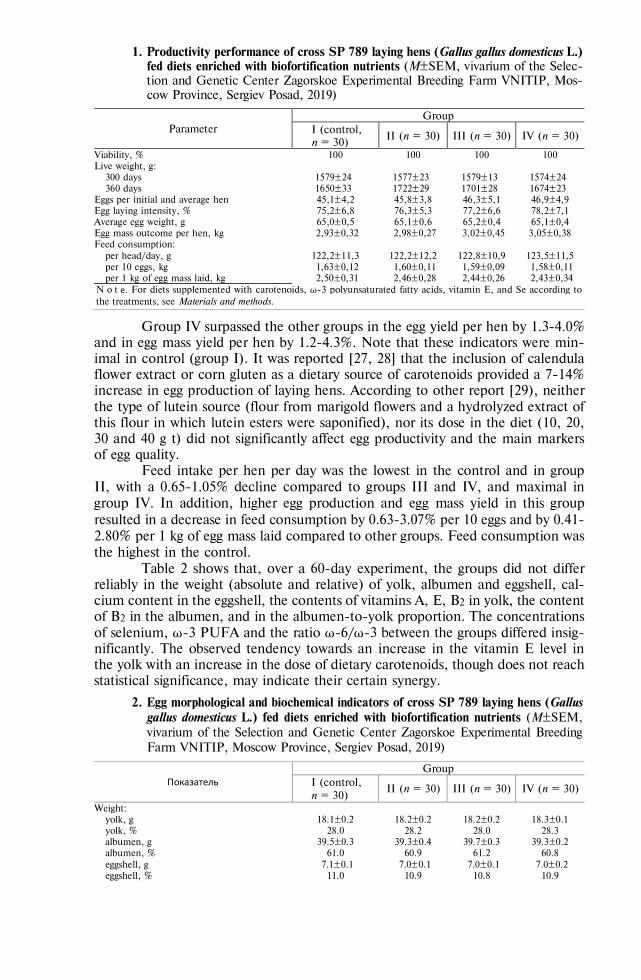
742
1. Productivity performance of cross SP 789 laying hens (Gallus gallus domesticus L.) fed diets enriched with biofortification nutrients (M±SEM, vivarium of the Selec-tion and Genetic Center Zagorskoe Experimental Breeding Farm VNITIP, Mos-cow Province, Sergiev Posad, 2019)
Parameter Group
I (control, n = 30)
II (n = 30) III (n = 30) IV (n = 30)
Viability, % 100 100 100 100 Live weight, g:
300 days 1579±24 1577±23 1579±13 1574±24 360 days 1650±33 1722±29 1701±28 1674±23
Eggs per initial and average hen 45,1±4,2 45,8±3,8 46,3±5,1 46,9±4,9 Egg laying intensity, % 75,2±6,8 76,3±5,3 77,2±6,6 78,2±7,1 Average egg weight, g 65,0±0,5 65,1±0,6 65,2±0,4 65,1±0,4 Egg mass outcome per hen, kg 2,93±0,32 2,98±0,27 3,02±0,45 3,05±0,38 Feed consumption:
per head/day, g 122,2±11,3 122,2±12,2 122,8±10,9 123,5±11,5 per 10 eggs, kg 1,63±0,12 1,60±0,11 1,59±0,09 1,58±0,11 per 1 kg of egg mass laid, kg 2,50±0,31 2,46±0,28 2,44±0,26 2,43±0,34
N o t е. For diets supplemented with carotenoids, ω-3 polyunsaturated fatty acids, vitamin E, and Se according to the treatments, see Materials and methods.
Group IV surpassed the other groups in the egg yield per hen by 1.3-4.0% and in egg mass yield per hen by 1.2-4.3%. Note that these indicators were min-imal in control (group I). It was reported [27, 28] that the inclusion of calendula flower extract or corn gluten as a dietary source of carotenoids provided a 7-14% increase in egg production of laying hens. According to other report [29), neither the type of lutein source (flour from marigold flowers and a hydrolyzed extract of this flour in which lutein esters were saponified), nor its dose in the diet (10, 20, 30 and 40 g t) did not significantly affect egg productivity and the main markers of egg quality.
Feed intake per hen per day was the lowest in the control and in group II, with a 0.65-1.05% decline compared to groups III and IV, and maximal in group IV. In addition, higher egg production and egg mass yield in this group resulted in a decrease in feed consumption by 0.63-3.07% per 10 eggs and by 0.41-2.80% per 1 kg of egg mass laid compared to other groups. Feed consumption was the highest in the control.
Table 2 shows that, over a 60-day experiment, the groups did not differ reliably in the weight (absolute and relative) of yolk, albumen and eggshell, cal-cium content in the eggshell, the contents of vitamins A, E, B2 in yolk, the content of B2 in the albumen, and in the albumen-to-yolk proportion. The concentrations of selenium, ω-3 PUFA and the ratio ω-6/ω-3 between the groups differed insig-nificantly. The observed tendency towards an increase in the vitamin E level in the yolk with an increase in the dose of dietary carotenoids, though does not reach statistical significance, may indicate their certain synergy.
2. Egg morphological and biochemical indicators of cross SP 789 laying hens (Gallus gallus domesticus L.) fed diets enriched with biofortification nutrients (M±SEM, vivarium of the Selection and Genetic Center Zagorskoe Experimental Breeding Farm VNITIP, Moscow Province, Sergiev Posad, 2019)
Показатель Group
I (control, n = 30)
II (n = 30) III (n = 30) IV (n = 30)
Weight: yolk, g 18.1±0.2 18.2±0.2 18.2±0.2 18.3±0.1 yolk, % 28.0 28.2 28.0 28.3 albumen, g 39.5±0.3 39.3±0.4 39.7±0.3 39.3±0.2 albumen, % 61.0 60.9 61.2 60.8 eggshell, g 7.1±0.1 7.0±0.1 7.0±0.1 7.0±0.2 eggshell, % 11.0 10.9 10.8 10.9

743
Continued Table 2
Intensity of yolk color, points 3.8±0.1 6.3±0.2* 6.7±0.2* 7.0±0.3* Albumen to yolk ratio 2.2 2.2 2.2 2.1 Concentration:
calcium in eggshell, % 36.6±3.3 36.6±2.4 36.1±3.8 36.4±2.9 in yolk, g/g:
carotenoids 5.6±1.1 13.0±2.6* 14.4±2.9* 21.9±4.4* vitamin А 4.2±0.3 4.1±0.4 4.5±0.2 4.6±0.5 vitamin Е 283±21 290±17 306±23 315±29 vitamin В2 5.6±0.4 6.5±0.7 5.8±0.2 5.8±0.4
vitamin В2 in albumen, g/g 3.9±0.4 4.3±0.3 4.3±0.6 4.3±0.4 Concentration per 100 g edible part of the egg:
carotenoids, g 177±35 424±85 454±91 701±140 selenium, g 62±0.7 61±0.7 62±0.9 62±1.1 vitamin Е, mg 8.9±0.9 9.5±1.2 9.6±0.8 10.1±1.4 ω-6 PUFA, mg 1093±52 1075±75 1140±67 1298±111 ω-3 PUFA, mg 347±19 393±31 412±43 417±28
ω-6 PUFA/ω-3 PUFA 3.1:1 2.7:1 2.8:1 3.1:1 N o t е. PUFA — polyunsaturated fatty acids. For diets supplemented with carotenoids, ω-3 polyunsaturated fatty acids, vitamin E, and Se according to the treatments, see Materials and methods. * Differences between treatment groups and control are statistically significant at p < 0.05.
Our findings do not agree with other investigations (26) which noted a significant increase in the yolk vitamin A level and a decrease in the vitamin E level in layers fed dietary extract of lutein powder (250 g/t), which may be due to a smaller amount of the dietary extract (10-18 g/t) in our trials. We also note that in a later work by the same researchers [30], when the marigold flower extract was used, the vitamin E concentration remained at the control level.
The biochemical indicators of eggs in the best group IV are noteworthy, e.g. 0.7 mg of carotenoids per 100 g edible egg part, 62 μg selenium, 10 mg vitamin Е, 417 mg ω-3 PUFA with the ω-6/ω-3 ratio of 3.1:1. So the increase is 3.96-fold (p < 0.05), 1.62-fold (p < 0.001), 4.37-fold (p < 0.01), and 2.42-fold (p < 0.001), respectively, as compared to unfortified eggs [10].
We assessed the color intensity and quantified carotenoids in the yolk as dependent on the fed diet (Fig.). The saturation curve for the first parameter gradually rose to a maximum, which was 3.9 times higher than in the control group (p < 0.05). Similarly, with an increase in the dose of dietary carotenoids, their concentration in the yolk significantly increased. A dose of carotenoids of about 20 g/t feed ensured their maximum concentration in the yolk. The poultry feed enrichment with carotenoids was accompanied by an increase in their amount in the yolk to values typical for eggs of poultry fed diets with an increased level of these micronutrients [29, 31, 32].
Egg yolk color intensity (1) and the concentra-tion of carotenoids (2) depending on the dose of dietary carotenoids fed to cross SP 789 laying hens (Gallus gallus domesticus L.) for bioforti-fication. The points correspond to groups I-IV (from left to right) according to the level of dietary carotenoids (see Materials and meth-ods). An asterisk (*) marks values that statisti-cally significantly (p < 0.05) differ from the control (M±SEM, vivarium of the Selection and Genetic Center Zagorskoe Experimental Breeding Farm VNITIP, Moscow Province, Sergiev Posad, 2019).
We evaluated the egg indicators reached upon biofortification as a per-centage of the recommended [33] or adequate level of a nutrient consumption [34] for humans (Table 3). Due to complex biofortification, one egg per day will provide 7.5% ω-6 PUFA, 12.0% ω-3 PUFA (at a 3.1:1 proportion), 39% vitamin

744
E and 51% Se of their recommended or adequate consumption. Thus, the resulting eggs meet the criteria for micronutrient-fortified food. As for carotenoids, their level increased 4 times, reaching 8% of the adequate daily intake. E.R. Kelly et al. [35] found that the increase in blood lutein levels in volunteers due to 0.9 mg of lutein in eggs was the same as for 5 mg of this carotenoid from a dietary supplement. Given that lutein from egg yolk is absorbed much better than from plant sources [12], we can assume that the nutritional value of the product we suggested has increased significantly.
3. Micronutrient value of eggs of cross SP 789 laying hens (Gallus gallus domes-ticus L.) fed dietary carotenoids for biofortification (M±SEM, vivarium of the Se-lection and Genetic Center Zagorskoe Experimental Breeding Farm VNITIP, Moscow Province, Sergiev Posad, 2019)
Micronutrient Recommended consumption (adequate consumption)
Concentration per fortified egg (group IV)
Compensation of daily reguirements, %
Carotenoids (5 mg) 0.4 mg 8 Selenium 55-70 g 35.7 g 51 Vitamin Е 15 mg 5.8 mg 39 Vitamin А 900 мg 84 g 9 ω-6 PUFA (10 g) 0.748 g 7.5 ω-3 PUFA (2 g) 0.240 g 12 N o t е. PUFA — polyunsaturated fatty acids. For diets supplemented with carotenoids, ω-3 polyunsaturated fatty acids, vitamin E, and Se according to the treatments, see Materials and methods.
However, remember that the consumption of two eggs, on the one hand, will overcome a 15% recommended intake for carotenoids and vitamin A, which is the lower limit for classification of the eggs as an enriched product. On the other hand, the consumption of selenium, although in a less dangerous organic form, will reach 100% of the physiological requirement. Selenium is also found in other food products. Therefore, it seems advisable to somewhat decline Se level in the diet of laying hens given that the upper permissible consumption limit for an adult person is 150 μg per day [33].
Yolks with a maximum level of selenium, PUFA, vitamin E and lutein will increase the micronutrient value of this component, not only when eaten directly. Eggs enriched with micronutrients can be a good raw material for pro-ducing melange, a component of functional food products. Experiments on ani-mals have shown that carotenoids are well absorbed from the enriched eggs [36]. High preservation of carotenoids in an omelet of eggs enriched by feeding chickens with corn grain rich in carotenoids (about 85% of carotenoids in a raw egg) [37] shows that heat treated eggs can serve as a source of these micronutrients in the diet of the population. PUFAs are also still stable in boiled eggs [38]. Consump-tion of eggs enriched with PUFA (6 pcs per week) for 8 weeks led to an increase in the amount of docosahexaenoic acid in the membranes of erythrocytes in healthy volunteers [39]. The use of whole boiled eggs has proven to be effective to increase the absorption of - and γ-tocopherol from fresh salad in healthy young adults [40]. The property of the phospholipids of chicken egg yolk to act as a “vehicle” for carotenoids was used to create a component based on yolks, pumpkin, and carrots for enriching curd products and dairy drinks with carote-noids [41].
So, our work has led us to conclude that the simultaneous supplementation with dietary carotenoids, ω-3 polyunsaturated fatty acids (PUFA), selenium and vitamin E provides a significant increase in their levels in chicken eggs. Due to higher level of carotenoids in the yolk, its color becomes more intense, which improves consumer qualities. Dietary carotenoids, upon constant profiles of other three micronutrients tested previously, did not significantly affect productivity

745
performance of hens, with 45.1-46.9 eggs over 60 days, feed conversion rate (1.58-1.63 kg per 10 eggs and 2.43-2.50 kg per 1 kg egg mass), and the egg, yolk, albumen and eggshell weight (65.0-65.2 g, 18.1-18.3 g, 39.3-39.7 g, and 7.0-7.1 g, respectively). However, there was a 2.40-3.96-fold increase in the concentration of carotenoids. In the best group, the concentration of carotenoids reached 0.7 mg per 100 g of edible part, selenium reached 62 μg, vitamin E reached 10 mg, and ω-3 PUFA reached 417 mg (at 3.1:1 proportion of ω-6/ω-3). These are 3.96 times higher (p < 0.05), 1.62 times higher (p < 0.001), 4.37 times higher (p <0.01), and 2.42 times higher (p < 0.001), respectively, then in unfortified eggs. One egg per day will provide a person with additional intake of 7.5 and 12% ω-3 and ω-6 PUFAs, 9% of vitamin A, 39% of vitamin E, 51% of selenium and 8% of easily assimilated carotenoids (mainly lutein) regarding the recommended doses. Eggs enriched with a complex of four micronutrients are both valuable natural food product, providing with lutein, selenium, PUFA and vitamin E, and an in-gredient of other functional foodstuffs, which facilitates assimilation of other die-tary components.
R E F E R E N C E S
1. Kodentsova V.M., Risnik D.V., Stefanova I.L. Ptitsa i ptitseprodukty, 2019, 1: 27-29 (doi:
10.30975/2073-4999-2019-21-1-27-29) (in Russ.). 2. Raederstorff D., Wyss A., Calder P. C., Weber P., Eggersdorfer M. Vitamin E function and
requirements in relation to PUFA. British Journal of Nutrition, 2015, 114(8): 1113-1122 (doi: 10.1017/S000711451500272X).
3. Kavtarashvili A.Sh., Stefanova I.L., Svitkin V.S., Novotorov E.N. Functional egg production. I. The role of -3 polyunsaturated fatty acids (review). Agricultural Biology [Sel'skokhozyaistvennaya bi-ologiya], 2017, 52(2): 349-366 (doi: 10.15389/agrobiology.2017.2.349eng).
4. Panaite T.D., Nour V., Vlaicu P.A., Ropota M., Corbu A.R., Saracila M. Flaxseed and dried tomato waste used together in laying hens diet. Archives of Animal Nutrition, 2019, 73(3): 222-238 (doi: 10.1080/1745039X.2019.1586500).
5. Leeson S., Namkung H., Caston L., Durosoy S., Schlegel P. Comparison of selenium levels and sources and dietary fat quality in diets for broiler breeders and layer hens. Poultry Science, 2008, 87(12): 2605-2612 (doi: 10.3382/ps.2008-00174).
6. Kavtarashvili A.Sh., Stefanova I.L., Svitkin V.S., Novotorov E.N. Functional egg production. II. The roles of selenium, zinc, and iodine (review). Agricultural Biology [Sel'skokhozyaistvennaya biologiya], 2017, 52(4): 700-715 (doi: 10.15389/agrobiology.2017.4.700eng).
7. Weill P., Schmitt B., Chesneau G., Daniel N., Safraou F., Legrand P. Effects of introducing linseed in livestock diet on blood fatty acid composition of consumers of animal products. Annals of Nutrition and Metabolism, 2002, 46(5): 182-191 (doi: 10.1159/000065405).
8. Lemahieu C., Bruneel C., Termote-Verhalle R., Muylaert K., Buyse J., Foubert I. Impact of feed supplementation with different omega-3 rich microalgae species on enrichment of eggs of laying hens. Food Chemistry, 2013, 141(4): 4051-4059 (doi: 10.1016/j.foodchem.2013.06.078).
9. Cachaldora P., García-Rebollar P., Alvarez C., Blas J.D., Méndez J. Effect of type and level of fish oil supplementation on yolk fat composition and n-3 fatty acids retention efficiency in laying hens. British Poultry Science, 2006, 47(1): 43-49 (doi: 10.1080/00071660500475541).
10. Mazo V.K., Kavtarashvili A.Sh., Stefanova I.L., Kodentsova V.M., Bagryantseva O.V., Svitkin V.S., Novotorov E.N., Risnik D.V., Mokshantseva I.V., Sidorova Yu.S., Zorin S.N., Klimenkova A.Yu. Funktsional'nye yaitseprodukty [Functional egg products]. Moscow, 2018 (in Russ.).
11. Díez-Espino J., Basterra-Gortari F.J., Salas-Salvadó J., Buil-Cosiales P., Corella D., Schrö-der H., Estruch R., Ros E., Gómez-Gracia E., Arós F., Fiol M., Lapetra J., Serra-Majem L., Pintó X., Babio N., Quiles L., Fito M., Marti A., Toledo E. Egg consumption and cardiovascular disease according to diabetic status: The PREDIMED study. Clinical Nutrition, 2017, 36(4): 1015-1021 (doi: 10.1016/j.clnu.2016.06.009).
12. Chung H.Y., Rasmussen H.M., Johnson E.J. Lutein bioavailability is higher from lutein-en-riched eggs than from supplements and spinach in men. The Journal of Nutrition, 2004, 134(8): 1887-1893.
13. Park S.J., Jung J.H., Choi S.W., Lee H.J. Association between egg consumption and metabolic disease. Korean Journal for Food Science of Animal Resources, 2018, 38(2): 209-223 (doi:

746
10.5851/kosfa.2018.38.2.209). 14. Mah E., Chen C.O., Liska D.J. The effect of egg consumption on cardiometabolic health
outcomes: an umbrella review. Public Health Nutrition, 2020, 23(5): 935-955 (doi: 10.1017/S1368980019002441).
15. Missimer A., DiMarco D.M., Andersen C.J., Murillo A.G., Vergara-Jimenez M., Luz Fernandez M. Consuming two eggs per day, as compared to an oatmeal breakfast, decreases plasma ghrelin while maintaining the LDL/HDL ratio. Nutrients, 2017, 9(2): 89 (doi: 10.3390/nu9020089).
16. Lieblein-Boff J.C., Johnson E.J., Kennedy A.D., Lai C.S., Kuchan M.J. Exploratory metabolomic analyses reveal compounds correlated with lutein concentration in frontal cortex, hippocampus, and occipital cortex of human infant brain. PLoS ONE, 2015, 10(8): e0136904 (doi: 10.1371/jour-nal.pone.0136904).
17. Buscemi S., Corleo D., Di F.P., Petroni M.L., Satriano A., Marchesini G. The effect of lutein on eye and extra-eye health. Nutrients, 2018, 10(9): 1321 (doi: 10.3390/nu10091321).
18. Ames B.N. Prolonging healthy aging: longevity vitamins and proteins. Proceedings of the National Academy of Sciences, 2018, 115(43): 10836-0844 (doi: 10.1073/pnas.1809045115).
19. Chung R.W.S., Leanderson P., Lundberg A.K., Jonasson L. Lutein exerts anti-inflammatory ef-fects in patients with coronary artery disease. Atherosclerosis, 2017, 262: 87-93 (doi: 10.1016/j.ath-erosclerosis.2017.05.008).
20. Akdemir F., Orhan C., Sahin N., Sahin K., Hayirli A. Tomato powder in laying hen diets: effects on concentrations of yolk carotenoids and lipid peroxidation. British Poultry Science, 2012, 53(5): 675-680 (doi: 10.1080/00071668.2012.729142).
21. Omri B., Alloui N., Durazzo A., Lucarini M., Aiello A., Romano R., Santini A., Abdouli H. Egg yolk antioxidants profiles: effect of diet supplementation with linseeds and tomato-red pepper mixture before and after storage. Foods, 2019, 8(8): 320 (doi: 10.3390/foods8080320).
22. Zahroojian N., Moravej H., Shivazad M. Comparison of marine algae (Spirulina platensis) and synthetic pigment in enhancing egg yolk colour of laying hens. British Poultry Science, 2011, 52(5): 584-588 (doi: 10.1080/00071668.2011.610779).
23. Afanas'ev G.D., Popova L.A., Komarchev A.S., Trepak Zh.G. Ptitsa i ptitseprodukty, 2014, 5: 62-64 (in Russ.).
24. Kavtarashvili A.Sh., Stefanova I.L., Svitkin V.S. Functional egg production. III. The role of the carotenoids (review). Agricultural Biology [Sel'skokhozyaistvennaya biologiya], 2019, 54(4): 681-693 (doi: 10.15389/agrobiology.2019.4.681eng).
25. Mokshina N.Ya., Khokhlov V.Yu., Shlyakhina Yu.V., Selemenov V.F. Vestnik VGU. Seriya: KHimiya, Biologiya, Farmatsiya, 2003, 2: 53-55 (in Russ.).
26. Englmaierová M., Skřivan M., Bubancová I. A comparison of lutein, spray-dried Chlorella, and synthetic carotenoids effects on yolk colour, oxidative stability, and reproductive performance of laying hens. Czech J. Anim. Sci., 2013, 58(9): 412-419 (doi: 10.17221/6941-CJAS).
27. Koshchaev A.G. Khranenie i pererabotka sel'khozsyr'ya, 2007, 2: 34-38 (in Russ.). 28. Skřivan M., Marounek M., Englmaierova M., Skřivanova E. Effect of increasing doses of marigold
(Tagetes erecta) flower extract on eggs carotenoids content, colour and oxidative stability. Journal of Animal and Feed Sciences, 2016, 25: 58-64 (doi: 10.22358/jafs/65588/2016).
29. Lokaewmanee K., Yamauchi K., Komori Ts., Saito K. Enhancement of yolk color in raw and boiled egg yolk with lutein from marigold flower meal and marigold flower extract. The Journal of Poultry Science, 2011, 48(1): 25-32 (doi: 10.2141/jpsa.010059).
30. Skřivan M., Englmaierová M., Skřivanová E., Bubancová I. Increase in lutein and zeaxanthin content in the eggs of hens fed marigold flower extract. Czech J. Anim. Sci., 2015, 60(3): 89-96 (doi: 10.17221/8073-CJAS).
31. Kodentsova V.M., Vrzhesinskaya O.A., Beketova N.A., Kodentsova O.V. Voprosy pitaniya, 2005, 5: 19-24 (in Russ.).
32. Kavtarashvili A.Sh., Mazo V.K., Kodentsova V.M., Risnik D.V., Stefanova I.L. Biofortification of hen eggs: vitamins and carotenoids (review). Agricultural Biology [Sel'skokhozyaistvennaya bi-ologiya], 2017, 52(6): 1094-1104 (doi: 10.15389/agrobiology.2017.6.1094eng).
33. MR 2.3.1.2432-08. Normy fiziologicheskikh potrebnostei v energii i pishchevykh veshchestvakh dlya razlichnykh grupp naseleniya Rossiiskoi Federatsii. Metodicheskie rekomendatsii [MR 2.3.1.2432-08. Norms of energy and nutrient physiological requirements for various groups of the population of the Russian Federation. Guidelines]. Moscow, 2009 (in Russ.).
34. Edinye sanitarno-epidemiologicheskie i gigienicheskie trebovaniya k tovaram, podlezhashchim san-itarno-epidemiologicheskomu nadzoru (kontrolyu) Tamozhennogo soyuza EvrAzES (Prilozhenie 5) [Uniform sanitary, epidemiological and hygienic requirements for goods subject to sanitary and epidemiological supervision (control) of the EurAsEC Customs Union (Appendix 5)]. Moscow, 2010 (in Russ.).
35. Kelly E.R., Plat J., Haenen G.R., Kijlstra A., Berendschot T.T. The effect of modified eggs and an egg-yolk based beverage on serum lutein and zeaxanthin concentrations and macular pigment

747
optical density: results from a randomized trial. PLoS ONE, 2014, 9(3): e92659 (doi: 10.1371/jour-nal.pone.0092659).
36. Heying E.K., Ziemer K.L., Tanumihardjo J.P., Palacios-Rojas N., Tanumihardjo S.A. β-Crypto-xanthin-biofortified hen eggs enhance vitamin A status when fed to male Mongolian Gerbils. The Journal of Nutrition, 2018, 148(8): 1236-1243 (doi: 10.1093/jn/nxy117).
37. Sowa M., Yu J., Palacios-Rojas N., Goltz S.R., Howe J.A., Davis C.R., Rocheford T., Tanumihardjo S.A. Retention of carotenoids in biofortified maize flour and β-cryptoxanthin-enhanced eggs after household cooking. ACS Omega, 2017, 2(10): 7320-7328 (doi: 10.1021/acsomega.7b01202).
38. Douny C., Khoury R.E., Delmelle J., Brose F., Degand G., Moula N., Farnir F., Clinquart A., Maghuin-Rogister G., Scippo M.-L. Effect of storage and cooking on the fatty acid profile of omega-3 enriched eggs and pork meat marketed in Belgium. Food Science & Nutrition, 2015, 3(2): 140-152 (doi: 10.1002/fsn3.197).
39. Burns-Whitmore B., Haddad E., Sabaté J., Rajaram S. Effects of supplementing n-3 fatty acid enriched eggs and walnuts on cardiovascular disease risk markers in healthy free-living lacto-ovo-vegetarians: a randomized, crossover, free-living intervention study. Nutrition Journal, 2014, 13(1): 29 (doi: 10.1186/1475-2891-13-29).
40. Kim J.E., Ferruzzi M.G., Campbell W.W. Egg consumption increases vitamin E absorption from co-consumed raw mixed vegetables in healthy young men. The Journal of Nutrition, 2016, 146(11): 2199-2205 (doi: 10.3945/jn.116.236307).
41. Bessonova L.N., Antipova L.V., Cherkasova A.V. V sbornike: Nauka, pitanie i zdorov'e [In: Sci-ence, nutrition and health]. Minsk, 2019: 480-487 (in Russ.).

750
AGRICULTURAL BIOLOGY, ISSN 2412-0324 (English ed. Online)
2020, V. 55, ¹ 3, pp. 750-769 (SEL’SKOKHOZYAISTVENNAYA BIOLOGIYA) ISSN 0131-6397 (Russian ed. Print)
ISSN 2313-4836 (Russian ed. Online) UDC 636.4:591.1:636.084 doi: 10.15389/agrobiology.2020.4.750eng
doi: 10.15389/agrobiology.2020.4.750rus
PHYSIOLOGICAL AND BIOCHEMICAL EFFECTS OF TWO FEED ANTIOXIDANTS IN MODELING TECHNOLOGICAL STRESS IN PIGS
(Sus scrofa domesticus Erxleben, 1777)
Yu.P. FOMICHEV1, N.V. BOGOLYUBOVA1, R.V. NEKRASOV1, M.G. CHABAEV1, R.A. RYKOV1, A.A. SEMENOVA2
1Ernst Federal Science Center for Animal Husbandry, 60, pos. Dubrovitsy, Podolsk District, Moscow Province, 142132 Russia, e-mail [email protected], [email protected] ( corresponding author), [email protected], [email protected], [email protected]; 2Gorbatov Federal Center for Food Systems RAS, 26, ul. Talalikhina, Moscow, 109316 Russia, e-mail [email protected]
ORCID: Fomichev Yu.P. orcid.org/0000-0003-0213-5526 Chabaev M.G. orcid.org/0000-0003-1889-6063 Bogolyubova N.V. orcid.org/0000-0002-0520-7022 Rykov R.A. orcid.org/0000-0003-0228-8901 Nekrasov R.V. orcid.org/0000-0003-4242-2239 Semenova A.A. orcid.org/0000-0002-4372-6448 The authors declare no conflict of interests Acknowledgements: Supported financially by Russian Science Foundation, project No. 19-16-00068 Received February 5, 2020
A b s t r a c t
Intensive livestock technologies do not fit well with the physiology of domestic species and put farm animals at risk of various health problems and disorders, which most negatively affects highly productive animals with intensive metabolism. Feed antioxidants might be a solution to improve pro-ductive health, adaptive capabilities and stress resistance of livestock. The outcome of adaptogen ap-plication depends on thorough elucidation of mechanisms of their action on physiological and bio-chemical processes in the body compromised by stress. Our study imitated social stress (modeled stress, MS) as the most common in intensive livestock to compare two dietary antioxidant additives of different origin and chemical composition. Thirty-six hybrid boars F2 (Large White ½ Landrace) ½ Duroc aged 103 days (35 kg live weight) were assigned for four treatments (9 animals per each): 1 — control without MS, 2 — control with MS, 3 — MS + proteinate Se (PSe) (B-TRAXIM Selenium-11, PANCOSMA CANADA, Inc.; 0.2 mg a.i. per 1 kg feed), and 4 — MS + dihydroquercetin (DHQ) Ekostimul-2 drug (ООО Ametic, Russia; 32 mg a.i. per 1 kg feed). To simulate social stress, boars were moved every 14 days within the group to change the neighbors. Blood for assay was sampled three times over the trial from five boars of each group. With age, the blood cortisol level was revealed first to decrease by 36.8. 22.5, 41.3 and 52.8 % from the initial values in groups 1, 2, 3 and 4, respec-tively, though by the end of the final feeding there was a 46.4; 37.4; 8.1 and 60.4 % increase in the parameter. The cortisol concentration was the highest in group 3 (MS + PSe) during intensive growth (282 nmol/l vs. 211 and 214 nmol/l for groups 1 and 2). In groups 1, 2 (MS), and 3 (MS + PSe), the cortisol concentration reached 309, 294 and 305 nmol/l by the end of feeding. Blood cortisol level was the lowest in group 4 (+ DHQ), 134 nmol/l (p = 0.07 compared to group 2) at intensive growth and 215 nmol/l at final fattening, corresponding with thiobarbituric acid reactive substances (TBARS) levels which were also 6.7 and 12.3 % lower, respectively. Lactate dehydrogenase (LDH) activity and creatine phosphokinase (CPK) activity also altered. In group 1, LDH activity decreased from 459.4 to 377.5 IU/l over the trial. The same pattern was characteristic of group 4 (MS + DHQ). In group 2 (MS) and group 3 (MS + PSe), LDH activity declined to 317.0 and 289.3 IU/l by the end of feeding, which was 16.0 (p < 0.01) and 23.4 % (p < 0.01) less than in group 1. In group 4, the index, being constantly decreasing over the trial as in group 1, remained 15.0, 7.0 (p = 0.06 compared to control group 2 with MS) and 2.7 % lower than in groups 1, 2 and 3. The MS lowered the blood glucose concentration by 13.8 % (p < 0.05) compared to control 1 during the final fattening period. In group 3, this indicator as influenced by MS was also 7.4 % lower (p < 0.05), but due to PSe, a 7.4 % increase occurred compared to control group 2. Dietary DHQ led to the leveling of negative effects of MS, which, together with low cortisol indices in this group, stabilized the blood glucose concentration at the control level without MS in group 1. The DHQ was found to also contribute to a 25 % (p < 0.01) increase in blood triglycerides during final fattening compared to group 1 without stress. Pigs also differed in pathogenetic resistance. MS mobilized the cellular immunity through an increase in phag-ocytic activity PA (p < 0.05), phagocytic index PI (p < 0.001), and phagocytic number PN (p < 0.05) in control group 2 at the end of the test. PSe and DHQ normalized these indicators compared to control

751
(group 2) practically to the control values without MS (p < 0.01 for PI; p < 0.01 and p < 0.05 for PN, respectively). Thus, in animals fed adaptogens, the resistance indices at the end of the experiment corresponded to those in the control group 1, which indicates higher stress resistance, and DHQ additionally promoted humoral immunity as compared to the control group 1 (p < 0.05), which con-firms the ability the adaptogens to enhance the body resistance to stress. Interestingly, despite the absence of statistically significant differences between most of the studied biochemical parameters (p > 0.05) which indicate a balanced animal diet, changes were noted that characterize the effect of the adaptogens under MS. In groups 2-4, blood phosphorus concentration was higher than in control group 1, 4.42 (p = 0.07), 4.52 (p = 0.1) and 4.64 mmol/l (p < 0.05) vs. 3.94 mmol/l. Thereof, the Ca/P ratio changed significantly during fattening. In group 2 (MS), group 3 (MS + PSe) and group 4 (MS + DHQ), the values 1.01 (p < 0.05), 0.99 (p < 0.05) and 0.89 (p < 0.001) vs. 1.15 in group 1 without stress. Blood morphology in pigs also changed as influenced by MS and the adaptogens. The counts of blood leukocytes in groups 2, 3 and 4 was 12.5; 5.4 and 6.1 % higher than in group 1 in the middle of fattening period, and 32.5 (p < 0.05), 40.1 (p < 0.05) and 21.7 % (p = 0.07) higher at the final fattening. A decrease in the number of erythrocytes and an increase in the hemoglobin amount in blood were characteristic of all groups. In general, by the end of feeding animals subjected to MS these two indicators were 6.6, 14.3, 9.7 % and 1.09, 6.09, 4.27 % higher than in group 1. An increase in blood erythrocytes (p < 0.05), hemoglobin (p < 0.05) and hematocrit (p < 0.01) in animals fed selenium vs. control group 2 indicates a decisive role of Se adaptogen as antioxidant during fatten-ing. The observed changes were associated both with the action of cortisol generated by physiological stress, and with an increase in nonspecific resistance of boars due to the adaptogens. Importantly, the average daily weight gain in boars was close to that genetically conditioned for the genotype F2 (Large White ½ Landrace) ½ Duroc. As a result, over the entire period of the trial, groups 1, 2, 3, and 4 showed an average increase of 1047, 1035, 1003 and 1042 g, respectively, of which the weight gain was the greatest in control 1 (without stress) and in group 4 fed DHQ at MS. Thus, our findings give grounds for further studying effects of these feed adaptogens for their proper use in intensive indus-trial pig breeding.
Keywords: stress, pigs, adaptogens, antioxidants, dihydroquercetin, selenium, cortisol, lactate dehydrogenase, hematological indicators, creatine phosphokinase, TBA-active products, nonspecific resistance
Intensive rearing of livestock commonly used nowadays antagonize the evolutionary physiology of farm animals [1]. The current priorities of animal breeding are genetically determined high productivity performance. However, the realization of such productive potential imposes extra physiological loads on an animal, aggravates effects of stress factors and significantly affects homeostasis that worsens quality of final products [2].
Stress triggers a cascade of non-specific adaptive (normal) responses of the body to the effects of various unfavorable factors (stressors) violating homeostasis, which includes special state of the nervous system as a whole. Hans Sely (1907-1982) was the first to introduce the concept of stress as a non-specific response of the body to any change [3]. In animal husbandry, depending on the cause, stress is classified as social (technological), ecological, dietary, and immunological [4]. In particular, social (or technological) stress in pigs occurs when animals are kept in pens with unfamiliar neighbors, upon isolation, weaning, transportation, veter-inary measures, etc. Sensitivity to social stress is always individual and is often associated with genetic factors, which has been confirmed in many research works [5]. Social stress can be acute (immediately after regrouping) or chronic, when animals are kept in group pens or in isolation, and after a repeated regrouping [6]. The frequency of social interactions and aggressive behavior of animals increase with increasing housing density [7, 8], and the growth rate is positively correlated with the area of the pen per animal [9]. It is also known that the response to social stress depends on gender, and it is higher in males than in females [10, 11]. The weaning and transfer to growing and finishing are critical periods in the technology of raising pigs. The deprivation of maternal presence and milk, transfer to a new house, regrouping, change of feed and service personnel cause technological stress in piglets, as a result, morbidity increases, and the growth rate slows down. This decreases natural resistance and of humoral immunity, allowing conditionally

752
pathogenic microflora to become active, which leads to indigestion or respiratory pathology [12]. With chronic exposure to various stressful situations, free radical oxidation is activated with the depletion of antioxidant defense. In the body, syn-dromes of stress maladjustment, ketosis, hepatodystophia appear, and autoimmune processes occur [13].
Dietary antioxidants ensure stabilization of the level of free radicals, re-duce the impact of environmental stress factors and increase the adaptive capacity of the body [14]. Bioflavonoid dihydroquercetin and the essential trace element selenium are substances with antioxidant activity. Dihydroquercetin from Daurian larch (Larix dahurica Turez) has a wide range of biological properties. It regulates metabolic processes, positively influences functions of organs and body systems (in particular, the cardiovascular system), is involved in the protection of healthy cells of the body from pathological changes by neutralizing radical activity. Sele-nium also belongs to strong antioxidants, prevents cardiovascular diseases, has an immunomodulatory effect on cellular and humoral immunity. Se is found in many functionally active proteins, the selenoproteins which, in particular, include glu-tathione peroxidases and thyrodoxin reductases. In addition, it prevents alimentary muscular dystrophy in animals.
Recent years have seen a rapid rise in a renewed interest of plant raw materials, since many plants, including vegetables and spices, contain antioxidants [15, 16], in particular, flavonoids and phenolic components [17, 18]. Flavonoids are secondary plant metabolites with a spectrum of pharmacological and biological properties [19, 20]. Dihydroquercetin (C15H12O7) (DHQ) is the dominant com-ponent of the bioflavonoid complex of Diquertin. Dietary dihydroquercetin blocked lipid peroxidation during pig growing and fattening [21]. Ecostimul-2 (Russia), a DHQ-based feed additive fed to piglets for post-weaning period (50 mg per head per day) significantly weakened the impact of environmental stress factors and increased animal adaptiveness ensuring average daily weight gain of 496 g after weaning, that is, 20.6% higher than in the control individuals [22]. DHQ as a dietary additive blocked free radical lipid oxidation, enhanced antioxidant defense and improved liver functions during growing and fattening. In another experiment, quercetin weakened oxidative stress and decreased intestinal inflammation, with the decrease in levels of blood endotoxin, reactive oxygen species (ROS) and malondialdehyde (MDA) in the intestine, and the increase in height of villi of the jejunum [23]. There is a report that series of new 3-monoacylated dihydroquerce-tin derivatives with enhanced antioxidant properties have been synthesized [24].
Therefore, better reproductive health and stress resistance are currently critical parts of pig commercial rearing. This requires more detail understanding mechanisms of the effect of alimentary antioxidants on the body’s physiology and biochemistry.
This work presents a complex evaluation of metabolic, hormonal, antioxi-dant and immune status in pigs under simulation of one of the most common tech-nological stresses, the social stress, upon correction with antioxidant drugs to com-pare their capability of leveling the impact of the stressor.
The investigation aimed to compare the physiological and biochemical parameters characterizing productive and adaptive status in pigs that received antioxidant drugs based on dihydroquercetin and selenium under model techno-logical stress.
Materials and methods. Thirty six cross-breed boars (Sus scrofa domesticus) F2 (Large White ½ Landrace) ½ Duroc (35 kg live weight, 103 day old) after the growing period were assigned for four treatments (9 animals per each): 1 — con-trol without MS, 2 — control with MS, 3 — MS + proteinate Se (PSe) (B-TRAXIM Selenium-11, PANCOSMA CANADA, Inc.; 0.2 mg a.i. per 1 kg feed),

753
and 4 — MS + dihydroquercetin (DHQ) Ekostimul-2 drug (ООО Ametic, Rus-sia; 32 mg a.i. per 1 kg feed). The basal diet (BD) included compound feeds SK-5 and SK-6 (Agrovitex LLC, Russia), balanced for nutrients and energy according to modern standards [25]. Doses of additives (as per active ingredients, a.i.) were 0.2 mg PSe and 32 mg DHQ per 1 kg feed according to the design of trials.
To simulate social stress, animals were moved from one pen to another within the group at the beginning of the experiment, and then every 14 days. After each relocation, animals were in the pen that were not previously neighbors.
Blood for assay was sampled from jugular vein of five boars of each group by puncture before the start of the experiment, before final fattening and before slaughter to analyze blood biochemical indicators (an automatic biochemical analyzer Chem Well (Awareness Technology, USA), morpho-hematological pa-rameters (an ABC VET hematological analyzer, HORIBA ABX Diagnostics Inc., France), cortisol levels (by enzyme immunoassay), thiobarbituric acid reactive substances, TBARS (with a diagnostic kit, OOO Agat-Med, Russia), the total amount of water-soluble antioxidants (WA) (a device Tsvet-Yauza-01-AA, OAO NPO Khimavtomatika, Russia), activity of lactate dehydrogenase (LDH) and creatine phosphokinase (CPK) (a biochemical analyzer Bio Chem FC-360, HTI, USA). To assessed immune status, blood serum bactericidal activity (BSBA, a nonspecific resistance indicator) was assessed photonephelometrically, blood se-rum lysozyme activity (BSLA) by the Mutovin’s method; phagocytic activity (PA) was assessed based on blood cell ingesting and digesting ability.
Obtained data were processed biometrically by the analysis of variance (ANOVA) method using STATISTICA 10 software (StatSoft, Inc., USA). The arithmetic means (M), the mean square error (±SEM) and the level of signifi-cance (p; differences were considered statistically significant at p < 0.05) were calculated. Comparison was performed with control group I (without MS) and group II (with MS).
Results. Social stress (overstrain associated with social adaptation to inter-action in the community) very often occurs when raising pigs. In our tests, in ani-mals of the control group I, which were kept without changing the pen, there were rare cases of aggressive behavior. MS in groups II (control), III and IV induced by animal relocation caused more restless and aggressive behavior. Animals from groups II, III and IV were more active when moving, often interacted with each other. The frequency of aggressive behavior directly depended on the percentage of restless animals in the group (Fig.).
Incidence of aggressive behavior (%) and the propor-tion of restless animals (%) in pigs (Sus scrofa domes-ticus) F2 (Large White ½ Landrace) ½ Duroc under modeling social stress. Outer circle corresponds to the number of cases of aggressive behavior (absolute value for the observation period and percentage of the total number of cases); inner circle is the number of rest-less animals (absolute value for the observation period and the percentage of the total number of cases) (ex-perimental animal yard of Ernst Federal Science Center for Animal Husbandry, 2019, N = 36, n = 9).
The effect of MS was recorded by acts of aggressive behavior, which was espe-cially clearly manifested in the first 2-3 days after relocation. Each time this period was
characterized by the building of a hierarchical structure, a decrease in feed consump-tion, bites and increased injuries, anxiety, especially in groups II and III. So, aggressive behavior was least frequent in group 1 (39 cases) vs. 65, 67 and 56 cases (or 29, 29,

754
and 25% of their total number during the experiment) in groups 2, 3 and 4, respec-tively. In the group experienced MS, but fed dietary DHQ, the aggressive behavior diminished while anxiety was at the same level.
Importantly, the pig productivity (average daily weight gain) was close to genetically limited for the genotype F2 (Large White ½ Landrace) ½ Duroc. As a result, over the experiment, there was an average increase of 1047, 1035, 1003 and 1042 g in groups 1, 2, 3 and 4, respectively, that is, it was greatest in control group 1 (without stress) and in group 4 (MS + DHQ).
We revealed significant differences in protein, nitrogen, carbohydrate, lipid and mineral metabolism in relationship with growth rate, MS and effects of dietary PSe and DHQ both between the groups of pigs and feeding periods. The blood levels of total protein and its fractions characterize the intensity and the type of protein metabolism and is closely related to growth, the influence of environmen-tal factors and the diet [26]. In pigs of all groups, before fattening, the total blood protein ranged from 65.8 to 73.1 g/l, being within the physiological norm of 55-82 g/l. The same was observed for albumins (A) and globulins (G), 36.7-38.7 and 29.1-43.4 g/l, respectively, given a physiological norm of 19-43 and 26-57 g/l. The A/G ratio was higher than the average reference value, which indicated intense anabolism (Table 1). During the tests, the pattern of the change in total blood protein and its fractions in all groups was similar, that is, A/G ratio increased during intensive growth due to the albumin fraction and decreased by the end of fattening due to the globulin fraction. As a result, the A/G ratio increased over observation, which we consider positive. However, there were differences between the groups. So, in group 2 influenced by MS, the total protein level was 3.5% lower (p < 0.05) than in group 1 due to the albumin fraction and with an equal concentration of globulins, which could indicate the increased use of plastic sub-stances for the energy needs of the body. During final fattening, the total blood protein in this group remained at the control level. The effect of adaptogens on protein metabolism was positive and differed depending on their biological activity. PSe and DHQ suppressed catabolism and enhanced anabolic processes during intensive growth of pigs experienced MS, as a result, total blood protein and its fractions remained the same as in the control group 1. At final fattening, the best characteristics of protein metabolism was in group 4 receiving DHQ. That is, the blood albumin level was 44.4 g/l at A/G = 1.78, with 41.9 g/l at A/G = 1.78, 44.4 g/l at A/G = 1.70, 42.4 g/l at A/G = 1.55, and 41.8 g/l at A/G = 1.64 for groups 1, 2 and 3, respectively, that is, the A/G value was the smallest in control group 2 under MS without correction (Table 1).
1. Metabolic parameters in pigs (Sus scrofa domesticus) F2 (Large White ½ Land-race) ½ Duroc (n = 5, M±SEM, experimental animal yard of Ernst Federal Sci-ence Center for Animal Husbandry, 2019)
Parameter Group
1 (control, BD) modeling technological stress
2 (control, BD) 3 (BD + PSe) 4 (BD + DHQ) P r i o r t o g r o w i n g (35 kg living weight)
Total protein, g/l 65.82±1.29 70.28±6.23 73.19±6.43 71.73±2.95 Albumins (A), g/l 36.72±1.60 38.79±1.48 38.73±0.94 37.37±2.17 Globulins (G), g/l 29.10±0.74 34.49±6.22 43.47±6.98 34.37±3.71 A/G 1.27±0.08 1.36±0.22 1.24±0.18 1.15±0.19 Cholesterol, mmol/l 3.02±0.13 3.08±0.06 3.13±0.14 3.07±0.08 Phospholipids, mmol/l 0.30±0.05 0.36±0.08 0.35±0.05 0.35±0.12 Total bilirubin, mol/l 4.46±1.30 5.64±1.03 3.03±0.69 5.07±0.70 AAT, IU/l 49.48±5.47 52.22±2.85 43.48±2.53 39.55±1.91 AST, IU/l 52.81±1.82 55.43±5.43 54.00±2.22 48.29±2.98
P r i o r t o f a t t e n i n g (70 kg living weight) Total protein, g/l 78.36±0.59 75.64±1.12* 77.55±3.05 78.21±1.82 Albumins (A), g/l 47.04±0.73 44.06±1.20* 47.23±1.25 45.43±1.43

755
Continued Table 1
Globulins (G), g/l 31.32±0.55 31.58±1.98 30.32±2.46 32.79±1.98 A/G 1.50±0.05 1.42±0.13 1.59±0.12 1.40±0.11 Urea, mmol/l 5.73±0.47 5.88±0.58 8.59±1.25 6.81±1.07 Cholesterol, mmol/l 2.66±0.12 2.66±0.19 2.58±0.13 2.71±0.07 Phospholipids, mmol/l 0.46±0.10 0.44±0.14 0.55±0.53 0.56±0.13 AAT, IU/l 50.79±5.07 53.88±2.85 49.96±3.57 51.38±1.51 AST, IU/l 40.62±2.34 38.90±4.03 36.63±1.15 32.89±2.63*
P r i o r t o s l a u g h t e r i n g (100 kg living weight) Total protein, g/l 66.96±2.49 70.06±1.25 68.26±0.75 70.34±0.97 Albumins (A), g/l 41.98±1.46 42.44±1.58 41.83±2.68 44.44±1.63 Globulins (G), g/l 24.98±1.48 27.62±1.05 26.43±2.19 25.90±12.12 A/G 1.70±0.12 1.55±0.10 1.64±0.22 1.78±0.24 Urea, mmol/l 7.36±0.19 5.74±0.46** 6.64±0.56 6.73±0.86 Cholesterol, mmol/l 2.41±0.09 2.37±0.24 2.41±0.13 2.17±0.33 Phospholipids, mmol/l 0.69±0.13 0.75±0.16 0.64±0.16 0.59±0.20 AAT, IU/l 42.29±5.75 47.04±4.40 42.11±4.56 40.98±3.76 AST, IU/l 16.24±1.25 17.54±1.32 20.64±4.33 21.89±6.39 N o t е. BD — basal diet, АAT — alanine aminotransferase, AST — aspartat aminotransferase. For design of trials and description of treatments, see Materials and methods. *, ** Differences between the treatment and the control group 1 are statistically significant at р < 0.05 and р < 0.01, respectively.
In the trials, the blood urea concentration in all groups corresponded to physiological limits (3.5-9.2 mmol/l). During intensive growth, its value in animals from control groups was the same. At final fattening, the blood urea increased in control group 1 (by 28.4%), but not in control group 2 (5.88 vs. 5.74 mmol/l, respectively). It should be emphasized that the urea concentra-tion in the groups subjected to MS was lower than the control values without MS, and in the control group 2, we recorded its significant decrease (p < 0.01). Elevation of blood urea levels in a body with a normal urea cycle in the liver could be associated with a decrease in the intensity of protein renewal in tissues and anabolic processes. The effect of adaptogens we observed was as follows. The blood urea in pigs from group 3 (MS + PSe) and group 4 (MS + DHQ) was 49.9 and 18.8% higher, respectively, than in control 1 (without MS), and 46.0 and 15.5% higher, than in group 2 (MS). At final fattening, this indicator decreased by 22.8 and 1.2%, respectively, due to adaptogens, as a result, it equaled and amounted to 6.64 and 6.73 mmol/l, while in the control groups 1 and 2, the values were 7.36 and 5.74 mmol/l (see Table 1). We did not find statistically significant differences with group 2 (control with MS) when ana-lyzing obtained data.
Lipids, namely cholesterol, phospholipids and triglycerides, as consti-tutive parts of cell membranes, are vital factors in the body life, especially under physiological stress. The blood cholesterol level in pigs of all groups was practically equal and fluctuated within 2.17-3.13 mmol l. Nevertheless, there was a regularity in its 10.1, 13.1, 13.1 and 29.4% decrease by the end of fat-teningn for groups 1, 2, 3 and 4, respectively, but without statistically signifi-cant differences with the control groups, which indicates the stability of this parameter under MS. The blood phospholipids varied in the level within 0.30-0.75 mmol/l and increased by the end of fattening. Note that the phospholipids increased during intensive growth in groups with adaptogens (0.55 and 0.56 mmol/l, respectively, vs. 0.46 and 0.44 mmol l in groups 1 and 2). In contrast, by the end of fattening, the increase was more significant in the control groups (up to 0.69 and 0.75 mmol/l, respectively, vs. 0.64 and 0.59 mmol/l in the groups with adaptogens) (see Table 1). In general, analysis of blood lipids dur-ing the trials revealed the effect of MS on energy metabolism and its correction due to the adaptogens. Note that the DHQ was more effective.
The activity of blood ALT and AST in all groups during trials was within

756
physiological limits (for ALT and AST, 22-98 and 19-96 IU/l, respectively), var-ying within 39.55-53.88 and 16.2-55.43 IU/l, respectively. There was a slight in-crease in ALT activity during intensive growth with a decrease by the end of fat-tening. The change in AST activity was more significant and had a different char-acter. A constant decrease occurred over the experiment, with 69.3, 68.3, 52.6 and 44.7% final decrease as per groups, respectively, which could indicate suppression of protein metabolism and the use of amino acids for energy purposes (see Table 1). DHQ smoothed the change in group 4 under MS, which was also expressed in a more noticeable (p < 0.05) decrease in values during the first period of grow-ing, but at the end of the trials in the groups with adaptogens, this indicator was more stable, while in the control groups it was below the physiological norm
MS and the adaptogens used for its biocorrection influenced some of the processes of mineral metabolism. In phosphorus-calcium metabolism, phospha-tases are essential which cleave ether bounds to remove the phosphoric acid residues from its organic ethers [27, 28]. Alkaline phosphatase is found in almost all body tissues [28]. In pigs of all groups, the activity of blood alkaline phos-phatase varied within 222.0-516.1 IU/l at a physiological norm of 130-501 IU/l. Its highest and practically equal activity between the groups was observed in pigs receiving adaptogens, and the lowest in control 2. The observed characteristic pat-tern was a decrease in the enzyme activity by the end of fattening, which was ap-proximately the same in all groups, by 48.4, 45.1, 42.4 and 50.2%, of the initial values) (Table 2).
2. Mineral metabolism in pigs (Sus scrofa domesticus) F2 (Large White ½ Land-race) ½ Duroc (n = 5, M±SEM, experimental animal yard of Ernst Federal Sci-ence Center for Animal Husbandry, 2019)
Indicator Group
1 (control, BD) modeling technological stress
2 (control, BD) 3 (BD + PSe) 4 (BD + DHQ) P r i o r t o g r o w i n g (35 kg living weight)
Alkaline phosphatase, IU/l 447.35±64.31 403.93±26.35 446.27±11.27 516.11±80.53 Ca, mmol/l 3.36±0.13 3.28±0.09 3.32±0.16 3.45±0.45 P, mmol/l 3.03±0.21 3.20±0.20 3.02±0.12 2.99±0.23 Са/Р 1.46±0.12 1.35±0.11 1.43±0.09 1.52±0.12 Mg, mmol/l 1.68±0.06 1.63±0.04 1.66±0.03 1.65±0.09 Fe, mmol/l 25.27±0.86 21.51±2.55 24.02±2.23 22.23±2.06 Chlorides, mmol/l 96.04±1.58 95.51±3.24 94.97±2.30 95.89±3.12
P r i o r t o f a t t e n i n g (70 kg living weight) Alkaline phosphatase, IU/l 406.90±17.70 389.77±11.86 446.27±11.27b 445.46±43.20 Ca, mmol/l 3.49±0.03 3.45±0.08 3.43±0.11 3.18±0.17 P, mmol/l 3.94±0.17 4.42±0.20 4.52±0.30 4.64±0.28* Са/Р 1.15±0.04 1.01±0.03* 0.99±0.07* 0.89±0.04***a Mg, mmol/l 0.99±0.03 1.16±0.07* 1.09±0.03* 1.17±0.11 Fe, μmol/l 25.10±1.74 28.49±1.92 31.38±3.58 25.91±1.54 Chlorides, mmol/l 119.22±3.35 117.47±2.62 119.39±1.82 119.32±7.64
P r i o r t o s l a u g h t e r i n g (100 kg living weight) Alkaline phosphatase, IU/l 230.95±31.73 222.06±29.27 257.38±37.69 255.09±23.90 Ca, mmol/l 2.96±0.06 2.95±0.09 2.91±0.08 2.93±0.03 P, mmol/l 3.06±0.16 3.09±0.19 3.11±0.18 3.26±0.14 Са/Р 1.26±0.07 1.26±0.10 1.23±0.09 1.17±0.05 Mg, mmol/l 0.84±0.03 0.81±0.02 0.85±0.05 0.86±0.06 Fe, μmol/l 25.27±1.60 28.55±0.81 24.92±2.81 28.38±2.72 Chlorides, mmol/l 111.38±2.60 109.21±2.34 106.03±1.64 105.87±2.84 N o t е. BD — basal diet. For design of trials and description of treatments, see Materials and methods. *, **, *** Differences between the treatment and the control group 1 are statistically significant at р < 0.05, р < 0.01 and р < 0.001, respectively. a, b Differences between the treatment and the control group 2 are statistically significant at р < 0.05 and р < 0.01, respectively.
The blood calcium level in all groups was similar during the trials and varied over periods within 2.91-3.49 mmol/l, being within the physiological limits (2.0-3.6 mmol/l). Nevertheless, its amount slightly increased during intensive

757
growth (except for group 4) and decreased up to 2.91-2.96 mmol/l in all groups by the end of fattening. The P concentration changed in a similar way, did not exceed the physiological limits of 2.3-4.9 mmol/l, though depended on effects of MS and adaptogens. In blood phosphorus, there was a 17.2, 38.1, 49.6 and 55.1% increase during intensive growth, and a 22.4, 30.1, 31.2 and 29.8% decrease by the end of fattening compared to the period of intensive growth. As a result, the blood phosphorus concentration was practically equal between groups, 3.06, 3.09, 3.11 and 3.26 mmol/l (see Table 2). Changes in phosphorus levels significantly influenced the Ca/P ratio. During intensive growth, the Ca/P value decreased by 21.3 and 25.2% in the controls, and by 30.8 and 41.5% with adaptogens. By the end of fattening, the Ca/P value was almost equal in all groups, 1.26, 1.26, 1.23 and 1.17, respectively. We explain the variability of blood phosphorus at different stages of growing and fattening by its participation in energy metabolism. In par-ticular, with MS during intensive growth in groups 2-4, the blood phosphorus concentration was higher than in control group 1, 4.42 (p = 0.07), 4.52 (p = 0.1) and 4.64 mmol/l (p <0.05) vs. 3.94 mmol/l. In this regard, the Ca/P ratio in the middle of the growing and fattening periods changed significantly. In control group 2 and groups with PSe and DHQ, its value was lower, 1.01 (p < 0.05), 0.99 (p < 0.05) and 0.89 (p < 0.001) vs. 1.15 in the control group 1.
The physiological level of magnesium in pigs is 0.9-1.7 mmol/l. In our trials, blood magnesium did not differ between groups, decreasing during intensive growth towards the end of final fattening. At the beginning and end of the exper-iment, this indicator was within the reference values (see Table 2). In the groups with MS its values in the middle of the fattening period were higher than the control values (p < 0.05).
Blood Fe of 25.10-25.27 μmol/l was stable in the control group 1 through-out the trials. In the control group 2 under MS, it increased from 21.5 to 28.49 μmol/l during intensive growth, and remained unchanged until the end of fattening. In groups 3 and 4, blood Fe also increased during intensive growth, but then it decreased in group 3 to the initial value by the end of fattening, while continued to rise in group 4 (with DHQ) to the value in the control group 3; in groups 3 and 4, the Fe concentration was 28.5 and 28.4 µmol/l, respectively (see Table 2). Changes in blood chlorides throughout the trials were similar in all groups, with a 22.9-25.7% increase during intense growth and a 6.4-7.0% decrease by the end of fattening in the control groups and an 11.21-11.3% decrease in groups with adaptogens (see Table 2).
Cortisol is a stress hormone that protects the body from sudden fluctua-tions in physiological balance [29]. Cortisol enhances proteolysis, followed by syn-thesis of carbohydrates from protein breakdown products [30]. The body’s antiox-idant defense is designed to maintain the balance of BAC (biologically active com-pounds — lipids, peptides, vitamins, and other compounds) in the organs and tissues to protect from ROS [31].
The blood levels of cortisol, TBARS and water-soluble antioxidants (WA) did not significantly differ under the influence of MS and adaptogens over all periods. though there were some changes. In all groups, the blood cortisol level during intensive growth decreased with age by 36.8, 22.5, 41.3 and 52.8% from the initial values, and increased by 46.4, 37.4, 8.1 and 60.4%, respectively, at the end of fattening. The smallest values of 134 nmol/l (p = 0.07 vs. group 2) during intensive growth and of 215 nmol/l during final fattening were in group 4. The highest value was characteristic of the group with PSe during intensive growth, 282 nmol/l vs. 211 and 214 nmol/l in control groups 1 and 2. At the end of fattening in groups 1-3, the blood cortisol concentration became equal, 309, 294 and 305 nmol/l. Thus, before the final fattening, in stressed pigs fed DHQ the

758
blood cortisol level was minimal compared to other groups and 37.3 and 47.5% lower than in group 2 (MS) and group 3 (MS + PSe). This corresponds with TBARS levels which were also 6.7 and 12.3% lower, respectively (Table 3).
3. Blood cortisol concentration and antioxidant status in pigs (Sus scrofa domesticus) F2 (Large White ½ Landrace) ½ Duroc (n = 5, M±SEM, experimental animal yard of Ernst Federal Science Center for Animal Husbandry, 2019)
Indicator Group
1 (control, BD) modeling technological stress
2 (control, BD) 3 (BD + PSe) 4 (BD + DHQ) P r i o r t o g r o w i n g (35 kg living weight)
Cortisol, nmol/l 334±116 276±54 480±48 284±73 TBARS, mol/l 4.18±0.29 3.67±0.48 4.10±0.80 4.45±0.50 Total WA, mg/g 26.12±0.67 24.26±1.10 26.06±0.97 25.40±1.11
P r i o r t o f a t t e n i n g (70 kg living weight) Cortisol, nmol/l 211±65 214±34 282±45 134±26 TBARS, mol/l 3.08±0.21 3.14±0.14 3.34±0.45 2.93±0.54 Total WA, mg/g 12.20±0.64 11.28±0.88 12.20±0.90 11.76±1.00
P r i o r t o s l a u g h t e r i n g (100 kg living weight) Cortisol, nmol/l 309±107 294±111 305±61 215±53 TBARS, mol/l 3.22±0.35 3.41±0.34 3.08±0.25 3.47±0.50 Total WA, mg/g 12.54±0.48 12.04±0.69 13.30±1.30 12.32±0.96 N o t е. BD — basal diet. TBARS — thiobarbituric acid reactive substances, WA — water-soluble antioxidants (total). For design of trials and description of treatments, see Materials and methods.
TBARS values [31] were in line with the level of total water-soluble blood antioxidants. These indices during the entire trial had similar values in all groups with a noticeable decrease under the action of MS during intensive growth and before slaughter, and an upward trend in pigs received adaptogens (see Table 3).
4. Activity of blood enzymes in pigs (Sus scrofa domesticus) F2 (Large White ½ Land-race) ½ Duroc in a relationship with metabolism of carbohydrates and lipids (n = 5, M±SEM, experimental animal yard of Ernst Federal Science Center for Animal Husbandry, 2019)
Indicator Group
1 (control, BD) modeling technological stress
2 (control, BD) 3 (BD + PSe) 4 (BD + DHQ) P r i o r t o g r o w i n g (35 kg living weight)
Triglycerides, mmol/l 0.35±0.07 0.32±0.06 0.37±0.04 0.36±0.03 Glucose, mmol/l 9.17±1.27 8.47±0.45 7.07±0.69 6.49±0.50 LDH, IU/l 459.4±171.4 378.5±39.8 375.9±34.3 390.9±24.0 CPK, IU/l 2769.2±1698.6 1996.0±715.5 2656.5±856.5 2927.5±1315.2 Creatinine, mol/l 76.3±5.7 92.5±13.0 82.6±9.0 86.4±11.3
P r i o r t o f a t t e n i n g (70 kg living weight) Triglycerides, mmol/l 0.32±0.04 0.32±0.03 0.34±0.09 0.25±0.02a Glucose, mmol/l 8.41±0.80 7.65±0.56 7.81±0.48 7.32±0.44 LDH, IU/l 407.5±22.4 450.9±28.4 423.9±25.1 379.2±24.3 CPK, IU/l 878.9±260.9 1649.5±779.4 1591.4±654.0 1018.7±429.2 Creatinine, mol/l 124.2±8.8 108.2±6.8 122.0±10.9 122.0±8.3
P r i o r t o s l a u g h t e r i n g (100 kg living weight) Triglycerides, mmol/l 0.28±0.03 0.28±0.01 0.32±0.04 0.35±0.05** Glucose, mmol/l 6.87±0.70 5.92±0.32* 6.36±0.56* 6.48±0.68 LDH, IU/l 377.5±44.3 317.0±23.8** 289.3±34.5** 367.4±33.6 CPK, IU/l 2484.0±916.6 1796.9±643.8 1368±573.5 3229.5±1261.6 Creatinine, mol/l 112.9±11.9 101.4±9.1 103.4±4.5 107.8±10.7 N o t е. BD — basal diet. LDH — lactate dehydrogenase, CPK — creatine phosphokinase. For design of trials and description of treatments, see Materials and methods. *, ** Differences between the treatment and the control group 1 are statistically significant at р < 0.05 and р < 0.01, respectively. a Differences between the treatment and the control group 2 are statistically significant at р < 0.05.
Creatinine, a product of protein metabolism is formed in muscle tissue from creatine [32]. The blood creatinine level in pigs over the trials was within the physiological norm (78-148 μmol/l) with an increase during intensive growth and a decrease by the end of fattening. However, there were differences between the groups. During intensive growth, the creatinine level increased by 17.0% in the

759
control group 2 under MS, and by 62.8% in the control group 1. In groups 3 and 4 with adaptogens, the increase level was intermediate between groups 1 and 2, by 47.7 and 41.2%, respectively. As a result, in groups 1, 3 and 4 the indicator was 124.2, 122.0 and 122.0 vs. 108.2 µmol/l in group 2 (Table 4), which is associated, among other things, with the action of adaptogens under MS. Before slaughter, the creatinine pattern was the same with its lowest value in group 2.
Glucose metabolism at the cellular level largely depends on the hormonal status of the body. In particular, glucocorticoids (cortisol and others), promoting synthesis of carbohydrates from nitrogen-free amino acid residues, inhibit the ox-idation of glucose, which leads to an increase in its blood level [30]. Thyroid hormones increase the absorption of glucose from the intestine, which also leads to its accumulation in the blood [33]. The hormones of the anterior lobe of the hypophysis, the somatotropin, corticotropin, and thyrotropin also exhibit a hyper-glycemic effect [34]. In our trials, the blood concentration of glucose across groups and over periods ranged within 5.92-9.17 mmol/l. A pattern appeared in a decrease in this indicator by the end of the trials. Blood glucose was more stable in group 4 (+ DHQ). At final fattening, the adaptogens stabilized the blood glucose level. MS reduced the glucose concentration by 13.8% (p < 0.05) compared to the con-trol without MS. In group 3, MS reduced this indicator by 7.4% (p < 0.05), but under the influence of PSe it increased compared to group 2 by the same 7.4%. Dietary DHQ allowed animals to escape negative effects of MS, which, given low cortisol indices in this group, stabilized the blood glucose concentration at the control level without MS (see Table 4).
Lactate dehydrogenase (LDH) is zinc-containing intracellular enzyme re-sponsible for energy metabolism through the catalysis of lactic acid oxidation to pyruvate. LDH is found in almost all cells of the body and is most active in skeletal muscles, heart muscle, kidneys, liver, and erythrocytes [35]. Before the start of the trials, the blood LDH activity in pigs was 375.9- 390.9 IU l in groups 2-4, and 459.4 IU/l in the control group 1. Further, in group I, it constantly decreased, to 407.5 IU/l before final fattening and to 377.5 IU/l before slaughter. In groups 2 and 3, the activity of LDH under MS before the final fattening increased by 19.1 and 12.8%, then decreased to 317.0 and 289.3 IU/l, which was 16.0% (p < 0.01) and 23.4% (p < 0.01) less than in group 1. In group 4, this indicator steadily decreased over the trials, as in group 1, remaining 15.0, 7.0 (p = 0.06 vs. group 2) and 2.7% lower than in groups 1, 2 and 3, respectively (see Table 4). Blood LDH activity and its dynamics characterize physiological load on the body in the ex-periment and the effect of antioxidants on the energy metabolism in the muscles. It should be noted that due to DHQ, the LDH activity was the same as in animals that did not undergo MS, that is, it did not change significantly under the influ-ence of a stress factor.
Creatine phosphate is a phosphagen that prevents the rapid depletion of ATP pool by supplying an easily usable macroergic phosphate required for ATP resynthesis from ADP. In the process of regeneration of ATP during muscle re-laxation, creatine phosphate transfers a high-energy phosphate to ADP. The prod-ucts of this reaction are ATP and creatine. Phosphorylation of creatine is catalyzed by creatine phosphokinase (CPK), an enzyme specific for muscles [36]. In all groups at the beginning of the trials, the CPK activity was within 1996-2927 IU/l. With intensive growth, the indicator decreased across groups by 78.3, 17.4, 40.1 and 65.3%. At the end of fattening (before slaughter), the CPK activity increased by 282.9, 8.9 and 316.9% in groups 1, 2 and 4, while decreased in group 3 by 14.1%. These results reflect the change in energy expenditure in the form of macroergs in different periods of the trials and the effect of antioxidants (see Table 4).

760
Blood triglycerides varied in all groups in range of 0.66-0.95 mmol/l and increased by the end of the final fattening. The smallest increase in concentration (by 27.1%) occurred in group 1, the largest (by 42.4%) in group 2. Under the effect of adaptogens, the increase was 30.1 and 30.9%, which was close to the indicators in group 1 (control without MS). The use of DHQ promoted a 25% increase (p < 0.01) in triglyceride amount as compared to the control group with-out stress.
5. Nonspecific immunity indicators in pigs (Sus scrofa domesticus) F2 (Large White ½ Landrace) ½ Duroc in a relationship with metabolism of carbohydrates and lipids (n = 5, M±SEM, experimental animal yard of Ernst Federal Science Center for Animal Husbandry, 2019)
Indicator Group
1 (control, BD) modeling technological stress
2 (control, BD) 3 (BD + PSe) 4 (BD + DHQ) P r i o r t o g r o w i n g (35 kg living weight)
Lysis, % 41.16±1.29 40.40±0.85 40.41±1.53 40.68±2.00 Lysozyme: g/ml blood serum 0.75±0.03 0.75±0.03 0.72±0.03 0.71±0.05 specific activity units (u/mg protein 3.36±0.18 3.21±0.04 3.15±0.15 3.08±0.13
BSBA, % 52.38±0.95 54.29±1.65 44.76±5.04 50.48±5.30 PA, % 40.33±1.86 39.0±2.31 43.33±2.60 49.67±4.67 PI 2.85±0.10 2.72±0.05 2.73±0.16 2.47±0.18 PN 1.15±0.03 1.06±0.08 1.19±0.13 1.23±0.17
P r i o r t o s l a u g h t e r i n g (100 kg living weight) Lysis, % 22.82±2.48 27.81±6.21 30.10±2.86 42.04±4.84* Lysozyme: g/ml blood serum 0.44±0.04 0.52±0.09 0.57±0.05 0.79±0.10* specific activity units (u/mg protein 2.19±0.28 2.40±0.45 2.56±0.17 3.25±0.32
BSBA, % 52.69±1.08 52.69±1.08 54.84±1.86 52.69±2.15 PA, % 48.68±1.46 54.67±2.33 48.88±1.05 49.89±3.62 PI 2.36±0.04 2.92±0.04*** 2.50±0.07b 2.38±0.07b PN 1.15±0.05 1.60±0.08** 1.22±0.01b 1.19±0.12a N o t е. BD — basal diet. BSBA — blood serum bactericidal activity, PA — phagocytic activity, PI — phagocytic index, PN — phagocytic number PN. For design of trials and description of treatments, see Materials and methods. *, **, *** Differences between the treatment and the control group 1 are statistically significant at р < 0,05, р < 0.01 and р < 0.001, respectively. a, b Differences between the treatment and the control group 2 are statistically significant at р < 0.05 and р < 0.01, respectively.
Unfavorable environmental factors directly affect the pathogenetic re-sistance, involving bactericidal and lysozyme activity of blood serum and the phag-ocytic activity of the blood [37]. Before the start of the experiment, these indica-tors in all animals had similar values. Before slaughter, in the control groups, the BSLA value decreased and the phagocytic activity significantly increased with a relatively stable BSBA. In the groups receiving adaptogens, the indicators of re-sistance also changed, but differed significantly from those in groups 1 and 2. By the end of fattening in pigs receiving PSe, with a decrease in lysis and lysozyme concentration in the blood serum, there was an increase in BSBA (from 44.76 to 54.84%) and FA (from 43.3 to 48.3%), whereas in group 2 BSBA decreased (from 54.29 to 52.69%) and FA increased (from 39.0 to 54.6%). Dietary DHQ increased lysis from 40.68 to 42.04%, the lysozyme concentration from 0.71 to 0.79 μg/ml and BSBA from 50.48 to 52.69% with relatively stable FA values. These data indicated the mobilization of the body’s resources when exposed to simulated stress. MS promoted mobilization of cellular immunity, expressed in an increase in phag-ocytic activity PA (p < 0.05), phagocytic index PI (p < 0.001), and phagocytic number PN (p < 0.05) in the control group 2 at the end of the trials. PSe and DHQ normalized these parameters as compared to the control group 2 practically to values without MS (PI at p < 0.01, PN at p < 0.01 and p < 0.05 in the first and the second case, respectively. Thus, in animals fed adaptogens, the resistance indices at the end

761
of the trials corresponded to those in the control group I, which additionally indi-cates an increase in resistance to stress (Table 5). DHQ additionally contributed to an increase in humoral immunity compared to group 1 (p < 0.05).
6. Morpho-hematological indicators in pigs (Sus scrofa domesticus) F2 (Large White ½ Landrace) ½ Duroc in a relationship with metabolism of carbohydrates and lipids (n = 5, M±SEM, experimental animal yard of Ernst Federal Science Center for Animal Husbandry, 2019)
Indicator Group
1 (control, BD) modeling technological stress
2 (control, BD) 3 (BD + PSe) 4 (BD + DHQ) P r i o r t o g r o w i n g (35 kg living weight)
Leukocytes, ½109/l 15.32±1.70 16.12±1.28 13.27±1.09 14.89±2.00 Erythrocytes, ½1012/l 11.30±0.17 11.07±0.37 11.58±0.70 11.05±0.32 Hemoglobin, g/l 130.96±2.86 127.00±3.00 126.44±6.52 131.10±3.58 Hematocrit, % 63.02±1.14 60.99±1.74 61.04±3.31 62.43±1.85 Color index, points 3.43 3.46 3.29 3.57
P r i o r t o f a t t e n i n g (70 kg living weight) Leukocytes, ½109/l 13.57±1.10 15.27±1.01 14.31±0.71 14.41±0.76 Erythrocytes, ½1012/l 10.51±0.23 10.57±0.31 11.29±0.05**a 10.55±0.32 Hemoglobin, g/l 133.13±3.05 130.06±5.79 136.12±2.72 135.78±4.50 Hematocrit, % 61.94±1.19 60.23±2.73 62.81±1.17 62.47±2.46 Color index, points 3.80 3.71 3.64 3.87
P r i o r t o s l a u g h t e r i n g (100 kg living weight) Leukocytes, ½109/l 9.04±0.74 11.98±1.16* 12.67±1.45* 11.01±0.78 Erythrocytes, ½1012/l 9.02±0.69 9.62±0.29 10.31±0.15a 9.90±0.30 Hemoglobin, g/l 118.24±8.06 121.14±2.15 131.30±3.44a 106.98±26.7 Hematocrit, % 52.82±3.81 53.91±0.88 58.91±1.45b 57.09±1.89 Color index, points 3.94 3.78 3.82 3.24 N o t е. BD — basal diet. For design of trials and description of treatments, see Materials and methods. *, ** Differences between the treatment and the control group 1 are statistically significant at р < 0.05 and р < 0.01, respectively. a, b Differences between the treatment and the control group 2 are statistically significant at р < 0.05 and р < 0.01, respectively.
Morpho-hematological traits showed characteristic differences between groups over trials, both in the values and dynamics. As compared to group 1, the number of leukocytes under MS in groups 2, 3 and 4 was 12.5, 5.4, 6.1% higher during intensive growth, and 32.5 (p < 0.05), 40.1 (p < 0.05), 21.7% (p = 0.07) higher at the final fattening. Their counts decreased in all groups except for group 3 where a slight increase occurred during intensive growth. The observed changes are caused both by the effect of cortisol under physiological stress and an increase in nonspecific resistance due to adaptogens (see Table 5). The level of erythro-cytes, the hemoglobin and hematocrit values during intensive growth differed little. A decrease in the number of erythrocytes and an increase in the level of hemo-globin were characteristic of all groups of pigs. In general, by the end of fattening, these two indicators were 6.6, 14.3, 9.7%, and 1.09, 6.09, 4.27% higher, respec-tively, under MS than in group 1 (Table 6). Also note higher levels of erythrocytes (p < 0.05), hemoglobin (p <0.05) and hematocrit (p <0.01) in the group fed dietary selenium as compared to the control group 2 subjected to MS without adaptogens, which indicates a significant antioxidant role of Se in fattening.
Thus, the stress in the trials generated a long-term effect characteristic of distress. The leading factor was the struggle for leadership, which led to overexci-tation of animals and, as a result, to injuries, loss of appetite, variation in periods of rest and feed intake, and to some changes in the physiological and biochemical status. The consequences of this type of stress can be observed up to 15-20 days after the onset [2, 9]. In our tests, regrouping occurred every 14 days, during which animals experienced all stages of stress response. However, it was more pro-nounced in the first 2-3 days after the regrouping. Physiological and biochemical parameters of pigs were assessed on day 42, that is, after 3-fold stress cycle, and

762
before slaughter on day 70 after two final cycles. It should be noted that regroup-ing, due to increased motor activity, can also have a positive effect on muscle tissue formation in pigs and, therefore, on the meat quality.
The absence of significant (p < 0.05) differences in a number of bio-chemical, morpho-hematological and other indicators of boars can be explained by the fact that their productivity realized in our experiment, i.e. the average daily live weight gain, was close to that genetically conditioned for the genotype F2 (Large White ½ Landrace) ½ Duroc due to a balanced ration and an optimal microclimate. Under these conditions, only simulated social stress remained a factor influencing animals.
The absence of a significant difference, together with blood cortisol, TBARS and WA levels close to the control, in general, indicate the adaptation of boars to the cyclic long-term MS. However, the same groups differed in the blood glucose levels, LDH and CPK activity and dynamics, which indicates a better stress resistance of stressed animals when fed adaptogens. Note that when PSe was used as an adaptogen, these stress indicators turned out to be the highest, and in the group receiving DHQ, they were the lowest compared to both control groups. Similar results were obtained on broilers by Pirgoziev et al. [38] who found that dietary DHQ at different doses and/or under unfavorable conditions (for example, under heat stress) had positive effects, including an increase in the general anti-oxidant status [39].
Leveling stresses, in particular, stress of transportation, due to the dietary selenium additives was studied in pigs by Liu et al. [40]. Dietary selenium (0.24-1 mg/kg) and vitamin E (17-100 mg/kg) decreased hyperthermia in growing pigs [40]. Other studies dealt with the effect of dietary selenium on antioxidant status and meat quality [41-43]. In our study, we observed a positive effect of PSe on innate immunity, i.e. an increase in the blood bactericidal activity, and on hema-tological parameters, including an increase in the level of erythrocytes (p < 0.05), hemoglobin (p <0.05) and hematocrit (p < 0.01) compared to the animals stressed but not fed adaptogens.
Our study is mainly focused on the effect of DHQ on biochemical, anti-oxidant and hormonal status of boars under MS. It should be mentioned that this issue has been little studied. Therefore, special attention should be paid to the works that reveal the physiological mechanisms of quercetin activity. Thus, it has been shown that the bioavailability of quercetin is due to the chemical form of flavonoids and dietary factors [44, 45]. Conjugated quercetin is the main metabo-lite in blood 24 hours after the intake of this supplement. The highest concentra-tions of quercetin and its metabolites were found in the liver and kidneys (5.87 and 2.51 nmol/g tissue, respectively), the organs responsible for the excretion of metabolic products, while the lowest in the brain, heart and spleen [46]. The blood level of quercetin in pigs after 3 days of consumption of high doses (up to 500 mg/kg) did not exceed 1.25 mmol/l [46, 47]. Also, no differences were found between long-term and repeated use compared to single application [48]. Of the dietary factors, the bioavailability of quercetin was influenced by the dietary fat content [45]. It was found that the combination of vitamin E with quercetin leads to the best positive effect [49]. Quercetin did not affect the activity of glutathione peroxidase, glutathione reductase, and glutamate-cysteine ligase in the mucous membrane of small intestine and liver of piglets after weaning, while the activity of hepatic glutathione transferase significantly increased on day 5 after weaning when quercetin was fed at 100, 300 и 900 mg/kg live weight [50]. Quercetin en-hanced the protection of pig intestinal erythrocytes from oxidative stress [51]. It was found that when contaminated feed with mycotoxins, quercetin, alone or in

763
combination with vitamins and selenium, contributed to the partial restoration of the oxidative status [52], which is consistent with the data obtained and described above. In pigs, quercetin reduced the effect of transportation stress through a de-crease in the amount of serum endotoxin, ROS, and malondialdehyde in the in-testine, an increase in the size of the villi of the jejunum with a simultaneous decrease in the expression of inflammatory cytokines [53]. An increase in the adaptive capabilities of technologically stressed animals due to an antioxidant was noted, which is also consistent with the results of our studies.
Thus, dietary adaptogens, the Se proteinate (PSe) and especially dihy-droquercetin (DHQ) prevent negative effect of modeling technological stress (MS) on metabolism in boars, including lipid peroxidation, stimulate anabolic processes, and positively affect clinical health and nonspecific resistance. The smallest con-centration of cortisol, a hormone involved in the development of stress response, 134 nmol/l during intensive growth and 215 nmol/l during final fattening, was in pigs fed DHQ, the highest in pigs fed PSe during intensive growth (282 vs. 211 and 214 nmol/l in the control groups). The TBARS level which characterizes lipid peroxidation was the smallest (2.93 nmol/l) in boars fed DHQ during intensive growth and the highest (3.47 nmol/l) in the same group during final fattening. In boars fed DHQ, there was an increase in humoral immunity, that is, an increase in the levels of lysozyme (by 51.9%, p < 0.05) and lysis (by 19.22%, p < 0.05) com-pared to the control group 1 without MS. As a result, in different periods, the average daily weight gain of boars was 921-1103 g (+ PSe) and 950-1152 g (+ DHQ). In general, over the trials, the average gain was the greatest in the control (without stress) and in the stressed boars fed DHQ. Taken together, our findings indicate the promise of using PSe and DHQ in intensive pig rearing to level negative con-sequences of technological stresses and give grounds for further studying effects of these feed adaptogens for their proper use in intensive industrial pig breeding.
R E F E R E N C E S
1. Smirnov A.M., Shabunin S.V., Retskii M.I., Donnik I.M., Skira V.N., Suvorov A.V.,
Babyshova L.V. Novye metody issledovaniya po problemam veterinarnoi meditsiny. Chast’ III [New research methods in veterinary medicine. Part III]. Moscow, 2007 (in Russ.).
2. Plyashchenko S.I., Sidorov V.T. Preduprezhdenie stressov u sel'skokhozyaistvennykh zhivotnykh [Stress prevention of in farm animals]. Minsk, 1983 (in Russ.).
3. Selye H. A syndrome produced by diverse nocuous agents. Nature, 1936, 138: 32. 4. Martinez-Miro S., Tecles F., Ramon M., Escribano D., Hernandez F., Madrid J., Orengo J.,
Martinez-Subiela S., Manteca X., Ceron J.J. Causes, consequences and biomarkers of stress in swine: an update. BMC Veterinary Research, 2016, 12(1): 171 (doi: 10.1186/s12917-016-0791-8).
5. Zupan M., Zanella A.J. Peripheral regulation of stress and fear responses in pigs from tail-biting pens. Revista brasileira de zootecnia-brazilian journal of animal science, 2017, 46: 33-38 (doi: 10.1590/S1806-92902017000100006).
6. Coutellier L., Arnould C., Boissy A., Orgeur P., Prunier A., Veissier I., Meunier-Salaün M.C. Pig’s responses to repeated social regrouping and relocation during the growing-finishing period. Applied Animal Behavior Science, 2007, 105: 102-115 (doi: 10.1016/j.applanim.2006.05.007).
7. Remience V., Wavreille J., Canart B., Meunier-Salau M.C., Prunier A., Bartiaux-Thill N., Nicks B., Vandenheede M. Effects of space allowance on the welfare of dry sows kept in dynamic groups and fed with an electronic sow feeder. Applied Animal Behavior Science, 2008, 112: 284-296 (doi: 10.1016/j.applanim.2007.07.00).
8. Verdon M., Hansen C.F., Rault J.L., Jongman E., Hansen L.U., Plush K., Hemsworth P.H. Effects of group housing on sow welfare: a review. Journal of Animal Science, 2015, 93: 1999-2017 (doi: 10.2527/jas.2014-8742).
9. Pereira T.L., Corassa A., Komiyama C.M., Araújo C.V., Kataoka A. The effect of transport density and gender on stress indicators and carcass and meat quality in pigs. Spanish Journal of Agricultural Research, 2015, 13(3): 11 (doi: 10.5424/sjar/2015133-6638).
10. Zupan M., Zanella A.J. Peripheral regulation of stress and fear responses in pigs from tail-biting pens. Revista brasileira de zootecnia-brazilian journal of animal science, 2017, 46: 33-38 (doi: 10.1590/S1806-92902017000100006).

764
11. Holinger M., Fruh B., Stoll P., Graage R., Wirth S., Bruckmaier R., Prunier A., Kreuzer M., Hillmann E. Chronic intermittent stress exposure and access to grass silage interact differently in their effect on behaviour, gastric health and stress physiology of entire or castrated male growing-finishing pigs. Physiology & Behavior, 2018, 195: 58-68 (doi: 10.1016/j.physbeh.2018.07.019).
12. Danilevskaya N.V., Tukhfatova N.F. Agrarnyi vestnik Urala, 2012, 5: 36-39 (in Russ.). 13. Minotti G., Aust S.D. The role of iron in oxygen radical mediated lipid peroxidation. Chemico-
Biological Interactions, 1989, 71(1): 1-19. 14. Tolkushkina G.D. V sbornike: Sel'skoe khozyaistvo Sibiri na rubezhe vekov: itogi i perspektivy
razvitiya [Siberian agriculture at the turn of the century: achievements and development]. Novo-sibirsk, 2001: 136-137 (in Russ.).
15. Carpenter R., O’Grady M.N., O’Callaghan Y.C., O’Brien N.M., Kerry J.P. Evaluation of the antioxidant potential of grape seed and bearberry extracts in raw and cooked pork. Meat Science, 2007, 76(4): 604-610 (doi: 10.1016/j.meatsci.2007.01.021).
16. Habanova M., Haban M. Evaluation of antiradical activity of chosen plant resources. Acta Fytotechnica et Zootechnica, 2008, 1: 21-24.
17. Sekretar S., Schmidt S., Vajdak M., Zahradnikova L. & Annus J. Antioxidative and antimicrobial effects of some natural extracts in lard. Czech Journal of Food Sciences, 2018, 22: 215-218 (doi: 10.17221/10664-CJFS).
18. Shaposhnikov M.V., Shilova L.A., Plyusnina E.N., Volodina S.O., Volodin V.V., Moskalev A.A. Influence of preparations containing phytoecdysteroids and plant steroid glycosides on the life span and stress resistance of Drosophila melanogaster. Russian Journal of Genetics: Applied Research, 2016, 6(2): 215-222 (doi: 10.1134/S2079059716020106).
19. Plotnikov M.B., Tyukavkina N.A., Plotnikova T.M. Lekarstvennye preparaty na osnove dikvertina [Dicvertin-based medicines]. Tomsk, 2005 (in Russ.).
20. Costa L.G., Garrick J.M., Roquè P.J., Pellacani C. Mechanisms of neuroprotection by quercetin: counteracting oxidative stress and more. Oxidative Medicine and Cellular Longevity, 2016, 2016: 2986796 (doi: 10.1155/2016/2986796).
21. Fomichev Yu.P., Nikanova L.A., Dorozhkin V.I., Torshkov A.A., Romanenko A.A., Es'kov E.K., Semenova A.A., Gonotskii V.A., Dunaev A.V., Yaroshevich G.S., Lashin S.A., Stol'naya N.I. Digidrokvertsetin i arabinogalaktan — prirodnye bioregulyatory v zhiznedeyatel'nosti cheloveka i zhivotnykh, primenenie v sel'skom khozyaistve i pishchevoi promyshlennosti [Dihydroquercetin and arabinogalactan, the natural bioregulators in the life of humans and animals — the use in agricul-ture and food industry]. Moscow, 2017 (in Russ.).
22. Nikanova L.A., Fomichev Yu.P. Rossiiskii zhurnal. Problemy veterinarnoi sanitarii, gigieny i ekologii, 2012, 1(7): 62-65 (in Russ.).
23. Zou Y., Wei H. K., Xiang Q.-H., Wang J., Zhou Y.-F., Peng J. Protective effect of quercetin on pig intestinal integrity after transport stress is associated with regulation oxidative status and inflam-mation. Journal of Veterinary Medical Science, 2016, 78(9): 1487-1494 (doi: 10.1292/jvms.16-0090).
24. Knyazev V.V., Rogovskii V.S., Sveshnikova E.D., Semeikin A.V., Matyushin A.I., Fe-dotcheva T.A., Shimanovski, N.L., Pozdeev A.O., Koroteev A.M., Koroteev M.P. Synthesis and antioxidant and cytotoxic activity of new dihydroquercetin derivatives. Pharmaceutical Chemistry Journal, 2018, 52(3): 205-208 (doi: 10.1007/s11094-018-1788-3).
25. Nekrasov R.V., Golovin A.V., Makhaev E.A., Anikin A.S., Pervov N.G., Strekozov N.I., My-sik A.T., Duborezov V.M., Chabaev M.G., Fomichev Yu.P., Gusev I.V. Normy potrebnostei mo-lochnogo skota i svinei v pitatel'nykh veshchestvakh /Pod redaktsiei R.V. Nekrasova, A.V. Golovina, E.A. Makhaeva [Nutrient requirements for dairy cattle and pigs. R.V. Nekrasov, A.V. Golovin, E.A. Makhaev (eds.)]. Moscow, 2018 (in Russ.).
26. Zaitsev S.Yu., Konopatov Yu.V. Biokhimiya zhivotnykh. Fundamental'nye i klinicheskie aspekty [Biochemistry of animals. Fundamental and clinical aspects]. St. Petersburg, 2004 (in Russ.).
27. Schiller K.R., Mauro L.J. Tyrosine phosphatases as regulators of skeletal development and me-tabolism. Journal of Cellular Biochemistry, 2005, 96(2): 262-277 (doi: 10.1002/jcb.20515).
28. Kolp E., Wilkens M.R., Pendl W., Eichenberger B., Liesegang A. Vitamin D metabolism in growing pigs: influence of UVB irradiation and dietary vitamin D supply on calcium homeostasis, its regulation and bone metabolism. Journal of Animal Physiology and Animal Nutrition, 2017, 101: 79-94 (doi: 10.1111/jpn.12707).
29. Ayala I., Martos N.F., Silvan G., Gutierrez-Panizo C., Clavel J.G., Illera J.C. Cortisol, adreno-corticotropic hormone, serotonin, adrenaline and noradrenaline serum concentrations in relation to disease and stress in the horse. Research in Veterinary Science, 2012, 93(1): 103-107 (doi: 10.1016/j.rvsc.2011.05.013).
30. Hart K.A. The use of cortisol for the objective assessment of stress in animals: pros and cons. The Veterinary Journal, 2012, 192(2): 137-139 (doi: 10.1016/j.tvjl.2012.03.016).
31. Aldini G., Yeum K.-J., Niki E., Russell R.M. Biomarkers for antioxidant defense and oxidative damage: principles and practical applications. Blackwell Publishing Ltd., 2010.
32. Dvořák Z. Creatine as an indicator of net muscle proteins. Journal of the Science of Food and
765
Agriculture, 1981, 32(10): 1033-1036 (doi: 10.1002/jsfa.2740321013). 33. Mondal S., Raja K., Schweizer U., Mugesh G. Chemistry and biology in the biosynthesis and
action of thyroid hormones. Angewandte Chemie International Edition, 2016, 55(27): 7606-7630 (doi: 10.1002/anie.201601116).
34. Kyrou I., Chrousos G.P., Tsigos C. Stress, visceral obesity, and metabolic complications. Annals of the New York Academy of Sciences, 2006, 1083(1): 77-110 (doi: 10.1196/annals.1367.008).
35. Russell K.E., Roussel A.J. Evaluation of the ruminant serum chemistry profile. Veterinary Clinics of North America: Food Animal Practice, 2007, 23(3): 403-426 (doi: 10.1016/j.cvfa.2007.07.003).
36. Tambovtseva R.V. Novye issledovaniya, 2014, 1(38): 68-75 (in Russ.). 37. Plyashchenko S.I., Sidorov V.G. Estestvennaya rezistentnost' organizma zhivotnykh [Natural resistance
in animals]. Leningrad, 1979 (in Russ.). 38. Pirgozliev V., Westbrook C., Woods S., Karageçili M., Karadas F., Rose S.P., Mansbridge S.
Feeding dihydroquercetin to broiler chickens. British Poultry Science, 2018, 60(3): 241-245 (doi: 10.1080/00071668.2018.1556387).
39. Pirgozliev V., Westbrook C., Woods S., Mansbridge S., Rose S., Whiting I., Yovchev D., At-anasov A., Kljak K., Staykova G., Ivanova S., Karagecili M., Karadas F., Stringhini J.H. Feeding dihydroquercetin and vitamin E to broiler chickens reared at standard and high ambient temper-atures. Preprint. bioRxiv, 2020.05.19.104398 (doi: 10.1101/2020.05.19.104398).
40. Liu F., Cottrell J., Leury B.J., Chauhan S., Celi P., Abrasaldo A., Dunshea F.R Selenium or vitamin e mitigates hyperthermia in growing pigs. Journal of Nutrition & Intermediary Metabolism, 2014, 1: 22 (doi: 10.1016/j.jnim.2014.10.073).
41. Jiang J., Tang X., Xue Y., Lin G., Xiong Y.L. Dietary linseed oil supplemented with organic sele-nium improved the fatty acid nutritional profile, muscular selenium deposition, water retention, and tenderness of fresh pork. Meat Science, 2017, 131: 99-106 (doi: 10.1016/j.meatsci.2017.03.014).
42. Chen J., Tian M., Guan W., Wen T., Chen F., Zhang S., Song J., Ren C., Zhang Y., Song. H. Increasing selenium supplementation to a moderately-reduced energy and protein diet improves antioxidant status and meat quality without affecting growth performance in finishing pigs. Journal of Trace Elements in Medicine and Biology, 2019, 56: 38-45 (doi: 10.1016/j.jtemb.2019.07.004).
43. Son A.R., Jeong J.Y., Park K.R., Kim M., Lee S.D., Yoo J.H., Do Y.J., Reddy K.E., Lee H.J. Effects of graded concentrations of supplemental selenium on selenium concentrations in tissues and prediction equations for estimating dietary selenium intake in pigs. PeerJ, 2018, 6: e5791 (doi: 10.7717/peerj.5791).
44. Cermak R., Landgraf S., Wolffram S. The bioavailability of quercetin in pigs depends on the lycoside moiety and on dietary factors. The Journal of Nutrition, 2003, 133 (9): 2802-2807 (doi: 10.1093/jn/133.9.2802).
45. Lesser S., Cermac R., Wolffram S. Bioavaibilty of quercetin in pigs is influenced by the dietary fat content. The Journal of Nutrition, 2004, 134(6): 508-1511 (doi: 10.1093/jn/134.6.1508).
46. De Boer V.C.J., Dihal A.A., Boer H., van der Woude, Arts I.C.W., Wolffram S., Alink G.M., Rietjens I.M.C.M., Keijer J., Hollman P.C.H. Tissue distribution of quercetin in rats and pigs. The Journal of Nutrition, 2005, 135(7): 1718-1725 (doi: 10.1093/jn/135.7.1718).
47. Percival S.S. Commentary on: tissue distribution of quercetin in rats and pigs. The Journal of Nutrition, 2005, 135: 1617-1618 (doi: 10.1093/jn/135.7.1617).
48. Bieger J., Cermak R., Blank R., de Boer V.C.J., Hollman P.C.H., Kamphues J., Wolffram S. Tissue distribution of quercetin in pigs after long-term dietary supplementation. The Journal of Nutrition, 2008, 138(8): 1417-1420 (doi: 10.1093/jn/138.8.1417).
49. Luehring M., Blank R., Wolffram S. Vitamin E-sparing and vitamin E-independent antioxidative effects of the flavonol quercetin in growing pigs. Animal Feed Science and Technology, 2011, 169: 199-207 (doi: 10.1016/j.anifeedsci.2011.06.006).
50. Ma Z. The effect of dietary quercetin on glutathione kinetics in the gut mucosa of weaned piglets. Master’s dissertation. Gent University, 2016. Available: https://lib.ugent.be/en/catalog/rug01:002305194. Ac-cessed: 15.09.2019.
51. Chen F., Yuan Q., Xu G., Chen H., Lei H., Su I. Effects of quercetin on proliferation and H2O2-
induced apoptosis of intestinal porcine enterocyte cells. Molecules, 2018, 23: 2012 (doi: 10.3390/molecules23082012).
52. Thanh B.V.L., Lemay M., Bastien A., Lapointe J., Lessard M., Chorfi G., Guay F. The potential effects of antioxidant feed additives in mitigating the adverse effects of corn naturally contami-nated with Fusarium mycotoxins on antioxidant systems in the intestinal mucosa, plasma, and liver in weaned pigs. Mycotoxin Research, 2016, 32(2): 99-116 (doi: 10.1007/s12550-016-0245-y).
53. Zou Y., Xiang Q., Wang J., Wei H., Peng J. Effects of oregano essential oil or quercetin supplementation on bodyweight loss, carcass characteristics, meat quality and antioxidant status in finishing pigs under transport stress. Livestock Science, 2016, 192: 33-38 (doi: 10.1016/j.livsci.2016.08.005).

770
AGRICULTURAL BIOLOGY, ISSN 2412-0324 (English ed. Online)
2020, V. 55, ¹ 3, pp. 770-783 (SEL’SKOKHOZYAISTVENNAYA BIOLOGIYA) ISSN 0131-6397 (Russian ed. Print)
ISSN 2313-4836 (Russian ed. Online) UDC 636.32/.38:591.132:636.084 doi: 10.15389/agrobiology.2020.4.770eng
doi: 10.15389/agrobiology.2020.4.770rus Sel’skokhozyaistvennaya biologiya [Agricultural Biology], 2020, V. 55, ¹ 4, pp. 770-783
COMPARATIVE ASSESSMENT OF NATURAL FEED ADDITIVES FOR FUNCTIONAL EFFECTS ON THE DIGESTIVE PROCESSES
IN THE RUMEN OF SHEEP (Ovis aries)
Yu.P. FOMICHEV, N.V. BOGOLYUBOVA, V.N. ROMANOV, E.N. KOLODINA
Ernst Federal Science Center for Animal Husbandry, 60, pos. Dubrovitsy, Podolsk District, Moscow Province, 142132 Russia, e-mail [email protected], [email protected] (corresponding author ), [email protected], [email protected] ORCID: Fomichev Yu.P. orcid.org/0000-0003-0213-5526 Romanov V.N. orcid.org/0000-0002-3542-5370 Bogolyubova N.V. orcid.org/0000-0002-0520-7022 Kolodina E.N. orcid.org/0000-0002-4017-3390 The authors declare no conflict of interests Acknowledgements: Supported financially from the Ministry of Education and Science of the Russian Federation within a support for fundamental research, state registration number NIOKR AAAA-A18-118021590136-7 Received April 7, 2020
A b s t r a c t
In ruminant husbandry, ergotropics, fungal cultures, modifiers, antioxidants, enzymes, and other feed additives with various biological properties help practitioners to create optimal conditions for ruminal microorganisms to grow and to digest various feed substrates of the diet. This paper first compares functional effects of bioactive natural substances (dihydroquercetin, organic iodine, micellar calcium, spirulina, and shungite) on the rumen digestion in Romanov sheep. The work aimed to investigate the peculiarities of the influence of natural feed additives different in their biological properties on fermentative and microbiological processes in the rumen of sheep. Dietary trials were performed in the conditions of the physiological yard, the Ernst Federal Science Center for Animal Husbandry, with Romanov sheep (Ovis aries). In 2014, 12 animals aged 4-5 months were assigned to four dietary treatments: a basal diet without supplementation (BD, the control animals), BD + shungite (0.3 % dry matter), BD + shungite (0.9 % dry matter), and BD + shungite (1.5 % dry matter). In 2018-2019, six fistulated sheep aged 2 years were fed according to two schemes. The first scheme of dietary treatment was as follows: BD for equalization, BD (control), dihydroquercetin (DHQ) (100 mgʺhead-1ʺday-1) + organic iodine (OI) (1.05 mgʺhead-1ʺday-1), BD for equalization, and DHQ (100 mgʺhead-1ʺday-1), each period lasted 14 days. In the second scheme, calcium carbonate micellate (M-Ca) injected into the rumen through a fistula (50 µlʺhead-1ʺday-1) was used instead of DHQ + OI, and spirulina (1.25 gʺhead-1ʺday-1, the dry powder mixed with 23.75 g filler) instead of DHQ. In all tests, at the end of each period, 3 hours after feeding, rumen contents were sampled to assess pH, redox potential (ORP), oxidization, total amount of volatile fatty acids (VFA), concentration of ammonia nitrogen, amylolytic activity, the biomass of ruminal protozoa and bacteria, and the microbial profiles. It was found that DHQ and spirulina have the most pronounced effect on ruminal pH, with a 12.7 and 9.5 % increase in the chyme pH, respectively. All dosages of shungite and M-Ca provide higher production of volatile fatty acids (VFA), the 20.6; 27.2 (р < 0.05), 22.8 and 6.6 % of the controls. DHQ + OI, DHQ and spirulina, in contrast, depress VFA production to 93.5, 81.2 and 81.8 % levels of the controls. The dietary additives, except for DHQ + OI, decrease the chyme level of ammonia, which was the greatest for DHQ and M-Ca, by 24.1 % at p < 0.01 and 26.3 %, respectively. Shungite also depresses the chyme ammonia concentration by 18.7 (p < 0.05), 14.9 (p < 0.05) and 9.4 % compared to the control. Dietary DHQ + OI, on the contrary, increases this indicator by 16.5 %. Dietary spirulina, M-Ca and DHQ + OI resulted in the 6.1, 1.7 and 1.1 % increase in the chyme amylolytic activity, respectively, the dietary shungite causes its significant decrease, by 16.8-19.9 %, while the influence of DHQ is the least with a 7.3 % decrease. It was shown that the rumen fermentation affects counts and species composition of the microbiota. The greatest increase in the chyme microbial biomass results from dietary shungite and depends on the mineral dosage. Dietary shungite increases the overall number of microorganisms 2 times (p < 0.01), by 47.4 % (p < 0.05) and 25.2 %, respectively, mainly due to infusoria, and changes the percentage ratio between infusoria and bacteria. DHQ + OI, DHQ and M-Ca also lead to an increase in the chyme microbial biomass but to a much lesser extent as compared to the shungite, i.e. to 14.6, 2.2 and 1.8 % of the control, respectively.

771
Dietary spirulina depresses the total number of microorganisms by 14.3 %. The study traces a positive relationship between the effects of the studied natural feed additives on the rumen microbiocenosis and the ammonia/VFA ratio of, which can characterize the rate of feed conversion to microbial protein.
Keywords: feed additives, shungite, dihydrouridine, organic iodine, spirulina, Romanov sheep, rumen, fermentation, chyme, amylase, volatile fatty acids, ammonia, microbiota
The rumen plays a primary role in the digestive system of ruminants. In this part of a compound stomach, due to microbiological fermentation, up to 80% of the needs for energy, 30-50% in protein, to a large extent in macro- and microelements and vitamins are satisfied, and up to 70% of crude fiber is digested. There is a relationship between the chemical composition and nutritional value of the forage substrate, the number of rumen microorganisms, and productivity performance [1, 2]. Substrates rich in nitrogen, protein, fat, nitrogen-free extractive substances significantly stimulate growth and reproduction of microorganisms. The acetic fermentation and pH values of 6.6-6.9 are optimal for the reproduction of rumen microorganisms. conditions include propionic fermentation and pH of 6.2-6.5 are less favorable. In the latter case, the greatest additional load to neutralize pH of remen content lies on the salivary glands [3, 4]. In practice, ergotropics, mushroom cultures, modifiers, antioxidants, enzymes, and other feed additives with various biological properties are used to provide optimal environment for vital activity of rumen microorganisms and digestion [5-7]. Natural biologically active substances are preferred as an alternative to antibiotics [8-10].
Dihydroquercetin (DHQ) from Dahurian larch (Larix dahurica Turez), can regulate metabolic processes, positively affects the functions of internal organs, is involved cell protection from pathologies. DHQ is widely used in medicine, food industry, etc. [11]. At the request of the European Commission (EC), a group of dietary and nutritional scientists have demonstrated the safety of using Dahurian larch extract rich in dihydroquercetin (taxifolin) as a food ingredient in accordance with Regulation (EC) No 258/97 of the European Parliament and of the Council of 27 January 1997 concerning novel foods and novel food ingredients. Dihydroquercetin was used in the form of a feed additive Ecostimul 2 (JSC Ametis, Russia), containing up to 88% dihydroquercetin and about 10% of associated bioflavonoids [12].
An organic iodine (OI)-based feed additive (FA) Prost (LLC InBioTech, Russia) is a mixture of complete proteins of milk serum, which contain 2.5% of covalently bound I. Due to the covalent bond with proteins, bioiodine is highly stable, e.g. it can withstand heating up to 300 С, is resistant to light and upon long-term storage. Iodine is a component of thyroid hormones in the form of iodinated derivatives of L-tyrosine residues. The physiological effect of thyroid hormones is associated with regulation of the rate of cell respiration, a direct effect on the absorption of oxygen by mitochondria and other cellular compartments, an increase in oxidative reactions and basic metabolism. Thyroid hormones have a significant effect on the activity of enzymes and the gene apparatus of cells, on morphogenesis and reproductive function [13, 14]. Feeding organic iodine to small ruminants contributed to a significant increase in live weight and average daily gain, which may due to high concentration of thyroxine [13]. Iodine organification occurs in the thyroid gland, in the mammary and salivary glands, in other tissues and organs. About 90% of the iodine contained in animal diets is accumulated in the thyroid gland as iodides. The latter are oxidized to iodine with the participation of hydrogen peroxide, which is catalyzed by thyroid peroxidase. Preliminary studies have shown that the combined feeding of antioxidants with organic iodine promoted, on the one hand, an increase in the activity of glutathione peroxidase, correcting the imbalance of the oxidative and antioxidant systems, and on the

772
other hand, the regulation of thyroid function [14, 15]. Calcium carbonate micellate (M-Ca) is a complex suspension containing
calcium carbonate as the main component and serving as one of the regulators of its metabolism in the body. When M-Ca colloidal solution enters the gastrointestinal tract, it is neutralized with hydrochloric acid with the formation of calcium ions (remember, only its ionized form contributes to the replenishment of calcium deficiency), as well as reactive oxygen species (ROS). ROS are produced in small amounts in the aquatic environment, their concentration does not go beyond the physiological norm. ROS promote production of free electrons. The presence of M-Ca in water initiates electronic activation, which corrects disorders caused by various pathological factors. This normalizes oxidative phosphorylation and improves cellular antioxidant status [16, 17]. As a result, previously weakened functions of organs and tissues are restored, including those responsible for bone remodeling and regeneration of damaged bones, destructive processes in bone tissue slow down, the level of osteoclastogenesis regulators changes towards their balance, epithelial tissue regeneration is stimulated, and the peripheral microcirculation is improved [18-20].
Microalgae Spirulina (Spirulina plаtensis) contains absolutely all substances that are necessary for humans and animals for normal life [21, 22] and also has a wide spectrum of biological activity, e.g. stimulates metabolism, strengthens the skeleton, normalizes the condition of the skin, hair and mucous membranes, neutralizes toxins, improves digestion, ultimately contributing to an increase in the productivity and resistance of animals to various diseases [23, 24]. This product has been shown to serve as a valuable feed for many animal species. Feeding spirulina improves the quantitative and qualitative composition of lactic bacteria in the intestine, maintains higher levels of blood hemoglobin and erythrocytes, increases growth rate and animal fertility, and betters the quality of food products. Researcher associate the positive effect of spirulina with a rich set of biologically active substances and a unique protein composition [25, 26].
Shungite is a natural mineral, unusual in origin, the structure of its carbon and the structure of the rocks themselves [27, 28]. The shungite carbon which constitutes 30% of the mineral is a multilayer globule about 10 nm in size. This structure is very active in oxidation-reduction reactions, and possesses sorption and catalytic properties. Oxides of macro- and microelements (more than 20 in total) make up 70% of the mineral and are mainly silicon compounds. The mineral part of shungite has adsorption, binding, buffering and ion-exchange properties. Silicon oxides contribute to an increase in the absorption of macro- and microelements, affect natural resistance of animals, the metabolism of vitamins and biologically active substances. Fullerenes in the composition of shungite increase the resistance of cell membranes to damaging factors, have antioxidant and radioprotective properties due to the suppression of excess formation of free radicals, and have a positive effect on the energy systems of the body [29, 30]. The antioxidant properties of fullerenes are due to inactivation of reactive oxygen species, in particular, hydroxyl radicals (via their binding to double bonds which are abundant in fullerenes), and also to accumulation in mitochondria and a decrease in the transmembrane potential [31]. The study of the biological effects of shungite and its derivatives in biomedicine and veterinary medicine remains relevant [32, 33]. Studies on farm animals and poultry have established a high efficiency of shungite, which does not have cumulative, allergenic, embryotoxic, teratogenic effects, does not irritate mucous membranes and has no negative effects on the functional activity of the liver [32, 34].

773
Thus, numerous positive effects of the described feed factors are known and have found practical application. However, the current understanding of how they act on the enzymatic and microbiological systems of the rumen is limited.
This study is the first to disclose effect of these natural feed components on the sheep rumen enzymes and microbiota that improve feed conversion and ensure metabolic and clinical health of animals.
The work aimed to assess the impact of bioactive feed additives with different properties on sheep digestion and rumen microbiota.
Materials and methods. Dietary trials were performed in the conditions of the physiological yard, the Ernst Federal Science Center for Animal Husbandry, with Romanov sheep (Ovis aries). In 2014, 12 animals aged 4-5 months were assigned to four dietary treatments: a basal diet without supplementation (BD, the control animals), BD + shungite (0.3 % dry matter), BD + shungite (0.9 % dry matter), and BD + shungite (1.5 % dry matter). BD consisted of 2 kg of feed mixture and 0.35 kg of concentrates, with a total nutritional value of 9 MJ OE (0.9 ECE), natural shungite (Zazhoginskoe deposit, Republic of Kareliya, LLC NPK Carbon-shungite, Russia) dosage was 0.3, 0.9 and 1.5% BD dry matter.
In 2018-2019, six fistulated sheep aged 2 years were fed according to two schemes. The first scheme of dietary treatment was as follows: BD for equalization, BD (control), BD + dihydroquercetin (DHQ) (100 mgʺhead-1ʺday-
1) + organic iodine (OI) (1.05 mgʺhead-1ʺday-1), BD for equalization, and BD + DHQ (100 mgʺhead-1ʺday-1), each period lasted 14 days. In the second scheme, calcium carbonate micellate (M-Ca) injected into the rumen through a fistula (50 µlʺhead-1ʺday-1) was used instead of DHQ + OI, and spirulina (1.25 gʺhead-1ʺday-1) instead of DHQ. BD consisted of 1.5 kg of hay and 0.4 kg of concentrates (total nutritional value 13.2 MJ OE = 1.32 ECU) containing 180 g of protein, 40 g of fat, 380 g of fiber. Bioactive substrates (10 gʺhead-1ʺday-1) were applied as mixtures. The mixture for BD + DHQ + OI contained filler (dry crushed seedlings) and feed additives Ecostimul-2 (DHQ 80%, JSC Ametis, Russia) + Prost (iodine 7 mg/g, LLC InBioTech, Russia), for BD + DHQ, the mixture contained filler (dry crushed seedlings) and feed additive Ecostimul-2. М-Са was applied as a stock solution (ZAO Petrokhim, Russia), spirulina (ООО Biosolar MSU, Russia) was added as dry powder mixed with 23.75 g filler.
In all tests, at the end of each period, 3 hours after feeding, rumen contents were sampled to assess pH (a device Akvilon 420, AO Akvilon, Russia), redox potential (ORP) (an ORP meter ORP-2069, YIERYI, Chine), oxidization by reaction with thiobarbituric acid, total amount of volatile fatty acids (VFA) by steam distillation in a Markham apparatus, concentration of ammonia nitrogen by Conway microdiffusion technique, amylolytic activity photometrically (a spectrophotometer KFK-3-01, ZOMZ, Russia), the biomass of ruminal protozoa and bacteria by differential centrifugation (J2-21, Beckman Coulter GmbH, Germany), and the microbial profiles in surface and submerged cultures using 10-fold dilution series (growth and differential media, FBUN SSC PMB, Obolensk, Russia; HiMedia Laboratories Pvt. Ltd, India) to estimate microbial counts (CFU/g).
Biometric processing was performed by analysis of variance (ANOVA) method (STATISTICA 10, StatSoft, Inc., USA). The arithmetic mean values (M), standard error of the mean (±SEM) were calculated, and Student’s t-test was used to assess significance levels (p).
Results. The intensity and peculiarities of rumen digestion in ruminants, and, consequently, the efficiency of feed use, are closely related to the composition of the aquatic fraction of the chyme, which is determined by pH and ORP and

774
depends on the structure and nutritional value of the diet, the quality of drinking water, and the intensity of secretion in the salivary glands, as well as by the state of microbiocenosis. With an increase in the ORP of water, its bioenergetic, metabolic and immunostimulating properties improve, which favors the development of microorganisms [16, 17].
Dietary DHQ, fed separately and together with organic iodine, had a significant effect on the physicochemical properties of rumen chyme, enzymatic and microbiological processes in sheep. In the control period, the pH of the chyme was 5.69±0.135 at an oxidation rate of 0.146±0.049 U eq. DHQ + OI fed during the first test period increased pH value of the chyme to 5.85±0.078 together with a significant 2.8-fold increase in its oxidation (p < 0.01). Under the effect of DHQ, the pH of the chyme reached 6.41±0.053 with a 1.80- and 5.08-fold decrease in oxidation compared to the control and the first test period (p < 0.05) (Table 1).
ORP values of the chyme showed that dietary OI intensifies oxidation while DHQ, on the contrary, intensified the reduction processes (see Table 1). This is consistent with the data on the oxidation and pH of the chyme and illustrates characteristic oxidizing properties of iodine and the reducing properties of dihydroquercetin as an antioxidant.
1. Biochemical and microbiological indicators of rumen chyme in Romanov sheep (Ovis aries) fed dietary dihydroquercetin (DHQ) and organic iodine (OI) (n = 6, M±SEM, experimental animal yard of Ernst Federal Science Center for Animal Husbandry, 2018-2019)
Indicator Period
control (BD) 1 (BD + DHQ + ОI) 2 (BD + DHQ) рН 5.69±0.135 5.85±0.078 6.41±0.053**/ºº Oxidation, U eq. 0.15±0.049 0.41±0.032** 0.08±0.003ºº ORP, mV 272±15.9 +107±10.27*** 404±7.78***/ººº VFA, mmol/100 ml 11.84±0.325 11.07±0.226 9.61±0.613* Ammonia, mg% 17.42±2.777 20.31±1.171 13.23±1.892ºº Ammonia/VFA 1.47 1.83 1.37 Amylolytic activity, U/ml 17.52±0.249 17.72±0.324 16.25±0.621 Microorganisms, g/100 ml:
total 0.89±0.059 1.02±0.064 0.91±0.040 including
infusoria, g/100 ml 0.36±0.040 0.45±0.031 0.26±0.024*/ºº infusoria, % 40.3 43.6 28.6 bacteria, g/100 ml 0.53±0.024 0.57±0.053 0.65±0.033* bacteria, % 59.7 56.4 71.4
N o t е. BD — basal diet. ORP — redox potential, VFA — volatile fatty acids. For design of trials and description of treatments, see Materials and methods. *, **, *** Differences between the treatment and the control are statistically significant at р < 0.05, р < 0.01 and р < 0,001, respectively. º, ºº, ººº Differences between the treatment 2 and the treatment 1 are statistically significant at р < 0.05, р < 0.01 and р < 0.001, respectively.
The amylolytic activity of chyme is determined by microorganisms, for which an acidic environment (pH of 5.4-6.2) is most favorable. In our studies, the pH of the chyme in the control and upon adding DHQ + OI were close, which determined a similar amylolytic activity (see Table 1). An increase in the pH of the chyme when using only DHQ led to a decrease in amylolytic activity to 16.25±0.621 U/ml. The more acidic environment of the chyme is also favorable for the vital activity of microorganisms fermenting sugars to lactic, acetic, propionic and butyric acids, which are almost completely absorbed in the proventriculus. In this regard, the amount of volatile fatty acids (VFA) in the chyme of sheep in the control and under DHQ + OI was similar, 11.84±0.325 and 11.07±0.226 mmol/100 ml, respectively. The dietary antioxidant DHQ used separately increased pH of the chyme, which could affect microbial profile and fermentation, resulting in an 18.83% decrease of the chyme VFA level (p < 0.05) (see Table 1).

775
Significant differences between treatment occurred in the chyme levels of ammonia, infusoria and bacteria, which was associated with the specific properties of the used bioactive substances, DHQ and OI. The chyme ammonia concentration increased by 16.6% during period 1 and decreased by 24.1% (p < 0.01) during period 2 compared to the control, which may be due to the intensity of its use in microbiological protein synthesis. This is also confirmed by changes in microbial profile of the chyme. The total number of microorganisms in the control period was 0.89±0.059 g/100 ml, with a 14.6% increase during period 1 and a 2.2% increase during period 2. The species composition also changed. E.g., the ratio of infusoria to bacteria counts was 40.3:59.7 % in the control period, 43.6:56.4% during period 1, and 28.6:71.4% during period 2. That is, the number of infusoria increased by 25.0% and bacteria by 7.5% during period 1, in contrast to period 2, when the abundance of infusoria decreased by 27.8% (p < 0.05), and bacteria increased by 22.6% (p < 0.05) (see Table 1) as compared to the control.
DHQ with OI also significantly influenced the composition of the rumen microbiota. Under their combined action, the number of lactic bacteria in the rumen content increased 10 times, under the action of DHQ 11.5 times. In period 1 the abundance lactic bacteria increased 10 times compared to the control (Table 2). Mesophilic aerobic and facultatively anaerobic microorganisms are a significant part of gut microbiota. Dihydroquercetin, together with iodine and separately, had a specific effect on the quantity of mesophilic aerobic and facultative anaerobic microorganisms (QMAFAnM), Escherichia coli, Candida fungi, molds and yeasts in the sheep proventriculus. QMAFAnM increased 4-fold during period 1 and 21-fold during period 2 as compared to the control. Candida fungi, molds and yeasts decreased 3.6 times and increased 1.8 times, for periods 1 and 2, respectively. As compared to the control. When comparing period 2 with period 1, these microorganisms were 6.4 times more abundant (see Table 2.). Note that Bacillus spp. was found while Clostridia and lactose-negative E. coli were not detected in all periods (see Table 2).
2. Members of rumen microbiocenosis (КОЕ/мл) in Romanov sheep (Ovis aries) fed dietary dihydroquercetin (DHQ) and organic iodine (OI) (n = 6, M±SEM, experimental animal yard of Ernst Federal Science Center for Animal Husbandry, 2018-2019)
Indicator Period
control (BD) 1 (BD + DHQ + ОI) 2 (BD + DHQ) Lactic bacteria 5.6½103±2.90½103 5.6½104±2.20½104 6.4½104±2.70½103
Spore forming microorganisms: Bacillus spp. Revealed Revealed Revealed Clostridia Not found Not found Not found QMAFAnM 1.2½103±1.10½102 4.9½103±1.12½103 2.6½104±8.75½103 Escherichia coli: lactose-positive 6.8½103±5.98½103 0.8½101±0.74½101 5.3½102±1.42½102 lactose-negative Not found Not found Not found Genus Candida, molds and yeasts 2.7½102±5.98½101 7.5½101±0.74½101 4.8½102±1.42½102 N o t е. BD — basal diet. QMAFAnM — mesophilic aerobic and facultative anaerobic microorganisms. For design of trials and description of treatments, see Materials and methods.
The differences between DHQ + OI and DHQ in the eliminating effect are most likely associated with the antiseptic properties of iodine and, then, with the DHQ selectiveness towards various microbial strains. Dihydroquercetin has strong bactericidal properties that inhibit putrefactive processes, which gives reason to consider it a natural analogue of antibiotics [9].
M-Ca and dietary spirulina also showed positive trends. The acid-base balance, enzymatic activity and chyme microbiota changed. Under the influence of M-Ca and spirulina, the chyme pH increased by 3.6 and 9.5%, respectively, with a 2.4 and 19.5% decrease in its oxidation combined with a decrease in the

776
reduction potential (Table 3). M-Ca in the chyme favored VFA production and amylolytic activity, with a 6.6% and 1.7% increase, respectively, and decrease the ammonia concertation by 26.3% and the ammonia/VFA index to 1.46, which indicates a better use of ammonia by the microbiota.
3. Biochemical and microbiological indicators of rumen chyme in Romanov sheep (Ovis aries) upon application of calcium carbonate micellate (M-Ca) and dietary spirulina (n = 6, M±SEM, experimental animal yard of Ernst Federal Science Center for Animal Husbandry, 2018-2019)
Indicator Period
control (BD) 1 (BD + М-Са) 2 (BD + spirulina) рН 6.31±0.107 6.54±0.137 6.91±0.111 Oxidation, U eq. 0.41±0.050 0.40±0.018 0.33±0.009 ORP, mV 282±10.7 237±13.1 261±14.9 VFA, mmol/100 ml 8.59±0.489 9.16±0.355 7.03±0.197 Ammonia, mg% 18.19±0.552 13.40±1.047 17.57±1.608 Ammonia/VFA 2.11 1.46 2.49 Amylolytic activity, U/ml 16.70±0.448 16.99±0.160 17.72±0.483 Microorganisms, g/100 ml:
total 1.12±0.083 1.14±0.147 0.96±0.113 including
infusoria, g/100 ml 0.58±0.053 0.58±0.096 0.47±0.060 infusoria, % 51.3 50.9 48.2 bacteria, g/100 ml 0.55±0.064 0.56±0.096 0. 50±0.055 bacteria, % 48.7 49.1 51.8
N o t е. BD — basal diet. ORP — redox potential, VFA — volatile fatty acids. For design of trials and description of treatments, see Materials and methods.
Dietary spirulina decreased the VFA concentration by 18.2%, with a slight increase in amylolytic activity and a decrease in the ammonia level compared to the control period, which affected the state of the microbiota (see Table 3). The total number of microorganisms in the chyme decreased compared to the control period by 14.3%, including infusoria by 19.0%, bacteria by 9.1%. This effect of spirulina on rumen digestion is possibly associated with insufficient energy in the diet. Thus, in dairy Black-and-White cows, the dietary premix containing 2 g of spirulina when added at a dose of 20 g per head per day improved physiological and microbiological processes in the rumen [26]. In this, there was a 32.95% vs. 9.4% increase in VFA production, and 37.7% vs. 11.29% increase in infusoria counts at the beginning and by the end of the trial, respectively. The QMAFanM increased by 5.2%, abundance of lactobacillus by 6.9% compared to the control, which indicates the probiotic properties of spirulina.
4. Biochemical and microbiological indicators of rumen chyme in Romanov sheep (Ovis aries) fed dietary shungite (n = 3, experimental animal yard of Ernst Federal Science Center for Animal Husbandry, 2014)
Показатель Группа
контроль опыт (шунгит, % сухого вещества)
0,3 0,9 1,5 рН 5.7±0.39 5.7±0.06 5.9±0.09 5.9±0.33 VFA, mmol/100 ml 9.2±0.43 11.1±0.95 11.7±0.36* 11.3±0.97 Ammonia, mg% 24.7±0.46 20.1±0.96* 21.6±0.53* 22.6±1.38 Ammonia/VFA 2.67 1.81 1.85 1.99 Amylolytic activity, U/ml 22.6±1.38 18.6±0.41 18.8±0.40 18.1±1.40 Microorganisms, g/100 ml:
total 1.35±0.100 2.70±0.280** 1.99±0.540* 1.69±0.170 including
infusoria, g/100 ml 0.93±0.090 2.03±0.110** 1.57±0.500** 0.92±0.040 infusoria, % 68.8 75.2 78.9 54.4 bacteria, g/100 ml 0.42±0.060 0.67±0.190 0.42±0.080 0.77±0.170 bacteria, % 31.2 24.8 21.1 45.6
N o t е. BD — basal diet. VFA — volatile fatty acids. For design of trials and description of treatments, see Materials and methods. *, ** Differences between the treatment and the control are statistically significant at р < 0.05 and р < 0.01, respectively.
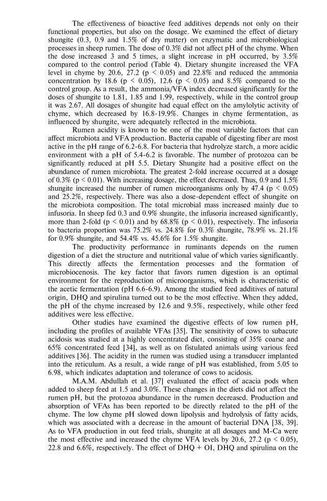
777
The effectiveness of bioactive feed additives depends not only on their functional properties, but also on the dosage. We examined the effect of dietary shungite (0.3, 0.9 and 1.5% of dry matter) on enzymatic and microbiological processes in sheep rumen. The dose of 0.3% did not affect pH of the chyme. When the dose increased 3 and 5 times, a slight increase in pH occurred, by 3.5% compared to the control period (Table 4). Dietary shungite increased the VFA level in chyme by 20.6, 27.2 (p < 0.05) and 22.8% and reduced the ammonia concentration by 18.6 (p < 0.05), 12.6 (p < 0.05) and 8.5% compared to the control group. As a result, the ammonia/VFA index decreased significantly for the doses of shungite to 1.81, 1.85 and 1.99, respectively, while in the control group it was 2.67. All dosages of shungite had equal effect on the amylolytic activity of chyme, which decreased by 16.8-19.9%. Changes in chyme fermentation, as influenced by shungite, were adequately reflected in the microbiota.
Rumen acidity is known to be one of the most variable factors that can affect microbiota and VFA production. Bacteria capable of digesting fiber are most active in the pH range of 6.2-6.8. For bacteria that hydrolyze starch, a more acidic environment with a pH of 5.4-6.2 is favorable. The number of protozoa can be significantly reduced at pH 5.5. Dietary Shungite had a positive effect on the abundance of rumen microbiota. The greatest 2-fold increase occurred at a dosage of 0.3% (p < 0.01). With increasing dosage, the effect decreased. Thus, 0.9 and 1.5% shungite increased the number of rumen microorganisms only by 47.4 (p < 0.05) and 25.2%, respectively. There was also a dose-dependent effect of shungite on the microbiota composition. The total microbial mass increased mainly due to infusoria. In sheep fed 0.3 and 0.9% shungite, the infusoria increased significantly, more than 2-fold (p < 0.01) and by 68.8% (p < 0.01), respectively. The infusoria to bacteria proportion was 75.2% vs. 24.8% for 0.3% shungite, 78.9% vs. 21.1% for 0.9% shungite, and 54.4% vs. 45.6% for 1.5% shungite.
The productivity performance in ruminants depends on the rumen digestion of a diet the structure and nutritional value of which varies significantly. This directly affects the fermentation processes and the formation of microbiocenosis. The key factor that favors rumen digestion is an optimal environment for the reproduction of microorganisms, which is characteristic of the acetic fermentation (pH 6.6-6.9). Among the studied feed additives of natural origin, DHQ and spirulina turned out to be the most effective. When they added, the pH of the chyme increased by 12.6 and 9.5%, respectively, while other feed additives were less effective.
Other studies have examined the digestive effects of low rumen pH, including the profiles of available VFAs [35]. The sensitivity of cows to subacute acidosis was studied at a highly concentrated diet, consisting of 35% coarse and 65% concentrated feed [34], as well as on fistulated animals using various feed additives [36]. The acidity in the rumen was studied using a transducer implanted into the reticulum. As a result, a wide range of pH was established, from 5.05 to 6.98, which indicates adaptation and tolerance of cows to acidosis.
M.A.M. Abdullah et al. [37] evaluated the effect of acacia pods when added to sheep feed at 1.5 and 3.0%. These changes in the diets did not affect the rumen pH, but the protozoa abundance in the rumen decreased. Production and absorption of VFAs has been reported to be directly related to the pH of the chyme. The low chyme pH slowed down lipolysis and hydrolysis of fatty acids, which was associated with a decrease in the amount of bacterial DNA [38, 39]. As to VFA production in out feed trials, shungite at all dosages and M-Ca were the most effective and increased the chyme VFA levels by 20.6, 27.2 (p < 0.05), 22.8 and 6.6%, respectively. The effect of DHQ + OI, DHQ and spirulina on the

778
VFA production was negative, with a decrease to 93.5, 81.2 and 81.8% of the control level. All the studied feed additives, except for DHQ + OI, led to a decrease in the chyme ammonia, with the largest decrease of 24.1% (p < 0.05) and 26.3% under the influence of DHQ and M-Ca. Shungite also reduced the chyme ammonia concentration to 81.3, 87.45 and 91.5% for 0.3, 0.9 and 1.5% shungite, respectively, compared to the control period.
Ghaffari et al. [40] reported similar results when replacing alfalfa hay in the diet of sheep with by-products. The concentration of ammonia decreased with an increase in the portion of pistachio husks which replaced hay. However, no differences in rumen pH were observed in the sheep [40]. Waste from the pro-cessing of the date palm crop (9, 18, and 27% DM) decreased the ammonium nitrogen concentration in the rumen and enhanced synthesis of microbial protein, but the diet composition did not affect the rumen pH [41]. In our trials, dietary DHQ + OI increased the ammonia level in the chyme by 16.5% compared to the control period, which may be associated with an increase in oxidative processes and a decrease in the VFA production under the influence of iodine.
The effects of the studied feed additives on the chyme amylolytic activity also differed. An increase by 6.1, 1.7 and 1.1% occurred upon the use of spirulina, M-Ca and DHQ + OI, respectively. Under the influence of shungite, the amylo-lytic activity significantly decreased, by 17.7, 16.8 and 19.9% of the control activity at a dosage of 0.3, 0.9 and 1.5%, respectively. Under the action of DHQ, the decrease was minimal, by 7.3%.
Enzymatic processes and their changes directly affected the microbiota. Shungite caused the greatest increase in the chyme microbial mass, but the effect was dose-depended. Dietary shungite increased the total number of microorgan-isms increased 2 times (p <0.01), by 47.4% (p < 0.05) and 25.2% compared to the control, mainly due to infusoria, as a result, the infusoria to bacteria ratio changed. With an increase in dietary shungite dosage, the total number of mi-croorganisms decreased and the percentage ratio of infusoria and bacteria changed. DHQ + OI, DHQ and M-Ca influenced the increase in microbial mass in the chyme to a much lesser extent than shungite, the indicators increased by 14.6, 2.2 and 1.8%, respectively, compared to the control period. Under the influence of spirulina, the total number of microorganisms decreased by 14.3% due to both infusoria and bacteria.
In analyzing effects of the studied feed additive, their positive relationship with the ammonia/VFA index was traced, which characterizes the feed conversion rate into microbial protein. The amount of glucogenic fermentation products and the formation of microbial protein largely depend on the rate of fermentation. Slow fermentation of substrates reduces the formation of these products, increases the proportion of propionic acid, and decreases the levels of acetic and butyric acids [42, 43).
There are also publications about modulation of rumen microflora in sheep and young cattle by biologically active substances [44-47]. The use of dietary phy-tobiotics and mineral components promoted enzymatic and microbial processes in the rumen. Among the feed products, seaweeds which are used as prebiotics to increase the productivity and productive health of animals deserve special atten-tion. Dietary algae can improve the intestinal ecology in animals [48].
So, a comparative study of feed additives of natural origin, the dihy-droquercetin, organic iodine, calcium carbonate micellate, spirulina and shungite showed their specific effect on enzymatic and microbiological processes in the rumen of sheep. Dihydroquercetin increases pH and reduction potential in the rumen, stimulates bacterial growth. Organic iodine increases pH and oxidative potential of the chyme and promotes bacterial growth. M-Ca increases pH and

779
decreased the reduction potential of the chyme. Spirulina raises the chyme pH values. The 0.3 and 0.9% shungite more effectively promotes production of volatile fatty acids and chyme biomass compared to 1.5% shungite. Our findings can be used in feeding sheep in order to modulate functionality of diets. As in choosing feed additives, one should consider their effect on metabolic processes and animal immunity, dihydroquercetin and shungite seem to be the most promising.
R E F E R E N C E S
1. Russell J.B., Rychlik J.L. Factors that alter rumen microbial ecology. Science, 2001, 292(5519):
1119-1222 (doi: 10.1126/science.1058830). 2. Agarwal N., Kamra D.N., Chaudhary L.C. Rumen microbial ecosystem of domesticated rumi-
nants. In: Rumen microbiology: from evolution to revolution. A. Puniya, R. Singh, D. Kamra (eds.). Springer, New Delhi. 2015 (doi: 10.1007/978-81-322-2401-3_2).
3. Russell J.B., Rychlik, J.L. Factors that alter rumen microbial ecology. Science, 2001, 292(5519): 1119-1122 (doi: 10.1126/science.1058830).
4. Krehbie C.R. Invited review: applied nutrition of ruminants: fermentation and digestive physiology. The Professional Animal Scientist, 2014, 30(2): 129-139 (doi: 10.15232/S1080-7446(15)30100-5).
5. Chernyshev N.I., Panin I.G., Shumskii N.I., Grechishnikov V.V. Antipitatel'nye faktory kormov. Spravochnaya kniga [Anti-nutritional factors of feed]. Voronezh, 2013 (in Russ.).
6. Ward J. Probiotic yeast for optimal rumen balance. All About Feed, 2017, 25(8): 24-25. 7. Maksimov R.T. Veterinariya sel'skokhozyaistvennykh zhivotnykh, 2008, 3: 64-65 (in Russ.). 8. Bogolyubova N., Korotky V., Zenkin A., Ryzhov V., Buryakov N. Digestion and metabolism
indices of sheep when using activated charcoal supplement. OnLine Journal of Biological Sciences, 2017, 17(2): 121-127 (doi: 10.3844/ojbsci.2017.121.127).
9. Artem’eva O.A., Pereselkova D.A., Fomichev Yu.P. Dihydroquercetin, the bioactive substance, to be used against pathogenic microorganisms as an alternative to antibiotics. Agricultural Biology [Sel'skokhozyaistvennaya biologiya], 2015, 50(4): 513-519 (doi: 10.15389/agrobiol-ogy.2015.4.513eng).
10. Aliev M.M., Gulieva K.A. Vestnik APK Stavropol'ya, 2018, 1(29): 57-59 (in Russ.). 11. EFSA Panel on Dietetic Products, Nutrition and Allergies (NDA), Turck D., Bresson J.-L., Burlin-
game B., Dean T., Fairweather-Tait S., Heinonen M., Hirsch-Ernst K., Mangelsdorf I., McArdle H.J., Naska A., Neuhäuser-Berthold M., Nowicka G., Pentieva K., Sanz Y., Siani A., Sjodin A., Stern M., Tome D., Vinceti M., Willatts P., Engel K.-H., Marchelli R., Pöting A., Poulsen M., Schlatter J., Gelbmann W., Loveren H.V. Scientific opinion on taxifolin-rich extract from Da-hurian Larch (Larix gmelinii). EFSA Journal, 2017, 15(2): 4682 (doi: 10.2903/j.efsa.2017.4682).
12. Fomichev Yu.P., Nikanova L.A., Dorozhkin V.I., Torshkov A.A., Romanenko A.A., Es'kov E.K., Semenova A.A., Gonotskii V.A., Dunaev A.V., Yaroshevich G.S., Lashin S.A., Stol'naya N.I. Digidrokvertsetin i arabinogalaktan — prirodnye bioregulyatory: primenenie v sel'skom khozyaistve i pishchevoi promyshlennosti. Monografiya [Dihydroquercetin and arabinogalactan, the natural bio-regulators: application in agriculture and food industry. Monograph]. Moscow, 2017 (in Russ.).
13. Aghwan Z.A., Sazili A.Q., Kadhim K.K., Alimon A.R., Goh Y.M., Adeyemi K.D. Effects of dietary supplementation of selenium and iodine on growth performance, carcass characteristics and histology of thyroid gland in goats. Animal Science Journal, 2015, 87(5): 690-696 (doi: 10.1111/asj.12484).
14. Aghwan Z.A, Sazili A.Q., Alimon A.R., Goh Y.M., Hilmi M.A. Blood haematology, serum thy-roid hormones and glutathione peroxidase status in Kacang goats fed inorganic iodine and sele-nium supplemented diets. Asian-Australasian Journal of Animal Sciences, 2013, 26(11): 1577-1582 (doi: 10.5713/ajas.2013.13180).
15. Xu J., Liu X.-L., Yang X.-F., Guo H.-L., Zhao L.-N., Sun X.-F. Supplemental selenium allevi-ates the toxic effects of excessive iodine on thyroid. Biological Trace Element Research, 2011, 141: 110-118 (doi: 10.1007/s12011-010-8728-8).
16. Rakhmanin Yu.A., Stekhin A.A., Yakovleva G.V. Biofizika vody [Biophysics of water]. Moscow, 2016 (in Russ.).
17. Zatsepina O.V., Stekhin A.A., Yakovleva G.V., P'yanzina I.P. Gigiena i sanitariya, 2013, 5: 37-39 (in Russ.).
18. Ametov A.S., Doskina E.V., Kapustina N.V. Endokrinologiya, 2016, 4(17): 65-72 (in Russ.). 19. Shibil'skis P. Aktivirovannaya voda [Activated water]. Available: https://water-ionizer.ru/useful-
information/aktivirovannaya-voda. Accessed: 16.03.2019 (in Russ.). 20. Rekomendatsii po ispol'zovaniyu ionizirovannoi vody [Recommendations for the use of ionized water].
Available: https://akvalife.club/ru/rekomendatsii-po-primeneniyu-ionizirovannoj-vody.html. Ac-cessed: 16.03.2019 (in Russ.).
21. Becker E.W. Microalgae for human and animal nutrition. In: Handbook of microalgal culture: applied phycology and biotechnology, second edition. Richmond A., Hu Q. (eds.). Blackwell Pub-lishing Ltd., 2013 (doi: 10.1002/9781118567166.ch25).
780
22. Kedik S.A., Yartsev E.I., Gul'tyaeva N.V. Spirulina — pishcha XXI veka [Spirulina as a food of the XXI century]. Moscow, 2006 (in Russ.).
23. Kedik S.A., Yartsev E.I., Sakaeva I.V., Zhavoronok E.S., Panov A.V. Biofarmatsevticheskii zhur-nal, 2011, 3(3): 3-10 (in Russ.).
24. Zaitsev V.V., Tarabrin V.V., Voishcheva E.A. Vestnik meditsinskogo instituta "REAVIZ": reabili-tatsiya, vrach i zdorov'e, 2011, 1: 17-21 (in Russ.).
25. Holman B.W.B., Malau-Adul A.E.O. Spirulinaas a livestock supplement and animal feed. Journal of Animal Physiology and Animal Nutrition, 2012, 97(4): 615-623 (doi: 10.1111/j.1439-0396.2012.01328.x).
26. Rimkus M., Simkus A., Syvys R., Birutis S. Dry powdery fodder additive, supplement or fodder containing algae spirulina platensis. WO/2010/106468 MKI3 A23K 1/00 2006.01, A23K 1/02 2006.01, C12N 1/12 2006.01. SPILA, UAB (LT). ¹ PCT/IB2010/051039. Decl. 10.03.2010. Publ. 23.09.2010.
27. Vetrov S.I., Lenkova N.I., Gorbatkina I.E. Shungit. Rossiiskii mineral [Shungite. Russian mineral]. Moscow, 2011 (in Russ.).
28. Andrievsky G.B., Bruskov V.I., Tykhomyrov A.A., Gudkov S.V. Peculiarities of the antioxidant and radioprotective effects of hydrated C60 fullerene nanostructures in vitro and in vivo. Free Radical Biology and Medicine, 2009, 47(6): 786-793 (doi: 10.1016/j.freeradbiomed.2009.06.016).
29. Xiao Y., Wiesner M.R. Characterization of surface hydrophobicity of engineered nanoparticles. Journal of Hazardous Materials, 2012, 215-216: 146-151 (doi: 10.1016/j.jhazmat.2012.02.043).
30. Zhang Y., Matsuo, Y., Li C.-Z., Tanaka H., Nakamura E. A scalable synthesis of meth-ano[60]fullerene and congeners by the oxidative cyclopropanation reaction of silylmethylfullerene. Journal of the American Chemical Society, 2011, 133(21): 8086-8089 (doi: 10.1021/ja201267t).
31. Ponomarev A.P., Podkolzin I.V., Amelin V.G. Nanotekhnologii: nauka i proizvodstvo, 2012, 4(11): 48-55 (in Russ.).
32. Bogolyubova N.V., Romanov V.N., Devyatkin V.A., Kalinin Yu.K. Veterinariya i kormlenie, 2014, 5: 88-90 (in Russ.).
33. Tremasova A.M. Materialy nauchno-prakticheskoi konferentsii «Innovatsiya v sovremennoi farma-kologii» [Proc. Conf. “Innovation in modern pharmacology”]. Kazan'-Moscow, 2012: 180-181 (in Russ.).
34. Nasrollahi S.M., Zali A., Ghorbani G.R., Moradi S.M., Heydari S.A.M. Variability in suscepti-bility to acidosis among high producing mid-lactation dairy cows is associated with rumen pH, fermentation, feed intake, sorting activity, and milk fat percentage. Animal Feed Science and Technology, 2017, 228: 72-82 (doi: 10.1016/j.anifeedsci.2017.03.007).
35. Dijkstra J., Ellis J.L., Kebread E., Strathe A.B., López S., France J., Bannink A. Ruminal pH regulation and nutritional consequences of low pH. Animal Feed Science and Technology, 2012, 172(1-2): 22-33 (doi: 10.1016/j.anifeedsci.2011.12.005).
36. Malekkhanhi M., Tahmasbi A.M., Naserian A.A., Danesh-Mesgaran M., Kleen J.L., AlZahal O., Ghaffari M.H. Effects of supplementation of active dried yeast and malate during subacute rumi-nal acidosis on rumen fermentation, microbial population, selected blood metabolites, and milk production in dairy cows. Animal Feed Science and technology, 2016, 213: 29-43 (doi: 10.1016/j.anifeedsci.2015.12.018).
37. Abdullah M.A.M., Farghaly M.M., Youssef I.M.I. Effect of feeding Acacia nilotica pods to sheep on nutrient digestibility, nitrogen balance, ruminal protozoa and rumen enzymes activity. Journal of Animal Physiology and Animal Nutrition, 2018, 102(3): 662-669 (doi: 10.1111/jpn.12874).
38. Benchaar C., McAllister T.A., Petit H.V., Chouinard P.Y. Whole flax seed and oil supplementa-tion of dairy cows fed high-forage or high-concentrate diets: effect on digestion/ruminal fermen-tation characteristics, protozoal population and milk fatty acid profile. Animal Feed Science and technology, 2014, 198: 117-129 (doi: 10.1016/j.anifeedsci.2014.10.003).
39. Cone J.W., Becker P.M. Fermentation kinetics and production of volatile fatty acids and micro-bial protein by starchy feedstuffs. Animal Feed Science and technology, 2012, 172 (1-2): 34-41 (doi: 10.1016/j.anifeedsci.2011.12.006).
40. Ghaffari M.H., Tahmasbi A.M., Khorvash M., Naserian A.-A., Ghaffari A. H., Valizadeh H. Effects of pistachio by-products in replacement of alfalfa hay on populations of rumen bacteria involved in biohydrogenation and fermentative parameters in the rumen of sheep. Journal of An-imal Physiology and Animal Nutrition, 2013, 98(3): 578-586 (doi: 10.1111/jpn.12120).
41. Khezri A., Dayani O., Tahmasbi R. Effect of increasing levels of wasted date palm on digestion, rumen fermentation and microbial protein synthesis in sheep. Journal of Animal Physiology and Animal Nutrition, 2016, 101(1), 53-60 (doi: 10.1111/jpn.12504).
42. Ariko T., Kass M., Henno M., Fievez V., Kärt O., Kaart T., Ots M. The effect of replacing barley with glycerol in diet of dairy cows on rumen parameters and milk fatty acid profile. Animal Feed Science and Technology, 2015, 209: 69-78 (doi: 10.1016/j.anifeedsci.2015.08.004).
43. Qiao G.H., Zhou X.H., Li Y., Zhang H.S., Li J.H., Wang C.M., Lu Y. Effect of several supple-mental Chinese herbs additives on rumen fermentation, antioxidant function and nutrient digest-ibility in sheep. Journal of Animal Physiology and Animal Nutrition, 2011, 96(5): 930-938 (doi: 10.1111/j.1439-0396.2011.01211.x).

781
44. Schofield B.J., Lachner N., Le O.T., McNeill D.M., Dart P., Ouwerkerk D., Hugenholtz P., Klieve A.V. Beneficial changes in rumen bacterial community profile in sheep and dairy calves as a result of feeding the probiotic Bacillus amyloliquefaciens H57. Journal of Applied Microbiology, 2018, 124(3): 855-866 (doi: 10.1111/jam.13688).
45. Bogolyubova N., Korotky V., Zenkin A., Romanov V., Ryzhov V. Assessing efficiency of the coniferous energy supplement in the diet of dairy cows for maintaining productive health. Asian Journal of Pharmaceutical and Clinical Research, 2017, 10(10): 117-120 (doi: 10.22159/aj-pcr.2017.v10i10.19860).
46. Hosoda K., Matsuo M., Miyaji M., Matsuyama H., Maeda H., Ohta H., Kato H., Nonaka K. Fermentative quality of purple rice (Oryza sativa L.) silage and its effects on digestibility, ruminal fermentation and oxidative status markers in sheep: A preliminary study. Grassland Science, 2012, 58(3): 161-169 (doi: 10.1111/j.1744-697x.2012.00256.x).
47. Váradyová Z., Mravčáková D., Holodová M., Grešáková Ľ., Pisarčíková J., Barszcz M., Taciak M., Tuśnio A., Kišidayová S., Čobanová K. Modulation of ruminal and intestinal fermentation by medicinal plants and zinc from different sources. Journal of Animal Physiology and Animal Nutri-tion, 2018, 102(5): 1131-1145 (doi: 10.1111/jpn.12940).
48. Makkar H.P.S., Tran G., Heuze V., Giger-Reverdin S., Lessire M., Lebas F., Ankers P. Seaweeds for livestock diets: a review. Animal Feed Science and Technology, 2016, 212: 1-17 (doi: 10.1016/j.anifeedsci.2015.09.018).

784
AGRICULTURAL BIOLOGY, ISSN 2412-0324 (English ed. Online)
2020, V. 55, ¹ 3, pp. 784-793 (SEL’SKOKHOZYAISTVENNAYA BIOLOGIYA) ISSN 0131-6397 (Russian ed. Print)
ISSN 2313-4836 (Russian ed. Online)
Reproduction, developmental physiology UDC 636.2:591.465.12:576:576.3/.7.086.83:591.04 doi: 10.15389/agrobiology.2020.4.784eng
doi: 10.15389/agrobiology.2020.4.784rus
THE INFLUENCE OF HIGHLY DISPERSED SILICA NANOPARTICLES ON THE FUNCTIONAL ACTIVITY OF MITOCHONDRIA
AND CHROMATIN STATE IN NATIVE AND DEVITRIFIED Bos taurus OOCYTES
T.I. KUZMINA1, I.V. CHISTYAKOVA1, D.N. TATARSKAYA2
1All-Russian Research Institute for Farm Animal Genetics and Breeding — branch of Ernst Federal Science Center for Animal Husbandry,, 55А, Moskovskoe sh., St. Petersburg—Pushkin, 196625 Russia, e-mail [email protected] (corresponding author ), [email protected] (corresponding author ); 2Pushkin Leningrad State University, 10, Petersburg Sh., St. Petersburg—Pushkin, 196605, e-mail [email protected] ORCID: Kuzmina T.I. orcid.org/0000-0002-4218-6080 Tatarskaya D.N. orcid.org/0000-0002-8834-1912 Chistyakova I.V. orcid.org/0000-0001-7229-5766 The authors declare no conflict of interests Acknowledgements: The work was performed according to the theme of the project No. 18-016-00147A funded by the Russian Foun-dation for Basic Research. Received May 28, 2020
A b s t r a c t
Mitochondria are the only cellular compartments which generate and transform energy in the cell. These organelles are among the first to respond to changes in extra- and intracellular condi-tions (e.g., ionic homeostasis, dehydration level, temperature). Exposition to ultra-low temperatures, due to lipid peroxidation, causes ATP synthase complex disorders and destruction of genetic material (E.A. Novocherkina et al., 2016). Highly dispersed silica nanoparticles (HDSns) can be proposed as cytoprotectors. An amorphous form of silicon dioxide, or highly dispersed silica, which exhibits its biological activity through high adsorption capacity, reduces the concentration of ions and biopolymers upon cell dehydration during cryopreservation (T.T. Turov et al., 2011). This paper deals with the first report of the increase in cryoresistance and maturation rate of Bos taurus oocytes due to exposure to 0,001% HDSns during vitrification and in vitro culture, which ensures higher functional activity of mitochondria and preserves structural properties of chromatin of native and devitrified (DV) oocytes. The study aimed to identify the impact of HDSns on functional activity of mitochondria and chromatin state of cow’s native and devitrified oocytes during in vitro culture. In the experiments, we used cu-mulus—oocyte complexes (COCs) of Holsteinized cows. The oocytes with homogeneous cytoplasm surrounded by five or more layers of cumulus cells were subjected to vitrification. COCs intended for vitrification were exposed in three solutions of cryoprotective agents (CPAs) based on T-199 medium with 10 % fetal bovine serum (FBS, HyClone, UK), the CPA-1 containing 0.7 M dime-thyl sulfoxide (DMSO) + 0.9 M ethylene glycol (EG) (for 30 s), CPA-2 (1.4 M DMSO + 1.8 M EG (for 30 s), and CPA-3 (2.8 M DMSO + 3.6 M EG + 0.65 M trehalose) (for 20 s). The straws with oocytes were directly plunged into liquid nitrogen. During thawing, the oocytes were washed with T-199 medium containing 0.25 M (for 3 min), 0.19 M (for 3 min), and 0.125 M (for 3 min) trehalose, and finally with T-199 medium. In the test group of oocytes, highly dispersed silica nano-particles (HDSns, 4-17 nm, mass concentration 0.001 %) synthesized by high-temperature hydrolysis were added to the CPAs, devitrification solutions and washing solutions. In the control group, native and devitrified oocytes were cultured for 24 hours in maturation medium consisted of the T-199 with 106 granulosa cells/ml supplemented with 10 % fetal bovine serum and 50 ng/ml bovine prolactin (38.5 C, 90 % humidity, and 5 % CO2 atmosphere). In the test group, the maturation medium for native and devitrified oocytes was supplemented with HDSns at a final concentration of 0.001 %. Mitochon-drial activity was measured by fluorescence intensity (FI) of MitoTracker Orange CMTMRos (Thermo Fisher Scientific, UK) in µA. In studding HDSns effects on the nuclear maturation, oocytes were exposed for 5-10 min to 0.9 % sodium citrate solution and mechanically denuded from cumulus cells with a needle. Then the cells were placed on a glass slide and fixed with methanol/acetic acid solution (3:1). The dry-air samples were stained with azure-eosin by Romanowsky-Giemsa method. In DV

785
oocytes exposed to HDSns, the FI of MitoTracker Orange CMTMRos probe increased from 77±6.3 μA to 169±12.8 μA (p < 0.05). The functional activity of mitochondria in DV oocytes which were treated with HDSns increased from 169±12.8 μA to 181±7.7 μA (p < 0.05) during the period from diplotene to the metaphase I stages, and subsequently decreased to 141±11.2 μA at metaphase II stage, what is probably associated with the completion of nuclear and cytoplasmic maturation of oocytes. When assessing the chromatin state of oocytes in the HDSns-treated group, we revealed a decrease in the number of oocytes with chromatin degeneration at diplotene and metaphase II stages compared to those in the HDSns -untreated DV oocytes (40 % vs. 21 %, and 59 % vs. 38 %, p < 0.01), that is probably associated with the DNA reparation processes. In general, our findings reveal a positive effect of HDSns on the functional activity of mitochondria and the chromatin state in DV female gametes of Bos taurus. The obtained results expand views and available infor-mation on the functioning of cell compartments at ultra-low temperatures and the mechanisms of HDSns action on female gametes.
Keywords: oocyte, vitrification, highly dispersed silica nanoparticles, functional activity of mitochondria, MitoTracker Orange CMTMRos, chromatin, Bos taurus
Improvement of cryoprotective media for vitrification of oocytes through the introduction of various compounds necessary to maintain viability after the thawing procedure continues to be an urgent problem in cryopreservation tech-nology. Recently, biologically active substances with a nanoscale structure synthe-sized from various compounds of trace elements and minerals, for example, hy-droxyapatite (HA), silicon dioxide (SiO2), aluminum oxide (Al2O3), and titanium dioxide (TiO2) [1, 2] have been widely used for modernization of combined cry-oprotective media. Cryoprotective agents containing nanoparticles improve freez-ing by increasing the thermal conductivity of vitrification solutions and reducing the consequences of recrystallization processes during thawing compared to that when the standard cryoprotective agents such as ethylene glycol and dimethyl sulfoxide [1, 2] are used.
Highly dispersed (pyrogenic) silica (HDS) is an amorphous form of sil-icon dioxide with a range of spherical particle size 4-17 nm (90% of the range) [3]. Due to the pronounced adsorption activity resulted from a large specific surface area (Ssp. = 200 m2/g), HDS nanoparticles are able to reduce the con-centration of polymers and ions during cell dehydration [3, 4], and can bind extracellular water [5]. The large size of the finely dispersed silica aggregates and the low density of particles in these agglomerates ensure the binding of a significant amount of extracellular water, which is not subject to changes in interactions with cells during freezing/thawing [5].
Under oxidative stress conditions during freezing, there are a denaturing effect of free radicals on ion-transporting proteins, impaired DNA synthesis and thermotropic mitochondrial defects, which ultimately leads to the cell death [6]. Mitochondria provide cell with the ATP necessary for the completion of meiotic maturation. The features of their functioning determine the gamete quality [7, 8]. Anomalies in the chromatin transformation of the cell nucleus under temperature-dependent oxidative stress are mainly caused by the disruption of the biosynthesis of nuclear proteins and their connection with DNA, the destruction of nucleotides, and single- and double-stranded DNA breaks [9].
This paper for the first time shows that when nanoparticles of highly dis-persed silica at a concentration of 0.001% are used in the technology of vitrifica-tion and extracorporeal maturation of devitrified (DV) bovine oocytes, the mito-chondrial potential of DV oocytes increases and the level of degenerated cells drops.
The work aimed to identify the nature of the effect of highly dispersed silica nanoparticles on the functional activity of mitochondria and chromatin sta-tus in native and devitrified bovine oocytes during extracorporeal maturation.
Materials and methods. The ovaries of Holsteinized cows (Bos taurus) after ovariectomy were delivered from the slaughterhouse to the laboratory in a 0.9%

786
NaCl solution with penicillin (100 IU/ml), streptomycin (100 μg/ml) and amphotericin B (0.25 ng/ml) at 30-35 С. Cumulus—oocyte complexes (COCs) were aspirated from follicles with a diameter of 3-8 mm.
Oocytes with homogeneous cytoplasm surrounded by five or more layers of cumulus cells were subjected to vitrification. COCs intended for vitrification were treated with three solutions of cryoprotective agents (CPA) based on T-199 medium supplemented with 10% fetal bovine serum (FBS, HyClone, UK). The CPA solutions for vitrification of oocytes in control (without treatment with HDSns) contained 0.7 M dimethyl sulfoxide (DMSO) and 0.9 M ethylene glycol (EG) for CPA-1, 1.4 M DMSO and 1.8 M EG for CPA-2, and 2.8 M DMSO, 3.6 M EG and 0.65 M trehalose (Trehalose) for CPA-3. All reagents, with the exception of those specifically indicated in the text, are manufactured by Sigma-Aldrich (USA), plastic laboratory glassware is manufactured by BD Falcon™ (Bec-ton Dickinson and Co., BD Biosciences, USA). HDSns (4-17 nm, mass concen-tration 0.001%) were synthesized at the Chuiko Institute of Surface Chemistry of NAS of Ukraine by high-temperature hydrolysis. HDSns were used in the form of a stable suspension consisting mainly of submicron aggregates. The choice of concentration was based on the data provided by the developers [10].
COCs were exposed to CPA-1 for 30 s, then to CPA-2 for 30 s, and to CPA-3 for 20 s. Thereafter, the straws with oocytes were plunged into Dewar flask with liquid nitrogen. COCs were removed from the liquid nitrogen not earlier than after 1 h and thawed by placing sequentially into a 0.25 M trehalose solution prepared in T-199 medium supplemented with 10% FBS for 3 min at 37 С, then into 0.19 M trehalose solution for 3 min at 37 С, and finally into 0.125 M trehalose solution for 3 min at 37 С, after which they were washed three times in T-199 medium with 10% FBS. In the control, all the solutions did not contain HDSns, in the experimental groups all media were supplemented with HDSns to a final concentration of 0.001%.
Native and devitrified oocytes were cultured for 24 h at 38.5 C and 90% humidity in a 5% СО2 atmosphere using culture medium containing T-199 + 10% FBS + 106 granulosa cells/ml + 50 ng bovine prolactin/ml. During culturing native and devitrified oocytes in the control group, this medium did not contain HDSns; in the testing groups, HDSns were added to a final concentration of 0.001% [11].
MitoTracker Orange CMTMRos probe (Thermo Fisher Scientific, UK) was used to assess the functional state of mitochondria in native and DV oocytes at different stages of meiosis. COCs, 15-20 per drop, were placed into 500 μl drops of 500 nM MitoTracker Orange CMTMRos solution and incubated for 30 min in the dark at 37 С. Then oocytes were washed from the dye in phosphate buffered saline (PBS) with 0.3% bovine serum albumin. The washed oocytes were denuded from cumulus cells by treatment with a 0.1% trypsin solution at 37 С for 5-10 min, transferred into Hanks solution containing 3.7% paraformaldehyde, and then fixed for 15 min at 37 С. After fixation, oocytes were washed from paraformaldehyde in PBS and placed on Super frost glasses into drops of Hoechst 33258 solution (2.5 μg/ml, Thermo Fisher Scientific, UK).
Fluorescence intensity (IF) of MitoTracker Orange CMTMRos were measured and nuclear maturation were assessed using the Hoechst 33258 dye and a ZEISS Axio Lab.A1 fluorescence microscope with a photometric attachment (Karl Zeiss, Germany). The excitation wavelength for MitoTracker Orange CMTMRos was 554 nm, the emission wavelength - 576 nm; for Hoechst 33258 these were 352 nm and 461 nm, respectively. IF of MitoTracker Orange CMTM-Ros was measured in μA.

787
To identify the HDSns effect on the nuclear maturation of female gametes, oocytes were exposed for 5-10 min to 0.9% sodium citrate solution and mechani-cally denuded from cumulus using a needle. Then the cells were transferred to dry defatted glass and fixed with a mixture of methanol/acetic acid 3:1. Dry-air prep-arations were stained with azure-eosin according to Romanowsky-Giemsa method.
The results were processed by the method of two-way analysis of variance ANOVA using the SigmaStat statistical program package (Jandel Scientific Soft-ware, USA). Data are presented as means (M) and standard errors of means (±SEM). The Student’s t-test and Pearson’s χ2 test adjusted for likelihood were used to assess the significance of differences between the compared mean value. The significance of the differences was assessed at p < 0.05, p <0.01 and p <0.001 for 3-5 independent experiments.
Results. In previous works, we found a positive effect of 0.001% HDSns on the chromatin status of somatic cells of cow ovarian follicles [12], on oocyte-cumulus interactions during maturation of female gametes [13], and on the devel-opment of pre-implantation embryos [11]. In the present study, it is found that the introduction HDSns in cryoprotective media contributes to the increase of MitoTracker Orange CMTMRos probe fluorescence in DV oocytes compared to gametes not exposed to HDSns before vitrification (169±12.8 vs. 77±6.3 μA, p < 0.05) (Table, Fig. 1).
Intensity of fluorescence (FI, μA) of MitoTracker Orange CMTMRos probe (Thermo Fisher Scientific, UK) in native and devitrified oocytes of Holsteinized cows (Bos taurus) during meiosis, treated by highly dispersed silica nanoparticles (HDSns) (М±SEM, number of experiments N = 5, total number of oocytes n = 529, in vitro culture)
Oocytes HDSns n FI of MitoTracker Orange CMTMRos diplotene metaphase I metaphase II
Native − 133 331±16.2a 188±11.3e 143±10.1i + 139 309±15.6b 203±11.8f 149±14.7j
Devitrified − 128 77±6.3c 139±11.7g 101±9.7k + 129 169±12.8d 181±7.7h 141±11.2l
a:c; c:d; b:d; g:h; e:g; i:k; k:l; a:d; b:c; f:g; j:k Differences are statistically significant at p < 0.05 (Student’s t-test).
In 14 h, that is, af-ter the devitrified oocytes reached the stage of meta-phase I, we detected statis-tically significant differ-ences in the IF of Mito-Tracker Orange CMTMRos probe between cells treated and not exposed to HDSns (181±7.7 vs. 139±11.7 μA, p < 0.05). By the time the oocytes reached metaphase I, the IF of native oocytes did not differ from the de-gree of DV oocytes treated with HDSns (188±11.3
and 181±7.7 μA, respectively). In DV oocytes, an increase in mitochondrial ac-tivity occurred from diplotene to metaphase I (p < 0.05) (see Table). At the final stages of maturation, a general drop in the IF of MitoTracker Orange CMTMRos was observed in all experimental groups, and the indicators of mitochondrial ac-tivity were minimal.
Fig. 1. Representative image of mitochondria in devitrified oocytes of Holsteinized cows at diplotene stage with low (a) and high (b) fluorescence intensities of the MitoTracker Orange MTMRos probe (Thermo Fisher Scientific, UK), treated with highly dispersed silica nanoparticles (HDSns): a — oocytes not treated with HDSns), b — oocytes treated with 0.001% HDSns (ZEISS Axio Lab.A1, Karl Zeiss, Germany, in vitro culture).

788
In assessing the chro-matin state of native and DV oocytes at diplotene stage, there was a higher level of un-treated DV gametes with signs of chromosome degeneration compared to the cells treated with HDSns (40 vs. 21%, p < 0.01) (Fig. 2). During culture to the metaphase I stage, we did not find signifi-cant differences in the number of oocytes with normal or de-generated chromatin between control and HDSns-treated na-tive and DV gametes (Fig. 3, A).
However, 24 h after DV oocytes reached the met-
aphase II stage (Fig. 4), there was an increase in the number of cells with signs of degeneration of nuclear material among oocytes that were not treated with HDSns (59% vs. 38%, p < 0.01, see Fig. 3, B).
A
B
Fig. 3. Oocytes of Holsteinized cows with normal (1) and degenerated (2) chromatin at the metaphase I (A) and metaphase II (B) stages, treated with highly dispersed silica nanoparticles (HDSns): I — native oocytes (control), II — native oocytes treated with 0.001% HDSns, III — devitrified oocytes (control), IV — devitrified oocytes treated with 0.001% HDSns. (24 h of culture, number of oocytes n = 419, number of trials N = 4; in vitro culture).
A: a:b; c:d; g:h; a:e; a:g; c:e; c:g; b:f; b:h; d:f; d:h; a:d; a:f; a:h; c:b; c:f; c:h; e:b; e:d; e:h; g:b; g:d; g:f — differences are statistically significant at p < 0.001; differences for e:f are statistically significant at p < 0.01.
B: a:b; c:d; g:h; a:e; a:g; c:e; c:g; b:f; b:h; d:f; d:h; a:d; a:f; a:h; c:b; c:f; c:h; e:b; e:d; g:b; g:d — differences for the pairs are statistically significant at p < 0.001; e:g; f:h — differences are statistically significant
at p < 0.01; differences for e:f are statistically significant at p < 0.05 (Pearson’s χ2 test).
During freezing functional activity of mitochondria decreases, that is caused
Fig. 2. Oocytes of Holsteinized cows with normal (1) and de-generated (2) chromatin at the diplotene stage, treated with highly dispersed silica nanoparticles (HDSns): I — native oocytes (control), II — devitrified oocytes (control), III — devitrified oocytes treated with 0.001% HDSns. (0 h of culture, number of oocytes n = 293, number of experiments N = 4; in vitro culture). Differences are statistically significant: for a:b; a:c; e:f; b:d; b:f;
a:d; a:f; c:b; c:f; e:b; d:e at p < 0.001; for c:d; c:e; d:f at p < 0.01 (Pear-son’s χ2 test).

789
not only by the disruption of the antioxidant system, but also by the formation of non-selective mitochondrial pores (MP) [7, 14]. Vitrifi-cation changes calcium re-ceptors on the membrane of the endoplasmic reticu-lum, leading to atypical fluc-tuations in the Ca2+ level, which causes unregulated opening of the MP [15]. The spontaneous membrane per-meabilization triggers a cas-cade of apoptosis events
[7]. Our results suggest that HDSns optimize processes associated with calcium regulation of MP functioning and increased ion exchange between the cell cyto-plasm [3, 6]. The oxidative stress of mitochondria during cryopreservation is as-sociated with the oxidative modification of proteins [16] and the formation of protein fibrillar complexes [17, 18]. These processes provoke oxidation of the cell membrane, disrupt ionic homeostasis, the functioning of the nuclear apparatus, and inter- and intracellular signal transmission [18]. It was shown that nanoparti-cles ranging in size from 3 to 9 nm exhibit the properties of quantum dots and are capable to inhibit the formation of protein oxidation products [16] and aggregate protein structures [17, 19]. Ultra-small particles of silica supposedly have antiox-idant and antiaggregatory effects, due to which the preservation of cellular func-tions, including nuclear apparatus, occurs. It should also be noted that the effect of HDSns on cell mitochondria may be due to the concentration of particles in solution [20, 21]. An increase in the functional activity of mitochondria in oocytes treated with HDSns and control DV oocytes during the transition period from the diplotene to the metaphase I stages is possibly explained by the restoration of the electrochemical gradient and increased ATP production [22], which is necessary for the completion of the nuclear-cytoplasmic maturation of the DV oocytes.
HDSns have no genotoxic effect [23] and are able to initiate an anti-stress response of cells to ultra-low temperatures through the activation of DNA repair proteins [24, 25]. Importantly, the HDSns genotoxicity, as cytotoxicity, have a size- and dose-dependent effect [25, 26]. In our experiments, HDSns had a pos-itive effect on the nuclear maturation of DV oocytes, which was probably associ-ated with the induction of an anti-stress response and a weakening of the effects of oxidative stress caused by the ultra-small size of HDS particles and their low concentration in maturation medium.
The observed effect of a decrease in mitochondrial activity in all experi-mental groups during culture may be a consequence of the completion of the nuclear-cytoplasmic maturation of the oocyte with a further block of meiosis at the metaphase II stage before the activation of the oocyte by the sperm. However, the decrease in the IF of MitoTracker Orange CMTMRos and the rise in the proportion of cells with degenerated chromatin in the DV group of oocytes at the metaphase II stage as compared to other experimental groups, apparently, also indicates a significant damage to mitochondria and chromatin caused by cryopres-ervation procedure.
Thus, the evidence from this study suggests the increase in the mitochon-drial potential and the decrease in the number of degenerated cells because of the use of highly dispersed silica nanoparticles (HDSns) in extracorporeal maturation
Fig. 4. Representative image of normal (a) and degenerated (b) chromatin in devitrified oocytes of Holsteinized cows at methaphase II stage, treated with highly dispersed silica nanoparticles (0,001%) (cytogenetic analysis, staining with azure-eosin according to Ro-manowsky-Giemsa method, ZEISS Axio Lab.A1, Karl Zeiss, Ger-many, in vitro culture).
790
procedure of devitrified (DV) bovine oocytes, which may indicate the increase in ion exchange and repair processes. An increase in mitochondrial activity when DV oocytes reach the metaphase I stage indicates the increase in the energy supply of cells during this period. The general decrease in the transmembrane potential in native and DV oocytes at the metaphase II stage can be associated either with the completion of nuclear-cytoplasmic maturation, or with a large number of cryo-injuries in the structure of mitochondria (membranes) and chromatin. Conse-quently, 0.001% HDSns have a positive effect on the indices of nuclear-cytoplas-mic maturation of DV oocytes during vitrification of Bos taurus female gametes. Creating a cryobank of oocytes is of particular relevance in the context of large-scale selection, a decrease in the reproductive qualities of highly productive cows, and necessity to preserve the gene pool of elite individuals for constructing new genotypes, including by the CRISPR-Cas9 technology. The obtained data can be also used to improve technique of extra- and intra-ovarian vitrification of oocytes of other animal species, and to address problems of human infertility.
R E F E R E N C E S
1. Xu H.F., Hao B.T., Liu L.J., Tang L.L., Liu B.L. Calorimetric studies on thermal properties of
nano-cryoprotectant solutions during vitrification. CryoLetters, 2016, 37(6): 406-410. 2. Li W., Zhou X., Dai J., Zhang D., Liu B., Wang H., Xu L. Effect of hydroxyapatite nanoparticles
on MII-stage porcine oocytes vitrification and the study of its mechanism. Biochemical Engineering Journal, 2013, 30(4): 789-793.
3. Turov V.V., Barvinchenko V.N., Krupskaya T.V., Gun'ko V.M., Chekhun V.F. Biotekhnologiya, 2011, 4(4): 34-48 (in Russ.).
4. Rubio L., Pyrgiotakis G., Beltran-Huarac J., Zhang Y., Gaurav J., Deloid G., Spyrogianni A., Sarosiek K., Bello D., Demokritou P. Safer-by-design flame-sprayed silicon dioxide nanoparti-cles: the role of silanol content on ROS generation, surface activity and cytotoxicity. Particle and Fibre Toxicology, 2019, 16(1): 40 (doi: 10.1186/s12989-019-0325-1).
5. Galagan N.P., Gun’ko V.M., Porkhun N.G., Novikova E.A., Turov V.V. Dopovidi Natsional'noї akademiї nauk Ukraїni, 2012, 5: 126-133 (in Russ.).
6. Savchenko D.S. Meditsina i obrazovanie v Sibiri, 2013, 6: 23-30 (in Russ.). 7. Al-Zubaidi U., Liu J., Cinar O., Robker R.L., Adhikari D., Carroll J. The spatio-temporal dy-
namics of mitochondrial membrane potential during oocyte maturation. Molecular Human Repro-duction, 2019, 25(11): 695-705 (doi: 10.1093/molehr/gaz055).
8. Pozhilova E.V., Novikov V.G., Levchenkova O.S. Obzory po klinicheskoi farmakologii i lekarstvennoi terapii, 2014, 12(3): 13-19 (in Russ.).
9. Yoon S.Y., Eum J.H., Cha S.K., Yoon T.K., Lee D.R., Lee W.S. Prematuration culture with phosphodiesterase inhibitors after vitrification may induce recovery of mitochondrial activity in vitrified mouse immature oocytes. Biopreservation and Biobanking, 2018, 16(4): 296-303 (doi: 10.1089/bio.2018.0010).
10. Galagan N.P., Klymenko N.Y., Orel I.L., Novikova E.A., Turov V.V. Biofunctional nanomateri-als based on ultra-fine silica, protein and aminocarbohydrates. Biopolymers and Cell, 2010, 26(3): 205-213 (doi: 10.7124/bc.000158).
11. Chistyakova I.V., Kuzmina T.I., Stanislavovich T.I., Kovtun S.V. Effects of highly dispersed silica nanoparticles on the cryoresistance of the bovine cumulus-oocyte complexes. Cryobiology, 2018, 85: 176 (doi: 10.1016/j.cryobiol.2018.10.215).
12. Kuz'mina T.I., Chistyakova I.V. Aktual'nye voprosy veterinarnoi biologii, 2019, 3(43): 8-12 (doi: 10.24411/2074-5036-2019-10031) (in Russ.).
13. Kuz'mina T.I., Stanislavovich T.I., Molchanov A.V. Agrarnyi nauchnyi zhurnal, 2019, 3: 29-34 (doi: 10.28983/asj.y2019i3pp29-34) (in Russ.).
14. Novoderezhkina E.A., Zhivotovskii B.D., Gogvadze V.G. Molekulyarnaya biologiya, 2016, 50(1): 51-68 (doi: 10.7868/S002689841601016X) (in Russ.).
15. Kim B., Yoon S.-Y., Cha S.K., Kwak K.H., Fissore R.A., Parys J.B., Yoon T.K., Lee D.R. Alterations in calcium oscillatory activity in vitrified mouse eggs impact on egg quality and sub-sequent embryonic development. Pflügers Archiv — European Journal of Physiology, 2011, 461: 515-526 (doi: 10.1007/s00424-011-0955-0).
16. Mostek A., Słowińska M., Judycka S., Karol H., Ciereszko A., Dietrich M.A. Identification of oxidatively modified proteins due to cryopreservation of carp semen. Journal of Animal Science, 2018, 96(4): 1453-1465 (doi: 10.1093/jas/sky063).
791
17. Xiao L., Zhao D., Chan W.-H., Choi M.M.F., Li H.-W. Inhibition of beta 1-40 amyloid fibril-lation with N-acetyl-L-cysteine capped quantum dots. Biomaterials, 2010, 31(1): 91-98 (doi: 10.1016/j.biomaterials.2009.09.014).
18. Shin W.S., Di J., Cao Q., Li B., Seidler P.M., Murray K.A., Bitan G., Jiang L. Amyloid β-protein oligomers promote the uptake of tau fibril seeds potentiating intracellular tau aggregation. Alzheimers Research and Therapy, 2019, 11(1): 86 (doi: 10.1186/s13195-019-0541-9).
19. Sukhanova A., Poly S., Bozrova S., Lambert E., Ewald M., Karaulov A., Molinari M., Nabiev I. Nanoparticles with a specific size and surface charge promote disruption of the secondary struc-ture and amyloid-like fibrillation of human insulin under physiological conditions. Frontiers in Chemistry, 2019, 7: 480-486 (doi: 10.3389/fchem.2019.00480).
20. Desai J., Foresto-Neto O., Honarpisheh M., Steiger S., Nakazawa D., Popper B., Buhl E.M., Boor P., Mulay S.R., Anders H.-J. Particles of different sizes and shapes induce neutrophil necroptosis followed by the release of neutrophil extracellular trap-like chromatin. Scientific Re-ports, 2017, 7(1): 15003 (doi: 10.1038/s41598-017-15106-0).
21. Sun L., Li Y., Liu X., Jin M., Zhang L., Du Z., Guo C., Huang P., Sun Z. Cytotoxicity and mitochondrial damage caused by silica nanoparticles. Toxicology in Vitro, 2011, 25(8): 1619-1629 (doi: 10.1016/j.tiv.2011.06.012).
22. Barros V.R.P., Monte A.P.O., Santos J.M.S., Lins T.L.B.G., Cavalcante A.Y.P, Gouveia B.B., Müller M.C., Oliveira Junior J.L., Barberino R.S., Donfack N.J., Araújo V.R., Matos M.H.T. Effects of melatonin on the in vitro growth of early antral follicles and maturation of ovine oo-cytes. Domestic Animal Endocrinology, 2019, 71: 106386 (doi: 10.1016/j.domaniend.2019.106386).
23. Savchenko D.S. Vestnik novykh meditsinskikh tekhnologii, 2013, 20(4): 44-47 (in Russ.). 24. Yang Y., Du X., Wang Q., Liu J., Zhang E., Sai L., Peng C., Lavin M.F., Yeo A.J., Yang X.,
Shao H., Du Z. Mechanism of cell death induced by silica nanoparticles in hepatocyte cells is by apoptosis. International Journal of Molecular Medicine, 2019, 44(3): 903-912 (doi: 10.3892/ijmm.2019.4265).
25. Murugadoss S., Lison D., Godderis L., Van Den Brule S., Mast J., Brassinne F., Sebaihi N., Hoet P.H. Toxicology of silica nanoparticles: an update. Archives of Toxicology, 2017, 91(9): 2967-3010 (doi: 10.1007/s00204-017-1993-y).
26. Li Y., Duan J., Chai X., Yang M., Wang J., Chen R., Sun Z. Microarray-assisted size-effect study of amorphous silica nanoparticles on human bronchial epithelial cells. Nanoscale, 2019, 11(47): 22907-22923 (doi: 10.1039/c9nr07350g).

794
AGRICULTURAL BIOLOGY, ISSN 2412-0324 (English ed. Online)
2020, V. 55, ¹ 3, pp. 794-803 (SEL’SKOKHOZYAISTVENNAYA BIOLOGIYA) ISSN 0131-6397 (Russian ed. Print)
ISSN 2313-4836 (Russian ed. Online) UDC 636.5:591.3:577.1 doi: 10.15389/agrobiology.2020.4.794eng
doi: 10.15389/agrobiology.2020.4.794rus
THE EMBRYONIC METABOLISM OF NITRIC OXIDE AND ITS INTERRELATION WITH POSTEMBRYONIC DEVELOPMENT IN CHICKEN (Gallus gallus domesticus L.) AND QUAILS (Coturnix coturnix L.)
A.M. DOLGORUKOVA1, V.Yu. TITOV1, 2, I.I. KOCHISH2, V.I. FISININ1, I.N. NIKONOV2, O.V. KOSENKO1, O.V. MYASNIKOVA2
1Federal Scientific Center All-Russian Research and Technological Poultry Institute RAS, 10, ul. Ptitsegradskaya, Sergiev Posad, Moscow Province, 141311 Russia, e-mail [email protected], [email protected] ( сorresponding author), [email protected], [email protected]; 2Skryabin Moscow State Academy of Veterinary Medicine and Biotechnology, 23, ul. Akademika K.I. Skryabina, Mos-cow, 109472 Russia, e-mail [email protected], [email protected], [email protected]
ORCID: Dolgorukova A.M. orcid.org/0000-0002-9958-8777 Nikonov I.N. orcid.org/0000-0001-9495-0178 Titov V.Yu. orcid.org/0000-0002-2639-7435 Kosenko O.V. orcid.org/0000-0002-9516-5769 Kochish I.I. orcid.org/0000-0001-8892-9858 Myasnikova O.V. orcid.org/0000-0002-9869-0876 Fisinin V.I. orcid.org/0000-0003-0081-6336 The authors declare no conflict of interests Acknowledgements: Supported financially by Russian Foundation for Basic Research, project No. 20-016-00204-а Received May 10, 2020
A b s t r a c t
The embryonic development is accompanied by the intense synthesis of nitric oxide (NO). Many processes of the embryogenesis (e.g. tissue differentiation, apoptosis) were found to be NO-dependent. However, due to the difficulties related to the control of NO metabolites in living tissues the physiological effects of NO have been studied by the indirect methods exclusively, via the effects of the inhibitors of NO synthesis or the effects of NO donor compounds and arginine as the precursor in the NO biosynthesis. But this does not allow us to establish the mechanism of the relationship between the observed effect and the metabolism of nitric oxide. Myogenesis is also considered NO-dependent since arginine, NO-synthase inhibitors, and NO donors were reported to affect the devel-opment of muscles. However, these effects are quite contradictory. The lack of data on the relationship between nitric oxide metabolism and these effects does not allow us to suggest in detail the role of NO in myogenesis and the mechanism of its influence on muscle development. And the lack of under-standing of this mechanism does not allow the use of nitric oxide to correct the animal development. In this study we are presenting a pioneer view on the interrelationships of embryonic NO metabolism with the features of the postembryonic body development in different poultry species determined with the use of highly sensitive and highly specific enzymatic sensor for determination of the NO metabo-lites. The study was aimed at the determination of interrelationships between the intensities of embry-onic NO synthesis and its oxidation and embryonic and postembryonic body growth in poultry and at the evaluation of possible application of these interrelationships for the enhancement of meat produc-tivity. The experiments were performed in 2017-2019 on different breeds of chickens and quails. It was found that the intensity of embryonic NO synthesis is similar within any given poultry species. In most cases no significant differences between the breeds (p > 0,05). This was determined by the total con-centration of all NO metabolites in the embryo. However, the intensity of embryonic NO oxidation to nitrate can vary drastically. Differences between embryos of egg and meat breeds, lines and crosses on this indicator reach several orders of magnitude. In the embryos of egg breeds, there is mainly an accumulation of nitric oxide in the so-called donor compounds. By the end of embryogenesis, their concentration reaches several hundred of micromoles. In meat breed embryos NO is mainly oxidized to nitrate. The variance of the intensity of embryonic NO oxidation within a given breed does not exceed 10-15 %. This oxidation was found to occur predominantly in the embryonic muscle tissues. The intensity of NO oxidation is similar for endogenous (synthesized by embryos) and exogenous (injected in ovo) NO donors. The injections of inhibitors of NO synthesis decreased the embryonic concentration of total NO metabolites while the NO donors to nitrate ratio was not affected. These effects suggest that the intensity of NO oxidation to nitrate is directly correlated with certain features of embryonic tissues. Therefore, it can be considered as a biochemical marker of these features. It correlates with meat productivity and is an indicator inherent to a given breed. It is not sex-linked and
795
does not depend on the layer age, nutrition, etc. Thus, it can be regarded as a highly sensitive and highly specific genetically preconditioned marker. The 2-fold increase or decrease of embryonic con-centration of oxidized NO (by the intraembryonic injections of NO donors or inhibitors of NO syn-thesis, respectively) did not significantly affect the postnatal body growth rate. The exact mechanism of the embryonic NO oxidation and the interrelationships of the latter with the development of mus-cular tissues are still to be elucidated.
Keywords: poultry, nitric oxide, NO donors, nitrate, embryogenesis, post-embryonic growth
Embryogenesis is known to be accompanied by the synthesis of NO [1-4]. It is believed that many processes occurring in the developing embryo, for exam-ple, implantation [5], cell differentiation and apoptosis [6, 7], adaptation to hy-poxia [8], are NO-dependent. Nitric oxide has been reported to affect muscle development [9, 10]. This is shown both at cellular [11-14] and at body [13, 15, 16] levels, including the embryonic level [9, 17]. The conclusions were made on the basis of experiments with NO donor compounds [9, 11, 18], studies of the consequences of blocking NO synthesis [9, 10, 12, 13], and the use of arginine as a source of NO [9, 12, 17]. The observed effects are rather contradictory and do not allow researchers to suggest in detail any mechanism of NO influence on myogenesis. The drawback of all previous studies, from our point of view, is the lack of control estimates of the drug metabolism and the initial level of NO in the tissue. Until now, such measurements have been methodologically problematic [19, 20], but these estimates are obligatory to clarify the mechanism of NO par-ticipation in physiological processes.
Using a highly sensitive enzymatic sensor, which allows quantification of all physiologically significant nitro- and nitroso compounds [20], we have previ-ously identified the main regularities of the synthesis and metabolism of nitric oxide in an avian embryo [21]. It was found that in the embryos of breeds, lines and crosses resulted from breeding for improved meat productivity, there is an intense oxidation of NO synthesized in the embryo to nitrate. In embryos of the initial and egg poultry, NO accumulates mainly in so-called NO donor com-pounds: the nitrosothiols (RSNO), dinitrosyl iron complexes (DNIC), high mo-lecular weight nitro compounds (RNO2) that are capable of transforming into DNIC. The difference in the intensity of NO oxidation between meat and egg forms can be up to two orders of magnitude [21].
This paper presents findings on the relationship between the rate of syn-thesis and metabolism of nitric oxide (NO) in the embryo, on the one hand, and the embryonic and postembryonic development of birds, on the other hand. It was shown for the first time that the oxidation of NO to nitrate occurs predominantly in the muscles. It is the intensity of this oxidation that correlates with meat produc-tivity. It is conditioned by genetically determined characteristics of embryonic tis-sues, and these features are associated with the growth rate of muscle tissue.
The work was aimed to quantify the intensity of synthesis and oxidation of nitric oxide in the embryo and to collate these processes with embryonic and postembryonic growth rate of poultry in order to reveal regulatory potential of NO in increasing meat productivity.
Materials and methods. The experiment involved chicken (Gallus gallus domesticus L.) breeds Andalusian blue, Yurlovskaya golosistaya, Orlovskaya sittse-vaya, Cornish (line B56), Plymouth Rock (line B79), Malay and kulangi fighting breeds, and crosses Hisex White, Smena 8, Cobb 500, Ross 308, as well as quail (Coturnix coturnix L.) breeds Estonian meat-egg, Japanese gray, Manchurian golden, Pharaoh and heavy white. Seventy fertilized eggs from each breed and cross provided by Genofond LLC (Russia) were placed in incubators (Stimul-Ink-1000, Stimul Group LLC, Russia) at 37.6 C during incubation period, and 37.2

796
C during hatching (a vivarium of Breeding Genetic Center Zagorskoe, VNITIP, Sergiev Posad, Moscow Province, 2017-2019). Each egg was weighted before in-cubation.
Four eggs of each breed, line and cross were taken before incubation and during incubation, on days 7 and 17 for chickens, and every day from day 1 to day 13 for quails, to remove embryos for quantification of NO metabolites (anal-ysis was performed no later than 30 min after sampling). The embryos were me-chanically cut into small pieces and poured with a 158 mM sodium chloride so-lution containing 10 mM potassium phosphate, pH 7.4, 20 ml solution per 1 g tissue, and homogenized (a glass homogenizer, 8 min, 40 frictions per min, 6 С). De-shelled eggs without embryos were subjected to a similar procedure.
The dinitrosyl iron complex with two glutathione molecules (DNIC/GSH), prepared according to the method described earlier [19, 20], was used as an exog-enous NO donor compound. L-nitroarginine (NA) (Sigma-Aldrich, USA) was used to block the NO synthesis. Donor and blocker solutions were prepared in sterile physiological saline and injected in ovo into the air chamber before setting eggs in incubation according to the experimental scheme: 0.3 ml of saline (con-trol), 0.3 ml of 13.0 mM DNIC/GSH (donor), 0.3 ml 6.0 mM NA (blocker).
An enzymatic sensor developed by us was used to quantify NO metabo-lites. The sensor is based on the ability of nitrite, nitrosamines (RNNO), nitro-sothiols (RSNO), dinitrosyl iron complexes (DNIC), and nitro derivatives of high molecular weight compounds (RNO2) to inhibit catalase in the presence of halo-gen ions and on the loss of this property under the influence of factors specific to each group of compounds. Nitrates were reduced to nitrites with vanadium tri-chloride and quantified [20]. The method allows quantification of NO derivatives with a sensitivity of up to 50 nM without sample preparation [20] using facility based on a Dithermanal device (Vaskut-EMG, Hungary).
Classical Griess test was also used to measure nitrite (NO2) ions [22]. The hatched young birds were grown in KBN-1640 cages, 20 birds each up
to 10 days of life and 8-10 birds each from day 10 to day 28 of life. Feeding corre-sponded to the recommendations of VNITIP for the age and the breed (cross) (Mos-cow, 2014). Growth was estimated by individual weighing on days 1, 14 and 28.
Results are shown as means (M) and standard errors of means (±SEM). The significance of the differences between the compared indicators was assessed by the Student’s t-test. Differences were deemed statistically significant at p < 0.05.
Results. Synthes is and oxida t ion of NO in avian embryo s. The development of the avian embryo is accompanied by intense production of nitric oxide. Synthesized NO, according to recent concepts, is involved in formation of NO-derived complexes, the so-called NO donors. These compounds are assumed to directly interact with the physiological target [19, 23-25]. As can be seen from the data presented in the figure, by day 3, the total concentration of NO metab-olites, i.e. the NO donors and nitrates in the homogenous content of a quail egg reaches 150 μmol/l. We have found a similar relationship in chickens [21]. The NO donor compounds accumulate in the amnion, while nitrate ions concentrate outside the amniotic bladder. Nitrite and nitrosamines appeared in trace amounts in all tissues of the embryo [25]. From day 3 to day 6 in quails (see Fig.) and to day 11 in chickens [21], the total concentration of nitro and nitroso compounds in the embryo homogenate is practically unchanged. Further, the indicator sharply increases (see Fig.), which is associated with the onset of intense synthesis of NO in embryonic tissues [21].
Until this time, the total concentration of NO donors and nitrates derived from NO oxidation, in embryos of all breeds of one species is approximately the

797
same (see Fig., Table 1). However, embryos in different breeds, lines, and crosses differed in the proportion of nitrates and NO donors. Within one species, this proportion can vary by several orders of magnitude (see Table 1). As we previously found, this is due to the fact that in some embryos NO is intensively oxidized to nitrate, while in others it is predominantly accumulated in NO donors [21]. This oxidation begins on days 2-3 and continues throughout embryogenesis (see Fig.).
Dynamics of total NO metabolites (A), NO donors NO (B) and nitrates (C) concentration in intact quail (Coturnix coturnix L.) embryos of breeds Pharaoh (1), Manchurian golden (2), Estonian meat-egg (3) and Japanese gray (4) during incubation (M±SEM, n = 4 for each point; a vivarium of Breeding Genetic Center Zagorskoe, VNITIP, Sergiev Posad, Moscow Province, 2017-2019).
1. Relation of NO metabolites (nitrates and NO donors, mol/l) in homogenates of intact 7-day-old embryos of chickens and quails of different breeds, lines and crosses in collation with the dynamics of live weight (M±SEM, n = 50 for each breed, line and cross; a vivarium of Breeding Genetic Center Zagorskoe, VNITIP, Sergiev Posad, Moscow Province, 2017-2019)
Breeds, lines and crosses Egg weight, g Chick weight, g Proportion
of NO metab-olites, day 7 day 1 day 14
day 28 ♀ ♂
C h i c k e n (Gallus gallus domesticus L.)
Hisex White 64.2±0.8 42.4±3.1 79.8±3.9 222.4±5.2 < 0.1
138.9±8.9
Smena 8 64.8±0.6 47.5±0.7 311.9±19.2 1157.0±50.0 145.4±9.8
3.3±2.1
Сobb 500 62.7±0.7 48.5±1.9 276.5±17.5 1244.5±38.4 151.3±10.1
2.3±1.5
Yurlovskaya golosistaya 60.4±0.7 39.1±0.8 107.5±4.5 241.7±8.9 < 0.1
149.6±9.1
Malay fighting breed 54.5±0.5 37.9±0.8 98.8±4.4 214.8±9.1 148.4±9.3
5.8±1.9
Orlovskaya sittsevaya 51.7±0.5 35.5±0.9 93.1±5.8 167.8±8.7 < 0.1
131.5±6.9
Andalusian blue 48.5±0.6 37.2±0.97 85.4±3.1 170.6±5.4 < 0.1
141.8±8.4
Kulangi 55.7±0.6 41.3±0.9 92.4±3.8 223.7±8.3 143.4±7.6
8.8±1.6
Cornish (line B56) 65.8±0.6 49.3±0.7 291.7±9.4 1287.5±49.1 152.2±9.9
9.5±1.7
Plymouth Rock (line B79) 64.3±0.8 44.9±0.7 265.2±9.7 1058.4±32.6 < 0.1
141.8±8.4
Ross 308 65.6±0.7 43.4±0.5 341.7±10.1 1146.4±30.5 138.5±8.6
4.4±1.9 Q u a i l s (Coturnix coturnix L.)
Manchurian golden 12.7±0.2 11.7±0.2 95.8±1.1 178.2±3.7 163.6±1.7 < 0.1
131.8±4.3
Estonian 13.0±0.1 12.1±0.2 111.0±1.1 194.5±3.8 181.8±2.3 139.4±11.2
3.1±1.5 ♀Manchurian golden ½ ♂ Esto-nian
13.6±0.2 12.6±0.2 109.3±1.4 184.6±4.1 176.9±3.8 168.5±10.9
1.5±0.8
Con
centr
atio
n,
μmol/
l
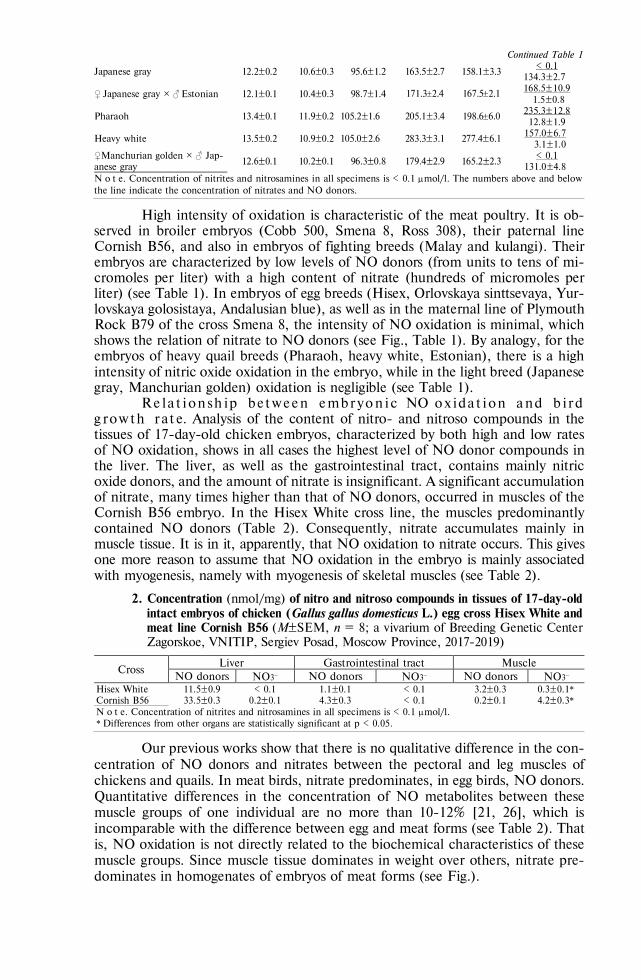
798
Continued Table 1
Japanese gray 12.2±0.2 10.6±0.3 95.6±1.2 163.5±2.7 158.1±3.3 < 0.1
134.3±2.7
♀ Japanese gray ½ ♂ Estonian 12.1±0.1 10.4±0.3 98.7±1.4 171.32.4 167.52.1 168.5±10.9 1.5±0.8
Pharaoh 13.4±0.1 11.9±0.2 105.2±1.6 205.1±3.4 198.66.0 235.3±12.8 12.8±1.9
Heavy white 13.5±0.2 10.9±0.2 105.0±2.6 283.3±3.1 277.4±6.1 157.0±6.7
3.1±1.0 ♀Manchurian golden ½ ♂ Jap-anese gray ♀
12.6±0.1 10.2±0.1 96.3±0.8 179.4±2.9 165.2±2.3 < 0.1
131.0±4.8 N o t е. Concentration of nitrites and nitrosamines in all specimens is < 0.1 mol/l. The numbers above and below the line indicate the concentration of nitrates and NO donors.
High intensity of oxidation is characteristic of the meat poultry. It is ob-served in broiler embryos (Cobb 500, Smena 8, Ross 308), their paternal line Cornish B56, and also in embryos of fighting breeds (Malay and kulangi). Their embryos are characterized by low levels of NO donors (from units to tens of mi-cromoles per liter) with a high content of nitrate (hundreds of micromoles per liter) (see Table 1). In embryos of egg breeds (Hisex, Orlovskaya sinttsevaya, Yur-lovskaya golosistaya, Andalusian blue), as well as in the maternal line of Plymouth Rock B79 of the cross Smena 8, the intensity of NO oxidation is minimal, which shows the relation of nitrate to NO donors (see Fig., Table 1). By analogy, for the embryos of heavy quail breeds (Pharaoh, heavy white, Estonian), there is a high intensity of nitric oxide oxidation in the embryo, while in the light breed (Japanese gray, Manchurian golden) oxidation is negligible (see Table 1).
Re la t ion sh ip be tween embryon ic NO ox ida t ion and b ir d g row th r a t e. Analysis of the content of nitro- and nitroso compounds in the tissues of 17-day-old chicken embryos, characterized by both high and low rates of NO oxidation, shows in all cases the highest level of NO donor compounds in the liver. The liver, as well as the gastrointestinal tract, contains mainly nitric oxide donors, and the amount of nitrate is insignificant. A significant accumulation of nitrate, many times higher than that of NO donors, occurred in muscles of the Cornish B56 embryo. In the Hisex White cross line, the muscles predominantly contained NO donors (Table 2). Consequently, nitrate accumulates mainly in muscle tissue. It is in it, apparently, that NO oxidation to nitrate occurs. This gives one more reason to assume that NO oxidation in the embryo is mainly associated with myogenesis, namely with myogenesis of skeletal muscles (see Table 2).
2. Concentration (nmol/mg) of nitro and nitroso compounds in tissues of 17-day-old intact embryos of chicken (Gallus gallus domesticus L.) egg cross Hisex White and meat line Cornish B56 (M±SEM, n = 8; a vivarium of Breeding Genetic Center Zagorskoe, VNITIP, Sergiev Posad, Moscow Province, 2017-2019)
Cross Liver Gastrointestinal tract Muscle
NO donors NO3 NO donors NO3 NO donors NO3 Hisex White 11.5±0.9 < 0.1 1.1±0.1 < 0.1 3.2±0.3 0.3±0.1* Cornish B56 33.5±0.3 0.2±0.1 4.3±0.3 < 0.1 0.2±0.1 4.2±0.3* N o t е. Concentration of nitrites and nitrosamines in all specimens is < 0.1 mol/l. * Differences from other organs are statistically significant at р < 0.05.
Our previous works show that there is no qualitative difference in the con-centration of NO donors and nitrates between the pectoral and leg muscles of chickens and quails. In meat birds, nitrate predominates, in egg birds, NO donors. Quantitative differences in the concentration of NO metabolites between these muscle groups of one individual are no more than 10-12% [21, 26], which is incomparable with the difference between egg and meat forms (see Table 2). That is, NO oxidation is not directly related to the biochemical characteristics of these muscle groups. Since muscle tissue dominates in weight over others, nitrate pre-dominates in homogenates of embryos of meat forms (see Fig.).

799
We found that 8-15 days after hatching, the concentration of NO donors and nitrate in chick tissues sharply decreases and becomes approximately the same in all breeds, lines and crosses [21]. That is, a high content of nitro- and nitroso compounds and intensive oxidation of NO donors to nitrates in muscle tissue are embryonic phenomena. It was also shown that the development of muscle tissue in embryos with high and low rates of NO oxidation proceeds at the same rate and does not have a qualitative difference in any parameters [26]. The difference may be in the intensity of muscle growth. But how can you evaluate it? Relation of gutted carcasses weight of broilers to their body weight is no than 5% more than that index of egg breeds [21]. It was the live weight that we decided to use as a criterion for the growth of muscle and bone tissue.
On day 1 after hatching, chickens of different breeds differ slightly in live weight (see Table 1). The increase in weight in embryos with high and low rates of embryonic NO oxidation, based on the data in Table 3, also occurs without significant differences. However, after hatching, the chickens show significant dif-ferences in growth (see Table 1).
3. Weigh (g) of intact quail (Coturnix coturnix L.) embryos during incubation (M±SEM, n = 8; a vivarium of Breeding Genetic Center Zagorskoe, VNITIP, Sergiev Posad, Moscow Province, 2017-2019)
Breed Days on incubation
8 10 12 15 day 1 after hatching Manchurian golden 0.53±0.02 1.23±0.08 2.63±2.1 5.32±3.1 11.7±0.2 Pharaoh 0.57±0.02 1.20±0.10 2.75±2.1 5.34±3.4 11.9±0.2
The difference in live weight and its gain between broilers and egg chick-ens, as well as between heavy and light quail breeds, is obvious (see Table 1). But Cornish and Plymouth Rock, the paternal and maternal lines of the Smena 8 cross with high and low rates of embryonic NO oxidation, respectively, differed insig-nificantly in terms of live weight. A significant difference (p < 0.05) occurred only on week 3 after hatching. There was no significant difference in live weight be-tween fighting breeds and egg breeds Yurlovskaya golosistaya and Hisex White (see Table 1).
Quails of heavy (Pharaoh, white heavy, Estonian) and light (Japanese gray, Manchurian golden) breeds clearly differed in live weight (p < 0.05). In the em-bryos of hybrids of Japanese gray and Estonian, as well as Manchurian golden and Estonian breeds, as follows from the proportion of nitrate and NO donors on day 7, embryonic oxidation of nitric oxide occurs with a high intensity (it is not lower than in embryos of the Estonian breed). In terms of live weight, these hybrids occupied an intermediate position between the parental breeds, while the hybrids of the Manchurian golden and Japanese gray breeds were similar in live weight and the rate of NO oxidation to their parents (see Table 1).
Thence two questions arise. What is the mechanism of the relationship between NO oxidation and the rate of weight gain resulted primarily from the development of bone and muscle tissue? And can this process be controlled to increase meat productivity?
Exogenous NO donors, introduced into eggs before incubation, underwent oxidation to nitrate in those embryos where there was intense oxidation of endog-enously synthesized NO (Ross 308), and practically did not oxidize where the latter was not oxidized (Hisex White) (Table 4).
It means that the intensity of oxidation is determined by certain features of the embryo tissues. The data in Table 2 allow us to assume that oxidation occurs in muscle tissue. These features are determined genetically, since they are inherent in specific breeds, lines and crosses regardless of the age of the laying hen, the

800
conditions of its keeping and the conditions of eggs incubation [21], and can also be fully or partially inherited (see Table 1). The intensity of post-embryonic growth is somehow connected with these features. In this case, as an explanation, it can be suggested that nitric oxide is intensively oxidized in the embryo, and pheno-typic manifestations associated with this process are observed after hatching (see Table 1).
4. Concentration (mol/l) of NO donors and NO3 in homogenates of 7-day-old em-bryos in chicken (Gallus gallus domesticus L.) crosses, as influenced by an exoge-nous blocker of NO synthesis and DNIC/GSH applied prior to incubation (M±SEM, n =25 for each group; a vivarium of Breeding Genetic Center Zagor-skoe, VNITIP, Sergiev Posad, Moscow Province, 2017-2019)
Treatment NO donors NO3 Cross Hisex White:
+ 0.3 ml saline (control) 138.3±7.8 < 0.1 + 0.3 ml 13.0 мМ DNIC/GSH 210.9±9.4* < 0.1 + 0.3 ml 6.0 мМ NA 39.8±3.1* < 0.1
Cross Ross 308: + 0.3 ml saline (control) 3.7±1.2 131.2±7.5 + 0.3 ml 5.0 мМ DNIC/GSH 3.9±1.3 165.0±8.3* + 0.3 ml 13.0 мМ DNIC/GSH 34.1±2.9 179.6±8.1* + 0.3 ml 6.0 мМ NA 3.6±1.2 46.7±4.2*
N o t е. NA — Nω-nitro-L-arginine, DNIC/GSH — dinitrosyl-iron-di-glutathione. Concentration of nitrites and nitrosamines in all specimens is < 0.1 mol/l. * Differences from corresponding controls are statistically significant at р < 0.05.
Thus, NO oxidation serves as a marker of the presence of such features, which, apparently, can manifest themselves not only in the embryonic, but also in the postembryonic period. This marker can be used both to determine phenotyp-ically unexpressed forms [27] and, possibly, to control the breeding process.
Consequently, if the intensity of endogenous NO oxidation is low, then the exogenous NO donor compounds, most likely, should not have any effect. Or, if a similar effect does occur, then it is not associated with NO. According to our data, exogenous NO donors, when injected into the embryo of both egg and meat chickens at the concentrations used in Table 4, did not have a significant effect on the live weight of chickens during the first 3 weeks after hatching (p > 0.05).
The use of NO synthase blockers reduced the amount of nitro and nitroso compounds in the embryo without changing the ratio between NO donors and ni-trate (see Table 4). A 70% decrease in the total concentration of NO donors and nitrates did not significantly affect the increase in live weight in the first three weeks after hatching (p > 0.05). That is, we cannot yet give an unambiguous answer whether the oxidation of NO itself affects myogenesis, or whether this oxidation is a side effect of specific processes in the developing muscles of em-bryos of meat poultry.
So, we revealed that the rate of nitric oxide synthesis in the embryos of birds of the same species is approximately the same, while the rate of NO oxidation to nitrate varies up to several orders of magnitude when comparing egg and meat breeds, lines and crosses. In egg poultry, nitric oxide in embryos accumulates in amounts of up to several hundred micromoles, mainly as NO donor compounds, while in meat poultry, oxidation of NO to nitrate predominates. Within the breed, line and cross, the rate of NO oxidation varies by no more than 10-15%. Oxidation occurs mainly in muscle tissue. Exogenous NO donors are oxidized in the embryo with the same intensity as endogenous ones, and the blocker of NO synthesis, decreasing the total concentration of its metabolites, does not affect the quantita-tive ratio between NO donors and nitrate. Consequently, the intensity of embry-onic oxidation is a biochemical marker of gene expression that determines the rate of muscle growth. The identification of these genes is an important scientific and

801
practical task. The possibility of regulation of meat productivity at the embryonic level is seen primarily in the regulation of the expression of these genes.
R E F E R E N C E S
1. Tiwari M., Prasad S., Pandey A., Premkumar K., Tripathi A., Gupta A., Chetan D., Yadav P.,
Shrivastav T., Chaube S. Nitric oxide signaling during meiotic cell cycle regulation in mammalian oocytes. Frontiers in Bioscience (Scholar Edition), 2017, 9: 307-18 (doi: 10.2741/s489).
2. Khan H., Kusakabe K., Wakitani S., Hiyama M., Takeshita A., Kiso Y. Expression and localiza-tion of NO synthase isoenzymes (iNOS and eNOS) in development of the rabbit placenta. J. Reprod. Dev., 2012, 58(2): 231-236 (doi: 10.1262/jrd.11-128t).
3. Von Mandach U., Lauth D., Huch R. Maternal and fetal nitric oxide production in normal and abnormal pregnancy. Journal of Maternal-Fetal and Neonatal Medicine, 2003, 13(1): 22-27 (doi: 10.1080/jmf.13.1.22.27).
4. Blum J., Morel C., Hammon H., Bruckmaier R., Jaggy A., Zurbriggen A., Jungi T. High con-stitutional nitrate status in young cattle. Comparative Biochemistry and Physiology A-Molecular & Integrative Physiology, 2001, 130(2): 271-282 (doi: 10.1016/s1095-6433(01)00390-7).
5. Battaglia C., Ciottii P., Notarangelo L., Fratto R., Facchinetti F., De Aloysio D. Embryonic production of nitric oxide and its role in implantation: a pilot study. Journal of Assisted Reproduc-tion and Genetics, 2003, 20(11): 449-454 (doi: 10.1023/B:JARG.0000006706.21588.0d).
6. Kim Y., Chung H., Simmons R., Billiar T. Cellular non-heme iron content is a determinant of nitric oxide-mediated apoptosis, necrosis, and caspase inhibition. J. Biol. Chem., 2000, 275(15): 10954-10961 (doi: 10.1074/jbc.275.15.10954).
7. Li J., Billiar T., Talanian R., Kim Y. Nitric oxide reversibly inhibits seven members of the caspase family via S-nitrosylation. Biochemical and Biophysical Research Communications, 1997, 240(2): 419-424 (doi: 10.1006/bbrc.1997.7672).
8. Malyshev I., Zenina T., Golubeva L., Saltykova V., Manukhina E., Mikoyan V., Kubrina L., Vanin A. NO-dependent mechanism of adaptation to hypoxia. Nitric Oxide, 1999, 3(2): 105-113 (doi: 10.1006/niox.1999.0213).
9. Cazzato D., Assi E., Moscheni C., Brunelli S., De Palma C., Cervia D., Perrotta C., Clementi E. Nitric oxide drives embryonic myogenesis in chicken through the upregulation of myogenic differ-entiation factors. Experimental Cell Research, 2014, 320 (2): 269-280 (doi: 10.1016/j.yexcr.2013.11.006).
10. Stamler J., Meissner G. Physiology of nitric oxide in skeletal muscle. Physiol Rev., 2001, 81(1): 209-237 (doi: 10.1152/physrev.2001.81.1.209).
11. Ulibarri J., Mozdziak P., Schultz E., Cook C., Best T. Nitric oxide donors, sodium nitroprusside and S-nitroso-N-acetylpencillamine, stimulate myoblast proliferation in vitro. In Vitro Cell Dev. Biol. Anim., 1999, 35(4): 215-218 (doi: 10.1007/s11626-999-0029-1).
12. Long J., Lira V., Soltow Q., Betters J., Sellman J., Criswell D. Arginine supplementation induces myoblast fusion via augmentation of nitric oxide production. J. Muscle Res. Cell Motil., 2006, 27(8): 577-584 (doi: 10.1007/s10974-006-9078-1).
13. Anderson J.E. A role for nitric oxide in muscle repair: nitric oxide–mediated activation of muscle satellite cells. Molecular Biology of the Cell, 2000, 11(5): 1859-1874 (doi: 10.1091/mbc.11.5.1859).
14. Lee H., Baek M., Moon K., Song W., Chung Ch., Ha D., Kang M-S. Nitric Oxide as a messenger molecule for myoblast fusion. J. Biol. Chem., 1994, 269(20): 14371-4.
15. Ribeiro M., Ogando D., Farina M., Franchi A. Epidermal growth factor modulation of prosta-glandins and nitrite biosynthesis in rat fetal membranes. Prostaglandins Leukotrienes and Essential Fatty Acids, 2004, 70(1): 33-40 (doi: 10.1016/j.plefa.2003.08.003).
16. Samengo G., Avik A., Fedor B., Whittaker D., Myung K., Wehling-Henricks M., Tidball J. Age-related loss of nitric oxide synthase in skeletal muscle causes reductions in calpain S-nitrosylation that increase myofibril degradation and sarcopenia. Aging Cell, 2012, 11(6): 1036-1045 (doi: 10.1111/acel.12003).
17. Li Y., Wang Y., Willems E., Willemsen H., Franssens L., Buyse J., Decuypere E., Everaert N. In ovo L-arginine supplementation stimulates myoblast differentiation but negatively affects mus-cle development of broiler chicken after hatching. Journal of Animal Physiology and Animal Nu-trition, 2016, 100(1): 167-177 (doi: 10.1111/jpn.12299).
18. Tirone M., Conti V., Manenti F., Nicolosi P., D’Orlando C., Azzoni E., Brunelli S. Nitric oxide donor molsidomine positively modulates myogenic differentiation of embryonic endothelial pro-genitors. PLoS ONE, 2016, 11(10): e0164893 (doi: 10.1371/journal.pone.0164893).
19. Vanin A. EPR characterization of dinitrosyl iron complexes with thiol-containing ligands as an approach to their identification in biological objects: an overview. Cell Biochem. Biophys., 2018, 76(1): 3-17 (doi: 10.1007/s12013-017-0811-8).
20. Titov V.Yu., Kosenko O.V., Starkova E.S., Kondratov G.V., Borkhunova E.N., Petrov V.A., Osipov A.N. Enzymatic sensor detects some forms of nitric oxide donors undetectable by other methods in living tissues. Bulletin of Experimental Biology and Medicine, 2016, 162(1): 107-110

802
(doi: 10.1007/s10517-016-3557-1) (in Russ.). 21. Titov V.Yu., Dolgorukova A.M., Fisinin V.I., Borkhunova Ye.N., Kondratov G.V., Slesarenko N.A.,
Kochish I.I. The role of nitric oxide (NO) in the body growth rate of birds. World Poultry Science Journal, 2018, 74(4): 675-686 (doi: 10.1017/S0043933918000661).
22. Tarpey M., Wink D., Grisham M. Methods for detection of reactive metabolites of oxygen and nitrogen: in vitro and in vivo considerations. American Journal of Physiology-Regulatory Integrative and Comparative Physiology, 2004, 286(3): R431-R444 (doi: 10.1152/ajpregu.00361.2003).
23. Hickok J., Sahni S., Shen H., Arvind A., Antoniou C., Fung L., Thomas D. Dinitrosyliron complexes are the most abundant nitric oxide-derived cellular adduct. Biological parameters of assembly and disappearance. Free Radic. Biol. Med., 2011, 51(8): 1558-1566 (doi: 10.1016/j.fre-eradbiomed.2011.06.030).
24. Severina I., Bussygina O., Pyatakova N., Malenkova I., Vanin A. Activation of soluble guanylate cyclase by NO donors—S-nitrosothiols, and dinitrosyl-iron complexes with thiol-containing lig-ands. Nitric Oxide, 2003, 8(3): 155-163 (doi: 10.1016/s1089-8603(03)00002-8).
25. Vanin A. Dinitrosyl iron complexes with thiolate ligands: physico-chemistry, biochemistry and physiology. Nitric Oxide, 2009, 21(1): 1-13 (doi: 10.1016/j.niox.2009.03.005).
26. Borkhunova E.N., Kondratov G.V., Titov V.Yu. Rossiiskii veterinarnyi zhurnal. Sel'skokho-zyaistvennye zhivotnye, 2014, 3: 22-30 (in Russ.).
27. Vinnikova E.Z., Titov V.Yu. Ptitsevodstvo, 2008, 12: 33-34 (in Russ.).

804
AGRICULTURAL BIOLOGY, ISSN 2412-0324 (English ed. Online)
2020, V. 55, ¹ 3, pp. 804-815 (SEL’SKOKHOZYAISTVENNAYA BIOLOGIYA) ISSN 0131-6397 (Russian ed. Print)
ISSN 2313-4836 (Russian ed. Online)
Veterinary microbiology UDC 636.52/.58:619:579.62 doi: 10.15389/agrobiology.2020.4.804eng
doi: 10.15389/agrobiology.2020.4.804rus
BIOCHEMICAL, ANTIGENIC AND PROTEOMIC PROPERTIES OF ISOLATES AND STRAINS OF THE CAUSATIVE AGENT OF CHICKEN INFECTIOUS CORYZA Avibacterium paragallinarum (Biberstein and White 1969)
Blackall et al. 2005
А.V. POTEHIN, N.B. SHADROVA, О.V. PRUNTOVA, V.S. RUSALEEV, Val.А. EVGRAFOVA
Federal Center for Animal Health Control, FGBU VNIIZZh, mkr. Yurievets, Vladimir, 600901 Russia, e-mail [email protected], [email protected], [email protected] (corresponding author ), [email protected], [email protected] ORCID: Potehin A.V. orcid.org/0000-0002-3529-4809 Rusaleev V.S. orcid.org/0000-0002-4972-6326 Shadrova N.B. orcid.org/0000-0001-7510-1269 Evgrafova Val.A. orcid.org/0000-0003-3053-6976 Pruntova O.V. orcid.org/0000-0003-3143-7339 The authors declare no conflict of interests Acknowledgements: We are thankful to D.B. Andreychuk, Head of Reference Laboratory for Viral Avian Diseases, ARRIAH, for the assistance in Avibacterium paragallinarum antigenic typing using multiplex-PCR. Received September 4, 2019
A b s t r a c t
Infectious coryza (haemophilus infection) of chickens is a disease reported in many coun-tries of the world. In the Russian Federation, there is no information both about the extent of the disease spread across the poultry farms and about the serotype diversity of the agent circulating in the country. The paper for the first time demonstrates biochemical properties and specifies antigenic relatedness of new Avibacterim paragallinarum isolates recovered from chickens with respiratory signs in Russia and in Belarus. The paper also shows results of creation of the Avibacterim paragallinarum subsection in the database of tested microorganisms’ mass-spectra which can be used as reference ones for the identification and protein profiling of the infectious coryza agent strains and isolates. The work aimed to determine biochemical and antigenic properties of A. paragallinarum isolates and to produce the species-specific mass-spectra as a tool for Avibacterim paragallinarum species identifi-cation and intraspecies differentiation. A. paragallinarum No. 29545 АТСС (serotype А1) form the collection of the Federal Centre for Animal Health (ARRIAH) served as a reference strain. Thirteen A. paragallinarum isolates were used in the study. The isolates were recovered from the pathological material collected from chickens with respiratory pathology (nasal exudates, contents of infraorbital sinus and conjunctival sac, lung tissues) in 2015. The isolates were inoculated onto Columbia agar supplemented with 5 % defibrinated sheep blood, together with a streak of Staphylococcus epidermid-is. Pure cultures of the agent were grown on the serum agar containing NADP at 20 μkg/cm3 and 5 % of horse blood serum. The bacteria were cultured for 24-72 at high CO2 concentration and 37 С. Bilateral antigenic relatedness of the reference strain АТСС No. 29545 and A. paragallinarum isolates was determined using slide agglutination test (SAT). Hemagglutination activity was assessed using hemagglutination inhibition test (HI). Homogeneity of the tested isolates’ serogroup was con-firmed by PCR. Amplified 800 bp DNA fragments were indicative of the presence of serogroup A A. paragallinarum genome, 1000-1100 bp of serogroup В, and 1500-1600 bp of serogroup С. A. para-gallinarum identification was performed using MALDI Autoflex III Biotyper mass-spectrometer (Bruker Daltonik GmbH, Germany). The resulted mass-spectra were recorded, processed and ana-lyzed using FlexControl 3.4 software (Bruker Daltonik Gmb», Germany) according to MALDIBio-typer 2.0. UserManual, Version 2.0 SR1, Germany, 2008. Testing of biochemical properties indi-cated that all 13 isolates of A. paragallinarum form a diverse group. The A. paragallinarum isolates’ growth absolutely depended upon presence of blood serum in the culture medium. Saccharolytic activity of the A. paragallinarum isolates also varied. All isolates and the reference strain can utilize glucose and sucrose, but not lactose, trehalose and galactose. The property also varied for mannitol and mannose. As for antigenic properties, all tested isolates belonged to the same serogroup B that

805
was also confirmed by real-time polymerase chain reaction method. Examination of proteomic prop-erties of A. paragallinarum isolates revealed typical MS peaks, m/z 4768-4770 and 5347-5349, that were similar to those for the reference strain A. paragallinarum ATCC No. 29545. Protein profile mass-spectra were entered into MALDI Autoflex III Biotyper database to improve the reliability of A. paragallinarum species identification. Basing on the examined biochemical, antigenic and proteo-mic properties, three A. paragallinarum isolates were deposited inещ the strain collection of the Fed-eral Centre for Animal Health (ARRIAH) as A. paragallinarum strain No. 1818, A. paragallinarum strain No. 5111, and A. paragallinarum strain No. 1116.
Keywords: strain, isolate, Avibacterium paragallinarum, identification, biochemical charac-terization, antigenic properties, proteomic properties, mass spectrometry, agglutination test, hemag-glutination inhibition assay
Infectious rhinitis of chickens caused by Avibacterium paragallinarum of family Pasteurellaceae, a gram-negative bacterium previously classified as Hae-mophilus paragallinarum, is a disease widespread in industrial poultry farms in Argentina, Africa, Australia, Bangladesh, Brazil, Bulgaria, Indonesia, India, China, Mexico, Malaysia, USA (states of California, Oregon and Alabama), Thailand, and Japan [1-4]. It is characterized by catarrhal inflammation of the mucous membranes of the nasal cavity, respiratory airways and conjunctiva, sub-cutaneous edema of the head, and occasionally pneumonia [5-7]. Infectious rhi-nitis causes significant economic damage to the poultry industry due to growth retardation in chicks and up to 40% loss of egg production in chickens [7]. The mortality rate of young chickens can reach 10% [1]. The extent of the spread of this disease in the poultry farms of the Russian Federation is unknown, as is the serotypic diversity of the pathogen, since the laboratory diagnosis of infectious rhinitis in chickens is not regulated by the relevant documents
Traditionally, the causative agent of the disease is identified by its growth, morphological and biochemical properties. Mass spectrometric methods based on the analysis of protein or lipid fractions of a microbial cell has in-creased the capabilities of veterinary specialists in identifying bacteria. Proteomic methods for determining the species of microorganisms are not inferior to geno-typical ones in many parameters, e.g. in sensitivity and resolution, and have a clear advantage in the cost of consumables, the speed of analysis and the ab-sence of the influence of nonspecific DNA on the result. Protein profiling by mass spectrometry is a powerful analytical tool both in fundamental fields and in clinical practice [8, 9]. The use of proteomic methods for the identification and study of the properties of A. paragallinarum isolates, an economically significant but still insufficiently studied pathogen with a complex antigenic structure, al-lows researchers to obtain new knowledge about its biology, which can be used to address practical problems in veterinary medicine.
MALDI-TOF (matrix-assisted laser desorption/ionization, time-of-flight mass spectrometry) is a method for identifying microorganisms, which is based on analysis of a set of proteins that is unique for each species. It consists in the release and ionization of membrane proteins using laser radiation in the presence of an auxiliary substance (matrix) and subsequent separation of ions in a time-of-flight mass analyzer [10, 11]. However, in the MALDI BioTyper identifica-tion database (Bruker Daltonik GmbH, Germany), data on the protein mass spectra of A. paragallinarum species were previously absent, which made it im-possible to use this method to identify A. paragallinarum isolates.
This work reports on creation of a new reference mass spectra database for identification and protein profiling of A. paragallinarum isolates and strains. Also, here we characterize biochemical properties and the antigenic relationship of A. paragallinarum strains isolated by us earlier on poultry farms of the Russian Federation and the Republic of Belarus from chickens with respiratory pathology.

806
The work aimed to examine biochemical and antigenic properties of Avi-bacterium paragallinarum isolates from Russia and Belarus and to obtain mass spectra specific to representatives of the species, followed by their analysis and use for identification and intraspecific differentiation.
Materials and methods. The reference A. paragallinarum strain No. 29545 ATCC (serotype A1) was obtained from the State Collection of Microorganism Strains (Federal Center for Animal Health, ARRIAH). The study involved 13 strains of A. paragallinarum isolated from infraorbital sinuses and conjunctival sac, nasal cavity exudates, and lung tissue of chickens with respiratory pathology from poultry farms of the Russian Federation (Vladimirskaya, Kostromskaya, Moscow, Orenburg, Yaroslavl, Ulyanovsk regions, Republic of Mordovia, Re-public of Tatarstan) and the Republic of Belarus in 2015.
Bacteria were cultured on the following nutrient media with growth-stimulating additives: Columbia broth and agar (Columbia Columbia Broth, Co-lumbia Agar, Becton, Dickinson and Co., USA), Mueller-Hinton agar (HiMedia Laboratories Pvt. Ltd, India), normal equine serum for cultures of microorgan-isms (JSC NPO Mikrogen, Russia), NADP (Bontac Bio-Engineering Co., Ltd, China), Giss medium (NPO Nutrient media, Russia).
A. paragallinarum cultures were isolated from pathological materials by plating on Columbia agar with 5% defibrinated ram blood and a streak of Staph-ylococcus epidermidis as “feeding” bacteria. Pure cultures of the pathogen were grown on serum agar containing 20 μg/cm3 NADP and 5% horse blood serum. Bacteria were cultured for 24-72 h at 37 С and the increased air level of CO2.
Bacterial morphology was examined by light microscopy of Gram stained preparations (EclipseNi-U microscope, Nikon Corporation, Japan, ½1000 mag-nification) [12]. Capsules in bacteria were identified by Gins’s staining [12]. The biochemical properties of the isolates were assessed using commercial API®NH kit (bioMerieux S.A., France) and by plating on Giss media with glucose, su-crose, lactose, mannitol, mannose, trehalose, galactose monocarbohydrates. Cat-alase production was assessed on a glass slide with 3% H2O2solution. Oxidase activity was measured using a commercial API®NH kit (bioMerieux S.A., France) in accordance with the manufacturer’s instructions.
Antigenic relationship between pairs of A. paragallinarum isolates and antigenic properties of the reference strain ATCC No. 29545 were determined in slide agglutination test (SAT) as proposed by L.A. Page [13]. Hemagglutinating activity was assessed in the hemagglutination inhibition test (HI) as described by P.J. Blackall et al. [14]. The degree of bilateral antigenic relationship (R) of A. paragallinarum isolates was calculated using the formula I. Archetti and F.L. Horsfall [15] and expressed as a percentage. The value of R 70% at 3-fold rep-etition of titration was considered as confirmation of the statistically significant relationship of the investigated isolates [15].
The serogroup homogeneity of A. paragallinarum isolates was confirmed by real-time multiplex polymerase chain reaction (RT-PCR, qPCR) (microchip nucleic acid amplifier Ari-aDNA®, Lumex LLC, Russia). Bacterial DNA was extracted using a kit for the isolation of nucleic acids (OOO Biocom, Russia). Thermocycling of the samples was carried out in the following mode: 10 min at 95 °С (1 cycle); at 30 s at 95 °С, 30 s at 56 °С, 90 s at 32 °С (35 cycles). We used four primers proposed by R. Sakamoto et al. [16] (OOO Beagle, St. Peters-burg): forward primer (common for genome amplification in three serogroups of A. paragallinarum) and three reverse primers specific for a particular serogroup.
The amplification products were analyzed by electrophoresis in 1.7% agarose gel with ethidium bromide (15 V/cm of the gel length, 45 min) (OOO

807
Lumex, Russia). Electrophoregrams were visualized using TCP-26.LMX transil-luminator (Vilber Lourmat, France) in ultraviolet light ( = 254 nm). Amplified DNA fragments were identified as orange band.
The results were deemed positive when the 800 bp, 1000-1100 bp, and 1500-1600 bp amplicons were detected. The 800 bp fragments indicated the ge-nome of A. paragallinarum serogroup A, 1000-1100 bp of serogroup B, and 1500-1600 bp of serogroup C. The results were considered negative if the indi-cated amplified DNA fragments were not detected or their size did not corre-spond to the given values [16, 17].
MALDI-TOF spectrometry (an Autoflex III Biotyper mass spectrometer, Bruker Daltonik GmbH, Germany) was used to identify A. paragallinarum bac-teria. The direct application mode was used in which single colonies of a fresh culture were introduced into the wells of a metal plate using a sterile loop, and a 1 μl matrix was placed on top. A saturated solution of CHCA (НС-cyano-4-hydroxycinnamic acid) in 50% aqueous acetonitrile with 2.5% trifluoroacetic acid was a matrix. The device was calibrated before each experiment with the Bruker Bacterial Standard (Bruker Daltonik GmbH, Germany) as a calibrant. Mass spectrometric analysis of A. paragallinarum cultures was conducted in a linear laser mode at a frequency of 50 Hz. The analysis parameters were opti-mized for the m/z (mass/charge) mass range from 2000 to 20,000 Da, the re-sultant spectrum from summing 20 single spectra was recorded. The mass spectra were recorded and analyzed using the FlexControl 3.4 and FlexAnalysis 3.0 software (Bruker Daltonik GmbH, Germany) in accordance with MALDIBio-typer 2.0. UserManual, Version 2.0 SR1 (Germany, 2008).
The obtained mass spectra were statistically processed using the Biotyper 3.0 RTC program (Bruker Daltonik GmbH, Germany). The reliability of the identification of microorganisms was assessed by comparing Score values with the data of the mass spectra of the Biotyper 3.0 reference library. The identifica-tion at Score values < 1.7 was considered unreliable. When analyzing the degree of bilateral antigenic relationship (R, %) in replicates, the mean (M) and stand-ard errors of the means (±SEM) were calculated.
Results. On blood agar with a “feeding bacterium” (a source of V-growth factor), the A. paragallinarum isolates after 24 hours of culture looked like small (0.1-0.5 mm) satellite colonies in zone 0.5-1.5 cm from the streak of S. epidermidis. With the distance from the feeding culture, the size of the colo-nies decreased until the complete disappearance of growth. Satellite colonies were gray-white, had a smooth convex surface without a hemolysis zone and a rounded shape with smooth edges.
When comparing isolates with the reference A. paragallinarum strain No. 29545 ATCC, we assessed the culture growth on blood serum-free nutrient media, under an increased carbon dioxide in the atmosphere, the ability to uti-lize carbohydrates and to produce various enzymes and metabolites. The results allowed conclusion that the isolates and the reference strain form a heteroge-neous group in terms of growth properties and enzymatic activity (Table 1). The reference A. paragallinarum strain and all the isolates were NAD-dependent though the NAD-independent isolates of the pathogen are also known [18]. All A. paragallinarum isolates can reduce nitrates to nitrites and did not produce ox-idase, -fructosidase, β-galactosidase, indole, hydrogen sulfide, urease, and cata-lase. We found the dependence of the growth on the blood serum in the nutrient medium to be absolute. All A. paragallinarum strains we tested failed to grow on serum-free medium even at the optimal V-factor concentration. There is a view-point that an increased carbon dioxide in the atmosphere is obligatory for

808
A. paragallinarum growth [18-20]. In our test, only 9 of 13 isolates (Nos. 1, 4, 5, 7, 8, 9, 10 and 11) showed such dependence. The morphology and size of the colonies in the four isolates grown in a normal air atmosphere did not differ in any way from those in the colonies under increased CO2 concentration. Ability of the A. paragallinarum strains to metabolize sugars was also unequal.
1. Growth and biochemical traits of the reference strain Avibacterium paragallinar-um No. 29545 АТСС and A. paragallinarum isolates (Russia and Belarus, 2015) from chickens with respiratory pathology
Свойства бактерий Strain, isolate No.
1 2 3 4 5 6 7 8 9 10 11 12 13 29545 Need in:
V growth factor
+
+
+
+
+
+
+
+
+
+
+
+
+
+ СО2 + + + + + + + + + + blood serum + + + + + + + + + + + + + +
Reduction of nitrates + + + + + + + + + + + + + + Production:
indole hydrogen sulfide urease catalase oxidase -fructosidase β-galactosidase
Fermentation: glucose
+
+
+
+
+
+
+
+
+
+
+
+
+
+
sucrose + + + + + + + + + + + + + + lactose mannitol + + + + + + + + + mannose + + + + + + + trehalose galactose
N o t е. «+» means positive test, «» means negative test.
All isolates and the reference strain fermented sucrose and glucose, but not trehalose, lactose and galactose. The trait was variability toward mannose and mannitol, as it was also reported by other researchers [20-22].
For the identification of A. paragallinarum isolates, it was important to de-termine the antigenic relationship in SAT. The test did not reveal the antigenic relationship of the reference strain and the isolates. The A. paragallinarum isolates formed a homologous group with the exception of Nos. 6, 7, 10, and 11. The an-tigens of these isolates showed only one-sided relationship with the tested sera, which did not allow conclusion that they belong to different serological groups.
We also determined the antigenic relationship of A. paragallinarum iso-lates in the hemagglutination inhibition test (Table 2) to compare the results of serotyping for agglutination and hemagglutination [23]. Our data indicate that all the studied isolates were a homogeneous in hemagglutinin. The minimum value of two-sided relationship (R) was 78.4%. That is, it can be assumed that all iso-lates belonged to one serogroup and one serotype.
Based on the obtained results of the similarity of the isolates in growth, antigenic and morphological properties, for further work 10 most promising isolates were selected, which retained the stability of hemagglutinating, anti-genic, virulent and immunogenic properties for 20 consecutive passages.
The A. paragallinarum serotyping by PCR analysis was shown by R. Sa-kamoto et al. [16] and V.V. Patil et al. [17]. In our work, the homogeneity of the serogroup of the isolates from birds with respiratory pathology was confirmed using multiplex qPCR (Fig. 1). In all isolates, we revealed 1000-1100 bp ampli-cons which correspond to the PCR amplification product characteristic of A. para-gallinarum serogroup B.

809
2. A
ntig
enic
rel
atio
nshi
p (R
, %
) of
Avi
bact
eriu
m p
arag
allina
rum
iso
late
s (R
ussia
and
Bel
arus
, 20
15)
from
chi
cken
s with
res
pira
tory
pat
holo
gy (
he-
mag
glut
inat
ion
inhi
bition
tes
t, n
= 3
, М
±SE
M)
No.
of isol
ate
Ser
um s
peci
fic
to a
ntige
n N
o.
1 2
3 4
5 6
7 8
9 10
11
12
13
1
100
100
96.8
±2.
0 86
.8±
2.0
92.4
±1.
2 86
.4±
2.6
88.8
±2.
3 92
.4±
2.3
96.7
±1.
2 96
.2±
0.2
87.7
±2.
2 97
.7±
1.1
90.4
±0.
6 2
92.6
±2.
0 10
0 94
.6±
0.8
92.6
±1.
8 98
.2±
0.4
94.6
±3.
0 86
.6±
2.6
100
90.2
±0.
9 82
.4±
1.8
85.2
±0.
8 89
.4±
1.0
89.1
±0.
9 3
98.6
±1.
2 96
.4±
1.2
100
100
84.2
±1.
0 94
.8±
0.5
90.4
±1.
2 98
.4±
1.2
86.2
±1.
5 84
.8±
1.2
86.6
±2.
0 87
.9±
0.4
86.4
±1.
6 4
100
94.8
±2.
0 95
.2±
3.0
100
94.2
±1.
6 88
.4±
2.8
96.8
±0.
5 10
0 94
.6±
1.2
98.8
±0.
1 88
.0±
2.0
91.6
±0.
4 10
0 5
87.6
±2.
0 93
.0±
2.0
87.4
±2.
8 97
.8±
2.0
100
98.2
±1.
2 84
.6±
2.0
90.2
±2.
2 97
.2±
1.0
100
79.9
±1.
9 92
.5±
0.8
96.2
±0.
8 6
90.2
±1.
3 10
0 79
.6±
2.1
96.2
±2.
0 86
.2±
1.0
100
88.2
±2.
2 92
.6±
1.8
100
96.4
±0.
8 89
.4±
1.3
92.7
±0.
6 94
.3±
0.2
7 94
.6±
2.1
96.6
±2.
4 91
.3±
3.4
100
96.8
±0.
5 10
0 10
0 98
.2±
1.0
99.3
±0.
5 86
.8±
1.8
94.4
±1.
0 94
.8±
0.2
82.3
±0.
3 8
88.2
±0.
6 10
0 96
.4±
1.0
96.3
±1.
7 98
.4±
1.0
92.8
±2.
0 96
.8±
1.2
100
100
88.2
±1.
6 10
0 90
.0±
1.5
88.6
±0.
2 9
86.6
±0.
5 90
.8±
1.8
87.8
±2.
6 78
.4±
0.2
92.6
±0.
8 94
.8±
0.5
94.2
±1.
3 96
.2±
0.9
100
100
92.2
±0.
9 80
.2±
1.6
84.3
±0.
4 10
96
.6±
2.0
92.4
±2.
2 92
.8±
2.0
86.4
±1.
0 88
.4±
2.6
86.2
±0.
2 98
.0±
1.0
100
98.4
±0.
4 10
0 91
.8±
0.6
79.8
±0.
2 92
.0±
1.5
11
78.9
±1.
3 89
.3±
3.0
92.5
±0.
5 89
.9±
1.1
94.4
±2.
4 10
0 79
.0±
2.0
88.3
±0.
4 90
.2±
2.0
97.2
±0.
4 10
0 81
.6±
0.2
85.5
±0.
5 12
80
.3±
2.2
93.5
±0.
5 88
.5±
2.4
94.2
±2.
0 89
.6±
0.5
88.5
±0.
6 84
.2±
2.1
92.3
±1.
3 10
0 91
.8±
0.8
88.3
±0.
9 10
0 89
.9±
0.5
13
86.2
±1.
9 84
.2±
0.2
87.3
±2.
7 98
.8±
0.8
88.1
±1.
9 78
.6±
1.0
88.4
±2.
0 87
.2±
2.0
94.3
±1.
6 94
.3±
0.5
91.1
±1.
1 95
.1±
0.6
100

810
Fig. 1. Serotyping reference strain Avibacterium paragallinarum No. 29545 ATCC and A. paragalli-narum isolates (Russia and Belarus, 2015) from chickens with respiratory pathology by multiplex qPCR: a — serogroup С (1500-1600 bp), b — serogroup B (1000-1100 bp), c — serogroup А (800 bp); 1-10 — isolates Nos., А — strain A. paragallinarum No. 29545, М — DNA fragment length marker (Zao Rvrogen, Russia).
Based on the morphological, biochemical and antigenic characteristics, three isolates of A. paragallinarum were deposited in the State Collection of Mi-croorganism Strains (ARRIAH), isolate No. 4 as strain No. 1818, isolate No. 8 as strain No. 5111, and isolate No. 12 as strain No. 1116.
Fig. 2. Mass-spectra of individual colonies of reference strain Avibacterium paragallinarum No. 29545 ATCC (A) accession strains Nos. 1818 (B), 1116 (C), 5111 (D) derived from A. paragallinarum iso-lates (Russia and Belarus, 2015) from chickens with respiratory pathology (peaks of m/z 4667 and 9334 were common for all strains of A. paragallinarum).

811
The next stage of our research was generation of a database of mass spectra for accession strains A. paragallinarum No. 5111, No. 1818 and No. 1116 and the reference strain ATCC 29545, which are supposed for future automated identification and profiling representatives of this species based on similari-ties/differences in mass spectrometric characteristics.
For strains of A. paragallinarum, protein profiles were constructed (Fig. 2) and mass lists were formed to determine characteristic peaks (Table 3). We compared the positions of the peaks, their frequency and intensity to identify strains and isolates of A. paragallinarum as per the protocol by J.H.K Chen et al. [24] and N. Takeuchi et al. [25].
Analysis of the obtained spectra revealed that they were in the range of m/z 2000-10000. Peaks for m/z 4667 and 9334 turned out to be common for all strains and isolates of A. paragallinarum. A unique feature of the MALDI Bio-typer program is the option to replenish the personal database with new spectra and analyze mass spectra based on comparing the obtained protein profiles with a personal library of reference spectra. A result of this work is creation of the corresponding subsection of this database of mass spectra at the level of A. para-gallinarum species, which can be used in the future to identify strains and iso-lates of the infectious rhinitis pathogen in chickens.
3. Proteomic characterization of Avibacterium paragallinarum strains and isolates (Russia and Belarus, 2015) from chickens with respiratory pathology
Peaks, m/z S/N intensity Peak frequency, % Presence or absence of the peak in A. para-gallinarum ATCC 29545/ratio of S/N
2067-2069 4.4-12.4 43 3109-3111 4.1-5.9 36 3208-3210 4.0-8.6 57 3580-3582 4.0-10.1 71 +/6.2 3930-3932 4.3-9.5 14 4186-4188 4.3-8.1 71 +/6.6 4278-4280 4.6-11.7 36 4433-4435 4.4-7.6 57 +/5.1 4480-4482 4.0-7.9 28 +/6.1 4667 9.2-39.7 100 +/19.3 4768-4770 4.2-7.6 57 +/5.2 5011 4.9-8.8 86 +/5.7 5065 4.8-9.6 78 +/6.4 5098-5060 8.7-16.7 57 5347-5349 4.3-5.3 28 +/4.2 5542-5543 5.1-7.7 21 5567-5569 4.4-7.7 64 6084 5.0-14.7 78 6261-6263 4.2-17.7 50 6420 6.4-24.7 86 +/13.9 6552-6553 4.3-16.8 36 +/16.8 6643-6645 4.3-5.7 21 6855-6857 4.3-5.1 28 7164 10.7-22.7 93 +/15.6 7308-7310 7.6-11.2 50 7864-7866 5.9-12.2 57 8373-8375 6.9-10.7 64 8522-8524 4.4-6.7 28 9317-9319 10.0-10.7 14 9334 24.0-62.3 100 +/33.2 9536-9538 7.2-12.3 71 9964-9965 10.1-13.0 50
Thus, the revealed biochemical properties indicate a heterogenicity of Avibacterium paragallinarum isolates from chickens with respiratory pathology from poultry farms in regions of Russia and Belarus, while the hemagglutination inhibition test indicates their belonging not only to one serogroup, but also to

812
one serotype. The homology of the isolates at the group level is confirmed by multiplex qPCR-based serotyping. We identified characteristic peaks (m/z 4768-4770 and 5347-5349) in proteomes of the isolates similar to those in the reference A. paragallinarum ATCC strain No. 29545. These peaks are added to the mass spectrometer database. This expands the existing database with new spectra and profiles obtained for the tested strains, which improves the reliability of species identification. Based on the studied biochemical, antigenic and proteomic char-acteristics, three isolates of A. paragallinarum were deposited in the State Collec-tion of Microorganism Strains of the Federal Center for Animal Health as strain No. 1818, strain No. 5111, and strain No. 1116.
R E F E R E N C E S
1. Rozhdestvenskaya T.N., Kononenko E.V., Emel'yanova S.A., Yakovlev S.S., Teimurazov M.G.,
Svetoch E.A., Tazina O.I., Platonov M.E., Detushev K.V., Khatyushin Yu.I. Ptitsa i ptitsepro-dukty, 2016, 4: 50-53 (in Russ.).
2. Chukiatsiri K., Chotinun S., Chansiripornchai N. An outbreak of Avibacterium paragallinarum serovar B in a Thai layer farm. The Thai Journal of Veterinary Medicine, 2010, 40(4): 441-444.
3. Poernomo S., Sutarma S., Rafiee M., Blackall P.J. Characterisation of isolates of Haemophilus para-gallinarum from Indonesia. Australian Veterinary Journal, 2000, 78(11): 759-762 (doi: 10.1111/j.1751-0813.2000.tb10447.x).
4. Akhter S., Ali M., Das P.M., Hossain M.M. Isolation and identification of Avibacterium para-gallinarum the causal agent of infectious coryza (IC) from layer chickens in Bangladesh. Journal of the Bangladesh Agricultural University, 2014, 11: 87-96 (doi: 10.3329/jbau.v11i1.18218).
5. Vargas E.S., Terzolo H.R. Haemophillus paragallinarum: etiology of infectious coryza. Veterinar-ia México, 2004, 35(3): 245-259.
6. Byarugaba D.K., Minga U.M., Gwakisa P.S., Katunguka-Rwakishaya E., Bisgaard M., Olsen J.E. Virulence characterization of Avibacterium paragallinarum isolates from Uganda. Avian Patholo-gy, 2007, 36(1): 35-42 (doi: 10.1080/03079450601102947).
7. Lizun R.P. Diagnostika gemofileza kur (obzor). Ekologiya i zhivotnyi mir, 2016, 1: 42-49 (in Russ.).
8. Lau A.F., Drake S.K., Calhoun L.B., Henderson C.M., Zelazny A.M. Development of a clini-cally comprehensive database and a simple procedure for identification of molds from solid me-dia by matrix-assisted laser desorption ionization-time of flight mass spectrometry. Journal of Clinical Microbiology, 2013, 51(3): 828-834 (doi: 10.1128/JCM.02852-12).
9. Clark A.E., Kaleta E.J., Arora A., Wolk D.M. Matrix-assisted laser desorption ionization-time of flight mass spectrometry: a fundamental shift in the routine practice of clinical microbiology. Clinical Microbiology Reviews, 2013, 26(3): 547-603 (doi: 10.1128/CMR.00072-12).
10. Chalupová J., Raus M., Sedlářová M., Šebela M. Identification of fungal microorganisms by MALDI-TOF mass spectrometry. Biotechnology Advances, 2014, 32(1): 230-241 (doi: 10.1016/j.biotechadv.2013.11.002).
11. Alshawa K., Beretti J.-L., Lacroix C., Feuihade M., Dauphin B., Quesne G., Hassouni N., Hassif X., Bougnoux M.-E. Successful identification of clinical dermatophyte and Neoscytalidi-um species by matrix-assisted laser desorption ionization-time of flight mass spectrometry. Jour-nal of Clinical Microbiology, 2012, 50(7): 2277-2281 (doi: 10.1128/JCM.06634-11).
12. Metody obshchei bakteriologii. Tom 1 /Pod redaktsiei F. Gerkhardta, R. Myurrei, R.N. Kostilo-va, E.V. Nester, V.A. Vud, N.R. Krieg, F.G. Briggs; perevod s angliiskogo pod redaktsiei E.N. Kondrat'evoi, L.V. Kalakutskogo [Methods of general bacteriology. Vol. 1. F. Gerkhardt, R. Myurrei, R.N. Kostilov, E.V. Nester, V.A. Vud, N.R. Krieg, F.G. Briggs (eds.). E.N. Kon-drat’eva, L.V. Kalakutskii (translation eds.)]. Moscow, 1983 (in Russ.).
13. Page L.A. Haemophilus infections in chickens. I. Characteristics of 12 Haemophilus isolates re-covered from diseased chickens. American Journal of Veterinary Research, 1962, 23: 85-95.
14. Blackall P.J., Eaves L.E. Serological classification of Australian and South African isolates of Haemophilus paragallinarum. Australian Veterinary Journal, 1988, 65(11): 362-363 (doi: 10.1111/j.1751-0813.1988.tb14271.x).
15. Archetti I., Horsfall F.L. Persistent antigenic variation of influenza A virus after incomplete neutralization in ovo with heterologous immune serum. The Journal of Experimental Medicine, 1950, 92(5): 441-462 (doi: 10.1084/jem.92.5.441).
16. Sakamoto R., Kino Y., Sakaguchi M. Development of a multiplex PCR and PCR-RFLP meth-od for serotyping of Avibacterium paragallinarum. The Journal of Veterinary Medical Science, 2012, 74(2): 271-273 (doi: 10.1292/jvms.11-0319).
17. Patil V.V., Mishra D., Mane D.V. 16S ribosomal RNA sequencing and molecular serotyping of

813
Avibacterium paragallinarum isolated from Indian field conditions. Veterinary World, 2017, 10(8): 1004-1007 (doi: 10.14202/vetworld.2017.1004-1007).
18. Kumar A., Rawat M., Verma R. Studies on absolute requirement of NAD and reduced oxygen ten-sion for growth of field isolates of Avibacterium paragallinarum of poultry origin. Indian Journal Poultry Science, 2012, 47(1): 90-92.
19. Banani M., Pourbakhsh S.A., Khaki P., Goudarzi H., Moazeni-Joula G., Ghodsian N. Isola-tion, identification and antibiotic sensitivity of Haemophilus paragallinarum isolates from com-mercial layer flocks affected by infectious coryza. Pajouhesh-Va-Sazandegi, 2007, 19(1): 128-135.
20. Patil V.V., Mishra D.N, Mane D.V. Isolation, characterization and serological study of Avibac-terium paragallinarum field isolates from Indian poultry. Journal of Animal and Poultry Sciences, 2016, 5(1): 13-20.
21. Kaur J., Sharma N.S., Gupta K., Singh A. Epidemiological studies on infectious coryza in chickens in Northern India. Indian Journal of Animal Sciences, 2004, 74(5): 462-465.
22. Teimurazov M.G., Platonov M.E., Tazina O.I., Manin T.B. Veterinariya, 2016, 6: 26-29 (in Russ.).
23. Eaves L.E., Rogers D.G., Blackall P.J. Comparison of hemagglutinin and agglutinin schemes for the serological classification of Haemophilus paragallinarum and proposal of a new hemag-glutinin serovar. Journal of Clinical Microbiology, 1989, 27(7): 1510-1513 (doi: 10.1128/jcm.27.7.1510-1513.1989).
24. Chen J.H.K., Cheng V.C.C., Wong C.-P., Wong S.C.Y., Yam W.-C., Yuen K.-Y. Rapid differ-entiation of Haemophilus influenzae and Haemophilus haemolyticus by use of matrix-assisted laser desorption ionization-time of flight mass spectrometry with ClinProTools mass spectrum analy-sis. Journal of Clinical Microbiology, 2017, 55(9): 2679-2685 (doi: 10.1128/JCM.00267-17).
25. Takeuchi N., Segawa S., Ishiwada N., Ohkusu M., Tsuchida S., Satoh M., Matsushita K., Nomura F. Capsular serotyping of Haemophilus influenzae by using matrix-associated laserde-sorption ionization-time of flight massspectrometry. Journal of Infection and Chemotherapy, 2018, 24(7): 510-514 (doi: 10.1016/j.jiac.2018.02.007).

816
AGRICULTURAL BIOLOGY, ISSN 2412-0324 (English ed. Online)
2020, V. 55, ¹ 3, pp. 816-829 (SEL’SKOKHOZYAISTVENNAYA BIOLOGIYA) ISSN 0131-6397 (Russian ed. Print)
ISSN 2313-4836 (Russian ed. Online) UDC 579.62:579.852.11:615.33:575.113 doi: 10.15389/agrobiology.2020.4.816eng
doi: 10.15389/agrobiology.2020.4.816rus
GENOMIC AND PHENOTYPICAL POTENTIAL OF ANTIMICROBIAL ACTIVITY OF A BACILLUS STRAIN Bacillus megaterium В-4801
G.Yu. LAPTEV1, E.A. YILDIRIM2, T.P. DUNYASHEV1, L.A. ILYINA2, D.G. TYURINA2, V.A. FILIPPOVA2, E.A. BRAZHNIK2, N.V. TARLAVIN2, A.V. DUBROVIN2, N.I. NOVIKOVA2, V.K. MELIKIDI2, S.N. BIKONYA2
1Saint Petersburg State Agrarian University, 2, lit A, Peterburgskoe sh., St. Petersburg—Pushkin, 196601 Russia, e-mail [email protected] (corresponding author ), [email protected]; 2JSC «Biotrof+», 19, korp. 1, Zagrebskii bulv., St. Petersburg, 192284 Russia, e-mail [email protected], [email protected], [email protected], [email protected], [email protected], [email protected], [email protected], [email protected], [email protected], [email protected] ORCID: Laptev G.Yu. orcid.org/0000-0002-8795-6659 Brazhnik E.A. orcid.org/0000-0003-2178-9330 Yildirim E.A. orcid.org/0000-0002-5846-4844 Tarlavin N.V. orcid.org/0000-0002-6474-9171 Dunyashev T.P. orcid.org/0000-0002-3918-0948 Dubrovin A.V. orcid.org/0000-0001-8424-4114 Ilyina L.A. orcid.org/0000-0003-2490-6942 Novikova N.I. orcid.org/0000-0002-9647-4184 Tyurina D.G. orcid.org/0000-0001-9001-2432 Melikidi V.K. orcid.org/0000-0002-2883-3974 Filippova V.A. orcid.org/0000-0001-8789-9837 Bikonya S.N. orcid.org/0000-0002-3900-6341 The authors declare no conflict of interests Acknowledgements: Supported financially from the Russian Foundation for Basic Research, grant No. 19-316-90041 “Whole-genome sequencing of bacilli strains isolated from the cicatricial contents of various ruminants” Received June 13, 2020
A b s t r a c t
The genetic determinants of bacterial strains Bacillus sp., which determine the possibility of biosynthesis of various antimicrobial compounds, are of particular scientific interest, since thanks to them these microorganisms are widely used as the basis of probiotics. An important stage in the sys-temic analysis of the mechanisms of probiotic action, in particular the antimicrobial activity of mi-croorganisms, is the reconstruction of its metabolic map, that is, the collection and visualization of all potential cell processes. In this work, for the first time, the potentially inherent genetic mecha-nisms for the synthesis of a number of biologically active substances in the bacterial strain Bacillus megaterium are described, in particular, the possibility of synthesizing kanosamine, a bacteriocin belonging to the aminoglycoside group, which can play an important role in the implementation of probiotic properties due to its pronounced antimicrobial activity. Our goal was to study the antimi-crobial activity of the strain Bacillus megaterium B-4801 against pathogenic and opportunistic bacte-ria, as well as to search for genes associated with antimicrobial activity based on whole genome se-quencing. The B. megaterium B-4801 strain deposited in the collection of OOO BIOTROF+, pos-sesses a pronounced probiotic activity. Its antimicrobial activity against Staphylococcus aureus, Can-dida tropicalis, Clostridium sp., and Escherichia coli was assessed by the method of delayed antago-nism using wells. A DNA library for whole genome sequencing was generated using Nextera XT kit (Illumina, Inc., USA). Nucleotide sequences were determined using a MiSeq instrument (Illumina, Inc., USA) and MiSeq Reagent Kit v3 (300-cycle) (Illumina, Inc., USA). Invalid sequences and adapters were removed using the Trimmomatic-0.38 program. Filtered in length from 50 to 150 bp pair-terminal sequences were assembled de novo using genomic assembler SPAdes-3.11.1. Func-tional annotation of the genome was performed with PROKKA 1.12 and RAST 2.0 programs. The pool of genes associated with antimicrobial activity was assessed and the metabolic map was con-structed using the KEGG Pathway database (http://www.genome.jp/kegg/). The antagonistic ac-tivity of B. megaterium B-4801 against pathogenic and opportunistic microorganisms was revealed by cultural methods. The growth inhibition zones of the test strains ranged from 2±0.15 to 25±1.4 mm. The genome of the B. megaterium B-4801 strain is a single circular chromosome with a size of 6,113,972 bp, containing 37.5 % GC pairs. More than 45 % of B. megaterium B-4801 genes are involved in the transport and metabolism of amino acids, transcription, translation, transport and metabolism of carbohydrates and proteins. The key genetic loci that determine the synthesis of anti-microbial metabolites have been identified. The sequenced genome of the strain contains genes (FabD, FabF, FabG, FabZ, FabI, etc.) associated with the production of proteins involved in the synthesis of aliphatic unsaturated C3-C18 carboxylic acids, in particular, butyric, nylon, caprylic,

817
capric, lauric, myristic, palmitic, stearic, oleic. According to the information accumulated by world science, all these substances have pronounced antimicrobial properties. The whole-genome sequenc-ing also discovered a cluster of genes (Asm22-24, Asm43-45, and Asm47) associated with the bio-synthesis of bacteriocin kanosamin, which belongs to the aminoglycoside group, and polyketide ansamycin antibiotics from the macrolide group. The established probiotic potential indicates the role of the investigated strain as a potential probiotic candidate, in particular for use in animal husbandry. The performed genomic analysis revealed new systems of operons that control the metabolic pathways for the synthesis of antimicrobial substances, which were not previously de-scribed for B. megaterium.
Keywords: whole-genome sequencing, Bacillus megaterium, acid biosynthesis, bacteriocins, antimicrobial activity, kanosamine, ansamycin antibiotics, probiotics
Nowadays, animal husbandry and poultry farming, due to the entry into force of Federal Law No. 280-FZ “On organic products and on amendments to certain legislative acts of the Russian Federation” from January 1, 2020, show a vital interest to natural biological products. According to the law, the use of an-tibiotics is prohibited in the organic farming, with the exception of those permit-ted for use by the national, interstate and international standards of organic pro-duction in force in Russia.
Genetic determinants of Bacillus sp. encoding biosynthesis of various an-timicrobial compounds are of particular interest, since wide application of these microorganisms as biopesticides in crop production and as a basis for therapeutic agents and probiotics in animal husbandry. According to some researchers [1], strains of the genus Bacillus are more promising for probiotics preparations as compared to traditionally used lactobacilli, as Bacillus strains form endospores in the life cycle and, therefore, are more resistant to aggressive factors in the diges-tive tract. Bacillus strains are typical commensal bacteria of the rumen and intes-tines of farm animals [2], where as an increase in the abundance of lactobacilli in the rumen is associated with the occurrence of lactic acidosis, a decrease in pH and a decrease in the number of bacteria synthesizing cellulases [2].
On average, 87% of total pool of antimicrobial metabolites of bacilli are organic acids, alcohols, ketones, alkanes, aldehydes, alkenes, and 13% are other substances, the ribosomal peptides (bacteriocins and enzymes), polyketides, non-ribosomal peptides [3]. A case study of a B. subtilis strain as an example has been shown that at least 4-5% of its genome are operons associated with the synthesis of antimicrobial compounds [4].
Interest in studying the ability of Bacillus sp. to synthesize bacteriocins, peptide and lipopeptide antibiotics is currently rising [5, 6], as information about previously unknown substances appears [7-9]. Many of the Bacillus bacteriocins are classified as lantibiotics, the post-translationally modified peptides widely distributed among various bacterial taxa.
Bacteria of genus Bacillus also produce many unmodified bacteriocins, some of which are similar to pediocin-like bacteriocins of lactobacilli while pep-tide sequences of other bacteriocins are completely new [10] Bacteriocins of Ba-cillus sp. are of practical interest due to the ability to inhibit various pathogenic forms, including gram-negative and gram-positive bacteria, yeast and micromy-cetes [7-9].
The synthesis of organic acids by Bacillus microorganisms is studied to a much lesser extent. There are indications of Bacillus sp. ability to produce lactic acid [11] known for its antimicrobial activity against Escherichia coli, Pseudomo-nas aeruginosa, Salmonella enterica, etc. [12]. Bacillus megaterium ELI 24 strain isolated from soil in Mexico produced significant amounts of succinic acid, which was identified by spectroscopic methods [13]. Chromatographic analysis showed that Bacillus megaterium P1 isolated from soil, depending on the compo-sition of the culture medium, synthesized volatile organic acids (formic, acetic,

818
propionic, and butyric) and non-volatile organic acids (oxalic, malic, succinic, tartaric, and citric) in various combinations [14].
Whole genome sequencing is currently recognized as the most effective technology for detailed genetic characterization of microbial strains, their prop-erties, and metabolic processes [15)]. An important stage in the analysis of mechanisms of probiotic action, in particular the antimicrobial activity of mi-croorganisms, can be the re-construction of its metabolic map, that is, the col-lection and visualization of all processes potentially occurring in the cell. Exper-imental data on antimicrobial activity combined with whole genome sequencing allow researchers to reconstruct models that are as close to virtual organisms as possible. This also allows predicting the relationship of a microorganism with various pathogenic and conditionally pathogenic forms in animal digestive sys-tem in order to create effective probiotic products.
Currently, work is underway to study the genomes of Bacillus sp. strains which are promising for the creation of biological products. P. Li et al. [15] used whole genome sequencing to assess the probiotic potential of the Bacillus sp. DU-106, an active producer of L-lactic acid. Other researchers [16] used this technique to study the genome of Bacillus clausii ENTPro as the base for the production of the probiotic Enterogermina (Sanofi Synthelabo Pvt. Ltd., India). whole genome sequencing in B. megaterium was performed by M. Eppinger et al. [17] who analyzed the strains QM B1551 and DSM319. J.I. Vílchez et al. [18] sequenced genome of TG1-E1 strain which is promising for producation of ferti-lizers. L. Liu et al. [19] studied the genome of the WSH-002 strain promising for the creation of recombinant proteins and vitamins.
This work is the first to disclose in a Bacillus megaterium strain potential-ly inherent genetic mechanisms for synthesis of a number of bioactive substanc-es, in particular, the ability to synthesize kanosamine, a bacteriocin of the group of aminoglycosides, which can play an important role in probiotic properties due to a pronounced antimicrobial activity.
Our goal was to measure antimicrobial activity of Bacillus megaterium B-4801 strain against pathogenic and opportunistic bacteria, and to search for genes associated with antimicrobial activity based on whole genome sequencing.
Materials and methods. B. megaterium B-4801 a strain with a pro-nounced probiotic activity (the collection of OOO BIOTROF+) was used in the study. Test cultures for estimation of antagonistic properties were obtained from the All-Russian collection of non-pathogenic microorganisms for agricultural purposes (the All-Russian Research Institute for Agricultural Microbiology, St. Petersburg—Pushkin).
Antimicrobial activity against pathogenic and opportunistic microorgan-isms (Staphylococcus aureus, Candida tropicalis, Clostridium sp., Escherichia coli) was assessed in vitro by the delayed antagonism method (wells method). A Petri dish with agar medium GRM (State Research Center for Applied Micro-biology and Biotechnology, Russia) containing 7 g/l glucose and culture of opportunistic test strains was dried for 24-48 hours, then B. megaterium B-4801 culture sus-pension (107 CFU/ml, 100 μl) was poured in a well in the center of the dish. After 48 h of incubation at 37±1 С, the zones of growth inhibition of the test strains were measured.
DNA was extracted by standard procedures using the Genomic DNA Purification Kit (Fermentas, Inc., Lithuania) according to the attached instruc-tions [2]. The analysis is based on selective detergent-mediated precipitation of DNA using solutions for lysis of cell walls and DNA precipitation, a solution of 1.2 M sodium chloride, and chloroform.
DNA library for whole genome sequencing was constructed Nextera

819
XT kit (Illumina, Inc., USA). Nucleotide sequences were determined using a MiSeq instrument (Illumina, Inc., USA) with MiSeq Reagent Kit v3 (300-cycle) (Illumina, Inc., USA). Invalid sequences and adapters were removed using the Trimmomatic-0.38 program (https://www.osc.edu/book/ex-port/html/4385) [20]. Paired-end sequences filtered by length not less than 50 to 150 bp were assembled de novo (the SPAdes-3.11.1 genomic assembler, http://cab.spbu.ru/software/spades/) [21]. Functional genome annotation was performed using programs PROK-KA 1.12 (https://github.com/kbase-apps/ProkkaAnnotation) [22] and RAST 2.0 (https://rast.nmpdr.org) [23] The KEGG Pathway database (http://www.ge-nome.jp/kegg/) was used to assess the pool of genes associated with antimi-crobial activity for constructing a metabolic map [24, 25].
Mathematical and statistical processing was carried out using the software packages Microsoft Office Excel 2003, R-Studio (Version 1.1.453) (https://rstudio.com). The mean value for each sample (M) and the standard deviation (±SD) were estimated.
Results. B. megaterium B-4801 strain had a pronounced antagonistic ef-fect against the test cultures of Staphylococcus aureus and Candida tropicalis, with the size of growth inhibition zones of 25±1.4 and 10±0.7 mm, respective-ly (Fig. 1). Clostridium sp. proved to be the most resistant to B. megaterium B-4801 (2±0.15 mm). For Escherichia coli, the size of the growth retardation zone was 5±0.3 mm. This suggests that the B. megaterium B-4801 culture liquid con-tains antimicrobial substances that diffuse into agar.
These results are of great practical importance, since S. aureus is associ-ated with the occurrence of diseases in cattle, primarily mastitis [26]. Previously, we have proved the relationship between the increased abundance of Staphylo-coccus sp. in the rumen and an increase in the number of somatic cells in the milk of dairy cows [2]. C. tropicalis is also pathogenic for cattle, in particular, it can spread through the bloodstream to peripheral organs, moreover, its associa-tion with abortion has been found [27, 28]. These facts allow us to conclude that the B. megaterium B-4801 strain is a promising biocontrol agent for suppressing pathogenic microbiota, in particular, through its introduction into the digestive system of animals.
A B
Fig. 1. Antagonistic properties of Bacillus megaterium В-4801 towards Staphylococcus aureus (A) and Candida tropicalis (B). Petri dishes with colonies of pathogen test cultures, in the center zones of growth suppression are visualized.
The B. megaterium B-4801 genome was annotated using the RAST toolkit under a unique genomic identifier 1404.252. The genome is organized into a single circular chromosome of 6,113,972 bp in size, containing 37.5% GC pairs. The chromosome contains 6324 open reading frames capable of determin-
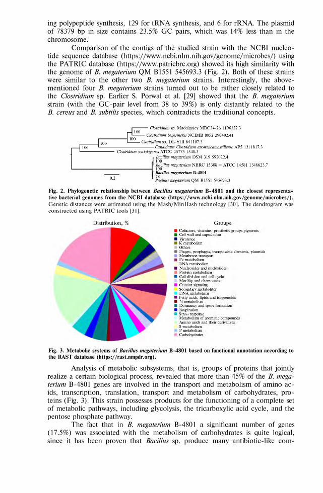
820
ing polypeptide synthesis, 129 for tRNA synthesis, and 6 for rRNA. The plasmid of 78379 bp in size contains 23.5% GC pairs, which was 14% less than in the chromosome.
Comparison of the contigs of the studied strain with the NCBI nucleo-tide sequence database (https://www.ncbi.nlm.nih.gov/genome/microbes/) using the PATRIC database (https://www.patricbrc.org) showed its high similarity with the genome of B. megaterium QM B1551 545693.3 (Fig. 2). Both of these strains were similar to the other two B. megaterium strains. Interestingly, the above-mentioned four B. megaterium strains turned out to be rather closely related to the Clostridium sp. Earlier S. Porwal et al. [29] showed that the B. megaterium strain (with the GC-pair level from 38 to 39%) is only distantly related to the B. cereus and B. subtilis species, which contradicts the traditional concepts.
Fig. 2. Phylogenetic relationship between Bacillus megaterium В-4801 and the closest representa-tive bacterial genomes from the NCBI database (https://www.ncbi.nlm.nih.gov/genome/microbes/). Genetic distances were estimated using the Mash/MinHash technology [30]. The dendrogram was constructed using PATRIC tools [31].
Fig. 3. Metabolic systems of Bacillus megaterium В-4801 based on functional annotation according to the RAST database (https://rast.nmpdr.org).
Analysis of metabolic subsystems, that is, groups of proteins that jointly realize a certain biological process, revealed that more than 45% of the B. mega-terium B-4801 genes are involved in the transport and metabolism of amino ac-ids, transcription, translation, transport and metabolism of carbohydrates, pro-teins (Fig. 3). This strain possesses products for the functioning of a complete set of metabolic pathways, including glycolysis, the tricarboxylic acid cycle, and the pentose phosphate pathway.
The fact that in B. megaterium B-4801 a significant number of genes (17.5%) was associated with the metabolism of carbohydrates is quite logical, since it has been proven that Bacillus sp. produce many antibiotic-like com-
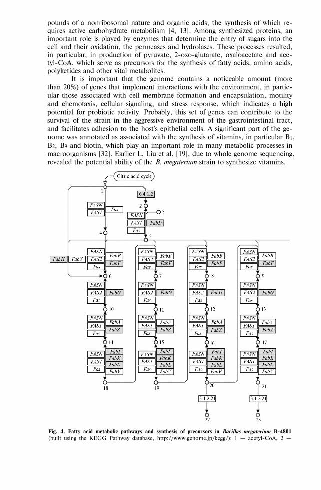
821
pounds of a nonribosomal nature and organic acids, the synthesis of which re-quires active carbohydrate metabolism [4, 13]. Among synthesized proteins, an important role is played by enzymes that determine the entry of sugars into the cell and their oxidation, the permeases and hydrolases. These processes resulted, in particular, in production of pyruvate, 2-oxo-glutarate, oxaloacetate and ace-tyl-CoA, which serve as precursors for the synthesis of fatty acids, amino acids, polyketides and other vital metabolites.
It is important that the genome contains a noticeable amount (more than 20%) of genes that implement interactions with the environment, in partic-ular those associated with cell membrane formation and encapsulation, motility and chemotaxis, cellular signaling, and stress response, which indicates a high potential for probiotic activity. Probably, this set of genes can contribute to the survival of the strain in the aggressive environment of the gastrointestinal tract, and facilitates adhesion to the host's epithelial cells. A significant part of the ge-nome was annotated as associated with the synthesis of vitamins, in particular В1, В2, В9 and biotin, which play an important role in many metabolic processes in macroorganisms [32]. Earlier L. Liu et al. [19], due to whole genome sequencing, revealed the potential ability of the B. megaterium strain to synthesize vitamins.
Fig. 4. Fatty acid metabolic pathways and synthesis of precursors in Bacillus megaterium В-4801 (built using the KEGG Pathway database, http://www.genome.jp/kegg/): 1 — acetyl-CоА, 2 —

822
malonyl-CоА, 3 — acyl carrier protein (ACP), 4 — acetyl-[ACP], 5 — malonyl-[ACP], 6 — acetoacetyl-[ACP], 7 — 3-oxohexanoyl-[ACP], 8 — 3-oxooctanoyl-[ACP], 9 — 3-oxodecanoyl-[ACP], 10 — (R)-3-hydroxibutanoyl-[ACP], 11 — (R)-3-hydroxihexanoyl-[ACP], 12 — (R)-3-hydroxioctanoyl-[ACP], 13 — (R)-3-hydroxidecanoyl-[ACP], 14 — but-2-enoyl-[ACP], 15 — trans-hex-2-enoyl-[ACP], 16 — trans-oct-2-enoyl-[ACP], 17 — trans-dec-2-enoyl -[ACP], 18 — butyryl-[ACP], 19 — hexanoyl-[ACP], 20 — octanoyl-[ACP], 21 — decanoyl-[ACP], 22 — caprylic acid, 23 — capric acid. The numbers in the boxes are the enzyme codes in the IUPAC nomenclature; genes present in the genome are marked in gray; arrows pointing to circles indicate the direction of steps in the reaction.
We identified in B. megaterium B-4801 the key genetic loci that deter-mine the synthesis of a number of antimicrobial metabolites, including fatty ac-ids, kanosamine, which belongs to the aminoglycoside group, and polyketide ansamycin bacteriocins from the group of macrolides. The identification of the key products involved in the pathway for the synthesis of antimicrobial metabo-lites in B. megaterium B-4801 strain was performed using the Kegg Pathway da-tabase (Fig. 4).
The B. megaterium B-4801 genome contained genes (FabD, FabF, FabG, FabZ, FabI, etc.) associated with the production of proteins that are involved in the synthesis of aliphatic unsaturated carboxylic acids C3-C18, in particular, butyric, nylon, caprylic, capric, lauric, myristic, palmitic, stearic, oleic acids. Metabolic maps of the synthesis of some carboxylic acids in the B. megaterium B-4801 strain are shown in Figure 4 as an illustration.
According to the KEGG Pathway database (see Fig. 4), the precursors for the biosynthesis of fatty acids in B. megaterium B-4801 were formed from the acetyl-CoA pool. At the first stage, the acetyl group was transferred from acetyl-CoA to an acyl carrier protein (ACP) molecule. Acetyl-ACP acts as an initiator to which the C2 fragment is attached. The malonyl-ACP molecule also synthesized from acetyl-CoA is a donor of the C2 fragment. This reaction is catalyzed by FabD enzyme (transacylase, EC 2.3.1.39, the malonyl CoA-acyl carrier protein), which was previously shown for Streptomyces coelicolor as, but not described for Bacillus sp. [33].
Further transformations which in B. megaterium B-4801 differed for spe-cific fatty acids we will describe on the example of butyric acid. The C2 frag-ment binding to acetyl-ACP resulted in acetoacetyl-ACP. According to the KEGG Pathway database, FabF protein catalyzed the condensation of acetyl-CoA with malonyl-ACP to form acetoacetyl-ACP. A similar pathway was previ-ously described for Escherichia coli [34]. According to these authors, the synthet-ic pathway leading to the formation of fatty acids in E. coli requires two special-ized proteins, FabA and FabB. However, the genes associated with the synthesis of FabA and FabB are found only in gram-negative Proteobacteria. It was shown [35] that the gram-positive Enterococcus faecalis has a protein annotated as FabZ1, which functionally replaces the E. coli FabA protein. Probably, FabF in B. megaterium B-4801 is similar in function to FabA and FabB of E. coli.
The next stage in the B. megaterium B-4801 involved reduction of oxi-dized carbon atoms in aceto-acetyl-ACP via a series of enzymatic reactions with the participation of FabG, FabZ, FabI (FabK, FabL) proteins. This, through the stages of precursors, resulted in formation of butyryl-ACP, the butyric acid radi-cal. The final stage in the biosynthesis of fatty acids was catalyzed by the enzyme FabI (synonyms FabK, FabL) which initiates the hydration of the 2,3-double bond in the derivatives of enoyl-ACP previously shown for E. coli [36].
Thus, by genome sequencing we identified in B. megaterium B-4801 almost all the main enzymes responsible for the formation of fatty acids (C3-C18). Earlier, in the Bacillus sp. DU-106 [15] enzymes were discovered which

823
were involved in the synthesis of organic acids, in particular, lactate produc-tion. These enzymes were L-lactate dehydrogenase, D-lactate dehydrogenase, lactaldehyde dehydrogenase and malate/lactate dehydrogenase. Note that such enzymes were not detected in B. cereus [37]. Nevertheless, in a later study [15], genes associated with lactate synthesis were identified in the genome of Bacil-lus cereus DU-106.
The data we obtained at the genomic level additionally confirm that the B. megaterium B-4801 strain has potential probiotic properties, since the antimi-crobial activity of these acids has been proven. The pronounced antimicrobial activity of butyric acid against Escherichia coli, Salmonella enterica serovar Typhimurium, Campylobacter jejuni, Clostridium perfringens, Streptococcus pneu-moniae, and Str. suis [38]. A number of fatty acids, in particular nylon, caprylic, capric, lauric and myristic, showed antagonism against Streptococcus mutans, Str. gordonii, Str. sanguis, Candida albicans, Fusobacterium nucleatum, and Porphy-romonas gingivalis [39]. V. Prabhadevi et al. [40] noted the antimicrobial activity of stearic acid, and M.M. Rahman et al. [41] of palmitic acid.
It worth noting that that we revealed in B. megaterium B-4801 a cluster of genes (Asm22-24, Asm43-45, Asm47) associated with biosynthesis of bacterioc-in kanosamine of the aminoglycoside group, and polyketide ansamycin antibiot-ics of the macrolide group. The Asm43-45 genes are involved in biosynthesis of antimicrobial substance kanosamine (3-amino-3-deoxy-D-glucose) through the intermediate UDP--D-kanosamine. S. Umezawa et al. [42] demonstrated a similar pathway for kanosamine biosynthesis in B. pumilus (formerly known as B. aminoglucosidicus), although the genes involved have not been identified. The ability to synthesize kanosamine was also found in Bacillus cereus [43]. Later, in B. cereus, a set of genes involved in the kanosamin synthesis were described, alt-hough the pathway itself was not determined [44). N.D. Vetter et al. [45] dis-covered genes, in particular NtdA, NtdB and NtdC in Bacillus subtilis 168, associ-ated with the biosynthesis of this antimicrobial substance. We are the first to re-veal genetic potential of Bacillus megaterium strains to produce kanosamine.
Kanosamine and UDP--D-canosamine also are intermediates in the biosynthesis of 3-amino-5-hydroxybenzoic acid [46]. 3-Amino-5-hydroxybenzoic acid in microbial cells is a starting block for assembling the main carbon chain of anamycin precursors by modular polyketide synthase I. Ansamycins are a class of bacterial macrocyclic polyketides produced mainly by the phylum Actinobacte-ria and the genus Bacillus [47, 48]. It has been proven [49] that ansamycins ex-hibit a wide spectrum of antimicrobial activity. We discovered in B. megaterium B-4801 the transketolase enzyme, as well as genes, in particular Asm47, Asm23 and Asm24, that can provide synthesis of 3-amino-5-hydroxybenzoic acid. For example, it is well known [50] that Asm23 encodes dehydroquinate dehydratase II (DHQase II) which catalyzes dehydration of 5-deoxy-5-amino-3-dehydro-quinate, a precursor of 3-amino-5-hydroxybenzoic acid. Product of Asm24 gene presumably catalyzes the dehydration of 5-deoxy-5-amino-3-dehydroximate to 3-amino-5-hydroxybenzoic acid. Previously, genes Asm22-24, Asm43-45, and Asm47 were found in Actinosynnema pretiosum ssp. auranticum ATCC 31565, which were associated with the synthesis of 3-amino-5-hydroxybenzoic acid [50].
The probability of bacteriocin production by B. megaterium B-4801 is expectable, since bacteria of the genus Bacillus are known producers of such substances [8]. For example, gene for gallidermin which effectively prevents the formation of biofilms in pathogenic S. aureus and S. epidermidis was identified in B. clausii genome [51]. Lacticin 3147 A2 and leukocyclin Q found in B. amylo-liquefaciens are broad-spectrum bacteriocins. Lacticin has been effectively used
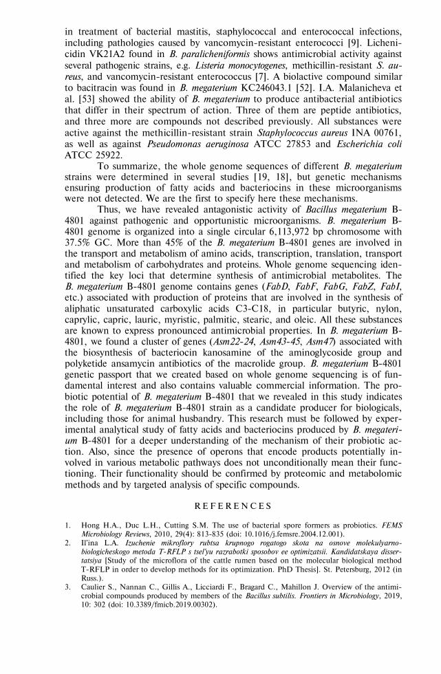
824
in treatment of bacterial mastitis, staphylococcal and enterococcal infections, including pathologies caused by vancomycin-resistant enterococci [9]. Licheni-cidin VK21A2 found in B. paralicheniformis shows antimicrobial activity against several pathogenic strains, e.g. Listeria monocytogenes, methicillin-resistant S. au-reus, and vancomycin-resistant enterococcus [7]. A biolactive compound similar to bacitracin was found in B. megaterium KC246043.1 [52]. I.A. Malanicheva et al. [53] showed the ability of B. megaterium to produce antibacterial antibiotics that differ in their spectrum of action. Three of them are peptide antibiotics, and three more are compounds not described previously. All substances were active against the methicillin-resistant strain Staphylococcus aureus INA 00761, as well as against Pseudomonas aeruginosa ATCC 27853 and Escherichia coli ATCC 25922.
To summarize, the whole genome sequences of different B. megaterium strains were determined in several studies [19, 18], but genetic mechanisms ensuring production of fatty acids and bacteriocins in these microorganisms were not detected. We are the first to specify here these mechanisms.
Thus, we have revealed antagonistic activity of Bacillus megaterium B-4801 against pathogenic and opportunistic microorganisms. B. megaterium B-4801 genome is organized into a single circular 6,113,972 bp chromosome with 37.5% GC. More than 45% of the B. megaterium B-4801 genes are involved in the transport and metabolism of amino acids, transcription, translation, transport and metabolism of carbohydrates and proteins. Whole genome sequencing iden-tified the key loci that determine synthesis of antimicrobial metabolites. The B. megaterium B-4801 genome contains genes (FabD, FabF, FabG, FabZ, FabI, etc.) associated with production of proteins that are involved in the synthesis of aliphatic unsaturated carboxylic acids C3-C18, in particular butyric, nylon, caprylic, capric, lauric, myristic, palmitic, stearic, and oleic. All these substances are known to express pronounced antimicrobial properties. In B. megaterium B-4801, we found a cluster of genes (Asm22-24, Asm43-45, Asm47) associated with the biosynthesis of bacteriocin kanosamine of the aminoglycoside group and polyketide ansamycin antibiotics of the macrolide group. B. megaterium B-4801 genetic passport that we created based on whole genome sequencing is of fun-damental interest and also contains valuable commercial information. The pro-biotic potential of B. megaterium B-4801 that we revealed in this study indicates the role of B. megaterium B-4801 strain as a candidate producer for biologicals, including those for animal husbandry. This research must be followed by exper-imental analytical study of fatty acids and bacteriocins produced by B. megateri-um B-4801 for a deeper understanding of the mechanism of their probiotic ac-tion. Also, since the presence of operons that encode products potentially in-volved in various metabolic pathways does not unconditionally mean their func-tioning. Their functionality should be confirmed by proteomic and metabolomic methods and by targeted analysis of specific compounds.
R E F E R E N C E S
1. Hong H.A., Duc L.H., Cutting S.M. The use of bacterial spore formers as probiotics. FEMS
Microbiology Reviews, 2010, 29(4): 813-835 (doi: 10.1016/j.femsre.2004.12.001). 2. Il'ina L.A. Izuchenie mikroflory rubtsa krupnogo rogatogo skota na osnove molekulyarno-
biologicheskogo metoda T-RFLP s tsel'yu razrabotki sposobov ee optimizatsii. Kandidatskaya disser-tatsiya [Study of the microflora of the cattle rumen based on the molecular biological method T-RFLP in order to develop methods for its optimization. PhD Thesis]. St. Petersburg, 2012 (in Russ.).
3. Caulier S., Nannan C., Gillis A., Licciardi F., Bragard C., Mahillon J. Overview of the antimi-crobial compounds produced by members of the Bacillus subtilis. Frontiers in Microbiology, 2019, 10: 302 (doi: 10.3389/fmicb.2019.00302).

825
4. Stein T. Bacillus subtilis antibiotics: structures, syntheses and specific functions. Molecular Mi-crobiology, 2005, 56(4): 845-857 (doi: 10.1111/j.1365-2958.2005.04587.x).
5. Cotter P.D., Hill C., Ross R.P. What's in a name? Class distinction for bacteriocins. Nature Reviews Microbiology, 2006, 4(2): 160 (doi: 10.1038/nrmicro1273-c2).
6. Gabrielsen C., Brede D.A., Nes I.F., Diep D.B. Circular bacteriocins: biosynthesis and mode of action. Applied and Environmental Microbiology, 2014, 80(22): 6854-6862 (doi: 10.1128/AEM.02284-14).
7. Begley M., Cotter P.D., Hill C., Ross R.P. Identification of a novel two-peptide lantibiotic, lichenicidin, following rational genome mining for LanM proteins. Applied and Environmental Microbiology, 2009, 75(17): 5451-5460 (doi: 10.1128/AEM.00730-09).
8. Saising J., Dube L., Ziebandt A.-K., Voravuthikunchai S.P., Nega M., Götz F. Activity of gallidermin on Staphylococcus aureus and Staphylococcus epidermidis biofilms. Antimicrobial Agents and Chemotherapy, 2012, 56(11): 5804-5810 (doi: 10.1128/AAC.01296-12).
9. Field D., Cotter P.D., Hill C., Ross R.P. Bioengineering lantibiotics for therapeutic success. Frontiers in Microbiology, 2015, 6: 1363 (doi: 10.3389/fmicb.2015.01363).
10. Abriouel H., Franz C.M., Ben Omar N., Gálvez A. Diversity and applications of Bacillus bacteri-ocins. FEMS Microbiology Reviews, 2011, 35(1): 201‐232 (doi: 10.1111/j.1574-6976.2010.00244.x).
11. Poudel P., Tashiro Y., Miyamoto H., Miyamoto H., Okugawa Y., Sakai K. Direct starch fer-mentation to L-lactic acid by a newly isolated thermophilic strain, Bacillus sp. Journal of Indus-trial Microbiology and Biotechnology, 2015, 42: 143-149 (doi: 10.1007/s10295-014-1534-0).
12. Alakomi H.L., Skyttä E., Saarela M., Mattila-Sandholm T., Latva-Kala K., Helander I.M. Lactic acid permeabilizes gram-negative bacteria by disrupting the outer membrane. Applied and Environmental Microbiology, 2000, 66(5): 2001‐2005 (doi: 10.1128/aem.66.5.2001-2005.2000).
13. Ortiz A., Sansinenea E. Succinic acid production as secondary metabolite from Bacillus megaterium ELI24. The Natural Products Journal, 2020, 10(2): 154-157 (doi: 10.2174/2210315509666190410153811).
14. Chuanqing Z., Jiang A.U., Huang A., Qi W. Cao X. Guangxiang studies on the acid-production characteristics of Bacillus megaterium strain P17. AIP Conference Proceedings, 2017, 1839(1): 020054 (doi: 10.1063/1.4982419).
15. Li P., Tian W., Jiang Z., Liang Z., Wu X., Du B. Genomic characterization and probiotic po-tency of Bacillus sp. DU-106, a highly effective producer of L-lactic acid isolated from ferment-ed yogurt. Frontiers in Microbiology, 2018, 9: 2216 (doi: 10.3389/fmicb.2018.02216).
16. Khatri I., Sharma G., Subramanian S. Composite genome sequence of Bacillus clausii, a probi-otic commercially available as Enterogermina®, and insights into its probiotic properties. BMC Microbiology, 2019, 19(1): 307 (doi: 10.1186/s12866-019-1680-7).
17. Eppinger M., Boyke B., Mitrick A.J., Janaka N.E., Kirthi K.K., Koenig S.S.K., Creasy H.H., Rosovitz M.J., Riley D.R., Daugherty S., Martin M., Elbourne L.D.H., Paulsen I., Biedendieck R., Braun C., Grayburn S., Dhingra S., Lukyanchuk V., Ball B., Ul-Qamar R., Seibel J., Bremer E., Jahn D., Ravel J., Vary P.S. Genome sequences of the biotechnologically important Bacillus megaterium strains QM B1551 and DSM319. Journal of Bacteriology, 2011, 193(16): 4199-4213 (doi: 10.1128/JB.00449-11).
18. Vílchez J.I., Tang Q., Kaushal R., Wang W., Suhui L., Danxia H., Zhaoqing C., Heng Z., Renyi L., Huiming Z. Complete genome sequence of Bacillus megaterium strain TG1-E1, a plant drought tolerance-enhancing bacterium. Microbiology Resource Announcements, 2018, 7(12): e00842-18 (doi: 10.1128/MRA.00842-18).
19. Liu L., Li Y., Zhang J., Zou W., Zhou Z., Liu J., Li X., Wang L., Chen J. Complete genome sequence of the industrial strain Bacillus megaterium WSH-002. Journal of Bacteriology, 2011, 193(22): 6389-6390 (doi: 10.1128/JB.06066-11).
20. Bolger A.M., Lohse M., Usadel B. Trimmomatic: a flexible trimmer for Illumina sequence data. Bioinformatics, 2014, 30(15): 2114‐2120 (doi: 10.1093/bioinformatics/btu170).
21. Nurk S., Bankevich A., Antipov D., Gurevich A., Korobeynikov A., Lapidus A., Prjibelsky A., Pyshkin A., Sirotkin A., Sirotkin Y., Stepanauskas R., McLean J., Lasken R., Clingenpeel S.R., Woyke T., Tesler G., Alekseyev M.A., Pevzner P.A. Assembling genomes and mini-metagenomes from highly chimeric reads. In: Research in Computational Molecular Biology. RECOMB 2013. Lecture Notes in Computer Science, vol. 7821. Springer, Berlin, Heidelberg, 2013: 158-170 (doi: 10.1007/978-3-642-37195-0_13).
22. Seemann T. Prokka: rapid prokaryotic genome annotation. Bioinformatics, 2014, 30(14): 2068‐2069 (doi: 10.1093/bioinformatics/btu153).
23. Aziz R.K. The RAST server: rapid annotations using subsystems technology. BMC Genomics, 2008, 9(75) (doi: 10.1186/1471-2164-9-75).
24. Kanehisa M., Goto S. KEGG: Kyoto encyclopedia of genes and genomes. Nucleic Acids Re-search, 2000, 28(1): 27-30 (doi: 10.1093/nar/28.1.27).
25. Kanehisa M., Goto S., Sato Y., Furumichi, M. Tanabe M. KEGG for integration and interpre-tation of large-scale molecular datasets. Nucleic Acids Research, 2012, 40(D1): D109-D114

826
(doi: 10.1093/nar/gkr988). 26. Boss R., Cosandey A., Luini M., Artursson K., Bardiau M., Breitenwieser F., Hehenberger E.,
Lam Th., Mansfeld M., Michel A., Mösslacher G., Naskova J., Nelson S., Podpečan O., Rae-my A., Ryan E., Salat O., Zangerl P., Steiner A., Graber H.U. Bovine Staphylococcus aureus: Subtyping, evolution, and zoonotic transfer. Journal of Dairy Science, 2016, 99(1): 515‐528 (doi: 10.3168/jds.2015-9589).
27. Loken K.I., Thompson E.S., Hoyt H.H., Ball R.A. Infection of the bovine udder with Candida tropicalis. Journal of the American Veterinary Medical Association, 1959, 134(9): 401‐403.
28. Wohlgemuth K., Knudtson W. Bovine abortion associated with Candida tropicalis. Journal of the American Veterinary Medical Association, 1973, 162(6): 460‐461.
29. Porwal S., Lal S., Cheema S., Kalia V.C. Phylogeny in aid of the present and novel microbial lineages: diversity in Bacillus. PLoS ONE, 2009, 4(2): e4438 (doi: 10.1371/journal.pone.0004438).
30. Ondov B.D., Treangen T.J., Melsted P., Mallonee A.B., Bergman N.H., Koren S., Phillip-py A.M. Mash: fast genome and metagenome distance estimation using MinHash. Genome Biol-ogy, 2016, 17: 132 (doi: 10.1186/s13059-016-0997-x).
31. Wattam A.R., Davis J.J., Assaf R., Boisvert S., Brettin T., Bun C., Conrad N., Dietrich E.M., Disz T., Gabbard J.L., Gerdes S., Henry C.S., Kenyon R.W., Machi D., Mao C., Nord-berg E.K., Olsen G.J., Murphy-Olson D.E., Olson R., Overbeek R., Parrello B., Pusch G.D., Shukla M., Vonstein V., Warren A., Xia F., Yoo H., Stevens R.L. Improvements to PATRIC, the all-bacterial bioinformatics database and analysis resource center. Nucleic Acids Research, 2017, 45(D1): D535-D542 (doi: 10.1093/nar/gkw1017).
32. Frye T.M., Williams S.N., Graham T.W. Vitamin deficiencies in cattle. Veterinary Clinics of North America: Food Animal Practice, 1991, 7(1): 217‐275 (doi: 10.1016/s0749-0720(15)30817-3).
33. Szafranska A.E., Hitchman T.S., Cox R.J., Crosby J., Simpson T.J. Kinetic and mechanistic analysis of the malonyl CoA:ACP transacylase from Streptomyces coelicolor indicates a single catalytically competent serine nucleophile at the active site. Biochemistry, 2002, 41(5): 1421-1427 (doi: 10.1021/bi012001p).
34. Cronan J.E., Thomas J. Bacterial fatty acid synthesis and its relationships with polyketide synthetic pathways. Methods in Enzymology, 2009, 459: 395‐433 (doi: 10.1016/S0076-6879(09)04617-5).
35. Wang H., Cronan J.E. Functional replacement of the FabA and FabB proteins of Escherichia coli fatty acid synthesis by Enterococcus faecalis FabZ and FabF homologues. Journal of Biologi-cal Chemistry, 2004, 279(33): 34489‐34495 (doi: 10.1074/jbc.M403874200).
36. Yu X., Liu T., Zhu F., Khosla C. In vitro reconstitution and steady-state analysis of the fatty acid synthase from Escherichia coli. Proceedings of the National Academy of Sciences, 2011, 108(46): 18643-18648 (doi: 10.1073/pnas.1110852108).
37. Johnson S.L., Daligault H.E., Davenport K.W., Jaissle J., Frey K.G., Ladner J. T., Broomall S.M., Bishop-Lilly K.A., Bruce D.C., Gibbons H.S., Coyne S.R., Lo C.-C., Meincke L., Munk A.C., Koroleva G.I., Rosenzweig C.N., Palacios G.F., Redden C.L., Minogue T.D., Chain. P.S. Complete genome sequences for 35 biothreat assay-relevant Bacillus species. Genome Announcements, 2015, 3(2): e00151-15 (doi: 10.1128/genomeA.00151-15).
38. Kovanda L., Zhang W., Wei X., Luo J., Wu X., Atwill E.R., Vaessen S., Li X., Liu Y. In vitro antimicrobial activities of organic acids and their derivatives on several species of gram-negative and gram-positive bacteria. Molecules, 2019, 24(20): 3770 (doi: 10.3390/molecules24203770).
39. Huang C.B., Alimova Y., Myers T.M., Ebersole J.L. Short- and medium-chain fatty acids ex-hibit antimicrobial activity for oral microorganisms. Archives of Oral Biology, 2011, 56(7): 650‐654 (doi: 10.1016/j.archoralbio.2011.01.011).
40. Prabhadevi V., Sahaya S.S., Johnson M., Venkatramani B., Janakiraman N. Phytochemical studies on Allamanda cathartica L. using GC-MS. Asian Pacific Journal of Tropical Biomedicine, 2012, 2(2): S550-S554 (doi: 10.1016/S2221-1691(12)60272-X).
41. Rahman M.M., Ahmad S.H., Mohamed M.T., Ab Rahman M.Z. Antimicrobial compounds from leaf extracts of Jatropha curcas, Psidium guajava, and Andrographis paniculata. Scientific World Journal, 2014, 2014: 635240 (doi: 10.1155/2014/635240).
42. Umezawa S., Shibahara S., Omoto S., Takeuchi T., Umezawa H. Studies on the biosynthesis of 3-amino-3-deoxy-D-glucose. The Journal of Antibiotics, 1968, 21(8): 485-491 (doi: 10.7164/antibiotics.21.485).
43. Milner J.L., Silo-Suh L., Lee J.C., He H., Clardy J., Handelsman J. Production of kanosamine by Bacillus cereus UW85. Applied and Environmental Microbiology, 1996, 62(8): 3061-3065.
44. Kevany B.M., Rasko D.A., Thomas M.G. Characterization of the complete zwittermicin A biosynthesis gene cluster from Bacillus cereus. Applied and Environmental Microbiology, 2009, 75(4): 1144-1155 (doi: 10.1128/AEM.02518-08).
45. Vetter N.D., Langill D.M., Anjum S., Boisvert-Martel J., Jagdhane R.C., Omene E., Zheng H., van Straaten K.E., Asiamah I., Krol E.S., Sanders D.A., Palmer D.R. A previously unrecog-nized kanosamine biosynthesis pathway in Bacillus subtilis. Journal of the American Chemical So-ciety, 2013, 135(16): 5970-5973 (doi: 10.1021/ja4010255).

827
46. Arakawa K., Müller R., Mahmud T., Yu T.W., Floss H.G. Characterization of the early stage aminoshikimate pathway in the formation of 3-amino-5-hydroxybenzoic acid: the RifN protein specifically converts kanosamine into kanosamine 6-phosphate. Journal of the American Chemi-cal Society, 2002, 124(36): 10644-10645 (doi: 10.1021/ja0206339).
47. Kim W.-G., Song N.-K., Yoo I.-D. Trienomycin G, a new inhibitor of nitric oxide production in microglia cells, from Streptomyces sp. 91614. The Journal of Antibiotics, 2002, 55(2): 204-207 (doi: 10.7164/antibiotics.55.204).
48. Kang Q., Shen Y., Bai L. Biosynthesis of 3,5-AHBA-derived natural products. Natural Product Reports, 2012, 29: 243-263 (doi: 10.1039/C2NP00019A).
49. Wrona I.E., Agouridas V., Panek J.S. Design and synthesis of ansamycin antibiotics. Comptes Rendus Chimie, 2008, 11: 1483-1522 (doi: 10.1016/j.crci.2008.07.003).
50. Yu T.W., Bai L., Clade D., Hoffmann D., Toelzer S., Trinh K.Q., Xu J., Moss S.J., Leistner E., Floss H.G. The biosynthetic gene cluster of the maytansinoid antitumor agent an-samitocin from Actinosynnema pretiosum. Proceedings of the National Academy of Sciences, 2002, 99(12): 7968‐7973 (doi: 10.1073/pnas.092697199).
51. Khatri I., Sharma G., Subramanian S. Composite genome sequence of Bacillus clausii, a probi-otic commercially available as Enterogermina®, and insights into its probiotic properties. BMC Microbiology, 2019, 19(1): 307 (doi: 10.1186/s12866-019-1680-7).
52. Al-Thubiani A.S.A., Maher Y.A., Fathi A., Abourehab M.A.S., Alarjah M., Khan M.S.A., Al-Ghamdi S.B. Identification and characterization of a novel antimicrobial peptide compound produced by Bacillus megaterium strain isolated from oral microflora. Saudi Pharmaceutical Journal, 2018, 26(8): 1089‐1097 (doi: 10.1016/j.jsps.2018.05.019).
53. Malanicheva I.A., Kozlov D.G., Sumarukova I.G., Efremenkova O.V., Zenkova V.A., Kat-rukha G.S., Reznikova M.I., Tarasova O.D., Sineokiĭ S.P., Él'-Registan G.I. Antimicrobial activity of Bacillus megaterium strains. Microbiology, 2012, 81: 178-185 (doi: 10.1134/S0026261712020063).

830
AGRICULTURAL BIOLOGY, ISSN 2412-0324 (English ed. Online)
2020, V. 55, ¹ 3, pp. 830-842 (SEL’SKOKHOZYAISTVENNAYA BIOLOGIYA) ISSN 0131-6397 (Russian ed. Print)
ISSN 2313-4836 (Russian ed. Online)
Anthelmintics UDC 619:615.284 doi: 10.15389/agrobiology.2020.4.830eng
doi: 10.15389/agrobiology.2020.4.830rus
BIOLOGICAL ACTIVITY OF FENBENDAZOLE BASED ON SUPRAMOLECULAR DELIVERY SYSTEM WITH DISODIUM
SALT OF GLYCYRRHIZIC ACID
A.I. VARLAMOVA, I.A. ARKHIPOV
Skryabin All-Russian Research Institute of Fundamental and Applied Parasitology of Animals and Plants — Branch of Federal Science Center Kovalenko All-Russian Research Institute of Experimental Veterinary RAS, 28, Bolshaya Cheremu-shkinskaya ul., Moscow, 117218 Russia, e-mail [email protected] (corresponding author ), [email protected] ORCID: Varlamova A.I. orcid.org/0000-0001-8364-5055 Arkhipov I.A. orcid.org/0000-0001-5165-0706 The authors declare no conflict of interests Acknowledgements: The authors are grateful to D.Sc. S.S. Khalikov (Nesmeyanov Institute of Organoelement Compounds RAS) for providing samples of solid dispersions of fenbendazole, and to P.P. Kochetkov (Skryabin All-Russian Research Institute of Fundamental and Applied Parasitology of Animals and Plants) for his help in pharmacokinetic studies. Received November 11, 2019
A b s t r a c t
Due to the wide spread of animal helminthiasis, it becomes necessary to use innovative an-tiparasitic drugs. Fenbendazole is widely used all over the world for chemotherapy of helminthiasis, but in some cases, it is effective only in a high dose. This study, for the first time, has shown changes of physicochemical properties, pharmacokinetic parameters and an increase in the anthelmintic effi-cacy of mechanochemically obtained complexes of fenbendazole with disodium salt of glycyrrhizic acid for targeted delivery. Our research aimed to increase the biological activity of a solid dispersion of fenbendazole with disodium salt of glycyrrhizic acid (SDF with Na2GA), to evaluate the solubility of SDF compositions with Na2GA, pharmacokinetic parameters, and anthelmintic efficacy on labor-atory models of Trichinella spiralis and Hymenolepis nana and in field tests on sheep naturally infect-ed with gastrointestinal nematodes and moniesia. SDF with Na2GA was obtained in one-stage pro-cess in LE-101 ball mill (Hungary). The ratio of fenbendazole (Changzhou Yabong Pharmaceuticals Co., Ltd., China) and disodium salt of glycyrrhizic acid (Yuli County Jinxing Licorice Products Co., China) was 1:10. The process continued for 4 hours at 90 rpm. Pharmacokinetic parameters of fen-bendazole and its metabolites in sheep were studied by high-performance liquid chromatography-tandem mass spectrometry detection. SDF with Na2GA and the substance of fenbendazole (FBZ) was administered to two groups of clinically healthy sheep (5 animals each) once per or at the dose of 2 mg/kg of active substance. Blood samples were taken from the jugular vein 0, 1, 2, 4, 6, 8, 12, 24, 33, 48, 72, and 144 hours after administration of SDF with Na2GA and the basic drug. The ab-sorption rate constant, absorption half-life, clearance of the drug from the blood plasma, maximum drug concentration, time to reach maximum plasma drug concentration following drug administra-tion, elimination half-life, area under the concentrationtime curve and mean residence time were calculated. The efficacy of SDF with Na2GA against Hymenolepis nana and Trichinella spiralis was studied with 50 white inbred female BALB/c mice weighing 16-18 g. The eggs of H. nana were ad-ministered intragastrically with a syringe, 200 eggs per animal. On day 13 after infection, SDF with Na2GA in 1 % starch gel was administered into the stomach of mice of I, II and III groups (10 ani-mals each) at doses of 3.0; 2.0 and 1.0 mg/kg of active substance, respectively. FBZ was the basic drug which was applied at the dose of 2.0 mg/kg (experimental group IV). The animals of the con-trol group received the same volume of the starch gel. Trichinella spiralis was isolated by serial pas-sages of the first stage larvae to female rats. Before infection, the mice were kept on a starvation diet for 12 hours, and then 200 larvae were injected into their stomachs using a tuberculin syringe. On day 3 after infection, the mice were divided into four experimental and one control groups (10 ani-mals each). SDF with Na2GA in 1 % starch gel was administered into the stomachs of mice of ex-perimental groups I, II, and III at doses of 3.0; 2.0 and 1.0 mg/kg of active substance, respectively. The FBZ substance was administered to mice of IV experimental group at the dose of 2 mg/kg. Control group of animals received 1,5 % starch gel at the same dose. The efficacy of the drugs against H. nana and T. spiralis was determined from necropsy data. The anthelmintic activity of SDF with Na2GA was also studied on young Stavropol merino sheep in field tests (LTD Agroresurs, Sa-

831
mara Province, Pestravsky District) in the summer 2016-2017. SDF with Na2GA was administered per or to the animals of the experimental groups (a single application per or at the doses of 3.0; 2.0 and 1.0 mg/kg of active substance vs. FBZ at the dose of 2.0 mg/kg. The control group of animals did not receive the drugs. Anthelmintic activity of drugs was determined according to the data of necrop-sy of the intestines of mice and the results of studies of sheep feces samples by the McMaster meth-od before and after administration of the drugs. The data of physicochemical studies have shown an increase in solubility, a decrease in the particle size of the compositions of SDF with Na2GA, and the formation of irregularly shaped aggregates. The pharmacokinetic parameters indicated a signifi-cant increase in the rate of absorption of SDF with Na2GA and their entry into the blood, a 2,5-fold increase in the maximum concentration of fenbendazole and its metabolites in the blood, as well as a decrease in the rate of drug elimination from the body compared to the FBZ. SDF with Na2GA (3.0; 2.0 and 1.0 mg/kg) showed 100; 98,05 and 92,74 % activity against T. spiralis, 100, 98.67 and 89.04 % against H. nana, 100, 95.37 and 92.07 % against Nematodirus spp., 100, 95.42 and 90.75 % against gastrointestinal strongylates, and 96.44, 91.61 and 81.12 % against Moniezia expansa. The FBZ (2.0 mg/kg) anthelmintic activity was 3.4 times lower than that of the same dose of SDF with Na2GA upon experimental trichinellosis of mice. Its efficacy was 28.88 % against experimental hy-menolepiasis of mice. The FBZ substance showed low efficacy against Nematodirus spp. (33.33 %), other gastrointestinal strongylates (39.14 %) and Moniezia spp. (17.55 %). These findings allow us to conclude that the development of drugs based on fenbendazole solid dispersion with glycyrrhizic acid disodium salt is promising, and the production technology can be scaled up.
Keywords: fenbendazole, solid dispersion, disodium salt of glycyrrhizic acid, efficacy, pharmacokinetics, helminthiasis
Fenbendazole (Panacur) is a broad-spectrum anthelmintic of the group of benzimidazole carbamates [1, 2]. It has high activity against animal nematodes at a dose of 7.5-10 mg/kg, against Protostrongylus spp. at a dose of 15 mg/kg, against Fasciola hepatica and Dicrocoelium lanceatum at a dose of 100 mg/kg [3], being less effective against Trichocephalus spp. and Strongyloides spp. [1]. The mechanism of the anthelmintic action of fenbendazole is the destruction of cytoplasmic microtubules in the cells of the parasite. This is accompanied by impaired absorption and transport of glucose, as well as a decrease in the activity of fumarate reductase, which subsequently leads to the death of hel-minths [2]. At present, fenbendazole is widely used all over the world at the recommended doses [4-7]. According to the Bio-pharmaceutics classification system (BCS) guidance, Food and Drug Administration (FDA, USA) (https://www.fda.gov/), it belongs to the IV class of drugs with low permeabil-ity and poor solubility, that is, it has low bioavailability. Therefore, an increase in its water solubility will affect the anthelmintic properties.
Recently, drug delivery systems (DDS) of biologically active molecules have been widely developed in order to improve the solubility of drugs and their bioavailability by increasing absorption, blood concentration and permea-bility of the drug through biological membranes to the receptors. DDS tech-nologies improve the effectiveness of the drugs, reduce their therapeutic dose and possible side effects. To improve the solubility of drugs, different methods are used, e.g. grinding and changing the shape of the crystal lattice, creating solid dispersions of drugs with fillers, changing the particle size and crystal structure [8-10]. Cyclodextrins, polysaccharides, liposomes, micelles, and na-nosized inorganic particles, which form supramolecular systems, are most often used as drug delivery vehicles [11-13].
Mechanochemical modification of solid medicinal substances and excipi-ents is one of the DDS technologies. Under the influence of pressure and shear deformations in mills, the crystal structure of substances can be disordered until complete amorphization followed by polymorphic transitions and chemical reac-tions with the formation of complexes or micelles with increased solubility [14, 15].
Selyutina et al. [16] found that derivatives of glycyrrhizic acid (GA) are able to integrate into biological membranes of cells, providing lipid motility. Due to its amphiphilicity, GA can form micelles with hydrophobic drug compounds and participate in their transmembrane transfer [17-20].

832
In our previous work we investigated the effect of mechanochemical technology on the anthelminthic efficacy of a solid dispersion of fenbendazole with polyvinylpyrrolidone and revealed its high therapeutic effect in laboratory models of parasitosis and in farm conditions at a reduced dose. The data of physicochemical studies showed about 3-fold increase in the solubility of the resultant dispersion, a decrease in the particle size to 5-20 microns and amor-phization of the fenbendazole substance [21].
Here, we have shown for the first time a change in the physicochemical properties, pharmacokinetic parameters and an increase in the anthelmintic effi-cacy of fenbendazole obtained by mechanochemical treatment using the disodi-um salt of glycyrrhizic acid for its targeted delivery
This paper aimed to increase the biological activity of a solid dispersion of fenbendazole with a disodium salt of glycyrrhizic acid, to assess the solubility of compositions of a solid dispersion of fenbendazole (SDF) with Na2GA, the parameters of pharmacokinetics and anthelmintic efficacy on laboratory models of Trichinella spiralis and Hymenolepis nana in field conditions on sheep natural-ly infected with gastrointestinal nematodes and moniesia.
Materials and methods. Animal experiments followed the Guidelines for Experimental (Preclinical) Study of New Pharmacological Substances [22], the Rules of Good Laboratory Practice of the Russian Federation (Order of the Ministry of Health of the Russian Federation No. 199n of 04/01/2016 “On Ap-proval of the Rules of Good Laboratory Practice”) and the European Conven-tion for the Protection of Vertebrate Animals used for Experimental or Other Scientific Purposes (ETS 123, Strasbourg, 1986).
A solid dispersion of fenbendazole (methyl 5-(phenylthio)-2-benzimida-zole carbamate, 99.0%, molecular weight 299.35) with a disodium salt of glycyrrhizic acid (SDF with Na2GA) was obtained at the Nesmeyanov Institute of Organoelement Compounds RAS (Moscow) at one-stage mechanochemical process in a LE-101 ball mill (Hungary). The ratio of fenbendazole (Changzhou Yabong Pharmaceuticals Co., Ltd., China) and disodium salt of glycyrrhizic acid (Yuli County Jinxing Licorice Products Co., China) in the experiments was 1:10. The process continued for 4 hours at 90 rpm until particle aggregates with a size of 0.1-10 microns were formed.
The solubility of SDF with Na2GA in water was assessed in samples with a component ratio of 1:5, 1:10, and 1:20 obtained after 4 hours of mechanochemical treatment, and in the initial fenbendazole substance. Solubility was determined after stirring in a shaker-incubator GFL-3031 (GFL, Germany) at 25 С and 180 rpm for 3 hours. Then the suspension was centrifuged (5810R, Eppendorf AG, Germa-ny) and the concentration of the drug was estimated in solution by high-performance liquid chromatography (an Agilent 1100, Agilent Technologies, Ger-many) with a Hypersil C18 column (length 150 mm, diameter 4.6 mm, column temperature 30 С, diode array detector). Eluent was acetonitrile-acetate buffer, pH 3.4 (1:1), at flow rate of 1 ml/min. Detection was carried out at = 290 nm. The volume of an injected sample was 1 μl [23].
The pharmacokinetic parameters of fenbendazole and its metabolites were studied on Stavropol Merino sheep by high-performance liquid chroma-tography with tandem mass spectrometric detection [24]. Clinically healthy sheep (5 animals each) were assigned into two treatments groups, the group I was once administered SDF with Na2GA orally, the group II received the substance of fenbendazole (FBZ) at a dose of 2 mg/kg. During the experi-ment, the animals were kept under the same conditions (Podolsk department of VNIIP — Branch of FSC VIEV RAS). Blood samples were taken from the jugular vein before and 1, 2, 4, 6, 8, 12, 24, 33, 48, 72, and 144 h after ad-

833
ministration of SDF with Na2GA and the basic drug (FBZ). Samples were analyzed by high-performance liquid chromatography (an Agilent 1290 with Ag-ilent 6430 mass spectrometric detector, Agilent Technologies, Germany); chromatographic separation on a Kromasil Eternity XT-2.5-C18 column, length 100 mm, 2.1 mm inner diameter, sorbent particle size 2.5 µm, with a Kromasil Eternity guard column 2.1½10 mm (Nouryon, Sweden). Detection of analytes and internal standard was conducted using tandem mass spectrometry in the mode of recording signals of selected ionic reactions for negatively charged ions at an ionization temperature of 350 C, a gas flow of 10 l/min, and a nebulizer pressure of 40 psi. Absorption rate constant (ka), absorption half-life (t1/2ka), clearance (CL), maximum blood concentration (Cmax), time of maxi-mum concentration (Tmax), elimination half-life (T1/2), area under the plasma drug concentration-time curve (AUC0-t), and mean residence time (MRT) were calculated based on the blood levels of fenbendazole, fenbendazole sul-foxide and fenbendazole sulfone.
The efficacy of SDF with Na2GA against Hymenolepis nana and Trichi-nella spiralis was tested on 50 female white inbred BALB/c mice (16-18 g) (Stolbovaya Branch of Science and Technology Center for Biological Medicine, FMBA of Russia, Moscow Province). Mice, after a 7-day quarantine, were con-fined in polycarbonate cages, 10 animals each. The ambient temperature in the vivarium was 20-22 С with 60-70% humidity, lighting was natural and artificial. Mice were fed a standard feed (OOO Laboratorkorm, Russia) in accordance with the RF feeding standards (Order of the USSR Ministry of Health No. 1179 dated 10.10.1983 “On the approval of standards for feed costs for laboratory an-imals in health care institutions”).
The mice were injected intragastrically with the eggs of Hymenolepis nana (200 eggs per animal) using a syringe. To obtain cestode eggs, the worms of H. nana were destroyed in a small amount of tap water by repeated pipetting using a syringe with a cannula for oral infection. On day 13 after infection, SDF with Na2GA in 1% starch gel was administered via direct injection into the stomach of mice of groups I, II, and III (10 animals each) at doses of active ingredient (a.i.) of 3.0, 2.0 and 1.0 mg/kg, respectively. FBZ was a basic drug which was given to 10 mice of the group IV at a dose of 2.0 mg/kg. Ten mice of the control group received starch gel in the same volume. On day 4 after admin-istration of the drugs, all mice were decapitated. Anthelmintic action was deter-mined by counting cestodes in the dissected small intestine of mice and calculat-ing efficacy [25].
Trichinella spiralis was isolated after a serial passage of the I stage larvae in female rats. Infective larvae were isolated by digestion of rat muscle tissue. The tissue was treated for 12 hours in a liquid for digestion (1 liter of saline with 20 ml of concentrated hydrochloric acid and 20 g pepsin) at 37 С with constant mixing (an RK-1D mechanical shaker, DAIHAN Scientific, South Korea). The suspension was centrifuged for 2 min at 1000 rpm (5810R, Eppendorf AG, Germany). The precipitate was washed with saline (0.9 % NaCl), centrifuged and resuspended in 1.5% gelatin in saline to obtain a stable suspension. A hemo-cytometer (MiniMed, Russia) was used to calculate the required number of lar-vae for infection. Before infection, the mice were kept on a starvation diet for 12 hours, then 200 larvae per animal were injected into the stomach with a tubercu-lin syringe [25]. On day 3 after infection, the mice were assigned into five treat-ments (10 animals per each group). SDF with Na2GA in 1% starch gel was di-rectly injected into the stomachs of mice of the groups I, II, and III at doses (a.i.) of 3.0, 2.0 and 1.0 mg/kg; FBZ was administered to mice of the group IV at a dose of 2 mg/kg; control animals received 1.5% starch gel at the same dose.

834
On day 2 after the drug administration, the animals were decapitated. The effi-cacy of the anti-nematode agents was determined post mortem. The small intes-tine of the mice was cut with scissors along its entire length, placed in saline in a Berman apparatus, and kept at 37-39 С in a thermostat for 2 hours. The sedi-ment was examined under a binocular magnifying glass to count the number of T. spiralis. Activity of SDF with Na2GA, in comparison with the control group of mice, was measured by the average number of detected nematodes and calcu-lating the efficacy.
Anthelmintic action of SDF with Na2GA was tested in field trials on young Stavropol Merino sheep at the Agroresurs LLC farm (Samara Province, Pestravsky District) where high helminth levels of infection were recorded. The tests were conducted in the summer 2016-2017 at maximum infection of sheep. Of 141 sheep (17-34 kg) involved in the trials 50 animals were naturally infected with Nematodirus spp., 52 animals by other gastrointestinal nematodes of Stron-gylata suborder, and 39 animals with Moniezia expansa. At each infection, the animals of the groups I, II and III were once received orally SDF with Na2GA at a dose of 3.0, 2.0 and 1.0 mg/kg, respectively, the animals of the group IV received FBZ at a dose of 2.0 mg/kg, and no drugs were administered to the control animals. Sheep fecal samples were examined by the McMaster method [26] before and 15 days after drug administration. The drug efficacy was calculated from the number of helminth eggs in the feces of test and control sheep [27].
Statistical processing was carried out with the SAS/Stat No. 9.4 SAS System for Windows computer program (https://www.sas.com/en_us/software/sas9.html). The pharmacokinetic parameters were calculated using a one-chamber model (Microsoft Excel PKSolver 2.0) [28]. The mean number of helminths/eggs (M) with the standard error of the mean (±SEM), relative standard deviation (RSD) for pharmacokinetic parameters and significance level (p) using the Student’s t-test were calculated.
Results. The physicochemical properties of anthelmintics with the disodi-um salt of glycyrrhizic acid as a targeted delivery system were studied in detail on the example of albendazole (ABZ), praziquantel (PZQ) and fenbendazole (FBZ) by Meteleva et al. [19, 29] and Arkhipov et al. [23]. It was shown that solubility of the drug in the system with Na2GA can increase 300-fold (300 mg/l) for ABZ, 3.5-fold for PZQ, and 71-fold for FBZ. However, in terms of the concentration of medicinal substances (drugs) in aqueous solutions, compositions FBZ/Na2GA and ABZ/Na2GA with a mass ratio of components 1:10 were selected, since an increase in the proportion of drugs leads to a decrease in solubility, and Na2GA “overloads” the mass of the dosage form intended for oral administration [19]. We found that the SDF solubility for SDF with Na2GA in a ratio of 1:5, 1:10 and 1:20 increased 31.2, 40.6 and 70.9 times, respectively (Table 1).
1. Water solubility of albendazole (ABZ), praziquantel (PZQ), fenbendazole (FBZ) and their solid dispersions with disodium salt of glycyrrhizic acid (Na2GA) (ana-lytical error of ±3 %) [19, 23, 29]
Sample (mass ratio) Solubility, g/l Increase in solubility ABZ 0.001 ABZ/Na2GA (1:5) 0.042 42-fold ABZ/Na2GA (1:10) 0.200 200-fold ABZ/Na2GA (1:20) 0.300 300-fold PZQ 0.234 PZQ/Na2GA (1:5) 0.557 2.38-fold PZQ/Na2GA (1:10) 0.687 2.94-fold PZQ/Na2GA (1:20) 0.819 3.49-fold FBZ 0.19 FBZ/Na2GA (1:5) 12.1 31.20-fold FBZ/Na2GA (1:10) 17.4 40.63-fold FBZ/Na2GA (1:20) 34.5 70.96-fold

835
X-ray phase analysis, thermal analysis, and electron microscopy showed particle fragmentation and formation of aggregates irregular in shape [23, 29]. Meteleva et al. [19], based on phase diagrams of solubility and dynamic 1Н NMR spectroscopy, noted the appearance of intermolecular interaction of praziquantel with 80 kDa micelles which are formed in Na2GA aqueous solu-tion. The assay of PZQ permeability through an artificial membrane using the PAMPA method (parallel artificial membrane permeability assay) and a mono-layer of Caco-2 cells showed that the diffusion rate of PZQ molecules with Na2GA is much higher than that of the initial PZQ [19].
Tests on Stavropol Merino sheep showed a significant difference in the kinetics of fenbendazole used at a dose of 2.0 mg/kg in the forms of the basic drug and a supramolecular complex (Tables 2, 3). Fenbendazole and its sulfoxide and sulfone metabolites began to be detected in blood serum 2 hours after a single oral administration of SDF with Na2GA and only 4-6 hours after application of FBZ. The concentration of FBZ and its metabolites after the administration of SDF with Na2GA was 2-3 times higher. The maximum levels of fenbendazole, fenbendazole sulfoxide and fenbendazole sulfone in blood serum were 58.4, 64.0 and 54.0 ng/ml, respectively, in 33 hours after administration of SDF with Na2GA, and 22.1, 16.6 and 18.6 ng/ml after administration of basic drug FBZ.
The anthelmintic effect of SDF with Na2GA against experimental trichi-nellosis of white mice confirmed an increase in the efficacy of the drug form obtained by mechanochemical technology as compared to fenbendazole sub-stance (Table 4). The efficacy against T. spiralis increased at increasing the doses of SDF with Na2GA. Thus, a 100% effect was obtained for SDF with Na2GA at a dose of 3.0 mg/kg. The SDF with Na2GA also showed high nematodicidal ef-ficacy (98.05 and 92.74%, respectively) at doses of 2.0 and 1.0 mg/kg. For the FBZ at a dose of 2.0 mg/kg, this indicator was 3.4 times lower than for SDF with Na2GA at the same dose. On average 107.3±5.6 T. spiralis helminths were found in the control group of mice. The cestodocidal activity of SDF with Na2GA showed 100, 98.67 and 89.04% efficacy of the drug against H. nana at 3.0, 2.0 and 1.0 mg/kg, respectively. The efficacy of FBZ was 28.88% at a dose of 2.0 mg/kg. On average 3.74±0.4 H. nana helminths were found in the control mice.
2. Pharmacokinetic parameters of fenbendazole (FBZ) and its metabolites in blood of Stavropol Merino sheep after administration of FBZ and its solid dispersion (SDF) with disodium salt of glycyrrhizic acid (Na2GA) (a.i. 2.0 mg/kg, model test)
Parameter Fenbendazole Fenbendazole sulfoxide Fenbendazole sulfone
М RSD М RSD М RSD F B Z d r u g (n = 5)
ka, h1 0.031 6.7 0.038 1.7 0.034 3.2 t1/2ka, h 25.62 6.7 18.36 1.6 19.08 3.2 CL, l/h 0.92 6.6 1.56 3.2 1.36 3.3 Cmax, ng/ml 19.86 1.6 16.68 1.6 18.14 1.7 Tmax, h 40.64 6.2 27.62 2.4 27.84 3.0 T1/2, h 28.84 6.5 20.12 3.0 21.60 3.1 AUC0-t, ng/(mlʺh) 1156.26 7.0 930.10 2.2 1012.16 3.5 МRТ, h 61.62 8.2 55.26 2.5 56.24 3.2
S D F w i t h N a 2 G A (n = 5) ka, h-1 0.058 5.6 0.032 2.2 0.024 4.2 t1/2ka, h 13.90 8.4 21.54 1.8 26.34 4.4 CL, l/h 0.20 7.2 0.48 5.4 0.38 6.5 Cmax, ng/ml 50.80 4.2 41.76 4.2 42.12 4.8 Tmax, h 42.84 9.4 31.70 2.4 40.16 4.2 T1/2, h 102.26 12.3 24.62 3.5 28.63 3.6 AUC0-t, ng/(mlʺh) 3042.82 3.6 2484.70 4.0 2468.26 4.7 МRТ, h 364.26 9.5 69.10 2.5 80.44 4.2 N o t е. ka — absorption rate constant, t1/2ka — absorption half-life (time to absorb a half of the administered dose), CL — clearance (the volume of plasma from which a substance is completely removed per unit time), Cmax — maximum drug concentration in blood, Tmax — time of maximum concentration, T1/2 — elimination half-life, AUC0-t — area under the plasma drug concentration-time curve, МRТ — mean residence time; М — means, RSD — relative standard deviation, %.

836
3. Concentration (ng/ml) of fenbendazole (FBZ) and its metabolites in blood of Stavropol Merino sheep after administration of FBZ and its solid dispersion (SDF) with disodium salt of glycyrrhizic acid (Na2GA) (a.i. 2.0 mg/kg, model test)
Time after ad-ministration, h
Fenbendazole Fenbendazole sulfoxide Fenbendazole sulfone М RSD, % М RSD, % М RSD, %
F B Z d r u g (n = 5) 0 < LOQ < LOQ < LOQ 1 < LOQ < LOQ < LOQ 2 < LOQ < LOQ < LOQ 4 6.4 3,1 < LOQ < LOQ 6 6.6 8,2 6,2 4,6 6,0 4,6 8 6.7 6,0 8,4 6,2 8,5 6,0 12 8.2 11,6 12,5 6,8 13,0 6,5 24 15.8 6,2 19,4 9,6 20,8 3,6 33 22.1 7,0 16,6 8,2 18,6 4,0 48 23.3 6,4 12,4 6,0 15,4 4,2 72 12.4 9,6 8,8 4,6 8,2 8,4
S D F w i t h N a 2 G A (n = 5) 0 < LOQ < LOQ < LOQ 1 < LOQ < LOQ < LOQ 2 9.8 8,0 6,4 6,0 6,0 6,4 4 12.4 7,2 16,2 8,4 8,0 7,2 6 20.0 10,4 16,4 7,8 13,0 8,4 8 26.6 11,2 17,6 9,2 20,4 9,3 12 34.2 13,6 28,2 10,0 22,2 10,6 24 41.2 12,6 30,6 11,6 30,8 11,4 33 58.4 12,2 64,0 12,8 54,0 12,2 48 49.6 11,0 37,8 10,6 44,2 11,4 72 44.2 10,3 24,6 9,8 32,0 9,6 N o t е. LOQ — Limit of Quantification, RSD — relative standard deviation, %.
4. Efficacy of solid dispersion of fenbendazole with disodium salt of glycyrrhizic acid upon experimental trichinellosis and hymenolepiosis of white mice BALB/c (n = 10, M±SEM)
Group Average number of helminths Efficacy, % р T r i c h i n e l l o s i s (Trichinella spiralis)
I 0 100 < 0.0001 II 2.1±0.2 98.05 < 0.001 III 7.8±0.8 92.74 < 0.001 IV 76.5±7.0 28.71 < 0.01 Control 107.3±5.6
H y m e n o l e p i o s i s (Hymenolepis nana) I 0 100 < 0.0001 II 0.05±0.002 98.67 < 0.001 III 0.41±0.06 89.04 < 0.001 IV 2.66±0.3 28.88 < 0.01 Control 3.74±0.4 N o t е. For description of the groups, see Materials and methods.
The data for laboratory models showed an increase in the efficacy of SDF with Na2GA in comparison with basic FBZ. For this reason, we tested SDF with Na2GA in the field trials on sheep naturally infected by Nematodirus spp., gastrointestinal strongylates and Moniezia spp. (Table 5). Coproovoscopic examination revealed a significant increase in the efficacy of SDF with Na2GA compared to FBZ. SDF with Na2GA had 2.8-, 2.4- and 5.2-fold efficacy against nematodiruses, other gastrointestinal strongylates and moniesia compared to FBZ at 2.0 mg/kg. The highest anthelmintic activity of SDF with Na2GA was registered at a dose of 3.0 mg/kg, while FBZ showed low efficacy.
An increase in the efficacy of SDF with Na2GA in our experiments was due to higher solubility, higher absorption, and, as a consequence, a higher bioa-vailability of fenbendazole in the solid dispersion with Na2GA. A similar mani-festation of the effect was noted for solid compositions of drugs and Na2GA [18, 20, 30-32]. Since glycyrrhizic acid contains hydrophilic and hydrophobic com-ponents, it is capable of forming complexes with organic molecules [33] and self-

837
5. E
ffic
acy
of s
olid
dis
pers
ion
of f
enbe
ndaz
ole
with
diso
dium
sal
t of
gly
cyrr
hizi
c ac
id a
gain
st n
atur
al h
elm
inth
iasis
of S
tavr
opol
Mer
ino
shee
p (M
±SE
M, co
ntro
l te
st, fiel
d trial
s, A
gror
esur
s LLC
far
m, Sam
ara
Pro
vinc
e, P
estrav
sky
Distr
ict, 2
016-
2017
)
Gro
up
Dos
e a.
i.,
mg/
kg
Infe
cted
prior
to
tria
l, h
eads
A
vera
ge n
umbe
r of
hel
min
ths’
egg
s pe
r 1
g fe
ces
Effic
acy,
%
prio
r to
tes
t af
ter
trea
tmen
t 1
2 3
1 2
3 1
2 3
1 2
3 I
3.
0 10
9
7 26
6.0±
9.8
328.
3±8.
9 35
6.2±
9.3
0 0
13.6
±1.
4 10
0*
100*
96
.44*
II
2.
0 10
12
8
260.
8±9.
3 33
4.0±
9.5
364.
0±9.
4 12
.1±
1.1
15.3
±1.
4 32
.0±
2.3
95.3
7*
95.4
2*
91.6
1*
III
1.
0 11
10
9
257.
2±9.
6 34
1.4±
9.8
357.
8±9.
2 20
.4±
1.9
31.6
±2.
1 72
.0±
4.7
92.0
7*
90.7
5*
81.1
2 IV
2.
0 10
11
8
270.
4±8.
8 32
5.3±
8.9
370.
3±9.
1 18
0.3±
5.1
198.
0±5.
3 31
4.3±
8.8
33.3
3**
39.1
4**
17.5
5**
Con
trol
9 10
7
265.
4±9.
0 33
0.6±
9.2
364.
0±8.
9 27
1.0±
8.8
342.
3±9.
6 38
1.2±
9.4
N
otе.
For
des
crip
tion
of th
e gr
oups
, se
e M
ater
ials a
nd m
etho
ds; 1
— N
emat
odirus
spp
., 2
— S
tron
gyla
ta, 3
— M
onie
zia
spp.
*,
**
Diffe
rence
s be
twee
n the
trea
tmen
t an
d th
e co
ntr
ol (a
nim
als
not
adm
iniste
red
the
drug)
are
sta
tist
ical
ly s
ignific
ant at
p <
0.0
01 a
nd p
< 0
.01,
res
pect
ivel
y.

838
associates in aqueous-alcoholic and aqueous solutions [34, 35]. Glycyrrhizic acid derivatives increase lability of lipids in biological membranes [17, 34], which fa-cilitates the penetration of drug molecules into the cell. By PAMPA assay meth-od with the use of artificial membrane, it was shown that the diffusion rate of the anthelmintic praziquantel molecules from its composition with Na2GA sig-nificantly increases compared to pure praziquantel. The incorporation of pra-ziquantel molecules into Na2GA micelles provides an increased concentration of praziquantel molecules in the premembrane layer, that is, the drug delivery oc-curs faster, and the rate of absorption into the bloodstream is higher [19]. GA disodium salt acts as a solubilizing agent and a carrier for drug molecules, since in its native form it does not penetrate the walls of the gastrointestinal tract, but undergoes enzymatic hydrolysis in the intestine [20]. In addition, the interaction of HA with the lipid bilayer of cell membranes can also improve bioavailability [16], including through interaction with the intestinal epithelium. This can in-crease its permeability to drug molecules and contribute to an increase in the concentration gradient of the anthelmintic directly on the cell wall, which, in turn, enchases absorption. This increase in bioavailability contributes to a signifi-cant reduction in the drug dose [19, 36].
Thus, the technology proposed here to produce a solid dispersion of fen-bendazole (SDF) is simple one-stage active mechanochemical process, i.e. mix-ing the powdery substance of the fenbendazole drug and disodium salt of glycyr-rhizic acid (Na2GA) in a grinder of abrasive type to the formation of particles 0.1-10 microns in size. The resulting powder is a solid dispersion of components that form supramolecular complexes when dissolved in water, and has a higher water solubility, absorption and anthelmintic efficacy. As compared to the fen-bendazole substance applied separately, the SDF with Na2GA at doses 2.5-3.5 times less than the therapeutic dose had greater anthelmintic efficacy in mice experimentally infected with Trichinella spiralis, Hymenolepis nana and in sheep naturally infected with gastrointestinal nematodes and moniesia. This is due to the fact that under mechanochemical treatment the molecules of the anthelmin-tic substance are distributed in the pores and on the surface of the carrier mac-romolecules. This improves the absorption of the active substance in the diges-tive tract at oral administration due to its rapid release and delivery through bio-logical membranes. An increase in the efficacy of SDF with Na2GA is due to a higher rate of intake and a 2.5-2.9-fold increase in the maximum blood concen-tration of fenbendazole and its metabolites, a decrease in the rate of drug excre-tion from the body and an increase in the time of drug residence in bloodstream.
R E F E R E N C E S
1. Holsback L., Luppi P.A.R., Silva C.S., Negrão G.C., Conde G., Gabriel H.V., Balestrieri J.V.,
Tomazella L. Anthelmintic efficiency of doramectin, fenbendazole, and nitroxynil, in combina-tion or individually, in sheep worm control. Rev. Bras. Parasitol. Vet., 2016, 25(3): 353-358 (doi: 10.1590/S1984-29612016025).
2. Riviere J.E., Papich M.G. Veterinary pharmacology and therapeutics. Wiley-Blackwell, Hobo-ken, 2009.
3. Arkhipov I.A. Antigel'mintiki: farmakologiya i primenenie [Anthelmintics: pharmacology and ap-plication]. Moscow, 2009 (in Russ.).
4. Torres-Acosta J.F.J., Hoste H. Alternative or improved methods to limit gastro-intestinal para-sitism in grazing sheep and goats. Small Ruminant Research, 2008, 77(2-3): 159-173 (doi: 10.1016/j.smallrumres.2008.03.009).
5. Islam M., Islam S., Howlader M.R., Lucky N.S. Comparative efficacy of Albendazole, Fen-bendazole and Levamisole against gastrointestinal nematodiasis in cattle of Bangladesh. Interna-tional Journal of Biological Research, 2015, 3(1): 25-35.
6. Tramboo S.R., Shahardar R.A., Allaie I.M., Wani Z.A., Abbas M. Efficacy of ivermectin,

839
closantel and fenbendazole against gastrointestinal nematodes of sheep in Kashmir valley. J. Parasit. Dis., 2017, 41(2): 380-382 (doi: 10.1007/s12639-016-0810-5).
7. Bushra M., Shahardar R.A., Allaie I.M., Wani Z.A. Efficacy of closantel, fenbendazole and ivermectin against GI helminths of cattle in central Kashmir. J. Parasit. Dis., 2019, 43(2): 289-293 (doi: 10.1007/s12639-019-01091-w).
8. Kalpana P., Manish S., Dinesh S.K., Surendra J.K. Solid dispersion: approaches, technology involved, unmet need & challenges. Drug Invent. Today, 2010, 2(7): 349-357.
9. Krishnaiah Y.S.R. Pharmaceutical technologies for enhancing oral bioavailability of poorly solu-ble drugs. J. Bioequiv. Availab., 2010, 2(2): 28-36 (doi: 10.4172/jbb.1000027).
10. Ye Y., Zhang X., Zhang T., Wang H., Wu B. Design and evaluation of injectable niclosamide nanocrystals prepared by wet media milling technique. Drug Development and Industrial Pharma-cy, 2015, 41(9): 1416-1424 (doi: 10.3109/03639045.2014.954585).
11. Polysaccharides for drug delivery and pharmaceutical applications. ACS Symposium Series, vol. 934. R.H. Marchessault, F. Ravenelle, X.X. Zhu (eds.). Washington DC, 2006 (doi: 10.1021/bk-2006-0934.fw001).
12. Kang J., Kumar V., Yang D., Chowdhury P.R., Hohl R.J. Cyclodextrin complexation: influ-ence on the solubility, stability and cytotoxicity of camptothecin, an antineoplastic agent. Eu-ropean Journal of Pharmaceutical Sciences, 2002, 15(2): 163-170 (doi: 10.1016/s0928-0987(01)00214-7).
13. Loftsson T., Vogensen S.B., Brewster M.E., Konráðsdóttir F. Effects of cyclodextrins on drug delivery through biological membranes. Journal of Pharmaceutical Sciences, 2007, 96(10): 2532-2546 (doi: 10.1002/jps.20992).
14. Shakhtshneider T.P., Boldyrev V.V. Mechanochemical synthesis and mechanical activation of drugs. In: Reactivity of molecular solids. E.V. Boldyreva, V.V. Boldyrev (eds.). John Wiley & Sons, New York, 1999.
15. Dushkin A.V., Suntsova L.P., Khalikov S.S. Fundamental'nye issledovaniya, 2013, 1(chast' 2): 448-457 (in Russ.).
16. Selyutina O.Yu., Polyakov N.E., Korneev D.V., Zaitsev B.N. Influence of glycyrrhizin on per-meability and elasticity of cell membrane: perspectives for drugs delivery. Drug Delivery, 2016, 23(3): 848-855 (doi: 10.3109/10717544.2014.919544).
17. Selyutina O.Yu., Apanasenko I.E., Polyakov N.E. Izvestiya Akademii nauk. Seriya khimicheskaya, 2015, 64(7): 1555-1559 (in Russ.).
18. Dushkin A.V., Tolstikova T.G., Khvostov M.V., Tolstikov G.A. Complexes of polysaccharides and glycyrrhizic acid with drug molecules. Mechanochemical synthesis and pharmacological ac-tivity. In: The complex world of polysaccharides. D.N. Karunaratn (ed.). InTech, Rijeka, 2012.
19. Meteleva E.S., Chistyachenko Y.S., Suntsova L.P., Khvostov M.V., Polyakov N.E., Selyuti-na O.Y., Tolstikova T.G., Frolova T.S., Mordvinov V.A., Dushkin A.V., Lyakhov N.Z. Diso-dium salt of glycyrrhizic acid — a novel supramolecular delivery system for anthelmintic drug praziquantel. Journal of Drug Delivery Science and Technology, 2019, 50: 66-77 (doi: 10.1016/j.jddst.2019.01.014).
20. Graebin C.S. The pharmacological activities of glycyrrhizinic acid (“glycyrrhizin”) and glycyr-rhetinic acid. In: Sweeteners. Reference series in phytochemistry. J.M. Merillon, K. Ramawat (eds.). Springer, Cham, 2016 (doi: 10.1007/978-3-319-26478-3_15-1).
21. Arkhipov I.A., Khalikov S.S., Sadov K.M., Dushkin A.V., Meteleva E.S., Varlamova A.I., Odoevskaya I.M., Danilevskaya N.V. Influence of mechanochemical technology on anthelmin-tic efficacy of the supramolecular complex of fenbendazole with polyvinylpyrrolidone. J. Adv. Vet. Anim. Res., 2019, 6(1): 133-141 (doi: 10.5455/javar.2019.f323).
22. Rukovodstvo po eksperimental'nomu (doklinicheskomu) izucheniyu novykh farmakologicheskikh substantsii /Pod redaktsiei R.U. Khabrieva [Guidelines for experimental (preclinical) study of new pharmacological substances. R.U. Khabriev (ed.)]. Moscow, 2005 (in Russ.).
23. Arkhipov I.A., Khalikov S.S., Dushkin A.V., Varlamova A.I., Musaev M.B., Polyakov N.E., Chistyachenko Yu.S., Sadov K.M., Khalikov M.S. Supramolekulyarnye kompleksy antigel'mint-nykh benzimidazol'nykh preparatov. Poluchenie i svoistva [Supramolecular complexes of anthel-mintic benzimidazole-based drugs. Synthesis and properties]. Moscow, 2017 (in Russ.).
24. Kochetkov P.P., Varlamova A.I., Abramov V.E., Misyura N.S., Abramova E.V., Abramov S.V., Koshevarov N.I., Arkhipov I.A. Rossiiskii parazitologicheskii zhurnal, 2016, 38(4): 554-562 (doi: 10.12737/23082) (in Russ.).
25. Astaf'ev B.A., Yarotskii L.S., Lebedeva M.N. Eksperimental'nye modeli parazitozov v biologii i meditsine /Pod redaktsiei I.V. Tarasevicha [Experimental parasitosis models in biology and med-icine. I.V. Tarasevich (ed.)]. Moscow, 1989 (in Russ.).
26. Ministry of Agriculture Fisheries and Food (MAFF). Manual of veterinary parasitological labora-tory techniques. Reference Book 418, Her Majesty’s Stationery Office, London, 1986.
27. Wood I.B., Amaral N.K., Bairden K., Dunkan J.L., Kassai T., Malone J.B., Pancavich J.A., Reinecke R.K., Slocombe O., Taylor S.M., Vercruysse J. World Association for the Advance-

840
ment of Veterinary Parasitology (WAAVP) second edition of guidelines for evaluating the effica-cy of anthelmintics in ruminants (bovine, ovine, caprine). Veterinary Parasitology, 1995, 58(3): 181-213 (doi: 10.1016/0304-4017(95)00806-2).
28. Zhang Y., Huo M., Zhou J., Xie S. PKSolver: An add-in program for pharmacokinetic and pharmacodynamic data analysis in Microsoft Excel. Computer Methods and Programs in Biomed-icine, 2010, 99(3): 306-314 (doi: 10.1016/j.cmpb.2010.01.007).
29. Meteleva E.S., Chistyachenko Yu.S., Suntsova L.P., Tsyganov M.A., Vishnivetskaya G.B., Avgustinovich D.F., Khvostov M.V., Polyakov N.E., Tolstikova T.G., Mordvinov V.A., Dush-kin A.V., Lyakhov N.Z. Doklady Akademii nauk, 2018, 481(6): 694-697 (doi: 10.31857/S086956520002111-5) (in Russ.).
30. Wang Y., Zhao B., Wang S., Liang Q., Cai Y., Yang F., Li G. Formulation and evaluation of novel glycyrrhizic acid micelles for transdermal delivery of podophyllotoxin. Drug Delivery, 2016, 23(5): 1623-1635 (doi: 10.3109/10717544.2015.1135489).
31. Kong R., Zhu X., Meteleva E.S., Chistyachenko Y.S., Suntsova L.P., Polyakov N.E., Khvos-tov M.V., Baev D.S., Tolstikova T.G., Yu J., Dushkin A.V., Su W. Enhanced solubility and bi-oavailability of simvastatin by mechanochemically obtained complexes. International Journal of Pharmaceutics, 2017, 534(1-2): 108-111 (doi: 10.1016/j.ijpharm.2017.10.011).
32. Deese A.J., Dratz E.A., Hymel L., Fleischer S. Proton NMR T1, T2, and T1ρ relaxation studies of native and reconstituted sarcoplasmic reticulum and phospholipid vesicles. Biophys. J., 1982, 37(1): 207-216 (doi: 10.1016/s0006-3495(82)84670-5).
33. Sakamoto S., Nakahara H., Uto T., Shoyama Y., Shibata O. Investigation of interfacial behavior of glycyrrhizin with a lipid raft model via a Langmuir monolayer study. Biochimica et Biophysica Acta (BBA) — Biomembranes, 2013, 1828(4): 1271-1283 (doi: 10.1016/j.bbamem.2013.01.006).
34. Kornievskaya V.S., Kruppa A.I., Leshina T.V. NMR and photo-CIDNP investigations of the glycyrrhizinic acid micelles influence on solubilized molecules. J. Incl. Phenom. Macrocycl. Chem., 2008, 60(1): 123-130 (doi: 10.1007/s10847-007-9360-x).
35. Matsuoka K., Miyajima R., Ishida I., Karasawa S., Yoshimura T. Aggregate formation of glycyrrhizic acid. Colloids and Surfaces A: Physicochemical and Engineering Aspects, 2005, 500: 112-117 (doi: 10.1016/j.colsurfa.2016.04.032).
36. Kong R., Zhu X., Meteleva E.S., Chistyachenko Y.S., Suntsova L.P., Polyakov N.E., Khvos-tov M.V., Baev D.S., Tolstikova T.G., Yu J., Dushkin A.V., Su W. Enhanced solubility and bi-oavailability of simvastatin by mechanochemically obtained complexes. International Journal of Pharmaceutics, 2017, 534(1-2): 108-118 (doi: 10.1016/j.ijpharm.2017.10.011).