Biology 1306/1406
7
All diagrams, tables, and external information included in this document are property of Integrating Concepts in Biology by Campbell, Heyer and Paradise, unless otherwise specific. Biology 1306/1406 – ICB Textbook Spring 2022 Week of March 14, 2022 Hey everyone, hope everyone had a great break! There are just a few more weeks left in the semester but it’s not too late to change your study habits and better your grades if needed! Keep working and improving! Just a reminder that I will be offering weekly Group Tutoring sessions for biology 1306/1406 in Room 74 of Sid Rich every Monday from 6:30-7:30 PM. Please visit baylor.edu/tutoring for more information about group tutoring. Keywords: Ingestion, Digestion, Assimilation, Alimentary Canal, Lumen, Peristalsis TOPIC OF THE WEEK: Digestion This week we will be looking at the complexities of the digestive system by going over its specialized cells, processes, and connectiveness. • Ingestion à Intake of food into the body (eating) • Digestion à Occurs after ingestion; Breaking down food in the body • Assimilation à Occurs after digestion; Absorbing the nutrients from the broken down food • Alimentary Canal à Long tube that food passes through after it’s ingested (eaten) • Lumen à Space inside of an organ (lumen of our stomach would be the inside of our stomach) • Peristalsis à Contraction of the muscles in the digestive system that helps food move through A general hierarchy for Biological Components: • Cells à tissues à organs à organ systems à animal
-
Upload
khangminh22 -
Category
Documents
-
view
4 -
download
0
Transcript of Biology 1306/1406
All diagrams, tables, and external information included in this document are property of Integrating Concepts in Biology by Campbell, Heyer and Paradise, unless otherwise specific.
Biology 1306/1406 – ICB Textbook Spring 2022 Week of March 14, 2022
Hey everyone, hope everyone had a great break! There are just a few more weeks left in the semester but it’s not too late to change your study habits and better your grades if needed! Keep working and improving! Just a reminder that I will be offering weekly Group Tutoring sessions for biology 1306/1406 in Room 74 of Sid Rich every Monday from 6:30-7:30 PM. Please visit baylor.edu/tutoring for more information about group tutoring.
Keywords: Ingestion, Digestion, Assimilation, Alimentary Canal, Lumen, Peristalsis
TOPIC OF THE WEEK:
Digestion
This week we will be looking at the complexities of the digestive system by going over its specialized cells, processes, and connectiveness.
• Ingestion à Intake of food into the body (eating) • Digestion à Occurs after ingestion; Breaking down food in the body • Assimilation à Occurs after digestion; Absorbing the nutrients from the
broken down food • Alimentary Canal à Long tube that food passes through after it’s ingested
(eaten) • Lumen à Space inside of an organ (lumen of our stomach would be the
inside of our stomach) • Peristalsis à Contraction of the muscles in the digestive system that helps
food move through
A general hierarchy for Biological Components:
• Cells à tissues à organs à organ systems à animal
All diagrams, tables, and external information included in this document are property of Integrating Concepts in Biology by Campbell, Heyer and Paradise, unless otherwise specific.
The digestive system exemplifies all 5 Big Ideas of Biology.
1. Cells à Specialized cells are present in the lining of the alimentary canal so that specific functions can be performed to digest the food
2. Information à Information is detected by the cells in the digestive system so that the proper actions can be taken (ex: secreting digestive enzymes)
3. Homeostasis à The absorption of nutrients helps leads to maintaining homeostasis in the body
4. Evolution à The digestive system has evolved to be unique and advantageous for each species.
5. Emergent Property à All the cells/tissues/organs functioning together makes it a functioning system and therefore and emergent property.
Highlight 1:
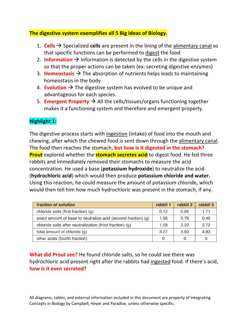
The digestive process starts with ingestion (intake) of food into the mouth and chewing, after which the chewed food is sent down through the alimentary canal. The food then reaches the stomach, but how is it digested in the stomach? Prout explored whether the stomach secretes acid to digest food. He fed three rabbits and immediately removed their stomachs to measure the acid concentration. He used a base (potassium hydroxide) to neutralize the acid (hydrochloric acid) which would then produce potassium chloride and water. Using this reaction, he could measure the amount of potassium chloride, which would then tell him how much hydrochloric was present in the stomach, if any.
What did Prout see? He found chloride salts, so he could see there was hydrochloric acid present right after the rabbits had ingested food. If there’s acid, how is it even secreted?
All diagrams, tables, and external information included in this document are property of Integrating Concepts in Biology by Campbell, Heyer and Paradise, unless otherwise specific.
Back to the Main Topic:
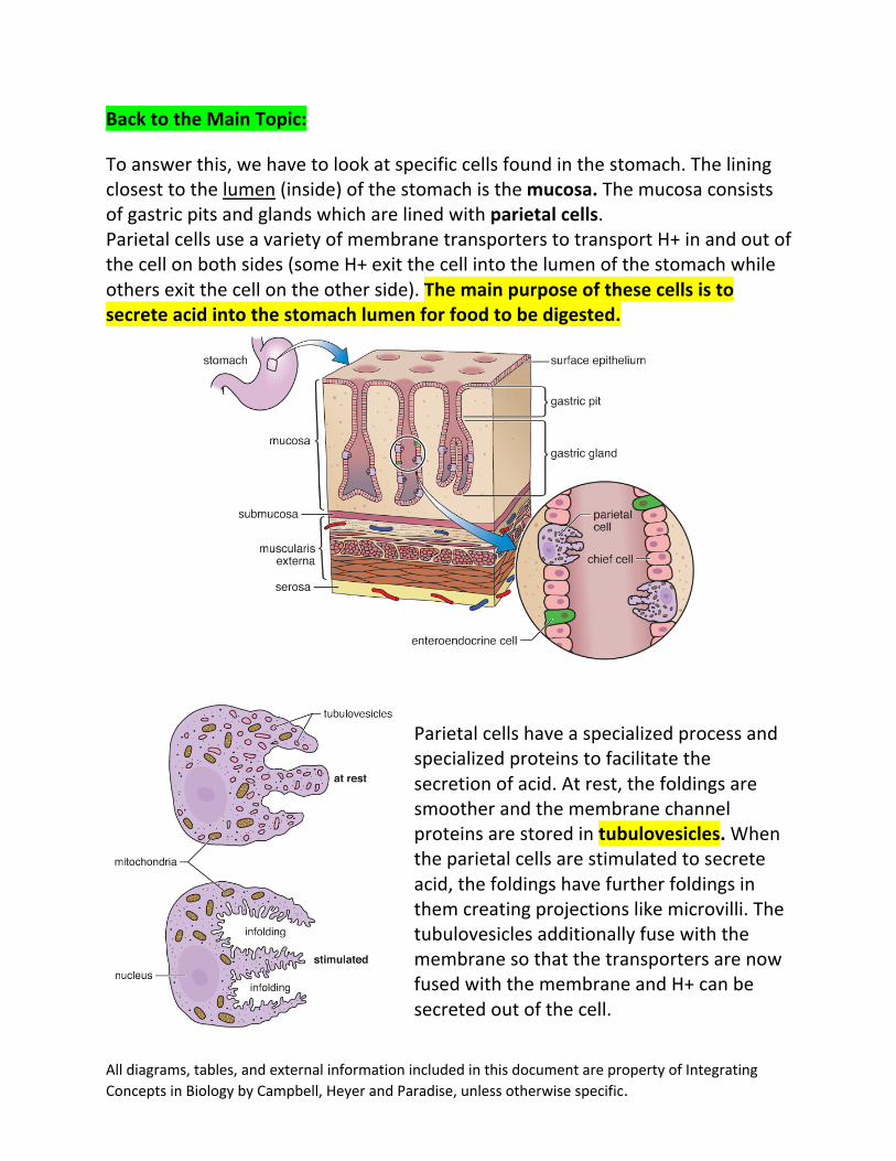
To answer this, we have to look at specific cells found in the stomach. The lining closest to the lumen (inside) of the stomach is the mucosa. The mucosa consists of gastric pits and glands which are lined with parietal cells. Parietal cells use a variety of membrane transporters to transport H+ in and out of the cell on both sides (some H+ exit the cell into the lumen of the stomach while others exit the cell on the other side). The main purpose of these cells is to secrete acid into the stomach lumen for food to be digested.
Parietal cells have a specialized process and specialized proteins to facilitate the secretion of acid. At rest, the foldings are smoother and the membrane channel proteins are stored in tubulovesicles. When the parietal cells are stimulated to secrete acid, the foldings have further foldings in them creating projections like microvilli. The tubulovesicles additionally fuse with the membrane so that the transporters are now fused with the membrane and H+ can be secreted out of the cell.
All diagrams, tables, and external information included in this document are property of Integrating Concepts in Biology by Campbell, Heyer and Paradise, unless otherwise specific.
If acid is secreted, however, why doesn’t the lining of the stomach get damaged by the acid?
The stomach lining is protected from hydrochloric acid and other acids by the epithelial cells. The epithelial cells secrete a bicarbonate-rich mucus layer that coats the mucosa (side of wall closest to the lumen/inside of stomach). Because bicarbonate is a base, this base-rich mucus layer neutralizes the acid content in the stomach before it can degrade the stomach lining. Once the food is digested, where does it go next in the system?
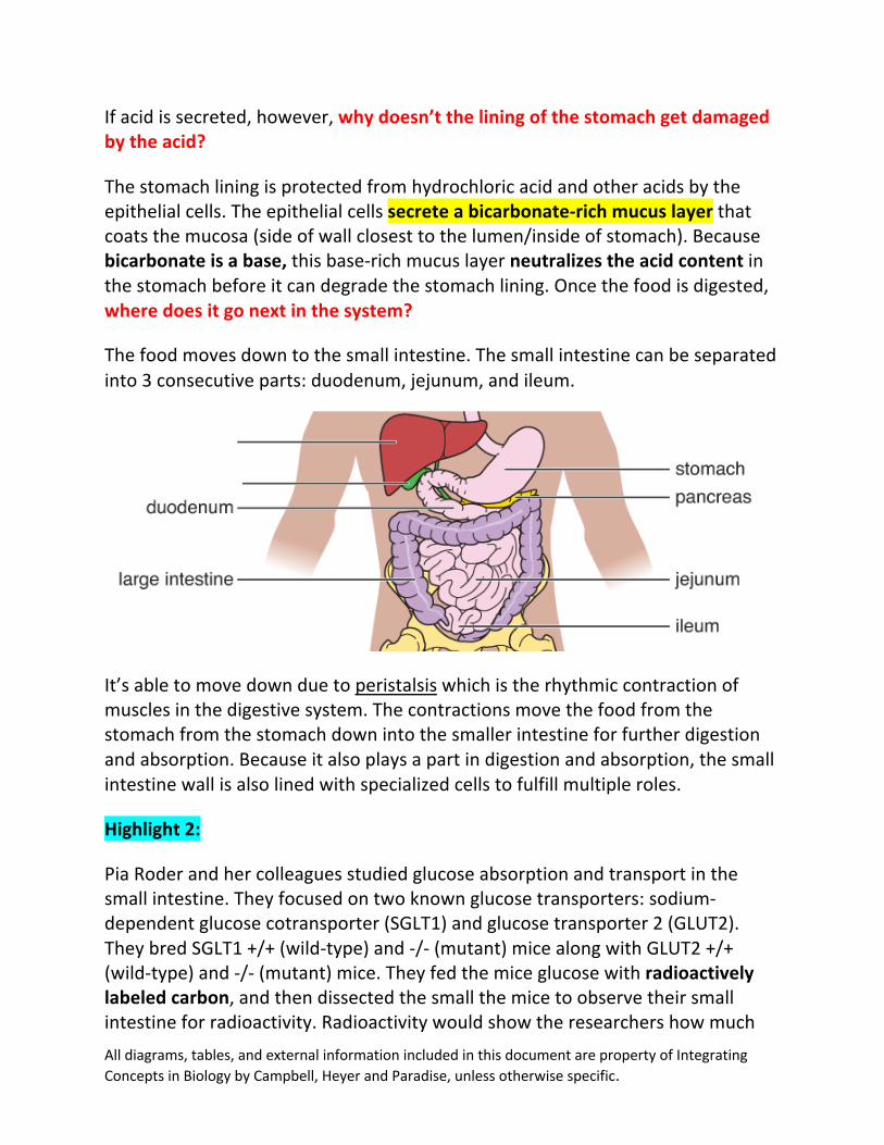
The food moves down to the small intestine. The small intestine can be separated into 3 consecutive parts: duodenum, jejunum, and ileum.
It’s able to move down due to peristalsis which is the rhythmic contraction of muscles in the digestive system. The contractions move the food from the stomach from the stomach down into the smaller intestine for further digestion and absorption. Because it also plays a part in digestion and absorption, the small intestine wall is also lined with specialized cells to fulfill multiple roles.
Highlight 2:
Pia Roder and her colleagues studied glucose absorption and transport in the small intestine. They focused on two known glucose transporters: sodium-dependent glucose cotransporter (SGLT1) and glucose transporter 2 (GLUT2). They bred SGLT1 +/+ (wild-type) and -/- (mutant) mice along with GLUT2 +/+ (wild-type) and -/- (mutant) mice. They fed the mice glucose with radioactively labeled carbon, and then dissected the small the mice to observe their small intestine for radioactivity. Radioactivity would show the researchers how much

All diagrams, tables, and external information included in this document are property of Integrating Concepts in Biology by Campbell, Heyer and Paradise, unless otherwise specific.
glucose was absorbed, and therefore how much the transporters were used. So, what did they see?
The two graphs on the left (A and C) show the glucose concentration in just the epithelial cells and the two graphs on the right (B and D) show the glucose concentration in the blood). The glucose is usually absorbed from the small intestine lumen into the epithelial cell. The glucose then exits the epithelial cell into the bloodstream. Looking at the first top two graphs (A and B) just looking at the SGLT1 transporter, it’s seen that the wild-type mice with the transporter had a significantly greater concentration of glucose in the epithelial cells and in the bloodstream than the mutant mice did. This indicates that the SGLT1 transporter is needed on both sides of the epithelial cells to successfully absorb glucose into the bloodstream.
The bottom two graphs (C and D), however, tell a different story. The glucose concentration in the mutant mice without GLUT2 transporters was significantly higher in epithelial cells than wild-type mice with the transporter. Conversely, the wild-type mice with the GLUT2 transporter had a significantly greater concentration of glucose in the blood than the mutant mice without the
All diagrams, tables, and external information included in this document are property of Integrating Concepts in Biology by Campbell, Heyer and Paradise, unless otherwise specific.
transporter. This indicates that the transporter may not be needed for the glucose to enter the epithelial cells from the lumen. However, the transporter is then needed for glucose to exit the epithelial cell into the bloodstream. Without the transporter, the mutant mice had a store of glucose in their epithelial cells that couldn’t enter the bloodstream, which is why they had a significantly larger concentration of glucose in their cells than wild-type mice. Despite that, the glucose couldn’t leave the cells of the mutant mice because they didn’t have the transporter, so the wild-type mice still had a greater concentration of glucose in the bloodstream.
Back to the Main Topic:
The epithelial cells in the small intestine have two transmembrane proteins to help with the absorption of glucose. One of them helps glucose enter the epithelial cell from the lumen, and the other helps glucose exit the cell into the bloodstream.
Any material that has not been digested and absorbed by the small intestine travels to the large intestine. Any semisolid waste (feces) is moved to the rectum and then removed through the anus through peristalsis (contractions). This is a small snapshot of how the digestive system works. Remember that there a lot more moving parts that help this complex system function efficiently!
All diagrams, tables, and external information included in this document are property of Integrating Concepts in Biology by Campbell, Heyer and Paradise, unless otherwise specific.
Let’s Test Ourselves!! 1. How is the stomach lining protected from the acid secreted? 2. How does food move down through the digestive system? 3. Which cells in the stomach lining play a large part in secreting acid and
maintaining an acidic pH in the stomach? 4. In the described experiment, what would happen to glucose in organisms
with the mutated SGLT1 -/- gene?
Things Students May Struggle With:
1. Understanding acidity/basicity. Just the first few paragraphs of this website should be enough to understand the concepts in this section. Acid Base Neutralization.
2. Take time to look at the anatomy of the digestive system that you understand the overview of how food passes through the body. This will make the details talked about in the chapter a little less confusing.
3. A lot of this section is memorization as well, so take the time to memorize the structures, the types of cells, and secretions.
Answers:
1. Epithelial cells secrete a bicarbonate-rich mucus layer. 2. Food moves through using peristaltic contractions. 3. Parietal cells play a large part in secreting acid/maintaining an acidic pH 4. Glucose would not enter the intestinal epithelial cells and therefore, it would not
find its way to the blood (lot more glucose left in the lumen of the small intestine).
I hope this resource was helpful for you! Don’t forget that these weekly resources can be found at:
https://www.baylor.edu/case/index.php?id=978656