Motility and trafficking in B-cell non-Hodgkin's lymphoma (Review)

Amaya-Gómez et al. BMC Microbiology (2015) 15:58 DOI 10.1186/s12866-015-0390-z
RESEARCH ARTICLE Open Access
Biofilm formation assessment in Sinorhizobiummeliloti reveals interlinked control with surfacemotilityCarol V Amaya-Gómez1, Ann M Hirsch2 and María J Soto1*
Abstract
Background: Swarming motility and biofilm formation are opposite, but related surface-associated behaviors thatallow various pathogenic bacteria to colonize and invade their hosts. In Sinorhizobium meliloti, the alfalfa endosymbiont,these bacterial processes and their relevance for host plant colonization are largely unexplored. Our previouswork demonstrated distinct swarming abilities in two S. meliloti strains (Rm1021 and GR4) and revealed that bothenvironmental cues (iron concentration) and bacterial genes (fadD, rhb, rirA) play crucial roles in the control ofsurface motility in this rhizobial species. In the current study, we investigate whether these factors have an impacton the ability of S. meliloti to establish biofilms and to colonize host roots.
Results: We found that strain GR4, which is less prone to translocate on solid surfaces than strain Rm1021, is moreefficient in developing biofilms on glass and plant root surfaces. High iron conditions, known to prevent surface motilityin a wild-type strain of S. meliloti, promote biofilm development in Rm1021 and GR4 strains by inducing the formation ofmore structured and thicker biofilms than those formed under low iron levels. Moreover, three different S. melilotimutants (fadD, rhb, and rirA) that exhibit an altered surface translocation behavior compared with the wild-typestrain, establish reduced biofilms on both glass and alfalfa root surfaces. Iron-rich conditions neither rescue thedefect in biofilm formation shown by the rhb mutant, which is unable to produce the siderophore rhizobactin1021 (Rhb1021), nor have any impact on biofilms formed by the iron-response regulator rirA mutant. On theother hand, S. meliloti FadD loss-of-function mutants do not establish normal biofilms irrespective of iron levels.
Conclusions: Our studies show that siderophore Rhb1021 is not only required for surface translocation, but alsofor biofilm formation on glass and root surfaces by strain Rm1021. In addition, we present evidence for theexistence of control mechanisms that inversely regulate swarming and biofilm formation in S. meliloti, and thatcontribute to efficient plant root colonization. One of these mechanisms involves iron levels and the iron globalregulator RirA. The other mechanism involves the participation of the fatty acid metabolism-related enzyme FadD.
Keywords: FadD, Iron, Rhizobium, RirA, Root colonization, Siderophore, Swarming
BackgroundSwarming motility and biofilms are two different andopposite behaviors displayed by bacteria living on sur-faces. Biofilms are sessile assemblages of microorganismsembedded in a self-produced polymeric matrix that ad-here to a surface or are associated with interfaces [1,2].By contrast, swarming is a mode of surface translocation
* Correspondence: [email protected] de Microbiología del Suelo y Sistemas Simbióticos, EstaciónExperimental del Zaidín, Consejo Superior de Investigaciones Científicas(CSIC), Profesor Albareda 1, 18008 Granada, SpainFull list of author information is available at the end of the article
© 2015 Amaya-Gómez et al.; licensee BioMedCreative Commons Attribution License (http:/distribution, and reproduction in any mediumDomain Dedication waiver (http://creativecomarticle, unless otherwise stated.
that depends on rotating flagella and is characterized bythe rapid and coordinated movement of multicellulargroups of bacteria [3]. Several studies have revealed theexistence of a link between swarming and biofilm forma-tion: i) both are surface-associated multicellular pro-cesses in which cell-cell communication and quorumsensing play important roles; ii) in both processes, theparticipation of the same cell surface-associated structuressuch as flagella, a polysaccharide matrix, and biosurfac-tants has been reported; and iii) swarming bacteria, similarto bacteria in biofilms, show increased resistance to sev-eral antimicrobial agents [3-8]. Studies performed in
Central. This is an Open Access article distributed under the terms of the/creativecommons.org/licenses/by/4.0), which permits unrestricted use,, provided the original work is properly credited. The Creative Commons Publicmons.org/publicdomain/zero/1.0/) applies to the data made available in this

Amaya-Gómez et al. BMC Microbiology (2015) 15:58 Page 2 of 14
Pseudomonas aeruginosa, Vibrio cholerae, or V. parahae-molyticus show that the two lifestyles are inversely regu-lated by a common pathway, which is modulated by theintracellular second messenger cyclic di-GMP [9-14].Swarming motility and biofilm formation have been
studied almost exclusively in pathogenic bacteria. How-ever, little is known about these multicellular surface-associated responses in rhizobia, soil-dwelling bacteria,which induce nitrogen-fixing nodules on the roots oflegume plants following a complex and continuous mo-lecular dialogue that co-ordinates bacterial infectionwith nodule organogenesis [15].Sinorhizobium meliloti, the alfalfa symbiont, forms
biofilms on both abiotic surfaces and roots [16]. On abi-otic surfaces, the capability of S. meliloti to form bio-films is affected by environmental stresses and nutrientstatus [17]. As in many bacteria, rhizobial exopolysac-charides (EPS) and flagella are involved in biofilm forma-tion and mutants defective in either of these twocomponents exhibit a significant reduction in the abilityto develop biofilms [16,18-20]. Remarkably, the produc-tion of a low-molecular-weight fraction of galactoglucan(EPS II), the production of which is dependent on afunctional ExpR/Sin quorum sensing system, is crucialfor biofilm formation and root colonization. EPS II-producing strains are able to develop highly structuredbiofilms under low-phosphate conditions, but not underhigh phosphate conditions where flat and unstructuredbiofilms are formed [18]. Besides EPS and flagella, coreNod Factor, an essential molecule for the nodulationprocess, has been shown to be critical for biofilm forma-tion in S. meliloti [21]. In addition to the LuxR-typetranscriptional regulator ExpR, different regulatory pro-teins that control several phenotypes including EPS pro-duction and motility have been involved in regulation ofbiofilm formation in S. meliloti. This is the case for thetwo-component system ExoS/ChvI and its periplasmic in-hibitor ExoR [16,22]; or EmrR, a TetR-family transcrip-tional repressor that controls expression of emrA andemrB, which encode a putative MFS-type transporter [23].Swarming motility has also been reported in S. meliloti
[24-27], and was first described for a fadD mutant of theGR4 strain [24]. Wild-type GR4 cells normally do nottranslocate over semisolid surfaces, but inactivating thefadD gene, which codes for a long-chain fatty acyl-coenzyme A ligase, promotes swarming motility onsemisolid minimal medium. This finding strongly suggeststhat FadD plays a role in the control of this multicellularsurface-associated behavior. However, in contrast to GR4,the commonly used S. meliloti laboratory strain Rm1021moves over semisolid surfaces using flagella-dependentand -independent mechanisms [25,26]. The fact that wild-type GR4 cells do not translocate in contrast to Rm1021cells and that a mutation in the fadD gene promotes
surface translocation for both strongly suggests the exist-ence of different control mechanisms for surface motilityin these two strains [25]. A transcriptomic analysis of afadD mutant of S. meliloti strain Rm1021 under swarming-inducing conditions showed that iron and also genesrequired for siderophore rhizobactin 1021 (Rhb1021) syn-thesis are critical for surface translocation of the wild-typestrain Rm1021 [25,26]. S. meliloti rhb mutants that are un-able to produce the siderophore are non-motile on the sur-face of semisolid media. On the other hand, an rhtAmutant, which lacks the outer membrane receptor forRhb1021 utilization, is motile indicating that the swarmingdeficiency shown by rhb mutants was not due to iron defi-ciency and furthermore, that Rhb1021’s involvement inswarming was exerted outside the cell. Surfactant proper-ties inherent to the Rhb1021 structure, a citrate-based sid-erophore containing a long-chain fatty acid, could beresponsible for the promotion of surface translocation in S.meliloti. Interestingly, the lack of a functional fadD generestored surface motility in Rhb1021-deficient strains, indi-cating that the effect caused on surface motility by fadDloss-of-function is epistatic to mutations affecting sidero-phore production. Also, the same study showed that highiron conditions inhibited swarming motility in Rm1021,most likely by preventing Rhb1021 production. This inhibi-tory effect, however, was not observed in mutants lackingeither RirA, an iron limitation response regulator, or FadD[25]. The rirA mutant’s phenotype could be explained bythe bacteria’s ability to produce Rhb1021 under high ironconditions. However, the mechanism responsible for theiron-independent swarming phenotype shown by fadDmutants is unknown.The connection between swarming motility and bio-
film formation in S. meliloti has not yet been explored.In this work, we investigated whether factors known toinfluence swarming motility in S. meliloti have an impacton its capability to form biofilms. The study was per-formed using ExpR-deficient strains that display differentsurface motility behaviors. Although expR− mutants arereported not to form the type of highly structured bio-films observed for expR+ strains, our findings show that,similar to flagella, Rhb1021 is required for both surfacetranslocation and biofilm formation. In addition, dataobtained from this study provide evidence for the exist-ence in S. meliloti of common regulatory mechanismsgoverning surface motility and biofilm formation inwhich iron, the iron global regulator RirA, and FadDhave important roles.
ResultsBiofilm formation analysis in S. meliloti Rm1021 and GR4strainsThe existing knowledge on swarming motility in S. meli-loti has mostly been obtained from studying the ExpR-
Amaya-Gómez et al. BMC Microbiology (2015) 15:58 Page 3 of 14
deficient reference strains Rm1021 and GR4. In this way,the interference of a sliding motility promoted by EPS IIproduction can be avoided [24-26]. Rm1021 and GR4strains show different surface motility phenotypes.Rm1021 spreads over semisolid surfaces using flagella-dependent and -independent mechanisms, whereas GR4is non-motile under the same conditions [25,26]. Wethus tested whether this difference in behavior on semi-solid surfaces could correlate with distinct biofilm for-mation abilities by analyzing biofilms established bythese two strains on both abiotic (PVC and glass) andbiotic surfaces (alfalfa roots).Swarming motility, like biofilm formation, is highly in-
fluenced by medium composition [8,17,18,20,24]. InExpR-deficient S. meliloti strains, swarming motility hasonly been shown to occur on semisolid MM [24-26].This medium contains a relatively high phosphate con-centration (3.5 mM), a condition known to slightly in-crease biofilm formation in expR mutants compared tothat observed under low phosphate conditions (0.1 mM)[18]. To rule out any influence of the medium compos-ition and to be able to correlate the swarming behaviorwith the biofilm formation ability of the different strains,in vitro biofilm formation was analyzed by growing cellsin MM as described in the Methods section.Under our experimental conditions, all the different S.
meliloti strains used in this study exhibited poor biofilmformation capabilities on PVC microtiter plates, making
10 dpi3 dpi
Rm1021
GR4
Rm1021
GR4
A
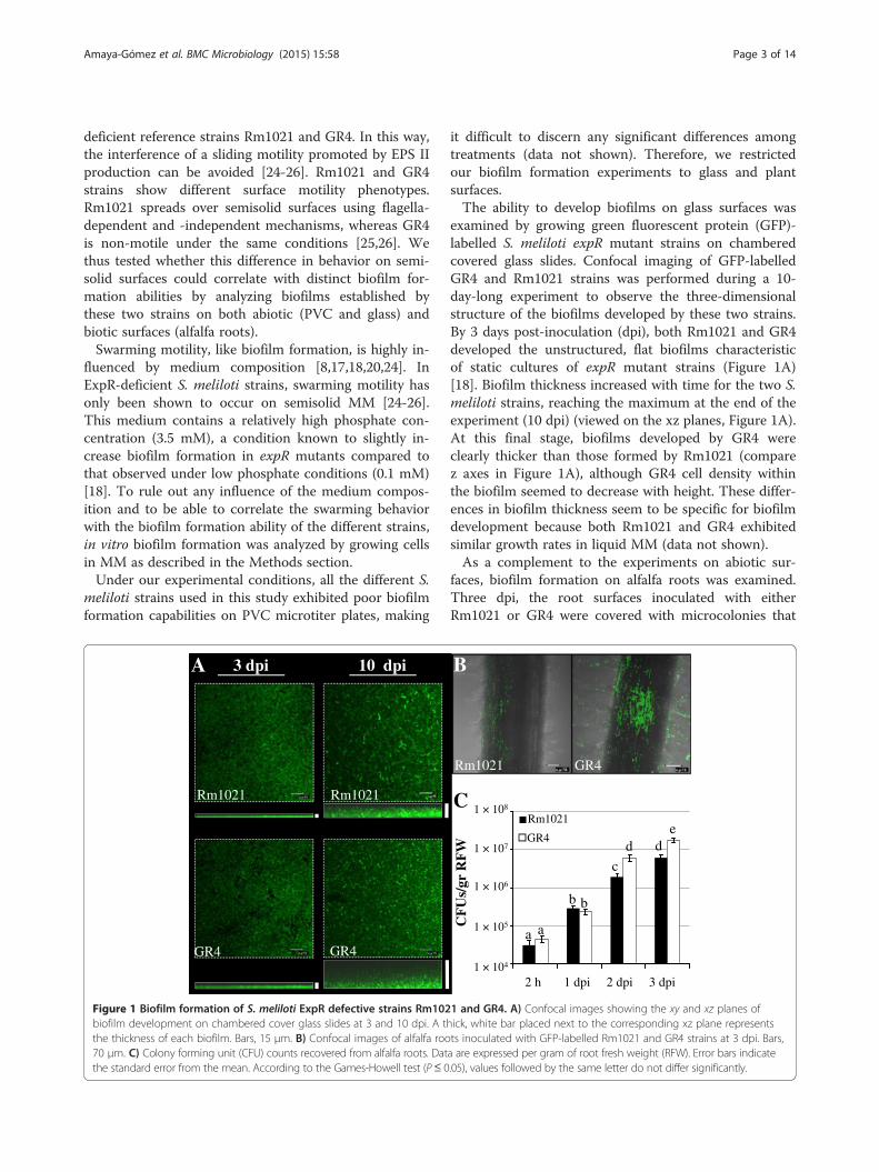
Figure 1 Biofilm formation of S. meliloti ExpR defective strains Rm102biofilm development on chambered cover glass slides at 3 and 10 dpi. A ththe thickness of each biofilm. Bars, 15 μm. B) Confocal images of alfalfa roo70 μm. C) Colony forming unit (CFU) counts recovered from alfalfa roots. Datthe standard error from the mean. According to the Games-Howell test (P≤ 0
it difficult to discern any significant differences amongtreatments (data not shown). Therefore, we restrictedour biofilm formation experiments to glass and plantsurfaces.The ability to develop biofilms on glass surfaces was
examined by growing green fluorescent protein (GFP)-labelled S. meliloti expR mutant strains on chamberedcovered glass slides. Confocal imaging of GFP-labelledGR4 and Rm1021 strains was performed during a 10-day-long experiment to observe the three-dimensionalstructure of the biofilms developed by these two strains.By 3 days post-inoculation (dpi), both Rm1021 and GR4developed the unstructured, flat biofilms characteristicof static cultures of expR mutant strains (Figure 1A)[18]. Biofilm thickness increased with time for the two S.meliloti strains, reaching the maximum at the end of theexperiment (10 dpi) (viewed on the xz planes, Figure 1A).At this final stage, biofilms developed by GR4 wereclearly thicker than those formed by Rm1021 (comparez axes in Figure 1A), although GR4 cell density withinthe biofilm seemed to decrease with height. These differ-ences in biofilm thickness seem to be specific for biofilmdevelopment because both Rm1021 and GR4 exhibitedsimilar growth rates in liquid MM (data not shown).As a complement to the experiments on abiotic sur-
faces, biofilm formation on alfalfa roots was examined.Three dpi, the root surfaces inoculated with eitherRm1021 or GR4 were covered with microcolonies that
1,E+04
1,E+05
1,E+06
1,E+07
1,E+08
2h 1 dpi 2 dpi 3 dpi
CF
Us/
root
g
2 h 1 dpi 2 dpi 3 dpi
1 × 10⁸
1 × 10⁷
1 × 10⁶
1 × 10⁵
1 × 10⁴
CF
Us/
gr R
FW
a a
b b
c
dde
Rm1021
GR4
C
GR4Rm1021
B
1 and GR4. A) Confocal images showing the xy and xz planes ofick, white bar placed next to the corresponding xz plane representsts inoculated with GFP-labelled Rm1021 and GR4 strains at 3 dpi. Bars,a are expressed per gram of root fresh weight (RFW). Error bars indicate.05), values followed by the same letter do not differ significantly.
Amaya-Gómez et al. BMC Microbiology (2015) 15:58 Page 4 of 14
remained attached along the oldest part of the root evenafter extensive washing (Figure 1B). Very few cells ad-hered to the younger root tissues and apical region.However, it is worth noting that more cell clusters wereobserved colonizing the root surface and especially theroot hair zone of plants, when inoculated with GR4(Figure 1B). Although the number of CFUs per gram ofroot tissue revealed no differences in the attachmentability of Rm1021 and GR4 2 h and 24 h post-inoculation, the number of GR4 cells attached to rootsurfaces 2 and 3 dpi was approximately 3-fold higherthan the values for Rm1021 (Figure 1C). Overall, thisindicates that GR4 is more efficient than Rm1021 in es-tablishing biofilms on glass and root surfaces.
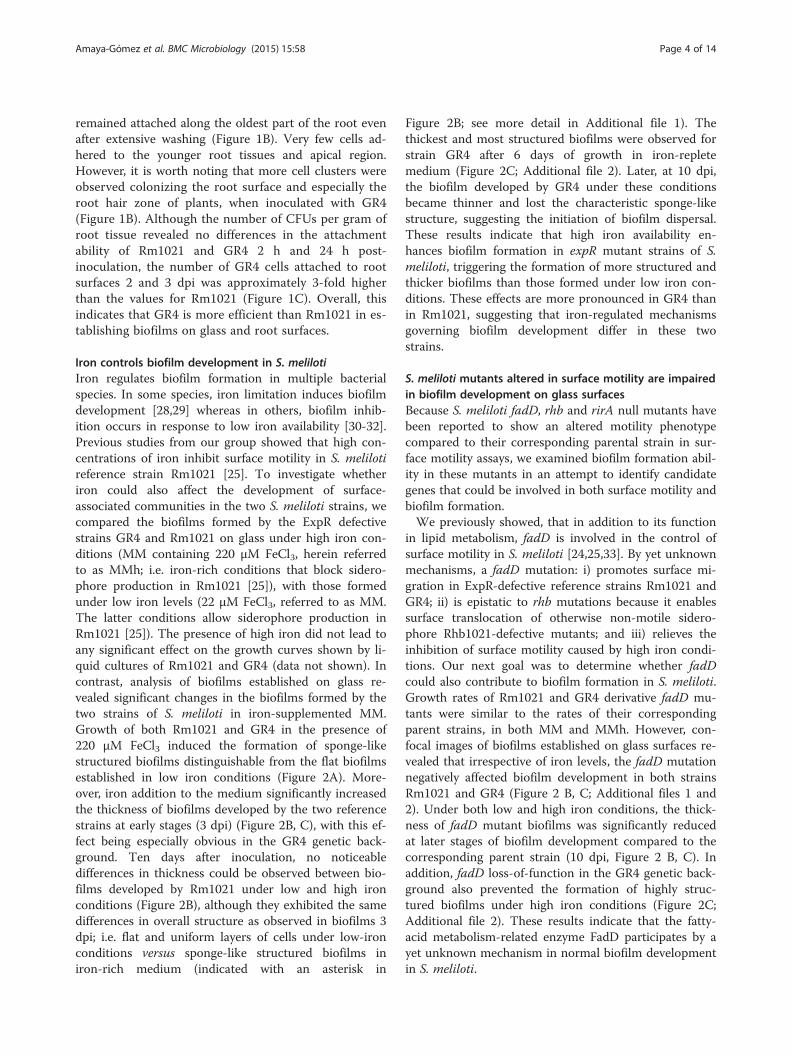
Iron controls biofilm development in S. melilotiIron regulates biofilm formation in multiple bacterialspecies. In some species, iron limitation induces biofilmdevelopment [28,29] whereas in others, biofilm inhib-ition occurs in response to low iron availability [30-32].Previous studies from our group showed that high con-centrations of iron inhibit surface motility in S. melilotireference strain Rm1021 [25]. To investigate whetheriron could also affect the development of surface-associated communities in the two S. meliloti strains, wecompared the biofilms formed by the ExpR defectivestrains GR4 and Rm1021 on glass under high iron con-ditions (MM containing 220 μM FeCl3, herein referredto as MMh; i.e. iron-rich conditions that block sidero-phore production in Rm1021 [25]), with those formedunder low iron levels (22 μM FeCl3, referred to as MM.The latter conditions allow siderophore production inRm1021 [25]). The presence of high iron did not lead toany significant effect on the growth curves shown by li-quid cultures of Rm1021 and GR4 (data not shown). Incontrast, analysis of biofilms established on glass re-vealed significant changes in the biofilms formed by thetwo strains of S. meliloti in iron-supplemented MM.Growth of both Rm1021 and GR4 in the presence of220 μM FeCl3 induced the formation of sponge-likestructured biofilms distinguishable from the flat biofilmsestablished in low iron conditions (Figure 2A). More-over, iron addition to the medium significantly increasedthe thickness of biofilms developed by the two referencestrains at early stages (3 dpi) (Figure 2B, C), with this ef-fect being especially obvious in the GR4 genetic back-ground. Ten days after inoculation, no noticeabledifferences in thickness could be observed between bio-films developed by Rm1021 under low and high ironconditions (Figure 2B), although they exhibited the samedifferences in overall structure as observed in biofilms 3dpi; i.e. flat and uniform layers of cells under low-ironconditions versus sponge-like structured biofilms iniron-rich medium (indicated with an asterisk in
Figure 2B; see more detail in Additional file 1). Thethickest and most structured biofilms were observed forstrain GR4 after 6 days of growth in iron-repletemedium (Figure 2C; Additional file 2). Later, at 10 dpi,the biofilm developed by GR4 under these conditionsbecame thinner and lost the characteristic sponge-likestructure, suggesting the initiation of biofilm dispersal.These results indicate that high iron availability en-hances biofilm formation in expR mutant strains of S.meliloti, triggering the formation of more structured andthicker biofilms than those formed under low iron con-ditions. These effects are more pronounced in GR4 thanin Rm1021, suggesting that iron-regulated mechanismsgoverning biofilm development differ in these twostrains.
S. meliloti mutants altered in surface motility are impairedin biofilm development on glass surfacesBecause S. meliloti fadD, rhb and rirA null mutants havebeen reported to show an altered motility phenotypecompared to their corresponding parental strain in sur-face motility assays, we examined biofilm formation abil-ity in these mutants in an attempt to identify candidategenes that could be involved in both surface motility andbiofilm formation.We previously showed, that in addition to its function
in lipid metabolism, fadD is involved in the control ofsurface motility in S. meliloti [24,25,33]. By yet unknownmechanisms, a fadD mutation: i) promotes surface mi-gration in ExpR-defective reference strains Rm1021 andGR4; ii) is epistatic to rhb mutations because it enablessurface translocation of otherwise non-motile sidero-phore Rhb1021-defective mutants; and iii) relieves theinhibition of surface motility caused by high iron condi-tions. Our next goal was to determine whether fadDcould also contribute to biofilm formation in S. meliloti.Growth rates of Rm1021 and GR4 derivative fadD mu-tants were similar to the rates of their correspondingparent strains, in both MM and MMh. However, con-focal images of biofilms established on glass surfaces re-vealed that irrespective of iron levels, the fadD mutationnegatively affected biofilm development in both strainsRm1021 and GR4 (Figure 2 B, C; Additional files 1 and2). Under both low and high iron conditions, the thick-ness of fadD mutant biofilms was significantly reducedat later stages of biofilm development compared to thecorresponding parent strain (10 dpi, Figure 2 B, C). Inaddition, fadD loss-of-function in the GR4 genetic back-ground also prevented the formation of highly struc-tured biofilms under high iron conditions (Figure 2C;Additional file 2). These results indicate that the fatty-acid metabolism-related enzyme FadD participates by ayet unknown mechanism in normal biofilm developmentin S. meliloti.

GR
4
3
10
6
GR
4FD
SCSS 3
10
6
22 µM FeCl3 220 µM FeCl3dpi
C
Rm
1021
22 µM FeCl3 220 µM FeCl3
3
1021
FD
CSS
1021
rhbA
10
dpi
3
10
3
10
3
10
B
G21
2rir
A
Rm1021 Rm1021 GR4
220 µM FeCl322 µM FeCl3
GR4
22 µM FeCl3 220 µM FeCl3A
*
*
**
**
**
2.4 ± 0.4aA
9.2 ± 1.1aB
5.4 ± 0.8aC
10.8 ± 1.5aB
2.3 ± 0.4aA
4.2 ± 0.2bB
6.1 ± 0.9aB
5.6 ± 0.8bB
1.6 ± 0.4aA
2.7 ± 0.9bAB
2.5 ± 0.1bAB
3.5 ± 0.3cB
5.6 ± 0.8bA
6.5 ± 0.5cA
5.5 ± 0.4aA
7 ± 1.0bA
2.6 ± 0.2aA
23.5 ± 3.2aB
32.3 ± 2.0aC
16.8 ± 2.1aD
43.1 ± 13.3aE
22 ± 3.0aB
2.2 ± 0.4aA
3.5 ± 0.8bAB
5.2 ± 0.3bBD
14.4 ± 0.4aC
4.1 ± 0.7bBD
5.9 ± 0.3bD
Figure 2 Biofilm formation of S. meliloti strains on glass surfaces in response to iron availability. A) Confocal Laser Scanning Microscopy(CLSM) images showing the architecture of 3-day-old biofilms developed by GFP-labelled Rm1021 and GR4 cells on chambered cover glass slidesafter growth in MM containing different concentrations of FeCl3. Bars, 15 μm. B) CLSM images showing the thickness (represented by the xzplane) of 3- and 10-day-old biofilms developed by Rm1021 and its derivative mutant strains on chambered cover glass slides in response to low(22 μM FeCl3) and high (220 μM FeCl3) iron availability. C) CLSM images showing the thickness (represented by the xz plane) of 3-, 6-, and 10-day-oldbiofilms developed by GR4 and GR4FDCSS. The mean thickness (± SD) of biofilms formed by the different strains under the two iron conditions isshown in parenthesis below the representative CLSM image. Values having different letters are significantly different from each other (Tukey’s test,P < 0.05). Lowercase letters indicate statistical differences between the thickness of the wild type strain and its derivative mutant biofilms developedunder the same conditions. Capital letters are used to indicate the statistical differences observed in the biofilms developed under the differentconditions by each strain. Structured biofilms developed in MM containing 220 μM of FeCl3 are indicated with an asterisk (*). Each experimentwas repeated three times.
Amaya-Gómez et al. BMC Microbiology (2015) 15:58 Page 5 of 14
Two other S. meliloti mutants affected in surfacetranslocation and assessed for biofilm development inthis study are rhb, which is affected in iron uptake andrirA an iron homeostasis mutant. Rm1021 rhb mutantsare unable to synthesize the siderophore Rhb1021 andcannot translocate on semisolid surfaces. This, togetherwith the demonstration that the siderophore uptake rhtAmutant exhibits surface movement [25], suggests a spe-cific role for the siderophore in Rm1021 surface motilityin addition to its function in iron nutrition. In contrastto Rm1021, GR4 does not produce Rhb1021, whichmight explain the inability of this strain to translocateover semisolid MM [24,25].Analyses of biofilm formation on glass surfaces made
it possible to see that the Rm1021 rhbA mutant ishighly impaired in biofilm development. Although nosignificant changes were detected in the structure
exhibited by the rhbA mutant biofilms compared tothat shown by Rm1021 biofilms in either low or highiron conditions, significant reductions in mutant bio-film thickness were observed at later stages (10 dpi)compared to wild-type Rm1021 (Figure 2B; Additionalfile 1). Similar results were obtained with an in-framerhbD deletion mutant (data not shown; [34]). No sig-nificant changes in growth rates have been detected inRm1021 rhb mutants compared to the wild-type strainin liquid MM or MMh (data not shown). More import-antly, the fact that increased iron availability condi-tions, in which Rhb1021 production is abolished [25],could not restore biofilm development of the rhbA mu-tant suggests that, Rhb1021 synthesis is essential atsome stages of biofilm development and is involved ina way that is not exclusively related to its function assiderophore.

Amaya-Gómez et al. BMC Microbiology (2015) 15:58 Page 6 of 14
On the other hand, we demonstrated that loss-of-function of the iron response regulator RirA in Rm1021does not affect surface motility under low iron condi-tions, but leads to a hypermobile phenotype in the pres-ence of high levels of iron, suggesting a role for thisregulator in the control of surface motility in responseto iron concentration [25]. Under iron-sufficient condi-tions, S. meliloti rirA mutants cannot down-regulateiron uptake systems, including siderophore Rhb1021synthesis, which leads to oxidative stress and a corre-sponding reduction in growth [35].Confocal Laser Scanning Microscopy (CLSM) images
of biofilms established on glass surfaces by the rirA mu-tant revealed that the mutation affected normal biofilmdevelopment (Figure 2B; Additional file 1). Under lowiron conditions, the rirA mutant formed flat, unstruc-tured biofilms, which at early stages (3 dpi) were thickerthan those developed by its parental strain Rm1021.However, rirA mutant biofilms did not increase in thick-ness with time; this led to the formation of slightly thin-ner biofilms than the ones formed by Rm1021 at the endof the experiment. The most relevant difference betweenRm1021 and rirA mutant biofilms was observed underhigh iron conditions. RirA loss-of-function abolished theformation of thicker and sponge-like structured biofilms,and thus no changes were detected in rirA mutant bio-films formed in iron-replete medium compared to thoseformed under low iron levels (Figure 2B; Additional file 1).These results strongly suggest that this regulator plays apivotal role in controlling functions required for biofilmdevelopment in response to iron availability.
S. meliloti fadD, rhb and rirA mutants show defects inalfalfa root colonizationWe decided to investigate whether mutations in S. meliloticausing alterations in two different surface-associatedphenotypes (swarming and biofilm formation) couldalso have an impact on biofilm development in planta,i.e. alfalfa root colonization. Confocal images and CFU
GR4 GR4FDCSS
A
Figure 3 In planta biofilm formation by GR4 and its fadD derivative msurfaces 3 dpi. Bars, 70 μm. B) Colony forming unit (CFU) counts recovered(RFW). Error bars indicate the standard error from the mean of three indepvalues followed by the same letter do not differ significantly.
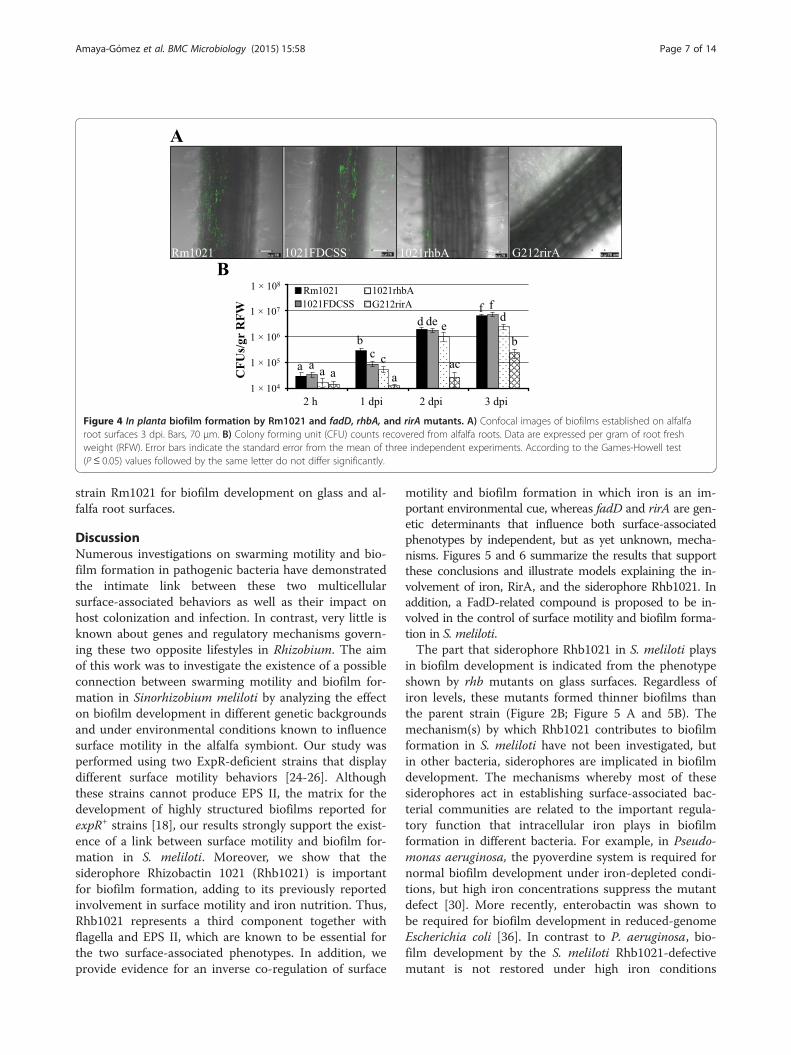
counting revealed that the GR4-derivative fadD mutant(GR4FDCSS) exhibited a significantly decreased abilityto attach to and to colonize root surfaces compared toGR4 (Figure 3), although this difference diminishedwith time (see 3 dpi in Figure 3B). Reduced biofilm forma-tion on alfalfa roots was also detected for 1021FDCSS(Figure 4), but the effect was transitory and less noticeablethan for GR4FDCSS. We could discern differences onlywhen counting CFU 1 dpi, at which time the number ofattached 1021FDCSS cells was approximately 3-fold lowerthan that of Rm1021 (Figure 4B). No significant differ-ences in root colonization were detected at later stagesby either confocal imaging or CFU counting between1021FDCSS and Rm1021 (Figure 4 A, B). Therefore, afadD mutation in S. meliloti negatively interferes withnormal biofilm development on root surfaces, althoughthe effects are more pronounced in the GR4 strainbackground than in the Rm1021 genetic background.The Rm1021 siderophore defective rhbA mutant was
also impaired in establishing bacterial communities onthe surface of alfalfa roots (Figure 4A). In contrast to thesignificant root colonization observed for Rm1021, plantroots inoculated with the rhbA mutant supported onlysmall groups of a few bacteria that were attached to thecentral part of the root and root hairs. CFU counting ofroot-associated bacteria confirmed this result and dem-onstrated that the number of rhbA mutant cells coloniz-ing alfalfa roots was 5, 2, and 2.6-fold lower that thoseobserved for Rm1021 at 1, 2, and 3 dpi, respectively(Figure 4B).The most striking phenotype was observed for the rirA
mutant, which was severely impaired in alfalfa rootcolonization. Only a few mutant cells could be observedto be attached to the main root and root hairs by CLSM(Figure 4A). Moreover, CFU counting revealed that thenumber of rirA mutant cells recovered from alfalfa rootswas 22, 68, and 25-fold lower than for Rm1021 at 1, 2,and 3 dpi, respectively (Figure 4B). Thus, the ironhomeostasis-related genes rhb and rirA are critical in
ab
ef
1,00E+04
1,00E+05
1,00E+06
1,00E+07
1,00E+08
CF
Us/
gr R
FW
1 × 10⁷
1 × 10⁶
1 × 10⁵
1 × 10⁴
2 h 1 dpi 2 dpi 3 dpi
1 × 10⁸
cd
g gGR4FDCSSGR4
B
utant. A) Confocal images of biofilms established on alfalfa rootfrom alfalfa roots. Data are expressed per gram of root fresh weightendent experiments. According to the Games-Howell test (P ≤ 0.05)
Rm1021 1021rhbA G212rirA1021FDCSS
A
ac
acaa a
b
c
de
b
f fd
1 × 10⁸
1 × 10⁷
1 × 10⁶
1 × 10⁵
1 × 10⁴
CF
Us/
gr
RF
W
Rm1021
1021FDCSS
1021rhbA
G212rirA
a
d e
2 h 1 dpi 2 dpi 3 dpi
B
Figure 4 In planta biofilm formation by Rm1021 and fadD, rhbA, and rirA mutants. A) Confocal images of biofilms established on alfalfaroot surfaces 3 dpi. Bars, 70 μm. B) Colony forming unit (CFU) counts recovered from alfalfa roots. Data are expressed per gram of root freshweight (RFW). Error bars indicate the standard error from the mean of three independent experiments. According to the Games-Howell test(P ≤ 0.05) values followed by the same letter do not differ significantly.
Amaya-Gómez et al. BMC Microbiology (2015) 15:58 Page 7 of 14
strain Rm1021 for biofilm development on glass and al-falfa root surfaces.
DiscussionNumerous investigations on swarming motility and bio-film formation in pathogenic bacteria have demonstratedthe intimate link between these two multicellularsurface-associated behaviors as well as their impact onhost colonization and infection. In contrast, very little isknown about genes and regulatory mechanisms govern-ing these two opposite lifestyles in Rhizobium. The aimof this work was to investigate the existence of a possibleconnection between swarming motility and biofilm for-mation in Sinorhizobium meliloti by analyzing the effecton biofilm development in different genetic backgroundsand under environmental conditions known to influencesurface motility in the alfalfa symbiont. Our study wasperformed using two ExpR-deficient strains that displaydifferent surface motility behaviors [24-26]. Althoughthese strains cannot produce EPS II, the matrix for thedevelopment of highly structured biofilms reported forexpR+ strains [18], our results strongly support the exist-ence of a link between surface motility and biofilm for-mation in S. meliloti. Moreover, we show that thesiderophore Rhizobactin 1021 (Rhb1021) is importantfor biofilm formation, adding to its previously reportedinvolvement in surface motility and iron nutrition. Thus,Rhb1021 represents a third component together withflagella and EPS II, which are known to be essential forthe two surface-associated phenotypes. In addition, weprovide evidence for an inverse co-regulation of surface
motility and biofilm formation in which iron is an im-portant environmental cue, whereas fadD and rirA are gen-etic determinants that influence both surface-associatedphenotypes by independent, but as yet unknown, mecha-nisms. Figures 5 and 6 summarize the results that supportthese conclusions and illustrate models explaining the in-volvement of iron, RirA, and the siderophore Rhb1021. Inaddition, a FadD-related compound is proposed to be in-volved in the control of surface motility and biofilm forma-tion in S. meliloti.The part that siderophore Rhb1021 in S. meliloti plays
in biofilm development is indicated from the phenotypeshown by rhb mutants on glass surfaces. Regardless ofiron levels, these mutants formed thinner biofilms thanthe parent strain (Figure 2B; Figure 5 A and 5B). Themechanism(s) by which Rhb1021 contributes to biofilmformation in S. meliloti have not been investigated, butin other bacteria, siderophores are implicated in biofilmdevelopment. The mechanisms whereby most of thesesiderophores act in establishing surface-associated bac-terial communities are related to the important regula-tory function that intracellular iron plays in biofilmformation in different bacteria. For example, in Pseudo-monas aeruginosa, the pyoverdine system is required fornormal biofilm development under iron-depleted condi-tions, but high iron concentrations suppress the mutantdefect [30]. More recently, enterobactin was shown tobe required for biofilm development in reduced-genomeEscherichia coli [36]. In contrast to P. aeruginosa, bio-film development by the S. meliloti Rhb1021-defectivemutant is not restored under high iron conditions
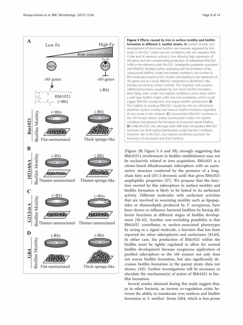
Figure 5 Effects caused by iron in surface motility and biofilmformation in different S. meliloti strains. A) Surface motility anddevelopment of structured biofilms are inversely regulated by ironlevels in Rm1021. Under low-iron conditions, the iron regulator RirAis free and its repressor activity is low allowing high expression ofrhb genes and the corresponding production of siderophore Rhb1021(+Rb) in the reference strain Rm1021. Amphiphilic properties associatedwith Rhb1021 facilitate surface spreading and the formation of flat,unstructured biofilms. Under iron-replete conditions, the number ofRirA molecules bound to Fe-S clusters rises leading to the repression ofrhb genes and as a result, Rhb1021 production is abolished (−Rb),thereby preventing surface motility. This together with putativeadditional functions regulated by iron favors biofilm formation.Most likely, even under iron-replete conditions, some areas withina wild type biofilm might suffer low iron availability, which couldtrigger Rhb1021 production and impact biofilm architecture. B)The inability to produce Rhb1021 caused by rhb loss-of-functionabolishes surface motility and reduces biofilm thickness regardlessof iron levels in the medium. C) Uncontrolled Rhb1021 synthesis inthe rirA mutant allows surface translocation under iron-repleteconditions but prevents the formation of structured mature biofilms.D) Unlike Rm1021, the wild type strain GR4 does not produce Rhb1021and does not show surface translocation under low-iron conditions.However, like in Rm1021, iron-replete conditions promote theformation of structured and thick biofilms.
Amaya-Gómez et al. BMC Microbiology (2015) 15:58 Page 8 of 14
(Figure 2B; Figure 5 A and 5B), strongly suggesting thatRhb1021’s involvement in biofilm establishment may notbe exclusively related to iron acquisition. Rhb1021 is acitrate-based dihydroxamate siderophore with an asym-metric structure conferred by the presence of a long-chain fatty acid ((E)-2-decenoic acid) that gives Rhb1021amphiphilic properties [37]. We propose that the func-tion exerted by this siderophore in surface motility andbiofilm formation is likely to be linked to its surfactantactivity. Different molecules with surfactant activitiesthat are involved in swarming motility such as lipopep-tides or rhamnolipids produced by P. aeruginosa, havebeen shown to influence bacterial biofilms by having dif-ferent functions at different stages of biofilm develop-ment [38-42]. Another non-excluding possibility is thatRhb1021 contributes to surface-associated phenotypesby acting as a signal molecule, a function that has beenreported for other siderophores and surfactants [43,44].In either case, the production of Rhb1021 within thebiofilm must be tightly regulated to allow for normalbiofilm development because exogenous application ofpurified siderophore to the rhb mutant not only doesnot rescue biofilm formation, but also significantly de-creases biofilm formation in the parent strain (data notshown, [34]). Further investigations will be necessary toelucidate the mechanism(s) of action of Rhb1021 in bio-film formation.Several results obtained during this study suggest that,
as in other bacteria, an inverse co-regulation exists be-tween the ability to translocate over surfaces and biofilmformation in S. meliloti. Strain GR4, which is less prone
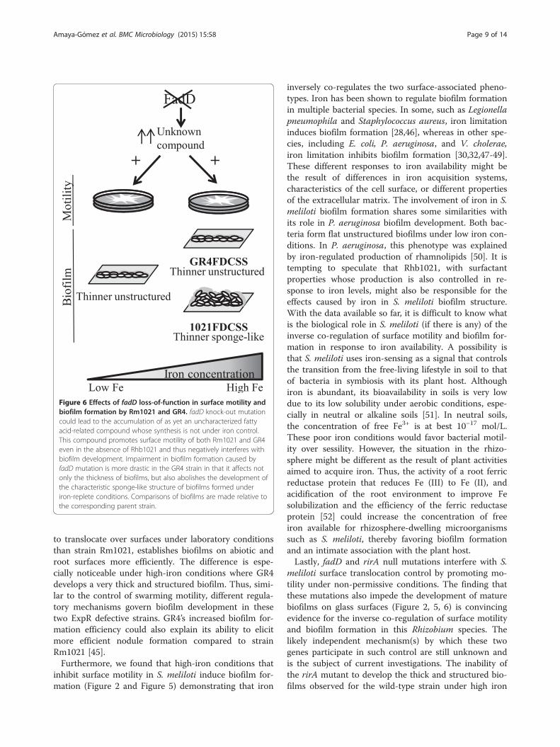
Figure 6 Effects of fadD loss-of-function in surface motility andbiofilm formation by Rm1021 and GR4. fadD knock-out mutationcould lead to the accumulation of as yet an uncharacterized fattyacid-related compound whose synthesis is not under iron control.This compound promotes surface motility of both Rm1021 and GR4even in the absence of Rhb1021 and thus negatively interferes withbiofilm development. Impairment in biofilm formation caused byfadD mutation is more drastic in the GR4 strain in that it affects notonly the thickness of biofilms, but also abolishes the development ofthe characteristic sponge-like structure of biofilms formed underiron-replete conditions. Comparisons of biofilms are made relative tothe corresponding parent strain.
Amaya-Gómez et al. BMC Microbiology (2015) 15:58 Page 9 of 14
to translocate over surfaces under laboratory conditionsthan strain Rm1021, establishes biofilms on abiotic androot surfaces more efficiently. The difference is espe-cially noticeable under high-iron conditions where GR4develops a very thick and structured biofilm. Thus, simi-lar to the control of swarming motility, different regula-tory mechanisms govern biofilm development in thesetwo ExpR defective strains. GR4’s increased biofilm for-mation efficiency could also explain its ability to elicitmore efficient nodule formation compared to strainRm1021 [45].Furthermore, we found that high-iron conditions that
inhibit surface motility in S. meliloti induce biofilm for-mation (Figure 2 and Figure 5) demonstrating that iron
inversely co-regulates the two surface-associated pheno-types. Iron has been shown to regulate biofilm formationin multiple bacterial species. In some, such as Legionellapneumophila and Staphylococcus aureus, iron limitationinduces biofilm formation [28,46], whereas in other spe-cies, including E. coli, P. aeruginosa, and V. cholerae,iron limitation inhibits biofilm formation [30,32,47-49].These different responses to iron availability might bethe result of differences in iron acquisition systems,characteristics of the cell surface, or different propertiesof the extracellular matrix. The involvement of iron in S.meliloti biofilm formation shares some similarities withits role in P. aeruginosa biofilm development. Both bac-teria form flat unstructured biofilms under low iron con-ditions. In P. aeruginosa, this phenotype was explainedby iron-regulated production of rhamnolipids [50]. It istempting to speculate that Rhb1021, with surfactantproperties whose production is also controlled in re-sponse to iron levels, might also be responsible for theeffects caused by iron in S. meliloti biofilm structure.With the data available so far, it is difficult to know whatis the biological role in S. meliloti (if there is any) of theinverse co-regulation of surface motility and biofilm for-mation in response to iron availability. A possibility isthat S. meliloti uses iron-sensing as a signal that controlsthe transition from the free-living lifestyle in soil to thatof bacteria in symbiosis with its plant host. Althoughiron is abundant, its bioavailability in soils is very lowdue to its low solubility under aerobic conditions, espe-cially in neutral or alkaline soils [51]. In neutral soils,the concentration of free Fe3+ is at best 10−17 mol/L.These poor iron conditions would favor bacterial motil-ity over sessility. However, the situation in the rhizo-sphere might be different as the result of plant activitiesaimed to acquire iron. Thus, the activity of a root ferricreductase protein that reduces Fe (III) to Fe (II), andacidification of the root environment to improve Fesolubilization and the efficiency of the ferric reductaseprotein [52] could increase the concentration of freeiron available for rhizosphere-dwelling microorganismssuch as S. meliloti, thereby favoring biofilm formationand an intimate association with the plant host.Lastly, fadD and rirA null mutations interfere with S.
meliloti surface translocation control by promoting mo-tility under non-permissive conditions. The finding thatthese mutations also impede the development of maturebiofilms on glass surfaces (Figure 2, 5, 6) is convincingevidence for the inverse co-regulation of surface motilityand biofilm formation in this Rhizobium species. Thelikely independent mechanism(s) by which these twogenes participate in such control are still unknown andis the subject of current investigations. The inability ofthe rirA mutant to develop the thick and structured bio-films observed for the wild-type strain under high iron

Amaya-Gómez et al. BMC Microbiology (2015) 15:58 Page 10 of 14
conditions, could be simply the result of growth defectscaused by oxidative stress due to derepressed iron up-take. In addition, the increased and deregulated produc-tion of Rhb1021 associated with the rirA mutationinhibits biofilm formation in S. meliloti just as overpro-duction and deregulation of rhamnolipids impede bio-film formation in P. aeruginosa [40]. Our data do notexclude the possibility that, besides Rhb1021 production,RirA also regulates other functions important for biofilmdevelopment.Our previous work revealed that fadD loss-of-function
deregulates normal surface motility in S. meliloti. Herewe show that fadD loss-of-function also obstructs nor-mal biofilm development on both glass and root surfacesin the two S. meliloti strains studied. However, the ef-fects are more pronounced in the GR4 strain than in theRm1021 genetic background. In contrast to the rirA mu-tant, the defects in biofilm formation shown by the twodifferent fadD mutants are not due to differences ingrowth or Rhb1021 production relative to the wild typesuggesting the involvement of a different mechanism[25]. Interestingly, gene expression analyses indicate thatfadD transcription is induced under low iron levels inboth Rm1021 and GR4 (Additional file 3). Whether thiseffect is somehow relevant to iron control over surfacemotility and biofilm formation in the wild-type situationis unknown, but if this is the case, it would be lost infadD loss-of-function mutants. In S. meliloti, FadD al-lows the utilization of exogenous and endogenous longchain fatty acids via their activation with CoA [33]. Inculture, S. meliloti fadD mutants accumulate a mixtureof free fatty acids during stationary phase [33], and fattyacids and fatty acid-related signals are known to influ-ence surface motility and biofilm formation in differentbacteria (reviewed in [53-55]). The hypothesis ofsurface-associated phenotypes in S. meliloti being af-fected either by changes in cellular fatty acid compos-ition and membrane fluidity or by the accumulation offatty acid-related compounds acting as biosurfactantsand/or signal molecules deserves further investigation.Although it is accepted that biofilm formation allows
rhizobia to survive under adverse environmental condi-tions, the exact function of biofilms in the Rhizobium-legume symbiosis remains elusive [20]. Recently, studiesof a nodD1 mutant and a quorum sensing-defectivestrain demonstrated that biofilm formation is crucial foroptimal root colonization and symbiosis between Sinor-hizobium fredii and soybean plants [56]. Our data alsosupport the importance of biofilm formation in promot-ing an effective symbiosis with the legume host. Al-though able to initiate nitrogen-fixing nodules, thestrains used in this study had been earlier described asbeing less competitive in nodule formation, and herewere found to be less efficient in biofilm formation. This
is true for Rm1021 vs GR4 [45] and the fadD mutant vsits parental strain [24]. Likewise, rhb and rirA mutantsof S. meliloti are able to nodulate and fix nitrogen insymbiosis with alfalfa plants [57,58]. However, by per-forming nodule competition experiments against thecorresponding parental strain, we found that the rhbmutant was as competitive as the parental strain (com-plementation caused by wild-type siderophore produc-tion could hinder any symbiotic defect), but a significantreduction in competitive ability was observed for therirA mutant, which occupied 27% fewer nodules thanthe wild-type strain (data not shown). It is conceivablethat biofilm formation defects in rhizobial strains mightcause only subtle symbiotic deficiencies, which occur atthe beginning of the interaction during the rootcolonization process. To be an efficient root colonizerwould be an important attribute under field conditionswhere competition with other microorganisms and ad-verse environmental conditions impose a strong selectivepressure.Moreover, the fact that fadD and rirA mutants are af-
fected in surface motility, biofilm formation, and plantroot colonization strongly suggests that genes that par-ticipate in the coordinated control of surface-associatedphenotypes are required for optimal interaction with thehost plant. Future research on iron acquisition systemsand iron-dependent regulation in S. meliloti as well as ofthe effects caused by the disruption of fadD will un-doubtedly facilitate the identification of new componentsthat govern biofilm formation and plant colonization inthis group of bacteria.
ConclusionsIn recent years, a growing appreciation has been devel-oping of the impact that biofilm formation has on thesuccess and outcome of plant-microbe interactions. Bac-terial biofilms seem not only to foster bacterial survivalagainst environmental stresses, but also to promote theinitiation of beneficial plant-microbe associations bymaintaining a dense population of cells in a specific lo-cation for a sufficient length of time. Our research demon-strates that by investigating the molecular mechanismsinvolved in bacterial surface motility, we can uncovercomponents that are key factors for biofilm formation andalso essential for plant root colonization. The major mo-lecular mechanisms known to govern surface motility in S.meliloti were found to be either essential for biofilm for-mation or to regulate both processes inversely in this rhi-zobial strain. We also conclude that the function exhibitedby rhizobactin 1021 in surface motility and biofilm devel-opment goes beyond its role as siderophore. AlthoughRhb1021 is not essential for the formation of nitrogen-fixing nodules, we demonstrate that it contributes to opti-mal root colonization, a characteristic that might be
Amaya-Gómez et al. BMC Microbiology (2015) 15:58 Page 11 of 14
relevant under field conditions. Furthermore, the environ-mental concentration of iron and genes fadD and rirAwere shown to coordinate surface motility and biofilm for-mation inversely. This study supports the hypothesis thatthe intimate regulation of surface motility and biofilm for-mation enables rhizobia to colonize roots optimally and toinitiate the establishment of the symbiosis with alfalfaplants successfully.
MethodsBacterial strains, plasmids, and growth conditionsThe bacterial strains and plasmids used in this study arelisted in Table 1. E. coli strains were grown in Luria-Bertani medium (LB) [59] at 37°C. S. meliloti strains wereroutinely cultured in Tryptone-Yeast extract complexmedium (TY) [60], or in Minimal Medium (MM) contain-ing glutamate (6.5 mM), mannitol (55 mM), mineral salts(K2HPO4, 1.3 mM; KH2PO4
.3H2O, 2.2 mM; MgSO4.7H2O,
0.6 mM; CaCl2.2H2O, 0.34 mM; FeCl3
.6H2O, 0.022 mM;NaCl, 0.86 mM) and vitamins (biotin (0.2 mg/L); calciumpantothenate (0.1 mg/L)) [61] at 30°C. Iron-replete MM(MMh) containing 220 μM of FeCl3 was prepared by add-ing the appropriate volume of a 100-fold concentratedstock solution of FeCl3 to MM without iron. The plasmidpHC60 [62] was introduced into S. meliloti strains by a bi-parental mating using the E. coli mobilizing strain S17-1.Antibiotics were added, as appropriate, at the followingfinal concentrations: for E. coli, tetracycline (Tc) 10 μg/ml;for S. meliloti, streptomycin (Sm) 200 μg/ml, spectino-mycin (Sp) 100 μg/ml, kanamycin (Km) 100 μg/ml, neo-mycin sulphate (Nm) 100 μg/ml, and Tc 10 μg/ml. ThefadD mutant strain GR4FDCSS was obtained followingthe same procedure described for 1021FDCS [25].
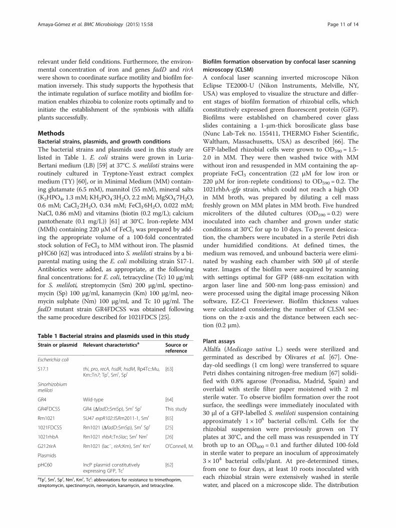
Table 1 Bacterial strains and plasmids used in this study
Strain or plasmid Relevant characteristicsa Source orreference
Escherichia coli
S17.1 thi, pro, recA, hsdR, hsdM, Rp4Tc::Mu,Km::Tn7; Tpr, Smr, Spr
[63]
Sinorhizobiummeliloti
GR4 Wild-type [64]
GR4FDCSS GR4 (ΔfadD::SmSp), Smr Spr This study
Rm1021 SU47 expR102::ISRm2011-1, Smr [65]
1021FDCSS Rm1021 (ΔfadD::SmSp), Smr Spr [25]
1021rhbA Rm1021 rhbA::Tn5lac; Smr Nmr [26]
G212rirA Rm1021 (lac−, rirA::Km), Smr Kmr O’Connell, M.
Plasmids
pHC60 IncP plasmid constitutivelyexpressing GFP, Tcr
[62]
aTpr, Smr, Spr, Nmr, Kmr, Tcr: abbreviations for resistance to trimethoprim,streptomycin, spectinomycin, neomycin, kanamycin, and tetracycline.
Biofilm formation observation by confocal laser scanningmicroscopy (CLSM)A confocal laser scanning inverted microscope NikonEclipse TE2000-U (Nikon Instruments, Melville, NY,USA) was employed to visualize the structure and differ-ent stages of biofilm formation of rhizobial cells, whichconstitutively expressed green fluorescent protein (GFP).Biofilms were established on chambered cover glassslides containing a 1-μm-thick borosilicate glass base(Nunc Lab-Tek no. 155411, THERMO Fisher Scientific,Waltham, Massachusetts, USA) as described [66]. TheGFP-labelled rhizobial cells were grown to OD590 = 1.5-2.0 in MM. They were then washed twice with MMwithout iron and resuspended in MM containing the ap-propriate FeCl3 concentration (22 μM for low iron or220 μM for iron-replete conditions) to OD590 = 0.2. The1021rhbA-gfp strain, which could not reach a high ODin MM broth, was prepared by diluting a cell massfreshly grown on MM plates in MM broth. Five hundredmicroliters of the diluted cultures (OD590 = 0.2) wereinoculated into each chamber and grown under staticconditions at 30°C for up to 10 days. To prevent desicca-tion, the chambers were incubated in a sterile Petri dishunder humidified conditions. At defined times, themedium was removed, and unbound bacteria were elimi-nated by washing each chamber with 500 μl of sterilewater. Images of the biofilm were acquired by scanningwith settings optimal for GFP (488-nm excitation withargon laser line and 500-nm long-pass emission) andwere processed using the digital image processing Nikonsoftware, EZ-C1 Freeviewer. Biofilm thickness valueswere calculated considering the number of CLSM sec-tions on the z-axis and the distance between each sec-tion (0.2 μm).
Plant assaysAlfalfa (Medicago sativa L.) seeds were sterilized andgerminated as described by Olivares et al. [67]. One-day-old seedlings (1 cm long) were transferred to squarePetri dishes containing nitrogen-free medium [67] solidi-fied with 0.8% agarose (Pronadisa, Madrid, Spain) andoverlaid with sterile filter paper moistened with 2 mlsterile water. To observe biofilm formation over the rootsurface, the seedlings were immediately inoculated with30 μl of a GFP-labelled S. meliloti suspension containingapproximately 1 × 10⁶ bacterial cells/ml. Cells for therhizobial suspension were previously grown on TYplates at 30°C, and the cell mass was resuspended in TYbroth up to an OD600 = 0.1 and further diluted 100-foldin sterile water to prepare an inoculum of approximately3 × 104 bacterial cells/plant. At pre-determined times,from one to four days, at least 10 roots inoculated witheach rhizobial strain were extensively washed in sterilewater, and placed on a microscope slide. The distribution

Amaya-Gómez et al. BMC Microbiology (2015) 15:58 Page 12 of 14
of bacteria along the root was observed using a LeicaDMI600B epifluorescence inverted microscope (Wetzlar,Germany). Digital images of biofilms on roots were takenusing a Nikon Eclipse TE2000-U inverted microscope(488-nm argon laser excitation and 500-nm long-passemission) and analyzed using digital image processing EZ-C1 Freeviewer Nikon software.Colonization of the bacteria on the root and the root
hairs was also evaluated by determining colony-formingunits (CFUs). Two-day-old seedlings were inoculatedwith 30 μl of a suspension containing approximately 3 ×10⁵ bacterial cells, prepared as described above. At de-fined times, 15 seedlings were extensively washed withsterile water to remove the not-firmly attached bacteria,and any water excess was removed using sterile filterpaper, and the roots were weighed. Three roots wereplaced into a 1.5 ml Eppendorf tube containing 1 ml ofsterile TE buffer and the attached cells were released by2 sonication pulses of 1 min each in an Ultrasons-H (J.P.Selecta, Barcelona, Spain) sonication bath with a pausetime of 1 min between the pulses, and subsequentlyquantified by counting CFUs (normalized to grams ofroot). The experiment was repeated three times.
Statistical analysisValues presented are the means and standard deviation(SD) of repeated experiments. Data were subject to one-way ANOVA test followed by comparison of multipletreatment levels with the control using the Games-Howell test. Biofilm thickness data was analysed ascounts of number of CLSM z-sections using generalizedlinear models (GLMs) followed by multiple comparisonsof treatments with Tukey’s tests. Statistical analyses wereperformed with SPSS 15.0 and R version 3.1.0 for Win-dows (package “multcomp”).
Additional files
Additional file 1: Effect of iron concentration and different genemutations on the structure of Rm1021 biofilms. Confocal laserscanning microscopy (CLSM) images showing the xy view of 3- and 10-day-old biofilms developed by GFP-labelled Rm1021 and Rm1021-derivedfadD, rhb and rirA mutant cells on chambered cover glass slides aftergrowth in MM containing different iron concentrations. All differentstrains develop flat unstructured biofilms when they are grown in MMcontaining low iron concentration (22 μM FeCl3). Growth in the presenceof high iron conditions (220 μM FeCl3) induces the formation of sponge-like structured biofilms except in the case of the rirA mutant. Bars, 15 μm.
Additional file 2: Effect of iron concentration and fadD-loss-offunction on the structure of S. meliloti GR4 biofilms. CLSM imagesshowing the xy view of 3-, 6- and 10-day-old biofilms developed by GFP-labelled GR4 and fadD derivative mutant cells on chambered cover glassslides after growth in MM containing different iron concentrations. Bothstrains develop flat unstructured biofilms when they are grown in MMcontaining low iron concentration (22 μM FeCl3). In the presence of highiron conditions (220 μM FeCl3), the wild-type strain GR4 develops highlystructured biofilms. The characteristic sponge-like structure of thesebiofilms is lost after 10 days, suggesting the initiation of biofilm dispersal.
The fadD mutant (GR4FDCSS) is unable to develop structured biofilmseven in the presence of iron-rich conditions. Bars, 15 μm.
Additional file 3: Effect of iron concentration on fadD gene expressionin S. meliloti GR4 and Rm1021.
Competing interestsThe authors declare that they have no competing interests.
Authors’ contributionsCVAG conducted all the experiments. CVAG and MJS designed the study,analyzed and interpreted the data and wrote the manuscript. AMH providedUCLA facilities and was involved in drafting the manuscript and revising itcritically. All authors read and approved the final manuscript.
AcknowledgementsThis work was supported by grants BIO2007-62988, BIO2010-18005, andBIO2013-42801-P from the Ministerio de Ciencia e Innovación (MICINN,Spain), CVI 03541 from the Junta de Andalucía (Spain), and FEDER funds.CAVG was supported by a FPI fellowship from MICINN. We also thank thehelp of the Microscopy Service of the Estación Experimental del Zaidín (CSIC).Lastly, we are grateful to Nancy A. Fujishige (UCLA) for her help with the biofilmexperiments. Work in the Hirsch lab was funded by grant IOB-0537497 from theNational Science Foundation (USA).
Author details1Departamento de Microbiología del Suelo y Sistemas Simbióticos, EstaciónExperimental del Zaidín, Consejo Superior de Investigaciones Científicas(CSIC), Profesor Albareda 1, 18008 Granada, Spain. 2Department of Molecular,Cell, and Developmental Biology and Molecular Biology Institute, Universityof California-Los Angeles, Los Angeles, CA 90095-1606, USA.
Received: 5 November 2014 Accepted: 18 February 2015
References1. Davey ME, O'Toole GA. Microbial biofilms: from ecology to molecular
genetics. Microbiol Mol Biol Rev. 2000;64:847–67.2. O'Toole G, Kaplan HB, Kolter R. Biofilm formation as microbial development.
Annu Rev Microbiol. 2000;54:49–79.3. Kearns DB. A field guide to bacterial swarming motility. Nat Rev Micro.
2010;8:634–44.4. Daniels R, Vanderleyden J, Michiels J. Quorum sensing and swarming
migration in bacteria. FEMS Microbiol Rev. 2004;28:261–89.5. Lai S, Tremblay J, Déziel E. Swarming motility: a multicellular behaviour
conferring antimicrobial resistance. Environ Microbiol. 2009;11:126–36.6. Overhage J, Bains M, Brazas MD, Hancock REW. Swarming of Pseudomonas
aeruginosa is a complex adaptation leading to increased production ofvirulence factors and antibiotic resistance. J Bacteriol. 2008;190:2671–9.
7. Tambalo DD, Yost CK, Hynes MF. Characterization of swarming motility inRhizobium leguminosarum bv. viciae. FEMS Microbiol Lett. 2010;307:165–74.
8. Verstraeten N, Braeken K, Debkumari B, Fauvart M, Fransaer J, Vermant J,et al. Living on a surface: swarming and biofilm formation. Trends Microbiol.2008;16:496–506.
9. Caiazza NC, Merritt JH, Brothers KM, O'Toole GA. Inverse regulation ofbiofilm formation and swarming motility by Pseudomonas aeruginosa PA14.J Bacteriol. 2007;189:3603–12.
10. Kuchma SL, Brothers KM, Merritt JH, Liberati NT, Ausubel FM, O'Toole GA.BifA, a cyclic-di-GMP phosphodiesterase, inversely regulates biofilm formationand swarming motility by Pseudomonas aeruginosa PA14. J Bacteriol.2007;189:8165–78.
11. Kuchma SL, Griffin EF, O'Toole GA. Minor pilins of the type IV pilus systemparticipate in the negative regulation of swarming motility. J Bacteriol.2012;194:5388–403.
12. Merritt JH, Brothers KM, Kuchma SL, O'Toole GA. SadC reciprocallyinfluences biofilm formation and swarming motility via modulation ofexopolysaccharide production and flagellar function. J Bacteriol.2007;189:8154–64.
13. Pratt JT, McDonough E, Camilli A. PhoB regulates motility, biofilms, andcyclic di-GMP in Vibrio cholerae. J Bacteriol. 2009;191:6632–42.

Amaya-Gómez et al. BMC Microbiology (2015) 15:58 Page 13 of 14
14. Trimble MJ, McCarter LL. Bis-(3′-5′)-cyclic dimeric GMP-linked quorumsensing controls swarming in Vibrio parahaemolyticus. Proc Natl Acad Sci US A. 2011;108:18079–84.
15. Oldroyd GED, Downie JA. Coordinating nodule morphogenesis withrhizobial infection in legumes. Annu Rev Plant Biol. 2008;59:519–46.
16. Fujishige NA, Kapadia NN, De Hoff PL, Hirsch AM. Investigations ofRhizobium biofilm formation. FEMS Microbiol Ecol. 2006;56:195–206.
17. Rinaudi L, Fujishige NA, Hirsch AM, Banchio E, Zorreguieta Á, Giordano W.Effects of nutritional and environmental conditions on Sinorhizobium melilotibiofilm formation. Res Microbiol. 2006;157:867–75.
18. Rinaudi LV, González JE. The low-molecular-weight fraction of exopolysaccharideII from Sinorhizobium meliloti is a crucial determinant of biofilm formation.J Bacteriol. 2009;191:7216–24.
19. Sorroche FG, Spesia MB, Zorreguieta Á, Giordano W. A positive correlationbetween bacterial autoaggregation and biofilm formation in nativeSinorhizobium meliloti isolates from Argentina. Appl Environ Microbiol.2012;78:4092–101.
20. Rinaudi LV, Giordano W. An integrated view of biofilm formation in rhizobia.FEMS Microbiol Lett. 2010;304:1–11.
21. Fujishige NA, Lum MR, De Hoff PL, Whitelegge JP, Faull KF, Hirsch AM.Rhizobium common nod genes are required for biofilm formation. MolMicrobiol. 2008;67:504–15.
22. Wells DH, Chen EJ, Fisher RF, Long SR. ExoR is genetically coupled to theExoS-ChvI two-component system and located in the periplasm ofSinorhizobium meliloti. Mol Microbiol. 2007;64:647–64.
23. Santos MR, Marques AT, Becker JD, Moreira LM. The Sinorhizobium melilotiEmrR regulator is required for efficient colonization of Medicago sativa rootnodules. Mol Plant-Microbe Interact. 2014;27:388–99.
24. Soto MJ, Fernández-Pascual M, Sanjuán J, Olivares J. A fadD mutant ofSinorhizobium meliloti shows multicellular swarming migration and isimpaired in nodulation efficiency on alfalfa roots. Mol Microbiol.2002;43:371–82.
25. Nogales J, Domínguez-Ferreras A, Amaya-Gómez C, van Dillewijn P, CuéllarV, Sanjuán J, et al. Transcriptome profiling of a Sinorhizobium meliloti fadDmutant reveals the role of rhizobactin 1021 biosynthesis and regulationgenes in the control of swarming. BMC Genomics. 2010;11:157.
26. Nogales J, Bernabéu-Roda L, Cuéllar V, Soto MJ. ExpR is not required forswarming but promotes sliding in Sinorhizobium meliloti. J Bacteriol.2012;194:2027–35.
27. Gao M, Coggin A, Yagnik K, Teplitski M. Role of specific quorum-sensingsignals in the regulation of exopolysaccharide II production withinSinorhizobium meliloti spreading colonies. PLoS One. 2012;7:e42611.
28. Hindré T, Brüggemann H, Buchrieser C, Héchard Y. Transcriptional profilingof Legionella pneumophila biofilm cells and the influence of iron on biofilmformation. Microbiology. 2008;154:30–41.
29. Trappetti C, Potter AJ, Paton AW, Oggioni MR, Paton JC. LuxS mediatesiron-dependent biofilm formation, competence, and fratricide inStreptococcus pneumoniae. Infec Immun. 2011;79:4550–8.
30. Banin E, Vasil ML, Greenberg EP. Iron and Pseudomonas aeruginosa biofilmformation. Proc Natl Acad Sci U S A. 2005;102:11076–81.
31. Ojha A, Hatfull GF. The role of iron in Mycobacterium smegmatis biofilmformation: the exochelin siderophore is essential in limiting iron conditions forbiofilm formation but not for planktonic growth. Mol Microbiol. 2007;66:468–83.
32. Wu Y, Outten FW. IscR controls iron-dependent biofilm formation in Escherichiacoli by regulating type I fimbria expression. J Bacteriol. 2009;191:1248–57.
33. Pech-Canul Á, Nogales J, Miranda-Molina A, Álvarez L, Geiger O, Soto MJ,et al. FadD is required for utilization of endogenous fatty acids releasedfrom membrane lipids. J Bacteriol. 2011;193:6295–304.
34. Amaya-Gómez C. Transcriptomic approach for the identification of genesand signals playing a role in swarming motility of Sinorhizobium meliloti:Connection with biofilm formation and symbiosis. PhD thesis. University ofGranada Spain, Department of Microbiology; 2013. http://hdl.handle.net/10481/29516.
35. Chao T-C, Buhrmester J, Hansmeier N, Pühler A, Weidner S. Role of theregulatory gene rirA in the transcriptional response of Sinorhizobium melilotito iron limitation. Appl Environ Microbiol. 2005;71:5969–82.
36. May T, Okabe S. Enterobactin is required for biofilm development inreduced-genome Escherichia coli. Environ Microbiol. 2011;13:3149–62.
37. Persmark M, Pittman P, Buyer JS, Schwyn B, Gill PR, Neilands JB. Isolationand structure of rhizobactin 1021, a siderophore from the alfalfa symbiontRhizobium meliloti 1021. J Am Chem Soc. 1993;115:3950–6.
38. Mireles JR, Toguchi A, Harshey RM. Salmonella enterica Serovar Typhimuriumswarming mutants with altered biofilm-forming abilities: surfactin inhibitsbiofilm formation. J Bacteriol. 2001;183:5848–54.
39. Kuiper I, Lagendijk EL, Pickford R, Derrick JP, Lamers GEM, Thomas-Oates JE,et al. Characterization of two Pseudomonas putida lipopeptide biosurfactants,putisolvin I and II, which inhibit biofilm formation and break down existingbiofilms. Mol Microbiol. 2004;51:97–13.
40. Davey ME, Caiazza NC, O'Toole GA. Rhamnolipid surfactant productionaffects biofilm architecture in Pseudomonas aeruginosa PAO1. J Bacteriol.2003;185:1027–36.
41. Boles BR, Thoendel M, Singh PK. Rhamnolipids mediate detachment ofPseudomonas aeruginosa from biofilms. Mol Microbiol. 2005;57:1210–23.
42. Pamp SJ, Tolker-Nielsen T. Multiple roles of biosurfactants in structural biofilmdevelopment by Pseudomonas aeruginosa. J Bacteriol. 2007;189:2531–9.
43. Daniels R, Reynaert S, Hoekstra H, Verreth C, Janssens J, Braeken K, et al.Quorum signal molecules as biosurfactants affecting swarming in Rhizobiumetli. Proc Natl Acad Sci U S A. 2006;103:14965–70.
44. Lamont IL, Beare PA, Ochsner U, Vasil AI, Vasil ML. Siderophore-mediatedsignaling regulates virulence factor production in Pseudomonas aeruginosa.Proc Natl Acad Sci U S A. 2002;99:7072–7.
45. Toro N, Olivares J. Characterization of a large plasmid of Rhizobium melilotiinvolved in enhancing nodulation. Mol Gen Genet. 1986;202:331–5.
46. Johnson M, Cockayne A, Williams PH, Morrissey JA. Iron-responsive regulationof biofilm formation in Staphylococcus aureus involves Fur-dependent andFur-independent mechanisms. J Bacteriol. 2005;187:8211–5.
47. Banin E, Brady KM, Greenberg EP. Chelator-induced dispersal and killing ofPseudomonas aeruginosa cells in a biofilm. Appl Environ Microbiol.2006;72:2064–9.
48. Singh PK, Parsek MR, Greenberg EP, Welsh MJ. A component of innateimmunity prevents bacterial biofilm development. Nature. 2002;417:552–5.
49. Mey AR, Craig SA, Payne SM. Characterization of Vibrio cholerae RyhB: the RyhBregulon and role of ryhB in biofilm formation. Infect Immun. 2005;73:5706–19.
50. Glick R, Gilmour C, Tremblay J, Satanower S, Avidan O, Déziel E, et al.Increase in rhamnolipid synthesis under iron-limiting conditions influencessurface motility and biofilm formation in Pseudomonas aeruginosa. J Bacteriol.2010;192:2973–80.
51. Lindsay WL, Schwab AP. The chemistry of iron and its availability to plants.J Plant Nutr. 1982;5:821–42.
52. Kobayashi T, Nishizawa NK. Iron uptake, translocation, and regulation inhigher plants. Annu Rev Plant Biol. 2012;63:131–52.
53. Ryan RP, Dow JM. Communication with a growing family: diffusible signalfactor (DSF) signaling in bacteria. Trends Microbiol. 2011;19:145–52.
54. Tiaden A, Spirig T, Hilbi H. Bacterial gene regulation by alpha-hydroxyketonesignaling. Trends Microbiol. 2010;18:288–97.
55. Winans SC. A new family of quorum sensing pheromones synthesized usingS-adenosylmethionine and Acyl-CoAs. Mol Microbiol. 2011;79:1403–6.
56. Pérez-Montaño F, Jiménez-Guerrero I, Del Cerro P, Baena-Ropero I, López-Baena FJ, Ollero FJ, et al. The symbiotic biofilm of Sinorhizobium frediiSMH12, necessary for successful colonization and symbiosis of Glycine maxcv. Osumi, is regulated by quorum sensing systems and inducing flavonoidsvia NodD1. PLoS One. 2014;9:e105901.
57. Lynch D, O'Brien J, Welch T, Clarke P, Cuiv PO, Crosa JH, et al. Geneticorganization of the region encoding regulation, biosynthesis, and transportof rhizobactin 1021, a siderophore produced by Sinorhizobium meliloti.J Bacteriol. 2001;183:2576–85.
58. Viguier C, Cuiv PO, Clarke P, O'Connell M. RirA is the iron response regulatorof the rhizobactin 1021 biosynthesis and transport genes in Sinorhizobiummeliloti 2011. FEMS Microbiol Lett. 2005;246:235–42.
59. Sambrook J, Russell D. Molecular cloning: a laboratory manual. New York:Cold Spring Harbor Laboratory Pres; 2001.
60. Beringer JE. R factor transfer in Rhizobium leguminosarum. J Gen Microbiol.1974;84:188–98.
61. Robertsen BK, Åman P, Darvill AG, McNeil M, Albersheim P. The structure ofacidic extracellular polysaccharides secreted by Rhizobium leguminosarumand Rhizobium trifolii. Plant Physiol. 1981;67:389–400.
62. Cheng HP, Walker GC. Succinoglycan is required for initiation andelongation of infection threads during nodulation of alfalfa by Rhizobiummeliloti. J Bacteriol. 1998;180:5183–91.
63. Simon R, Priefer U, Pühler A. A broad host range mobilization system for Invivo genetic engineering: Transposon mutagenesis in Gram negativebacteria. Nat Biotech. 1983;1:784–91.

Amaya-Gómez et al. BMC Microbiology (2015) 15:58 Page 14 of 14
64. Casadesús J, Olivares J. Rough and fine linkage mapping of the Rhizobiummeliloti chromosome. Mol Gen Genet. 1979;174:203–9.
65. Meade HM, Signer ER. Genetic mapping of Rhizobium meliloti. Proc NatlAcad Sci U S A. 1977;74:2076–8.
66. Russo DM, Williams A, Edwards A, Posadas DM, Finnie C, Dankert M, et al.Proteins exported via the PrsD-PrsE type I secretion system and the acidicexopolysaccharide are involved in biofilm formation by Rhizobiumleguminosarum. J Bacteriol. 2006;188:4474–86.
67. Olivares J, Casadesús J, Bedmar EJ. Method for testing degree of infectivityof Rhizobium melioti strains. Appl Environ Microbiol. 1980;39:967–70.
Submit your next manuscript to BioMed Centraland take full advantage of:
• Convenient online submission
• Thorough peer review
• No space constraints or color figure charges
• Immediate publication on acceptance
• Inclusion in PubMed, CAS, Scopus and Google Scholar
• Research which is freely available for redistribution
Submit your manuscript at www.biomedcentral.com/submit
Copyright © 2022 FDOKUMEN