Bimetallic Cu–Ni nanoparticles of varying composition (CuNi 3, CuNi, Cu 3Ni
7
Colloids and Surfaces A: Physicochem. Eng. Aspects 331 (2008) 206–212 Contents lists available at ScienceDirect Colloids and Surfaces A: Physicochemical and Engineering Aspects journal homepage: www.elsevier.com/locate/colsurfa Bimetallic Cu–Ni nanoparticles of varying composition (CuNi 3 , CuNi, Cu 3 Ni) Jahangeer Ahmed a , Kandalam V. Ramanujachary b , Samuel E. Lofland b , Anthony Furiato b , Govind Gupta c , S.M. Shivaprasad c , Ashok K. Ganguli a,∗ a Department of Chemistry, Indian Institute of Technology, Hauz Khas, New Delhi 110016, India b Department of Chemistry and Biochemistry, Rowan University, 201 Mullica Hill Road, Glasboro, NJ 08028, USA c Surface Chemistry Division, National Physical Laboratory, New Delhi 110012, India article info Article history: Received 19 March 2008 Received in revised form 2 August 2008 Accepted 7 August 2008 Available online 19 August 2008 Keywords: Microemulsion Bimetallic nanoparticles Cu–Ni Magnetism abstract Cu–Ni (1:3, 1:1, 3:1) bimetallic nanoparticles have been synthesized by a microemulsion route using CTAB (cetyltrimethyl ammoniumbromide) as the surfactant and hydrazine/NaOH as the precipitating agent fol- lowed by reduction in hydrogen atmosphere. This is the first report of the synthesis of CuNi 3 and Cu 3 Ni nanoparticles. Transmission electron microscopy (TEM) studies show nearly monodisperse and uniform nanoparticles of average grain size of 7nm for the 1:1 alloy nanoparticles while the 1:3 and 3:1 com- positions show uniform nanoparticles with the grain size in the range of 20–30nm. X-ray photoelectron spectroscopy (XPS) studies confirm the expected Cu/Ni ratios in these alloys. These bimetallic nanopar- ticles show ferromagnetic behavior and the magnetization decreases with increase in concentration of copper. The specific surface area of these alloy nanoparticles was found to be in the range of 6–9.42 m 2 /g. © 2008 Elsevier B.V. All rights reserved. 1. Introduction The preparation of nanoparticles with controlled size, mor- phology and composition are of great interest because of their unique physical and chemical properties. Bimetallic nanoparticles are particularly attractive due to their unusual magnetic [1–6], electronic [1,7], biological [8,9] and catalytic properties [10–12]. Among various alloys the Cu–Ni system shows good catalytic prop- erties that depend on the ratio of Cu:Ni e.g. in the hydrogenation of refined soybean oil [13] and for direct oxidation of methane in solid-oxide fuel cells [14]. The catalysts that have a very high or very low Cu:Ni ratio are almost inactive as compared to CuNi 3 and Cu 2 Ni. Bimetallic Cu–Ni particles composed of a nickel-rich and copper-rich solid solution have been synthesized earlier using carbonates [15]. A number of other techniques have been used for producing bimetallic nanoparticles such as the hydrothermal method [16,17], mechanical alloying [3,18], the sol–gel method [5], the polyol process [19], and pulsed-laser induced quenching [20]. Nanocrystalline alloy films of Au, Ag and Cu have been formed at the organic–aqueous interface [21]. A microemulsion route has been employed to obtain the bimetallic nanoparticles of Au–Ag [22], Au–Pd [23] and Au–Pt [24]. The average size of these alloys lies in the range of 10–20 nm. Several ∗ Corresponding author. Tel.: +91 11 26591511; fax: +91 11 26854715. E-mail address: [email protected] (A.K. Ganguli). metal nanoparticles (Pt, Bi, Pb, Fe, Cd, Ag, Au, Cu and Ni) have been synthesized using water-in-oil microemulsions [25,26]. Reverse micelles are water-in-oil microemulsions consisting of nanometer- sized water droplets that are dispersed in an oil medium and stabilized by surfactants [27]. These water droplets are suitable as reaction media for the synthesis of nanoparticles where the size of particles can be controlled by varying the molar ratio of water to surfactant. The microemulsion route has been used in the recent past to obtain several oxalates and oxides with a high degree of homogeneity and phase purity [28–30]. The synthesis of the nanoparticles of Cu–Ni alloy in 1:1 ratio by the microemulsion method using sodium dodecyl sulfate (as anionic surfactant) and oleic acid has been reported earlier [31,32]. In this paper we report the synthesis as well as properties of nanocrystalline bimetallic alloys of varying composition i.e. CuNi 3 , CuNi and Cu 3 Ni using the microemulsion route. We have charac- terized the alloy powders in detail using powder X-ray diffraction, transmission electron microscopy (TEM), X-ray photoelectron spec- troscopy (XPS), magnetization and surface area studies. 2. Materials and methods Cu(NO 3 ) 2 ·3H 2 O (CDH, 98%), Ni(NO 3 ) 2 ·6H 2 O (Qualigens, AR, 98%), NaOH (Qualigens, LR, 97%), N 2 H 4 ·H 2 O (Qualigens, 99%), cetyl trimethylammonium bromide (CTAB) (Spectrochem, AR, 99%), 1- butanol (Qualigens, 99.5%), and iso-octane (spectrochem, 99%) were used in the synthesis of the Cu–Ni alloy nanoparticles. 0927-7757/$ – see front matter © 2008 Elsevier B.V. All rights reserved. doi:10.1016/j.colsurfa.2008.08.007
-
Upload
independent -
Category
Documents
-
view
1 -
download
0
Transcript of Bimetallic Cu–Ni nanoparticles of varying composition (CuNi 3, CuNi, Cu 3Ni

Colloids and Surfaces A: Physicochem. Eng. Aspects 331 (2008) 206–212
Contents lists available at ScienceDirect
Colloids and Surfaces A: Physicochemical andEngineering Aspects
journa l homepage: www.e lsev ier .com/ locate /co lsur fa
Bimetallic Cu–Ni nanoparticles of varying composition (CuNi3, CuNi, Cu3Ni)
Jahangeer Ahmeda, Kandalam V. Ramanujacharyb, Samuel E. Loflandb,Anthony Furiatob, Govind Guptac, S.M. Shivaprasadc, Ashok K. Ganguli a,∗
a Department of Chemistry, Indian Institute of Technology, Hauz Khas, New Delhi 110016, Indiab Department of Chemistry and Biochemistry, Rowan University, 201 Mullica Hill Road, Glasboro, NJ 08028, USAc Surface Chemistry Division, National Physical Laboratory, New Delhi 110012, India
a r t i c l e i n f o
Article history:Received 19 March 2008Received in revised form 2 August 2008Accepted 7 August 2008Available online 19 August 2008
Keywords:
a b s t r a c t
Cu–Ni (1:3, 1:1, 3:1) bimetallic nanoparticles have been synthesized by a microemulsion route using CTAB(cetyltrimethyl ammoniumbromide) as the surfactant and hydrazine/NaOH as the precipitating agent fol-lowed by reduction in hydrogen atmosphere. This is the first report of the synthesis of CuNi3 and Cu3Ninanoparticles. Transmission electron microscopy (TEM) studies show nearly monodisperse and uniformnanoparticles of average grain size of 7 nm for the 1:1 alloy nanoparticles while the 1:3 and 3:1 com-positions show uniform nanoparticles with the grain size in the range of 20–30 nm. X-ray photoelectron
MicroemulsionBimetallic nanoparticlesCM
spectroscopy (XPS) studies confirm the expected Cu/Ni ratios in these alloys. These bimetallic nanopar-ticles show ferromagnetic behavior and the magnetization decreases with increase in concentration of
e are 2
1
puaeAeosvaacfmtNt
bT
msmssrpsph
baInCttt
0d
u–Niagnetism
copper. The specific surfac
. Introduction
The preparation of nanoparticles with controlled size, mor-hology and composition are of great interest because of theirnique physical and chemical properties. Bimetallic nanoparticlesre particularly attractive due to their unusual magnetic [1–6],lectronic [1,7], biological [8,9] and catalytic properties [10–12].mong various alloys the Cu–Ni system shows good catalytic prop-rties that depend on the ratio of Cu:Ni e.g. in the hydrogenationf refined soybean oil [13] and for direct oxidation of methane inolid-oxide fuel cells [14]. The catalysts that have a very high orery low Cu:Ni ratio are almost inactive as compared to CuNi3nd Cu2Ni. Bimetallic Cu–Ni particles composed of a nickel-richnd copper-rich solid solution have been synthesized earlier usingarbonates [15]. A number of other techniques have been usedor producing bimetallic nanoparticles such as the hydrothermal
ethod [16,17], mechanical alloying [3,18], the sol–gel method [5],he polyol process [19], and pulsed-laser induced quenching [20].anocrystalline alloy films of Au, Ag and Cu have been formed at
he organic–aqueous interface [21].A microemulsion route has been employed to obtain the
imetallic nanoparticles of Au–Ag [22], Au–Pd [23] and Au–Pt [24].he average size of these alloys lies in the range of 10–20 nm. Several
∗ Corresponding author. Tel.: +91 11 26591511; fax: +91 11 26854715.E-mail address: [email protected] (A.K. Ganguli).
2
9tbu
927-7757/$ – see front matter © 2008 Elsevier B.V. All rights reserved.oi:10.1016/j.colsurfa.2008.08.007
a of these alloy nanoparticles was found to be in the range of 6–9.42 m /g.© 2008 Elsevier B.V. All rights reserved.
etal nanoparticles (Pt, Bi, Pb, Fe, Cd, Ag, Au, Cu and Ni) have beenynthesized using water-in-oil microemulsions [25,26]. Reverseicelles are water-in-oil microemulsions consisting of nanometer-
ized water droplets that are dispersed in an oil medium andtabilized by surfactants [27]. These water droplets are suitable aseaction media for the synthesis of nanoparticles where the size ofarticles can be controlled by varying the molar ratio of water tourfactant. The microemulsion route has been used in the recentast to obtain several oxalates and oxides with a high degree ofomogeneity and phase purity [28–30].
The synthesis of the nanoparticles of Cu–Ni alloy in 1:1 ratioy the microemulsion method using sodium dodecyl sulfate (asnionic surfactant) and oleic acid has been reported earlier [31,32].n this paper we report the synthesis as well as properties ofanocrystalline bimetallic alloys of varying composition i.e. CuNi3,uNi and Cu3Ni using the microemulsion route. We have charac-erized the alloy powders in detail using powder X-ray diffraction,ransmission electron microscopy (TEM), X-ray photoelectron spec-roscopy (XPS), magnetization and surface area studies.
. Materials and methods
Cu(NO3)2·3H2O (CDH, 98%), Ni(NO3)2·6H2O (Qualigens, AR,8%), NaOH (Qualigens, LR, 97%), N2H4·H2O (Qualigens, 99%), cetylrimethylammonium bromide (CTAB) (Spectrochem, AR, 99%), 1-utanol (Qualigens, 99.5%), and iso-octane (spectrochem, 99%) weresed in the synthesis of the Cu–Ni alloy nanoparticles.

J. Ahmed et al. / Colloids and Surfaces A: Physicochem. Eng. Aspects 331 (2008) 206–212 207
Ff
mmifNTsoIsMsasoffit
Fw
ig. 1. Variation in the conductance of (CTAB/1-butanol/iso-octane) system as aunction of water content.
Cu–Ni bimetallic nanoparticles were synthesized using theicroemulsion route with CTAB (cetyltrimethyl ammonium bro-ide) as the surfactant, 1-butanol as the co-surfactant and
so-octane as the oil phase. Four microemulsions with dif-erent aqueous phases containing 0.1 M Cu(NO3)2·3H2O, 0.1 Mi(NO3)2·6H2O, 20 M N2H4·H2O and 0.1 M NaOH were prepared.he weight fractions of various constituents in these microemul-ions are as follows: 16.76% of CTAB, 13.90% of 1-butanol, 59.29%f iso-octane and 10.05% of the aqueous phase. Microemulsion(containing 0.1 M Cu(NO3)2·3H2O) was mixed to microemul-
ion II (containing 0.1 M Ni(NO3)2·6H2O) which is our system A.icroemulsion III (containing hydrazine) was mixed to microemul-
ion IV (containing NaOH) which we call as system B. Systems And B were optically transparent. These two systems were mixed
lowly on a magnetic stirrer and a brown-coloured precipitate wasbtained immediately. The colour of the colloidal solution changedrom light brown to dark brown (∼12 h) to crimson (∼40 h) andnally a green precipitate (∼70 h) was obtained after centrifuga-ion. Colloidal particles have a tendency to aggregate on agingig. 2. Phase diagram of blend (CTAB + 1-butanol)/water/iso-octane system ineight fraction.
F∼
acowwT5(nan
Bw(twtap
ig. 3. Powder X-ray diffraction patterns of (X) brown colour powder (aged for12 h) and (Y) crimson colour powder (aged for ∼40 h).
nd the size of colloidal particles increases with aging. The opti-al properties (colour) would have varied as it depends on the sizef colloidal particles in solution. The green precipitate was washedith 1:1 chloroform/methanol mixture and dried in air and thisas used as a precursor for the synthesis of alloy nanoparticles.
he above precursor when annealed in hydrogen atmosphere at00 ◦C for 5 h led to the formation of monophasic bimetallic CuNi1:1) alloy powder. We have also synthesized CuNi3 and Cu3Nianoparticles using the same method as given above, starting withppropriate stoichiometries of Cu and Ni solutions. All the alloyano-powders were black in colour.
Powder X-ray diffraction (PXRD) studies were carried out on aruker D8 Advance diffractometer with Ni-filtered Cu K� radiationith a scan speed of 1 s and scan step of 0.05◦. The crystallite size
t) was determined from the X-ray line broadening studies usinghe Scherrer’s formula [33] (t = 0.9 �/(Bcos �)) where � is the X-ray
avelength, B the full-width half-maximum of the Bragg diffrac-ion peak at angle �. The details of the data collection methodologynd evaluation of crystallite size are given elsewhere [28]. The cellarameters were determined from a least-squares fitting procedure
208 J. Ahmed et al. / Colloids and Surfaces A: Physicochem. Eng. Aspects 331 (2008) 206–212
Table 1CTAB as cationic surfactant/1-butanol as co-surfactant/iso-octane (oil phase)/water
S. no. Amount ofsurfactant (g)
Volume ofco-surfactant (ml)
Volume of oil phase (ml) Volume of water after whichturbidity appears (ml)
Conductance (Sm−1)
1234
osop
FC
5.2075 5.3325 26.2505.2075 5.3325 39.3755.2075 5.3325 52.5005.2075 5.3325 65.625
f all observed reflections with quartz as the external standard. TEMtudies were carried out with a Technai G2 20 electron microscopeperated at 200 kV. The specimens for TEM were prepared by dis-ersing the powder in acetone by ultrasonic treatment, dropping
ig. 4. Powder X-ray diffraction patterns of nanocrystalline bimetallic alloys (a)uNi3 (b) CuNi and (c) Cu3Ni.
odpItw
lAwbw(MXsfetp
rQ
3
gssfsbbbttw
ba
Ft
4.3 4.532.3 1.421.5 0.251.3 0.18
nto a porous carbon film supported on a copper grid, and thenrying in air. Light scattering experiments were performed on aarticle size analyzer, model Nano ZS90 (Malvern Instruments, UK).
nfrared (IR) spectroscopy studies were carried out on a Nicolet Pro-ege 460 Fourier-transform infrared (FTIR) spectrometer. The dataere recorded on a KBr disk in the range of 400–4000 cm−1.
The bimetallic nanoparticles were also probed by XPS. The core-evels were measured by exciting the secondary electrons usingl K� radiation. X-ray photoelectron spectroscopy measurementas performed in an ultra-high vacuum chamber (P H1257) withase pressure of 4 × 10⁻¹⁰ Torr. The XPS spectrometer was equippedith a high-resolution hemispherical electron energy analyzer
279.4 mm diameter) with 25 meV resolution, and a dual anodeg/Al K� X-ray source. The source used for this study was the Al K�
-ray excitation of energy 1486.6 eV. The general scan showed verymall C and O impurities. Ar+ ion bombardment of 4 keV was usedor different times with simultaneous XPS studies, to determine thelemental composition along the depth of the nanoparticles. Sincehe incident X-ray beam size was 2–3 mm in diameter, the resultsrovide an average for the nanoparticles.
The magnetization studies were carried out at temperaturesanging from 5 to 300 K, in applied fields of up to 10 kOe with auantum Design Physical Properties Measurement System.
. Results and discussion
Electrical conductivity studies were first carried out to distin-uish between compositions leading to water-in-oil microemul-ions (where the conductivity is low) and oil-in-water microemul-ions (where the conductivity is high) [34]. The conductivity studiesor the system using CTAB as cationic surfactant/1-butanol as co-urfactant/iso-octane (oil phase) were studied (Table 1) and phaseoundaries were established by observation of appearance of tur-idity (Fig. 1). The main barrier to conductance is the interfaceetween the aqueous phase and the bulk phase (iso-octane), andherefore conductance is initially low. On further addition of water,
he micelles are expected to grow in size with higher inclusion ofater, which contributes to the increase in conductivity [35].The phase diagram obtained for the amount of water solubilisedy the blend (surfactant + co-surfactant) in iso-octane with varyingmounts of iso-octane using the titrimetric method and electri-
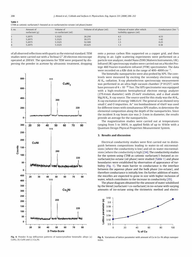
ig. 5. Variations of lattice parameter with amount of Cu in Cu–Ni alloys nanopar-icles.

J. Ahmed et al. / Colloids and Surfaces A: Physicochem. Eng. Aspects 331 (2008) 206–212 209
age of
cm(Tspdd
psaomtaiaitgb
haoet
0(piFCss
uwnsSot
aicmwa
Fig. 6. TEM micrograph of (a) CuNi (b) HRTEM im
al conductivity measurements is shown in Fig. 2. The water-in-oilicroemulsion phase merges continuously with the oil-in-water
high blend content) phase as the amount of water is increased.he region with lower blend and water content is the monopha-ic region (Wo = 12; calculated from Table 1) which is suitable forroducing a microemulsion containing monodisperse water-in-oilroplets. The size of these droplets was found to be 4–5 nm usingynamic light scattering studies.
Using the above microemulsion system we have obtained therecursors (see Section 2) which on reducing in H2 at 500 ◦C yield aeries of bimetallic alloys of varying stoichiometry (CuNi3, CuNind Cu3Ni). Fig. 3 shows the powder X-ray diffraction patternsf the brown- and crimson-coloured powder obtained from theicroemulsions on aging of ∼12 and 40 h respectively at room
emperature. Mixture of phases (CuO, Cu(OH)2, Ni(OH)2, coppermmine nitrate, and nickel ammine nitrate) could be identifiedn the PXRD patterns. Around 40% of the Cu–Ni alloy phase couldlso be observed in the powder obtained after aging for 40 h. Thisndicates that alloy formation takes place even at room tempera-ure. On longer aging (70 h) the crimson-coloured solution turnedreen, and the powder obtained after centrifugation was found toe amorphous by X-ray diffraction.
This green-coloured amorphous precursor was annealed in
ydrogen atmosphere (500 ◦C) which resulted in pure bimetalliclloys (Fig. 4). All the reflections of each pattern could be indexedn the basis of a face-centered cubic cell. The refined lattice param-ter of the 1:1 composition was 3.5561(7) Å, which is similar tohat reported for CuNi (1:1) bimetallic nanoparticles (JCPDS # 3-pn
cn
CuNi (c) CuNi3 and (d) Cu3Ni alloy nanoparticles.
65-7246). It was observed that the lattice parameter increasedFig. 5) with the increase in copper content. Note that the latticearameter of nickel is 3.5238 Å while that of Cu is 3.615 Å. A sim-
lar variation in lattice parameter for Pt–Fe alloys as a function ofe content has been reported earlier [36]. The phase diagram ofu–Ni binary system [37] shows that it forms a substitutional solidolution as both Cu and Ni crystallize in the face-centered cubictructure.
TEM studies (Fig. 6a) show nearly monodisperse, spherical andniform particles of CuNi (1:1) and the average size of the particlesas found to be 7 nm. HRTEM studies exhibit the single crystallineature of the nanoparticles showing lattice fringes of 2.05 Å con-istent with the (1 1 1) planes of the face-centered lattice (Fig. 6b).ome degree of agglomeration was observed in the nanoparticlesf CuNi3 and Cu3Ni (Fig. 6c and d), whose average size was foundo be larger (20–30 nm) compared to the 1:1 composition.
FTIR spectra of Cu–Ni alloy nanoparticles (Fig. 7) showed a bandt a frequency of 455 cm−1 which may be assigned to the stretch-ng vibration of metal–metal (Cu–Ni) band. The band at 3405 cm−1
ould be assigned to the stretching vibration of –OH group whichay be present due to adsorbed water molecules. Some othereak bands also appear at 2918, 2849, 2356 and 1384 cm−1 prob-
bly due to the C–H stretching modes [38] arising from the
resence of surfactant molecules associated with the CuNi alloyanoparticles.From the depth profile curve obtained from XPS studies, wean observe the variation of Cu and Ni XPS signals as we slice theanoparticle layer by layer using the Ar+ ion beam. Knowing that

210 J. Ahmed et al. / Colloids and Surfaces A: Physicochem. Eng. Aspects 331 (2008) 206–212
ttpefacmc[c(1mtotn5Nco
f
˚
wEd
˚
Ia
˚
htar
Fig. 8. Depth profile curve obtained using X-ray photoelectron spectroscopy of (a)CuNi, (b) CuNi3 and (c) Cu3Ni alloy nanoparticles.
Fig. 7. FTIR spectra of CuNi alloy nanoparticles.
he average particle size is about 7 nm (for the 1:1 Cu–Ni nanopar-icles) from the TEM data, we calibrate the ion dose in terms ofarticle size along the beam axis. The composition (relative%) ofach metal is calculated from the corresponding atomic scatteringactors. The profile in Fig. 8a, shows a core of 4 nm with Cu to Ni ratios 1:1 in the core, covered by a shell of 1.5 nm. Using geometricalonsideration and ignoring the preferential sputtering and inelasticean free path of the core electrons, we qualitatively estimate the
omposition ratio by assuming a radial dependence of the density39,40] of each element. We estimate the shell of the nanoparti-le to be 20% richer in Cu (∼1.5 nm) while the core is 10% Ni-rich∼4 nm), with the average stoichiometry of the nanoparticle being:1. However, rigorous calculations and the results of other comple-entary techniques (imaging) are essential to precisely determine
he core–shell structure. Fig. 8b and c shows that the nanoparticlesf CuNi3 and Cu3Ni are more homogeneous. A careful visualiza-ion of the depth profile curve for the bimetallic alloys of Cu–Nianoparticles shows that the compositions of Cu3Ni and CuNi3 are.7:1 and 1:1.5 respectively. The reduction of Cu2+ is faster thani2+ because of higher reduction potential of Cu2+/Cu (+0.34 V)ompared to Ni2+/Ni (−0.26 V) leading to preferential nucleationf copper compared to nickel.
For spherical samples of radius R, the flux ˚E of element E as aunction of ablation x can be written as
E(x) =
⎛⎝2�
∫ √R2−(R−x)2
0
�E(r)rdr
⎞⎠ dx
dt
here �E is the spherically symmetric number density of elementor radial coordinate r and dx/dt is the ablation rate. For constantensity �0, this yields
E(x) = ��0x(2R − x)dx
dt
f we assume that there is no preferential ablation, dx/dt is constantnd thus
E(t) =
⎛⎝2�
∫ √R2−(R−t(dx/st))2
0
�E(r)rdr
⎞⎠ dx
dt
Thus, in order to get the radial dependence of the density, oneas to deconvolute the flux since even the outer shell always con-ributes to the flux. When the sample is half ablated, one gets theverage. The spheres probably have a Cu-rich outer core and a Ni-ich inner core, with an average ratio of 1:1.
J. Ahmed et al. / Colloids and Surfaces A: Physico
FC
antsoh
n(whcbct
nadp6es�ua
4
tionb
A
oS0
R
[
[
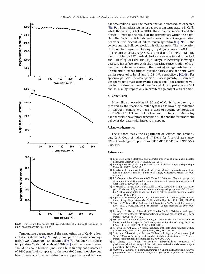
ig. 9. Temperature dependence of the magnetization of (a) CuNi3, (b) CuNi and (c)u3Ni alloy nanoparticles at 1 kOe.
Temperature dependence of the magnetization of Cu–Ni alloyst 1 kOe is shown in Fig. 9. Cu1Ni3 nanoparticles show ferromag-
etism well above room temperature (Fig. 7a). For Cu3Ni, the Curieemperature TC should be about 350 K [41] and the magnetizationhould be about 1700 emu/mol, even bulk Ni only has a momentf 3400 emu/mol, compared to the near 6000 emu/mol observedere. However, as the concentration of copper increased in these[
[
chem. Eng. Aspects 331 (2008) 206–212 211
anocrystalline alloys, the magnetization decreased, as expectedFig. 9b). Magnetism sets in just above room temperature in CuNi,hile the bulk TC is below 100 K. The enhanced moment and theigher TC may be the result of the segregation within the parti-les. The Cu3Ni particles showed a very different magnetizationehavior, reminiscent of dilute ferromagnetism (Fig. 9c) – theorresponding bulk composition is diamagnetic. The percolationhreshold for magnetism for Cu1 − xNix alloys occurs at x = 0.4.
The surface area analysis was carried out for the Cu–Ni alloyanoparticles by BET method. Surface area was found to be 9.42nd 6.01 m2/g for CuNi and Cu3Ni alloys, respectively showing aecrease in surface area with the increasing concentration of cop-er. The specific surface areas of the pure Cu (average particle size of7 nm) and Ni nanoparticles (average particle size of 47 nm) werearlier reported to be 11 and 14.23 m2/g respectively [42,43]. Forpherical particles, the ideal specific surface is given by 3/(�r) whereis the volume mass density and r the radius – the calculated val-
es for the aforementioned pure Cu and Ni nanoparticles are 10.1nd 14.32 m2/g respectively, in excellent agreement with the size.
. Conclusion
Bimetallic nanoparticles (7–30 nm) of Cu–Ni have been syn-hesized by the reverse micellar synthesis followed by reductionn hydrogen atmosphere. Pure phases of specific compositionsf Cu–Ni (1:1, 1:3 and 3:1) alloys were obtained. CuNi3 alloyanoparticles show ferromagnetism at 320 K and the ferromagneticehavior decreases with increase in copper.
cknowledgements
The authors thank the Department of Science and Technol-gy, CSIR, Govt. of India, and IIT Delhi for financial assistance.EL acknowledges support from NSF DMR 0520471, and NSF DMR603644.
eferences
[1] C. Jo, J. Lee, Y. Jang, Electronic and magnetic properties of ultrathin Fe–Co alloynanowires, Chem. Mater. 17 (2005) 2667–2671.
[2] P.P. Singh, Relativity and magnetism in Ni–Pd and Ni–Pt alloys, J. Magn. Magn.Mater 261 (2003) 347–352.
[3] E. Jartych, J.K. Zurawicz, D. Oleszak, M. Pekala, Magnetic properties and struc-ture of nanocrystalline Fe–Al and Fe–Ni alloys, Nanostruct. Mater. 12 (1999)927–930.
[4] E.E. Carpenter, J.A. Wienmann, W.L. Zhou, C.J. O’Connor, Magnetic propertiesof iron and iron platinum alloys synthesized via microemulsion techniques, J.Appl. Phys. 87 (2000) 5615–5617.
[5] G. Mattei, C.D.J. Fernandez, P. Mazzoldi, C. Sada, G. De, G. Battaglin, C. Sangre-gorio, D. Gatteschi, Synthesis, structure, and magnetic properties of Co, Ni, andCo–Ni alloy nanocluster-doped SiO2 films by sol–gel processing, Chem. Mater.14 (2002) 3440–3447.
[6] P. James, O. Eriksson, B. Johansson, I.A. Abrikosov, Calculated magnetic proper-ties of binary alloys between Fe, Co, Ni, and Cu, Phys. Rev. B 59 (1999) 419–430.
[7] S.W. Han, Y. Kim, K. Kim, Dodecanethiol-derivatized Au/Ag bimetallic nanopar-ticles: TEM, UV/VIS, XPS, and FTIR analysis, J. Colloid Interface Sci. 208 (1998)272–278.
[8] R. Hong, N.O. Fischer, T. Emrick, V.M. Rotello, Surface PEGylation and ligandexchange chemistry of FePt Nanoparticles for biological applications, Chem.Mater. 17 (2005) 4617–4621.
[9] D.K. Kim, D. Kan, T. Veres, F. Normadin, J.K. Liao, H.H. Kim, S.H. Lee, M. Zahn, M.Muhammed, Monodispersed Fe–Pt nanoparticles for biomedical applications,J. Appl. Phys. 97 (2005), 10Q918 (1)–10Q918 (3).
10] A. Fortunelli, A.M. Velaso, A theoretical study of the catalytic properties of Pt/Fenanoclusters, J. Mol. Struct. (Theochem.) 586 (2002) 17–27.
11] S. Spriano, F. Rosalbino, M. Baricco, P.V. Morra, E. Angelini, C. Antonione, J.M.Siffre, P. Marcus, Surface and electrochemical characterization of Ni–Zr inter-metallic compounds, Intermetallics 8 (2000) 299–304.
12] X. Zhang, K.Y. Chan, Water-in-oil microemulsion synthesis ofplatinum–ruthenium nanoparticles, their characterization and electrocatalyticproperties, Chem. Mater. 15 (2003) 451–459.
13] W. Qiwu, Y. Jianlong, R. Jingfang, H. Minming, Y. Chunhu, Structure and catalyticproperties of Cu–Ni bimetallic catalysts for hydrogenation, Catal. Lett. 4 (1990)63–74.

2 hysico
[
[
[
[
[
[
[
[
[
[
[
[
[
[
[
[
[
[
[
[
[
[
[
[
[
[
[
[
12 J. Ahmed et al. / Colloids and Surfaces A: P
14] H. Kim, C. Lu, W.L. Worrell, J.M. Vohs, R.J. Gorte, Cu–Ni cermet anodes for directoxidation of methane in solid-oxide fuel cells, J. Electrochem. Soc. 149 (2002)A247–A250.
15] F. Bonet, S. Grugeon, L. Dupont, R.H. Urbina, C. Guery, J.M. Tarascon, Synthe-sis and characterization of bimetallic Ni–Cu particles, J. Solid State Chem. 172(2003) 111–115.
16] W. Wang, G. Cao, Synthesis and structural investigation of Pd/Ag bimetallicnanoparticles prepared by the solvothermal method, J. Nanoparticle Res. 9(2007) 1153–1161.
17] H.L. Niu, Q.W. Chen, Y.S. Lin, Y.S. Jia, H.F. Zhu, M. Ning, Hydrothermal formationof magnetic Ni–Cu alloy nanocrystallites at low temperatures, Nanotechnology15 (2004) 1054–1058.
18] E. Botcharova, J. Freudenberger, L. Schultz, Cu–Nb alloys prepared by mechan-ical alloying and subsequent heat treatment, J. Alloys Compd. 365 (2004)157–163.
19] C. Liu, X. Wu, T. Klemmer, N. Shukla, D. Weller, A.G. Roy, M. Tanase, D. Laughlin,Reduction of sintering during annealing of FePt nanoparticles coated with ironoxide, Chem. Mater. 17 (2005) 620–625.
20] Q.X. Liu, C.X. Wang, W. Zhang, G.W. Wang, Immiscible silver–nickel alloyingnanorods growth upon pulsed-laser induced liquid/solid interfacial reaction,Chem. Phys. Lett. 382 (2003) 1–5.
21] V.V. Agrawal, P. Mahalakshmi, G.U. Kulkarni, C.N.R. Rao, Nanocrystalline films ofAu–Ag, Au–Cu, and Au–Ag–Cu alloys formed at the organic–aqueous interface,Langmuir 22 (2006) 1846–1851.
22] D.H. Chen, C.J. Chen, Formation and characterization of Au–Ag bimetal-lic nanoparticles in water-in-oil microemulsions, J. Mater. Chem. 12 (2002)1557–1562.
23] M.L. Wu, D.H. Chen, T.C. Huang, Synthesis of Au/Pd bimetallic nanoparticles inreverse micelles, Langmuir 17 (2001) 3877–3883.
24] M.L. Wu, D.H. Chen, T.C. Huang, Preparation of Au/Pt bimetallic nanoparticlesin water-in-oil microemulsions, Chem. Mater. 13 (2001) 599–606.
25] I. Capek, Preparation of metal nanoparticles in water-in-oil (w/o) microemul-sions, Adv. Colloid Interface Sci. 110 (2004) 49–74.
26] C. Stubenrauch, T. Wielputz, T. Sottmann, C. Roychowdhury, F.J. DiSalvo,Microemulsions as templates for the synthesis of metallic nanoparticles, Col-
loid Surf. A: Physicochem. Eng. Aspects 317 (2008) 328–338.27] M.P. Pileni, Reverse micelle as microreactors, J. Phys. Chem. 97 (1993)6961–6973.
28] T. Ahmad, R. Chopra, K.V. Ramanujachary, S.E. Lofland, A.K. Ganguli, Nanorods ofcopper and nickel oxalates synthesized by the reverse micellar route, J. Nanosci.Nanotech. 5 (2005) 1840–1845.
[
[
chem. Eng. Aspects 331 (2008) 206–212
29] T. Ahmad, R. Chopra, K.V. Ramanujachary, S.E. Lofland, A.K. Ganguli, Cantedantiferromagnetism in copper oxide nanoparticles synthesized by the reverse-micellar route, Solid State Sci. 7 (2005) 891–895.
30] T. Ahmad, K.V. Ramanujachary, S.E. Lofland, A.K. Ganguli, Nanorods ofmanganese oxalate: a single source precursor to different manganeseoxide nanoparticles (MnO, Mn2O3, Mn3O4), J. Mater. Chem. 14 (2004)3410–3606.
31] J. Feng, C.P. Zhang, Preparation of Cu–Ni alloy nanocrystallites in water-in-oilmicroemulsions, J. Colloid Interface Sci. 293 (2006) 414–420.
32] M. Wen, Q.Y. Liu, Y. Wang, Y.Z. Zhu, Q.S. Wu, Positive microemulsion synthesisand magnetic property of amorphous multicomponent Co-, Ni- and Cu-basedalloy nanoparticles, Colloids Surf. A: Physicochem. Eng. Aspects 318 (2008)238–244.
33] L.S. Birks, H. Friedman, Particle-size determination from X-ray line broadening,J. Appl. Phys. 17 (1946) 687–692.
34] K. Holmberg, Handbook of Applied Surface and Colloid Chemistry, vols. I–II,John Wiley and sons, 2001.
35] C. Kumar, D. Balasubramanian, Studies on the triton X-100: alcohol:waterreverse micelles in cyclohexane, J. Colloid Interface Sci. 69 (1979) 271–279.
36] A.R. Malheiro, L.C. Varanda, J. Perez, H.M. Villullas, The aerosol OT + n-butanol + n-heptane + water system: phase behavior, structure characteriza-tion, and application to Pt70Fe30 nanoparticle synthesis, Langmuir 23 (2007)11015–11020.
37] T.B. Massalski, in: Binary alloy Phase Diagrams, ASM International the MaterialsInformation Society, 2nd edition, vol. 2, 1990, pp 1442–1446.
38] R.A. MacPhail, H.L. Strauss, R.G. Snyder, Carbon–hydrogen stretching modesand the structure of n-alkyl chains. 2. Long, all-trans chains, J. Phys. Chem. 88(1984) 334–341.
39] I. Tunc, S. Suzer, M.A. Correa-Duarte, L.M. Marzan, XPS characterization of Au(core)/SiO2 (shell) nanoparticles, J. Phys. Chem. B 109 (2005) 7597–7600.
40] D.Q. Yang, J.N. Gillet, M. Meunier, E. Sacher, Room temperature oxidation kinet-ics of Si nanoparticles in air, determined by X-ray photoelectron spectroscopy,J. Appl. Phys. 97 (2005), 024303 (1)–024303 (6).
41] E.O. Wollan, Evidence for a Heisenberg type of exchange interaction in nickeland the NiCu alloy system, Phys. Rev. 148 (1966) 517–519.
42] Z. Wei, T. Xia, W. Feng, J. Dai, Q. Wang, W. Li, P. Yan, Preparation and particlesize characterization of Cu nanoparticles prepared by anodic arc plasma, RareMetals 25 (2006) 172–176.
43] Z. Wei, P. Yan, W. Feng, J. Dai, Q. Wang, T. Xia, Microstructural characterizationof Ni nanoparticles prepared by anodic arc plasma, Mater. Characterization 57(2006) 176–181.