Coordinated Amino Acid Changes in the Evolution of Mammalian Defensins
Upload
triestearchitetturaCategory
view
1download
0
This article appeared in a journal published by Elsevier. The attachedcopy is furnished to the author for internal non-commercial researchand education use, including for instruction at the authors institution
and sharing with colleagues.
Other uses, including reproduction and distribution, or selling orlicensing copies, or posting to personal, institutional or third party
websites are prohibited.
In most cases authors are permitted to post their version of thearticle (e.g. in Word or Tex form) to their personal website orinstitutional repository. Authors requiring further information
regarding Elsevier’s archiving and manuscript policies areencouraged to visit:
http://www.elsevier.com/copyright

Author's personal copy
Big defensins and mytimacins, new AMP families of the Mediterranean musselMytilus galloprovincialis
Marco Gerdol a, Gianluca De Moro a, Chiara Manfrin a, Paola Venier b, Alberto Pallavicini a,⇑a Laboratorio di Genetica, Department of Life Sciences, University of Trieste, Via Licio Giorgeri 5, 34126 Trieste, Italyb Department of Biology, CRIBI Biotechnology Center, University of Padova, Padova, Italy
a r t i c l e i n f o
Article history:Received 23 June 2011Revised 29 July 2011Accepted 10 August 2011Available online 24 August 2011
Keywords:Mytilus galloprovincialisInnate immunityAntimicrobial peptidesBig defensinMytimacin
a b s t r a c t
Antimicrobial peptides (AMPs) play a fundamental role in the innate immunity of invertebrates, prevent-ing the invasion of potential pathogens. Mussels can express a surprising abundance of cysteine-richAMPs pertaining to the defensin, myticin, mytilin and mytimycin families, particularly in the circulatinghemocytes.
Based on deep RNA sequencing of Mytilus galloprovincialis, we describe the identification, moleculardiversity and constitutive expression in different tissues of five novel transcripts pertaining to the macinfamily (named mytimacins) and eight novel transcripts pertaining to the big defensins family (namedMgBDs). The predicted antimicrobial peptides exhibit a N-terminal signal peptide, a positive net chargeand a high content in cysteines, allegedly organized in intra-molecular disulfide bridges. Mytimacins andbig defensins therefore represent two novel AMP families of M. galloprovincialis which extend the reper-toire of cysteine-rich AMPs in this bivalve mollusk.
� 2011 Elsevier Ltd. All rights reserved.
1. Introduction
Antimicrobial peptides (AMPs) are humoral components of theinnate immunity, present in all metazoans and essential to theimmediate defense reactions of invertebrate organisms lackingadaptive immunity. Antibacterial activity was first reported inmollusks in the ‘80s (Kubota et al., 1985) whereas the isolationand characterization of true AMPs from the mussels Mytilus gallo-provincialis (Hubert, 1996) and Mytilus edulis (Charlet et al., 1996)date back to 1996.
In the Mediterranean mussel M. galloprovincialis, cysteine-richantimicrobial peptides are produced as precursor molecules andprocessed into mature peptides within the hemocyte granules(Mitta et al., 2000c). All the four AMP classes described so far inmussels, namely defensins (Hubert, 1996; Mitta et al., 2000a,1999b), myticins (Mitta et al., 1999a; Pallavicini et al., 2008), myti-lins (Mitta et al., 2000a, 2000b; Roch et al., 2008) and the strictlyantifungal mytimycins (Charlet et al., 1996; Sonthi et al., 2011), re-tain a cysteine array essential to stabilize the mature peptide in ahighly compact, cationic and amphipatic structure (Mitta et al.,2000c; Yeaman and Yount, 2007). More in detail, eight cysteineresidues defining four intra-molecular disulfide bridges are present
in defensins, myticins and mytilins, whereas 12 cysteines and twoadditional disulfide bridge characterize mytimycins. The structuresof mussel defensin (Yang et al., 2000) and mytilin (Roch et al.,2008) have been determined by NMR, confirming the expectedpattern of intra-molecular disulfide bonds.
Each of the above mentioned AMP classes comprises severalmembers and the recent identification of 12 additional sequencetranscripts sensibly extended the number of mussel AMPs in M.galloprovincialis (Venier et al., 2011). New massive sequencing ofthe M. galloprovincialis transcriptome allowed us to prepare ahigh-coverage transcript collection and to study identity andmolecular variability of two classes of previously uncharacterizedcysteine-rich mussel AMPs, namely big defensins (MgBDs) andmytimacins.
Big defensins have been originally identified in the horseshoecrab Tachypleus tridentatus (Saito et al., 1995), specifically storedin granules within hemocytes (Kawabata and Iwanaga, 1997) like-wise many molluscan AMPs (Mitta et al., 2000c). The structure ofbig defensins typically includes one N-terminal highly hydropho-bic region, one C-terminal cysteine-rich and positively charged re-gion, and six cysteine residues arranged to form 1–5, 2–4, 3–6disulfide bonds in the mature peptide (Saito et al., 1995), in a sim-ilar fashion to mammalian b-defensins (Kouno et al., 2008; Selstedet al., 1993; Zhao et al., 2010). The disulfide array is therefore dif-ferent from the classic 1–4, 2–5, 3–6 cysteines arrangement ofarthropod defensins (Dimarcq et al., 1998). Furthermore thecysteine-stabilized a-helix and b-sheet (CSab) motif characterizing
0145-305X/$ - see front matter � 2011 Elsevier Ltd. All rights reserved.doi:10.1016/j.dci.2011.08.003
Abbreviations: AMPs, antimicrobial peptides; MgBDs, Mytilus galloprovincialisbig defensins.⇑ Corresponding author. Tel.: +39 0405588736; fax: +39 0405582449.
E-mail address: [email protected] (A. Pallavicini).
Developmental and Comparative Immunology 36 (2012) 390–399
Contents lists available at SciVerse ScienceDirect
Developmental and Comparative Immunology
journal homepage: www.elsevier .com/locate /dc i

Author's personal copy
many plant and invertebrate defensins (including those of mussel)(Cornet et al., 1995) cannot be observed in big defensins.
The two terminal regions of the molecule display remarkabledifferences in antimicrobial properties, with the N-terminal frag-ment being more active towards Gram� bacteria and the C-termi-nal fragment being more effective against Gram+ bacteria (Saitoet al., 1995). NMR-based studies indicated that a globular N-termi-nal hydrophobic domain plays a fundamental role in the dynamicinteraction with target membranes (Kouno et al., 2009). To dateonly two other big defensins have been extensively studied: AiBDof the bay scallop Argopecten irradians and VpBD of the clam Ven-erupis philippinarum were significantly up-regulated in the bivalvehemocytes in response to bacterial challenges and both displayed abroad spectrum of antimicrobial activity (Zhao et al., 2010, 2007).Transcripts encoding big defensins have been also identified in themollusks Crassostrea gigas, Mytilus chilensis and Bathymodiolusazoricus and in the lancelets Branchiostoma belcheri tsingtauenseand Branchiostoma floridae, suggesting a broader taxonomic distri-bution of this AMP class.
Macins are positively charged secreted peptides which havebeen first described in the annelids Theromyzon tessulatum(Tasiemski et al., 2004) and Hirudo medicinalis (Schikorski et al.,2008) and have been later identified in the cnidarian Hydra magni-papillata (Jung et al., 2009) and in the mollusk Hyriopsis cumingii(Xu et al., 2010). Macins are characterized by a disulfide array ofeight cysteines, with the optional presence of a fifth intra-molecu-lar disulfide bridge involving a C-terminal sequence extension intheromacin. The structure of hydramacin has been determinedby NMR, revealing a compact organization with an uneven distri-bution of positively charged residues which divide the molecularsurface into two large hydrophobic hemispheres, characterizedby the arrangement of cysteine bonds in a knottin fold, found inall the proteins pertaining the scorpion-toxin-like superfamilymembers, including mussel defensins (Jung et al., 2009).
Contrary to the majority of cysteine-rich AMPs, macins are notspecifically expressed in the circulating cells, being instead local-ized in the endodermal epithelium (Bosch et al., 2009) or periphe-ral Large Fat Cells (LFCs) functionally resembling the insect fatbody and often in close contact with the coelomic cavity (Tasiem-ski et al., 2004) or, in the case of neuromacin, in the central nervoussystem (Schikorski et al., 2008). The only reported exception is rep-resented by the freshwater pearl oyster H. cumingii theromacin-like protein, which was found to be preferentially expressed inhemocytes (Xu et al., 2010). Macin expression is induced afterexposure to bacteria (Tasiemski et al., 2004; Xu et al., 2010), andneuromacin localizes especially at the site of tissue injury (Schikor-ski et al., 2008). Increased expression of a theromacin-like tran-script was also observed in response to both infection and tissueinjury in the snail Biomphalaria glabrata (Ittiprasert et al., 2010).
Macins display membrane aggregating and permeabilizing activ-ity, effective against Gram+ bacteria in theromacin and neuromacin(Schikorski et al., 2008; Tasiemski et al., 2004) and against Gram�bacteria in hydramacin (Jung et al., 2009). On the basis of the tertiarystructure of hydramacin determined by NMR, a mechanistic modelpostulates its interaction with the bacterial membranes, with cellaggregation and microbe morphology changes preceding full per-meabilization and their effective killing (Jung et al., 2009).
Although both macins and big defensins have already been re-ported in mollusks, the knowledge of these two AMP families isstill extremely limited and their occurrence and evolutionary rela-tionship in the animal kingdom have not been adequately studied.Here we report the identification in M. galloprovincialis, thanks to awhole-transcriptome sequencing approach, of novel transcriptspertaining to the macin family (named mytimacins) and to thebig defensins family (named MgBDs) and discuss their moleculardiversity and constitutive expression in different tissues.
2. Materials and methods
2.1. Identification of transcripts encoding macins and big defensinsfrom M. galloprovincialis
Using second generation sequencing systems (454 Life Sciencesand Illumina platforms) we sequenced the transcriptome of Medi-terranean mussels (M. galloprovincialis) from tissues (hemocytes,gills and digestive gland) of different individuals. Following accu-rate processing, we could locally assemble a transcript collectionwhich updates and enrich the pre-existing Mytibase (http://mus-sel.cribi.unipd.it) (Venier et al., 2009). The predicted peptides orig-inated from the assembly process were scanned with HMMER 3(http://hmmer.janelia.org/) to find mussel transcripts matchingthe big defensin and macin profiles generated by multiple align-ments of the GenBank sequences pertaining to these two AMP clas-ses. Significant hits were cut-off at e-values <10�5. The search wasre-iterated by including the new results into the alignment and bygenerating new profiles until no new hits were found.
2.2. dbEST data mining
A similar iterative approach was applied to the NCBI dbESTdatabase (http://www.ncbi.nlm.nih.gov/dbEST) in order to extendthe search from Mytilus spp. to other organisms and to assess thetaxonomic distribution of the two AMP classes. The EST sequencesmatching the above mentioned HMMER profiles were assembledinto contigs with the CLC Genomic Workbench 4.5.1 (CLC Bio, Kat-rinebjerg, Denmark) to remove redundancy. Only complete se-quences were considered for further analysis.
2.3. Sequence analysis
All transcript sequences related to macins and big defensinswere translated with the Expasy Translate tool (http://exp-asy.org/tools/dna.html) to obtain the virtual encoded peptides: sig-nal peptides were predicted using SignalP 3.0 (http://www.cbs.dtu.dk/services/SignalP), isoelectric point and molecularweight were calculated with the Expasy Compute pInMW tool(http://expasy.org/tools/pi_tool.html) and functional role wasevaluated with the antimicrobial peptide predictor APD2 (http://aps.unmc.edu/AP/prediction/prediction_main.php). Structuralhomology with the T. tridentatus big defensin (2RNG) and the H.magnipapillata hydramacin-1 (2K35) was evaluated by automatedtridimensional modeling with Phyre2 (Kelley and Sternberg, 2009).
An analysis of SNPs frequency in the transcript sequences ofmytimacins and big defensins was performed with the CLC Geno-mic Workbench 4.5.1 (CLC Bio, Katrinebjerg, Denmark). To excludepotential sequencing errors, sites with low-coverage (less than 10sequencing reads) were not analyzed and SNPs occurring with verylow frequency (<2%) or not covered by at least three independentreads were not considered reliable.
2.4. Phylogenetic analysis
Multiple sequence alignments displayed in figures and thoseused for the generation of HMMER profiles and Bayesian phyloge-netic analysis were produced with MEGA5.02 (Tamura et al., 2011)using the MUSCLE algorithm (Edgar, 2004), with gap opening andextension penalties of �2 and �1, respectively.
Phylogenies of big defensins and macins were estimated withMrBayes 3.2 (Ronquist and Huelsenbeck, 2003) starting from analignment of the entire mature predicted peptides. The GTR substi-tution model of molecular evolution with a proportion of invari-able sites, and a Gamma-shaped distribution of rates across sites
M. Gerdol et al. / Developmental and Comparative Immunology 36 (2012) 390–399 391

Author's personal copy
(GTR + c + I), was chosen as the best-fitting model for our datasetswith ProtTest (Abascal et al., 2005). We ran two independent anal-yses with four chains each (one ‘‘cold’’, three ‘‘warm’’) for1,000,000 generations, sampling a single tree each 1000 genera-tions. The first 25% of the generated trees were discarded for theburn-in procedure and the remaining trees were used to calculatethe posterior probability for each node in a 50% consensus trees.
2.5. Mussel samples
To evaluate the tissue-specific expression of mytimacin-1, myti-macin-2, mytimacin-3, MgBD1, MgBD3 and MgBD6 transcripts,mussels of 6.5–7 cm shell length were collected from the Gulf ofTrieste, Italy. Total RNA was individually purified from hemolymphand from digestive gland mantle, posterior abductor muscle, gill,and foot, previously homogenized in RNATidy G according to themanufacturer’s instructions (AppliChem, Darmstadt, Germany).Following extraction, the RNA quality was assessed by electropho-resis on denaturing agarose gel and its quantity was estimated byUV-spectrophotometry. Complementary DNA was prepared by ret-ro-transcription with the iScript™ cDNA Synthesis Kit (Bio-Rad)from pooled RNA samples representing five individuals.
2.6. Quantitative PCR expression analysis
The expression levels of the mytimacin-1, mytimacin-2, myti-macin-3, MgBD1, MgBD3 and MgBD6 transcripts were assessedin samples representing hemolymph, digestive gland, mantle, pos-terior abductor muscle, gills and foot of five adult mussels. Primerpairs were designed (Table 1) and used to obtain specific PCRamplicons. The primers for MgBD3 were specifically designed toco-amplify the three sequences MgBD3a, MgBD3b and MgBD3c.
All the PCR assays were performed using a Bio-Rad CFX96 sys-tem. The 15 lL reaction mix included 7.5 lL of 2� IQ™ SYBRGreen� Supermix (Biorad), 0.3 lL of each 10 lM primer and 2 lLof a 1:10 cDNA dilution. The following thermal profile was used:an initial 30 denaturation step at 95 �C, followed by 40 cycles at95� for 2000, 60� for 1500 and 72� for 2000. Amplification productswere analyzed with a 65�/95 �C melting curve. The expression lev-els of the selected transcripts were determined using the compar-ative Ct method (2�DDCt method) (Livak, 2001). Ct values used forquantification were corrected based on PCR efficiencies using Lin-RegPCR (Ramakers et al., 2003). The expression values were nor-malized using the elongation factor EF-1 as housekeeping gene(EF-1 primers are shown in Table 1). Results are given as the meanwith standard deviation of three technical replicates.
3. Results and discussion
3.1. Big defensins
3.1.1. Computational identification and sequence features of MgBDsThe mussel transcriptomic collection, assembled starting from a
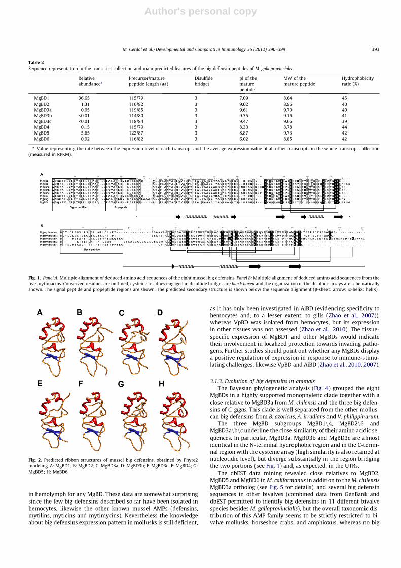
total of 24901 Sanger, 150857 454 Life Sciences and 108620377Illumina sequencing reads, conprises110259 contigs (with an aver-age length of 590 nucleotides; the N50 parameter of the assemblywas 658). In this transcriptomic mussel collection we could iden-tify eight different sequences encoding big defensins, named MgBD1–6 and deposited at EMBL under the accession IDs FR873266–FR873273. Three sequences showing remarkable similarity areindicated as MgBD3a, MgBD3b and MgBD3c.
The eight inter-related sequences differ quite widely for theirrepresentation in the transcript collection, with MgBD1 showinga very high coverage in respect with the average of mussel tran-scripts and MgBD3b and MgBD3c showing, in contrast, an extre-mely low relative abundance (Table 2). An open reading frame(ORF) encoding the full-length peptide precursor was identifiedin all the eight different nucleotidic sequences (the alignment ofthe full length precursor peptides is shown in Fig. 1).
The virtual translation yielded aminoacid sequences rangingfrom 114 and 122 residues in length (the shortest being MgBD3band the longest one being MgBD5). A short N-terminal signalpeptide was predicted in all cases, and the alignment with thebig defensin isolated from T. tridentatus revealed that MgBDsare produced as prepropeptides. Following the cleavage of thesignal peptide, a second proteolytic cleavage could result in themature peptides, whose molecular weight range from 8.64 to9.70 kDa.
In the C-terminal region of all big defensin transcripts, six con-served cisteines define the motif C-X6-C-X3-C-X13-C-X4-C-C,essential for the disulfide bridge formation (see Fig. 1). Six out ofthe eight predicted mature peptides show a basic isoelectric point(8.3–9.6) meaning a a positive net charge at neutral pH whereasthe isoelectric point of MgBD1 and MgBD6 is closer to neutrality.As reported in Table 2, the prediction of antimicrobial features per-formed with the APD2 software also revealed a rather high per-centage of hydrophobic residues, comparable to those of mytilinsand defensins (Roch et al., 2008).
The tertiary structure modeling by Phyre2 was successful for allthe eight MgBDs and a percentage of residues ranging from 88%(MgBD3a) to 96% (MgBD4) were modeled with >90% confidencebased on the tertiary structure of the T. tridentatus big defensin(PDB accession: 2RNG), denoting a high structural conservationwithin this AMP family (see Fig. 2).
Overall, the analysis of the eight MgBDs sequences highlightsproperties common to many AMPs, such as a basic isoelectric pointand a high hydrophobicity ratio. Furthermore, the conserved cys-teine array, N-terminal hydrophobic domain and high sequenceand predicted structural similarity with big defensins previouslycharacterized in other organism further suggest that they repre-sent genuine big defensins of M. galloprovincialis.
3.1.2. Constitutive tissue expression of MgBDsThe expression analysis of MgBD1, MgBD3 (with primers co-
amplifying the three isoforms MgBD3a, MgBD3b and MgBD3c)and MgBD6 revealed very low or negligible constitutive expressionin most of the six tissues analyzed but each transcript resulted tobe selectively expressed in a given tissue (Fig. 3). MgBD1, the se-quence represented with the highest sequence coverage in our col-lection (see Table 2) was expressed only in the digestive glandwhereas MgBD3 and MgBD6 expression was mainly traced in gillsand mantle, respectively (Fig. 3). Almost no expression was evident
Table 1Primers designed for assessing the tissue-specific levels ofmytimacin-1, mytimacin-2, mytimacin-3, MgBD1, MgBD3 andMgBD6 transcripts.
Primer name Primer sequence
EF-1 FOR cctcccaccatcaagacctaEF-1 REV ggctggagcaaaggtaacaaMytimacin-1 FOR ctcctgcaaattcccacatcMytimacin-1 REV atcttttgttccgccagagaMytimacin-2 FOR gtggtggtggaagtggaagtMytimacin-2 REV tccaagctctttgcatctgtMytimacin-3 FOR acaatcaccaatgggaccacMytimacin-3 REV tttgggcagcaaattctctcMgBD1 FOR gcgtagattccatatgcagcaMgBD1 REV tgttgatactccctgctcagMgBD3 FOR ccgattctaggacgagttgtggcaMgBD3 REV ggcaactttccaagcgccatatgcMgBD6 FOR agcatcatacgcaggattgtcMgBD6 REV tagctctacaccatcctctg
392 M. Gerdol et al. / Developmental and Comparative Immunology 36 (2012) 390–399
Author's personal copy
in hemolymph for any MgBD. These data are somewhat surprisingsince the few big defensins described so far have been isolated inhemocytes, likewise the other known mussel AMPs (defensins,mytilins, myticins and mytimycins). Nevertheless the knowledgeabout big defensins expression pattern in mollusks is still deficient,
as it has only been investigated in AiBD (evidencing specificity tohemocytes and, to a lesser extent, to gills (Zhao et al., 2007)),whereas VpBD was isolated from hemocytes, but its expressionin other tissues was not assessed (Zhao et al., 2010). The tissue-specific expression of MgBD1 and other MgBDs would indicatetheir involvement in localized protection towards invading patho-gens. Further studies should point out whether any MgBDs displaya positive regulation of expression in response to immune-stimu-lating challenges, likewise VpBD and AiBD (Zhao et al., 2010, 2007).
3.1.3. Evolution of big defensins in animalsThe Bayesian phylogenetic analysis (Fig. 4) grouped the eight
MgBDs in a highly supported monophyletic clade together with aclose relative to MgBD3a from M. chilensis and the three big defen-sins of C. gigas. This clade is well separated from the other mollus-can big defensins from B. azoricus, A. irradians and V. philippinarum.
The three MgBD subgroups MgBD1n4, MgBD2n6 andMgBD3anbnc underline the close similarity of their amino acidic se-quences. In particular, MgBD3a, MgBD3b and MgBD3c are almostidentical in the N-terminal hydrophobic region and in the C-termi-nal region with the cysteine array (high similarity is also retained atnucleotidic level), but diverge substantially in the region bridgingthe two portions (see Fig. 1) and, as expected, in the UTRs.
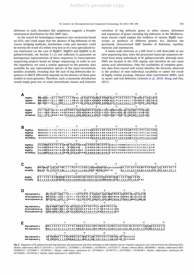
The dbEST data mining revealed close relatives to MgBD2,MgBD5 and MgBD6 in M. californianus in addition to the M. chilensisMgBD3a ortholog (see Fig. 5 for details), and several big defensinsequences in other bivalves (combined data from GenBank anddbEST permitted to identify big defensins in 11 different bivalvespecies besides M. galloprovincialis), but the overall taxonomic dis-tribution of this AMP family seems to be strictly restricted to bi-valve mollusks, horseshoe crabs, and amphioxus, whereas no big
Table 2Sequence representation in the transcript collection and main predicted features of the big defensin peptides of M. galloprovincialis.
Relativeabundancea
Precursor/maturepeptide length (aa)
Disulfidebridges
pI of thematurepeptide
MW of themature peptide
Hydrophobicityratio (%)
MgBD1 36.65 115/79 3 7.09 8.64 45MgBD2 1.31 116/82 3 9.02 8.96 40MgBD3a 0.05 119/85 3 9.61 9.70 40MgBD3b <0.01 114/80 3 9.35 9.16 41MgBD3c <0.01 118/84 3 9.47 9.66 39MgBD4 0.15 115/79 3 8.30 8.78 44MgBD5 5.65 122/87 3 8.87 9.73 42MgBD6 0.92 116/82 3 6.02 8.85 42
a Value representing the rate between the expression level of each transcript and the average expression value of all other transcripts in the whole transcript collection(measured in RPKM).
Fig. 1. Panel A: Multiple alignment of deduced amino acid sequences of the eight mussel big defensins. Panel B: Multiple alignment of deduced amino acid sequences from thefive mytimacins. Conserved residues are outlined, cysteine residues engaged in disulfide bridges are black boxed and the organization of the disulfide arrays are schematicallyshown. The signal peptide and propeptide regions are shown. The predicted secondary structure is shown below the sequence alignment (b-sheet: arrow; a-helix: helix).
Fig. 2. Predicted ribbon structures of mussel big defensins, obtained by Phyre2modeling. A: MgBD1; B: MgBD2; C: MgBD3a; D: MgBD3b; E. MgBD3c; F: MgBD4; G:MgBD5; H: MgBD6.
M. Gerdol et al. / Developmental and Comparative Immunology 36 (2012) 390–399 393

Author's personal copy
defensins were detected in many other large invertebrate classes.Such a distribution is quite unusual, as mollusks and horseshoecrabs (phylum Arthropoda, subphylum Chelicerata, class Merosto-
mata) are distantly related and no big defensins could be identifiedneither in other large Arthropoda subgroups (crustaceans, insects,etc.) or Lophotrocozoans. Nevertheless, the presence of big
Fig. 3. Tissue-specific expression of the transcripts mytimacin-1, mytimacin-2, mytimacin-3, BD1, BD3 and BD6. The expression values (bars) are relative to the elongationfactor EF-1. Results are mean ± SD of three technical replicates. The Y axis of each graph is scaled based on the highest level of expression. HEM: Hemolymph, DIG: digestivegland, MAN: mantle, PAM: posterior abductor muscle, GIL: gills, FOO: foot. ND: not detected (fluorescence did not reach threshold after 40 cycles of PCR or the melting peakanalysis did not reveal any specific product). �: not quantifiable (fluorescence did not reach threshold after 40 cycles of PCR but the melting peak analysis revealed a limitedproduction of the specific amplicon).
Fig. 4. Bayesian phylogeny of big defensins inferred from the alignment of the predicted mature peptides. Posterior probabilities are shown for each branch. Entry IDs: T.tridentatus BD: P80957.2; B. belcheri BD: Q86QN6.1; B. floridae BD: ADH03419.1; A. irradians AiBD: Q0H293.1; V. philippinarum BD: ADM25826.1); M. chilensis BD:AEE60906.1; B. azoricus BD: HM756150.1; C. gigas BD1, BD2 and BD3: AEE92785.1, AEE92787.1 and AEE92790.1.
394 M. Gerdol et al. / Developmental and Comparative Immunology 36 (2012) 390–399
Author's personal copy
defensins in early chordates like amphioxus suggests a broadertaxonomical distribution for this AMP class.
As the search for homologous sequences was exclusively basedon ESTs, one could argue that the absence of big defensins in theclasses bridging mollusks, horsheshoe crabs and lancelets couldbe merely the result of a either very low or of a very specialized tis-sue expression (as the case of MgBD1, MgBD3 and MgBD6 in M.galloprovincialis, see Section 3.1.2), not sufficient to guarantee anhomogenous representation of these sequences in transcriptomicsequencing projects based on Sanger sequencing. In order to testthis hypothesis, we used a similar approach to the genomic dataavailable for any representative species of the main invertebratefamilies available, revealing that the lack of big defensin-like se-quences in dbEST effectively depends on the absence of these genemodels in most genomes. Therefore, such a taxonomic distributionwould imply gene loss in some invertebrate classes and selective
retention of big defensin genes in other classes. Retentionand expansion of genes encoding big defensins in the Mediterra-nean mussel could explain the evidence of various MgBD tran-scripts as products of different genetic loci, likewise thepreviously characterized AMP families of defensins, mytilins,myticins and mytimycins.
A lower-scale diversity at a SNP level is still detectable in ournew sequencing data, since the processed transcript sequences de-rived from many individuals of M. galloprovincialis, although mostSNPs are located in the UTR regions and therefore do not causeamino acid substitutions. Only the availability of complete geno-mic data from mussel will reveal whether the diversity observedis the product of inter-individual variability or rather the resultof highly similar paralogs, likewise other invertebrate AMPs, suchas oyster and tick defensins (Schmitt et al., 2010; Wang and Zhu,2011).
Fig. 5. Alignment of M. galloprovincialis big defensins and mytimacins and their orthologs in other Mytilus species. Peptides sequences were inferred from the following ESTs:Mytilus californianus BD2n6: GE761911.1, GE763207.1, GE764803.1, GE756683.1, GE749104.1, GE753537.1; Mytilus chilensis BD3a: AEE60906.1; Mytilus californianus BD5:ES398618.1, GE759807.1, GE760702.1; Mytilus californianus mytimacin-3a: GE754022.1, GE749772.1, GE747980.1, GE749598.1; Mytilus californianus mytimacin-3b:GE752669.1, GE750343.1; Mytilus edulis mytimacin-5: AM879320.1.
M. Gerdol et al. / Developmental and Comparative Immunology 36 (2012) 390–399 395

Author's personal copy
3.2. Mytimacins
3.2.1. Computational identification and sequence features ofmytimacins
Five different transcripts encoding macins, named mytimacin-1–5, were identified as reported for the MgBDs. Nucleotidic se-quences were deposited at EMBL under the accession IDsFR873274–FR873278. Sequence representation in the mussel tran-script collection was variable, with mytimacin-1 showing the high-est coverage and mytimacin-5, displaying the lowest one (seeTable 3).
An open reading frame (ORF) encoding a full-length peptidewas identified in four out of five nucleotidic sequences. Mytimac-in-5 appears incomplete, since no stop codon was identified atthe 30end of the sequence; its full length can nevertheless be pre-dicted to be 105 amino acids by the comparison with the M. edulishomologue (EST AM879320.1, see Fig. 5). The multiple alignmentof the deduced amino acidic sequences of mytimacins is shownin Fig. 1.
The predicted mytimacins are characterized by a length of85–101 residues (the complete sequence of mytimacin-5 isunavailable). A N-terminal signal peptide was predicted with highprobability, suggesting that mytimacins are produced as precur-sors targeted to the secretory pathway. Predicted molecularweights of mature peptides range between 6.79 and 9.17 kDa.The eight cysteine residues, arranged in four intra-molecular disul-fide bridges characterizing all macins, are conserved also in myti-macins. The two additional cysteines engaged in the fifth,optional, disulfide bond typical of the longer, theromacin-likeAMPs, can be identified only in mytimacin-1, -4 and -5, whichpresent, indeed, a remarkable extension at their C-terminus, like-wise the theromacins of the segmented worm T. tessulatum(Tasiemski et al., 2004) and of the mollusk H. cumingii (Xu et al.,2010). On the contrary, mytimacin-2 and -3 lack this portion,therefore more closely structurally resembling hydramacin (Junget al., 2009) and neuromacin (Schikorski et al., 2008). The myti-macin-2 sequence is nevertheless substantially different from thatof mytimacin-3, as it is characterized by a peculiar, potentiallyhighly flexible, glycine-rich stretch at the N-terminus of the ma-ture peptide, which cannot be observed in any other macin re-ported so far.
The predicted mature peptides of mytimacins are characterizedby basic isoelectric points, thus carrying a positive net charge atneutral pH. Their analysis performed with the antimicrobial pep-tide predictor APD2 revealed additional typical AMPs characteris-tics, i.e. a positive net charge and a rather high percentage ofhydrophobic residues, which is also in this case comparable tothose of other mussel AMPs (Roch et al., 2008). The main featuresof mytimacins are detailed in Table 3.
The tertiary structure modeling by Phyre2 based on the tertiarystructure of hydramacin-1 (Protein database accession: 2K35) wassuccessful for all the five mytimacins and a high percentage ofresidues were modeled with >90% confidence (the lowest onebeing mytimacin-5, with 69%). In particular, the predicted tertiary
structure of mytimacin-3 resulted highly similar to hydramacin-1,as 97% residues were modeled with high confidence and both mol-ecules are characterized by the presence of eight cysteines. Fig. 6displays the highly conserved positions of lysine and arginine res-idues on the molecular surfaces of hydramacin-1 and mytimacin-3.The distribution of these residues, forming a positively charged‘‘belt’’ dividing two hydrophobic hemispheres is postulated to beessential for the antimicrobial activity of hydramacin-1 (Junget al., 2009), and the retention of this feature in mytimacin-3 sug-gests that this molecule may exert a similar mode of action. Giventheir cationic nature, the presence of a conserved knottin-likedisulfide array and the highly significant predicted structuralsimilarity with hidramacin-1, the five M. galloprovincialis mytimac-ins are likely to act as AMPs.
3.2.2. Constitutive expression of mytimacinsThe expression analysis of mytimacin-1 highlighted its consti-
tutive expression, at comparable levels, in all the tissues analyzed,with the exception of hemocytes where the transcript expressionwas much lower (Fig. 3). Similarly, mytimacin-2 was not expressedat all in hemocytes, whereas it showed a rather specific localizationto the gills and, to a lesser extent, to the foot. Mytimacyn-3 was al-most exclusively detected in the mantel, although its expressionlevel was particularly low also in this tissue.
Our data therefore point out that mytimacins, unlike the vastmajority of known molluscan AMPs, are not specifically synthe-sized and stored in circulating hemocytes. This is not surprising,considering that most macins are produced in highly specializedcells, called LFCs, located in tissues in contact with the coelomiccavity, with the intestinal epithelium and with the epidermis insegmented worms (Tasiemski et al., 2004), or in the secondaryendoderm in Hydra (Bosch et al., 2009). The theromacin of thefreshwater mussel H. cumingii represents the only reported excep-tion, as it is mainly expressed in hemocytes (Xu et al., 2010). Thecompletely different expression pattern of mytimacin-1 in respectwith Hc theromacin is consistent with the presence of specializedproducing cells evenly distributed in the whole animal body, like-wise segmented worms and Hydra.
On the contrary, mytimacin-2 and -3 resulted to be expressed inspecific tissues, and hypothesizing the reasons for such a specific-ity is particularly tricky, considering that they do not show anystriking similarity with other macins which have been describedso far, although mytimacin-3 could be linked to neuromacin andhydramacin, considering its similar molecular organization.
3.2.3. Evolution of macins in animalsThe Bayesian phylogenetic analysis revealed that macins are
highly heterogeneous sequences (Fig. 7). Macins of segmentedworms and cnidarians were grouped in highly supported clades,whereas molluscan macins could not be grouped together, but in-stead formed several, distantly related, subgroups, reflecting thesequence diversity we observed in M. galloprovincialis. While noobvious hortologues to mytimacin-2, -3 and -5 could be identifiedin other organisms, mytimacin-1 and -4, which share a high
Table 3Summary of mytimacins sequence coverage in Mytibase and main features of the corresponding predicted peptides.
Relativeabundancea
Precursor/maturepeptide length (aa)
Disulfide bridges pI of themature peptide
MW of themature peptide
Hydrophobicityratio (%)
Mytimacin-1 4.87 101/78 5 9.10 9.11 38Mytimacin-2 0.78 92/64 4 8.65 8.12 32Mytimacin-3 1.00 85/61 4 9.06 6.79 34Mytimacin-4 0.17 101/78 5 8.04 9.17 38Mytimacin-5 0.03 100+/78+ 6? ? ? ?
a Value representing the rate between the expression level of each transcript and the average expression value of all other transcripts in the whole transcript collection(measured in RPKM).
396 M. Gerdol et al. / Developmental and Comparative Immunology 36 (2012) 390–399

Author's personal copy
identity percentage at an amino acidic level (77%), are grouped in astrongly supported clade with the theromacin-like sequences ofthe freshwater mussels Alasmidonta heterodon and Alasmidontavaricosa, similarly characterized by C-terminal extensions and thepresence of two additional cysteines in conserved positions.
Our dbEST data mining strategy revealed orthologuessequences to mytimacin-3 and mytimacin-5 in M. edulis and
M. californianus (for details, see Fig. 5). More importantly, the datamining also evidenced that macins represent an ancient and wide-spread AMP family. Indeed, a 8-cysteines macin can be identified inthe sponge Leucetta chagoensis, suggesting that macins were al-ready exploited in antimicrobial defense in primitive multicellularanimals. Globally, our analysis identified macins in more than 40different species, pertaining to Cnidarians, to most of the majorgroups of protostomes, including Mollusca, Insecta, Arachnida,Crustacea, Nematoda, Annelida and Tardigrada, ranking up to thebasal Deuterostomes Patiria miniata and Asterina pectinifera (phy-lum Echinodermata, class Asteroidea), although they seem to bemore extensively represented in some groups (i.e. Cnidaria andLophotrochozoa) and just sporadically in others (i.e. Ecdysozoa).A phylogenetic analysis of the whole set of sequences evidencedextremely complex relationships between the macins of differentorganism and was not able to shed definitive light on this topic(data not shown). The picture is made even more complex by themain structural differences observed, which can, in turn, be helpfulto categorize macins into four subclasses as follows: (a) short mac-ins with four disulfide bridges (8-Cys macins); (b) short macinswith four disulfide bridges and a N-terminal glycine-rich stretch(8-Cys + poly-Gly macins); (c) long macins with five disulfidebridges (10-Cys macins); (d) long macins with six disulfide bridges(12-Cys macins).
The distribution of the four subclasses of macins in metazoansis exemplified in Fig. 8. While all the four subclasses are repre-sented in M. galloprovincialis, only 8-Cys and 10-Cys macins seemto be widespread in animals, whereas the diffusion of the previ-ously uncharacterized 8-Cys + poly-Gly and 12-Cys macins seemto be restricted to a few classes only. The presence of four disulfide
Fig. 6. Ribbon structures and molecular surfaces of hydramacin-1 (Panels A and C,PDB accession: 2K35) and mytimacin-3 (B and D, obtained by Phyre2 modeling).Positive charges of lysine and arginine are colored, highlighting the high conser-vation of the positively charged residues distribution. (For interpretation of colormentioned in this figure the reader is referred to the web version of the article.)
Fig. 7. Bayesian phylogeny of macins inferred from the alignment of the predicted mature peptides. Posterior probabilities are shown for each branch. (Entry IDs: H. cumingiitheromacin: ADK94899.1; A. varicosa theromacin-like: HP640944.1; A. heterodon theromacin-like: HP640617.1; H. medicinalis theromacin: ABV56207.1; T. tessulatumtheromacin: Q6T6C2.1; A. californica neuromacin-like: A5GZY1.1; E. complanata neuromacin-like: HP640944.1; H. asinina neuromacin-like: EZ420620.1; E. timidaneuromacin-like: HP157026.1; H. magnipapillata hydramacin: B3RFR8.1; H. magnipapillata hydramacin-like: XP_002163468.1; H. medicinalis neuromacin: A8V0B3.1; P.ocellatus neuromacin-like 1: HP232903.1; P. ocellatus neuromacin-like 2: HP231655.1).
M. Gerdol et al. / Developmental and Comparative Immunology 36 (2012) 390–399 397

Author's personal copy
bonds combined with a poly-Glycine N-terminal stretch observedin mytimacin-2 has never been described before, although pep-tides with an astounding similarity can be detected in the distantlyrelated phylum of Cnidaria (predicted peptides retrieved from ESTdata of Clitya hemisphaerica and Podocoryna carnea showed, respec-tively, 74% and 65% identity with mytimacin-2). The comparison ofmytimacin-5 with its orthologues in M. edulis and Pinctada maximawas useful to reveal the presence of two additional cysteines inconserved position (the first one located immediately before theC4, the second one in the C-terminal extension), suggesting thatthey may be involved in the creation of an additional, 6th disulfidebond, adding even more structural complexity to this subclass of12-Cys macins, which was only identified in Mollusca (see Fig. 8).
Our data are consistent enough to affirm that the five mytimac-ins are the product of different genetic loci, revealing for the firsttime macins as a multi-genic family within a single species. Thepresence of a minor inter-individual variability was evidenced bythe SNP analysis performed with the CLC Genomic Workbench4.5.1, although the variability observed was rather low.
4. Conclusions
The advent of next generation sequencing technologies pro-vided a valuable resource for the bioinformatic identification ofpreviously uncharacterized protein families in non-model organ-isms on a transcriptomic or on a genomic scale (Patrzykat andDouglas, 2003). Such methodologies have also been successfullyused also in the identification of potential AMPs in plants (Belarmi-no and Benko-Iseppon, 2010; Graham et al., 2008) and more re-cently also in invertebrate genomes (Tian et al., 2010; Wang andZhu, 2011).
We chose to use a similar approach in the identification ofmembers of two previously uncharacterized mussel AMPs families,big defensins and macins. Our analysis revealed the presence ofeight novel big defensins (MgBDs) and five novel macins (myti-macins) in the transcriptome of the Mediterranean mussel M. gal-loprovincialis, which further extend the rich and complexantimicrobial peptides repertoire of this organism (Venier et al.,2011). Our data point out that most of these sequences are theproducts of multi-genic families, suggesting that a strategy of geneexpansion similar to the one described for oyster and thick defen-sins (Schmitt et al., 2010; Wang and Zhu, 2011) has been implied inMgBDs and mytimacins. Furthermore, the data mining analysis re-vealed a widespread distribution of macins in invertebrates and, onthe contrary, a very restricted distribution of big defensins to a fewtaxonomic classes.
Further studies should be focused on the investigation of thephysiological role and the sites of synthesis and storage of these
two newly discovered AMP families in mussel, as well as on theirspectrum and mode of action against invading pathogens.
Acknowledgements
This work was supported by the project FP7-KBBE-2010-4-266157 (Bivalife) and by Regione Friuli Venezia Giulia, DirezioneCentrale Risorse Agricole, Naturali, Forestali e Montagna, L.R. 26/2005 prot. RAF/9/7.15/47174.
References
Abascal, F., Zardoya, R., Posada, D., 2005. ProtTest: selection of best-fit models ofprotein evolution. Bioinformatics 21, 2104–2105.
Belarmino, L.C., Benko-Iseppon, A.M., 2010. Data bank based mining on the track ofantimicrobial weapons in plant genomes. Current Protein and Peptide Science11, 195–198.
Bosch, T.C.G., Augustin, R., Anton-Erxleben, F., Fraune, S., Hemmrich, G., Zill, H.,Rosenstiel, P., Jacobs, G., Schreiber, S., Leippe, M., Stanisak, M., Grötzinger, J.,Jung, S., Podschun, R., Bartels, J., Harder, J., Schröder, J.M., 2009. Uncovering theevolutionary history of innate immunity: the simple metazoan Hydra usesepithelial cells for host defence. Developmental and Comparative Immunology33, 559–569.
Charlet, M., Chernysh, S., Philippe, H., Hetru, C., Hoffmann, J.A., Bulet, P., 1996.Innate immunity: isolation of several cysteine-rich antimicrobial peptides fromthe blood of a mollusc, Mytilus edulis. Journal of Biological Chemistry 271,21808–21813.
Cornet, B., Bonmatin, J.-M., Hetru, C., Hoffmann, J.A., Ptak, M., Vovelle, F., 1995.Refined three-dimensional solution structure of insect defensin A. Structure(London, England: 1993) 3, 435–448.
Dimarcq, J.-L., Bulet, P., Hetru, C., Hoffmann, J., 1998. Cysteine-rich antimicrobialpeptides in invertebrates. Peptide Science 47, 465–477.
Edgar, R.C., 2004. MUSCLE: A multiple sequence alignment method with reducedtime and space complexity. BMC Bioinformatics 5.
Graham, M.A., Silverstein, K.A.T., VandenBosch, K.A., 2008. Defensin-like genes:genomic perspectives on a diverse superfamily in plants. Crop Science 48, S3–S11.
Hubert, F., 1996. A member of the arthropod defensin family from edibleMediterranean mussels (Mytilus galloprovincialis). European Journal ofBiochemistry 240, 302–306.
Ittiprasert, W., Miller, A., Myers, J., Nene, V., El-Sayed, N.M., Knight, M., 2010.Identification of immediate response genes dominantly expressed in juvenileresistant and susceptible Biomphalaria glabrata snails upon exposure toSchistosoma mansoni. Molecular and Biochemical Parasitology 169, 27–39.
Jung, S., Dingley, A.J., Augustin, R., Anton-Erxleben, F., Stanisak, M., Gelhaus, C.,Gutsmann, T., Hammer, M.U., Podschun, R., Bonvin, A.M.J.J., Leippe, M., Bosch,T.C.G., Grötzinger, J., 2009. Hydramacin-1, structure and antibacterial activity ofa protein from the basal metazoan Hydra. Journal of Biological Chemistry 284,1896–1905.
Kawabata, S., Iwanaga, S., 1997. Big defensin and tachylectins-1 and -2. Methods inMolecular Biology (Clifton, NJ) 78, 51–61.
Kelley, L.A., Sternberg, M.J.E., 2009. Protein structure prediction on the Web: a casestudy using the Phyre server. Nature Protocols 4, 363–371.
Kouno, T., Fujitani, N., Mizuguchi, M., Osaki, T., Nishimura, S.I., Kawabata, S.I.,Aizawa, T., Demura, M., Nitta, K., Kawano, K., 2008. A novel b-defensinstructure: a potential strategy of big defensin for overcoming resistance byGram-positive bacteria. Biochemistry 47, 10611–10619.
Kouno, T., Mizuguchi, M., Aizawa, T., Shinoda, H., Demura, M., Kawabata, S.I.,Kawano, K., 2009. A novel b-defensin structure: big defensin changes its N-terminal structure to associate with the target membrane. Biochemistry 48,7629–7635.
Fig. 8. taxonomic distribution of macins, inferred from the dbEST data mining analysis. 8-cys: short macins with four disulfide bridges; 8-Cys + poly-Gly: short macins withfour disulfide bridges and a N-terminal glycine-rich stretch; 10-Cys: long macins with five disulfide bridges; 12-Cys: long macins with six disulfide bridges.
398 M. Gerdol et al. / Developmental and Comparative Immunology 36 (2012) 390–399

Author's personal copy
Kubota, Y., Watanabe, Y., Otsuka, H., Tamiya, T., Tsuchiya, T., Matsumoto, J.J., 1985.Purification and characterization of an antibacterial factor from snail mucus.Comparative Biochemistry and Physiology C 82, 345–348.
Livak, K., 2001. Analysis of relative gene expression data using real-timequantitative PCR and the 2�DDCT method. Methods 25, 402–408.
Mitta, G., Hubert, F., Dyrynda, E.A., Boudry, P., Roch, P., 2000a. Mytilin B and MGD2,two antimicrobial peptides of marine mussels: gene structure and expressionanalysis. Developmental and Comparative Immunology 24, 381–393.
Mitta, G., Hubert, F., Noël, T., Roch, P., 1999a. Myticin, a novel cysteine-richantimicrobial peptide isolated from haemocytes and plasma of the musselMytilus galloprovincialis. European Journal of Biochemistry 265, 71–78.
Mitta, G., Vandenbulcke, F., Hubert, F., Roch, P., 1999b. Mussel defensins aresynthesised and processed in granulocytes then released into the plasma afterbacterial challenge. Journal of Cell Science 112, 4233–4242.
Mitta, G., Vandenbulcke, F., Hubert, F., Salzet, M., Roch, P., 2000b. Involvement ofmytilins in mussel antimicrobial defense. Journal of Biological Chemistry 275,12954–12962.
Mitta, G., Vandenbulcke, F., Noel, T., Romestand, B., Beauvillain, J.C., Salzet, M., Roch,P., 2000c. Differential distribution and defence involvement of antimicrobialpeptides in mussel. Journal of Cell Science 113, 2759–2769.
Pallavicini, A., del Mar Costa, M., Gestal, C., Dreos, R., Figueras, A., Venier, P., Novoa,B., 2008. High sequence variability of myticin transcripts in hemocytes ofimmune-stimulated mussels suggests ancient host–pathogen interactions.Developmental and Comparative Immunology 32, 213–226.
Patrzykat, A., Douglas, S.E., 2003. Gone gene fishing: how to catch novel marineantimicrobials. Trends in Biotechnology 21, 362–369.
Ramakers, C., Ruijter, J.M., Deprez, R.H.L., Moorman, A.F.M., 2003. Assumption-freeanalysis of quantitative real-time polymerase chain reaction (PCR) data.Neuroscience Letters 339, 62–66.
Roch, P., Yang, Y., Toubiana, M., Aumelas, A., 2008. NMR structure of mussel mytilin,and antiviral–antibacterial activities of derived synthetic peptides.Developmental and Comparative Immunology 32, 227–238.
Ronquist, F., Huelsenbeck, J.P., 2003. MrBayes 3: Bayesian phylogenetic inferenceunder mixed models. Bioinformatics 19, 1572–1574.
Saito, T., Kawabata, S.-I., Shigenaga, T., Tokayenoki, Y., Cho, J., Nakajima, H., Hirata,M., Iwanaga, D., 1995. A novel big defensin identified in horseshoe crabhemocytes: isolation, amino acid sequence, and antibacterial activity. Journal ofBiochemistry 117, 1131–1137.
Schikorski, D., Cuvillier-Hot, V., Leippe, M., Boidin-Wichlacz, C., Slomianny, C.,Macagno, E., Salzet, M., Tasiemski, A., 2008. Microbial challenge promotes theregenerative process of the injured central nervous system of the medicinalleech by inducing the synthesis of antimicrobial peptides in neurons andmicroglia. Journal of Immunology 181, 1083–1095.
Schmitt, P., Gueguen, Y., Desmarais, E., Bachere, E., de Lorgeril, J., 2010. Moleculardiversity of antimicrobial effectors in the oyster Crassostrea gigas. BMCEvolutionary Biology 10, 23.
Selsted, M.E., Tang, Y.Q., Morris, W.L., McGuire, P.A., Novotny, M.J., Smith, W.,Henschen, A.H., Cullor, J.S., 1993. Purification, primary structures, andantibacterial activities of beta-defensins, a new family of antimicrobialpeptides from bovine neutrophils. Journal of Biological Chemistry 268, 6641–6648.
Sonthi, M., Toubiana, M., Pallavicini, A., Venier, P., Roch, P., 2011. Diversity of codingsequences and gene structures of the antifungal peptide mytimycin (MytM)from the mediterranean mussel, Mytilus galloprovincialis. Marine Biotechnology13, 857–867.
Tamura, K., Peterson, D., Peterson, N., Stecher, G., Nei, M., Kumar, S., 2011. MEGA5:molecular evolutionary genetics analysis using maximum likelihood,evolutionary distance, and maximum parsimony methods. Molecular Biologyand Evolution. doi:10.1093/molbev/msr121. [Epub ahead of print].
Tasiemski, A., Vandenbulcke, F., Mitta, G., Lemoine, J., Lefebvre, C., Sautière, P.E.,Salzet, M., 2004. Molecular characterization of two novel antibacterial peptidesinducible upon bacterial challenge in an annelid, the leech Theromyzontessulatum. Journal of Biological Chemistry 279, 30973–30982.
Tian, C., Gao, B., Fang, Q., Ye, G., Zhu, S., 2010. Antimicrobial peptide-like genes inNasonia vitripennis: a genomic perspective. BMC Genomics 11, 187.
Venier, P., De Pittà, C., Bernante, F., Varotto, L., De Nardi, B., Bovo, G., Roch, P., Novoa,B., Figueras, A., Pallavicini, A., Lanfranchi, G., 2009. MytiBase: a knowledgebaseof mussel (M. galloprovincialis) transcribed sequences. BMC Genomics 10.
Venier, P., Varotto, L., Rosani, U., Millino, C., Celegato, B., Bernante, F., Lanfranchi, G.,Novoa, B., Roch, P., Figueras, A., Pallavicini, A., 2011. Insights into the innateimmunity of the Mediterranean mussel Mytilus galloprovincialis. BMC Genomics12, 69.
Wang, Y., Zhu, S., 2011. The defensin gene family expansion in the tick Ixodesscapularis. Developmental & Comparative Immunology 35, 1128–1134.
Xu, Q., Wang, G., Yuan, H., Chai, Y., Xiao, Z., 2010. CDNA sequence and expressionanalysis of an antimicrobial peptide, theromacin, in the triangle-shell pearlmussel Hyriopsis cumingii. Comparative Biochemistry and Physiology – BBiochemistry and Molecular Biology 157, 119–126.
Yang, Y.S., Mitta, G., Chavanieu, A., Calas, B., Sanchez, J.F., Roch, P., Aumelas, A., 2000.Solution structure and activity of the synthetic four-disulfide bondMediterranean mussel defensin (MGD-1). Biochemistry 39, 14436–14447.
Yeaman, M.R., Yount, N.Y., 2007. Unifying themes in host defence effectorpolypeptides. Nature Reviews – Microbiology 5, 727–740.
Zhao, J., Li, C., Chen, A., Li, L., Su, X., Li, T., 2010. Molecular characterization of a novelbig defensin from clam Venerupis philippinarum. PLoS ONE 5.
Zhao, J., Song, L., Li, C., Ni, D., Wu, L., Zhu, L., Wang, H., Xu, W., 2007. Molecularcloning, expression of a big defensin gene from bay scallop Argopecten irradiansand the antimicrobial activity of its recombinant protein. MolecularImmunology 44, 360–368.
M. Gerdol et al. / Developmental and Comparative Immunology 36 (2012) 390–399 399
Copyright © 2022 FDOKUMEN