Neurotoxicological effects on marine mussel Mytilus galloprovincialis caged at petrochemical...
9
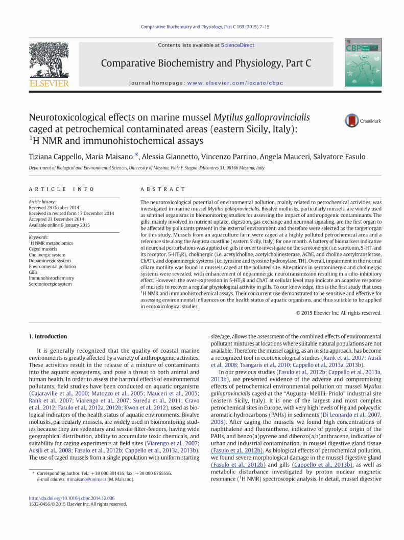
Neurotoxicological effects on marine mussel Mytilus galloprovincialis caged at petrochemical contaminated areas (eastern Sicily, Italy): 1 H NMR and immunohistochemical assays Tiziana Cappello, Maria Maisano ⁎, Alessia Giannetto, Vincenzo Parrino, Angela Mauceri, Salvatore Fasulo Department of Biological and Environmental Sciences, University of Messina, Viale F. Stagno d'Alcontres 31, 98166 Messina, Italy abstract article info Article history: Received 29 October 2014 Received in revised form 17 December 2014 Accepted 23 December 2014 Available online 6 January 2015 Keywords: 1 H NMR metabolomics Caged mussels Cholinergic system Dopaminergic system Environmental pollution Gills Immunohistochemistry Serotoninergic system The neurotoxicological potential of environmental pollution, mainly related to petrochemical activities, was investigated in marine mussel Mytilus galloprovincialis. Bivalve mollusks, particularly mussels, are widely used as sentinel organisms in biomonitoring studies for assessing the impact of anthropogenic contaminants. The gills, mainly involved in nutrient uptake, digestion, gas exchange and neuronal signaling, are the first organ to be affected by pollutants present in the external environment, and therefore were selected as the target organ for this study. Mussels from an aquaculture farm were caged at a highly polluted petrochemical area and a reference site along the Augusta coastline (eastern Sicily, Italy) for one month. A battery of biomarkers indicative of neuronal perturbations was applied on gills in order to investigate on the serotonergic (i.e. serotonin, 5-HT, and its receptor, 5-HT 3 R), cholinergic (i.e. acetylcholine, acetylcholinesterase, AChE, and choline acetyltransferase, ChAT), and dopaminergic systems (i.e. tyrosine and tyrosine hydroxylase, TH). Overall, impairment in the normal ciliary motility was found in mussels caged at the polluted site. Alterations in serotoninergic and cholinergic systems were revealed, with enhancement of dopaminergic neurotransmission resulting in a cilio-inhibitory effect. However, the over-expression in 5-HT 3 R and ChAT at cellular level may indicate an adaptive response of mussels to recover a regular physiological activity in gills. To our knowledge, this is the first study that uses 1 H NMR and immunohistochemical assays. Their concurrent use demonstrated to be sensitive and effective for assessing environmental influences on the health status of aquatic organisms, and thus suitable to be applied in ecotoxicological studies. © 2015 Elsevier Inc. All rights reserved. 1. Introduction It is generally recognized that the quality of coastal marine environments is greatly affected by a variety of anthropogenic activities. These activities result in the release of a mixture of contaminants into the aquatic ecosystems, and pose a threat to both animal and human health. In order to assess the harmful effects of environmental pollutants, field studies have been conducted on aquatic organisms (Cajaraville et al., 2000; Matozzo et al., 2005; Mauceri et al., 2005; Rank et al., 2007; Viarengo et al., 2007; Sureda et al., 2011; Cravo et al., 2012; Fasulo et al., 2012a, 2012b; Kwon et al., 2012), used as bio- logical indicators of the health status of aquatic environments. Bivalve mollusks, particularly mussels, are widely used in biomonitoring stud- ies because they are sedentary and sessile filter-feeders, having wide geographical distribution, ability to accumulate toxic chemicals, and suitability for caging experiments at field sites (Viarengo et al., 2007; Ausili et al., 2008; Fasulo et al., 2012b; Cappello et al., 2013a, 2013b). The use of caged mussels from a single population with uniform starting size/age, allows the assessment of the combined effects of environmental pollutant mixtures at locations where suitable natural populations are not available. Therefore the mussel caging, as an in situ approach, has become a recognized tool in ecotoxicological studies (Rank et al., 2007; Ausili et al., 2008; Tsangaris et al., 2010; Cappello et al., 2013a, 2013b). In our previous studies (Fasulo et al., 2012b; Cappello et al., 2013a, 2013b), we presented evidence of the adverse and compromising effects of petrochemical environmental pollution on mussel Mytilus galloprovincialis caged at the “Augusta–Melilli–Priolo” industrial site (eastern Sicily, Italy). It is one of the largest and most complex petrochemical sites in Europe, with very high levels of Hg and polycyclic aromatic hydrocarbons (PAHs) in sediments (Di Leonardo et al., 2007, 2008). After caging the mussels, we found high concentrations of naphthalene and fluoranthene, indicative of pyrolytic origin of the PAHs, and benzo(a)pyrene and dibenzo(a,b)anthracene, indicative of urban and industrial contamination, in mussel digestive gland tissue (Fasulo et al., 2012b). As biological effects of petrochemical pollution, we found severe morphological damage in the mussel digestive gland (Fasulo et al., 2012b) and gills (Cappello et al., 2013b), as well as metabolic disturbance investigated by proton nuclear magnetic resonance ( 1 H NMR) spectroscopic analysis. In detail, mussel digestive Comparative Biochemistry and Physiology, Part C 169 (2015) 7–15 ⁎ Corresponding author. Tel.: +39 090 391435; fax: +39 090 6765556. E-mail address: [email protected] (M. Maisano). http://dx.doi.org/10.1016/j.cbpc.2014.12.006 1532-0456/© 2015 Elsevier Inc. All rights reserved. Contents lists available at ScienceDirect Comparative Biochemistry and Physiology, Part C journal homepage: www.elsevier.com/locate/cbpc
-
Upload
independent -
Category
Documents
-
view
4 -
download
0
Transcript of Neurotoxicological effects on marine mussel Mytilus galloprovincialis caged at petrochemical...
Comparative Biochemistry and Physiology, Part C 169 (2015) 7–15
Contents lists available at ScienceDirect
Comparative Biochemistry and Physiology, Part C
j ourna l homepage: www.e lsev ie r .com/ locate /cbpc
Neurotoxicological effects on marine mussel Mytilus galloprovincialiscaged at petrochemical contaminated areas (eastern Sicily, Italy):1H NMR and immunohistochemical assays
Tiziana Cappello, Maria Maisano ⁎, Alessia Giannetto, Vincenzo Parrino, Angela Mauceri, Salvatore FasuloDepartment of Biological and Environmental Sciences, University of Messina, Viale F. Stagno d'Alcontres 31, 98166 Messina, Italy
⁎ Corresponding author. Tel.: +39 090 391435; fax: +E-mail address: [email protected] (M. Maisano).
http://dx.doi.org/10.1016/j.cbpc.2014.12.0061532-0456/© 2015 Elsevier Inc. All rights reserved.
a b s t r a c t
a r t i c l e i n f oArticle history:Received 29 October 2014Received in revised form 17 December 2014Accepted 23 December 2014Available online 6 January 2015
Keywords:1H NMR metabolomicsCaged musselsCholinergic systemDopaminergic systemEnvironmental pollutionGillsImmunohistochemistrySerotoninergic system
The neurotoxicological potential of environmental pollution, mainly related to petrochemical activities, wasinvestigated in marine mussel Mytilus galloprovincialis. Bivalve mollusks, particularly mussels, are widely usedas sentinel organisms in biomonitoring studies for assessing the impact of anthropogenic contaminants. Thegills, mainly involved in nutrient uptake, digestion, gas exchange and neuronal signaling, are the first organ tobe affected by pollutants present in the external environment, and therefore were selected as the target organfor this study. Mussels from an aquaculture farm were caged at a highly polluted petrochemical area and areference site along the Augusta coastline (eastern Sicily, Italy) for onemonth. A battery of biomarkers indicativeof neuronal perturbationswas applied on gills in order to investigate on the serotonergic (i.e. serotonin, 5-HT, andits receptor, 5-HT3R), cholinergic (i.e. acetylcholine, acetylcholinesterase, AChE, and choline acetyltransferase,ChAT), and dopaminergic systems (i.e. tyrosine and tyrosine hydroxylase, TH). Overall, impairment in the normalciliary motility was found in mussels caged at the polluted site. Alterations in serotoninergic and cholinergicsystems were revealed, with enhancement of dopaminergic neurotransmission resulting in a cilio-inhibitoryeffect. However, the over-expression in 5-HT3R and ChAT at cellular level may indicate an adaptive responseof mussels to recover a regular physiological activity in gills. To our knowledge, this is the first study that uses1H NMR and immunohistochemical assays. Their concurrent use demonstrated to be sensitive and effective forassessing environmental influences on the health status of aquatic organisms, and thus suitable to be appliedin ecotoxicological studies.
© 2015 Elsevier Inc. All rights reserved.
1. Introduction
It is generally recognized that the quality of coastal marineenvironments is greatly affected by a variety of anthropogenic activities.These activities result in the release of a mixture of contaminantsinto the aquatic ecosystems, and pose a threat to both animal andhuman health. In order to assess the harmful effects of environmentalpollutants, field studies have been conducted on aquatic organisms(Cajaraville et al., 2000; Matozzo et al., 2005; Mauceri et al., 2005;Rank et al., 2007; Viarengo et al., 2007; Sureda et al., 2011; Cravoet al., 2012; Fasulo et al., 2012a, 2012b; Kwon et al., 2012), used as bio-logical indicators of the health status of aquatic environments. Bivalvemollusks, particularly mussels, are widely used in biomonitoring stud-ies because they are sedentary and sessile filter-feeders, having widegeographical distribution, ability to accumulate toxic chemicals, andsuitability for caging experiments at field sites (Viarengo et al., 2007;Ausili et al., 2008; Fasulo et al., 2012b; Cappello et al., 2013a, 2013b).The use of caged mussels from a single population with uniform starting
39 090 6765556.
size/age, allows the assessment of the combined effects of environmentalpollutantmixtures at locationswhere suitable natural populations are notavailable. Therefore themussel caging, as an in situ approach, has becomea recognized tool in ecotoxicological studies (Rank et al., 2007; Ausiliet al., 2008; Tsangaris et al., 2010; Cappello et al., 2013a, 2013b).
In our previous studies (Fasulo et al., 2012b; Cappello et al., 2013a,2013b), we presented evidence of the adverse and compromisingeffects of petrochemical environmental pollution on mussel Mytilusgalloprovincialis caged at the “Augusta–Melilli–Priolo” industrial site(eastern Sicily, Italy). It is one of the largest and most complexpetrochemical sites in Europe,with very high levels of Hg and polycyclicaromatic hydrocarbons (PAHs) in sediments (Di Leonardo et al., 2007,2008). After caging the mussels, we found high concentrations ofnaphthalene and fluoranthene, indicative of pyrolytic origin of thePAHs, and benzo(a)pyrene and dibenzo(a,b)anthracene, indicative ofurban and industrial contamination, in mussel digestive gland tissue(Fasulo et al., 2012b). As biological effects of petrochemical pollution,we found severe morphological damage in the mussel digestive gland(Fasulo et al., 2012b) and gills (Cappello et al., 2013b), as well asmetabolic disturbance investigated by proton nuclear magneticresonance (1H NMR) spectroscopic analysis. In detail, mussel digestive

Fig. 1.Map showing location of the mussel caging sites, Priolo (37°12′10″N; 15°13′44″E) and Vendicari (36°47′35″N; 15°08′52″E).
8 T. Cappello et al. / Comparative Biochemistry and Physiology, Part C 169 (2015) 7–15
glands showed changes in metabolites involved in energy and lipidmetabolism (Fasulo et al., 2012b), and activation of detoxificationand antioxidant defense mechanisms, namely cytochrome P4504Y1(CYP4Y1) and glutathione S-transferase (GST), indicative of xenobioticdetoxification, and catalase (CAT) as oxidative stress index (Cappelloet al., 2013a). The changes observed in the 1HNMRmetabolomic profileof gills suggested alteration in osmotic regulation, energy metabolismand neurotransmission (Cappello et al., 2013b).
The gills of marine mollusks, mainly involved in nutrient uptake,digestion, gas exchange and neuronal signaling, have been used inecotoxicological studies as indicators of aquatic pollution (Gregoryet al., 2002; Gagné et al., 2007; Fasulo et al., 2008; Zhang et al., 2011;Ciacci et al., 2012; Cappello et al., 2013b). Gill activity is regulated bysympathetic and parasympathetic innervations of the autonomicnervous system running through the branchial connective tissue(Catapane et al., 1974), with various neurotransmitters participat-ing in regulation of ciliary beating (Stefano, 1990). Serotonin, or5-hydroxytryptamine (5-HT), involved in the serotonergic system,plays a cilioexcitatory activity in the gills of lamellibranchiates(Gosselin, 1961; Carroll and Catapane, 2007). The molluskan gill
Table 1Details of primary antibodies.
Antigen Animalsource
Supplier Dilution
Serotonin (5-HT) Mouse Dako Cytomation,Milan, IT
1:50
Serotonin receptor(5-HT3R)
Rabbit Sigma-Aldrich,St. Louis, MO, USA
1:100
Acetylcholine esterase(AChE)
Mouse Chemicon International,Temecula, CA, USA
1:50
Choline Acetyltransferase(ChAT)
Rabbit Abcam, Cambridge, UK 1:250
Tyrosine hydroxylase(TH)
Mouse Sigma-Aldrich,St. Louis, MO, USA
1:100
contains significant amounts of endogenous 5-HT, which producesprompt, sustained, and reversible stimulation of ciliary beat frequency,and the magnitude of the response is graded over a wide range of 5-HTlevels (Gosselin, 1961). Impairment in the serotoninergic system mayoccur after exposure to toxicant compounds, as observed in gills of themussel M. galloprovincialis exposed to chromium and copper, resultingin increase of the 5-HT-stimulated adenylate cyclase activity in vivoand over-expression of 5-HT receptors (Fabbri and Capuzzo, 2006).
Acetylcholine is a neural transmitter used in efferent nervoussystems, and it is synthesized in the cytoplasm of cholinergic neuronsby the enzyme choline acetyltransferase (ChAT) and split into cholineand acetate in cholinergic synapses and neuromuscular junctions byacetylcholinesterase (AChE) (Matozzo et al., 2005). AChE is essentialfor the normal functioning of the central and peripheral nervous system(Lionetto et al., 2013). The measurement of AChE activity is a validatedbiomarker of neurotoxic compounds in aquatic organisms (Cajaravilleet al., 2000; Matozzo et al., 2005; Ciacci et al., 2012; D'Agata et al.,2014). AChE activity is directly inhibited by pollutants like organophos-phate and carbamate pesticides (Lionetto et al., 2013), even thoughits responsiveness to other chemicals, such as heavy metals andhydrocarbons, has been demonstrated (Rank et al., 2007).
Tyrosine is a non-essential amino acid that serves as substrateprecursor for the synthesis of catecholamines, which includeadrenaline, noradrenaline, and dopamine. The conversion of tyrosineinto catecholamines is catalyzed by the enzyme tyrosine hydroxylase(TH), involved in the adrenergic–dopaminergic system that, inMytilus, has an inhibitory effect on lateral cilia beating (Zhu et al.,2005). TH is a mixed-function oxidase that uses molecular oxygenand tyrosine as its substrates, and biopterin as its cofactor (Shiman et al.,1971). TH activity may be affected by toxic compounds present in theenvironments, as reported by Ciacci et al. (2012) with a noticeabledose-dependent increase of TH in M. galloprovincialis exposed to Cr(VI),resulting in an enhancement of dopaminergic neurotransmission.
The aim of this study was to assess the neurotoxic potential ofenvironmental pollution, mainly related to Hg and PAHs, in marine

Fig. 3. 1H NMR measurements of serotonin (mM) from mussel gill tissue, expressed asmeans ± standard deviation (n = 12). Asterisks indicate significant differences relativeto control (**p b 0.005).
Fig. 2.Western blot (WB) of proteins from (A)whole cell lysates and (B)mussel gill tissueextracts. SKBR-3, HeLa and PC-12 whole cell lysates were used as WB positive control forAChE, ChAT and TH, respectively.
9T. Cappello et al. / Comparative Biochemistry and Physiology, Part C 169 (2015) 7–15
musselM. galloprovincialis caged in an eastern Sicily petrochemical area.In order to extend our previous 1H NMR findings (Cappello et al.,2013b), a battery of biomarkers indicative of neuro-endocrinal pertur-bations was applied on mussel gills in order to investigate on the(i) serotoninergic (i.e. 5-HT and its receptor, 5-HT3R), (ii) cholinergic(i.e. acetylcholine, AChE and ChAT), and (iii) dopaminergic systems(i.e. tyrosine and TH). The environmental metabolomics, based onNMR spectroscopy, is a qualitative and quantitative approach that canbe used to accurately determine metabolite concentration with a veryhigh sensitivity (Hines et al., 2007; Espina et al., 2009; Kwon et al.,2012). To our knowledge, this is the first study that has used 1H NMRmeasurements of neurotransmitter concentrations in combinationwith immunohistochemical assays.
2. Materials and methods
2.1. Study area
As reported previously (Ausili et al., 2008; De Domenico et al., 2011,2013; Fasulo et al., 2012b), the petrochemical area of “Augusta–Melilli–Priolo” was declared a “site of national interest” by the Italian Ministryof Environment (Law No. 426/98; Ministerial Decree of 10.01.2000)owing to the high level of pollution, mainly due to Hg and PAHs, andsubsequent risk for human health. Conversely, the natural reserveof Vendicari (southeastern Sicily) was considered as a reference sitenot known to be impacted by petrochemical contamination (Fig. 1).Physical and chemical parameters of water (i.e. temperature, salinity,pH, dissolved oxygen) were measured at both sampling sites at thecage deployment depth (8 m) three times, as reported in Cappelloet al. (2013a).
2.2. Experimental design
The experimental design was performed as detailed in our previouspapers (Fasulo et al., 2012b; Cappello et al., 2013a, 2013b). Briefly,mussels M. galloprovincialis (mean shell length: 6.1 ± 0.54 cm) werepurchased in October 2009 from an aquaculture farm in Goro (Ferrara,Italy), named Consortium of Fishermen. After maintaining in aeratedseawater in the laboratory for one week, mussels were transplanted tothe two selected sites at 8 m depth in stainless steel cages (about 200specimens at each site) for 30 days. The translocation period of onemonth allows the adaptation of the organisms to the new environmen-tal conditions, and it is used in a number of national and internationalMussel Watch programs (i.e. RAMOGE). From each area, twelve maleindividuals, sexed by microscopic observation of gonad tissue, of com-parable body length and mass (6.6 ± 0.46 cm shell length; 27.8 ±3.2 g wet weight) were selected randomly and sacrificed. Gill sampleswere rapidly excised and flash-frozen in liquid nitrogen, then trans-ferred to the laboratory and stored at−80 °C prior to the NMR analysis.Further, small pieces of each dissected tissuewere taken formorpholog-ical and immunohistochemical analyses.
2.3. 1H NMR-based metabolomic analysis
Polarmetabolites were extracted from gills (n=12 per site) using a“two-step” methanol/chloroform/water procedure (Wu et al., 2008),as previously described (Cappello et al., 2013b). Briefly, the gill tissue(ca. 100 mg) was homogenized in 4 ml/g of cold methanol and0.85 ml/g of cold water by an Ultraturrax homogenizer. The homog-enates were transferred to glass vials, and 4 ml/g chloroform and2 ml/g water were added. Samples were vortexed and centrifugedfor 5 min at 2000 g at 4 °C. The upper methanol layer with thepolar metabolites was transferred to glass vials, dried in a centrifugalvacuum concentrator (Eppendorf 5301) and stored at −80 °C. Prior toNMR analysis, the dried polar extracts were resolvated in 100 μl ofD2O (Armar AG, Döttingen, Switzerland) buffered in 240 mM sodium
phosphate, pH 7.0, containing 1 mM 2,2-dimethyl-2-silapentane-5-sulfonate (DSS) (Sigma-Aldrich Co) as chemical shift standard. Fiftymicroliters of each resuspended sample was then pipetted into4 mm-diameter zirconia rotors.
NMR analyses were performed using a Bruker Avance-700 NMRspectrometer operating at 6000 Hz at 298 K, and equipped with aHigh Resolution-Magic Angle Spinning (HR-MAS) probe used for ourconvenience. One-dimensional (1-D) 1H NMR spectra were obtainedas previously described (Cappello et al., 2013b), and manually phased,baseline-corrected, and calibrated (DSS at 0.0 ppm) using XWIN-NMRsoftware (version 3.5; Bruker). The peaks of interest (i.e. serotonin,acetylcholine and tyrosine) within the 1H NMR spectra were identifiedand quantified using Chenomx Profiler, a module of Chenomx NMRSuite software (version 5.1; Chenomx Inc., Edmonton, Canada), whichuses the concentration of a known DSS signal to determine theconcentrations of individual metabolites (Kwon et al., 2012).
2.4. Immunohistochemical analysis
Gill tissues of twelve mussels from each sampling site were fixed in4% paraformaldehyde (Immunofix, Bio-OpticaMilano, Italy) for 4 h, andthen rinsed in 0.1 M phosphate buffered saline (PBS, pH 7.4). After de-hydration in a graded series of ethanol and embedding in Paraplast(Bio-Optica Milano, Italy), 5 μm thick histological sections were cutwith a rotary automatic microtome (Leica Microsystems, Wetzlar,Germany), mounted on glass slides and processed for indirect immuno-fluorescence method (Mauceri et al., 1999) for localization of 5-HT andits receptor, 5-HT3R, AChE and ChAT, and TH. Briefly, non-specificbinding sites for immunoglobulins were blocked with normal goatserum (NGS) in PBS (1:5) by incubation for 1 h. The sections werethen incubated overnight at 4 °C in a humid chamber in the presenceof the primary antisera (dilutions and suppliers as indicated inTable 1). After a rinse in PBS for 10 min, binding sites of the primaryantibodies were visualized by corresponding fluorescein isothiocyanate
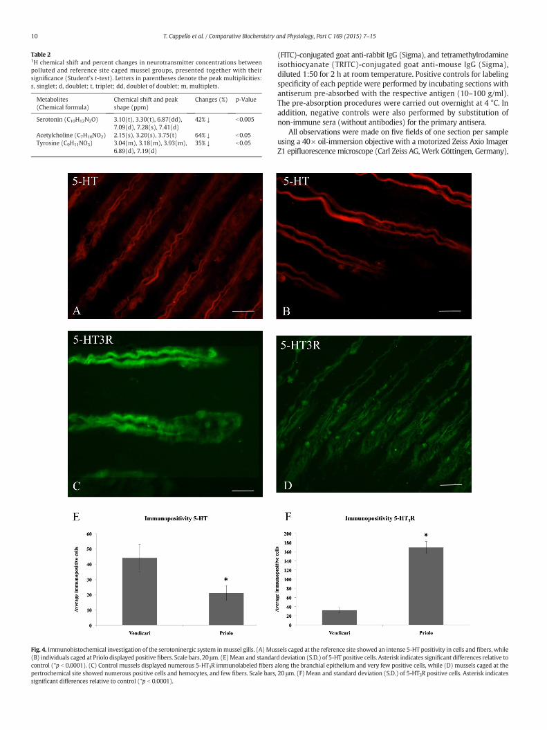
Fig. 4. Immunohistochemical investigation of the serotoninergic system inmussel gills. (A) Mus(B) individuals caged at Priolo displayed positive fibers. Scale bars, 20 μm. (E)Mean and standarcontrol (*p b 0.0001). (C) Control mussels displayed numerous 5-HT3R immunolabeled fibers apertrochemical site showed numerous positive cells and hemocytes, and few fibers. Scale bars,significant differences relative to control (*p b 0.0001).
Table 21H chemical shift and percent changes in neurotransmitter concentrations betweenpolluted and reference site caged mussel groups, presented together with theirsignificance (Student's t-test). Letters in parentheses denote the peak multiplicities:s, singlet; d, doublet; t, triplet; dd, doublet of doublet; m, multiplets.
Metabolites(Chemical formula)
Chemical shift and peakshape (ppm)
Changes (%) p-Value
Serotonin (C10H12N2O) 3.10(t), 3.30(t), 6.87(dd),7.09(d), 7.28(s), 7.41(d)
42% ↓ b0.005
Acetylcholine (C7H16NO2) 2.15(s), 3.20(s), 3.75(t) 64% ↓ b0.05Tyrosine (C9H11NO3) 3.04(m), 3.18(m), 3.93(m),
6.89(d), 7.19(d)35% ↓ b0.05
10 T. Cappello et al. / Comparative Biochemistry and Physiology, Part C 169 (2015) 7–15
(FITC)-conjugated goat anti-rabbit IgG (Sigma), and tetramethylrodamineisothiocyanate (TRITC)-conjugated goat anti-mouse IgG (Sigma),diluted 1:50 for 2 h at room temperature. Positive controls for labelingspecificity of each peptide were performed by incubating sections withantiserum pre-absorbed with the respective antigen (10–100 g/ml).The pre-absorption procedures were carried out overnight at 4 °C. Inaddition, negative controls were also performed by substitution ofnon-immune sera (without antibodies) for the primary antisera.
All observations were made on five fields of one section per sampleusing a 40× oil-immersion objective with a motorized Zeiss Axio ImagerZ1 epifluorescence microscope (Carl Zeiss AG, Werk Göttingen, Germany),
sels caged at the reference site showed an intense 5-HT positivity in cells and fibers, whiled deviation (S.D.) of 5-HT positive cells. Asterisk indicates significant differences relative tolong the branchial epithelium and very few positive cells, while (D) mussels caged at the20 μm. (F) Mean and standard deviation (S.D.) of 5-HT3R positive cells. Asterisk indicates
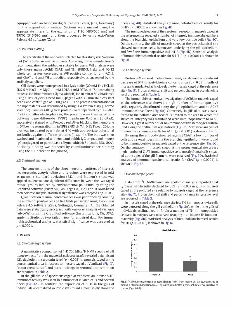
Fig. 5. 1H NMRmeasurements of acetylcholine (mM) frommussel gill tissue, expressed asmeans± standard deviation (n= 12). Asterisk indicates significant differences relative tocontrol (*p b 0.05).
11T. Cappello et al. / Comparative Biochemistry and Physiology, Part C 169 (2015) 7–15
equipped with an AxioCam digital camera (Zeiss, Jena, Germany)for the acquisition of images. Sections were imaged using theappropriate filters for the excitation of FITC (480/525 nm) andTRITC (515/590 nm), and then processed by using AxioVisionRelease 4.5 software (Zeiss).
2.5. Western blotting
The specificity of the antibodies selected for this study was WesternBlot (WB) tested in marine mussels. According to the manufacturer'srecommendation, the antibodies suitable for use in WB analysis wereonly those against AChE, ChAT, and TH. SKBR-3, HeLa and PC-12whole cell lysates were used as WB positive control for anti-AChE,anti-ChAT and anti-TH antibodies, respectively, as suggested by theantibody suppliers.
Gill tissues were homogenized in a lysis buffer (20 mM Tris-HCl, 2%SDS, 5 M NaCl, 1 MMgCl2, 1 mM EDTA, 1 mM EGTA, pH 7.4) containingprotease inhibitormixture (Sigma-Aldrich) for 10min at 50 vibrations/susing a TissueLyser LT bead mill (Qiagen) with 3.2 mm stainless steelbeads, and centrifuged at 3000 g at 4 °C. The protein concentration ofthe supernatants was determined by using BCA Protein assay (Thermoscientific). Samples (40 μg total protein) were separated by SDS-PAGE(12%) and after electrophoresis, the proteins were transferred to apolyvinylidene difluoride (PVDF) membrane 0.45 μm (BioRad),successively stained with Ponceau red. After blocking the membranein 5% bovine serum albumin in TPBS (PBS pH 7.4, 0.1% Tween 20), theblot was incubated overnight at 4 °C with appropriate polyclonalantibodies against different proteins (1 μg/ml). The blot was thenwashed and incubated with goat anti-rabbit IgG or goat anti-mouseIgG conjugated to peroxidase (Sigma-Aldrich St. Louis, MO, USA).Antibody binding was detected by chemiluminescence stainingusing the ECL detection kit (BioRad).
2.6. Statistical analysis
The concentrations of the three neurotransmitters of interest,i.e. serotonin, acetylcholine and tyrosine, were expressed in mMas means ± standard deviation (S.D.), and Student's t-test wasapplied to determinate significant differences between the two cagedmussel groups induced by environmental pollutants, by using theGraphPad software (Prism 5.0, San Diego CA, USA). For 1H NMR-basedmetabolomic analysis, statistical significance was accepted at p b 0.05.
Quantification of immunoreactive cells was performed by countingthe number of positive cells on five fields per section using Axio VisionRelease 4.5 software (Zeiss, Göttingen, Germany). All the obtaineddata were statistically processed with one-way analysis of variance(ANOVA) using the GraphPad software (Instat, La Jolla, CA, USA),applying Student's two-tailed t-test for unpaired data. For immu-nohistochemical analysis, statistical significance was accepted atp b 0.0001.
3. Results
3.1. Serotoninergic system
A quantitative comparison of 1-D 700 MHz 1H NMR spectra of gilltissue extracts from themusselM. galloprovincialis revealed a significant42% depletion in serotonin level (p b 0.005) in mussels caged at thepetrochemical area in respect to mussels caged at Vendicari (Fig. 3).Proton chemical shift and percent change in serotonin concentrationare reported in Table 2.
In the gill tissue of specimens caged at Vendicari an intense 5-HTimmunoreactivity was seen in a number of ciliated cells and severalfibers (Fig. 4A). In contrast, the expression of 5-HT in the gills ofindividuals acclimatized in Priolo was found almost solely along the
fibers (Fig. 4B). Statistical analysis of immunohistochemical results for5-HT (p b 0.0001) is shown in Fig. 4E.
The immunodetection of the serotonin receptor in mussels caged atthe reference site revealed a number of intensely immunolabeled fibersalong the branchial epithelium and very few positive cells (Fig. 4C).On the contrary, the gills of mussels caged at the petrochemical siteshowed numerous cells, hemocytes underlying the gill epithelium,and few fibers immunopositive to 5-HT3R (Fig. 4D). Statistical analysisof immunohistochemical results for 5-HT3R (p b 0.0001) is shown inFig. 4F.
3.2. Cholinergic system
Proton NMR-based metabolomic analysis showed a significantdecrease of 64% in acetylcholine concentration (p b 0.05) in gills ofmussels transplanted at Priolo relative tomussels caged at the referencesite (Fig. 5). Proton chemical shift and percent change in acetylcholinelevel are reported in Table 2.
The immunohistochemical investigation of AChE in mussels cagedat the reference site showed a high number of immunopositivecells, regularly distributed along the gill epithelium, and no AChEimmunopositive fibers (Fig. 6A). Conversely, in gills of mussels trans-ferred to the polluted area few cells limited to the area in which thestructural integrity was maintained were immunopositivite to AChE.However, a great number of AChE-immunopositive hemocytes distrib-uted along the epithelium was noticed (Fig. 6B). Statistical analysis ofimmunohistochemical results for AChE (p b 0.0001) is shown in Fig. 6E.
By using the antibody directed against ChAT, a low number ofcells and several fibers lining the branchial epithelium were foundto be immunopositive in mussels caged at the reference site (Fig. 6C).On the contrary, in mussels caged at the petrochemical site a veryhigh number of ChAT-immunopositive cells, mostly frontal cells situat-ed at the apex of the gill filament, were observed (Fig. 6D). Statisticalanalysis of immunohistochemical results for ChAT (p b 0.0001) isshown in Fig. 6F.
3.3. Dopaminergic system
Data from 1H NMR-based metabolomic analysis reported thattyrosine significantly declined by 35% (p b 0.05) in gills of musselscaged at the polluted site relative to mussels caged at the referencesite (Fig. 7). Proton chemical shift and percent change in tyrosine levelare reported in Table 2.
Inmussels caged at the reference site few TH-immunopositivite cellswere detected along the gill epithelium (Fig. 8A), while in the gills ofindividuals acclimatized in Priolo a number of TH-immunopositivecells and hemocyteswere observed, resulting in an intense TH immuno-reactivity (Fig. 8B). Statistical analysis of immunohistochemical resultsfor TH (p b 0.0001) is shown in Fig. 8C.

12 T. Cappello et al. / Comparative Biochemistry and Physiology, Part C 169 (2015) 7–15
3.4. Western blotting
Immunoreaction with AChE antibody revealed the predicted bandof 68 kDa both in the SKBR-3 extract and mussel gills. Specific ChATimmunoreactive bands of molecular mass of 70 kDa, corresponding tothe expected size for ChAT, were observed in both HeLa and musselgill extracts. Finally, the antibody against TH in the PC-12 whole celllysate revealed the presence of the predicted band of approximately60 kDa. A similar band was visualized in mussel gill extract. The WBresults for AChE, ChAT and TH are reported in Fig. 2.
Fig. 6. Immunohistochemical investigation of the cholinergic system inmussel gills. (A)Musselsdistributed along the gill epithelium, while (B) inmussels caged at Priolo AChE-immunopositivistandard deviation (S.D.) of AChEpositive cells. Asterisk indicates significant differences relativeseveral fibers along the gill epithelium, while (D)mussels caged at Priolo showed numerous podeviation (S.D.) of ChAT positive cells. Asterisk indicates significant differences relative to cont
4. Discussion
The neurotoxicological potential of environmental pollution, mainlyrelated to petrochemical activities, was assessed in the marine musselM. galloprovincialis caged in an eastern Sicily industrial area. The gills,mainly involved in nutrient uptake, digestion, gas exchange andneuronal signaling, were chosen as the target organ. Evidencesof metabolomic disturbances in neurotransmission were previouslyreported in Cappello et al. (2013b), therefore the neuronal pertur-bations on the serotoninergic, cholinergic, and dopaminergic
caged at the reference site showed a high number of AChE-immunopositive cells regularlytywas reduced, and numerous hemocytes were observed. Scale bars, 20 μm. (E)Mean andto control (*p b 0.0001). (C) Controlmussels displayed fewChAT-immunolabeled cells andsitive cells mostly at the apex of the gill filament. Scale bars, 20 μm. (F)Mean and standardrol (*p b 0.0001).

Fig. 7. 1H NMR measurements of tyrosine (mM) from mussel gill tissue, expressed asmeans ± standard deviation (n = 12). Asterisks indicate significant differences relativeto control (*p b 0.05).
13T. Cappello et al. / Comparative Biochemistry and Physiology, Part C 169 (2015) 7–15
systems were examined more closely, by the use of 1H NMR andimmunohistochemical assays.
The environmental metabolomics is a well-established techniquebased on the identification of low molecular weight endogenousmetabolites within a biological system (i.e. cells, tissues, organs orwhole individuals), whose production and levels vary in response toenvironmental stressors, diseases or exposure to toxicants (Viantet al., 2003; Iacono et al., 2010; Liu et al., 2011; Zhang et al., 2011;Fasulo et al., 2012b; Kwon et al., 2012; Cappello et al., 2013b). In thisstudy, 1H NMR-based metabolomics measurements of the metabolitesof interest frommussel gill extracts, allowed the successful investigationon the changes in neurotransmitter levels between mussels caged inthe reference site and those transplanted in the polluted area in re-sponse to environmental stress.
The significant decline in serotonin in gills of mussels caged at thepetrochemical area relative to mussels caged at the reference site wasrevealed both by 1HNMRand immunohistochemical analyses. As amat-ter of fact, these results are consistent with the impairments in energymetabolism highlighted in our previous work (Cappello et al., 2013b),in which we supposed the occurrence of glycogenolysis that may beassociated with compensatory mechanisms elicited by mussels inresponse to compromised gill ciliary activity, likely due to interferencesof environmental pollutants with the serotoninergic system. In fact, it iswell-known that serotonin is involved in hormonal and neuronalpathways, and plays a key role in cilioexcitatory activity, reproductivesuccess and regulating food intake in invertebrates (Gosselin, 1961;Stefano, 1990). Noteworthy, a concomitant 5-HT3R over-expression atcellular level in mussels caged at the polluted area was detected,while in mussels caged at Vendicari solely nerve fibers were 5-HT3Rimmunopositive. The rise of 5-HT3R could be related to an adaptiveresponse mediated by paracrine signaling activities in order to recovera regular physiological activity in gills. Indeed, it has been reportedthat gill epithelial cells containing the lateral cilia present 5-HTreceptors to increasing the ciliary beating rate, if activated (Carroll andCatapane, 2007). Fabbri and Capuzzo (2006), after exposing musselM. galloprovincialis to Cr(VI) and Cu(II), found increased 5-HT-stimulated adenylate cyclase activity in vivo, and suggested the possibil-ity that metal accumulation in mussel gills might induce the over-expression of 5-HT receptors. Further evidences of alterations in seroto-nin signaling pathwayswere provided by the significantly up-regulatedtranscription of 5-HT receptor in both sexes of M. galloprovincialisexposed to 0.1, 1, and 10 μg Cr(VI) L-1 animal−1 for 96 h, with malesshowing more sensitivity to metal treatment than females (Ciacciet al., 2012).
Quantitative 1H NMR analysis showed a 64% significant decreasein the concentration of acetylcholine, a cholinergic neurotransmitter,in gills of mussels caged at Priolo in respect to mussels caged atVendicari. In studies of ecotoxicology, measurements of AChE activ-ity have been used to test for neurotoxicity in aquatic organismssince it can be inhibited by the presence of contaminants in complexmixtures (Matozzo et al., 2005; Cajaraville et al., 2000; Tsangariset al., 2010; Cravo et al., 2012; D'Agata et al., 2014), by pollutantslike organophosphate and carbamate pesticides (Lionetto et al.,2013), or heavy metals and hydrocarbons (Rank et al., 2007; Ciacciet al., 2012). As expected, in mussels caged at the petrochemicalsite of Priolo AChE immunopositivity was reduced, and limited almostsolely to hemocytes. In marine mussels, hemocytes possess a complexcell signaling network with high homology with that of vertebrates,that allows them to modulate their own functions (Humphries andYoshino, 2003; Franzellitti and Fabbri, 2013). However, little it isknown on the physiological roles of these signaling pathways in musselhemocytes. Also, the presence of ChAT, which enzymatic action resultsin synthesis of acetylcholine, implicating a negative correlation withAChE, was found noticeably increased. As amatter of fact, the inhibitionof AChE and increment in ChAT are followed by accumulation ofacetylcholine. However, in the present study the opposite result of
reduced acetylcholine was found in gills of mussels transferred at thepetrochemical site for one month, likely to be ascribed to an ongoingacetylcholine synthesis process. Similar findings have been showed byZhang et al. (2011), which reported a decline in acetylcholine in gillsof adult Manila clams Ruditapes philippinarum after a 24 h exposure toboth low (10 μg L−1) and high (40 μg L−1) doses of Cu.
The concentration of tyrosine was found significantly reduced by35% in mussels caged at the petrochemical area in respect to musselscaged at the reference site. In the same individuals, the presence ofthe enzyme tyrosine hydroxylase (TH), which serves for conversion oftyrosine into catecholamines, was significantly induced in mussel gills.This data is consistent with the depletion of tyrosine, which is negative-ly correlated to the elevation of TH. Previous experimental studies havereported a noticeable dose-dependent increase of TH in the gills ofM. galloprovincialis exposed to 0.1, 1, 10 μg Cr(VI) L−1 animal−1 for96 h (Ciacci et al., 2012). The increase in THmay result in the enhance-ment of dopaminergic neurotransmission that has an inhibitory effecton lateral ciliary activity of mussel gills (Carroll and Catapane, 2007).Zhu et al. (2005) demonstrated that tyrosine and tyramine mayaugment endogenous ganglionic morphine and dopamine levelsin Mytilus edulis under in vitro and in vivo experiments, with theinvolvement of CYP2D6 and TH in the process, resulting in a cilio-inhibitory effect.
5. Conclusions
This study provides evidence that environmental pollution, mainlyrelated to petrochemical activities, adversely affects the neurotrans-mission system in caged M. galloprovincialis individuals, resulting inimpairment in the normal ciliary motility, and thus in feeding effi-ciencies. 1H NMR-based environmental metabolomics offers an al-ternative method of measuring metabolite concentration. Indeed,the high sensitivity of 1H NMR allowed measurement of changesin neurotransmitter levels, namely serotonin, acetylcholine andtyrosine. The depletion in neurotransmitter concentrations wassupported by evidence of neuronal perturbations in mussel gills.In detail, alterations in serotoninergic and cholinergic systemswere revealed, together with enhancement of dopaminergic neuro-transmission, resulting in a cilio-inhibitory effect. However, theconcomitant over-expression in serotonin receptors and cholineacetyltransferase at cellular level may indicate an adaptive responsemediated by paracrine signaling activities in order to recover aregular physiological activity in gills. Therefore, the concurrent useof 1H NMR and immunohistochemical assays demonstrated to besensitive and effective for assessing environmental influences onthe health status of aquatic organisms, and thus suitable to beapplied in ecotoxicological studies.

Fig. 8. Immunohistochemical investigation of the dopaminergic system inmussel gills. (A) Controlmussels showed a slight TH-immunopositivity in cells of the gill epithelium,while (B) inmussels caged at Priolo an intense immunoreactivity of TH was revealed, with a number of positive cells and hemocytes. Scale bars, 20 μm. (C) Mean and standard deviation (S.D.) of THpositive cells. Asterisk indicates significant differences relative to control (*p b 0.0001).
14 T. Cappello et al. / Comparative Biochemistry and Physiology, Part C 169 (2015) 7–15
Acknowledgments
This research was supported by a National Interest Research Project(PRIN 2007-20079FELYB).
References
Ausili, A., Gabellini, M., Cammarata, G., Fattorini, D., Benedetti, M., Pisanelli, B., Gorbi, S.,Regoli, F., 2008. Ecotoxicological and human health risk in a petrochemical districtof southern Italy. Mar. Environ. Res. 66, 217–219.
Cajaraville, M.P., Bebianno, M.J., Blasco, J., Porte, C., Sarasquete, C., Viarengo, A., 2000. Theuse of biomarkers to assess the impact of pollution in coastal environments of theIberian Peninsula: a practical approach. Sci. Total Environ. 247, 295–311.
Cappello, T., Maisano, M., D'Agata, A., Natalotto, A., Mauceri, A., Fasulo, S., 2013a. Effects ofenvironmental pollution in caged mussels (Mytilus galloprovincialis). Mar. Environ.Res. 91, 52–60.
Cappello, T., Mauceri, A., Corsaro, C., Maisano, M., Parrino, V., Lo Paro, G., Messina, G.,Fasulo, S., 2013b. Impact of environmental pollution on caged mussels Mytilusgalloprovincialis using NMR-based metabolomics. Mar. Pollut. Bull. 77, 132–139.
Carroll, M.A., Catapane, E.J., 2007. The nervous system control of lateral ciliary activity ofthe gill of the bivalve mollusc, Crassostrea virginica. Comp. Biochem. Physiol. A 148,445–450.
Catapane, E., Aiello, E., Stefano, G.B., 1974. Ganglionic mediationmechanism of lateral ciliain Mytilus edulis gill. Physiologist 17–372.
Ciacci, C., Barmo, C., Gallo, G., Maisano, M., Cappello, T., D'Agata, A., Leonzio, C., Mauceri, A.,Fasulo, S., Canesi, L., 2012. Effects of sublethal, environmentally relevant concentrationsof hexavalent chromium in the gills ofMytilus galloprovincialis. Aquat. Toxicol. 120–121,109–118.
Cravo, C., Pereira, C., Gomes, T., Cardoso, C., Serafim, A., Almeida, C., Rocha, T., Lopes, B.,Company, R., Medeiros, A., Norberto, R., Pereira, R., Araújo, O., Bebianno, M.J.,2012. A multibiomarker approach in the clam Ruditapes decussatus to assessthe impact of pollution in the Ria Formosa lagoon, South Coast of Portugal. Mar.Environ. Res. 75, 23–34.
D'Agata, A., Cappello, T., Maisano, M., Parrino, V., Giannetto, A., Brundo, M.V., Ferrante, M.,Mauceri, A., 2014. Cellular biomarkers in the mussel Mytilus galloprovincialis(Bivalvia: Mytilidae) from Lake Faro (Sicily, Italy). Ital. J. Zool. 81 (1), 43–54.
De Domenico, E., Mauceri, A., Giordano, D., Maisano, M., Gioffrè, G., Natalotto, A.,D'Agata, A., Ferrante, M., Brundo, M.V., Fasulo, S., 2011. Effects of “in vivo” exposureto toxic sediments on juveniles of sea bass (Dicentrarchus labrax). Aquat. Toxicol.105, 688–697.
De Domenico, E., Mauceri, A., Giordano, D., Maisano, M., Giannetto, A., Parrino, V.,Natalotto, A., D'Agata, A., Cappello, T., Fasulo, S., 2013. Biological responses of juvenileEuropean sea bass (Dicentrarchus labrax) exposed to contaminated sediments.Ecotoxicol. Environ. Saf. 97, 114–123.
Di Leonardo, R., Bellanca, A., Capotondi, L., Cundy, A., Neri, R., 2007. Possible impacts of Hgand PAH contamination on benthic foraminiferal assemblages: an example from theSicilian coast, central Mediterranean. Sci. Total Environ. 388, 168–183.
Di Leonardo, R., Bellanca, A., Angelone, M., Leonardi, M., Neri, R., 2008. Impact of humanactivities on the central Mediterranean offshore: evidence from Hg distribution inbox-core sediments from the Ionian Sea. Appl. Geochem. 23, 3756–3766.
Espina, R., Yu, L., Wang, J., Tong, Z., Vashishtha, S., Talaat, R., Scatina, J., Mutlib, A., 2009.Nuclear magnetic resonance spectroscopy as a quantitative tool to determine theconcentrations of biologically produced metabolites: implications in metabolites insafety testing. Chem. Res. Toxicol. 22, 299–310.
Fabbri, E., Capuzzo, A., 2006. Adenylyl cyclase activity and its modulation in the gills ofMytilus galloprovincialis exposed to Cr6+ and Cu2+. Aquat. Toxicol. 76, 59–68.
Fasulo, S., Mauceri, A., Giannetto, A., Maisano, M., Bianchi, N., Parrino, V., 2008. Expressionof metallothionein mRNAs by in situ hybridization in the gills of Mytilusgalloprovincialis, from natural polluted environments. Aquat. Toxicol. 88, 62–68.
Fasulo, S., Maisano, M., Sperone, E., Mauceri, A., Bernabo, I., Cappello, T., D'agata, A.,Tripepi, S., Brunelli, E., 2012a. Toxicity of Foroozan crude oil to ornate wrasse(Thalassoma pavo, Osteichthyes, Labridae): ultrastructure and cellular biomarkers.Ital. J. Zool. 79, 182–199.
Fasulo, S., Iacono, F., Cappello, T., Corsaro, C., Maisano, M., D'Agata, A., Giannetto, A., DeDomenico, E., Parrino, V., Lo Paro, G., Mauceri, A., 2012b. Metabolomic investigationof Mytilus galloprovincialis (Lamarck 1819) caged in aquatic environments.Ecotoxicol. Environ. Saf. 84, 139–146.
Franzellitti, S., Fabbri, E., 2013. Cyclic-AMPmediated regulation of ABCBmRNA expressionin mussel haemocytes. PLoS ONE 8 (4), e61634.
Gagné, F., Cejka, P., André, C., Hausler, R., Blaise, C., 2007. Neurotoxicological effects of a prima-ry and ozonated treatedwastewater on freshwatermussels exposed to an experimentalflow-through system. Comp. Biochem. Physiol. C Toxicol. Pharmacol. 146, 460–470.
Gosselin, R.E., 1961. The cilioexcitatory activity of serotonin. J. Cell. Comp. Physiol.58, 17–25.
Gregory, M.A., Marshall, D.J., George, R.C., Anandraj, A., McClurg, T.P., 2002. Correlationsbetween metal uptake in the soft tissue of Perna perna and gill filament pathologyafter exposure to mercury. Mar. Pollut. Bull. 45, 114–125.
Hines, A., Yeung, W.H., Craft, J., Brown, M., Kennedy, J., Bignell, J., Stentiford, G.D., Viant,M.R., 2007. Comparison of histological, genetic, metabolomics, and lipid-basedmethods for sex determination in marine mussels. Anal. Biochem. 369, 175–186.
Humphries, J.E., Yoshino, T.P., 2003. Cellular receptors and signal transduction inmolluscan hemocytes: connections with the innate immune system of vertebrates.Integr. Comp. Biol. 43, 305–312.

15T. Cappello et al. / Comparative Biochemistry and Physiology, Part C 169 (2015) 7–15
Iacono, F., Cappello, T., Corsaro, C., Branca, C., Maisano, M., Gioffre, G., De Domenico, E.,Mauceri, A., Fasulo, S., 2010. Environmental metabolomics and multibiomarkerapproaches on biomonitoring of aquatic habitats. Comp. Biochem. Physiol. A157, S50.
Kwon, Y.K., Jung, Y.S., Park, J.C., Seo, J., Choi, M.S., Hwang, G.S., 2012. Characterizing theeffect of heavy metal contamination on marine mussels using metabolomics. Mar.Pollut. Bull. 64, 1874–1879.
Law No. 426 of 9.12.1998, 14.12.1998. Nuovi interventi in campo ambientale. G.U. n. 291.Lionetto,M.G., Caricato, R., Calisi, A., Giordano,M.E., Schettino, T., 2013. Acetylcholinesterase
as a biomarker in environmental and occupational medicine: new insights and futureperspectives. Biomed Res. Int. 2013 (321213).
Liu, X., Zhang, L., You, L., Yu, J., Zhao, J., Li, L., Wang, Q., Li, F., Li, C., Liu, D., Wu, H., 2011. Differ-ential toxicological effects induced by mercury in gills from three pedigrees of manilaclam Ruditapes philippinarum by NMR-based metabolomics. Ecotoxicology 20, 177–186.
Matozzo, V., Tomei, A., Marin, M., 2005. Acetylcholinesterase as a biomarker of exposureto neurotoxic compounds in the clam Tapes philippinarum from the Lagoon of Venice.Mar. Pollut. Bull. 50, 1686–1693.
Mauceri, A., Fasulo, S., Ainis, L., Licata, A., Lauriano, E.R., Martinez, A., Mayer, B.,Zaccone, G., 1999. Neuronal nitric oxide synthase (nNOS) expression in theepithelial neuroendocrine cell system and nerve fibers in the gill of the catfish,Heteropneustes fossilis. Acta Histochem. 101, 437–448.
Mauceri, A., Fossi, M.C., Leonzio, C., Ancora, S., Minniti, F., Maisano, M., Lo Cascio, P.,Ferrando, S., Fasulo, S., 2005. Stress factors in the gills of Liza aurata (Perciformes,Mugilidae) living in polluted environments. Ital. J. Zool. 72, 285–292.
Ministerial Decree of 10.01.2000, 23.02.2000. Perimetrazione del sito di interessenazionale di Gela e Priolo. G.U. n. 44.
Rank, J., Lehtonen, K.K., Strand, J., Laursen, M., 2007. DNA damage, acetylcholinesteraseactivity and lysosomal stability in native and transplanted mussels (Mytilus
edulis) in areas close to coastal chemical dumping sites in Denmark. Aquat.Toxicol. 84, 50–61.
Shiman, R., Akino, M., Kaufman, S., 1971. Solubilization and partial purification of tyrosinehydroxylase from bovine adrenal medulla. J. Biol. Chem. 246, 1330–1340.
Stefano, G.B., 1990. Neurobiology of Mytilus edulis. Manchester University Press, ND(312 pp.).
Sureda, A., Box, A., Tejada, S., Blanco, A., Caixach, J., Deudero, S., 2011. Biochemicalresponses of Mytilus galloprovincialis as biomarkers of acute environmentalpollution caused by the Don Pedro oil spill (Eivissa Island, Spain). Aquat. Toxicol.101, 540–549.
Tsangaris, C., Kormas, K., Strogyloudi, E., Hatzianestis, I., Neofitou, C., Andral, B., Galgani, F.,2010. Multiple biomarkers of pollution effects in caged mussels on the Greekcoastline. Comp. Biochem. Physiol. C 151, 369–378.
Viant, M.R., Rosenblum, E.S., Tjeerdema, R.S., 2003. NMR-based metabolomics: a powerfulapproach for characterizing the effects of environmental stressors on organismhealth. Environ. Sci. Technol. 37, 4982–4989.
Viarengo, A., Lowe, D., Bolognesi, C., Fabbri, E., Koehler, A., 2007. The use of biomarkersin biomonitoring: a 2-tier approach assessing the level of pollutant-induced stresssyndrome in sentinel organisms. Comp. Biochem. Physiol. C 146, 281–300.
Wu, H., Southam, A.D., Hines, A., Viant, M.R., 2008. High-throughput tissue extraction pro-tocol for NMR- and MS-based metabolomics. Anal. Biochem. 372, 204–212.
Zhang, L.B., Liu, X.L., You, L.P., Zhou, D., Wu, H.F., Li, L.Z., Zhao, J.M., Feng, J.H., Yu, J.B., 2011.Metabolic responses in gills of Manila clam Ruditapes philippinarum exposed tocopper using NMR-based metabolomics. Mar. Environ. Res. 72, 33–39.
Zhu, W., Mantione, K.J., Shen, L.H., Cadet, P., Esch, T., Goumon, Y., Bianchi, E., Sonetti, D.,Stefano, G.B., 2005. Tyrosine and tyramine increase endogenous ganglionic morphineand dopamine levels in vitro and in vivo: cyp2d6 and tyrosine hydroxylasemodulation demonstrates a dopamine coupling. Med. Sci. Monit. 11, Br397–Br404.