Effects of sublethal, environmentally relevant concentrations of hexavalent chromium in the gills of...
10
Aquatic Toxicology 120–121 (2012) 109–118 Contents lists available at SciVerse ScienceDirect Aquatic Toxicology jou rn al h om epa ge: www.elsevier.com/locate/aquatox Effects of sublethal, environmentally relevant concentrations of hexavalent chromium in the gills of Mytilus galloprovincialis Caterina Ciacci b , Cristina Barmo a , Gabriella Gallo a , Maria Maisano c , Tiziana Cappello c , Alessia D’Agata c , Claudio Leonzio d , Angela Mauceri c , Salvatore Fasulo c , Laura Canesi a,∗ a Dip.Te.Ris., Dipartimento per lo studio del Territorio e delle sue Risorse, Università di Genova, Italy b DISTEVA, Dipartimento di Scienze della Terra, della Vita e dell’Ambiente, Università “Carlo Bo” di Urbino, Italy c Dipartimento di Biologia Animale ed Ecologia Marina, Università di Messina, Italy d Dipartimento di Scienze Ambientali, Università di Siena, Italy a r t i c l e i n f o Article history: Received 20 February 2012 Received in revised form 26 March 2012 Accepted 24 April 2012 Keywords: Mytilus Cr(VI) Neurotransmission Glutathione Glycolysis Gene expression a b s t r a c t Hexavalent chromium Cr(VI) is an important contaminant released from both domestic and industrial effluents, and represents the predominant chemical form of the metal in aquatic ecosystems. In the marine bivalve Mytilus galloprovincialis exposure to non-toxic, environmentally relevant concentrations of Cr(VI) was shown to modulate functional parameters and gene expression in both the digestive gland and hemocytes. In this work, the effects of exposure to Cr(VI) (0.1–1–10 g L −1 animal −1 for 96 h) in mussel gills were investigated. Gill morphology and immunolocalization of GSH-transferase (GST), of components involved in cholinergic (AChE and ChAT), adrenergic (TH) and serotoninergic (5-HT 3 recep- tor) systems, regulating gill motility, were evaluated. Total glutathione content, activities of GSH-related enzymes (glutathione reductase – GSR, GST), of catalase, and of key glycolytic enzymes (phosphofructok- inase – PFK and pyruvate kinase – PK) were determined. Moreover, mRNA expression of selected Mytilus genes (GST-, metallothionein isoforms MT10 and MT20, HSP70 and 5-HT receptor) was assessed by RT-q-PCR. Cr(VI) exposure induced progressive changes in gill morphology and in immunoreactivity to components involved in neurotransmission that were particularly evident at the highest concentration tested, and associated with large metal accumulation. Cr(VI) increased the activities of GST and GSR, and total glutathione content to a different extent at different metal concentrations, this suggesting Cr(VI) detoxication/reduction at the site of metal entry. Cr(VI) exposure also increased the activity of glycolytic enzymes, indicating modulation of carbohydrate metabolism. Significant changes in transcription of dif- ferent genes were observed. In particular, the mRNA level for the 5-HTR was increased, whereas both decreases and increases were observed for GST-, MT10, MT20 and HSP70 mRNAs, showing sex- and concentration-related differences. The results demonstrate that Cr(VI) significantly affected functional and molecular parameters in mussel gills, and indicate that this tissue represents the major target of exposure to environmentally relevant concentrations of the metal. © 2012 Elsevier B.V. All rights reserved. 1. Introduction Chromium (Cr) is a transition metal that exists in many different oxidation states in the environment, with Cr(VI) and Cr(III) being the most stable forms. The biological outcome of chromium exposure includes the bioavailability, solubility of chromium compounds and chemical speciation, intracellular reduction and interaction with cellular components. The oxi- dized form Cr(VI) is considered a serious health hazard due to its carcinogenic effect, and it is an important environmental ∗ Corresponding author at: Dip.Te.Ris., Università di Genova, Corso Europa 26, 16132 Genova, Italy. Tel.: +39 0103538259; fax: +39 0103538267. E-mail address: [email protected] (L. Canesi). contaminant released from both domestic and industrial effluents (Velma et al., 2009). Moreover, it represents the predomi- nant soluble metal form in aquatic systems, with reported levels in natural waters in the 0.08–0.15 g L −1 range (Parlak et al., 1999; Maanan, 2007). In biological systems, the sol- uble forms of Cr(VI) are absorbed more easily than Cr(III) through anion-exchange carriers; uptake is associated with cytosolic non-enzymatic reduction to Cr(III) by reduced glu- tathione (GSH), cysteine, ascorbate, and trapping within the cell (Salnikow and Zhitkovich, 2008). The reduction process can also generate variable amounts of reactive oxygen species depend- ing on the reducer and the ratio of reactants; in this light, Cr(VI) is considered as a model oxidative toxicant (Salnikow and Zhitkovich, 2008; Lushchak, 2011 and references quoted therein). 0166-445X/$ – see front matter © 2012 Elsevier B.V. All rights reserved. http://dx.doi.org/10.1016/j.aquatox.2012.04.015
-
Upload
independent -
Category
Documents
-
view
3 -
download
0
Transcript of Effects of sublethal, environmentally relevant concentrations of hexavalent chromium in the gills of...

Ec
CAa
b
c
d
a
ARRA
KMCNGGG
1
dCoordt
1
0h
Aquatic Toxicology 120– 121 (2012) 109– 118
Contents lists available at SciVerse ScienceDirect
Aquatic Toxicology
jou rn al h om epa ge: www.elsev ier .com/ locate /aquatox
ffects of sublethal, environmentally relevant concentrations of hexavalenthromium in the gills of Mytilus galloprovincialis
aterina Ciaccib, Cristina Barmoa, Gabriella Galloa, Maria Maisanoc, Tiziana Cappelloc,lessia D’Agatac, Claudio Leonziod, Angela Mauceri c, Salvatore Fasuloc, Laura Canesia,∗
Dip.Te.Ris., Dipartimento per lo studio del Territorio e delle sue Risorse, Università di Genova, ItalyDISTEVA, Dipartimento di Scienze della Terra, della Vita e dell’Ambiente, Università “Carlo Bo” di Urbino, ItalyDipartimento di Biologia Animale ed Ecologia Marina, Università di Messina, ItalyDipartimento di Scienze Ambientali, Università di Siena, Italy
r t i c l e i n f o
rticle history:eceived 20 February 2012eceived in revised form 26 March 2012ccepted 24 April 2012
eywords:ytilus
r(VI)eurotransmissionlutathionelycolysisene expression
a b s t r a c t
Hexavalent chromium Cr(VI) is an important contaminant released from both domestic and industrialeffluents, and represents the predominant chemical form of the metal in aquatic ecosystems. In themarine bivalve Mytilus galloprovincialis exposure to non-toxic, environmentally relevant concentrationsof Cr(VI) was shown to modulate functional parameters and gene expression in both the digestive glandand hemocytes. In this work, the effects of exposure to Cr(VI) (0.1–1–10 �g L−1 animal−1 for 96 h) inmussel gills were investigated. Gill morphology and immunolocalization of GSH-transferase (GST), ofcomponents involved in cholinergic (AChE and ChAT), adrenergic (TH) and serotoninergic (5-HT3 recep-tor) systems, regulating gill motility, were evaluated. Total glutathione content, activities of GSH-relatedenzymes (glutathione reductase – GSR, GST), of catalase, and of key glycolytic enzymes (phosphofructok-inase – PFK and pyruvate kinase – PK) were determined. Moreover, mRNA expression of selected Mytilusgenes (GST-�, metallothionein isoforms MT10 and MT20, HSP70 and 5-HT receptor) was assessed byRT-q-PCR. Cr(VI) exposure induced progressive changes in gill morphology and in immunoreactivity tocomponents involved in neurotransmission that were particularly evident at the highest concentrationtested, and associated with large metal accumulation. Cr(VI) increased the activities of GST and GSR, andtotal glutathione content to a different extent at different metal concentrations, this suggesting Cr(VI)detoxication/reduction at the site of metal entry. Cr(VI) exposure also increased the activity of glycolytic
enzymes, indicating modulation of carbohydrate metabolism. Significant changes in transcription of dif-ferent genes were observed. In particular, the mRNA level for the 5-HTR was increased, whereas bothdecreases and increases were observed for GST-�, MT10, MT20 and HSP70 mRNAs, showing sex- andconcentration-related differences. The results demonstrate that Cr(VI) significantly affected functionaland molecular parameters in mussel gills, and indicate that this tissue represents the major target ofally r
exposure to environment. Introduction
Chromium (Cr) is a transition metal that exists in manyifferent oxidation states in the environment, with Cr(VI) andr(III) being the most stable forms. The biological outcomef chromium exposure includes the bioavailability, solubilityf chromium compounds and chemical speciation, intracellular
eduction and interaction with cellular components. The oxi-ized form Cr(VI) is considered a serious health hazard dueo its carcinogenic effect, and it is an important environmental∗ Corresponding author at: Dip.Te.Ris., Università di Genova, Corso Europa 26,6132 Genova, Italy. Tel.: +39 0103538259; fax: +39 0103538267.
E-mail address: [email protected] (L. Canesi).
166-445X/$ – see front matter © 2012 Elsevier B.V. All rights reserved.ttp://dx.doi.org/10.1016/j.aquatox.2012.04.015
elevant concentrations of the metal.© 2012 Elsevier B.V. All rights reserved.
contaminant released from both domestic and industrial effluents(Velma et al., 2009). Moreover, it represents the predomi-nant soluble metal form in aquatic systems, with reportedlevels in natural waters in the 0.08–0.15 �g L−1 range (Parlaket al., 1999; Maanan, 2007). In biological systems, the sol-uble forms of Cr(VI) are absorbed more easily than Cr(III)through anion-exchange carriers; uptake is associated withcytosolic non-enzymatic reduction to Cr(III) by reduced glu-tathione (GSH), cysteine, ascorbate, and trapping within the cell(Salnikow and Zhitkovich, 2008). The reduction process can alsogenerate variable amounts of reactive oxygen species depend-
ing on the reducer and the ratio of reactants; in this light,Cr(VI) is considered as a model oxidative toxicant (Salnikowand Zhitkovich, 2008; Lushchak, 2011 and references quotedtherein).
1 logy 120– 121 (2012) 109– 118
cadaiah
bwtt0coeossai2
ttslbgE5poatcm
mmor–tmTgapo�s
2
2
o2cdI
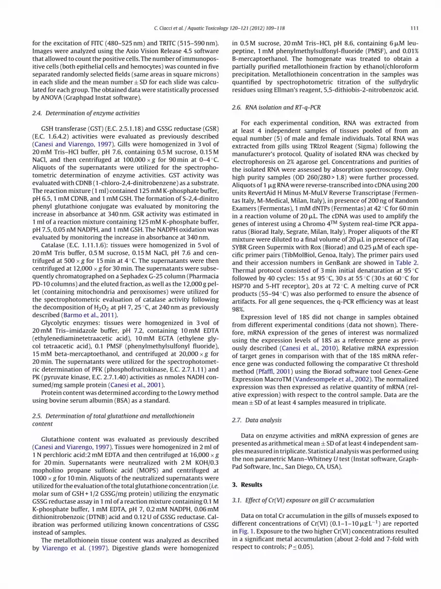
Table 1Details of primary antibodies used.
Antigen Animal source Company Dilution
Glutathione-S-transferase Rabbit Sigma 1:100Serotonin receptor (5HT3) Rabbit Sigma 1:100Acetylcholinesterase (AChE) Mouse Chemicon 1:100
10 C. Ciacci et al. / Aquatic Toxico
In fish larvae, Cr(VI) has been shown to be neurotoxic (inhibitedholinesterase), to inhibit glutathione transferase (GST) activitynd to interfere with cellular metabolic activity (changes in lactateehydrogenase activity) (Domingues et al., 2010). In adults, Cr(VI)ffected oxidative stress markers, glutathione status and antiox-dant enzymes (Lushchak et al., 2008; Kubrak et al., 2010; Velmand Tchounwou, 2011). All these effects were observed at relativelyigh concentrations (>1 mg L−1).
In the marine bivalve Mytilus spp., uptake and loss of Cr haseen investigated in animals exposed to either Cr(VI) or Cr(III),ith total metal accumulation being higher in mussels exposed
o Cr(VI) than to Cr(III) (Parlak et al., 1999). Recent data showedhat in M. galloprovincialis exposure to Cr(VI) at concentrations of.1–10 �g L−1 did not result in strong toxicity or oxidative stressonditions in the digestive gland, but exerted pleiotropic effectsn mussel physiology, from modulation of glutathione-relatednzymes, to lipid and carbohydrate metabolism, and up-regulationf estrogen-responsive genes, the estrogen receptor MeER2 anderotonin receptor (5-HTR) (Barmo et al., 2011). Moreover, in theame conditions of exposure, Cr(VI) induced changes in functionalnd molecular immune parameters in mussel hemocytes, withmmunomodulatory but not immunotoxic effects (Ciacci et al.,011).
In aquatic organisms, including bivalve molluscs, the gills arehe main site of uptake of metals present in soluble forms, andhe possible first target for metal toxicity. In clams, gills werehown as among the principal targets of Cr(VI) toxicity at sub-ethal concentrations (Teh et al., 2000). In Mytilus gills, Cr(VI) haseen shown to cause DNA oxidation and changes in transcription ofenes involved in the stress response (Franzellitti and Fabbri, 2005;mmanouil et al., 2007). In M. galloprovincialis, Cr(VI) increased the-HT-stimulated adenylate cyclase activity in vivo, suggesting theossibility that metal accumulation in the tissue might induce thever-expression of 5-HT receptors (Fabbri and Capuzzo, 2006). Gillctivity is regulated by sympathetic and parasympathetic innerva-ion of autonomic nervous system running through the branchialonnective tissue (Catapane et al., 1974), with different neurotrans-itters participating in regulation of ciliary beating (Stefano, 1990).In this work, the effects of Cr(VI) were investigated in the gills of
ussels exposed to 0.1–1–10 �g Cr(VI) L−1 animal−1 for 96 h. Gillorphology and immunolocalization of GST (GSH transferase), and
f components involved in serotoninergic (5-hydroxytriptamineeceptors), cholinergic (AChE – acetyl choline esterase and ChAT
choline acetyl transferase), and adrenergic-dopaminergic (TH –yrosine hydroxylase) systems were evaluated. Biochemical and
olecular responses to Cr(VI) exposure were also investigated.otal glutathione content, activities of GSH-related enzymes (GST,lutathione reductase – GSR), of the antioxidant enzyme catalase,nd of key glycolytic enzymes (phosphofructokinase – PFK andyruvate kinase – PK) were evaluated. Further mRNA expressionf selected Mytilus genes involved in the stress response (GST-, HSP70, metallothionein isoforms MT10 and MT20) and of the
erotonin receptor 5-HTR was assessed by RT-q-PCR.
. Materials and methods
.1. Animals and treatments
Mussels (Mytilus galloprovincialis Lam.), 4–5 cm long, werebtained from a commercial dealer in Arborea (OR, Italy) in June
010 and kept for 1–3 days in static tanks containing 36 ppt artifi-ial sea water (ASW) (1 L mussel−1) at 16 ◦C. Sea water was changedaily. Animals were fed a commercial algal preparation (Liquifry,nterpret Ltd., Surrey, UK).
Choline acetyltransferase (ChAT) Rabbit Abcam 1:200Tyrosine hydroxylase (TH) Mouse Sigma, St. Louis
(USA)1:100
Groups of mussels (4 of 15 animals each) were exposed for 96 hto 0.1–1–10 �g L−1 animal−1 of Cr(VI) from K2Cr2O7 commercialstock solutions. Cr(VI) was administered every day in ASW renewed2 h after feeding. At the end of exposure, tissues were dissected,frozen in liquid N2 and stored at −80 ◦C. For samples utilized formRNA expression analysis of genes, animals were sexed by micro-scopic inspection of mantle smears.
2.2. Determination of metal content
Total Cr content in mussel gills was evaluated as previouslydescribed (Barmo et al., 2011). Before analysis, tissues werelyophilized and then homogenized in a mortar. Aliquots of about200–300 mg were weighed and digested with 8 ml of 65% HNO3 and2 ml of 30% H2O2 in high-pressure Teflon vessels using a microwavedigestion system (Mod. Milestone Ethos 1). Digested samples weretransferred to polyethylene vessels and diluted 1:5 with ultra-purewater. Total Chromium concentrations in samples were deter-mined by atomic absorption spectrometry equipped with graphitefurnace (Mod. Analytik Jena ContraAA 700). The method of addi-tions was used and standards were prepared by serial dilutionof commercial stock solution of Cr for atomic absorption spec-trometry (1000 mg Cr L−1) within the linear range. Reagents werepro-analysis grade and the analytical procedure was checked byconcomitant running of reagent blanks. Accuracy was checkedby analyzing certified reference materials: Mussel Tissue (ERM-CE278) from Institute for Reference Materials and MeasurementsIRMM of European Commission and Dogfish Muscle (Dorm-2) fromthe National Research Council of Canada. Our measurements didnot differ significantly from the certified values.
2.3. Gill morphology and immunohistochemistry
Samples of gill tissues were fixed in 4% paraformaldehyde in0.1 M phosphate buffered solution (pH 7.4) at 4 ◦C, dehydrated inethanol and embedded in Paraplast (Bio-Optica, Italy). Histologicalsections (5 �m thick) were cut with a rotary automatic microtome(Leica Microsystems, Wetzlar, Germany). Sections were glass-slidemounted and stained with hematoxylin/eosin (Bio-Optica, Italy).Micrographs were obtained using Axio Vision release 4.5 (Zeiss)software. Sections were also processed for immunofluorescenceas previously reported (Mauceri et al., 1999) to reveal variousantigens and peptides. The sections were incubated in the pres-ence of primary antisera (concentrations and suppliers as indicatedin Table 1) overnight at 4 ◦C in a moist chamber. Binding sitesof primary antibodies were visualized by corresponding fluores-cein isothiocyanate (FITC)-conjugated goat anti-mouse IgG (Sigma)and tetramethylrodamine isothiocyanate (TRITC)-conjugated goatanti-rabbit IgG (Sigma), diluted 1:100 for 2 h at room tempera-ture. Specificity of the peptide labeling was verified by incubatingsections with each antiserum preabsorbed with the respectiveantigen (10–100 g/ml). Preabsorption procedures were carried out
overnight at 4 ◦C.A Zeiss Axio Imager Z1 microscope integrated with Axio Vision4.5 and an AxioCam digital camera (Zeiss) was used for image acqui-sition. Sections were imaged using the appropriate filter settings
logy 1
fItisilb
2
((2NAteTppi1pe
2tcqPlttd
2(c12rPs
u
2c
(1fm1umGKdii
b
C. Ciacci et al. / Aquatic Toxico
or the excitation of FITC (480–525 nm) and TRITC (515–590 nm).mages were analyzed using the Axio Vision Release 4.5 softwarehat allowed to count the positive cells. The number of immunopos-tive cells (both epithelial cells and hemocytes) was counted in fiveeparated randomly selected fields (same areas in square microns)n each slide and the mean number ± SD for each slide was calcu-ated for each group. The obtained data were statistically processedy ANOVA (Graphpad Instat software).
.4. Determination of enzyme activities
GSH transferase (GST) (E.C. 2.5.1.18) and GSSG reductase (GSR)E.C. 1.6.4.2) activities were evaluated as previously describedCanesi and Viarengo, 1997). Gills were homogenized in 3 vol of0 mM Tris–HCl buffer, pH 7.6, containing 0.5 M sucrose, 0.15 MaCl, and then centrifuged at 100,000 × g for 90 min at 0–4 ◦C.liquots of the supernatants were utilized for the spectropho-
ometric determination of enzyme activities. GST activity wasvaluated with CDNB (1-chloro-2,4-dinitrobenzene) as a substrate.he reaction mixture (1 ml) contained 125 mM K-phosphate buffer,H 6.5, 1 mM CDNB, and 1 mM GSH. The formation of S-2,4-dinitrohenyl glutathione conjugate was evaluated by monitoring the
ncrease in absorbance at 340 nm. GSR activity was estimated in ml of a reaction mixture containing 125 mM K-phosphate buffer,H 7.5, 0.05 nM NADPH, and 1 mM GSH. The NADPH oxidation wasvaluated by monitoring the increase in absorbance at 340 nm.
Catalase (E.C. 1.11.1.6): tissues were homogenized in 5 vol of0 mM Tris buffer, 0.5 M sucrose, 0.15 M NaCl, pH 7.6 and cen-rifuged at 500 × g for 15 min at 4 ◦C. The supernatants were thenentrifuged at 12,000 × g for 30 min. The supernatants were subse-uently chromatographed on a Sephadex G-25 column (PharmaciaD-10 columns) and the eluted fraction, as well as the 12,000 g pel-et (containing mitochondria and peroxisomes) were utilized forhe spectrophotometric evaluation of catalase activity followinghe decomposition of H2O2 at pH 7, 25 ◦C, at 240 nm as previouslyescribed (Barmo et al., 2011).
Glycolytic enzymes: tissues were homogenized in 3 vol of0 mM Tris–imidazole buffer, pH 7.2, containing 10 mM EDTAethylenediaminetetraacetic acid), 10 mM EGTA (ethylene gly-ol tetraacetic acid), 0.1 PMSF (phenylmethylsulfonyl fluoride),5 mM beta-mercaptoethanol, and centrifuged at 20,000 × g for0 min. The supernatants were utilized for the spectrophotomet-ic determination of PFK (phosphofructokinase, E.C. 2.7.1.11) andK (pyruvate kinase, E.C. 2.7.1.40) activities as nmoles NADH con-umed/mg sample protein (Canesi et al., 2001).
Protein content was determined according to the Lowry methodsing bovine serum albumin (BSA) as a standard.
.5. Determination of total glutathione and metallothioneinontent
Glutathione content was evaluated as previously describedCanesi and Viarengo, 1997). Tissues were homogenized in 2 ml of
N perchloric acid:2 mM EDTA and then centrifuged at 16,000 × gor 20 min. Supernatants were neutralized with 2 M KOH/0.3
opholino propane sulfonic acid (MOPS) and centrifuged at000 × g for 10 min. Aliquots of the neutralized supernatants weretilized for the evaluation of the total glutathione concentration (i.e.olar sum of GSH + 1/2 GSSG/mg protein) utilizing the enzymaticSSG reductase assay in 1 ml of a reaction mixture containing 0.1 M-phosphate buffer, 1 mM EDTA, pH 7, 0.2 mM NADPH, 0.06 mMithionitrobenzoic (DTNB) acid and 0.12 U of GSSG reductase. Cal-
bration was performed utilizing known concentrations of GSSGnstead of samples.
The metallothionein tissue content was analyzed as describedy Viarengo et al. (1997). Digestive glands were homogenized
20– 121 (2012) 109– 118 111
in 0.5 M sucrose, 20 mM Tris–HCl, pH 8.6, containing 6 �M leu-peptine, 1 mM phenylmethylsulfonyl-fluoride (PMSF), and 0.01%B-mercaptoethanol. The homogenate was treated to obtain apartially purified metallothionein fraction by ethanol/chloroformprecipitation. Metallothionein concentration in the samples wasquantified by spectrophotometric titration of the sulfydrylicresidues using Ellman’s reagent, 5,5-dithiobis-2-nitrobenzoic acid.
2.6. RNA isolation and RT-q-PCR
For each experimental condition, RNA was extracted fromat least 4 independent samples of tissues pooled of from anequal number (5) of male and female individuals. Total RNA wasextracted from gills using TRIzol Reagent (Sigma) following themanufacturer’s protocol. Quality of isolated RNA was checked byelectrophoresis on 2% agarose gel. Concentrations and purities ofthe isolated RNA were assessed by absorption spectroscopy. Onlyhigh purity samples (OD 260/280 > 1.8) were further processed.Aliquots of 1 �g RNA were reverse-transcribed into cDNA using 200units RevertAid H Minus M-MuLV Reverse Transcriptase (Fermen-tas Italy, M-Medical, Milan, Italy), in presence of 200 ng of RandomExamers (Fermentas), 1 mM dNTPs (Fermentas) at 42 ◦C for 60 minin a reaction volume of 20 �L. The cDNA was used to amplify thegenes of interest using a Chromo 4TM System real-time PCR appa-ratus (Biorad Italy, Segrate, Milan, Italy). Proper aliquots of the RTmixture were diluted to a final volume of 20 �L in presence of iTaqSYBR Green Supermix with Rox (Biorad) and 0.25 �M of each spe-cific primer pairs (TibMolBiol, Genoa, Italy). The primer pairs usedand their accession numbers in GenBank are showed in Table 2.Thermal protocol consisted of 3 min initial denaturation at 95 ◦Cfollowed by 40 cycles: 15 s at 95 ◦C, 30 s at 55 ◦C (30 s at 60 ◦C forHSP70 and 5-HT receptor), 20 s at 72 ◦C. A melting curve of PCRproducts (55–94 ◦C) was also performed to ensure the absence ofartifacts. For all gene sequences, the q-PCR efficiency was at least98%.
Expression level of 18S did not change in samples obtainedfrom different experimental conditions (data not shown). There-fore, mRNA expression of the genes of interest was normalizedusing the expression levels of 18S as a reference gene as previ-ously described (Canesi et al., 2010). Relative mRNA expressionof target genes in comparison with that of the 18S mRNA refer-ence gene was conducted following the comparative Ct thresholdmethod (Pfaffl, 2001) using the Biorad software tool Genex-GeneExpression MacroTM (Vandesompele et al., 2002). The normalizedexpression was then expressed as relative quantity of mRNA (rel-ative expression) with respect to the control sample. Data are themean ± SD of at least 4 samples measured in triplicate.
2.7. Data analysis
Data on enzyme activities and mRNA expression of genes arepresented as arithmetical mean ± SD of at least 4 independent sam-ples measured in triplicate. Statistical analysis was performed usingthe non parametric Mann–Whitney U test (Instat software, Graph-Pad Software, Inc., San Diego, CA, USA).
3. Results
3.1. Effect of Cr(VI) exposure on gill Cr accumulation
Data on total Cr accumulation in the gills of mussels exposed to
different concentrations of Cr(VI) (0.1–1–10 �g L−1) are reportedin Fig. 1. Exposure to the two higher Cr(VI) concentrations resultedin a significant metal accumulation (about 2-fold and 7-fold withrespect to controls; P ≤ 0.05).
112 C. Ciacci et al. / Aquatic Toxicology 1
FsU
3
ftsfilCab(
wsosihnie5C(
dgtgdc1aaa
cpIe
ig. 1. Total chromium concentration in the gills of control and Cr(VI)-exposed mus-els. Data, representing the mean ± SD (n = 4), were analyzed by the Mann–Whitney
test; *P ≤ 0.05, all treatments vs controls.
.2. Histopathology and immunofluorescence microscopy
Representative images of hematoxylin/eosin sections of gillsrom control mussels and mussels exposed to different concen-rations of Cr(VI) are shown in Fig. 2. The gills of control musselshowed a typical morphology, consisting of filaments that branchrom a longitudinal axis, with the filaments of the same line form-ng a branchial lamina, with the laminae in the intermediate regioninked by cilia (Fig. 2A). Exposure to increasing concentrations ofr(VI) induced changes in gill morphology with progressive alter-tions in the epithelial cells and in the cilia, reduction of contactsetween adjacent filaments, and increasing hemocytic infiltrationFig. 2B–D).
Immunofluorescence microscopy utilizing different antibodiesas performed in gill sections of control and Cr(VI)-exposed mus-
els. Representative images are reported in Figs. 3 and 4 and datan the average numbers of positive cells for each antibody areummarized in Fig. 5. Both GST- and 5HT3R-like immunoreactiv-ty were observed in the gill epithelium of control mussels. At theighest metal concentration tested, a significant increase in theumber of GST immunopositive cells and a slight increase in 5HT3R
mmunoreactivity were observed with respect to control and lowerxposure groups (Figs. 3 and 5). No significant changes in GST- andHT3R-positive cells were observed in the gills exposed to 1 �g L−1
r(VI) with respect to controls or to the lowest metal concentrationnot shown).
Fig. 4 shows the images of mussel gills incubated with antibodiesirected against mammalian enzymes involved in the choliner-ic (AChE and ChAT) and adrenergic (TH) systems. Cells positiveo both anti-AChE and anti-ChAT antibodies were observed in theill epithelium of control mussels. In all metal-exposed groups aramatic decrease in AChE immunopositivity was observed, withomplete lack of AChE-positive cells after exposure to 0.1 and
�g L−1, and only few cells at the highest dose. A similar decrease,lthough less pronounced, was also observed with the anti-ChATntibody, with a marked decline in the number of positive cells atll Cr(VI) concentrations (Fig. 5).
No immunoreactivity toward TH antibodies was observed in
ontrol gills, whereas a noticeable dose-dependent increase in TH-ositivity was observed in all metal-exposed exposure groups.n particular, only TH-positive hemocytes were found in gillsxposed to 0.1 �g L−1; few adrenergic cells were observed in the gill
20– 121 (2012) 109– 118
epithelium of mussels treated with 1 �g L−1; both epithelial cellsand hemocytes were found to be positive for TH after exposure tothe highest Cr(VI) level (Fig. 5).
3.3. Effects of Cr(VI) on activities of GST, GSR, catalase and ontotal glutathione content
The effects of Cr(VI) on the activity of enzymes involved in glu-tathione metabolism, of the antioxidant enzyme catalase and ontotal glutathione in mussel gills were evaluated and the results arereported in Fig. 6. Cr(VI) exposure induced a large stimulation ofGST activity at all the concentrations tested (P ≤ 0.05), reaching upto a 3.5-fold increase with respect to controls at 1 �g L−1 Cr(VI)(Fig. 6A). A similar, although smaller increase in GSR activity wasalso observed (up to a 50% increase at the same concentration;P ≤ 0.05) (Fig. 6B). An increase in catalase activity was observedonly at the lowest Cr(VI) concentration (+45%; P ≤ 0.05) (Fig. 6C).Metal exposure also induced a large increase in total glutathionecontent that was significant at all the concentrations tested (from+80 to +130%, depending on the concentration; P ≤ 0.05) (Fig. 6D).
3.4. Effects of Cr(VI) on the activities of glycolytic enzymes
In order to investigate the possible effects of Cr(VI) on gillcarbohydrate metabolism, the activities of the two key glycolyticenzymes PFK and PK, that have been recently shown to be stim-ulated by Cr(VI) exposure in mussel digestive gland (Barmo et al.,2011) were evaluated, and the results are shown in Fig. 7. Basalspecific activities of PFK and PK in gills of control mussels were0.050 ± 0.004 and 5.23 ± 0.50 U/mg sample protein, respectively.The results reported in Fig. 7 show that metal exposure increasedthe activities of both PFK (Fig. 7A) and PK (Fig. 7B), with strongesteffects at the highest concentrations tested (+200% and +70%,respectively, P ≤ 0.05).
3.5. Effects of Cr(VI) on mRNA expression of selected genes
The possible effects of Cr(VI) exposure on gene transcriptionin the gills were evaluated by RT-q-PCR utilizing the primer pairsreported in Table 2. When data were pooled between samplesobtained from males and females (n = 10), changes on the mRNAexpression of these genes could be detected, although only a fewvalues were significant, due to the large variability of data (datanot shown). In order to investigate possible sex-related differences,as those previously observed in the digestive gland in the sameexperimental conditions (Barmo et al., 2011) data obtained fromthe tissues of females and males were analyzed separately (n = 5).The results indicate a distinct pattern in gill mRNA expressionof genes in response to different concentrations of Cr(VI) in thetwo sexes (Figs. 8 and 9). Metal exposure induced small increasesin the transcription of the Mytilus 5-HTR in both sexes (Fig. 8A)that were observed in males at 0.1 and 1 �g L−1 Cr(VI), whereasin females only at the highest metal concentration (P ≤ 0.05). Inmales, the level of mRNA for GST-�, the main GST isoform in mus-sel tissue (Hoarau et al., 2006), showed a dramatic decrease at 0.1and 1 �g L−1 Cr(VI) (−83 and −50% with respect to control mus-sels, respectively, P ≤ 0.05) (Fig. 8B). On the other hand, a 3-foldincrease was observed in female gills exposed to 1 and 10 �g L−1
Cr(VI) (P ≤ 0.05).mRNA levels of the two metallothionein isoforms MT10 and
MT20 were also evaluated. In males, Cr(VI) induced a dramaticdecrease in transcription of MT10 at all the concentrations tested
(Fig. 9A). In females, downregulation of MT10 was observed at0.1 �g L−1 Cr(VI) (−77% with respect to control samples; P ≤ 0.05),whereas no changes were observed at 1 �g L−1 and a large increaseat 10 �g L−1 (about four-folds with respect to controls; P ≤ 0.05).
C. Ciacci et al. / Aquatic Toxicology 120– 121 (2012) 109– 118 113
Fig. 2. Light micrographs of sections through gills of M. galloprovincialis showing histological structure of control mussels (A), and mussels exposed to different concentrationsof Cr(VI) (�g L−1 animal−1): (B) 0.1 �g; (C) 1 �g; (D) 10 �g. GF: gill filaments; FC: frontal cilia; LC: lateral cilia; GE: gill epithelium. In all treated groups progressive alterationsof the gill epithelium can be observed, in particular associated with disruption of lateral cilia. Scale bar: 20 �m.
Fig. 3. Gill GST and 5HT3R immunoreactivity. Representative immunofluorescence microscopy images of gill sections from control mussels and mussels exposed to Cr(VI)(0.1 and 10 �g L−1 animal−1). Upper panels: images show nearly absence of GST immunopositive cells in control group, appearance of positive cell (arrows) at 0.1 �g, and amarked immunopositivity at 10 �g. Lower panels: 5HT3R positive cells (arrows) can be visualized in all control and treated groups, to a greater extent at 0.1 �g and 10 �g.Scale bar: 20 �m.
Table 2Oligonucleotide primers used for quantitative RT-PCR analysis.
Gene Primer forward (5′–3′) Primer reverse (5′–3′) GenBank
GST-� TCCAGTTAGAGGCCGAGCTGA CTGCACCAGTTGGAAACCGTC AF527010MT10 GGGCGCCGACTGTAAATGTTC CACGTTGAAGGCCTGTACACC AY566248MT20 TGTGAAAGTGGCTGCGGA GTACAGCCACATCCACACGC AY566247HSP70 GGTGGTGAAGACTTTGACAACAG CTAGTTTGGCATCGCGTAGAGC AY8616845-HTR CAGCTGCAAGATCGAGGATT TGAAGCCATCTTGACTGACG AB52621818S TCGATGGTACGTGATATGCC CGTTTCTCATGCTCCCTCTC L33451

114 C. Ciacci et al. / Aquatic Toxicology 120– 121 (2012) 109– 118
Fig. 4. AChE, ChAT and TH immunoreactivity. Representative immunofluorescence microscopy images of gill sections from control mussels and mussels exposed to Cr(VI)(0.1–1–10 �g L−1 animal−1). Scale bar: 20 �m. Upper panels: AChE positive cells (arrows). Middle panels: ChAT positive cells (arrows). Lower panels: no TH-positive epithelialc ent ina
o1owa6wot
ou1t
4
0at
ells were observed in control gills; TH-positive hemocytes (arrowheads) were prest 1 �g and both TH-positive cells and hemocytes at 10 �g.
In males, Cr(VI) exposure induced significant downregulationf MT20 mRNA at 0.1 and 10 �g L−1 Cr(VI), whereas exposure to
�g L−1 induced a significant 3.5-fold induction (Fig. 9B). On thether hand, in females a general increase in transcription of MT20as observed, that was significant at all the concentrations tested
nd especially relevant at the lowest concentration tested, with a-fold induction. When the total MT content at the protein levelas evaluated in tissue extracts, no significant differences were
bserved between control and Cr-exposed mussels at any concen-ration tested (data not shown).
Finally, transcription of HSP70 was decreased only in the gillsf males exposed to 1 �g L−1 Cr(VI). On the other hand, in females,pregulation of HSP70 mRNA transcripts were observed at 1 and0 �g L−1 Cr(VI) (+75 and +65%, respectively, with respect to con-rols; P ≤ 0.05) (Fig. 9C).
. Discussion
The results indicate exposure to Cr(VI), at concentrations from.1 to 10 �g L−1, induced significant morphological, biochemicalnd molecular changes in mussel gills. Alterations in gill his-ology were accompanied by changes in immunoreactivity to
gills exposed to 0.1 �g Cr(VI); few positive epithelial cells (arrows) were observed
heterologous antiobodies directed toward different components ofneurotransmission that have been shown to be present in Mytilustissues, including gills (Stefano, 1990). A general decrease in thenumber of AChE- and ChAT-like immunoreactive epithelial cellswas observed, indicating that Cr(VI) affects cholinergic signaling.AChE (acetylcholinesterase) is essential to the correct transmissionof nerve impulses; although AChE activity is generally utilized as abiomarker of exposure to pesticides, AChE inhibition by some met-als has been recently demonstrated (Tsangaris et al., 2007; Attiget al., 2010; Kopecka-Pilarczyk, 2010). On the other hand, a gen-eral tendency to increase was observed for immunoreactivity ofserotonin-like receptors, as well as a significant rise in the numberof cells (both epithelial cells and hemocytes) positive to antibodiesdirected against TH, the enzyme involved in conversion of tyro-sine to l-DOPA. The increase in 5HT3R and TH immunoreactivitymay reflect an imbalance between serotoninergic and dopamin-ergic neurotransmission that, in Mytilus, have a stimulatory andan inhibitory effect, respectively, on lateral cilia beating (Stefano,
1990; Zhu et al., 2005).Cr(VI) exposure increased the activities of GST and GSR, theenzymes responsible for GSH conjugation and for recycling oxi-dized glutathione, respectively, at all the concentrations tested;

C. Ciacci et al. / Aquatic Toxicology 120– 121 (2012) 109– 118 115
F controA
ioiiChfC(gcGscc
ig. 5. Quantification of cells positive to different antibodies in gill sections of
NOVA*P < 0.001; **P < 0.01; ***P < 0.0001, all treatments vs controls.
ncreased activity of the antioxidant enzyme catalase was observednly at the lowest concentration. The large increase in GST activ-ty, leading to consumption of GSH, was accompanied by increasesn total glutathione content, suggesting that in mussel tissuesr(VI) also affects GSH synthesis as previously shown with othereavy metals (Canesi et al., 1999). These effects were distinct
rom those previously observed in the digestive gland, wherer(VI) largely stimulated GSR activity, without affecting that of GSTBarmo et al., 2011). Moreover, in gills, changes in components oflutathione metabolism were observed also at lower metal con-entrations. The results support the hypothesis that non-enzymatic
SH-mediated intracellular reduction of Cr(VI) occur in mussel tis-ues with GSR playing a role in recycling GSSG like in mammalianells (Gunaratnam and Grant, 2001). The reduction process of Cr(VI)an generate variable amounts of unstable intermediates Cr(V) andl and Cr(VI)-exposed mussels. Data, expressed as mean ± SD, were analyzed by
Cr(IV), involved in the formation of oxyradicals or hydroperoxidesthat are thought to be responsible for the effects of Cr(VI) as anoxidant (Salnikow and Zhitkovich, 2008; Lushchak, 2011). In mus-sel gills, reduction of Cr(VI) to the stable Cr(III) form may occur,although to a lower extent than in the digestive gland. On the otherhand, the present data indicate that in gills, at concentrations ofCr(VI) from 0.1 to 10 �g L−1, glutathione conjugation by GST mayplay a major role in metal detoxification. In this tissue, basal GSTactivity due to different GST isoforms indicates higher potential forPhase II conjugation reactions than in the digestive gland (Hoarauet al., 2006).
In mammals, Cr(III) is believed an essential trace element,with a role in promoting insulin stimulation of glucose oxida-tion and lipogenesis (Mertz, 1998; Moukarzel, 2009). In musselgills, Cr(VI) greatly increased the activities of the key glycolytic

116 C. Ciacci et al. / Aquatic Toxicology 120– 121 (2012) 109– 118
Fig. 6. Effects of Cr(VI) exposure on the specific activities of glutathione and redox-related enzymes and on total glutathione content in mussel gills. (A) glutathione transferase( ssed ai SD o*
eommzmtbtn(
Fp*
GST); (B) GSSG reductase (GSR); (C) catalase (CAT); (D) enzyme activities are expres expressed as nmoles (GSH + 1/2GSSG)/mg protein. Data, representing the mean ±P ≤ 0.01, all treatments vs controls.
nzymes PFK and PK. The effect was stronger than that previ-usly observed in the digestive gland (Barmo et al., 2011). Inussel digestive cells, PFK and PK activities were increased byammalian insulin, as well as by physiological concentrations of
inc, an essential metal, through activation of tyrosine kinase-ediated pathways (Canesi et al., 1999, 2001). Our data support
he hypothesis that different forms of chromium can modulate car-
ohydrate metabolism in invertebrate as well as in mammalianissues. In mussel gills, induction of glycolysis may be related to theeed of high energy supply for the synthesis of neurotransmittersStefano, 1990).ig. 7. Effects of Cr(VI) exposure on digestive gland glycolytic enzyme activities phosphercent specific activities (s.a.) with respect to controls. Data, representing the mean ± SDP ≤ 0.01, all treatments vs controls.
s percent specific activities (s.a.) with respect to controls. Total glutathione contentf at least 4 experiments in triplicate, were analyzed by the Mann–Whitney U test;
Transcription of different genes was also affected by Cr(VI) expo-sure. Cr(VI) significantly up-regulated transcription of the Mytilus5HTR in both sexes, to a different extent and in response to differentconcentrations in males and females, and with males being appar-ently more sensitive than females to metal treatment. A similartrend was observed in the digestive gland of Cr(VI) exposed mus-sels (Barmo et al., 2011). A general but not significant increase in
5HT3R-like immuno-reactivity was observed in the gills of Cr(VI)-exposed mussels. The discrepancy observed at mRNA and proteinlevel may be due to both sex-related differences and the type ofantibody utilized. 5HT3R immuno-reactivity has previously beenofructokinase (PFK) (A) and pyruvate kinase (PK) (B). The results are expressed as of at least 4 experiments in triplicate, were analyzed by the Mann–Whitney U test;

C. Ciacci et al. / Aquatic Toxicology 120– 121 (2012) 109– 118 117
F emaleR ontroli
otsta
awmamdGdocd2
Fa
ig. 8. Effects of Cr(VI) exposure on mRNA expression of Mytilus genes in the gills of fT-PCR as described in methods. Relative expression was calculated with respect to c
n triplicate. *P ≤ 0.05 Mann–Whitney U test, all treatments vs controls.
bserved in cell bodies of buccal ganglia of the gastropod H. poma-ia (Hernádi et al., 2008). The Mytilus 5HTR partial gene sequencehows a 40% similarity to the Patinopecten yessoensis 5-HTpy andhe Aplysia californica 5-HTap1 and 30% similarity to mouse 5-HT1And human 5-HT1D (Cubero-Leon et al., 2010).
As a general trend, the levels of mRNA for GST�, MT isoformsnd HSP70 were decreased in males, and increased in females,ith some exceptions depending on the gene sequence andetal concentration. Interestingly, for GST, both specific activity
nd number of GST-like immunoreactive cells were increased byetal treatment in unsexed animals. The discrepancy between
ata obtained on GST� transcription in males and females andST protein activity/expression in unsexed mussels could beue not only to possible sex related differences or to the use
f heterologous antibodies, but also to the fact that the bio-hemical assay evaluates total GST activity, corresponding toifferent GST isoforms possibly expressed in gills (Hoarau et al.,006).ig. 9. Effects of Cr(VI) exposure on mRNA expression of Mytilus genes in the gills of fems described in legend to Fig. 8. *P ≤ 0.05 Mann–Whitney U test, all treatments vs controls
and male mussels: (A) 5HTR; (B) GST-�· Expression was determined by quantitative mussels. Data are the mean ± SD obtained from at least 4 independent RNA samples
Transcription of MT isoforms was generally decreased in males,whereas increases in females were observed for MT10 and MT20,although at different concentrations, not showing a concentration-related response. Similar effects were previously observed in thedigestive gland of Cr(VI)-exposed mussels (Barmo et al., 2011).Information on regulation of MT transcription in bivalves is stilllimited compared to vertebrate systems (Dondero et al., 2005), andthe mechanisms involved in the response to Cr(VI) in mussel tissuesare at present difficult to explain. In both cases, the distinct effectsmay be related to sex-related differences in tissue metal accumula-tion that were not determined. MT-downregulation or interferencewith MT induction by Cr has been shown both in fish (Woo et al.,2006) and mammalian systems (Majumder et al., 2003; Kimuraet al., 2008). As in the digestive gland, the changes in MT mRNA lev-
els were not paralleled by changes in total MT protein (not shown);such a discrepancy between mRNA and protein levels is widelydocumented for MTs, possibly depending on post-transcriptionalevents (Haq et al., 2003).ale and male mussels. (A) MT10; (B) MT20; (C) HSP70. Expression was determined.

1 logy 1
snUs2
rvigtwseaerm
F
R4
A
t
R
A
B
C
C
C
C
C
C
C
D
D
E
F
18 C. Ciacci et al. / Aquatic Toxico
Finally, Cr(VI) induced a small but significant increase in tran-cription of HSP70 in females at higher concentrations, showingo effect in the gills of males, except for a decrease at 1 �g L−1.p-regulation of HSP70 was observed in the digestive gland of mus-
els exposed to few ng L−1 (CrVI) for 7 days (Franzellitti and Fabbri,005).
Overall, the results demonstrate that in Mytilus, the gillsepresent the major target of exposure to environmentally rele-ant concentrations of Cr(VI). Significant stimulation of enzymesnvolved in glutathione metabolism and increases in totallutathione content were observed, indicating Cr(VI) detoxica-ion/reduction at the site of metal entry. These data are in lineith those obtained in fish tissues, suggesting that the glutathione
ystem may be responsible for protecting against the deleteriousffects of Cr(VI) and indicating the possible development of andaptive response during similar conditions of exposure (Lushchakt al., 2008). In mussel gills, the effects of the metal are apparentlyelated to changes in different parameters related to neurotrans-ission, energy metabolism and gene expression.
unding
This work was partially supported by the Italian Ministry ofesearch (PRIN2007) and by Fondazione CARIGE (prot. 2009.0659-2).
cknowledgments
A special thank to Dr. Rita Fabbri and Dr. Nicola Bianchi for theirechnical assistance.
eferences
ttig, H., Dagnino, A., Negri, A., Jebali, J., Boussetta, H., Viarengo, A., Dondero, F., Banni,M., 2010. Uptake and biochemical responses of mussels Mytilus galloprovincialisexposed to sublethal nickel concentrations. Ecotoxicology and EnvironmentSafety 73, 1712–1719.
armo, C., Ciacci, C., Fabbri, R., Olivieri, S., Bianchi, N., Gallo, G., Canesi, L., 2011.Pleiotropic effects of hexavalent chromium (CrVI) in Mytilus galloprovincialisdigestive gland. Chemosphere 83, 1087–1095.
anesi, L., Viarengo, A., 1997. Age-related differences in glutathione metabolismin mussel tissues. Comparative Biochemistry and Physiology 116B,217–221.
anesi, L., Leonzio, C., Filippelli, M., Viarengo, A., Gallo, G., 1999. Heavy metaleffects on glutathione metabolism in mussel tissues. Aquatic Toxicology 46,67–76.
anesi, L., Betti, M., Ciacci, C., Gallo, G., 2001. Insulin-like effect of zinc in Mytilusdigestive gland cells: modulation of tyrosine kinase mediated cell signalling.General and Comparative Endocrinology 122, 60–66.
anesi, L., Barmo, C., Fabbri, R., Ciacci, C., Vergani, L., Roch, P., Gallo, G., 2010. Effects ofvibrio challenge on digestive gland biomarkers and antioxidant gene expressionin Mytilus galloprovincialis. Comparative Biochemistry and Physiology. Part C:Toxicology and Pharmacology 152, 399–406.
atapane, E.J., Aiello, E., Stefano, G.B., 1974. Ganglionic mediation mechanism oflateral cilia in Mytilus edulis gill. The Physiologist, 17–372.
iacci, C., Barmo, C., Fabbri, R., Canonico, B., Gallo, G., Canesi, L., 2011. Immunomod-ulation in Mytilus galloprovincialis by non-toxic doses of hexavalent chromium.Fish & Shellfish Immunology 31, 1026–1033.
ubero-Leon, E., Ciocan, C.M., Hill, E.M., Osada, M., Kishida, M., Itoh, N., Kondo,R., Minier, C., Rotchell, J.M., 2010. Estrogens disrupt serotonin receptor andcyclooxygenase mRNA expression in the gonads of mussels (Mytilus edulis).Aquatic Toxicology 98, 178–187.
omingues, I., Oliveira, R., Lourenc o, J., Grisolia, C.K., Mendo, S., Soares, A.M., 2010.Biomarkers as a tool to assess effects of chromium (VI): comparison of responsesin zebrafish early life stages and adults. Comparative Biochemistry and Physiol-ogy. Part C: Toxicology and Pharmacology 152, 338–434.
ondero, F., Piacentini, L., Banni, M., Rebelo, M., Burlando, B., Viarengo, A., 2005.Quantitative PCR analysis of two molluscan metallothionein genes unveils dif-ferential expression and regulation. Gene 345, 259–270.
mmanouil, C., Sheehan, T.M.T., Chipman, J.K., 2007. Macromolecule oxidation and
DNA repair in mussel (Mytilus edulis L.) gill following exposure to Cd and Cr(VI).Aquatic Toxicology 82, 27–35.abbri, E., Capuzzo, A., 2006. Adenylyl cyclase activity and its modulation in thegills of Mytilus galloprovincialis exposed to Cr6+ and Cu2+. Aquatic Toxicology 76,59–68.
20– 121 (2012) 109– 118
Franzellitti, S., Fabbri, E., 2005. Differential HSP70 gene expression in the Mediter-ranean mussel exposed to various stressors. Biochemical and BiophysicalResearch Communications 336, 1157–1163.
Gunaratnam, M., Grant, M.H., 2001. The role of glutathione reductase in thecytotoxicity of chromium (VI) in isolated rat hepatocytes. Chemico-BiologicalInteractions 134, 191–202.
Haq, F., Mahoney, M., Koropatnick, J., 2003. Signaling events for metallothioneininduction. Mutation Research 533, 211–226.
Hernádi, L., Kárpáti, L., Gyori, J., Vehovszky, A., Hiripi, L., 2008. Humoral serotoninand dopamine modulate the feeding in the snail, Helix pomatia. Acta BiologicaHungarica 59 (Suppl.), 39–46.
Hoarau, P., Damiens, G., Roméo, M., Gnassia-Barelli, M., Bebianno, M.J., 2006. Cloningand expression of a GST-pi gene in Mytilus galloprovincialis. Attempt to use theGST-pi transcript as a biomarker of pollution. Comparative Biochemistry andPhysiology 143C, 196–203.
Kimura, T., Li, Y., Okumura, F., Itoh, N., Nakanishi, T., Sone, T., Isobe, M., Andrews,G.K., 2008. Chromium(VI) inhibits mouse metallothionein-I gene transcriptionby preventing the zinc dependent formation of a MTF-1–p300 complex. Bio-chemical Journal 415, 477–482.
Kopecka-Pilarczyk, J., 2010. The effect of pesticides and metals on acetyl-cholinesterase (AChE) in various tissues of blue mussel (Mytilus trossulus L.) inshort-term in vivo exposures at different temperatures. Journal of EnvironmentScience and Health, Part B Pesticides 45, 336–346.
Kubrak, O.I., Lushchak, O.V., Lushchak, J.V., Torous, I.M., Storey, J.M., Storey, K.B.,Lushchak, V.I., 2010. Chromium effects on free radical processes in goldfishtissues: comparison of Cr(III) and Cr(VI) exposures on oxidative stress mark-ers, glutathione status and antioxidant enzymes. Comparative Biochemistry andPhysiology. Part C: Toxicology and Pharmacology 152, 360–370.
Lushchak, O.V., 2011. Environmentally induced oxidative stress in aquatic animals.Aquatic Toxicology 101, 13–30.
Lushchak, O.V., Kubrak, O.I., Nykorak, M.Z., Storey, K.B., Lushchak, V.I., 2008. Theeffect of potassium dichromate on free radical processes in goldfish: possibleprotective role of glutathione. Aquatic Toxicology 87, 108–114.
Maanan, M., 2007. Biomonitoring of heavy metals using Mytilus galloprovincialis inSafi coastal waters, Morocco. Environmental Toxicology 22, 525–531.
Majumder, S., Ghoshal, K., Summers, D., Bai, S., Datta, J., Jacob, S.T., 2003.Chromium(VI) down-regulates heavy metal-induced metallothionein genetranscription by modifying transactivation potential of the key transcriptionfactor, Metal-responsive Transcription Factor 1. Journal of Biological Chemistry278, 26216–26226.
Mauceri, A., Fasulo, S., Ainis, L., Licata, A., Lauriano, E.R., Martinez, A., Mayer, B.,Zaccone, G., 1999. Neuronal nitric oxide synthase (nNOS) expression in theepithelial neuroendocrine cell system and nerve fibers in the gill of the catfish,Heteropneustes fossilis. Acta Histochemica 101, 437–448.
Mertz, W., 1998. Chromium research from a distance: from 1959 to 1980. Journal ofthe American College of Nutrition 17, 544–547.
Moukarzel, A., 2009. Chromium in parenteral nutrition: too little or too much?Gastroenterology 137, S18–S28.
Parlak, H., Katalay, S., Büyükisik, B., 1999. Accumulation and loss of chromium bymussels (M. galloprovincialis). Bulletin of Environment Contamination and Tox-icology 62, 286–292.
Pfaffl, M.W., 2001. A new mathematical model for relative quantification in real-timeRT-PCR. Nucleic Acids Research 29, e45.
Salnikow, K., Zhitkovich, A., 2008. Genetic and epigenetic mechanisms in metalcarcinogenesis and cocarcinogenesis: nickel, arsenic, and chromium. ChemicalResearch in Toxicology 21, 28–44.
Stefano, G.B., 1990. Neurobiology of Mytilus edulis. Manchester University Press ND,312 pp.
Teh, S.J., Werner, I., Hinton, D.E., 2000. Sublethal effects of chromium-VI in the Asianclam (Potamocorbula amurensis). Marine Environment Research 50, 295–300.
Tsangaris, C., Papathanasiou, E., Cotou, E., 2007. Assessment of the impact of heavymetal pollution from a ferro-nickel smelting plant using biomarkers. Ecotoxi-cology and Environment Safety 66, 232–243.
Vandesompele, J., De Preter, K., Pattyn, F., Poppe, B., Van Roy, N., De Paepe, A., Spele-man, F., 2002. Accurate normalization of real-time quantitative RT-PCR data bygeometric averaging of multiple internal control genes. Genome Biology 3 (7)(RESEARCH00341-11).
Velma, V., Vutukuru, S.S., Tchounwou, P.B., 2009. Ecotoxicology of hexavalentchromium in freshwater fish: a critical review. Reviews on EnvironmentalHealth 24, 129–145.
Velma, V., Tchounwou, P.B., 2011. Hexavalent chromium-induced multiplebiomarker responses in liver and kidney of goldfish, Carassius auratus. Envi-ronmental Toxicology 26, 649–656.
Viarengo, A., Ponzano, E., Dondero, F., Fabbri, R., 1997. A simple spectrophotomet-ric method for metallothionein evaluation in marine organisms: an applicationto Mediterranean and Antarctic molluscs. Marine Environment Research 44,69–84.
Woo, S., Yum, S., Jung, J.H., Shim, W.J., Lee, C.H., Lee, T.K., 2006. Heavy metal-induceddifferential gene expression of metallothionein in Javanese medaka, Oryziasjavanicus. Marine Biotechnology (New York) 8, 654–662.
Zhu, W., Mantione, K.J., Shen, L., Cadet, P., Esch, T., Goumon, Y., Bianchi, E., Sonetti,D., Stefano, G.B., 2005. Tyrosine and tyramine increase endogenous ganglionicmorphine and dopamine levels in vitro and in vivo: cyp2d6 and tyrosine hydrox-ylase modulation demonstrates a dopamine coupling. Medical Science Monitor11, 397–404.