Chronic exposure to sublethal hexavalent chromium affects organ histopathology and serum cortisol...
9
This article appeared in a journal published by Elsevier. The attached copy is furnished to the author for internal non-commercial research and education use, including for instruction at the authors institution and sharing with colleagues. Other uses, including reproduction and distribution, or selling or licensing copies, or posting to personal, institutional or third party websites are prohibited. In most cases authors are permitted to post their version of the article (e.g. in Word or Tex form) to their personal website or institutional repository. Authors requiring further information regarding Elsevier’s archiving and manuscript policies are encouraged to visit: http://www.elsevier.com/copyright
Transcript of Chronic exposure to sublethal hexavalent chromium affects organ histopathology and serum cortisol...

This article appeared in a journal published by Elsevier. The attachedcopy is furnished to the author for internal non-commercial researchand education use, including for instruction at the authors institution
and sharing with colleagues.
Other uses, including reproduction and distribution, or selling orlicensing copies, or posting to personal, institutional or third party
websites are prohibited.
In most cases authors are permitted to post their version of thearticle (e.g. in Word or Tex form) to their personal website orinstitutional repository. Authors requiring further information
regarding Elsevier’s archiving and manuscript policies areencouraged to visit:
http://www.elsevier.com/copyright

Author's personal copy
Chronic exposure to sublethal hexavalent chromium affects organ histopathology andserum cortisol profile of a teleost, Channa punctatus (Bloch)
Ashish K. Mishra, Banalata Mohanty ⁎Department of Zoology, University of Allahabad, Allahabad-211002, India
a b s t r a c ta r t i c l e i n f o
Article history:Received 2 March 2009Received in revised form 26 May 2009Accepted 26 May 2009Available online 25 June 2009
Keywords:Hexavalent chromiumChronic exposureHistopathologyCortisol assayChanna punctatus
Effects of chronic exposures (one and twomonths) to sublethal doses of hexavalent chromium (2 and 4 mg/Lpotassium dichromate) on organ histopathology and serum cortisol profile were investigated and theiroverall impact on growth and behavior of a teleost fish, Channa punctatus was elucidated. Histopathologicallesions were distinct in the vital organs gill, kidney and liver. The gill lamellae became lifted, fused, andshowed oedema. Hyperplasia and hypertrophy of lamellar epithelial cells were distinct with desquamation.Hypertrophy of epithelial cells of renal tubules and reduction in tubular lumens were observed in the trunkkidney. The atrophy of the head kidney interrenal cells and decreased serum cortisol level indicatedexhaustion of interrenal activity. Hepatocyte vacuolization and shrinkage, nuclear pyknosis and increase ofsinusoidal spaces were observed in the liver. Abnormal behavioral patterns and reduced growth rate werealso noticed in the exposed fish. The chronic hexavalent chromium exposure thus by affecting histopathologyof gill, kidney (including interrenal tissue) and liver could impair the vital functions of respiration, excretion,metabolic regulation and maintenance of stress homeostasis which in the long-run may pose serious threatto fish health and affect their population.
© 2009 Elsevier B.V. All rights reserved.
1. Introduction
Hexavalent chromium [Cr (VI)] is one of the most common heavymetal pollutants of the environment, discharged with the effluents ofleather, stainless steel, textile and various other industries. Theimproper industrial waste disposal practices increase the instances ofchromium contaminations in both surface and ground waterresources around the world. The indiscriminate introduction of thisheavy metal into the water resources may pose serious threat to thesurvival of aquatic fauna including fish populations. In aqueoussolution Cr (VI) predominately exists as chromate ion, which easilypenetrates biological membranes and causes cellular damage byinducing oxidative stress (Irwin et al., 1997; Begum et al., 2006). Theaccumulation of chromium was also reported in vital tissues of fish(Yamazaki et al., 1996; Farag et al., 2006; Vinodhini and Narayanan,2008). Therefore, chronic low dose exposure to this heavy metal mayadversely affect various organs/systems in fish, which ultimatelycould lead to overall systemic toxicity. The overall toxic impact onorgans like gill, kidney and liver may seriously affect the metabolic aswell as physiologic activities and could impair the growth andbehavior of fish. Studies of histopathology of vital organs on chronicCr (VI) exposure are scarce and limited to trunk kidney (Farag et al.,2006) and liver (Mishra andMohanty, 2008a). Except in a few studies
(de Oliveira Ribeiro et al., 2002; Thophon et al., 2003), chronicexposure effects of other heavy metals on these vital organs are alsofragmentary (Kirubagaran and Joy, 1988; Olojo et al., 2005;Figueiredo-Fernandes et al., 2007). No attempts have been made sofar for simultaneous elucidation of the toxic impacts of chromium onthe gill, kidney and liver. The present investigation, therefore, was anattempt to elucidate effect of chronic hexavalent chromium exposureon gill, kidney and liver histology of a teleost Channa punctatus.Besides organ histopathology, serum cortisol profile was alsorecorded. The cortisol is the major corticosteroids of teleosts,secreted from the head kidney interrenal cells, the functionalequivalent of mammalian adrenal cortex. The level of this hormoneis being used as indicator of physiological stress in vertebrates andcould be correlated to overall health of an animal (Romero, 2004).There are reports of various types of pollutants including heavymetals acting as stressors and impair the interrenal function inseveral fish species (Hontela et al., 1995; Leblond et al., 2001;Levesque et al., 2003). Growth impairment and behavioral alterationare frequently being correlated to impact of various stressors; henceeffects on these two parameters were also recorded.
Recently, high levels of chromiumwere reported from contaminatedsurface and ground water resources of the United States (1 mg/L, SanFernandoValley of Southern California, ChromiumContamination in theSan Fernando Valley; http://www.waterboards.ca.gov/losangeles/water_issues/programs/remediation/chromium/chromium_s1.shtml), Algeria (1.46 mg/L, Mouttas river, Jijel; Leghouchi et al., 2009)and India (6.2 mg/L, Kanpur; Blacksmith Institute: Projects; http://
Science of the Total Environment 407 (2009) 5031–5038
⁎ Corresponding author. Tel./fax: +91 532 2460788.E-mail address: [email protected] (B. Mohanty).
0048-9697/$ – see front matter © 2009 Elsevier B.V. All rights reserved.doi:10.1016/j.scitotenv.2009.05.042
Contents lists available at ScienceDirect
Science of the Total Environment
j ourna l homepage: www.e lsev ie r.com/ locate /sc i totenv

Author's personal copy
www.blacksmithinstitute.org/projects/pollutants/hc). The chronicexposure to sublethal doses (2 and 4 mg/L potassium dichromate;chromium 0.71 and 1.41 mg/L), higher than the environmentallyrealistic concentration of hexavalent chromium (0.05 mg/L; WHO,1993) used in the present study hencemay throw some light upon howthe fish health/populations of such areas are affected.
2. Materials and methods
2.1. Fish and maintenance regimens
Over 60 adult specimens of the teleost C. punctatus (weight 50–55 g;length 16–20 cm) were collected from clean and unpolluted localfreshwater pond, washed with 0.1% potassium permanganate solutionto remove dermal infection, if any, and were acclimatized to laboratoryconditions for two weeks before experimentation. They were main-tained in glass aquaria containing seasoned tap water under naturalphotoperiod (13 L: 11 D) and ambient temperature (18 °C to 21 °C). Thephysicochemical properties of water (pH 7.3±0.05, DO 7.5±1.0 mg/L,total hardness 215.3±7.0mg/L as CaCO3 and alkalinity 133.2±5.0mg/L as CaCO3) were determined by using the standard protocol of theAmerican Public Health Association (1995). Fish were provided withcommercial dry fish feed pellets ad libitum (Hello fish dry pellets; CVMproducts, Beijing, China). The total amount was no less than 2–3% oftheir body weight per day.
2.2. Experimental design
Three groups of fishweremaintained for amaximumperiod of twomonths; each group in two batches of ten individuals (total 20 fish pergroup). Group I was kept as unexposed control and the other twogroups (II and III) were exposed to sublethal concentrations ofhexavalent chromium salt, potassium dichromate (K2Cr2O7; MERCK,
Mumbai, India; Purity 99%); 2 mg/L (≈5% of 96 h LC50) and 4 mg/L(≈10% of 96 h LC50) respectively. Exposure concentrations weredecided on the basis of 96 h LC50 value of K2Cr2O7 for C. punctatus
Fig. 1. Sagittal section showing gill histology in control. Note normal appearance ofprimary (PL), secondary lamellae (SL) and cartilaginous core (C).
Fig. 2. Gill histology of fish exposed to 2 mg/L Cr (VI) for one month (a) and twomonth(b). Note epithelial hyperplasia and hypertrophy, lamellar fusion (LF), curling ( ),epithelial lifting (EL), oedema (⁎⁎) and disruption of cartilaginous core. (400×).
5032 A.K. Mishra, B. Mohanty / Science of the Total Environment 407 (2009) 5031–5038

Author's personal copy
which was determined to be 41.75 mg/L (Mishra and Mohanty,2008b). The exposure medium was changed every alternate day tomaintain the desired concentration of chromium salt. The water incontrol group was also changed at the same time. The body weights offish were recorded at the regular interval of fifteen days. Oncompletion of one month of exposure, ten fish randomly selectedfrom each replicate batch of control as well as exposure groups weresacrificed by decapitation after anesthetizing with tricaine methane-sulfonate (MS222; 0.1 g/L of water). The rest of the fish weresacrificed after two months of exposure. The behavioral patterns ofeach of the fish were recorded for half an hour before scarification bysimple observation technique (Gopal et al., 1989; Kumari et al., 1997).The observed behavioral end points were hypoactivity (hangingduration), rate of swimming (distance traveled per minute), opercularactivity (opercular beats per minute), loss of balance (frequency ofbalance loss per minute) and convulsions (violent disturbances perminute). To avoid observational bias, behavioral patterns of fish ofeach of the replicate of every group was scored by two differentindividuals and the mean value was calculated. The variation inbehavioral patterns of exposed fish as compared to control waspresented as mild (b25%), moderate (25% to 50%) and strong (N50%).
2.3. Histopathology
The gill, kidney and liver tissues were dissected out and fixed inBouin's solution. After 24 h of fixation, the tissues were processed forparaffin (m.p. 62 °C) embedding. Sections of 6 μm thickness were cutand stretched on sterilized glass slides. After deparaffinizationsections were processed for hematoxylin–eosin staining and observedunder light microscope. The histopathological changes in the tissueswere examined in the randomly selected ten sections from each fish.The mean prevalence of each histopathological parameter wascategorized as mild (b25% of sections), moderate (25% to 50% ofsections) and severe (N50% of sections) as described earlier (Mishraand Mohanty, 2008b). Histopathological changes were photographedusing Nikon photomicroscope.
2.4. Hormone assay
The blood was collected from the fish by caudal puncture in non-heparinized tubes and allowed to clot. Serum was separated bycentrifuging the samples at 3000 rpm for 20min and stored at−20 °Cuntil assay. Samples from each replicate batch of control as well asexposure groups were pooled and assayed in duplicates. Cortisol assaywas done using a commercially available EIAgen Cortisol ELISA kit(Adaltis Italia, No LI 4003K). Inter- and intra-assay coefficients ofvariation were 9.32% and 7.0%, respectively.
2.5. Statistical analysis
All values were represented as means±standard error of means(SEM). The analysis of the differences between control and exposuregroups was tested by one-way analysis of variance (ANOVA) and posthoc comparisonwas done using the Tukey test with the help of Origin7.5 statistical package (OriginLab Corporation, USA). Significance was
Fig. 3. Gill histology of fish exposed to 4 mg/L Cr (VI) for one month (a) and two month(b) showing epithelial hyperplasia, lamellar fusion (LF), curling ( ), aneurism (LA),epithelial lifting (EL), oedema (⁎⁎) and disruption of cartilaginous core. Note epithelialhypertrophy in one month and reduction of lamellar size in two month exposed group.(400×).
Table 1Impact of chronic hexavalent chromium exposures on the histopathology of vital organsof the Channa punctatus.
Organ Parameters One month Two months
Control 2 mg/L
4 mg/L
Control 2 mg/L
4 mg/L
Gill Epithelial hypertrophy − + ++ − − −Hyperplasia − ++ + − +++ +Lamellar fusion − +++ + − ++ +Curling of secondarylamellae
− + + − ++ ++
Aneurism − − − − + ++Epithelial necrosis anddesquamation
− − + − ++ +++
Epithelial lifting andoedema
− + ++ − ++ ++
Reduction in lamellarsize
− − − − − ++
Kidney Pyknotic nuclei intubular epithelium
− − + − − +
Hypertrophied epithelialcells of renal tubules
− + + − + ++
Reduction of spaceinside renal tubules
− + + − + ++
Expansion of spaceinside Bowman's capsule
− − − − + ++
Contraction ofglomerulus
− − − − + ++
Liver Vacuolization − + + − + −Pyknotic nuclei inhepatocytes
− − + − + ++
Cytolysis − − ++ − + +Shrinkage ofhepatocytes
− − + − ++ +++
Increased hepaticsinusoidal space
− − − − − ++
(−) none, (+) mild, (++) moderate, and (+++) severe.
5033A.K. Mishra, B. Mohanty / Science of the Total Environment 407 (2009) 5031–5038

Author's personal copy
determined at Pb0.05. The observations noted during the behavioralpattern study and microscopic examinations were not subjected tostatistical analysis.
3. Results
3.1. Effect on gill
Histopathological alterations were distinct in the gill of fish ex-posed chronically to sublethal doses of hexavalent chromium (Figs. 1–3, Table 1). On one month of exposure to 2 mg/L, distinct hyperplasiaand hypertrophy occurred to lamellar epithelial cells leading to fusionof the gill lamellae. Epithelial lifting, oedema and curling of secondarylamellae were also evident (Fig. 2a). On two month exposure to thisdose, the alterations became more prominent except epithelialhypertrophy. In addition, epithelial necrosis and desquamations aswell as lamellar aneurisms were distinct (Fig. 2b). Similar pattern ofhistopathological alterations occurred on exposure to 4 mg/L for onemonth (Fig. 3a). On twomonth exposure to this dose, shrinkage of gillfilaments and reduction in lamellar size were noticed. Disintegrationof the cartilaginous core of the visceral arch, sloughing off ofsecondary lamellae at the tips and lamellar aneurism were alsoprominent as compared to all other exposed groups. Hyperplasia oflamellar epithelial cells and lamellar fusionwas, however, observed toa lesser extent (Fig. 3b).
3.2. Effect on kidney
The trunk kidney, the excretory component, exhibited distincthistopathological lesions in chromium-exposed fish as compared tocontrol (Figs. 4–6, Table 1). On 2 mg/L exposure to one month,hypertrophy occurred to epithelial cells of the renal tubules alongwith reduction of tubule lumens (Fig. 5a). Over two month exposureto the same dose, contraction of glomerulus and increase of space
inside the Bowman's capsule were noted (Fig. 5b). Histopathologicallesions on renal tubules were of similar pattern except nuclearpyknosis of epithelial cells on exposure to 4mg/L. The extent of effectsbecame more prominent on two months of exposure (Fig. 6a, b). The
Fig. 4. Transverse sections of trunk kidney of control fish showing normal appearance ofrenal tubules ( ) and glomerulus (G).
Fig. 5. Trunk kidney histology of fish exposed to 2 mg/L Cr (VI) for one month (a) andtwo month (b) showing epithelial hypertrophy and reduction of lumens. Notedegeneration of tubules in two month exposed group. (400×).
5034 A.K. Mishra, B. Mohanty / Science of the Total Environment 407 (2009) 5031–5038

Author's personal copy
interrenal cells of head kidney, the functional equivalent of mam-malian adrenal cortex, also exhibited distinct alterations (Figs. 7–9).Atrophy occurred to interrenal cells with pyknosis of some nuclei inthe chromium-exposed groups. The interrenal cells were not
organized into chords, but remain dispersed. The space between theinterrenal cells also increased as compared to control. The extents ofthese changes were less in the 2 mg/L exposure group for one month(Fig. 8), became prominent on two months of exposure to 4 mg/L(Fig. 9).
3.3. Effect on liver
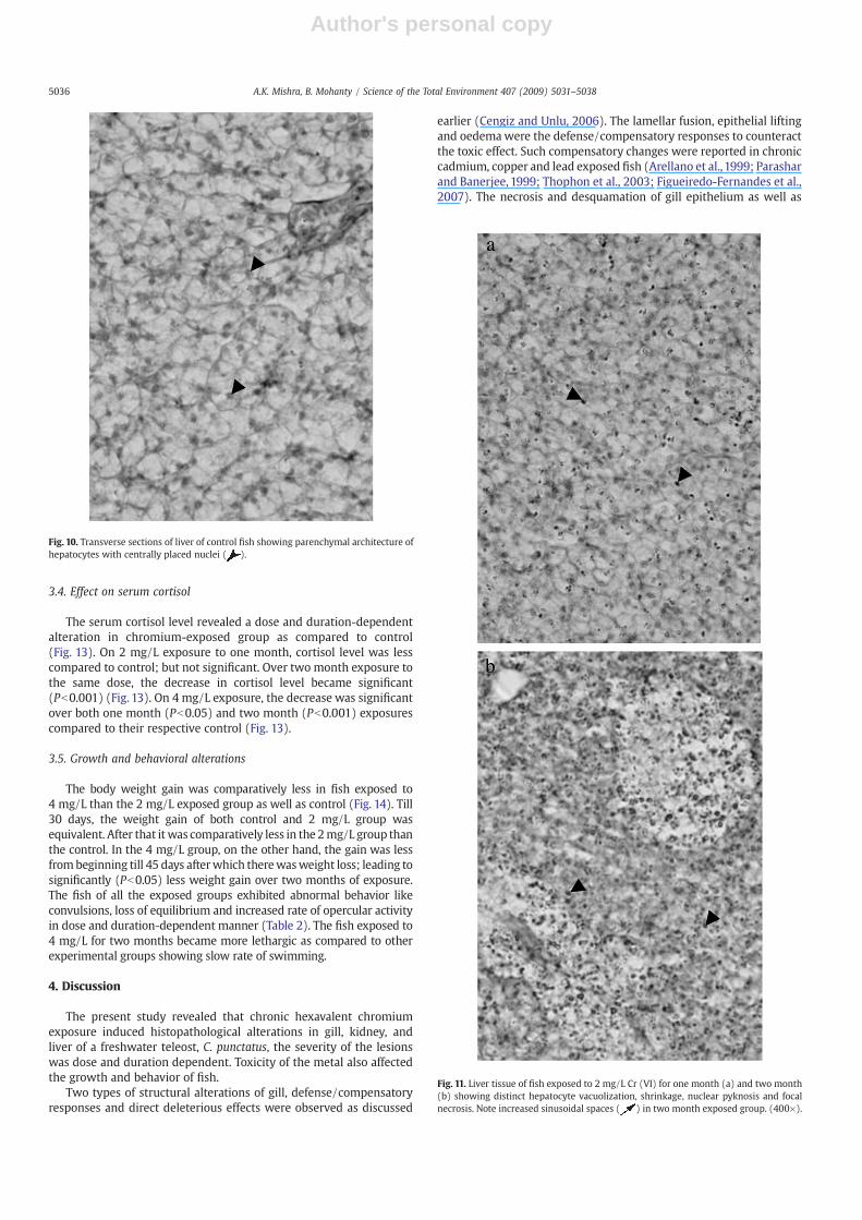
The liver parenchyma of fish showed varied histopathologicalalterations depending on the dose and duration of exposure (Figs. 10–12, Table 1). On exposure to 2 mg/L for one month (Fig. 11a), distinctvacuolization occurred to the hepatocytes along with the mildhepatocellular necrosis. The pyknotic nuclei were noticed in some ofthe hepatocytes (Fig. 11a). Two month exposure to the same doseinduced shrinkage of hepatocytes. Vacuolization of hepatocytes wasobserved only at localized areas. An increase in pyknotic nuclei wasobserved indicating more apoptotic hepatocytes (Fig. 11b). Onexposure to 4 mg/L, the lesions in the liver were comparable to thatof the respective duration of the 2 mg/L groups but to a larger extent(Fig. 12a, b). Extensive shrinkage of the hepatocytes on 4 mg/Lexposure for two months resulted in marked increase in sinusoidalspaces (Fig. 12b).
Fig. 6. Trunk kidney histology of fish exposed to 4 mg/L Cr (VI) for one month (a) andtwo month (b) showing epithelial hypertrophy and reduction of lumens. Note degen-eration of tubules in two month exposed group. (400×).
Fig. 7. Transverse section of the head kidney of control showing arrangement of interrenalcells ( ) in cords.
Fig. 8. Head kidney interrenal cells of fish exposed to 2 mg/L Cr (VI) for one monthshowing cellular atrophy and pyknotic nuclei. (800×).
Fig. 9. Head kidney interrenal cells of fish exposed to 4 mg/L Cr (VI) for two monthshowing disrupted arrangement of interrenal cells aswell as cellular atrophy and pyknoticnuclei. (800×).
5035A.K. Mishra, B. Mohanty / Science of the Total Environment 407 (2009) 5031–5038
Author's personal copy
3.4. Effect on serum cortisol
The serum cortisol level revealed a dose and duration-dependentalteration in chromium-exposed group as compared to control(Fig. 13). On 2 mg/L exposure to one month, cortisol level was lesscompared to control; but not significant. Over two month exposure tothe same dose, the decrease in cortisol level became significant(Pb0.001) (Fig. 13). On 4 mg/L exposure, the decrease was significantover both one month (Pb0.05) and two month (Pb0.001) exposurescompared to their respective control (Fig. 13).
3.5. Growth and behavioral alterations
The body weight gain was comparatively less in fish exposed to4 mg/L than the 2 mg/L exposed group as well as control (Fig. 14). Till30 days, the weight gain of both control and 2 mg/L group wasequivalent. After that it was comparatively less in the 2mg/L group thanthe control. In the 4 mg/L group, on the other hand, the gain was lessfrombeginning till 45 days afterwhich therewasweight loss; leading tosignificantly (Pb0.05) less weight gain over two months of exposure.The fish of all the exposed groups exhibited abnormal behavior likeconvulsions, loss of equilibrium and increased rate of opercular activityin dose and duration-dependent manner (Table 2). The fish exposed to4 mg/L for two months became more lethargic as compared to otherexperimental groups showing slow rate of swimming.
4. Discussion
The present study revealed that chronic hexavalent chromiumexposure induced histopathological alterations in gill, kidney, andliver of a freshwater teleost, C. punctatus, the severity of the lesionswas dose and duration dependent. Toxicity of the metal also affectedthe growth and behavior of fish.
Two types of structural alterations of gill, defense/compensatoryresponses and direct deleterious effects were observed as discussed
earlier (Cengiz and Unlu, 2006). The lamellar fusion, epithelial liftingand oedemawere the defense/compensatory responses to counteractthe toxic effect. Such compensatory changes were reported in chroniccadmium, copper and lead exposed fish (Arellano et al., 1999; Parasharand Banerjee, 1999; Thophon et al., 2003; Figueiredo-Fernandes et al.,2007). The necrosis and desquamation of gill epithelium as well as
Fig. 10. Transverse sections of liver of control fish showing parenchymal architecture ofhepatocytes with centrally placed nuclei ( ).
Fig. 11. Liver tissue of fish exposed to 2 mg/L Cr (VI) for one month (a) and two month(b) showing distinct hepatocyte vacuolization, shrinkage, nuclear pyknosis and focalnecrosis. Note increased sinusoidal spaces ( ) in two month exposed group. (400×).
5036 A.K. Mishra, B. Mohanty / Science of the Total Environment 407 (2009) 5031–5038

Author's personal copy
lamellar curling and aneurisms were the direct deleterious effects;also reported in chronic lead exposed Clarias gariepinus (Olojo et al.,2005). The renal lesions were proven good indicators of environ-mental pollution as the kidney of fish performs important function ofelectrolyte and water balance (Ortiz et al., 2003). On chronicexposures, hexavalent chromium severely affected the renal tubulescausing hypertrophy of epithelial cells, reduction of tubular lumen,
contraction of glomeruli and epithelial and glomerular necrosis.Necrosis and fibrosis of tubular lumen was reported earlier in chronicchromium-exposed chinook salmon (Farag et al., 2006). Similarpattern of renal tubular lesions was also reported in chronic mercuryexposed Clarias batrachus (Kirubagaran and Joy, 1988). The atrophyoccurred to the interrenal cells on chronic exposure in contrast tohypertrophy and vacuolization on acute exposure (Mishra andMohanty, 2008b). The more number of apoptotic cells was alsoobserved on chronic exposures. Hepatocyte vacuolization, necrosis,shrinkage, nuclear pyknosis and increase of sinusoidal spaceswere thedistinct changes observed in the liver of chromium-exposed fish.Similar types of liver histopathology were reported in fish on chroniclead and copper exposures (Olojo et al., 2005; Figueiredo-Fernandeset al., 2007). Extensive histopathological lesions observed in gill,kidney and liver on chronic exposure to metal may be caused byoxidative stress. Easy penetrability through biological membranes andhigh oxidative stress potential of Cr (VI) has been reported earlier infish (Irwin et al., 1997; Begum et al., 2006).
The alterations in behavior of fish have been reported on exposureto various types of pollutants including heavy metals (Kirubagaranand Joy,1988; Gopal et al., 1989; Kumari et al., 1997; Olojo et al., 2005).Behavioral abnormalities in Cr (VI)-exposed Channa, to some extent,may be the manifestation of impaired functioning of the gill, kidneyand liver due to extensive histopathological lesions of these organs.The lesions on gills might have caused respiratory distress thatreflected increased opercular activity. In copper exposed fish, Esomusdanricus, decreased oxygen consumption and increased opercularactivity have been reported due to gill damage (Vutukuru et al., 2005).The physiological as well as metabolic dysfunctions also might haveresulted in hypoactivity and reduced rate of swimming. Reducedlocomotor activity has been reported in chronic chromium-exposedGambusia affinis (Begum et al., 2006). Significantly low serum cortisollevel on two months of exposure to both 2 mg/L and 4 mg/L dosesindicated the impairment of functioning of interrenal tissue. The im-paired stress response of this fish has been demonstrated even on onemonth of exposure to 2 mg/L, though there was no significantalteration of cortisol level (Mishra and Mohanty, in press). Thedisruption of functioning of interrenal tissue, responsible for main-tenance of stress homeostasis, reflected in some of the behavioral
Fig. 12. Liver tissue of fish exposed to 4 mg/L Cr (VI) for one month (a) and two month(b) showing distinct hepatocyte vacuolization, shrinkage, nuclear pyknosis and focalnecrosis. Note increased sinusoidal spaces ( ) in two month exposed group. (400×).
Fig.13. Serum cortisol level (means±SE) in one and twomonth control and chromium-exposed groups. ⁎Pb0.05, ⁎⁎Pb0.001 significant compared to respective control.
Fig. 14. Percentage body weight gain (means±SE) in control and chromium-exposedgroups. Note significantly less weight gain in the 4 mg/L two month exposure group.⁎Pb0.05 significant compared to respective control.
Table 2Impact of chronic hexavalent chromium exposures on the behavioral patterns of theChanna punctatus.
Parameters One month Two months
Control 2 mg/L 4 mg/L Control 2 mg/L 4 mg/L
Hypoactivity − − + − + ++Loss of balance − − − − − +Decreased rate of swimming − − − − − ++Rate of opercular activity + ++ ++ + ++ +++Convulsions − − − − + ++
(−) none, (+) mild, (++) moderate, and (+++) strong.
5037A.K. Mishra, B. Mohanty / Science of the Total Environment 407 (2009) 5031–5038

Author's personal copy
patterns such as loss of balance and convulsions. Impairment ofinterrenal activity might also have affected the general health of fishand led to the reduction of growth. Growth inhibition of fish has beenreported on exposure to various stressors (Wendelaar Bonga, 1997;Hontela, 1998). The growth of the fish also might have affectedbecause of metabolic dysregulations induced by lesions in liver. Thehexavalent chromium induced depletion in the profiles of liverglycogen, total protein and total lipid has been reported (Jha andJha, 1995; Saxena and Tripathi, 2007). The growth impairment of fishof metal contaminated sites has been reported to be due to alterationof liver glycogen and triglycerides as well as the activities of metabolicenzymes (Levesque et al., 2002).
5. Conclusion
Exposure to sublethal concentrations of Cr (VI), thus, caused doseand duration-dependent histopathological alterations in gill, kidney(including interrenal tissue) and liver of C. punctatus. The lesions inthese vital organs might have resulted in physiologic and metabolicdysregulations, which further led to behavioral alterations and growthimpairment. In the long-run, therefore, Cr (VI) exposures to evensublethal concentrations may pose serious threat to fish health andaffect their population.
Acknowledgements
The authors are thankful to the Central Instrumentation Facility ofthe Department of Zoology for use of equipment. Research Fellowshipof the University of Allahabad, India to AK is also thankfullyacknowledged.
References
APHA. Standard methods for the examination of water and wastewater. 19th ed.Washington, DC: American Public Health Association; 1995.
Arellano JM, Storch V, Sarasquete C. Histological changes and copper accumulation inliver and gills of the Senegales sole, Solea senegalensis. Ecotoxicol Environ Saf1999;44:62–72.
Begum G, Venkateswara Rao J, Srikanth K. Oxidative stress and changes in locomotorbehavior and gill morphology of Gambusia affinis exposed to chromium. ToxicolEnviron Chem 2006;88:355–65.
Blacksmith Institute: Projects. (bhttp://www.blacksmithinstitute.org/projects/pollu-tants/hcN).
Cengiz EI, Unlu E. Sublethal effects of commercial deltamethrin on the structure of thegill, liver and gut tissues of mosquito fish, Gambusia affinis: a microscopic study.Environ Toxicol Pharmacol 2006;21:246–53.
Chromium Contamination in the San Fernando Valley (bhttp://www.waterboards.ca.gov/losangeles/water_issues/programs/remediation/chromium/chromium_s1.shtmlN).
de Oliveira Ribeiro CA, Belger L, Pelletier E, Rouleau C. Histopathological evidence ofinorganicmercury andmethylmercury toxicity in the arctic charr (Salvelinus alpinus).Environ Res 2002;90:217–25.
Farag AM, May T, Marty GD, Easton M, Harper DD, Little EE, et al. The effect of chronicchromium exposure on the health of chinook salmon (Oncorhynchus tshawytscha).Aquat Toxicol 2006;76:246–57.
Figueiredo-Fernandes A, Ferreira-Cardoso JV, Garcia-Santos S, Monteiro SM, Carrola J,Matos P, et al. Histopathological changes in liver and gill epithelium of Nile tilapia,Oreochromis niloticus, exposed to waterborne copper. Pesqui Vet Bras 2007;27:103–9.
Gopal K, Ram MD, Anand M, Ray PK. Toxicity and fate of lindane in fresh water fish,Channa punctatus (Bloch). Environ Ecol 1989;7:571–6.
Hontela A. Interrenal dysfunction in fish from contaminated sites: in vivo and in vitroassessment. Environ Toxicol Chem 1998;17:44–8.
Hontela A, Dumout P, Duclos D, Fortin R. Endocrine andmetabolic dysfunction in yellowperch, Perca flavescens exposed to organic contaminants and heavymetals in the St.Lawrence River. Environ Toxicol Chem 1995;14:725–31.
Irwin RJ, Van Mouwerik M, Stevens L, Seese MD, Basham W. Chromium VI (hexavalentchromium). Environmental contaminants encyclopedia. Fort Collins, Colorado:National Park Service, Water Resources Division; 1997.
Jha BS, Jha AK. Biochemical profiles of liver, muscle and gonads of the freshwater fish,Heteropneustes fossilis, under chromium stress. In: Prakash R, editor. Toxicity andmonitoring of xenobiotics. New Delhi: Venus Publishing House; 1995. p. 127–37.
Kirubagaran R, Joy KP. Toxic effects of three mercurial compounds on survival, andhistology of the kidney of the catfish, Clarias batrachus. Ecotoxicol Environ Saf1988;15:172–279.
Kumari R, Singh RK, Khanna YP, Sharma B. Carbofuran induced stress mediated diseasesyndromes in Clarias batrachus, a fresh water fish. Proceedings of the internationalconferences of pollution assessment; 1997. p. 278–97.
Leblond VS, Bisson M, Hontela A. Inhibition of cortisol secretion in dispersed headkidney cells of rainbow trout (Oncorhynchus mykiss) by endosulfan, an organo-chlorine pesticide. Gen Comp Endocrinol 2001;121:48–56.
Leghouchi E, Laib E, Guerbet M. Evaluation of chromium contamination in water,sediment and vegetation caused by the tannery of Jijel (Algeria): a case study.Environ Monit Assess 2009;153:111–7.
Levesque HM, Dorval J, Van Der Kraak G, Campbell PGC, Hontela A. Hormonal,morphological and physiological responses of yellow perch (Perca flavescens) tochronic environmental metal exposure. J Toxicol EnvironHealth A 2003;66:657–76.
Levesque HM, Moon TW, Campbell PGC, Hontela A. Seasonal variation in carbohydrateand lipid metabolism of yellow perch (Perca flavescens) chronically exposed tometals in the field. Aquat Toxicol 2002;60:257–67.
Mishra AK, Mohanty B. Histopathological effects of hexavalent chromium in the ovaryof a fresh water fish, Channa punctatus (Bloch). Bull Environ Contam Toxicol2008a;81:507–11.
Mishra AK, Mohanty B. Acute toxicity impacts of hexavalent chromium on behavior andhistopathology of gill, kidney and liver of the freshwater fish, Channa punctatus(Bloch). Environ Toxicol Pharmacol 2008b;26:136–41.
Mishra AK, Mohanty B. (in press). Effect of hexavalent chromium exposure on thepituitary-interrenal axis of a teleost, Channa punctatus (Bloch). Chemosphere.doi:10.1016/j.chemosphere.2009.04.012.
Olojo EAA, Olurin KB, Mbaka G, Oluwemimo AD. Histopathology of the gill and livertissues of the African catfish, Clarias gariepinus exposed to lead. Afr J Biotechnol2005;4:117–22.
Ortiz JB, De Canales MLG, Sarasquete C. Histopathological changes induced by lindane(γ-HCH) in various organs of fishes. Sci Mar 2003;67:53–61.
Parashar RS, Banerjee TK. Histopathological analysis of sublethal toxicity induced by leadnitrate to the accessory respiratory organs of the air-breathing teleost, H. fossilis(Bloch). Pol Arch Hydrobiol 1999;46:194–205.
Romero LM. Physiological stress in ecology: lessons from biomedical research. TrendsEcol Evol 2004;19:50–5.
Saxena D, Tripathi M. Hexavalent chromium induces biochemical alterations in air-breathing fish, Channa punctatus. J Ecophys Occup Health 2007;7:171–5.
Thophon S, Kruatrachue M, Upatham ES, Pokethitiyook P, Sahaphong S, Jaritkhuan S.Histopathological alterations of white seabass, Lates calcarifer, in acute andsubchronic cadmium exposure. Environ Pollut 2003;121:307–20.
Vinodhini R, Narayanan M. Bioaccumulations of heavy metals in the organs of freshwater fish Cyprinus carpio (Common carp). Int J Environ Sci Tech 2008;5:179–82.
Vutukuru SS, Suma C, RadhaMadhavi K, Juveria, Smitha Pauleena J, Venkateswara Rao J,et al. Studies on the development of potential biomarkers for rapid assessment ofcopper toxicity to freshwater fish using Esomus danricus as model. Int J Environ ResPublic Health 2005;2:63–73.
Wendelaar Bonga SE. The stress responses in fish. Physiol Rev 1997;77:591–625.WHO. Guidelines for drinking water quality. Geneva, Switzerland: World Health
Organization; 1993. p. 188.Yamazaki M, Tanizaki Y, Shimokawa T. Silver and other trace elements in a freshwater
fish, Carasius auratus langsdorfii, from the Asakawa river in Tokyo, Japan. EnvironPollut 1996;94:83–90.
5038 A.K. Mishra, B. Mohanty / Science of the Total Environment 407 (2009) 5031–5038