Acute toxicity impacts of hexavalent chromium on behavior and histopathology of gill, kidney and...
7
This article was originally published in a journal published by Elsevier, and the attached copy is provided by Elsevier for the author’s benefit and for the benefit of the author’s institution, for non-commercial research and educational use including without limitation use in instruction at your institution, sending it to specific colleagues that you know, and providing a copy to your institution’s administrator. All other uses, reproduction and distribution, including without limitation commercial reprints, selling or licensing copies or access, or posting on open internet sites, your personal or institution’s website or repository, are prohibited. For exceptions, permission may be sought for such use through Elsevier’s permissions site at: http://www.elsevier.com/locate/permissionusematerial
-
Upload
independent -
Category
Documents
-
view
2 -
download
0
Transcript of Acute toxicity impacts of hexavalent chromium on behavior and histopathology of gill, kidney and...

This article was originally published in a journal published by Elsevier, and the attached copy is provided by Elsevier for the
author’s benefit and for the benefit of the author’s institution, for non-commercial research and educational use including without
limitation use in instruction at your institution, sending it to specific colleagues that you know, and providing a copy to your institution’s
administrator.
All other uses, reproduction and distribution, including without limitation commercial reprints, selling or licensing copies or access,
or posting on open internet sites, your personal or institution’s website or repository, are prohibited. For exceptions, permission
may be sought for such use through Elsevier’s permissions site at:
http://www.elsevier.com/locate/permissionusematerial
Author's Personal Copy
Environmental Toxicology and Pharmacology 26 (2008) 136–141
Contents lists available at ScienceDirect
Environmental Toxicology and Pharmacology
journa l homepage: www.e lsev ier .com/ locate /e tap
Acute toxicity impacts of hexavalent chromium on behavior andhistopathology of gill, kidney and liver of the freshwater fish,Channa punctatus (Bloch)
Ashish K. Mishra, Banalata Mohanty ∗
tternto he5 mg/actersqual tubucapse intocyteincrely aff
Department of Zoology, University of Allahabad, Allahabad 211002, India
a r t i c l e i n f o
Article history:Received 12 November 2007Received in revised form 19 February 2008Accepted 20 February 2008Available online 6 March 2008
Keywords:ChromiumAcute toxicity effectBehavioral effectsHistopathologyGillKidneyLiverChanna punctatus
a b s t r a c t
Alteration in behavioral patatus, after acute exposurewas determined to be 41.7changes in gills were charepithelial necrosis and deof epithelial cells of renatraction in the Bowman’skidney of exposed fish. Thuolization. The liver hepatHepatocytes atrophy andchromium toxicity severefish populations.
1. Introduction
Chromium (VI) salts have several applications in diverseindustries and their indiscriminate introduction into the aquaticecosystem pose a serious threat to the growth and survival ofthe aquatic fauna including the fish populations. A considerableamount of experimental data on chromium toxicity to aquatic lifewas compiled and reviewed (US EPA, 1985; Eisler, 1986; Irwinet al., 1997). Data on chromium toxicity to fish are scarce andmostly restricted to the effects on biochemical/enzymological pro-files (Nath and Kumar, 1988; Al-Akel and Shamsi, 1996; Vutukuru,2003) and immune responses (Khangarot et al., 1999; Arunkumaret al., 2000). Studies on histopathological responses of vital organslike gills, kidney and liver to chromium toxicity are almost lackingin fish. Few reports are only available on alteration in histomor-phology of gill (Nath et al., 1997; Begum et al., 2006). Chromateion (CrO4
2−) is the dominant form of hexavalent chromium (Cr VI)in aqueous solution and can readily cross cellular membranes. Thechromium mediated cytotoxic effects on histopathology of various
∗ Corresponding author. Tel.: +91 532 2460788; fax: +91 532 2460788.E-mail address: drbana [email protected] (B. Mohanty).
1382-6689/$ – see front matter © 2008 Elsevier B.V. All rights reserved.doi:10.1016/j.etap.2008.02.010
s and histopathology of gill, kidney and liver were studied in Channa punc-xavalent chromium. The 96 h LC50 of chromium salt, potassium dichromateL. The exposed fish displayed erratic swimming and became lethargic. Theized by epithelial hyperplasia, lamellar fusion, oedema, epithelial lifting,mation, aneurism as well as curling of secondary lamellae. Hypertrophy
les with reduced lumens, atrophy of the renal tubules, glomeruli con-ules and necrosis of haematopoietic tissues were observed in the trunkerrenal cells of the head kidney exhibited distinct hypertrophy and vac-s showed cytoplasmic vacuolization with the lateral nuclei arrangement.
ase in sinusoidal space were also observed. The result showed that acuteects the vital organs and normal behavior which may be deleterious for
© 2008 Elsevier B.V. All rights reserved.
organs like kidney (Oliveira et al., 2006), testis (Murthy et al., 1991;Aruldhas et al., 2005; Acharya et al., 2006; Subramanian et al., 2006)and brain (Travacio et al., 2001) were observed in several mam-
mals. Keeping in view the cytotoxicity of chromium and in view ofthe fact that histopathological investigations have been proved tobe a sensitive tool to detect effects of chemical compounds withinthe target organ of fish in laboratory experiments (Santhakumar etal., 2001; Ortiz et al., 2003; Olojo et al., 2005; Cengiz, 2006; Cengizand Unlu, 2006; Rao et al., 2006; Figueiredo-Fernandes et al., 2007;Velmurugan et al., 2007), the present study focussed on chromium-induced histopathology of the vital organs like gill, kidney and liver.In addition, the toxic impact on alteration in behavioral pattern wasalso observed.2. Materials and methods
2.1. Fish and acclimation conditions
Over 106 adult specimens of the Channa punctatus (Order: Ophiocephaliformes,Family: Ophiocephalidae) weighing 50–55 g, length 18–20 cm were collected fromclean and unpolluted local freshwater pond, washed with 0.1% KMnO4 solution toremove dermal infection, if any, and were acclimatized to laboratory conditions for2 weeks before experimentation. They were maintained in glass aquaria containingseasoned tap water (pH 7.3 ± 0.05, DO 7.5 ± 1.0 mg/L, total hardness 215.3 ± 7.0 mg/Las CaCO3 and alkalinity 133.2 ± 5.0 mg/L as CaCO3; APHA (1995) method was fol-lowed for the water quality determination) under natural photoperiod (13L:11D)
rso
oxicolo
Author's Pe
A.K. Mishra, B. Mohanty / Environmental T
Table 1aToxicity testing of hexavalent chromium (K2Cr2O7) in a teleost fish, Channa punctatusfor 96 h
Concentration (mg/L) Number of fish Mortality (%)
15 10 1025 10 2035 10 4045 10 6055 10 7065 10 8075 10 100
and ambient temperature (18–21 ◦C). Fish were fed, ad libitum, with commercial drypellets (Hello fish dry pellets; CVM products, Beijing, China). The totalamount of feed provided was not less than 2–3% of their body weight perday.
2.2. Test chemical and determination of LC50
The potassium dichromate (K2Cr2O7) (MERCK, Mumbai, India; Purity 99%) wasused as the test compound for determination of median lethal dose (LC50) ofchromium. The LC50 value of chromium was determined according to the arithmeticmethod of Karber as adopted by Dede and Kaglo (2001) using the daily renewalbioassay system. The test concentrations (Table 1a) were chosen on the basis of ini-tial experiments to determine the LC50. The stock solution was prepared in distilledwater and the required concentrations were maintained in aquaria, renewing thewater every day. The day before and during the test the fish were not fed. In theexperiment no distinction was made between the sexes. The control group with-out chromium was maintained simultaneously. The mortality of fish was recordedduring 96 h of exposure in each concentration of the toxicant. The data was used toestimate the LC50 of potassium dichromate.
2.3. Experimental design
The fish were divided into three groups each containing 12 individuals. GroupI was kept as control, with no chromium added to the water, and other two groupswere exposed to potassium dichromate. Groups II and III were acutely exposed for96 h to 20 mg/L (≈50% of 96 h LC50) and 40 mg/L (≈96 h LC50), respectively. The wholeexposure medium was changed every day in both the treatment groups to maintainthe desired concentration of chromium salt. The water in control group was alsochanged at the same time. During the exposure, mortality and behavior of the fishwere monitored. The behavioral pattern was monitored regularly as suggested byKumari et al. (1997).
2.4. Tissue preparations
At the end of exposure time, fish of all the groups were sacrificed by decap-itation. Gill, kidney and liver of control and alive treated fish were dissected out,fixed in Bouin’s solution for 24 h and then were processed for paraffin (m.p. 62 ◦C)embedding.
2.5. Histology
Paraffin blocks of gills, kidney and liver of all the groups were cut at 6 �mthickness and stretched on sterilized glass slides. After deparaffinization, sectionswere stained with Haematoxylin–Eosine and observed under light microscopy. Thehistopathological changes in the tissues were examined in the randomly selectedten sections from each fish. The mean prevalence of each histopathological parame-
Table 1bDetermination of LC50 value of hexavalent chromium (K2Cr2O7) in a teleost fish, Channa p
Concentration (mg/L) Concentration difference Number of alive fish Nu
0 – 10 015 15 9 125 10 8 235 10 6 445 10 4 655 10 3 765 10 2 875 10 0 10
LC50 = LC100 −∑
(Mean death×Concentration difference)
Number of fish per group = 75 − 332.510 = 75 − 33.25 LC50 = 41.75
nal Copy
gy and Pharmacology 26 (2008) 136–141 137
Table 2Impact of hexavalent chromium (K2Cr2O7) on the behavior of a teleost fish, Channapunctatus for 96 h
Parameters Control Cr (20 mg/L) Cr (40 mg/L)
Hyperactivity − + +++Loss of balance − + +++Rate of swimming + + ++Rate of opercular activity + ++ +++Convulsions − + ++
(−) None, (+) mild, (++) moderate, (+++) strong.
ters was categorized as mild (+, <25% of sections), moderate (++, 25–50% of sections)and severe (+++, >50% of sections). Histopathological changes induced by treatmentsin the tissues were photographed using Nikon photomicroscope.
3. Results
The 96 h LC50 of chromium salt, potassium dichromate for C.punctatus was determined to be 41.75 mg/L (Table 1b). In the20 mg/L group during the 96 h acute exposure 2 fish died whilein 40 mg/L group 6 fish died. The fish exposed to 20 and 40 mg/Lchromium exhibited abnormal behavior like erratic swimming andloss of equilibrium. They become lethargic and mucous substancewas secreted from the whole body. The exposed fish swam to sur-face more often than the control fish. Neither mortality nor anyvisible changes in behavior were observed in the control group.Summary of the different behavioral changes observed due tochromium exposure is presented in Table 2.
The gill of C. punctatus is made up of primary lamellae arranged
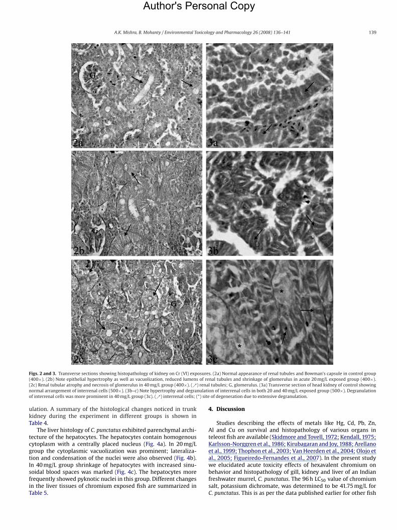
in double rows, projecting on the lateral sides of which are a seriesof alternately arranged secondary lamellae. In the core there is acartilaginous supporting rod and blood vessels with traces of sinu-soidal blood spaces. No histopathological changes were found inthe control (Fig. 1a). In the acute chromium exposed fish epithelialhyperplasia, lamellar fusion, epithelial lifting, epithelial necrosisand desquamation, aneurism as well as curling of secondary lamel-lae were observed (Fig. 1b, c). The gills were swollen in comparisonto the control fish because of the hypertrophy and hyperplasia of thegill epithelial cells. Fusion of secondary lamellae, oedema, necro-sis and desquamation of lamellar epithelium was observed. Thecartilaginous rod at the core of primary lamellae was seen to be dis-rupted in numerous areas. In 40 mg/L group aneurism and curlingof secondary lamellae was more prominent (Fig. 1c). The summaryof these histopathological changes observed in gills is presented inTable 3.The histology of trunk kidney, the excretory componentof Channa was characterized by the renal tubules and Bow-man’s capsules with glomeruli distributed in the interstitium ofhaematopoietic tissues (Fig. 2a). Acute exposures to chromium (20and 40 mg/L), drastically altered all the components of trunk kid-
unctatus for 96 h.
mber of dead fish Mean death Mean death × concentration difference
0 00.5 7.51.5 153.0 305.0 506.5 657.5 759.0 90∑
= 332.5
mg/L

Author's Personal Copy
138 A.K. Mishra, B. Mohanty / Environmental Toxicology and Pharmacology 26 (2008) 136–141
Fig. 1. Sagittal sections showing gill histopathology on Cr (VI) exposures. (a) Note normacore (C) in control group (400×). (b–c’) Hyperplasia in lamellar epithelium leading to lameand disruption of cartilaginous core in both 20 and 40 mg/L group (400×). Lamellar aneurlamellar aneurism; (**) epithelial oedema. (↗) curling of secondary lamellae; (↗↗) epith
ney (Fig. 2b, c). Hypertrophy of epithelial cells of the renal tubuleswith the consequent reduction in tubular lumens was observed.Some of the epithelial cells showed distinct vacuolization. Contrac-tion of glomeruli and disruption of haematopoietic tissues werealso observed in 20 mg/L group (Fig. 2b). Atrophy of renal tubulesand degeneration of Bowman’s capsules were more distinct in
Table 3Impact of hexavalent chromium (K2Cr2O7) in gills of a teleost fish, Channa punctatusfor 96 h
Parameters Control Cr (20 mg/L) Cr (40 mg/L)
Epithelial hyperplasia − ++ +Aneurism − + ++Epithelial necrosis anddesquamation
− ++ +++
Epithelial lifting and oedema − ++ +++Lamellar fusion − ++ +Curling of secondary lamellae − + ++
(−) None, (+) mild, (++) moderate, (+++) severe.
l appearance of primary (PL) and secondary lamellae (SL) as well as cartilaginousllar fusion, epithelial lifting, curling of secondary lamellae as well as desquamationism was evident in 40 mg/L group (c’). LF, lamellar fusion; EL, epithelial lifting; LA,elial necrosis and desquamation.
40 mg/L group (Fig. 2c). The head kidney, the endocrine compo-nent, constituted of interrenal and chromaffin cells. After acuteexposure the interrenal cells, functional equivalent of mammalianadrenal cortex, showed hypertrophy and degranulation in both20 and 40 mg/L group (Fig. 3b, c). In 40 mg/L chromium exposedgroup the changes were more prominent with extensive degran-
Table 4Impact of hexavalent chromium (K2Cr2O7) in kidney of a teleost fish, Channa punc-tatus for 96 h
Parameters Control Cr (20 mg/L) Cr (40 mg/L)
Pyknotic nuclei in tubular epithelium − − +Hypertrophied epithelial cells ofrenal tubules
− ++ +
Reduction of space inside renaltubules
− ++ +++
Expansion of space inside Bowman’scapsule
− ++ +++
Contraction of the glomerulus − ++ +++
(−) None, (+) mild, (++) moderate, (+++) severe.
Author's Personal Copy
A.K. Mishra, B. Mohanty / Environmental Toxicology and Pharmacology 26 (2008) 136–141 139
Figs. 2 and 3. Transverse sections showing histopathology of kidney on Cr (VI) exposure(400×). (2b) Note epithelial hypertrophy as well as vacuolization, reduced lumens of re(2c) Renal tubular atrophy and necrosis of glomerulus in 40 mg/L group (400×). (↗) renanormal arrangement of interrenal cells (500×). (3b–c) Note hypertrophy and degranulatiof interrenal cells was more prominent in 40 mg/L group (3c). (↗) interrenal cells; (*) site
ulation. A summary of the histological changes noticed in trunkkidney during the experiment in different groups is shown inTable 4.
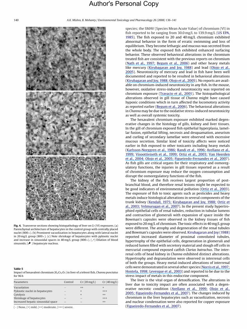
The liver histology of C. punctatus exhibited parenchymal archi-tecture of the hepatocytes. The hepatocytes contain homogenouscytoplasm with a centrally placed nucleus (Fig. 4a). In 20 mg/Lgroup the cytoplasmic vacuolization was prominent; lateraliza-tion and condensation of the nuclei were also observed (Fig. 4b).In 40 mg/L group shrinkage of hepatocytes with increased sinu-soidal blood spaces was marked (Fig. 4c). The hepatocytes morefrequently showed pyknotic nuclei in this group. Different changesin the liver tissues of chromium exposed fish are summarized inTable 5.
s. (2a) Normal appearance of renal tubules and Bowman’s capsule in control groupnal tubules and shrinkage of glomerulus in acute 20 mg/L exposed group (400×).l tubules; G, glomerulus. (3a) Transverse section of head kidney of control showingon of interrenal cells in both 20 and 40 mg/L exposed group (500×). Degranulationof degeneration due to extensive degranulation.
4. Discussion
Studies describing the effects of metals like Hg, Cd, Pb, Zn,Al and Cu on survival and histopathology of various organs inteleost fish are available (Skidmore and Tovell, 1972; Kendall, 1975;Karlsson-Norggren et al., 1986; Kirubagaran and Joy, 1988; Arellanoet al., 1999; Thophon et al., 2003; Van Heerden et al., 2004; Olojo etal., 2005; Figueiredo-Fernandes et al., 2007). In the present studywe elucidated acute toxicity effects of hexavalent chromium onbehavior and histopathology of gill, kidney and liver of an Indianfreshwater murrel, C. punctatus. The 96 h LC50 value of chromiumsalt, potassium dichromate, was determined to be 41.75 mg/L forC. punctatus. This is as per the data published earlier for other fish
Author's Per
140 A.K. Mishra, B. Mohanty / Environmental Toxicolo
Fig. 4. Transverse sections showing histopathology of liver on Cr (VI) exposures. (a)Parenchymal architecture of hepatocytes in the control group with centrally placednuclei (800×). (b) Prominent vacuolization in hepatocytes along with lateral nucleiin 20 mg/L group (800×). (c) Note shrinkage of hepatocytes with pyknotic nucleiand increase in sinusoidal spaces in 40 mg/L group (800×). (↗) Dilation of bloodsinusoids; ( ) hepatocyte nucleus.
Table 5Impact of hexavalent chromium (K2Cr2O7) in liver of a teleost fish, Channa punctatusfor 96 h
Parameters Control Cr (20 mg/L) Cr (40 mg/L)
Vacuolation − ++ +Pyknotic nuclei in hepatocytes − + +++Cytolysis − + ++Shrinkage of hepatocytes − − +++Increased hepatic sinusoidal space − − ++
(−) None, (+) mild, (++) moderate, (+++) severe.
sonal Copy
gy and Pharmacology 26 (2008) 136–141
species; the SMAV (Species Mean Acute Value) of chromium (VI) infish reported to be ranging from 30.0 mg/L to 139.9 mg/L (US EPA,1985). The fish exposed to 20 and 40 mg/L chromium exhibitedabnormal behavior in the form of erratic swimming and loss ofequilibrium. They become lethargic and mucous was secreted fromthe whole body. The exposed fish exhibited enhanced surfacingbehavior. These observed behavioral alterations in the chromiumtreated fish are consistent with the previous reports on chromium(Nath et al., 1997; Begum et al., 2006) and other heavy metalslike mercury (Kirubagaran and Joy, 1988) and lead (Olojo et al.,2005). Neurotoxicity of mercury and lead in fish have been welldocumented and reported to be resulted in behavioral alterations(Kirubagaran and Joy, 1988; Olojo et al., 2005). No reports are avail-able on chromium-induced neurotoxicity in any fish. In the mouse,however, oxidative stress-induced neurotoxicity was reported onchromium exposure (Travacio et al., 2001). The histopathologicalalterations observed in gill tissue of Channa might have causedhypoxic conditions which in turn affected the locomotory activityas reported earlier (Begum et al., 2006). The behavioral alterationsin Channa may be due to the oxidative stress-induced neurotoxicityas well as overall systemic toxicity.
The hexavalent chromium exposure exhibited marked degen-erative changes in the histology of gills, kidney and liver tissues.In the gill of chromium exposed fish epithelial hyperplasia, lamel-lar fusion, epithelial lifting, necrosis and desquamation, aneurismand curling of secondary lamellae were observed with excessivemucous secretion. Similar kind of toxicity affects were noticedearlier in fish exposed to other toxicants including heavy metals(Karlsson-Norggren et al., 1986; Randi et al., 1996; Arellano et al.,1999; Visoottiviseth et al., 1999; Ortiz et al., 2003; Van Heerdenet al., 2004; Olojo et al., 2005; Figueiredo-Fernandes et al., 2007).As fish gills are critical organs for their respiratory and osmoreg-ulatory functions, the injuries in gill tissues reported as a resultof chromium exposure may reduce the oxygen consumption anddisrupt the osmoregulatory functions of the fish.
The kidney of the fish receives largest proportion of post-branchial blood, and therefore renal lesions might be expected tobe good indicators of environmental pollution (Ortiz et al., 2003).The exposure of fish to toxic agents such as pesticides and heavymetals induce histological alterations in several components of thetrunk kidney (Kendall, 1975; Kirubagaran and Joy, 1988; Ortiz etal., 2003; Velmurugan et al., 2007). In the present study hypertro-phied epithelial cells of renal tubules, reduction in tubular lumensand contraction of glomeruli with expansion of space inside the
Bowman’s capsules were observed in the kidney tissues of fishexposed to 20 mg/L of chromium. The toxic effects in 40 mg/L groupwere different. The atrophy and degeneration of the renal tubulesand Bowman’s capsules were observed. Kirubagaran and Joy (1988)reported increased diameter of proximal convoluted tubules,hypertrophy of the epithelial cells, degeneration in glomeruli andreduced lumen filled with secretory material and slough off cells inmercurial compound exposed catfish Clarias batrachus. The inter-renal cells of head kidney in Channa exhibited distinct alterations.Hypertrophy and degranulation were observed in interrenal cellsof both the groups. Heavy metal-induced alterations of interrenalcells were demonstrated in several other species (Norris et al., 1997;Hontela, 1998; Levesque et al., 2003) and reported to be due to thestress impact of metals in this endocrine component.The liver is the vital organ of detoxification. The alterations inliver due to toxicity impact are often associated with a degen-erative necrotic condition (Arellano et al., 1999; Olojo et al.,2005; Figueiredo-Fernandes et al., 2007). The changes induced bychromium in the liver hepatocytes such as vacuolization, necrosisand nuclear condensation were also reported for copper exposure(Figueiredo-Fernandes et al., 2007).

rso
oxicolo
Santhakumar, M., Balaji, M., Ramudu, K., 2001. Gill lesions in the perch, Anabas
Author's Pe
A.K. Mishra, B. Mohanty / Environmental T
Hexavalent chromium is highly water soluble, easily pene-trates biological membranes and cause cellular damage by inducingoxidative stress (Irwin et al., 1997; Begum et al., 2006). Hence,the histopathological alterations in gill, kidney and liver tissuesof Channa were distinct. The degenerative changes were compara-tively more pronounced in gill than kidney and liver tissues. Thismay be due to the fact that gills remain in direct contact withthe metallic water. The observed abnormal behavior and alteredhistopathology of vital organs demonstrate the severe adverseeffects to acute exposure of hexavalent chromium in Channaspecies. Thus acute chromium exposure may cause serious phys-iological problems ultimately leading to the death of fish.
References
Acharya, U.R., Mishra, M., Tripathy, R.R., Mishra, I., 2006. Testicular dysfunction andantioxidative defense system of Swiss mice after chromic acid exposure. Reprod.Toxicol. 22, 187–191.
Al-Akel, A.S., Shamsi, M.J.K., 1996. Hexavalent chromium: toxicity and impact on car-bohydrate metabolism and haematological parameters of carp (Cyprinus carpioL.) from Saudi Arabia. Aquat. Sci. 58 (1), 24–30.
American Public Health Association (APHA), 1995. Standard Methods for the Exam-ination of Water and Wastewater, 19th ed. APHA, Washington, DC.
Arellano, J.M., Storch, V., Sarasquete, C., 1999. Histological changes and copper accu-mulation in liver and gills of the Senegales sole, Solea senegalensis. Ecotoxicol.Environ. Saf. 44, 62–72.
Aruldhas, M.M., Subramanian, S., Sekar, P., Vengatesh, G., Chandrahasan, G., Govin-darajulu, P., Akbarsha, M.A., 2005. Chronic chromium exposure-induced changesin testicular histoarchitecture are associated with oxidative stress: study in anon-human primate (Macaca radiata Geoffroy). Hum. Reprod. 20, 2801–2813.
Arunkumar, R.I., Rajasekaran, P., Michael, R.D., 2000. Differential effects of chromiumcompounds on the immune responses of the African mouth breeder, Oreochromismossambicus (Peters.). Fish Shellfish Immunol. 10, 667–676.
Begum, G., Venkateswara, R.J., Srikanth, K., 2006. Oxidative stress and changesin locomotor behavior and gill morphology of Gambusia affinis exposed to
chromium. Toxicol. Environ. Chem. 88, 355–365.Cengiz, E.I., 2006. Gill and kidney histopathology in the freshwater fish Cyprinuscarpio after acute exposure to deltamethrin. Environ. Toxicol. Pharmacol. 22,200–204.
Cengiz, E.I., Unlu, E., 2006. Sublethal effects of commercial deltamethrin on thestructure of the gill, liver and gut tissues of mosquitofish, Gambusia affinis: amicroscopic study. Environ. Toxicol. Pharmacol. 21, 246–253.
Dede, E.B., Kaglo, H.D., 2001. Aqua-toxicological effects of water soluble fractions(WSF) of diesel fuel on O. niloticus fingerlings. J. Appl. Sci. Environ. Mgt. 5, 93–96.
Eisler, R., 1986. Chromium hazards to fish, wildlife and invertebrates: a synopticreview. US Fish and Wildlife Service Biological Report 85, Laurel, MD.
Figueiredo-Fernandes, A., Ferreira-Cardoso, J.V., Garcia-Santos, S., Monteiro, S.M.,Carrola, J., Matos, P., Fontainhas-Fernandes, A., 2007. Histopathological changesin liver and gill epithelium of Nile tilapia, Oreochromis niloticus, exposed towaterborne copper. Pesqui. Vet. Bras. 27 (3), 103–109.
Hontela, A., 1998. Interrenal disfunction in fish from contaminated sites: in vivo andin vitro assessment. Annual review. Environ. Toxicol. Chem. 17, 44–48.
Irwin, R.J., Van Mouwerik, M., Stevens, L., Seese, M.D., Basham, W., 1997. Chromium VI(Hexavalent chromium). Environmental Contaminants Encyclopedia. NationalPark Service, Water Resources Division, Fort Collins, Colorado.
Karlsson-Norggren, L., Dickson, W., Ljungberg, O., Runn, P., 1986. Acid water and alu-minium exposure: gill lesions and aluminium accumulation in farmed, browntrout, Salmo trutta (L.). J. Fish. Dis. 9, 1–9.
Kendall, M.W., 1975. Acute effect of methyl mercury toxicity in channel catfish kid-ney. Bull. Environ. Contam. Toxicol. 13 (5), 570–575.
Khangarot, B.S., Rathore, R.S., Tripathi, D.M., 1999. Effects of chromium on humoraland cell-mediated immune responses and host resistance to disease in a
nal Copy
gy and Pharmacology 26 (2008) 136–141 141
freshwater catfish, Saccobranchus fossilis (Bl). Ecotoxicol. Environ. Saf. 43,11–30.
Kirubagaran, R., Joy, K.P., 1988. Toxic effects of three mercurial compounds on sur-vival, and histology of the kidney of the catfish, Clarias batrachus. Ecotoxicol.Environ. Saf. 15, 172–279.
Kumari, R., Singh, R.K., Khanna, Y.P., Sharma, B., 1997. Carbofuran induced stress-mediated disease syndromes in Clarias batrachus, a fresh water fish. In:Proceedings of the international conferences of pollution assessment, pp.278–297.
Levesque, H., Dorval, J., Van Der Kraak, G., Campbell, P.G.C., Hontela, A., 2003.Hormonal, morphological and physiological responses of yellow perch (Percaflavescens) to chronic environmental metal exposure. J. Toxicol. Environ. Health66, 657–676.
Murthy, R.C., Saxena, D.K., Gupta, S.K., Chandra, S.V., 1991. Ultrastructural obser-vations in testicular tissue of chromium-treated rats. Reprod. Toxicol. 5, 443–447.
Nath, K., Kumar, N., 1988. Hexavalent chromium: toxicity and its impact on certainaspects of carbohydrate metabolism in freshwater teleost, Colisa fasciatus. Sci.Total Environ. 72, 175–181.
Nath, K., Kumar, N., Srivastav, A.K., 1997. Chromium induced histological alterationsin the gills of a freshwater teleost, Colisa fasciatus. Fish. Biol. J. Medaka 9, 37–40.
Norris, D.O., Felt, S.B., Woodling, J.D., Dores, R.M., 1997. Immunocytochemical andhistological differences in the interrenal axis of feral brown trout, Salmo trutta,in metal-contaminated water. Gen. Comp. Endocrinol. 108, 343–351.
Oliveira, H., Santos, T.M., Ramalho-Santos, J., Pereira, M.L., 2006. Histopathologicaleffects of hexavalent chromium in mouse kidney. Bull. Environ. Contam. Toxicol.76, 977–983.
Olojo, E.A.A., Olurin, K.B., Mbaka, G., Oluwemimo, A.D., 2005. Histopathology of thegill and liver tissues of the African catfish, Clarias gariepinus exposed to lead. Afr.J. Biotechnol. 4 (1), 117–122.
Ortiz, J.B., De Canales, M.L.G., Sarasquete, C., 2003. Histopathological changesinduced by lindane (�-HCH) in various organs of fishes. Sci. Mar. 67 (1), 53–61.
Randi, A.S., Monserrat, J.M., Rodriguez, E.M., Romano, L.A., 1996. Histopatholog-ical effects of cadmium on the gills of the freshwater fish, Macropsobryconuruguayanae Eigenmann (Pisces, Atheinidae). J. Fish. Dis. 19, 311–322.
Rao, J.V., Begum, G., Jakka, N.M., Srikanth, K., Rao, R.N., 2006. Sublethal effects ofprofenofos on locomotor behavior and gill architecture of the mosquito fish,Gambusia affinis. Drug Chem. Toxicol. 29, 255–267.
testudineus, exposed to monocrotophos. J. Environ. Biol. 22 (2), 87–90.Skidmore, J.F., Tovell, P.W.A., 1972. Toxic effects of zinc sulphate on the gills of rain-
bow trout. Water Res. 6, 217–230.Subramanian, S., Rajendiran, G., Sekhar, P., Chandrahasan, G., Govindarajulu, P.,
Aruldhas, M.M., 2006. Reproductive toxicity of chromium in adult bonnet mon-keys (Macaca radiata Geoffrey). Reversible oxidative stress in the semen. Toxicol.Appl. Pharmacol. 215 (3), 237–249.
Thophon, S., Kruatrachue, M., Upatham, E.S., Pokethitiyook, P., Sahaphong, S., Jar-itkhuan, S., 2003. Histopathological alterations of white seabass, Lates calcarifer,in acute and subchronic cadmium exposure. Environ. Pollut. 121, 307–320.
Travacio, M., Polo, J.M., Llesuy, S., 2001. Cr (VI) induces oxidative stress in the mousebrain. Toxicology 162, 139–148.
US EPA, 1985. Ambient Water Quality Criteria For Chromium- 1984. EPA-440/5-84-029. Office of Water Regulations and Standards, Washington.
Van Heerden, D., Vosloo, A., Nikinmaa, M., 2004. Effects of short-term copper expo-sure on gill structure, metallothionein and hypoxia-inducible factor-1a (HIF-1a)levels in rainbow trout (Oncorhynchus mykiss). Aquat. Toxicol. 69, 271–280.
Velmurugan, B., Selvanayagam, M., Cengiz, E.I., Unlu, E., 2007. The effects of fenvaler-ate on different tissues of freshwater fish Cirrhinus mrigala. J. Environ. Sci. HealthB 42 (2), 157–163.
Visoottiviseth, P., Thamamaruitkun, T., Sahaphong, S., Riengrojpitak, S., Kruatrachue,M., 1999. Histopathological effects of triphenyltin hydroxide on liver, kidneyand gill of Nile tilapia (Oreochromis niloticus). Appl. Organometal. Chem. 13,749–763.
Vutukuru, S.S., 2003. Chromium induced alterations in some biochemical profile ofthe Indian major carp Labeo rohita (Hamilton). Bull. Environ. Contam. Toxicol.70, 118–123.