Solid-State NMR Structure Determination of Whole Anchoring Threads from the Blue Mussel Mytilus...
10
Solid-State NMR Structure Determination of Whole Anchoring Threads from the Blue Mussel Mytilus edulis Alexandre A. Arnold, † Fre ́ de ́ ric Byette, ‡ Marc-Olivier Se ́ guin-Heine, † Andre ́ LeBlanc, † Lekha Sleno, † Re ́ jean Tremblay, § Christian Pellerin, ‡ and Isabelle Marcotte* ,† † Department of Chemistry, Universite ́ du Qué bec a ̀ Montre ́ al, P.O. Box 8888, Downtown Station, Montreal, Canada H3C 3P8 ‡ Department of Chemistry, Universite ́ de Montre ́ al, P.O. Box 6128, Downtown Station, Montre ́ al, Que ́ bec, Canada H3C 3J7 § Institut des Sciences de la Mer de Rimouski, Universite ́ du Que ́ bec a ̀ Rimouski, 310 allé e des Ursulines, Rimouski, Canada G5L 3A1 * S Supporting Information ABSTRACT: The molecular structure of the blue mussel Mytilus edulis whole anchoring threads was studied by two- dimensional 13 C solid-state NMR on fully labeled fibers. This unique material proves to be well ordered at a molecular level despite its heterogeneous composition as evidenced by the narrow measured linewidths below 1.5 ppm. The spectra are dominated by residues in collagen environments, as determined from chemical shift analysis, and a complete two-dimensional assignment (including minor amino acids) was possible. The best agreement between predicted and experimental backbone chemical shifts was obtained for collagen helices with torsion angles (−75°, +150°). The abundant glycine and alanine residues can be resolved in up to five different structural environments. Alanine peaks could be assigned to collagen triple-helices, β-sheets (parallel and antiparallel), β-turns, and unordered structures. The use of ATR-FTIR microscopy confirmed the presence of these structural environments and enabled their location in the core of the thread (collagen helices and antiparallel β-sheets) or its cuticle (unordered structures). The approach should enable characterization at the molecular level of a wide range of byssus macroscopic properties. ■ INTRODUCTION In the marine environment, the settlement ability of mussels depends on the secretion of byssus promoting attachment to surfaces. The mechanical properties of these beard-like collagenous fibers are tuned to the pressure of the animals’ environment. Mussels usually reside in intertidal rocky zones where current, tides, and waves are a major threat of dislodgment. 1−3 The water movement can exert acceleration, lift, and drag forces that depend on the season and exposition of the shore. Therefore, under such conditions, sessile organisms like mussels need reliable holdfasts to ensure their survival. Byssus filaments are formed in the groove located in a retractile organ of the mussel called “foot” by a process similar to the injection-molding of polymers. This processing differs from the formation of dragline silk, which is spun through a spinneret. 4 The mussel’s byssus is composed of a stem that supports 20−60 threads 5 (Figure 1a). Each thread can be divided into three distinct regions, that is, an initial corrugated and elastic proximal part (Figure 1b) followed by a smooth and stiff distal segment (Figure 1c) ending with the plaque that glues the animal to the substrate. Byssal threads have an extensibility close to that of elastin and are six times tougher than tendons. 6−8 They are complex biopolymers essentially composed of proteins (95% by weight). 9 The fiber core is composed of three pepsin-resistant proteins called PreCol-P, PreCol-D, and PreCol-NG, 7 as illustrated in Figure 1d. Each of these proteins is composed of a collagen center-flanked by elastin-like (PreCol-P), silk-like (PreCol-D), or plant cell wall-like (PCW) (PreCol-NG) regions according to sequence homologies. These flanking regions are preceded and followed by histidine-rich domains. In addition, the Precols are initiated by short signal peptide sequences and the collagen domains are followed by acidic patches. The preCols are gradually distributed along the fiber. While PreCol-P concentration is maximal near the stem and disappears at the middle of the byssus, PreCol-D progressively increases from the stem up to the plaque. The PreCol-NG concentration is constant along the fiber, 7 but its relative amount with respect to the other PreCols is not known. According to the primary amino acid sequence of the byssal proteins, PreCols account for 92−96% of the distal region and 66−72% of the proximal region. 9,10 Atomic force microscopy (AFM) studies showed that the collagen core of the fiber is composed of 6 + 1 collagen helices that form banana-shaped bundles in byssal threads of Mytilus galloprovincialis. 11 These central bent collagen domains are assembled to form the fiber via metal cross-linking of adjacent histidine-rich domains. 12 The remaining fiber is made of thread Received: September 22, 2012 Revised: October 28, 2012 Published: November 19, 2012 Article pubs.acs.org/Biomac © 2012 American Chemical Society 132 dx.doi.org/10.1021/bm301493u | Biomacromolecules 2013, 14, 132−141
-
Upload
independent -
Category
Documents
-
view
1 -
download
0
Transcript of Solid-State NMR Structure Determination of Whole Anchoring Threads from the Blue Mussel Mytilus...

Solid-State NMR Structure Determination of Whole AnchoringThreads from the Blue Mussel Mytilus edulisAlexandre A. Arnold,† Frederic Byette,‡ Marc-Olivier Seguin-Heine,† Andre LeBlanc,† Lekha Sleno,†Rejean Tremblay,§ Christian Pellerin,‡ and Isabelle Marcotte*,†
†Department of Chemistry, Universite du Quebec a Montreal, P.O. Box 8888, Downtown Station, Montreal, Canada H3C 3P8‡Department of Chemistry, Universite de Montreal, P.O. Box 6128, Downtown Station, Montreal, Quebec, Canada H3C 3J7§Institut des Sciences de la Mer de Rimouski, Universite du Quebec a Rimouski, 310 allee des Ursulines, Rimouski, Canada G5L 3A1
*S Supporting Information
ABSTRACT: The molecular structure of the blue musselMytilus edulis whole anchoring threads was studied by two-dimensional 13C solid-state NMR on fully labeled fibers. Thisunique material proves to be well ordered at a molecular leveldespite its heterogeneous composition as evidenced by thenarrow measured linewidths below 1.5 ppm. The spectra aredominated by residues in collagen environments, asdetermined from chemical shift analysis, and a completetwo-dimensional assignment (including minor amino acids)was possible. The best agreement between predicted and experimental backbone chemical shifts was obtained for collagen heliceswith torsion angles (−75°, +150°). The abundant glycine and alanine residues can be resolved in up to five different structuralenvironments. Alanine peaks could be assigned to collagen triple-helices, β-sheets (parallel and antiparallel), β-turns, andunordered structures. The use of ATR-FTIR microscopy confirmed the presence of these structural environments and enabledtheir location in the core of the thread (collagen helices and antiparallel β-sheets) or its cuticle (unordered structures). Theapproach should enable characterization at the molecular level of a wide range of byssus macroscopic properties.
■ INTRODUCTION
In the marine environment, the settlement ability of musselsdepends on the secretion of byssus promoting attachment tosurfaces. The mechanical properties of these beard-likecollagenous fibers are tuned to the pressure of the animals’environment. Mussels usually reside in intertidal rocky zoneswhere current, tides, and waves are a major threat ofdislodgment.1−3 The water movement can exert acceleration,lift, and drag forces that depend on the season and exposition ofthe shore. Therefore, under such conditions, sessile organismslike mussels need reliable holdfasts to ensure their survival.Byssus filaments are formed in the groove located in a
retractile organ of the mussel called “foot” by a process similarto the injection-molding of polymers. This processing differsfrom the formation of dragline silk, which is spun through aspinneret.4 The mussel’s byssus is composed of a stem thatsupports 20−60 threads5 (Figure 1a). Each thread can bedivided into three distinct regions, that is, an initial corrugatedand elastic proximal part (Figure 1b) followed by a smooth andstiff distal segment (Figure 1c) ending with the plaque thatglues the animal to the substrate.Byssal threads have an extensibility close to that of elastin
and are six times tougher than tendons.6−8 They are complexbiopolymers essentially composed of proteins (95% byweight).9 The fiber core is composed of three pepsin-resistantproteins called PreCol-P, PreCol-D, and PreCol-NG,7 as
illustrated in Figure 1d. Each of these proteins is composedof a collagen center-flanked by elastin-like (PreCol-P), silk-like(PreCol-D), or plant cell wall-like (PCW) (PreCol-NG)regions according to sequence homologies. These flankingregions are preceded and followed by histidine-rich domains. Inaddition, the Precols are initiated by short signal peptidesequences and the collagen domains are followed by acidicpatches. The preCols are gradually distributed along the fiber.While PreCol-P concentration is maximal near the stem anddisappears at the middle of the byssus, PreCol-D progressivelyincreases from the stem up to the plaque. The PreCol-NGconcentration is constant along the fiber,7 but its relativeamount with respect to the other PreCols is not known.According to the primary amino acid sequence of the byssalproteins, PreCols account for 92−96% of the distal region and66−72% of the proximal region.9,10
Atomic force microscopy (AFM) studies showed that thecollagen core of the fiber is composed of 6 + 1 collagen helicesthat form banana-shaped bundles in byssal threads of Mytilusgalloprovincialis.11 These central bent collagen domains areassembled to form the fiber via metal cross-linking of adjacenthistidine-rich domains.12 The remaining fiber is made of thread
Received: September 22, 2012Revised: October 28, 2012Published: November 19, 2012
Article
pubs.acs.org/Biomac
© 2012 American Chemical Society 132 dx.doi.org/10.1021/bm301493u | Biomacromolecules 2013, 14, 132−141

matrix proteins PTMP-1 (proximal)13 and TMP (distal).14
Finally, the fiber is coated by a 2−5 μm thick cuticle composedof the protein mefp-1 (Mytilus edulis foot protein), which is richin dihydroxyphenylalanine residues (DOPA). Metal cross-linking of DOPA residues further enhances the toughness ofthe byssal threads.15
The molecular structure of the byssus protein domains in thecore and coating needs to be ascertained experimentally.Furthermore, the molecular organization of the byssus thatconfers its outstanding mechanical properties is still not wellunderstood. In this work, we have thus applied high-resolutionsolid-state nuclear magnetic resonance (SS-NMR) spectrosco-py to characterize the byssus proteins at a molecular level. Todo so, we have established a labeling protocol to obtain 13C-enriched byssus by feeding blue mussels Mytilus edulis withisotopically labeled microalgae, the main food source for thisspecies. One-dimensional SS-NMR has been previouslyemployed to study single cross-linking mechanisms in byssalthreads by specific labeling16 or to address the dynamics inunlabeled material.17 In contrast to these studies that providegeneral information about the byssus structure, 13C-labeledbyssal threads allow us to study the molecular environment ofeach amino-acid residue in the whole threads. The SS-NMRresults are complemented by FTIR microscopy experimentsallowing structural elements in the core and the coating of thebyssal threads to be distinguished.
■ MATERIALS AND METHODSByssus Labeling and Isotope Enrichment Determination. A
nonspecific 13C labeling of the byssal proteins was achieved by feedingthe mussels with commercially available uniformly 13C-labeled (98%+)lyophilized freshwater algae Chlorella vulgaris (Cambridge IsotopeLaboratories, Inc., Andover, U.S.A.). Five mussels Mytilus edulis of 62.2± 3.8 mm from Magdalen Islands (Quebec, Canada) were kept in 40 Lseawater tanks at 16 °C. They were fed a daily mixture of 0.24 g of C.vulgaris mixed with 20000 cells mL−1 (total of 0.059 mg) of marinephytoplankton mixture composed of Nannochloropsis oculata, Isochrysisgalbana, and Pavlova lutheri algae (1:1:1) for two days. During thislabeling period, the water tanks were placed in recirculating mode withbubbling to ensure water oxygenation and 70% of the water volumewas replaced each day with water at 16 °C filtered on 1 μm filters.After this initial feeding period, the mussels were placed indemountable pierced plastic spheres suspended in 180 L tanks andfed 50 mL of Nutrocean cocktail per day. Byssus threads were severedfrom the stem on a daily basis. To improve byssal production, a starfishand a crab were placed at the bottom of the tank.
Labeling efficiency and kinetics were determined by massspectrometry following the protocol of LeBlanc et al.18 The percentageof labeling was determined by dividing the area of the fully labeledpeak (ex: MH+ + 13C2 for glycine, MH+ + 13C3 for alanine, etc.) anddividing by the sum of all possibilities (MH+, MH+ + 13C1, MH+ +13C2 for glycine). The results are shown in Supporting Information,Figure S1. The isotopic enrichment peaked five days after the 48 h of13C-Chlorella feeding. Average full labeling percentages varied between2.4% for serine and 9.1% for valine. The most abundant amino acidswere fully labeled at 6.0% (glycine and proline) and 8.1% (alanine).
Solid-State NMR and Chemical Shift Prediction. All spectrawere recorded at the National Ultra-high-field NMR Facility for Solidsin Ottawa (Canada) on a Bruker (Milton, ON, Canada) Avance IIspectrometer operating at frequency of 899.95 MHz for 1H and 226.35MHz for 13C using a triple resonance 4 mm HCN magic-anglespinning (MAS) probe and a spinning frequency of 15 kHz. All spectrawere initiated by a 2 ms long ramped cross-polarization with radiofrequency fields of 50 kHz on 13C and 65 kHz on 1H with a 15% ramp.13C homonuclear recoupling was performed under dipolar-assistedrotary resonance (DARR)19 conditions during mixing times of 20, 50,and 150 ms. The signal was finally detected under 1H decoupling usingTPPM at 100 kHz.20 A 3 s recycle delay was used and 48 scans wereaccumulated for each of the 256 increments in the indirect dimension.The temperature was maintained at 1 °C resulting in an approximatesample temperature of 15 °C due to sample spinning. Samplesconsisted of whole byssal threads from which the stem and the plaquewere severed and were rinsed in nanopure water before measurement.Note that the use of nanopure water instead of seawater (which highsalt content could interfere with the NMR measurement) has beenshown to only have a minor effect on byssal threads.21 Approximately20 mg of hydrated byssal threads were packed in a 4 mm sealed rotorwith an excess of water resulting in equal masses of water and drybyssus in the rotor. Samples were weighted before and after the NMRexperiments to ensure that they remained fully hydrated throughoutthe study. Spectra processing and peak assignment were carried-outusing the NMR Pipe package.22 All spectra were externally referencedwith respect to TMS by setting adamantane’s downfield peak at 38.23ppm. Type I rat tendon collagen was purchased from Sigma-Aldrich(Oakville, ON, Canada) and used without further purification.
Chemical shift prediction of collagen regions was performed by firstcreating a fictitious protein composed of all central collagen domainsof the three PreCols as determined by Waite et al.7 or proteinscorresponding to the isolated silk-like or plant cell wall-like flankingdomains. These fictitious proteins were first generated in an extendedconformation and then the desired torsion angles were imposed andthe corresponding PDB files generated using dedicated scripts fromthe Xplor suite. These last files constituted the input to the ShiftXprogram, which predicts chemical shifts from atomic coordinates.23 Astatistical analysis of the ShiftX results was then carried out using
Figure 1. Mytilus edulis whole byssus, including stem and plaque (a),proximal part (b), and distal part (c). Schematics of proteindistribution along the thread (d).
Biomacromolecules Article
dx.doi.org/10.1021/bm301493u | Biomacromolecules 2013, 14, 132−141133

dedicated Matlab scripts in order to determine the most abundantchemical shifts (Supporting Information, Figure S2).Infrared Microscopy Imaging of Individually Sectioned
Byssus Threads. Byssus distal parts were manually split along thelongitudinal direction under a reflection microscope and subsequentlyfixed on a glass slide. FTIR spectroscopy analyses of the cross sectionswere performed in attenuated total reflectance (ATR) mode using agermanium hemispherical internal reflection element (IRE). The IREwas fixed to a Varian UMA 600 microscope coupled to a Digilab FTS7000 FTIR (Randolph, MA) equipped with a 32 × 32 pixels (1.4 μm/pixel) focal plane array detector. Both background and sampleacquisitions consisted of 512 scans with a 4 cm−1 spectral resolution.To emphasize spectral differences between the sheath and the core ofthe fiber, the resulting ATR-FTIR image was processed using Matlabto represent spatial variation of the peak position of the amide I band.Finally, Grams AI software (Thermo Fisher Scientific, Waltham, MA)was employed to average 40 spectra of each region (core and sheath).Results were smoothed using a 9 point Savitzky-Golay algorithm, andthe secondary structure components within the amide I band in bothregions were determined using peak fitting techniques. Characteristicspectra and peak fitting examples are given as Supporting Information,Figures S3 and S4.
■ RESULTSRelative Occurrence of the Amino Acid Residues in
Specific Domains. The primary sequence of M. edulis byssusproteins has been reported by Waite and co-workers;7
therefore, we have first evaluated the possibility of resolvingvarious environments of the byssus threads using the relativeoccurrence of each amino acid residue within a specific domain(collagen, silk-type, matrix, etc.). This sequence analysis wasalso carried out to facilitate the NMR resonances assignment.The calculation, which is detailed in Supporting Information, isbased on the established model,9 relative lengths of proximaland distal parts,10,24 and considers an average thread thicknessof 100 μm and a cuticle thickness of 4 μm. The results arereported in Table 1. Finally, if the PCW contribution isassumed to be 10%, the relative occurrence of each amino acidtype in a given domain can be calculated and the result isshown in Figure 2a.
Solid-State NMR of Whole Byssal Threads. To enrichthe mussel byssus with 13C and increase the signal-to-noise ofthe NMR spectra, a feeding protocol was developed using amixture of labeled and unlabeled microalgae. The uptake of13C-enriched food by the mollusks allowed reaching maximumlabeling levels of 9% in the byssus depending on the amino acidresidues (Supporting Information). Because of the highcollagen content of the byssus, we have first compared thecross-polarization spectra of 13C-labeled byssus and naturalabundance rat tendon collagen (Figure 2B). As can be seenfrom the superposition, the one-dimensional (1D) spectrum ofbyssal threads is very similar to that of collagen. However, some
resonances have different intensities such as those convention-ally assigned in collagen to Gly Cα, Pro Cα, and Hyp Cα andCγ.
25 These intensity differences can be explained by therelative abundance of each of these amino acids in the twomaterials and cannot be related to different labeling levelsbecause the peak intensities are similar on the naturalabundance spectra of labeled and unlabeled byssus (notshown). Figure 2B shows additional resonances for the byssuswhen compared to collagen; therefore, differences in resonanceintensities for collagen-related residues due to resonanceoverlap cannot be ruled out. A multidimensional approachwas thus necessary to first resolve and unambiguously assignthe resonances for each amino acid type and to second assigneach of these resonances to a specific byssus domain. Once theresonances are assigned, their chemical shift values can beinterpreted in terms of structure.26
The 13C-labeling levels attained with our enrichmentprotocol enabled the acquisition of high-resolution 2D 13CNMR spectra. More specifically, 13C−13C correlation spectrawere recorded using a DARR sequence at a MAS frequency of15 kHz on a 900 MHz spectrometer. Considering themaximum byssus labeling level of 9% and the relatively shortmixing times (20, 50, and 150 ms), the spectra are dominatedby intraresidue spin correlations and can thus be used forassignment. This is confirmed by the fact that no additionalcrosspeaks could be detected when the mixing time wasincreased. As shown in Figure 3, the spectra present anexcellent resolution with full widths at half height below 1.5ppm. No notable increase in resolution was observed betweenspectra acquired at 14.1 (not shown) and 21.2 T. The observed
Table 1. Estimation of the Relative Proportions of ProteinDomains in the Byssus of M. edulis
byssus domain proportion (%)
collagen 42signal 2histidine 11acidic 3flanking 22cuticle proteins <15thread matrix proteins <5
Figure 2. Calculated amino acid relative abundance in each type ofspecific domain for byssus threads (histidine-rich and matrix domainsare excluded for clarity). Cross-polarization 13C spectra of hydratedbyssal threads (black) and type I rat tendon collagen (red), contacttime of 2 ms, and 10 kHz spinning frequency.
Biomacromolecules Article
dx.doi.org/10.1021/bm301493u | Biomacromolecules 2013, 14, 132−141134

linewidths are therefore inhomogeneous and most likely resultfrom a small distribution in isotropic chemical shifts. Indeed,linewidths in the 1.5−2.5 ppm range are characteristic of well-ordered noncrystalline solids;27 the presence of large conforma-tional distributions can thus be ruled out.Interestingly, chemical shift correlations were detected in the
region from 70 to 110 ppm, a chemical range which isclassically assigned to glycosylated moieties or polysaccharides.To our knowledge, only the thread matrix protein PTMP1 hasbeen reported to be glycosylated.13
Because of the high spectral resolution, connectivities withinthe spin systems of the dominant amino acid residues could beunambiguously assigned. Tables 2 and 3 list the experimentalbackbone chemical shift values compared to the closestliterature values. They also include the chemical shiftpredictions performed with the program ShiftX23 using afictitious protein composed of the central collagen domains ofthe three PreCols7 or proteins corresponding to the silk-like orPCW-like flanking domains. An average chemical shiftdifference Δδlit to the literature or predicted chemical shiftvalues Δδpred is also provided. Δδlit is defined as
δ δ δ δ δ
δ δ
Δ = | − | + | − |
+ | − |α α β β[ ( C C ) ( C C )
( CO CO ) ]/3
litexp lit exp lit
exp lit
First, the observed chemical shifts for the most intenseglycine, alanine, and proline peaks reported in Table 2 are closeto those previously assigned to polyglycine 31-helices,
28
collagen tendon,29 and (Pro-Ala-Gly)n triple helices,29
respectively. They also show very good agreement with thepeak positions predicted for a collagen triple helix, as detailedbelow. Interestingly, the difference between the chemical shiftsof prolines’ Cβ and Cγ is 5.1 ppm, indicating that these residuesare in the trans-conformation,30 as expected in a collagen triple-helix. The only existing 13C chemical shift literature values ofhydroxyproline,29 which were obtained for (Hyp)n 31-helices,strongly deviate (Δδlit = 1.4 ppm) from our experimental data.
Our Hyp chemical shifts, however, are in very close agreementto those obtained for a mefp-1 model peptide solubilized inDMSO whose structure was identified as a left-handedpolyproline II (PPII) helix,31 a folding closely related tocollagen triple helices.32
The remaining byssus spin systems could be identified asbelonging to arginine, glutamic acid, valine, isoleucine, serine,lysine, threonine, and leucine residues, as detailed in Table 2.Due to the lack of 2D NMR spectra of solid collagen in theliterature, there is, to our knowledge, no unambiguousassignment for the resonances of its minor amino acids. Toconfirm the assignment of the byssus cross peaks and refine thestructure of the collagen domains by specifying their dihedralangles, we have thus proceeded to chemical shift predictionsusing the software ShiftX.23 This was done by varying torsionangle pairs for the fictitious collagen protein. This approach isbecoming widespread and its accuracy has been tested in thesolid state.33 An excellent agreement (Δδpred ≤ 0.5 ppm) wasobtained for Gly, Ala, Pro, Arg, Glu, Val, and Ile (Cα, Cβ, C′)for a torsion angle pair (Φ,Ψ) of (−75°,+150°) expected for acollagen triple helix.34 A good agreement was obtained for Cα
and Cβ carbons of serine with the same dihedral angles, but thecarbonyl resonance could not be detected. In all cases, thestrongest deviation between experimental and predictedchemical shifts was for CO resonances, but it should benoted that the accuracy of ShiftX predictions is lower forcarbonyls.23
Stronger deviations (Δδpred ≥ 1.0 ppm) from the idealcollagen triple helix prediction are observed for lysine,threonine, and leucine. Interestingly, both lysine and threonineare more abundant in the coating of the byssus and leucineresidues are mostly found in PCW and silk-like domains, ascompared to collagen (Figure 3a). These amino acids will thusbe examined individually, but the most abundant amino acidresidues will first be explored.As expected from the primary sequence analysis of M. edulis
byssus, the 2D 13C NMR spectrum is dominated by residuesfrom collagen, the most abundant protein in the anchoringthreads. Remarkably, glycines can be resolved in up to threeenvironments. By comparison to literature values, the threepairs of Gly chemical shifts can be assigned to (Gly)n or (Ala-Gly-Gly)n 31-helices as well as collagen fibrils (see Table 3).The second and third most abundant structural elements forglycine in the mussel byssus, namely, PCW and silk-likedomains, are therefore apparently not detected. However, thiscould be due to spectral overlap, indeed, due to the smallvariation of glycine chemical shifts with structure,35 theexperimental values could also be consistent with β-sheet andeven random-coil values (Table 3). Indeed, literature chemicalshifts for glycines in an ideal β-strand or random coil arereported to be (43.3, 170.6 ppm) and (43.6, 172.0 ppm),36
respectively, and would thus not be resolvable from thedominant collagen peaks at (42.7, 169.9 ppm) and (43.4, 171.9ppm) considering the observed line widths.More structural information can be gained, however, from
alanine’s carbons, which have a much stronger chemical shiftdependence upon structure. Five different alanines could bedetected on the byssus high-resolution spectrum as seen fromthe Cα−Cβ cross peaks shown in Figure 4 (chemical shiftsreported in Table 3). A cross peak (Ala2) almost as intense asAla1 assigned to the collagen triple helix corresponds to a spinsystem with chemical shift values of (49.2, 20.7, 172.7 ppm).These fall extremely close to those reported for spider dragline
Figure 3. 13C−13C dipolar correlation spectra obtained with 50 msDARR at 15 kHz MAS showing the assignment of main peaks.
Biomacromolecules Article
dx.doi.org/10.1021/bm301493u | Biomacromolecules 2013, 14, 132−141135

silk (49.2, 20.9, 172.6 ppm),37 which forms antiparallel β-sheets. The best agreement between predicted and exper-imental chemical shifts is, in this case, obtained for a torsionangle pair (Φ,Ψ) of (−140°,+170°), yielding chemical shifts of(48.8, 20.6, 172.6 ppm), which compare well to standard values(48.6, 20.6, 173.7 ppm) for antiparallel β-sheets (−142°,+145°).29 As reported by Asakura et al.,38 parallel andantiparallel β-sheets cannot be unambiguously distinguishedfrom 13C chemical shifts solely. However, differences in T1
longitudinal relaxation times have been reported betweenparallel and antiparallel β-sheets with T1 values beingconsistently shorter in antiparallel structures.37,38 Holland etal.37 reported T1 values of 690 and 1040 ms for antiparallel andparallel β-sheets in silk, respectively. We measured a T1 value of670 ms (at a field of 14.1 T) for alanine Cβ (20.7 ppm), which
would support the presence of antiparallel β-sheet structures.To further confirm this result, FTIR microscopy experimentswere carried out and will be described later in thecorresponding section.The third and fourth most intense alanine Cα−Cβ cross
peaks have chemical shifts of (48.2, 15.2, 172.8 ppm) for Ala3and (48.6, 23.6, 172.8 ppm) for Ala4. According to literaturevalues, Ala3's set of chemical shifts could be assigned tocollagen triple helices39 or β-turns also reported in silk,40 with aΔδlit value of 0.9 in both cases. Ala4 chemical shift values are inexcellent agreement (Δδlit = 0.3) with alanines also found inspider dragline silk and assigned to parallel β-sheets.37 Norelaxation measurements could be performed to confirm thisassignment due to strong spectral overlap. Finally, the leastintense peak (Ala5) belongs to the chemical shift triplet (52.1,
Table 2. Experimental Backbone 13C Chemical Shifts (δ), Best Agreement to Literature Values (lit.), Predicted Chemical Shift(ShiftX), Average Chemical Shift Difference to the Literature (Δδlit) or Predicted (Δδpred) Values for Backbone Resonances in aCollagen Triple Helixa
residue atom δ lit. Δδlit structural motif related to the literature values Shift X (−75,+150) ΔδpredGly1 Cα 42.7 42.9 0.6 (Gly)n 31 helix
26 42.5 0.2C′ 169.9 168.9 170.0
Ala1 Cα 49.6 50.3 0.8 collagen tendon27 49.7 0.2Cβ 17.2 16.4 17.3C′ 176.2 175.9
Pro Cα 59.8 59.6 0.5 (Pro-Ala-Gly)n triple helix27 59.8 0.3Cβ 30.1 29.5 30.3C′ 173.9 173.1 174.7
Hyp Cα 58.7 58.3 0.2 model decapeptide PPII helix29 n.a. n.a.Cβ 37.8 37.8C′ 174.9
Arg Cα 53.7 54.5 0.4 random coil34 53.7 0.3Cβ 28.3 28.7 28.5C′ 174.2 174.1 174.8
Glu Cα 54.0 55.0 0.6 53.6 0.4Cβ 28.4 28.3 28.6C′ 175.3 174.5 174.8
Val Cα 59.3 60.1 0.7 59.1 0.5Cβ 30.4 30.8 30.5C′ 173.5 173.7 174.6
Ile Cα 58.1 59.1 0.7 58.7 0.6Cβ 37.2 36.7 36.8C′ 173.1 173.6 173.9
Ser Cα 56.4 56.5 0.2 55.5 0.7Cβ 61.7 62.1 62.2C′ n.a. 172.6 172.6
Lys1 Cα 53.6 54.7 1.7 54.5 1.2Cβ 31.4 30.9 31.5C′ 178.0 174.4 175.3
Lys2 Cα 52.4 54.7 1.9 54.5 2.4Cβ 30.5 30.9 31.5C′ 171.3 174.4 175.3
Thr1 Cα 61.0 59.7 1.2 59.8 1.1Cβ 67.0 68.2 66.0C′ n.a. 172.8 172.7
Thr2 Cα 63.4 63.7 0.5 α-helix34
Cβ 67.7 67.0C′ n.a. 174.0
Leu Cα 52.6 52.2 1.0 β-sheet34 52.6 1.0Cβ 41.1 41.9 39.5C′ 171.7 173.7 173.3
aAll values in ppm. Only dominant glycine and alanine peaks are reported, their less intense resonances are listed in Table 3.
Biomacromolecules Article
dx.doi.org/10.1021/bm301493u | Biomacromolecules 2013, 14, 132−141136
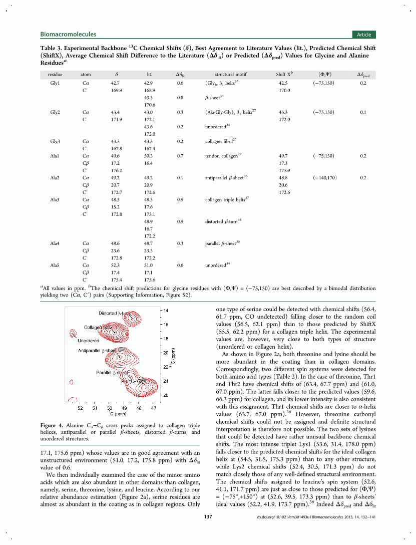
17.1, 175.6 ppm) whose values are in good agreement with anunstructured environment (51.0, 17.2, 175.8 ppm) with Δδlitvalue of 0.6.We then individually examined the case of the minor amino
acids which are also abundant in other domains than collagen,namely, serine, threonine, lysine, and leucine. According to ourrelative abundance estimation (Figure 2a), serine residues arealmost as abundant in the coating as in collagen regions. Only
one type of serine could be detected with chemical shifts (56.4,61.7 ppm, CO undetected) falling closer to the random coilvalues (56.5, 62.1 ppm) than to those predicted by ShiftX(55.5, 62.2 ppm) for a collagen triple helix. The experimentalvalues are, however, very close to both types of structure(unordered or collagen helix).As shown in Figure 2a, both threonine and lysine should be
more abundant in the coating than in collagen domains.Correspondingly, two different spin systems were detected forboth amino acid types (Table 2). In the case of threonine, Thr1and Thr2 have chemical shifts of (63.4, 67.7 ppm) and (61.0,67.0 ppm). The latter falls closer to the predicted values (59.6,66.3 ppm) for collagen, and its lower intensity is also consistentwith this assignment. Thr1 chemical shifts are closer to α-helixvalues (63.7, 67.0 ppm).36 However, threonine carbonylchemical shifts could not be assigned and definite structuralinterpretation is therefore not possible. The two sets of lysinesthat could be detected have rather unusual backbone chemicalshifts. The most intense triplet Lys1 (53.6, 31.4, 178.0 ppm)falls closer to the predicted chemical shifts for the ideal collagenhelix at (54.5, 31.5, 175.3 ppm) than to any other structure,while Lys2 chemical shifts (52.4, 30.5, 171.3 ppm) do notmatch closely those of any well-defined structural environment.The chemical shifts assigned to leucine’s spin system (52.6,41.1, 171.7 ppm) are just as close to those predicted for (Φ,Ψ)= (−75°,+150°) at (52.6, 39.5, 173.3 ppm) than to β-sheets’ideal values (52.2, 41.9, 173.7 ppm).36 Indeed Δδpred and Δδlit
Table 3. Experimental Backbone 13C Chemical Shifts (δ), Best Agreement to Literature Values (lit.), Predicted Chemical Shift(ShiftX), Average Chemical Shift Difference to the Literature (Δδlit) or Predicted (Δδpred) Values for Glycine and AlanineResiduesa
residue atom δ lit. Δδlit structural motif Shift Xb (Φ,Ψ) ΔδpredGly1 Cα 42.7 42.9 0.6 (Gly)n 31 helix
26 42.5 (−75,150) 0.2C′ 169.9 168.9 170.0
43.3 0.8 β-sheet34
170.6Gly2 Cα 43.4 43.0 0.3 (Ala-Gly-Gly)n 31 helix
27 43.3 (−75,150) 0.1C′ 171.9 172.1 172.0
43.6 0.2 unordered34
172.0Gly3 Cα 43.3 43.3 0.2 collagen fibril27
C′ 167.8 167.4Ala1 Cα 49.6 50.3 0.7 tendon collagen27 49.7 (−75,150) 0.2
Cβ 17.2 16.4 17.3C′ 176.2 175.9
Ala2 Cα 49.2 49.2 0.1 antiparallel β-sheet35 48.8 (−140,170) 0.2Cβ 20.7 20.9 20.6C′ 172.7 172.6 172.6
Ala3 Cα 48.3 48.3 0.9 collagen triple helix37
Cβ 15.2 17.6C′ 172.8 173.1
48.9 0.9 distorted β-turn44
16.7172.2
Ala4 Cα 48.6 48.7 0.3 parallel β-sheet35
Cβ 23.6 23.3C′ 172.8 172.2
Ala5 Cα 52.3 51.0 0.6 unordered34
Cβ 17.4 17.1C′ 175.4 175.6
aAll values in ppm. bThe chemical shift predictions for glycine residues with (Φ,Ψ) = (−75,150) are best described by a bimodal distributionyielding two (Cα, C′) pairs (Supporting Information, Figure S2).
Figure 4. Alanine Cα−Cβ cross peaks assigned to collagen triplehelices, antiparallel or parallel β-sheets, distorted β-turns, andunordered structures.
Biomacromolecules Article
dx.doi.org/10.1021/bm301493u | Biomacromolecules 2013, 14, 132−141137

are both 1 ppm. According to our relative abundanceestimation, leucine would be more abundant in the PCWenvironment closely followed by silk-type domains andcollagen.In summary, serine, threonine, and lysine residues, which are
expected to be abundant in the threads’ coating, are ambiguousand indicate that the mefp-1 protein of the coating would thusbe unstructured. Interestingly, this result is further supportedby the fifth alanine peak with chemical shifts of (52.3, 17.4,175.4 ppm), which is also in good agreement with thoseexpected for an unordered protein.36 To further establish thestructure of the cuticle, the latter was studied by infraredmicroscopy.Infrared Microscopy Imaging of Individually Sec-
tioned Byssus Threads. Previous work on infrared character-ization of the byssus was performed on whole byssal threads intransmission mode and therefore could not discriminate thecuticle from the core.17,21 In contrast to the transmission mode,ATR is based on the application of an evanescent wave at thesurface of the sample. The depth of penetration may be variedwith the incidence angle of the source beam and the refractiveindex of the ATR element used. Nevertheless, the penetrationwill never exceed a few micrometers, thus, providinginformation about the surface of a sample rather than thebulk. However, by cutting the byssus along its long axis, thecore and the sheath of the threads can be independentlyimaged.As seen in Figure 5, FTIR microimaging of the byssus core
and sheath shows different amide I band maxima as a functionof the mapping position. Averaged FTIR spectra of the amide Iband of the cuticle and core are indeed shifted by 6 cm−1, from1637 to 1631 cm−1, respectively (Figure 5B,C). In addition, thespectrum of the core also exhibits shoulders at 1695 and 1652cm−1. Peak fitting applied to the amide I band of the coreevidence strong contributions of components located at 1625and 1695 cm−1. These bands are attributable to antiparallel β-sheets secondary structures41 and, thus, strongly support thesolid-state NMR results obtained by analysis of the alaninechemical shifts. A shoulder is also detected around 1260 cm−1
in the amide III region of the core spectrum (SupportingInformation). This band, which is present in the silk fibroinspectrum, is known to arise from antiparallel β-sheetconformation.42 As shown in Table 4, this conformationaccounts for the relative amount of 37%. The next mostabundant secondary structure comes from a component locatedat 1652 cm−1, with a contribution percentage of 30%. This bandcan either be assigned to collagen triple helix, the PG II and PPII helices that are structurally similar to collagen, or to α-helicalstructures. Because it is well-known that byssal threads aremade of aligned PreCol block copolymers, which comprisecentral collagen-like domains, the most plausible assignment forthis abundant type of structure is the collagen triple helix, inexcellent agreement with our NMR results. To unambiguouslyconfirm the presence of collagen in the core, bands around1030 and 1079 cm−1, as well as bands in the amide III regionlocated about 1204, 1236, 1280, and 1338 cm−1 can all beobserved in the spectrum (Supporting Information).43,44
Nevertheless, minor contributions from other types ofstructures cannot be excluded and the remainder of the corewould mostly be made of β-turns and 31-helices, aggregates,and some unstructured zones.As shown in Figure 5B,C, the amide I band maximum of the
byssus cuticle is located around 1637 cm−1. Curve fitting on the
amide I band averaged spectrum in this region indicates a highcontribution of a component located around 1640 cm−1.Unordered or random coil proteins and β-helix (parallel β-
Figure 5. Micrograph of a longitudinally split byssus thread imaged byATR-FTIR microscopy (top), resulting infrared color map showingthe spatial variation of the maximum of amide I band (middle), andinfrared spectra in the amide I range for the core and sheath (bottom).
Table 4. Relative Amount of Secondary Structure, AsDetermined by Peak Fitting Analysis of the Amide I Bandsof the Byssus Core and Sheatha
% contribution
band (cm−1) core sheath
antiparallel β-sheet 1625/1695 37 19β-turn/31-helix 1684/1670 18 31collagen/α-helix 1652 30 9unordered/parallel β-sheet 1639 9 34aggregates 1609 7 8
aThe estimated error on the contribution percentage is 5%.
Biomacromolecules Article
dx.doi.org/10.1021/bm301493u | Biomacromolecules 2013, 14, 132−141138
sheets) are possible molecular structures, which may absorbaround this wave number.41,43 However, according to the NMRanalysis of the amino acid residues abundantly found in thebyssus cuticle such as serine, threonine, and lysine, the threadmefp-1 would most likely be unstructured. Other structurespresent in the sheath spectrum include aggregated strands,antiparallel β-sheets, and collagen triple helix or α-helix. Eventhough a germanium ATR element with a high refractive indexwas employed to limit the penetration depth, the possibilitythat structures occurring in the core could contribute to theoverall cuticle spectrum cannot be excluded. Indeed, aconsiderable load must be applied on the sample to obtain asatisfactory signal, thus, compressing the thread and reducingits thickness. This could explain the minor detection of thedominant structural elements from the core (antiparallel β-sheets and collagen triple helices) in the sheath spectrum. Itcould also explain the presence of collagen bands near 1030,1204, and 1280 cm−1, which appear as poorly resolvedshoulders or with a lower intensity. It is also possible thatdefects in the sheath allow the partial detection of the core. Onthe other hand, because little is known about the definitiveprotein conformation in the byssus sheath, the presence ofsmall amounts of other conformational domains cannot beruled out.
■ DISCUSSIONMussel byssal threads are high performance protein fiberswhich combine extensibility and tensile strength. In this work,we have explored the molecular organization responsible for theoutstanding mechanical properties of “sea silk” by recording, forthe first time, 2D high-resolution 13C SS-NMR spectra on fullylabeled threads. This was enabled by a 13C enrichment protocoldeveloped by our group with the blue mussel Mytilus edulis. Upto 9% labeling was achieved using a mixture of freshwater 13C-labeled Chlorella and saltwater microalgae as part of the normaldiet of M. edulis. Chemical shift analysis was combined tochemical shift predictions to assign the resonances arising fromseveral amino acid residues. This allowed us to confirm, refine,or propose a structural organization in several domains in thebyssus biofibers, as presented in Figure 4.First by analyzing the conformation-dependent chemical
shifts of the dominant peaks, our results confirm that thecollagen triple helix is the prevailing conformation in whichamino acids are found in the material. An excellent agreementwas obtained between experimental and predicted chemicalshifts for the main byssus amino acids (glycine, alanine,proline) and most of the minor ones (arginine, glutamic acid,valine, isoleucine) using a single set of torsion angles of (Φ,Ψ)= (−75°,+150°) corresponding to an ideal collagen triple-helix.34 Moreover, 13C linewidths were below 1.5 ppm.Therefore, our results indicate that this complex biopolymeris surprisingly well ordered at a molecular level. This highdegree of molecular order is consistent with the model of amaterial that possesses several specialized environments(collagen, silk-like, elastin-like, etc.) in order to perform theparadoxical task of both toughness and shock absorption. Incontrast, spider silk comprises only two main environments toperform somewhat similar tasks and is characterized by a lowerdegree of molecular order.35 The presence of collagen triplehelices in the byssus core was also confirmed by ATR-FTIRspectroscopy. It is interesting to remark that defects in thecollagen sequences of M. edulis threads, that is, one glycinedeletion in (Gly-Xaa-Yaa) triads for Pre-Col P and NG, as well
as two glycine deletions and one incomplete triad for PreCol-D),7 which should induce kinks in the structure,11 only induceminor structural perturbations in the otherwise well-orderedcollagen regions.In addition to proving the collagenous nature of the byssus
and allowing a refinement of its structure, the 2D 13C SS-NMRspectrum allowed identifying alanines in five differentconformational environments, thus, improving the structuralmodel of the byssus. Alanine is the second most abundantamino acid in this material. It has a strong 13C chemical shiftvariation according to conformation which is well documentedin the literature. The chemical shift analysis of alanine peaksparticularly allowed us to gain an insight on the structure ofnoncollagenous environments in the byssal threads. Althoughthe cross peak intensity in multidimensional SS-NMR is notrelated in a straightforward manner to the abundance of a givennucleus, the relative intensities of the peaks in combinationwith the obtained structural information can qualitatively beused to assign them to the different environments in the byssalthreads. According to our relative abundance estimation, thesecond most intense alanine cross peaks (Ala2) correspond tosilk-like domains. Their chemical shifts and relaxation proper-ties are strongly indicative of antiparallel β-sheets, the presenceof which was confirmed by FTIR, and located in the core of thethread. We thus conclude that the β-sheets in the silk-likeflanking domains are antiparallel. Interestingly, antiparallel β-sheets are known to provide higher stiffness and strength thantheir parallel counterpart;44 therefore, their presence wouldcontribute to the exceptional mechanical properties of thebyssal threads.The third most intense alanine chemical shift set (Ala3) can
be assigned to collagen helices but is also consistent withdistorted β-turns similar to those observed in silk.45 Because ofthe presence of antiparallel β-sheets in byssal threads, as shownby both high-resolution SS-NMR and ATR-FTIR spectroscopy,we see the occurrence of β-turns as a reasonable hypothesis. β-Turns would link two adjacent β-strands in a crystallite, linking(i-i+3) residues as opposed to (i-i+2) in the case of γ-turns,resulting in a larger spacing between β-strands to accommodateamino acids with bulkier side chains such as arginine orglutamine within the β-sheet.7
The best agreement between literature values and exper-imental chemical shifts for Ala4 cross peaks is obtained forparallel β-sheets. Although the β-sheet nature is unambiguous,the parallel directionality would need additional proof since ourFTIR results only reveal a small contribution of parallel β-sheets (9% in core and 34% in sheath) also assignable tounordered protein. According to the composition profile of thebyssal threads (Figure 3a), 20% of the alanines would belocated in PCW flanking domains. Therefore, Ala4 can mostlikely be attributed to alanines in PCW. Similarly, leucineresidues that are expected to be more abundant in a PCWenvironment also show a reasonably good agreement with a β-sheet structure. The β-sheet nature of the PCW environment isalso supported by recently published FTIR results.21
Finally, investigation of the less intense alanine cross peak(Ala5), as well as serine, threonine, and lysine chemical shifts,gives strong insights into the conformation of the byssuscuticle. Indeed, lysine and threonine are more abundant in thecoating, while serine is equally found in the mefp-1 coating andcollagen domains. While serine chemical shifts are in goodagreement with random coil values, they are also compatiblewith their presence in collagen helices. More interestingly,
Biomacromolecules Article
dx.doi.org/10.1021/bm301493u | Biomacromolecules 2013, 14, 132−141139

threonine and lysine chemical shifts only poorly agree withthose of well characterized secondary structures. The unusualchemical shifts of lysine residues cannot a priori be due to thepresence of hydroxylysine because no lysyl oxidase activity oraldehyde-derived cross-links have been detected in the byssus.7
Although one of the threonine (Cα−Cβ) pairs falls close to α-helix chemical shifts, this assignment is not only incomplete(CO resonances could not be detected or assigned) but is alsothe single case which would reveal the presence of α-helices.We, therefore, consider it to be ambiguous. Overall, theinconsistency in structural assignments for residues in thebyssal thread sheath is likely to indicate an unstructured nature.This would further be supported by Ala5 which chemical shiftsare close to published random coil values.36 The SS-NMRresults are confirmed by the ATR-FTIR microscopy spectra,indicating a dominant contribution of unstructured protein inthe sheath. Dynamic light scattering studies of the mefp-1 inphysiological conditions concluded that the structure resembleda random coil structure consisting of helical and turnedsegments.46
Our SS-NMR study could not confirm the presence of anelastin-like domain in the byssal threads for several reasons.First, this collagen flanking domain is mainly found in theproximal part and would represent less than 10% of alldomains. Moreover, it is rich in Gly residues whose 13Cchemical shifts are not a good protein conformation probe.Additionally, elastin has no defined secondary structure;47
therefore, the chemical shifts of Gly in the elastin domainswould be similar to those in the cuticle. Finally, it is interestingto compare our result with recently published data on cartilage,also obtained by SS-NMR.48 The mechanical properties of thismaterial stem from the association of highly mobileglycosaminoglycans and less mobile collagen. A direct analogywith the case of byssal threads would hint to differences inmobility between the PreCols and, if highly mobile regions arepresent, as might be expected for the elastin-like regions, theirsignal intensity would further be attenuated.A small number of amino acids could not be detected due to
their very low abundance in the material (cysteine, methionine,phenylalanine, histidine, DOPA, and tyrosine) or moderatelylow abundance (glutamine, asparagine, and aspartic acid).49 Inthe latter case, spectral overlap cannot be entirely ruled out. Animprovement in labeling efficiency is necessary to enable thedetection of DOPA, tyrosine, and histidine, which is crucial forthe understanding of byssal fiber formation. A better labelingshould also allow the detection of inter-residue contacts, thus,providing a tool to study the supramolecular organization of thefiber such as interhelical cross-links. Interestingly, even at thismoderate level of labeling, the presence of glycoproteins orpolysaccharides in whole byssal threads could be detected.The overall portrait of whole byssal threads which emerges
from our data is one of a fiber composed of extremelyspecialized well ordered environments. Whereas domains ofnearly ideal collagen triple helices confer toughness to thethreads as is the case in other biological superstructures liketendons, heart valves, and bones, stiffness is provided by silk-like regions, which adopt a structure surprisingly similar to theantiparallel β-sheets found in spider dragline silk. The core ofthe fiber is finally coated by a globally unordered extensible andself-healing cuticle, which confers the required elasticity to thethreads.
■ CONCLUSIONWe have described, for the first time, a multidimensional SS-NMR study of nonspecifically 13C-labeled byssal threads. The2D SS-NMR spectra complemented by ATR-FTIR spectros-copy data allowed us to refine the current structural model ofthese high-performance mussel anchoring fibers. The collagentriple helices were shown to be well-ordered environments withregular (Φ,Ψ) torsion angles of (−75°,+150°). The silk-likeflanking domains found in PreCol-D consist of antiparallel β-sheets with most probably β-turns linking β-strands. The β-sheet structure of the PCW regions in the PreCol-NG wasconfirmed and would be comprised of parallel β-sheets. Ourresults indicate that the coating of the byssal threads is mostlyunordered. Interestingly, the presence of a non-negligible partof glycosylated proteins or polysaccharides could be ascer-tained.In addition to improving the fundamental understanding of
natural protein fibers, these results pave the way for a molecularlevel evaluation of various byssal fiber macroscopic properties.The study of important structural mechanisms such as DOPAcross-linking or histidine complexation will require an increasein isotopic labeling. Improved labeling strategies are currentlybeing developed for this purpose.
■ ASSOCIATED CONTENT
*S Supporting InformationDetails of amino acid relative abundance calculation, completeamino acid specific isotopic enrichment data, examples ofchemical shift prediction results, full 13C chemical shiftassignment of residues in collagen domains, and examples ofFTIR spectra with corresponding curve fitting. This material isavailable free of charge via the Internet at http://pubs.acs.org.
■ AUTHOR INFORMATION
Corresponding Author*E-mail: [email protected].
NotesThe authors declare no competing financial interest.
■ ACKNOWLEDGMENTSF.B. is grateful to the Fonds de Recherche du Quebec - Natureet Technologies (FRQNT) and to the Canadian Institutes ofHealth Research (CIHR) Strategic Training Initiative inChemical Biology for the award of scholarships. M.-O.S.-H.would like to acknowledge the Reseau Aquaculture Quebec(RAQ) and the Universite du Quebec a Montreal for the awardof scholarships. The authors would like to thank A. Siemer(University of Southern California), B. Genard (ISMER), andB. Myrand (Merinov) for insightful discussions. Access to the900 MHz NMR spectrometer was provided by the NationalUltrahigh-Field NMR Facility for Solids (Ottawa, Canada)funded by the Canada Foundation for Innovation (CFI), theOntario Innovation Trust, Recherche Quebec, the NationalResearch Council of Canada, and Bruker BioSpin and ismanaged by the University of Ottawa. The Natural Sciencesand Engineering Research Council of Canada (NSERC) isacknowledged for a major resources support grant. Dr. V.Terskikh is thanked for technical support. This work wassupported by a strategic research grant of the NSERC. I.M. andR.T. are members of the RAQ and the Centre Quebecois surles Materiaux Fonctionnels (CQMF).
Biomacromolecules Article
dx.doi.org/10.1021/bm301493u | Biomacromolecules 2013, 14, 132−141140
■ REFERENCES(1) Carrington, E.; Moeser; Dimond; Mello; Boller. Limnol.Oceanogr. 2009, 54, 978−986.(2) Lachance; Myrand, B.; Tremblay, R.; Koutitonsky; Carrington, E.Aquat. Biol. 2008, 2, 119−129.(3) Moeser; Leba; Carrington. J. Exp. Biol. 2006, 209, 881−890.(4) Simmons; Michal, C.; Jelinsky, L. Science 1996, 271, 84−87.(5) Bell, E. C.; Gosline, J. M. Mar. Ecol.: Prog. Ser. 1997, 159, 197−208.(6) Bell, E. C.; Gosline, J. M. J. Exp. Biol. 1996, 199, 1005−1017.(7) Waite, H. J.; Qin, X.-X.; Coyne, K. J. Matrix Biol. 1998, 17, 93−106.(8) Gosline, J. M.; Lillie, M.; Carrington, E.; Guerette, P.; Ortlepp,C.; Savage, K. Phil. Trans. R. Soc., B 2002, 357, 121−132.(9) Waite, H. J.; Vaccaro, E.; Sun, C.; Lucas, J. M. Philos. Trans. R.Soc., B 2002, 357, 143−153.(10) Lucas, J. M.; Vaccaro, E.; Waite, H. J. J. Exp. Biol. 2002, 205,1807−1817.(11) Hassenkam, T.; Gutsmann, T.; Hansma, P.; Sagert, J.; Waite, H.J. Biomacromolecules 2004, 5, 1351−1355.(12) Harrington, M. J.; Gupta, H. S.; Fratzl, P.; Waite, H. J. J. Struct.Biol. 2009, 167, 47−54.(13) Sun, C.; Lucas, J. M.; Waite, H. J. Biomacromolecules 2002, 3,1240−1248.(14) Sagert, J.; Waite, H. J. J. Exp. Biol. 2009, 212, 2224−2236.(15) Harrington, M. J.; Masic, A.; Holten-Andersen, N.; Waite, H. J.;Fratzl, P. Science 2010, 328, 216−220.(16) Mcdowell, L. M.; Burzio, L. A.; Waite, H. J.; Schaefer, J. J. Biol.Chem. 1999, 274 (29), 20293−20295.(17) Hagenau, A.; Scheidt, H. A.; Serpell, L.; Huster, D.; Scheibel, T.Macromol. Biosci. 2009, 9, 162−168.(18) LeBlanc, A.; Arnold, A. A.; Genard, B.; Nadalini, J.-B.; Seguin-Heine, M.-O.; Marcotte, I.; Tremblay, R.; Sleno, L. Rapid Commun.Mass Spectrom. 2012, 26 (10), 1165−1174.(19) Takegoshi, K.; Nakamura, S.; Terao, T. Chem. Phys. Lett. 2001,344, 631−637.(20) Bennett, A. E.; Rienstra, C. M.; Auger, M.; Lakshmi, K. V.;Griffin, R. G. J. Chem. Phys. 1995, 103, 6951−6958.(21) Hagenau, A.; Papadopoulos, P.; Kremer, F.; Scheibel, T. J. Struct.Biol. 2011, 175 (3), 339−347.(22) Delaglio, F.; Grzesiek, S.; Vuister, G. W.; Zhu, G.; Pfeifer, J.;Bax, A. J. Biomol. NMR 1995, 6, 277−293.(23) Neal, S.; Nip, A. M.; Zhang, H.; Wishart, D. S. J. Biomol. NMR2003, 26, 215−240.(24) Mascolo, J. M.; Waite, H. J. J. Exp. Zool. 1986, 240, 1−7.(25) Saito, H.; Yokoi, M. J. Biochem. 1992, 111, 376−382.(26) Saito, H.; Ando, I.; Ramamoorthy, A. Prog. Nucl. Magn. Reson.Spectrosc. 2010, 57, 181−228.(27) Tycko, R. Q. Rev. Biophys. 2006, 39 (1), 1−55.(28) Kricheldorf, H. R.; Muller, D. Macromolecules 1983, 16, 615−623.(29) Saito, H.; Tabeta, R.; Shoji, A.; Ozaki, T.; Ando, I.; Miyata, T.Biopolymers 1984, 23, 2279−2297.(30) Sarkar, S. K.; Torchia, D. A.; Kopple, K. D.; Vanderhart, D. L. J.Am. Chem. Soc. 1984, 106, 3328−3331.(31) Kanyalkar, M.; Srivastava, S.; Coutinho, E. Biomaterials 2002,23, 389−396.(32) Bella, J.; Liu, J.; Kramer, R.; Brodsky, B.; Berman, H. M. J. Mol.Biol. 2006, 362, 298−311.(33) Seidel, K.; Etzkorn, M.; Schneider, R.; Ader, C.; Baldus, M. SolidState Nucl. Magn. Reson. 2009, 35, 235−242.(34) Gautieri, A.; Vesentini, S.; Redaelli, A.; Buehler, J. Nano Lett.2011, 11, 757−766.(35) Marcotte, I.; Van Beek, J. D.; Meier, B. H. Macromolecules 2007,40, 1995−2001.(36) Zhang, H.; Neal, S.; Wishart, D. S. J. Biomol. NMR 2003, 25,173−195.(37) Holland, G. P.; Creager, M. S.; Jenkins, J. E.; Lewis, R. V.;Yarger, J. L. J. Am. Chem. Soc. 2008, 130, 9871−9877.
(38) Asakura, T.; Okonogi, M.; Nakazawa, Y.; Yamauchi, K. J. Am.Chem. Soc. 2006, 128, 6231−6238.(39) Saito, H. Magn. Reson. Chem. 1986, 24, 835−852.(40) Asakura, T.; Ashida, J.; Yamane, T.; Kameda, T.; Nakazawa, Y.;Ohgo, K.; Komatsu, K. J. Mol. Biol. 2001, 306, 291−305.(41) Jackson, M.; Mantsch, H. H. Crit. Rev. Biochem. Mol. Biol. 1995,30 (2), 95−120.(42) Byette, F.; Bouchard, F.; Pellerin, C.; Paquin, J.; Marcotte, I.;Mateescu, M. Polym. Bull. 2011, 67 (1), 159−175.(43) Khurana, R.; Fink, A. L. Biophys. J. 2000, 78 (2), 994−1000.(44) Xiao, S.; Stacklies, W.; Cetinkaya, M.; Markert, B.; Grater, F.Biophys. J. 2009, 96, 3997−4005.(45) Asakura, T.; Yao, J. Protein Sci. 2002, 11, 2706−2713.(46) Haemers, S.; Van Der Leeden, M. C.; Frens, G. Biomaterials2005, 26, 1231−1236.(47) Debelle, L.; Tamburro, A. M. Int. J. Biochem. Cell Biol. 1999, 31,261−272.(48) Xu, J.; Zhu, P.; Morris, M. D.; Ramamoorthy, A. J. Phys. Chem. B2011, 115, 9948−9954.(49) Qin, X.-X.; Waite, H. J. J. Exp. Biol. 1995, 198, 633−644.
Biomacromolecules Article
dx.doi.org/10.1021/bm301493u | Biomacromolecules 2013, 14, 132−141141