Holdfast heroics: comparing the molecular and mechanical properties of Mytilus californianus byssal...
12
1 Introduction The key to survival on rocky wave- and wind-swept seashores for many mussel species is a beard-like array of tethering attachment threads known collectively as the byssus, which is used to secure the mussel to the hard substratum against the lift and drag forces of waves (Yonge, 1962). In addition, the byssus is emerging as an important paradigm for the bio- inspired engineering of water-resistant adhesives (Lee et al., 2006), functionally graded polymers (van Hest and Tirrell, 2001; Waite et al., 2004), and injection molding of liquid crystals (Hassenkam et al., 2004). The utility of such paradigms relies on the depth to which structure–function relationships are understood. A particularly effective strategy for defining structure–function relationships is to investigate homologous structures in closely related species (Hayashi et al., 1999; Brooks et al., 2005). With this report, byssal thread mechanics and the protein chemistry of three species of Mytilus are compared. Of these, the byssus of the California mussel Mytilus californianus (Conrad), stands out as mechanically superior. The threads that make up the byssus of M. californianus are several centimeters long with a diameter of about 200·m, function outside the body of the organism, and are formed from soluble protein precursors secreted by the foot of the mussel. Byssal threads are subdivided into four morphologically and mechanically distinct regions: the stem, the plaque, and the proximal and the distal portions of the thread (Fig.·1). The stem attaches the thread to the mussel tissue, and the plaque contains the adhesive that connects the thread to the hard substratum. The thread connecting the stem to the plaque is further divided into two mechanically distinct regions. The proximal end (closest to the organism) is extensible up to 200% of its original length, has a low initial stiffness, and a corrugated appearance. The distal portion of the thread, in contrast, is characterized by a high initial stiffness followed by a yield point at about 15% strain and a noticeable stress softening (Bell and Gosline, 1996). For most engineered polymeric materials, yield is not reversible and leads to permanent deformation rendering the material functionally useless. However, in distal threads, damage due to yield is reversible in a time-dependent, self-healing manner with threads recovering 25% of the lost modulus and strain energy in 10·min following a cycle to 35% strain (Carrington and Gosline, 2004). Thread formation begins when the mussel foot touches down on a surface it finds suitable for attachment (Waite, 1992). Once The marine mussel Mytilus californianus (Conrad) inhabits the most wave-exposed regions of the rocky intertidal by dint of its extraordinary tenacity. Tenacity is mediated in large part by the byssus, a fibrous holdfast structure. M. californianus byssal threads, which are mechanically superior to the byssal threads of other mytilids, are composed almost entirely of a consortium of three modular proteins known as the preCols. In this study, the complete primary sequence of preCols from M. californianus was deduced and compared to that of two related species with mechanically inferior byssal threads, M. edulis (Linnaeus) and M. galloprovincialis (Lamarck) in order to explore structure–function relationships. The preCols from M. californianus are more divergent from the other two species than they are from one another. However, the degree of divergence is not uniform among the various domains of the preCols, allowing us to speculate on their mechanical role. For instance, the extra spider silk- like runs of alanine-rich sequence in the flanking domains of M. californianus may increase crystalline order, enhancing strength and stiffness. Histidine-rich domains at the termini, in contrast, are highly conserved between species, suggesting a mechanical role common to all three. Mechanical testing of pH-treated and chemically derivatized distal threads strongly suggests that histidine side chains are ligands in reversible, metal-mediated cross- links in situ. By combining the mechanical and sequence data, yield and self-healing in the distal region of threads have been modeled to emphasize the intricate interplay of enthalpic and entropic effects during tensile load and recovery. Key words: mussel, byssus, self-healing, histidine–metal, collagen. Summary The Journal of Experimental Biology 210, 000-000 Published by The Company of Biologists 2007 doi:10.1242/jeb.009753 Holdfast heroics: comparing the molecular and mechanical properties of Mytilus californianus byssal threads Matthew J. Harrington* and J. Herbert Waite Department of Molecular, Cellular, and Developmental Biology, University of California at Santa Barbara (UCSB), Santa Barbara, CA 93106, USA *Author for correspondence (e-mail: [email protected]) Accepted 2 October 2007 Page nos Page total Colour pages: Facing pages: Issue Ms order
Transcript of Holdfast heroics: comparing the molecular and mechanical properties of Mytilus californianus byssal...
1
IntroductionThe key to survival on rocky wave- and wind-swept
seashores for many mussel species is a beard-like array oftethering attachment threads known collectively as the byssus,which is used to secure the mussel to the hard substratum againstthe lift and drag forces of waves (Yonge, 1962). In addition, thebyssus is emerging as an important paradigm for the bio-inspired engineering of water-resistant adhesives (Lee et al.,2006), functionally graded polymers (van Hest and Tirrell,2001; Waite et al., 2004), and injection molding of liquidcrystals (Hassenkam et al., 2004). The utility of such paradigmsrelies on the depth to which structure–function relationships areunderstood. A particularly effective strategy for definingstructure–function relationships is to investigate homologousstructures in closely related species (Hayashi et al., 1999;Brooks et al., 2005). With this report, byssal thread mechanicsand the protein chemistry of three species of Mytilus arecompared. Of these, the byssus of the California mussel Mytiluscalifornianus (Conrad), stands out as mechanically superior.
The threads that make up the byssus of M. californianus areseveral centimeters long with a diameter of about 200·�m,function outside the body of the organism, and are formed from
soluble protein precursors secreted by the foot of the mussel.Byssal threads are subdivided into four morphologically andmechanically distinct regions: the stem, the plaque, and theproximal and the distal portions of the thread (Fig.·1). The stemattaches the thread to the mussel tissue, and the plaque containsthe adhesive that connects the thread to the hard substratum. Thethread connecting the stem to the plaque is further divided intotwo mechanically distinct regions. The proximal end (closest tothe organism) is extensible up to 200% of its original length,has a low initial stiffness, and a corrugated appearance. Thedistal portion of the thread, in contrast, is characterized by a highinitial stiffness followed by a yield point at about 15% strainand a noticeable stress softening (Bell and Gosline, 1996). Formost engineered polymeric materials, yield is not reversible andleads to permanent deformation rendering the materialfunctionally useless. However, in distal threads, damage due toyield is reversible in a time-dependent, self-healing manner withthreads recovering 25% of the lost modulus and strain energyin 10·min following a cycle to 35% strain (Carrington andGosline, 2004).
Thread formation begins when the mussel foot touches downon a surface it finds suitable for attachment (Waite, 1992). Once
The marine mussel Mytilus californianus (Conrad)inhabits the most wave-exposed regions of the rockyintertidal by dint of its extraordinary tenacity. Tenacity ismediated in large part by the byssus, a fibrous holdfaststructure. M. californianus byssal threads, which aremechanically superior to the byssal threads of othermytilids, are composed almost entirely of a consortium ofthree modular proteins known as the preCols. In this study,the complete primary sequence of preCols from M.californianus was deduced and compared to that of tworelated species with mechanically inferior byssal threads,M. edulis (Linnaeus) and M. galloprovincialis (Lamarck) inorder to explore structure–function relationships.
The preCols from M. californianus are more divergentfrom the other two species than they are from one another.However, the degree of divergence is not uniform amongthe various domains of the preCols, allowing us to speculate
on their mechanical role. For instance, the extra spider silk-like runs of alanine-rich sequence in the flanking domainsof M. californianus may increase crystalline order,enhancing strength and stiffness. Histidine-rich domains atthe termini, in contrast, are highly conserved betweenspecies, suggesting a mechanical role common to all three.Mechanical testing of pH-treated and chemicallyderivatized distal threads strongly suggests that histidineside chains are ligands in reversible, metal-mediated cross-links in situ. By combining the mechanical and sequencedata, yield and self-healing in the distal region of threadshave been modeled to emphasize the intricate interplay ofenthalpic and entropic effects during tensile load andrecovery.
Key words: mussel, byssus, self-healing, histidine–metal, collagen.
Summary
The Journal of Experimental Biology 210, 000-000Published by The Company of Biologists 2007doi:10.1242/jeb.009753
Holdfast heroics: comparing the molecular and mechanical properties of Mytiluscalifornianus byssal threads
Matthew J. Harrington* and J. Herbert WaiteDepartment of Molecular, Cellular, and Developmental Biology, University of California at Santa Barbara (UCSB),
Santa Barbara, CA 93106, USA*Author for correspondence (e-mail: [email protected])
Accepted 2 October 2007
Page nos Page total Colour pages: Facing pages: Issue Ms order

2
committed, soluble thread precursors are secreted into theventral groove of the foot where muscular contractions moldthem into functional threads. The whole process takes a total of2–5·min. The main protein component preCol, also known asbyssal collagen, makes up 96% of distal thread and 66% of theproximal (Waite et al., 2002). There are three preCol variants,each of which is divided into several distinct domains withparticular motifs resembling known structural proteins.
Each preCol has a central collagen domain, which deviatesfrom fibrillar collagens (Types I, II, III, V and XI) in containing1–5 interruptions in the canonically repeated Gly-X-Y collagensequence. These substitutions and deletions are believed tocause bends in the normal rod-like shape of the collagen trimeras exhibited in the C1q heterotrimer (Kilchherr et al., 1985), andseveral other invertebrate collagens (Sicot et al., 1997; Fowleret al., 2000). It has been experimentally demonstrated thatproline or hydroxyproline in the X and Y position produce themost stable triple helix and all other amino acid residues havea tendency to destabilize the helix (Persikov et al., 2005). In thepreCol collagen domain the X and Y residues tend to bedestabilizing residues. Despite this and the breaks in the repeatstructure, the presence of a stable collagen triple helix issuggested in threads by wide angle X-ray scattering of distalthreads (Mercer, 1952; Rudall, 1955) as well as by the resistanceof the byssal collagens to pepsin treatment (Qin and Waite,1995).
At either end of the collagen region are flanking domainsthat differ between the three variants and resemble motifs ofknown load bearing proteins. PreCol D, which is dominant inthe distal end of the thread and decreases in abundance axiallytoward the proximal end (Fig.·1), has flanking domains thatresemble a motif sequence of spider dragline silk. PreCol P,which exists in a gradient complementary to preCol D, hasflanking regions that closely resemble elastin, and preCol NG,which is uniformly present throughout the thread, has Gly-richflanking domains that resemble plant cell wall proteins (Waiteet al., 1998). Since the mechanical properties of dragline silk(as in preCol D) differ substantially from elastin (as in preColP), the graded distribution of preCols with different flankingregions is suspected to play a major role in the gradedmechanical properties along the distal to proximal axis (Waiteet al., 2002).
Beyond the flanks, at the N- and C-termini are regions withabundant histidine and 2–4 residues of dopamine (DOPA),which is a post-translational modification of tyrosine commonto other byssal proteins in the plaque and outer coating of the
thread. The histidines are proposed to be reversible ligands forcoordination bonds with transition metal ions such as zinc(II)and copper(II), as demonstrated with shorter synthetic peptides(Waite et al., 1998; Schmitt et al., 2000). Such reversibly brokencoordination complexes are stronger than non-covalent bonds,but possess only half the strength of a covalent bond (Lee et al.,2006) and are suspected to play an integral role as sacrificialbonds in yield and self-healing in the distal portion of the thread.DiDOPA cross-links have been proposed as covalent bondslinking preCols end to end (Waite et al., 2002).
Many species of mussels produce a byssus, but thecomponent threads of byssi from different species are notmechanically equivalent (Brazee and Carrington, 2006). Thereare five species in the genus Mytilus including M.galloprovinicialis (Lamarck), M. edulis (Linnaeus), and M.californianus (Conrad) (Gosling, 1992). M. californianus isphylogenetically distant from M. galloprovincialis and M.edulis and is readily distinguishable from them (Santaclara etal., 2006). Whereas the mechanical properties of the threads ofM. galloprovinicialis and M. edulis are very similar to oneanother (Lucas et al., 2002), M. californianus has much thickerand mechanically superior threads. M. californianus distalthreads are 2–3 times stiffer and 30% more extensible than distalthreads from the other two species (Bell and Gosline, 1996).Distal threads from M. californianus also self-heal more quicklythan those of M. edulis and M. galloprovincialis after yield(Carrington and Gosline, 2004). These mechanical differencesare probably related to adaptations in M. californianus that helpit survive in the more exposed, wave-swept parts of theintertidal. Interestingly, these differences appear limited to thedistal region since mechanical testing of the proximal region hasnot revealed significant differences in modulus, strength, orextensibility between the three species (Bell and Gosline, 1996).
cDNA sequence has been deduced for all three preCols fromM. galloprovinicialis and M. edulis (Qin et al., 1997; Coyne etal., 1997; Qin and Waite, 1998; Lucas et al., 2002), but not forM. californianus. The purpose of this study was twofold: (i) todetermine the complete primary sequence of collagenouspreCols from M. californianus to enable a direct comparisonwith those from other species, and (ii) to reconcile bothdivergent and conserved features of preCol biochemistry withthe comparative mechanical performance of the distal portionof the byssal thread. The results suggest that the single mostinfluential factor in the tensile superiority of M. californianus isthe greater abundance of silk-like polyalanine tracts and that thehighly conserved histidine-rich domains play an integral role assacrificial bonds in threads of all three species.
Materials and methodsRACE ready cDNA construction
Whole mussel feet were dissected from Mytilus californianus(Conrad) collected from Goleta Pier (Goleta, CA, USA) andstored in flowing seawater (SW) at 12–15°C. Foot tissue wasimmediately homogenized under liquid nitrogen, and total RNAwas extracted with the RNeasy Plant Mini Kit from Qiagenusing the manufacturer’s protocol (Qiagen, Valencia, CA,USA). Freshly purified RNA was reverse transcribed to 3� and5� RACE ready cDNA using the SMART RACE cDNA Kit(Clontech, Mountain View, CA, USA).
M. J. Harrington and J. H. Waite
P D
NG
DistalProximal PlaqueStem
Fig.·1. Schematic of an isolated byssal thread showing importantstructural features. Morphological and mechanical differences gradedfrom distal to proximal correspond with a gradient in the relativecomposition of preCol variants D, NG and P.

3Holdfast fibers
PCR amplification and cloning of the preColsPrimers designed for the preCols from Mytilus edulis (Lucas
et al., 2002) and degenerate primers based on highly conservedsequence in M. edulis and M. galloprovincialis were used toscreen the M. californianus cDNA, assuming some sequencesimilarity. Preliminary sequence obtained with these primerswas used to create gene-specific primers used in PCR, 5� RACEand 3� RACE to obtain the remaining sequence. Regions ofsequence where degenerate primers or primers for M. edulislanded were confirmed by overlap with sequence-specificprimers. The universal primer mix supplied with the RACE kit(Clontech) was used for 5� and 3� RACE. The relevant reactionsare described in Table·1.
PCR and RACE products were cloned into Pgem-T Easyplasmid (Promega, Madison, WI, USA) and transformed intoOne Shot chemically competent E. coli cells (Invitrogen,Carlsbad, CA, USA). Miniprepped plasmids were contractuallysequenced (UC Davis DNA Sequencing Facility) andoverlapping sequences were assembled and translated to givethe deduced protein sequences. M. californianus preCol cDNAsequences were entered into GenBank under accession numbersEU120661, EU120662 and EU120663 for preCol D, NG and P,respectively. The deduced protein sequences from the cDNAwere compared to preCol sequence from M. galloprovincialisand M. edulis in the database using ClustalW on the EBI server(Chenna et al., 2003) to determine percent identity betweenspecific preCol variants of the three species. Alignments wereperformed with the identity matrix using default settings for allthe other parameters.
Mechanical testingDistal portions of fresh tank-grown threads were dissected
from M. californianus and allowed to rest in seawater for at least
48·h prior to testing. Thread ends were secured between piecesof cardstock with cyanoacrylate glue. The thread cross-sectionwas assumed to be slightly elliptical, and the area was calculatedusing the average of the long and short side as the diameter.Cardstock loaded threads were placed in the grips of a Bionix200 tensile tester (MTS, Eden Prairie, MN, USA) and subjectedto mechanical testing. Threads were hydrated by submersion inseawater or buffer immediately prior to tensile testing, whichwas performed within an MTS environmental chamber with therelative humidity raised to 99.9±5% in order to maintain threadhydration. Testing at high humidity was necessary to reduceslippage of the thread from the cardstock at higher strains, whichwas seen in test runs with submerged threads. Threads werecycled consecutively to 10, 20, 30, 40, 50, 60% strain, andfinally to break, at an extension rate of 5·mm·min–1 with no restbetween, recording the extension and load on MTS Testworks4 software. Extension was converted to engineering strain usingthe equation �=(l–lo)/lo where l is the extension of the threadduring tensile loading and lo is initial length of the unloadedthread with zero load. Load data were converted to engineeringstress using the equation �=F/A, where F is the tensile forceapplied to the thread and A is the cross-sectional area of thethread. Young’s modulus (E, stiffness) for each strain intervalwas determined as the maximum slope of the stress–strain curveduring loading after the characteristic toe region. To assess thestress softening incurred with each successive yield, themodulus of each cycle is presented as a percentage loss of theinitial stiffness (% loss=[1–E/Eo]�100%) where E is thestiffness of a particular cycle and Eo is the stiffness measuredduring the first cycle of a previously unstrained thread.
Histidine–metal coordination cross-links are suspected toplay an important role in yield and hysteresis in the distalthreads. Such bonds, however, are known to be pH sensitive
Table 1. Reactions used to determine regions of sequence where degenerate primers or primers for M. edulis landed, confirmed byoverlap with sequence-specific primers
PreCol D 5�RACE/DR-1, DF-2/DR-2, DF-3/DR-3McDR-1 5� TCCGCCACCTACAACTGGACC 3�McDF-2 5� GCAGCAGCAAACGCAGCAGCAGGAGGAT 3�McDR-2 5� CTCCGGCTGTTCTGAGGTCTTCAAT 3�McDF-3* 5� GTGGTATGGGTAGACGAG 3�McDR-3* 5� AACACTTGCAGATTTTATTGATA 3�
PreCol NG 5�RACE/NGR-1, NGF-2/NGR-2dg, NGF-3/NGR-3, NGf-4/NGR-4McNGR-1 5� AAGTGGGAATCCGCCGCCACCAGTTGC 3�McNGF-2 5� GCGGCTATCGCCCGCACAGGACTA 3�McNGR-2dg 5� YTGWCCWGGDGCWCCWCCDGCATTWCCWGGTTGG 3�McNGF-3 5� GCAACTGGTGGCGGCGGATTCCCACTT 3�McNGR-3 5� CCACCAGCCTCTCCTGCTGGTCCTTCT 3�McNGF-4* 5� AAGGAGAACTTGGACCAGTCG 3�McNGR-4* 5� AGGAACTTGCACTTTTTAT 3�
PreCol P 5�RACE/PR-1, PF-2/PR-2, PF-3/3�RACEMcPR-1 5� GGATCAGGCAGTGGCAGAGCATTTGGTGG 3�McPR-2* 5� GAATAACACCTGGTGCTCCT 3�McPF-2* 5� GAGGATTCGGTGGACCAGGTAC 3�McPF-3 5� GGGACCAGGAGGTGAAAGAGGAGGCCAAGG 3�
*Primers from M. edulis. McDR-3 and McNGR-4 are based on the 3� UTR.dgDegenerate primer.
4
since they depend on the deprotonation of a histidinenitrogen, with a pKa ~6.5 (Fig.·2). In order to investigatethe effect of pH on mechanical performance and, byextension, on histidine–metal cross-links, threads weretreated in citrate-phosphate buffer at pH·3, 4, 5, 6, 7 and8 at 4°C for at least 24·h prior to testing. A subset ofthreads was equilibrated in citrate-phosphate buffer atpH·5.5 for 24·h and treated with diethylpyrocarbonate(DEPC) by adding an ~tenfold molar excess(65·�mol·l–1) to the buffered solution (molar excess isbased upon estimated concentration of His in a thread).In moderate excess, DEPC reacts with histidine residuesby carbethoxylating one of the nitrogen atoms in theimidazole ring, rendering it unable to participate in metalcoordination complexes (Fig.·2). The reaction wasperformed at pH·5.5 since DEPC targeting of histidineresidues vis-à-vis other amino acids is highly specificthere (Lundblad, 2005). Although metal-bound histidineis protected from attack by DEPC, at pH·5.5 there will besome unbound, exposed histidine residues. DEPC-treatedthreads and a control group of untreated threads at pH·5.5were then re-equilibrated 24·h in pH·8 buffer prior tomechanical testing. One-way analysis of variance(ANOVA) was performed to compare differences inYoung’s modulus between treated threads (pH andDEPC) and untreated threads (SW ctrl) during their firststrain cycle. A two-way ANOVA was performed toassess the effects of pH and strain on the Young’smodulus (E) of threads during multiple cycles ofincreasing strain.
ResultsSequence of M. californianus preCols
The complete cDNA sequence generated from PCR, 5�RACE, and 3� RACE was acquired for each of the threepreCol variants, each consisting of 3–4 overlappingclones. Figs·3–5 show the full deduced protein sequencesfor preCol D, NG and P from M. californianus,respectively, with relevant domains labeled for each. Weare confident that the assemblies represent completecDNAs of expressed proteins. Table·2 shows thesequence identity to the preCols from M.galloprovinicialis and M. edulis. The collagen region,signal sequence and acidic patch from each preCol are highlyconserved (83–100%) and are part of different overlappingconstructs, giving us confidence in our overall constructcomposition.
In M. californianus, the preCol variants are all slightly largerproteins than their counterparts in M. galloprovinicialis and M.edulis. Over the length of the whole protein, each particularvariant from M. edulis is around 90% identical to the samevariant in M. galloprovinicialis, whereas M. californianusvariants are only 80% identical to their counterparts (Table·2).It should be noted that the alignment scores indicated in Table·2represent the number of identities in the best match asdetermined by ClustalW divided by the number of residuesaligned, and therefore do not account for inserts. Domainswhere the score does not accurately reflect the degree ofdivergence between species due to inserted sequence include the
flanking and His-rich domains. This will be discussed later. Allthree collagen domains conserve the tendency for destabilizingresidues in the X and Y positions of the canonical collagenrepeat.
PreCol DTable·2 reveals that the collagen domain of preCol D is more
conserved than the silk or His-rich regions. However, there isone significant difference in the collagen that will be potentiallyimportant to the function and assembly of the protein. Asmentioned, the preCol collagen regions are distinct from typicalfibrillar collagens in containing one to several deviations fromthe Gly-X-Y repeat motif, which typically lead to kinks in thecharacteristically rod-like collagen morphology (Kilchherr etal., 1985). In preCol D of M. californianus there are threeaberrations in the collagen domain instead of the five seen in
M. J. Harrington and J. H. Waite
–H+
+H+
Carbethoxyhistidine
DEPC
M2+
H2N CH C
CH2
OH
O
NH
NH
H2N CH C
CH2
OH
O
N
NH
H2N CH C
CH2
OH
O
N
N
H2N CH C
CH2
OH
O
N
NH C
O
EtOH
�
�
�
DEPC
M2+
M2+M2+
pKa=6.5
I II
III IV
Fig.·2. Schematics of histidine chemistry concerning protonation, metalcoordination and reaction with diethyl pyrocarbonate (DEPC). (I,II) Thetitration of a proton on the imidazole ring of His, which has a pKa ~6.5. Thenitrogen shown losing the proton in the figure is the more likely of the twosince it has slightly lower pKa; however, depending on the local environment,either can be deprotonated. As shown in (III), it is possible for thedeprotonated nitrogen to contribute a lone pair of electrons as a ligand in ametal coordination bond with a divalent transition metal ion such as Zn2+ orCu2+. When both nitrogens are protonated (I), the ring is unable to participateas a ligand in metal bonding. Reacting unbound His residues with DEPC (IV)results in a carbethoxylation of the imidazole ring, rendering it unable toparticipate in coordination metal chemistry. Metal bound His will not reactwith DEPC. DEPC treatment is specific for His at pH·5.5 and at this pH, therewill be few side chains bound to metal. EtOH, ethanol.

5Holdfast fibers
Signal
His-rich
Silk
Collagen
Acidic
MVFKILTVCLVASLLETCLA DYGRKYGKPSYGEYGGKRGGGRVSGAVAHAHAHAHASSGADGRSRAHARAVAHSHSGGGAAHGHPGFP AGSASAAARAAARAGAVGGFGGGGFGSASAAAAARAAAAGGGLGGGFGSASAAARAAAAAGGFGGGGFGSASANAAANAAAGGFGGFGGGFGPGF GGP GQP GGP GGP GGP GGP GGP GMP GGP GGP GGP GMP GGP GGP GGP GTG GPG *QP GGL GGP GGP GMP GGP GGP GGP GMP GGP GGP GGP GGP GGP VVGG GIP GIT GPA GPP GPA GPQ GPQ GEQ GPR GRT *PA GTP GPP GNP GEP GQA GAP GAP GAP GHG GKP GTA GAT GKA GRP GPR GQP GAS GSS GQH GAS GAP GRP GNP GST GRP GAT GDP GRP GAT GTA GRP GPS GAP GNP GAP GAI GAP GPR GAP GFV GLP GPR GSP GEP GNQ GPI GGP GYP GPT GPQ GPD GAM GPQ GPC GNR GAP GVP GKQ GPV GGQ GPA GPR GPR GDE GPV GPK GEP GAK GAD GKP GDK GGD GDT GPQ GPA GPK GEV GDQ GAP GPK GET GDQ GAR GEA GKA GEQ GPG GIQ GPK GPA GGQ GPA GPA GPL GPQ GPM GER GAQ GPT GPE GPV GAP GPK GSV GDQ GAQ GDQ GAT GAD GKQ GEP GER GQQ GAA GPV GRP GPR GDR GAK GIQ GSR GRP GAM GRR GNR GSQ GAV GPR GET GPD GNQ GQR GEQ GAP GVI TLVIEDLRTAGVESPVEAFDA GAGPGGAGPFGGAGPFGGAGAGAGAGGAGPFGAGPFGAGPFGGVGGAGVGGAGPGGAAGPGGAAGPGGAAGAGGLGGLGAGGLGGLGAGGLGGLGAGGLGGQGAGGLGGLGAGGLGGLGAGLGGGLGGGAGAAAAAQAAAAANGGLGGGSAAAAARAAAAANAGLGGGAVAAAQAAASAAANSGLGAGAARAAASAAARATVAGAGRGTAAAAASAAAQAHAATKAQGGSHAHAAAAAQAAA SSVIHGGGHGGHGGDYHKPGY
Silk
His-rich
Fig.·3. Deduced protein sequence ofpreCol D from M. californianus.Histidine residues are in bold type.Breaks in the Gly-X-Y canonical repeatare underlined in the collagen domain anddeletions are denoted with an asterisk.
MVHNFLTVFLVAAIARTGLA GSIGYGKPGYGSGGDFFNGHGGHGGHGGGHGHGGGGG GSASAAAQAAAAARALGGGGGGSASAAARAAAAARALGGGGGGSASAAAQAAAAARALGGVGGGSASAAARAAAAASALGGGGGGFGGLGGLGGGPGGIGGIGGGPGGLGGGPGGLGGGPGGFGIGPGGIGVGPGGNGGGPGGLFGPGGALGPGGGAGAFGTGGAVGGPGGAGASASAGAFATGGGGFPLP GAQ GPQ GPR GPA GAP GDQ GES GPP GPP GNS *PQ GPQ GSR GAP GQP GEQ GAN GNP GQP GNA GAP GQP GAP GQA GAP GAR GPS GAA GHQ GAQ GGL GQP GSP GQQ GSA GQP GSP GNP GAP GAP GST GQA GSV GNV GGP GEQ GPQ GNA GPR GIQ GRP GCK GLP GPK GPD GAQ GAP GKP GAD GPA GNR GPM GPA GGK GPT GDK GAP GDV GPE GPE GKP GGP GPK GPN GPQ GAK GSP GED GDP GAE GEP GSK GAD GLP GHG GPR GNP GPQ GPE GEV GDK GAP GEA GGP GQP GPY GPQ GPA GEQ GDL GEI GPA GDA GAP GVP GSK GIQ GPQ GEL GPV GKE GPA GEA GGK GRL GQK GPA GEP GQP GEE GKQ GDM GAT GTR GAT GVG GVK GPT GFR GAR GKS GNA GRP GRP GRN GPR GPQ GPQ GLR GNQ GPD GEQ GGP GVG GIS GTI TIIVDDDGARYGDFADITGPNSDEVNRQLVREFIGDLDTFLSLNGPGGPAGV GAGDIGIGGGLGPGGAGGIGPGGAGGPGGAGGVAGGPGGVGGFGVGPGGAAGLGGVGGLGAGLGGVGGLGAGLGGVGGLGGGVGGLGAGLGGVGGVGGLGAGGALGGLGGLGFGGLGGGAGASAGAAAGAHAVSGGAGGGASA HAHAHAHAASVSGGGGGVSHAVSHAVSHAVSHSVSHSGGGGHAHAAASAHAHAVSHGGSSGGHPLHYNDPFYGKKHKADY
Signal
His-rich
Gly-rich
Collagen
Acidic
Gly-rich
His-rich
MVRFSLASVLLLAVTSTAFA GPLGDYGGNGIKIVPYHGGGGGGGGGGGGGHGGSWSHGPYHRHGLGGGSGSGGSSAHAHSSASAHVHHFGSGSSHASAGSSANAHTFGGGPSHASAGSSSHASASHSGLGGGSAHAHSSSSAHALS GGFGGFGGIGPGGIGGIGGGGLGGGPGGIGGIGGPGGIGGGGIGPGGIIFGPGGPGSASGSGSGRAFGGNGGSSASANAAARASASGGGGF GGP GSP GNA GAP GQP GIP GAP GVP GRP GIT *QP GRP GNA GPP GQP GNP GRP GAG GRP GAP GRP GGP GRP GIP GKP GNL GQL GQP GSP GQP GHP GAS GQP GRN GSP GNP GKP GTP GHS GTP GSR GSP GTP GTP GQP GVP GTT GGR GPR GPA GII GLI GPK GNP GVA GNP GAP GIP GEG GPR GPQ GPA GGP GSS GPS GDK GSP GTP GGN GPR GAI GPQ GPS GPP GDG GPQ GGR GTP GTP GKP GAK GPQ GSN GDV GPQ GAS GPK GPQ GPQ GKA GVK GPA GDQ GAR GAE GTA GPP GPQ GEQ GLK GPS GGQ GPA GPA GPS GEQ GPG GER GGQ GPQ GPE GPT GPA GPR GAS GSQ GPA GER GAP GAP GKK GPN GDR GNQ GLP GAP GKN GAR GDR GAR GSN GSP GRT GAP GSR GKI GPQ GPH GPR GAR GSQ GPK GQR GDQ GAP GVI RIVIDDQRTGPEVEEF PGFGGFPGFPGSAGAGSSSGASAFGGPGGAGGFPFGPFGGAGGPGGGPGGPGSPGGPGGPGGPGGPGGPGGPGGPGGPGGLVGGGAGGPGGLGLGGAGGPGGLGGGAGGPGGFGGGVGPGGLGGFGGLGGGGLGGGASAGASSSGSASASGGGPFGVLNVGPGGGGSSSASAASRA HALALGGLGGGSASAGSHSSSSAHSFGGHLFHSVTHHGGPSHASAHATAHAHASASGGGGHGGHGGHGGHGGPYKPGY
Signal
His-rich
Elastin
Collagen
Acidic
Elastin
His-rich
Fig.·5. Deduced protein sequence ofpreCol P from M. californianus. Histidineresidues are in bold type. Breaks in theGly-X-Y canonical repeat are underlinedin the collagen domain and deletions aredenoted with an asterisk.
Fig.·4. Deduced protein sequence ofpreCol NG from M. californianus.Histidine residues are in bold type.Breaks in the Gly-X-Y canonical repeatare underlined in the collagen domain anddeletions are denoted with an asterisk.

6
both M. galloprovinicialis and M. edulis (Fig.·3). Two of theaberrations are typical deletions of a single Gly also seen in M.galloprovinicialis and M. edulis, but they surround a novelaberration not seen in the other two species in which thesequence Val-Val-Gly-Gly is inserted between Gly-X-Yrepeats. This is the first instance of a four amino acid insert ina preCol collagen domain, but it is uncertain how the triple helixwould be perturbed by such an insert.
The alignment scores of both the N- and C-terminal silkdomains are low compared to the collagen domain (67% and69% identity, respectively, vs 93% when compared with M.edulis), but still do not fully represent the degree of deviationsince both are significantly longer in M. californianus due toinserts. The inserts come in the form of two alanine-rich regionsof 24 and 10 amino acid residues respectively in the N terminus(Fig.·6A) and three inserts of different sizes in the C terminus.If the number of identical residues in the N-terminal silk domainof M. edulis and M. californianus is divided by the number ofresidues in the larger of the two (M. californianus=95, M.edulis=55, M. galloprovincialis=55) instead of the number ofaligned residues, the percent identity drops from 70% to about40%, indicating the large amount of deviation in the flankingdomains of M. californianus not accounted for by the ClustalWalignment score.
The His-rich domains have a relatively low alignment score,but Fig.·6B reveals that while the sequence outside of thesuspected metal binding motif is very divergent, the histidinecontaining sequence is highly conserved. Every single histidinein the N-terminal His-rich domain is conserved, along with Glyand Ala spacer residues. The C-terminal His-rich region is lessconserved, but the number of His residues is mostly conservedwith M. galloprovincialis having one extra histidine.
PreCol NGConservation of preCol NG domains in M. californianus vis-
à-vis preCol NG from the other two species parallels the trends
seen with preCol D. The collagen region shows the highestidentity score outside of the acidic patch and the signal sequence(Table·2). The only aberration is a single deletion of a glycinein the eleventh repeat (Fig.·4), identical to that of M. edulis. M.galloprovincialis has two additional deviations in the form ofsubstitutions of Gly with an Asp and an Arg, respectively.
The suspected His-rich metal binding motifs are lessconserved in NG than in D, but are still relatively similar. TheN-terminal His region of all three species has five histidineresidues; however, in M. californianus the histidines are moreclosely packed. The C-terminal His-rich region is moreconserved than the N-terminal, with 14 out of 17 histidineresidues identical between all three species.
M. J. Harrington and J. H. Waite
Table·2. Percent identity of the preCol variants for M. californianus, M. edulis and M. galloprovincialis by domain
Identity (%)
PreColvariant Overall Signal N-His N-flank Collagen Acidic C-flank C-His
DMc:Me 82 85 67 70 93 95 69 66Mc:Mg 79 85 65 52 87 95 72 100Me:Mg 89 100 92 83 93 100 89 76
NGMc:Me 83 100 80 79 89 90 71 70Mc:Mg 81 95 83 85 89 90 68 62Me:Mg 89 95 89 87 97 100 78 79
PMc:Me 79 100 82 75 83 93 62 88Mc:Mg 80 100 82 75 85 86 63 83Me:Mg 91 100 84 91 94 93 91 93
Mc, M. californianus; Me, M. edulis; Mg, M. galloprovincialis.Identity scores were determined by ClustalW and represent the number of identical residues in the best pairwise match, as determined by the
program divided by the number of residues that were matched. This score does not take into account regions that do not overlap due to insertedsequence.
Fig.·6. ClustalW alignment of (A) the N-terminal silk domain and (B)histidine-rich domain of preCol D from M. californianus (Mc), M.edulis (Me) and M. galloprovincialis (Mg). Silk alignment reveals thatM. californianus has two major inserts consisting of polyalanine runsin the N terminus. His-rich domain alignment reveals that while overallthe identity between M. californianus and the other species is low, theregions containing histidine are more conserved. Residues in bold typeare conserved between all three species.
Mc -AGSASAAARAAARAGAVGGFGGGGFGSASAAAAARAAAAGGGLGGGFGSA Me VGGSASAAARAAARASAGG------------------------LGGFG--- Mg IGGSASAAARAAARASAGG------------------------LGGLGGFV Mc SAAARAAAAAG-GFGGGGFGSASANAAANAAAGGFGGFGGGFGPGF Me SAAANAAAAARAGAGFGGFGGL----------GGFGGLGGV----- Mg SAAANAAAAANAGAGFGGFG-------------GFGGLGGV----- Mc DY-GRKYGKPSYGEYGGKR--GGGRVSGAVAHAHAHAHASSGADGRSRAHA Me DYNGNKQYGGRYGNRYGNGLGGGNGGAGAVAHAHAHAHASAGANGRARAHA Mg DYHGNKQYGGRYGNRFG----GGIGGAGAVAHAHAHAHASAGANGRARAHA Mc RAVAHSHSGGGAAHGHPGFP Me RALAHAHAGGGAAHGHPGFP Mg RALAHGHAGGSAAHGHPGFP
A
B

7Holdfast fibers
As with preCol D, the major differences are in the flankingdomains (Table·2), especially the Gly-rich region within the C-terminal flanking domains, which contains numerous insertedregions rich in Gly, Val and Leu. Gly residues are mostlyconserved between species, and the non-Gly residues exhibitvariation but are always hydrophobic residues such as Ala, Leuand Val. The introduction of considerably more Pro in theflanking domains of NG in M. californianus than in the othertwo species is notable since it could lead to a more amorphoussecondary structure.
PreCol POf the three variants, preCol P has the least conserved
collagen domain at 83% identity with M. edulis (Table·2).However, the single Gly deletion in the eleventh Gly-X-Yrepeat (Fig.·5) is conserved in all three species. The N-terminalHis-rich domain of P is also the most altered of the three variantsin terms of the position and number of His residues, but is stillmostly conserved.
The flanking domains of preCol-P are again the leastconserved regions with the C-terminal end having a 20% loweridentity than the N-terminal. They are not as insertionallymodified as the flanking domains of D and NG, but are moresubstituted. The C-terminal region is especially different andreminiscent of the flanking domains of preCol-NG in certainregions with runs rich in Gly and Leu and many PGG repeats.A BLAST of the C-terminal flank sequence against the databaseshows it is over 50% similar to flagelliform spider silk whereasit is only about 60% similar to the C-terminal flanks of M. edulisand M. galloprovincialis.
Mechanical testingCyclic quasistatic mechanical testing (Carrington and
Gosline, 2004) showed that the larger the strain that distalthreads are pulled to, the larger the resulting hysteresis. Thisstudy also revealed that the stiffness disparity (stresssoftening) between the first and second cycles increased withincreasing strain of the first cycle. Both observations implythat damage is occurring at the molecular level during yieldand is intimately correlated with hysteresis and reduction ofmodulus. The mechanical tests performed in this study weredesigned to probe these observations more thoroughly and toinvestigate more specifically the role of histidine–metalcoordination complexes as important sacrificial cross-links intensile stress.
As Fig.·7A indicates, a distal thread incubated in seawatercycled to any strain within the yield region will have a secondyield point if it is taken to a strain slightly beyond the maximumstrain it achieved in the previous cycle. After each successiveyield, threads show a reduction in modulus from that of theprevious cycle. If the softening is presented as a percentage lossof the initial stiffness (% loss=[1–E/Eo]�100%) and plottedagainst the strain value that caused the reduction in stiffness(Fig.·7B), a few things become apparent. First, there is littlesoftening observed during the 10% strain cycle, which is beforethe yield point typically occurs. The largest reduction in thestiffness occurs during the 20% strain cycle with the initialmodulus reduced by almost 40% from the first cycle. Smallerreductions are seen after the 30% and 40% strain cycles, after
which there does not appear to be further significant loss inmodulus.
Threads incubated in pH·8 buffer are not statistically differentfrom seawater threads (P<0.01). Treating threads with buffersat acidic pH, e.g. at pH·5 and below, reduces the modulus toalmost half the value measured in seawater and pH·8. In Fig.·8,the initial modulus (mean ± s.e.m.) for each treatment is plottedagainst pH, revealing a roughly sigmoidal curve with a midpointat about 615·MN·m–2 and a pH of 6.6, as predicted in apreliminary report (Waite et al., 2006). The stiffness vs pH curvefollows the same trend as the histidine titration curve that isplotted alongside it in light gray in Fig.·8.
The initial stiffness of threads incubated at pH·5.5 for 24·hand then re-equilibrated at pH·8 for 24·h are statisticallyindistinguishable from citrate-phosphate pH·8 and seawaterthreads (P<0.02), which demonstrates that the pH-inducedreduction of stiffness is reversible (Fig.·8). Threads incubated atpH·5.5 in the presence of DEPC, and reequilibrated in citrate-phosphate buffer at pH·8 do not show this same reversibility andhave a significantly different initial modulus from the controlthreads that went from pH·5.5 to pH·8 without DEPC treatment(P<0.02). As Fig.·8 shows, the initial modulus of DEPC-treated
Fig.·7. (A) Representative stress–strain curve of a single distal threadcycled to incrementally increasing strain values from 10% to 70%.Yield, as seen in cycles to 20, 30 and 40%, is followed by a loss instiffness (stress softening) in the subsequent cycle. (B) Percentage lossin modulus plotted as a function of the strain that caused the loss.Values are means ± s.e.m.; N=??. Most stress softening is occurringbetween 10% and 40% strain.
0 0.1 0.2 0.3 0.4 0.5 0.6
0 0.1 0.2 0.3 0.4 0.5 0.6
0.70
20
40
60
80
100
Str
ess
(MP
a)A
0
10
20
30
40
50
60
% lo
ss in
mod
ulus
Strain (mm mm–1)
B

8
threads falls around 680·MN·m–2, which is slightly below themean value for threads treated in buffer at pH·7.
Plotting the modulus of each strain cycle against thepreceding strain value for each pH treatment (Fig.·9A) revealsthat the pH-dependent stiffness variation seen in Fig.·7 becomesless prominent with increasing strain (two-way ANOVA,P<0.001). However, the stiffness values of pH·8 and pH·5treated threads are still significantly different at all strains tested(P<0.01). Fig.·9B shows the magnitude of the modulusreduction (�E) caused by a particular strain cycle plotted againstthe strain that caused it, revealing that for all pH treatments themajority of damage occurs between 20–30% strain (consistentwith Fig.·7B). Statistical analysis of �E values at pH·5 and pH·8reveals that softening due to cycles of 20, 30 and 40% strain aresignificantly different (P<0.01) whereas values at 10, 50 and60% are not. It should be noted that treatment at pH·5 and belowdoes not eliminate stress softening entirely, but it does greatlyreduce the magnitude.
DiscussionComparing the primary sequence of the preCols from the
mechanically superior byssal threads of M. californianusto those of M. galloprovinicialis and M. edulis provides aunique opportunity to examine the structure–functionrelationship between preCol biochemistry and the mechanicalproperties of the byssal thread. Byssal collagen is a modularprotein with clearly defined domains resembling commonlyfound load-bearing protein motifs. A comparative approachreveals that the degree of sequence deviation is not uniform
throughout the whole protein, but instead varies from domainto domain.
The collagen domain, signal peptide and acidic patch are themost highly conserved regions between all three species withsequence identity between 83–100%. The His-rich domainshave relatively low identity scores (77.3±11.3%), but closerexamination of the whole domain reveals that the putative metalbinding motifs are very well preserved. For all three variants thelargest sequence deviations are localized in the flankingdomains (mean identity score=70.0±8.5%), which are highlysubstituted in M. californianus. In every case except for the N-terminal flank in preCol P, M. californianus flanking domainsare also significantly longer due to multiple insert regions.
Flanking domainsAs stated earlier, the gradient of the variants is believed to be
responsible for the graded stiffness from distal to proximal, andthe flanking domains are believed to play a major role in thesemechanical differences because of their similarity to known
M. J. Harrington and J. H. Waite
2 4 6 8
400
500
600
700
800
–0.2
0
0.2
0.4
0.6
0.8
1.0pH treatment
Youn
g’s
mod
ulus
(M
N m
–2)
pH
A/(A+AH)
A/[A
+A
H]
ControlDEPC
Fig.·8. Initial modulus of distal byssal threads plotted against the pHat which they were incubated. Red dotted line and circle denote themidway point of the Young’s modulus and the corresponding pH of6.6. The titration of a nitrogen proton on the histidine imidazole ring(pKa=6.6) is plotted in light gray (A/A=AH) to illustrate that themodulus curve closely follows the characteristic sigmoidal shape,suggesting that histidine protonation may influence thread mechanics.The blue inverted triangle represents the initial stiffness of threadstreated at pH·5.5 and brought back to pH·8 and tested, indicating thatpH-induced softening is reversible. The pink triangle represents theinitial stiffness of threads treated in DEPC at pH·5.5, brought back topH·8, and then tested. DEPC treatment further supports the role of Hisin the mechanical properties of threads. Values are means ± s.e.m.; Nranged from 6 to 9 threads for each treatment.
100
200
300
400
500
600
700
800
0 0.2 0.4 0.6
pH 8
Youn
g’s
mod
ulus
(M
N m
–2)
A
pH 7pH 6pH 5pH 4pH 3
SW
0.1 0.2 0.3 0.4 0.5 0.6
0
50
100
150
200
250
ΔE (
MN
m–2
)
Strain (mm mm–1)***
B
Fig.·9. (A) Young’s modulus plotted against the previous strain cyclevalue for each pH treatment. Threads treated at pH·5 and below showa pH-induced reduction in modulus that becomes less prominent as thethread is cycled to higher strain values. (B) Reduction in modulus (�E)between strain cycles in Fig.·9A plotted against the strain that causedit for each of the pH treatments. Asterisks indicate where �E at pH·5is significantly different from �E at pH·8 (P<0.01). Values are means± s.e.m.; N=??.

9Holdfast fibers
structural proteins of varying stiffness (Waite et al., 2002).Thus, significant changes in the sequence of these domains in aparticular variant between species should affect the stiffness ofthe threads. Spiders use a similar strategy to create a toolkit ofsilks with a range of mechanical properties by adjusting thepresence and amount of certain modules associated withstiffness or elasticity. For example, polyalanine runs areassociated with beta sheet formation and stiffening of the silk,whereas GPGXX sequences are thought to confer elasticity(Hayashi and Lewis, 1998). In this light, the extra polyalanineruns in the silk-like flanking domains of preCol D of M.californianus could stiffen the molecule by adding more betasheet crystal structure, which could in turn stiffen the wholedistal portion of the thread. Evidence for beta-sheet structure inbyssal threads has been seen in wide angle X-rays of the distalregion (Mercer, 1952; Rudall, 1955). In contrast, the increasedproline content of the NG flanking domains could reduce theamount of secondary structure and add more entropic elasticityto the region.
PreCol P also shows significant changes in the C-terminalflanking domain including Gly- and Leu-rich runs reminiscentof preCol NG and a [GGP]12 repeat motif not present in the othertwo species. A BLAST of the sequence reveals it has over 50%similarity to flagelliform silk rather than elastin as in M.galloprovincialis and M. edulis. Although the flanking domainsof preCol P in M. californianus are significantly different fromthose of the congeners, previous studies have shown that themechanical properties of the proximal region do not varybetween the three species (Bell and Gosline, 1996). It is worthrecalling that preCols make up only 66% of the proximal region(Waite et al., 2002). Consequently, contributions of proteinssuch as ptmp-1 (Sun et al., 2002) in the remaining 34% couldsignificantly obscure detection of the mechanical effect of subtlevariations in preCol P.
Collagen domainThe collagen domains of the three variants are highly
conserved between all three species, maintaining both thepresence of destabilizing residues and aberrations in the Gly-X-Y repeat sequence. Fibrillar collagens (types I, II, III, V and XI)show no tolerance for breaks in the Gly-X-Y repeat structure;however, breaks regularly occur in non-fibrillar collagen (typesIV and VIII) (Soininen et al., 1987; Brazel et al., 1987;Yamaguchi et al., 1989). Although deviations are commonplacein these non-fibrillar collagens, deleterious Gly missensemutations do occur in type IV collagen (Hudson et al., 2003)suggesting that non-lethal breaks in the collagen repeatsequence are site-specific and serve a functional purpose.
Model peptides based on the Gly-X-Gly deletions of TypeVIII collagen have been shown by X-ray crystallography toalign into layers with the deleted regions lined up (Bella et al.,2006). This is very reminiscent of the smectic ordering ofpreCols seen in byssal threads by atomic force microscopy(AFM) (Hassenkam et al., 2004). Bella et al. suggest thedeletion may provide a ‘registration marker’ favoring in-registerparallel packing rather than the quarter stagger typical offibrillar collagens (Bella et al., 2006). It is possible that thehighly conserved breaks in the Gly-X-Y sequence of the preColsare similarly directing the smectic packing in byssal threads,
which has been proposed to have important mechanicalconsequences for the thread as a whole.
(Gly-Pro-Hyp)n is the sequence paradigm for producing athermally stable triple helix, and deviations in the form of lessstabilizing residues in the X or Y position or breaks in the tripletwill lead to a less stable conformation (Persikov et al., 2005;Bella et al., 2006). PreCols are wrought with breaks andaberrations from this stable paradigm that should obviate helixformation, yet they still form triple helices and, at low strains,exhibit mechanical properties appropriate for collagens. It isinteresting that the mussel would have such an unstable collagenfor its lifeline. What could the mechanical consequences of thishelix destabilization be on the thread compared with traditionalfibrillar collagens and what benefits might it offer for themussel? At present, there are no answers to these questions.
Histidine-rich domainHistidine containing sequences in the N- and C-termini
resembling known metal-binding motifs (Papallardo et al.,2002) are highly conserved between all three species, implyinga functional role for these sequences, possibly as cross-linkparticipants. The conservation of His-rich domains between M.californianus and the other two species was unexpected in lightof their mechanical differences in the distal region. Self-healingis believed to be the result of the reformation of histidine–metalcoordination bonds sacrificially broken during yield, and sinceM. californianus exhibits significantly faster recovery ofstiffness and hysteresis, it has been proposed that it might havesignificant differences in the histidine-rich regions (Lucas et al.,2002). Even where it is not entirely conserved, there is no trendthat would favor M. californianus as a faster healer. The factthat this region is not significantly different in M. californianusdoes not exclude the His–metal interaction as the reversiblybreakable bond responsible for recovery, but instead suggeststhat there may be a separate force driving these bonds backtogether and that this force is different between species,resulting in different rates of healing. This requires a separateelastomeric component in parallel with the His-rich domain andpossible candidates are proposed at the end of the Discussion.
Mechanical testingIn order to correlate our sequence homology data with the
mechanical data, we must make several assumptions. First, weassume that threads are composed entirely of preCols and thatthey alone determine the mechanical properties. Thisassumption seems reasonable for the distal region consideringthat preCol D and NG make up 96% of the protein component.Second, we assume that the molecular packing is similarbetween species, and that any differences in architecture play aminor role in mechanical differences. Since the overall domainstructure of the preCols has been shown to be similar in allspecies, it is a safe assumption that the preCols of M.californianus maintain a similar smectic alignment as threads ofM. galloprovincialis (Hassenkam et al., 2004).
Mechanical testing of threads performed in this studyrevealed that in native seawater-treated distal threads, nomodulus reduction and low hysteresis are seen between 0–10%strain (Fig.·7). Stress softening occurs once threads yield (~16%strain) and continues until about 40% strain. However, this
10
softening is reversible in a time-dependent manner (Carringtonand Gosline, 2004). Histidine–metal interactions are the idealcandidate for a sacrificial cross-link since they are known tobreak and reform reversibly and are significantly weaker thancovalent bonds (Schmitt et al., 2000). Due to consistentconservation of abundant histidine compositions in the preColtermini, the presence of metals in elevated levels in the threadcore (Coombs and Keller, 1981), and the loss of yield andrecovery in EDTA-treated threads in which about 50% of themetal was removed (Vaccaro and Waite, 2001), we believethese cross-links function mechanically in mussel byssalthreads.
To further probe the role of histidine in mechanicalperformance, threads were exposed to citrate-phosphate bufferat pH 3, 4, 5, 6, 7 and 8 prior to mechanical testing. The localpH dictates the degree of protonation of histidine within thisrange since the pKa of a protein histidyl residue is ~6.5(Sundberg and Martin, 1974). Protonation of histidine gives theside chain a positive charge, preventing it from binding metal(Fig.·2). Since pH treatment can be used to control the degreeof protonation and consequently the density of metal cross-linkswithin the thread, we predict that treatment at low pH willadversely affect the mechanical performance. Histidine is theonly amino acid side chain that will undergo a completedissociation between pH·3 and 8 so we can be reasonablyconfident that the acidic pH treatment is primarily targetingthese residues.
As Fig.·8 reveals, the initial modulus of threads over the pHrange traces a roughly sigmoidal shape with a midpoint around615·MN·m–2 corresponding to pH·6.6. As mentioned, this pH issignificant since the pKa of histidine usually falls in this rangeand at the pKa you would expect 50% of the histidine residuesprotonated and unable to bind metals and 50% deprotonated andbound in metal coordination crosslinks. Since the halfway pointof the modulus reduction between pH·3 and 8 falls at thisparticular pH, it is very suggestive that His–metal bonds areserving as cross-links against tensile stress.
The loss in stiffness from low pH treatment is completelyreversible by re-equilibrating the low pH thread in buffer atpH·8. This is consistent with pH-mediated disruption ofHis–metal cross-links, which would be completely reversible aslong as some metal remains in solution. This reversibility canbe prevented by treating threads with DEPC at an acidic pH,trapping unbound, deprotonated histidine residues in acarbethoxylated state where they are unable to participate incoordinate bonds (Fig.·2), and thus maintaining threads at lowerstiffness even after the pH is returned to 8.
Fig.·9A reveals that at low strain values, there is a largedifference in stiffness between threads treated at pH·8 and pH·5that becomes less prominent the more the thread is strained.Fig.·9B reveals that below yield and above 40% strain themagnitude of stress softening after each strain cycle betweenpH·8- and pH·5-treated threads is not significantly different, butbetween the onset of yield and 40% strain they are. If the onlydifference between threads equilibrated to pH·5 and pH·8 istheir respective density of His–metal cross-links, the implicationarises that these bonds are not broken before yield and are fullysacrificed by around 40% strain. It should be noted that threadstreated at acidic pH still show a measure of yield and stress-
softening, just to a smaller degree (Fig.·9A,B). It is possible thatnot all His–metal cross-links were disrupted, but this seemsunlikely, especially at pH·3. More plausibly, the histidineresidues are not the only source of reversibly breakable bondsin the threads and these other bonds are not affected by the pHchange from 3 to 8. A possible candidate for the pH-independentreversible elasticity is the Gly-rich flank of preCol NG. Thiswill be amplified in the molecular model below.
These data reveal a sacrificial cross-link that is present at highpH and diminished in abundance at low pH and are consistentwith our proposal that His–metal coordination mediates thiscrosslink. Based on these results, we propose the followingmolecular model of yield and self-healing in the distal threadsof the mussel byssus.
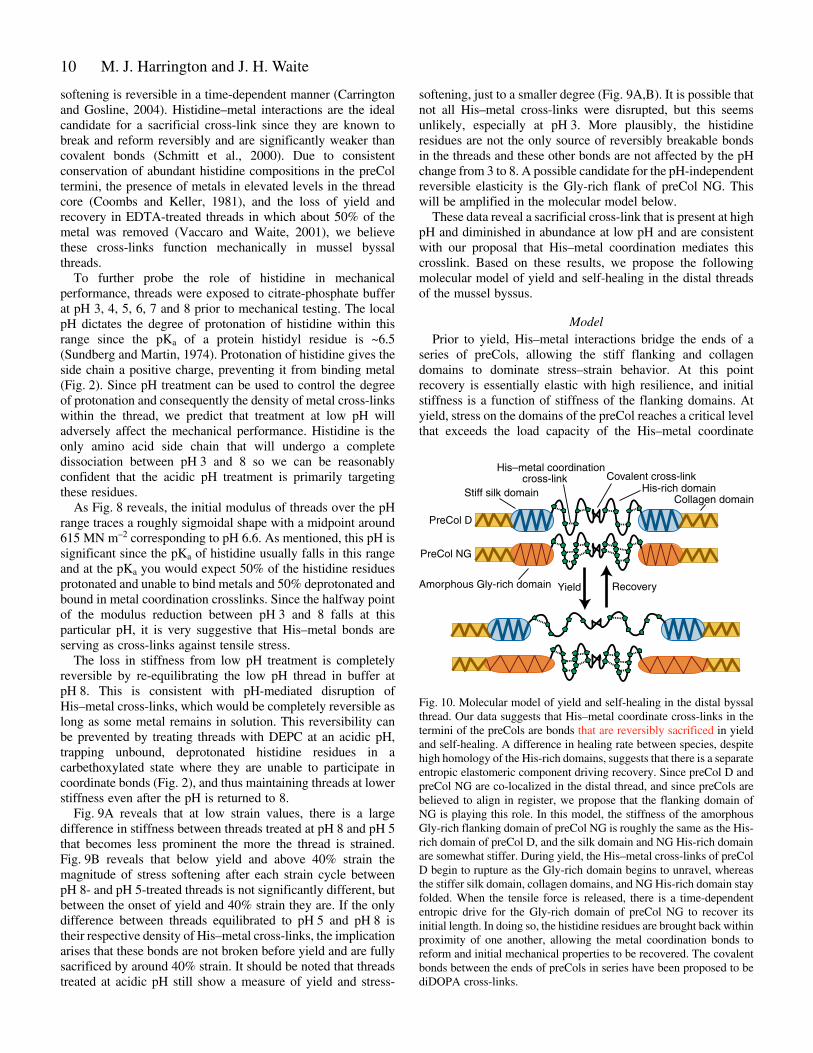
ModelPrior to yield, His–metal interactions bridge the ends of a
series of preCols, allowing the stiff flanking and collagendomains to dominate stress–strain behavior. At this pointrecovery is essentially elastic with high resilience, and initialstiffness is a function of stiffness of the flanking domains. Atyield, stress on the domains of the preCol reaches a critical levelthat exceeds the load capacity of the His–metal coordinate
M. J. Harrington and J. H. Waite
Fig.·10. Molecular model of yield and self-healing in the distal byssalthread. Our data suggests that His–metal coordinate cross-links in thetermini of the preCols are bonds that are reversibly sacrificed in yieldand self-healing. A difference in healing rate between species, despitehigh homology of the His-rich domains, suggests that there is a separateentropic elastomeric component driving recovery. Since preCol D andpreCol NG are co-localized in the distal thread, and since preCols arebelieved to align in register, we propose that the flanking domain ofNG is playing this role. In this model, the stiffness of the amorphousGly-rich flanking domain of preCol NG is roughly the same as the His-rich domain of preCol D, and the silk domain and NG His-rich domainare somewhat stiffer. During yield, the His–metal cross-links of preColD begin to rupture as the Gly-rich domain begins to unravel, whereasthe stiffer silk domain, collagen domains, and NG His-rich domain stayfolded. When the tensile force is released, there is a time-dependententropic drive for the Gly-rich domain of preCol NG to recover itsinitial length. In doing so, the histidine residues are brought back withinproximity of one another, allowing the metal coordination bonds toreform and initial mechanical properties to be recovered. The covalentbonds between the ends of preCols in series have been proposed to bediDOPA cross-links.
Yield Recovery
Stiff silk domain
Amorphous Gly-rich domain
Collagen domain
PreCol D
PreCol NG
His-rich domainCovalent cross-link
His–metal coordinationcross-link

11Holdfast fibers
bonds and they begin to rupture, dissipating the applied energyin the form of molecular friction (hysteresis). In sacrificingHis–metal complexes, the covalent bonds in the backbone arespared and catastrophic failure is avoided. This may also be thepoint at which the non-His sacrificial bonds begin to rupturesince yield, stress softening, and hysteresis are also seen in lowpH threads. The best candidates for these are the NG flankswhich, with a predicted amorphous structure, are likely to bemore compliant than the collagen cores and the Ala-rich silk-like flanks. Beyond 40% strain, most His–metal bonds arebroken, and further stress softening (which is small) is not dueto the His–metal bonds breaking. Evidence for this comes fromthe fact that �E is not significantly different between high pHand low pH above 40% strain.
Since coordinate bonds are known to be reversibly breakable,His–metal could play a role in post-yield recovery if theseparated ligands and metals are brought together again. Hisdomains are not different between species, and as M.californianus is a faster healer, differences in recovery rate mustcome from another source. This suggests the presence ofseparate elastomeric components in parallel with the histidine-rich domains, which restore them after deformation. Theflanking domains of NG are again the best candidates as therestoring elastomeric component. Since preCol D and NG areboth known to be present in the distal region of the thread andsince the preCols are believed to assemble in register, acomposite model of yield and self-healing is outlined in Fig.·10.In this model, the stiffness of the flanking domains of NG andof the His-rich domains in D are roughly comparable. Bothbegin to break near the critical yield stress. When the appliedload is released, the flanking regions of NG recoverentropically, dragging the separated histidine residues betweentwo preCol Ds back into mutual proximity and thus restore theirmetal coordination.
The assumed entropic recovery of NG flanks is an importantcomponent of our model and deserves closer scrutiny in futureexperimental studies. It has been proposed that Gly-rich proteinsin plant cell walls homologous to those in NG flanking domainsform a loose structure known as a glycine loop, reminiscent ofkeratin termini (Steinert et al., 1991). Glycine loops are believedto behave like molecular VELCROTM, in which the orderedstructure is easily disrupted by moderate tension, but is able torecover its initial conformation after stress is released (Sachetto-Martins et al., 2000). This sort of moderately stiff, entropicelastic component would be a good fit as the component drivingrecovery.
This work was supported in part by the NASA UniversityResearch, Engineering and Technology Institutes on Bio-inspired Materials under award No. NCC-1-02037 and by theNational Institutes of Health Grant R01 DEO 14672.
ReferencesBell, E. and Gosline, J. (1996). Mechanical design of mussel byssus: material
yield enhances attachment strength. J. Exp. Biol. 199, 1005-1017.Bella, J., Liu, J., Kramer, R. Z., Brodsky, B. and Berman, H. M. (2006).
Conformational effects of Gly-X-Gly interruptions in the collagen triplehelix. J. Mol. Biol. 362, 298-311.
Brazee, S. L. and Carrington, E. (2006). Interspecific comparison of themechanical properties of mussel byssus. Biol. Bull. 211, 263-274.
Brazel, D., Oberbaumer, I., Dieringer, H., Babel, W., Glanville, R. W.,Deutzmann, R. and Kuhn, K. (1987). Completion of the amino acidsequence of the �1 chain of human basement membrane collagen (type IV)reveals 21 non-triplet interruptions located within the collagenous domain.Eur. J. Biochem. 168, 529-536.
Brooks, A. E., Steinkraus, H. B., Nelson, S. R. and Lewis, R. V. (2005). Aninvestigation of the divergence of major ampullate silk fibers from Nephilaclavipes and Argiope aurantia. Biomacromolecules 6, 3095-3099.
Carrington, E. and Gosline, J. (2004). Mechanical design of mussel byssus:load cycle and strain rate dependence. Am. Malacol. Bull. 18, 135-142.
Chenna, R., Sugawara, H., Koike, T., Lopez, R., Gibson, T. J., Higgins, D.G. and Thompson, J. D. (2003). Multiple sequence alignment with theClustal series of programs. Nucleic Acids Res. 31, 3497-3500.
Coombs, T. L. and Keller, P. J. (1981). Mytilus byssal threads as anenvironmental marker for metals. Aquatic Toxicol. 1, 291-300.
Coyne, K. J., Qin, X.-X. and Waite, J. H. (1997). Extensible collagen inmussel byssus: a natural block copolymer. Science 277, 1830-1832.
Fowler, S. J., Jose, S., Zhang, X., Deutzmann, R., Sarras, M. P., Jrand Boot-Handford, R. P. (2000). Characterization of Hydra type IVcollagen: type IV collagen is essential for head regeneration and itsexpression is up-regulated upon exposure to glucose. J. Biol. Chem. 275,39589-39599.
Gosling, E. (1992). Systematic and geographic distribution of Mytilus. In TheMussel Mytilus: Ecology, Physiology, Genetics and Culture. Vol. 25 (ed. E.Gosling), pp. 1-20. Amsterdam: Elsevier.
Hassenkam, T., Gutsmann, T., Hansma, P., Sagert, J. and Waite, J. H.(2004). Giant bent-core mesogens in the thread forming process of marinemussels. Biomacromolecules 5, 1351-1354.
Hayashi, C. Y. and Lewis, R. V. (1998). Evidence from flagelliform silk cDNAfor the structural basis of elasticity and modular nature of spider silks. J. Mol.Biol. 275, 773-784.
Hayashi, C. Y., Shipley, N. H. and Lewis, R. V. (1999). Hypotheses thatcorrelate the sequence, structure, and mechanical properties of spider silkproteins. Int. J. Biol. Macromol. 24, 271-275.
Hudson, B. G., Tryggvason, K., Sundaramoorthy, M. and Neilson, E. G.(2003). Alport’s syndrome, Goodpasture’s syndrome, and type IV collagen.N. Engl. J. Med. 348, 2543-2556.
Kilchherr, E., Hofmann, H., Steigemann, W. and Engel, J. (1985). Structuralmodel of the collagen-like region of C1q comprising the kink region and thefibre-like packing of the six triple helices. J. Mol. Biol. 186, 403-415.
Lee, H., Scherer, N. F. and Messersmith, P. B. (2006). Single-moleculemechanics of mussel adhesion. Proc. Natl. Acad. Sci. USA 103, 12999-13003.
Lucas, J. M., Vaccaro, E. and Waite, J. H. (2002). A molecular,morphometric and mechanical comparison of the structural elements ofbyssus from Mytilus edulis and Mytilus galloprovincialis. J. Exp. Biol. 205,1807-1817.
Lundblad, R. L. (2005). Chemical Reagents for Protein Modification. BocaRaton, FL: CRC Press.
Mercer, E. H. (1952). Observations on the molecular structure of byssus fibres.Aust. J. Mar. Freshw. Res. 3, 199-205.
Pappalardo, G., Impellizzeri, G., Bonomo, R. P., Campagna, T., Grasso, G.and Saita, M. G. (2002). Copper(II) and nickel(II) binding modes in ahistidine-containing model dodecapeptide. New J. Chem. 26, 593-600.
Persikov, A. V., Ramshaw, J. A. M. and Brodsky, B. (2005). Prediction ofcollagen stability from amino acid sequence. J. Biol. Chem. 280, 19343-19349.
Qin, X. X. and Waite, J. H. (1995). Exotic collagen gradients in the byssus ofthe mussel Mytilus edulis. J. Exp. Biol. 198, 633-644.
Qin, X.-X. and Waite, J. H. (1998). A potential mediator of collagenous blockcopolymer gradients in mussel byssal threads. Proc. Natl. Acad. Sci. USA 95,10517-10522.
Qin, X.-X., Coyne, K. J. and Waite, J. H. (1997). Tough tendons: musselbyssus has collagen with silk-like domains. J. Biol. Chem. 272, 32623-32627.
Rudall, K. M. (1955). The distribution of collagen and chitin. Symp. Soc. Exp.Biol. 9, 49-71.
Sachetto-Martins, G., Franco, L. O. and de Oliveira, D. E. (2000). Plantglycine-rich proteins: a family or just proteins with a common motif. Biochim.Biophys. Acta 1492, 1-14.
Santaclara, F. J., Espineira, M., Cabado, A. G., Aldasoro, A., Gonzalez-Lavin, N. and Vieites, J. M. (2006). Development of a method for thegenetic identification of mussel species belonging to Mytilus, Perna,Aulacomya, and other genera. J. Agric. Food Chem. 54, 8461-8470.
Schmitt, L., Ludwig, M., Gaub, H. E. and Tampe, R. (2000). A metal-chelating microscopy tip as a new toolbox for single-molecule experimentsby atomic force microscopy. Biophys. J. 78, 3275-3285.

12
Sicot, F.-X., Exposito, J.-Y., Masselot, M., Garrone, R., Deutsch, J. and Gaill,F. (1997). Cloning of an annelid fibrillar-collagen gene and phylogeneticanalysis of vertebrate and invertebrate collagens. Eur. J. Biochem. 246, 50-58.
Soininen, R., Haka-Risku, T., Prockop, D. J. and Tryggvason, K. (1987).Complete primary structure of the �1-chain of human basement membrane(type IV) collagen. FEBS Lett. 225, 188-194.
Steinert, P. M., Mack, J. W., Korge, B. P., Gan, S.-Q., Haynes, S. R. andSteven, A. C. (1991). Glycine loops in proteins: their occurrence in certainintermediate filament chains, loricrins, and single-stranded RNA bindingproteins. Int. J. Biol. Macromol. 13, 130-139.
Sun, C., Lucas, J. M. and Waite, J. H. (2002). Collagen-binding matrixproteins from elastomeric extraorganismic byssal fibers. Biomacromolecules3, 1240-1248.
Sundberg, R. J. and Martin, R. B. (1974). Interactions of histidine and otherimidazole derivatives with transition metal ions in chemical and biologicalsystems. Chem. Rev. 74, 471-517.
Vaccaro, E. and Waite, J. H. (2001). Yield and post-yield behavior of musselbyssal thread: a self-healing biomolecular material. Biomacromolecules 2,906-911.
van Hest, J. C. M. and Tirrell, D. A. (2001). Protein-based materials, towarda new level of structural control. Chem. Commun. 2001, 1897-1904.
Waite, J. H. (1992). The formation of mussel byssus: anatomy of a naturalmanufacturing process. Results Probl. Cell Differ. 19, 27-54.
Waite, J. H., Qin, X.-X. and Coyne, K. J. (1998). The peculiar collagens ofmussel byssus. Matrix Biol. 17, 93-106.
Waite, J. H., Vaccaro, E., Sun, C. and Lucas, J. (2002). Elastomericgradients: a hedge against stress concentration in marine holdfasts? Philos.Trans. R. Soc. Lond. B Biol. Sci. 357, 143-153.
Waite, J. H., Lichtenegger, H. C., Stucky, G. D. and Hansma, P. (2004).Exploring molecular and mechanical gradients in structural bioscaffolds.Biochemistry 43, 7653-7662.
Waite, J. H., Weaver, J. C. and Vaccaro, E. (2006). Molecular consequencesof biomolecular gradients in byssal threads. In Bionanotechnology: Proteinsto Nanodevices (ed. V. Renugopalakrishnan and R. V. Lewis), pp. 25-38.Dordrecht: Springer.
Yamaguchi, N., Benya, P., van der Rest, M. and Ninomiya, Y. (1989). Thecloning and sequencing of alpha 1(VIII) collagen cDNAs demonstrate thattype VIII collagen is a short chain collagen and contains triple- helical andcarboxyl-terminal non-triple-helical domains similar to those of type Xcollagen. J. Biol. Chem. 264, 16022-16029.
Yonge, C. M. (1962). On the primitive significance of the byssus in the bivalviaand its effects in evolution. J. Mar. Biol. 42, 113-125.
M. J. Harrington and J. H. Waite