
CCR2 and CCR5 genes polymorphisms in benign prostatic hyperplasia and prostate cancer
Upload
independentCategory
view
0download
0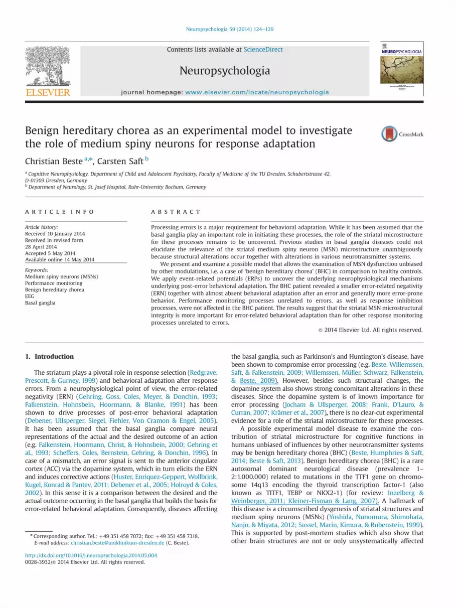
Benign hereditary chorea as an experimental model to investigatethe role of medium spiny neurons for response adaptation
Christian Beste a,n, Carsten Saft b
a Cognitive Neurophysiology, Department of Child and Adolescent Psychiatry, Faculty of Medicine of the TU Dresden, Schubertstrasse 42,D-01309 Dresden, Germanyb Department of Neurology, St. Josef Hospital, Ruhr-University Bochum, Germany
a r t i c l e i n f o
Article history:Received 10 January 2014Received in revised form28 April 2014Accepted 5 May 2014Available online 14 May 2014
Keywords:Medium spiny neurons (MSNs)Performance monitoringBenign hereditary choreaEEGBasal ganglia
a b s t r a c t
Processing errors is a major requirement for behavioral adaptation. While it has been assumed that thebasal ganglia play an important role in initiating these processes, the role of the striatal microstructurefor these processes remains to be uncovered. Previous studies in basal ganglia diseases could notelucidate the relevance of the striatal medium spiny neuron (MSN) microstructure unambiguouslybecause structural alterations occur together with alterations in various neurotransmitter systems.
We present and examine a possible model that allows the examination of MSN dysfunction unbiasedby other modulations, i.e. a case of ‘benign hereditary chorea’ (BHC) in comparison to healthy controls.We apply event-related potentials (ERPs) to uncover the underlying neurophysiological mechanismsunderlying post-error behavioral adaptation. The BHC patient revealed a smaller error-related negativity(ERN) together with almost absent behavioral adaptation after an error and generally more error-pronebehavior. Performance monitoring processes unrelated to errors, as well as response inhibitionprocesses, were not affected in the BHC patient. The results suggest that the striatal MSN microstructuralintegrity is more important for error-related behavioral adaptation than for other response monitoringprocesses unrelated to errors.
& 2014 Elsevier Ltd. All rights reserved.
1. Introduction
The striatum plays a pivotal role in response selection (Redgrave,Prescott, & Gurney, 1999) and behavioral adaptation after responseerrors. From a neurophysiological point of view, the error-relatednegativity (ERN) (Gehring, Goss, Coles, Meyer, & Donchin, 1993;Falkenstein, Hohnsbein, Hoormann, & Blanke, 1991) has beenshown to drive processes of post-error behavioral adaptation(Debener, Ullsperger, Siegel, Fiehler, Von Cramon & Engel, 2005).It has been assumed that the basal ganglia compare neuralrepresentations of the actual and the desired outcome of an action(e.g. Falkenstein, Hoormann, Christ, & Hohnsbein, 2000; Gehring etal., 1993; Scheffers, Coles, Bernstein, Gehring, & Donchin, 1996). Incase of a mismatch, an error signal is sent to the anterior cingulatecortex (ACC) via the dopamine system, which in turn elicits the ERNand induces corrective actions (Huster, Enriquez-Geppert, Wollbrink,Kugel, Konrad & Pantev, 2011; Debener et al., 2005; Holroyd & Coles,2002). In this sense it is a comparison between the desired and theactual outcome occurring in the basal ganglia that builds the basis forerror-related behavioral adaptation. Consequently, diseases affecting
the basal ganglia, such as Parkinson's and Huntington's disease, havebeen shown to compromise error processing (e.g. Beste, Willemssen,Saft, & Falkenstein, 2009; Willemssen, Müller, Schwarz, Falkenstein,& Beste, 2009). However, besides such structural changes, thedopamine system also shows strong concomitant alterations in thesediseases. Since the dopamine system is of known importance forerror processing (Jocham & Ullsperger, 2008; Frank, D'Lauro, &Curran, 2007; Krämer et al., 2007), there is no clear-cut experimentalevidence for a role of the striatal microstructure for these processes.
A possible experimental model disease to examine the con-tribution of striatal microstructure for cognitive functions inhumans unbiased of influences by other neurotransmitter systemsmay be benign hereditary chorea (BHC) (Beste, Humphries & Saft,2014; Beste & Saft, 2013). Benign hereditary chorea (BHC) is a rareautosomal dominant neurological disease (prevalence 1–2:1.000.000) related to mutations in the TTF1 gene on chromo-some 14q13 encoding the thyroid transcription factor-1 (alsoknown as TITF1, TEBP or NKX2-1) (for review: Inzelberg &Weinberger, 2011; Kleiner-Fisman & Lang, 2007). A hallmark ofthis disease is a circumscribed dysgenesis of striatal structures andmedium spiny neurons (MSNs) (Yoshida, Nunomura, Shimohata,Nanjo, & Miyata, 2012; Sussel, Marin, Kimura, & Rubenstein, 1999).This is supported by post-mortem studies which also show thatother brain structures are not or only unsystematically affected
Contents lists available at ScienceDirect
journal homepage: www.elsevier.com/locate/neuropsychologia
Neuropsychologia
http://dx.doi.org/10.1016/j.neuropsychologia.2014.05.0040028-3932/& 2014 Elsevier Ltd. All rights reserved.
n Corresponding author. Tel.: þ49 351 458 7072; fax: þ49 351 458 7318.E-mail address: [email protected] (C. Beste).
Neuropsychologia 59 (2014) 124–129
(for review: Inzelberg & Weinberger, 2011). Clinically, BHC isassociated with choreatic movements. As MSN dysfunctions arenot modulated by otherwise dysfunctional neurotransmitters inBHC, this disease may serve as a possible experimental modeldrawn from disease to uncover the relevance of the striatalmicrostructure for cognitive functions. The striatal microstructuremay be important because a fundamental step in error processingis the comparison between the desired and the actual outcome(Gehring et al., 1993; Scheffers et al., 1996). To do so, the basalganglia must be able to represent different outcomes in order tocompare them to each other. Several computational basal gangliamodels suggest that MSN is important for this process (Bard-Gad,Morris, & Bergman, 2003; Plenz, 2003). In BHC it is possible thaterror processing functions are diminished because a comparison ofthe actual outcome of an action against the desired outcome ofaction may not be possible at the basal ganglia level because oflow microstructural integrity of MSNs. Therefore, behavioraladaptation after an error may be diminished. The current studysets out to test this hypothesis in a clinical case-control study.
2. Materials and methods
2.1. Patient and controls
A sample of N¼20 healthy controls between 20 and 28 years of age (mean age24.6; SD¼3.6) was recruited for comparison to a single individual with benignhereditary chorea (BHC) using single-case bootstrap statistics. This individual whowas a 24-year old female with genetically confirmed mutation in the TTF1 genewas recruited. The BHC patient was unmedicated so that the results obtained areunbiased with respect to that factor. Controls and the BHC case were investigatedclinically with a neuropsychological test battery (see Table 1). The BHC patientunderwent neurological assessment of motor symptoms using the Unified Hun-tington's Disease Rating Scale (UHDRS) Huntington Study Group, 1996). This scalewas used because here choreatic movements can reliably be assessed andquantified. The BHC case showed severe signs of choreatic movement disturbances.The neuropsychological and neurological assessment of the patient and controls issummarized in Table 1. Using these standard neuropsychological tests, no differ-ence was evident between the BHC case and the controls using Craufurd andHowell's method (p4 .3) (cf. Crawford & Garthwaite, 2012). The study wasapproved by the Ethics Committee of the Ruhr-University of Bochum. The studywas conducted according to the Declaration of Helsinki. All healthy participantsand the BHC patient gave written informed consent.
2.2. Task
To examine error monitoring we used a flanker task. The paradigm is identicalto other studies done by our group (Beste, Gunturkun, Baune, Falkenstein & Konrad,
2011) and was structured as follows: vertically arranged visual stimuli werepresented. The target-stimulus (arrowhead or circle) was presented in the centerwith the arrowhead pointing to the left or right. The central stimuli were flankedby two vertically adjacent arrowheads which pointed in the same (compatible) oropposite (incompatible) direction as the target. In case of target stimuli (arrow-heads pointing to the left or right) participants were required to press a responsebutton with their left or right thumb. A circle as the central stimulus indicates aNogo trial, where the subject was required to inhibit the response. The flankerspreceded the target by 100 ms. The target (arrowhead or circle) was displayed for300 ms. The response-stimulus interval was 1600 ms. Flankers and target wereswitched off simultaneously. Time pressure was administered by asking thesubjects to respond within 600 ms. In trials with reaction times exceeding thisdeadline, a feedback stimulus (1000 Hz, 60 dB SPL) was given 1200 ms after theresponse; this stimulus had to be avoided by the subjects. Four blocks of 105stimuli each were presented in this task. Compatible (60%) and incompatiblestimuli (20%) and Nogo stimuli (circle) (20%) were presented randomly (cf.Beste etal., 2011).
Importantly, for the analysis of error processing only trials with incorrectbutton presses, but not trials with response times exceeding the response deadlineof 600 ms were used. This was done to avoid the fact that the errors observed inBHC may be due to the motor problems of the patient and indeed would reflecterror-prone choice behavior.
2.3. EEG recording and analysis and MRI scanning
During the task an EEG was recorded from 64 Ag–AgCl electrodes against areference electrode located at Cz at a sampling rate of 500 Hz applying a filterbandwidth 0–80 Hz to the EEG (Quickamp, Brain Products Inc.). To re-reference thedata, a CSD transformation was applied, which eliminates the reference potential(Nunez and Pilgreen, 1991). Electrode impedances were kept below 5 kΩ. The EEGwas filtered off-line from .5 to 20 Hz (48 db/oct)1. A raw data inspection wasapplied and technically occurring artifacts were discarded by manual inspection ofthe data. Afterwards, independent component analysis (ICA, Infomax algorithm)was applied. Independent components of blinks, saccades and pulse artifacts werediscarded by visual inspection. After segmenting the data into correct and errortrials, an automated artifact rejection procedure was applied with an amplitudethreshold of 780 mV. The response was set to time point 0 and a baselinecorrection procedure was applied from �200 ms till button press. The ERN andresponse related potentials after correct responses (Nc) were quantified inamplitude and latency at electrode FCz against the pre-response baseline. TheERN and the correct-related negativity (Nc) were defined as the most negative peakwithin 50–120 ms after the response. As can be seen in scalp topography maps,ERN was maximal at electrodes FCz and Cz. We only quantified the ERN at electrodeFCz because electrode Cz was used as reference electrode during data acquisition.The error positivity (Pe) was quantified at electrode Pz (Falkenstein et al., 2000)and defined as the most positive peak in the window between 300 and 600 msafter an erroneous response. As the task also contained Nogo-trials, neurophysio-logical processes underlying inhibition (i.e., Nogo-N2 and Nogo-P3, see: Huster,
Table 1Neuropsychological test data (raw scores) of the BHC case and the group of healthy controls. Testing possible differences between BHC caseand controls revealed no difference in any of the tests applied. The UHDRS motor score is necessarily different, as the controls do not sufferunder the genetic mutation causing BHC.
Test BHC Controls
UHDRS (motor score) 26 0IQ score (MWT-B) 105 113 (9)Stroop test (UHDRS)
Color naming 61 75 (6)Color reading 98 101 (15)Interference (Stroop condition) 36 40 (10)
Symbol-digit test (WAIS) correct items (Härting et al., 2000) 51 59 (12)Word fluency (Benton) number of words 39 42 (8)Digit span (WMS-R) number of items (Härting et al., 2000)
Forward 8 9 (3)Backward 5 5 (4)
Block Span (WMS-R) number of items (Härting et al., 2000)Forward 8 9 (3)Backward 7 7 (3)
Benton Test (visual memory) (Benton, Benton-Sivan, & Steck, 2009) 11 13 (3)Mini Mental Status examination (MMSE) (Folstein, Folstein, & McHugh, 1975) 30 30
1 We also analyzed the data with a more liberal filter (i.e., from .5 to 40 Hzincluding a 50 Hz notch filter; 48 db/oct). This did not change the pattern of results.
C. Beste, C. Saft / Neuropsychologia 59 (2014) 124–129 125

Enriquez-Geppert, Lavallee, Falkenstein, & Herrmann, 2013) were analyzed instimulus-locked data at electrode FCz. The Nogo-N2 was defined as the mostnegative deflection in the time interval between 200 and 300 ms after the stimulusand the Nogo-P3 were defined as the most positive deflection in the time windowbetween 300 and 600 ms after Nogo-stimulus presentation. In order to obtain anestimate about the reliability of the neurophysiological data, which is of specialimportance in light of single-case data, we calculated the signal-to-noise (SNR) inBHC and controls as implemented in the Brain Vision Analyzer II software package(BrainProducts Inc.). Calculation of the SNR is important for single-case data, sinceusually SNR is increased by averaging over a number of participants in anexperimental group. As this is not possible in single-case data, SNR gives importantinformation about the reliability of the data for the single case.
For an anatomical MRI scan, a T2 turbo spin echo scan was acquired on 3 TPhilips Intera System using a SENSE head coil (TR¼3721 ms; TE¼80 ms; 901 flipangle). A transversal section showing the striatum is shown in Fig. 1D.
2.4. Statistics
To compare the BHC case with the control group, single case statistics were runusing Crawford and Howell's methods (for review: Crawford & Garthwaite, 2012).This method offers the best way to compare single cases with groups of controlsubjects (for review: Crawford & Garthwaite, 2012). For within-subject effects inthe control group, usual t-tests were used and Bonferroni-corrected wherevernecessary.
3. Results
3.1. Error processing
For all statistics the mean and standard deviations are given.Error rates were higher in the BHC patient (28%), compared tocontrols (10.15%73.1) (t¼5.63, p¼ .00001; 95% confidence inter-val 3.89 to 7.64). In controls an error trial was followed by an anewerror in the following trial in 7% of cases (72.2). In the BHCpatient an error trial was followed by an anew error in thefollowing trial in 30% of cases (t¼10.20; po .00001; 95% con-fidence interval 7.12 to 13.77).
For the control cohort, RTs were longer on correct trials(389 ms787) compared to error trials (290 ms785) (t(19)¼23.33; po .001). The BHC patient did not differ from the controlgroup in these RTs (error: 315 ms; correct: 402 ms; all p4 .5). Incontrols it is further shown that correct responses succeedingerror trials were longer (425 ms773) than responses succeeding acorrect trial (386 ms770) (t(19)¼�17.90; po .001). This post-error slowing effect was thus 43 ms (79) for controls (referFig. 1A). In the BHC patient this post-error slowing effect was
Fig. 1. (A) Degree of post-error slowing (in ms) for the control group and the BHC case. (B) Event-related potentials (ERPs) including scalp topography plots on correct anderror trials at electrode FCz. Time point 0 denotes the time point of response execution. As can be seen, ERN is only evident in controls but not in the BHC case. Please notethe different scaling for the scalp topography plots. The scalp topography plots denote the mean value of the amplitude at its peak. (C) Event-related potentials (ERPs)including scalp topography plots on correct and error trials at electrode Pz to show the error-positivity (Pe) including the scalp topography of the Pe (all-in-one map). (D) MRItransversal section (T2 turbo spin echo) of the BHC case showing the striatum. A smaller caudate nucleus is evident.
C. Beste, C. Saft / Neuropsychologia 59 (2014) 124–129126

5 ms and was hence significantly lower than the effect in controls(t¼�4.21, and p¼ .0003; 95% confidence interval �5.61 to �2.81)and not different from zero (p4 .5).
The neurophysiological data (i.e., ERN and Nc) (Fig. 1B) showedthat ERN was larger (�25.57 mV/m274.45) than Nc (�11.7 mV/m274.13) in the control group (t(19)¼�13.34; po .001). The BHCpatient showed a much smaller ERN (�11.2 mV/m2), compared tocontrols (t¼�3.10, and p¼ .003; 95% confidence interval �4.26 to�2.08). Nc in the BHC patient was �10.5 mV/m2 and thus notdifferent from controls (p4 .7). ERN and Nc in the BHC patient didnot differ from Nc in controls (p4 .5). There were generally nolatency differences between BHC patient and controls (all p4 .7).Also when using the difference between ERN and Nc, thisdifference was smaller for the BHC patient (3.1 mV/m2) than forhealthy controls (14.8 mV/m276.44) (t¼�1.89, and p¼ .04; 95%confidence interval �2.25 to �1.08).
Yet, it may be argued that the smaller ERN is an artifact of thehigher rates of errors (Endrass, Klawohn, Gruetzmann, Ischebeck,& Kathmann, 2012) in the BHC patient. To control the effect oferror frequency on the modulation of ERN, we used the sameamount of trials in the BHC patient to compare ERN against ERN ofhealthy subjects. This controls for a possible biasing effect of errorfrequency on the differential modulation of ERN in the BHC patientand healthy controls. In this analysis, ERN was still lower in BHC(�14.2 mV/m2) compared to controls (t¼�2.66, and p¼ .009).Similarly, in the BHC patient the post-error slowing effect was12 ms and hence lower than slowing in controls (t¼�3.88, andp¼ .001). However, to control these effects it may be even moreimportant to take the relative probability of errors into account.This is the case because post-error slowing effects can beexplained by differences in error rate (Notebaert, Houtman,Opstal, Gevers, Flas, &Verguts, 2009) and high error rates lead todiminished post-error slowing (Nunez-Castellar, Houtman, Gevers,Morrens, Vermeylen, Sabbe et al., 2012). To control this effect were-analyzed ERN and the post-error slowing effect on the first 210trials of the experiment. The post-error slowing was still smaller inthe BHC patient (11 ms) compared to controls (49 ms79)(t¼�4.12, and p¼ .0003; 95% confidence interval �5.61 to�2.81), while the error rate in these first 210 trials was notdifferent between controls and BHC patient (p4 .6). Similarly, ERNwas smaller in BHC patient (�12.1 mV/m2) than in controls(�26.11 mV/m273.11) (t¼�4.39, and p¼ .0002; 95% confidenceinterval �5.98 to �3.01). Nc did not differ between BHC patientand controls (p4 .6) and there was no difference between Nc inthe BHC patient and ERN in controls (p4 .7). All analysis strategiestherefore lead to the same pattern of effects2. The error positivityis also shown in Fig. 1C. The analysis of the Pe amplitude did notshow differences between BHC patient and controls (p4 .5).
Calculation of the SNR, as implemented in the Brain VisionAnalyzer II software package, revealed that for correct trials SNRwas (.157 .05) in controls, and for error trials the SNR was (.347 .08)in controls. In the BHC patient, SNR was .16 for correct trials and .32for error trials. The SNRs did not differ between controls and BHCpatient on correct and error trials (p4 .8) showed that the EEGsignals compared are similarly reliable in controls and the BHCpatient. The same was evident for the error positivity (Pe) (p4 .6).
3.2. Response inhibition
The flanker task contained 20% Nogo trials. To examineresponse inhibition processes, the data was analyzed stimulus-
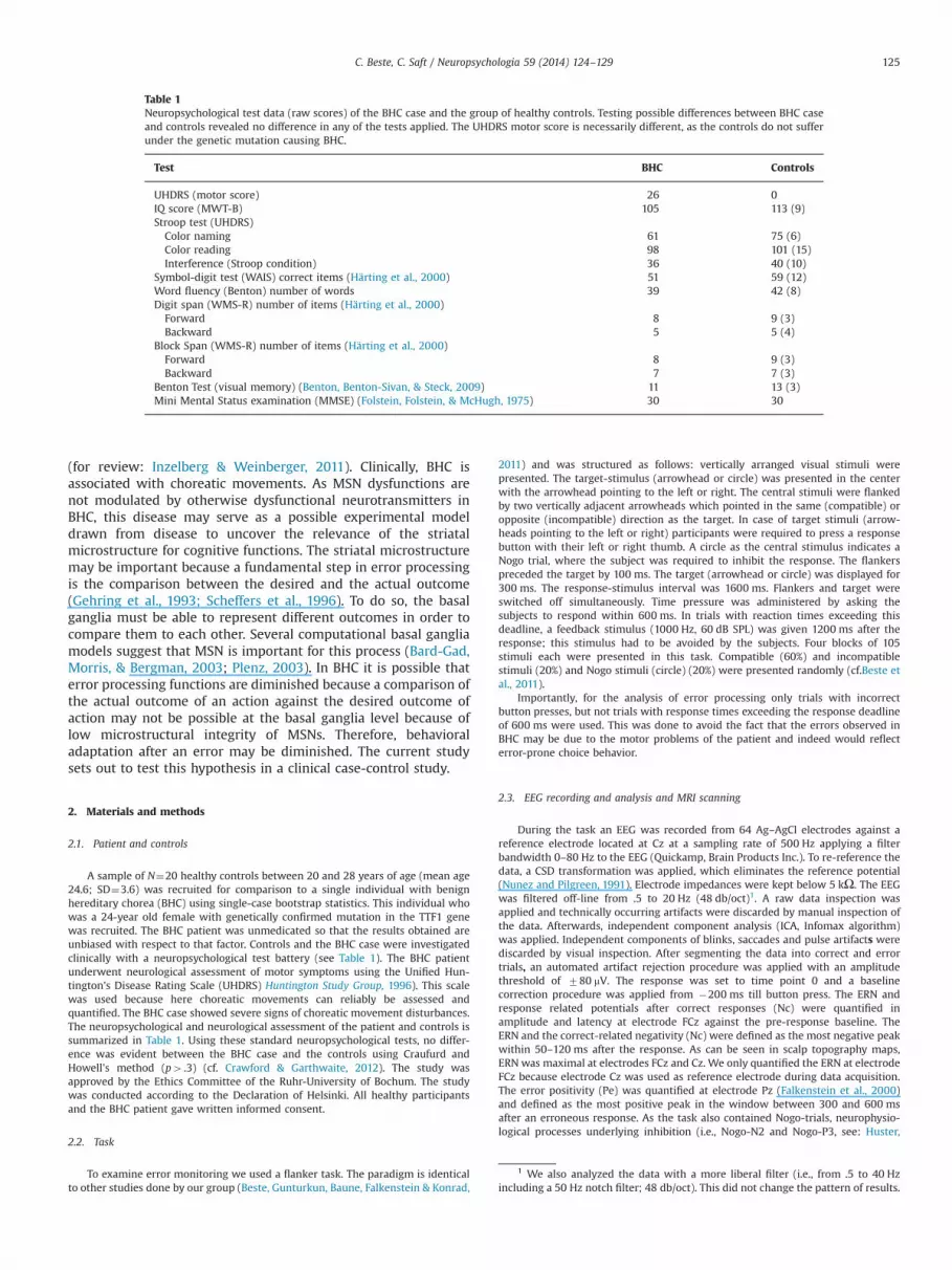
locked on Nogo trials. The neurophysiological data is shown inFig. 2.
Although numerically different, the rate of Nogo errors (i.e.,button presses on Nogo-stimuli) did not differ between BHCpatient (7.6) and controls (5.171.9) (p4 .6). Similarly, there wereno differences in the response times on Go trials (p4 .5). Therewas no difference in the amplitude of the Nogo-N2 (p4 .7) and theNogo-P3 (p4 .6), between the BHC patient and the control group.An analysis of the SNRs revealed no difference between BHCpatient (.22) and controls (.247 .1) (p4 .4).
4. Discussion
The current study aimed to investigate the relevance of themicrostructural integrity of striatal MSN for error-related behavioraladaptation processes. To this end, benign hereditary chorea (BHC), apossible experimental model (Beste & Saft, 2013) of circumscribedMSN dysfunction was investigated in a case-control study. Theresults show almost absent behavioral adaptation processes after anerror in the BHC case as indicated by the post-error slowingparameter. Similarly, error rates were higher in the BHC patient,which may reflect a consequence of diminished post-error slowing.ERN has been shown to be associated with corrective action andpost-error slowing (Debener et al., 2005). The reduced post-errorslowing effect observed for the BHC patient may be interpreted as alack of increase in response caution that is observed in controls andis likely to be reflected by post-error slowing as suggested by dataon reaction time modeling (Dutilh, Vendekerckhove, Forstmann,Keuleers, Brysbaert & Wagenmakers, 2012). However, in line withother conceptions on post-error slowing this may also suggestdeficits in an orienting response, due to the rareness of errors(Notebaert et al., 2009). Yet, as post-error slowing effects were notbiased with respect to the frequency of errors in the present data,our data speaks for an effect at the level of response caution. Theabsence of post-error slowing in the BHC patient together withdecrease in the post-error accuracy may be a consequence of anabsent ERN in BHC: The results show that the ERN is smaller in theBHC patient, compared to controls. The lack of difference betweenthe ERN in BHC and the Nc in controls suggests that ERN is evenabsent in the BHC patient. Despite of a single case for BHC, theanalysis of the SNR shows that the neurophysiological data obtained
Fig. 2. Event-related potentials on Nogo-trials in BHC and controls and compatibleGo-trials. No differences are evident for the Nogo-N2 and Nogo-P3 between BHCand controls. The maps denote the scalp topography plots at the peak of therespective components.
2 When not using the peak amplitude but the mean amplitude of the ERN andNc (calculated over the time window between 30 and 90 ms) the results remainedthe same.
C. Beste, C. Saft / Neuropsychologia 59 (2014) 124–129 127

from the BHC patient is as reliable as the data obtained from thecontrol cohort. The results suggest that MSNs may constitute animportant element in error processing and behavioral adaptation.As stated in (Section 1), it has been assumed that a comparisonbetween the desired and the actual outcome of an action forms thebasis for post-error behavioral adaptation (Falkenstein et al., 2000;Gehring et al., 1993; Scheffers et al., 1996). The data suggests that adysfunctional MSNs network is not able to differentiate between adesired and an actual erroneous outcome of a response. The resultssuggest that processes of response control per se (as reflected bythe Nc) are not deficient in a condition of MSN dysfunction. Whatseems to be particularly affected are behavioral adaptation pro-cesses after errors, which is underlined by the higher error rates inthe BHC patient compared to controls. In this regard the analysis ofresponse inhibition performance (behavioral and neurophysiologi-cal data) does not show differences in the BHC patient, compared tohealthy controls. All this data suggests that MSN dysfunctions, asobserved in the BHC patient, do not have a generally compromisingeffect on response monitoring processes, but are of importance forerror monitoring processes as reflected by ERN. This suggests thatthese response monitoring functions related to the error-relatedcorrection of actions, response inhibition processes and generalresponse monitoring functions unrelated to errors have differentfunctional neuroanatomical correlates.
The finding that ERN and Nc were differentially modulated in theBHC patient adds to research suggesting that both processes may beimplemented via different neurobiological and neurophysiologicalmechanisms (e.g. Hoffmann, Labrenz, Themann, Wascher, & Beste,2014; Beste, Domschke, et al., 2010; Beste, Baune, Domschke,Falkenstein, & Konrad, 2010; Jocham & Ullsperger, 2008). Interestingly,a lack of modulation between the ERN and the Nc (as found in BHC)has previously been shown to exist in patients with focal dorsolateralprefrontal lesions (Gehring & Knight, 2000). As such, it seems thatstructural damage to dorsolateral prefrontal cortex and basal gangliaMSNs both entail a lack of differentiation between desired and un-desired outcomes of an action. In this regard it is interesting that otherprocesses of error monitoring (reflected by the parietal Pe) did notchange in the BHC patient, compared to controls. Opposed to the ERN,Pe has been suggested to reflect conscious error recognition processes(e.g. Overbeeck, Nieuwenhuis, & Ridderinkhof, 2005; Falkenstein et al.,2000; Leuthold & Sommer, 1999). The current results suggest thatMSN function is not important for these processes. This is line withother data also showing no modulations of the Pe in a neurodegen-erative disease affecting MSNs (Beste, Saft, Konrad, Andrich, Habbel,Schepers et al., 2008) and is in line with data suggesting an importantrole of the anterior insula, but not of the striatum, for processes relatedto the Pe (Ullsperger, Harsay, Wessel, & Ridderinkhof, 2010).
An obvious limitation of the study is that only a single-case wasexamined, despite usage of robust single-case statistics. Futurestudies should provide data on larger sample sizes and also knock-out animal models may be investigated to examine the relevanceMSNs for the examined cognitive control functions in more detail.
In summary, the results provide first insights into the involve-ment of striatal MSNs for post-error behavioral adaptation pro-cesses. The study relates genetically determined structural micro-neuroanatomical changes with electrophysiological mechanismsof behavioral adaptation processes. It shows that in a clinicalcondition previously thought not to display cognitive disturbances(for review: Inzelberg & Weinberger, 2011) dysfunctions in execu-tive control are evident.
Acknowledgments
This research was supported by a Grant from the DeutscheForschungsgemeinschaft (DFG) BE4045/10-1 to C.B.
References
Bard-Gad, I., Morris, G., & Bergman, H. (2003). Information processing, dimension-ality reduction and reinforcement learning in the basal ganglia. Progress inNeurobiology, 71, 439–473.
Benton, A.L., Benton-Sivan, O., Steck, P. (2009). Der Benton Test. Hogrefe Testsystem.Beste, C., Güntürkün, O., Baune, B. T., Domschke, K., Falkenstein, M., & Konrad, C.
(2011). Double dissociated effects of the functional TNF-α -308G/A polymorph-ism on processes of cognitive control. Neuropsychologia, 49, 196–202.
Beste, C., Humphries, M., & Saft, C. (2014). Striatal disorders dissociate mechanismsof enhanced and impaired response selection – evidence from cognitive neuro-physiology and computational modelling. Neuroimage Clinical, 4, 623–634.
Beste, C., & Saft, C. (2013). Action selection in a possible model of striatal mediumspiny neuron dysfunction – behavioural and EEG data in a patient with benignhereditary chorea. Brain Structure and Function, http://dx.doi.org/10.1007/s00429-013-0649-9 ([Epub ahead of print]).
Beste, C., Saft, C., Konrad, C., Andrich, J., Habbel, A., Schepers, I., et al. (2008). Levelsof error processing in Huntington's disease: a combined study using event-related potentials and voxel-based morphometry. Human Brain Mapping, 29,121–130.
Beste, C., Willemssen, R., Saft, C., & Falkenstein, M. (2009). Error processing innormal aging and in basal ganglia disorders. Neuroscience, 59, 143–149.
Beste, C., Domschke, K., Kolev, V., Yordanova, J., Falkenstein, M., & Konrad, C. (2010).Functional 5-HT1a receptor polymorphism selectively modulates error-specificsubprocesses of performance monitoring. Human Brain Mapping, 31, 621–630.
Beste, C., Baune, B. T., Domschke, K., Falkenstein, M., & Konrad, C. (2010). Dissociableinfluences of NR2B-receptor related neural transmission on functions ofdistinct associative basal ganglia circuits. Neuroimage, 52, 309–315.
Crawford, J. R., & Garthwaite, P. H. (2012). Single-case research in neuropsychology:a comparison of five forms of t-test for comparing a case to controls. Cortex, 48,1009–1016.
Debener, S., Ullsperger, M., Siegel, M., Fiehler, K., von Cramon, D. Y., & Engel, A. K.(2005). Trial-by-trial coupling of concurrent electroencephalogram and func-tional magnetic resonance imaging identifies the dynamics of performancemonitoring. Journal of Neuroscience, 25, 11730–11737.
Dutilh, G., Vandekerckhove, J., Forstmann, B. U., Keuleers, E., Brysbaert, M., &Wagenmakers, E.-J. (2012). Testing theories of post-error slowing. AttentionPerception and Psychophysics, 74, 454–465.
Endrass, T., Klawohn, J., Gruetzmann, R., Ischebeck, M., & Kathmann, N. (2012).Response related negativities following correct and incorrect responses:evidence from a temporospatial principal component analysis. Psychophysiol-ogy, 49, 733–734.
Falkenstein, M., Hohnsbein, J., Hoormann, J., & Blanke, L. (1991). Effects of cross-modal divided attention on late ERP components. II. Error processing in choicereaction tasks. Electroencephalography and Clinical Neurophysiology, 78,447–455.
Falkenstein, M., Hoormann, J., Christ, S., & Hohnsbein, J. (2000). ERP components onreaction errors and their functional significance: a tutorial. Biological Psychol-ogy, 51, 87–107.
Folstein, M. F., Folstein, S. E., & McHugh, P. R. (1975). Mini-Mental State (a practicalmethod for grading the state of patients for the clinician). Journal of PsychiatryResearch, 12, 189–198.
Frank, M. J., D'Lauro, C., & Curran, T. (2007). Cross-task individual differences inerror processing: neural, electrophysiological, and genetic components. Cogni-tive Affective and Behavioral Neuroscience, 7, 297–308.
Gehring, W. J., Goss, B., Coles, M. G. H., Meyer, D. E., & Donchin, E. (1993). A neuralsystem for error detection and compensation. Psychological Science, 4, 385–390.
Gehring, W. J., & Knight, R. T. (2000). Prefrontal-cingulate interactions in actionmonitoring. Nature Neuroscience, 3, 516–520.
Härting, C., Markowitsch, H.-J., Neufeld, H., Calabrese, P., Deisinger, K., Kessler, J..(2000). Wechsler Memory Scale - Revised Edition, German Edition. Manual.Bern: Huber.
Hoffmann, S., Labrenz, F., Themann, M., Wascher, E., & Beste, C. (2014). CrosslinkingEEG time-frequency decomposition and fMRI in error monitoring. BrainStructure and Function, 219, 595–605.
Holroyd, C. B., & Coles, M. G. (2002). The neural basis of human error processing:reinforcement learning, dopamine, and the error-related negativity. Psycholo-gical Reviews, 109, 679–709.
Huntington Study Group (1996). Unified Huntington's disease rating scale: relia-bility and consistency. Movement Disorders, 11, 136–142.
Huster, R. J., Eichele, T., Enriquez-Geppert, S., Wollbrink, A., Kugel, H., Konrad, C.,et al. (2011). Multimodal imaging of functional networks and event-relatedpotentials in performance monitoring. Neuroimage, 56, 1588–1597.
Huster, R. J., Enriquez-Geppert, S., Lavallee, C. F., Falkenstein, M., & Herrmann, C. S.(2013). Electroencephalography of response inhibition tasks: functional net-works and cognitive contributions. International Journal of Psychophysiology, 87,217–233.
Inzelberg, R., & Weinberger, M.Gak. (2011). Benign hereditary chorea: an update.Parkinsonism and Related Disorders, 17, 301–307.
Jocham, G., & Ullsperger, M. (2008). Neuropharmacology of performance monitor-ing. Neuroscience and Biobehavioral Reviews, 33, 48–60.
Kleiner-Fisman, G., & Lang, A. E. (2007). Benign hereditary chorea revisited: ajourney to understanding. Movement Disorders, 22, 2297–2305.
Krämer, U. M., Cunillera, T., Camara, E., Marco-Pallares, J., Cucurell, D., Nager, W.,et al. (2007). The impact of catechol-O-methyltransferase and dopamine D4
C. Beste, C. Saft / Neuropsychologia 59 (2014) 124–129128

receptor genotypes on neurophysiological markers of performance monitoring.Journal of Neuroscience, 27, 14190–14198.
Leuthold, H., & Sommer, W. (1999). ERP correlates of error processing in spatial S-Rcompatibility tasks. Clinical Neurophysiology, 110, 342–357.
Notebaert, W., Houtman, F., Opstal, F. V., Gevers, W., Flas, W., & Verguts, T. (2009).Post- error slowing: an orienting account. Cognition, 111, 275–279.
Nunez, P. L., & Pilgreen, K. L. (1991). The spline-laplacian in clinical neurophysiol-ogy: a method to improve EEG spatial resolution. Journal of Clinical Neurophy-siology, 8, 397–# 413.
Nunez-Castellar, E., Houtman, F., Gevers, W., Morrens, M., Vermeylen, S., Sabbe, B.,et al. (2012). Increased orienting to unexpected action outcomes in schizo-phrenia. Frontiers in Human Neuroscience, 6, 32.
Overbeeck, T. M., Nieuwenhuis, S., & Ridderinkhof, K. R. (2005). Dissociablecomponents of error processing: on the functional significance of the Pe vis-a‘-vis the ERN/Ne. Journal of Psychophysiology, 19, 319–329.
Plenz, D. (2003). When inhibition goes incognito: feedback interaction betweenspiny projection neurons in striatal function. Trends in Neurosciences, 26,436–443.
Redgrave, P., Prescott, T. J., & Gurney, K. (1999). The basal ganglia: a vertebratesolution to the selection problem? Neuroscience, 89, 1009–1023.
Scheffers, M. K., Coles, M. G., Bernstein, P., Gehring, W. J., & Donchin, E. (1996).Event-related brain potentials and error-related processing: an analysis ofincorrect responses to go and no-go stimuli. Psychophysiology, 33, 42–53.
Sussel, L., Marin, O., Kimura, S., & Rubenstein, J. L. (1999). Loss of Nkx2.1 homeboxgene function results in a ventral to dorsal molecular respecification with thebasal telencephalon: evidence for a transformation of the pallidum into thestriatum. Development, 126, 3359–3370.
Ullsperger, M., Harsay, H. A., Wessel, J. R., & Ridderinkhof, K. R. (2010). Consciousperception of errors and its relation to the anterior insula. Brain Structure andFunction, 214, 629–643.
Willemssen, R., Müller, T., Schwarz, M., Falkenstein, M., & Beste, C. (2009). Responsemonitoring in de novo patients with Parkinson's disease. Plos One, 4, e4889.
Yoshida, Y., Nunomura, J., Shimohata, T., Nanjo, H., & Miyata, H. (2012). Benign hereditarychorea 2: pathological findings in an autopsy case. Neuropathology, 32, 557–565.
C. Beste, C. Saft / Neuropsychologia 59 (2014) 124–129 129
Copyright © 2022 FDOKUMEN