B-mode and Doppler ultrasonography of abdominal structures ...
12
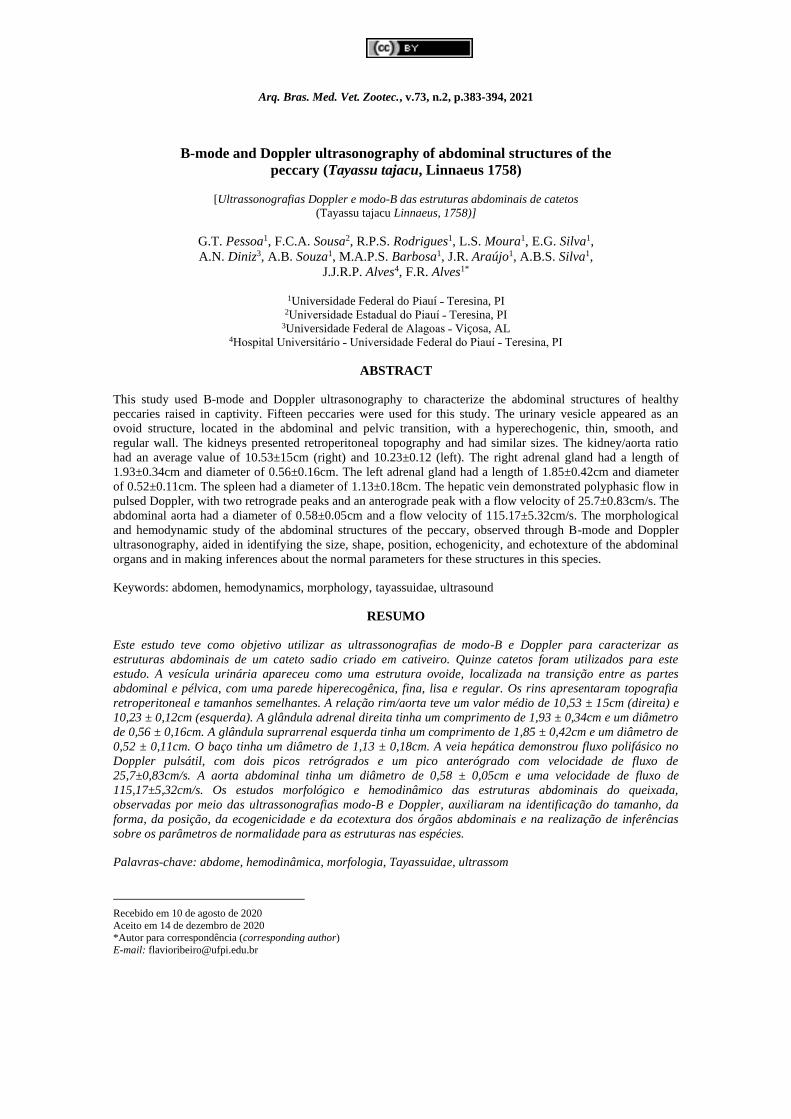
Arq. Bras. Med. Vet. Zootec., v.73, n.2, p.383-394, 2021 B-mode and Doppler ultrasonography of abdominal structures of the peccary (Tayassu tajacu, Linnaeus 1758) [Ultrassonografias Doppler e modo-B das estruturas abdominais de catetos (Tayassu tajacu Linnaeus, 1758)] G.T. Pessoa 1 , F.C.A. Sousa 2 , R.P.S. Rodrigues 1 , L.S. Moura 1 , E.G. Silva 1 , A.N. Diniz 3 , A.B. Souza 1 , M.A.P.S. Barbosa 1 , J.R. Araújo 1 , A.B.S. Silva 1 , J.J.R.P. Alves 4 , F.R. Alves 1* 1 Universidade Federal do Piauí ˗ Teresina, PI 2 Universidade Estadual do Piauí ˗ Teresina, PI 3 Universidade Federal de Alagoas ˗ Viçosa, AL 4 Hospital Universitário ˗ Universidade Federal do Piauí ˗ Teresina, PI ABSTRACT This study used B-mode and Doppler ultrasonography to characterize the abdominal structures of healthy peccaries raised in captivity. Fifteen peccaries were used for this study. The urinary vesicle appeared as an ovoid structure, located in the abdominal and pelvic transition, with a hyperechogenic, thin, smooth, and regular wall. The kidneys presented retroperitoneal topography and had similar sizes. The kidney/aorta ratio had an average value of 10.53±15cm (right) and 10.23±0.12 (left). The right adrenal gland had a length of 1.93±0.34cm and diameter of 0.56±0.16cm. The left adrenal gland had a length of 1.85±0.42cm and diameter of 0.52±0.11cm. The spleen had a diameter of 1.13±0.18cm. The hepatic vein demonstrated polyphasic flow in pulsed Doppler, with two retrograde peaks and an anterograde peak with a flow velocity of 25.7±0.83cm/s. The abdominal aorta had a diameter of 0.58±0.05cm and a flow velocity of 115.17±5.32cm/s. The morphological and hemodynamic study of the abdominal structures of the peccary, observed through B-mode and Doppler ultrasonography, aided in identifying the size, shape, position, echogenicity, and echotexture of the abdominal organs and in making inferences about the normal parameters for these structures in this species. Keywords: abdomen, hemodynamics, morphology, tayassuidae, ultrasound RESUMO Este estudo teve como objetivo utilizar as ultrassonografias de modo-B e Doppler para caracterizar as estruturas abdominais de um cateto sadio criado em cativeiro. Quinze catetos foram utilizados para este estudo. A vesícula urinária apareceu como uma estrutura ovoide, localizada na transição entre as partes abdominal e pélvica, com uma parede hiperecogênica, fina, lisa e regular. Os rins apresentaram topografia retroperitoneal e tamanhos semelhantes. A relação rim/aorta teve um valor médio de 10,53 ± 15cm (direita) e 10,23 ± 0,12cm (esquerda). A glândula adrenal direita tinha um comprimento de 1,93 ± 0,34cm e um diâmetro de 0,56 ± 0,16cm. A glândula suprarrenal esquerda tinha um comprimento de 1,85 ± 0,42cm e um diâmetro de 0,52 ± 0,11cm. O baço tinha um diâmetro de 1,13 ± 0,18cm. A veia hepática demonstrou fluxo polifásico no Doppler pulsátil, com dois picos retrógrados e um pico anterógrado com velocidade de fluxo de 25,7±0,83cm/s. A aorta abdominal tinha um diâmetro de 0,58 ± 0,05cm e uma velocidade de fluxo de 115,17±5,32cm/s. Os estudos morfológico e hemodinâmico das estruturas abdominais do queixada, observadas por meio das ultrassonografias modo-B e Doppler, auxiliaram na identificação do tamanho, da forma, da posição, da ecogenicidade e da ecotextura dos órgãos abdominais e na realização de inferências sobre os parâmetros de normalidade para as estruturas nas espécies. Palavras-chave: abdome, hemodinâmica, morfologia, Tayassuidae, ultrassom Recebido em 10 de agosto de 2020 Aceito em 14 de dezembro de 2020 *Autor para correspondência (corresponding author) E-mail: [email protected]
-
Upload
khangminh22 -
Category
Documents
-
view
1 -
download
0
Transcript of B-mode and Doppler ultrasonography of abdominal structures ...
Arq. Bras. Med. Vet. Zootec., v.73, n.2, p.383-394, 2021
B-mode and Doppler ultrasonography of abdominal structures of the
peccary (Tayassu tajacu, Linnaeus 1758)
[Ultrassonografias Doppler e modo-B das estruturas abdominais de catetos
(Tayassu tajacu Linnaeus, 1758)]
G.T. Pessoa1, F.C.A. Sousa2, R.P.S. Rodrigues1, L.S. Moura1, E.G. Silva1,
A.N. Diniz3, A.B. Souza1, M.A.P.S. Barbosa1, J.R. Araújo1, A.B.S. Silva1,
J.J.R.P. Alves4, F.R. Alves1*
1Universidade Federal do Piauí ˗ Teresina, PI 2Universidade Estadual do Piauí ˗ Teresina, PI
3Universidade Federal de Alagoas ˗ Viçosa, AL 4Hospital Universitário ˗ Universidade Federal do Piauí ˗ Teresina, PI
ABSTRACT
This study used B-mode and Doppler ultrasonography to characterize the abdominal structures of healthy
peccaries raised in captivity. Fifteen peccaries were used for this study. The urinary vesicle appeared as an
ovoid structure, located in the abdominal and pelvic transition, with a hyperechogenic, thin, smooth, and
regular wall. The kidneys presented retroperitoneal topography and had similar sizes. The kidney/aorta ratio
had an average value of 10.53±15cm (right) and 10.23±0.12 (left). The right adrenal gland had a length of
1.93±0.34cm and diameter of 0.56±0.16cm. The left adrenal gland had a length of 1.85±0.42cm and diameter
of 0.52±0.11cm. The spleen had a diameter of 1.13±0.18cm. The hepatic vein demonstrated polyphasic flow in
pulsed Doppler, with two retrograde peaks and an anterograde peak with a flow velocity of 25.7±0.83cm/s. The
abdominal aorta had a diameter of 0.58±0.05cm and a flow velocity of 115.17±5.32cm/s. The morphological
and hemodynamic study of the abdominal structures of the peccary, observed through B-mode and Doppler
ultrasonography, aided in identifying the size, shape, position, echogenicity, and echotexture of the abdominal
organs and in making inferences about the normal parameters for these structures in this species.
Keywords: abdomen, hemodynamics, morphology, tayassuidae, ultrasound
RESUMO
Este estudo teve como objetivo utilizar as ultrassonografias de modo-B e Doppler para caracterizar as
estruturas abdominais de um cateto sadio criado em cativeiro. Quinze catetos foram utilizados para este
estudo. A vesícula urinária apareceu como uma estrutura ovoide, localizada na transição entre as partes
abdominal e pélvica, com uma parede hiperecogênica, fina, lisa e regular. Os rins apresentaram topografia
retroperitoneal e tamanhos semelhantes. A relação rim/aorta teve um valor médio de 10,53 ± 15cm (direita) e
10,23 ± 0,12cm (esquerda). A glândula adrenal direita tinha um comprimento de 1,93 ± 0,34cm e um diâmetro
de 0,56 ± 0,16cm. A glândula suprarrenal esquerda tinha um comprimento de 1,85 ± 0,42cm e um diâmetro de
0,52 ± 0,11cm. O baço tinha um diâmetro de 1,13 ± 0,18cm. A veia hepática demonstrou fluxo polifásico no
Doppler pulsátil, com dois picos retrógrados e um pico anterógrado com velocidade de fluxo de
25,7±0,83cm/s. A aorta abdominal tinha um diâmetro de 0,58 ± 0,05cm e uma velocidade de fluxo de
115,17±5,32cm/s. Os estudos morfológico e hemodinâmico das estruturas abdominais do queixada,
observadas por meio das ultrassonografias modo-B e Doppler, auxiliaram na identificação do tamanho, da
forma, da posição, da ecogenicidade e da ecotextura dos órgãos abdominais e na realização de inferências
sobre os parâmetros de normalidade para as estruturas nas espécies.
Palavras-chave: abdome, hemodinâmica, morfologia, Tayassuidae, ultrassom
Recebido em 10 de agosto de 2020
Aceito em 14 de dezembro de 2020 *Autor para correspondência (corresponding author)
E-mail: [email protected]
ELIANA SILVA
Texto digitado
http://dx.doi.org/10.1590/1678-4162-11968
Editora
Carimbo
ELIANA SILVA
Texto digitado
G.T. Pessoa https://orcid.org/0000-0002-6285-404X F.C.A. Sousa https://orcid.org/0000-0001-7244-9729 R.P.S. Rodrigues https://orcid.org/0000-0002-8108-4669 L.S. Moura https://orcid.org/0000-0002-6070-2763 E.G. Silva https://orcid.org/0000-0002-1131-9856 A.N. Diniz https://orcid.org/0000-0002-2840-853X A.B. Souza https://orcid.org/0000-0001-8817-1493 M.A.P.S. Barbosa https://orcid.org/0000-0002-1071-1258 J.R. Araújo https://orcid.org/0000-0002-3108-8098 A.B.S. Silva https://orcid.org/0000-0002-6569-5515 J.J.R.P. Alves https://orcid.org/0000-0003-0070-932X F.R. Alves https://orcid.org/0000-0002-4935-3486

Pessoa et al.
384 Arq. Bras. Med. Vet. Zootec., v.73, n.2, p.383-394, 2021
INTRODUCTION
Ultrasonography has been increasingly used in
the routine diagnosis of pathological processes in
wild species, especially as it is a noninvasive
method and allows real-time characterization of
abdominal and thoracic structures in these
animals (Cruz and Freitas, 2001). A number of
studies have addressed the morphological aspects
of abdominal structures in wild species such as
giant anteaters (Myrmecophaga tridactyla)
(Lopes et al., 2015), forest foxes (Cerdocyon
thous) (Silva et al., 2014), lowland pacas
(Cuniculus paca) (Oliveira et al., 2003, 2007;
Feliciano et al., 2014), agouti (Dasyprocta
prymnolopha) (Sousa et al., 2012, 2016, 2017),
peccaries (Tayassu tajacu) (Peixoto et al., 2012),
coatis (Nasua nasua) (Ribeiro et al., 2013),
tufted capuchin monkeys (Cebus apella) (Alves
et al., 2007), cheetahs (Acinonyx jubatus)
(Carstens et al., 2006), and white-tufted-ear
marmosets (Callithrix jacchus) (Wagner and
Kirberger, 2005).
The peccary is a wild mammal belonging to the
order Artiodactyla, family Tayassuidae, and
genus Tayassu. Its weight varies between 18 and
25kg, and its height is between 40 and 50cm
(Orr, 1986). Peccaries are found from the
southwestern United States to Argentina living in
diverse habitats (Bodmer and Sowls, 1993;
Miller and Fowler, 2012), which demonstrates
their great resistance and adaptability. Although
some data on the ultrasonographic anatomy of
the peccary have been addressed by Peixoto et
al., (2012), this is the first study to acquire
morphological data from hemodynamic
measurements using ultrasonography in this
species. This study used B-mode and Doppler
ultrasonography to characterize and evaluate the
abdominal structures of peccaries raised in
captivity, generating data that allow inferences to
be made on parameters of morphological
normality, on the basis of ultrasonographic and
Doppler-velocimetric anatomical criteria, as part
of a collaboration for the ecological preservation
of these tayassuids.
MATERIALS AND METHODS
Fifteen adult peccaries (Tayassu tajacu,
Linnaeus, 1758) were used for this study: eight
males and seven females, with ages varying
between 1 and 2 years and weights between 15
and 18kg. All animals were healthy, with
nutritional management characterized by the
offer of commercial pig feed containing 18.0%
crude protein, 3,300kcal /kg of digestible energy,
twice a day, in addition to continuous access to
water. These peccaries were acquired from the
Center for the Study and Preservation of Wild
Animals (NEPAS) (IBAMA Registry N° 02/08-
618), Center for Agricultural Sciences (Centro de
Ciências Agrárias - CCA), Federal University of
Piauí (Universidade Federal do Piauí - UFPI),
Teresina, Piauí State, Brazil (5°02'45.7"S
42°46'53.7"W).
This study was approved by the Committee on
Ethics in Animal Experimentation (Comitê de
Ética em Experimentação Animal –
CEEA/UFPI) (N° 013/15) and authorized by the
System of Authorization and Information on
Biodiversity (Sistema de Autorização e
Informação da Biodiversidade – SISBIO) of the
Brazilian Institute of Environment and
Renewable Natural Resources (Instituto
Brasileiro do Meio Ambiente e Recursos
Naturais Renováveis – IBAMA) (N° 47199-1).
The animals were fasted from food for 12 hours
and water for 3 hours and captured in their
enclosures with hand nets. For chemical
immobilization, a combination of 15mg/kg of
ketamine hydrochloride (Vetanarcol®) and
3mg/kg of midazolam maleate (Dormonid®) was
applied intramuscularly. The animals were
manipulated after 15 minutes, when the onset of
the anesthetic effect was verified, for a duration
of 45 minutes, and no reinforcement dose was
necessary to complete the ultrasonographic
procedure.
The animals were positioned in dorsal decubitus,
and a wide trichotomy of the abdomen was
performed. The exams were carried out using M-
Turbo equipment (Sonosite FUJIFILM®) coupled
to a multifrequency sector transducer (P10x,
from 4.0 to 8.0 MHz). The exam was initiated
using the urinary bladder as an acoustic window
and with a counterclockwise abdominal scan.
The kidneys, left adrenal gland, spleen, stomach,
pancreas, liver, right kidney, and right adrenal
gland were inspected. The abdominal vessels and
intestinal loops were inspected after this primary
analysis. The analyzed morphological
characteristics are described in Table. 1.
B-mode and Doppler et al.
Arq. Bras. Med. Vet. Zootec., v.73, n.2, p.383-394, 2021 385
The aorta, in its abdominal portion (at the level
of the renal artery); the renal arteries; caudal
vena cava; hepatic vein; and portal vein were
accessed by color and pulsed Doppler. The
measurements of flow by colored Doppler
measured the streamlines and direction of blood
flow in the vessels. Pulsed Doppler was used to
evaluate the systolic peak velocity (SPV), final
diastolic velocity (FDV), and resistivity index
(RI). Insonation angles smaller than 60° were
used to obtain the maximum Doppler signal
response. The data were subjected to error
normality tests (Shapiro-Wilk and Kolmogorov-
Smirnov tests), and the means were then
analyzed using a paired Student’s t-test to
interpret the parameters, with a confidence
interval of 5% (P<0.05).
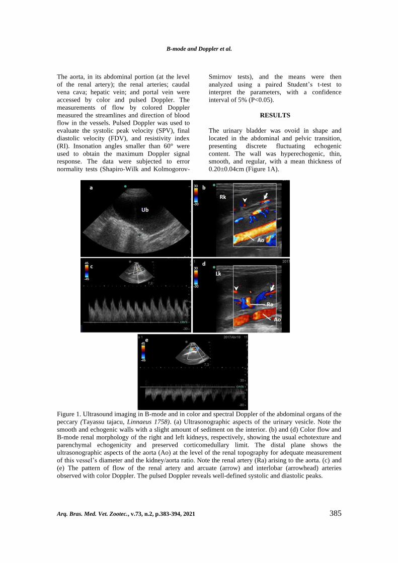
RESULTS
The urinary bladder was ovoid in shape and
located in the abdominal and pelvic transition,
presenting discrete fluctuating echogenic
content. The wall was hyperechogenic, thin,
smooth, and regular, with a mean thickness of
0.20±0.04cm (Figure 1A).
Figure 1. Ultrasound imaging in B-mode and in color and spectral Doppler of the abdominal organs of the
peccary (Tayassu tajacu, Linnaeus 1758). (a) Ultrasonographic aspects of the urinary vesicle. Note the
smooth and echogenic walls with a slight amount of sediment on the interior. (b) and (d) Color flow and
B-mode renal morphology of the right and left kidneys, respectively, showing the usual echotexture and
parenchymal echogenicity and preserved corticomedullary limit. The distal plane shows the
ultrasonographic aspects of the aorta (Ao) at the level of the renal topography for adequate measurement
of this vessel’s diameter and the kidney/aorta ratio. Note the renal artery (Ra) arising to the aorta. (c) and
(e) The pattern of flow of the renal artery and arcuate (arrow) and interlobar (arrowhead) arteries
observed with color Doppler. The pulsed Doppler reveals well-defined systolic and diastolic peaks.

Pessoa et al.
386 Arq. Bras. Med. Vet. Zootec., v.73, n.2, p.383-394, 2021
The kidneys showed retroperitoneal topography,
with the right kidney positioned more cranially.
The splenic caudal margin appeared to coexist
with the cranial pole of the left kidney. The
cranial pole of the right kidney was shown to be
near the liver, within the same antimere
(hepatorenal fossa). A two-dimensional
ultrasonographic examination of the left kidney
showed a homogeneous, thin, and
hypoechogenic echotexture compared to the
spleen, and the right kidney showed an
isoechogenic or discreetly hypoechogenic
echotexture compared to the liver. The
corticomedullary border was preserved and well
individualized, and the pelvic recess was free of
obstructive processes, dilations, and stones
(Figure 1B and D).
The right and left kidneys were similar in length
and diameter, and no significant difference
existed between them for these variables
(P>0.05). The kidney/aorta ratio showed mean
values of 10.53±0.15cm (right kidney) and
10.23±0.12cm (left kidney), with no significant
difference (P>0.05) between these averages
(Table. 2). A positive and elevated correlation
was observed between the renal length and aortic
diameter: r=0.91 (right kidney/aorta) and r=0.97
(left kidney/aorta) (Figure 2).
Figure 2. Correlation between the renal length and aortic diameter of the peccary (Tayassu tajacu,
Linnaeus 1758). (a) Correlation between the right kidney and the diameter of the aorta. (b) Correlation
between the left kidney and the diameter of the aorta.
Renal artery hemodynamic evaluation
demonstrated the presence of laminar flow
throughout, without the occurrence of stenosis,
dilations, or hypo flow. This artery bifurcates
into the renal hilum and provides the origin of
the interlobular and arcuate arteries within the
renal parenchyma (Figure 1B and D). Pulsed
Doppler ultrasonography of the renal artery
demonstrated the presence of semi parabolic
flow with a pattern of low resistance, a high
systolic peak (velocities of 114.33±5.42cm/s in
the right kidney and 116.58±5.26cm/s in the left
kidney), a characteristic protodiastolic notch, and
a continuous and full diastolic portion, which

B-mode and Doppler et al.
Arq. Bras. Med. Vet. Zootec., v.73, n.2, p.383-394, 2021 387
decreases gradually during diastole (Figure 1C
and E, respectively). The resistivity indices for
these animals showed mean values of 0.64±0.11
(right kidney) and 0.65±0.08 (left kidney), with
no significant difference for these variables
(P>0.05) (Table. 2).
The abdominal aorta was observed in its
abdominal path from the aortic hiatus, giving rise
to the celiac trunk, cranial mesenteric artery,
renal artery, and iliac arteries. No significant
difference existed in the diameter over the entire
path examined (P>0.05). Thus, the measurement
of the diameter and the Doppler-velocimetric
indices were standardized at the level of the renal
arteries. Laminar flow was observed with a flat
and high systolic peak, a wide spectral window,
and a flow pattern of high resistivity. The
diameter of the abdominal aorta was
0.58±0.05cm, and the flow velocity measured
115.17±5.32cm/s, with an RI measuring 0.78
(Figure 3A and B).
Figure 3. Ultrasound imaging in B-mode and in color and spectral Doppler of the abdominal organs of the
peccary (Tayassu tajacu, Linnaeus 1758). (a) and (b) The color and pulsed characteristics of the
abdominal aorta (Ao). Laminar flow was observed with a flat and high systolic peak, a wide spectral
window, and flow pattern of high resistivity. The left kidney (Lk) was observed in the proximal plane and
the left renal artery (LRa) was observed arising from the aorta. (c) The right adrenal gland (R Adr) is
located cranially to the right renal artery (RRa) and medially to the cranial pole of the right kidney (Rk).
(d) The left adrenal gland (L adr) was observed cranially to the left renal artery, ventrally to the aorta, and
in varied coexistence with the cranial pole of the left kidney (Lk).
The adrenal glands were paired, bilobed, and
elliptical in shape, with greater variation
occurring in the right adrenal gland. They were
situated ventrally to the second or third lumbar
vertebrae, in the retroperitoneal space. The right
adrenal gland is located cranially to the right
renal artery, medially to the cranial pole of the
right kidney, in close proximity to the origin of
the celiac and cranial mesenteric arteries, and
dorsally to the caudal vena cava. The left adrenal

Pessoa et al.
388 Arq. Bras. Med. Vet. Zootec., v.73, n.2, p.383-394, 2021
gland was seen cranially to the left renal artery,
ventrally to the aorta, and in varied coexistence
with the cranial pole of the left kidney,
depending on the location of this organ. The
right adrenal gland showed a variation in length
between 1.55 and 2.66cm (1.93±0.34cm) and a
diameter ranging from 0.36 to 1.05cm
(0.56±0.16cm). The left adrenal gland had a
length between 1.25 and 2.84cm (1.85±0.42cm)
and a diameter between 0.36 and 0.77cm
(0.52±0.11cm), with no difference between the
means for the variables studied (Figure 3C and
D) (P>0.05).
The pancreas had a regular and defined contour
and was distinguishable in its topography from
other structures. The right lobe was positioned
dorsomedially to the descending duodenum,
ventrally to the right kidney, ventrolaterally to
the portal vein, and in close relation with the
pyloric portion of the stomach. Its echotexture
was homogeneous and hyperechogenic. The
pancreatic duct was distinguished within this
organ by the echogenic characteristic of its walls.
Discernment of precise segments of the left lobe
was well defined with similar echotexture of the
right pancreas. The thickness of the pancreas was
0.79±0.23cm (right lobe) and 0.87±0.21cm (left
lobe). The pancreatic duct measured
0.17±0.05cm (right lobe) and 0.14±0.02cm (left
lobe) (Figure 4A and B).
The stomach contained gases and was positioned
caudally to the liver from a longitudinal
approach. The stomach wall was 0.18±0.05cm in
thickness, echogenic with a homogeneous
echotexture, and positioned to the left of the
spleen and the right of the proximal duodenum
and transverse colon (Figure 4C). The proximal
duodenum had a thickness of 0.4±0.13cm and
was positioned laterally to the right wall. The
other segments of the duodenum and intestinal
loops could not be adequately identified due to
the amount of gases present in the loops during
the examination.
Figure 4. Ultrasound imaging in B-mode of the abdominal organs of the peccary (Tayassu tajacu,
Linnaeus 1758). (a) and (b) Right and left lobes of the pancreas, respectively. Its echotexture was
homogeneous and hyperechogenic. The pancreatic duct was distinguished within this organ by the
echogenic characteristic of its walls. (c) The stomach, echogenic and with homogeneous echotexture, and
its closed relationship with the transverse colon. (d) The spleen with filiform shape, with pointed poles,
maintaining a syntopic relation with the greater curvature of the stomach.

B-mode and Doppler et al.
Arq. Bras. Med. Vet. Zootec., v.73, n.2, p.383-394, 2021 389
The spleen had a filiform shape, with pointed
poles and a diameter of 1.13±0.18cm,
maintaining a syntopic relation with the greater
curvature of the stomach and the cranial pole of
the left kidney. Its echotexture appeared to be
fine and homogeneous throughout its length,
being more echogenic than the cortical of the left
kidney (Figure 4D). The liver of the peccary
occupies the cranial space of the abdominal
cavity and is in direct contact with the diaphragm
(Figure 5A). The hepatic lobulation cannot be
adequately described by ultrasonography because
the echotexture and similar acoustic impedances
between lobes prevent proper individualization
in normal animals (without the presence of
peritoneal effusion).
However, this organ presents impressions of the
adjacent structures, which can be adequately
described, relating centrally to the stomach, with
the right kidney (hepatorenal fossa) and
duodenum in the most cranial portion. The
echotexture of the liver is thin and homogeneous,
isoechogenic in relation to the renal cortical
parenchyma, and hypoechoic in relation to the
spleen. Although approaches were attempted on
the sagittal and subcostal planes of both
antimeres, the gallbladder was not visible in any
of the studied animals.
Figure 5. Ultrasound imaging in B-mode and in color and spectral Doppler of the liver of the peccary
(Tayassu tajacu, Linnaeus 1758). (a)-(c) Using color Doppler, the portal flow showed a color map with a
red (hepatopetal) signal, while the hepatic veins showed a blue (hepatofugal) signal. Note the laminar
flow in both vessels. The portal vein showed a relatively broad and linear pulse discretely influenced by
the respiratory phase. (d) The pulsed Doppler in the hepatic vein showed a polyphasic flow, with two
retrograde peaks and one anterograde peak. (e)-(f) The profile wave of the caudal vena cava, laminar and
with two evident anterograde peaks.
The portal vein was distinguished in the
ultrasound examination by the echogenic pattern
of its walls compared to the hepatic veins. Using
color Doppler, the portal flow showed a color
map with a red (hepatopetal) signal, while the
hepatic veins showed a blue (hepatofugal) signal

Pessoa et al.
390 Arq. Bras. Med. Vet. Zootec., v.73, n.2, p.383-394, 2021
(Figure 5A and C). The flows were laminar in
both vessels. When analyzed with pulsed
Doppler, the portal vein showed a relatively
broad and linear pulse that was discretely
influenced by the respiratory phase, with flow
velocity measured at 21.17±1.32cm and an RI of
0.37±0.09 (Figure 5B).
The color Doppler in the hepatic vein showed
laminar flow (Figure 5C). Pulsed Doppler in this
vein showed a polyphasic flow, with two
retrograde peaks and one anterograde peak with
a velocity of 25.7±0.83cm/s; this last peak
reflected the influence of the cardiac cycle
(Figure 5D). The caudal vena cava receives the
hepatic veins and could be seen in the distal
plane of the liver, accessed from the left
subcostal region. The caudal vena cava had
laminar flow, with a flow velocity of
46.47±2.34cm/s and an RI of 0.42±0.06 (Figure
5E and F).
DISCUSSION
The ultrasound evaluations of the abdominal
structures of the studied peccaries were
performed in planes cut along the longitudinal,
transverse, and dorsal axes (Barberet et al.,
2008). Although the animals were fasted from
food for 12 hours and water for 3 hours, the
difficulty of emptying the gastrointestinal tract of
wild animals has been recognized (Sousa et al.,
2012). This difficulty is a limiting factor in the
adequate preparation for ultrasonographic
examinations, with the main implication being
the amount of gas found in the intestinal loops,
as observed in this study. However, Garcia and
Froes (2014) report that no significant changes
were observed in the ultrasound patterns of
abdominal structures of animals with and without
fasting.
Chemical immobilization is part of the
ultrasonographic protocol for wild species,
especially in order to minimize immobilization
stress and ensure adequate manipulation safety
for the animal and the examiner (Batista et al.,
2009). The anesthetic protocol used in this study
produced adequate sedation without
hemodynamic compromise, which would alter
the flow measurements taken (all animals were
monitored for cardiovascular changes using an
electrocardiogram and blood pressure
measurements). This anesthetic protocol was
used with great success in the peccary by Pessoa
et al., (2014) to collect adipose tissue, by Argôlo
Neto et al., (2016) to collect bone marrow, and
by Bezerra et al., (2014) in establishing a
preclinical model of renal injury from renal
artery clamping. Batista et al., (2009) reported
that chemical tranquilization in the peccary
significantly reduces heart rate, rectal
temperature, biochemical parameters, and stress
indicators, making it easier to manipulate the
animal.
The urinary vesicle presented similar shape,
location, and syntropy to those observed in other
wild and domestic animals, such as cheetahs
(Acinonyx jubatus) (Carstens et al., 2006), forest
foxes (Cerdocyon thous) (Silva et al., 2014),
coatis (Nasua nasua) (Ribeiro et al., 2013), and
lowland pacas (Cuniculus paca) (Oliveira et al.,
2003, 2007; Feliciano et al., 2014). In the white-
tufted-ear marmoset (Callithrix jacchus), the
urinary vesicle is multilobulated (Wagner and
Kirberger, 2005), differing from the ovoid shape
found in the peccary in the present study. The
urinary vesicle of the animals studied presented a
thickness of 0.20±0.04cm, corroborating the
descriptions made for the same species by
Peixoto et al., (2012) (0.20±0.08cm). The lower
standard deviation observed in the present study
probably occurred because of the larger number
of samples evaluated and the use of a probe that
allowed greater measurement precision. In
addition, similar values were found by Alves et
al., (2007) in tufted capuchin monkeys (Cebus
apella).
The kidneys of the evaluated peccary presented
an ultrasonographic appearance similar to that
described for healthy dogs of similar size (Hart et
al., 2013). Regarding renal morphometry, the
measurements of this study were close to the
values established by Sampaio and Araújo
(2002) for dogs of unspecified breed with
weights between 10.1 and 20kg. The preserved
corticomedullary limit encountered in the
peccaries was also observed by Silva et al.,
(2014) in forest foxes (Cerdocyon thous) and by
Feliciano et al., (2014) in lowland pacas
(Cuniculus paca). Renal size is an important
parameter in evaluating renal disease with an
acute or chronic outcome. In dogs, renal size is
naturally difficult to measure using an
ultrasound, principally due to the great variation
in animal size and breed differentiation (Barberet

B-mode and Doppler et al.
Arq. Bras. Med. Vet. Zootec., v.73, n.2, p.383-394, 2021 391
et al., 2008). Mareschal et al., (2007) established
a standard of renal size normality (kidney/aorta
ratio) between 5.5 and 9.1 when the relation
between the kidney length and luminal diameter
of the aorta is measured. The kidney/aorta ratio
in the peccaries studied (10.53±0.15 for the right
kidney and 10.23±0.12 for the left) can be
elucidative when determining the size and
function of kidneys because of the small
variation in the peccary body score.
Renal evaluation using color Doppler showed
laminar flow and absence of stenosis, dilations,
or hypoflows. Similarly, pulsed Doppler
(114.33±5.42cm/s in the right kidney and
116.58±5.26cm/s in the left kidney) showed
values higher than those observed in dogs by
Melo et al., (2006). This divergence is probably
found in the hemodynamic response
characteristic of the vascular wall, as occurs in
humans (Viazzi et al., 2014), where velocity
variations that reach 100cm/s are considered
normal in the presence of laminar flow, spectral
wave morphology, and preserved resistivity
indices. In fact, in the present study, pulsed flow
patterns were similar to those described in the
literature, and resistivity indices indicated a low
resistivity flow pattern (0.64±0.11 in the right
renal artery and 0.65±0.08 in the left renal
artery). In wild felines (Acinonyx jubatus),
Carstens et al., (2006) found an index with an
average value (0.58) similar to that observed in
the peccary.
As with domestic animals (dogs and cats), where
ultrasonographic abdominal study is well
established, evaluations of the adrenal gland
show high variation and standard deviations.
These results are caused by variation in size and
breeds (Mareschal et al., 2007). Wild species are
devoid of measurement values for the adrenal
gland (Alves et al., 2007). In dogs, the threshold
value suggesting an adrenal increase is 0.74cm
(Barthez et al., 1995). Soulsby et al., (2015)
stratified maximum values according to the size
of the animals and based on the caudal adrenal
pole thickness in the sagittal plane: canines
weighing <10kg (0.54cm), between 10 and 30kg
(0.68cm), and >30kg (0.80cm). For the peccaries
examined in the present study, the average values
of 0.56±0.16cm and 0.52±0.11cm were obtained
for the right and left adrenal glands, respectively.
Thus, the peccaries in this experiment fit into the
weight range of <10kg, although the right
adrenal gland had a slightly elevated mean value.
The spleen of the tayassuids had a thin
homogeneous echotexture, similar to that
observed by Silva et al., (2014) in forest foxes
(Cerdocyon thous) and by Feliciano et al., (2014)
in lowland pacas (Cuniculus paca). The
echogenicity of the spleen was higher than that
of the left renal cortical, a fact also observed in
coatis (Nasua nasua) by Ribeiro et al., (2013)
and in forest foxes (Cerdocyon thous) by Silva et
al., (2014) The diameter of this organ
(1.13±0.18cm) established in this study was
lower than that reported by Peixoto et al., (2012)
for the same species. However, our research used
a larger number of animals, of both sexes, and
showed lower standard deviation when compared
to the data reported by Peixoto et al., 2012.
The liver occupies the cranial space of the
abdominal cavity and shows a homogeneous
echotexture and the absence of a gallbladder. The
absence of the gallbladder had previously been
reported in anatomical studies by Sowls (1974).
A hepatic parenchyma with homogeneous
contrast characteristics was also described by
Ryu et al., (2009) in computed tomography
studies. In the peccary, flow velocities of the
hepatic vein (25.7±0.83cm/s) and caudal vena
cava (46.47±2.34cm/s) were observed to be
greater than the velocities established by
Carstens et al., (2006) in cheetahs
(21.23±0.41cm/s and 33.8±19.8cm/s,
respectively) for the same veins. The hepatic
vein flow pattern exhibited by pulsed Doppler
was characterized by a peak above the baseline
and two peaks below it, showing the influence of
the cardiac cycle on this blood vessel.
Corroborating our results, Huang et al., (2004)
also identified biphasic flow of the hepatic vein
correlated with breathing or pressure changes in
the right atrium. Bogin et al., (2005) reported
that hepatic and portal vein flow velocities are
important in evaluating occlusive vascular
diseases in humans, mainly with inverted portal
vein flow and reduced monophasic hepatic flow
velocity.
Barberet et al., (2008) quantified the detection of
pancreatic ultrasound images and verified that
the right pancreatic lobe, pancreatic body, and
left pancreatic lobe had frequencies of 56%,
60%, and 87%, respectively, in 100 canine

Pessoa et al.
392 Arq. Bras. Med. Vet. Zootec., v.73, n.2, p.383-394, 2021
patients. In the peccary, only the right lobe was
visualized, located dorsomedially to the
descending duodenum, ventrally to the right
kidney, and ventrolaterally to the portal vein.
This characteristic was also described by Santis-
Prada and Duti Neto (1978) in swine. These
authors reported a pancreas of small size and
annular appearance, completely enfolding the
intestine. Although a detailed anatomical
description of this organ has been made, the
relation of its proximity to the proximal
duodenum and its small dimensions may have
made it difficult to fully visualize the pancreas
using ultrasonographic examination, making its
echogenicity diffuse, marginally delineated, and
difficult to distinguish.
The ultrasound characteristics of the stomach of
Tayassu tajacu are unique. According to
Cavalcante-Filho et al., (1998), these animals
have a pluricavitary stomach formed by two
blind sacs, one cranioventral and one
caudodorsal; one gastric pouch; and one
compartment located in the right antimere. The
walls of the stomach (0.18±0.05cm) and
duodenum (0.4±0.13cm) of the peccary are
thicker than those of the stomach (0.10±0.01cm)
and duodenum (0.19±0.04cm) of rabbits
(Banzato et al., 2015) and forest foxes
(0.39±0.05cm) (Silva et al., 2014).
Kamikawa and Bombonato (2007) described the
mean diameter of the abdominal aorta at 0.74cm,
whereas for the peccary, these values were
0.58±0.05cm. Nevertheless, this characteristic
does not appear to have produced hemodynamic
interference in the flow values of this artery.
Carvalho et al., (2008) described mean aortic
flow velocities of 95.49±35.43cm/s for domestic
dogs, and in this study, the aortic flow for the
peccary was 115.17±5.32cm/s, which is slightly
higher than the flow found in dogs of equal size.
CONCLUSIONS
The morphological and hemodynamic study of
the abdominal structures of the peccary, by
means of B-mode and Doppler ultrasonography,
aided in identifying the size, shape, position,
echogenicity, and echotexture of abdominal
organs. Together with Doppler-velocimetric
index measurement, this study allows the
inference of normal parameters for these
structures in this species.
REFERENCES
ALVES, F.R.; COSTA, F.B.; AROUCHE,
M.M.S. et al. Avaliação ultra-sonográfica do
sistema urinário, figado e útero do macaco-
prego, Cebus apella. Pesq. Vet. Bras., v.27,
p.377-382, 2007.
ARGÔLO NETO, N.M.A.; FEITOSA, M.L.T.;
SOUSA, S.S. et al. Isolation, expansion,
differentiation and growth kinetics essay in
mesenchymal stem cells culture from the bone
marrow of collared peccaries (Tayassu tajacu).
Acta Sci. Vet., v.44, p.1-11, 2016.
BANZATO, T.; BELLINI, L.; CONTIERO, B.,
SELLERI, P.; ZOTTI, A. Abdominal ultrasound
features and reference values in 21 healthy
rabbits. Vet. Rec., v.176, p.101, 2015.
BARBERET, V.; SCHERERS, E.;
RADEMACHER, N. et al. Quantification of the
effect of various patient and image factors on
ultrasonographic detection of select canine
abdominal organs. Vet. Radiol. Ultrasound, v.49,
p.273-276, 2008.
BARTHEZ, P.Y.; NYLAND, T.G.; FELDMAN,
E.C. Ultrasonographic evaluation of the adrenal
glands in dogs. Vet. Radiol. Ultrasound, v.207,
p.1180-1183, 1995.
BATISTA, J.S.; BEZERRA, F.S.B.; AGRA,
E.G.D. et al. Efeitos da contenção física e
química sobre os parâmetros indicadores de
estresse em catetos (Tayassu tajacu). Acta Vet.
Bras., v.3, p.92-97, 2009.
BEZERRA, D.O.; FEITOSA, M.L.; ALMEIDA,
H.M. et al. Collared pecary (Tayassu tajacu) as a
new model of renal ischemic injury induced by
clamping the renal artery. Acta Cir. Bras., v.29,
p.560-572, 2014.
BODMER, R.E.; SOWLS, L.K. The collared
Peccary (Tayassu tajacu). In: OLIVER, W.L.R.
(Ed.). Pigs, peccaries and hippos: status survey
and conservation action plan. Switzerland:
IUCN, 1993. p.7-13.
BOGIN, V.; MARCOS, A.; SHAW-STIFFEL, T.
Budd-Chiari syndrome: in evolution. Eur. J.
Gastroenterol. Hepatol., v.17, p.33-35, 2005.

B-mode and Doppler et al.
Arq. Bras. Med. Vet. Zootec., v.73, n.2, p.383-394, 2021 393
CARSTENS, A.; KIRBERGER, R.M.;
SPOTSWOOD, T.; WAGNER, W.M.;
GRIMBEEK, R.J. Ultrasonography of the liver,
spleen, and urinary tract of the cheetah (Acinonyx
jubatus). Vet. Radiol. Ultrasound, v.47, p.376-
383, 2006.
CARVALHO, C.F.; CHAMMAS, M.C.;
STERMAN, F.D.A., NESTOR, B., GUIDO,
C.G. Ultra-sonografia dúplex-Doppler na
avaliação morfológica e hemodinâ¬mica das
artérias aorta e mesentérica cranial em cães.
Braz. J. Vet. Res. Anim. Sci., v.45, p.24-31, 2008.
CAVALCANTE-FILHO, M.; MIGLINO, M.;
MACHADO, G.; BEVILACQUA, E.; NEVES,
W.C. Comparative study of the morphology of
the stomach of white lipped peccary (Tayassu
pecari) and of the collared peccary (Tayassu
tajacu). Braz. J. Vet. Anim. Sci., v.35, p.20-24,
1998.
CRUZ, J.; FREITAS, V. A ultra-sonografia em
tempo real na reprodução de caprinos. Cienc.
Anim., v.11, p.45-53, 2001.
FELICIANO, R.; ANTONIO, M.; BARROS,
F.F. et al. Conventional and doppler abdominal
ultrasonography in pacas (Cuniculus paca). Acta
Sci.Vet., v.42, p.1-6, 2014.
GARCIA, D.A.; FROES, T.R. Importance of
fasting in preparing dogs for abdominal
ultrasound examination of specific organs. J.
Small Anim. Pract., v.55, p.630-634, 2014.
HART, D.V.; WINTER, M.D.; CONWAY, J.;
BERRY, C.R. Ultrasound appearance of the
outer medulla in dogs without renal dysfunction.
Vet. Radiol. Ultrasound, v.54, p.652-658, 2013.
HUANG, T.L.; CHEN, T.Y.; TSANG, L.L. et al.
Hepatic venous stenosis in partial liver graft
transplantation detected by color Doppler
ultrasound before and after radiological
interventional management. Transplant Proc.,
v.36, p.2342-2343, 2004.
KAMIKAWA, L.; BOMBONATO, P.P. Ultra-
sonografia da aorta abdominal e de seus ramos
em cães. Ciênc. Rural, v.37, p.412-417, 2007.
LOPES, É.R.; MORGADO, T.O.; MEIRELES,
Y.S. et al. Ultrassonografia abdominal de
tamanduás-bandeira (Myrmecophaga tridactyla
Linnaeus, 1758) mantidos em cativeiro. Pesq.
Vet. Bras., v.35, p.919-924, 2015.
MARESCHAL, A.; D’ANJOU, M.A.;
MOREAU, M.; ALEXANDER K,
BEAUREGARD G. Ultrasonographic
measurement of kidney-to-aorta ratio as a
method of estimating renal size in dogs. Vet.
Radiol. Ultrasound, v.48, p.434-438, 2007.
MELO, M.B.; VEADO, J.C.C.; SILVA, E.F.;
MOREIRA, S.M.; PASSOS, L.M.F.
Dopplerfluxometria das artérias renais: valores
normais das velocidades sistólica e diastólica e
do índice resistivo nas artérias renais principais.
Arq. Bras. Med. Vet. Zootec., v.58, p.691-693,
2006.
MILLER, R.E.; FOWLER, M.E. (Ed.). Zoo &
wild animal medicine. Philadelphia: W. B.
Saunders, 2012. 512p.
OLIVEIRA, F.S.; MACHADO, M.R.F.;
CANOLA, J.C. Real time B-mode ultrasound in
pacas pregnancy (Agouti paca, Linnaeus, 1766).
Braz. J. Vet. Res. Anim. Sci., v.40, p.73-78, 2003.
OLIVEIRA, F.S.; MACHADO, M.R.F.;
CANOLA, J.C. Uniparidade em pacas criadas
em cativeiro (Agouti paca, Linnaeus, 1766). Arq.
Bras. Med. Vet. Zootec., v.59, p.387-389, 2007.
ORR, R.T. (Ed.). Biologia dos vertebrados. San
Francisco: Academy of Sciences, 1986. 518p.
PEIXOTO, G.C.; OLIVEIRA, I.R.; ALVES,
N.D.; OLIVEIRA, M.F.; SILVA, A.R.
Abdominal exploration in captive collared
peccaries (Tayassu tajacu) by ultrasonography.
Anat. Histol. Embryol., v.41, p.256-261, 2012.
PESSOA, G.T.; FEITOSA, M.L.T.; ARGÔLO
NETO, N.M. et al. Isolation, culture and
differentiation potential of collared peccary
(Tayassu tajacu) adipose-derived stem cells.
Acta Sci. Vet., V.42, p.1-10, 2014.
RIBEIRO, R.G.; COSTA, A.P.; BRAGATO, N.
Normal sonographic anatomy of the abdomen of
coatis (Nasua nasua Linnaeus 1766). BMC Vet.
Res., v.9, p.124, 2013.
RYU, J.M.; KIM, D.H.; LEE, M.Y. et al.
Imaging evaluation of the liver using multi-
detector row computed tomography in micropigs
as potential living liver donors. J. Vet. Sci., v.10,
p.93-98, 2009.
SAMPAIO, K.M.O.R.; ARAUJO, R. Ultra-
sonografia de características lineares e
estimativas do volume de rins de cães. Arq. Bras.
Med. Vet. Zootec., v.54, p.248-254, 2002.

Pessoa et al.
394 Arq. Bras. Med. Vet. Zootec., v.73, n.2, p.383-394, 2021
SANTIS-PRADA, I.L.; DUTI NETO, J.P.
Pâncreas anular em suíno. Rev. Facul. Med. Vet.
Zootec. Univ., v.15, p.23-30, 1978.
SILVA, A.S.L.; FELICIANO, M.A.R.;
MOTHEO, T.F. et al. Mode B ultrasonography
and abdominal Doppler in crab-eating-foxes
(Cerdocyon thous). Pesq. Vet. Bras., v.34, p.23-
28, 2014.
SOULSBY, S.N.; HOLLAND, M.; HUDSON,
J.A.; BEHREND, E.M. Ultrasonographic
evaluation of adrenal gland size compared to
body weight in normal dogs. Vet. Radiol.
Ultrasound, v.56, p.317-326, 2015.
SOUSA, F.C.; PESSOA, G.T.; MOURA, L.S.
Doppler ultrasound of the placenta and maternal
and fetal vessels during normal gestation in
captive agoutis (Dasyprocta prymnolopha,
Wagler, 1831). Theriogenology, v.86, p.1921-
1930, 2016.
SOUSA, F.C.A.; ALVES, F.R.; FORTES,
E.A.M. et al. Pregnancy in hystricomorpha:
gestational age and embryonic-fetal development
of agouti (Dasyprocta prymnolopha, Wagler
1831) estimated by ultrasonography.
Theriogenology, v.78, p.1278-1285, 2012.
SOUSA, F.C.A.; PESSOA, G.T.; MOURA, L.S.
et al. Organogenesis and foetal haemodynamics
during the normal gestation of healthy black-
rumped agoutis (Dasyprocta prymnolopha,
Wagler, 1831) bred in captivity. Reprod. Domest.
Anim., v.52, p.60-66, 2017.
SOWLS, L.K. Social behavior of the collared
peccary, Dicotyles tajacu, (L). In: GEIST, V.;
WALTHER, F. (Eds.). The behavior of ungulates
and its relation to management. Switzerland:
IUCN MORGES, 1974. p.144-165.
VIAZZI, F.; LEONCINI, G.; DERCHI, L.E.;
PONTREMOLI, R. Ultrasound Doppler renal
resistive index: a useful tool for the management
of the hypertensive patient. J. Hypertens., v.32,
p.149-153, 2014.
WAGNER, W.M.; KIRBERGER, R.M.
Transcutaneous ultrasonography of the abdomen
in the normal common marmoset (Callithrix
jacchus). Vet. Radiol. Ultrasound, v.46, p.251-
258, 2005.