the abdominal mechanisms - Smithsonian Institution
96
SMITHSONIAN MISCELLANEOUS COLLECTIONS VOLUME 94, NUMBER 6 THE ABDOMINAL MECHANISMS OF A GRASSHOPPER BY R. E. SNODGRASS Jureau of Entomology and Plant Quarantir U. S. Department of Agrici:lture (Publication 3335) CITY OF WASHINGTON PUBLISHED BY THE SMITHSONIAN INSTITUTION SEPTEMBER 25, 1935
-
Upload
khangminh22 -
Category
Documents
-
view
0 -
download
0
Transcript of the abdominal mechanisms - Smithsonian Institution

SMITHSONIAN MISCELLANEOUS COLLECTIONSVOLUME 94, NUMBER 6
THE ABDOMINAL MECHANISMSOF A GRASSHOPPER
BY
R. E. SNODGRASSJureau of Entomology and Plant Quarantir
U. S. Department of Agrici:lture
(Publication 3335)
CITY OF WASHINGTON
PUBLISHED BY THE SMITHSONIAN INSTITUTION
SEPTEMBER 25, 1935

C6e S.otb (§aitimoxi (preeeBALTIMORE, MD., (J. 8. A.

THE ABDOMINAL MECHANISMS OF A GRASSHOPPER
By R. E. SNODGRASSBureau of Enfoinology and Plant Quarantine, U. S. Department of Agriculture
CONTENTS PAGE
Introduction i
I. General structure of the abdomen 2
Characteristic features of the abdomen of Acridoidea 3
Relation of the abdomen to the thorax 8
The abdominal spiracles 11
The tympanal organs 12
The cerci 15
II. The abdominal musculature 16
Muscles of the first segment 16
Muscles of the second segment 18
Muscles of the third segment 20
Muscles of the eighth segment 24
Muscles of the ninth segment 26
Muscles of the tenth segment 29
Muscles of the eleventh segment 31
The transverse muscles 31
III. The diaphragms and the dorsal blood vessel 31
IV. The proctodaeum 34
V. The ovipositor and associated structures 37
Structure of the ovipositor 37
The female genital chamber and the spermathecal opening 45
Development of the ovipositor 48
Oviposition 54
VI. The external male genitalia 61
General structure of the m.ale genitalia of Acridoidea 61
Copulation, and insemination of the female /i
Examples of the male genitalia of Acrididae 73
Abbreviations used on the figures .-. . 86
References 87
INTRODUCTION
This paper on the abdomen of Acridoidea is intended to follow
sequentially an earlier paper in the same series entitled " The Thoracic
Mechanism of a Grasshopper" (Smithsonian Misc. Coll., vol. 82,
no. 2, 1929). Hence it will be observed that the numerical designation
of the abdominal muscles continues from that of the thorax.
Smithsonian Miscellaneous Collections, Vol. 94, No. 6

2 SxMITHSONIAN MISCELLANEOUS COLLECTIONS VOL. 94
The primary object of the work here presented has been to arrive
at an understanding of the mechanisms of copulation and oviposition
in the Acrididae, which in this family present many peculiar features.
Neither of these processes, the writer believes, has been fully under-
stood or correctly described, though careful observations have been
made on the processes of copulation and egg-laying among grass-
hoppers. With the closer studies on the behavior of insects now found
necessary for economic purposes, it is becoming obvious that we must
understand more fully the structure and mechanics of the anatomical
mechanisms on which depends so much of the insect's activities. In
addition to the functional phase of morphology, however, there is the
no less important taxonomic aspect. Hence, in the following pages
much attention is given to structures bearing on the relationships be-
tween the Acrididae, Tetrigidae, and Tridactylidae. and a brief com-
parative study of the anatomy of the external male genitalia is included,
since these structures will undoubtedly be found to contain many
characters of importance for the separation of species where other
features are not sufficient for exact determinations.
The writer follows Blatchley (1920), Walker (1922), Brues and
Melander (1932), and others in regarding the grouse locusts as
constituting a family (Tetrigidae, or Acrydidae) distinct from that
of the typical grasshoppers (Acrididae). Aside from superficial differ-
ences in such characters as the length of the pronotum, and in certain
features of the tarsi, the grouse locusts are distinguished from the
grasshoppers by the lack of the characteristic tympanal organs of the
latter, and in the totally different nature of the external male geni-
talia, which in the grasshoppers have a unique and highly standardized
type of structure that distinguishes the Acrididae from all other
Orthoptera. The tetrigids, of course, in many ways, particularly in
the general structure of the abdomen and in the structure and mecha-
nism of the female ovipositor, show their relationship with the Acridi-
dae, but this relationship is much more distant than is that of the
several acridid subfamilies with one another. Some orthopterists,
furthermore, would link the Tridactylidae with the Tetrigidae and
Acrididae, but to the writer a close association of the tridactylids with
the acridoid families seems doubtful, notwithstanding the close simi-
larity of the ovipositor in these two groups.
I. GENERAL STRUCTURE OF THE ABDOMEN
The morphology of the adult insect abdomen is difficult to under-
stand because of the complete suppression of the segmental appendages
in the pregenital region, and the probable union of the appendage bases

NO. 6 GRASSHOPPER ABDOMEN SNODC.RASS 3
with the primitive sterna in the definitive sternal plates. The lateral
tergo-sternal muscles of the ahdomen appear to have no counterparts
in the thorax, unless it is to he assumed that they represent the leg
muscles that have retained their ventral connections with the coxal
elements of the definitive sterna, hut a study of larval insects seems t6
indicate that the limb muscles have been lost with the suppression of
the appendages. The abdomen of the imago is so completely adapted
to its principal mechanical functions of respiration, copulation, and
oviposition that the generalized structure in this region of the body
is almost entirely obscured by secondary modifications. The acridid
abdomen is a good subject for anatomical study, but it throws no light
on the general morphology of the insect abdomen.
CHARACTERISTIC FEATURES OF THE ABDOMEN OF ACRIDOIDEA
The acridid abdomen consists of ii distinct segments (fig. i). The
enlarged first segment is firmly attached to the thorax by its dorsal
\aiIT IXT XT Eppt
\ \ / / CerPapt
Sg CxCs IS IIS VlllStn
Fig. I.—Abdomen and base of thorax of Dissostcira Carolina, female.
and ventral plates (IT, IS), though these plates are widely separated
from each other laterally by the hind coxal cavities (C.vCs). On the
sides of the first tergum are situated the tympanal organs (Tin)
characteristic of the Acrididae, and the first spiracles (ISp) are located
in the anterior parts of the tympanal depressions. The following
seven segmental annuli (II-VIII) are simple secondary segments
separated by ample conjunctivae that allow a considerable extension of
the abdomen, as that of the female abdomen during oviposition. The
tergal and sternal plates are united by inflected lateral membranes that
permit the respiratory movements of vertical expansion and compres-
sion. The spiracles of these segments are located in the lower margins
of the terga.
In the female the sternum of the eighth segment (fig. i, V^IIIStn)
is the last of the series of ventral segmental plates. It is prolonged

4 SMITHSONIAN MISCELLANEOUS COLLECTIONS \0L. 94
beyond the tergum, and its posterior margin is reflected into the floor
of the genital chamber beneath the base of the ovipositor. In the male
(fig. 33 A) the abdomen terminates ventrally with the ninth sternum,
which is much enlarged and subdivided into a proximal sternal plate
(IXS) and a distal sternal lobe (IXSL). The terga of the ninth and
tenth segments are narrow (figs, i, 33 A) and are united with each
other in both sexes. The tenth tergum of some species bears a pair
of small median processes, known as the furculae, projecting backward
from its posterior margin (figs. 38 A, B, 39, /). The ventral part of
the ninth segment in the female is reduced to a narrow median space
between the bases of the dorsal prongs of the ovipositor, and the venter
of the tenth segment is a small membranous area above the base of
the ovipositor. In the male the venter of the tenth segment is con-
tained in the membranous dorsal wall of the genital chamber (fig. 24 A,
X.V). The eleventh segment is the conical end piece of the body
formed of a triangular dorsal plate, the epiproct (fig. i, Eppt), and of
two lateroventral plates, the paraprocts (Papt). Between the apices of
these plates is the anus. The appendicular cerci (Cer) arise laterally
on the base of the eleventh segment from membranous areas between
the adjoining angles of the epiproct and paraprocts. The exposed
part of the female ovipositor consists of four short, strongly sclero-
tized prongs (Ovp) projecting backward from the ventral parts of
the eighth and ninth segments. The complex copulatory apparatus of
the male (fig. 33 B) is ordinarily concealed within a genital chamber
between the terminal lobes of the eleventh segment and the upturned
lobe of the ninth sternum (fig. 24 A).
The abdomen of Tetrigidae is in general similar to that of the Acri-
didae, though it difi^ers from the latter in several respects. The tergum
of the first segment (fig. 2 C, IT) is solidly joined to the thorax, but
the sternum (D, IS) has a flexible connection. Tympanal organs are
absent. The first spiracles (C) are contained in the first tergum, but
the other spiracles lie in membranous lateral areas of the dorsum
beneath the lower edges of the terga, though the last two on each side
(fig. 18 A) are contained in weakly developed laterotergal sclerites.
Between the spiracles and the sterna of segments // to VII there is on
each side a series of small laterosternal, or " pleural," sclerites (fig.
2 C, 1st) best developed anteriorly, where there are two sclerites in
segments // to IV. The terminal segments of the tetrigid abdomen,
in both the female (fig. 18 A) and the male (fig. 27 A), are essentially
the same as those of Acrididae, and the female ovipositor (fig, 18)
has little to distinguish it from the acridid ovipositor. The phallic

NO. 6 GRASSHOPPER ABDOMEN SNODGRASS 5
organs of the male, however, are very simple in structure and in no
way resemble those of Acrididae (fig. 27 D).
The abdomen of Tridactylidae has certain features that are sug-
gestive of the tetrigid abdomen, but in many respects it is quite dififer-
AN2 AN3 PN3 IT
S2 Prcx3 S5 IS IIS y
Fig. 2.—Relation of the abdomen to the thorax in Acrididae, Tetrigidae, and
Tridactylidae.
A, B, Mclanoplus mcxicamis. C, D, Tettigidca lateralis. E, F, Rhipipteryx
biolleyi.
ent from the abdomen of either the Tetrigidae or the Acrididae. The
base of the tridactylid abdomen (fig. 2 E, F) presents characters that
are peculiar to the family, and will be described later. The first seven
pairs of spiracles lie in the lateral membranous areas of the dorsum
beneath the edges of the terga, where some of them may be contained

SMITHSONIAN INITSCKLLANEOUS COLLECTIONS \OL. 94
ill narrow laterotergal sclerites (E, Itg). The spiracles of the eighth
segment He in the lower parts of the tergum of this segment (fig.
19 A). The median sternal plates of segments // to VI or F// are
flanked by narrow laterosternites (fig. 2 E, 1st) and the sterna overlap
the edges of the terga, the laterosternites being inflected. In Tridacty-
lus and Rhipipteryx a small internal vesicle opens by an external pore
(E, 3;) on the laterosternite of the third segment. According to Car-
pentier (personal communication) a similar anterior vesicle opens onthe laterosternite of the second segment in Rhipipteryx carhonaria.
The terminal segments of the tridactylid abdomen have many peculiar
features, as will be shown in the description of the genital organs ; but
atg' acs AN
2Ph
Fig. 3.—Relation of the phragmata to the segmental plates of the dorsum.Dtssosteira Carolina.
A, vertical section of dorsum of metathorax just to right of median plane,showing the antecostal sutures {acs) and phragmata {sPh, sPh) marking thetrue intersegmental lines ; the dorsum is occupied by a wing-bearing plate, thealinotum {AN.%), and a postalar postnotum (PNs) equivalent to the acrotergite(atg) of the alinotum. B, posterior view of the first abdominal tergum, thelobes of the third phragma, and the right tympanal capsule.
the well-developed ovipositor of Rhipipteryx (fig. 19 A, Ovp) is
surprisingly similar to the ovipositor of Tetrigidae and Acrididae.
The male organs, on the other hand, have no resemblance whatever
to those of Acrididae or to those of Tetrigidae.
The abdominal terga of the Acrididae, except the tergum of the
first segment, are siinple plates with no sutural divisions (fig. i).
The dorsal muscles arise on each tergum some distance behind the
anterior margin (fig. 10 A), and the line of attachment here is marked,
particularly in the male, by a short secondary tergal ridge (tr) on
each side. True antecostae appear to be absent, since the muscles are
inserted posteriorly on the weak anterior margins of the tergal plates.
In the Tetrigidae, on the other hand, each tergum has a distinct margi-
nal antecosta. Tergal apodemes are absent, except in the ninth segment,
where, as in Dissostcira (fig. 14), there may be a pair of apodemal

NO. 6 GRASSHOPPER ABDOMEN SNODCRASS 7
lobes (Ap) projecting forward from the anterior margin of the tergum
for muscle attachments.
The abdominal sterna of the Acrididae reseml)le the terga in that
each is an undivided plate, but the sterna, as with pterygote insects
generally, are presumably coxosternal plates in composition, though
there are no styli on any of the abdominal segments. The first ab-
dominal sternum (fig. 4, IS) is closely united with the metasternum
of the thorax by an anterior extension (ast), which appears to be
the acrosternite ; otherwise it is a simple plate. The following sterna
have each a pair of large apodemes on their anterior angles. The
apodemes of the second and eighth sterna in the female (fig. 4),
or of the second and ninth in the male (fig. 12), are simple anterior
arms ; but the intervening apodemes have lateral expansions that
form distinct lateral apodemes in the more anterior segments of
the female (fig. 4, lAp) and in all the segments of the male be-
tween the second segment and the ninth (fig. 12). The lateral
apodemes give attachment to the dilator muscles of the abdomen (fig.
10 B, lie), which have their dorsal attachments ventrally on the lower
edges of the terga. The intersegmental ventral muscles of the abdomen
have their anterior attachments on the sterna some distance back of
the anterior margins of the latter (figs. 8, 10 A) , but they are attached
posteriorly on the anterior margins of the sterna following. In the
male the lines of origin of these muscles are strengthened in each
segment by a well-developed transverse sternal ridge (fig. 12, sr);
in the female the ridges are present only on the sterna of the more
anterior segments (fig. 4). The musculature of the abdomen, and
cuticular developments related to the muscles are in general weaker
in the female than in the male.
In the Tetrigidae the median sternal plates of the abdomen appear
to correspond with the sternal plates of Acrididae since they bear the
sternal apodemes on their anterior angles. The small laterosternites
(fig. 2 C, D, 1st), therefore, are probably secondary developments in
the membranes laterad of the sterna, and in a loose sense may be
termed " pleurites," though there is nothing to suggest that they repre-
sent remnants of limb bases. According to Ford (1923) there are
no muscles attached on the laterosternites of Tetrigidae, but there are
groups of small lateral muscles attached dorsally in the membrane be-
fore and behind the spiracles and ventrally on the sterna. These mus-
cles are evidently dorsosternal muscles, since the region of the spir-
acles is to be regarded as a part of the dorsum. The principal lateral
muscles in Tetrigidae, as in Acrididae, are tergosternal muscles.

O SMITHSONIAN MISCELLANEOUS COLLECTIONS VOL. 94
RELATION OF THE ABDOMEN TO THE THORAX
In both the Acrididae and the Tetrigidae the tergum of the first
abdominal segment is firmly attached to the tergal and pleural sclero-
tization of the metathorax, and in Acrididae the first abdominal ster-
num is solidly joined to the metasternum. The movements of the ab-
domen as a whole take place between the first and second segments
of the latter, and are produced by the longitudinal muscles of the first
abdominal segment attached posteriorly on the second. In the female
of Dissosteira there is one pair of very small oblique lateral muscles
between the metathorax and the first abdominal segment (fig. 9. 140).
The union of the first abdominal tergum with the metathorax in
Acrididae and Tetrigidae is formed by the greatly expanded acro-
tergite of the first abdominal tergum, which becomes a large post-
notum in the dorsum of the metathorax (fig. 2 A, C, PNs). The
postnotum is separated from the main part of the first abdominal
tergum (IT) by a prominent transverse antecostal suture (acs) , which
extends across the back and downward on the sides. From this suture
there depend internally the two lobes of the third phragma (fig. 3 A,
B, sPh). In Dissosteira the inner margin of each phragmatal lobe is
braced posteriorly on a secondary ridge (B, v), which is marked
externally by a short tergal suture on each side (fig. i, v) behind
the antecostal suture. The lobes of the third phragma give attachment
to the posterior ends of the dorsal muscles of the metathorax (fig.
3 A), and thus attest that the antecostal suture {acs) through their
bases is the true (primary) intersegmental line of the dorsum be-
tween the metathorax and the first abdominal segment.
Anteriorly the postnotum is continuous (fig. 3 A, PN^) with the
inflected scutellar margin of the alinotum of the metathorax (AN^) ;
its lateral extensions are united with the posterior (or dorsal) margins
of the metathoracic epimera (figs, i, 2 A, C, Eprih). By these con-
nections of the postnotum with the dorsal and pleural sclerotic parts
of the metathorax, the lobes of the third phragma are securely braced
against the pull of the dorsal muscles attached on them (fig. 3 A) . The
force of the muscles, therefore, is expended on the alinotum of the
metathorax (AN3), which responds by an upward curvature that de-
presses the wings on the pleural fulcra. In the usual intersegmental
mechanism of secondary segmentation, in which the acrotergite is a
mere flange on the anterior margin of the tergum following, and is
separated by a conjunctival membrane from the preceding tergum, the
contraction of the longitudinal muscles produces an approximation
or overlapping of the consecutive segmental plates. The enlargement

NO. 6 GRASSHOPPER ABDOMEN SNODGRASS
of the acrotergite of the first abdominal tergum, accompanied by an
obliteration of the conjunctiva behind the wing-bearing plate of the
metathorax, is clearly, therefore, a device to suppress intertergal move-
ment at this intersegmental junction.
Papt
Fig. 4.—Dorsal view of the inner surface of the skeletal plates of the meta-thorax and abdomen of Dissosteira Carolina, female ; ovipositor removed ex-posing the floor of the genital chamber, the gonopore (Gpr), and egg guide (eg).
The ventral union of the abdomen with the thorax in Acrididae is
even more complete than is the dorsal union. The sternum of the first
abdominal segment (fig. 2 B, IS) forms virtually a part of the ptero-
thoracic plastron. Its acrosternite is either a broad lobe (fig. 4, ost),
or a narrow tongue (fig. 2 B, ast), but in either case it is solidly fused

10 SMITHSONIAN MISCELLANEOUS COLLECTIONS \0L. 94
with the metasternum in the notch between the sternellar lobes (SI3).
There are no ventral muscles that extend from the thorax into the
abdomen in Acrididae, and the first ventral muscles of the abdomen
take their origin on a transverse ridge of the first abdominal sternum
at the base of the acrosternite (fig. 8, 143). This ridge, therefore, is
evidently the antecosta of the first sternum, and corresponds with the
phragma of the first tergum, that is, it marks the true intersegmental
line of the venter between the thorax and the abdomen. In the Tetrigi-
dae the sternum of the first abdominal segment (fig. 2D, IS) has a
rounded anterior edge inserted into a wide emargination of the meta-
sternum, but it is attached to the latter by a narrow, flexible mem-branous suture, and, therefore, does not give the abdomen a firm
ventral connection with the thorax as in Acrididae. There is no evi-
dence, therefore, that the small median area between the bases of the
metasternal apophyses (sas) in the Tetrigidae represents the acroster-
nite of the first abdominal sternum ; it appears rather to be the ster-
nellum of the metathorax, which is suppressed medially in the
Acrididae.
When we turn to the Tridactylidae by way of comparison it is to
be seen that there is little similarity, either in the thoracic sclerotiza-
tion or in the basal structure of the abdomen, between this family and
the Acrididae or Tetrigidae. The pleural sclerites of the pterothorax
in the tridactylids are reduced and widely separated by membranous
areas (fig. 2 E). The sterna are simple segmental plates (F, 6^2, ^3)
entirely separated from each other. In the mesosternum the bases of
the apophyses (sa) are far apart at opposite ends of a transverse
sternacostal suture (k). The metathoracic apophyses are somewhat
more approximated, and from each a suture extends forward in the
basisternal region. These sutures in Rhipiptcryx (fig. 2 F) are con-
tinuous anteriorly in a transverse arc, but in Tridactylus they remain
separate, as shown by Ander (1934). The sternellum of each ptero-
thoracic sternum is a narrow margined area behind the sternacostal
suture {k), and is not produced into lateral lobes as in Acrididae. Thefirst abdominal sternum {IS) is entirely distinct from the metasternum.
In the relations of the base of the abdomen to the thorax the tri-
dactylids present some very unusual features. The tergum of the
first abdominal segment is much reduced and does not contain the first
spiracles (fig. 2 E, IT) ; the posterior dorsal and lateral parts of the
segment are membranous. The acrotergite (PN3) is a strongly de-
veloped though narrow sclerite on the anterior margin of the first
abdominal tergum, but it is widely separated dorsally from the wing-
bearing plate of the metathorax (AA^s) by a large membranous area

NO. 6 GRASSHOPPER ABDOMEN SNODGRASS II
(Mb). Laterally, however, it is connected on each side with the pos-
terior angle of the metanotum (ANs), and by a strong postalar arm
(Pa) with the lower end of the narrow metapleuron (Ph)- The third
phragma (sPh) consists of a pair of long lobes projecting posteriorly
from the antecostal suture of the first abdominal tergum through the
first and second abdominal segments. The extraordinarily long dorsal
muscles of the metathorax extending back to the third phragmatal
lobes are plainly visible through the membrane separating the post-
notum from the metathoracic alinotum.
THE ABDOMINAL SPIRACLES
The spiracles of insects, the writer assumes, belong to the dorsum.
In a generalized arthropod the limb bases lie between the dorsum and
the venter, and there is no evidence that the insect spiracles are de-
veloped on the bases of the limbs. The spiracles may be included in
the tergal sclerotization of the dorsum, or they may lie free in a
laterodorsal membrane, or again, they may be situated in small latero-
dorsal sclerites. The abdominal spiracles of Acrididae are all con-
tained in the lower parts of the tergal plates (figs. i. 2 A) ; in the
Tetrigidae all but the first lie in the laterodorsal membranes below the
terga (fig. 2 C) ; in the Tridactylidae the first two spiracles on each
side are in the laterodorsal membranes of their segments (E), the
others are contained in small laterotergites (Itg), except the last, which
lies in the lateral part of the eighth tergum (fig. 19 A).
The abdominal spiracles of Acrididae are of the type of structure
in which the closing apparatus is at the inner end of the atrium where
the latter is joined by the spiracular trachea. They thus diflfer, as
abdominal spiracles usually do, from the thoracic spiracles, which are
closed by an approximation of the outer lips of the atrium.
The large first abdominal spiracles of Dissosteira, as already ob-
served, lie in the anterior walls of the tympanal capsules (figs, i, 6 A,
9 A, ISp). Each of these spiracles presents externally an oval aper-
ture, the long axis of which is somewhat oblique. The walls of the
atrium are direct inflections of the body wall. The dorsal atrial wall is
immovable and is firmly supported by a dense sclerotization of the body
wall above it ; the ventral atrial wall, on the other hand, is a freely
movable plate, and a small area of the body wall below it is mem-branous. Viewed internally (fig. 5 A), it is seen that the movable
ventral wall of the atrium (c) is produced posteriorly in a handle-
like process, or manubrium (g), on which the spiracular muscles are
inserted. The short occlusor muscle (i4(S) takes its origin on the

12 SMITHSONIAN MISCELLANEOUS COLLECTIONS VOL. 94
margin of the tympanal capsule just above the spiracle ; the long slender
dilator muscle( 147) , together with the tensor of the tympanum ( 146)
,
arises ventrally on an inflection of the membranous body wall (fig.
9 A) posterior and mesad of the hind coxa behind the small triangular
lateral sclerite of the metasternum (fig. i, t). The occlusor muscle
closes the inner aperture of the atrium into the spiracular trachea by
bringing the inner margin of the movable plate of the ventral atrial
wall against the inner margin of the immovable dorsal wall. Theantagonistic dilator muscle counteracts against the occlusor and o^^ens
the tracheal aperture.
The other abdominal spiracles have essentially the same structure
as the first spiracle, though they are successively smaller to the eighth,
f
Fig. 5.—Structure of the abdominal spiracles. Dissosteira Carolina.
A, right spiracle of first segment in rim of tympanum, inner view, showingocclusor {148) and dilator (/-//) muscles. B, right spiracle of eighth segmentwith end of trachea, inner view, showing occlusa {osp) and dilator {dlsp)
muscles. C, same, trachea removed, showing tracheal entrance (t) from atriumand movable anterior valve {e) with manubrium (</) on which muscles are
attached.
which again is of larger size (fig. i) ; also the obliquity of the aperture
is more pronounced in these spiracles (fig. 5 B, C), so that the movable
wall of the atrium {c) becomes anterior, with the manubrium {g)
directed downward, and the immovable wall (/) posterior. The short,
fan-shaped occlusor muscle of each spiracle {osp) arises on the tergal
wall behind the spiracle, and the long dilator muscle (dlsp) takes its
origin ventrally on the anterior part of the lateral margin of the cor-
responding segmental sternum.
THE TYMPANAL ORGANS
On the lower part of each lateral area of the first abdominal tergum
just behind the spiracle is located the large tympanal organ of Acridi-
dae (fig. I, Tm). In Mclauophis the tympanum is contained in a

GRASSHOPPER ABDOMEN SNODGRASS
simple oval depression of the tergum (fig. 2 A), the margin of which
is interrupted ventrally, and the tympanum is thus continuous through
a narrow cleft in its frame with the membranous body wall below the
tergum. The same is true but less evident in Dissosteira (fig. i). The
development of the organ in the nymph shows clearly that the tym-
panum is derived from a part of the laterodorsal membrane of the first
abdominal segment enclosed in a notch in the lower margin of the
tergum. In Dissosteira the tympanal depression is much deeper than
in Melanophis and forms a large capsule-like cavity expanded within
the outer opening (fig. 6 A, x). The rear wall of the capsule is deeper
Fig. 6.—Tympanal organ of the first abdominal segment. Dissosteira Carolina.
A, external view of left tympanal capsule and surrounding parts of body wall.
B, inner view of right tympanum and associated structures. C, tympanal sense
organ and its supports, inner view.
a, sclerotic tubercle of tympanum with arms (b, c) supporting the sense
organ; CpCls, cap cells of sense organ; d, pyriform sclerite of tympanum; e,
muscle process of tympanal capsule; ISp, first abdominal spiracle; IT, tergumof first abdominal segment; Nv, nerve of sense organ; sPh, part of third
phragma ; PN-.-., lateral part of postnotum of metathorax ; SCls, sense cells
;
Sco, sense rods, scolopes ; SO, scolopophorous sense organ ; Tm, tympanum
;
n, subtympanal lobe of metathorax; x, tympanal capsule; /^6. tensor muscle of
tympanum ; 14/, dilator muscle of spiracle ; 148, occlusor muscle of spiracle.
than the front wall, and the plane of the tympanum is, therefore,
oblique, its outer surface being directed outward and posteriorly. The
first abdominal spiracle {ISp) is situated in the anterior wall of the
tympanal capsule, and a lobe (m) at the lower end of the metathoracic
epimeron (fig. i) forms the lower lip of the outer opening of the cap-
sule. In the Acridinae the tympanal capsule is much narrower than
in Oedipodinae and Cyrtacanthacrinae.
The tympanum is a thin mcmljrane stretched tightly between the
inner margins of the tympanal depression or capsule (fig. 6B). Asmall apodemal process (e) projects ventrally from the lower margin
of the latter and gives insertion to a muscle {146) arising ventrally
from a point in the membrane behind the base of the metacoxa laterad

14 SMITHSONIAN MISCELLANEOUS COLLECTIONS VOL. 94
of the first abdominal sternum (fig. 9 A). The dilator muscle of the
spiracle (14/) takes its origin at the same point. The muscle of the
tympanal frame appears to be a tensor of the tympanum. In the an-
terior part of the tympanum are two small cuticular thickenings that
support the sense organ on the inner surface (fig. 6 B, SO). The prin-
cipal support is a wide-angled V-shaped sclerite with a narrow dorsal
arm (B, C, b) and a broader ventral arm (c) diverging from an apical
knob (a). The last is a pitlike invagination of the external surface
of the tympanum, and the major part of the sense organ (B. C) is
attached directly to its ventral surface. The other support is a muchsmaller pyriform sclerite (d) lying posterior to the angle of the \'-
shaped sclerite, to which is attached a small fusiform branch of the
main sense organ (C).
The tympanal sense organ (fig. 6 B, SO) is a small oval body com-
posed mostly of a mass of sense cells (C, SCls), but in its upper part
is a stratum of elongate parallel cells containing sense rods, or scolopes
(Sco), beyond which is a layer of large cap cells (CpCls) by which
the organ is attached to the knob (a) at the angle of the V-shaped sup-
porting sclerite. A slender posterior branch of the main organ contains
a second smaller set of scolopes, and is attached by a fascicle of slender
cap cells to the pyriform sclerite (d). From the ventral end of the
organ the sensory nerve (Nv) proceeds to the large composite ganglion
of the ventral nerve cord lying in the metathorax.
Between the tympana of the opposite sides of the body are two large
air sacs given off from the lateral tracheal trunks in the base of the
abdomen. The sacs completely occupy the cavity of the first abdominal
segment above the alimentary canal, and their outer walls are pressed
close against the tympana. The two sacs form such a large air-filled
space in the base of the abdomen of Dissostcira that it is possible to
look clear through the body of the insect. /. c, into one " ear " and
out of the other.
The tympanal organ of the Acrididae is usually regarded as a sound
receptor, though little or no evidence of hearing on the part of the
grasshoppers has yet been produced. Few species are capable of mak-ing sounds, and an auditory " sense " would not seem to be one of great
importance to a grasshopper, but the elaborate mechanical and sensory
structure of the tympanal organs suggest that the latter must subserve
some function of importance in the life of the insect. Perhaps we are
too prone to conceive of insect " senses " as sensory perception of
stimuli. An insect merely reacts through its motor mechanism to
certain stimuli. The reaction to stimuli from a tympanal receptor
organ, therefore, may be something quite different from a general
sensitivitv to sound in the audible sense.

NO. 6 GRASSHOPPER ABDOMEN SNODGRASS IS
THE CERCI
The cerci of Acrididae vary in form and size from simple peglike
organs (fig. ^^ A, Ccr) to broader lobes of irregular shape (fig. 35 A),
sometimes provided with accessory processes (fig. 36). The cerci of
Dissosteira are of the simple type; they are longer in the male (fig.
7B) than in the female (A), but they have the same structure
in both sexes. The appendages arise from membranous areas behind
the posterior margin of the tenth abdominal tergum between the
bases of the epiproct and the paraprocts (B, Cer). The base of each
cercus has a large irregular lobe (b) extending mesally beneath the
Fig. -The cercus and its associated musculature. Dissosteira Carolina.
A, left cercus of female, dorsal view. B,terminal part of male abdomen, dorsal view,cercus and its muscles.
same of male. C, left half of
D, inner view of base of right
edge of the epiproct, but it is not articulated to the surrounding scle-
rites. The shaft of the organ is clothed with long and short setae, the
short setae being more numerous on the apical part. Many of the
larger hairs, especially on the proximal half of the cercus, arise from
large, conspicuous, rosette-like alveoli with dark scalloped margins.
Each cercus is penetrated by a large nerve, and its setae apparently are
tactile organs.
Four muscles are intimately associated with each cercus, and are
clearly concerned with its movements, though only two of them are
inserted directly on the base of the cercus (fig. 7 D). The cerci of the
male are erected during copulation and grasp the base of the sub-
genital plate of the female. The elevation of each appendage is pro-
duced by the two muscles, abroad median muscle (-S8) and a smaller

ID SMITHSONIAN MISCELLANEOUS COLLECTIONS VOL. 94
lateral muscle {^8p), both arising on the anterior margin of the tenth
tergum. The median muscle is inserted on a small sclerite in the
membrane behind the tenth tergum before the mesal lobe of the base
of the cercus (C, a) ; the lateral muscle is inserted in the same mem-brane very close to the outer angle of the base of the cercus. A' third
muscle (D, 28y) arises anteriorly on the tenth tergum just mesad
of 288, and is inserted on the posterior margin of the basal lobe of
the cercus. This muscle is evidently a depressor of the cercus. Thefourth cereal muscle (D, 2pj) is an adductor. It arises mesally on
the anterior part of the epiproct (fig. 14) and is inserted on the ex-
tremity of the basal lobe of the cercus. Because of the oblique plane
of the cereal base this muscle produces an adduction of the appendage.
It is interesting to note that the cerci, which appear to be appendages
of the eleventh segment, have only one pair of muscles (-pj) taking
their origins in this segment, and that they have no muscular con-
nections with the paraprocts.
II. THE ABDOMINAL MUSCULATURE
The body muscles are well developed in the abdomen of the grass-
hopper, particularly in the male, and individual muscles are easily
identified. The several groups of muscles in the pregenital segments
conform with the classification of the abdominal muscles into dorsal
iiiuscles, ventral muscles, lateral muscles, transverse muscles, and
spiracular muscles as given by the writer in an earlier paper (Ab-
domen, Part I, Smithsonian Misc. Coll., vol. 85, no. 6, 1931). The
plan of musculature in the pregenital segments, however, is lost in the
genital and postgenital segments, and the muscles of these segments
must be studied separately. The series of numerals designating the
abdominal muscles follows that of the thorax of Dissosteira (Smith-
sonian Misc. Coll., vol. 82, no. 2, 1929). The transverse muscles are
omitted from the descriptions of the segmental musculature and are
treated as a separate topic.
MUSCLES OF THE FIRST SEGMENT
The musculature of the first abdominal segment is simpler than that
of the following segments because of the elimination of some of the
dorsal muscles and most of the lateral muscles.
140. Lateral oblique intersegmental muscle (figs. 5 A, 9 A).—
A
very slender muscle, observed only in the female, attached ventrally
on the apex of the lateral arm of the metasternal apophysis, extending
dorsally and posteriorly, mesad of the leg muscles, to the anterior

NO. 6 GRASSHOPPER ABDOMEN—SNODGRASS 17
margin of the tympanal capsule of the first abdominal segment, to
which it is attached dorsal to the spiracle. This is the only thoracico-
abdominal muscle in the grasshopper.
141. Longitudinal dorsal muscles (fig. 8).—A broad sheet of mus-
cles above the tympanal capsule, arising anteriorly on the first tergum
somewhat behind the base of the phragma, inserted posteriorly on the
anterior margin of the second tergum.
142a, 142b. Lateral oblique dorsal muscles (fig. 9 A).—Two small
muscles arising laterally on the first tergum external to the longitudi-
nals, extending ventrally and posteriorly, close to the tympanal capsule,
14-1
ast 145 143 154 156 172 187 202 204
Fig. 8.-—Muscles of the right side of the first five abdominal segments of
Dissosteira Carolina, male, together with the dorsal muscles {112) of the meta-thorax. (See fig. 10 A for identification of muscles.)
to their insertions behind the latter on the anterior margin of the
second tergum.
14s. Median internal ventral muscle (fig. 8).—A wide band of
intersternal fibers over the lateral half of the sternal surface, arising
anteriorly on the antecosta of the first sternum, inserted posteriorly on
the anterior margin of the second sternum.
144. Lateral internal ventral muscle (fig. 8) .—A cylindrical muscle
arising laterally on the antecosta of the first sternum, inserted pos-
teriorly on the anterior end of the apodeme of the second sternum.
145. External ventral muscle (figs. 8, 9).—This muscle is a sternal
protractor. It takes its origin laterally on the posterior part of the

l8 SMITHSONIAN MISCELLANEOUS COLLECTIONS VOL. 94
first sternum (fig. 9B), and extends anteriorly and dorsally to its
insertion on the under surface of the anterior apodeme of the second
sternum. Its contraction separates the two sterna.
146. Tensor of the tympanum (figs. 8, 9 A).—This muscle is the
only representative of the lateral muscles in the first abdominal seg-
ment of Dissosteira. It is a slender muscle arising laterad of the first
sternum in the membrane behind the base of the hind coxal cavity,
and extends dorsally to its insertion on the ventral process of the
tympanal capsule. Its contraction evidently serves to stretch the
tympanum.
14/'. Dilator muscle of the spiracle (figs. 5 A, 8, 9 A).—A very
slender muscle arising with 146 in the membrane laterad of the first
abdominal sternum, extending dorsally to its insertion on the manu-
brium of the ventral atrial plate of the first spiracle.
148. Occlusor muscle of the spiracle (figs. 5 A, 9 A).—A very
short muscle arising on the anterior part of the tympanal capsule above
the spiracle, inserted on the manubrium of the ventral atrial plate
opposite the dilator.
MUSCLES OF THE SECOND SEGMENT
The musculature of the second abdominal segment conforms closely
with that of the following segments except in the arrangement of the
lateral muscles.
I4p. Median internal dorsal muscles (fig. 8) .—A flat band of four
more or less distinct groups of longitudinal fibers against the upper
part of the tergum within the pericardial cavity. Origins anteriorly
on the submarginal ridge of the second tergum, insertions posteriorly
on the anterior margin of the third tergum.
750. Lateral internal dorsal muscle (figs. 8, 9 A).—A broad extra-
pericardial muscle on the lateral part of the tergum above the upper
ends of the posterior lateral muscles, inserted in line with the intra-
pericardial dorsals on the anterior margin of the third tergum.
i^i. Paradorsal muscle (fig. 9).—This muscle lies against the side
of the tergum below the extrapericardial dorsal muscle, external to
the internal lateral muscle {139). It is inserted posteriorly on the
anterior margin of the third tergum.
75^, 13^. External dorsal muscles.—Two small oblique or trans-
verse muscles lying external to the internal dorsals in the posterior
fold of the segment, where they arise on the posterior part of the
second tergum. One extends dorsally, the other (fig. 9 B, 13s) cen-
trally to their insertions on the anterior margin of the third tergum.
NO. 6 GRASSHOPPER ABDOMEN SNODGRASS 19
These muscles in the second segment are similar to those of the third
segment (fig. 10 A, lyo, 171). The external dorsals evidently pro-
duce a torsion of the adjoining tergal plates on each other.
i54> i55> ^5^- Ventral muscles (fig. 8).—The ventral muscles of
the second segment are the same as those of the first segment and of
the segments following the second. They comprise median (154) and
lateral (135) internal ventrals, which are sternal retractors, and an
external muscle {136) on each side, which is a sternal protractor.
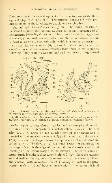
137-164. Lateral muscles (fig. 9).—The lateral muscles of the
second segment differ in many respects from those of the segments
following. They comprise an outer and an inner series of tergosternal
14-23 14-2b IITI ^
Fig. 9.—Lateral muscles of the first and second abdominal segments of
Dissosteira Carolina, right side, inner view, female.
A, all muscles in place. B, external lateral muscles of second segment (160,
162, 163, 164) exposed by cutting and partial removal of overlying muscles.
muscles, a pair of tergopleural muscles, and a sternopleural muscle.
The inner series of tergosternals includes three muscles. The first
(fig. 9 A, 137) arises on the anterior lobe of the tergum and is
inserted on the anterior apodeme {Ap) of the sternum ; the second
{138) arises on the tergum above the spiracle and has the same in-
sertion as 137. The third {139) is a much larger muscle arising on
the tergum beneath the edge of the lateral dorsal muscle {130) and
inserting on the lateral margin of the sternum. The outer series of
tergosternals includes a short muscle (fig. 9 B, 160) from the anterior
ventral angle of the tergum to the anterior end of the sternal apodeme,
and a broad posterior muscle (A, 161) arising external to the para-
dorsal muscle {131) and inserted on the edge of the sternum behind

20 SMITHSONIAN MISCELLANEOUS COLLECTIONS VOL. 94
/5p. The two tergopleural muscles (B, 162, 163) arise anteriorly and
posteriorly on the lower part of the tergum external to 159 and 161,
and converge upon a narrow linear sclerite in the membrane between
the tergum and the sternum. In all the other segments the external
lateral muscles are attached directly on the sternum. The sternopleural
muscle of the second segment is a group of very short fibers (B, 164)
connecting the pleural sclerite with the sternunL
i6s, 166. Muscles of the spiracle (fig. 9B).—The dilator of the
spiracle {16^) is a long slender muscle arising on the apodeme of
the sternum, the occlusor {166) a short muscle arising on the tergum;
both are inserted on the manubrium of the movable valve of the spi-
racular atrium.
MUSCLES OF THE THIRD SEGMENT
The musculature of the third segment presents the typical abdominal
musculature of the grasshopper, since its pattern is repeated in seg-
ments /// to r// in both sexes, and its dorsal and ventral muscles
are duplicated in the second segment.
/d/. Median internal dorsal muscles (fig. 10 A).—These muscles
of the third segment, as those of the second, consist of four flat groups
of fibers {a, h, c, d) lying within the pericardial chamber, extending
from the anterior tergal ridge {tr) to the anterior edge of the fol-
lowing tergum. In the succeeding segments they become more oblique
(figs. 8, 12, 182, ip/, 22/') with their posterior ends dorsal to their
anterior ends.
168. Lateral internal dorsal muscle (fig. 10 A).—The lateral dorsal
muscle is separated from the median dorsals by the attachments of
the transverse muscles of the dorsal diaphragm on the tergum (td),
and is, therefore, extrapericardial. In the following segments this
muscle becomes conspicuously fan-shaped (figs. 8, 12, i8_^, ip8, 228,
243)-
i6g. Paradorsal muscle (fig. 10 A).—The paradorsal muscle is dis-
tinguished from the other lateral dorsal muscle {i6y, 168) by the
fact that it lies external to the internal lateral muscles {175, 176).
It has the same relations in some other insects, though it is a muscle
not generally present. In Dissosteira it is repeated in the segments //
to VII of both sexes, and in segment VIII of the male (fig. 12, 244).
The paradorsal muscle has been termed a " pleural " muscle, but it
lies well within the area of the dorsum. Since it occurs in some larval
insects lacking tergal plates, the writer here discards the former name
of " paratergal " muscle. (Snodgrass, 193 1.)

NO. 6 GRASSHOPPER ABDOMEN SNODGRASS 21
ijo, iji. External dorsal muscles (fig. loA).—External dorsal
muscles occur in segments // to VII of both sexes, and also in seg-
ment VIII of the male. They take their origins on the posterior parts
of the terga within the intersegmental folds, and are inserted on the
overlapped anterior margin of the tergum following in each case. The
median external dorsal of segment /// (figs. lo A, ii B, 770) arises
167b-.
167c
167d
Fig. 10.—Muscles of the third abdominal segment of Dissosteira Carolina.
A, muscles of the right side, inner view. 16/, median internal dorsals ; 16S,
lateral internal dorsal ; 169, paradorsal muscle ; i/O, median external dorsal
;
17T, lateral external dorsal ; 17^, median internal ventral ; 173, lateral internal
ventral ; 174, external ventral ; 175, 176, internal laterals ; 177. 178, 179, external
laterals; td, attachment of dorsal transverse (diaphragm) muscles.
B, position of the muscles in cross-section of third segment, diagrammatic.
dil, lateral internal dorsals ; dim, median internal dorsals ; DS, dorsal sinus
;
DV, dorsal blood vessel ; lAp, lateral apodeme of sternum ; ilc, first ex-
ternal lateral muscle ; // , internal lateral muscle ; />. paradorsal muscle ; PvS,perivisceral sinus; td, dorsal transverse muscles (of dorsal diaphragm); tv.
•ventral transverse muscles (of ventral diaphragm) ; vil, lateral internal ventral
muscle; vim, median internal ventral muscle; VNC, ventral nerve cord; VS.ventral sinus.
dorsal to the lateral muscle (///) ; the first proceeds dorsally to its
insertion, the second ventrally. In the posterior segments the corre-
sponding muscles become much longer ; the base of the median muscle
has migrated ventrally, that of the lateral muscle dorsally. until the
two muscles cross each other obliquely on the side of the terguni.
The relations of the two muscles to each other and to the successive
terga on which they are attached is best seen when the terga are pulled
apart (fig. 11 C). The external dorsals in Acrididae, as already noted,

22 SMITHSONIAN MISCELLANEOUS COLLECTIONS VOL. 94
are evidently torsion muscles, their transverse positions enabling them
to give a movement of partial rotation to the terga on each other.
In the generalized condition the external dorsal muscles are longi-
tudinal in position and lie external to the internal dorsals, but they
are commonly shorter than the latter ; and have a tendency to become
restricted to the posterior part of the segment. In many of the higher
insects they become completely reversed in position, since they take
their origins on the posterior part of the tergum and extend forward
in the intersegmental fold to their insertions on the invaginated an-
terior margin of the following tergum. They thus become tergal pro-
tractors. The position of the external dorsals of the grasshopper is
seen to be intermediate between the more primitive condition and that
of complete reversal. The external ventral muscles, on the other hand,
are reversed and hence function as sternal protractors.
1^2, i/j, 17^. The ventral muscles (fig. loA).—The ventral
musculature of segment /// is typical of that of all the pregenital
segments (figs. 8, 12). The median internal ventrals (fig. 10 A, 1/2)
are the principal sternal retractors ; the short lateral internal ventral
on each side (i/j) arises at the base of the anterior apodeme (aAp)
just before the angle of the sternal ridge (sr), and is inserted on the
anterior end of the corresponding apodeme of the following sternum.
The lateral external ventral on each side (174) is a sternal protractor,
being completely reversed in position, with its origin on the posterior
part of the sternum and its insertion anterior on the under face of
the anterior apodeme of the following sternum.
The lateral musculature is alike in segments /// to VII, there being
in each of these segments representatives of the following five muscles
of segment ///, two of which are internal laterals, and three external
laterals.
17^. First internal lateral muscle (fig. 10 A).—A slender muscle
arising dorsally beneath the edge of the lateral dorsal {168), extending
ventrally and anteriorly to its insertion on the base of the lateral
apodeme of the sternum.
i/d. Second internal lateral muscle (fig. 10 A).—A broad muscle
arising on the side of the tergum just behind 775 and also beneath the
edge of the lateral dorsal {168), extending ventrally to its insertion
on the lateral margin of the sternum. The internal laterals are the
principal expiratory muscles, since their contraction lifts the sternum
and contracts the abdomen in a vertical direction.
177. First external lateral muscle (fig. 10 A).—This muscle arises
ventrally on the anterior part of the ventral margin of the tergum,

NO. 6 GRASSHOPPER ABDOMEN—SNODGRASS 23
and goes dorsally to its insertion on the outer face of the lateral apo-
deme of the sternum (fig. 10 B, lie). It is thus a dilator of the
abdomen and an inspiratory muscle in respiration, since its contrac-
tion separates the sternum from the tergum (fig. 11 F, G).
lyS, ijg. Second and third external lateral muscles (fig. 10 A).—
•
These two muscles arise on the lateral part of the tergum below the
vim vel
Fig. II.—Abdominal mechanisms of Acrididae.
A, mechanism of tergosternal movements : tergum and sternum approximatedby internal lateral muscles {ili, 2li) , separated by first external lateral {ile),
moved lengthwise on each other by oblique second and third external laterals
{2le,3le).
B, C, mechanism of torsion, or partial rotary movements of segments, by the
transverse external dorsal muscles, best developed in posterior segments (C).D, mechanism of tergosternal and intersternal movements : dorsoventral dila-
tion produced by first external lateral muscle {ile) ; lengthwise sternal contrac-
tion by internal ventrals {vim, vil);protraction by external ventral {vel).
E, the sternal apodemes, right side, anterior view.
F, G, mechanism of respiration: expiratory movement (F) produced by in-
ternal lateral muscles (/;', see A), inspiratory movement (G) by first external
lateral {lie).
paradorsal muscle {i6q), and cross each other obliquely, the first
going anteriorly, the second posteriorly, to their insertions on the op-
posite ends of the lateral margin of the sternum (fig. 11 A, 2le, sle).
The muscles of this pair evidently serve to give forward and back-
ward movements to the tergum and sternum on each other.

24 SMITHSONIAN MISCELLANEOUS COLLECTIONS VOL. 94
180, 181. Muscles of the spiracles.—The spiracular muscles are
alike in segments II to VIII, and the description of those of the
second spiracles {165, 166) will serve for each of the following
spiracles.
MUSCLES OF THE EIGHTH SEGMENT
The muscles of the eighth segment are quite different in the male
and the female. The musculature of this segment in the male (fig. 12)
conforms with that of the preceding segments except for the reduc-
tion of the internal dorsals to a single broad band of fibers on each
side {242), and in the absence of the first internal lateral. In the
female most of the usual muscles are retained in modified form, but
there are several muscles pertaining to the ovipositor and the ovi-
ducts that have no counterparts in the male. The muscles of the
eighth segment of the female are as follows
:
242, 24s. Internal dorsal muscles (fig. 14).—A transverse series
of six longitudinal groups of fibers on each side of the eighth tergum
(VIIIT), inserted posteriorly on the apodeme and anterior margin of
the ninth tergum (IXT). The lateral muscle on each side (^43)
is much larger than the others.
244. Paradorsal muscle.—Absent in the eighth segment of the
female.
24J. Median external dorsal muscle (fig. 14).—A broad muscle
arising on the posterior margin of the eighth tergum, the fibers con-
verging anteriorly and mesally to their insertions on the anterior
apodeme {Ap) of the ninth tergum.
246. Lateral external dorsal muscle.—Absent in the female.
24"/. Median internal ventral muscle.—A slender muscle arising
anterolaterally on the eighth sternum (fig. 13), inserted posteriorly
on the median apodemal process of the anterior intervalvula of the
ovipositor (fig. 17 D).
248. Lateral ventral muscle.—This muscle arises at the base of the
apodeme of the eighth sternum (fig. 13) as in the preceding seg-
ments ; but it is attached posteriorly in Dissosteira on the anterior basal
sclerite of the first valvula of the ovipositor (fig. 17 A, B, E), and in
Melanopliis (fig. 20 C) on the lateral pocket of the genital chamber.
24p. External ventral muscle.—Absent in the female, unless repre-
sented by the depressor of the first valvula (fig. 17 A, B, 2^2).
2^0. Internal lateral muscle (figs. 13, 14).—A very large triangular
muscle arising laterally on the eighth tergum, its fibers spreading ven-
trally to their insertions along the entire lateral margin of the eighth
sternum (fig. 13). This muscle evidently corresponds with the second

NO. 6 GRASSHOPPER ABDOMEN SNODGRASS 25
internal lateral of the preceding segments, the first being absent in
the eighth segment both in the female and the male (fig. 12).
231. First external lateral muscle (figs. 13, 14)-—A thick muscle
arising in the lower anterior angle of the eighth tergum, inserted an-
teriorly on the outer face of the apodeme (Ap) of the eighth sternum.
2^2. Second external lateral muscle (figs. 13, 14).—A small muscle
arising on the lower part of the eighth tergum below the spiracle,
inserted on the base of the apodeme of the eighth sternum.
23J. Third external lateral muscle.—Absent in the female.
254, 255. Muscles of the spiracle (figs. 13, 14).—Same as in the
preceding segments.
VII VIII IX
24-7 267
Fig. 12.—Muscles of the seventh, eighth, and ninth segments of the male
abdomen of Dissosteira Carolina, right side, inner view.
The following muscles of the eighth segment of the female have no
representatives in the male.
2j6. Short protractor of the ovipositor.—A short muscle with a
broad base arising on the side of the eighth tergum anterior to 2jo
(figs. 13, 14), inserted anteriorly on the anterior end of the apodeme
of the ovipositor (fig. 17 A, C).
2§/. Anterior muscle of the median oviduct (fig. 13).—A slender
muscle arising on the end of the apodeme of the eighth sternum, ex-
tending mesally to its insertion on the anterior end of the median ovi-
duct. This muscle is absent in Melanoplus.
2^8. Posterior muscle of the median oviduct (fig. 13).—A long
flat muscle arising on the end of the apodeme of the eighth sternum,
extending mesally and posteriorly to the posterior end of the median
oviduct.

2b SMITHSONIAN MISCELLANEOUS COLLECTIONS VOL. 94
2^p. The muscular sheath of the oviducts.—The walls of the median
oviduct and of the proximal parts of the lateral ducts have a muscular
sheath of internal circular fibers and external longitudinal fibers. The
longitudinal fibers are continued upon the walls of the calyces, but the
circular fibers appear to be absent in these parts of the lateral ducts,
and no muscles are present on the anterior glandular parts.
MUSCLES OF THE NINTH SEGMENT
The musculature of the ninth segment differs so much between the
male and the female that few muscles can be identified with each other
in the two sexes, or homologized with muscles of the pregenital seg-
ments. Besides the segmental muscles there are in the female special
muscles of the ovipositor, and in the male special muscles of the
phallic organs.
In the male grasshopper the following nine muscles take their origins
on the segmental plates of the ninth segment.
260. Internal dorsal muscle (fig. 12).-—A small band of fibers
arising near the mid-dorsal line on the anterior edge of the ninth
tergum, the fibers spreading posteriorly and laterally to their insertions
on the anterior margin of the tenth tergum. This small muscle is the
only representative of the intertergal dorsals in the ninth segment of
the male.
261. Retractor of the phallus.—A short, thick, conical muscle aris-
ing by a wide base dorsolaterally on the ninth tergum (fig. 12). in-
serted posteriorly and ventrally on a small oval sclerite in the wall of
the genital chamber just laterad of the epiphallus (fig. 25 D).
262. 26J. Muscles of the female not represented in the male.
264. Ventral dilator of the rectum.—A fan-shaped muscle arising
on the ninth sternum at the base of the sternal apodeme (fig. 12
shows point of origin), the slender fibers spreading dorsally in a
longitudinal plane to their insertions on the ventral wall of the rectum
(fig. 16 A).
263. Ventral muscles (fig. 12).—A pair of straplike muscles on each
side arising laterally on the ninth sternum at the base of the anterior
apodeme, going posteriorly and dorsally to the membranous venter
of the tenth segment just before the base of the paraproct.
266. Retractor of the aedeagus.—A broad, thin sheet of fibers aris-
ing from a median ridge of the ninth sternum (figs. 12, 25 x\), at-
tached dorsally to the wall of the genital chamber laterad of the base
of the aedeagus (fig. 25 x\).
267. Protractor of the aedeagus.—A large, triangular muscle aris-
ing by a long base on the median ridge of the ninth sternum, mesad

NO. 6 GRASSHOPPER ABDOMEN SNODGRASS 27
of 266 (figs. 12, 25 A), the fibers converging dorsally and anteriorly
to their insertion on the lateral lobe of the epiphallus (fig. 25 A, D) ;
its contraction probably elevates the distal part of the phallic apparatus.
268, 26g. Internal lateral muscles (fig. 12).—Two large oblique
muscles on each side in the position of the second internal lateral of
the pregenital segments. The first is inserted ventrally on the lateral
margin of the ninth sternum ; the second is inserted by a narrowed
stalk at the edge of the ninth sternum between the basal and distal
plates of the latter.
2/0. External lateral muscle (fig. 12).—This muscle clearly cor-
responds with the first external lateral of the pregenital segments.
It arises on the anterior lateral area of the ninth tergum and is in-
serted on the outer face of the apodeme of the ninth sternum.
In the ninth segment of the female there are the following 10 paired
muscles or sets of muscles, including the segmental muscles and the
muscles of the ovipositor.
260. Internal dorsal muscles (fig. 14).—A transverse series of
five small bands of longitudinal fibers on each side extending from the
anterior margin of the ninth tergum to the anterior margin of the
tenth tergum.
261. Not represented in the female.
262. Long protractor of the otnpositor.—Origin laterally on the
posterior margin of the ninth tergum (figs. 14, 17 C), extends for-
ward to its insertion on the anterior end of the apodeme of the ovi-
positor (fig. 17 C).
26^. Retractor of the ovipositor.—Origin on the anterior margin
of the ninth tergum (figs. 14, 17 C) ventrad of 262, extends pos-
teriorly to its insertion laterally in the base of the dorsal valvula of
the ovipositor (fig. 17 C).
264. Ventral dilator of the rectum.—A fan-shaped group of slender
fibers arising from the dorsal surface of the apodeme of the oviposi-
tor (fig. 17 B), spreading to their insertions on the ventrolateral line
of the rectum (fig. 16 A). If the ventral dilators of the rectum are
identical in the male and female, their origins would seem to identify
the apodemes of the ovipositor with the anterior apodemes of the
ninth sternum in the male.
26§-2yo.—These muscles of the ninth segment present in the male
(fig. 12) cannot be identified with any certainty in the female, though
it is possible some of them are included in the following musculature
of the ovipositor.
2yi. Levator of the dorsal vcdvula (fig. 17 A, B, C).—A large thick
muscle lying on the dorsal surface of the apodeme of the ovipositor.

SMITHSONIAN MISCELLANEOUS COLLECTIONS \0L. 94
attached anteriorly on the latter, and posteriorly on the dorsal margin
of the base of the dorsal valvula.
2^2. Depressor of the ventral valvula (fig. 17 A, B).—A massive
bundle of fibers arising on the ventral face of the apodeme of the
ovipositor, inserted on the basal plates of the ventral valvula. This
muscle possibly corresponds with the intersternal protractors, or
external ventrals, of the pregenital segments.
2-/^. Adductors of the ventral valvulae (fig. 17 C, D).—A pair of
flat muscles arising anteriorly on the proximal parts of the inner
margins of the apodemes of the ovipositor, the two converging pos-
teriorly to the median apodeme of the ventral intervalvula (/). Theretraction of the intervalvula causes an adduction of the valvulae.
Fig. 13.—Laterodorsal and ventral muscles of the seventh and eighth segmentsof the female abdomen of Dissosteira Carolina, right side, inner view.
2/4. Adductors of the dorsal valvulae (fig. 17 A, B, C).—Origin
on the proximal part of the dorsal surface of the ovipositor apodemes,
insertion posteriorly on the posterior intervalvula (B, C, piv). The
contraction of the convergent muscles of this pair approximates the
valvulae of opposite sides.
2/j. Muscle of the second valvula (fig. 17 B, C).—A small muscle
arising laterally on the dorsal surface of the anterior intervalvula, in-
serted posteriorly in the distal end of the small second valvula.
2/6. Dilator of the spermathecal aperture.—A very small muscle
of a few delicate fibers arising on the lateral basal plate of the ventral
valvula (fig. 17 E, a), inserted mesally on the side of the groove in the
dorsal wall of the genital chamber containing the aperture of the
spermathecal duct (fig. 20 D).

NO. 6 GRASSHOPPER ABDOMEN—SNODGRASS 29
2^"/. Muscles of the spermathecal duct.—The entire length of the
spermathecal duct is covered by a muscular sheath consisting of outer
longitudinal fibers and inner circular fibers.
The following muscles of the ninth segment of the male pertain
entirely to the phallic organs and tlie ejaculatory duct. It is impossible
to discover any identity between them and muscles of the female.
2y8. Epiphallic muscle of the aedear/us (fig. 25 B, C).—A long
muscle lying dorsally in the basal fold of the phallus, attached an-
teriorly on the lateral lobe of the epiphallus (h), and posteriorly on
the zygoma (s) of the aedeagal apodemes.
i'/p. Lateral muscle of the aedeagus (fig. 25 B).—A short muscle
arising lateroventrally in the base of the aedeagus, inserted dorsally
on the lower edge of the lateral plate {m) of the aedeagus.
280. Muscle of the ventral lobe of the aedeagus (fig. 25 B).—
A
delicate muscle arising within the base of the aedeagus, inserted dis-
tally near the apex of the ventral lobe of the latter.
281. Lateral dilator of the endophallus (fig. 25 C, E).—A broad
sheet of muscle arising dorsally on the aedeagal apodeme (C, Apa),
the fibers extending ventrally and anteriorly to the endophallic apo-
deme (C, E, w).
282. Dorsal dilator of the endophallus (fig. 25 F).—A broad flat
muscle on the dorsal surface of the endophallic bulb, arising laterally
on the inner face of the aedeagal apodeme (Apa), inserted mesally on
the dorsal edge {x) of the lateral plate of the endophallus.
28^. Compressor of the endophallus (fig. 25, E, F. G).—An un-
paired transverse muscle uniting the endophallic apodemes (w), the
fibers covering the anterior and anteroventral walls of the endophallic
bulb (G). This muscle approximates the endophallic plates and dilates
the orifice of the ejaculatory sac.
284. Compressor of the ejaculatory sac (fig. 25 C, E).—A broad
sheet of muscle arising internal to 281 (C) on the lateral plate of the
endophallus, the fibers converging ventrally to their insertions on the
lateral wall of the ejaculatory sac (E, ejs).
285. Muscles of the ejaculatory duct (fig. 25 E).—A thick sheath
of circular fibers surrounds the ejaculatory duct from the entrance of
the mucous glands to the beginning of the ejaculatory sac.
MUSCLES OF THE TENTH SEGMENT
The muscles of the tenth segment have no evident relation to the
muscles of the preceding segments. They comprise muscles to the
cerci, the epiproct and the paraprocts, dilators of the rectum, and in the
female a transverse intrasegmental muscle.

30 SMITHSONIAN MISCELLANEOUS COLLECTIONS NOL. 94
286. Dorsal dilator of the rectum.—A group of slender fibers aris-
ing dorsally on the tenth tergum mesad of the other muscles (fig. 14),
spreading fanwise ventrally to their insertions laterodorsally on the
posterior part of the rectum (fig. 16 A).
287. Depressor of the cercus (figs. 7 D, 14).-—A narrow muscle
arising anteriorly on the median part of the tenth tergum, inserted
posteriori}- on the posterior margin of the median basal lobe of the
cercus.
288. Median levator of the cercus (figs. 7 D. 14 ).—A broad muscle
arising anteriorly on the tenth tergum laterad of 287, inserted pos-
teriorly on the small sclerite between the tenth tergum and the basal
Fig. 14.—Dorsal muscles of eighth, ninth, tenth, and eleventh abdominal seg-
ments of female of Dissostcira Carolina, ventral view.
lobe of the cercus, some of the mesal fibers in some cases inserted on
the basal angle of the epiproct.
28p. Lateral levator of the cercus (figs. 7 D, 14).—A slender mus-
cle taking its origin on the tenth tergum immediately laterad of 288,
inserted posteriorly in the membrane behind the tenth tergum close
to the outer angle of the base of the cercus.
2^0. Lateral dilator of the rectum.—A fan of fibers arising an-
teriorly on the lateral part of the tenth tergum (fig. 14, 2po), spread-
ing mesad in a horizontal plane to their insertions along the lateral
line of the posterior part of the rectum (fig. 16 A).
2pi. Ventral muscle of the paraproct (fig. 14).—A broad muscle
arising on the anterior margin of the lateral part of the tenth tergum,
inserted posteriorly on the base of the paraproct ventrally.

NO. 6 GRASSHOPPER ABDOMEN SNODGRASS 3I
2^2. Transverse muscle (fig. 14).—An unpaired, straplike trans-
verse muscle, present only in the female, lying dorsal to the base of
the ovipositor and attached laterally on the ends of the tenth tergum.
(Only the ends of this muscle shown in the figure.)
MUSCLES OF THE ELEVENTH SEGMENT
The musculature of the eleventh segment includes muscles from the
epiproct to the cerci and paraprocts. and muscles from the epiproct and
paraprocts to the circumanal membrane.
2p^. Adductor of the cercus (figs. 7 D, 14).—A slender muscle
arising anteromedially on the epiproct, inserted on the inner extremity
of the basal lobe of the cercus.
2Cf4. Adductor of the paraproct (fig. 14).—A large muscle arising
medially on the epiproct just behind 2pj in the female, extending
laterally and posteriorly to its insertion on the upper part of the para-
proct behind the base of the cercus. In the male this muscle arises
mesad of 2g^ and underlaps the base of the latter.
2g5. Dorsal dilator of the anus.—A median unpaired muscle arising
centrally on the epiproct (fig. 14), its fibers spreading distally to their
insertions on the dorsal part of the circumanal membrane (fig. 16 A).
2q6. Lateral dilator of the anus.—Origin on the paraproct near the
base of the outer wall of the latter (fig. 14) ; extends dorsally, mesally,
and posteriorly to its insertion ventrolaterally on the rectum just
within the anus (fig. 16 A).
THE TRANSVERSE MUSCLES
The transverse muscles of the abdomen comprise dorsal transverse
muscles (fig. 10 B, td) and ventral transverse muscles (tv). The
former are always the muscles of the dorsal diaphragm ; the ventral
muscles may consist of segmentally individual bundles of transverse
fibers, but in the Acrididae they form a continuous muscular sheet, or
ventral diaphragm. The muscle uniting the opposite ends of the tenth
tergum in the female of Dissosteira (fig. 14, 292) is literally a dorsal
transverse muscle, but it evidently does not belong to the series of
diaphragm muscles.
III. THE DIAPHRAGMS AND THE DORSAL BLOOD VESSEL
The so-called diaphragms of insects are transverse dorsal and ven-
tral partitions of the body cavity that separate from the axial pervis-
ceral sinus (fig. 10 B, PvS) a dorsal sinus, or pericardial cavity (DS),

32 S-MITHSONIAX MISCELLANEOUS COLLECTION: \0L. 94
and a ventral sinus, or perineural cavity (VS). Each of the dia-
phragms differs much in the degree of its development in different
insects. The dorsal diaphragm is almost always present in some form,
but the ventral diaphragm is frequently absent ; the first is confined
principally to the abdomen, the second may extend into the thorax.
Probably each diaphragm consists of a double peritoneal membrane,
the layers of which are reflected from the walls of the body cavity ; but
the membranes enclose between them the dorsal and ventral transverse
muscles, and the muscles become the more important elements of the
DTrc m^
Tra'^^^;-
AFig.
Ht tra C[5.—The dorsal blood vessel and diaphragms of Dissostcira Carolina.
A, ventral view of anterior part of dorsal diaphragm extending to lobes of third
phragma {sPh) , showing segmental groups of transverse muscles (/</), anddorsal blood vessel along median line above the diaphragm. B, posterior part
of dorsal diaphragm and dorsal blood vessel in segments /7//, IX, and A'. C,
dorsal view of part of ventral diaphragm, attached on lateral parts of sterna.
diaphragms, which by the vibratory contractions of the muscles serve
as important adjuncts to the heart in the circulation of the blood.
The dorsal diaphragm of Acrididae extends from the anterior end
of the first abdominal segment to the posterior part of the ninth seg-
ment, and is continued into the metathorax as a narrow membranous
fringe along each side of the aorta. In the first abdominal segment the
broad anterior margin of the diaphragm is attached to the posterior
faces of the lobes of the third phragma (fig. 15 A) ; the lateral edges
in this segment are free and deeply emarginate. In the following seg-
ments the limits of the dorsal diaphragm are difficult to define in a
ventral dissection, except bv the muscle attachments, for the lower

NO. 6 GRASSHOFTER ABDOMEN SNODGRASS 33
diaphragm membrane appears to be everywhere continuous with a
deHcate peritoneal covering over the inner surfaces of the somatic
muscles lying lateral of the pericardial cavity. The upper membrane
of the diaphragm, however, being reflected upon the dorsal pericardial
wall, more clearly marks the limits of the diaphragm itself. The two
membranes of the diaphragm can be distinguished in whole prepara-
tions under the microscope by the two layers of nuclei, one dorsal to
the muscle fibers, the other ventral. It is apparent that the two mem-branes, however, are simply continuations of a peritoneal lining of the
perivisceral cavity and of a similar lining of the pericardial cavity,
with the transverse muscles between them.
The muscles of the dorsal diaphragm in Dissostcira begin in the
second segment of the abdomen (fig. 15 A) and end in the ninth
segment (B). They consist of a double series of transverse fibers,
separated into segmental groups, but for the most part approximately
parallel. In all but the second and ninth segments the fibers are slightly
divided into secondary anterior and posterior groups. This intra-
segmental segregation of the fibers is more accentuated in Mclanoplus
than in Dissosteira. The fibers arise laterally on the tergal plates be-
tween the median and the lateral longitudinal dorsal muscles (figs.
8, 10) . Their median ends branch toward the ventral wall of the heart,
on which they break up into fine fibrils, and the fibrils from opposite
sides appear to unite in an intricate plexus.
The dorsal blood vessel extends from beneath the brain into the
tenth abdominal segment. Ostia and slight segmental enlargements of
the tube are present in abdominal segments // to IX (fig. 15 A, B).
Dorsal ampullar enlargements of the aorta occur in the mesothorax,
metathorax, and first abdominal segment. Posteriorly the heart term-
inates in a narrow tapering tube extending into the tenth abdominal
segment. For most of its length the dorsal vessel is accompanied by
strands of nephrocytes. and the aortic ampullae are capped by dense
masses of nephrocytic cells. The pericardial cavity contains also loosely
scattered fat cells, and is penetrated by loops of the Malpighian tubules.
The dorsal longitudinal tracheal trunks (fig. 15 A, B) lie along the
sides of the blood vessel and are connected with the lateral trunks bytransverse tracheae in the posterior part of each abdominal segment.
It would appear that the blood has entrance into the pericardial cavity
only above the free lateral margins of the diaphragm in the first ab-
dominal segment, and at the posterior end of the diaphragm in the
eighth and ninth segments.
The ventral diaphragm in Dissosteira extends from the head into
the seventh (female) or eighth (male) abdominal segment. In the

34 SMITHSONIAN MISCELLANEOUS COLLECTIONS VOL. 94
anterior part of the thorax this diaphragm is merely a very delicate
membrane attached laterally on the salivary glands and on masses of
fat tissue. Between the w^idely spreading bases of the metasternal
apophyses, however, there begins in the membrane a series of trans-
verse muscle fibers, which continues to the posterior end as the prin-
cipal tissue of the diaphragm. The fibers are attached in the meta-
thorax to the metasternal apophyses, and in the abdomen on the lateral
parts of the sternal plates (fig. 15 C). Most of the fibers go continu-
ously across from one side to the other, but in each segment the
anterior and posterior fibers spread somewhat forward and backward
to bridge the spaces between the consecutive sternal plates. Posteriorly
the ventral diaphragm ends al:)ruptly in a free transverse margin, which
in the female crosses the anterior part of the seventh abdominal seg-
ment, but in the male is in the anterior part of the eighth segment. In
the female the last two ganglia of the ventral nerve cord lie beyond
the diaphragm and are dorsal to the spermatheca, the anterior end of
which may extend into the ventral sinus. In the male only the last
ganglion is not covered by the diaphragm.
IV. THE PROCTODAEUMThe proctodaeum of the grasshopper is a tube of fairly uniform
diameter composed of anterior and posterior sections separated by a
narrower and usually bent middle section (fig. 16 A), but the relative
size of the parts varies much in different specimens according to the
distension, or according to the state of contraction of the muscles.
The anterior end of the proctodaeum is marked externally by the
origins of the Malpighian tubules (Mai), which are disposed in 12
groups of about 10 tubules each, arranged in a circle immediately be-
hind the ventriculus (Vent). There is no clear anatomical division of
the proctodaeum into an anterior intestine and posterior intestine,
and there are no specifically developed internal valves, but four fairly
well-marked proctodaeal regions may be distinguished by external and
internal characters. The first is a short pylorus (Py) into which the
Malpighian tubules open, the second is a long saclike ileum (//), the
third a narrower and usually bent colon (Cln), and the fourth is the
large rectum (Red) comprising a wide anterior rectal sac and a
narrow terminal part extending to the anus.
The muscularis of the proctodaeum consists of external longitudinal
fibers and internal circular fibers, the relation of the two sets of
muscles on the proctodaeum being thus the same as that of the ven-
tricular muscles (fig. 16 A), though the muscle fibers of these two
parts of the alimentary canal are not continuous with each other.
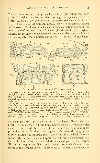
NO. 6 GRASSHOPPER ABDOMEN—SNODGRASS 35
The circular muscles of the proctodaeum begin just behind the bases
of the Malpighian tubules, forming here a pyloric sphincter of large
fibers (A, B, c), and continue {d) uninterruptedly over the entire
length of the rest of the stomodaeal tube. They are particularly strong
on the colon. The longitudinal muscles arise as distinct fibers on the
anterior end of the proctodaeum in the neighborhood of the Malpighian
tubules, but the fibers immediately converge over the pyloric sphincter
into six equally spaced muscle bands (<?) on the wall of the ileum.
E
Eppt
29S
Mai J\
Fig. i6.—The proctodaeum of Dissosfcira Carolina.
A, general view of the proctodaeum, showing its subdivisions, its muscles,
and groups of Malpighian tubules given off from anterior end. B, internal view
of pyloric region between ventriculus and ileum, with pockets from which the
Malpighian tubules arise. C, anterior ends of rectal "glands," inner view of
rectal wall.
a, b, circular and longitudinal muscles of ventriculus; c, pyloric sphincter;
Cln, colon; d, circular muscles of proctodaeum; e, bands of longitudinal procto-
daeal muscles ; Eppf. epiproct; /, fold marking terminus of ventricular wall
;
g, Malpighian pockets ; h, i, internal folds of wall of ileum ; //, ileum ; /, rectal
pads ; Mai Malpighian tubules ; Py, pylorus ; Rect, rectum ; Vent, ventriculus
;
264, 286, 2go, ventral, dorsal, and lateral dilator muscles of posterior part of
rectum; 2^5, 296, epiproctial and paraproctial dilators of the anus (see fig. 14).
and continue thus to the posterior end of the latter. Here each band
breaks up into a group of fibers branching on the colon, some of which
appear to go beneath the circular fibers to attach on the intima of the
proctodaeal wall. On the posterior part of the colon the longitudinal
fibers reassemble in six bands that traverse the outer wall of the rectal
sac, and then again branch and appear to go beneath the circular mus-
cles to be inserted on the wall of the terminal part of the rectum.
Finally the longitudinal fibers appear once more as six short external
bands on the terminal part of the rectum, and end with attachments on

36 SMITHSONIAN MISCELLANEOUS COLLECTIONS \ OL. 94
the lips of the anus. The colon is the most strongly musculated part
of the intestine, and, when in a state of contraction, it often appears
as a very short connective between the distended ileal and rectal sacs.
The posterior part of the rectum is provided with fan-shaped dorsal,
lateral, and ventral dilator muscles arising on the body wall and in-
serted on the proctodaeum in line with the longitudinal muscle bands
of the latter (fig. i6A). The dorsal dilators (286) arise medially
on the tergum of the tenth abdominal segment (fig. 14), and their
spreading fibers are inserted on the rectum along the lines of the latero-
dorsal longitudinal muscles. The lateral dilators (fig. 16 A. 2po) arise
laterally on the tenth tergum (fig. 14). and are inserted in line with
the lateral longitudinal muscles of the rectum. The ventral dilators
(fig. 16 A, 264) arise in the male at the bases of the anterior apodemes
of the ninth abdominal sternum (fig. 12). in the female on the
apodemes of the ovipositor (fig. 17 B), and are inserted in line with
the lateroventral longitudinal muscles of the rectum.
The structure of the inner wall of the proctodaeum of Dissostcira
Carolina has been described and figured by Tietz (1923), who shows
that the several parts of the intestinal tube present characteristic his-
tological differences. Mewed internally, it is seen that the Malpighian
tubules open into 12 pockets of the pyloric region (fig. 16 B, g), and
that the pockets are somewhat overlapped anteriorly by a circular fold
of the enteric wall (/). The proctodaeal intima lines the Malpighian
pockets and is reflected to the edge of the overhanging fold. It is
clear, therefore, that the crest of this fold (/) is the true line of
separation between the mesenteron and the proctodaeum, and that
the Alalpighian tubules arise from the proctodaeum. Between the
Alalpighian pockets the wall of the proctodaeal pylorus forms 12 broad,
padlike thickenings (/?), which are crossed externally by the sphincter
muscle (f), and which, therefore, may constitute collectively a pyloric
closing apparatus between the stomach and the colon. Posteriorly the
pyloric pads are narrowed and are either continued as well-marked
folds on the wall of the ileum ( /) , or they are broken up into numer-
ous small folds, according to the degree of tension in the ileal wall.
The external longitudinal muscle bands of the ileum {e) lie between
each alternate pair of internal folds. When the folds are accentuated
by contraction of the proctodaeal muscles, they extend posteriorly
through the colon to the rectum. According to Tietz (1923) the proc-
todaeal intima has a thickness of .008 mm in the ileum, and of .012 mmin the colon.
The inner wall of the rectal sac presents six long, flat, parallel thick-
enings lying between the external bands of longitudinal muscles, each

XO. 6 GRASSHOPPER ABDOMEN SNODGRASS 37
tapering or rounded at the ends, and having sharply defined margins
formed by the covering cuticula (fig. i6 C, /). These structures are
the so-called " rectal glands." In the grasshopper there is nothing to
suggest that they have a secretory function, the surface cuticula being
relatively thick, and the epithelium, as shown by Tietz (1923). con-
sisting of simple columnar cells. For the same reasons, also, it does
not seem probable that the rectal pads are organs for the absorption
of water from the faeces (see Wigglesworth. 1932) ; in fact, any
other part of the rectum would appear to be better adapted to an
absorptive function. On the other hand, the hard flat surfaces of the
pads, forming six plaques in strong relief on the inner wall of the
rectum, suggest that, by contraction of the surrounding circular mus-
cles, the structures may serve to compress the contents of the rectal
sac and thus extract water from the faeces.
V. THE OVIPOSITOR AND ASSOCIATED STRUCTURES
The ovipositor of the x\crididae is primarily a digging organ that
works by a forcible separation of the short recurved valvulae. It thus
dififers radically in its action from the usual egg-laying organ of other
insects, in which the valvulae are interlocked and move lengthwise
on one another. The acridid ovipositor, therefore, has quite a dififer-
ent mechanism from that of the sliding type of ovipositor. Though
the prongs of the grasshopper's ovipositor, as shown by their develop-
ment, are without doubt homologues of the valvulae of other insects.
it is apparent that the usual supporting basal plates, or valvifers, are
absent, and that the musculature of the acridid organ has little relation
to that of an ovipositor in which the muscles of the valvifers are the
principal motor elements. A second important function of the acridid
ovipositor, however, is that of manipulating the eggs, as the latter
issue from the oviducal opening, in such a manner that they may be
placed appropriately in the egg cavity for the exit of the young
grasshoppers.
STRUCTURE OF THE OVIPOSITOR
The exposed part of tlie grasshopper's ovipositor consists of a lower
and an upper pair of strong, sclerotic, pronglike processes with curved
tips turned ventrally and dorsally (fig. i, Ovp). These processes
are respectively the first and third valvulae (fig. 17 A, iVl, 3VI).
The second valvulae {2VI) are small, and are ordinarily concealed
between the others, but they are not rudimentary in the sense of being
functionless structures. The ovipositor projects posteriorly at the

38 SMITHSONIAN MISCELLANEOUS COLLECTIONS VOL. 94
271 Ap 274- 263
256 \
-—iVl
Fig. I/.—The ovipositor and its muscles. Dissosteira Carolina.
A, ovipositor and muscles, left side. B, median section of ovipositor showingmesal view of right valvulae and muscles. C, dorsal view of ovipositor andmuscles. D, ventral view of second and third valvulae, with apodemes, andmuscles of anterior intervalvula. E, ventral view of ventral valvulae, dorsal wallof genital chamber with spermathecal aperture, and spermatheca.
a, lateral basivalvular sclerite ; aiv, anterior intervalvula ; Ap, apodeme of
ovipositor ; b, c, first and second ventral basivalvular sclerites ; e, f, apodemeand apodemal sclerite of anterior intervalvula
; g, base of third valvula ; h,
ventral lip of apodemal invagination; i, articular process on ramus of third
valvula ; IXT, lateral parts of ninth tergum; piv, posterior intervalvula ; ra,
ramus of third valvula; Spr, spermathecal aperture; Spt, spermatheca; SptD,spermathecal duct; iVl, 2VI, 3VI, first, second, and third valvulae.

NO. 6 GRASSHOPPER ABDOMEN SNODGRASS 39
end of the abdomen (fig. i) beyond the eighth sternum (VlllStn)
from beneath the lobes of the eleventh segment {Eppt, Papt), and
thus might appear to belong to the ninth and tenth segments ; the first
valvulae, however, are developed in the nymph from the eighth seg-
ment, immediately behind the eighth sternum (fig. 22 A, C, iVl)
,
and the second and third valvulae from the ninth segment (A, C, D).
In the adult several basivalvular sclerites are differentiated from the
bases of the first valvulae (figs, i, 17 A, a, b, c) , v^hich, though par-
tially overlapped by the eighth sternum, are entirely separated from
the latter by an inflection of the poststernal membrane that forms the
female genital chamber. Between the bases of the dorsal valvulae are
anterior and posterior intervalvular sclerites (fig. 17 B, aiv, piv) ; and
a pair of large apodemes projects forward in the body cavity from
the angles between the bases of the dorsal and ventral valvulae (A, B,
D, Ap). An important accessory of the acridid ovipositor is the egg
guide, a median process of the eighth sternum (fig. 20 A, eg).
The first, or ventral, valvulae of Dissosteira Carolina (fig. 17 A,
B, E, I VI) are somewhat elongate lobes, flattened from side to side,
ending each in a decurved point. Proximally they are united by the
membranous integument between their bases, and their ventral walls
are continued into the dorsal wall of the genital chamber (fig. 20 A).
Each first valvula is differentiated into a strongly sclerotic terminal
lobe (fig. 17 A, iVl), and into a basal part containing a large lateral
basivalvular sclerite (a) and two narrow ventral sclerites {b, c). The
upper surface of the terminal lobe (B) is produced proximally as an
elongate plate, or ramus, at the end of which is a wide transverse de-
pression that fits closely upon a prominent abutment from the under
surface of the basal ramus of the corresponding third valvula (D, i).
Proximal to this articulation the dorsal wall of the ventral valvula
is membranous and shortly ends at the ventral lip of the hollow base
of the lateral apodeme (Ap). The lateral basivalvular sclerite of the
first valvula (A, a) is a prominent plate exposed on the side of the
abdomen behind the eighth sternum (fig. i). The posterior ventral
sclerite (fig. 17 A, B, b) is ordinarily partly exposed behind the
eighth sternum, but the anterior ventral sclerite (c) is concealed in
the dorsal wall of the genital chamber (fig. 20 A), where it flanks a
median channel containing the spermathecal aperture (fig. 17 E).
The third, or dorsal, valvulae of Dissosteira (fig. 17 A, B, 3VI)
resemble the ventral valvulae in general form, except that their points
are turned upward, but they are larger and stronger than the ventral
valvulae, and they have no basivalvular sclerites. Their dorsal surfaces
proximal to the upcurved points are broad and flat. The under surface

40 SMITHSONIAN' MISCIlLLANEOUS COr,LF.CTIONS VOL. 94
of each dorsal valvula is prolonged anteriorly in a strong ventral ramus
(D, ra), the expanded base of which (g) is firmly hinged to the dorsal
lip of the exposed base (li) of the lateral apodeme of the same side.
The apodemal bases, therefore, are the fulcral points for the move-
ments of the dorsal valvulae. The ventral valvulae, on the other hand,
have their fulcra of movement on the strongly protruding articular
ridges (/) on the bases of the dorsal valvulae. The dorsal valvulae
are united between their bases by a membranous integument containing
the anterior intervalvula (D, aiv), the bases of the second valvulae
{2VI), and the posterior intervalvula (B, C, piv).
The second, or intermediate, valvulae (fig. 17 A. B, D, 2]!) are
short lobes projecting from the membrane between the ventral rami
of the dorsal valvulae. The two are adnate mesally for most of their
length, but their sclerotic lateral and terminal parts form free lobes en-
closing a trough-like depression between them (D). The united bases
of the second valvulae are supported on a median process of the an-
terior intervalvula (aiv).
The intervalvulae are small sclerites lying between the bases of the
dorsal valvulae, where, because of the position of the latter, they
become dorsal and ventral relative to each other. The ventral anterior
intervalvula (fig. 17 D, aiv) is a transverse sclerite bridging the space
between the anterior ends of the ventral rami of the third valvulae,
and giving support by a median process to the united bases of the
second valvulae. Just before the transverse sclerite is a small, oval,
median sclerite (/) bearing a short slender apodeme (e) on which are
attached the muscles of the anterior intervalvula. The posterior inter-
valvula is a small hexagonal plate lying dorsally between the bases of
the third valvulae (B, C, piv).
A characteristic feature of the acridid ovipositor is the presence of
the pair of long, flat, lateral apodemes (fig. 17 D, Ap) extending
forward from the angles between the bases of the dorsal and ventral
valvulae. These apodemes are well developed also in Tetrigidae and
Tridactylidae, but they have no apparent homologues in the ovipositor
of other insects. They give attachment to the levator and depressor
muscles of the valvulae, to the muscles of the posterior intervalvulae,
and to the ventral dilators of the rectum, while the proctractor muscles
of the ovipositor are inserted on their anterior ends.
The stalk of each apodeme is hollow, and its base appears as the
strongly sclerotic lips of a transverse cleft in the angle between the
bases of the dorsal and ventral valvulae, where, as already observed,
the ventral ramus of the dorsal valvula is hinged to the dorsal lip of
the apodemal invagination. It might be supposed, therefore, that the

NO. 6 GRASSHOPPER ABDOMEN SXODGRASS 4I
apodemes of the acridid ovipositor represent anterior apodemal proc-
esses of the second valvifers in other insects, but it is quite impossible
to reconcile the musculature of the acridid apodemes with that per-
taining to the second valvifers in the usual type of ovipositor. Accord-
ing to Walker (1919) the ovipositor apodemes of Melanoplus are
formed in the nymph as invaginations at the angles between the bases
of the valvulae. It is perhaps possible that they are highly developed
anterior apodemes of the ninth sternum (the latter being represented
by the intervalvulae), since the space between the bases of the dorsal
valvulae is bridged by the anterior intervalvula. It is significant that
the ventral dilators of the rectum, which in the male arise anteriorly
on the ninth sternum, take their origins in the female on the ovipositor
apodemes (fig. 17 B, 264).
The muscles of the acridid oviix)sitor function as protractors and
retractors of the entire organ, as levators and depressors of the first
and third valvulae, as abductors and adductors of the same valvulae.
and as motors of the second valvulae : but it appears that some of
them may act in more than one capacity. As above noted, it will be
fruitless to attempt to trace any homologies between these muscles
and the usual muscles of the ovipositor in other insects. It should be
observed, however, that with the absence of valvifers in the acridid
ovipositor there is correlated an absence of dorsal muscles correspond-
ing with those ordinarily inserted on the valvifers.
The exsertion of the ovipositor evidently is brought about by two
pairs of muscles inserted on the anterior ends of the lateral apodemes
(fig. 17 A, C, 2f,6, 262). Of these muscles those of the first pair are
the short protractors {2^6) arising by wide bases on the anterior
lateral parts of the eighth abdominal tergum (fig. 13). The others
are the long protractors (fig. 17 C, 262) arising laterally on the pos-
terior margin of the ninth tergum (IXT). Retraction of the ovi-
])Ositor is accomplished apparently by a pair of ventral muscles of
the first valvulae. by lateral muscles of the third valvulae, and by
muscles of the anterior intervalvula. The retractors of the first val-
vulae arise anteriorly on the eighth sternum (fig. 13, 248) and in
Dissosteira are inserted on the anterior ventral basivalvular sclerites
(fig. 17 E, 248). In Melanoplus, however, these muscles are inserted
on anterior pockets of the genital chamber (fig. 20 C, 248). The re-
tractors of the third valvulae are lateral muscles arising anteriorly
on the ninth tergum (fig. 17 C, 26^) and inserted posteriorly on the
lateral margins of the bases of the third valvulae. These muscles would
appear to act also as abductors of the valvulae. The retractors of the

42 SMITHSONIAN MISCELLANEOUS COLLECTIONS VOL. 94
anterior intervalvula (C, D, 241) are a pair of slender muscles aris-
ing anteriorly on the eighth sternum (fig. 13) ; they probably assist
also in the adduction of the valvulae.
The muscles that open the valvulae dorsoventrally consist of the
four huge bundles of fibers arising on the lateral apodemes, one pair
dorsally (fig. 17 A, B, 2yi), the other pair ventrally {2'/2). Thedorsal muscles, which are the levators of the third valvulae, are in-
serted dorsally in the bases of these valvulae ; the ventral muscles, or
depressors of the first valvulae, are inserted within the bases of the
first valvulae on the lateral basivalvular sclerites and on the posterior
ventral sclerites (A, E, a, b). These four powerful muscles of the
ovipositor produce the movements of the valvulae by which the earth
is compressed peripherally in the digging of the egg chamber in the
ground.
The closing of the valvulae evidently is produced by muscles of
the anterior intervalvula, there being no muscles inserted on the
valvulae that directly oppose the opening muscles. The muscles of
the anterior intervalvula include the slender retractor muscles (fig.
17 C, D, 24/) arising anteriorly on the eighth sternum (fig. 13), and
a pair of short, broad muscles (fig. 17 C, D, ^7j) arising on the ovi-
positor apodemes. Since the anterior intervalvula lies between the
l:)ases of the dorsal and ventral valvulae, a pull on its muscles brings
the valvulae together. These same muscles also eflfect an adduction
of the valvulae of opposite sides.
Transverse movements of the valvulae, i. e., movements of abduc-
tion and adduction, are not as pronounced as the dorsal and ventral
movements, but it can be shown experimentally on a dead specimen
that some of the muscles of the ovipositor separate or approximate the
valvulae of opposite sides. The only muscles that may serve as ab-
ductors of the valvulae are the retractor muscles inserted on the lateral
basal margins of the third valvulae (fig. 17 C, 26^), which arise
laterally on the ninth tergum. The adductors are muscles of the inter-
valvulae ; a forward pressure on these sclerites brings the valvulae of
opposite sides together. The anterior intervalvular muscles comprise
the median pair of slender muscles (fig. 17 C, D, 24'j') arising an-
teriorly on the ninth sternum (fig. 13), and the lateral pair of wide
muscles (^/j) arising on the inner margins of the basal parts of the
lateral apodemes ; both pairs converge to their insertions on the small
median apodeme of the anterior intervalvula. The posterior inter-
valvular muscles consist of a single pair of muscles (B, C, 2^4)
arising on the bases of the lateral apodemes, and converging pos-
teriorly to their insertions on the posterior intervalvula (piv). There

NO. 6 GRASSHOPPER ABDOMEN SNODGRASS 43
are no tergal muscles in Acrididae corresponding with those inserted
on the intervalvulae in Gryllidae. (See Abdomen, Part II, Smith-
sonian Misc. Coll., vol. 89, no. 8, fig. 17 E, H, 5, 8.)
The second valvulae are provided with a pair of short muscles aris-
ing anteriorly on the anterior intervalvula (fig. 17 B, C, 2/^), and
inserted distally in the free ends of the second valvulae. These small
intermediate valvulae evidently are functional in guiding the eggs
properly between the other valvulae in their passage through the
ovipositor.
An important accessory of the acridid ovipositor is the egg guide.
This organ is a small, tapering median process arising from the re-
IXTXT
Fig. 18.—End of abdomen and ovipositor of Tcttigidca lateralis.
A, terminal segments of abdomen, with ovipositor. B, left valvulae andapodeme of ovipositor. C, ventral view of second and third valvulae, inter-
valvulae, and apodemes of ovipositor.
fleeted distal end of the eighth sternum (figs. 4, 13, eg) directly in
line with the opening of the oviduct {Gpr) in the floor of the genital
chamber. The egg guide normally projects between the bases of the
ventral valvulae, and serves to direct the eggs issuing from the gono-
pore upward and into the intervalvular space of the ovipositor. It is
an immovable process developed in the nymph as a median outgrowth
from the posterior margin of the eighth sternum.
The ovipositor of Tetrigidae, as illustrated by Tettigidea lateralis
(fig. 18), is of the acridid type of structure in that it is composed of
the first and third valvulae (B), and has a pair of lateral intervalvu-
lar apodemes (B, C, Ap) with muscles for opening the valvulae. The

44 SMITHSOXIAX MISCELLANEOUS COLLECTIONS VOL. 94
second valvulae are small (C, 2VI) as in Acrididae and are supported
by the anterior intervalvula (aiv). The first and third valvulae, how-
ever, are longer and slenderer than in the typical acridid ovipositor,
and the apodemes are relatively short. Basivahailar sclerites are ab-
sent in Teffigidca. but Walker (1919) describes and figures basivalvu-
lar sclerites associated with the first valvulae in Acrydium ornatuui.
and Chopard (1920) shows a lateral sclerite (pileolus) at the base
of the first valvula in Paramastax lacta. Walker describes also in
Acrydium a sclerite interposed between the liase of the third valvula
and the lower edge of the ninth tergum, which he regards as a valvifer.
The ovipositor of the tridactylid genus Rhipipteryx, as Walker
(1919) observes. " is remarkably similar to that of the Acridoidea."
The female abdomen of R. bioUeyi is elongate and slender. The last
unmodified segment is the seventh (fig. 19 A, VII). The eighth seg-
ment, which is partly concealed within the seventh, has its tergum di-
vided into two lateral plates by a median membranous area of the
dorsum (A, B, VIIIT). The spiracles of this segment lie in the lower
parts of the tergal plates. The tergum of the ninth segment (A, IXT)consists of two widely separated lateral plates (E, IXT), the anterior
dorsal angles of which (C, E) are produced into a pair of slender
arms invaginated beneath the dorsal membrane of the eighth segment.
The tergum of the tenth segment (F, XT) is broken up into two large
lateral tergites and a group of three small dorsal tergites. The lateral
tergites extend forward between the arms of the ninth tergal plates,
where they are united with each other anteriorly. The eleventh seg-
ment is represented by a shield-shaped epiproct ( F, Eppt), and two
large projecting paraprocts (Papt) bearing each a terminal lobe
(papfl). The cerci (Cer) arise laterad of the epiproct at the bases of
the paraprocts.
The exposed part of the ovipositor of RJiipipteryx biolleyi consists
of four elongate conical processes (fig. 19 A, Ovp), which, as in
Acrididae and Tetrigidae, are the first and third valvulae. Each ventral
first valvula (C, iVl) has an accessory tooth on its outer surface and
a large lateral basivalvular sclerite («) at its base. The latter appears
on the side of the abdomen as a prominent plate behind the eighth
sternum (A, a). The dorsal third valvulae (C, jF/) are somewhat
longer than the ventral valvulae, and each is supported on the distal
margin of the lateral tergite of the ninth segment (IXT), to which
it is articulated ventrally (E) by a condyle of the latter. Below and
between the bases of the dorsal valvulae are two small intermediate
second valvulae (E, ^Vl), united at their bases bv the anterior inter-

GRASSHOPPER ABDOMEN SNODGRASS 45
valvula (aiv). Both anterior and posterior intervalvulae (E, aiv, piv)
are present in Rhipipteryx as in Acrididae and Tetrigidae, and a small
median sclerite (/) before the anterior intervalvula gives attachment
to a pair of convergent muscles (5).
The lateral apodemes of the ovipositor of Rhipipteryx are long
spatulate plates arising between the bases of the valvulae, but each is
more specifically connected with the corresponding ventral valvula
(fig. 19 D, Ap), rather than with the dorsal valvula as in Acrididae.
Walker describes the ovipositor apodemes of R. forcipata as shelflike
extensions of the lower edges of the ninth tergum, having the same
tQ.—Abdomen and ovipositnr (jf Rhipipteryx biollcyi.
A, terminal half of abdomen with ovipositor. B, lateral view of eighth seg-
ment removed from seventh. C, left view of ninth segment and ovipositor. D,mesal view of right ventral valvula, with right apodeme and muscles. E, ven-tral view of ninth tergum supporting second and third valvulae. F, dorsal viewof tenth and eleventh segments, showing lobes (papfl) of paraprocts. (Letter-
ing as on fig. 17.)
relation to the vahulae as the free apodemes of Acrididae. The writer,
however, finds no tergal connections of the apodemes in R. biollcyi,
in which the structures appear to be identical with the intervalvular
apodemes of Acrididae. They give attachment to muscles very nearly
the same as those of the Acrididae in their distribution to the basival-
vulae (fig. 19 C. D. /). the ventral valvulae (_'). the ninth tergum
(C, 5), and the dorsal valvulae (C, E, 4).
THE FEMALE GENITAL CH.AMBER AND THE SPERMATHECAL OPENING
The genital chamber, or copulatory pouch, of the female grass-
hopper is a fiat horizontal invagination of the integument beneath the
bases of the ventral valvulae and above the posterior margin of the

46 SMITHSONIAN MISCELLANEOUS COLLECTIONS VOL. 94
eighth abdominal sternum (fig. 20 A, GC). The anterior basivalvular
sclerites of the first valvulae extend into its dorsal wall (figs. 17 E,
20 A, C, c), and in a depression between them is situated the aperture
of the spermatheca (Spr). The opening of the median oviduct, or
gonopore, lies anteriorly in the floor of the genital chamber (fig. 20 A,
Gpr). A short distance before the spermathecal and oviducal aper-
tures the genital chamber ends as a blind pouch. Here, in Dissosteira,
a pair of muscles from the eighth sternum have their insertions on the
ends of the anterior basivalvular sclerites (fig. 17 E, 248). In Mela-
nopliis the anterior end of the genital chamber is provided with two
large lateral pockets (fig. 20 C, /), and the muscles {248), inserted on
the basivalvular sclerites (c) in Dissosteira, are attached in Melano-
pliis on the walls of the lateral pockets. Similar pockets of the genital
chamber in Anacridimn aegyptiiim are described as " vesicules ovi-
ductaires " by Varde (1929), who shows from a study of their his-
tology that the pouches are glandular structures in this species.
The female genital chamber of insects generally is usually said to
be an invagination between the eighth and ninth abdominal sterna.
In the Acrididae, however, it is quite clearly an ingrowth between
the eighth sternum and the bases of the first gonopods, which are
located behind the sternum. This fact is noted by Nel (1929), whoobserves that the genital chamber in the female of Colemania and
Locustana is " formed at the hind margin of the eighth sternum by
the sternum overgrowing the bases of the anterior ovipositor lobes."
The basal sclerites of the first valvulae thus come to lie in the dorsal
wall of the genital chamber.
The female gonopore of adult Acrididae, as above noted, is situated
on the floor of the genital chamber above the reflected posterior end
of the eighth abdominal sternum. It is an elongate median aperture
(figs. 4, 13, Gpr) between membranous folds that converge and unite
posteriorly on the dorsal surface of the base of the tgg guide. The
oviductus communis (figs. 13, 20 A, Ode) extends forward to the
seventh segment where it receives the lateral oviducts. The definitive
position of the female gonopore in Acrididae, it is claimed by Nel
(1929), is not the site of the primary invagination that gives rise to
the median oviduct. In late embryos of Locustana, Nel says, the
common oviduct first appears as an invagination of the conjunctival
membrane between the seventh and eighth segments (fig. 20 B, Ode'),
the aperture of which runs out as a groove on the venter of the eighth
segment. In this respect, therefore, the acridid appears to recapitulate
a more primitive condition permanently retained in Dermaptera. Dur-
ing the first nymphal stage of the grasshopper, as described by Nel,

NO. 6 GRASSHOPPER ABDOMEN SNODGRASS 47
the gonopore undergoes a posterior transposition that gives it its defi-
nite location behind the eighth sternum. In the early part of the first
instar, Nel says, the oviduct opens on the extreme anterior part of the
eighth sternum in the groove extending posteriorly from its aperture.
During this instar the lips of the groove unite ventrally, the union pro-
ceeding from in front backward ; the oviduct is thus extended pos-
VmT DCr XT
Fig. 20.—Structure and development of the female copulatory apparatus.
A, vertical section of end of abdomen of Dissosfcira Carolina just to left of
median plane, showing genital chamber (GC) invaginated between bases of
ventral valvulae and eighth sternum, with gonopore {Gpr) in its ventral wall
and spermathecal aperture {Spr) in its dorsal wall, the eighth sternum termi-
nating in the egg guide {eg). B, diagrammatic section of end of abdomen of
first instar nymph of Locustana (from Nel, 1929), showing origin of medianoviduct {Ode') behind seventh sternum, and spermathecal invagination {Spt)
at end of eighth sternum. C, ventral view of first valvulae and dorsal wall of
genital chamber of Mclanoplns femur-rubriun. D, spermathecal aperture of
Dissosfeira earolina. E, same of Melanoptus mexieonus (structure variable in
this species).
teriorly, and its opening, the gonopore, migrates in the same direction,
until finally, in the second instar, it takes its definitive position in the
newly forming genital chamber behind the eighth sternum.
The typical acridid spermatheca is a long tubular organ extending
forward in the ventral sinus of the body cavity beneath the ventral
diaphragm (figs. 17 E, 20 A, Spt) . The middle part is variously coiled,
and the tube ends in an enlarged bifid terminal section. The sperma-
theca of the grasshopper is formed as a median invagination -in a
4

48 SMITHSONIAN MISCFXLANEOUS COLLECTIONS \0L. 94
groove of the body wall between the bases of the first valvulae (fig.
20 B, Spt), and, therefore, belongs to the eighth abdominal segment.
The usual bifid structure of the adult organ in insects suggests that
the spermatheca may have been paired in its origin, but one branch
is generally the sperm storage chamber, and the other a glandular
accessory.
The opening of the spermatheca in the dorsal wall of the genital
chamber in adult grasshoppers lies in a median channel of the mem-branous space between the anterior basivalvular sclerites of the first
A^alvulae (figs. 17 E, 20 C, Spr). In Dissosteira Carolina the aperture
is transverse above the posterior margin of a weakly sclerotic heart-
shaped sclerite (fig. 20 D, ;). Structural details associated with the
spermathecal opening, however, may be quite different in different
acridid species. In Melanoplus femur-rubrum, for example, the
spermatheca opens through a crescentic longitudinal slit in an oval
area or sclerite contained in a median pocket of the genital chamber
wall (fig. 20 C, Spr). Behind it are two small triangular sclerites {k)
in the wall of the pocket. In M. mcxicamis (E) the aperture is a cleft
between two lateral lips of a thick oval body (wi) projecting from a
depression in the w'all of the genital chamber. It is possible that struc-
tural differences in the female spermathecal opening may be found to
be correlated with differences in the male intromittent organ, since
coition is effected by way of the spermathecal duct.
Accessory genital glands of the ninth abdominal segment are usually
not developed in the Acrididae. According to Nel (1929), however, a
small median invagination is formed betw-een the ovipositor lobes of
the ninth segment in young nymphs of Locustana and Colemania
(fig. 20 B, AcGI), which becomes a short tube, but remains vestigial
even in the adult. The function of the usual female accessory glands
is assumed in Acrididae by a long tubular diverticulum of each lateral
oviduct, or more strictly of the oviducal calyx, in which is secreted
the frothy material of which the egg pod is formed.
DEVELOPMENT OF THE OVIPOSITOR
It is commonly assumed that the ovipositor of pterygote insects is
formed from the limb appendages of the eighth and ninth abdominal
segments, that the valvulae are processes of the appendage bases, and
that the usual supporting plates, or valvifers, are derived from the limb
bases themselves. There is no doubt that the organ is formed from
ventral outgrowths and sclerites of the two genital segments, but it
is quite a different matter to prove that these parts represent true
NO. 6 GRASSHOPPER ABDOMEN SNODGRASS 49
segmental appendages. Even the fact that the first rudiments of the
valvulae appear in some insects on the embryo in hne with vestigial
appendages on the pregenital segments is not necessarily evidence that
they are homodynamous with the latter, since secondary structures
arising in the same relative positions as the true limbs would be very
likely to assume the same form in early stages of growth. The best
evidence of the origin of the ovipositor from limb structures is fur-
nished by the Thysanura. in which the valvulae are outgrowths of
lateroventral plates of the genital segments that are clearly equivalent
to the stylus-bearing plates present in some forms on the preceding
segments, which plates, there seems little reason to doubt, represent
the bases of true abdominal limbs. The ovipositor of Thysanura, there-
fore, appears to be formed of mesal processes (gonapophyses) of the
coxopodites of the appendages of the eighth and ninth abdominal seg-
ments, and the fundamental similarity of the ovipositor in Thysanura
and Pterygota leads us to conclude that the organ is an homologous
structure in all insects in which it occurs.
The facts of the development of the ovipositor in Orthoptera are
easy to ascertain and are in general well known. In a young nympth
of the cricket Nemobius (fig. 21 A) two small conical processes (iVl)
project from the membranous ventral part of the eighth segment
behind the eighth sternum (VlllStn), entirely free from the latter.
These processes are the rudiments of the first valvulae. The valvulae
of the ninth segment are not yet in evidence ; the sternal region of this
segment (IXS) shows no differentiation except two slight rounded
swellings of its posterior margin. At a somewhat later stage (B, C),
however, a pair of valvular processes is present on each genital seg-
ment. Those of the eighth segment (B, iVl) still arise from the mem-
brane behind the reduced eighth sternum (VlllStii). The processes
of the ninth segment (C, 3VI), on the other hand, which become the
third valvulae of the adult, arise directly from a median sclerotization
of the ventral wall of the segment, at the sides of which is a pair of
small but conspicuous oval lateral sclerites (x) . There is thus no ster-
nal plate in the ninth segment distinct from the bases of the valvulae.
The two primary pairs of valvular processes increase in length with
successive instars (fig. 21 E), and the rudimentary second valvulae
appear ventrally between the bases of the third valvulae (F, i'F/),
but the relations of the valvulae to their respective segmental areas
remain unaltered. Up to a late stage there is no evidence of the pres-
ence of valvifers, except for the small lateral sclerites (x) of the ninth
segment, which increase in size and become more dorsal in posi-
tion (F).

50 SMITHSONIAN MISCELLANEOUS COLLECTIONS VOL. 94
In the last nymphal stage of the cricket, as illustrated in Gryllus
(fig. 21 G), the valvifers appear as weak sclerotizations in the areas
previously membranous laterad of the bases of the valvulae. The
H '^"; : I ~r KFig. 21.—Development of the ovipositor of Gryllidae and Tettigoniidae.
A, ventral view of end of abdomen of very young nymph of Nemobuis. B,
seventh and eighth sterna of Nemobius at later stage. C, ninth and postgenital
segments of same. D, end of abdomen and ovipositor of young nymph of
Ncoconocephalus. E, late instar nymph of Nemobius. F, same, lower part of
ninth segment and valvulae. G, lower parts of genital segments with base of
ovipositor of last nymphal instar of GryHtis, showing origin of first valvifer in
podial area of eighth segment. H, base of ovipositor of adult Gryllus assimilis.
I, same of Orocharis saltator. J, same of Neoxabia bipiinctata. K, same of
Cyrfoxipha coluinbiaiia.
first valvifer (iVlf) lies in the ample membrane of the eighth seg-
ment behind the small eighth sternum. Ventrally it is connected with
the base of the first valvula (iVl), and posteriorly it is prolonged as
a prominent lobe (y) inserted between the base of the third valvula
and the lateral sclerite (x) of the ninth segment, which latter is now

NO. 6 GRASSHOPPER ABDOMEN SNODGRASS 5I
closely associated with the lower margin of the ninth tergum. The
second valvifer {2VIf) is clearly differentiated in the ninth segment,
and has essentially the adult form (H, 2Vlf). Between the bases of
the second and third valvulae of opposite sides are formed the small
median sclerites that become the intervalvulae of the ninth segment in
the adult.
Throughout the development of the gryllid ovipositor, it is to be
observed, there is a significant difference in the position of the parts
derived from the two genital segments. The first valvulae and the
first valvifers are developed from the ventral membrane of the
eighth segment entirely behind the eighth sternum. The sternal plate
of the eighth segment, therefore, does not contain the limb bases of
this segment. The valvifers and valvulae of the ninth segment, on
the other hand, arise from the entire ventral region of this segment,
except for a small median part from which are formed the inter-
valvulae. We may presume, therefore, that the apparent sternal region
of the ninth segment has a coxosternal composition, as have the usual
definitive sternal plates of the abdomen. The median sternal part
forms the intervalvulae ; the lateral coxal areas give rise to the valvulae
and valvifers. In each genital segment the dorsal muscles of the re-
spective valvifers arise on the tergum.
From the above it is evident that the facts of the development of
the ovipositor need " interpretation " to make them fit with the the-
oretical origin of the ovipositor from segmental limbs, but, it should
be observed, they are at least not inconsistent with this theory. It is
important to note, furthermore, that the first valvulae are the gona-
pophyses of the first gonopods, while the first formed processes of
the ninth segment are the third valvulae, which are elongations of the
coxopodites ; the second valvulae, or true gonapophyses of the ninth
segment, are of later development. This same order of development
of the ninth segment processes recurs in most insects with three valvu-
lar components in the ovipositor. In Gryllidae, as in Acrididae, the
second valvulae remain rudimentary.
The primitive segmental relations of the valvifers in Gryllidae are
somewhat confused in the final development of the basal mechanism of
the ovipositor. Each first valvifer, as we have seen, in the last nymphal
stage of Gryllus (fig. 21 G) has a posterior lobe (3;) interposed be-
tween the base of the third valvula {3VI) and the small lateral sclerite
of the ninth segment (x), which latter has become closely associated
with the lower edge of the ninth tergum (IXT). In the adult cricket
(H) the sclerite .r is solidly fused with the lobe y and thus becomes

52 SMITHSONIAN MISCELLANEOUS COLLECTIONS VOL. 94
virtually a part of the definitive first valvifer, by which the latter
articulates w^ith the ninth tergum ; but the sclerite acquires also a
flexible union with the second valvifer {2Vlf), which gives the two
valvifers on the same side a point of motion on each other. This
same structure and mechanism occurs in other members of the same
family (I, J, K). In the Gryllidae, therefore, the definitive first valvi-
fer is a composite plate formed of the true first valvifer and of a small
dorsal sclerite derived from the coxopodite region of the ninth seg-
ment, and thus acquires its secondary articulations with the ninth
tergum and with the second valvifer. In the Tettigoniidae the valvifers
have a simpler structure, and in the nymph (fig. 21 D) each is a small
plate {iVlf, 2Vlf) in its respective segment; those of the first pair
are entirely separated from the small eighth sternum (VlllStn).
It will now be of interest to study the development of the ovipositor
in a member of the x\crididae in order to discover if possible the nature
of the disparity, so evident in the adult structure, between the acridid
type of ovipositor and that characteristic of other insects. In a very
young nymph of Melanoplus (fig. 22 A) the ventral plates of both
genital segments are well developed and of approximately equal size.
Rudiments of the first valvulae are evident as a pair of flattened lobes
{iVl) slightly protruding from behind the sternum of the eighth
segment ; but the third valvulae {3VI) already have the form of small
conical processes arising from the posterior part of the ninth sternum.
Here, then, we encounter again the same differences in the relations
of the valvulae to the sternal plates as w^as observed in Gryllidae and
Tettigoniidae, namely, the origin of the first valvulae behind the
sternum of their segment, and that of the third valvulae directly from
the sternal plate. At a later stage in the growth of Melanoplus (B, C)
the first valvulae have become conical processes, and the small second
valvulae (C, 2VI) have appeared between the bases of the third val-
vulae. From this stage to that of the adult but few external changes
take place in the ovipositor. The intervalvular sclerites are developed
medially before and behind the bases of the valvulae of the ninth
segment, the ninth sternal region becomes otherwise reduced, while
the eighth sternum increases its length and acquires a small median
process on its posterior border, which is to be the egg guide. The
valvulae take on the form characteristic of the adult, and those of
the first and third pairs become densely sclerotic in the mature insect.
The acridid ovipositor is thus seen to be an organ formed entirely
of the valvulae, there being no dififerentiation of valvifers in the cox-
opodite areas of either genital segment. In the eighth segment the
coxopodite areas must lie in the membrane behind the eighth sternum

NO. 6 GRASSHOPPER ABDOMEN SNODC.RASS 53
(fig. 2.2 h), but they are never apparent as specific structures. The
coxopodites of the ninth segment, on the other hand, are evidently
contained in the posterolateral parts of the apparent ninth sternum
of the very young nymph (A, IXS) ; later they appear as distinct
membranous lateral areas (C, IXCxpd) from which the third valvulae
(jF/) project as direct continuations, and from which the small sec-
ond valvulae {2VI) arise medially as endite lobes. The coxopodite
areas remain membranous in the adult. The true sternal region of
Stn aiv
Cxpd^_
^/^
^Cxpd ly,j\
Papt
A^l 3Vi B ""''^ "^ D
Fig. 22.—Development of the ovipositor of Acrididae.
A, ventral view of end of abdomen of first instar nymph of Mclanophis, show-
ing first valvulae as small lobes arising between eighth and ninth sterna, andthird valvulae as processes of ninth sternal plate. B, later stage of same, lateral
view. C, same as last, ventral view, showing intermediate second valvulae.
D, valvulae of ninth segment seen as processes of the coxopodite areas, in which
valvifers are not formed in Acrididae.
the ninth segment becomes reduced to a narrow median band between
the bases of the valvulae, which includes the areas (D, aiv, piv)
in which finally will be formed the intervalvular sclerites.
From the above it seems clear that the peculiar feature_ of the acri-
did ovipositor is the lack of valvifer sclerites, a conclusion which
might be deduced also from the absence of dorsal muscles correspond-
ing with the tergovalvifer muscles of other insects. A different view
of the matter, however, has been taken by Nel (1929), who contends
that the manner and place of origin of the two first-formed pairs of
ovipositor processes leave no doubt that the latter are serially ho-

54 SMITHSONIAN MISCELLANEOUS COLLECTIONS VOL. 94
inologous, while the absence of median lobes between the processes of
the eighth segment shows that true gonapophyses are not developed
on the gonopods of this segment. Nel concludes, therefore, that the
first and third valvulae are developments of the gonocoxae, and that
the second valvulae have no morphological equivalents on the eighth
segment. As we have seen, however, the valvular processes of the
eighth and ninth segments do not have a similar place of origin rela-
tive to the sternal regions or plates of their segments, and that a com-
parison of the development of the ovipositor of Acrididae with that
of Gryllidae and Tettigoniidae shows clearly that the elements of the
usual ovipositor that are absent in the acridid organ are the valvifers,
which are the true representatives of the coxopodites. It may still
be difficult to prove that the first valvulae are gonapophyses homo-dynamous with the second valvulae, and not coxal processes corre-
sponding with the third valvulae ; but the identical relations of the
first and second valvulae to their respective valvifers in most insects,
and the fact that these valvulae constitute the usual blades in the shaft
of the ovipositor, to which the third valvulae are mere ensheathing
lobes, leaves little basis for questioning the apparent and generally
accepted homologies of the ovipositor components. There can be no
doubt, at least, that the prongs of the acridid ovipositor correspond
with the valvulae of the ovipositor of other insects.
OVIPOSITION
The females of Acrididae lay their eggs in holes made by the ovi-
positor ; most species dig the egg cavity in the ground, a few bore into
decayed wood or into the stems of living plants. The ovipositor, there-
fore, is both an excavating and an egg-laying instrument. In pene-
trating an even soil the abdomen usually extends downward in a
slanting direction from the insect and then turns more or less parallel
with the surface of the ground (fig. 23 F) ; the curvature of the ex-
tended abdomen is perhaps attributable to the fact that the protractor
muscles of the abdominal sterna (fig. 8, 14^204) have no dorsal op-
position, since the external muscles of the back are transverse in posi-
tion and give a lateral twist to the segments on one another. The shape
of the burrow, however, is subject to much irregularity, especially
where ovipositing insects are crowded on a small area, or where
obstacles are encountered in the soil. When the abdomen is fully
extended it may reach a length two or three times that of its usual
retracted condition. The great extension of the abdomen is made pos-
sible by the size of the conjunctival membranes ordinarily inflected
between the sclerotic parts of the segments (fig. 23 A, E).

NO. 6 GRASSHOPPER ABDOMEN SNODGRASS 55
The excavation of the egg cavity in the ground and the deposition
of the eggs therein have frequently been described in a general way,
but the process has been closely studied in the case of Anacridiuni
aegyptium by Fedorov (1927), who confined females of this species
in cages each having a narrow, glass-walled extension of the floor,
7 mm wide, filled with earth. The diameter of the female's abdomen
being 7 mm, the procedure of digging and oviposition could be ob-
served and photographed.
The female grasshopper, according to Fedorov's account, at the be-
ginning of excavation arches her abdomen upward and directs the tip
of the ovipositor downward against the soil. The valves of the ovi-
positor now begin opening and closing, making a hole in the earth,
and the ovipositor gradually enters deeper and deeper, while the ab-
domen extends. The lengthening of the abdomen is accompanied by
an unfolding of the conjunctival membranes principally between seg-
ments IV and V, V and VI, and VI and VII, and to a lesser degree
of those between segments /// and IV, and VII and VIII. The seg-
ments beyond the eighth do not extend, but are even more closely
drawn together than usual. The entire extended abdomen may reach
a length of 9 or 10 cm, its ordinary length being about 3^ cm. While
digging, the part of the abdomen beyond the sixth segment twists
through an angle of 90°, now to one side, now to the other. The
entire process of excavating the cavity in suitable earth without special
obstacles takes from i to i^ hours. " The whole complex work of
digging," Fedorov says, " may be analyzed as consisting of the follow-
ing simple movements : ( i ) putting the valves of the ovipositor to-
gether, (2) a jerk downward, and (3) opening the valves; apart
from that the ovipositor is turned by muscles now to the left, now to
the right."
Most observers of ovipositing grasshoppers have been much puzzled
to understand the mechanism of the digging apparatus, or particularly
the means by which the abdomen is extended to such a great length
and apparently with sufficient force to penetrate the earth. Kiinckel
d'Herculais (1894) noted that the fully extended abdomen of Schisto-
cerca percgrina has a length of 8 cm, while the retracted abdomen,
though filled with eggs, is only 5 cm long. On dissecting specimens
with the abdomen protruded at maximum length he found the ali-
mentary canal to contain air ; by letting out the air the abdomen could
be restored to its ordinary size. Hence he concluded that the digging
insects swallow air into the alimentary tract in order to give the
abdomen the necessary extension, the surrounding blood serving to
regulate the pressure. Contrary to the opinion of most writers.

56 SMITHSONIAN MISCELLANEOUS COLLECTIONS \0L. 94
Kiinckel d'Herculais says, the muscles play only a secondary role in
the expansion of the abdomen. It has been suggested also that the
abdomen is distended by blood pressure created by a contraction of
the thorax, but Grasse (1922) observes that there is no external evi-
dence of any such contraction. A contraction of the tergosternal
abdominal muscles might be supposed to extend the abdomen length-
wise, but these muscles could not in-oduce the extreme elongation
E flJ FFig. 23.—^Oviposition of Acrididae.
A, Schistoccrca percgrina, showing extent to which the female abdomen can
be pulled out without tearing the conjunctival membranes (from Vosseler, 1905).
B, C, two attitudes of Chrysochraon dispar ovipositing in cut ends of raspberry
stems (from photographs by Ramme, 1927). D, E, diagrams showing relative
lengths of retracted and extended abdomen of Chrysochraon dispar (from Ramme,1927). F, grasshopper ovipositing in the ground, showing usual position of ab-
domen (from Walton, 1916).
attained during the digging process. Grasse maintained that the ex-
planation of Kiinckel d'Herculais is correct, since he was able to
demonstrate the extension of the abdomen by gently inflating the ali-
mentary canal with a pipette inserted into the mouth of the insect and
ligatured in the oesophagus through a hole at the back of the head.
However, a different explanation of the abdominal extension has been
proposed by Fedorov ( 1927) , who says :" When the abdomen is fully
extended it becomes obvious that the expansion is due to the air-sacs
;

MO. 6 GRASSHOPPER ABDOMEN SNODGRASS 57
one can see that the rhythmical movements of the ahdomen, which ap-
parently facihtate the work of the spiracles, result in the filling of the
sacs with air, in their expansion and in the expansion of the abdomen."
In his summary Fedorov definitely states :" Inflation of the air-sacs is
the cause of the expansion of the abdomen."
Without having made more than casual observations on egg-laying
female grasshoppers, the writer, after studying carefully the mechan-ism of the ovipositor, has no hesitation in saying that the above ex-
planations of the extension of the abdomen during the digging process
are not only highly improbable, but are quite unnecessary. It is true
that the muscles of the abdomen are entirely inadequate to distend the
telescopic abdominal tube to the length observed, and, as we haveseen, the protractor muscles that are present are limited to the sternal
region. Furthermore, the writer has frequently observed that females
of Dissosteira Carolina taken in late summer and fall have all the mus-cles of the visceral part of the abdomen in a very lax and apparently
semidegenerate condition, contrary to what is found in the male. Thereis no reason to doubt the observations of Kiinckel d'Herculais that
the alimentary canal contains air during the digging process, or that
of Grasse that an inflation of the food tract will distend the abdomen;
nor is there any reason to question the statement of Fedorov that the
air sacs become expanded as the abdomen lengthens. It is, however,
quite too much to believe that the observed inflation of the alimentary
canal or of the air sacs could be the means by which the abdomen is
thrust out with sufificient pressure against the ovipositor to drive the
latter into the ground. Insects can and do swallow air in sufficient
amount to distend the body, as in moulting, but there is no demon-strated mechanism by which they can pump air into the air sacs anddistend these delicate vesicles against any considerable opposing
pressure.
An examination of the mechanism of the grasshopper's ovipositor
shows that the latter is an organ fully competent to dig its own wayinto the ground; it is a boring machine, v/hich, once set in motionwith its prongs against the soil, must automatically bury itself, andin so doing it will stretch the easily extended abdomen to its full
capacity, so long as the insect maintains its hold on the surface of
the ground. There is thus no question of the abdomen forcing the
ovipositor into the earth ; the ovipositor digs the hole and pulls the
abdomen in after it. The extension of the abdomen undoubtedly in-
volves a distention of the air sacs, and is probably facilitated by an
active swallowing of air on the part of the insect ; in fact, if the ab-
domen is actually increased in bulk by the drawing apart of its seg-
58 SMITHSONIAN MISCELLANEOUS COLLECTIONS VOL. 94
ments, it becomes a matter of necessity that there should be a com-pensation from some source, and the expansion of the air sac wouldtake place automatically as in the inspiratory phase of breathing. Theobserved filling of the stomach and air sacs with air, therefore, is
certainly not the active cause of the elongation of the abdomen ; it
seems much more probable that the inflation of these organs, fol-
lowed by a closure of the mouth and spiracles, serves to maintain
the extension of the abdomen when the valvulae of the ovipositor are
closed, since the latter has then no means of holding its position in
the earth.
If we consider the several digging movements of the ovipositor con-
cisely enumerated by Fedorov, we can readily correlate them with mus-cles in the motor apparatus. The preliminary closing of the valvulae is
effected by the muscles of the anterior intervalvula (fig. 17 D, C, 24"/,
273) ; the downward thrust must be produced by the protractors
inserted on the ends of the lateral apodemes (C, 2^6, 262) ; the open-
ing of the valvulae is the work of the powerful levators and depressors
(A, B, 2yi, 272) ; the twisting movements of the abdomen are ac-
complished by the transverse outer dorsal muscles between the suc-
cessive tergal plates (fig. 11 B, C). In addition to these muscles there
are the lateral retractors of the dorsal valvulae (fig. 17 C, 26^), evi-
dently capable of pulling the ovipositor back into the genital segments.
The muscular equipment of the ovipositor, therefore, is such that there
is no need to invoke any other mechanism to account for the operation
of the digging apparatus and the stretching of the abdomen than that
of the ovipositor itself.
Species of Acrididae known to oviposit in dead wood or in the stems
of plants include Chloealtis conspcrsa Harris of North America, and
Chrysochraon dispar Germ, of Europe. Females of Dissosteira Caro-
lina are often to be seen along railroad tracks with the end of the
abdomen inserted into a decayed part of a tie, though, so far as
the writer knows, there is no record of their eggs being deposited in
such places.
Chloealtis conspersa is said by Scudder (1874) to select for ovi-
position short sticks of decaying, charred, or pithy wood, but never
to choose upright pieces of timber. " The holes," Scudder says, " are
pierced at a slight angle to the perpendicular, away from the insect
;
they are straight for about a quarter of an inch, then turn abruptly and
run horizontally along the grain for about an inch. The eggs (from 10
to 14 in number) are almost always laid in the horizontal portion of
the nest." Blatchley (1920) also records observations on the wood-
excavating habits of the same species. One female he discovered in

NO. 6 GRASSHOPPER ABDOMEN SNODGRASS 59
the act of boring a hole in the upper edge of the topmost board of a
six-plank fence. " The abdomen," he says " was curved downward,
and the toothed forcipate valves of the ovipositor used as pincers with
which small pieces of wood were broken off." Within a distance of
30 feet on the top boards of the same fence, which were perfectly
sound pine, he found 15 other holes, but none of them contained eggs,
the wood apparently being too hard for the proper completion of
the borings. Cavities made in stumps and logs, however, were found
to have eggs in the horizontal part of each.
The oviposition habits of Chrysochraon dispar have been recorded
by Ramme (1927), who says that all nests found in the neighborhood
of Berlin except one were in stems of raspberry bushes, though other
observers report finding them in rotten poplar stumps and in broken
stems of Angelica sylvestris. Ramme studied the insects in cages,
where they were supplied with short pieces of raspberry stems stuck
into moist sand. A female about to oviposit, he says, crawls up a
stem ; reaching the cut top she examines the pith with her antennae,
and then climbs over the top and down the opposite side a short dis-
tance. As soon as the ovipositor touches the pith it begins digging into
the latter, and soon forms a hole in which the end of the abdomen dis-
appears (fig. 23 B) ; deeper and deeper it sinks until after a half
hour or an hour the cavity is completed (C). Woody as well as fresh
stems are accepted. When the boring is finished, the abdomen is
buried to the fourth or at least to the middle of the fifth segment (E).
The length of the abdomen beyond this point is ordinarily only i or
i| cm (D), but during the digging process it may be stretched to a
length of 4 cm. In cages Ramme found that the insects were unable
to penetrate the stems unless they had access to the cut tops of the
latter, from which he concludes that in nature they must use injured
or broken canes. In each nest 12 to 30 eggs are deposited, placed
obliquely one above the other.
The action of the ovipositor in manipulating the eggs issuing from
the oviduct has not received as much attention from students of
acridian behavior as have the processes of digging and oviposition.
Judging from the anatomical relation of the gonopore to the egg guide
and the ovipositor (fig. 20 A), it is clear that an issuing egg must be
conducted by the egg guide posteriorly and upward between the bases
of the free parts of the ventral valvulae. The eggs are normally so
oriented in the oviduct that the anterior pole (the head end of the
future embryo) is anterior ; the protruding egg, therefore, has its pos-
terior pole directed posteriorly and upward. Riley (1878) says the

60 SMITHSONIAN MISCELLANEOUS COLLECTIONS VOL. 94
newly emerging egg is received between the closed valvulae, and he
figures it as being extruded upward and posteriorly from between the
dorsal valvulae ; at least, he assures us, this is what we should see
"if we could manage to watch a female " during oviposition. If the
female grasshopper habitually curves the end of her abdomen forward,
as shown in Riley's familiar illustration, the eggs extruded in this
way would naturally take the proper position in the nest with their
anterior ends upward and sloped toward the exit. Most studies of
the egg cavity, however, show that the burrow, when unobstructed,
extends obliquely downward and backward from the insect. In this
case the eggs must be tilted in a direction opposite that of their first
position, namely, with the anterior pole upward and forward, if the
young grasshopper is to have an easy escape from the nest.
Observations by Giardina (1901) on the extrusion of the eggs by
females of Pamphagus marmoratus, which oviposited on the bottoms
of cardboard boxes in which they were confined, seem to show that
each egg is revolved through an angle of 45 ° as it leaves the ovipositor.
Giardina observes that each egg issuing from the oviduct is conducted
by the egg guide posteriorly and upward into the ovipositor, where
it is at first received and held in this position between the ventral
valvulae ; but at this time the upper and lower valvulae are wide open,
and the prongs of the lower valvulae are somewhat separated. Now,however, the valves suddenly close, and the egg is thrust between the
dorsal valvulae, where it remains suspended until the arrival of the
next egg, which causes the first to take a horizontal position with the
posterior pole directed backward. Finally, with the advent of a third
egg, the first, already liberated from the ovipositor, receives another
push, which tilts it into a third position in which the anterior pole is
directed upward and forward, i. c, obliquely toward the upper wall
and the exit of the nest (fig. 23 F).
The issuing eggs are always accompanied by a large amount of
viscous frothy material, which soon hardens and forms the much-
vacuolated mass enclosing the eggs, known as the " egg pod." Thefoamy nature of the egg covering is said to be imparted to the liquid
fresh substance by movements of the valvulae. The female Acrididae
lack the usual accessory glands of the genital apparatus that ordinarily
form whatever adhesive or covering material is extruded with the
eggs ; the substance of the acridid egg pods is produced in long glandu-
lar diverticula of the anterior ends of the large calyces of the lateral
oviducts, and is discharged with the eggs through the gonopore.

NO. 6 GRASSHOPPER ABDOMEN SNODGRASS 6l
VI. THE EXTERNAL MALE GENITALL\
The external genital structures of the male are so different in the
grasshoppers from these organs in other Orthoptera that little attempt
will be made here to establish homologies between the acridid organs
and the various types of genital structures found in the other orthop-
teroid families. Walker (1922) has presented a plausible though
theoretical scheme for tracing the evolution of the male organs in
the Orthoptera, and his ideas will be discussed in a future, more
general paper. The acridid genitalia have been but little studied from
a comparative standpoint, and only one writer (Hubbell, 1932) has
attempted to make use of their characters for taxonomic purposes.
The basic structure of the organs is surprisingly alike throughout the
family, but there can be no doubt that distinctive specific variations
are well marked in many cases.
GENERAL STRUCTURE OF THE MALE GENITALL\ IN ACRIDOIDEA
The terminal part of the adult male abdomen in both Acrididae and
Tetrigidae is characterized by a great enlargement of the ninth ster-
num, and by a partial or complete separation of the latter into a
proximal part and a distal part that are more or less movable on
each other (figs. 27 A, 33 A, 35 A, 36 B, 39 A, IXS, IXSL). Theproximal sternal plate (IXS) may be designated the ninth sternum
proper; the distal lobe (IXSL) is the male subgenital plate. Styli
are absent in all Acridoidea, but if they were present on the ninth
abdominal segment they undoubtedly would be carried by the genital
lobe of the sternum, and for this reason the lobe is sometimes re-
garded as representing the united coxopodites of the ninth segment.
Since, however, in the young male nymph there is no suggestion of
the later division of the ninth sternum, it seems probable that the
two sternal plates of the adult are the result of a secondary subdivision
of the usual coxosternum of the ninth segment without reference to
its more primitive composition.
The subgenital lobe of the ninth sternum is usually turned upward
on the end of the proximal plate, and its dorsal margin may be tightly
closed against the lobes of the eleventh segment (figs. 29 A, 30 A,
34 A). More generally, however, there is continued forward from
the free margin of the subgenital plate a thick membrane, the pallium
(fig. 33 A, Pal), which presents a rounded or hoodlike dorsal surface
closing the space between the upper end of the genital plate and the
eleventh segment. Behind the latter the pallium is always deeply
inflected to form the posterior wall of the genital chamber (fig. 24 A,

62 SMITHSONIAN MISCELLANEOUS COLLECTIONS VOL. 94
Pal'). In the Tetrigidae the exposed dorsal part of the pallium forms
a long valvelike flap containing two elongate plates (fig. 2y A, B,
PlVlv). In Tettiffidea the plates are separated by a median mem-branous line (B), but in Acrydium and Paratettix, according to
Walker (1922), the pallial valve is armed betv^^een the plates w^ith a
pair of bars terminating anteriorly in upcurved hooks.
The male genital chamber of the Acrididae (fig. 24 A, GC) has
the usual relations of the male genital pouch to the ninth and tenth
ixs Aejs Ki J-^ L>ej ejs CFig. 24.-—Diagrams showing the general structure of the male genitalia of
Acrididae.
A, vertical longitudinal section of end of male abdomen somewhat to left of
median plane, showing the organs contained in the genital chamber (GC), andthe endophallus (Enph) projecting into the body cavity. B, the aedeagus andits basal apodemes. C, the aedeagus and endophallus.Aed, aedeagus; An, anus; Apa, aedeagal apodeme ; bf, basal fold; Dej, ductus
ejaculatorius ; dl, dorsal lobe of aedeagus; ejs, ejaculatory sac; Enph, endophal-lus; Epph, epiphallus ; Eppf, epiproct; GC, genital chamber; IXS, sternum of
ninth abdominal segment; IXSL, genital lobe of ninth sternum; Pal, pallium;Pal', inner fold of pallium; Papf, paraproct ; Phtr, pallotreme (external openingof endophallus) ; Rcct, rectum ; sjys, spermatophore sac of endophallus ; vl, ventral
lobe of aedeagus; AT, tergum of tenth abdominal segment, X.F, venter of tenth
segment. (For alphabetical lettering see fig. 25.)
abdominal segments—that is, it is an invagination cavity between
the end of the ninth sternum and the venter of the tenth segment.
Owing to the vertical position of the subgenital plate, however, and
the forward extension of the pallium from the latter, the opening of
the cavity is dorsal between the eleventh segment and the inflected
margin of the pallium. The anterior part of the genital chamber is
covered by the ventral walls of the tenth and eleventh segments, the
posterior part by the hood of the pallium. The floor of the chamber
usually slopes downward posteriorly from the venter of the tenth
segment (X.V) to the base of the inner pallial fold {Pal').

NO. 6 GRASSHOPPER ABDOMEN SNODGRASS 63
The phallic organs of the Acrididae consist of a complex of struc-
tures arising from the floor of the genital chamber (fig. 24 A), and
ordinarily they are entirely enclosed within the genital chamber.
Posteriorly is the intromittent organ, or aedeagus (Aed) , which has
an upright position and is lodged in the pocket beneath the pallial
hood. In front of the aedeagus is a broad sloping area of the genital
chamber floor, often rounded and elevated, which rises posteriorly
in a prominent transverse basal fold (bf) that more or less conceals
the proximal part of the aedeagus. In the anterior pocket of the
genital chamber, seated transversely on the floor of the latter beneath
the venter of the tenth abdominal segment, is the epiphallus (Epph),
a large irregular sclerite characteristic of the Acrididae.
The aedeagus (fig. 24 A, Aed) is a complex organ, somewhat
variable in the relative size and shape of its parts in different genera,
but having a constant basic structure that can be recognized in all
cases without difficulty. It consists essentially of an irregular dorsal
lobe (dl), and of a simple ventral lobe (z/l). Because of the vertical
position of the organ the dorsal lobe is anterior and the ventral lobe
posterior. These two principal parts of the acridid aedeagus are
apparently to be identified with corresponding lobes of the intromit-
tent organ in Tettigoniidae ; the ventral lobe is the most constant
structural feature of the diverse copulatory apparatus of Blattidae,
Mantidae, Tettigoniidae, and Gryllidae, since it always has the form
of a soft or partly sclerotized flap projecting below the external
genital opening. In the Acrididae the outer genital aperture, or phallo-
trcuie, is a vertical cleft in the entire length of the ventral (posterior)
surface of the dorsal lobe of the aedeagus (figs. 24 C, 2>7 ^^ Phtr),
but the ventral lobe (vl) projects beneath its proximal part, and thus
has the same relation to the genital opening as has the corresponding
lobe in the families above mentioned.
The dorsal lobe of the aedeagus is divided typically into a broad
proximal part (fig. 24 B, m) , and a smaller, usually cylindrical distal
part (r), from the end of which there project two pairs of apical
processes (n, p). In some forms, however, the distal part of the
aedeagus is small or absent, and in such cases the apical processes are
generally relatively large (fig. 31 C) and are carried directly by the
proximal part (m). The distal part of the dorsal lobe is best developed
in the Cyrtacanthacrinae (fig. 37 A). Both the distal and the proximal
parts of the dorsal lobe are deeply cleft posteriorly by the phallotreme
(figs. 24 C, 37 C, Phtr), which invades the extremity of the organ
between the apical processes, and extends proximally to the base of
the ventral lobe.

64 SMITHSONIAN MISCELLANEOUS COLLECTIONS \OL. 94
The lateral walls of the proximal part of the dorsal aedeagal lobe
(fig. 24 B, 111) are usually strongly sclerotized and are often produced
dorsally in a pair of rounded lobes. The lateral sclerotizations are
continued anteriorly into a pair of large aedeagal apodemes (Apa)
invaginated in deep lateral pockets beneath the basal fold (A, B, bf)
that arches over the base of the aedeagus. Within the dorsal part of
this fold, in the wall of its ventral lamella, the bases of the apodemes
are solidly united with each other by a strong, transverse sclerotic
bridge, or zygoma (B, s). The aedeagal apodemes are the " enda-
pophyses " of Walker (1922), and the zygoma the "arch of the
endapophyses." The aedeagal apodemes give attachment to muscles
inserted on the walls of the endophallus.
The ventral lobe of the aedeagus has the form of a broad trough-
like fold (fig. 24 A, id), usually membranous though sometimes more
or less sclerotized, extending upward from the floor of the genital
chamber at the base of the inner fold of the pallium {Pal'), and
closely embracing the base of the dorsal lobe of the aedeagus. It thus
conceals the lower part of the phallotreme. The ventral aedeagal lobe
is termed the " subventral lobe" by Walker (1922).
The most highly developed and characteristic feature of the acridid
phallic apparatus is a large endophallic structure deeply invaginated
from the phallotreme into the ventral part of the ninth abdominal
segment (fig. 24 A, C, Enph). The walls of the endophallus are
covered by broad plaques of muscle fibers, which give the structure
the appearance of a strong muscular bulb (fig. 25 C, E, F). It is
necessary to remove these muscles in order to study the skeletal details
of the endophallus as presented in the following descriptions.
The long, vertical, slitlike phallotreme opens directly into a laterally
compressed cavity of the dorsal lobe of the aedeagus. At the base
of the latter this open cleft passes into a short tubular meatus, which
leads into a large inner chamber of the endophallus. In each lateral
wall of the phallotreme cleft are two elongate parallel sclerites (fig.
24 C, o, q), from the outer ends of which project the external apical
processes {n, p) . The dorsal (anterior) sclerites (o) end proximally
in the meatus, where they are connected with each other by a strong
transverse arch {t) in the dorsal wall of the passage. The ventral
(posterior) sclerites ((/) extend proximally beyond the dorsal sclerites
a short distance, where they become much narrowed, and then each
makes an abrupt sigmoid flexure {s) dorsally in the lateral wall of
the meatus, beyond which it expands anteriorly as a large plate (m)
in the lateral wall of the endophallic chamber. The anterior end of
each lateral endophallic plate is produced beyond the lumen of the
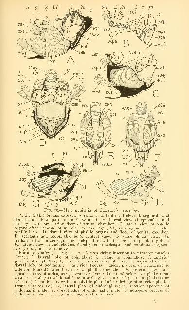
h g' k bf m_^Pal 267 Epph bf z
Dej G ejs y Pal'
Fig. 25.—Male genitalia of Dissostcira Carolina.
A, the phallic organs exposed by removal of tenth and eleventh segments anddorsal and lateral parts of ninth segment. B, lateral view of epiphallus andaedeagus with supporting floor of genital chamber. C, lateral view of phallic
organs after removal of muscles s66 and 26/ (A), showing muscles of endo-phallic bulb. D, dorsal view of phallic organs and floor of genital chamber.E, aedeagus and endophallic bulb, ventral view. F, same, dorsal view. G,median section of aedeagus and endophallus, with terminus of ejaculatory duct.
H, lateral view of endophallus, distal part of aedeagus, and terminus of ejacu-latory duct, muscles removed.For abbreviations, see fig. 24. c/. sclerites giving insertion to retractor muscles
(261); h, lateral lobe of epiphallus; ;', bridge of epiphallus; /, anteriorprocess of epiphallus; k, posterior process of epiphallus; Jii, proximal part of
dorsal lobe of aedeagus; n, anterior (dorsal) apical process of aedeagus; 0.
anterior (dorsal) lateral sclerite of phallotreme cleft; p, posterior (ventral)apical process of aedeagus; q, posterior (ventral) lateral sclerite of phallotremecleft ; ;-, distal part of dorsal lobe of aedeagus ; j-, arm of posterior phallotremesclerite (q) continuous with endophallic plate («) ; t, bridge of anterior phallo-treme sclerites (0); u, lateral plate of endophallus; «', anterior apodeme of
endophallic plate; .r. dorsal edge of endophallic plate; y. gnnopore process of
endophallic plate ; ::, zygoma of aedeagal apodemes.
65

66 SMITHSONIAN MISCELLANEOUS COLLECTIONS VOL. 94
endophallus as a broad apodeme (w) for muscle attachments. The
endophalHc plates are the " endoparameres " of Walker (1922), whobelieves that they are representatives of external or variously invagi-
nated plates or processes (" parameres ") of other Orthoptera. Bycomparison w^ith Tettigoniidae it does appear probable that the endo-
phallus of Acrididae is produced as an invagination of the posterior
surface of the dorsal lobe of the aedeagus.
The cavity of the endophallus is mostly a narrow vertical space
between the lateral endophallic plates, but its posterior membranous
part, somewhat expanded behind the oblique posterior margins of
the lateral plates, forms a distinct section of the lumen, and may be
designated the spennatophore sac (fig. 24 C, sps). The ejaculatory
duct (Dej) opens into the ventral part of the spermatophore sac
through a terminal ejaculatory sac (ejs). The aperture between the
two sacs is the true gonopore. Dorsally the spermatophore sac com-
municates with the phallotreme cleft in the dorsal lobe of the aedeagus
through the meatus at the base of the latter. From the lower anterior
angle of each lateral endophallic plate a long process {y) projects
backward in the membranous connecting wall between the spermato-
phore sac and the ejaculatory sac. The two processes thus closely
embrace the gonopore (fig. 29 E, F, fig. 33 C), and, as will be shown
later, by the action of the endophallic muscles they regulate the open-
ing and closing of the gonopore. Lateral vesicles of the ejaculatory
sac, such as are present in most other Orthoptera, are absent in the
Acrididae.
The curious sclerite known as the epiphallus is a very prominent
feature of the acridid genitalia. It is situated on the floor of the
anterior pocket of the genital chamber beneath the venter of the
tenth and eleventh segments (fig. 24 A, Epph), and is separated from
the aedeagus by the sloping surface that culminates posteriorly in the
hoodlike fold (&/) covering the base of the aedeagus. The morpho-
logical nature of the epiphallus is doubtful, since the sclerite cannot
be satisfactorily identified with any part of the phallic structure
in other insects. The plate is termed the " pseudosternite " by
Walker (1922) and by Ford (1923). It has muscular connections
both with the ninth sternum and with the zygoma of the aedeagal
apodemes. In form the epiphallus is an irregular transverse sclerite
(fig. 31 B) consisting of two expanded lateral lobes {h, h) connected
by a narrow median bridge {i). Anteriorly the lateral lobes bear a
pair of hooklike processes (/) directed forward, and posteriorly each
is produced upward in a large thick irregular transverse process {k).
Closely associated with the epiphallus laterally are two small oval

NO. 6 GRASSHOPPER ABDOMEN—SNODGRASS 67
sclerites (g) in the floor of the genital chamber (figs. 25 D, 33 B,
35 B), on which are inserted strong muscles (^61) from the lateral
parts of the ninth abdominal tergum.
The aedeagus is ordinarily entirely concealed within the posterior
part of the genital chamber beneath the hood of the pallium (fig.
24 A). In the protracted condition, however, the organ is exposed
by a retraction of its coverings (fig. 33 B). The genital lobe of the
ninth sternum (IXSL) is now depressed, and the pallium is turned
inside-out, appearing in this condition as a large posterior fold (Pal)
around the base of the aedeagus, while the basal fold of the genital
chamber floor (bf) correspondingly embraces the aedeagus anteriorly;
the epiphallus (Epph) has emerged from its pocket beneath the
eleventh segment, and stands boldly exposed on the projected floor
of the genital chamber anterior to the ensheathing folds of the
aedeagus.
The exsertion of the phallic organs would appear to be accomplished
by pressure resulting from a contraction of the pregenital part of the
abdomen, since there are no muscles connected with the genital organs
capable of producing the protracted condition of the latter (fig. 33 B).
The aedeagus apparently is held in the position of retraction by a
broad sheet of muscles on each side (fig. 25 A, 266) arising medially
on the ninth abdominal sternum and attached dorsally on the lateral
margins of the genital chamber floor. To be exserted, the aedeagus
must first be drawn forward from the pocket of the pallium ; its
release from the latter evidently is effected by the contraction of the
strong muscles (D, 261) inserted on the small sclerites (g) at the
sides of the epiphallus, which take their origins on the lateral parts
of the ninth tergum. The epiphallus itself is provided with a pair of
large muscles (A, 26/) arising medially on the ninth sternum, which
curve upward around the anterior end of the endophallic bulb and
insert on the lateral lobes (h) of the epiphallus. It is probable that
a contraction of these muscles brings about an elevation of the distal
parts of the phallic apparatus, and that pressure from within the
abdomen then protrudes the aedeagus. A second pair of epiphallic
muscles (B, 2/8) arises posteriorly on the zygoma of the aedeagal
apodemes and extends anteriorly to the lateral lobes of the epiphallus.
The action of these muscles is not clear, but the muscles undoubtedly
play some part in the function of the epiphallus in copulation.
The following description of the musculature of the aedeagus and
endophallus is based on a study of Dissosteira Carolina, but a cursory
examination of the other species suggests that the musculature and
mechanism of the acridid male organs are the same throughout the
family.

68 SMITHSONIAN MISCELLANEOUS COLLECTIONS NOL. 94
Two pairs of small muscles lie within the aedeagus, both arising
within the base of the latter (fig. 25 B) ; those of one pair (^/p)
extend dorsally to the lateral plate (111) in the proximal part of the
dorsal lobe, those of the other pair {^80) are attached distally on
the ventral lobe. The other muscles of the aedeagus include the epi-
phallic muscles (2/8) already mentioned, which arise on the zygoma
of the apodemes, and muscles that functionally pertain to the
endophallus.
The endophallus is mostly ensheathed in broad muscle plaques,
which make of the organ a strong muscular bulb, into which opens
the ejaculatory duct. The endophallic musculature comprises dilator
IX X Eppfc Papt
PhtrRect
Dej 267 IXS ej^
Fig. 26.—Median vertical section of the end of the male abdomen of Dissostcira
Carolina showing the retracted position of the phallic organs.
and compressor muscles of the endophallus, and compressors of the
ejaculatory sac. The dilators of the endophallus include a pair of lat-
eral muscles (fig. 25 C, E, 281) and a pair of dorsal muscles (F, 282)
.
The lateral dilators are wide sheets of muscle fibers arising dorsally
on the aedeagal apodemes (C, Apa), and extending ventrally and
anteriorly to the outer surfaces of the anterior apodemes of the lateral
endophallic plates (C, E, w). The dorsal dilators, which likewise are
broad sheets of fibers (F, 282), arise laterally on the inner margins
of the aedeagal apodemes and are inserted mesally on the dorsal
margins {x) of the endophallic plates. The single compressor muscle
of the endophallus consists of a thick mass of fibers stretched trans-
versely over the anterior end of the endophallic bulb (E, F, G, 28^)

NO. 6 GRASSHOPPER ABDOMEN SNODGRASS 69
between the inner faces of the anterior apodemes of the lateral plates.
The compressors of the ejaculatory sac are broad plaques of fibers
closely applied to the lateral walls of the endophallus (C, E, 284) ;
each arises on the entire outer wall of the lateral endophallic plate,
and its fibers converge ventrally to their insertions on the lateral wall
of the ejaculatory sac. In some cases a distinct branch of this muscle
takes its origin on the endophallic apodeme (fig. 29 C, 284a). The
ejaculatory duct has a strong sheath of circular fibers (fig. 25 E, 28^=])
extending to the membranous terminal sac.
The function of the endophallic muscles is to regulate the gonopore,
/. e., the aperture of the ejaculatory sac into the spermatophore sac,
and to drive the spermatophores through the gonopore, through the
spermatophore sac of the endophallus, and through the phallotreme
cleft of the aedeagus. If the endophallic cavity is opened from above
by cutting its dorsal wall, and the lateral plates are spread apart (fig.
33 C), the slithke gonopore {Gpr) is to be seen in the floor of the
spermatophore sac between the posterior ends of the convergent
gonopore processes {y) of the lateral plates. The gonopore processes
are hinged to each other by points of contact just before the anterior
end of the gonopore; as a consequence, an approximation of the
endophallic plates, produced naturally by a contraction of the muscle
between their anterior apodemes (fig. 25 E, F, 28s), results in an
opening of the gonopore.
The endophallic mechanism is well illustrated in Mcrmiria maculi-
pcnnis (fig. 29 E, F). When the lateral plates are separated, as in the
ordinary state (E), the gonopore {Gpr) is closed to a narrow slit;
but when the plates are brought together (F) the gonopore becomes
a widely open aperture. Immediately beneath the gonopore is the
membranous ejaculatory sac (fig. 25 G, H, ejs), which is a terminal
enlargement ot the ejaculatory duct. The compressor muscles inserted
on the lateral walls of the ejaculatory sac (C, E, 284) probably
contract in unison with the compressor muscle of the endophallic
plates, and force the spermatophore from the ejaculatory sac through
the open gonopore into the spermatophore sac. The passage of the
spermatophore through the spermatophore sac is not so easily ex-
plained, in the absence of direct observations on the action of the endo-
phallic apparatus, and it seems probable that the endophallic muscles
must produce movements of the endophallic walls other than those con-
cerned with the opening and closing of the gonopore and the com-
pression of the ejaculatory sac described above.
The male genitalia of the Tetrigidae, by comparison with the
acridid organs, are not only very simple in structure, but, as observed

70 SMITHSONIAN MISCELLANEOUS COLLECTIONS VOL, 94
by Walker (1922), they " are surprisingly unlike those of the Acridi-
dae." The phallus of Tettigidea lateralis (fig. 27 D) consists of a
low ovate elevation on the floor of the genital chamber beneath the
pallial valve (C). Sclerites in the lateral walls of the organ converge
and unite anteriorly in a median process armed with small spines.
Between the lateral plates is a large, widely open, membranous cavity,
into the bottom of which the ejaculatory duct opens anteriorly (D,
Gpr). The posterior wall of the cavity is directly continuous with
the inner membranous fold of the pallium {Pal'). The ejaculatory
PlVlv ..Phi
C / "'^'"'^^^^ DFig. 2y.—Abdomen and male genitalia of Tettigidea lateralis (Tetrigidae).
A, terminal part of abdomen. B, dorsal view of pallial valve (PlVlv) andanterior process of phallic organ. C, lateral view of ninth sternum and pallial
valve, with phallic organ exposed beneath the latter. D, the phallic organ, dorsal
view, with base of inner pallial fold.
duct has strongly muscular walls, and groups of muscle fibers arising
on the phallic sclerites are inserted on its terminal part. Coition is
probably effected by evagination of the endophallic sac. According
to Walker the external phallic plates of the tetrigid organ represent
the epiphallus (" pseudosternite ") of the Acrididae ; but the attach-
ment of the ejaculatory muscles upon them would make it seem more
probable that they are external representatives of the invaginated
endophallic plates of the Acrididae. These plates, Walker himself
contends, are derived from external plates or processes (" para-
meres ") of other Orthoptera.

NO. 6 GRASSHOPPER ABDOMEN—SNODGRASS 7I
It is thus rather curious to find that, while the external parts of
the male tetrigid abdomen (fig. 27 A) present the typical acridian
characters, the structure of the phallic organs should have so little
in common with these organs in the Acrididae. The male organs of
Tridactylidae, furthermore, are entirely different from those either
of the Tetrigidae or the Acrididae, which fact again is surprising
considering the close resemblance of the female ovipositor in all three
of these families. The lack of uniformity in the male organs, as
compared with the female organs, suggests that the common basic
structure of the phallus is something less fundamental than is that
of the ovipositor.
COPULATION, AND INSEMINATION OF THE FEMALE
Preliminary to copulation the male grasshopper places himself well
forward on the back of the female. With his fore legs he clasps the
pronotum of the female, the claws holding at the notch in the anterior
margin of the prothorax between the pronotum and the small exposed
part of the episternum ; the intermediate legs clasp the middle of
the female's body ; the hind legs are held in various positions and
take little part in the copulatory act. The male then lowers his ab-
domen along the side of the female's abdomen (in pictures almost
invariably on the left side, but Boldyrev says, sometimes on the left,
sometimes on the right). The genital lobe of the ninth segment of
the male is now depressed and the phallic organs protruded, the dorsal
lobe of the aedeagus being turned upward and forward. In order to
expose the spermathecal aperture of the female, which receives the end
of the male organ in copulation, the male, as described by Boldyrev
(1929) for Locusta migratoria, depresses the subgenital plate of the
female with the anterior hooks of the epiphallus. The penis is then
introduced into the genital chamber between and beneath the ventral
valves of the ovipositor and is inserted into the spermathecal canal.
In Locusta miffratoria, according to Boldyrev, the separation of the
lower valves of the ovipositor by the organ of the male stretches the
dorsal wall of the genital chamber and pulls back the folds that
ordinarily conceal the spermathecal opening ; the latter is now " opened
wide and the penis is plunged into it right up to its base." The penis,
or dorsal lobe of the aedeagus, in Locusta migratoria is long, slender,
and tapering (fig. 32 B); in forms in which the terminal part is
short and thick, as in Melanophis (figs. 37, 38, 40), it seems hardly
possible that the entire organ can be inserted;probably in such cases
only the apical processes enter the spermathecal orifice. During copu-
lation the cerci of the male are said to grasp the base of the subgenital

^2 SMITHSONIAN MISCELLANEOUS COLLECTIONS \"0L. 94
plate of the female, while the distal part of the j^late is held downby the epiphallus.
Paired grasshoppers remain thus together for a number of hours,
or for a day or more, in some cases for more than two days, the
duration of copulation apparently being determined by the length of
time necessary for the formation of the spermatophore, or spermato-
phores, and the transference of the latter to the female, or by the
number of spermatophores produced. Individuals of each sex mayhave several successive matings.
It is now well established for the Acrididae that during copulation
the spermatozoa are transferred from the male to the female in true
spermatophores, which, as defined by Cholodkowsky (1910), are
sperm-containing capsules foniicd in the genital passage of the male.
In some cases only a single spermatophore is produced at each mating,
which, with one end remaining in the genital passage of the male
and the other inserted well into the spermathecal duct of the female,
forms a conduit from one individual to the other through which the
sperm are discharged by the action of the endophallic apparatus of
the male ; in other cases a number of small spermatophores are in-
jected into the female. The spermatozoa, as in Tettigoniidae and
Gryllidae, are united by their head ends in bundles, or spermato-
desmata.
The spermatophores of Locusta migratoria and their formation
have been studied by Iwanowa (1926), Sokolow (1926), and Bol-
dyrev (1929). It appears that normally only one spermatophore is
produced by this species at a single mating. Iwanowa reports finding
sometimes three or four spermatophores in the receptaculum of the
female, but Boldyrev gives evidence that insemination is accomplished
properly with one spermatophore, and that if the male attempts to
insert a second into the spermathecal orifice the process is not natural
and cannot result in the discharge of the spermatozoa.
A spermatophore of Locusta migratoria, as described and figured
by Boldyrev (fig. 32 D), is an elongate structure with transparent
walls, consisting of a proximal sac constricted into two bladderlike
compartments, and of a long slender distal tube. The length of the
entire spermatophore is usually 25 to 27 mm, but it may reach 29 or
30 mm. The spermatophore thus greatly exceeds the length of the
intromittent organ (fig. 32 B), since the latter measures not more
than 5 or 6 mm from the gonopore at the bottom of the spermato-
phore sac of the endophallus {sps) to the tips of the apical processes
of the aedeagus. Only the distal tubular part of the spermatophore
is introduced into the spermathecal canal. The extrusion of the tube,

NO. 6 GRASSHOPPER ABDOMEN SNODGRASS "J^
Boldyrev says, requires an hour or more, and the discharge of the
sperm is not completed until 5| to i8 hours after copulation begins,
the time apparently being dependent on the temperature. On opening
the genital organs of males killed during copulation, Boldyrev found
that the first bladder of the spermatophore is held in the ejaculatory
sac (fig. 32 B, e']s), while the second lies in the membranous part of
the endophallic cavity here termed the spermatophore sac (sps). Thelong spermatophore tube extends through the phallotreme cleft and
protrudes forward from the distal end of the aedeagus between the
bases of the anterior apical processes (C) ; in copulation it is deeply
inserted into the spermathecal duct. During mating the two sexes
are thus united by a tubular conduit through which the sperm are
driven by the action of the endophallic apparatus from the male into
the female. At the end of copulation the spermatophore tube is not
drawn out of the female, but is broken ofif near its base. The detached
tube is retained a long time in the spermathecal canal ; the basal part
of the spermatophore is soon rejected from the male.
With Anacridium aegyptium, as shown by Fedorov (1927), in-
semination of the female is accomplished by the introduction of from
6 to 30 spermatophores into the spermatheca, the usual number being
from 12 to 18. In this species, however, the spermatophores are of
relatively small size, about i mm in length. Each capsule is a hyaline
body, broader at the anterior end, which.bears a small appendage easily
broken off. The spermatophores are formed in the beginning of
the ejaculatory duct, but they do not attain their final shape until they
reach the terminal part of the aedeagus. Fedorov believes that the
spermatophores are all prepared during copulation, and that this
accounts for the length of the copulatory period—1| or 2 hours
being necessary for the completion of one capsule and its transference
to the female. He finds thus that 6 to 12 spermatophores correspond
with 18 to 24 hours of copulation, 18 to 24 with 36 hours, and 30with 60 hours. In about 4 to 6 hours after the beginning of copula-
tion, Fedorov says, a milky-white jellylike mass containing the empty
spermatophores that have been ejected from the spermatheca collects
between the lower valves of the ovipositor where it becomes brittle
and yellow as it dries, and after a few hours is lost, leaving no evi-
dence of the insemination that has taken place.
EXAMPLES OF THE MALE GENITALIA OF ACRIDIDAE
The following descriptions of the male genitalia of representative
species of the several acridid subfamilies will serve to illustrate the
nature of specific variations in the form of the organs, and will show
the fundamental unity of structure throughout the family.

74 SMITHSONIAN MISCELLANEOUS COLLECTIONS VOL. 94
Chorthippus bicolor Charp.—The Acridinae (Tryxalinae) have the
typical acridid structure of the male genital organs, except that the
distal part of the dorsal lobe of the aedeagus is suppressed, and the
apical processes, which have the form of four long, closely appressed
prongs, arise directly from the proximal part (figs. 28 E, 29 C).
The structure of the male genital organs of Chorthippus bicolor
is shown in figure 28. The aedeagus (E, Aed) includes a dorsal lobe
(dl) and a ventral lobe (vl), but the former consists principally of
the usual proximal part of the dorsal lobe (m), which bears directly
-Male genitalia of Chorthippus bicolor (Acridinae).
A, end of abdomen. B, dorsal lobe of aedeagus and lateral apodeme, left side.
C, aedeagal apodemes, dorsal view. D. epiphallus, dorsal view. E, phallic or-
gans and floor of genital chamber, lateral view. F, endophallus and distal
part of aedeagus.
the four large apical processes above noted (B, n, p). The aedeagal
apodemes are well developed as long tapering arms extending forward
from the base of the aedeagus (B, Apa) in deep invaginations be-
neath the basal fold (E) ; their proximal parts are united by a strong
zygoma (C, s) in the under surface of the basal fold (B). From each
of the apical processes of the aedeagus (F, n, p) a. sclerite extends
proximally in the inner wall of the endophallic meatus (o, q). Here
the extremities of the dorsal pair of sclerites are united by a wide
dorsal bridge (t) , while the tapering ends of the ventral sclerites are
sharply bent upward (s) and then gradually expanded anteriorly to

NO. 6 GRASSHOPPER ABDOMEN SNODGRASS 75
form the large lateral plates (u) of the endophalic walls. The ejacu-
latory sac (ejs) opens ventrally into the spermatophore sac of the
endophallus (sps) between the gonopore processes (y) of the lateral
plates, and the spermatophore sac discharges through the wide meatus
and the phallotreme.
The epiphallus is a large sclerite of the usual type of structure
(fig. 28 D) seated on the floor of the genital chamber (E, Epph) at
the anterior end of the surface that forms the fold {hf) overlapping
the base of the aedeagus.
IXSL
Male genitalia of Mcrmiria maculipcnnis (Acridinae).
A, end of abdomen. B, epiphallus. C, phallic organs with floor of genital
chamber and muscles of endophallic bulb. D, endophallus and distal part of
aedeagus. E, regulator mechanism of the gonopore, ventral view, closed. F,
same, gonopore open.
Mermiria macidipennis Bruner.—The only differences in the male
genitalia between this species and the last are in details of form and
relative size of the parts (fig. 29). The epiphallus (B, C, Epph) has
the usual shape ; the basal fold (C, hf) forms a large hoodlike cover-
ing over the base of the aedeagus. The four large apical processes
of the aedeagus, in the retracted condition, project dorsally from the
supporting proximal part {m) of the dorsal lobe; the ventral lobe
{vl) projects like a trough beneath the latter. The endophallus is
large (D), but its lateral plates {11) with their apodemes (w) and
gonopore processes (y) are of typical form. The closing and opening
mechanism of the gonopore is easily studied in this species (E, F).
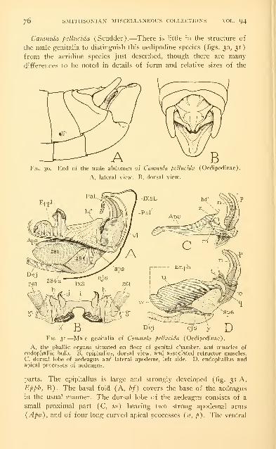
1^ SMITHSONIAN MISCELLANEOUS COLLECTIONS VOL. 94
Camiuda pcllucida (Scudder).—There is little in the structure of
the male genitalia to distinguish this oedipodine species (figs. 30, 31)
from the acridine species just described, though there are many
differences to be noted in details of form and relative sizes of the
A ^ BFig. 30.—End of the male abdomen of Camnula pellucida (Oedipodinae).
A, lateral view. B, dorsal view.
Dej ejs yFig. 31.—Male genitalia of Camnula pellucida (Oedipodinae).
A, the phallic organs situated on floor of genital chamber, and muscles of
endophallic bulb. B, epiphallus, dorsal view, and associated retractor muscles.C, dorsal lobe of aedeagus and lateral apodeme, left side. D, endophallus andapical processes of aedeagus.
parts. The epiphallus is large and strongly developed (fig. 31 A,
Epph, B). The basal fold (A, hf) covers the base of the aedeagus
in the usual manner. The dorsal lobe of the aedeagus consists of a
small proximal part (C, ni) bearing two strong apodemal arms
(Apa), and of four long curved apical processes (11, p). The ventral

NO. 6 GRASSHOPPER ABDOMEN—SNODGRASS 17
lobe (A, vl) is unusually large and ensheaths much of the dorsal lobe.
The endophallus has the typical form (D, Enph), with well-developed
apodemes (ze^) and gonopore processes (y).
Locusfa migratoria L.—The aedeagus of Locusta migratoria is
quite different in shape from that of the other species here described,
but its peculiarities may be seen as an exaggeration of the structure in
Camnula. The ventral lobe is very large and, in the retracted position
Fig. ^2.—Male genitalia of Locusta migratoria (Oedipodinae), and acridid
spermatophores.
A, phallic organs on floor of genital chamber, with endophallus and ejaculatory
sac beneath the latter. B, dorsal lobe of aedeagus, with aedeagal apodemes andendophallus exposed by removal of floor of genital chamber and ventral lobe
of aedeagus. C, aedeagus in protracted position, with protruding spermatophore(Sphr). D, spermatophore of Locusta migratoria. E, spermatophore of Cal-liptamus ifalicus. (C, D, E from Boldyrev, 1929.)
of the phallic organs (fig. 32 A), completely conceals all but the
terminal parts of the dorsal lobe. Its upturned lateral walls contain
each a large quadrate plate (vl) lying in a vertical plane at the side
of the base of the dorsal lobe ; ventrally the two plates are united by
a median membranous area of the lobe. When the ventral lobe is
removed (B) the dorsal lobe of the aedeagus (dl) is seen to have the
form of a long, tapering tube, curved upward and ending in four
slender apical processes. The organ, however, is not literally tubular,
since the posterior wall is deeply cleft to its base : the opening is the

78 SMITHSONIAN MISCELLANEOUS COLLECTIONS VOL. 94
phallotreme, and in its inner walls are the usual phallotreme scleritcs
ending distally in the apical processes {n, p). The proximal part
of the dorsal lobe (B, m) is small, but is quite distinct from the
cylindrical distal part (r) and bears the two aedeagal apodemes (Apa).
The phallotreme cleft leads through the meatus in the neck of the
endophallus into the endophallic cavity. The spermatophore sac of the
latter is small (sps), but the ejaculatory sac (ejs) is unusually large
;
the two communicate by the gonopore, which lies between the gono-
pore processes (y) of the lateral endophallic plates.
VIII IX X Eppt Cer Papt Pal
Fig. 33.—Male genitalia of Dissostcira Carolina (Oedipodinae).
A, end of abdomen with phallic organs concealed in genital chamber. B,same, with phallic organs protracted. C, endophallic chamber opened from above,showing gonopore situated in its floor. D, proximal lobes of aedeagus (w)and aedeagal apodemes with basal zygoma, dorsal view.
In the position of protraction (fig. 32 C), as shown by Boldyrev
(1929), the ventral lobe of the aedeagus is depressed and the dorsal
lobe is projected upward with its apical processes turned forward.
The spermatophore (Sphr) issues anteriorly from the upper end of
the phallotreme cleft between the bases of the anterior processes.
Dissosteira Carolina (Linn.).—The end of the male abdomen of
Dissosteira is obtusely pointed (fig. 33 A) because of the conical form
of the genital lobe of the ninth sternum (IXSL). The lobe is movable
on the anterior sternal plate (IXS) by a wide membranous area sepa-
rating the two. From its dorsal margin the pallium (Pal) is continued

NO. 6 GRASSHOPPER ABDOMEN SNODGRASS 79
forward as a hoodlike fold that meets the lobes of the eleventh seg-
ment, and is then reflected inward to form the posterior wall of the
genital chamber (fig. 26). The genital chamber and the contained
phallic organs are thus ordinarily entirely concealed beneath the tenth
and eleventh segments in front, and the pallial hood behind. If the
pallium is pulled back from the eleventh segment it is to be seen that
the epiphallus occupies an anterior pocket of the genital chamber
beneath the venter of the tenth segment, and that the aedeagus is con-
tained in a posterior pouch lined by the inflected fold of the pallium
{Pal'), which latter extends inward and ventrally to the base of the
ventral lobe of the aedeagus {vl). The floor of the genital chamber
laterally slopes downward from in front (fig. 25 A), where the epi-
phallus is seated upon it, to the base of the inner fold of the pallium,
but medially, between the epiphallus and the aedeagus, it presents a
broad, smooth, rounded surface (D), the posterior margin of which
forms the basal fold (A, D, hf) overlapping the proximal part of the
aedeagus. The epiphallus is a large irregular sclerite, consisting of
two lateral lobes (D, h, h) connected by an arched bridge {i) , and
provided with the usual hooked anterior processes (/) and broad,
strong posterior processes {k) having a vertical position. Just laterad
of the epiphallus, in the wall of the genital chamber, are to be seen
the small oval sclerites {g) that give insertion to the large retractor
muscles {261) from the lateral parts of the ninth abdominal tergum.
The structure of the aedeagus is well shown in the protracted con-
dition (fig. 33 B), in which the organ projects dorsally from a basal
sheath formed of the everted pallium {Pal) and the basal fold {hf)
of the genital chamber floor. The two parts of the dorsal lobe of the
aedeagus (r, m) are quite distinct, the narrow distal part (r), ending
in the small apical processes {n, p), being exserted from between the
lobate lateral walls of the proximal part (m). The ventral aedeagal
lobe {vl) embraces the dorsal lobe posteriorly, and between the two
is a deep cavity into which opens the vertical slitlike phallotreme in
the posterior wall of the dorsal lobe. The aedeagal apodemes (D,
Apa) project downward and forward from the base of the dorsal
lobe beneath the basal fold (B, hf), and their proximal parts are united
by a wide zygoma (D, s) in the under side of the fold (B, s).
The phallotreme is a deep cleft in the dorsal lobe of the aedeagus
;
in its lateral walls are the usual two pairs of sclerites (fig. 25 H, o, q)
.
The sclerites of the dorsal (anterior) pair end in the meatus, where
they are united with each other by a transverse bridge {t) \n the dorsal
wall of the latter ; the ventral (posterior) sclerites are continuous by
narrow upcurved arms (s) with the lateral plates {n) of the en-
6
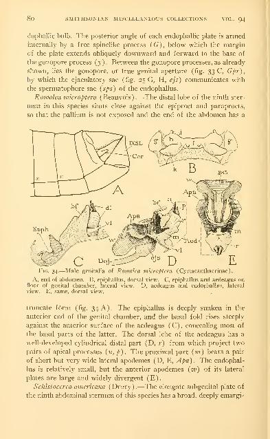
8o SMITHSONIAN MISCELLANEOUS COLLECTIONS VOL. 94
dophallic bulb. The posterior angle of each endophallic plate is armed
internally by a free spinelike process (G), below which the margin
of the plate extends obliquely downward and forward to the base of
the gonopore process (3')- Between the gonopore processes, as already
shown, lies the gonopore, or true genital aperture (fig. 33 C, Gpr),
by which the ejaculatory sac (fig. 25 G, H, ejs) communicates with
the spermatophore sac {sps) of the endophallus.
Romalea microptera (Beauvois).—The distal lobe of the ninth ster-
num in this species shuts close against the epiproct and paraprocts,
so that the pallium is not exposed and the end of the abdomen has a
Fig.
A, endfloor of
view. E
34.—Male genitalia of Romalea microptera (Cyrtacanthacrinae).
of abdomen. B, epiphallus, dorsal view. C, epiphallus and aedeagus ongenital chamber, lateral view.
, same, dorsal view.
D, aedeagus and endophallus, lateral
truncate f6rm (fig. 34 A). The epiphallus is deeply sunken in the
anterior end of the genital chamber, and the basal fold rises steeply
against the anterior surface of the aedeagus (C), concealing inost of
the basal parts of the latter. The dorsal lobe of the aedeagus has a
well-developed cylindrical distal part (D, r) from which project two
pairs of apical processes {n, p). The proximal part (in) bears a pair
of short but very wide lateral apodemes (D, E, Apa). The endophal-
lus is relatively small, but the anterior apodemes (iv) of its lateral
plates are large and widely divergent (E).
Schistocerca amcricana (Drury).—-The elongate subgenital plate of
the ninth abdominal sternum of this species has a broad, deeply emargi-

NO. 6 GRASSHOPPER ABDOMEN SNODGRASS
iiate extension projecting far beyond the origin of the palhum from its
dorsal lamella (fig. 35 A, B, IXSL). The exposed part of the pallium
(Pal) forms a thick, transversely corrugated fold against the para-
procts. When the tenth and eleventh segments are removed (B) the
genital chamber is exposed from above, and there are to be seen on
its floor the phallic structures lying anterior to the aedeagus, the latter
being still concealed beneath the pallial hood. The epiphallus consists
of large lateral lobes (h, h) connected by a narrow median bridge;
anterior processes are absent, but the posterior processes are present
Fig. 35.—Male genitalia of Schlsfoccrca amcricana (Cyrtacanthacrinae).
A, end of abdomen. B, dorsal view of genital lobe of ninth sternum, pallium,
and anterior part of genital chamber, exposed by removal of tenth and eleventh
segments. C, phallic organs, posterior view. D, same, lateral view. E, aedeagus
with apodeme, endophallus, and ejaculatory sac. F, endophallus and apex of
aedeagus.
in the form of large triangular plates {k, k). Between the bases of
the latter the floor of the genital chamber presents a deep transverse
groove, the part behind the groove terminating in the basal fold {hf).
The aedeagus is small (C, D, Aed), but its ventral lobe {vl) is rela-
tively large. The principal part of the dorsal lobe is formed of the
usual proximal subdivision (D, E, m), the distal part (E, r) being
much reduced and ending in a small spoutlike terminal lobe without
apical processes. The aedeagal apodemes (E, Apa) are short but
broad at their bases. The endophallus (F) has the usual structure,
but has characteristic features. The phallotreme sclerites {o, q) are
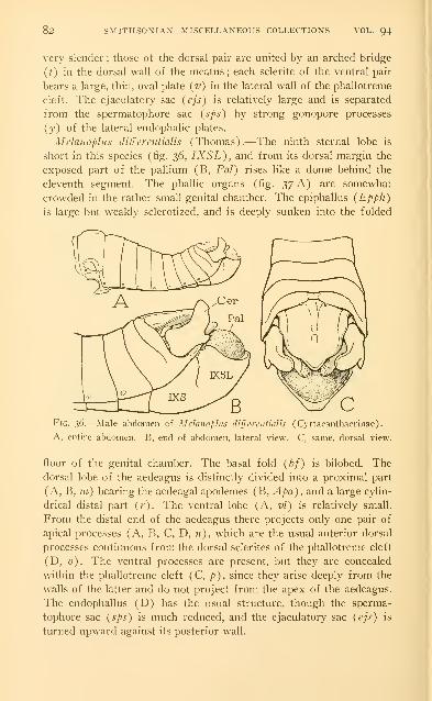
82 SMITHSONIAN MISCELLANEOUS COLLECTIONS VOL. 94
very slender; those of the dorsal pair are united by an arched bridge
(t) in the dorsal wall of the meatus; each sclerite of the ventral pair
bears a large, thin, oval plate (v) in the lateral wall of the phallotreme
cleft. The ejaculatory sac (ejs) is relatively large and is separated
from the spermatophore sac (sps) by strong gonopore processes
(y) of the lateral endophalic plates.
Melanoplus differentialis (Thomas).—The ninth sternal lobe is
short in this species (fig. 36, IXSL), and from its dorsal margin the
exposed part of the pallium (B, Pal) rises like a dome behind the
eleventh segment. The phallic organs (fig. 37 A) are somewhat
crowded in the rather small genital chamber. The epiphallus (Epph)
is large but weakly sclerotized, and is deeply sunken into the folded
Fig. 36.—Male abdomen of Mclaiwfliis differentialis (Cyrtacanthacrinae).
A, entire abdomen. B, end of abdomen, lateral view. C, same, dorsal view.
floor of the genital chamber. The basal fold {hf) is bilobed. The
dorsal lobe of the aedeagus is distinctly divided into a proximal part
(A, B, 77z) bearing the aedeagal apodemes (B, Apa), and a large cylin-
drical distal part (r). The ventral lobe (A, vl) is relatively small.
From the distal end of the aedeagus there projects only one pair of
apical processes (A, B, C, D, n), which are the usual anterior dorsal
processes continuous from the dorsal sclerites of the phallotreme cleft
(D, 0). The ventral processes are present, but they are concealed
within the phallotreme cleft (C, p), since they arise deeply from the
walls of the latter and do not project from the apex of the aedeagus.
The endophallus (D) has the usual structure, though the sperma-
tophore sac {sps) is much reduced, and the ejaculatory sac {ejs) is
turned upward against its posterior w-all.

Fig. yj-—Male genitalia of Melaiwpliis diff'croitialis (Cyrtacanthacrinae).
A, the external phallic organs on floor of genital chamber. B, aedeagus andendophallus. C, dorsal lobe of aedeagus, posterior view, showing phallotreme
(Phtr) and apical processes («, p). D, endophallus with apex of aedeagus,
and ejaculatory duct.
Fig. 38.—Male abdomen and genitalia of Mclaiwplits mexicaiius (Cyrta-canthacrinae).
A, end of abdomen, lateral view. B, same, dorsal view. C, right half of
epiphallus. D, external and internal phallic organs, with floor of genital chamber.E, aedeagus and apodeme, right side.
83
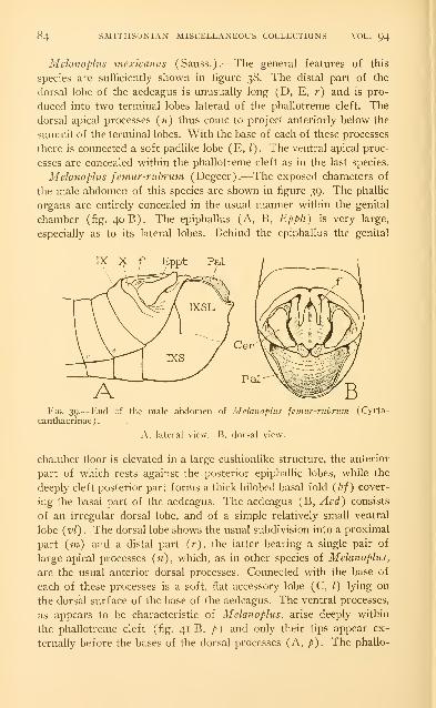
84 SMITHSONIAN MISCELLANEOUS COLLECTIONS \OL. 94
Melanoplus mexicanus (Sauss.).—The general features of this
species are sufficiently shown in figure 38. The distal part of the
dorsal lobe of the aedeagus is unusually long (D, E, r) and is pro-
duced into two terminal lobes laterad of the phallotreme cleft. The
dorsal apical processes («) thus come to project anteriorly below the
summit of the terminal lobes. With the base of each of these processes
there is connected a soft padlike lobe (E, /). The ventral apical proc-
esses are concealed within the phallotreme cleft as in the last species.
Melanoplus femur-rubruin (Degeer).^—The exposed characters of
the male abdomen of this species are shown in figure 39. The phallic
organs are entirely concealed in the usual manner within the genital
chamber (fig. 40 B). The epiphallus (A, B, Epph) is very large,
especially as to its lateral lobes. Behind the epiphallus the genital
Fig. 39.-—End of the male abdomen of Melanoplus fciiiitr-nibruin (Cyrta-
canthacrinae).
A, lateral view. B, dorsal view.
chamber floor is elevated in a large cushionlike structure, the anterior
part of which rests against the posterior epiphallic lobes, while the
deeply cleft posterior part forms a thick bilobed basal fold (bf) cover-
ing the basal part of the aedeagus. The aedeagus (B, Aed) consists
of an irregular dorsal lobe, and of a simple relatively small ventral
lobe (vl). The dorsal lobe shows the usual subdivision into a proximal
part (m) and a distal part (r), the latter bearing a single pair of
large apical processes (n), which, as in other species of Melanoplus,
are the usual anterior dorsal processes. Connected with the base of
each of these processes is a soft, flat accessory lobe (C, /) lying on
the dorsal surface of the base of the aedeagus. The ventral processes,
as appears to be characteristic of Melanoplus, arise deeply within
the phallotreme cleft (fig. 41 B, p) and only their tips appear ex-
ternally before the bases of the dorsal processes (A, p). The phallo-
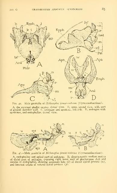
NO. 6 GRASSHOPPER ABDOMEN SNODGRASS 85
h ^Epph^ j
Enph
Fig. 40. Male genitalia of Mclanophis jemur-rnbrmn (Cyrtacanthacrinae).
A, the external phallic organs, dorsal view. B, same, lateral view, with part
of genital chamber wall. C, aedeagus and apodeme, left side. D, aedeagus with
apodemes, and endophallus, dorsal view.
ejs A'Fig. 41.—Male genitalia of Melanophis femur-rubnim (Cyrtacanthacrinae).
A, endophallus and apical part of aedeagus. B, diagrammatic median section
of distal part of aedeagus, exposing right inner wall of phallotreme cleft and
meatus of endophallus, showing accessory lobe (0 of dorsal apical process (n),
and internal origin of ventral apical process (p).

86 SMITHSONIAN MISCELLANEOUS COLLECTIONS VOL. 94
treme sclerites (B, o, q) extend forward in the walls of the phallo-
treme cleft from the bases of their respective processes {n, p).
Anterior to the phallotreme a tubular meatus (A) leads into the cavity
of the endophallus, and the ejaculatory sac {ejs) opens between large
gonopore processes {y) into the small spermatophore sac {sps). Thephallotreme cleft in the proximal part of the dorsal lobe of the aedeagus
has its lateral walls strengthened by a pair of thin oval plates (fig.
41A, B,t/).
The foregoing studies of three species of Melanoplus suggest that
the phallic structures will be found to be more variable and more spe-
cialized among the Melanopli than in the other acridid groups.
ABBREVIATIONS USED ON THE FIGURES
aAp, anterior sternal apodeme.
iied, aedeagus.
AcGl, accessory genital gland.
acs, antecostal suture.
aiv, anterior intervalvula.
AN, alinotum.
An, anus.
Ap, apodeme.
Apa, apodeme of aedeagus.
a^t, acrosternite (precostal lip of
sternum).
atg, acrotergite (precostal lip of
tergum).
bf, basal phallic fold.
Bs, basisternum.
Cer, cercus.
Cln, colon.
CpCls, cap cells of sense organ.
CxC, coxal cavity.
C.vpd, coxopodite (limb basis).
Dej, ductus ejaculatorius.
dil, lateral internal dorsal muscles.
dim, median internal dorsal muscles.
dl, dorsal lobe of aedeagus.
DMcl, longitudinal dorsal muscles.
DS, dorsal sinus.
DV , dorsal blood vessel.
eg, egg guide.
cjs, ejaculatory sac.
Enph, endophallus.
Epm, epimeron.
Epph, epiphallus.
Eppt, epiproct.
Eps, episternum.
GC, genital chamber.
Gpr, gonopore.
//, ileum.
IXSL, genital lobe of ninth abdominal
sternum.
lAp, lateral sternal apodeme.
Ic, external lateral muscles.
//, internal lateral muscles.
1st, lalterosternite.
Itg, laterotergite.
Nv, nerve.
Ode, oviductus communis.
OdI, oviductus lateralis.
Ovp, ovipositor.
Pa, postalar arm of postnotum.
Pal, pallium.
Pal' , inner fold of pallium.
Papt, paraproct.
paptl, lobe of paraproct.
Ph, phragma.
Phi, phallus.
Phtr, phallotreme.
piv, posterior intervalvula.
PI, pleuron.
PIS, pleural suture.
PlVlv, pallial valve.
PN, postnotum.

NO. 6 GRASSHOPPER ABDOMEN SNODGRASS 87
Prcx, precoxal pleural sclerite. sr, sternal ridge.
PvS, perivisceral sinus. Stn, primitive sternum.
Py, pylorus.
T, tergum.
Rect, rectum. id, dorsal transverse muscles.
Tm, tympanum.
S, definitive sternum. tr, tergal ridge.
sa, external pit of sternal apophysis. tz% ventral transverse muscles.
SCls, sense cells.
Sco, scolops (sense rod). J'eut, ventriculus.
SI, sternellum. I'H, lateral internal ventral muscles.
SO, sense organ. rim. median internal ventral muscles.
Sp, spiracle. I 'i valvula.
Sphr, spermatophore. vl, ventral lobe of aedeagus.
spn, external pit of sternal spina. Vlf, valvifer.
Spr, spermathecal aperture. VNC, ventral nerve cord.
sps, spermatophore sac of endophallus. VS, ventral sinus.
Spt, spermatheca.
SpfD. spermathecal duct. IVP, pleural wing process.
REFERENCESAnder, K.
1934. t)ber die Gattung Cylindracheta und ihre systematische Stellung.
Archiv Zool. K. Svenska Vetenskapsakad., vol. 26 A, no. 21, 16 pp.,
13 pis.
Blatchley, W. S.
1920. Orthoptera of Northeastern America. Indianapolis.
BOLDYREV, B. T.
1929. Spermatophore fertilization in the migratory locust (Locusta migra-
toria L.). Reports on Applied Entomology (Russian), vol. 4, pp.
189-218, 18 figs.
Brues, C. T., and Melander, A. L.
1932. Classification of insects. Bull. Mus. Comp. Zool.. vol. 73, 669 pp.,
1 121 figs.
Cholodo\v.skv, N.
1 910. t)ber die Spermatophoren, besonders bei den Insekten. Trav. Soc.
Imp. Naturl. St. P'ctersbourg, vol. 41, pp. 128, 129.
Chopard. L.
1920. Recherches sur la conformation et le developpement des derniers
segments abdominaux chez les Orthopteres. Rennes.
Fedorov, S. M.
1927. Studies in the copulation and oviposition of Anacridium aegyptium
L. Trans. Ent. Soc. London, vol. 75, pp. 53-6i. pls. 5-8.
Ford, Norma1923. A comparative study of the abdominal musculature of orthopteroid
insects. Trans. Roy. Canadian Inst., vol. 14, pp. 207-319, pis. 7-23.
GlARDINA, A.
1901. Funzionamento deU'arniatura genitale femminile e considerazioni in-
torno alle ooteche degli Acridii. Giorn. Sci. Nat. Econ. Palermo,
vol. 23, pp. 54-61. 8 figs.

88 SMITHSONIAN MISCELLANEOUS COLLECTIONS VOL. 94
Grasse, p.
1922. fitude biologique sur le criquet figyptien, Orthacanthacris aegyptia
(L.). Bull. Biol. France et Belgique, vol. 56, pp. 545-578.
HUBBELL, T. H.
1932. A revision of the Puer group of the North American genus Melano-
plus, with remarks on the taxonomic value of the concealed male
genitalia in the Cyrtacanthacrinae. Univ. Michigan Mus. Zool.,
Mi.sc. Pubs., no. 23, 64 pp., 4 pis.
IWANOWA, S. A.
1926. Zur Frage iiber die Spermatophorbefruchtung bei den Acridodea
(Locusta migratoria L.). Zool. Anz., vol. 65, pp. 75-86, 10 figs.
KiJNCKEL d'Herculais, J.
1894. Mecanisnie physiologique de la ponte chez les insectes orthop-
teres de la famille des Acridides.—Role de I'air comme agent
mecanique et fonctions multiples de I'armure genitale. C. R. Acad.
Sci. Paris, vol. 119, pp. 244-247.
Nel, R. I.
1929. Studies on the development of the genitalia and the genital ducts in
insects. I. Female of Orthoptera and Dermaptera. Quart. Journ.
Micr. Sci., vol. "jz, PP- 25-85, pis. 2-4.
Ramme, W.1927. Die Eiablage von Chrysochraon dispar. Zeitschr. ]\[orph. Okol. Tiere,
vol. 7, pp. 127-133, 8 figs.
Riley, C. V.
1878. The Rocky Mountain locust. First Ann. Rep. U. S. Ent. Comm.(Oviposition, pp. 223-225.)
Scudder, S. H.
1874. The distribution of insects in New Hampshire. Geol. New Hampshire
(Hitchcock Rep.), pt. i, pp. 331-384.
Snodgrass, R. E.
1929. The thoracic mechanism of a grasshopper. Smithsonian Misc. Coll.,
vol. 82, no. 2, III pp., 54 figs.
1 931. Morphology of the insect abdomen. Part I. General structure of the
abdomen and its appendages. Smithsonian Misc. Coll., vol. 85, no. 6,
128 pp., 46 figs.
SOKOLOW, A. J. •
1926. Zur Frage der Spermatophorbefruchtung bei der Wanderheuschrecke(Locusta migratoria L.). Das Weibchen. Zeitschr. wiss. Zool.,
vol. 127. pp. 608-618, 4 figs.
TiETZ, H. M.
1923. The anatomy of the digestive system of the Carolina locust (Dis-
sosteira Carolina Linn.). Ann. Ent. Soc. Amer., vol. 16, pp. 256-
268, pis. 17-21.
UvARov, B. p.
1928. Locusts and grasshoppers, a handbook for their study and control.
Imp. Bur. Ent., London.
Varde, V. P.
1929. Contribution a I'etude morphologique et ethnologique des Orthop-
teres Acrididae. L Les vesicules oviductaires de Anacridium aegyp-
tium L. Bull. Soc. Zool. France, vol. 54, pp. 477-483, 4 ^gs-

NO. 6 GRASSHOPPER ABDOMEN SNODGRASS 89
VOSSELER, J.
1905. Die Wanderheuschrecken in Usambara im Jahre 1903/1904, zugleich
ein Beitrag zu ihre Biologic. Ber. Ld.- u. Forstw. D.-Ostafrica,
vol. 2, pp. 291-374, pis. 12, 13.
Walker, E. M.
1919. The terminal abdominal structures of orthopteroid insects. (Female.)
Ann. Ent. Soc. Amer., vol. 12, pp. 267-316, pis. 20-28.
1922. The terminal structures of orthopteroid insects. (Male.) Ann. Ent.
Soc. Amer., vol. 15, pp. 1-76, pis. i-ii.
Walton, W. R.
1916. Grasshopper control in relation to cereal and forage crops. U. S.
Dep. Agr. Farmers' Bull. no. 747.
WiGGLESWORTH, V. B.
1932. On the function of the so-called " rectal glands " of insects. Quart.
Journ. Micr. Sci., vol. 75, pp. 131-150, 2 figs.