closing the abdominal wall in high-risk abdominal surgery.
121
From Department of Clinical Science and Education, Södersjukhuset Karolinska Institutet, Stockholm, Sweden CLOSING THE ABDOMINAL WALL IN HIGH-RISK ABDOMINAL SURGERY. Harald Söderbäck Stockholm 2021
-
Upload
khangminh22 -
Category
Documents
-
view
1 -
download
0
Transcript of closing the abdominal wall in high-risk abdominal surgery.

From Department of Clinical Science and Education, Södersjukhuset
Karolinska Institutet, Stockholm, Sweden
CLOSING THE ABDOMINAL WALL IN HIGH-RISK ABDOMINAL SURGERY.
Harald Söderbäck
Stockholm 2021

All previously published papers were reproduced with permission from the publisher.
Published by Karolinska Institutet.
Printed by Universitetsservice US-AB, 2021
© Harald Söderbäck, 2021
ISBN 978-91-8016-141-1
Cover illustration: Incisional hernia. © Lucy Bai 2021

Closing the abdominal wall in high-risk abdominal surgery. THESIS FOR DOCTORAL DEGREE (Ph.D.)
By
Harald Söderbäck
The thesis will be defended in public at Hörsalen Capio S:t Görans sjukhus Stockholm, 2021-
06-04 09.00
Zoom-meeting is available. Contact [email protected] or follow link
https://ki-se.zoom.us/meeting/register/u50od-iurjgjHtMDmuVL-d1tR7nGt4RqZZVH
Principal Supervisor:
Doc Gabriel Sandblom
Karolinska Institutet
Department of clinical science and education
Södersjukhuset
Co-supervisor(s):
MD PhD Rami Klaff
Capio S:t Görans sjukhus
Department of Surgery
Prof Ulf Gunnarsson
Umeå universitet
Department of Surgical and
Perioperative Sciences
Prof Per Hellman
Uppsala University
Department of Surgical Sciences
Opponent:
MD PhD Gabrielle Van Ramshorst
Ghent University Hospital
Department of Gastrointestinal Surgery
Laboratory for experimental surgery
Examination Board:
Doc Abbas Chabok
Uppsala University
Center for Clinical Research
County of Västmanland
Doc Mirna Abraham Nordling
Karolinska Institutet
Department of Molecular Medicine and Surgery
Division of Colorectal Surgery
Doc Jakob Freedman
Karolinska Institutet
Department of Clinical Sciences
Danderyds Hospital


To my family, Elin, Tove and Karin


POPULAR SCIENCE SUMMARY OF THE THESIS
Wound dehiscence and Incisional hernia are anticipated complications to abdominal
surgery. Wound dehiscence is when the sutures in the abdominal wall separate, bursting the
wound under the skin. Wound dehiscence is always a severe condition, especially in elderly
and critically ill patients. Wound dehiscence can cause injury to intestines, as well as
infection and organ failure. In cases where the sutures in the skin also fail, abdominal
content can poke out through the skin. This is called burst abdomen. Normally patients that
are affected by wound dehiscence need renewed surgery where the abdomen is resutured.
Sometimes, because of swelling of the abdominal organs and infection, it is not possible to
close the abdomen when wound dehiscence has occurred. In these cases, the abdomen must
be left open and the patient will require repeated operations and intensive care before
getting well. Wound dehiscence patients have a higher postoperative mortality rate than
surgery patients in general, and there is also a high risk for incisional hernia after wound
dehiscence.
Incisional hernia on the other hand develops when the wound has healed, but due to
impaired healing of the abdominal tendon small bits of the peritoneum bulge under the skin,
sometimes containing intra-abdominal fat. An incisional hernia can be very disabling,
causing pain and local symptoms. In unlucky cases a sling of the small bowel can be caught
in the incisional hernia. This is called incarceration and is a potentially life-threatening
condition. Symptomatic incisional hernias normally need surgery. After an operation for
incisional hernia patients may suffer from stiffness and pain from the abdominal wall.
Yearly about 1800 incisional hernia surgeries are performed in Sweden according to the
Swedish ventral hernia registry. A reduction of incisional hernias would save considerable
health care resources and reduce suffering.
In recent years the methods to close the abdominal wall have developed, and studies show
that a meticulous suturing technique for closing the abdominal wall can reduce the above-
mentioned complications significantly. The new technique uses a suture that is slowly
absorbed by the body. With this technique stitches in the abdominal tendon are made small
and placed close together. The wound and the length of the suture is measured, and a quota
is calculated to ensure that the right technique has been used.
Even after the new suturing technique has been introduced 5-10% of patients still develop
an incisional hernia and a few percent a wound dehiscence. To further reduce these
numbers, high risk patients must be identified, and special precautions made to protect
them.
Many hospitals use the new technique for closing the abdominal wall, but far from all do.
The department of Surgery at Capio S:t Görans hospital introduced the technique as early
as 2012 in a structured quality effort, and the technique is integrated in standard practice
and used by all abdominal surgeons.
The aim of this thesis has been to follow up on the structured implementation of the new
surgical technique to see the long-term results.
By looking at large Swedish patient registries, this thesis has intended to identify risk
factors for wound dehiscence and incisional hernia in the population.

Furthermore, to test a new technique to reinforce the abdominal closure to avoid wound
dehiscence and incisional hernia.
Paper 1 – A retrospective study of the implementation of the new surgical technique at
Capio S:t Görans hospital.
The study investigates the long-term impact of the change in technique by examining data
on patients operated on before the implementation of the technique in 2012 and some years
after the implementation.
93% of patients in the post implementation group had their abdomen closed according to
the new technique, and the suturing technique was also correctly noted in the medical
records. This compared to only 1% in the pre-implementation group. Incisional hernia and
wound dehiscence proved to be at the same levels both before and after the implementation
of the new technique, about 4-5%, which is normally considered an acceptable level. High
BMI and postoperative wound infection proved to be risk factors for incisional hernia
development. Male gender, high age, chronic obstructive pulmonary disease and wound
infection were risk factors for wound dehiscence development.
Four years after the implementation, the new technique was fully integrated as standard
practice at S:t Görans Hospital. The reason that there was no difference in complications
between the groups is most likely due to that a sufficient surgical technique was already in
use before the structured implementation.
Paper 2 – A study of the Swedish colorectal cancer registry to find the incidence of
incisional hernia in the Swedish population.
This paper studies the incidence of incisional hernia in the Swedish population and looks
for relevant risk factors by building a database combining the Swedish colorectal cancer
registry and the Swedish national patient registry. The risk for developing an incisional
hernia in the population was 5,3% for the population as a whole. Male gender, long surgery
duration, age less than 70 years old, high BMI and wound infection were risk factors for
incisional hernia development.
Paper 3 - A study of the Swedish colorectal cancer registry to find the incidence of wound
dehiscence in the Swedish population.
By looking at the same database as paper 2, the incidence of wound dehiscence and
relevant risk factors can be explored. A significantly higher risk for postoperative death is
recorded for wound dehiscence patients. High age, male gender, high BMI, chronic
obstructive pulmonary disease, systemic inflammatory disease, and a short surgery duration
were risk factors for wound dehiscence in the study.
Paper 4 – Pilot study to test a new surgical devise.
In this study we tested a new technique where the abdominal wall, after suturing, is
reinforced with a surgical net. The net that was tested is made from a synthetic material that
is absorbed by the body in about 6 months. In this pilot study we found that the technique
works and that there were no serious adverse events recorded.

Conclusions
Wound dehiscence and incisional hernia are dangerous and resource consuming
complications to abdominal surgery.
High age, high BMI, long operation time, chronic obstructive pulmonary disease, and
systematic inflammatory disease are risk factors for complication after abdominal surgery.
There is also an increased risk for men.
Postoperative wound infection is a strong risk factor for further wound complications, and
all possible actions should be undertaken to avoid wound infection.
A structured implementation when introducing a new surgical technique works well and
has a long-lasting effect.
Implantation of TIGR® Matrix surgical mesh is a possible way to reinforce the abdominal
wall after surgery.

POPULÄRVETENSKAPLIG SAMMANFATTNING
Stor bukkirurgi innebär dessvärre att komplikationer kan uppstå. Sårruptur och ärrbråck är två
fruktade sådana komplikationer. En sårruptur innebär att suturerna i bukväggen släppt och att
såret under huden spricker upp. En sårruptur är alltid ett allvarligt tillstånd, framför allt hos
äldre, och kan leda till tarmskador, infektioner och organsvikt. Det kan också vara associerat
med att hudsuturerna spricker upp, vilket i sin tur kan få till följd att bukinnehållet glider ut ur
buken. Vanligen behöver patienter som drabbas av sårruptur en omoperation där buken sys
ihop igen. Det händer att man pga. svullna tarmar och inflammation inte kan stänga buken
efter en sårruptur. Då behöver buken lämnas öppen och det krävs intensivvård och upprepade
operationer innan patienten är färdigbehandlad. Efter en operation av sårruptur är risken att
senare utveckla ärrbråck mycket hög.
Till skillnad från sårrupturer innebär ärrbråck att operationssåret har läkt, men på grund av en
defekt läkning av bukväggssenan buktar en mindre eller större snip av bukhinna och
bukinnehåll fram under huden. Ett ärrbråck kan vara mycket invalidiserande och ge uttalade
lokala besvär. I olyckliga fall kan en tarmslynga glida ut i ärrbråcket och klämmas fast. Detta
kallas inklämning och är ett potentiellt livshotande tillstånd. Om patienten har uttalade besvär
så behöver detta opereras. En operation av ärrbråck kan ge smärtor och stelhet i bukväggen.
Årligen utförs c:a 1800 sådana operationer enligt svenska bukväggsbråckregistret. En
minskning av antalet ärrbråck skulle spara stora sjukvårdsresurser och minskat lidande.
De senaste tio åren har metoden att försluta buken efter operation utvecklats och studier har
visat att komplikationer enligt ovan kan minskas med en strukturerad och noggrann
bukförslutningsteknik. Tekniken går ut på att man använder en tråd som långsamt löses upp
av kroppen efter c:a sex månader och att man syr med små täta tag i bukväggssenan. Sedan
mäter trådåtgång och sårets längd och räknar ut en kvot för att kontrollera att rätt teknik har
använts.
Även med modern sutureringsteknik drabbas c:a 5-10% av patienterna av ärrbråck och några
procent av sårruptur. För att ytterligare minska dessa risker behöver högriskpatienter kunna
identifieras och ytterligare åtgärder vidtas för att skydda dessa.
Många sjukhus, inte bara i Svergie, använder idag en strukturerad teknik för
bukförslutningen, men långt ifrån alla gör det. Kirurgkliniken vid Capio S:t Görans sjukhus
introducerade tidigt den nya tekniken och började redan 2012 att försluta buken enligt ett
standardiserat vetenskapsbaserat protokoll som sedan dess följs av alla klinikens kirurger.
Syftet med detta avhandlingsarbete har varit att följa upp hur införandet av en standardiserad
bukförslutningsteknik fungerat på Capio S:t Ggörans sjukhus och hur resultaten på lång sikt
påverkats.

Genom att använda de stora svenska patientregistren identifiera riskfaktorer för ärrbråck och
sårruptur i den svenska befolkningen.
Testa en ny teknik för att förstärka bukförslutningen så att inte sårruptur och ärrbråck uppstår.
Arbete 1 – En återblick på införandet av den strukturerade bukförslutningstekniken vid Capio
S:t Görans sjukhus
Vi undersökte vad bytet till den moderna strukturerade bukförslutningstekniken hade för
effekt på lång sikt. Genom att samla in data på patienter opererade innan införandet 2012 och
data frånpatienter som opererats fyra år efter införandet samlade vi totalt 1120 patienter. Vi
fann att 93% av patienterna i den sena gruppen hade fått buken försluten med en korrekt
teknik, vilket också noterats korrekt i journalen, att jämföras med 1% före införandet.
Ärrbråck och sårruptur visade sig vara lika vanligt före som efter införandet av den nya
tekniken, c: a 4–5%, vilket anses vara en acceptabel nivå. Högt BMI och postoperativ
sårinfektion visade sig vara riskfaktorer för att utveckla ärrbråck. Manligt kön, hög ålder,
kronisk obstruktiv lungsjukdom samt sårinfektion var riskfaktorer för sårruptur.
Fyra år efter införandet var den nya tekniken helt integrerad i vårt arbetssätt. Att skillnaden
inte var så stor beror sannolikt på att de flesta kirurger använde en tillräckligt bra teknik redan
före införandet av den strukturerade tekniken.
Arbete 2 – Studie på det svenska Colorektalcancerregistret för att få en bild av ärrbråcks
utbredning hos patienter opererade för Colorektalcancer.
I den här studien tittade vi på förekomsten av ärrbråck och sökte efter riskfaktorer för
ärrbråcksutveckling genom att bygga en databas där vi kombinerade data från det svenska
Colorektalcancerregistret och Slutenvårdsregistret. Risken att utveckla ärrbråck I
studiepopulationen var 5,3%. Manligt kön, lång operationstid och ålder under 70 år, högt
BMI och sårinfektion visade sig vara signifikanta riskfaktorer för ärrbråcksutveckling
Arbete 3 -– Studie på det svenska Colorektalcancerregistret för att få en bild av sårrupturers
utbredning.
Genom att titta i samma databas som i arbete 2 kunde vi utvärdera förekomsten av sårruptur
efter Colorektalcancerkirurgi och dess riskfaktorer. Det var en signifikant ökad risk att dö i
inom 30 dagar från operationen för de patienter som utvecklade sårruptur. Hög ålder, manligt
kön, högt BMI, Kronisk obstruktiv lungsjukdom, systemisk inflammatorisk sjukdom och kort
operationstid visade sig vara riskfaktorer.
Arbete 4 – Pilotstudie för att testa nytt nätmaterial.
I det här arbetet testade vi en ny teknik där man förstärker bukförslutningen med ett nät. Nätet
som vi testade var av ett resorberbart material som tas upp av kroppen och med tiden
försvinner. I den här pilotstudien fann vi att tekniken fungerade och att inga allvarliga
komplikationer inträffade.

Slutsatser
Ärrbråck och sårruptur är farliga och kostsamma komplikationer till bukkirurgi.
Hög ålder, högt BMI, lång operationstid, kronisk obstruktiv lungsjukdom, systemisk
inflammatorisk sjukdom är riskfaktorer för komplikation efter öppen bukkirurgi. Det
föreligger också en ökad risk för män.
Infektion i operationssåret är en stark riskfaktor för ytterligare komplikationer och man bör
vidta alla åtgärder för att undvika infektion.
Strukturerat införande av en ny kirurgisk teknik fungerar och har en bestående effekt.
Implantation av ett TIGR® Matrix nät är ett möjligt sätt att förstärka bukväggen efter kirurgi.

ABSTRACT
Background
Incisional hernia and Wound dehiscence are potentially serious complications to midline
incisions. Recent studies have shown that a meticulous suturing technique can reduce the rate
of these complications significantly, but even with optimal technique there is 5-15% risk of
abdominal wall complications. At Capio S:t Görans hospital the new abdominal wall closure
technique 2012 was implemented in a standardised quality improvement project.
The aim of these studies was to investigate the effect of a structured implementation of the
new surgical technique, to study which risk factors for incisional hernia and wound
dehiscence are relevant in a Swedish population and to test new techniques to reinforce the
abdominal wall after open abdominal surgery.
Methods
Study 1.
All procedures performed via a midline incision 2010-2011 before, and 2016-2017 after the
new protocol was introduced at Capio S:t Görans Hospital were identified and assessed for
complications and risk factors for wound dehiscence and incisional hernias
Study 2.
All procedures registered in the Swedish Colorectal Cancer Register (SCRCR) 2007–2013
were identified. Patients with comorbid disease diagnoses, registered at admissions and visits
prior to the procedure and relevant to this study, were obtained from the National Patient
Register (NPR). Data on occurrence of incisional hernias were obtained by combining data
from the SCRCR and the NPR).
Study 3.
Like study 2 all open abdominal procedures for colorectal cancer registered in the SCRCR
2007–2013 were identified. Potential risk factors for wound dehiscence were identified by
cross-matching between the SCRCR and the NPR. The endpoint in this study was reoperation
for wound dehiscence registered in either the SCRCR or NPR.
Study 4
Sixteen patients with three or more risk factors for wound dehiscence or incisional hernia
were included. A TIGR® Matrix mesh was placed on the aponeurosis with an overlap of five
cm on either side and fixated with continuous monofilament polydioxanone suture. All
postoperative complications were registered at clinical follow-up.

Results
Study 1
After the implementation of new guidelines, 93% of procedures were performed using the
standardised technique for abdominal wall closure. There was no significant difference in
incidence of incisional hernia or wound dehiscence between the two periods. BMI>25 and
postoperative wound infection were found to be independent risk factors for incisional hernia.
Male sex, high age, chronic obstructive pulmonary disease, and postoperative wound
infection were risk factors for wound dehiscence.
Study 2
The cumulative incidence of incisional hernia in the population was 5.3%. In multivariate
analysis male gender, operation time exceeding 180 min, body mass index (BMI) > 30, age <
70 years and postoperative wound complication were significant risk factors for incisional
hernia.
Study 3
In multivariable analysis, age > 70 years, male gender, BMI
> 30, chronic obstructive pulmonary disease, generalised inflammatory disease, and duration
of surgery less than 180 min were significant risk factors for wound dehiscence. The hazard
ratio for postoperative
death was 1.24 for patients who underwent reoperation for wound dehiscence compared with
that for controls.
Study 4
One patient developed a seroma that needed drainage and antibiotic treatment. One patient
had a wound infection that needed antibiotic treatment. There was no complication requiring
a reoperation. No wound dehiscence or incisional hernia was seen.

Conclusions
High age, high BMI, long operation time, chronic obstructive pulmonary disease, systemic
inflammatory disease, and male gender should be considered risk factors for postoperative
adverse events after a midline incision.
Postoperative wound infection is a strong predictor of incisional hernia and wound
dehiscence and all measures possible should be taken to avoid wound infection.
Structured implementation of a standardised surgical technique is possible and has a long-
lasting effect.
Implantation of TIGR® Matrix mesh is a feasible way to reinforce the abdominal wall after
high-risk surgery.

LIST OF SCIENTIFIC PAPERS
I. Small Stitch Small Bites technique: a long-term follow-up.
Submitted
II. Incisional hernia after surgery for colorectal cancer: a population-based
register study.
Int J Colorectal Dis. 2019 Oct;34(10):1757-1762
III. Incidence of wound dehiscence after colorectal cancer surgery: results from a
national population-based register for colorectal cancer.
Int J Colorectal Dis. 2018 Oct;33(10):1411-1417
IV. Prophylactic Resorbable Synthetic Mesh to Prevent Wound Dehiscence and
Incisional Hernia in High High-risk Laparotomy: A Pilot Study of Using
TIGR Matrix Mesh
Front Surg. 2016 May 18;3:28

LIST OF ABBREVIATIONS
SSSB Small Stitch Small Bites Technique for closing the abdominal
wall
RCT Randomized controlled trial
NPWT Negative pressure wound therapy
COPD Chronic obstructive pulmonary disease
PRF Perirenal fat surface area
BMI Body mass index
IR Insulin resistance
ERAS Enhanced recovery after surgery
MMP Matrix metalloproteinase
ASA American society of anesthesiologist’s physical status
classification
SCRCR Swedish colorectal cancer registry
NPR Swedish national patient registry
PDS Polydioxanone suture material
ICD International classification of disease
SSI Surgical site infection
HIPEC Hyperthermic intraperitoneal chemotherapy
CT Computed tomography

CONTENTS
1. Background
1.1 Introduction
1.2 Anatomy of the abdominal wall
1.3 The midline incision
1.4 Principles for wound healing
1.5 Incisional hernia
1.6 Abdominal wound dehiscence
1.7 Risk factors for wound dehiscence and incisional hernia
1.8 The Small-Stich-Small-Bites technique
1.9 Prophylactic mesh augmentation
1.10 Prophylactic retention sutures
1.11 Swedish Colorectal cancer registry and the National patient registry.
1.12 Implementation of a new technique
2. Patients and methods
2.1 Paper 1. Small-Stitch-Small-Bites technique: a long-term follow-up.
2.2 Paper 2. Incisional hernia after surgery for colorectal cancer: a population-
based register study
2.3 Paper 3. Incidence of wound dehiscence after colorectal cancer surgery: results
from a national population-based register for colorectal cancer
2.4 Subgroup analysis of acute surgery based on data from the SCRCR.
2.5 Paper 4. Prophylactic Resorbable Synthetic Mesh to Prevent Wound
Dehiscence and Incisional Hernia in High High-risk Laparotomy: A Pilot Study
Using TIGR Matrix Mesh
2.6 Statistical Methods
2.7 Ethical considerations
3. Results
3.1 Paper 1
3.2 Paper 2
3.3 Paper 3
3.4 Subgroups analysis of acute surgery
3.5 Paper 4

4. Discussion
4.1 General comments
4.2 Implementation of SSSB at S:t Görans Hospital
4.3 Incidence of wound dehiscence and incisional hernia
4.4 Wound dehiscence
4.5 Risk factors for incisional hernia and wound dehiscence
4.6 Prophylactic mesh augmentation and TIGR® Matrix mesh
4.7 The surgeon as a risk factor
4.8 Limitations of studies
5. Conclusions
6. Future Perspectives
7. Reference list
8. Appendices
8.1 Appendix 1 A3-tool for implementing the SSSB at S:t Görans Hospital.
8.2 Appendix 2 Local guidelines for opening and closure of abdominal incisions.
8.3 Appendix 3 Study protocol PrevMesh.


1
1 BACKGROUND
1.1 INTRODUCTION
In 2012 I became aware that the rate of wound dehiscence in our general surgery department
was too high. I investigated the problem and found good evidence to suggest that we should
implement a structured closure technique after midline incision of the abdominal wound, to
reduce this complication. After discussions and education of the surgical staff, we
implemented the Small-Stitch-Small-Bites technique (SSSB) in the summer of 2012 to
improve the safety of abdominal wall closure. This had an immediate and dramatic impact on
outcome. In the spring prior to implementation, twelve patients suffered a burst abdomen
which needed acute reoperation and prolonged hospital stay. In the autumn that same year
only one patient needed reoperation for a burst abdomen. I was amazed that such a simple
intervention could have such an impact. In the years following implementation of SSSB the
lower rate of wound dehiscence persisted with four-five cases per year. In my initial follow-
up, I found that wound dehiscence patients needed a long hospital stay and that our
intervention saved us more than one bed per day all year round in our ward. Furthermore,
resources were saved due to a reduction in incisional hernia surgery. This experience led me
to further investigate the mechanisms of wound dehiscence and incisional hernia, and to see if
it is possible to reduce the risks associated with midline incisions. This thesis is the result of
these studies.
The aim of this thesis has been to follow up on the structured implementation of the new
surgical technique to see the long-term effect.
Studying the incidence of incisional hernia and Wound dehiscence after a midline incision.
Identifying risk factors for wound dehiscence and incisional hernia in the Swedish
population.
Testing a new technique to reinforce the abdominal closure to avoid wound dehiscence and
incisional hernia.

2
1.2 ANATOMY OF THE ABDOMINAL WALL (1–3)
The abdominal wall is a complex construction of muscles, nerves, vessels, aponeuroses, and
tendons bridging between the thorax and the pelvis. The functional purpose of the abdominal
wall is to keep the trunk upright and support movement. The abdominal wall also protects the
internal organs and keeps them in place. Defects in the abdominal wall can thus affect
posture, impair mobility, and cause displacement of internal organs.
The lateral abdominal wall has three flat muscles. The transversus abdominis, as its name
suggests, runs transversely and is the innermost muscle. The main function of the transversus
abdominis is to hold the internal organs in place during activity. The internal oblique muscle
runs laterocaudally from the midline. The internal oblique takes part in breathing and can turn
and tilt the upper body. The external oblique muscle runs laterocranially from the midline.
The main function of the external oblique is rotation and flexion of the upper body. These
three flat muscles form the core construction of the abdominal wall that supports movement
and posture, and confines and protects the internal organs. The rectus abdominis muscles run
in craniocaudal direction on either side of the midline. The aponeuroses of the three flat
muscles form the rectus sheath within which the rectus abdominis muscle runs. The recti
abdominis are the main flexors of the upper body. The rectus sheaths meet in the midline
where they join to form the linea alba, also named the rectus aponeurosis. The blood supply
to the abdominal wall comes from two systems. The epigastric arteries run in a craniocaudal
direction parallel to the midline on both sides. The intercostal and subcostal arteries as well as
the circumflex iliac arteries form branches that run between the transverse and the internal
oblique muscles. The nerve supply to the abdominal wall muscles is derived from the lower
intercostal nerves, the thoracoabdominal nerves and the iliohypogastric nerves. The rectus
aponeurosis is a strong tendinous, avascular structure with little nerve supply.
Anatomy of the abdominal wall © Lucy Bai 2021

3
1.3 THE MIDLINE INCISION
Throughout surgical history a variety of incisions have been used to access the abdominal
organs, all having advantages and disadvantages (4). The midline incision is associated with
low risk for damage to nerves or vessels (1,5). Furthermore, it does not require transection of
muscles, which makes it fast and easy to perform (3,6,7). The level of the midline incision
may be chosen depending on what procedure is intended, and it is easy to extend the incision
if necessary. The midline incision provides good access to most abdominal organs and is
suitable for acute surgery. A disadvantage of the midline incision is the relatively high risk
for incisional hernia (6–11).
Abdominal Incisions © Lucy Bai 2021

4
1.4 PRINCIPLES OF WOUND HEALING
Wound healing after an incision can be divided into three phases (9,12,13). During the first
acute inflammatory phase, vasodilatation leads to an invasion of leucocytes and
macrophages. The main role of these cells is to remove dead cells and debris from the
incision site and to protect the wound from bacterial invasion. The macrophages also attract
fibroblasts, which are important in the early phase of regeneration (9,12,13). The extent of the
acute inflammatory reaction ranges from 0.5 cm to 1.5 cm from the fascial edges, somewhat
wider if wound infection is present (9,12,14,15). During the acute inflammatory phase, which
lasts for about four days, the wound itself has no tensile strength. During this period the
suture holds the entire tensile strength of the wound which explains why wound dehiscence
most often occur during this phase (14,16,17). Infection during the acute inflammatory phase
increases the number of leukocytes but not macrophages. This prolongs the acute
inflammatory phase, but the decline in macrophages decreases the number of fibroblasts
attracted (12). There is evidence that the use of diathermy, especially in the coagulation
mode, can affect the blood supply of the fascial edges and inhibit wound healing (18).
The second, regenerative phase, is characterised by collagen formation mediated by invading
fibroblasts (9,12). Disturbances in collagen metabolism is believed to play a major role in the
formation of incisional hernia (12,17,19–22). There are over twenty collagen types, all with
individual characteristics. Collagen type I gives strength to the scar whereas the role of
collagen type III is primarily to form a matrix. Collagen type III is produced in the early
remodeling phase. The weaker collagen type III is then remodeled to collagen type I. The
collagen type I/III ratio is used as a measure of collagen quality (19,21). A lower collagen
I/III ratio is seen in incisional hernia patients than in controls (19,22). The collagen I/III ratio
can be measured in skin, fascia, or in rectus aponeurosis tissue itself to identify patients with
a high risk for incisional hernia (19,21,22). Another way to identify patients at risk is to
measure the turnover of different types of collagen. Reduced turnover of collagen type V
(Matrix) and increased turnover of collagen type IV (strength) is seen in patients who develop
an incisional hernia. (23) Matrix metalloproteinases (MMP) break down extracellular matrix
and may serve as markers of collagen turnover. Levels of certain MMPs are believed to
predict hernia formation (17,19,21). MMP-2 activity is increased in obese patients, resulting
in an increased breakdown of type I collagen during the regenerative phase. This leads to a
reduced type I/type III collagen ratio, and thus the synthesis of mechanically weaker tissue
(24). Abdominal aortic aneurysm is significantly associated with hernia formation and shares
many risk factors. MMP-2, MMP-9 and MMP-13 activities are associated with both hernia
and aneurysm disease, indicating that both could be symptoms of a systematic connective
tissue disease (22,25,26).
The regenerative phase lasts for approximately three weeks. At the end of the regenerative
phase the wound has achieved 15-20% of the original tissue’s strength (2,9,12).
The third remodeling phase lasts up to one year. During this phase, mechanical tension of the
wound stimulates collagen remodeling leading to more organized and stronger structures. By

5
the end of this phase, the rectus aponeurosis has regained 60-90% of its original strength
(9,12,13).
1.5 INCISIONAL HERNIA
Incisional hernia is defined as any abdominal wall gap with or without a bulge in the area of a
postoperative scar perceptible or palpable by clinical examination or imaging (27). Simply
put, an incisional hernia is a defect in the abdominal wall covered by neoperitoneum, formed
at the site of a previous surgical incision. Sometimes the hernia is just a defect, and
sometimes a lump is formed under the scar. The hernia sac can contain abdominal fat or
organs and in unfavourable cases this can lead to incarceration and strangulation of the hernia
sac contents. The rate of incisional hernia after open abdominal surgery varies in the
literature. It is normally reported to be up to 20% (28,29), but in more recent studies it has
been shown to be as low as 5-13% with meticulous suturing technique (30,31). An incisional
hernia can present years after index surgery (32–34). The late diagnosis of incisional hernia
can partly be due to authors using different definitions of incisional hernia. With the
definition above, about 90% of hernias can be detected within a year after index surgery (6).
In most cases, incisional hernias can be diagnosed by thorough clinical examination, but
complementation with computed tomography can be helpful in obese patients (32,35). In a
study by Ah-Kee et al, one third of patients were not aware they had an incisional hernia, and
one third had noticed the defect but were asymptomatic (29). Typical symptoms of incisional
hernia include pain, discomfort, functional disability, and cosmetic disturbance (29,36). The
natural course of incisional hernia is not known, nor the probability of an asymptomatic
patient developing symptoms. Reports in the literature state that only 1% of hernia patients
suffer incarceration (36). In a systematic review by Bosanquet et al 2015 (37), the overall
cumulative incidence of incisional hernia was 12%. Forty-nine per cent of patients with an
incisional hernia were symptomatic and 36% underwent surgery. Incisional hernia is costly
(38,39) and prevention of may thus be considered a very cost-effective measure (39,40). In
the USA, the number of operations for incisional hernia and the cost of each operation
increased each year between 2007 and 2011. One of the reasons for this is that we tend to
operate on older patients with greater comorbidity (41).

6
1.6 ABDOMINAL WOUND DEHISCENCE
Wound dehiscence is defined as separation of the abdominal fascia after surgery (9) . There
are four principal mechanisms of wound dehiscence: suture rupture, knot failure, slack suture
(because of large bites including fatty tissue), or sutures cutting through the fascia; the last-
named being the most common (14,42–44). Early wound dehiscence can be dramatic with
protrusion of abdominal organs through the wound; also known as burst abdomen. More
frequently, however, wound dehiscence is a subclinical condition that is seen as a precursor
of incisional hernia (16,45,46). Of patients that have a radiologically diagnosed wound
dehiscence at an early stage, 92% later develop an incisional hernia (46). Wound dehiscence
usually occurs during the first 3-7 days (47–50) but diagnosis is often delayed. Burst
abdomen is rare, with an incidence of approximately two per cent of elective abdominal
surgery patients. In acute or emergency cases, the reported incidence of burst abdomen is as
high as 12-50% (9,51,52). At clinical examination, early wound dehiscence presents with
secretion from the wound, that is often misinterpreted as an infection. More severe cases can
be easily diagnosed with CT-scan where dehiscence in the fascia is seen with abdominal
organs close to the skin. Depending on the size of the fascial defect, there is a risk for
ischaemia of protruding organs (9). Burst abdomen is a very serious condition with high
morbidity and a reported mortality of 25% or higher (48,52–56), and hospital stay is
significantly longer (44). In the case of a large fascial dehiscence, emergency surgery is
needed. There are no large RCTs on the various treatment options for burst abdomen, and the
evidence level is weak. If possible, an attempt is made to close the wound. However, due to
intra-abdominal swelling, deep infection, or poor fascia quality, the wound is sometimes left
open for a while before closure. Patients that are primarily sutured for burst abdomen have a
very high incidence of incisional hernia (52,53,56–58). Closure and reconstruction with a
mesh may provide a safer outcome and should be considered in these patients (52,59,60). The
placement of a mesh does not seem to increase formation of enterocutaneous fistulae, (59)
and seems safe even in a contaminated environment. For patients where primary closure is
not possible, the recommendation is to use a dynamic closure technique where the fascial
edges are kept under traction over a period until it is possible to close the wound. There are
several different systems for dynamic wound closure, some of which can be used in
combination with negative pressure wound therapy (NPWT) (60,61). A promising new
concept for dynamic wound closure, where a combination of mesh augmentation and NPWT
is used, has been described by Petersson et al (62). Patients that are operated for burst
abdomen suffer a poor body image and low quality-of-life (57).

7
Wound dehiscence © Lucy Bai 2021

8
1.7 RISK FACTORS FOR WOUND DEHISCENCE AND INCISIONAL HERNIA.
Many studies have been performed to identify risk factors for incisional hernia and wound
dehiscence (55,63–65) but only a few have been performed after new guidelines for closing
the abdominal wall were published in 2014 (4,49). Most studies do not describe the surgical
technique used to close the aponeurosis and thus neglect one of the most important risk
factors (2). All the risk factors presented here were reported in studies performed before the
closure paradigm shift, and may be considered irrelevant since studies performed after the
2014 guidelines fail to show any significant risk factors (66). When Millburn et al first
reported the excellent outcome of the SSSB technique, they found no significant risk factors.
In a retrospective study by Aksamija et al on over 3000 patients operated 2013-2016, 44
(1,25%) cases of clinically significant wound dehiscence were found; too few to find
significant risk factors (49).
In 2018, Wiegering et al presented an interesting retrospective control-matched cohort study
(67) where they could exactly specify which surgical technique had been used. The same
technique was used in all patients (continuous monofilament, with a suture length /wound
length ratio of over 4:1). In that study, no significant risk factors for incisional hernia could
be found, and they failed to show that abdominal aortic aneurysm surgery patients have a
higher risk than colon cancer patients for incisional hernia. This confirms the findings of
Israelsson et al (68,69), that with a good suturing technique the disease behind surgery has
little importance for the development or not of incisional hernia.
With meticulous suturing technique, prospective randomized trials predict a cumulative
incidence of incisional hernia around 6-10% and burst abdomen approximately 1%. With
such small incidences, large multicentre studies are required to further assess the underlying
risk factors.
Wound dehiscence and incisional hernia represent two different conditions with the same
pathogenesis (46), both having their origin in the early separation of the fascial edges. Table 1
shows known risk factors for incisional hernia and wound dehiscence and the studies that
support them. Although surgical technique is the most important risk factor (31,66), we still
need to be aware of the other risk factors. The presence of five or more of these risk factors
has been suggested as a cut-off for high risk (65).
One systematic review by Bosanquet et al 2015, including data from 14 618 patients, found
that age, previous midline incision, previous hernia, AAA-surgery, surgery for obesity, and
upper abdominal surgery were all risk factors (37).
Several attempts have been made to design a risk score for incisional hernia and wound
dehiscence.
Ramshorst et al, in a large retrospective case-control study including patients between 1985
and 2005, assessed risk factors for wound dehiscence and developed a risk score model (64).
Their aim was interesting, but the risk score they developed is limited by the fact that it

9
includes two strong postoperative risk factors, wound infection and postoperative coughing.
These two risk factors have an overwhelming influence on the risk for abdominal wall
complications but are impossible to use as targets for prevention, making the model
unsuitable to identify patients that require pre- or perioperative prophylactic measures.
Goodenough et al, in a study based on open vs laparoscopic surgery taking into account a
variety of methods for closing the abdominal aponeurosis, prospectively assessed risk factors
for incisional hernia and suggested a risk score (70). They found that apart from open
surgery, obesity and COPD were significant risk factors.
The most recent risk calculator comes from the University of Pennsylvania. In a retrospective
study, Basta et al collected information from over 29 000 patients from large databases and
developed a risk score based on preoperatively known risk factors for incisional hernia (71).
The material for this study was wide and included gynaecologic and urologic surgery, as well
as open and laparoscopic approaches. The overall rate of incisional hernia in the study was
3.8%. Risk factors included colorectal surgery, history of previous abdominal surgery, and
smoking. No information on the method used for closing the abdominal wall was given. The
risk predictor is easily available as a smartphone app or online (72).
These risk models are useful tools for preoperative workup to identify patients at high risk for
incisional hernia. As yet, no risk model exists that assumes use of the small-stitch-small-bites
surgical technique, and there is a need for more studies to identify risk factors that remain
relevant using this modern surgical technique.
Itatsu et al, 2014, published a large well-designed study including over 4000 patients (73).
They concluded that high BMI, thick subcutaneous fat, age, chemotherapy, female sex, and
blood transfusion were independent risk factors. Although the authors state that a continuous
slowly absorbable monofilament suture should be standard for closure, only 443 of the 4000
patients had the aponeurosis closed with this technique. In the risk analysis, there was an
increased risk for incisional hernia when using interrupted sutures, but this was not
significant.
Wound infection is a very strong risk factor for both incisional hernia and wound dehiscence
(64,65,69,74,75). Wound infection is a postoperative outcome with its own risk factors
(76,77). To lower the rate of incisional hernia and wound dehiscence risk factors for wound
infection can be assessed preoperatively in the risk score. One theory is that some cases
reported as a wound infection might, in fact, be early wound dehiscence. This theory has yet
to be confirmed, although Millbourn et al concluded that compared to closure with small
stitches, the use of large stitches doubled the risk for wound infection and that is an
independent risk factor (77). This suggests that wound infection in some cases is caused by
early wound dehiscence, not the opposite, but more studies are needed to examine this.

10
Obesity (BMI>30) (65,70,73,75,77–81) and male gender (64,75,77,82) are frequently
reported risk factors for incisional hernia and wound dehiscence. In a retrospective study,
Aquina et al found that visceral obesity, not high BMI per se, should be regarded as a risk
factor for incisional hernia (83). Visceral obesity is easily approximated by measuring the
perirenal fat surface area (PRF). Studies confirm that a high PRF predisposes to postoperative
complications (84–86). Another suggested measurement is the Hip-Waist Ratio which has
been shown to be a better predictor of complications after surgery than BMI (87). Male sex
predisposes to visceral obesity (88), and this possibly explains some of the gender difference.
One underlying factor associated with obesity and postoperative complications is insulin
resistance (IR). Insulin resistance is common after major surgery, even in non-diabetic
patients (89), and animal studies show that IR leads to impaired wound healing after surgery
(90). Obesity has a strong association with IR and about 40% of obese patients can be
expected to be insulin resistant (91,92). This association is also evident for visceral obesity
(93). Perioperative IR can be prevented by carbohydrate loading (94), and because of its
association with postoperative complications, IR is targeted and treated in the enhanced
recovery after surgery (ERAS) programme (95).
The role of connective tissue quality in the development of incisional hernia is a topic of great
interest. As explained in the section on hernia formation, the turnover of collagen type V
(matrix) is reduced, and the turnover of collagen type IV (strength) is elevated in hernia
patients. Biomarkers of collagen type IV turnover are altered in hernia patients (17) as is the
collagen I/III ratio (19). Pogacnik et al, 2014, examined the risk for incisional hernia in colon
cancer patients and diverticulitis patients undergoing sigmoid colectomy. They found an
increased risk for incisional hernia in the diverticulitis group. This suggests that diverticulitis
patients have impaired connective tissue function that predisposes to their disease as well as
to incisional hernia (78). Future studies on incisional hernia should recognize connective
tissue quality and collagen metabolism as potential risk factors. Smoking is a strong
independent risk factor for fascial dehiscence (82,96) and this should also be included in
future studies.

11
1.7.1 Table 1 Reported risk factors for wound complication
Significant risk factors Ref nr
High age (37,55,64,65,69,73,81,82)
Emergency surgery (55,64)
Cancer (11,55,65,81)
Haemodynamic instability (55,65,73,78)
Male gender (64,75,77,82)
Female gender (73,88)
Hypertension (64,65)
Chronic pulmonary disease (64,65,70,75,81)
Ascites (64,65)
Anaemia (11,64,81)
Jaundice (64)
Corticosteroid use (64,65,78,81,89)
Sepsis (64,65,81)
Postoperative coughing (63,64)
Wound infection (63–65,68,69,74,75,77)
Obesity (11,65,68,70,73,75,77–81,88)
Ostomy (65,78)
Hypoproteinaemia (65,88)
Uraemia (65)
Left colon or rectal cancer (88)
Chemoterapy (73,81,90)
Previous hernia (37,78,90,91)
Thick subcutaneous fat (73)
High ASA score (78)
Abdominal aortic aneurysm repair (37,80)
Smoking (11,81,82)
Diabetes (81)
Radiotherapy (81)

12
1.8 THE SMALL-STITCH-SMALL-BITES (SSSB) TECHNIQUE
There has been a paradigm shift on midline incision closure over the past ten years based on
the findings of the Sundsvall group (30,30,40,42,66,68,68,77,97,98). The Sundsvall results
have also been reproduced in the STITCH trial (31). This method has become the
recommended standard since 2014 (4,28,99) and has seen widespread use since 2010.
The work that made way for the modern technique for abdominal wall closure began in 1976
when Jenkins suggested that a running monofilament suture with a suture length to wound
length ratio of at least 4:1 would prevent suture cutting through the tissue, thereby avoiding
burst abdomen (100). In 1994, Israelsson et al showed that a slowly absorbable suture was as
safe as nylon when closing the abdominal wall (101). Sahlin et al conducted a randomised
controlled trial that failed to show that the continuous suture is superior to the interrupted
technique, though the continuous suture was as good as the interrupted (102). In 1995,
Niggebrugge et al showed that a running monofilament suture was superior to the interrupted
technique to prevent incisional hernia (74).
Jenkins ideas were taken up by Israelsson et al in1993 when they proved that a suture length
to wound length ratio greater than 4:1 reduces the rate of incisional hernia (69). They also
found that the 4:1 method reduced midline incision complications (68,103), and that the
technique was cost effective (39).
In animal studies, Cengiz et al 2001 showed that only including the aponeurosis in the sutures
instead of mass layer suture leads to less fascial separation (98), and that small tissue bites
with a suture length /wound length ratio of at least 4:1 gives higher bursting strength than
large bites (97). This was confirmed by Harlaar et al in 2009 showing that small stitch length
and small distance between the stitches gives the suture line better tensile strength than large
stitches (104). Millbourn et al, 2004, re-evaluated old data from the Sundsvall group,
concluding that short stitch length (small bites) is preferable. (30)
In a randomised controlled trial Millbourn et al tested the SSSB method. They showed that
closing the abdominal wall with a running suture and a suture length to wound length ratio of
at least 4:1 with small bites involving only the aponeurosis is superior to large (at least
10mm) bites in preventing wound dehiscence and incisional hernia (66,77). The rate of
incisional hernia using the small bites method was 5,6% compared to 18% in the large bites
group (77). In 2015, Deerenberg et al published their randomised controlled trial comparing
small bites to large bites, where small bites gave an incisional hernia rate of 13% and large
bites 21% (31).

13
In 2014, the European Hernia Society recommended “a slowly absorbable monofilament
suture in a single layer aponeurotic closure technique without separate closure of the
peritoneum, and that the small bites technique with a suture to wound length (SL/WL) ratio at
least 4/1” (4) should be used to prevent wound dehiscence and incisional hernia formation.
Recording suture length to wound length ratio in the surgical report is a routine that does
much to ensure that the appropriate technique is being applied (69,75). In 2014, Walming et
al attempted to confirm this in a retrospective study (75) but they failed to do so, probably
because a meticulous suturing technique was already standard even though the ratio was not
noted in the report. This highlights the need for a universal way to describe the technique
used for abdominal wall closure to be able to review outcomes retrospectively.
14
1.9 PROPHYLACTIC MESH AUGMENTATION
Despite meticulous suturing technique, approximately 5-10% of patients develop an
incisional hernia and 1% suffer burst abdomen. The use of prophylactic mesh augmentation
has been suggested to prevent these adverse events (4,105). In a meta-analysis from 2013,
Bhangu et al concluded that prophylactic mesh applied as onlay, inlay, or sublay,
significantly reduces the risk for incisional hernia in high risk patients (105).
In 2017, Borab et al concluded that prophylactic mesh dramatically reduces the risk for
incisional hernia, but at the cost of more seroma formation and the possibility of chronic pain
(106). Studies included in their review used both sublay and onlay placement. The increase in
risk for seroma formation was mostly seen in the onlay mesh population.
Caro-Tarrago et al, in a randomised controlled trial, found that onlay mesh reduces the rate of
incisional hernia. They also pointed out that although onlay mesh leads to more seroma
formation, all their patients could be managed conservatively (107). The onlay technique is
easily learned and fast compared to the sublay technique (107). This makes onlay placed
mesh a suitable prophylactic measure.
At present there is no consensus as to which patients require mesh, what material should be
used, and in which position the mesh should be placed (4). More studies on these issues are
needed.
1.10 PROPHYLACTIC RETENTION SUTURES
Another technique to reduce the rates of incisional hernia and burst abdomen is to reinforce a
tensioned suture line using retention sutures. Retention sutures are strong and are placed at
wide intervals across the incision line deep within the musculature and fasciae of the
abdominal wall. A skin protection device is often used, and the retention sutures are left for
some days to lessen the tension on the midline incision during the initial healing phase.
Retention sutures are easily removed by cutting the suture at the skin and then withdrawing
them. In 2013, Khorgami et al suggested that retention sutures could prevent wound
dehiscence in a high risk population (108). Modern closure techniques were not used in their
study and they reported a 13% clinically significant wound dehiscence rate in the control
group. The European Hernia Society reviewed the evidence for retention sutures and
concluded that they should not be recommended in their guidelines (4). More studies are
required to support the use of retention sutures.

15
1.11 SWEDISH COLORECTAL CANCER REGISTRY AND THE NATIONAL PATIENT REGISTRY.
The Swedish Colorectal Cancer Register (SCRCR) is a national quality register for colorectal
cancer. The register has collected data from all patients diagnosed with rectal cancer in
Sweden since 1995, and all patients with colon cancer since 2007. The Register includes data
on age, gender, ASA classification, treatment, and postoperative follow-up, but lacks data on
comorbidity and medical treatment. Completeness of the SCRCR is over 98% for both colon
and rectal cancer, and validity data shows an average agreement of 90% (109–111).
The Swedish National Patient Register (NPR) is supervised by the National Board of Health
and Welfare. The register contains data on all hospital admissions in Sweden since 1987,
including outpatient specialist care visits and outpatient emergency care. It does not, contain
data on primary healthcare visits. The validity of the NPR is estimated to be 85–90%
(112,113).
Onlay mesh augmentation © Lucy Bai 2021

16

17
1.12 IMPLEMENTATION OF A NEW TECHNIQUE
At the Capio S:t Görans Hospital the Donabedians triade structured model is used for quality
improvement. The triade states structure, process, and outcome as the three main pillars for
improvement (114). The goal is to achieve organisational learning. In this case, we aimed to
reduce the rate of complications after midline laparotomies by implementing new methods
for opening and closure of the abdomen, especially the suturing technique. The structure
included new suturing material, a new surgical routine, and documentation. Following
theories originally developed by Argyris (115), we formed double learning loops aimed at
organisational learning i.e., incorporation of knowledge and experience into the
organisation’s structure such that these persist over time despite changes in personnel.
Our work on wound dehiscence started with value stream mapping(116). For this we used the
A3 tool (117) - an example of A3 (in Swedish) can be found in Appendix 1.
After value stream mapping we concluded that too many patients developed wound
dehiscence. Thus, we collected data from all laparotomies performed during a 6-month period
and saw that compared to results in the literature we were doing poorly. After reviewing the
literature on abdominal wall closure, we formed a multidisciplinary team including surgeons,
a theatre nurse, a surgical assistant nurse, and an external product specialist from a suture
company. The team developed a strategy for the implementation of a new suture technique
using new material. The strategy included written guidelines for abdominal opening and
closure (Appendix 2), including routines for documentation and follow-up. Before
implementation several meetings were held with the surgeons and the surgical staff
discussing the new routine. Lectures were given by Leif Israelsson, founder of the SSSB
technique, and by the suture manufacturer. Feedback from these meetings was incorporated
in the guidelines before launching the project. The original trial lasted six months during
which time we closely monitored how things were going and were open to changes in
routine. To maintain motivation, a small symbolic gift was given as a reward to those
surgeons who kept to the guidelines. After six months we the entire staff had adapted to the
new technique and it was found both feasible and effective. Since then, loop learning has
continued, and we routinely communicate our results back to the staff.

18
2 PATIENTS AND METHODS
2.1 PAPER 1. SMALL-STITCH-SMALL-BITES TECHNIQUE: A LONG-TERM FOLLOW-UP.
Based on the evidence presented by Milbourn et al, (66,77), Capio S:t Görans Hospital
implemented the SSSB technique as standard for midline incision closure. Prior to the new
guidelines, abdominal wall closure was according to the preference of the surgeon, which
was usually a running polydioxanone (PDS) 0 loop suture on a large needle. No record of
layers included or suture length to wound length ratio was made in the surgical report. In the
revised local guidelines June 2012, the full SSSB technique was described step by step using
2-0 PDS suture on a small needle, taking small bites in aponeurosis only. It was also
obligatory to measure and work out the suture length to wound length ratio and to record
these in a separate document. To obtain a deep organisational learning of the new technique
we used a double loop learning process (115). Before the implementation, all surgeons were
educated in the new technique and got the chance to present their thoughts through local
seminars where the technique was discussed. There were guest lectures by experts of the
SSSB-technique, and organised self-studies. During the first six months the surgical
technique used was monitored closely so that all surgeons cohered. (Delete – repetition!)
This study was a retrospective review of all abdominal surgeries performed 2010-2011 and
2016-2017 at the Department of Surgery, Capio Sankt Göran Hospital, Stockholm. Cases
were identified by ICD10 codes JA-JX in the Cambio Cosmic medical records database
(118). Laparoscopic cholecystectomy, laparoscopic appendectomy, anal/perianal surgery, and
bariatric procedures were excluded. The remaining list was then cleared of all laparoscopic
procedures and procedures not performed through a midline incision. In a last step, all cases
not fitting the study design, for example where the abdomen was left open, were excluded.
The final list was then reviewed by two examiners who scrutinised each patient’s records,
starting at the index operation extracting data on potential patient-related risk factors and
verifying that the procedure was performed through a midline incision. The surgery records
were then reviewed to confirm if the study criteria were fulfilled i.e., SSSB with appropriate
suture used and the suture length, wound length, and ratio Over 4:1 were correctly noted in
the operation records. At the end of the study period, all patient records were reviewed to find
postoperative complications or time of death. The endpoint “incisional hernia” was defined as
either a clinically evident hernia noted on routine follow-up radiology or visit, incisional
hernia accidently diagnosed clinically or by radiology at any other visit to the hospital where
the abdomen was examined, or surgery for incisional hernia during the follow-up period.
There was no standard protocol for follow-up for all laparotomies since patients were
followed up according to the protocols for each disease.

19
The follow-up of patients in this study included medical records and radiology reports from
all contacts with the hospital until 31st March 2020. “Wound dehiscence” was defined as
clinically evident fascial dehiscence noted and treated conservatively in the postoperative
period, or an acute reoperation for wound dehiscence. End of follow-up for patients in the
study was defined as the 31st March 2020, or date of death registered in Cambio Cosmic
software, or 31st December the year the patient died in cases where the exact date of death
was not known.
Robotkirurgi 20 © Capio S:t Görans sjukhus 2021

20
2.2 PAPER 2. INCISIONAL HERNIA AFTER SURGERY FOR COLORECTAL CANCER: A POPULATION-BASED REGISTER STUDY.
This was a population-based cohort study. All procedures for colorectal cancer registered in
the Swedish Colorectal Cancer Register (SCRCR) 2007–2013 were identified. Diagnoses
from all admissions and visits prior to colorectal cancer resection (identified by the
International Classification of Diseases [ICD] code) were retrieved from the National Patient
Register (NPR). We identified diagnoses that were generally considered to be risk factors in
clinical practice, and collected general descriptive variables including:
• Peripheral vascular disease
• Connective tissue disorder
• Liver cirrhosis
• Renal failure
• Diabetes
• Chronic obstructive lung disease
• Chronic inflammatory condition
• Age
• Body mass index (BMI)
• Gender
• Operation time
• T-category
• Distant metastases
• Preoperative radiation therapy
• Type of incision
• Acute/planned surgery
• Tumour localisation
• Postoperative wound complication
• Adjuvant cytostatic treatment

21
Cross-matching between the SCRCR and the NPR was performed in 2015 using the Swedish
Personal Registration Number, a ten-digit identity number unique for each Swedish citizen
(119).
Data on incisional hernias were obtained by combining data from the SCRCR and the NPR.
The endpoint incisional hernia was defined as incisional hernia registered in the SCRCR by
the surgeon responsible, or ICD codes K43.0–K43.9 for incisional hernia or intervention
codes JAD10–JAD87 for surgery for incisional hernia recorded in the NPR. Analyses were
performed to assess the impact of each risk factor investigated and to estimate the cumulative
incidence of incisional hernia.

22
2.3 PAPER 3. INCIDENCE OF WOUND DEHISCENCE AFTER COLORECTAL CANCER SURGERY: RESULTS FROM A NATIONAL POPULATION-BASED REGISTER FOR COLORECTAL CANCER.
This study like Study 2 was based on data from the SCRCR combined with data from the
NPR on colorectal cancer procedures performed 2007–2013. Patient data and potential risk
factors included in the analyses were:
• Peripheral vascular disease
• Connective tissue disorder
• Liver cirrhosis
• Renal failure
• Diabetes,
• Chronic obstructive lung disease
• Chronic inflammatory condition
• Age
• Body mass index (BMI)
• Gender
• Operation time
• T-category
• Distant metastases
• Preoperative radiation therapy
• Type of incision
• Acute/planned surgery
• Tumour localisation
• Postoperative wound complication
• Adjuvant cytostatic treatment

23
Cross-matching between the SCRCR and the NPR was performed in 2015 using Swedish
Personal Registration Numbers.
Data on wound dehiscence were obtained by cross-matching data from the SCRCR and the
NPR. The endpoint, wound dehiscence, was defined as either wound dehiscence registered in
the SCRCR by the surgeon responsible, or the procedure code for wound dehiscence surgery
(JWA00) in the NPR. Analyses were performed to assess the impact of each risk factor
investigated and the incidence of wound dehiscence.
2.4 SUBGROUP ANALYSIS OF ACUTE SURGERY BASED ON DATA FROM THE SCRCR
After publication of Papers 2 and 3, where acute and elective surgery were treated as one
group, the question was raised whether patients undergoing emergency surgery should be
analysed separately in view of the higher risk for complications associated with acute
surgery. As a result, an acute surgery subgroup analysis was performed based on the same
cohort as in Studies 2 and 3 but including acute surgery only.

24
2.5 PAPER 4 PROPHYLACTIC RESORBABLE SYNTHETIC MESH TO PREVENT WOUND DEHISCENCE AND INCISIONAL HERNIA IN HIGH HIGH-RISK LAPAROTOMY: A PILOT STUDY OF USING TIGR MATRIX MESH.
The study was performed as a case series of patients from Uppsala University Hospital, the
Uppsala Cancer Clinic, and Karolinska University Hospital, Huddinge, who underwent
surgery through a midline incision. Inclusion criteria were the presence of at least three
documented risk factors for incisional hernia or wound dehiscence. Risk factors investigated
in this study were:
• Reoperation
• Age over 80 years
• Generalised malignant decease (presence of distant metastases at the time of
surgery)
• COPD Grades III–IV according to the GOLD classification (FEV1 <50% of the
expected)
• Serum albumin level <20 g/l
• Sepsis i.e., infection in combination with abnormal values of two or more of the
following: body temperature, heart rate, respiratory rate, blood gases, and white
blood cell count
• BMI >35
• haemoglobin <80 g/l
• Diabetes with secondary complications (angiopathy, nephropathy, or
neuropathy) and insulin treatment
• Steroid treatment (with at least 1 mg betamethasone daily or equivalent) for 7
days preoperatively
• Smoking (at least 10 cigarettes a day for 1 year)
• Chemotherapy (last administration within 2 weeks prior to surgery)
• Irradiation of the abdominal wall.
The abdominal wall incision was closed with continuous Polydioxanon Suture (PDS) with a
suture length to incision length ratio of 4:1, using the SSSB technique. After suturing, a
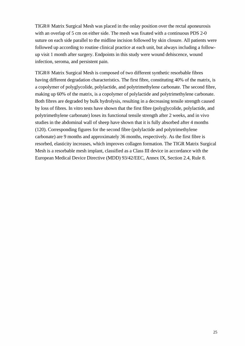
25
TIGR® Matrix Surgical Mesh was placed in the onlay position over the rectal aponeurosis
with an overlap of 5 cm on either side. The mesh was fixated with a continuous PDS 2-0
suture on each side parallel to the midline incision followed by skin closure. All patients were
followed up according to routine clinical practice at each unit, but always including a follow-
up visit 1 month after surgery. Endpoints in this study were wound dehiscence, wound
infection, seroma, and persistent pain.
TIGR® Matrix Surgical Mesh is composed of two different synthetic resorbable fibres
having different degradation characteristics. The first fibre, constituting 40% of the matrix, is
a copolymer of polyglycolide, polylactide, and polytrimethylene carbonate. The second fibre,
making up 60% of the matrix, is a copolymer of polylactide and polytrimethylene carbonate.
Both fibres are degraded by bulk hydrolysis, resulting in a decreasing tensile strength caused
by loss of fibres. In vitro tests have shown that the first fibre (polyglycolide, polylactide, and
polytrimethylene carbonate) loses its functional tensile strength after 2 weeks, and in vivo
studies in the abdominal wall of sheep have shown that it is fully absorbed after 4 months
(120). Corresponding figures for the second fibre (polylactide and polytrimethylene
carbonate) are 9 months and approximately 36 months, respectively. As the first fibre is
resorbed, elasticity increases, which improves collagen formation. The TIGR Matrix Surgical
Mesh is a resorbable mesh implant, classified as a Class III device in accordance with the
European Medical Device Directive (MDD) 93/42/EEC, Annex IX, Section 2.4, Rule 8.

26
2.6 STATISTICAL METHODS
All statistical calculations were performed using SPSS 22.0-26.0 (Chicago, IL).
Logistic regression was used to study the relationship between predictors (that can be
continuous or categorical) and a binary outcome such as incisional hernia. Logistic regression
uses the odds of the outcome to fit a curve and calculates odds ratios.
In our studies we used logistic regression to identify possible risk factors from a dataset of
multiple variables.
Survival analyses such as logistic regression can investigate predictors and their relationship
to a binary outcome. In survival analysis the observation time is included in the analysis
which improves the possibility to find survival differences. The Kaplan-Meier Estimator
(Kaplan-Meier curve) is a visualisation of the survival function, commonly used in medical
statistics to visually compare survival in different groups.
Cox proportions hazard analysis (121) studies the relationship between predictors and the
survival time related to a binary outcome. Cox regression calculates survival as a linear
function of the individual predictors with results presented as hazard ratios (relative risk).
Survival analysis and Cox regression is used in our studies to model the impact of potential
risk factors on survival.
2.6.1 Paper 1
Risk factors for wound dehiscence were analysed in uni- and multivariable logistic regression
analyses. Variables assumed to be risk factors at the beginning of the study were included in
the multivariate analysis. Risk factors for incisional hernia were analysed in uni- and
multivariable Cox proportional hazard analysis, adjustment was made for all covariates
assumed to increase the risk for development of incisional hernia. Subgroup analyses were
performed to investigate risk factors in each group. Potential risk factors were also analysed
for the two groups combined. All analyses were performed using the intention-to-treat
approach comparing early and late cohorts.

27
2.6.2 Paper 2
The impact of each potential risk factor on the risk for incisional hernia was evaluated in a
survival analysis, applying the date of the primary procedure as the time of entry into the
cohort. Date of death or end of follow-up were defined as censored events. Age, body mass
index (BMI), and comorbid disease as risk factors for incisional hernia were analysed using
Cox proportional hazard analysis. Gender, age, BMI, history of chronic obstructive
pulmonary disease, diabetes with secondary complications, chronic renal disease, liver
cirrhosis, systemic inflammatory disease, tumour stage category, distant metastases,
preoperative radiotherapy, acute/planned surgery, tumour localisation, operation time,
postoperative wound complication, and adjuvant cytostatic treatment were included as
covariates in the analysis.
2.6.3 Paper 3
The presumed risk factors age, BMI, comorbid disease, presence of distant metastases, and
operation time were analysed using univariable and multivariable logistic regression analyses.
Survival after reoperation for wound dehiscence was analysed with Cox proportional hazard
analysis.
2.6.4 Subgroup analysis of acute surgery based on data from the SCRCR
Difference in outcome between acute and elective surgery was examined with cross-
tabulation and Pearson’s chi-squared test. Acute surgery as a risk factor for incisional hernia
was examined using survival analysis and Cox regression. Wound dehiscence was explored
with logistic regression. Risk factors for incisional hernia were examined in univariable and
multivariable Cox regression analyses where all suspected risk factors were included. Uni-
and multivariable logistic regression was used to examine risk factors for wound dehiscence.

28
Medarbetare Operation © Capio S:t Görans sjukhus 2021

29
2.7 ETHICAL CONSIDERATIONS
2.7.1 Papers 1-3
All three studies were based on retrospective data from registers and patient records. Our
main ethical considerations were patient integrity and patient autonomy.
In Sweden, there is a great acceptance for using patient registers in decision-making and
healthcare allocation. Most patients agree to have their data stored in various registers, and
these are used to improve the quality of healthcare. In large retrospective studies such as
these, obtaining patient consent would be very time-consuming making them practically
impossible. Patient data was anonymised, and the code keys were kept separate from the data
sets. All data were stored within hospital walls according to safety regulations. The data sets
were processed and presented so that no patient could be recognised.
Paper 1. Approved by the Swedish Ethics Review Authority. Diary number (2019-05787)
Paper 2. Approved by the Regional ethics Review Board in Stockholm ref. (2014/1351-31/5).
Paper 3. Approved by the Regional Ethics Review Board in Stockholm, ref. (2014/1351-
31/5).
2.7.2 Paper 4
In this study we evaluated a new medical device. The product itself had already been tested
on humans and found to be safe, but this was a new application of the device.
The aim of the study was to improve patient safety i.e., there was a potential benefit for
treated patients assuming a decrease in risk for serious complications after surgery. There
was, however, a minor risk of discomfort from the mesh augmentation. Our assumption was
that patient benefit would outweigh any disadvantage. Informed consent was obtained to
ensure patient autonomy.
Paper 4. Approved by the Ethics Review Board of Stockholm ref. (2015/440-31).

30
3 RESULTS
3.1 PAPER 1
Altogether 1120 midline laparotomies were included in the study, 518 in the study cohort and
and 602 in the control cohort. A flow chart of the study assembly is presented in Fig 1. Mean
follow-up time was 32 and 73 months in the study and control groups, respectively.
Emergency surgery was 60% in both groups. In the study cohort, 481 (93%) had correct
SSSB suturing and a satisfactory suture length to wound length ratio (>4) noted in the
operation report compared to 7 (1%) in the control cohort. No significant differences in
wound dehiscence rates and incisional hernia rates were seen between the two cohorts.
A total of 51 patients developed wound dehiscence, 19 (3,7%) in the study group and 31
(5,1%) in the control group. Of these, 44 required emergency reoperation, 17 in the study
group and 27 in the control group. Nine patients (18%) with a documented wound dehiscence
later developed an incisional hernia.
Twenty-seven patients (5.2%) in the study group and 33 (5.5%) in the control group
developed an incisional hernia. There was no significant difference between the groups. Fig 2
shows the Kaplan- Meier curves for the cumulative incidence of incisional hernia for the two
groups. Fig 3 shows a per protocol analysis where patients sutured with SSSB are compared
to those closed with the surgeon’s method of choice. There was no signifficant difference
between these groups. Of the patients with an incisional hernia, 14 in the study group and 21
in the control group required surgery. Surgical site infection (SSI) requiring antibiotic
treatment developed in 16 patients (3%) in the study group and 23 (4%) in the control group.
This difference was not statistically significant.
Table 2 shows background data and a comparison of the two study cohorts.
In the subgroup multivariable analysis of the study cohort (2016-17), male gender (p=0,011)
was a significant risk factor for wound dehiscence. BMI>25 (p=0,007) and wound dehiscence
(p=0,003) were risk factors for incisional hernia. Acute surgery, high age, COPD, and
previous midline incision did not show a significant association in this group.
Tables 3 and 4 present the results of univariable and multivariable analyses of potential risk
factors for incisional hernia and wound dehiscence in the two cohorts combined.
Male gender (p=0.003) and COPD (p=0.022) were significant risk factors for wound
dehiscence. History of previous midline incision (p=0.051) and postoperative surgical site
infection (p=0.053) showed a tendency to predict wound dehiscence, though not significant at
the p<0.05 limit.

31
Fig 1 Flow chart of study assembly
Cox regression identified BMI>25 (p=0.002), postoperative wound infection (p=0.001), and
wound dehiscence (p<0.001) as independant risk factors for incisional hernia. Figures 4, 5
and 6 show Kaplan-Meier curves for incisional hernia related to the significant risk factors.

32
Fig 2 Cumulative incidence of incisional hernia intention-to-treat
Fig 3 Cumulative incidence of incisional hernia per protocol

33
Figure 4. Cumulative incidence of incisional hernia: different BMI categories
Figure 5. Cumulative incidence of incisional hernia: wound infection or not

34
Figure 6. Cumulative incidence of incisional hernia: wound dehiscence or not

35
Table 2 Background data
Background data 2010-2011 2016-2017 Total
Male 285 (47.3%)
235
(45.4%) 520 (46.4%)
Female 316 (52.5%) 283 (54.6%) 599 (53.5%)
Data on sex missing 1 (0.2%) 0 (0%) 1 (0.1%)
Age (Standard deviation) 70.4 (16.6) 68.3 (16.6) 69.4 (16.6)
Index surgery
Explorative laparotomy 102 (16.9%) 75 (14.4%) 177 (15.7%)
Stomach and duodenal surgey 20 (3.3%) 16 (3.1%) 36 (3.2%)
Liver and bile duct surgery 5 (0.8%) 0 (0%) 5 (0.4%)
Splenectomy 4 (0.7%) 2 (0.4%) 6 (0.5%)
Colon and small bowel surgery 387 (64.3%) 386 (74.5%) 773 (69%)
Appendectomy 20 (3.3%) 17 (3.3%) 37 (3.3%)
Rectal cancer surgery 52 (8.6%) 19 (3.6%) 71 (6.3%)
Hernia surgery 3 (0.5%) 1 (0.2%) 4 (0.36%)
other 9 (1.5%) 2 (0.4%) 11 (1.0%)
Acute surgery 363 (60.3%) 306 (59.1%) 669 (59.7%)
Elective surgery 239 (39.7%) 212 (40.9%) 451 (40.3%)
BMI (standard deviation) 24.4 (5.0) 24.7 (5.4) 24.5 (5.3)
ASA I 66 (14.1%) 53 (10.9%) 119 (12.4%)
ASA II 189 (40.5%) 184 (37.8%) 376 (39.2%)
ASA III 181 (38.8%) 205 (42.1%) 387 (40.4%)
ASA IV 30 (6.4%) 44 (9.0%) 75 (7.8%)
ASA V 1 (0.2%) 1 (0.2%) 2 (0.2%)
Data on ASA missing 135 (22.4%) 31 (6.0%) 166 (14.8%)
Cronic obstructive pulmonary disease 61 (10.2%) 51 (9.8%) 112 (10%)
Diabetes mellitus 67 (11.1%) 59 (11.3%) 126 (11.2%)
Haemoglobin, g/L (standard deviation) 130 (20) 129 (21) 130 (20)
Albumin, g/L (standard deviation) 26.4 (8.3) 31.0 (7.2) 29.2 (7.9)
C-Reactive Protein, mg/L (standard deviation) 90 (108) 91 (112) 89.9 (109.4)
Previous midline incision 170 (28.4%) 135 (26.1%) 307 (27.4%)
Standardised technique (SSSB) used 7 (1.2%) 481 (93.9%) 491 (44.0%)
Postoperative wound infection 23 (3.8%) 16 (3.1%) 39 (3.5%)

36
Table 3. Wound dehiscence risk. Univariable and multivariable analyses
Univariable
analysis
Multivariable
analysis
Odds ratio (95%
confidence interval) p
Odds ratio (95%
Confidence interval) p
Primary procedure 2016-
2017 (reference category
2010-2011) 0.70 (0.39-1.26) 0.231 0.72 (0.40-1.31) 0.283
Male (ref Women) 1.94 (1.08-3.47) 0.027 2.57 (1.39-4.76) 0.003
Age≥70 yrs (ref Age<70
yrs) 1.78 (0.97-3.26) 0.063 1.02 (0.99-1.03) 0.130
Acute surgery (ref elective
surgery) 1.21 (0.67-2.18) 0.526 1.30 (0.71-2.38) 0.399
BMI ≥25 (ref BMI<25) 0.78 (0.44-1.37) 0.385 0,76 (0.43-1.36) 0.361
COPD 2.36 (1.15-4.87) 0.020 2.40 (1.13-5.10) 0.022
Previous midline incision 1.82 (1.02-3.26) 0.043 1.82 (0,999-3.32) 0.051
Postoperative wound
infection 2.56 (0.87-7.51) 0.087 2.98 (0.988-8.96) 0.053
Table 4. Incisional hernia Risk. Univariable and multivariable analyses
Univariable
Analysis
Multivariable
Analysis
Hazard ratio (95%
Confidence
interval)
p Hazard ratio (95%
Confidence interval) p
Primary surgery 2016-2017
(ref 2010-2011) 1.12 (0.66-1.89) 0.688 1.34 (0.78-2.29) 0.294
Male (ref women) 1.46 (0.88-2.43) 0.148 1.24 (0.71-2.14) 0.440
Age ≥70 yrs (ref age <70
yrs) 0.76 (0.46-1.28) 0.764 1.00 (0.99-1.02) 0.724
Acute surgery (ref elective
surgery) 0.72 (0.43-1.19) 0.194 0.72 (0.43-1.21) 0.213
BMI ≥25 (ref BMI<25) 2.34 (1.33-4.10) 0.003 5.45 (1.39-4.33) 0.002
COPD 0.96 (3.38-2.39) 0.925 0.93 (0.36-2.38) 0.878
Previous midline incision 1.55 (0.92-2.62) 0.114 1.52 (0.89-2.59) 0.130
Postoperative wound
infection 3.68 (1.67-8.09) 0.001 3.86 (1.73-8.59) 0.001
Wound dehiscence 4.81 (2.36-9.81) 0.000 5.00 (2.39-10.48) 0.000

37
3.2 PAPER 2
Between 2007 and 2013, 39 984 patients were registered in the SCRCR. After excluding
laparoscopic procedures, procedures repeated on the same patient, procedures with
concomitant liver resection, and procedures without laparotomy, 28 913 patients remained in
the study. The study cohort assembly flow chart is shown in Figure 7. Baseline characteristics
of the study population are shown in Table 5. In all, 1352 (cumulative incidence 4,7 %)
patients were either diagnosed with an incisional hernia or underwent surgery for incisional
hernia. In the multivariate analysis, risk for incisional hernia was increased in males, long
operation time (exceeding 180 min), age < 70 years, BMI > 30 and postoperative wound
complication. Table 6 shows the results of the uni- and multivariable analyses of risk factors.
Figures 8-12 shows the Kaplan-Meier curves for the significant risk factors. History of
comorbid disease, tumour stage, tumour localisation, preoperative radiotherapy, adjuvant
chemotherapy, postoperative bleeding, and acute/elective surgery had no statistically
significant impact.
In an additional analysis performed after publication, wound dehiscence instead of
postoperative wound complication was analysed as risk factor. Wound dehiscence was seen
to be a statistically significant risk factor for incisional hernia, hazard ratio 3.04 [95%CI 2.95-
3.93] p<0.001. The Kaplan-Meier curve for wound dehiscence related to incisional hernia is
added as Figure 13.

38
Figure 7. Flow chart of cohort assembly Study 2
Patients registered in the Swedish Colorectal Cancer Register (N=39 984)
N=33 821
Data on resection missing (N= 6163)
N=32 348
Laparotomy without surgery (N=1473)
N=31 603
Treatment withou t laparotomy (TEM, local excision, appendectomy, other, N= 745)
N= 29 219
Laparoscopic resection (N=2 384)
Study population (N=28 913)
Concomitant liver resection (N=306)

39
Table 5. Baseline characteristics, study 2
Mean age, years (standard deviation) 71.1 (11.5)
Gender
Men 14 986 (51.8%)
Women 13 927 (48.2%)
T
0 292 (1.0%)
I 1585 (5.5%)
II 4595 (15.9%)
III 16920 (58.5%)
IV 5333 (18.4%)
TX/unknown 188 (0.6%)
N
0 15904 (55.0%)
I 7152 (24.7%)
II 5491 (19.0%)
NX/unknown 366 (1.2%)
M
0 24 207 (83.7%)
I 3606 (12.5%)
MX/unknown 1200 (3.8%)
Treatment
Resection of ascending colon (including ileocecal resection) 10666 (36.9%)
Resection of descending colon (including sigmoid colon) 8544 (29.6%)
Resection of rectum (anterior resection and abdominoperineal resection) 8271 (28.7%)
Other (including resection of transverse colon and total colectomy) 1432 (5.0%)
Stomy
Temporary 4307 (15.0%)
Permanent 6083 (21.2%)

40
Table 6. Univariable and multivariable Cox proportional hazard analysis of risk
for incisional hernia.
Univariable Cox proportional hazard analysis
Multivariable Cox proportional hazard analysis
Variable N
Hazard ratio (95% confidence interval) p
Hazard ratio (95% confidence interval) p
Gender
Women (ref) 13 927 (48.2%)
Men 14 986 (51.8%) 1.47 (1.29-1.67) <0.001 1.40 (1.21-1.62) <0.001
Age
>70 years (ref) 16 205 (56.0%)
≤70 years 12 704 (43.9%) 1.59 (1.37-1.82) <0.001 1.34 (1.16-1.56) <0.001
Data on age missing 4 (<0.1%)
BMI
<30 20 769 (71.8%)
≥30 4 036 (14.0%) 1.94 (1.65-2.22) <0.001 1.78 (1.51-2.09) <0.001
Data on BMI missing 4 108 (14.2%)
Chronic Obstructive Pulmonary disease
No 27 600 (95.5%)
Yes 1 313 (4.5%) 1.29 (0.94-1.76) 0.112
Diabetes with sec complications
No 28 011 (96.9%)
Yes 902 (3.1%) 0.74 (0.46-1.20) 0.225
Chronic kidney disease
No 28 309 (97.9%)
Yes 604 (2.1%) 0.87 (0.48-1.58) 0.656
Liver cirrhosis
No 28 841 (99.8%)
Yes 72 (0.2%) 2.67 (1.08-7.67) 0.035
Systemic inflammatory disease
No 28 376 (98.1%)
Yes 537 (1.9%) 1.10 (0.66-1.83) 0.725
Adjuvant chemo
No 23 206 (80.3%)
Yes 5 707 (19.7%) 1.14 (0.97-1.34) 0.103
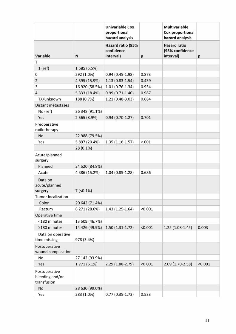
41
Univariable Cox proportional hazard analysis
Multivariable Cox proportional hazard analysis
Variable N
Hazard ratio (95% confidence interval) p
Hazard ratio (95% confidence interval) p
T
1 (ref) 1 585 (5.5%)
0 292 (1.0%) 0.94 (0.45-1.98) 0.873
2 4 595 (15.9%) 1.13 (0.83-1.54) 0.439
3 16 920 (58.5%) 1.01 (0.76-1.34) 0.954
4 5 333 (18.4%) 0.99 (0.71-1.40) 0.987
TX/unknown 188 (0.7%) 1.21 (0.48-3.03) 0.684
Distant metastases
No (ref) 26 348 (91.1%)
Yes 2 565 (8.9%) 0.94 (0.70-1.27) 0.701
Preoperative radiotherapy
No 22 988 (79.5%)
Yes 5 897 (20.4%) 1.35 (1.16-1.57) <.001
28 (0.1%)
Acute/planned surgery
Planned 24 520 (84.8%)
Acute 4 386 (15.2%) 1.04 (0.85-1.28) 0.686
Data on acute/planned surgery 7 (<0.1%)
Tumor localization
Colon 20 642 (71.4%)
Rectum 8 271 (28.6%) 1.43 (1.25-1.64) <0.001
Operative time
<180 minutes 13 509 (46.7%)
≥180 minutes 14 426 (49.9%) 1.50 (1.31-1.72) <0.001 1.25 (1.08-1.45) 0.003
Data on operative time missing 978 (3.4%)
Postoperative wound complication
No 27 142 (93.9%)
Yes 1 771 (6.1%) 2.29 (1.88-2.79) <0.001 2.09 (1.70-2.58) <0.001
Postoperative bleeding and/or transfusion
No 28 630 (99.0%)
Yes 283 (1.0%) 0.77 (0.35-1.73) 0.533

42
Figure 8 Cumulative incidence of incisional hernia: gender
Figure 9 Cumulative incidence of incisional hernia: operation time

43
Figure 10 Cumulative incidence of incisional hernia: age
Figure 11 Cumulative incidence of incisional hernia: BMI

44
Figure 12 Cumulative incidence of incisional hernia: wound complication
Figure 13 Cumulative incidence of incisional hernia: wound dehiscence

45
3.3 PAPER 3
Between 2007 and 2013, 39 984 patients were registered in the SCRCR. After excluding
patients who had not undergone surgery, patients on whom data on the procedure were
missing, laparoscopic procedures, appendectomies, transluminal endoscopic mucosectomies,
and local excisions, 30 505 patients remained in the study group. The flowchart for the cohort
assembly is shown in Figure 14. Baseline characteristics of the study population are shown in
Table 7.
The incidence of reoperation for wound dehiscence was 2.9%. In multivariable regression,
age > 70 years, male gender, BMI > 30, history of chronic obstructive pulmonary disease,
history of generalised inflammatory disease, and operation time less than 180 min were found
to be independent risk factors for wound dehiscence. Results of the uni- and multivariable
logistic regression analyses are presented in Table 8.
The single strongest risk factor was male gender (odds ratio 3.00; 95% confidence interval
2.52–3.57). Diabetes, chronic renal disease, liver cirrhosis, and distant metastases had no
impact on the risk for wound dehiscence. In univariable Cox regression analysis, the hazard
ratio for postoperative death was 1.24 (95% confidence interval 1.12–1.38, p < 0.001) for
patients that needed reoperation for wound dehiscence. When adjusting for the risk factors
found to have a significant association with wound dehiscence, the hazard ratio was 1.26
(95% confidence interval 1.11–1.43, p < 0.001). Fig15 shows Kaplan-Meier curves for
overall survival of patients that needed surgery for wound dehiscence and those who did not.

46

47
Table 7. Patient characteristics, Study 3
Mean age, years (standard deviation) 71.1 (11.6)
Gender Male 15 820 (51.9%)
Female 14 685 (48.1%)
BMI≥30 4160 (13.6%)
Chronic obstructive pulmonary disease 1383 (4.5%)
Complicated diabetes 952 (3.1%)
Chronic kidney disease 632 (2.1%)
Liver cirrhosis 78 (0.3%)
Generalised inflammatory disease 558 (1.8%)
Mean operation time, minutes (standard deviation) 207 (115)
TNM classification* T 0 297 (1.0%)
I 1594 (5.2%)
II 4623 (15.2%)
III 17166 (56.3%)
IV 5746 (18.8%)
TX/unknown 1079 (3.5%)
N 0 16046 (52.6%)
I 7302 (23.9%)
II 5697 (19.0%)
NX/unknown 1460 (4.8%)
M 0 24537(83.7%)
I 4649 (12.5%)
MX/unknown 1319 (4.3%)
* TNM staging system colorectal cancer. (122)

48
Table 8. Univariable and multivariable logistic regression analyses with wound
dehiscence as outcome.
Univariable logistic regression analysis
Multivariable logistic regression analysis
Variable N
Odds ratio (95% confidence interval) p
Odds ratio (95% confidence interval) p
Gender
Female (ref) 244/14685 (1.7%)
Male 649/15820 (4.1%) 2.53 (2.18-2.94) <0.001 3.00 (2.52-3.57) <0.001
Age
≤70 years 287/13420 (2.1%)
>70 years (ref) 606/17081 (3.5%) 1.68 (1.46-1.94) <0.001 1.72 (1.46-2.02) <0.001
Data on age missing 0/4 (0%)
BMI
<30 543/21700 (2.5%)
≥30 184/4160 (4.4%) 1.80 (1.52-2.14) <0.001 1.98 (1.66-2.36) <0.001
Data on BMI missing 166/4645 (3.6%)
Chronic obstructive pulmonary disease
No 809/29122(2.8%)
Yes 84/1383 (6.1%) 2.26 (1.80-2.85) <0.001 1.98 (1.52-2.58) <0.001
Complicated diabetes
No 863/29553 (2.9%)
Yes 30/952 (3.2%) 1.08 (0.75-1.57) 0.677
Chronic renal disease
No 870/29873 (2.9%)
Yes 23/632 (3.6%) 1.26 (0.83-1.92) 0.284

49
Univariate logistic regression analysis
Multivariate logistic regression analysis
Variable N
Odds ratio (95% confidence interval) p
Odds ratio (95% confidence interval) p
Liver cirrhosis
No 890/30427 (2.9%)
Yes 3/78 (3.8%) 1.33 (0.42-4.22) 0.631 Generalised inflammatory disease
No 861/29947 (2.9%)
Yes 32/558 (5.7%) 2.01 (1.43-3.00) <0.001 2.27 (1.49-3.45) <0.001
Liver and/or lung metastases
Yes 85/3372 (2.5%)
No 808/27133 (3.0%) 0.84 (0.67-1.06) 0.138
Operation time
≥180 minutes 402/14754 (2.7%)
<180 minutes 455/14525 (3.1%) 1.16 (1.008-1.32) 0.039 1.36 (1.17-1.59) <0.001
Data on operation time missing 36/1226 (2.9%)

50
Figure 15. Overall survival of patients undergoing and those not undergoing
reoperation for wound dehiscence.

51
3.4 SUBGROUP ANALYSIS OF ACUTE SURGERY BASED ON DATA FROM THE SCRCR
In the Study 2 cohort, 4386 (15,2%) of 24 520 had acute primary surgery. The cumulative
incidence of incisional hernia was 3.81% for acute surgery and 4.82% in the elective surgery
group (p=0,04). In survival analysis, there was no significant difference between the groups
(p=0.735). The Kaplan-Meier curve comparing acute and elective surgery is shown in Fig16.
BMI>30 (p=0.02) and wound dehiscence (p=0.00) were significant risk factors for
development of incisional hernia in the acute surgery group. The results of the uni- and
multivariable Cox regression analyses are presented in Table 9. Figs 17 and 18 show the
survival functions? for BMI and wound dehiscence related to incisional hernia.
In the Study 3 cohort, 5027 patients (16,5%) of the 30 505 underwent acute surgery. The
cumulative incidence of wound dehiscence was 4.0% following acute surgery and 2.8%
following elective surgery (p<0.01). In a logistic regression analysis, acute surgery was seen
to be a significant risk factor for wound dehiscence (odds ratio 1.4 [95 % confidence interval
1.2-1.7] P<0.01). In multivariable logistic regression analysis, male gender (p=0,00) and
COPD (p=0.03) were significant risk factors for wound dehiscence development in the acute
surgery group. The results of the uni- and multivariable logistic regression analyses are
presented in Table 10.21.
Fig 16 cumulative incidence of incisional hernia

52
Fig 17 Cumulative incidence of incisional hernia (Acute surgery): BMI
Fig 18 Cumulative incidence of incisional hernia (Acute surgery): wound
dehiscence

53
Table 9 Univariable and multivariable Cox regression analyses of risk factors
for incisional hernia
Table 10 Univariable and multivariable logistic regression analyses of risk
factors for wound dehiscence
Cox regression analyses of risk factors for Incisional hernia.
Hazard ratio Hazard ratio
Univariate p Multivariate p
BMI>30 2.2 (1.3-3.8) 0.04 1.97 (1.1-3.5) 0.02
Wound dehiscence 3.7 (1.9-7.4) 0.00 4.8 (2.1-10.8) 0.00
Age >70 years old 0.50 (0.34-0.75) 0.001 0.65 (0.39-1.1) 0.08
Male gender 1.5 (1.0-2.2) 0,033 1.3 (0.8-2.2) 0.240
Operation time >180 min 1.4 (0.9-2.0) 0.095 1.0 (0.6-1.8) 0.733
Postoperative wound
complication2.3 (1.3-4.4) 0.007 1.9 (0.8-4.1) 0.12
COPD 0.80 (0.25-2.52) 0.699 0.62 (0.14-2.66) 0,52
Diabetes 2.3 (0.85-6.3) 0.103 2.9 (0.9-9.6) 0.071
Logistic regressions analysis of risk factors for Wound dehiscece
Odds ratio Odds ratio
Univariate P Multivariate P
BMI >30 1.4 (0.9-2.3) 0.18 1.58 (0.9-2.7) 0.09
Diabetes 0.70 (0.26-1.9) 0.49 0.25 (0.03-1.8) 0.17
COPD 2.16 (1.3-3.5) 0.02 2.1 (1.1-3.9) 0.03
Male gender 2.10 (1.6-2.8) 0.00 2.3 (1.6-3.5) 0.00
Age >70 years old 1.38 (1.0-1.9) 0.037 1.0 (1.00-1.04) 0.01
Operation time >180 min 1.00 (0.74-1.4) 0.99 0.72 (0.48-1.08) 0.11
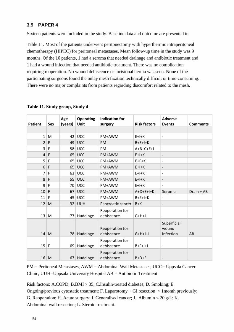
54
3.5 PAPER 4
Sixteen patients were included in the study. Baseline data and outcome are presented in
Table 11. Most of the patients underwent peritonectomy with hyperthermic intraperitoneal
chemotherapy (HIPEC) for peritoneal metastases. Mean follow-up time in the study was 9
months. Of the 16 patients, 1 had a seroma that needed drainage and antibiotic treatment and
1 had a wound infection that needed antibiotic treatment. There was no complication
requiring reoperation. No wound dehiscence or incisional hernia was seen. None of the
participating surgeons found the onlay mesh fixation technically difficult or time-consuming.
There were no major complaints from patients regarding discomfort related to the mesh.
Table 11. Study group, Study 4
Patient Sex Age (years)
Operating Unit
Indication for surgery Risk factors
Adverse Events Comments
1 M 42 UCC PM+AWM E+I+K - 2 F 49 UCC PM B+E+I+K - 3 F 58 UCC PM A+B+C+E+I - 4 F 65 UCC PM+AWM E+I+K - 5 F 65 UCC PM+AWM E+F+K - 6 F 65 UCC PM+AWM E+I+K - 7 F 63 UCC PM+AWM E+I+K - 8 F 55 UCC PM+AWM E+I+K - 9 F 70 UCC PM+AWM E+I+K -
10 F 67 UCC PM+AWM A+D+E+I+K Seroma Drain + AB
11 F 45 UCC PM+AWM B+E+I+K - 12 M 32 UUH Pancreatic cancer B+K -
13 M 77 Huddinge Reoperation for dehiscence G+H+I -
14 M 78 Huddinge Reoperation for dehiscence G+H+I+J
Superficial wound Infection AB
15 F 69 Huddinge Reoperation for dehiscence B+F+I+L -
16 M 67 Huddinge Reoperation for dehiscence B+D+F -
PM = Peritoneal Metastases, AWM = Abdominal Wall Metastases, UCC= Uppsala Cancer
Clinic, UUH=Uppsala University Hospital AB = Antibiotic Treatment
Risk factors: A.COPD; B.BMI > 35; C.Insulin-treated diabetes; D. Smoking; E.
Ongoing/previous cytostatic treatment: F. Laparotomy + GI resection < 1month previously;
G. Reoperation; H. Acute surgery; I. Generalised cancer; J. Albumin < 20 g/L; K.
Abdominal wall resection; L. Steroid treatment.

55
4 DISCUSSION
4.1 GENERAL COMMENTS
Incisional hernia and wound dehiscence are midline incision complications that share the
same pathogenesis i.e., impaired healing of the rectus aponeurosis. My clinical experience,
which is also supported by the findings in this thesis, is that the most vulnerable patients with
severe comorbidity develop acute wound dehiscence, whereas younger and stronger patients
endure the postoperative period and develop an incisional hernia at a later stage. There are
well-established methods to prevent these complications, meticulous suturing technique being
alternative number one. In second place, prophylactic mesh augmentation may further reduce
complications. However, for reasons of cost and time consumed, I do not think prophylactic
mesh implantation should be standard practice but preserved for patients at high risk for
impaired wound healing. All surgeons should be aware of these risk factors and use
stratification tools in the preoperative workup to identify those patients likely to benefit from
mesh reinforcement of the abdominal wall.
This thesis studies open surgery through a midline incision, thus ignoring one important
solution to the problem. Laparoscopic surgery has significantly lower risk for incisional
hernia (123,124) and there is also strong evidence that fascial dehiscence is less common
after laparoscopic surgery (125). Furthermore, laparoscopic surgery has other benefits such as
shorter hospital stay, less need for opioid treatment etc.
However, there will still be cases where open surgery via a midline incision is indicated.

56
Hospital Building © Capio S:t Görans sjukhus 2021

57
4.2 IMPLEMENTATION OF SSSB AT S:T GÖRANS HOSPITAL.
The structured implementation of the SSSB technique in clinical practice at S:t Görans
Hospital led to high compliance to use of the technique that remained four years later. In our
study, 93% of midline incisions were closed using a correct SSSB technique and accurately
registered in the surgical report. In a similar study, Tolstrup et al report 73% compliance to
the technique (126). However, in a questionnaire study by Bloemen et al (127) where Dutch
surgeons were asked about the technique they used for closing midline incisions, they found
that very few followed the latest guidelines. Our conclusion is that structural implementation
and education are essential when introducing a new surgical technique if one is to attain a
long-lasting effect.
No significant differences in rates of incisional hernia, wound dehiscence, or surgical site
infection before and after introduction of SSSB for abdominal closure were found in the
retrospective study carried out at Capio St Görans Hospital. The study found that already
2010 – 2011 i.e., prior to introduction and implementation of SSSB, acceptable rates of
incisional hernia and wound dehiscence existed. However, the structured implementation of
the SSSB technique was not in vain since the use of a standardised technique enables quality
follow-up and helps in the training of new surgeons. The SSSB technique is also a basic
fundament of many new techniques that are currently introduced for treating burst and open
abdomen.

58
4.3 INCIDENCE OF WOUND DEHISCENCE AND INCISIONAL HERNIA.
In the prospective randomised trials evaluating the SSSB technique, both Millburn (77) and
Deerenberg (31) found high incidences of incisional hernia in their control arms where a
large stitch technique was used. After implementation of the SSSB technique, the reported
incidence was 5% and 13% respectively. Deerenberg et al used the EHS definition of
incisional hernia where a defect seen on follow-up radiological examination, though not
palpable, was considered a hernia. In their study, 47% of hernias were found by radiological
examination only. This might explain the differences in incidences between these studies.
In the retrospective studies in this thesis, the incidence of incisional hernia was around 5%.
This suggests that the surgical techniques used by most Swedish surgeons before the SSSB
intervention at Capio S:t Görans hospital, were satisfactory. In these studies, only clinically
evident incisional hernias were registered. As mentioned above about 50% of insicional
hernias are asymptomatic and only found on focused examination or radiology (29,31,37).
Karlsson et al reported an incisional hernia rate of 25% after colorectal cancer surgery in a
study using postoperative CT scans for diagnosis (128). In their study, 12% of patients
needed incisional hernia surgery. When extrapolating this to the present studies, the true
incisional hernia rate in our cohorts would be expected to be at least 10%, which is still a
level that is generally considered acceptable in terms of patient safety. When the EHS
definition of incisional hernia is applied (4), clinical examination underestimates the
incidence of incisional hernia. On the other hand, results from retrospective materials such as
ours and the study by Walming et al (75) can be seen as an estimation of the rate of clinically
relevant incisional hernia. In which case this should be about 5% in a Swedish setting with
acute and emergency surgery.
Wound dehiscence is a rare complication of abdominal surgery. Kenig et al (51) and Webster
et al (54) reported the incidence to be 2% and 3% respectively. Slater et al reported the
incidence of wound dehiscence to be 0.4-1.2% in elective surgery and 12% in acute surgery
(9). Tolstrup et al reported a decrease of the incidence of wound dehiscence from 7% to 4%
after implemention of SSSB as standard technique for acute laparotomy (126). In prospective
studies evaluating SSSB, the incidence of wound dehiscence, though not being an endpoint,
was reported to be only 1% (31,77). In our initial prospective follow-up at S:t Görans
hospital, we first saw the same tendency, but in the larger retrospective follow-up we found
an incidence of 4%. In the register study based on SCRCR data, we found a reoperation rate
for wound dehiscence of 2,9% (2,8% after elective surgery and 4,0% after acute surgery).
Walming et al reported an incidence of 3,3% in a comparable retrospective study (75). Since
wound dehiscence is a rare complication, a few cases only can lead to the difference in rates
seen between different studies. There are also large differences in inclusion criteria in studies
published. For example, Millbourn et al excluded all patients with repeat midline incisions. A
fair assumption is that in current clinical practice the incidence of wound dehiscence after
elective or acute surgery should not exceed 4%. Future studies on high risk patients should
have the 4% level as their goal.

59
4.4 WOUND DEHISCENCE
Wound dehiscence normally leads to long hospital stay, reoperation, intensive care treatment,
and often several reoperations. Our register-based study showed a significantly higher
mortality risk for after reoperation for wound dehiscence. It was shown in both our
retrospective studies that wound dehiscence patients face a high risk for incisional hernia.
These findings were confirmed by Jensen et al who showed that wound dehiscence patients
have higher mortality and a high risk for developing an incisional hernia (56). To summarise,
I believe that in a hospital aiming at high quality, every case of wound dehiscence should be
regarded as an adverse event and that an event analysis should be performed to see if
anything could have been done pre-, peri- or postoperatively to prevent the event. For a
midsize hospital such as S:t Görans Hospital with around 350 midline laparotomies per year,
a reduction in the rate of wound dehiscence from 4% to 1% would save over one hundred
hospital days a year and many acute operations.
4.5 RISK FACTORS FOR INCISIONAL HERNIA AND WOUND DEHISCENCE
In our studies, male gender, high BMI, and postoperative wound infection were found to be
risk factors for incisional hernia and wound dehiscence.
Chronic obstructive pulmonary disease and generalised inflammatory disease were found to
be statistically significant risk factors for wound dehiscence.
Age over 70 was found to be a risk factor for wound dehiscence but the risk for incisional
hernia was lower.
Operation time less than 180 min was found to be a risk factor for wound dehiscence, and
long operation time an independant risk factor for incisional hernia.
Wound dehiscence was a risk factor for incisional hernia.
The studies in this thesis showed that male gender is a risk factor for wound complications,
though gender as a risk factor is disputed. Contrary to our findings, a study by Seo et al
showed that female gender was a risk factor for incisional hernia (129) and in the Swedish
Ventral Hernia Register, 58% of patients operated for incisional hernia were female (130).
From our studies there is strong evidence that high BMI increases the risk for incisional
hernia and wound dehiscence. One theory is that truncal obesity, rather than high BMI itself,
should be considered the risk factor for incisional hernia (83). Indeed, truncal obesity is more
common in men, and may be the reason for the gender factor seen in our studies. Based on
the findings in this thesis, I recommend considering men with a BMI>30 as having high risk
for wound complications. I also believe that standardised measurement of perirenal fat on
preoperative CT scans (measure of visceral obesity) combined with BMI would be a useful
combination to identify patients at high risk.

60
Postoperative wound infection is another risk factor for incisional hernia and wound
dehiscence. All possible measures should be undertaken to reduce the rate of wound
infection, the most important being correct surgical technique. In the early phase of wound
dehiscence there is serous discharge from the wound that may be misinterpreted as a wound
infection. This could lead to antibiotic treatment and delay in diagnosing wound dehiscence,
and also contribute to the strong association between wound infection and wound
dehiscence/incisional hernia.
Postoperative surgical site infection is not a useful parameter for predicting complications.
Furthermore, as surgical site infections are strongly associated with other wound
complications, surgical site infection may act as confounder that obscures score-model
assessment. Instead, future studies should focus on exploring risks associated with factors
known preoperatively.
High age is a well-known risk factor for wound dehiscence and has also been recognised as a
risk factor for incisional hernia in some studies. Our studies confirm that high age is a risk
factor for wound dehiscence, whereas lower age was a risk factor for incisional hernia. Since
the SCRCR does not include data from the primary healthcare service, this could be a source
of bias in this thesis. Elderly patients with a subclinical incisional hernia are likely to be
treated conservatively under the primary healthcare system and thus overlooked. We also
know that 50% or more of incisional hernias are asymptomatic. With increasing age and
reduced physical activity, this rate could be even higher. It is my opinion that high age should
be taken into consideration in the preoperative assessment since it is a well-established risk
factor for wound dehiscence. Patients with high age and comorbidity are the ones that are at
great risk if they develop wound dehiscence or burst abdomen, and they might not be fit for
reoperation. Prophylactic measures such as meticulous suturing and prophylactic mesh
augmentation can be lifesaving in these cases.
In our studies based on SCRCR data, we found conflicting results regarding the role of
operation time as a risk factor. That short operation time should be a risk factor for
complication seems counter-intuitive. One explanation could be that palliative surgery and
diagnostic surgery in critically ill patients may take less time though risk is high. A long
operation time is a known a risk factor probably due to exhaustion of the surgeon or
prolonged traction causing ischaemia in the abdominal wall. The patient also becomes
exhausted and may not be fit enough for a relaparotomy. It would thus seem logical to take
extra measures to prevent adverse events after a long operation.
The subgroup analysis found that acute surgery increases the risk for wound dehiscence but
does not seem to significantly increase the risk for incisional hernia. This finding is congruent
with previous findings (55,64). Because of the increased risk for wound dehiscence, acute
surgery should be included when designing a risk score for complication after midline
incision.

61
4.6 PROPHYLACTIC MESH AUGMENTATION AND TIGR® MATRIX MESH.
Prophylactic mesh placement to prevent incisional hernia in the high-risk patient is now a
well-established procedure (131,132). There is also evidence that prophylactic onlay mesh
can prevent wound dehiscence (133). In our study we found that onlay placed TIGR® Matrix
mesh is safe and is a feasible way to reinforce the midline suture. TIGR® Matrix has also
been tested as prophylaxis after open abdomen treatment (134). There are several possible
advantages of using absorbable mesh material, but these must be tested in future trials.
4.7 THE SURGEON AS A RISK FACTOR.
Garcia-Urena et al (135) discuss in their review that some surgeons regard closure of the
abdomen as “coffee time” and assign the task to an inexperienced junior colleague.
Sometimes there is pressure to complete the list in time, that may lead to stress at the end of
an operation. In other cases, the surgeon may be exhausted at the end of a demanding
operation, or perhaps the patient is not properly relaxed causing difficulty in closing the
abdomen. All these cultural issues need to be considered by hospitals aiming at high quality.
Closure of the abdomen is as important for the patient as the main surgery.
All surgeons performing abdominal and pelvic surgery, not just general surgeons, need to be
up to date on the importance of correct abdominal closure. Junior surgeons should be well
supervised in closing the abdomen. The whole surgical organisation must be aware of the risk
for incisional hernia so that individuals seeking to increase production consider the risk for
complications associated with inadequate closure of the abdominal wall. Lists must be
organised so there is time for meticulous suturing and prophylactic mesh augmentation. If the
surgeon is exhausted, a well-rested colleague should be called in to help finish the operation.
Good communication with the anaesthesiologist is crucial, so that the whole team
understands the importance of abdominal wall suturing. Much suffering can be avoided and
considerable savings for the healthcare system made if these simple principles are followed.

62
4.8 LIMITATIONS OF THE STUDIES
4.8.1 Paper 1
The intervention studied was not carried out with the aim of testing a scientific hypothesis, it
was undertaken as a quality improvement project. As a result, there was no standardised
follow-up protocol used in the study, instead it was performed retrospectively based on a
review of medical records. Many patients visited the hospital only sporadically during the
follow-up period, and patients were not examined in a strictly standard fashion. Incisional
hernias in the study were diagnosed by either CT-scan or clinical examination during the
follow-up period. Some of these hernias were asymptomatic. This limits comparison with
other studies as well as the external validity of the study. Although all patients were followed
throughout the study period, there was possible loss to follow-up if patients left the area or
had their incisional hernia diagnosed at another department in Stockholm. In this
retrospective study, many important parameters could not be analysed. One such clinically
important risk factor that could have played a crucial role was smoking, since many medical
records lack this information. To examine the effect of the SSSB technique more accurately,
patients should have been followed-up prospectively using a standardised protocol. Wound
dehiscence, on the other hand, is rarely neglected, and the rate of 3.7% found accurately
reflects wound dehiscence when SSSB is used.
4.8.2 Paper 2
It is well-known that approximately 50% of incisional hernias are overlooked at clinical
examination. In the present study, 5.3% of patients were diagnosed with incisional hernia or
underwent hernia surgery, whereas the true incidence of incisional hernia probably exceeded
10%. Data from primary healthcare are not included in the registers used in this study, and
there may have been patients that attended their GP with an asymptomatic incisional hernia
that were never referred for surgical consultation. Exclusion of asymptomatic incisional
hernias on the one hand allows focus on clinically relevant incisional hernias, but on the other
hand prevents comparison with studies where the EHS definition of incisional hernia is
applied (60). No association between comorbidity and risk for incisional hernia was seen.
Patients with an incisional hernia deemed unfit for surgery could have been neglected in this
analysis, thereby causing a selection bias. This could also explain why the rate of incisional
hernia was higher in patients younger than 70 years. Most of the open procedures for
colorectal cancer in this study were performed via a midline incision, but other approaches
such as a transverse incision may have been used. The length of incision also varied and there
was a mix of midline incisions above and below the umbilicus. These variations might bias
the outcome of the study since they were not adjusted for. A drawback of this study is that
there is no information about the surgical technique used to close the abdominal wall. During
the study period 2007-2013, the implementation of SSSB gradually spread, and many
Swedish colorectal cancer surgeons adopted the technique. The method of abdominal wall
closure is still not included in the register, but if a similar study was repeated today, almost all
patients would likely have their abdomen closed with the SSSB technique.

63
4.8.3 Paper 3
Surgery for colorectal cancer is performed on a heterogeneous group of patients, and includes
acute, elective, curative, and palliative procedures. Most procedures are performed through a
midline incision, but there could be cases in this cohort that underwent surgery using an
approach with a lower risk for complications, e.g. transverse incision or Pfannenstiel incision.
As in Study 2, the length and location of the incisions varied. The surgical technique used to
close the midline incision probably affects the risk for wound dehiscence. The modern
technique described by Milbourn et al was introduced in Sweden during the study period, but
no specific data on surgical technique were available in the SCRCR. This limits the external
validity of the study. More studies on wound dehiscence are needed, and these studies should
use the SSSB technique for abdominal wall closure.
4.8.4 Paper 4
The present study was too small to provide evidence of the effectiveness of the technique and
the new mesh material. Whether or not incisional hernias develop later when the mesh is
completely resorbed can only be determined in studies with longer follow-up times and under
tightly controlled circumstances. The present study was a pilot study in preparation for a
larger randomised clinical trial, the Prevmesh trial, which is described below in “Future
perspectives”. Three surgeons from three different hospitals performed the procedures. No
surgeon found the procedure technically difficult or time-consuming. Fig 1 used in the article
does not represent the correct technique described in the text and should not have been used.


65
5 CONCLUSIONS
Incisional hernia and wound dehiscence are serious and costly complications of abdominal
surgery via a midline incision.
The Small-Stitch-Small-Bites technique for closing the abdominal wall lowers the risk for
incisional hernia, but despite this, 5-10% of patients will develop an incisional hernia after
open abdominal surgery.
Wound dehiscence is rare, but every case of wound dehiscence is a disaster for the patient.
Wound dehiscence must be avoided at any cost.
High age, high BMI (especially visceral obesity), long operation time, acute surgery, chronic
obstructive pulmonary disease, systemic inflammatory disease, and male gender are risk
factors for wound complications after a midline incision.
Postoperative wound infection is a strong predictor of incisional hernia and wound
dehiscence. All possible measures must be taken to avoid wound infection.
Structured implementation of a standardised surgical technique is possible and has a long-
lasting impact on quality and outcome.
Onlay implantation of TIGR® Matrix mesh is a feasible way to reinforce the abdominal wall
after surgery on high-risk patients.

66
6 FUTURE PERSPECTIVES
In the workup leading up to this thesis, a randomised controlled trial, PrevMesh, was planned
and launched. The aim of the trial was to test the effectiveness of a prophylactic onlay
TIGR® Matrix mesh on the incidence of incisional hernia and wound dehiscence. The study
population comprised patients with high risk for postoperative wound complication
undergoing surgery through a midline incision. The aim of the study was to include acute as
well as planned procedures. Patients were to be randomised to two groups where the study
group had the abdominal wall closed with the SSSB technique and reinforced with a TIGR®
Matrix absorbable mesh. In the control group, the abdominal wall was to be closed with the
SSSB technique but without mesh. PrevMesh was planned as a large multicentre trial
including patients from large university hospitals to small rural hospitals. The study protocol
is presented in Appendix 2, and more information about the study can be found at
clinicaltrials.gov (Identifier: NCT02487134). Unfortunately, the study was paused before
starting patient inclusion and the research question remains unanswered. Nevertheless, we
intend to restart the study as soon as we have found sufficient funding.
Of all the studies referred to in this thesis, very few assume that the SSSB technique was
used. Their results might thus be considered outdated. The HULC trial (136), comparing
standardised closure using the SSSB technique to a study group where SSSB is used and the
suture line is reinforced with a prophylactic mesh, is therefore long awaited. The HULC trial
will include elective surgery only and excludes many risk factors for incisional hernia.
In future studies, patients at high risk of incisional hernia and wound dehiscence should be
included. It may be assumed, for instance, that patients with a previous midline incision or
previous ventral hernia repair would benefit even more from prophylactic mesh
augmentation.
There are promising risk calculation models for incisional hernia and wound dehiscence, and
these should be tested in prospective trials to see if they can help reduce these adverse events.
Another interesting field for future studies is prehabilitation. Howard et al use a standardised
programme for patient preparation prior to surgery. In their study, patients showed a better
physiological response to surgery and fewer complications after surgery (137). In a
systematic review, Gillis et al concluded that nutritional prehabilitation with or without
exercise decreases hospital stay after colorectal cancer surgery (138). Prehabilitation has also
been tested on ventral hernia patients (139) where the prehabilitation group were less likely to
suffer recurrence. Prehabilitation programmes prior to abdominal surgery should be the
subject of further investigation as this might be one way to avoid adverse events.

67
Robotkirurgi 19 © Capio S:t Görans sjukhus 2021

68
7 ACKNOWLEDGEMENTS
Jag vill tacka
Gabriel Sandblom Huvudhandledare. Tack för att du har satsat på mig. Tack för allt du lärt
mig. Tack för din lågmälda ledarstil som pushat på precis lagom. Att komma till ett möte och
tro att man skall avregistrera sig som doktorand, och istället gå därifrån och har bokat in
halvtidskontrollen är typiskt för hur det sett ut. Ingen motgång får dig att vika av från kursen
som är rakt upp. Mot toppen.
Ulf Gunnarsson Bihandledare. Jag fick tidigt i karriären tips av Rolf Heuman (som lärde
mig bråckkirurgi) att ”Om du skall forska, gör det med Ulf Gunnarsson. Då blir det klart” och
tänk det blev det. Tack för tusen motfrågor till varje fråga jag ställt.
Per Hellman Bihandledare Tack för återkoppling angående min något korthuggna stil att
skriva vetenskap, och för diskussioner tidigt kring upplägget.
Rami Klaff Bihandledare och chef. Tack för att du trots den svåra bemanningssituationen
och covid-19 ändå hittat tid åt mig att slutföra det här arbetet.
Kirurgkliniken Mora. Johanna Österberg, Rolf Heuman, Helena Laurell, Mats Hedberg,
Pernilla Olsson, Johan Wickström, Gert Nestler, Asbjörn Österberg, Patrik Lundström
och Viktor Wanjura. Av er lärde jag mig ALLT jag kan om bråckkirurgi,
bukväggsförslutning och vikten av ett vetenskapligt förhållningssätt. Att bli skolad av er i
Mora var den bästa starten en kirugkarriär kunnat få.
Mina forskarförebilder. Sophie Norenstedt. Det var på din disputationsfest jag bestämde mig
för att det där skall jag också göra! Din livsglädje och ditt prestigelösa sätt att ta dig an
utmaningar (nu senast skejtskidåkning) inspirerar mig varje dag. Anna Löf Granström. Av
dig har jag lärt mig att forskning kan (ska) vara roligt, och att man kan göra det lite när man
har tid. Daniel Millbourn hur inspirerande är det inte att det finns en till blivande
bukväggsurolog?! Din bok har varit min ständiga följeslagare sedan starten.
Mina doktorandsystrar Maria Hermann och Ebba Swedenhammar. Tack för pepp och
sällskap på resan.
Christian Kylander Tidigare verksamhetschef. Tack för att du alltid trott på mig, anställt
mig och gett mig chansen att utvecklas. Ett allvarligt samtal i samband med min
doktorandregistrering där du pressade på vikten av att prioritera rätt och inte glömma bort
familjen och fritiden har jag burit med mig. Det har varit viktigt.
Mina kloka mentorer på kliniken. Andreas Wladis, Carl-Eric Leijonmarck, Dan
Kornfeld, Lotta Anveden och Lars Häggarth. Tack för samtal, vägledning, uppmuntran
och allmän pepp. Kunskap är makt, det har ni lärt mig.

69
Mina Prostatabröder Martin Bergman och Andreas Thorstensson Tack för allt kul häng,
all fika och att ni lyssnar på mitt gnäll. Och tack för prostatajoggen.
Hänggänget Sandra, Mats, Emma, Hugo, Emma, Sara och Martin. Utan er hade jag inte
ens klarat mig igenom grundutbildningen, så då hade det inte blivit någon avhandling, kan
man säga. Tack för det.
The Hell Yeahs STHLM och Älgö Rangers HC.
Alla fina vänner. Ingen nämnd, ingen glömd.
Mina arbetskamrater på kirurgkliniken, för att ni gör det #kulpåjobbet. För att ni lär mig nya
saker varje dag. För alla tårtor.
Lucy Bai för de fantasktiska illustrationerna.
Mina syskon med familjer
Mamma och Pappa för möjligheten att bli vad jag vill.
Mina svärföräldrar Henrik och Lena. Gustav med familj. Tack!
Barnens gammelmormor Birgit Askered min största supporter!
Mina barn Tove och Karin allt jag gör det gör jag för er.
Elin Min livskamrat (och näst största supporter). Det finns inte plats att nämna allt du är för
mig. Men i korthet som Björn och Benny skrivit: Jag vore ingenting om du inte fanns.

70
8 REFERENCES
1. Ellis H. Applied anatomy of abdominal incisions. Br J Hosp Med. 2007 Feb
1;68(Sup2):M22–3.
2. Millbourn D. Closure of midline abdominal incisions with small stitches: studies on
wound complications and health economy. [Umeå]: Umeå universitet; 2012.
3. Burger JWA, van’t Riet M, Jeekel J. Abdominal Incisions: Techniques and
Postoperative Complications. Scand J Surg. 2002 Dec 1;91(4):315–21.
4. Muysoms FE, Antoniou SA, Bury K, Campanelli G, Conze J, Cuccurullo D, et al.
European Hernia Society guidelines on the closure of abdominal wall incisions.
Hernia J Hernias Abdom Wall Surg. 2015 Feb;19(1):1–24.
5. Luijendijk RW, Jeekel J, Storm RK, Schutte PJ, Hop WC, Drogendijk AC, et al. The
low transverse Pfannenstiel incision and the prevalence of incisional hernia and nerve
entrapment. Ann Surg. 1997 Apr;225(4):365–9.
6. Guillou PJ, Hall TJ, Donaldson DR, Broughton AC, Brennan TG. Vertical abdominal
incisions--a choice? Br J Surg. 1980 Jun;67(6):395–9.
7. Cox PJ, Ausobsky JR, Ellis H, Pollock AV. Towards no incisional hernias: lateral
paramedian versus midline incisions. J R Soc Med. 1986 Dec;79(12):711–2.
8. Lee L, Mata J, Droeser RA, Kaneva P, Liberman S, Charlebois P, et al. Incisional
Hernia After Midline Versus Transverse Specimen Extraction Incision: A
Randomized Trial in Patients Undergoing Laparoscopic Colectomy. Ann Surg.
2018;268(1):41–7.
9. Slater NJ, Bleichrodt RP, van Goor H. Wound dehiscence and incisional hernia. Surg
Oxf. 2012 Jun 1;30(6):282–9.
10. Le Huu Nho R, Mege D, Ouaïssi M, Sielezneff I, Sastre B. Incidence and prevention
of ventral incisional hernia. J Visc Surg. 2012 Oct;149(5 Suppl):e3-14.
11. Tecce MG, Basta MN, Shubinets V, Lanni MA, Mirzabeigi MN, Cooney L, et al. A
risk model and cost analysis of post-operative incisional hernia following 2,145 open
hysterectomies-Defining indications and opportunities for risk reduction. Am J Surg.
2017 Jun;213(6):1083–90.
12. Rath AM, Chevrel JP. The healing of laparotomies: review of the literature. Hernia.
1998 Sep 1;2(3):145–9.
13. Natarajan S, Williamson D, Stiltz AJ, Harding K. Advances in wound care and
healing technology. Am J Clin Dermatol. 2000 Oct;1(5):269–75.
14. Carlson MA. ACUTE WOUND FAILURE. Surg Clin North Am. 1997 Jun
1;77(3):607–36.
15. Douglas DM. The healing of aponeurotic incisions. BJS Br J Surg. 1952;40(159):79–
84.
16. Pollock AV, Evans M. Early prediction of late incisional hernias. BJS Br J Surg.
1989;76(9):953–4.
17. Henriksen NA. Systemic and local collagen turnover in hernia patients. Dan Med J.
2016 Jul;63(7).

71
18. Rappaport WD, Hunter GC, Allen R, Lick S, Halldorsson A, Chvapil T, et al. Effect
of electrocautery on wound healing in midline laparotomy incisions. Am J Surg.
1990 Dec 1;160(6):618–20.
19. Jansen PL, Mertens PR, Klinge U, Schumpelick V. The biology of hernia formation.
Surgery. 2004 Jul 1;136(1):1–4.
20. Henriksen NA, Yadete DH, Sorensen LT, Agren MS, Jorgensen LN. Connective
tissue alteration in abdominal wall hernia. Br J Surg. 2011 Feb;98(2):210–9.
21. Radu P, Brătucu M, Garofil D, Goleanu V, Popa F, Strâmbu V, et al. The Role of
Collagen Metabolism in the Formation and Relapse of Incisional Hernia. Chir
Buchar Rom 1990. 2015 Jun;110(3):224–30.
22. Henriksen NA, Sørensen LT, Jorgensen LN, Agren MS. Circulating levels of matrix
metalloproteinases and tissue inhibitors of metalloproteinases in patients with
incisional hernia. Wound Repair Regen Off Publ Wound Heal Soc Eur Tissue Repair
Soc. 2013 Oct;21(5):661–6.
23. Henriksen NA, Mortensen JH, Sorensen LT, Bay-Jensen AC, Ågren MS, Jorgensen
LN, et al. The collagen turnover profile is altered in patients with inguinal and
incisional hernia. Surgery. 2015 Feb 1;157(2):312–21.
24. Szczęsny W, Kuligowska-Prusińska M, Dąbrowiecki S, Szmytkowski J, Reśliński A,
Słupski M. Activity of metalloproteinases and adiponectin in obese patients-a
possible factor of incisional hernias after bariatric procedures. J Zhejiang Univ Sci B.
2018 Jan;19(1):65–70.
25. Antoniou GA, Georgiadis GS, Antoniou SA, Granderath FA, Giannoukas AD,
Lazarides MK. Abdominal aortic aneurysm and abdominal wall hernia as
manifestations of a connective tissue disorder. J Vasc Surg. 2011 Oct;54(4):1175–81.
26. Rosch R, Junge K, Knops M, Lynen P, Klinge U, Schumpelick V. Analysis of
collagen-interacting proteins in patients with incisional hernias. Langenbecks Arch
Surg. 2003 Feb 1;387(11):427–32.
27. Korenkov M, Paul A, Sauerland S, Neugebauer E, Arndt M, Chevrel JP, et al.
Classification and surgical treatment of incisional hernia. Langenbecks Arch Surg.
2001 Feb 1;386(1):65–73.
28. Patel SV, Paskar DD, Nelson RL, Vedula SS, Steele SR. Closure methods for
laparotomy incisions for preventing incisional hernias and other wound
complications. Cochrane Database Syst Rev [Internet]. 2017 [cited 2020 Dec 9];(11).
Available from: https://www-cochranelibrary-
com.proxy.kib.ki.se/cdsr/doi/10.1002/14651858.CD005661.pub2/full
29. Ah-kee EY, Kallachil T, O’Dwyer PJ. Patient Awareness and Symptoms From an
Incisional Hernia. Int Surg. 2014;99(3):241–6.
30. Millbourn D, Israelsson LA. Wound complications and stitch length. Hernia. 2004
Feb 1;8(1):39–41.
31. Deerenberg EB, Harlaar JJ, Steyerberg EW, Lont HE, van Doorn HC, Heisterkamp J,
et al. Small bites versus large bites for closure of abdominal midline incisions
(STITCH): a double-blind, multicentre, randomised controlled trial. Lancet Lond
Engl. 2015 Sep 26;386(10000):1254–60.
32. Björk D, Cengiz Y, Weisby L, Israelsson LA. Detecting Incisional Hernia at Clinical
and Radiological Examination. Surg Technol Int. 2015 May;26:128–31.

72
33. Mudge M, Hughes LE. Incisional hernia: A 10 year prospective study of incidence
and attitudes. BJS Br J Surg. 1985;72(1):70–1.
34. Ellis H, Gajraj H, George CD. Incisional hernias: When do they occur? BJS Br J
Surg. 1983;70(5):290–1.
35. Baucom RB, Ousley J, Beveridge GB, Phillips SE, Pierce RA, Holzman MD, et al.
Cancer Survivorship: Defining the Incidence of Incisional Hernia After Resection for
Intra-Abdominal Malignancy. Ann Surg Oncol. 2016;23(Suppl 5):764–71.
36. van Ramshorst GH, Eker HH, Hop WCJ, Jeekel J, Lange JF. Impact of incisional
hernia on health-related quality of life and body image: a prospective cohort study.
Am J Surg. 2012 Aug;204(2):144–50.
37. Bosanquet DC, Ansell J, Abdelrahman T, Cornish J, Harries R, Stimpson A, et al.
Systematic Review and Meta-Regression of Factors Affecting Midline Incisional
Hernia Rates: Analysis of 14 618 Patients. PLoS ONE [Internet]. 2015 Sep 21 [cited
2020 Dec 9];10(9). Available from:
https://www.ncbi.nlm.nih.gov/pmc/articles/PMC4577082/
38. Poulose BK, Shelton J, Phillips S, Moore D, Nealon W, Penson D, et al.
Epidemiology and cost of ventral hernia repair: making the case for hernia research.
Hernia. 2012 Apr 1;16(2):179–83.
39. Israelsson LA, Wimo A. Cost minimisation analysis of change in closure technique
of midline incisions. Eur J Surg Acta Chir. 2000 Aug;166(8):642–6.
40. Millbourn D, Wimo A, Israelsson LA. Cost analysis of the use of small stitches when
closing midline abdominal incisions. Hernia. 2014 Dec 1;18(6):775–80.
41. Shubinets V, Fox JP, Lanni MA, Tecce MG, Pauli EM, Hope WW, et al. Incisional
Hernia in the United States: Trends in Hospital Encounters and Corresponding
Healthcare Charges. Am Surg. 2018 Jan 1;84(1):118–25.
42. Cengiz Y, Månsson P, Israelsson LA. Conventional running suture and continuous
double loop closure: an experimental study of wound strength. Eur J Surg.
2000;166(8):647–9.
43. Niggebrugge AH, Trimbos JB, Hermans J, Steup W-H, Van De Velde CJ. Influence
of abdominal-wound closure technique on complications after surgery: a randomised
study. The Lancet. 1999 May 8;353(9164):1563–7.
44. Mäkelä JT, Kiviniemi H, Juvonen T, Laitinen S. Factors influencing wound
dehiscence after midline laparotomy. Am J Surg. 1995 Oct 1;170(4):387–90.
45. Xing L, Culbertson EJ, Wen Y, Franz MG. Early laparotomy wound failure as the
mechanism for incisional hernia formation. J Surg Res. 2013 Jun 1;182(1):e35–42.
46. Burger JWA, Lange JF, Halm JA, Kleinrensink G-J, Jeekel H. Incisional Hernia:
Early Complication of Abdominal Surgery. World J Surg. 2005 Dec 1;29(12):1608–
13.
47. Cliby WA. Abdominal Incision Wound Breakdown. Clin Obstet Gynecol. 2002
Jun;45(2):507–517.
48. Madsen G, Fischer L, Wara P. Burst abdomen--clinical features and factors
influencing mortality. Dan Med Bull. 1992 Apr;39(2):183–5.

73
49. Aksamija G, Mulabdic A, Rasic I, Aksamija L. Evaluation of Risk Factors of
Surgical Wound Dehiscence in Adults After Laparotomy. Med Arch Sarajevo Bosnia
Herzeg. 2016 Oct;70(5):369–72.
50. Ramneesh G, Sheerin S, Surinder S, Bir S. A prospective study of predictors for post
laparotomy abdominal wound dehiscence. J Clin Diagn Res JCDR. 2014
Jan;8(1):80–3.
51. Kenig J, Richter P, Żurawska S, Lasek A, Zbierska K. Risk factors for wound
dehiscence after laparotomy - clinical control trial. Pol Przegl Chir. 2012
Nov;84(11):565–73.
52. Petersson P, Montgomery A, Petersson U. Wound dehiscence: outcome comparison
for sutured and mesh reconstructed patients. Hernia. 2014 Oct 1;18(5):681–9.
53. van’t RMT, De Vos Van Steenwijk PJ, Bonjer HJ, Steyerberg EW, Jeekel J.
Incisional hernia after repair of wound dehiscence: incidence and risk factors. Am
Surg. 2004 Apr;70(4):281–6.
54. Webster C, Neumayer L, Smout R, Horn S, Daley J, Henderson W, et al. Prognostic
models of abdominal wound dehiscence after laparotomy. J Surg Res. 2003
Feb;109(2):130–7.
55. Pavlidis TE, Galatianos IN, Papaziogas BT, Lazaridis CN, Atmatzidis KS, Makris
JG, et al. Complete dehiscence of the abdominal wound and incriminating factors.
Eur J Surg Acta Chir. 2001 May;167(5):351–4; discussion 355.
56. Jensen KK, Oma E, van Ramshorst GH, Nordholm-Carstensen A, Krarup P-M.
Abdominal wound dehiscence is dangerous: a nationwide study of 14,169 patients
undergoing elective open resection for colonic cancer. Hernia [Internet]. 2021 Jan 4
[cited 2021 Jan 5]; Available from: https://doi.org/10.1007/s10029-020-02350-z
57. van Ramshorst GH, Eker HH, van der Voet JA, Jeekel J, Lange JF. Long-Term
Outcome Study in Patients with Abdominal Wound Dehiscence: a Comparative
Study on Quality of Life, Body Image, and Incisional Hernia. J Gastrointest Surg.
2013 Aug 1;17(8):1477–84.
58. Gislason H, Viste A. Closure of burst abdomen after major gastrointestinal
operations--comparison of different surgical techniques and later development of
incisional hernia. Eur J Surg Acta Chir. 1999 Oct;165(10):958–61.
59. Scholtes M, Kurmann A, Seiler CA, Candinas D, Beldi G. Intraperitoneal Mesh
Implantation for Fascial Dehiscence and Open Abdomen. World J Surg. 2012 Jul
1;36(7):1557–61.
60. López-Cano M, García-Alamino JM, Antoniou SA, Bennet D, Dietz UA, Ferreira F,
et al. EHS clinical guidelines on the management of the abdominal wall in the
context of the open or burst abdomen. Hernia J Hernias Abdom Wall Surg.
2018;22(6):921–39.
61. Petersson P, Petersson U. Dynamic Fascial Closure With Vacuum-Assisted Wound
Closure and Mesh-Mediated Fascial Traction (VAWCM) Treatment of the Open
Abdomen—An Updated Systematic Review. Front Surg [Internet]. 2020 [cited 2020
Dec 15];7. Available from:
https://www.frontiersin.org/articles/10.3389/fsurg.2020.577104/full

74
62. Petersson P, Montgomery A, Petersson U. Vacuum-Assisted Wound Closure and
Permanent Onlay Mesh–Mediated Fascial Traction: A Novel Technique for the
Prevention of Incisional Hernia after Open Abdomen Therapy Including Results
From a Retrospective Case Series. Scand J Surg. 2019 Sep 1;108(3):216–26.
63. Yılmaz KB, Akıncı M, Doğan L, Karaman N, Özaslan C, Atalay C. A prospective
evaluation of the risk factors for development of wound dehiscence and incisional
hernia. Ulus Cerrahi Derg. 2013;29(1):25–30.
64. van Ramshorst GH, Nieuwenhuizen J, Hop WCJ, Arends P, Boom J, Jeekel J, et al.
Abdominal Wound Dehiscence in Adults: Development and Validation of a Risk
Model. World J Surg. 2009 Nov 7;34(1):20.
65. Riou JP, Cohen JR, Johnson H. Factors influencing wound dehiscence. Am J Surg.
1992 Mar;163(3):324–30.
66. Millbourn D, Cengiz Y, Israelsson LA. Risk factors for wound complications in
midline abdominal incisions related to the size of stitches. Hernia J Hernias Abdom
Wall Surg. 2011 Jun;15(3):261–6.
67. Wiegering A, Liebetrau D, Menzel S, Bühler C, Kellersmann R, Dietz UA. The
incidence of incisional hernia after aortic aneurysm is not higher than after benign
colorectal interventions. Gefasschirurgie. 2018;23(Suppl 1):23–31.
68. Israelsson LA, Jonsson T, Knutsson A. Suture technique and wound healing in
midline laparotomy incisions. Eur J Surg Acta Chir. 1996 Aug;162(8):605–9.
69. Israelsson LA, Jonsson T. Suture length to wound length ratio and healing of midline
laparotomy incisions. BJS Br J Surg. 1993;80(10):1284–6.
70. Goodenough CJ, Ko TC, Kao LS, Nguyen MT, Holihan JL, Alawadi Z, et al.
Development and validation of a risk stratification score for ventral incisional hernia
after abdominal surgery: hernia expectation rates in intra-abdominal surgery (the
HERNIA Project). J Am Coll Surg. 2015 Apr;220(4):405–13.
71. Basta MN, Kozak GM, Broach RB, Messa CA, Rhemtulla I, DeMatteo RP, et al. Can
Predict Incisional Hernia?: Development of a Surgery-specific Decision-Support
Interface. Ann Surg. 2019;270(3):544–53.
72. Penn Hernia Risk Calculator [Internet]. [cited 2021 Jan 5]. Available from:
https://www.pennherniariskcalc.com/#/
73. Itatsu K, Yokoyama Y, Sugawara G, Kubota H, Tojima Y, Kurumiya Y, et al.
Incidence of and risk factors for incisional hernia after abdominal surgery. BJS Br J
Surg. 2014;101(11):1439–47.
74. Niggebrugge AH, Hansen BE, Trimbos JB, van de Velde CJ, Zwaveling A.
Mechanical factors influencing the incidence of burst abdomen. Eur J Surg Acta
Chir. 1995 Sep;161(9):655–61.
75. Walming S, Angenete E, Block M, Bock D, Gessler B, Haglind E. Retrospective
review of risk factors for surgical wound dehiscence and incisional hernia. BMC
Surg. 2017 Feb 22;17(1):19.
76. Ejaz A, Schmidt C, Johnston FM, Frank SM, Pawlik TM. Risk factors and prediction
model for inpatient surgical site infection after major abdominal surgery. J Surg Res.
2017 Sep 1;217:153–9.

75
77. Millbourn D. Effect of Stitch Length on Wound Complications After Closure of
Midline Incisions: A Randomized Controlled Trial. Arch Surg. 2009 Nov
16;144(11):1056.
78. Pogacnik JS, Messaris E, Deiling SM, Connelly TM, Berg AS, Stewart DB, et al.
Increased Risk of Incisional Hernia after Sigmoid Colectomy for Diverticulitis
Compared with Colon Cancer. J Am Coll Surg. 2014 May 1;218(5):920–8.
79. Kayashima H, Maeda T, Harada N, Masuda T, Guntani A, Ito S, et al. Risk factors
for incisional hernia after hepatic resection for hepatocellular carcinoma in patients
with liver cirrhosis. Surgery. 2015 Dec 1;158(6):1669–75.
80. Henriksen NA, Helgstrand F, Vogt KC, Jorgensen LN, Bisgaard T. Risk factors for
incisional hernia repair after aortic reconstructive surgery in a nationwide study. J
Vasc Surg. 2013 Jun 1;57(6):1524-1530.e3.
81. Spiliotis J, Tsiveriotis K, Datsis AD, Vaxevanidou A, Zacharis G, Giafis K, et al.
Wound dehiscence: is still a problem in the 21th century: a retrospective study.
World J Emerg Surg WJES. 2009 Apr 3;4:12.
82. Sørensen LT, Hemmingsen UB, Kirkeby LT, Kallehave F, Jørgensen LN. Smoking
is a risk factor for incisional hernia. Arch Surg Chic Ill 1960. 2005 Feb;140(2):119–
23.
83. Aquina CT, Rickles AS, Probst CP, Kelly KN, Deeb A-P, Monson JRT, et al.
Visceral Obesity, Not Elevated BMI, Is Strongly Associated With Incisional Hernia
After Colorectal Surgery. Dis Colon Rectum. 2015 Feb;58(2):220–227.
84. Hagopian O der, Dahlberg M, Heinius G, Nordberg J, Gustafsson J, Nordenvall C, et
al. Perirenal fat surface area as a risk factor for perioperative difficulties and 30-day
postoperative complications in elective colon cancer surgery. Colorectal Dis.
2018;20(12):1078–87.
85. Jung M, Volonté F, Buchs NC, Gayet-Ageron A, Pugin F, Gervaz P, et al. Perirenal
fat surface area as a risk factor for morbidity after elective colorectal surgery. Dis
Colon Rectum. 2014 Feb;57(2):201–9.
86. House MG, Fong Y, Arnaoutakis DJ, Sharma R, Winston CB, Protic M, et al.
Preoperative Predictors for Complications after Pancreaticoduodenectomy: Impact of
BMI and Body Fat Distribution. J Gastrointest Surg. 2008 Feb 1;12(2):270–8.
87. Kartheuser AH, Leonard DF, Penninckx F, Paterson HM, Brandt D, Remue C, et al.
Waist circumference and waist/hip ratio are better predictive risk factors for mortality
and morbidity after colorectal surgery than body mass index and body surface area.
Ann Surg. 2013 Nov;258(5):722–30.
88. Tchernof A, Després J-P. Pathophysiology of Human Visceral Obesity: An Update.
Physiol Rev. 2013 Jan 1;93(1):359–404.
89. Thorell A, Efendic S, Gutniak M, Häggmark T, Ljungqvist O. Insulin resistance after
abdominal surgery. BJS Br J Surg. 1994;81(1):59–63.
90. Otranto M, Nascimento AP do, Monte‐Alto‐Costa A. Insulin resistance impairs
cutaneous wound healing in mice. Wound Repair Regen. 2013;21(3):464–72.
91. Barazzoni R, Gortan Cappellari G, Ragni M, Nisoli E. Insulin resistance in obesity:
an overview of fundamental alterations. Eat Weight Disord - Stud Anorex Bulim
Obes. 2018 Apr 1;23(2):149–57.
76
92. Tewari N, Awad S, Macdonald IA, Lobo DN. Obesity-related insulin resistance:
implications for the surgical patient. Int J Obes. 2015 Nov;39(11):1575–88.
93. Klöting N, Fasshauer M, Dietrich A, Kovacs P, Schön MR, Kern M, et al. Insulin-
sensitive obesity. Am J Physiol-Endocrinol Metab. 2010 Jun 22;299(3):E506–15.
94. Awad S, Varadhan KK, Ljungqvist O, Lobo DN. A meta-analysis of randomised
controlled trials on preoperative oral carbohydrate treatment in elective surgery. Clin
Nutr. 2013 Feb 1;32(1):34–44.
95. Scott MJ, Baldini G, Fearon KCH, Feldheiser A, Feldman LS, Gan TJ, et al.
Enhanced Recovery After Surgery (ERAS) for gastrointestinal surgery, part 1:
pathophysiological considerations. Acta Anaesthesiol Scand. 2015
Nov;59(10):1212–31.
96. Abbas SM, Hill AG. Smoking is a major risk factor for wound dehiscence after
midline abdominal incision; case-control study. ANZ J Surg. 2009 Apr;79(4):247–
50.
97. Cengiz Y. Small Tissue Bites and Wound Strength: An Experimental Study. Arch
Surg. 2001 Mar 1;136(3):272.
98. Cengiz Y, Gislason H, Svanes K, Israelsson LA. Mass closure technique: an
experimental study on separation of wound edge. Eur J Surg. 2001;167(1):60–3.
99. Meijer EJ, Timmermans L, Jeekel J, Lange JF, Muysoms FE. The principles of
abdominal wound closure. Acta Chir Belg. 2013 Aug;113(4):239–44.
100. Jenkins TPN. The burst abdominal wound: A mechanical approach. BJS Br J Surg.
1976;63(11):873–6.
101. Israelsson LA, Jonsson T. Closure of midline laparotomy incisions with
polydioxanone and nylon: The importance of suture technique. BJS Br J Surg.
1994;81(11):1606–8.
102. Sahlin S, Ahlberg J, Granström L, Ljungström KG. Monofilament versus
multifilament absorbable sutures for abdominal closure. Br J Surg. 1993
Mar;80(3):322–4.
103. Israelsson LA, Jonsson T. Incisional hernia after midline laparotomy: a prospective
study. Eur J Surg Acta Chir. 1996 Feb;162(2):125–9.
104. Harlaar JJ, van Ramshorst GH, Nieuwenhuizen J, ten Brinke JG, Hop WCJ,
Kleinrensink G-J, et al. Small stitches with small suture distances increase
laparotomy closure strength. Am J Surg. 2009 Sep;198(3):392–5.
105. Bhangu A, Fitzgerald JE, Singh P, Battersby N, Marriott P, Pinkney T. Systematic
review and meta-analysis of prophylactic mesh placement for prevention of
incisional hernia following midline laparotomy. Hernia J Hernias Abdom Wall Surg.
2013 Aug;17(4):445–55.
106. Borab ZM, Shakir S, Lanni MA, Tecce MG, MacDonald J, Hope WW, et al. Does
prophylactic mesh placement in elective, midline laparotomy reduce the incidence of
incisional hernia? A systematic review and meta-analysis. Surgery. 2017 Apr
1;161(4):1149–63.

77
107. Caro-Tarrago A, Olona Casas C, Jimenez Salido A, Duque Guilera E, Moreno
Fernandez F, Vicente Guillen V. Prevention of Incisional Hernia in Midline
Laparotomy with an Onlay Mesh: A Randomized Clinical Trial. World J Surg. 2014
Sep 1;38(9):2223–30.
108. Khorgami Z, Shoar S, Laghaie B, Aminian A, Hosseini Araghi N, Soroush A.
Prophylactic retention sutures in midline laparotomy in high-risk patients for wound
dehiscence: A randomized controlled trial. J Surg Res. 2013 Apr 1;180(2):238–43.
109. Swedish Colorectal Cancer Registry (SCRCR) - Nationella Kvalitetsregister
[Internet]. [cited 2021 Jan 4]. Available from:
https://kvalitetsregister.se/englishpages/findaregistry/registerarkivenglish/swedishcol
orectalcancerregistryscrcr.2156.html
110. Nationellt kvalitetsregister för tjock- och ändtarmscancer - RCC [Internet]. [cited
2021 Jan 4]. Available from:
https://www.cancercentrum.se/samverkan/cancerdiagnoser/tjocktarm-andtarm-och-
anal/tjock--och-andtarm/kvalitetsregister/
111. Moberger P, Sköldberg F, Birgisson H. Evaluation of the Swedish Colorectal Cancer
Registry: an overview of completeness, timeliness, comparability and validity. Acta
Oncol. 2018 Dec 2;57(12):1611–21.
112. The National Patient Register [Internet]. Socialstyrelsen. [cited 2021 Jan 4].
Available from: https://www.socialstyrelsen.se/en/statistics-and-
data/registers/register-information/the-national-patient-register/
113. Ludvigsson JF, Andersson E, Ekbom A, Feychting M, Kim J-L, Reuterwall C, et al.
External review and validation of the Swedish national inpatient register. BMC
Public Health. 2011 Jun 9;11(1):450.
114. Donabedian A. Explorations in Quality Assessment and Monitoring: The definition
of quality and approaches to its assessment. Health Administration Press; 1980. 192
p.
115. Argyris C, Schön DA. Organizational Learning: A Theory of Action Perspective.
Addison-Wesley Publishing Company; 1978. 356 p.
116. Gellad ZF, Day TE. What Is Value Stream Mapping, and How Can It Help My
Practice? Am J Gastroenterol. 2016 Apr;111(4):447–8.
117. Ghosh M. A3 Process: A Pragmatic Problem-Solving Technique for Process
Improvement in Health Care. J Health Manag. 2012 Mar 1;14(1):1–11.
118. Cambio COSMIC journalsystem [Internet]. [cited 2020 Sep 17]. Available from:
https://www.cambio.se/vi-erbjuder/cosmic/
119. Ludvigsson JF, Almqvist C, Bonamy A-KE, Ljung R, Michaëlsson K, Neovius M, et
al. Registers of the Swedish total population and their use in medical research. Eur J
Epidemiol. 2016 Feb 1;31(2):125–36.
120. Hjort H, Mathisen T, Alves A, Clermont G, Boutrand JP. Three-year results from a
preclinical implantation study of a long-term resorbable surgical mesh with time-
dependent mechanical characteristics. Hernia. 2012 Apr 1;16(2):191–7.
121. Cox DR. Regression Models and Life-Tables. J R Stat Soc Ser B Methodol.
1972;34(2):187–202.

78
122. Bertero L, Massa F, Metovic J, Zanetti R, Castellano I, Ricardi U, et al. Eighth
Edition of the UICC Classification of Malignant Tumours: an overview of the
changes in the pathological TNM classification criteria—What has changed and
why? Virchows Arch. 2018 Apr 1;472(4):519–31.
123. Kössler-Ebs JB, Grummich K, Jensen K, Hüttner FJ, Müller-Stich B, Seiler CM, et
al. Incisional Hernia Rates After Laparoscopic or Open Abdominal Surgery-A
Systematic Review and Meta-Analysis. World J Surg. 2016 Oct;40(10):2319–30.
124. Pecorelli N, Greco M, Amodeo S, Braga M. Small bowel obstruction and incisional
hernia after laparoscopic and open colorectal surgery: a meta-analysis of comparative
trials. Surg Endosc. 2017 Jan;31(1):85–99.
125. Wu K-L, Lee K-C, Liu C-C, Chen H-H, Lu C-C. Laparoscopic versus Open Surgery
for Diverticulitis: A Systematic Review and Meta-Analysis. Dig Surg.
2017;34(3):203–15.
126. Tolstrup M-B, Watt SK, Gögenur I. Reduced Rate of Dehiscence After
Implementation of a Standardized Fascial Closure Technique in Patients Undergoing
Emergency Laparotomy. Ann Surg. 2017;265(4):821–6.
127. Bloemen A, De Kleijn RJCMF, Van Steensel S, Aarts F, Schreinemacher MHF,
Bouvy ND. Laparotomy closure techniques: Do surgeons follow the latest
guidelines? Results of a questionnaire. Int J Surg Lond Engl. 2019 Nov;71:110–6.
128. Karlsson N, Zackrisson S, Buchwald P. Computed tomography verified prevalence
of incisional hernia 1 year postoperatively after colorectal cancer resection. Scand J
Surg. 2020 Dec 16;1457496920976053.
129. Seo GH, Choe EK, Park KJ, Chai YJ. Incidence of Clinically Relevant Incisional
Hernia After Colon Cancer Surgery and Its Risk Factors: A Nationwide Claims
Study. World J Surg. 2018;42(4):1192–9.
130. Välkommen! | Kvalitetsregistret för Svenska Bukväggsbråck [Internet]. [cited 2021
Feb 9]. Available from: https://www.ventralhernia.se/
131. Ahmed J, Hasnain N, Fatima I, Malik F, Chaudhary MA, Ahmad J, et al.
Prophylactic Mesh Placement for the Prevention of Incisional Hernia in High-Risk
Patients After Abdominal Surgery: A Systematic Review and Meta-Analysis.
Cureus. 2020 Sep 16;12(9):e10491.
132. Jairam AP, López-Cano M, Garcia-Alamino JM, Pereira JA, Timmermans L, Jeekel
J, et al. Prevention of incisional hernia after midline laparotomy with prophylactic
mesh reinforcement: a meta-analysis and trial sequential analysis. BJS Open.
2020;4(3):357–68.
133. Lima HVG. Prevention of Fascial Dehiscence with Onlay Prophylactic Mesh in
Emergency Laparotomy: A Randomized Clinical Trial. J Am Coll Surg.
2020;230(1):12.
134. Schaaf S, Schwab R, Güsgen C, Willms A. Prophylactic Onlay Mesh Implantation
During Definitive Fascial Closure After Open Abdomen Therapy (PROMOAT):
Absorbable or Non-absorbable? Methodical Description and Results of a Feasibility
Study. Front Surg [Internet]. 2020 [cited 2021 Jan 15];7. Available from:
https://www.frontiersin.org/articles/10.3389/fsurg.2020.578565/full

79
135. Garcia-Urena MA, POP (Progress On Prevention) Surgical Group. Preventing
incisional ventral hernias: important for patients but ignored by surgical specialities?
A critical review. Hernia J Hernias Abdom Wall Surg. 2021 Feb;25(1):13–22.
136. Heger P, Feißt M, Krisam J, Klose C, Dörr-Harim C, Tenckhoff S, et al. Hernia
reduction following laparotomy using small stitch abdominal wall closure with and
without mesh augmentation (the HULC trial): study protocol for a randomized
controlled trial. Trials. 2019 Dec 16;20(1):738.
137. Howard R, Yin YS, McCandless L, Wang S, Englesbe M, Machado-Aranda D.
Taking Control of Your Surgery: Impact of a Prehabilitation Program on Major
Abdominal Surgery. J Am Coll Surg. 2019 Jan 1;228(1):72–80.
138. Gillis C, Buhler K, Bresee L, Carli F, Gramlich L, Culos-Reed N, et al. Effects of
Nutritional Prehabilitation, With and Without Exercise, on Outcomes of Patients
Who Undergo Colorectal Surgery: A Systematic Review and Meta-analysis.
Gastroenterology. 2018 Aug 1;155(2):391-410.e4.
139. Liang M, Bernardi K, Holihan J, Cherla D, Escamilla R, Lew D, et al. Modifying
Risks in Ventral Hernia Patients With Prehabilitation: A Randomized Controlled
Trial. Ann Surg. 2018 Oct;268(4):674–80.

80

81
9 APPENDICIES
9.1 APPENDIX 1 EXAMPLE OF A3 TOOL

82
9.2 APPENDIX 2 LOCAL GUIDELINES ON OPENING AND CLOSING THE ABDOMINAL WALL (TRANSLATED)
Background:
Midline incisions are associated with severe complications such as wound dehiscence,
wound infection, and incisional hernia. There is reliable evidence that the most effective
way of reducing these complications is meticulous surgical technique.
Our department has decided, without exception, to use the so-called Sundsvall technique
for closure of all midline abdominal incisions.
Opening:
• Spend time making your incision precisely in the midline. Apply the technique of
subcutaneous fat fractioning if possible.
• Free the aponeurosis from subcutaneous tissue 1 cm on each side before it is
incised, in preparation for closure.
• Use electrocautery to achieve clean aponeurosis edges for stitching.
Closure:
• Measure the incision length in the relaxed position. Use a ruler.
• Use PDS II 2-0, 150 cm, CT-1 needle.
• Suture length/wound length quote is measured by first measuring the incision
length. The total length of the suture used minus the lengths cut off at the ends is
assessed. Division of the suture length by the wound length gives the ratio.
• If the incision is longer than 30 cm you will need 2 sutures to reach a quote of at
least 4.
• Use self-locking start and stop knots.
• Make sure to start at the very end of the incision.
• Start at the end of the incision and suture continuously using small bites 5-8mm
from the aponeurosis edges. The interval between the stitches should be less than
5mm.
• Take bites in the aponeurosis only. Avoid mass layered sutures. If you leave the
midline, take bites in the anterior rectus aponeurosis only.
• A low-tension suture is important, pull the suture so that the edges of the
aponeurosis just adapt. The assistant should hold the suture without increasing the
tension. The suture must be visible but the distance between the edges should be
<10mm
• Suture length/wound length must be greater than 4, and the ratio must be measured
and noted in the operation notes.
• If a ratio of four is not achieved, the suture must be redone. The ratio has no
maximum.
• The theatre nurse has the responsibility to reach the surgeon a pair of scissors if a
ratio of 4 is not achieved. If the surgeon choses to deviate from this routine, a non-
compliance report must be made stating the reasons why.
• The skin must be closed with intracutaneous Monocryl® suture. If this is not
possible, staples may be used.

83
References:
Effect of stitch length on wound complications after closure of midline incisions: a
randomised controlled trial.
Millbourn D, Cengiz Y, Israelsson LA. Arch Surg. 2009 Nov;144(11):1056-9
Mass closure technique: an experimental study on separation of wound edge. Cengiz Y,
Gislason H, Svanes
K, Israelsson LA. Eur J Surg. 2001 Jan;167(1):60-3.
Risk factors for wound complications in midline abdominal incisions related to the size of
stitches. Millbourn D,
Cengiz Y, Israelsson LA. Hernia. 2011 Jun;15(3):261-6. Epub 2011 Jan 30.
Small tissue bites and wound strength: an experimental study. Cengiz Y, Blomquist P,
Israelsson LA. Arch
Surg. 2001 Mar;136(3):272-5.
The surgeon as a risk factor for complications of midline incisions. Israelsson LA. Eur J
Surg. 1998
May;164(5):353-9

84
9.3 APPENDIX 3
Study Protocol
Study Title: PrevMesh
A randomised study to compare routine abdominal wall closure with reinforcement using
TIGR Matrix surgical mesh in the onlay position.
Version: 2.0
Date: 1 September 2017
Study Identification Number: SCRO194
Sponsor/Principal Investigator: Gabriel Sandblom, Centre for Gastric Surgery Disease,
Karolinska University Hospital, Stockholm

85
Protocol signatures
Study title: PrevMesh -a randomised study to compare routine abdominal wall closure with
reinforcement using TIGR Matrix surgical mesh in the onlay position.
Gabriel Sandblom, MD PhD Sponsor/ Coordinating Investigator Date
Fredrik Linder, MD Assisting Coordinating Investigator Date

86
1 DOCUMENT REVISION HISTORY .................................................................................................... 87
2 ABBREVIATIONS.................................................................................................................................. 88
3 PROTOCOL SYNOPSIS ........................................................................................................................ 89
4 INTRODUCTION ................................................................................................................................... 89
5 STUDY OBJECTIVES ........................................................................................................................... 90
5.1 Primary Objective ............................................................................................................................ 90
5.2 Secondary Objective ........................................................................................................................ 91
6 STUDY DESIGN ..................................................................................................................................... 91
6.1 Selection of Study population .......................................................................................................... 91
6.2 Inclusion and Exclusion Criteria ...................................................................................................... 91
6.2.1 Inclusion Criteria ......................................................................................................................... 91
6.2.2 Exclusion criteria ......................................................................................................................... 91
6.2.3 Risk factors for wound dehiscence and incisional hernia ............................................................ 92
7 STUDY PROCEDURES ......................................................................................................................... 92
7.1 Randomisation procedure ................................................................................................................ 92
7.2 Wound closure ................................................................................................................................. 93
7.3 Visit schedule .................................................................................................................................. 93
8 INVESTIGATIONAL PRODUCT (IP) ................................................................................................. 93
9 CLINIAL ASSESSMENTS..................................................................................................................... 94
9.1 Screening/Baseline assessment ........................................................................................................ 94
9.2 Follow-up assessments .................................................................................................................... 94
10 DISCONTINUATION CRITERIA ................................................................................................... 94
11 DATA MANAGEMENT.................................................................................................................... 95
12 SAMPLE SIZE ESTIMATION......................................................................................................... 95

87
13 STATISTICAL PLAN........................................................................................................................ 95
13.1 Descriptive Statistics ........................................................................................................................ 95
13.2 Primary Endpoints ........................................................................................................................... 96
13.3 Secondary Endpoints ....................................................................................................................... 96
14 ADMINISTRATIVE AND REGULATORY REQUIREMENTS .................................................. 96
14.1 Patient Information and Informed Consent ...................................................................................... 96
14.2 Approvals from Ethics Committee and the Radiation Safety Authority .......................................... 96
14.3 Monitoring ....................................................................................................................................... 96
14.4 Data Protection and Patience Insurance ........................................................................................... 97
14.5 Suspension or early termination of the clinical investigation .......................................................... 97
15 INVESTIGATOR RESPONSIBILITIES ......................................................................................... 97
16 QUALITY ASSURANCE .................................................................................................................. 98
17 REFERENCES ................................................................................................................................... 98
18 APPENDIX A RAND-36 Questionnaire ........................................................................................... 99
19 APPENDIX B VHPQ Questionnaire ............................................................................................... 100
1. DOCUMENT REVISION HISTORY
Edition Date Changes
1 2015-01-15 Initial version
2 2017-09-01 Changes include:
Overall: New arrangement and administrative changes of the study protocol
New title: PrevMesh -a randomised study to compare routine abdominal wall closure with
reinforcement using TIGR Matrix surgical mesh in the onlay position.
5) Primary and secondary objectives: the objectives explained in more detail.
7) Study Procedures: clarified that the randomisation will be performed using randomisation
envelopes instead of electronic randomisation, changed eCRF system. Updated text on the
blinding procedure.
9) Clinical assessments: follow-up visit at 3 years instead of 2 years.
10) Discontinuation criteria: new section that includes information that the patients are free to
discontinue their participation in the study at any time, and that the patient can also be
withdrawn from the study if medically necessary.
11)Data management: changed eCRF system and included more clarification about password
etc.
12) Sample size estimation: 200 enrolled patients instead of the 400 in the initial protocol
version.
14.1) Patient information and informed consent: more detailed information added.
14.3) Monitoring: since a monitor will perform site visits to monitor the eCRF data, this is
clarified in the updated text.
14.4) Data protection and patience insurance: additional clarification
14.4) Suspension and early termination: new section
15) Investigator responsibilities: responsibilities clarified in more detail.
Appendices A and B: new sections added. SF-36 replaced by RAND-36.

88
2. ABBREVIATIONS
CE-marked European Conformity-marked
eCRF electronic Case Report Form
GCP Good Clinical Practice
EC Ethics Committee
IP Investigational Product
QC Quality Control
SAE Serious Adverse Event
SADE Serious Adverse Device Effect
SAP Statistical Analysis Plan
SCRO Scandinavian CRO

89
3. PROTOCOL SYNOPSIS
Rationale: Wound dehiscence and incisional hernias are common complications after major surgery. A wound dehiscence,
i.e., rupture of the wound along the stitches in the fascia, may turn into complete dehiscence if the skin sutures also rupture
and the abdominal contents protrude through the wall. Wound dehiscence can be lethal, especially in the elderly. It can
lead to bowel lesions, infections, and organ failure. The risk of repeated wound dehiscence is also increased once
dehiscence has occurred. The rationale of this study is to confirm patient benefit using a reinforcing surgical mesh in
patients undergoing abdominal surgery.
Inclusion Criteria:
• patients undergoing abdominal surgery
• 2 risk factors (See Section 5.2.3 below)
• Wound length >10 cm
• Signed informed consent
• Age ≥18 years
Exclusion Criteria:
• Presence of mesh after previous surgery
• Presence of incisional hernia
• Wound length <10 cm
• Pregnancy
• Age < 18 years
• Infected wounds
Primary Objective:
o To compare incisional hernia rates one year after surgery between the standard treatment group and
reinforcing surgical mesh group.
o To compare wound dehiscence rates one year after surgery between the standard treatment group and
reinforcing surgical mesh group
Secondary Objectives:
o To compare cumulative incisional hernia incidence three years and five years after surgery between the
standard treatment group and reinforcing surgical mesh group.
o To determine whether placing TIGR matrix surgical mesh in the onlay position is safe in routine clinical
practice.
o To compare seroma rates within one year and infection rates within one month after surgery, between the
standard treatment group and reinforcing surgical mesh group
o To compare numbers of patients with discomfort and/or persisting pain between the standard treatment
group and reinforcing surgical mesh group after 30-days,one, three and five years.
o To evaluate whether a resorbable TIGR.matrix mesh applied onlay reduces the risk for burst abdomen.
o To perform a Health Economics evaluation to estimate the costs related to incisional hernia and burst
abdomen (including a cost benefit estimation of placing a prophylactic resorbable mesh at laparotomy).
o To describe the health-related quality-of-life status after laparotomy with and without placing a
prophylactic mesh.
Study Design: Multicentre, parallel-group, randomised study on patients undergoing laparotomy.
Number of patients and sites:
There will be 200 patients enrolled in the study, 100 patients in each group. Estimated number of participating sites: 4-10
Duration of the study: The study will begin August 2017, patient enrolment will carry on until Dec 2018. The study is
scheduled to end Dec 2023.
4. INTRODUCTION
Wound dehiscence and incisional hernias are common? complications after major surgery. A
wound dehiscence, i.e., rupture of the wound along the stitches in the fascia, may turn into
complete dehiscence if the skin sutures also rupture and the abdominal contents protrude through
the abdominal wall. Wound dehiscence can be lethal, especially in the elderly. It may lead to bowel
lesions, infections, and organ failure. The risk for repeated wound dehiscence is also increased
once a dehiscence has occurred.

90
Whereas a wound dehiscence develops during the initial healing phase, an incisional hernia occurs
after the primary healing phase i.e., after new peritoneum has developed. Due to incomplete
healing, weakening of the abdominal wall may persist, leading to protrusion of the peritoneum and
its contents. An incisional hernia may cause severe local symptoms and limit the ability to perform
daily activities. If an incisional hernia incarcerates, it may become life-threatening. Techniques to
repair incisional hernias do exist, but these repairs are very costly. Furthermore, an incisional
hernia repair may lead to chronic pain and the functional results are seldom as favourable as after
primary closure without hernia development.
Despite the fact the main risk factors for wound dehiscence have been identified and the technique
for closing the abdominal wall has improved in recent years, the incidence of wound dehiscence
after colorectal surgery is still 5-10 %. The measurable costs, including the costs for reoperation,
prolonged hospital stay, and costs for other interventions are very high1. Measures to prevent
wound dehiscence will be very cost-effective, reducing healthcare costs and improving health-
related quality-of-life.
A way to prevent incisional hernias and wound dehiscence is to place a prophylactic mesh in the
onlay position. This has been shown to radically decrease the risk for incisional hernia 2-4. In a
previous randomised study, it was shown that fixating a polypropylene mesh on the aponeurosis
with 3 cm overlap on either side reduced the rate of incisional hernia within one year from more
than 30% to less than 2 %3. There are, however, problems with leaving a mesh permanently in
place, including seroma, infections, and persistent pain. A way to avoid this would be to reinforce
the closure using a resorbable mesh so that any problems related to the mesh will eventually cease
as the mesh is resorbed.
TIGR Matrix surgical mesh is a completely synthetic slowly resorbable mesh. It is knitted from
two different synthetic resorbable fibres, possessing different degradation characteristics. The first
fibre, making up 40% of the matrix by weight, is a copolymer of polyglycolide, polylactide, and
polytrimethylene carbonate. The second fibre, making up 60% of the matrix by weight, is a
copolymer of polylactide, and polytrimethylene carbonate. Both fibres degrade by bulk hydrolysis
once implanted, resulting in a decreasing strength retention followed by mass loss of the fibres.
In a pilot study at three units in Sweden, TIGR Matrix surgical mesh was placed in the onlay
position following procedures where the risk for postoperative burst abdomen or incisional hernia
was considered to be high. The only mesh-related complication requiring treatment was a seroma
in one patient, which was managed conservatively.
This investigation is a randomised controlled study to assess whether TIGR Matrix surgical mesh
in the onlay position reduces the risk for burst abdomen and incisional hernia. Routine wound
closure will be compared to the same routines but adding onlay TIGR Matrix surgical mesh.
5. STUDY OBJECTIVES
6. Primary Objectives
o To compare incisional hernia rates one year after surgery between the standard
treatment group and reinforcing surgical mesh group.
o To compare wound dehiscence rates one year after surgery, between the standard
treatment group and reinforcing surgical mesh group

91
7. Secondary Objectives
o To compare incisional hernia rates three and five years after surgery between the
standard treatment group and reinforcing surgical mesh group.
o To determine whether placing TIGR matrix surgical mesh in the onlay position is
safe in routine clinical practice.
o To compare the incidence of seroma within one year and infections within one
month after surgery
o To compare the numbers of patients with discomfort and/or persistent pain between
the standard treatment group and reinforcing surgical mesh group after 30-days,one,
three, and five years.
o To evaluate whether onlay resorbable TIGR matrix surgical mesh reduces the risk
forburst abdomen.
o To perform a Health Economics evaluation to estimate the costs related to
incisional hernias and burst abdomen (including a cost benefit estimation of placing
a prophylactic resorbable mesh at laparotomy).
o To describe the health-related quality-of-life status after laparotomy with and
without placing a prophylactic mesh.
8. STUDY DESIGN
A randomised, multicentre study aimed to compare the risk for incisional hernia and burst
abdomen between patients who undergo prophylactic reinforcement of the aponeurosis with onlay
TIGR matrix surgical mesh and patients undergoing wound closure according to established
routines.
9. Selection of Study population
Patients planned for elective surgery via a midline incision for colon or rectal cancer will be screened
for this study. Included are:
o Patients undergoing emergency surgery? with an abdominal midline incision for
peritonitis, intestinal obstruction and other diagnoses.
o Patients planned to undergo other types of abdominal incision? (transverse incision,
subcostal incision, large T-type incisions (“Mercedes incision”) or a large incision
combined with midline and transverse divisions of the abdominal wall.
10. Inclusion and Exclusion Criteria
The inclusion and exclusion criteria for enrolling patients in this clinical investigation are the
following:
11. Inclusion Criteria
▪ patients undergoing abdominal surgery
▪ 2 risk factors (See Section 5.2.3 below)
▪ Wound length >10 cm
▪ Signed informed consent
12. Exclusion criteria

92
▪ Presence of mesh after previous surgery
▪ Presence of incisional hernia
▪ Wound length <10 cm
▪ Pregnancy
▪ Age < 18 years
▪ Infected wounds
13. Risk factors for wound dehiscence and incisional hernia
▪ Reoperation
▪ Age over 80 years
▪ Generalised malignant decease (presence of distant metastases at the time
of surgery)
▪ COPD (Chronic obstructive pulmonary disease). Grades III-IV according
to the GOLD classification (FEV1 < 50% of the expected)
▪ Serum Albumin level <20 g/l
▪ Sepsis. Infection in combination with two or more of the following:
abnormal body temperature, heart rate, respiratory rate or blood gas, and
white blood cell count.
▪ BMI 35-45 (for patients with BMI>45, no additional risk factors are
required for inclusion)
▪ Haemoglobin <80 g/l
▪ Diabetes with secondary complications (angiopathy, nephropathy, or
neuropathy) and insulin treatment
▪ Steroid treatment (with at least 1 mg betamethasone daily or equivalent) for
7 days preoperatively
▪ Smoking (at least 10 cigarettes a day for one year)
▪ Chemotherapy (last administration within 2 weeks prior to surgery
▪ Radiation to the abdominal wall
14. STUDY PROCEDURES
15. Randomisation procedure
Patients who fulfil the inclusion criteria with none of the exclusion criteria are invited to participate in
the study prior to surgery.
The Investigator will be responsible for the verbal and written patient information and for
obtaining informed consent from each patient. The screening section in the eCRF will be
completed, if worksheets are used instead, the eCRF must be filled in later. The randomisation
procedure will be carried out after closure of the fascia, by opening a randomisation envelope
(located in the site master file). The envelope contains a note stating whether patient is
randomised to wound closure of the abdominal wall according to ordinary routine with fascial
sutures, or closure with fascial sutures according to ordinary routine plus the addition of TIGR matrix
surgical mesh applied onlay. An independent biostatistician not involved in other aspects of the
study will be responsible for the preparation of randomisation envelopes.
Postoperative management is the same in both groups and adapted to local routines and the
specific procedure the patient has undergone. Since postoperative complications related to the
mesh may occur, there will be no blinding of the investigators and study staff, but patients should not
be informed about the which treatment arm they are randomised to.

93
16. Wound closure
In cases where the patient is randomised to the TIGR Matrix surgical mesh group and antibiotics have
not been given for any other indication, peroperative 1.5 g Cefuroxim intravenously (iv) will be given
peroperatively as prophylaxis. In cases where the patient does not tolerate Cefuroxim, 600 mg
Clindamycin iv will be given instead. Prophylactic antibiotics given will be registered. The same
principles for wound closure will be applied in both groups5. The aponeurosis will be closed with
continuous PDS 2/0 sutures and self-locking anchor knots. The stitches will be placed 5-8 mm from
the wound edge, 4-5 mm apart. The suture to wound length ratio will be recorded.
If the patient is randomised to placement of TIGR Matrix surgical mesh, the subcutaneous tissue will
be dissected from the aponeurosis 4 cm on either side of the incision as well as in cranial and caudal
directions. A TIGR matrix surgical mesh 7 cm wide and 6 cm longer than the incision will be placed
onlay and fixated with continuous PDS 2/0 stitches to the aponeurosis on either side of the incision.
The stitches fixating the aponeurosis should be placed with an interval of 10 mm.
The time required to close the abdomen, including closing the midline incision and, if
applicable, placement of the mesh, will be registered.
17. Visit schedule
18.
19.
20.
21.
22. INVESTIGATION PRODUCT (IP) TIGR® Matrix Surgical Mesh is a CE-marked medical mesh knitted from two different synthetic resorbable fibres
possessing different degradation properties. The first fibre, making up 40% of the matrix by weight, is a copolymer of
polyglycolide, polylactide, and polytrimethylene carbonate. The second fibre, making up 60% of the matrix by weight,
is a copolymer of polylactide, and polytrimethylene carbonate. Both fibres degrade by bulk hydrolysis once
implanted, resulting in a decreasing strength retention followed by mass loss of the fibres. Based on the product’s
absorption characteristics, in vitro testing showed that the first fibre (polyglycolide, polylactide, and
polytrimethylene carbonate) loses its functional capabilities after 2 weeks and in vivo studies in the abdominal wall
of sheep showed that the first fibre was fully absorbed after 4 months. The same in vitro testing showed that the
Screening/
Baseline
Discharge from
hospital
1 month 1 year 3 years 5 years
Inclusion Criteria X
Exclusion Criteria X
Informed Consent X
Follow-up outpatient visit X X X X
CRF Completion X X X X X X
Fill in RAND-36 X X X X
Ventral Hernia Pain
Questionnaire6
X X X X
Adverse Event Assessment X X X X X
Computer tomography X

94
second fibre (polylactide, and polytrimethylene carbonate) loses its functional capabilities after 9 months and in
vivo studies in the abdominal wall of sheep indicated that the second fibre should be absorbed after approximately
36 months. TIGR Matrix Surgical Mesh, is a resorbable mesh implant, classified as a Class III device in accordance
with the European Medical Device Directive (MDD) 93/42/EEC, Annex IX, Section 2.4, Rule 8.
23. CLINIAL ASSESSMENTS
24. Screening/Baseline assessment
The following variables are registered in the eCRF:
• Birth Year
• Gender
• Length (cm)
• Weight (kg)
• BMI
• Inclusion/Exclusion criteria fulfilled
• Medical history and concurrent diseases
• Prior and concomitant medication/treatment
• Physical examination
• Preoperative risk factors ( 2 risk factors essential)
• Indication for surgery including ICD-code
• Incision and suture length, and length ratio
• Time from start of fascia closure to end of mesh suture
• Degree of wound contamination according to the American College of Surgeons
25.
26. Follow-up assessments
A clinical follow-up visit will be made at 30-days, one year, three years, and five years after
surgery. At the follow-up visits, all patients will be monitored for signs of infection, wound
rupture, incisional hernia, subcutaneous seroma, and postoperative symptoms. Other adverse events
related to the IP and serious adverse device effects (SADE) will be registered as well as any
serious complications that could be related to the presence of the mesh. Patients will also be
requested to fill in RAND-36 (Appendix A) and the item worst pain last week from the Ventral
Hernia Pain Questionnaire6 (Appendix B). A computer scan one year after surgery will also be
included in the follow-up schedule.
The patients are not allowed to participate concurrently in any other clinical investigation
concerning abdominal wall closure.
If more than three serious complications related to the mesh are registered, a data safety
committee will determine if the study should be interrupted or not.
27. DISCONTINUATION CRITERIA
Patients are free to discontinue participation in the clinical investigation at any time. A patient
can also be withdrawn from the clinical investigation if, in the opinion of the investigator, it is
medically necessary. If isolated visits are not attended, this is not a reason to withdraw the
patient. If a patient does not return for a scheduled follow-up visit, the investigator will make
reasonable efforts to contact the patient. Whatever, every effort should be made to document
patient outcome. For patients who are withdrawn, the date of withdrawal from the clinical

95
investigation and the reason for withdrawal will be recorded in the eCRF (e.g. lost to follow-
up, consent withdrawn, protocol violation, SADEs etc). All evaluations scheduled for the final
clinical investigation visit should be completed as soon as possible after the patient is
withdrawn from the clinical investigation. Withdrawn patients should not be re-entered into the
clinical investigation.
28. DATA MANAGEMENT
Data management based on Good Clinical Practice (GCP) refers to the activities defined to
achieve safe routines for entering patient information into a database, avoiding errors.
The data management routines include procedures for handling of e-CRF, database set-up and
management, data entry and verification, data validation, quality control (QC) of database, and
documentation of the performed activities including information of discrepancies in the process.
The database, data entry screens, and programme will be designed in accordance with the Study
Protocol by Scandinavian CRO.
Clinical data will be entered into an eCRF. The eCRF includes password protection and internal
quality checks, such as automatic range checks, to identify data that appear inconsistent,
incomplete, or inaccurate.
The site investigator must verify that all data entries in the eCRF are accurate and correct. This
is done with an investigator signature for each patient’s CRF data set. Sites will also be
monitored, see section 12.3. The eCRF used in this study will be “Trial on-line”.
29. SAMPLE SIZE ESTIMATION
Patients with at least two risk factors are expected to have a risk of at least 10 % to develop an
incisional hernia after one year if no prophylactic mesh is used7. If a prophylactic mesh reduces this
risk to 1.5% one year after surgery, 90 patients in each group with complete data are required to
achieve an 80% chance of detecting a difference at the p<0.05 level. To compensate for 10 %
dropout and death before end of follow-up, the goal is to include a total of 100 patients in each
group.
Estimated number of participating sites: 4-10
30. STATISTICS PLAN
Statistical study analyses will be performed after the study is completed and the database is released.
All statistics, including tables, figures, and listings, will be performed using SAS®, version 9.2 or
higher. The statistical analyses will be described in detail in the Statistical Analysis Plan (SAP)
which will be finalised and approved before database lock.
31. Descriptive Statistics
All data will be presented using descriptive statistics. Results will be presented in total and by
treatment group. Continuous variables will be summarised using number of patients, mean, standard
deviation, median, minimum and maximum. Categorical variables will be summarised using the
number and percentage of patients. The full analysis set is defined as all randomised patients who
had a placement of TIGR Matrix surgical mesh.

96
32.
33. Primary Endpoints
• incisional hernia one year after surgery
• wound dehiscence one year after surgery
34. Secondary Endpoints
• incisional hernia three years and five years after surgery
• incidence of seroma within one year
• infections within one month after surgery
• wound dehiscence 1 months after surgery
• RAND-36
• VHPQ
35. ADMINISTRATIVE AND REGULATORY REQUIREMENTS 36.
37. Patient Information and Informed Consent
Inclusion in the study requires oral and written consent. It is the responsibility of the Site
Investigator to give each patient adequate verbal and written information regarding the objectives
and the procedures of the study as well as any risks or inconvenience involved before including the
patient in the study. The patient should be informed that by signing the informed consent form,
he/she authorises monitor(s), auditor(s), and the EC to have direct access to their medical records
for verification of clinical study procedures. The patient must be informed about the right to
withdraw from the study at any time. The patient should be allowed sufficient time for
consideration of the proposal.
In signing the consent form, the patient or his/her legal representative shall
− agree to participate in and comply with the clinical investigation,
− agree to his/her personal physician being informed of his/her participation, or state his/her
refusal to release of this information,
− agree to the use of his/her relevant personal data for the purpose of the clinical
investigation.
The signed informed consent forms must be filed by the Site Investigator for possible future audits
and/or inspections. The final version of the patient information and informed consent forms will be
submitted to the EC and must not be changed without permission from the Sponsor.
38.
39. Approval from Ethics Committee and the Radiation Safety Authority
Approval from the Ethics Committee (EC) will be obtained. Any substantial amendment(s) that
arise(s) during the study will be submitted for approval to the same EC. Furthermore, as patients
will undergo computer tomography, approval will be obtained from the Swedish Radiation Safety
Authority.
40.
41. Monitoring

97
The Site Investigator at each unit is responsible for assuring that the principles of GCP are followed.
However, monitoring visits to each investigational site will be conducted by the assigned monitor as
described in the monitoring plan. The investigator will allow the monitor to inspect the clinical
facilities to assure compliance with GCP. The CRFs and patient’s corresponding original medical
records (source documents) are to be fully available for review by the monitor at regular intervals.
These reviews verify adherence to study protocol and data accuracy in accordance with federal
regulations and local regulations. A monitor from Scandinavian CRO (SCRO) will carry out all
monitoring activities.
42. Data Protection and Patience Insurance
For data protection purposes, the sponsor and the Site Investigators will ensure the confidentiality of
the data of the study participants. Personuppgiftsansvarig for this study is Karolinska
Universitetssjukhuset.
The patients will be informed that their personal identity information will be replaced by a code
“subject study number”. The investigator will keep a link (ID-log) that identifies a patient to
his/hers coded information, but this link will be kept secure and available only to the investigator or
selected members of the research team (this list should be preserved for monitoring and/or possible
future inspections/audits).
Any information that can identify a patient will remain confidential. Any personal
information that could identify a patient will be removed or changed before files are shared
with other researchers or results are made public. The Swedish regulations for the handling
of computerised data will be followed throughout the study.
43. Suspension or early termination of the clinical investigation
If an investigation is terminated prematurely or suspended, the sponsor shall promptly
inform the clinical investigators/investigation centres of the termination or suspension and
the reason(s) for this.
The Ethics Committee shall also be informed promptly and provided with the reason(s) for
the termination or suspension by the sponsor or by the clinical investigator/investigation
centres.
44. INVESTIGATOR RESPONSIBILITIES
The clinical investigator shall be responsible for the day-to-day conduct of the clinical investigation
as well as for the safety and well-being of the human patients involved in the clinical investigation.
The clinical investigator shall:
• make sure that the study protocol is followed by all responsible for the conduct of the
clinical trial
• ensure that the patient has adequate information to give informed consent
• ensure that informed consent is obtained and documented
• have primary responsibility for the accuracy, legibility, and security of all clinical
investigation data, documents and patient records at his investigation site both during and
after the clinical investigation.

98
• support the monitor and auditor, if applicable, in their activities to verify compliance with
the study protocol, to perform source data verification and to correct the CRF where
inconsistencies or missing values are identified.
45. QUALITY ASSURANCE
The study will be conducted in accordance with GCP. All study procedures will be
performed according to the study protocol and will be documented appropriately.
46. REFERENCES
1. Poulose BK1, Shelton J, Phillips S, Moore D, Nealon W, Penson D, Beck W,
Holzman MD. Epidemiology and cost of ventral hernia repair: making the case for
hernia research. Hernia. 2012 Apr;16(2):179-83.
2. Nieuwenhuizen J, Eker HH, Timmermans L, Hop WC, Kleinrensink GJ, Jeekel J,
Lange JF; PRIMA Trialist Group. A double blind randomised controlled trial
comparing primary suture closure with mesh augmented closure to reduce incisional
hernia incidence. BMC Surg. 2013 Oct 28;13:48
3. Caro-Tarrago A, Olona-Casas C, Olona-Cabases M, Vicente Guille´n V. Impact on
Quality of Life of Using an Onlay Mesh to Prevent Incisional Hernia in Midline
Laparotomy: A Randomised Clinical Trial. J Am Coll Surg 2014; 1e10
4. López-Cano M, Armengol M, Quiles MT, Biel A, Velasco J, Huguet P, Mestre A,
Delgado LM, Gil FX, Arbós MA. Preventive midline laparotomy closure with a
new bioabsorbable mesh: an experimental study. J Surg Res. 2013 May
1;181(1):160-9
5. Israelsson LA, Millbourn D. Prevention of incisional hernias: how to close a
midline incision. Surg Clin North Am. 2013 Oct;93(5):1027-40
6. Clay L, Fränneby U, Sandblom G, Gunnarsson U, Strigård K. Validation of a
questionnaire for the assessment of pain following ventral hernia repair-the VHPQ.
Langenbecks Arch Surg. 2012 Dec;397(8):1219-24
7. Itatsu K, Yokoyama Y, Sugawara G, Kubota H, Tojima Y, Kurumiya Y, Kono H,
Yamamoto H, Ando M, Nagino M. Incidence of and risk factors for incisional hernia
after abdominal surgery. Br J Surg 2014 Oct: 101 (11):1439-47

99
47. APPENDIX A RAND-36 Questionnaire

100
48. APPENDIX B VHPQ Questionnaire

101
Patient will assess worst perceived pain the past week according to a VHPQ item. The
following question will be asked “Gradera smärtan i magen då den var som värst under
senaste veckan?”
Ingen smärta
Smärta som lätt har kunnat ignoreras
Smärta som inte har kunnat ignoreras, men som inte påverkat dina
vardagsaktiviteter
Smärta som inte har kunnat ignoreras, och som påverkat koncentrationen på
sysslor/aktiviteter
Smärta som har förhindrat de flesta aktiviteter
Smärta som har krävt vila/sängläge
Smärta som har varit så svår att du var tvungen att söka omedelbar hjälp